EP3403100B1 - Procédé de diagnostic in vitro d'atteintes hépatiques - Google Patents
Procédé de diagnostic in vitro d'atteintes hépatiques Download PDFInfo
- Publication number
- EP3403100B1 EP3403100B1 EP17706533.1A EP17706533A EP3403100B1 EP 3403100 B1 EP3403100 B1 EP 3403100B1 EP 17706533 A EP17706533 A EP 17706533A EP 3403100 B1 EP3403100 B1 EP 3403100B1
- Authority
- EP
- European Patent Office
- Prior art keywords
- seq
- adh1b
- marker
- markers
- adh1a
- Prior art date
- Legal status (The legal status is an assumption and is not a legal conclusion. Google has not performed a legal analysis and makes no representation as to the accuracy of the status listed.)
- Active
Links
- 238000000034 method Methods 0.000 title claims description 81
- 230000008569 process Effects 0.000 title claims description 32
- 238000000338 in vitro Methods 0.000 title claims description 24
- 208000019423 liver disease Diseases 0.000 title claims description 17
- 238000003745 diagnosis Methods 0.000 title claims description 15
- 102100034044 All-trans-retinol dehydrogenase [NAD(+)] ADH1B Human genes 0.000 claims description 200
- 101000780453 Homo sapiens All-trans-retinol dehydrogenase [NAD(+)] ADH1B Proteins 0.000 claims description 200
- 102100034035 Alcohol dehydrogenase 1A Human genes 0.000 claims description 184
- 101000780443 Homo sapiens Alcohol dehydrogenase 1A Proteins 0.000 claims description 184
- 108090000765 processed proteins & peptides Proteins 0.000 claims description 128
- 108090000623 proteins and genes Proteins 0.000 claims description 125
- 102000004169 proteins and genes Human genes 0.000 claims description 125
- 102100025991 Betaine-homocysteine S-methyltransferase 1 Human genes 0.000 claims description 108
- 102100031795 All-trans-retinol dehydrogenase [NAD(+)] ADH4 Human genes 0.000 claims description 75
- 101000775437 Homo sapiens All-trans-retinol dehydrogenase [NAD(+)] ADH4 Proteins 0.000 claims description 75
- 102100036475 Alanine aminotransferase 1 Human genes 0.000 claims description 67
- 101710096214 Alanine aminotransferase 1 Proteins 0.000 claims description 66
- 102100037473 Glutathione S-transferase A1 Human genes 0.000 claims description 66
- 101001026125 Homo sapiens Glutathione S-transferase A1 Proteins 0.000 claims description 66
- 102100021723 Arginase-1 Human genes 0.000 claims description 63
- 101000752037 Homo sapiens Arginase-1 Proteins 0.000 claims description 62
- 102100026745 Fatty acid-binding protein, liver Human genes 0.000 claims description 61
- 101000800287 Homo sapiens Tubulointerstitial nephritis antigen-like Proteins 0.000 claims description 61
- 239000003550 marker Substances 0.000 claims description 58
- 102000004196 processed proteins & peptides Human genes 0.000 claims description 58
- 210000004369 blood Anatomy 0.000 claims description 57
- 239000008280 blood Substances 0.000 claims description 57
- 101000933413 Homo sapiens Betaine-homocysteine S-methyltransferase 1 Proteins 0.000 claims description 47
- 210000002966 serum Anatomy 0.000 claims description 43
- 239000000203 mixture Substances 0.000 claims description 41
- 208000008338 non-alcoholic fatty liver disease Diseases 0.000 claims description 32
- 206010067125 Liver injury Diseases 0.000 claims description 24
- 206010053219 non-alcoholic steatohepatitis Diseases 0.000 claims description 24
- 230000029087 digestion Effects 0.000 claims description 21
- 238000004949 mass spectrometry Methods 0.000 claims description 16
- 101000933386 Homo sapiens S-methylmethionine-homocysteine S-methyltransferase BHMT2 Proteins 0.000 claims description 15
- 102100025992 S-methylmethionine-homocysteine S-methyltransferase BHMT2 Human genes 0.000 claims description 15
- 230000002797 proteolythic effect Effects 0.000 claims description 12
- 102000004142 Trypsin Human genes 0.000 claims description 11
- 108090000631 Trypsin Proteins 0.000 claims description 11
- 239000012588 trypsin Substances 0.000 claims description 11
- 108090000288 Glycoproteins Proteins 0.000 claims description 10
- 102000003886 Glycoproteins Human genes 0.000 claims description 10
- 238000012544 monitoring process Methods 0.000 claims description 9
- 238000004393 prognosis Methods 0.000 claims description 7
- 208000004930 Fatty Liver Diseases 0.000 claims description 6
- 230000002255 enzymatic effect Effects 0.000 claims description 5
- 206010019708 Hepatic steatosis Diseases 0.000 claims description 4
- 231100000439 acute liver injury Toxicity 0.000 claims description 4
- 230000006862 enzymatic digestion Effects 0.000 claims description 4
- 231100000419 toxicity Toxicity 0.000 claims description 3
- 230000001988 toxicity Effects 0.000 claims description 3
- FGUUSXIOTUKUDN-IBGZPJMESA-N C1(=CC=CC=C1)N1C2=C(NC([C@H](C1)NC=1OC(=NN=1)C1=CC=CC=C1)=O)C=CC=C2 Chemical compound C1(=CC=CC=C1)N1C2=C(NC([C@H](C1)NC=1OC(=NN=1)C1=CC=CC=C1)=O)C=CC=C2 FGUUSXIOTUKUDN-IBGZPJMESA-N 0.000 claims description 2
- 238000012207 quantitative assay Methods 0.000 claims description 2
- 101000911317 Homo sapiens Fatty acid-binding protein, liver Proteins 0.000 claims 2
- 210000002381 plasma Anatomy 0.000 claims 1
- 235000018102 proteins Nutrition 0.000 description 105
- 101000892220 Geobacillus thermodenitrificans (strain NG80-2) Long-chain-alcohol dehydrogenase 1 Proteins 0.000 description 80
- FQVLRGLGWNWPSS-BXBUPLCLSA-N (4r,7s,10s,13s,16r)-16-acetamido-13-(1h-imidazol-5-ylmethyl)-10-methyl-6,9,12,15-tetraoxo-7-propan-2-yl-1,2-dithia-5,8,11,14-tetrazacycloheptadecane-4-carboxamide Chemical compound N1C(=O)[C@@H](NC(C)=O)CSSC[C@@H](C(N)=O)NC(=O)[C@H](C(C)C)NC(=O)[C@H](C)NC(=O)[C@@H]1CC1=CN=CN1 FQVLRGLGWNWPSS-BXBUPLCLSA-N 0.000 description 78
- 239000000090 biomarker Substances 0.000 description 75
- 108010088623 Betaine-Homocysteine S-Methyltransferase Proteins 0.000 description 61
- 239000000523 sample Substances 0.000 description 55
- 241000282414 Homo sapiens Species 0.000 description 40
- 208000007788 Acute Liver Failure Diseases 0.000 description 36
- 206010000804 Acute hepatic failure Diseases 0.000 description 36
- 238000011002 quantification Methods 0.000 description 36
- 231100000836 acute liver failure Toxicity 0.000 description 35
- 210000004185 liver Anatomy 0.000 description 30
- 238000001514 detection method Methods 0.000 description 28
- 238000004458 analytical method Methods 0.000 description 26
- RZVAJINKPMORJF-UHFFFAOYSA-N Acetaminophen Chemical compound CC(=O)NC1=CC=C(O)C=C1 RZVAJINKPMORJF-UHFFFAOYSA-N 0.000 description 21
- 229960005489 paracetamol Drugs 0.000 description 19
- 231100000234 hepatic damage Toxicity 0.000 description 18
- 230000008818 liver damage Effects 0.000 description 18
- -1 FABP1 Proteins 0.000 description 14
- 238000003556 assay Methods 0.000 description 14
- 230000001154 acute effect Effects 0.000 description 12
- 229940079593 drug Drugs 0.000 description 12
- 239000003814 drug Substances 0.000 description 12
- 150000001413 amino acids Chemical group 0.000 description 11
- 238000011088 calibration curve Methods 0.000 description 11
- LFQSCWFLJHTTHZ-UHFFFAOYSA-N Ethanol Chemical compound CCO LFQSCWFLJHTTHZ-UHFFFAOYSA-N 0.000 description 10
- 102000001708 Protein Isoforms Human genes 0.000 description 10
- 108010029485 Protein Isoforms Proteins 0.000 description 10
- 108090000340 Transaminases Proteins 0.000 description 10
- 210000003494 hepatocyte Anatomy 0.000 description 10
- BDAGIHXWWSANSR-UHFFFAOYSA-N methanoic acid Natural products OC=O BDAGIHXWWSANSR-UHFFFAOYSA-N 0.000 description 10
- 238000002553 single reaction monitoring Methods 0.000 description 10
- 239000000243 solution Substances 0.000 description 10
- WEVYAHXRMPXWCK-UHFFFAOYSA-N Acetonitrile Chemical compound CC#N WEVYAHXRMPXWCK-UHFFFAOYSA-N 0.000 description 9
- 206010019663 Hepatic failure Diseases 0.000 description 9
- 206010061998 Hepatic lesion Diseases 0.000 description 9
- XSQUKJJJFZCRTK-UHFFFAOYSA-N Urea Chemical compound NC(N)=O XSQUKJJJFZCRTK-UHFFFAOYSA-N 0.000 description 9
- 238000005516 engineering process Methods 0.000 description 9
- 102000004190 Enzymes Human genes 0.000 description 8
- 108090000790 Enzymes Proteins 0.000 description 8
- 102100037478 Glutathione S-transferase A2 Human genes 0.000 description 8
- 101001026115 Homo sapiens Glutathione S-transferase A2 Proteins 0.000 description 8
- 208000006454 hepatitis Diseases 0.000 description 8
- 230000004060 metabolic process Effects 0.000 description 8
- 239000002676 xenobiotic agent Substances 0.000 description 8
- 235000001014 amino acid Nutrition 0.000 description 7
- 201000010099 disease Diseases 0.000 description 7
- 208000037265 diseases, disorders, signs and symptoms Diseases 0.000 description 7
- 230000003902 lesion Effects 0.000 description 7
- 102100033305 Glutathione S-transferase A3 Human genes 0.000 description 6
- 208000000857 Hepatic Insufficiency Diseases 0.000 description 6
- 101000870590 Homo sapiens Glutathione S-transferase A3 Proteins 0.000 description 6
- 102000003929 Transaminases Human genes 0.000 description 6
- 210000004027 cell Anatomy 0.000 description 6
- OSWFIVFLDKOXQC-UHFFFAOYSA-N 4-(3-methoxyphenyl)aniline Chemical compound COC1=CC=CC(C=2C=CC(N)=CC=2)=C1 OSWFIVFLDKOXQC-UHFFFAOYSA-N 0.000 description 5
- 102100033370 Glutathione S-transferase A5 Human genes 0.000 description 5
- 101000870521 Homo sapiens Glutathione S-transferase A5 Proteins 0.000 description 5
- 102100027378 Prothrombin Human genes 0.000 description 5
- 108010094028 Prothrombin Proteins 0.000 description 5
- 235000019253 formic acid Nutrition 0.000 description 5
- RWSXRVCMGQZWBV-WDSKDSINSA-N glutathione Chemical compound OC(=O)[C@@H](N)CCC(=O)N[C@@H](CS)C(=O)NCC(O)=O RWSXRVCMGQZWBV-WDSKDSINSA-N 0.000 description 5
- 102000035122 glycosylated proteins Human genes 0.000 description 5
- 108091005608 glycosylated proteins Proteins 0.000 description 5
- 231100000283 hepatitis Toxicity 0.000 description 5
- 238000004895 liquid chromatography mass spectrometry Methods 0.000 description 5
- 238000002552 multiple reaction monitoring Methods 0.000 description 5
- 230000036470 plasma concentration Effects 0.000 description 5
- 229940039716 prothrombin Drugs 0.000 description 5
- 231100000240 steatosis hepatitis Toxicity 0.000 description 5
- 230000007704 transition Effects 0.000 description 5
- 108010088751 Albumins Proteins 0.000 description 4
- 102000009027 Albumins Human genes 0.000 description 4
- IJGRMHOSHXDMSA-UHFFFAOYSA-N Atomic nitrogen Chemical compound N#N IJGRMHOSHXDMSA-UHFFFAOYSA-N 0.000 description 4
- 241000195940 Bryophyta Species 0.000 description 4
- 206010016654 Fibrosis Diseases 0.000 description 4
- 102000005720 Glutathione transferase Human genes 0.000 description 4
- 108010070675 Glutathione transferase Proteins 0.000 description 4
- 206010019799 Hepatitis viral Diseases 0.000 description 4
- 208000026594 alcoholic fatty liver disease Diseases 0.000 description 4
- 239000004202 carbamide Substances 0.000 description 4
- 230000007882 cirrhosis Effects 0.000 description 4
- 208000019425 cirrhosis of liver Diseases 0.000 description 4
- 230000015271 coagulation Effects 0.000 description 4
- 238000005345 coagulation Methods 0.000 description 4
- 230000002596 correlated effect Effects 0.000 description 4
- 230000000875 corresponding effect Effects 0.000 description 4
- 230000009089 cytolysis Effects 0.000 description 4
- 238000000605 extraction Methods 0.000 description 4
- 230000002440 hepatic effect Effects 0.000 description 4
- 230000001965 increasing effect Effects 0.000 description 4
- 210000005229 liver cell Anatomy 0.000 description 4
- 238000002360 preparation method Methods 0.000 description 4
- 230000008929 regeneration Effects 0.000 description 4
- 238000011069 regeneration method Methods 0.000 description 4
- 230000007863 steatosis Effects 0.000 description 4
- 102000014898 transaminase activity proteins Human genes 0.000 description 4
- 201000001862 viral hepatitis Diseases 0.000 description 4
- 108010021809 Alcohol dehydrogenase Proteins 0.000 description 3
- 102000007698 Alcohol dehydrogenase Human genes 0.000 description 3
- 102100034042 Alcohol dehydrogenase 1C Human genes 0.000 description 3
- 208000007082 Alcoholic Fatty Liver Diseases 0.000 description 3
- 239000004475 Arginine Substances 0.000 description 3
- 208000023275 Autoimmune disease Diseases 0.000 description 3
- 206010003827 Autoimmune hepatitis Diseases 0.000 description 3
- 102000004506 Blood Proteins Human genes 0.000 description 3
- 108010017384 Blood Proteins Proteins 0.000 description 3
- 206010053567 Coagulopathies Diseases 0.000 description 3
- 208000005176 Hepatitis C Diseases 0.000 description 3
- 206010019728 Hepatitis alcoholic Diseases 0.000 description 3
- 208000002972 Hepatolenticular Degeneration Diseases 0.000 description 3
- 101000780463 Homo sapiens Alcohol dehydrogenase 1C Proteins 0.000 description 3
- 241000861223 Issus Species 0.000 description 3
- 102100033421 Keratin, type I cytoskeletal 18 Human genes 0.000 description 3
- 108010066327 Keratin-18 Proteins 0.000 description 3
- 102000011782 Keratins Human genes 0.000 description 3
- 108010076876 Keratins Proteins 0.000 description 3
- 208000001940 Massive Hepatic Necrosis Diseases 0.000 description 3
- 206010028851 Necrosis Diseases 0.000 description 3
- 208000008589 Obesity Diseases 0.000 description 3
- 102000004338 Transferrin Human genes 0.000 description 3
- 108090000901 Transferrin Proteins 0.000 description 3
- 241000700605 Viruses Species 0.000 description 3
- 208000018839 Wilson disease Diseases 0.000 description 3
- 208000002353 alcoholic hepatitis Diseases 0.000 description 3
- ODKSFYDXXFIFQN-UHFFFAOYSA-N arginine Natural products OC(=O)C(N)CCCNC(N)=N ODKSFYDXXFIFQN-UHFFFAOYSA-N 0.000 description 3
- 235000009697 arginine Nutrition 0.000 description 3
- 239000013060 biological fluid Substances 0.000 description 3
- 239000012472 biological sample Substances 0.000 description 3
- 230000015572 biosynthetic process Effects 0.000 description 3
- 230000034994 death Effects 0.000 description 3
- 238000001962 electrophoresis Methods 0.000 description 3
- 238000011156 evaluation Methods 0.000 description 3
- 230000014509 gene expression Effects 0.000 description 3
- 208000002672 hepatitis B Diseases 0.000 description 3
- 101150026046 iga gene Proteins 0.000 description 3
- 238000003318 immunodepletion Methods 0.000 description 3
- 238000004811 liquid chromatography Methods 0.000 description 3
- 208000007903 liver failure Diseases 0.000 description 3
- 231100000835 liver failure Toxicity 0.000 description 3
- 238000012986 modification Methods 0.000 description 3
- 230000004048 modification Effects 0.000 description 3
- 230000017074 necrotic cell death Effects 0.000 description 3
- 235000020824 obesity Nutrition 0.000 description 3
- 238000012545 processing Methods 0.000 description 3
- 239000011347 resin Substances 0.000 description 3
- 229920005989 resin Polymers 0.000 description 3
- 230000035945 sensitivity Effects 0.000 description 3
- 239000002904 solvent Substances 0.000 description 3
- 239000012581 transferrin Substances 0.000 description 3
- 238000000108 ultra-filtration Methods 0.000 description 3
- 208000007848 Alcoholism Diseases 0.000 description 2
- 102000005369 Aldehyde Dehydrogenase Human genes 0.000 description 2
- 108020002663 Aldehyde Dehydrogenase Proteins 0.000 description 2
- 101710081722 Antitrypsin Proteins 0.000 description 2
- BPYKTIZUTYGOLE-IFADSCNNSA-N Bilirubin Chemical compound N1C(=O)C(C)=C(C=C)\C1=C\C1=C(C)C(CCC(O)=O)=C(CC2=C(C(C)=C(\C=C/3C(=C(C=C)C(=O)N\3)C)N2)CCC(O)=O)N1 BPYKTIZUTYGOLE-IFADSCNNSA-N 0.000 description 2
- 108010039209 Blood Coagulation Factors Proteins 0.000 description 2
- 102000015081 Blood Coagulation Factors Human genes 0.000 description 2
- 108091003079 Bovine Serum Albumin Proteins 0.000 description 2
- 108010024636 Glutathione Proteins 0.000 description 2
- DHMQDGOQFOQNFH-UHFFFAOYSA-N Glycine Chemical compound NCC(O)=O DHMQDGOQFOQNFH-UHFFFAOYSA-N 0.000 description 2
- 102000014702 Haptoglobin Human genes 0.000 description 2
- 108050005077 Haptoglobin Proteins 0.000 description 2
- 241000282412 Homo Species 0.000 description 2
- 108090000144 Human Proteins Proteins 0.000 description 2
- 102000003839 Human Proteins Human genes 0.000 description 2
- FFFHZYDWPBMWHY-VKHMYHEASA-N L-homocysteine Chemical compound OC(=O)[C@@H](N)CCS FFFHZYDWPBMWHY-VKHMYHEASA-N 0.000 description 2
- KDXKERNSBIXSRK-UHFFFAOYSA-N Lysine Natural products NCCCCC(N)C(O)=O KDXKERNSBIXSRK-UHFFFAOYSA-N 0.000 description 2
- 239000004472 Lysine Substances 0.000 description 2
- 108010026552 Proteome Proteins 0.000 description 2
- 102000007056 Recombinant Fusion Proteins Human genes 0.000 description 2
- 108010008281 Recombinant Fusion Proteins Proteins 0.000 description 2
- 208000005718 Stomach Neoplasms Diseases 0.000 description 2
- 231100000354 acute hepatitis Toxicity 0.000 description 2
- 238000001042 affinity chromatography Methods 0.000 description 2
- 201000007930 alcohol dependence Diseases 0.000 description 2
- 230000001476 alcoholic effect Effects 0.000 description 2
- 150000001299 aldehydes Chemical class 0.000 description 2
- 230000001475 anti-trypsic effect Effects 0.000 description 2
- 230000006907 apoptotic process Effects 0.000 description 2
- 238000013459 approach Methods 0.000 description 2
- 239000011324 bead Substances 0.000 description 2
- 238000001574 biopsy Methods 0.000 description 2
- 208000015294 blood coagulation disease Diseases 0.000 description 2
- 239000003114 blood coagulation factor Substances 0.000 description 2
- 229940098773 bovine serum albumin Drugs 0.000 description 2
- 230000015556 catabolic process Effects 0.000 description 2
- 238000005119 centrifugation Methods 0.000 description 2
- 238000004182 chemical digestion Methods 0.000 description 2
- 239000003153 chemical reaction reagent Substances 0.000 description 2
- 238000004587 chromatography analysis Methods 0.000 description 2
- 230000009852 coagulant defect Effects 0.000 description 2
- 239000002131 composite material Substances 0.000 description 2
- DDRJAANPRJIHGJ-UHFFFAOYSA-N creatinine Chemical compound CN1CC(=O)NC1=N DDRJAANPRJIHGJ-UHFFFAOYSA-N 0.000 description 2
- 238000002405 diagnostic procedure Methods 0.000 description 2
- 235000014113 dietary fatty acids Nutrition 0.000 description 2
- 229940000406 drug candidate Drugs 0.000 description 2
- 230000000694 effects Effects 0.000 description 2
- 230000007717 exclusion Effects 0.000 description 2
- 229930195729 fatty acid Natural products 0.000 description 2
- 239000000194 fatty acid Substances 0.000 description 2
- 150000004665 fatty acids Chemical class 0.000 description 2
- 208000010706 fatty liver disease Diseases 0.000 description 2
- 238000005194 fractionation Methods 0.000 description 2
- 206010017758 gastric cancer Diseases 0.000 description 2
- 230000002068 genetic effect Effects 0.000 description 2
- 229960003180 glutathione Drugs 0.000 description 2
- 208000031169 hemorrhagic disease Diseases 0.000 description 2
- 238000004896 high resolution mass spectrometry Methods 0.000 description 2
- 238000010874 in vitro model Methods 0.000 description 2
- 238000011534 incubation Methods 0.000 description 2
- 238000012317 liver biopsy Methods 0.000 description 2
- 235000018977 lysine Nutrition 0.000 description 2
- 230000035772 mutation Effects 0.000 description 2
- 229910052757 nitrogen Inorganic materials 0.000 description 2
- 230000003647 oxidation Effects 0.000 description 2
- 238000007254 oxidation reaction Methods 0.000 description 2
- 230000007170 pathology Effects 0.000 description 2
- 229920001184 polypeptide Polymers 0.000 description 2
- 238000002203 pretreatment Methods 0.000 description 2
- 238000011084 recovery Methods 0.000 description 2
- 238000011160 research Methods 0.000 description 2
- 238000011895 specific detection Methods 0.000 description 2
- 201000011549 stomach cancer Diseases 0.000 description 2
- 239000000126 substance Substances 0.000 description 2
- 238000012360 testing method Methods 0.000 description 2
- 210000001519 tissue Anatomy 0.000 description 2
- 238000011282 treatment Methods 0.000 description 2
- 239000002753 trypsin inhibitor Substances 0.000 description 2
- 230000004143 urea cycle Effects 0.000 description 2
- 239000011782 vitamin Substances 0.000 description 2
- 235000013343 vitamin Nutrition 0.000 description 2
- 229940088594 vitamin Drugs 0.000 description 2
- 229930003231 vitamin Natural products 0.000 description 2
- KDXKERNSBIXSRK-JMKXWGMHSA-N (2S)-2,6-bis(15N)(azanyl)(1,2,3,4,5,6-13C6)hexanoic acid Chemical compound [15NH2][13CH2][13CH2][13CH2][13CH2][13C@H]([15NH2])[13C](O)=O KDXKERNSBIXSRK-JMKXWGMHSA-N 0.000 description 1
- MTCFGRXMJLQNBG-REOHCLBHSA-N (2S)-2-Amino-3-hydroxypropansäure Chemical compound OC[C@H](N)C(O)=O MTCFGRXMJLQNBG-REOHCLBHSA-N 0.000 description 1
- HRPVXLWXLXDGHG-UHFFFAOYSA-N Acrylamide Chemical compound NC(=O)C=C HRPVXLWXLXDGHG-UHFFFAOYSA-N 0.000 description 1
- 102000011767 Acute-Phase Proteins Human genes 0.000 description 1
- 108010062271 Acute-Phase Proteins Proteins 0.000 description 1
- 235000001674 Agaricus brunnescens Nutrition 0.000 description 1
- 101710096000 Alanine aminotransferase 2 Proteins 0.000 description 1
- 102100033814 Alanine aminotransferase 2 Human genes 0.000 description 1
- 101710193111 All-trans-retinol dehydrogenase [NAD(+)] ADH4 Proteins 0.000 description 1
- 102100026663 All-trans-retinol dehydrogenase [NAD(+)] ADH7 Human genes 0.000 description 1
- 101710129000 Arginase-1 Proteins 0.000 description 1
- 208000014644 Brain disease Diseases 0.000 description 1
- OKTJSMMVPCPJKN-UHFFFAOYSA-N Carbon Chemical compound [C] OKTJSMMVPCPJKN-UHFFFAOYSA-N 0.000 description 1
- 208000005623 Carcinogenesis Diseases 0.000 description 1
- 206010057248 Cell death Diseases 0.000 description 1
- 102000012437 Copper-Transporting ATPases Human genes 0.000 description 1
- 101100326341 Drosophila melanogaster brun gene Proteins 0.000 description 1
- 238000008157 ELISA kit Methods 0.000 description 1
- 208000032274 Encephalopathy Diseases 0.000 description 1
- 208000010334 End Stage Liver Disease Diseases 0.000 description 1
- 101710188974 Fatty acid-binding protein, liver Proteins 0.000 description 1
- 241000233866 Fungi Species 0.000 description 1
- WQZGKKKJIJFFOK-GASJEMHNSA-N Glucose Natural products OC[C@H]1OC(O)[C@H](O)[C@@H](O)[C@@H]1O WQZGKKKJIJFFOK-GASJEMHNSA-N 0.000 description 1
- 239000004471 Glycine Substances 0.000 description 1
- 206010019695 Hepatic neoplasm Diseases 0.000 description 1
- 101000928460 Homo sapiens Alanine aminotransferase 1 Proteins 0.000 description 1
- 101000690766 Homo sapiens All-trans-retinol dehydrogenase [NAD(+)] ADH7 Proteins 0.000 description 1
- 102000004157 Hydrolases Human genes 0.000 description 1
- 108090000604 Hydrolases Proteins 0.000 description 1
- DGAQECJNVWCQMB-PUAWFVPOSA-M Ilexoside XXIX Chemical compound C[C@@H]1CC[C@@]2(CC[C@@]3(C(=CC[C@H]4[C@]3(CC[C@@H]5[C@@]4(CC[C@@H](C5(C)C)OS(=O)(=O)[O-])C)C)[C@@H]2[C@]1(C)O)C)C(=O)O[C@H]6[C@@H]([C@H]([C@@H]([C@H](O6)CO)O)O)O.[Na+] DGAQECJNVWCQMB-PUAWFVPOSA-M 0.000 description 1
- QNAYBMKLOCPYGJ-REOHCLBHSA-N L-alanine Chemical compound C[C@H](N)C(O)=O QNAYBMKLOCPYGJ-REOHCLBHSA-N 0.000 description 1
- ODKSFYDXXFIFQN-BYPYZUCNSA-N L-arginine Chemical compound OC(=O)[C@@H](N)CCCN=C(N)N ODKSFYDXXFIFQN-BYPYZUCNSA-N 0.000 description 1
- 229930064664 L-arginine Natural products 0.000 description 1
- 235000014852 L-arginine Nutrition 0.000 description 1
- ODKSFYDXXFIFQN-BYPYZUCNSA-P L-argininium(2+) Chemical compound NC(=[NH2+])NCCC[C@H]([NH3+])C(O)=O ODKSFYDXXFIFQN-BYPYZUCNSA-P 0.000 description 1
- FFEARJCKVFRZRR-BYPYZUCNSA-N L-methionine Chemical compound CSCC[C@H](N)C(O)=O FFEARJCKVFRZRR-BYPYZUCNSA-N 0.000 description 1
- AYFVYJQAPQTCCC-GBXIJSLDSA-N L-threonine Chemical compound C[C@@H](O)[C@H](N)C(O)=O AYFVYJQAPQTCCC-GBXIJSLDSA-N 0.000 description 1
- 101001018085 Lysobacter enzymogenes Lysyl endopeptidase Proteins 0.000 description 1
- PWHULOQIROXLJO-UHFFFAOYSA-N Manganese Chemical compound [Mn] PWHULOQIROXLJO-UHFFFAOYSA-N 0.000 description 1
- 241001465754 Metazoa Species 0.000 description 1
- 108010062010 N-Acetylmuramoyl-L-alanine Amidase Proteins 0.000 description 1
- 206010028980 Neoplasm Diseases 0.000 description 1
- 108090000854 Oxidoreductases Proteins 0.000 description 1
- 102000004316 Oxidoreductases Human genes 0.000 description 1
- 108010089430 Phosphoproteins Proteins 0.000 description 1
- 102000007982 Phosphoproteins Human genes 0.000 description 1
- 208000005374 Poisoning Diseases 0.000 description 1
- 101100379247 Salmo trutta apoa1 gene Proteins 0.000 description 1
- 206010040047 Sepsis Diseases 0.000 description 1
- MTCFGRXMJLQNBG-UHFFFAOYSA-N Serine Natural products OCC(N)C(O)=O MTCFGRXMJLQNBG-UHFFFAOYSA-N 0.000 description 1
- 241000700584 Simplexvirus Species 0.000 description 1
- AYFVYJQAPQTCCC-UHFFFAOYSA-N Threonine Natural products CC(O)C(N)C(O)=O AYFVYJQAPQTCCC-UHFFFAOYSA-N 0.000 description 1
- 239000004473 Threonine Substances 0.000 description 1
- 206010070863 Toxicity to various agents Diseases 0.000 description 1
- HCHKCACWOHOZIP-UHFFFAOYSA-N Zinc Chemical compound [Zn] HCHKCACWOHOZIP-UHFFFAOYSA-N 0.000 description 1
- 230000002159 abnormal effect Effects 0.000 description 1
- 230000005856 abnormality Effects 0.000 description 1
- 238000009825 accumulation Methods 0.000 description 1
- 235000004279 alanine Nutrition 0.000 description 1
- 150000001298 alcohols Chemical class 0.000 description 1
- 125000003172 aldehyde group Chemical group 0.000 description 1
- 238000010171 animal model Methods 0.000 description 1
- 230000001640 apoptogenic effect Effects 0.000 description 1
- 238000002306 biochemical method Methods 0.000 description 1
- 230000023555 blood coagulation Effects 0.000 description 1
- 230000036765 blood level Effects 0.000 description 1
- 238000009534 blood test Methods 0.000 description 1
- GEHJBWKLJVFKPS-UHFFFAOYSA-N bromochloroacetic acid Chemical compound OC(=O)C(Cl)Br GEHJBWKLJVFKPS-UHFFFAOYSA-N 0.000 description 1
- 201000011510 cancer Diseases 0.000 description 1
- 230000036952 cancer formation Effects 0.000 description 1
- 229910052799 carbon Inorganic materials 0.000 description 1
- 231100000504 carcinogenesis Toxicity 0.000 description 1
- 239000013592 cell lysate Substances 0.000 description 1
- 210000000170 cell membrane Anatomy 0.000 description 1
- 230000001413 cellular effect Effects 0.000 description 1
- 238000006243 chemical reaction Methods 0.000 description 1
- 230000001684 chronic effect Effects 0.000 description 1
- 208000011444 chronic liver failure Diseases 0.000 description 1
- YKCWQPZFAFZLBI-UHFFFAOYSA-N cibacron blue Chemical compound C1=2C(=O)C3=CC=CC=C3C(=O)C=2C(N)=C(S(O)(=O)=O)C=C1NC(C=C1S(O)(=O)=O)=CC=C1NC(N=1)=NC(Cl)=NC=1NC1=CC=CC=C1S(O)(=O)=O YKCWQPZFAFZLBI-UHFFFAOYSA-N 0.000 description 1
- 238000003776 cleavage reaction Methods 0.000 description 1
- 239000012141 concentrate Substances 0.000 description 1
- 230000021615 conjugation Effects 0.000 description 1
- 229940109239 creatinine Drugs 0.000 description 1
- 230000001086 cytosolic effect Effects 0.000 description 1
- 230000006378 damage Effects 0.000 description 1
- 238000006731 degradation reaction Methods 0.000 description 1
- 238000001784 detoxification Methods 0.000 description 1
- 238000011161 development Methods 0.000 description 1
- 230000018109 developmental process Effects 0.000 description 1
- 206010012601 diabetes mellitus Diseases 0.000 description 1
- 238000010586 diagram Methods 0.000 description 1
- 238000010790 dilution Methods 0.000 description 1
- 239000012895 dilution Substances 0.000 description 1
- 230000008030 elimination Effects 0.000 description 1
- 238000003379 elimination reaction Methods 0.000 description 1
- 238000010828 elution Methods 0.000 description 1
- 239000000284 extract Substances 0.000 description 1
- 230000002349 favourable effect Effects 0.000 description 1
- 239000012634 fragment Substances 0.000 description 1
- 230000007614 genetic variation Effects 0.000 description 1
- 230000000762 glandular Effects 0.000 description 1
- 239000008103 glucose Substances 0.000 description 1
- 239000001963 growth medium Substances 0.000 description 1
- 230000036541 health Effects 0.000 description 1
- 230000002008 hemorrhagic effect Effects 0.000 description 1
- 208000007386 hepatic encephalopathy Diseases 0.000 description 1
- 230000009716 hepatic expression Effects 0.000 description 1
- 231100000753 hepatic injury Toxicity 0.000 description 1
- 208000005252 hepatitis A Diseases 0.000 description 1
- 201000010284 hepatitis E Diseases 0.000 description 1
- 231100000844 hepatocellular carcinoma Toxicity 0.000 description 1
- 206010073071 hepatocellular carcinoma Diseases 0.000 description 1
- 230000009390 immune abnormality Effects 0.000 description 1
- 230000001900 immune effect Effects 0.000 description 1
- 238000003018 immunoassay Methods 0.000 description 1
- 238000003364 immunohistochemistry Methods 0.000 description 1
- 238000000126 in silico method Methods 0.000 description 1
- 238000010348 incorporation Methods 0.000 description 1
- 230000001939 inductive effect Effects 0.000 description 1
- 230000003993 interaction Effects 0.000 description 1
- 230000035987 intoxication Effects 0.000 description 1
- 231100000566 intoxication Toxicity 0.000 description 1
- 238000011835 investigation Methods 0.000 description 1
- 238000002955 isolation Methods 0.000 description 1
- 238000004750 isotope dilution mass spectroscopy Methods 0.000 description 1
- 238000001948 isotopic labelling Methods 0.000 description 1
- 230000037356 lipid metabolism Effects 0.000 description 1
- 150000002632 lipids Chemical class 0.000 description 1
- 239000007788 liquid Substances 0.000 description 1
- 208000014018 liver neoplasm Diseases 0.000 description 1
- 125000003588 lysine group Chemical group [H]N([H])C([H])([H])C([H])([H])C([H])([H])C([H])([H])C([H])(N([H])[H])C(*)=O 0.000 description 1
- 229910052748 manganese Inorganic materials 0.000 description 1
- 239000011572 manganese Substances 0.000 description 1
- 238000004519 manufacturing process Methods 0.000 description 1
- 238000005259 measurement Methods 0.000 description 1
- 230000007102 metabolic function Effects 0.000 description 1
- 229930182817 methionine Natural products 0.000 description 1
- 210000003632 microfilament Anatomy 0.000 description 1
- 210000003470 mitochondria Anatomy 0.000 description 1
- 210000003205 muscle Anatomy 0.000 description 1
- 230000001338 necrotic effect Effects 0.000 description 1
- 235000016709 nutrition Nutrition 0.000 description 1
- 230000035764 nutrition Effects 0.000 description 1
- 210000000056 organ Anatomy 0.000 description 1
- 230000036542 oxidative stress Effects 0.000 description 1
- 239000008188 pellet Substances 0.000 description 1
- KHIWWQKSHDUIBK-UHFFFAOYSA-M periodate Chemical compound [O-]I(=O)(=O)=O KHIWWQKSHDUIBK-UHFFFAOYSA-M 0.000 description 1
- 231100000614 poison Toxicity 0.000 description 1
- 231100000572 poisoning Toxicity 0.000 description 1
- 230000000607 poisoning effect Effects 0.000 description 1
- 230000013777 protein digestion Effects 0.000 description 1
- 230000006916 protein interaction Effects 0.000 description 1
- 239000012460 protein solution Substances 0.000 description 1
- 230000017854 proteolysis Effects 0.000 description 1
- 238000000746 purification Methods 0.000 description 1
- 230000004044 response Effects 0.000 description 1
- 238000003757 reverse transcription PCR Methods 0.000 description 1
- 238000005070 sampling Methods 0.000 description 1
- 230000007017 scission Effects 0.000 description 1
- 238000011896 sensitive detection Methods 0.000 description 1
- 229910052708 sodium Inorganic materials 0.000 description 1
- 239000011734 sodium Substances 0.000 description 1
- 230000002269 spontaneous effect Effects 0.000 description 1
- 238000010561 standard procedure Methods 0.000 description 1
- 238000000528 statistical test Methods 0.000 description 1
- 235000000346 sugar Nutrition 0.000 description 1
- 150000008163 sugars Chemical class 0.000 description 1
- 230000009182 swimming Effects 0.000 description 1
- 208000024891 symptom Diseases 0.000 description 1
- 238000003786 synthesis reaction Methods 0.000 description 1
- 238000004885 tandem mass spectrometry Methods 0.000 description 1
- 238000002560 therapeutic procedure Methods 0.000 description 1
- 230000000451 tissue damage Effects 0.000 description 1
- 231100000827 tissue damage Toxicity 0.000 description 1
- 231100000331 toxic Toxicity 0.000 description 1
- 230000002588 toxic effect Effects 0.000 description 1
- 239000003440 toxic substance Substances 0.000 description 1
- 238000002054 transplantation Methods 0.000 description 1
- 230000002034 xenobiotic effect Effects 0.000 description 1
- 239000011701 zinc Substances 0.000 description 1
- 229910052725 zinc Inorganic materials 0.000 description 1
Images






Classifications
-
- G—PHYSICS
- G01—MEASURING; TESTING
- G01N—INVESTIGATING OR ANALYSING MATERIALS BY DETERMINING THEIR CHEMICAL OR PHYSICAL PROPERTIES
- G01N33/00—Investigating or analysing materials by specific methods not covered by groups G01N1/00 - G01N31/00
- G01N33/48—Biological material, e.g. blood, urine; Haemocytometers
- G01N33/50—Chemical analysis of biological material, e.g. blood, urine; Testing involving biospecific ligand binding methods; Immunological testing
- G01N33/68—Chemical analysis of biological material, e.g. blood, urine; Testing involving biospecific ligand binding methods; Immunological testing involving proteins, peptides or amino acids
- G01N33/6893—Chemical analysis of biological material, e.g. blood, urine; Testing involving biospecific ligand binding methods; Immunological testing involving proteins, peptides or amino acids related to diseases not provided for elsewhere
-
- G—PHYSICS
- G01—MEASURING; TESTING
- G01N—INVESTIGATING OR ANALYSING MATERIALS BY DETERMINING THEIR CHEMICAL OR PHYSICAL PROPERTIES
- G01N33/00—Investigating or analysing materials by specific methods not covered by groups G01N1/00 - G01N31/00
- G01N33/48—Biological material, e.g. blood, urine; Haemocytometers
- G01N33/50—Chemical analysis of biological material, e.g. blood, urine; Testing involving biospecific ligand binding methods; Immunological testing
- G01N33/53—Immunoassay; Biospecific binding assay; Materials therefor
- G01N33/576—Immunoassay; Biospecific binding assay; Materials therefor for hepatitis
-
- G—PHYSICS
- G01—MEASURING; TESTING
- G01N—INVESTIGATING OR ANALYSING MATERIALS BY DETERMINING THEIR CHEMICAL OR PHYSICAL PROPERTIES
- G01N33/00—Investigating or analysing materials by specific methods not covered by groups G01N1/00 - G01N31/00
- G01N33/48—Biological material, e.g. blood, urine; Haemocytometers
- G01N33/50—Chemical analysis of biological material, e.g. blood, urine; Testing involving biospecific ligand binding methods; Immunological testing
- G01N33/68—Chemical analysis of biological material, e.g. blood, urine; Testing involving biospecific ligand binding methods; Immunological testing involving proteins, peptides or amino acids
-
- G—PHYSICS
- G01—MEASURING; TESTING
- G01N—INVESTIGATING OR ANALYSING MATERIALS BY DETERMINING THEIR CHEMICAL OR PHYSICAL PROPERTIES
- G01N33/00—Investigating or analysing materials by specific methods not covered by groups G01N1/00 - G01N31/00
- G01N33/48—Biological material, e.g. blood, urine; Haemocytometers
- G01N33/50—Chemical analysis of biological material, e.g. blood, urine; Testing involving biospecific ligand binding methods; Immunological testing
- G01N33/68—Chemical analysis of biological material, e.g. blood, urine; Testing involving biospecific ligand binding methods; Immunological testing involving proteins, peptides or amino acids
- G01N33/6803—General methods of protein analysis not limited to specific proteins or families of proteins
- G01N33/6848—Methods of protein analysis involving mass spectrometry
-
- G—PHYSICS
- G01—MEASURING; TESTING
- G01N—INVESTIGATING OR ANALYSING MATERIALS BY DETERMINING THEIR CHEMICAL OR PHYSICAL PROPERTIES
- G01N2333/00—Assays involving biological materials from specific organisms or of a specific nature
- G01N2333/435—Assays involving biological materials from specific organisms or of a specific nature from animals; from humans
- G01N2333/46—Assays involving biological materials from specific organisms or of a specific nature from animals; from humans from vertebrates
- G01N2333/47—Assays involving proteins of known structure or function as defined in the subgroups
- G01N2333/4701—Details
- G01N2333/4742—Keratin; Cytokeratin
-
- G—PHYSICS
- G01—MEASURING; TESTING
- G01N—INVESTIGATING OR ANALYSING MATERIALS BY DETERMINING THEIR CHEMICAL OR PHYSICAL PROPERTIES
- G01N2333/00—Assays involving biological materials from specific organisms or of a specific nature
- G01N2333/90—Enzymes; Proenzymes
- G01N2333/902—Oxidoreductases (1.)
- G01N2333/904—Oxidoreductases (1.) acting on CHOH groups as donors, e.g. glucose oxidase, lactate dehydrogenase (1.1)
-
- G—PHYSICS
- G01—MEASURING; TESTING
- G01N—INVESTIGATING OR ANALYSING MATERIALS BY DETERMINING THEIR CHEMICAL OR PHYSICAL PROPERTIES
- G01N2333/00—Assays involving biological materials from specific organisms or of a specific nature
- G01N2333/90—Enzymes; Proenzymes
- G01N2333/91—Transferases (2.)
- G01N2333/91005—Transferases (2.) transferring one-carbon groups (2.1)
-
- G—PHYSICS
- G01—MEASURING; TESTING
- G01N—INVESTIGATING OR ANALYSING MATERIALS BY DETERMINING THEIR CHEMICAL OR PHYSICAL PROPERTIES
- G01N2333/00—Assays involving biological materials from specific organisms or of a specific nature
- G01N2333/90—Enzymes; Proenzymes
- G01N2333/91—Transferases (2.)
- G01N2333/91005—Transferases (2.) transferring one-carbon groups (2.1)
- G01N2333/91011—Methyltransferases (general) (2.1.1.)
- G01N2333/91017—Methyltransferases (general) (2.1.1.) with definite EC number (2.1.1.-)
-
- G—PHYSICS
- G01—MEASURING; TESTING
- G01N—INVESTIGATING OR ANALYSING MATERIALS BY DETERMINING THEIR CHEMICAL OR PHYSICAL PROPERTIES
- G01N2333/00—Assays involving biological materials from specific organisms or of a specific nature
- G01N2333/90—Enzymes; Proenzymes
- G01N2333/91—Transferases (2.)
- G01N2333/9116—Transferases (2.) transferring alkyl or aryl groups other than methyl groups (2.5)
- G01N2333/91165—Transferases (2.) transferring alkyl or aryl groups other than methyl groups (2.5) general (2.5.1)
- G01N2333/91171—Transferases (2.) transferring alkyl or aryl groups other than methyl groups (2.5) general (2.5.1) with definite EC number (2.5.1.-)
- G01N2333/91177—Glutathione transferases (2.5.1.18)
-
- G—PHYSICS
- G01—MEASURING; TESTING
- G01N—INVESTIGATING OR ANALYSING MATERIALS BY DETERMINING THEIR CHEMICAL OR PHYSICAL PROPERTIES
- G01N2333/00—Assays involving biological materials from specific organisms or of a specific nature
- G01N2333/90—Enzymes; Proenzymes
- G01N2333/91—Transferases (2.)
- G01N2333/91188—Transferases (2.) transferring nitrogenous groups (2.6)
-
- G—PHYSICS
- G01—MEASURING; TESTING
- G01N—INVESTIGATING OR ANALYSING MATERIALS BY DETERMINING THEIR CHEMICAL OR PHYSICAL PROPERTIES
- G01N2333/00—Assays involving biological materials from specific organisms or of a specific nature
- G01N2333/90—Enzymes; Proenzymes
- G01N2333/914—Hydrolases (3)
- G01N2333/978—Hydrolases (3) acting on carbon to nitrogen bonds other than peptide bonds (3.5)
-
- G—PHYSICS
- G01—MEASURING; TESTING
- G01N—INVESTIGATING OR ANALYSING MATERIALS BY DETERMINING THEIR CHEMICAL OR PHYSICAL PROPERTIES
- G01N2800/00—Detection or diagnosis of diseases
- G01N2800/08—Hepato-biliairy disorders other than hepatitis
-
- G—PHYSICS
- G01—MEASURING; TESTING
- G01N—INVESTIGATING OR ANALYSING MATERIALS BY DETERMINING THEIR CHEMICAL OR PHYSICAL PROPERTIES
- G01N2800/00—Detection or diagnosis of diseases
- G01N2800/08—Hepato-biliairy disorders other than hepatitis
- G01N2800/085—Liver diseases, e.g. portal hypertension, fibrosis, cirrhosis, bilirubin
Definitions
- the invention belongs to the field of hepatology (liver medicine) and relates to the use of biomarkers for diagnosing liver disorders. More specifically, this invention relates to methods of in vitro diagnosis and / or monitoring and / or prognosis in vitro of liver damage such as acute and fulminant hepatic insufficiency or steatohepatitis by the detection and / or determination of certain biomarkers.
- Liver damage includes a multitude of diseases affecting the liver chronically (such as cirrhosis) or acute (this is called acute liver failure). Their causes are varied and include alcoholism, hepatitis A, B, C and E viruses, genetic abnormalities (Wilson's disease) or immune abnormalities (autoimmune hepatitis), drug poisoning or fat overload associated with obesity.
- Acute liver failure (“Acute Liver Injury”, abbreviated here ALI) or fulminant (“Acute Liver Injury”, abbreviated here ALF) suddenly occurs in patients without pre-existing liver disease.
- Their causes are varied: drugs (including paracetamol), virus, autoimmune disease.
- Acute liver failure is characterized by the death of hepatic cells (hepatocytes) in a necrotic and / or apoptotic fashion and lead to a decrease in the functional mass of the liver. These liver damage are associated with blood clotting disorders and modification of the parameters of clinical and biological exploration of the liver (increase in transaminases and serum CK18). In their most severe form (fulminant hepatitis or ALF), encephalopathy is also present. Acute liver damage can lead to patient death without adequate medical care.
- Non-alcohol-induced fatty liver diseases are very common liver diseases. They include a wide range of diseases such as isolated steatosis (known by the acronym NAFL - “Non-Alcoholic Fat Liver”), non-alcoholic steatohepatitis (NASH - “Non-Alcoholic SteatoHepatitis”) and cirrhosis. They are associated with an abnormal accumulation of fat in the liver. They are linked to the increased prevalence of diabetes and obesity.
- Non-alcoholic steatohepatitis is a potentially serious disease that can progress to cirrhosis and lead to chronic or acute liver failure and / or hepatocellular carcinoma.
- Liver damage can be diagnosed by detecting or measuring certain specific molecules (biomarkers) in the blood. Cytolysis (hepatocyte necrosis) is assessed by detecting proteins, contained in liver cells, which are released into the bloodstream following necrosis of liver cells (mechanistic biomarkers): this is the case with transaminases which reflect a lysis of liver cells. Transaminases are not specific to the liver, however, and their blood levels can increase when other organs (heart, muscle) are affected. It can also be molecules synthesized by the liver, whose function is impacted by the decrease in their synthesis (functional biomarkers): this is the case of the prothrombin level which explores the coagulation factors I, II, V, VII and X synthesized by the liver. However, due to the blood half-life of a few hours of clotting factors, coagulation disorders are not immediate.
- non-alcoholic and alcoholic fatty liver disease the diagnosis of certainty is usually carried out on a biopsy of the patient's liver; thus hepatic tissue damage can be assessed and the severity of the disease determined.
- the transcripts of the ADH1 and ADH2 genes are increased in RT-PCR in a liver biopsy sample of individuals suffering from NAFLD or NASH and the protein ADH1B is detectable by immunohistochemistry in a liver biopsy sample of individuals with NAFLD ( An Afida Ashla et al., Hepatology Research, 2010, 40, 594-604 ).
- a biopsy is an invasive examination, associated with hemorrhagic risks, and requires hospital management; it is therefore an expensive examination. It would be preferable to be able to detect liver disorders and follow their evolution by non-invasive methods, preferably from a biological fluid, for example from a simple blood test.
- US 2006/0 172 286 describes a non-invasive method for the diagnosis of non-alcoholic or alcoholic steatohepatitis in a patient, comprising the measurement of the 3 biochemical markers ApoAI, ALT and AST in a serum or plasma sample.
- WO 2010/058,295 describes a method making it possible to quantify the lesions of the liver of a patient by means of the assay of one or more biomarkers (including ALT). Zhiyuan Hu et al. (Theranostics, 2014, 4, 215-228 ) and Beger et al. (Arch.
- Toxicol., 2015, 89, 1497-1552 describe biomarkers of acute liver damage induced by paracetamol including the conventional marker transaminase (ALT1), as well as other markers such as BHMT, in particular BHMT1.
- ALT1 the conventional marker transaminase
- BHMT the conventional marker transaminase
- WO 2015/071 669 describes a method of diagnosis, prognosis or monitoring of a liver tumor in an individual by means of the detection or the quantification of one or more markers present in particular in the blood; the list of markers is extremely wide and includes in particular the proteins ADH1A, ADH1B, GSTA1, FABP1, ARG1, GPT1 (corresponding to ALT1), BHMT and ADH4.
- absolute quantification that is to say the determination of the concentration of proteins or peptides present in a liquid sample, in particular a blood sample, can be a fairly complex process.
- the present invention seeks to propose a method for in vitro diagnosis of liver damage, more particularly a new non-invasive diagnostic test for hepatic disorders which is more precise than the diagnostic methods of the prior art.
- the present invention is as defined in the claims.
- the present invention relates to a method of diagnosis and / or monitoring and / or prognosis and / or theranostic in in vitro of hepatic disorders chosen from acute hepatic insufficiency, hepatic steatosis and steatohepatitis, from a blood sample originating from an individual, in which method the presence and / or the concentration of the marker ADH1B (SEQ ID No. 2) and / or the presence and / or concentration of the combination of markers ADH1B (SEQ ID No. 2) and ADH1A (SEQ ID No. 1).
- the presence and / or the concentration of at least one additional marker is determined.
- markers being selected from the group consisting of: ADH4 (SEQ ID N ° 3 & SEQ ID N ° 4), BHMT (SEQ ID N ° 5, SEQ ID N ° 6 & SEQ ID N ° 7), ARG1 (SEQ ID N ° 8, SEQ ID N ° 9 & SEQ ID N ° 10), FABP1 (SEQ ID N ° 11), GSTA1 (SEQ ID N ° 12) and ALT1 (SEQ ID N ° 13).
- At least the presence and / or the concentration of at least one additional marker selected from ADH4 (SEQ ID No.3 & SEQ ID No.4) and BHMT (SEQ ID) are determined. N ° 5, SEQ ID N ° 6 & SEQ ID N ° 7).
- the BHMT marker is the variant of sequence BHMT1 (SEQ ID No 5) and / or the combination of variants of sequence BHMT1 (SEQ ID No 5) and BHMT2 (SEQ ID N ° 6 & SEQ ID N ° 7).
- the presence and / or the concentration of the marker CK18 is also determined.
- signature peptides present in said markers are used.
- said signature peptides are generated from said markers by a digestion process, preferably enzymatic.
- step b) of the method or of the use described above is carried out by depletion and preferably by glycodepletion.
- step b) of the method or of the use described above is followed by a step of electrophoresis of the blood sample.
- the method is carried out at a time t 0 and at a time t 1 greater than t 0 , and comprises, after step f), the comparison of the concentration of the marker selected in the sample between t 1 and t 0 .
- step c) of the process described above is carried out by enzymatic digestion, preferably by the use of trypsin or of a mixture of EndolysC and trypsin.
- the liver disorder is acute liver failure, fatty liver or steatohepatitis, preferably acute liver failure or nonalcoholic steatohepatitis.
- Acute hepatitis is in particular viral hepatitis, alcoholic hepatitis, drug hepatitis, hepatitis linked to poisoning, for example with mushrooms or vitamins, autoimmune hepatitis or Wilson's disease; preferably it is a non-alcoholic hepatitis, such as in particular drug hepatitis, for example due to paracetamol or another drug.
- Hepatic steatosis is in particular a non-alcoholic steatosis, such as an isolated steatosis (NAFL for “Non-Alcoholic Fat Liver”).
- Steatohepatitis is in particular a non-alcoholic steatohepatitis (NASH for “Non-Alcoholic SteatoHepatitis”).
- NASH non-alcoholic steatohepatitis
- Another subject of the invention relates to a use of the method for evaluating the toxicity of molecules (drug candidates, drug, xenobiotic) previously administered to the individual from whom said blood sample was taken.
- hepatic damage or "hepatic disorders” or “hepatic lesions” means viral hepatitis, alcoholic hepatitis, medicinal hepatitis, hepatitis linked to intoxications such as those due to fungi or vitamins , autoimmune hepatitis, fatty liver disease (NAFLD), steatohepatitis (NASH), cirrhosis and Wilson disease excluding hepatocarcinomas.
- the proteins can be assayed by any technique conventionally used by those skilled in the art such as immunoassay methods using specific antibodies or methods based on mass spectrometry.
- immunoassay methods using specific antibodies or methods based on mass spectrometry.
- mass spectrometry we can cite the AQUA technology ( Gerber et al, “Absolute quantification of proteins and phosphoproteins from cell lysates by tandem MS” Proc Natl Acad Sci US A. (2003) 100 (12): 6940-6945 .) or the PSAQ TM technology (Protein Standard Absolute Quantification) described in the application WO 2008/145 763 .
- the method according to the invention is based on the detection and the determination of certain biomarkers resulting from the lysis of hepatic cells; these biomarkers are proteins.
- the assay is carried out by mass spectrometry via the PSAQ TM technology.
- the absolute quantification of a protein by mass spectrometry requires a standard.
- the expression PSAQ TM protein or PSAQ TM standard or standard protein means any whole recombinant protein isotopically labeled, chemically analogous to the native protein which it is desired to assay.
- the principle of the PSAQ TM method according to the invention is based on the introduction into a blood sample of a known amount of a whole recombinant standard protein isotopically labeled, identical or analogous (ie, structurally close, it may differ for example by adding a purification label) to the native protein which one wishes to assay. Unlike other absolute quantification methods, a known amount of the standard protein is injected directly into the blood sample before any pretreatment of this sample.
- the method according to the invention makes it possible to determine, in a blood sample such as plasma and serum, the concentrations of certain target proteins of which they constitute labeled analogs.
- the method according to the invention is advantageously carried out on a biological sample obtained from a blood sample from an individual.
- the method according to the invention can in particular be carried out on blood, plasma and / or serum.
- proteins contribute to the metabolic functions of the liver.
- the proteins making it possible to characterize lesions of the liver are chosen according to their predominant hepatic expression and their high detectability in plasma, namely ADH1A and ADH1B, ADH4, ARG1, BHMT (sequence variants 1 and 2), FABP1, GSTA1 and ALT1 .
- Alcohol dehydrogenases are oxidoreductases which participate in the detoxification of the organism by the elimination of toxic alcohols.
- DHA Alcohol dehydrogenases
- ADH1A (SEQ ID N ° 1) and ADH1B (SEQ ID N ° 2) participate in the oxidation of ethanol and play a major role in the catabolism of ethanol.
- ADH4 participates in the oxidation of aliphatic alcohols and / or aromatic alcohols.
- ADH4 exists in the form of 2 isoforms (SEQ ID No. 3 & SEQ ID No. 4). In the study, the inventors were interested in ADH1B but also in the mixture of variants ADH1A and ADH1B.
- BHMT Betaine-homocysteine-methyltransferase
- BHMT is a zinc-based metallo-enzyme which participates in the metabolism of homocysteine, glycine, serine, threonine and also methionine.
- BHMT is meant the variants of sequences BHMT1 (SEQ ID No. 5), BHMT2, as well as the isoforms of BHMT2 (SEQ ID No. 6 & SEQ ID No. 7).
- Arginase ARG1 is a hydrolase; this hepatic manganese-based metalloenzyme is involved in the urea cycle.
- ARG1 is meant the three isoforms of arginase 1 (SEQ ID No. 8, SEQ ID No. 9 & SEQ ID No. 10).
- FABP1 Fatty acid-binding protein, Liver
- SEQ ID No. 11 is a protein binding to specific fatty acids of the liver used to transport fatty acids from cell membranes to the mitochondria.
- Glutathione S-transferases are enzymes that catalyze conjugation reactions of reduced glutathione (GSH) with xenobiotics in order to detoxify them.
- GSH reduced glutathione
- the inventors studied the GSTA1 variant (SEQ ID No. 12) but also the pool of several variants, in particular the pool of variants GSTA1, GSTA2, GSTA3 and the pool of the 4 variants GSTA1, GSTA2, GSTA3 and GSTA5.
- the cytosolic alanine aminotransaminase 1 or ALT1 (SEQ ID No. 13), also known as glutamate-pyruvate transaminase 1, is an enzyme that is one of the transaminases that plays an essential role in the intermediate metabolism of glucose. This protein is known as a biomarker of liver damage caused by certain drugs, alcohol, and steatosis.
- the proteins ADH1A and ADH1B, ADH4, ARG1, BHMT (variants 1 and 2), FABP1, GSTA1 and ALT1 are mainly expressed in the liver.
- these proteins are mainly present in the liver (hepatocytes) and in a few other different cell types. This is the case, for example, of the protein ADH1B which is detectable in only three different cell types among the 80 analyzed (cf. table 1, atlas of human proteins).
- ALT1 which is a usual marker of hepatic lesions, is expressed in 29 cell types among the 78 analyzed.
- the proteins ADH1A and ADH1B, ADH4, ARG1, BHMT (variants 1 and 2), FABP1, GSTA1 are therefore much more specific for the liver than ALT1.
- cytokeratin 18 protein can be used in addition to analysis in order to confirm a hepatic disorder and to establish a diagnosis.
- CK18 cytokeratin 18 protein
- the hepatocytes are chronically exposed to oxidative stress and toxic substances, they swell, accumulate fat and show a disturbance in the network of keratin filaments, and form Mallory bodies, ie residual clusters microfilaments.
- a Mallory body is composed of abnormally phosphorylated and crosslinked keratins, such as cytokeratin CK18.
- Hepatocytes containing Mallory bodies are sensitive to apoptosis, inducing significant release of CK18 proteins and Mallory bodies into the blood.
- the proteins ADH1 (variant B or isoforms A and B) and ADH4 are present at concentrations below 2 ⁇ g / mL, ARG1 at concentrations below 0.3 ⁇ g / mL, FABP1 at concentrations below 1 ⁇ g / mL, GSTA1, GSTA2, GSTA2 and GSTA5 at concentrations below 1 ⁇ g / mL, BHMT (variants or isoforms 1 and 2) at concentrations below 2 ⁇ g / mL and the ALT1 at concentrations below 1 ⁇ g / mL (these threshold concentrations were evaluated after analysis of samples according to the protocol described in the figure 6 ).
- the Applicant has realized that the serum or plasma concentration of the proteins ADH1B, ADH1A and B, ADH4, ARG1, BHMT1 and BHMT1 and 2, FABP1, GSTA1 and ALT1 changes as a function of the liver damage and its severity.
- the concentration of the proteins ADH1B, ADH1A and ADH1B, ADH4, ARG1, FABP1, GSTA1, BHMT1 and BHMT increases during the acute phases of hepatic insufficiency induced by xenobiotics such as paracetamol, then drops to very low levels during the recovery / regeneration period of the liver.
- ARG1 variants ADH1A and ADH1B
- the ARG1 protein although detected, exhibits less marked changes in its plasma or serum concentration but which are correlated with the stage of involvement (acute phase / recovery phase).
- the markers ADH1 (ADH1B alone or combination of ADH1B and ADH1A), ADH4 and BHMT (variant 1 alone or combination of the 2 variants 1 and 2) can allow early identification of hepatic lesions induced by drugs such as paracetamol, so to adapt the patient's medical management (in particular the use of liver transplants).
- these markers ADH1 (ADH1B alone or combination of ADH1B and ADH1A), ADH4 and BHMT (variant 1 alone or combination of the 2 variants 1 and 2 ) have low serum levels during spontaneous liver regeneration.
- the markers ADH1 (ADH1B and / or (ADH1B and ADH1A)), ADH4, ARG1, FABP1, GSTA1, ALT1 and BHMT (variant 1 and / or the mixture of the two variants 1 and 2 ) can be used to detect liver damage.
- the detection of a hepatic lesion can be demonstrated by the detection and / or the quantification of the marker ADH1, preferably the marker ADH1B and / or the pool of ADH1A and ADH1B.
- the detection of a hepatic lesion can be demonstrated by the detection and / or the quantification of the marker ADH1 ((ADH1B and / or (ADH1B and ADH1A)) and of at least one additional marker, of at least two markers additional, at least three additional markers, at least four additional markers, at least five additional markers or at least six additional markers selected from: ADH4, BHMT (variant 1 and / or combination of the two variants 1 and 2), ARG1, FABP1, GSTA1 and ALT1.
- Detection of a liver lesion can be demonstrated by the detection and / or quantification of two biomarkers specifically, such as ADH1 ((ADH1B and / or (ADH1B and ADH1A)) and ADH4; ADH1 (ADH1B and / or ( ADH1B and ADH1A)) and BHMT (variant 1 and / or a mixture of the two variants 1 and 2); ADH1 (ADH1B and / or (ADH1B and ADH1A)) and ARG1; ADH1 (ADH1B and / or (ADH1B and ADH1A)) and FABP1; ADH1 (ADH1B and / or (ADH1B and ADH1A)) and GSTA1 or of the 2 biomarkers ADH1 (ADH1B and / or (ADH1B and ADH1A)) and ALT1.
- ADH1 ((ADH1B and / or (ADH1B and ADH1
- ADH1 ADH1B and / or (ADH1B and ADH1A)
- ADH4 and BHMT variant 1 and / or a mixture of the two variants 1 and 2
- ADH1 ADH1B and / or (ADH1B and ADH1A)
- ADH4 and ARG1 ADH1 (ADH1B and / or (ADH1B and ADH1A)), ADH4 and FABP1;
- ADH1 ADH1B and / or (ADH1B and ADH1A)
- ADH1 ADH1B and / or (ADH1B and ADH1A)
- ADH1 ADH1B and / or (ADH1B and ADH1A))
- BHMT variant 1 and / or a mixture of the two variants 1 and
- ADH1 ADH1B and / or (ADH1B and ADH1A)
- ADH4 BHMT variant 1 and / or a mixture of the two variants 1 and 2) and ARG1
- ADH1 ADH1B and / or (ADH1B and ADH1A)
- ADH4, BHMT variant 1 and / or a mixture of the two variants 1 and 2) and FABP1
- ADH1 ADH1B and / or (ADH1B and ADH1A)
- ADH4, BHMT variant 1 and / or a mixture of the two variants 1 and 2) and GSTA1
- ADH1 ADH1B and / or (ADH1B and ADH1A)
- ADH4, BHMT variant 1 and / or a mixture of the two variants 1 and 2) and ALT1
- ADH1 ADH1B and / or (ADH1B and ADH1A)
- ADH4, BHMT variant 1 and / or a mixture of the two
- Detection of a liver lesion can be demonstrated by the detection and / or quantification of five biomarkers specifically, such as ADH1 (ADH1B and / or (ADH1B and ADH1A)), ADH4, BHMT (variant 1 and / or a mixture of the two variants 1 and 2), ARG1 and FABP1; ADH1 (ADH1B and / or (ADH1B and ADH1A)), ADH4, BHMT (variant 1 and / or a mixture of the two variants 1 and 2), ARG1 and GSTA1; ADH1 (ADH1B and / or (ADH1B and ADH1A)), ADH4, BHMT (variant 1 and / or a mixture of the two variants 1 and 2), ARG1 and ALT1; ADH1 (ADH1B and / or (ADH1B and ADH1A)), ADH4, BHMT (variant 1 and / or a mixture of the two variants 1 and 2)
- Detection of a liver lesion can be demonstrated by the detection and / or quantification of six biomarkers specifically, such as ADH1 (ADH1B and / or (ADH1B and ADH1A)), ADH4, BHMT (variant 1 and / or a mixture of the two variants 1 and 2), ARG1, FABP1 and GSTA1; ADH1 (ADH1B and / or (ADH1B and ADH1A)), ADH4, BHMT (variant 1 and / or a mixture of the two variants 1 and 2), ARG1, FABP1 and ALT1; ADH1 (ADH1B and / or (ADH1B and ADH1A)), ADH4, BHMT (variant 1 and / or a mixture of the two variants 1 and 2), ARG1, GSTA1 and ALT1; ADH1 (ADH1B and / or (ADH1B and ADH1A)), ADH4, BHMT (variant 1 and / or a
- the detection of a hepatic lesion can be demonstrated by the detection and / or the quantification of seven biomarkers specifically namely ADH1 (ADH1B and / or (ADH1B and ADH1A)), ADH4, BHMT (variant 1 and / or a mixture of the two variants 1 and 2), ARG1, FABP1, GSTA1 and ALT1.
- the marker ADH4 and / or BHMT will preferably be used; in the event of hepatic injury, the plasma / serum concentration of ADH4 and that of BHMT is high and more easily quantifiable.
- the detection of a hepatic lesion is demonstrated by the detection and / or the quantification of the three biomarkers ADH1 ((ADH1B and / or (ADH1B and ADH1A)), ADH4 and BHMT (variant 1 and / or a mixture of the two variants 1 and 2).
- hepatic lesions can be detected by the analysis of three biomarkers among the proteins: ADH1 (ADH1B and / or ADH1B and ADH1A), ADH4, ARG1, FABP1, GSTA1, ALT1 and BHMT (variant 1 and / or combination of the two variants 1 and 2).
- the more biomarkers are detected in the detection of a hepatic lesion the more the analysis of the hepatic disorder will be improved.
- biomarkers are commonly detected and quantified by immunological methods. This assumes the availability of an antibody specifically binding to the protein to be assayed.
- Mass spectrometry makes it possible to detect and measure molecules, and more particularly peptides, directly and without having an antibody.
- mass spectrometry techniques have been developed over the last decade, in particular methods known under the signs SRM (“Selected Reaction Monitoring”) and MRM (“Multiple Reaction Monitoring”) making it possible to quantify one or more target molecules in a complex biological sample.
- the absolute assay that is to say the determination of the concentration of target proteins
- mass spectrometry requires quantification standards. It is possible to use different types of quantification standards which can correspond to isotopically labeled peptides, concateners of labeled peptides, isotopically labeled protein fragments or isotopically labeled whole proteins (see publications Brun et al, "Isotope dilution strategies for absolute quantitative proteomics.
- isotopically labeled protein homologous to the targeted marker or target biomarker or “standard protein”, any protein having the same amino acid sequence as the native protein which it is desired to assay and of which certain atoms are replaced by labeled atoms with stable isotopes.
- certain particular amino acids can be labeled isotopically, such as arginine and lysine.
- the arginine and lysine residues of the standard proteins are uniformly labeled with isotopes of carbon and nitrogen, ie 13 C and 15 N.
- the standard is introduced into the blood sample so that its final concentration in the blood sample is between 0.5 and 5 ⁇ g / mL, for acute liver disease of the ALI or ALF type.
- the standard is introduced at a concentration of between 20 and 200 ng / mL.
- optimal concentrations for the calibration were determined for each of the standard proteins and presented below in table 2.
- the biomarkers are detected not directly but by means of peptide subunits, called “signature peptides".
- signature peptide is understood here to mean any peptide having an amino acid sequence comprising between 5 and 30 amino acids, which is included in the sequence of the marker to be analyzed, making it possible to specifically identify in the blood sample the presence of said marker .
- signature peptides are generated by an enzymatic or chemical digestion of the sample or of a fraction thereof, as will be explained in more detail below.
- biomarkers chosen are identified by the detection of proteolytic peptides obtained after digestion of the proteins contained in the blood sample, ie signature peptides allowing the quantification of markers in the blood sample, as mentioned in the document. WO 2008/145 763 A1 .
- signature peptides make it possible to follow these biomarkers and in particular their predominant isoforms in the liver. Indeed, certain signature peptides (cf.
- figure 8 are shared between several isoforms, such as ADH1 where the signature peptide INEGFDLLHSGK makes it possible to follow both ADH1B and ADH1A, such as BHMT with the isoforms BHMT1 and BHMT2 or GSTA with the isoforms GSTA1, GSTA2 , GSTA3 or GSTA5 (see table 3, figure 8 ).
- ADH1 where the signature peptide INEGFDLLHSGK makes it possible to follow both ADH1B and ADH1A, such as BHMT with the isoforms BHMT1 and BHMT2 or GSTA with the isoforms GSTA1, GSTA2 , GSTA3 or GSTA5 (see table 3, figure 8 ).
- a digestion step can be carried out so as to obtain the proteolytic peptides of the proteins initially present in the sample, including the signature peptides of the target biomarkers, easily identifiable and quantifiable by LC-MS in SRM or MRM mode.
- isotopically labeled peptide standards called AQUA peptides for "Absolute quantification” or SIL for "Stable Isotope Labeling".
- the digestion of the proteins is carried out enzymatically, and more particularly in the presence of a solution of trypsin or else a mixture of trypsin and EndolysC.
- the amount of enzyme used for digestion generally corresponds to an enzyme / protein ratio (by weight) of 1/100 to 1/10.
- digestion is stopped by adding at least a sufficient amount of formic acid to the blood sample .
- proteolytic peptide means any peptide or amino acid sequence resulting from the degradation of a polypeptide, in particular by enzymatic or chemical digestion of said polypeptide.
- proteolytic peptides (including the signature peptides and the isotopically labeled peptides) thus obtained are then analyzed by LC-MS.
- LC-MS LC-MS
- the signature peptides making it possible to detect and quantify the target proteins will be different.
- These signature peptides can be predicted by carrying out an in silico digestion of the target proteins (the amino acid sequence of which is known). Their detectability in mass spectrometry is then verified experimentally.
- Table 3 lists the signature peptides obtained and monitored after digestion with trypsin or a mixture of trypsin and endolysC. EndolysC is a Lys-C endoproteinase.
- a step of pretreatment of the blood sample can advantageously be carried out. Indeed, this step makes it possible to eliminate at least in part abundant non-targeted proteins present in the blood sample and thus to increase the sensitivity of detection of the target biomarkers while preserving the integrity of the PSAQ TM standard (standard protein). and the target biomarker.
- pretreatment In order to maintain, during the process, the integrity of the target biomarkers and of the PSAQ TM standards, several modes of pretreatment can be used, such as the depletion of abundant proteins by the use of an affinity chromatography device. (immunodepletion), a chemical reagent with an affinity for albumin (cibacron blue type) or a process for glycodepletion of the blood sample (cf. figure 1 ). These pretreatment methods are known as such.
- Depletion of abundant proteins is an affinity method allowing at least part of the majority proteins to be extracted from serum, i.e. abundant proteins such as albumin, IgG, transferrin or even IgA.
- Glycodepletion is a method of capturing and at least partially extracting the glycoproteins present in the blood sample so as to leave in the blood sample other proteins such as target biomarkers which are not glycosilated.
- the glycoproteins are captured by magnetic beads functionalized with hydrazide.
- a sample pretreatment mode is chosen which is suitable for each biomarker.
- the proteins ARG1, ADH1 (variant A and B), BHMT (variants 1 and 2) and GSTA1 will preferably be treated by depletion of abundant proteins by affinity chromatography and the proteins ADH4, FABP1, ALT1 and BHMT by glycodepletion.
- an additional step of processing the blood sample can be performed by electrophoresis. This step refines the extraction of the proteins of interest.
- This electrophoresis step can be carried out on acrylamide gel. In this case, the enzymatic digestion is also carried out in gel.
- An additional step of enriching the target peptides by immunoaffinity before any analysis by LC-MS in SRM or MRM mode can advantageously be carried out. This makes it easier to detect and quantify these peptides by mass spectrometry.
- the proteolytic peptides are then analyzed by LC-MS in SRM mode. Calibration curves were performed for each biomarker in order to assess the analytical performance of the assay and guarantee the reliability of the quantification of target biomarkers in the blood sample.
- the calibration curves were carried out using samples containing an increasing quantity of unlabeled protein and a constant quantity of standard PSAQ TM protein, these samples having undergone the same treatments as the blood sample for which it is desired to detect and quantify a biomarker target, i.e. pretreatments and / or digestion. Blanks containing only endogenous proteins are also produced. All calibration points are carried out several times for the sake of reproducibility (cf. figures 2 and 3 ).
- the quantification results obtained for the different signature peptides of each target biomarker are consistent and show that the method according to the invention makes it possible to precisely detect and quantify target biomarkers.
- the quantification results are presented below in Table 4 for each biomarker and for each signature peptide obtained after immunodepletion or glycodepletion.
- Table 4 presents the lower limit of detection (LID) of the different proteins determined by the previously described calibration curves and the lower limit of quantification (LIQ) defined as being 3 times the LID.
- LC-SRM analyzes were performed on a QTRAP type mass spectrometer in a range from 400 to 1000 m / z. Before any analysis, the proteolytic peptides can be concentrated on a precolumn and separated on a liquid chromatography column in gradient mode so as to facilitate the subsequent analysis.
- the identification of the peaks of each peptide was carried out using Skyline software, known to specialists in mass spectrometry. In addition to the evaluation of the peptide signal (composite signal), all the transitions are individually inspected and excluded if they are deemed unsuitable for quantification (low signal-to-noise ratio, obvious interference). The ratios of the areas of the peaks relating to the labeled and unlabeled peptides are calculated for each SRM transition. These ratios are then used to determine the corresponding average ratio, and finally the average ratio of the abundance of the target protein is calculated from the ratios obtained for the different signature peptides. The concentration of biomarkers was then calculated from the average ratio obtained for each of the proteins.
- the lower limit of detection was determined using the calibration curve method and the lower limit of quantification (LIQ) is defined as being 3 times the LID.
- LIQ lower limit of quantification
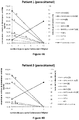
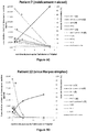
- the serum biomarker concentrations in particular ADH1B, ADH1A and ADH1B, ADH4 and BHMT1, BHMT1 and BHMT2 rise rapidly during the acute phase and drop to very low levels during liver regeneration ( figures 4 and 5 ).
- This serum kinetics is correlated with the evolution of the classic parameters of biological monitoring (coagulation parameters, transaminases and CK18) but the concentrations of the biomarkers ADH1B, ADH1B and ADH1A, ADH4, BHMT1, BHMT1 and BHMT2 recover earlier.
- biomarkers can be very useful for improving the patient's biological monitoring, evaluating the prognosis earlier (hepatic regeneration or worsening) and helping in the decision of liver transplantation.
- prognosis earlier (hepatic regeneration or worsening)
- serum biomarker concentrations may be helpful in the early identification of etiology, which is critical for the rapid administration of therapy and a prognosis favorable.
- biomarkers can also be used for the evaluation of the toxicity of chemical molecules (drug candidates), drugs (pharmacovigilance) or other xenobiotics on humans or on animal models, or on in vitro models (cultures of cell lines hepatic).
- drug candidates drugs
- drugs drugs
- in vitro models cultures of cell lines hepatic
- the assay can be performed on blood samples. In the case of in vitro models , this assay will be carried out from the culture medium.
- the invention is illustrated below by examples which however do not limit the invention. These examples relate to a method of in vitro diagnosis of liver damage from a blood sample and its use, in particular for detecting acute liver failure or non-alcoholic steatohepatitis in a patient.
- Amino acids from PSAQ TM proteins were quantified by high resolution mass spectrometry by comparison of the signals emitted by labeled amino acids from PSAQ TM protein and their unlabeled analogs from bovine serum albumin.
- ALI acute liver failure
- Serum samples from these patients were provided by the Biological Resource Center of the Paris-Sud Faculty of Medicine, France (approval number: 2011/39938 ).
- the French Blood Establishment (French Blood Establishment, The Tronche, France) provided seven anonymous serum samples from healthy donors. These serum samples were then collected in untreated tubes (BD Biosciences, Le Pont de Claix, France) and were centrifuged at 1000 g for 15 min to obtain the serum. Serum samples were aliquoted and immediately frozen at -80 ° C before further use. For each patient, several serum samples were taken on several days of sampling during hospitalization.
- Analyzes were performed on each patient to ensure that these patients were not suffering from viral hepatitis A, B and / or C. All patients were screened for viral hepatitis A, B and C ( HAV, HBV, HCV) and have been found negative for all of these viruses. Biological parameters of liver damage (INR, prothrombin time (PT), levels of transaminases, bilirubin, platelets and creatinine) were determined using standard methods. The levels of soluble CK18 protein and cleaved cytokeratin-18 protein (CK18Asp396) present in serum were measured using the ELISA kits M65 and M30Apoptosense (Peviva, Sweden).
- the PSAQ TM standard protein is injected directly into the serum sample before any pretreatment.
- This approach makes it possible to determine, in blood biological fluids such as plasma and serum, the concentrations of certain target proteins such as biomarkers of pathologies of which they constitute marked analogs.
- Several pretreatment modes such as the depletion of abundant proteins (by immunoaffinity or affinity with a chemical reagent) and the glycodepletion of the blood sample have been considered so as to preserve, during the process, the integrity of the PSAQ TM proteins.
- the proteins ARG1, ADH1B, BHMT1 and GSTA have been subject to the depletion of abundant proteins according to MARS technology from Agilent Technologies and the proteins ADH4, FABP1, ALT1 and BHMT1 to glycodepletion.
- PSAQ TM protein see table 2
- the PSAQ TM serum-protein solution was then left to incubate for 1 hour at 4 ° C. with gentle shaking.
- the sample comprising serum (or plasma) and the PSAQ TM proteins were deposited on an affinity column allowing the extraction of the six most abundant proteins (albumin, IgG, Transferrin, IgA, haptoglobin and antitrypsin) in the serum (MARS technology, spin cartridge from Agilent Technologies, Les Ulis, France) according to the manufacturer's instructions. This method extracts nearly 85 to 90% of all the proteins present in the serum.
- the serum samples thus free of albumin, IgG, transferrin, IgA, haptoglobin and antitrypsin were concentrated until at a volume of 50 ⁇ l on an ultrafiltration device having an exclusion limit of 3000 Da (Merck Millipore, Molsheim, France).
- a solution of 4 M urea and NH 4 HCO 3 at 50 mM was deposited on the sample in the ultrafiltration device so as to exchange the buffer.
- the resulting sparse protein concentrates were then subjected to an enzymatic digestion step.
- PSAQ TM protein Before any pretreatment, defined quantities of PSAQ TM protein (see table 2) were added to 18 ⁇ l of serum or plasma previously withdrawn from an individual, whether he is suffering from acute hepatic insufficiency or a healthy donor so as to be able to quantify the presence of this protein in the patient's serum. The serum-protein PSAQ TM solution was then left to incubate for 1 hour at 4 ° C. with gentle shaking.
- the sample comprising glycoproteins carrying aldehyde groups and non-glycosilated proteins was introduced onto an affinity column of the GlycoLink immobilization kit from Life technologies according to the manufacturer's instructions.
- the affinity column used comprises an UltraLink resin functionalized with hydrazide.
- the resin thus functionalized will make it possible to extract the glycoproteins present in the serum by producing stable bonds between the hydrazide of the resin and the aldehydes of the glycoproteins previously modified.
- the non-glycosylated proteins were then eluted by centrifugation in the presence of 400 ⁇ l of urea at a concentration of 8M.
- the sample was concentrated to 50 ⁇ L on an ultrafiltration device with an exclusion limit of 3000 Da and was then subjected to digestion.
- a digestion solution was prepared using a mixture of EndolysC and trypsin (Promega, Charbonippos-les-Bains, France) at a protein / enzyme ratio of 1/30 by mass in a solution of 4 M urea and NH 4 HCO 3 at 50 mM at 37 ° C.
- the samples were diluted four times and incubated overnight at 37 ° C. The digestion was stopped by adding formic acid (0.1% in final concentration). The samples were purified on a SpinColumns macro C18 column (Harvard Apparatus, Les Ulis, France) and dried by centrifugation under vacuum. The pellets were resolubilized in 15 ⁇ L of a solution containing 2% by volume of acetonitrile, formic acid at 0.1% by volume.
- LC-SRM analyzes were performed on a 6500 QTrap type mass spectrometer (AB Sciex, Les Ulis, France) equipped with a TurboV source and controlled by Analyst software (version 1.6.1, AB Sciex) in a range ranging from 400 to 1000 m / z.
- the instrument was coupled to an Ultima 3000 LC chromatography system (Thermo Scientific, Courtaboeuf, France). Sample chromatography was performed using a system combining 2 solvents, solvent A (2% acetonitrile and 0.1% formic acid) and solvent B (80% acetonitrile, 0.1% formic acid%).
- proteolytic peptides were concentrated on a 1 x 15 mm C18 PepMap precolumn (Thermo Scientific) and then were separated on a Kinetex XB-C18 column (2.1 x 100 mm, 1.7 ⁇ m, 100 ⁇ ; Phenomenex, Le Pecq , France) in gradient mode.
- Kinetex XB-C18 column 2.1 x 100 mm, 1.7 ⁇ m, 100 ⁇ ; Phenomenex, Le Pecq , France
- the graduated elution was carried out so as to modify the concentration of the initial solution of B to 3% to 35% in 30 minutes, then from 35% to 90% in 10 minutes, and this at a flow rate of 50 ⁇ l / min.
- the concentration of the biomarkers was then calculated from the average protein ratio
- the lower limit of detection (LID) was determined using the standard curve method swimming and the lower limit of quantification (LIQ) was defined as being 3 times the LID.
- LIQ lower limit of quantification
- the concentration of target biomarkers was determined at different times t.
- the Figures 4 and 5 set up the longitudinal monitoring of serum concentrations of the various biomarkers in patients with ALI or ALF and whose etiology is either paracetamol ( figure 4 ) or another cause ( figure 5 ). (cf. figures 4 and 5 ). This follow-up is analyzed in parallel with standard clinical and biological parameters (transaminase levels, coagulation parameters).
Landscapes
- Life Sciences & Earth Sciences (AREA)
- Health & Medical Sciences (AREA)
- Engineering & Computer Science (AREA)
- Molecular Biology (AREA)
- Immunology (AREA)
- Biomedical Technology (AREA)
- Chemical & Material Sciences (AREA)
- Hematology (AREA)
- Urology & Nephrology (AREA)
- Physics & Mathematics (AREA)
- Cell Biology (AREA)
- Pathology (AREA)
- Biotechnology (AREA)
- Food Science & Technology (AREA)
- Medicinal Chemistry (AREA)
- Microbiology (AREA)
- Analytical Chemistry (AREA)
- Biochemistry (AREA)
- General Health & Medical Sciences (AREA)
- General Physics & Mathematics (AREA)
- Proteomics, Peptides & Aminoacids (AREA)
- Bioinformatics & Cheminformatics (AREA)
- Bioinformatics & Computational Biology (AREA)
- Spectroscopy & Molecular Physics (AREA)
- Biophysics (AREA)
- Communicable Diseases (AREA)
- Investigating Or Analysing Biological Materials (AREA)
Description
- L'invention appartient au domaine de l'hépatologie (médecine du foie) et concerne l'utilisation de biomarqueurs permettant de diagnostiquer des troubles hépatiques. Plus précisément, cette invention concerne des méthodes de diagnostic in vitro et/ou de suivi et/ou de pronostic in vitro des atteintes hépatiques telles que les insuffisances hépatiques aiguës et fulminantes ou la stéatohépatite par la détection et/ou le dosage de certains biomarqueurs.
- Les atteintes hépatiques regroupent une multitude de maladies affectant le foie de manière chronique (comme la cirrhose) ou aigüe (on parle alors d'insuffisance hépatique aigüe). Leurs causes sont variées et incluent l'alcoolisme, les virus des hépatites A, B, C et E, des anomalies génétiques (maladie de Wilson) ou immunitaires (hépatites auto-immunes), des intoxications médicamenteuses ou une surcharge graisseuse associée à l'obésité.
- Les insuffisances hépatiques aiguës (« Acute Liver Injury», abrégé ici ALI) ou fulminantes (« Acute Liver Failure », abrégé ici ALF) se déclarent de manière soudaine chez des patients sans maladie hépatique préexistante. Leurs causes sont variées : médicaments (dont le paracétamol), virus, maladie auto-immune. Les insuffisances hépatiques aiguës se caractérisent par la mort des cellules hépatiques (hépatocytes) sur un mode nécrotique et/ou apoptotique et conduisent à une diminution de la masse fonctionnelle du foie. Ces atteintes hépatiques sont associées à des troubles de la coagulation sanguine et la modification des paramètres d'exploration clinico-biologique du foie (augmentation des transaminases et de la CK18 sérique). Dans leur forme la plus sévère (hépatite fulminante ou ALF), une encéphalopathie est également présente. Les atteintes hépatiques aiguës peuvent conduire à la mort du patient sans prise en charge médicale adaptée.
- Les maladies du foie gras non induites par l'alcool (connues sous le sigle NAFLD qui est l'abréviation de « Non-Alcoholic Fatty Liver Disease ») sont des maladies hépatiques très fréquentes. Elles englobent un large éventail de maladies telles que la stéatose isolée (connue sous le sigle NAFL - « Non-Alcoholic Fat Liver »), la stéatohépatite non alcoolique (NASH - « Non-Alcoholic SteatoHepatitis ») et la cirrhose. Elles sont associées à une accumulation anormale de graisse dans le foie. Elles sont liées à l'augmentation de la prévalence du diabète et de l'obésité. La stéatohépatite non alcoolique (NASH) est une maladie potentiellement grave pouvant évoluer en cirrhose et entrainer une insuffisance hépatique chronique ou aigüe et/ou un carcinome hépatocellulaire.
- Les atteintes hépatiques peuvent être diagnostiquées par la détection ou le dosage dans le sang de certaines molécules spécifiques (biomarqueurs). La cytolyse (nécrose des hépatocytes) est évaluée par la détection de protéines, contenues dans les cellules hépatiques, et qui sont déversées dans les voies sanguines suite à la nécrose des cellules hépatiques (biomarqueurs mécanistiques) : tel est le cas des transaminases qui reflètent une lyse des cellules du foie. Les transaminases ne sont cependant pas spécifiques du foie et leurs concentrations sanguines peuvent augmenter lors d'atteinte d'autres organes (cœur, muscle). Il peut également s'agir de molécules synthétisées par le foie, dont la fonction est impactée par la diminution de leur synthèse (biomarqueurs fonctionnels): c'est le cas du taux de prothrombine qui explore les facteurs de coagulation I, II, V, VII et X synthétisés par le foie. Cependant, du fait de la demi-vie sanguine de quelques heures des facteurs de coagulation, les troubles de la coagulation ne sont pas immédiats.
- En ce qui concerne la stéatose hépatique non alcoolique et alcoolique, le diagnostic de certitude est habituellement réalisé sur une biopsie du foie du patient; ainsi on peut évaluer les lésions tissulaires hépatiques et déterminer la sévérité de la maladie. Par exemple, les transcrits des gènes ADH1 et ADH2 sont augmentés en RT-PCR dans un échantillon de biopsie de foie d'individus atteints de NAFLD ou de NASH et la protéine ADH1B est détectable par immunohistochimie dans un échantillon de biopsie de foie d'individus atteints de NAFLD (An Afida Ashla et al., Hepatology Research, 2010, 40, 594-604). Une biopsie est un examen invasif, associé à des risques hémorragiques, et nécessite une prise en charge hospitalière; c'est donc un examen coûteux. Il serait préférable de pouvoir détecter les troubles hépatiques et suivre leur évolution par des méthodes non invasives, de préférence à partir d'un liquide biologique, par exemple provenant d'une simple prise de sang.
- Ces dernières années, un certain nombre de méthodes non invasives d'évaluation de lésions hépatiques ont été proposées, notamment pour détecter la stéatohépatite alcoolique et non alcoolique. A titre d'exemple,
US 2006 / 0 172 286 décrit un procédé non invasif permettant le diagnostic de la stéatohépatite non alcoolique ou alcoolique chez un patient, comprenant la mesure des 3 marqueurs biochimiques ApoAI, ALT et AST dans un échantillon de sérum ou de plasma.WO 2010 / 058 295 décrit un procédé permettant de quantifier les lésions du foie d'un patient par l'intermédiaire du dosage d'un ou plusieurs biomarqueur (dont ALT). Zhiyuan Hu et al. (Theranostics, 2014, 4, 215-228) et Beger et al. (Arch. Toxicol., 2015, 89, 1497-1552) décrivent des biomarqueurs des lésions hépatiques aiguës induites par le paracétamol incluant le marqueur conventionnel transaminase (ALT1), ainsi que d'autres marqueurs tels que BHMT, notamment BHMT1.WO 2015 / 071 669 décrit une méthode de diagnostic, de pronostic ou de surveillance d'une tumeur du foie chez un individu par l'intermédiaire de la détection ou la quantification d'un ou plusieurs marqueurs présents notamment dans le sang ; la liste des marqueurs est extrêmement vaste et comprend notamment les protéines ADH1A, ADH1B, GSTA1, FABP1, ARG1, GPT1 (correspondant à l'ALT1), BHMT et ADH4. - En tout état de cause, la quantification absolue, c'est-à-dire la détermination de la concentration de protéines ou peptides présents dans un échantillon liquide, notamment un échantillon sanguin peut être un procédé assez complexe.
- On constate que diagnostiquer des atteintes hépatiques à un stade précoce et prédire l'évolution de ces atteintes est devenu un enjeu de santé majeur, en particulier pour la stéatohépatite non alcoolique dont la prévalence augmente avec l'obésité.
- La présente invention cherche à proposer un procédé de de diagnostic in vitro d'atteintes hépatiques, plus particulièrement un nouveau test de diagnostic non invasif de troubles hépatiques plus précis que les procédés de de diagnostic de l'art antérieur.
- La présente invention est telle que définie dans les revendications. La présente invention concerne un procédé de diagnostic et/ou de suivi et/ou de pronostic et/ou théranostique in vitro de troubles hépatiques choisis parmi l'insuffisance hépatique aigüe, la stéatose hépatique et la stéatohépatite, à partir d'un échantillon sanguin issu d'un individu, dans lequel procédé on détermine la présence et/ou la concentration du marqueur ADH1B (SEQ ID N° 2) et/ou la présence et/ou la concentration de la combinaison des marqueurs ADH1B (SEQ ID N° 2) et ADH1A (SEQ ID N° 1).
- Dans un mode de réalisation du procédé ci-dessus, on détermine la présence et/ou la concentration d'au moins un marqueur supplémentaire, préférentiellement d'au moins deux marqueurs supplémentaires, et encore plus préférentiellement d'au moins trois marqueurs supplémentaires, lesdits marqueurs étant sélectionnés dans le groupe constitué par : ADH4 (SEQ ID N° 3 & SEQ ID N° 4), BHMT (SEQ ID N° 5, SEQ ID N° 6 & SEQ ID N° 7), ARG1 (SEQ ID N° 8, SEQ ID N° 9 & SEQ ID N° 10), FABP1 (SEQ ID N° 11), GSTA1 (SEQ ID N° 12) et ALT1 (SEQ ID N° 13).
- Dans un mode de réalisation du procédé ci-dessus, on détermine au moins la présence et/ou la concentration d'au moins un marqueur supplémentaire sélectionné parmi ADH4 (SEQ ID N° 3 & SEQ ID N° 4) et BHMT (SEQ ID N° 5, SEQ ID N° 6 & SEQ ID N° 7).
- Dans un mode de réalisation du procédé ci-dessus, on détermine au moins la présence et/ou la concentration du marqueur ADH1B (SEQ ID N° 2) ou de la combinaison des marqueurs ADH1B (SEQ ID N° 2) et ADH1A (SEQ ID N° 1), d'une part, et des marqueurs ADH4 (SEQ ID N° 3 & SEQ ID N° 4) et BHMT (SEQ ID N° 5, SEQ ID N° 6 & SEQ ID N° 7), d'autre part.
- Dans un mode de réalisation avantageux du procédé ci-dessus, le marqueur BHMT est le variant de séquence BHMT1 (SEQ ID N° 5) et/ou la combinaison des variants de séquence BHMT1 (SEQ ID N° 5) et BHMT2 (SEQ ID N° 6 & SEQ ID N° 7).
- Dans un mode de réalisation du procédé décrit ci-dessus, on détermine en outre la présence et/ou la concentration du marqueur CK18.
- Dans un mode de réalisation du procédé décrit ci-dessus, on utilise pour ladite détermination de la présence et/ou de la concentration desdits marqueurs des peptides signatures présents dans lesdits marqueurs.
- Dans un mode de réalisation avantageux du procédé décrit ci-dessus, lesdits peptides signatures sont générés à partir desdits marqueurs par un procédé de digestion, de préférence enzymatique.
- Dans un mode de réalisation du procédé décrit ci-dessus, la présence et/ou la concentration :
- a. de la combinaison des marqueurs ADH1A (SEQ ID N° 1) et ADH1B (SEQ ID N° 2), si elle est sélectionnée, est déterminée par la présence d'au moins un peptide signature choisi parmi INEGFDLLHSGK (SEQ ID N° 16) et FSLDALITHVLPFEK (SEQ ID N° 15),
- b. du marqueur ADH1B (SEQ ID N° 2), s'il est sélectionné, est déterminée par la présence du peptide signature AAVLWEVK (SEQ ID N° 17),
- c. du marqueur ADH4 (SEQ ID N° 3 & SEQ ID N° 4), s'il est sélectionné, est déterminée par la présence d'au moins un peptide signature choisi parmi FNLDALVTHTLPFDK (SEQ ID N° 20), IDDDANLER (SEQ ID N° 18) ou IIGIDINSEK (SEQ ID N° 19),
- d. du marqueur ARG1 (SEQ ID N° 8, SEQ ID N° 9 & SEQ ID N° 10), s'il est sélectionné, est déterminée par la présence d'au moins un peptide signature choisi parmi DVDPGEHYILK (SEQ ID N° 25), TIGIIGAPFSK (SEQ ID N° 24) ou EGLYITEEIYK (SEQ ID N° 26),
- e. du marqueur BHMT (SEQ ID N° 5, SEQ ID N° 6 & SEQ ID N° 7), s'il est sélectionné, est déterminée par la présence d'au moins un peptide signature choisi parmi EATTEQQLK (SEQ ID N° 21), AIAEELAPER (SEQ ID N° 23) ou EAYNLGVR (SEQ ID N° 22),
- f. du marqueur BHMT1 (SEQ ID N° 5), s'il est sélectionné, est déterminée par la présence du peptide signature EATTEQQLK (SEQ ID N° 21),
- g. de la combinaison des marqueurs BHMT1 (SEQ ID N°5) et BHMT2 (SEQ ID N° 6 & SEQ ID N° 7), si elle est sélectionnée, est déterminée par la présence d'au moins un peptide signature choisi parmi AIAEELAPER (SEQ ID N° 23) ou EAYNLGVR (SEQ ID N° 22),
- h. du marqueur FABP1 (SEQ ID N° 11), s'il est sélectionné, est déterminée par la présence d'au moins un peptide signature choisi parmi AIGLPEELIQK (SEQ ID N° 27), FTITAGSK (SEQ ID N° 28) ou TVVQLEGDNK (SEQ ID N° 29),
- i. du marqueur GSTA1 (SEQ ID N° 12), s'il est sélectionné, est déterminée par la présence du peptide signature LHYFNAR (SEQ ID N°30),
- j. du marqueur ALT1 (SEQ ID N° 13), s'il est sélectionné, est déterminée par la présence d'au moins un peptide signature choisi parmi ALELEQELR (SEQ ID N° 34), LLVAGEGHTR (SEQ ID N° 35) ou KPFTEVIR (SEQ ID N° 36), sachant que ALT1 (SEQ ID N° 13), s'il est sélectionné, est plus particulièrement déterminée par la présence du peptide signature ALELEQELR (SEQ ID N° 34) ou LLVAGEGHTR (SEQ ID N° 35).
- Dans un mode de réalisation avantageux du procédé décrit ci-dessus, le procédé dans lequel on dose au moins un desdits marqueurs (dit « marqueur sélectionné »), dont au moins un marqueur ADH1B (SEQ ID N° 2) et/ou la combinaison des marqueurs ADH1B (SEQ ID N° 2) et ADH1A (SEQ ID N° 1), à partir d'un échantillon sanguin prélevé sur un individu à un temps t0, par l'intermédiaire d'au moins un peptide signature caractéristique dudit marqueur, comprend les étapes suivantes :
- a) l'ajout d'une quantité connue d'une protéine isotopiquement marquée (appelée « standard ») homologue du marqueur sélectionné dans l'échantillon sanguin
- b) le traitement de l'échantillon sanguin pour extraire au moins une partie des protéines abondantes ou au moins une partie des glycoprotéines,
- c) le traitement de l'échantillon après extraction d'au moins une partie des protéines abondantes ou des glycoprotéines pour générer des peptides protéolytiques, de préférence par digestion, étant entendu que parmi les peptides protéolytiques obtenus se trouvent les peptides signatures des marqueurs sélectionnés,
- d) le dosage quantitatif par spectrométrie de masse d'au moins un peptide signature généré à partir du marqueur sélectionné,
- e) la détermination d'un rapport de l'abondance du peptide signature isotopiquement marqué, issu du standard, ajouté à l'étape a), sur l'abondance du peptide signature non marqué issu de l'échantillonsanguin,
- f) le calcul à partir du rapport obtenu à l'étape e) et de la quantité connue de ladite protéine isotopiquement marquée ajoutée à l'étape a), de la concentration dudit marqueur sélectionné dans l'échantillon.
- Dans un mode de réalisation avantageux, l'étape b) du procédé ou de l'utilisation décrite ci-dessus, est effectuée par déplétion et préférentiellement par glycodéplétion.
- Dans un mode de réalisation avantageux, l'étape b) du procédé ou de l'utilisation décrite ci-dessus, est suivie d'une étape d'électrophorèse de l'échantillon sanguin.
- Dans un mode de réalisation avantageux, le procédé est effectué à un temps t0 et à un temps t1 supérieur à t0, et comprend après l'étape f), la comparaison de la concentration du marqueur sélectionné dans l'échantillon entre t1 et t0.
- Dans un mode de réalisation avantageux, l'étape c) du procédé décrit ci-dessus est effectuée par digestion enzymatique, de préférence par l'emploi de trypsine ou d'un mélange d'EndolysC et de trypsine.
- Le trouble hépatique est une insuffisance hépatique aiguë, une stéatose hépatique ou une stéatohépatite, de préférence une insuffisance hépatique aiguë ou une stéatohépatite non-alcoolique. L'hépatite aigüe est notamment une hépatite virale, une hépatite alcoolique, une hépatite médicamenteuse, une hépatite liée à une intoxication, par exemple aux champignons ou aux vitamines, une hépatite auto-immune ou la maladie de Wilson ; de préférence il s'agit d'une hépatite non-alcoolique, telle que notamment une hépatite médicamenteuse, par exemple due au paracétamol ou un autre médicament. La stéatose hépatique est en particulier une stéatose non-alcoolique, telle qu'une stéatose isolée (NAFL pour « Non-Alcoholic Fat Liver »). La stéatohépatite est en particulier une stéatohépatite non-alcoolique (NASH pour « Non-Alcoholic SteatoHepatitis »).Un autre objet de l'invention concerne une utilisation du procédé pour évaluer la toxicité de molécules (candidats médicaments, médicament, xénobiotique) préalablement administrées à l'individu dont est issu ledit échantillon sanguin.
- Les
figures 1 à 8 illustrent certains aspects de l'invention. - La
figure 1 décrit le principe de la glycodépletion de protéines plasmatiques pour le dosage de biomarqueurs non glycosylés (comme les protéines de lyse hépatocytaire).
Le protéome plasmatique est composé (en poids) de 50% des protéines glycosylées et de 50% de protéines non glycosylées. Les protéines glycosylées peuvent être capturées sur des billes magnétiques fonctionnalisées par de l'hydrazide via la formation de liaisons covalentes. Les protéines non glycosylées sont ensuite éluées avec de l'urée permettant de casser les interactions protéines / protéines. - La
figure 2 présente les courbes d'étalonnage obtenues pour ADH1B, ARG1, BHMT1 et GSTA1 dans le plasma après immunodéplétion. Ces courbes d'étalonnage permettent d'évaluer les performances analytiques de la méthode de dosage. - La
figure 3 présente les courbes d'étalonnage obtenues pour ADH4, BHMT1, FABP1 et ALT1 dans le plasma après glycodéplétion. - Les
figures 4 (A à G) décrivent l'analyse des biomarqueurs dans des échantillons sanguins issus de patients ayant une insuffisance hépatique induite par du paracétamol. Les concentrations des biomarqueurs cibles ont été déterminées en utilisant les standards PSAQ™ après préfractionnement de sérum par LC-SRM. Le taux de prothrombine est mentionné comme un indicateur de troubles de la coagulation et d'une insuffisance hépatique ALI ou ALF. - Les
figures 5 (A à F) décrivent l'analyse des biomarqueurs dans des échantillons sanguinss issus de patients ayant une atteinte hépatique ALI ou ALF induite par une cause autre que le paracétamol. Les concentrations des biomarqueurs cibles ont été déterminées en utilisant les standards PSAQ™ (WO 2008 / 145 763 A1 ) après préfractionnement de sérum et analyse LC-SRM. Le taux de prothrombine est mentionné comme un indicateur de troubles de la coagulation et d'une insuffisance hépatique ALI/ALF. - La
figure 6 décrit la méthode de standardisation, préparation et analyse des échantillons. Le panel A correspond à la procédure de standardisation et de préparation. Les panels B et C correspondent aux signaux des différents peptides signatures obtenus par analyse LC-SRM. - La
figure 7 montre les concentrations plasmatiques de BHMT1 (cf.Figure 7a ) et les concentrations plasmatiques des variants A et B de l'ADH1 (cf.Figure 7b ) chez des patients atteints de stéatohépatites non alcooliques (NASH) en comparaison de donneurs sains appariés par âge et sexe (n = 11 individus dans chaque groupe). Les concentrations plasmatiques sont mentionnées en ordonnée en µg/mL. Un test statistique de Mann-Whitney a été appliqué avec p < 0.05 comme seuil de significativité. - La
figure 8 présente les séquences peptidiques des protéines ADH1A (SEQ ID N° 1), ADH1B (SEQ ID N° 2), ADH4 (SEQ ID N° 3 & SEQ ID N° 4), BHMT1 (SEQ ID N°5), BHMT2 (SEQ ID N° 6 & SEQ ID N° 7), ARG1 (SEQ ID N° 8, SEQ ID N° 9 & SEQ ID N° 10), FABP1 (SEQ ID N° 11), GSTA1 (SEQ ID N° 12) et ALT1 (SEQ ID N° 13). Les peptides analysés de ces protéines cibles sont visualisés dans la séquence peptidique des protéines par un encadrement. Ces peptides sont des peptides issus de la digestion des protéines cibles et servent de substituts pour l'analyse de ces protéines en spectrométrie de masse. - Dans la présente invention, on entend par « atteintes hépatiques » ou « troubles hépatiques » ou « lésions hépatiques », les hépatites virales, les hépatites alcooliques, les hépatites médicamenteuses, les hépatites liées à des intoxications tels que celles dues aux champignons ou aux vitamines, les hépatites auto-immunes, la stéatose hépatique (NAFLD), les stéatohépatites (NASH), les cirrhoses et la maladie de Wilson à l'exclusion des hépatocarcinomes.
- Dans le cadre de la présente invention, les protéines peuvent être dosées par n'importe quelle technique classiquement utilisée par l'homme de l'art comme des méthodes de dosages immunologiques utilisant des anticorps spécifiques ou des méthodes basées sur la spectrométrie de masse. Parmi les méthodes de dosage basées sur la spectrométrie de masse, on peut citer la technologie AQUA (Gerber et al, « Absolute quantification of proteins and phosphoproteins from cell lysates by tandem MS » Proc Natl Acad Sci U S A. (2003) 100(12):6940-6945.) ou la technologie PSAQ™ (Protein Standard Absolute Quantification) décrite dans la demande
WO 2008 / 145 763 . - La méthode selon l'invention est basée sur la détection et le dosage de certains biomarqueurs issus de la lyse des cellules hépatiques ; ces biomarqueurs sont des protéines. Selon un mode de réalisation préféré de l'invention, le dosage se fait par spectrométrie de masse via la technologie PSAQ™. Selon cette méthode, la quantification absolue d'une protéine par spectrométrie de masse nécessite un étalon. Dans la présente invention, on entend par protéine PSAQ™ ou standard PSAQ™ ou protéine étalon toute protéine entière recombinante marquée isotopiquement, chimiquement analogue à la protéine native que l'on souhaite doser. Le principe de la méthode PSAQ™ selon l'invention repose sur l'introduction dans un échantillon sanguin d'une quantité connue d'une protéine-étalon entière recombinante marquée isotopiquement, identique ou analogue (i.e, structurellement proche, elle peut différer par exemple par l'addition d'une étiquette de purification) à la protéine native que l'on souhaite doser. Contrairement aux autres méthodes de quantification absolue, une quantité connue de la protéine-étalon est injectée directement dans l'échantillon sanguin avant tout prétraitement de cet échantillon. La méthode selon l'invention permet de déterminer, dans un échantillon sanguin tel que le plasma et le sérum, les concentrations de certaines protéines cibles dont ils constituent des analogues marqués.
- Le procédé selon l'invention est avantageusement effectué sur un échantillon biologique issu d'un prélèvement de sang sur un individu.
- Le procédé selon l'invention peut notamment être effectué sur le sang, le plasma et/ou le sérum.
- De nombreuses protéines contribuent aux fonctions métaboliques du foie. Selon l'invention les protéines permettant de caractériser des lésions du foie sont choisies en fonction de leur expression hépatique prédominante et de leur forte détectabilité dans le plasma, à savoir l'ADH1A et l'ADH1B, l'ADH4, l'ARG1, la BHMT (variants de séquence 1 et 2), la FABP1, la GSTA1 et l'ALT1.
- Les alcools déshydrogénases (ADH) sont des oxydoréductases qui participent à la détoxication de l'organisme par l'élimination des alcools toxiques. Plusieurs types d'ADH existent (variants ou isoformes). Concernant l'ADH1, il en existe 3 variants : l'ADH1A, l'ADH1B et l'ADH1C. Ces variants ont été étudiés au niveau génétique. Duell et al. ont mis en évidence l'influence de certaines mutations génétiques de l'ADH, notamment sur la sensibilité à l'alcoolisme et les risques de cancer gastrique des individus porteurs de ces mutations génétiques ( « Genetic variation in alcohol dehydrogenase (ADH1A, ADH1B, ADH1C, ADH7) and aldehyde dehydrogenase (ALDH2), alcohol consumption and gastric cancer risk in the european prospective investigation into cancer and nutrition (EPIc) cohort », Carcinogenesis vol.33, n°2, p361-367 (2012)).
- L'ADH1A (SEQ ID N° 1) et l'ADH1B (SEQ ID N° 2) participent à l'oxydation de l'éthanol et jouent un rôle majeur dans le catabolisme de l'éthanol. L'ADH4 participe à l'oxydation des alcools aliphatiques et/ou des alcools aromatiques. L'ADH4 existe sous forme de 2 isoformes (SEQ ID N° 3 & SEQ ID N° 4). Dans l'étude, les inventeurs se sont intéressés à l'ADH1B mais aussi au mélange des variants ADH1A et ADH1B.
- La BHMT (Betaine-homocystéine-methyltransférase) est une métallo-enzyme à base de zinc qui participe au métabolisme de l'homocystéine, de la glycine, de la sérine, de la thréonine et aussi de la méthionine. Par ce terme BHMT on entend les variants de séquences BHMT1 (SEQ ID N° 5), BHMT2, ainsi que les isoformes de BHMT2 (SEQ ID N° 6 & SEQ ID N° 7).
- L'arginase ARG1 est une hydrolase ; cette métalloenzyme hépatique à base de manganèse intervient dans le cycle de l'urée. Par ce terme ARG1 on entend les trois isoformes de l'arginase 1 (SEQ ID N° 8, SEQ ID N° 9 & SEQ ID N° 10).
- La FABP1 (« Fatty acid-binding protein, Liver » ; SEQ ID N° 11) est une protéine de liaison aux acides gras spécifiques du foie servant à transporter les acides gras des membranes cellulaires vers les mitochondries.
- Les glutathione S-transférases (GSTA) sont des enzymes qui catalysent des reactions de conjugaison du glutathion réduit (GSH) avec des xénobiotiques afin de les détoxifier. Dans cette étude, les inventeurs ont étudié le variant GSTA1 (SEQ ID N° 12) mais aussi le pool de plusieurs variants en particulier le pool des variants GSTA1, GSTA2, GSTA3 et le pool des 4 variants GSTA1, GSTA2, GSTA3 et GSTA5.
- L'alanine cytosolique aminotransaminase 1 ou ALT1 (SEQ ID N° 13), également connue sous le nom de glutamate-pyruvate transaminase 1, est une enzyme faisant partie des transaminases jouant un rôle essentiel dans le métabolisme intermédiaire du glucose. Cette protéine est connue en tant que biomarqueur de lésion hépatique provoquée par certains médicaments, de l'alcool, et par la stéatose.
- Le tableau 1 résume certaines propriétés de ces protéines.
- Les protéines ADH1A et ADH1B, ADH4, ARG1, BHMT (variants 1 et 2), FABP1, GSTA1 et ALT1 sont principalement exprimées dans le foie. Chez un individu sain, ces protéines sont principalement présentes dans le foie (hépatocytes) et dans quelques autres types cellulaires différents. C'est le cas, par exemple, de la protéine ADH1B qui est détectable dans uniquement trois types cellulaires différents parmi les 80 analysés (cf. tableau 1, atlas des protéines humaines). A titre comparatif et selon l'atlas des protéines humaines, dans un tissu sain (www.proteinatlas.org), l'ALT1 qui est un marqueur usuel de lésions hépatiques est exprimé dans 29 types cellulaires parmi les 78 analysés. Les protéines ADH1A et ADH1B, ADH4, ARG1, BHMT (variants 1 et 2), FABP1, GSTA1 sont donc bien plus spécifiques du foie que l'ALT1.
Tableau 1 : Biomarqueurs sélectionnés dans le procédé selon l'invention pour le suivi d'atteintes hépatiques Protéine (SEQ ID N°) Identifiant Uniprot Nombre de types cellulaires dans un tissu sain dans lequel la protéine est détectée (a) Masse moléculaire [Da] Fonction biochimique ADH1B (SEQ ID N° 2) P00325 3 sur 80 analysés 39 855 Métabolisme des xénobiotiques ADH1A (SEQ ID N° 1) P07327 Hepatocytes 39 859 Métabolisme des xénobiotiques ADH4 (SEQ ID N° 3 & SEQ ID N° 4) P08319 6 sur 83 analysés 40 222 Métabolisme des xénobiotiques ARG1 (SEQ ID N° 8, SEQ ID N° 9 & SEQ ID N° 10) P05089 2 sur 83 analysés 34 735 Métabolisme de l'azote et cycle de l'urée BHMT1 (SEQ ID N° 5) Q93088 3 sur 83 analysés 44 998 Métabolisme de l'homocystéine FABP1 (SEQ ID N° 11) P07148 7 sur 83 analysés 14 208 Métabolisme des lipides et transport des lipides GSTA1 (SEQ ID N° 12) P08263 16 sur 81 analysés 25 631 Métabolisme des xénobiotiques et de la glutathione (a) Source: The Human Protein Atlas (www.proteinatlas.org). - Selon l'invention, d'autres biomarqueurs tels que la protéine cytokeratine 18 (CK18) peuvent être employés en complément d'analyse afin de confirmer un trouble hépatique et d'établir un diagnostic. En effet, lorsque les hépatocytes sont exposés de façon chronique au stress oxydatif et à des substances toxiques, ils gonflent, accumulent de la graisse et montrent une perturbation dans le réseau de filaments de kératine, et forment des corps de Mallory, i.e. des amas résiduels de microfilaments. Un corps de Mallory est composé de kératines anormalement phosphorylées et réticulées, telles que la cytokératine CK18. On sait que les hépatocytes contenant des corps de Mallory sont sensibles à l'apoptose, induisant une libération importante des protéines CK18 et des corps de Mallory dans le sang. La publication de Maher et al. « Cytokeratin 18 as a non invasive marker in diagnosis of NASH and its usefulness in correlation with disease severity in Egyptian patients » publiée dans The Egyptian Journal of Medical Human Genetics (2015) 16, p.41 à 46, présente la CK18 comme un marqueur non-invasif permettant de corréler sa concentration au degré de sévérité de la NASH. Effectivement, lorsque la protéine CK18 est libérée dans le sang sous forme entière (forme M65), elle indique un phénomène de nécrose. Quand elle est libérée sous forme tronquée (forme M30) elle indique un phénomène d'apoptose. Généralement les concentrations sanguines de CK18 sont mesurées sous ces 2 formes et le ratio entre ces deux formes est calculé. La CK18 n'est pas exprimée de manière spécifique dans les hépatocytes, elle est présente dans les épithéliums glandulaires.
- Dans le sérum ou plasma de patients sains, les protéines ADH1 (variant B ou isoformes A et B) et ADH4 sont présentes à des concentrations inférieures à 2 µg/mL, l'ARG1 à des concentrations inférieures à 0.3 µg/mL, la FABP1 à des concentrations inférieures à 1 µg/mL, la GSTA1, GSTA2, GSTA2 et GSTA5 à des concentrations inférieures à 1 µg/mL, la BHMT (variants ou isoformes 1 et 2) à des concentrations inférieures à 2 µg/mL et l'ALT1 à des concentrations inférieures à 1 µg/mL (ces concentrations seuil ont été évaluées après analyse d'échantillons selon le protocole décrit dans la
figure 6 ). - La Demanderesse s'est rendu compte que la concentration sérique ou plasmatique des protéines ADH1B, ADH1A et B, ADH4, ARG1, BHMT1 et BHMT1 et 2, FABP1, GSTA1 et ALT1 évolue en fonction de l'atteinte hépatique et de sa sévérité.
- Selon les observations de la Demanderesse, la concentration des protéines ADH1B, ADH1A et ADH1B, ADH4, ARG1, FABP1, GSTA1, BHMT1 et BHMT (variants 1 et 2) augmente au cours des phases aiguës de l'insuffisance hépatique induite par des xénobiotiques tels que le paracétamol, puis chute à des niveaux très faibles au cours de la période de récupération / régénération du foie. Cette cinétique est étroitement corrélée avec celle des caractéristiques biologiques de cette maladie telles que le taux de prothrombine. Certaines de ces protéines sont plus exprimées au niveau hépatique que d'autres et sont donc plus facilement quantifiables ; c'est le cas des protéines ADH4, BHMT (variants 1 et 2), ADH1 (variants ADH1A et ADH1B) qui peuvent, être avantageusement employées, de manière individuelle ou combinée, comme biomarqueurs de diagnostic, de suivi et de pronostic de l'ALI ou l'ALF. La protéine ARG1 bien que détectée présente des modifications de sa concentration plasmatique ou sérique moins marquées mais corrélées avec le stade de l'atteinte (phase aigüe/phase de récupération).
- Les marqueurs ADH1 (ADH1B seul ou combinaison d'ADH1B et ADH1A), ADH4 et BHMT (variant 1 seul ou combinaison des 2 variants 1 et 2) peuvent permettre une identification précoce de lésions hépatiques induites par des médicaments tels que le paracétamol, de manière à adapter la prise en charge médicale du patient (en particulier le recours à la greffe hépatique). De plus, contrairement aux marqueurs transaminases (dont ALT1) et CK18 dont les concentrations peuvent rester élevées, ces marqueurs ADH1 (ADH1B seul ou combinaison d'ADH1B et ADH1A), ADH4 et BHMT (variant 1 seul ou combinaison des 2 variants 1 et 2) présentent des taux sériques faibles au cours de la régénération spontanée du foie.
- Pour ce qui est des troubles hépatiques non induits par des médicaments une modification des concentrations plasmatiques/sériques des biomarqueurs, notamment des protéines ADH1 (ADH1B et/ou ADH1B et ADH1A), ADH4 et BHMT (variant 1 seul ou combinaison des 2 variants 1 et 2) est observée à des niveaux plus faibles qu'avec le paracétamol mais elle est cohérente avec le statut clinique du patient.
- Selon un mode de réalisation de l'invention, les marqueurs ADH1 (ADH1B et/ou (ADH1B et ADH1A)), ADH4, ARG1, FABP1, GSTA1, ALT1 et BHMT (variant 1 et/ou le mélange des deux variants 1 et 2) peuvent être employés pour la détection d'une lésion hépatique.
- La détection d'une lésion hépatique peut être mise en évidence par la détection et/ou la quantification du marqueur ADH1, de préférence le marqueur ADH1B et/ou le pool d'ADH1A et ADH1B.
- La détection d'une lésion hépatique peut être mise en évidence par la détection et/ou la quantification du marqueur ADH1 ((ADH1B et/ou (ADH1B et ADH1A)) et d'au moins un marqueur supplémentaire, d'au moins deux marqueurs supplémentaires, d'au moins trois marqueurs supplémentaires, d'au moins quatre marqueurs supplémentaires, d'au moins cinq marqueurs supplémentaires ou d'au moins six marqueurs supplémentaires choisis parmi : ADH4, BHMT (variant 1 et/ou combinaison des deux variants 1 et 2), ARG1, FABP1, GSTA1 et ALT1.
- La détection d'une lésion hépatique peut être mise en évidence par la détection et/ou la quantification de deux biomarqueurs spécifiquement, tels que ADH1 ((ADH1B et/ou (ADH1B et ADH1A)) et ADH4 ; ADH1 (ADH1B et/ou (ADH1B et ADH1A)) et BHMT (variant 1 et/ou un mélange des deux variants 1 et 2) ; ADH1 (ADH1B et/ou (ADH1B et ADH1A)) et ARG1 ; ADH1 (ADH1B et/ou (ADH1B et ADH1A)) et FABP1 ; ADH1 (ADH1B et/ou (ADH1B et ADH1A)) et GSTA1 ou des 2 biomarqueurs ADH1 (ADH1B et/ou (ADH1B et ADH1A)) et ALT1.
- La détection d'une lésion hépatique peut être mise en évidence par la détection et/ou la quantification de trois biomarqueurs spécifiquement, tels que ADH1 (ADH1B et/ou (ADH1B et ADH1A)), ADH4 et BHMT (variant 1 et/ou un mélange des deux variants 1 et 2); ADH1 (ADH1B et/ou (ADH1B et ADH1A)), ADH4 et ARG1 ; ADH1 (ADH1B et/ou (ADH1B et ADH1A)), ADH4 et FABP1 ; ADH1 (ADH1B et/ou (ADH1B et ADH1A)), ADH4 et GSTA1 ; ADH1 (ADH1B et/ou (ADH1B et ADH1A)), ADH4 et ALT1 ; ADH1 (ADH1B et/ou (ADH1B et ADH1A)), BHMT (variant 1 et/ou un mélange des deux variants 1 et 2) et ARG1 ; ADH1 (ADH1B et/ou (ADH1B et ADH1A)), BHMT (variant 1 et/ou un mélange des deux variants 1 et 2) et FABP1 ; ADH1 (ADH1B et/ou (ADH1B et ADH1A)), BHMT (variant 1 et/ou un mélange des deux variants 1 et 2) et GSTA1 ; ADH1 (ADH1B et/ou (ADH1B et ADH1A)), BHMT (variant 1 et/ou un mélange des deux variants 1 et 2) et ALT1 ; ADH1 (ADH1B et/ou (ADH1B et ADH1A)), ARG1 et FABP1 ; ADH1 (ADH1B et/ou (ADH1B et ADH1A)), ARG1 et GSTA1 ; ADH1 (ADH1B et/ou (ADH1B et ADH1A)), ARG1 et ALT1 ; ADH1 (ADH1B et/ou (ADH1B et ADH1A)), FABP1 et GSTA1 ; ADH1 (ADH1B et/ou (ADH1B et ADH1A)), FABP1 et ALT1 ou des 3 biomarqueurs ADH1 (ADH1B et/ou (ADH1B et ADH1A)), GSTA1 et ALT1.
- La détection d'une lésion hépatique peut être mise en évidence par la détection et/ou la quantification de quatre biomarqueurs spécifiquement, tels que ADH1 (ADH1B et/ou (ADH1B et ADH1A)), ADH4, BHMT (variant 1 et/ou un mélange des deux variants 1 et 2) et ARG1 ; ADH1 (ADH1B et/ou (ADH1B et ADH1A)), ADH4, BHMT (variant 1 et/ou un mélange des deux variants 1 et 2) et FABP1 ; ADH1 (ADH1B et/ou (ADH1B et ADH1A)), ADH4, BHMT (variant 1 et/ou un mélange des deux variants 1 et 2) et GSTA1 ; ADH1 (ADH1B et/ou (ADH1B et ADH1A)), ADH4, BHMT (variant 1 et/ou un mélange des deux variants 1 et 2) et ALT1 ; ADH1 (ADH1B et/ou (ADH1B et ADH1A)), ADH4, ARG1 et FABP1 ; ADH1 (ADH1B et/ou (ADH1B et ADH1A)), ADH4, ARG1 et GSTA1 ; ADH1 (ADH1B et/ou (ADH1B et ADH1A)), ADH4, ARG1 et ALT1 ; ADH1 (ADH1B et/ou (ADH1B et ADH1A)), ADH4, FABP1 et GSTA1 ; ADH1 (ADH1B et/ou (ADH1B et ADH1A)), ADH4, FABP1 et ALT1 ; ADH1 (ADH1B et/ou (ADH1B et ADH1A)), ADH4, GSTA1 et ALT1 ; ADH1 (ADH1B et/ou (ADH1B et ADH1A)), BHMT (variant 1 et/ou un mélange des deux variants 1 et 2), ARG1 et FABP1 ; ADH1 (ADH1B et/ou (ADH1B et ADH1A)), BHMT (variant 1 et/ou un mélange des deux variants 1 et 2), ARG1 et GSTA1 ; ADH1 (ADH1B et/ou (ADH1B et ADH1A)), BHMT (variant 1 et/ou un mélange des deux variants 1 et 2), ARG1 et ALT1 ; ADH1 (ADH1B et/ou (ADH1B et ADH1A)), BHMT (variant 1 et/ou un mélange des deux variants 1 et 2), FABP1 et GSTA1 ; ADH1 (ADH1B et/ou (ADH1B et ADH1A)), BHMT (variant 1 et/ou un mélange des deux variants 1 et 2), FABP1 et ALT1 ; ADH1 (ADH1B et/ou (ADH1B et ADH1A)), BHMT (variant 1 et/ou un mélange des deux variants 1 et 2), GSTA1 et ALT1 ; ADH1 (ADH1B et/ou (ADH1B et ADH1A)), ARG1, FABP1 et GSTA1 ; ADH1 (ADH1B et/ou (ADH1B et ADH1A)), ARG1, FABP1 et ALT1 ; ADH1 (ADH1B et/ou (ADH1B et ADH1A)), ARG1, GSTA1 et ALT1 ou des quatre marqueurs ADH1 (ADH1B et/ou (ADH1B et ADH1A)), FABP1, GSTA1 et ALT1.
- La détection d'une lésion hépatique peut être mise en évidence par la détection et/ou la quantification de cinq biomarqueurs spécifiquement, tels que ADH1 (ADH1B et/ou (ADH1B et ADH1A)), ADH4, BHMT (variant 1 et/ou un mélange des deux variants 1 et 2), ARG1 et FABP1 ; ADH1 (ADH1B et/ou (ADH1B et ADH1A)), ADH4, BHMT (variant 1 et/ou un mélange des deux variants 1 et 2), ARG1 et GSTA1 ; ADH1 (ADH1B et/ou (ADH1B et ADH1A)), ADH4, BHMT (variant 1 et/ou un mélange des deux variants 1 et 2), ARG1 et ALT1 ; ADH1 (ADH1B et/ou (ADH1B et ADH1A)), ADH4, BHMT (variant 1 et/ou un mélange des deux variants 1 et 2), FABP1 et GSTA1 ; ADH1 (ADH1B et/ou (ADH1B et ADH1A)), ADH4, BHMT (variant 1 et/ou un mélange des deux variants 1 et 2), FABP1 et ALT1 ; ADH1 (ADH1B et/ou (ADH1B et ADH1A)), ADH4, BHMT (variant 1 et/ou un mélange des deux variants 1 et 2), GSTA1 et ALT1 ; ADH1 (ADH1B et/ou (ADH1B et ADH1A)), ADH4, ARG1, FABP1 et GSTA1 ; ADH1 (ADH1B et/ou (ADH1B et ADH1A)), ADH4, ARG1, FABP1 et ALT1 ; ADH1 (ADH1B et/ou (ADH1B et ADH1A)), ADH4, ARG1, GSTA1 et ALT1 ; ADH1 (ADH1B et/ou (ADH1B et ADH1A)), ADH4, FABP1, GSTA1 et ALT1 ; ADH1 (ADH1B et/ou (ADH1B et ADH1A)), BHMT (variant 1 et/ou un mélange des deux variants 1 et 2), ARG1, FABP1 et GSTA1 ; ADH1 (ADH1B et/ou (ADH1B et ADH1A)), BHMT (variant 1 et/ou un mélange des deux variants 1 et 2), ARG1, FABP1 et ALT1 ; ADH1 (ADH1B et/ou (ADH1B et ADH1A)), BHMT (variant 1 et/ou un mélange des deux variants 1 et 2), ARG1, GSTA1 et ALT1 ; ADH1 (ADH1B et/ou (ADH1B et ADH1A)), BHMT (variant 1 et/ou un mélange des deux variants 1 et 2), FABP1, GSTA1 et ALT1 ou des cinq biomarqueurs ADH1 (ADH1B et/ou (ADH1B et ADH1A)), ARG1, FABP1, GSTA1 et ALT1.
- La détection d'une lésion hépatique peut être mise en évidence par la détection et/ou la quantification de six biomarqueurs spécifiquement, tels que ADH1 (ADH1B et/ou (ADH1B et ADH1A)), ADH4, BHMT (variant 1 et/ou un mélange des deux variants 1 et 2), ARG1, FABP1 et GSTA1 ; ADH1 (ADH1B et/ou (ADH1B et ADH1A)), ADH4, BHMT (variant 1 et/ou un mélange des deux variants 1 et 2), ARG1, FABP1 et ALT1 ; ADH1 (ADH1B et/ou (ADH1B et ADH1A)), ADH4, BHMT (variant 1 et/ou un mélange des deux variants 1 et 2), ARG1, GSTA1 et ALT1 ; ADH1 (ADH1B et/ou (ADH1B et ADH1A)), ADH4, BHMT (variant 1 et/ou un mélange des deux variants 1 et 2), FABP1, GSTA1 et ALT1 ; ADH1 (ADH1B et/ou (ADH1B et ADH1A)), ADH4, ARG1, FABP1, GSTA1 et ALT1 ou des six biomarqueurs ADH1 (ADH1B et/ou (ADH1B et ADH1A)), BHMT (variant 1 et/ou un mélange des deux variants 1 et 2), ARG1, FABP1, GSTA1 et ALT1 ;
- La détection d'une lésion hépatique peut être mise en évidence par la détection et/ou la quantification de sept biomarqueurs spécifiquement à savoir ADH1 (ADH1B et/ou (ADH1B et ADH1A)), ADH4, BHMT (variant 1 et/ou un mélange des deux variants 1 et 2), ARG1, FABP1, GSTA1 et ALT1.
- Dans un mode de réalisation particulier, en plus du marqueur ADH1 (ADH1B et/ou (ADH1B et ADH1A)), le marqueur ADH4 et/ou BHMT sera préférentiellement employé ; en cas de lésion hépatique, la concentration plasmatique/sérique de l'ADH4 et celle de la BHMT est élevée et plus facilement quantifiable.
- Dans un mode de réalisation préféré, la détection d'une lésion hépatique est mise en évidence par la détection et/ou la quantification des trois biomarqueurs ADH1 ((ADH1B et/ou (ADH1B et ADH1A)), ADH4 et BHMT (variant 1 et/ou un mélange des deux variants 1 et 2).
- Dans un mode préféré, les lésions hépatiques peuvent être détectées par l'analyse de trois biomarqueurs parmi les protéines : ADH1 (ADH1B et/ou ADH1B et ADH1A), ADH4, ARG1, FABP1, GSTA1, ALT1 et BHMT (variant 1 et/ou combinaison des deux variants 1 et 2).
- Avantageusement, plus la détection d'une lésion hépatique fait intervenir de biomarqueurs, plus l'analyse du trouble hépatique sera améliorée.
- La détection spécifique et la quantification absolue de biomarqueurs protéiques dans les fluides biologiques peut être réalisée de différentes manières. En laboratoire d'analyses médicales, les biomarqueurs sont couramment détectés et quantifiés par des procédés immunologiques. Cela suppose la disponibilité d'un anticorps se liant de manière spécifique à la protéine à doser. La spectrométrie de masse permet de détecter et de doser des molécules, et plus particulièrement des peptides, directement et sans disposer d'un anticorps. Ainsi ont été développées au cours de la dernière décennie des techniques de spectrométrie de masse, notamment des méthodes connues sous les signes SRM (« Selected Reaction Monitoring ») et MRM (« Multiple Reaction Monitoring ») permettant de quantifier une ou plusieurs molécules cibles dans un échantillon biologique complexe.
- Cependant, le dosage absolu (c'est-à-dire la détermination de la concentration des protéines cibles) par spectrométrie de masse nécessite des standards de quantification. Il est possible d'utiliser différents types de standards de quantification qui peuvent correspondre à des peptides isotopiquement marqués, des concaténères de peptides marquées, des fragments de protéines isotopiquement marquées ou des protéines entières isotopiquement marquées (voir les publications Brun et al, « Isotope dilution strategies for absolute quantitative proteomics. » Journal of Proteomics (2009) 72(5) pages 740-749 et Zeiler et al, "A Protein Epitope Signature Tag (PrEST) library allows SILAC-based absolute quantification and multiplexed determination of protein copy numbers in cell lines." Molecular and Cellular Proteomics (2012) 11(3):O111.009613). Dans tous les cas, les standards génèrent ou correspondent aux peptides signatures qui sont des peptides issus de la digestion des protéines cibles et qui servent de substituts pour l'analyse de ces protéines en spectrométrie de masse (l'obtention et l'analyse des peptides signatures sont décrites plus loin). Parmi ces approches de quantification, et comme évoqué dans le document
WO 2008 / 145 763 A1 , l'utilisation conjointe de la spectrométrie de masse en mode SRM ou MRM et de protéines étalons, analogues isotopiquement marqués des biomarqueurs cibles (standards PSAQ™) et introduits en quantité connue dans un échantillon, permet de détecter spécifiquement et de quantifier avec exactitude et précision, dans cet échantillon, les biomarqueurs ciblés. - On entend par « protéine isotopiquement marquée homologue du marqueur visé ou biomarqueur cible » ou « protéine étalon », toute protéine possédant la même séquence en acides aminés que la protéine native que l'on souhaite doser et dont certains atomes sont remplacés par des atomes marqués avec des isotopes stables. Avantageusement, certains acides aminés particuliers peuvent être marqués isotopiquement, tels que l'arginine et la lysine. Dans ce cas, les résidus arginines, et lysines des protéines étalons sont uniformément marquées avec des isotopes du carbone et de l'azote, i.e. du 13C et du 15N. Selon l'invention, avantageusement, le standard est introduit dans l'échantillon sanguin de manière à ce que sa concentration finale dans l'échantillon sanguin soit comprise entre 0,5 et 5 µg/mL, pour les pathologies hépatiques aiguës de type ALI ou ALF. Dans le cas des stéatohépatites non alcooliques ou alcooliques, le standard est introduit à une concentration comprise entre 20 et 200 ng/mL. En fonction de la nature de la protéine ciblée, des concentrations optimales pour la calibration ont été déterminées pour chacune des protéines étalons et présentées ci-dessous dans le tableau 2.
Tableau 2 : Concentration optimale (µg/mL) de la protéine étalon dans l'échantillon sanguin par type de protéine étalon (cas des atteintes hépatiques aiguës ALI/ALF) Protéine étalon (standard PSAQ™) Concentration optimale (µg/mL) de la protéine étalon dans l'échantillon sanguin ARG1 1 BHMT1 2 GSTA1 1 ADH1B 3 ADH4 2 FABP1 1 ALT1 3 - Dans un mode de réalisation préféré de l'invention, on détecte les biomarqueurs non pas directement mais par l'intermédiaire de sous-unités peptidiques, appelés « peptides signatures ». On entend ici par « peptide signature », tout peptide ayant une séquence d'acides aminés comprenant entre 5 et 30 acides aminés, qui est incluse dans la séquence du marqueur à analyser, permettant d'identifier spécifiquement dans un échantillon sanguin la présence dudit marqueur. Avantageusement, ces peptides signatures sont générés par une digestion enzymatique ou chimique de l'échantillon ou d'une fraction de celui-ci, comme cela sera expliqué plus en détail ci-dessous. Plus particulièrement, les biomarqueurs choisis sont identifiés par la détection de peptides protéolytiques obtenus après digestion des protéines contenues dans l'échantillon sanguin, i.e. peptides signatures permettant la quantification des marqueurs dans l'échantillon sanguin, comme évoqué dans le document
WO 2008 / 145 763 A1 . Ces peptides signatures permettent de suivre ces biomarqueurs et en particulier leur isoformes prédominantes dans le foie. En effet, certains peptides signatures (cf.figure 8 ) sont partagés entre plusieurs isoformes, tels que l'ADH1 où le peptide signature INEGFDLLHSGK permet de suivre à la fois l'ADH1B et l'ADH1A, tels que la BHMT avec les isoformes BHMT1 et BHMT2 ou la GSTA avec les isoformes GSTA1, GSTA2, GSTA3 ou GSTA5 (cf. tableau 3,figure 8 ).Tableau 3 : Peptides signatures des biomarqueurs utilisés dans le procédé selon l'invention et générés par une digestion avec de la trypsine ou un mélange de trypsine et endolysC Biomarqueur d'intérêt Peptide signature du marqueur d'intérêt Identification des séquences peptidiques (SEQ ID N°) Spécificité du peptide par rapport au biomarqueur ADH1B AAVLWEVK SEQ ID N° 17 spécifique ADH1B INEGFDLLHSGK SEQ ID N° 16 peptide présent dans ADH1A et ADH1B ADH1B FSLDALITHVLPFEK SEQ ID N° 15 peptide présent dans ADH1A et ADH1B ADH1B IDAASPLEK SEQ ID N° 14 peptide présent dans ADH1A, ADH1B, ADH1C et ADH1G ADH4 FNLDALVTHTLPFDK SEQ ID N° 20 spécifique ADH4 IDDDANLER SEQ ID N° 18 spécifique ADH4 IIGIDINSEK SEQ ID N° 19 spécifique ARG1 DVDPGEHYILK SEQ ID N° 25 spécifique ARG1 TIGIIGAPFSK SEQ ID N° 24 spécifique ARG1 EGLYITEEIYK SEQ ID N° 26 spécifique BHMT1 EATTEQQLK SEQ ID N° 21 spécifique BHMT1 AIAEELAPER SEQ ID N° 23 peptide présent dans BHMT1 et BHMT2 BHMT1 EAYNLGVR SEQ ID N° 22 peptide présent dans BHMT1 et BHMT2 FABP1 AIGLPEELIQK SEQ ID N° 27 spécifique FABP1 FTITAGSK SEQ ID N° 28 spécifique FABP1 TVVQLEGDNK SEQ ID N° 29 spécifique GSTA1 LHYFNAR SEQ ID N° 30 spécifique GSTA1 SHGQDYLVGNK SEQ ID N° 31 peptide présent dans GSTA1, GSTA2 et GSTA3 GSTA1 ISNLPTVK SEQ ID N° 33 peptide présent dans GSTA1, GSTA2, GSTA3 et GSTA5 GSTA1 AILNYIASK SEQ ID N° 32 peptide présent dans GSTA1, GSTA2, GSTA3 et GSTA5 ALT1 ALELEQELR SEQ ID N° 34 spécifique ALT1 LLVAGEGHTR SEQ ID N° 35 spécifique ALT1 KPFTEVIR SEQ ID N° 36 peptide présent dans ALT1 et ALT2 - Après introduction dans un échantillon sanguin d'une quantité connue de protéine étalon PSAQ™, identique ou structurellement très proche de la protéine native que l'on souhaite doser, une étape de digestion, enzymatique ou chimique, peut être réalisée de manière à obtenir les peptides protéolytiques des protéines initialement présentes dans l'échantillon dont les peptides signatures des biomarqueurs cibles facilement identifiables et quantifiables par LC-MS en mode SRM ou MRM. Il est également possible d'utiliser, à la place des standards PSAQ, des standards peptidiques isotopiquement marqués (appelés peptides AQUA pour "Absolute quantification" ou SIL pour "Stable Isotope Labelling". Ces peptides étalons marqués ont une séquence identique aux peptides signatures suivis et ils sont ajoutés dans l'échantillon juste avant ou juste après l'étape de digestion des protéines. Ces standards permettent une quantification absolue des protéines cibles mais qui est souvent moins exacte que la quantification par la méthode PSAQ.
- Avantageusement, la digestion des protéines est effectuée de manière enzymatique, et plus particulièrement en présence d'une solution de trypsine ou bien un mélange de trypsine et d'EndolysC. La quantité d'enzyme utilisée pour la digestion correspond généralement à un ratio (en poids) enzyme/protéines de 1/100 à 1/10. Après addition de la solution d'enzyme dans l'échantillon sanguin et incubation à 37°C, de manière à générer les peptides protéolytiques, la digestion est stoppée par addition d'au moins une quantité suffisante d'acide formique dans l'échantillon sanguin. On entend par « peptide protéolytique » tout peptide ou séquence d'acides aminés issu(e) de la dégradation d'un polypeptide, notamment par digestion enzymatique ou chimique dudit polypeptide.
- Les peptides protéolytiques (dont les peptides signatures et les peptides isotopiquement marqués) ainsi obtenus sont ensuite analysés par LC-MS. A noter qu'il est possible d'utiliser différents types d'enzymes pour effectuer la digestion des protéines. En fonction de la spécificité de clivage de l'enzyme choisie, les peptides signatures permettant de détecter et quantifier les protéines cibles seront différents. Ces peptides signatures peuvent être prédits en effectuant une digestion in silico des protéines cibles (dont on connait la séquence en acides aminés). Leur détectabilité en spectrométrie de masse est ensuite vérifiée expérimentalement. Le tableau 3 répertorie des peptides signatures obtenus et suivis après une digestion avec de la trypsine ou un mélange de trypsine et d'endolysC. L'endolysC est une endoprotéinase Lys-C.
- Afin de faciliter l'isolation et l'identification des protéines présentes dans les échantillons sanguins, une étape de prétraitement de l'échantillon sanguin peut avantageusement être réalisée. En effet, cette étape permet d'éliminer au moins en partie des protéines abondantes non ciblées présentes dans l'échantillon sanguin et ainsi d'accroitre la sensibilité de détection des biomarqueurs cibles tout en conservant l'intégrité du standard PSAQ™ (protéine étalon) et du biomarqueur cible.
- De manière à conserver, au cours du procédé, l'intégrité des biomarqueurs cibles et des standards PSAQ™, plusieurs modes de prétraitement peuvent être utilisés, tels que la déplétion des protéines abondantes par l'utilisation d'un dispositif de chromatographie d'affinité (immunodéplétion), un réactif chimique ayant une affinité pour l'albumine (type cibacron blue) ou un procédé de glycodéplétion de l'échantillon sanguin (cf.
figure 1 ). Ces méthodes de prétraitement sont connues en tant que telles. - La déplétion des protéines abondantes est une méthode d'affinité permettant d'extraire au moins en partie les protéines majoritaires du sérum, i.e. les protéines abondantes telles que l'albumine, l'IgG, la transferrine ou encore l'IgA.
- La glycodéplétion est une méthode permettant de capturer et d'extraire au moins en partie les glycoprotéines présentes dans l'échantillon sanguin de manière à laisser dans l'échantillon sanguin les autres protéines telles que les biomarqueurs cibles qui ne sont pas glycosilés. Dans le procédé qui fait l'objet du schéma selon la
figure 1 , la capture des glycoprotéines est réalisée par des billes magnétiques fonctionnalisés par de l'hydrazide. - Afin d'éviter d'effectuer une détection sensible et une quantification exacte des biomarqueurs cibles on choisit un mode de prétraitement de l'échantillon adapté à chaque biomarqueur. Ainsi, les protéines ARG1, ADH1 (variant A et B), BHMT (variants 1 et 2) et GSTA1 seront préférentiellement traités par déplétion des protéines abondantes par chromatographie d'affinité et les protéines ADH4, FABP1, ALT1 et BHMT par glycodéplétion.
- Après traitement de l'échantillon sanguin, une étape additionnelle de traitement de l'échantillon sanguin peut être effectuée par électrophorèse. Cette étape permet d'affiner l'extraction des protéines d'intérêt. Cette étape d'électrophorèse peut être effectuée sur gel d'acrylamide. Dans ce cas, la digestion enzymatique est également réalisée en gel.
- Une étape additionnelle d'enrichissement des peptides cibles par immunoaffinité avant toute analyse par LC-MS en mode SRM ou MRM peut avantageusement être réalisée. Cela permet de faciliter la détection et la quantification de ces peptides par spectrométrie de masse.
- Les peptides protéolytiques sont ensuite analysés par LC-MS en mode SRM. Des courbes d'étalonnage ont été réalisées pour chaque biomarqueur de manière à évaluer les performances analytiques du dosage et garantir la fiabilité de la quantification des biomarqueurs cibles dans l'échantillon sanguin. Les courbes d'étalonnages ont été réalisées en employant des échantillons contenant une quantité croissante de protéine non marquée et une quantité constante de protéine standard PSAQ™, ces échantillons ayant subis les mêmes traitements que l'échantillon sanguin dont on souhaite détecter et quantifier un biomarqueur cible, à savoir des prétraitements et/ou une digestion. Des blancs contenant uniquement les protéines endogènes sont aussi réalisés. Tous les points de calibration sont effectués plusieurs fois par souci de reproductibilité (cf.
figures 2 et3 ). - Les résultats de quantification obtenus pour les différents peptides signatures de chaque biomarqueur cible sont cohérents et montrent que le procédé selon l'invention permet de détecter et de quantifier avec précision des biomarqueurs cibles. Les résultats de quantification sont présentés ci-après dans le tableau 4 pour chaque biomarqueur et pour chaque peptide signature obtenu après immunodéplétion ou glycodéplétion. Le tableau 4 présente la limite inférieure de détection (LID) des différentes protéines déterminée par les courbes d'étalonnage précédemment décrites et la limite inférieure de quantification (LIQ) définie comme étant égale à 3 fois la LID.
- Les analyses LC-SRM ont été réalisées sur un spectromètre de masse de type QTRAP dans une gamme allant de 400 à 1000 m/z. Avant toute analyse, les peptides protéolytiques peuvent être concentrés sur une précolonne et séparées sur colonne de chromatographie liquide en mode gradient de manière à faciliter l'analyse ultérieure
- L'identification des pics de chaque peptide a été réalisée en utilisant le logiciel Skyline, connus des spécialistes de la spectrométrie de masse. En plus de l'évaluation du signal du peptide (signal composite), toutes les transitions sont inspectées individuellement et exclues si elles sont jugés inaptes pour la quantification (faible rapport signal sur bruit, interférences évidentes). Les ratios des surfaces des pics relatifs aux peptides marqués et non marqués sont calculés pour chaque transition SRM. Ces ratios sont ensuite utilisés pour déterminer le ratio moyen correspondant, et finalement le rapport moyen de l'abondance de la protéine cible est calculé à partir des rapports obtenus pour les différents peptides signatures. La concentration des biomarqueurs a ensuite été calculée à partir du ratio moyen obtenu pour chacune des protéines. La limite inférieure de détection (LID) a été déterminée selon la méthode de la courbe d'étalonnage et la limite inférieure de quantification (LIQ) est définie comme étant égale à 3 fois la LID. L'. Pour chaque patient, la concentration des biomarqueurs cibles est déterminée à différents instants t pendant leur hospitalisation.
Tableau 4 : Performance analytique du test pour chaque biomarqueur ciblé (limite de détection). Protéine Peptide signature Méthode biochimique Gamme de concentrations testées [µg/ml] Précision [%] LID [µg/ml] LIQ [µg/ml] Précision à LIQ (CV en %) ADH1B INEGFDLLHSGK MARS 0,81-73,5 79 10.66 31.99 41 IDAASPLEK 95 0.72 2.17 22-26 
97 0.77 2.30 ND AAVLWEVK 112 2.01 6.03 3-13 ARG1 DVDPGEHYILK MARS 0,04-1,50 146 0.31 0.94 9 TIGIIGAPFSK 118 0.30 0.90 4-8 EGLYITEEIYK 138 0.31 0.92 6-15 BHMT1 AIAEELAPER MARS 1,50-145,8 114 1.38 4.15 12-19 EAYNLGVR 88 3.49 10.48 15-40 EATTEQQLK 83 2.04 6.11 6-15 GSTA1 ISNLPTVK MARS 0,09-3,43 121 0.30 0.91 6-10 LHYFNAR 132 0.32 0.97 10-14 AILNYIASK 124 0.29 0.87 7-10 SHGQDYLVGNK 125 0.41 1.24 9-11 ADH4 
Glycodéplétion 0,25-24,8 139 1.59 4.77 9-16 IDDDANLER 148 1.75 5.25 8-10 IIGIDINSEK 136 1.60 4.81 0-8 BHMT1 AIAEELAPER Glycodéplétion 0,60-79,5 143 6.53 19.58 8 EAYNLGVR 151 4.15 12.45 NA EATTEQQLK 136 6.17 18.52 2-6 FABP1 AIGLPEELIQK Glycodéplétion 0,18-3,69 110 0.71 2.13 2-13 FTITAGSK 114 0.64 1.91 5-14 TVVQLEGDNK 108 0.66 1.99 1-8 ALT1 ALELEQELR Glycodéplétion 0,40-8,10 96 1.21 3.62 6-14 LLVAGEGHTR 92 1.10 3.29 15 KPFTEVIR 117 2.17 6.52 9-10 - Chez les patients atteints d'hépatites aiguës (ALI) ou fulminantes (ALF), les concentrations sériques des biomarqueurs (en particulier ADH1B, ADH1A et ADH1B, ADH4 et BHMT1, BHMT1 et BHMT2) s'élèvent rapidement pendant la phase aigüe et chutent à des niveaux très bas pendant la régénération hépatique (
figures 4 et5 ). Cette cinétique sérique est corrélée à l'évolution des paramètres classiques de suivi biologique (paramètres de coagulation, transaminases et CK18) mais les concentrations des biomarqueurs ADH1B, ADH1B et ADH1A, ADH4, BHMT1, BHMT1 et BHMT2 se rétablissent plus précocement. Ces résultats suggèrent que ces biomarqueurs peuvent être très utiles pour améliorer le suivi biologique du patient, évaluer plus précocement le pronostic (régénération hépatique ou aggravation) et aider à la décision de transplantation hépatique. Dans le cas des hépatites aiguës ou fulminantes induites par le paracétamol, les rapides et fortes augmentations des concentrations sériques des biomarqueurs peuvent être utiles à l'identification précoce de l'étiologie, ce qui est déterminant pour l'administration rapide du traitement et un pronostic favorable. - Ces biomarqueurs peuvent être également utilisés pour l'évaluation de la toxicité de molécules chimiques (candidats médicaments), de médicaments (pharmacovigilance) ou autres xénobiotiques sur l'homme ou sur des modèles animaux, ou sur des modèles in vitro (cultures de lignées cellulaires hépatiques). Chez l'homme et les animaux, le dosage peut être effectué sur des échantillon sanguins. Dans le cas des modèles in vitro, ce dosage sera effectué à partir du milieu de culture.
- L'invention est illustrée ci-dessous par des exemples qui cependant ne limitent pas l'invention. Ces exemples portent sur une méthode de diagnostic in vitro d'atteintes hépatiques à partir d'un échantillon sanguin et son utilisation, notamment pour déceler une insuffisance hépatique aiguë ou une stéatohépatite non alcoolique chez un patient.
- Des protéines recombinantes non marquées ont été achetées chez Abcam. Des protéines marquées isotopiquement (standards PSAQ™) ont été synthétisées en utilisant : soit un système d'expression de protéines acellulaire en présence de [13C6, 15N2] L-lysine et [13C6, 15N4] L-arginine (Eurisotop, Saint Aubin, France), comme décrit dans la publication de Lebert et al., « Production and use of stable isotope-labeled proteins for absolute quantitative proteomics » parue dans Methods in Molecular Biology vol.753, p.93-115 (2011), soit un système d'expression correspondant à une souche d'Escherichia coli auxotrophe pour la lysine et l'arginine comme décrit dans l'article de Picard et al., « PSAQ™ standards for accurate MS-based quantification ofproteins: from the concept to biomedical applications » J Mass Spectrom. 2012;47(10), p.1353-1363.
- La pureté des protéines étalon a été vérifiée (> 95% de pureté) et quantifiée par analyse d'acides aminés comme décrit dans la publication de Louwagie et al. « Introducing AAA-MS, a rapid and sensitive method for amino acid analysis using isotope dilution and high-resolution mass spectrometry » parue dans Journal of Proteome Research vol.11, p3929-3936 (2012). Pour ce faire, les protéines PSAQ™ ont été hydrolysées en voie acide en présence d'une quantité connue d'une protéine de référence l'albumine de sérum bovin fournie par l'Institut National des Standards et de la Technologie (Etats-Unis). Les acides aminés issus des protéines PSAQ™ ont été quantifiés par spectrométrie de masse haute résolution par comparaison des signaux émis par les acides aminés marqués issus de la protéine PSAQ™ et leurs analogues non marqués issus de l'albumine de sérum bovin. L'incorporation des acides aminés isotopiquement marqués, tel que déterminée par LC-MS et LC-SRM (chromatographie en phase liquide couplée à la spectrométrie de masse en mode de suivi de réaction sélectionnée), était supérieure à 99%.
- Treize patients porteurs d'une insuffisance hépatique aiguë (ALI) ont été recrutés au département d'hépatologie de l'Hôpital Paul Brousse (Villejuif, France). L'insuffisance hépatique aiguë de ces patients a été décelée par une activité élevée de transaminase reflétant la lyse active des cellules du foie présente dans le sang de ces patients, des troubles de la coagulation avec un INR (International Normalized Ratio) > 1.5 et par l'absence chez ces patients de maladie hépatique préexistante.
- Parmi ces patients, quatre ont présenté une encéphalopathie hépatique (grade >=1) en moins de 26 semaines après l'apparition des symptômes et ont été classés comme patients ALF (cf. tableau 5). Des échantillons de sérum de ces patients ont été fournis par le Centre de Ressources Biologiques de la Faculté Paris-Sud de Médecine, France (numéro d'agrément:
2011/39938 Tableau 5 : Caractéristiques cliniques et biologiques des patients atteints d'ALI (Insuffisance hépatique aiguë) ou d'ALF Numéro du patient Collecte des échantillons à x j après l'hospitalisation Aetiologie Sexe Age (années) activité ALT (U/L) PI* (%) INR** ALI or ALF*** Résultat CK18 (U/L) CK18 Asp396 (U/L) 1 0 Paracétamol F 20 1489 9 16 ALI Vivant 13800 3333 10 402 59 1.42 1048 944 2 0 Paracétamol F 29 634 19 4.8 ALF Vivant N/A N/A 2 4565 17 5.45 N/A N/A 3 8118 18 4.73 N/A N/A 3 0 Paracétamol F 24 6410 21 3.82 ALI Vivant 22243 5691 3 2295 66 1.31 1150 404 4 0 Paracétamol F 18 2653 31 2.6 ALI Vivant 19806 7212 2 1900 57 1.45 1618 479 5 0 Paracétamol M 25 173 41 1.99 ALI Vivant N/A N/A 2 2571 26 3.21 N/A N/A 6 683 75 1.2 N/A N/A 6 0 Paracétamol F 23 770 39 2.06 ALI Vivant 5138 1379 1 7165 18 4.8 23088 8938 5 1831 89 1.07 956 529 7 0 Médicament + alcool M 24 7300 13 7.47 ALI Vivant 12138 5350 7 481 100 0.89 631 338 8 0 Paracétamol F 16 15480 20 4.17 ALI Vivant 21363 533 4 NA 68 1.3 600 254 9 0 Paracétamol + alcool M 57 2710 16 5.53 ALF Mort (sepsis) 23025 15288 2 NA 42 1.97 9956 1117 7 NA 51 1.65 2000 421 10 0 maladie auto-immune F 58 2224 17 5.76 ALF Vivant 15638 9933 2 1861 11 10.69 15013 7275 5 NA 66 1.3 1275 450 11 0 maladie auto-immune M 56 2170 31 2.52 ALI Vivant 23056 15117 2 1119 16 5.59 6400 2417 12 NA 85 1.11 2256 617 12 0 Virus Herpès simplex F 22 1383 14 6.52 ALF Vivant 11669 4863 5 153 40 2.07 11388 4029 15 133 56 1.1 1088 229 13 0 Médicament F 32 892 28 2.8 ALI Vivant 14781 7458 2 984 54 1.58 3463 938 3 589 80 1.15 1856 375 - La protéine-étalon PSAQ™ est injectée directement dans l'échantillon de sérum avant tout prétraitement. Cette approche permet de déterminer, dans des fluides biologiques sanguins tels que le plasma et le sérum, les concentrations de certaines protéines cibles tels que des biomarqueurs de pathologies dont ils constituent des analogues marqués. Plusieurs modes de prétraitement tels que la déplétion des protéines abondantes (par immunoaffinité ou affinité avec un réactif chimique) et la glycodéplétion de l'échantillon sanguin ont été envisagés de manière à conserver, au cours du procédé, l'intégrité des protéines PSAQ™. Ainsi, les protéines ARG1, ADH1B, BHMT1 et GSTA ont été sujettes à la déplétion des protéines abondantes selon la technologie MARS d'Agilent Technologies et les protéines ADH4, FABP1, ALT1 et BHMT1 à la glycodéplétion.
- Avant tout prétraitement, des quantités définies de protéine PSAQ™ (voir tableau 2) ont été ajoutées à 14 µl de sérum ou de plasma précédemment prélevés sur un individu, qu'il soit porteur d'une insuffisance hépatique aiguë ou donneur sain de manière à pouvoir quantifier la présence des protéine dans le sérum de l'individu. La solution sérum - protéines PSAQ™ a ensuite été laissée incubée pendant 1 heure à 4 °C sous agitation douce.
- L'échantillon comprenant du sérum (ou du plasma) et les protéines PSAQ™, ont été déposés sur une colonne d'affinité permettant d'extraire les six protéines les plus abondantes (Albumine, IgG, Transferrine, IgA, haptoglobine et antitrypsine) dans le sérum (technologie MARS, cartouche spin d'Agilent Technologies, Les Ulis, France) selon les instructions du fabricant. Cette méthode permet d'extraire près de 85 à 90% de l'ensemble des protéines présentes dans le sérum.
- Suite au passage du sérum (ou plasma) sur la colonne d'affinité MARS, les échantillons de sérum ainsi exempts d'albumine, d'IgG, de transferrine, d'IgA, d'haptoglobine et de antitrypsine se sont retrouvés concentrés jusqu'à un volume de 50 µl sur un dispositif d'ultrafiltration possédant une limite d'exclusion de 3000 Da (Merck Millipore, Molsheim, France). Une solution d'urée 4 M et de NH4HCO3 à 50 mM a été déposée sur l'échantillon dans le dispositif d'ultrafiltration de manière à échanger le tampon.
- Les concentrés de protéines faiblement abondantes résultants ont ensuite été soumis à une étape de digestion enzymatique
- Avant tout prétraitement, des quantités définies de protéine PSAQ™ (voir tableau 2) ont été ajoutées à 18 µl de sérum ou de plasma précédemment prélevés sur un individu, qu'il soit porteur d'une insuffisance hépatique aiguë ou donneur sain de manière à pouvoir quantifier la présence de cette protéine dans le sérum du patient. La solution sérum - protéine PSAQ™ a ensuite été laissée incuber pendant 1 heure à 4 °C sous agitation douce.
- L'échantillon de sérum ou de plasma précédemment prélevé sur un patient, qu'il soit porteur d'une insuffisance hépatique aiguë ou donneur sain, et additionné de standards PSAQ, a été mélangé avec 11 µl d'une solution de meta-periodate de sodium à 0.1 M afin d'oxyder les glycoprotéines et de transformer les groupements cis-diol des sucres en aldéhydes. L'échantillon comportant des glycoprotéines porteuses de groupements aldéhydes et des protéines non glycosilées a été introduit sur une colonne d'affinité du kit d'immobilisation GlycoLink de Life technologies selon les instructions du fabricant. La colonne d'affinité employée comprend une résine UltraLink fonctionnalisée par de l'hydrazide. La résine ainsi fonctionnalisée va permettre d'extraire les glycoprotéines présentes dans le sérum par la réalisation de liaisons stables entre l'hydrazide de la résine et les aldéhydes des glycoprotéines précédemment modifiées. Les protéines non glycosylées ont ensuite été éluées par centrifugation en présence de 400 µl d'urée à une concentration de 8M. L'échantillon a été concentré jusqu'à 50 µL sur un dispositif d'ultrafiltration possédant une limite d'exclusion de 3000 Da puis a été soumis à la digestion.
- Afin de pouvoir quantifier la présence d'une protéine cible dans un échantillon de sérum humain, des courbes d'étalonnage ont été réalisées après les étapes de prétraitement de l'échantillon sanguin (
Figures 2 et3 ). Pour chaque courbe, 5 points de calibration ont été réalisés par ajout d'une quantité croissante de protéine non marquée et d'une quantité constante de protéine étalon PSAQ™ aux échantillons de plasma. La quantité de protéines non marquées ajoutée a été ajustée de manière à couvrir tous les concentrations sériques/plasmatiques les plus extrêmes dans la pathologie étudiée. Des blancs contenant uniquement les biomarqueurs endogènes ont aussi été réalisés. Tous les points de calibration ont été effectués au moins 3 fois par souci de reproductibilité. Pour les 2 plus faibles points de calibrations, 4 réplicas ont été réalisés. Ces courbes d'étalonnage ont permis d'évaluer les performances analytiques du dosage (linéarité de la réponse, précision, exactitude, sensibilité). Comme le montrent lesFigures 2 et3 , le dosage des protéines cibles est très performant et il peut être appliqué à l'analyse d'échantillons cliniques. - Une solution de digestion a été préparée en utilisant un mélange d'EndolysC et de trypsine (Promega, Charbonnières-les-Bains, France) à un rapport protéine / enzyme de 1 / 30 en masse dans une solution d'urée 4 M et de NH4HCO3 à 50 mM à 37 °C.
- Après 3 heures d'incubation, les échantillons ont été dilués quatre fois et incubées une nuit à 37 °C. La digestion a été stoppée par addition d'acide formique (0,1% en concentration finale). Les échantillons ont été purifiés sur colonne SpinColumns macro C18 (Harvard Apparatus, Les Ulis, France) et séchés par centrifugation sous vide. Les culots ont été resolubilisés dans 15 µL d'une solution contenant 2% en volume d'acétonitrile, d'acide formique à 0,1% en volume.
- 6 µl de cette solution ont été injectés sur le système de chromatographie liquide couplé au spectromètre de masse en mode SRM (LC-SRM).
- Les analyses LC-SRM ont été réalisées sur un spectromètre de masse de type 6500 QTrap (AB Sciex, Les Ulis, France) équipé d'une source TurboV et contrôlé par le logiciel Analyst (version 1.6.1, AB Sciex) dans une gamme allant de 400 à 1000 m/z. L'instrument a été couplé à un système de chromatographie Ultima 3000 LC (Thermo Scientific, Courtaboeuf, France). La chromatographie des échantillons a été effectuée en utilisant un système combinant 2 solvants, le solvant A (2% d'acétonitrile et acide formique à 0,1%) et le solvant B (80% acétonitrile, acide formique% 0,1%). Les peptides protéolytiques ont été concentrées sur une précolonne 1 x 15 mm C18 PepMap (Thermo Scientific) puis ont été séparées sur une colonne Kinetex XB-C18 (2,1 x 100 mm, 1,7 µm, 100 Å; Phenomenex, Le Pecq, France) en mode gradient. Ainsi l'élution graduée a été réalisée de manière à modifier la concentration de la solution initiale de B à 3% à 35% en 30 minutes, puis de 35% à 90% en 10 minutes, et ce à un débit de 50 µl/min.
- L'analyse des données LC-SRM et l"identification des pics de chaque peptide a ensuite été réalisée en utilisant le logiciel Skyline. En plus de l'évaluation du signal du peptide (signal composite issu de plusieurs transitions), toutes les transitions ont été inspectées individuellement et exclues si elles étaient jugés inaptes pour la quantification (faible rapport signal sur bruit, interférences évidentes). Les ratios des surfaces des pics relatifs aux peptides marqués et non marqués ont été calculés pour chaque transition SRM. Ces ratios ont ensuite été utilisés pour déterminer le ratio moyen correspondant, et finalement le rapport moyen de l'abondance de la protéine cible a été calculé à partir des rapports obtenus pour les différents peptides signatures. La concentration des biomarqueurs a ensuite été calculée à partir du ratio moyen en protéines. La limite inférieure de détection (LID) a été déterminée selon la méthode de la courbe d'étalonnage et la limite inférieure de quantification (LIQ) a été définie comme étant égale à 3 fois la LID. Pour chaque patient, la concentration des biomarqueurs cibles a été déterminée à différents instants t. Les
Figures 4 et5 montent le suivi longitudinal des concentrations sériques des différents biomarqueurs chez des patients atteint d'ALI ou ALF et dont l'étiologie est soit le paracétamol (figure 4 ) soit une autre cause (figure 5 ). (cf.figures 4 et5 ). Ce suivi est analysé en parallèle avec les paramètres clinico-biologiques standards (taux de transaminases, paramètres de coagulation). Chez les patients avec une ALI/ALF induite par le paracétamol (figure 4 ), les concentrations sériques des biomarqueurs augmentent avec le développement de l'atteinte hépatique, en particulier les biomarqueurs ADH1B, ADH1B et ADH1A, BHMT1, BHMT1 et BHMT2, et ADH4 qui atteignent des concentrations circulantes très élevées. Les concentrations baissent alors que l'état clinique et les paramètres clinico-biologiques classiques s'améliorent. Pour les patients atteints d'ALI/ALF d'étiologies variées, ces modifications sont également observables mais les niveaux sériques détectés sont globalement moins élevés. Lafigure 7 concerne des patients atteints de NASH. Chez ces patients le dosage des biomarqueurs cibles en phase aigüe de la maladie montre des concentrations sériques de BHMT1, ADH1A et ADH1B significativement plus élevées que chez des donneurs sains. Cette observation montre que les biomarqueurs cibles présentent un intérêt dans le diagnostic des NASH. -
- <110> Commissariat à l'énergie atomique et aux énergies alternatives
- <120> Procédé de diagnostic in vitro d'atteintes hépatiques
- <130> BCT170014 QT
- <160> 36
- <170> PatentIn version 3.5
- <210> 1
<211> 375
<212> PRT
<213> Homo sapiens - <400> 1


- <210> 2
<211> 375
<212> PRT
<213> Homo sapiens - <400> 2


- <210> 3
<211> 380
<212> PRT
<213> Homo sapiens - <400> 3


- <210> 4
<211> 399
<212> PRT
<213> Homo sapiens - <400> 4


- <210> 5
<211> 406
<212> PRT
<213> Homo sapiens - <400> 5


- <210> 6
<211> 363
<212> PRT
<213> Homo sapiens - <400> 6


- <210> 7
<211> 299
<212> PRT
<213> Homo sapiens - <400> 7


- <210> 8
<211> 322
<212> PRT
<213> Homo sapiens - <400> 8


- <210> 9
<211> 330
<212> PRT
<213> Homo sapiens - <400> 9


- <210> 10
<211> 236
<212> PRT
<213> Homo sapiens - <400> 10

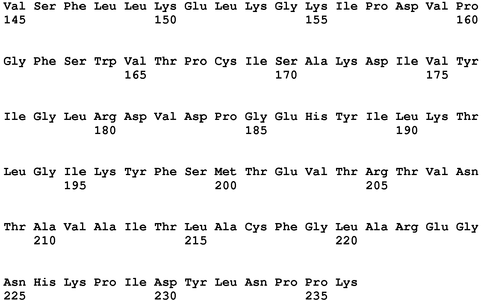
- <210> 11
<211> 127
<212> PRT
<213> Homo sapiens - <400> 11

- <210> 12
<211> 222
<212> PRT
<213> Homo sapiens - <400> 12

- <210> 13
<211> 496
<212> PRT
<213> Homo sapiens - <400> 13


- <210> 14
<211> 9
<212> PRT
<213> Homo sapiens - <400> 14

- <210> 15
<211> 15
<212> PRT
<213> Homo sapiens - <400> 15

- <210> 16
<211> 12
<212> PRT
<213> Homo sapiens - <400> 16

- <210> 17
<211> 8
<212> PRT
<213> Homo sapiens - <400> 17

- <210> 18
<211> 9
<212> PRT
<213> Homo sapiens - <400> 18

- <210> 19
<211> 10
<212> PRT
<213> Homo sapiens - <400> 19

- <210> 20
<211> 15
<212> PRT
<213> Homo sapiens - <400> 20

- <210> 21
<211> 9
<212> PRT
<213> Homo sapiens - <400> 21

- <210> 22
<211> 8
<212> PRT
<213> Homo sapiens - <400> 22

- <210> 23
<211> 10
<212> PRT
<213> Homo sapiens - <400> 23

- <210> 24
<211> 11
<212> PRT
<213> Homo sapiens - <400> 24

- <210> 25
<211> 11
<212> PRT
<213> Homo sapiens - <400> 25

- <210> 26
<211> 11
<212> PRT
<213> Homo sapiens - <400> 26

- <210> 27
<211> 11
<212> PRT
<213> Homo sapiens - <400> 27

- <210> 28
<211> 8
<212> PRT
<213> Homo sapiens - <400> 28

- <210> 29
<211> 10
<212> PRT
<213> Homo sapiens - <400> 29

- <210> 30
<211> 7
<212> PRT
<213> Homo sapiens - <400> 30

- <210> 31
<211> 11
<212> PRT
<213> Homo sapiens - <400> 31

- <210> 32
<211> 9
<212> PRT
<213> Homo sapiens - <400> 32

- <210> 33
<211> 8
<212> PRT
<213> Homo sapiens - <400> 33

- <210> 34
<211> 9
<212> PRT
<213> Homo sapiens - <400> 34

- <210> 35
<211> 10
<212> PRT
<213> Homo sapiens - <400> 35

- <210> 36
<211> 8
<212> PRT
<213> Homo sapiens - <400> 36

Claims (16)
- Procédé de diagnostic, de suivi, de pronostic et/ou théranostique in vitro de troubles hépatiques choisis parmi l'insuffisance hépatique aigüe, la stéatose hépatique et la stéatohépatite, à partir d'un échantillon sanguin issu d'un individu, dans lequel on détermine la présence et/ou la concentration du marqueur ADH1B (SEQ ID N° 2) et/ou la présence et/ou la concentration de la combinaison des marqueurs ADH1B (SEQ ID N° 2) et ADH1A (SEQ ID N° 1).
- Procédé in vitro selon la revendication 1, dans lequel on détermine la présence et/ou la concentration d'au moins un marqueur supplémentaire, préférentiellement d'au moins deux marqueurs supplémentaires, et encore plus préférentiellement d'au moins trois marqueurs supplémentaires, lesdits marqueurs étant sélectionnés dans le groupe constitué par : ADH4 (SEQ ID N° 3 & SEQ ID N° 4), BHMT (SEQ ID N° 5, SEQ ID N° 6 & SEQ ID N° 7), ARG1 (SEQ ID N° 8, SEQ ID N° 9 & SEQ ID N° 10), FABP1 (SEQ ID N° 11), GSTA1 (SEQ ID N° 12) et ALT1 (SEQ ID N° 13).
- Procédé in vitro selon l'une quelconque des revendications 1 à 2, dans lequel on détermine au moins la présence et/ou la concentration d'au moins un marqueur supplémentaire sélectionné parmi ADH4 (SEQ ID N° 3 & SEQ ID N° 4) et BHMT (SEQ ID N° 5, SEQ ID N° 6 & SEQ ID N° 7).
- Procédé in vitro selon l'une quelconque des revendications 1 à 3, dans lequel on détermine au moins la présence et/ou la concentration du marqueur ADH1B (SEQ ID N° 2) ou de la combinaison des marqueurs ADH1B (SEQ ID N° 2) et ADH1A (SEQ ID N° 1), d'une part, et des marqueurs ADH4 (SEQ ID N° 3 & SEQ ID N° 4) et BHMT (SEQ ID N° 5, SEQ ID N° 6 & SEQ ID N° 7), d'autre part.
- Procédé in vitro selon l'une quelconque des revendications 2 à 4 caractérisé en ce que le marqueur BHMT est le variant de séquence BHMT1 (SEQ ID N° 5) et/ou la combinaison des variants de séquence BHMT1 (SEQ ID N° 5) et BHMT2 (SEQ ID N° 6 & SEQ ID N° 7).
- Procédé in vitro selon l'une quelconque des revendications 1 à 5, dans lequel on détermine en outre la présence et/ou la concentration du marqueur CK18.
- Procédé in vitro selon l'une quelconque des revendications 1 à 6, dans lequel on utilise pour ladite détermination de la présence et/ou de la concentration desdits marqueurs des peptides signatures présents dans lesdits marqueurs.
- Procédé in vitro selon la revendication 7, dans lequel lesdits peptides signatures sont générés à partir desdits marqueurs par un procédé de digestion, de préférence enzymatique.
- Procédé in vitro selon la revendication 7 ou 8, dans lequel la présence et/ou la concentration:a. de la combinaison des marqueurs ADH1A (SEQ ID N° 1) et ADH1B (SEQ ID N° 2), si elle est sélectionnée, est déterminée par la présence d'au moins un peptide signature choisi parmi INEGFDLLHSGK (SEQ ID N° 16) et FSLDALITHVLPFEK (SEQ ID N° 15),b. du marqueur ADH1B (SEQ ID N° 2), s'il est sélectionné, est déterminée par la présence du peptide signature peptide signature AAVLWEVK (SEQ ID N° 17),c. du marqueur ADH4 (SEQ ID N° 3 & SEQ ID N° 4), s'il est sélectionné, est déterminée par la présence d'au moins un peptide signature choisi parmi FNLDALVTHTLPFDK (SEQ ID N° 20), IDDDANLER (SEQ ID N° 18) ou IIGIDINSEK (SEQ ID N° 19),d. du marqueur ARG1 (SEQ ID N° 8, SEQ ID N° 9 & SEQ ID N° 10), s'il est sélectionné, est déterminée par la présence d'au moins un peptide signature choisi parmi DVDPGEHYILK (SEQ ID N° 25), TIGIIGAPFSK (SEQ ID N° 24) ou EGLYITEEIYK (SEQ ID N° 26),e. du marqueur BHMT (SEQ ID N° 5, SEQ ID N° 6 & SEQ ID N° 7), s'il est sélectionné, est déterminée par la présence d'au moins un peptide signature choisi parmi EATTEQQLK (SEQ ID N° 21), AIAEELAPER (SEQ ID N° 23) ou EAYNLGVR (SEQ ID N° 22),f. du marqueur BHMT1 (SEQ ID N° 5), s'il est sélectionné, est déterminée par la présence du peptide signature EATTEQQLK (SEQ ID N° 21),g. de la combinaison des marqueurs BHMT1 (SEQ ID N°5) et BHMT2 (SEQ ID N° 6 & SEQ ID N° 7), si elle est sélectionnée, est déterminée par la présence d'au moins un peptide signature choisi parmi AIAEELAPER (SEQ ID N° 23) ou EAYNLGVR (SEQ ID N° 22),h. du marqueur FABP1 (SEQ ID N° 11), s'il est sélectionné, est déterminée par la présence d'au moins un peptide signature choisi parmi AIGLPEELIQK (SEQ ID N° 27), FTITAGSK (SEQ ID N° 28) ou TVVQLEGDNK (SEQ ID N° 29),i. du marqueur GSTA1 (SEQ ID N° 12), s'il est sélectionné, est déterminée par la présence du peptide signature LHYFNAR (SEQ ID N° 30),j. du marqueur ALT1 (SEQ ID N° 13), s'il est sélectionné, est déterminée par la présence d'au moins un peptide signature choisi parmi ALELEQELR (SEQ ID N° 34), LLVAGEGHTR (SEQ ID N° 35) ou KPFTEVIR (SEQ ID N° 36), sachant que ALT1 (SEQ ID N° 13), s'il est sélectionné, est plus particulièrement déterminée par la présence du peptide signature ALELEQELR (SEQ ID N° 34) ou LLVAGEGHTR (SEQ ID N° 35).
- Procédé in vitro selon l'une quelconque des revendications précédentes dans lequel on dose au moins un des marqueurs (dit « marqueur sélectionné ») tels que défini à l'une quelconque des revendications 1 à 6, dont au moins un marqueur ADH1B (SEQ ID N° 2) et/ou la combinaison des marqueurs ADH1B (SEQ ID N° 2) et ADH1A (SEQ ID N° 1) à partir d'un échantillon sanguin prélevé sur un individu à un temps t0, lequel procédé comprend les étapes suivantes :a) l'ajout d'une quantité connue d'une protéine isotopiquement marquée homologue du marqueur sélectionné dans l'échantillon sanguin,b) le traitement de l'échantillon sanguin pour extraire au moins une partie des protéines abondantes ou au moins une partie des glycoprotéines,c) le traitement de l'échantillon après extraction d'au moins une partie des protéines abondantes ou des glycoprotéines pour générer des peptides protéolytiques, de préférence par digestion, étant entendu que parmi les peptides protéolytiques obtenus se trouvent les peptides signatures des marqueurs sélectionnés tels que défini à l'une quelconque des revendications 7 à 9,d) le dosage quantitatif par spectrométrie de masse d'au moins un peptide signature généré à partir du marqueur sélectionné,e) la détermination d'un rapport de l'abondance du peptide signature isotopiquement marqué, ajouté à l'étape a), sur l'abondance du peptide signature non marqué issu de l'échantillon sanguin,f) le calcul à partir du rapport obtenu à l'étape e) et de la quantité connue de ladite protéine isotopiquement marquée ajoutée à l'étape a), de la concentration dudit marqueur sélectionné dans l'échantillon.
- Procédé in vitro selon la revendication 10, caractérisé en ce que l'étape b) est effectuée par déplétion et préférentiellement par glycodéplétion.
- Procédé in vitro selon la revendication 10 ou 11, caractérisé en ce que le procédé est effectué à un temps t0 et à un temps t1 supérieur à t0, et comprend après l'étape f), la comparaison de la concentration du marqueur sélectionné dans l'échantillon entre t1 et t0.
- Procédé in vitro selon les revendications 10 à 12, caractérisé en ce que l'étape c) est effectuée par digestion enzymatique, de préférence par l'emploi de trypsine ou d'un mélange d'EndolysC et de trypsine.
- Procédé in vitro selon l'une quelconque des revendications 1 à 13, caractérisé en ce que le trouble hépatique est une insuffisance hépatique aiguë ou une stéatohépatite non-alcoolique.
- Procédé in vitro pour évaluer la toxicité hépatique d'une molécule, comprenant le dosage d'au moins un des marqueurs (dit « marqueur sélectionné ») tels que défini à l'une quelconque des revendications 1 à 6, dont au moins un marqueur ADH1B (SEQ ID N° 2) et/ou la combinaison des marqueurs ADH1B (SEQ ID N° 2) et ADH1A (SEQ ID N° 1) à partir d'un échantillon sanguin issu d'un individu auquel ladite molécule a été préalablement administrée, selon les étapes a) à f) du procédé selon l'une quelconque des revendications 10 à 13.
- Procédé in vitro selon l'une quelconque des revendications 1 à 15, caractérisé en ce que l'échantillon sanguin est sélectionné dans le groupe constitué par le sang, le plasma et le sérum.
Applications Claiming Priority (3)
| Application Number | Priority Date | Filing Date | Title |
|---|---|---|---|
| FR1600081 | 2016-01-15 | ||
| FR1652087A FR3048780B1 (fr) | 2016-03-11 | 2016-03-11 | Procede de diagnostic in vitro d'atteintes hepatiques |
| PCT/FR2017/050089 WO2017121974A1 (fr) | 2016-01-15 | 2017-01-16 | Procédé de diagnostic in vitro d'atteintes hépatiques |
Publications (2)
| Publication Number | Publication Date |
|---|---|
| EP3403100A1 EP3403100A1 (fr) | 2018-11-21 |
| EP3403100B1 true EP3403100B1 (fr) | 2020-03-11 |
Family
ID=58098653
Family Applications (1)
| Application Number | Title | Priority Date | Filing Date |
|---|---|---|---|
| EP17706533.1A Active EP3403100B1 (fr) | 2016-01-15 | 2017-01-16 | Procédé de diagnostic in vitro d'atteintes hépatiques |
Country Status (2)
| Country | Link |
|---|---|
| EP (1) | EP3403100B1 (fr) |
| WO (1) | WO2017121974A1 (fr) |
Family Cites Families (2)
| Publication number | Priority date | Publication date | Assignee | Title |
|---|---|---|---|---|
| WO2013190075A2 (fr) * | 2012-06-20 | 2013-12-27 | Meyer Helmut E | Biomarqueurs spécifiques pour un carcinome hépatocellulaire (hcc) |
| GB201320061D0 (en) * | 2013-11-13 | 2013-12-25 | Electrophoretics Ltd | Materials nad methods for diagnosis and prognosis of liver cancer |
-
2017
- 2017-01-16 WO PCT/FR2017/050089 patent/WO2017121974A1/fr not_active Ceased
- 2017-01-16 EP EP17706533.1A patent/EP3403100B1/fr active Active
Non-Patent Citations (1)
| Title |
|---|
| None * |
Also Published As
| Publication number | Publication date |
|---|---|
| EP3403100A1 (fr) | 2018-11-21 |
| WO2017121974A1 (fr) | 2017-07-20 |
Similar Documents
| Publication | Publication Date | Title |
|---|---|---|
| Hess et al. | Simultaneous determination and validated quantification of human insulin and its synthetic analogues in human blood serum by immunoaffinity purification and liquid chromatography-mass spectrometry | |
| Kippen et al. | Development of an isotope dilution assay for precise determination of insulin, C-peptide, and proinsulin levels in non-diabetic and type II diabetic individuals with comparison to immunoassay | |
| Rayavarapu et al. | Identification of disease specific pathways using in vivo SILAC proteomics in dystrophin deficient mdx mouse | |
| Tsikas et al. | Homoarginine, arginine, and relatives: analysis, metabolism, transport, physiology, and pathology | |
| EP2336782B1 (fr) | Procédé pour prédire un diabète de type II | |
| Thomas et al. | Mass spectrometric identification of degradation products of insulin and its long-acting analogues in human urine for doping control purposes | |
| la Marca et al. | The inclusion of succinylacetone as marker for tyrosinemia type I in expanded newborn screening programs | |
| JP5735500B2 (ja) | エネルギー代謝の極性代謝物を分析するための方法 | |
| US8871527B2 (en) | Method and system for measuring a sample to determine the presence of and optionally treat a pathologic condition | |
| Hashimoto et al. | Highly-sensitive detection of free advanced glycation end-products by liquid chromatography-electrospray ionization-tandem mass spectrometry with 2, 4, 6-trinitrobenzene sulfonate derivatization | |
| Knoop et al. | Development of a mass spectrometry based detection method for the mitochondrion‐derived peptide MOTS‐c in plasma samples for doping control purposes | |
| CN103630693A (zh) | 肝脏炎症的生物标志物 | |
| Alayi et al. | Tandem mass tag-based serum proteome profiling for biomarker discovery in young duchenne muscular dystrophy boys | |
| Thevis et al. | Doping control analysis of bovine hemoglobin-based oxygen therapeutics in human plasma by LC− electrospray ionization-MS/MS | |
| Noso et al. | Metabolic signatures of β‐cell destruction in type 1 diabetes | |
| Daniel Opfermann et al. | Reactivity-based metabolomics reveal cysteine has glyoxalase 1-like and glyoxalase 2-like activities | |
| Korwar et al. | Comprehensive quantification of carboxymethyllysine-modified peptides in human plasma | |
| US11221341B2 (en) | Process for in vitro diagnosis of hepatic disorders | |
| Gunn et al. | Quantification of metallothionein-III in brain tissues using liquid chromatography tandem mass spectrometry | |
| Kingon et al. | Multi-peptide nLC-PC-IDMS-SRM-based assay for the quantification of biomarkers in the chicken ovarian cancer model | |
| Masaki et al. | GIP_HUMAN [22–51] is a new proatherogenic peptide identified by native plasma peptidomics | |
| EP1402257B1 (fr) | Proteines utiles dans l'analyse proteomique du diabete | |
| Bettahi et al. | Differences in protein expression, at the basal state and at 2 h of insulin infusion, in muscle biopsies from healthy Arab men with high or low insulin sensitivity measured by hyperinsulinemic euglycemic clamp | |
| EP3403100A1 (fr) | Procédé de diagnostic in vitro d'atteintes hépatiques | |
| Liu et al. | Multiplexed analysis of endogenous Guanidino compounds via isotope-coded doubly charged labeling: Application to lung Cancer tissues as a case |
Legal Events
| Date | Code | Title | Description |
|---|---|---|---|
| STAA | Information on the status of an ep patent application or granted ep patent |
Free format text: STATUS: UNKNOWN |
|
| STAA | Information on the status of an ep patent application or granted ep patent |
Free format text: STATUS: THE INTERNATIONAL PUBLICATION HAS BEEN MADE |
|
| PUAI | Public reference made under article 153(3) epc to a published international application that has entered the european phase |
Free format text: ORIGINAL CODE: 0009012 |
|
| STAA | Information on the status of an ep patent application or granted ep patent |
Free format text: STATUS: REQUEST FOR EXAMINATION WAS MADE |
|
| 17P | Request for examination filed |
Effective date: 20180711 |
|
| AK | Designated contracting states |
Kind code of ref document: A1 Designated state(s): AL AT BE BG CH CY CZ DE DK EE ES FI FR GB GR HR HU IE IS IT LI LT LU LV MC MK MT NL NO PL PT RO RS SE SI SK SM TR |
|
| AX | Request for extension of the european patent |
Extension state: BA ME |
|
| DAV | Request for validation of the european patent (deleted) | ||
| DAX | Request for extension of the european patent (deleted) | ||
| STAA | Information on the status of an ep patent application or granted ep patent |
Free format text: STATUS: EXAMINATION IS IN PROGRESS |
|
| 17Q | First examination report despatched |
Effective date: 20190604 |
|
| GRAP | Despatch of communication of intention to grant a patent |
Free format text: ORIGINAL CODE: EPIDOSNIGR1 |
|
| STAA | Information on the status of an ep patent application or granted ep patent |
Free format text: STATUS: GRANT OF PATENT IS INTENDED |
|
| INTG | Intention to grant announced |
Effective date: 20191209 |
|
| GRAS | Grant fee paid |
Free format text: ORIGINAL CODE: EPIDOSNIGR3 |
|
| GRAA | (expected) grant |
Free format text: ORIGINAL CODE: 0009210 |
|
| STAA | Information on the status of an ep patent application or granted ep patent |
Free format text: STATUS: THE PATENT HAS BEEN GRANTED |
|
| AK | Designated contracting states |
Kind code of ref document: B1 Designated state(s): AL AT BE BG CH CY CZ DE DK EE ES FI FR GB GR HR HU IE IS IT LI LT LU LV MC MK MT NL NO PL PT RO RS SE SI SK SM TR |
|
| REG | Reference to a national code |
Ref country code: GB Ref legal event code: FG4D Free format text: NOT ENGLISH |
|
| REG | Reference to a national code |
Ref country code: CH Ref legal event code: EP |
|
| REG | Reference to a national code |
Ref country code: AT Ref legal event code: REF Ref document number: 1243787 Country of ref document: AT Kind code of ref document: T Effective date: 20200315 |
|
| REG | Reference to a national code |
Ref country code: DE Ref legal event code: R096 Ref document number: 602017012938 Country of ref document: DE |
|
| REG | Reference to a national code |
Ref country code: IE Ref legal event code: FG4D Free format text: LANGUAGE OF EP DOCUMENT: FRENCH |
|
| REG | Reference to a national code |
Ref country code: SE Ref legal event code: TRGR |
|
| REG | Reference to a national code |
Ref country code: CH Ref legal event code: NV Representative=s name: VALIPAT S.A. C/O BOVARD SA NEUCHATEL, CH |
|
| REG | Reference to a national code |
Ref country code: DE Ref legal event code: R081 Ref document number: 602017012938 Country of ref document: DE Owner name: INSERM, FR Free format text: FORMER OWNERS: CENTRE HOSPITALIER UNIVERSITAIRE GRENOBLE ALPES, LA TRONCHE, FR; COMMISSARIAT A L'ENERGIE ATOMIQUE ET AUX ENERGIES ALTERNATIVES, PARIS, FR; INSERM, PARIS, FR; UNIVERSITE GRENOBLE ALPES, SAINT-MARTIN-D'HERES, FR; UNIVERSITE PARIS SUD, ORSAY, FR Ref country code: DE Ref legal event code: R081 Ref document number: 602017012938 Country of ref document: DE Owner name: CENTRE HOSPITALIER UNIVERSITAIRE GRENOBLE ALPE, FR Free format text: FORMER OWNERS: CENTRE HOSPITALIER UNIVERSITAIRE GRENOBLE ALPES, LA TRONCHE, FR; COMMISSARIAT A L'ENERGIE ATOMIQUE ET AUX ENERGIES ALTERNATIVES, PARIS, FR; INSERM, PARIS, FR; UNIVERSITE GRENOBLE ALPES, SAINT-MARTIN-D'HERES, FR; UNIVERSITE PARIS SUD, ORSAY, FR Ref country code: DE Ref legal event code: R081 Ref document number: 602017012938 Country of ref document: DE Owner name: UNIVERSITE PARIS-SACLAY, FR Free format text: FORMER OWNERS: CENTRE HOSPITALIER UNIVERSITAIRE GRENOBLE ALPES, LA TRONCHE, FR; COMMISSARIAT A L'ENERGIE ATOMIQUE ET AUX ENERGIES ALTERNATIVES, PARIS, FR; INSERM, PARIS, FR; UNIVERSITE GRENOBLE ALPES, SAINT-MARTIN-D'HERES, FR; UNIVERSITE PARIS SUD, ORSAY, FR Ref country code: DE Ref legal event code: R081 Ref document number: 602017012938 Country of ref document: DE Owner name: COMMISSARIAT A L'ENERGIE ATOMIQUE ET AUX ENERG, FR Free format text: FORMER OWNERS: CENTRE HOSPITALIER UNIVERSITAIRE GRENOBLE ALPES, LA TRONCHE, FR; COMMISSARIAT A L'ENERGIE ATOMIQUE ET AUX ENERGIES ALTERNATIVES, PARIS, FR; INSERM, PARIS, FR; UNIVERSITE GRENOBLE ALPES, SAINT-MARTIN-D'HERES, FR; UNIVERSITE PARIS SUD, ORSAY, FR Ref country code: DE Ref legal event code: R081 Ref document number: 602017012938 Country of ref document: DE Owner name: UNIVERSITE GRENOBLE ALPES, FR Free format text: FORMER OWNERS: CENTRE HOSPITALIER UNIVERSITAIRE GRENOBLE ALPES, LA TRONCHE, FR; COMMISSARIAT A L'ENERGIE ATOMIQUE ET AUX ENERGIES ALTERNATIVES, PARIS, FR; INSERM, PARIS, FR; UNIVERSITE GRENOBLE ALPES, SAINT-MARTIN-D'HERES, FR; UNIVERSITE PARIS SUD, ORSAY, FR |
|
| RAP2 | Party data changed (patent owner data changed or rights of a patent transferred) |
Owner name: UNIVERSITE PARIS-SACLAY Owner name: INSERM Owner name: UNIVERSITE GRENOBLE ALPES Owner name: CENTRE HOSPITALIER UNIVERSITAIRE GRENOBLE ALPES Owner name: COMMISSARIAT A L'ENERGIE ATOMIQUE ET AUX ENERGIES ALTERNATIVES |
|
| PG25 | Lapsed in a contracting state [announced via postgrant information from national office to epo] |
Ref country code: RS Free format text: LAPSE BECAUSE OF FAILURE TO SUBMIT A TRANSLATION OF THE DESCRIPTION OR TO PAY THE FEE WITHIN THE PRESCRIBED TIME-LIMIT Effective date: 20200311 Ref country code: FI Free format text: LAPSE BECAUSE OF FAILURE TO SUBMIT A TRANSLATION OF THE DESCRIPTION OR TO PAY THE FEE WITHIN THE PRESCRIBED TIME-LIMIT Effective date: 20200311 Ref country code: NO Free format text: LAPSE BECAUSE OF FAILURE TO SUBMIT A TRANSLATION OF THE DESCRIPTION OR TO PAY THE FEE WITHIN THE PRESCRIBED TIME-LIMIT Effective date: 20200611 |
|
| REG | Reference to a national code |
Ref country code: NL Ref legal event code: MP Effective date: 20200311 |
|
| REG | Reference to a national code |
Ref country code: BE Ref legal event code: PD Owner name: CENTRE HOSPITALIER UNIVERSITAIRE GRENOBLE ALPES; FR Free format text: DETAILS ASSIGNMENT: CHANGE OF OWNER(S), CESSION, OEB; FORMER OWNER NAME: UNIVERSITE PARIS-SUD Effective date: 20200709 |
|
| PG25 | Lapsed in a contracting state [announced via postgrant information from national office to epo] |
Ref country code: BG Free format text: LAPSE BECAUSE OF FAILURE TO SUBMIT A TRANSLATION OF THE DESCRIPTION OR TO PAY THE FEE WITHIN THE PRESCRIBED TIME-LIMIT Effective date: 20200611 Ref country code: GR Free format text: LAPSE BECAUSE OF FAILURE TO SUBMIT A TRANSLATION OF THE DESCRIPTION OR TO PAY THE FEE WITHIN THE PRESCRIBED TIME-LIMIT Effective date: 20200612 Ref country code: LV Free format text: LAPSE BECAUSE OF FAILURE TO SUBMIT A TRANSLATION OF THE DESCRIPTION OR TO PAY THE FEE WITHIN THE PRESCRIBED TIME-LIMIT Effective date: 20200311 Ref country code: HR Free format text: LAPSE BECAUSE OF FAILURE TO SUBMIT A TRANSLATION OF THE DESCRIPTION OR TO PAY THE FEE WITHIN THE PRESCRIBED TIME-LIMIT Effective date: 20200311 |
|
| REG | Reference to a national code |
Ref country code: LT Ref legal event code: MG4D |
|
| PG25 | Lapsed in a contracting state [announced via postgrant information from national office to epo] |
Ref country code: NL Free format text: LAPSE BECAUSE OF FAILURE TO SUBMIT A TRANSLATION OF THE DESCRIPTION OR TO PAY THE FEE WITHIN THE PRESCRIBED TIME-LIMIT Effective date: 20200311 |
|
| REG | Reference to a national code |
Ref country code: GB Ref legal event code: 732E Free format text: REGISTERED BETWEEN 20200917 AND 20200923 |
|
| PG25 | Lapsed in a contracting state [announced via postgrant information from national office to epo] |
Ref country code: SK Free format text: LAPSE BECAUSE OF FAILURE TO SUBMIT A TRANSLATION OF THE DESCRIPTION OR TO PAY THE FEE WITHIN THE PRESCRIBED TIME-LIMIT Effective date: 20200311 Ref country code: IS Free format text: LAPSE BECAUSE OF FAILURE TO SUBMIT A TRANSLATION OF THE DESCRIPTION OR TO PAY THE FEE WITHIN THE PRESCRIBED TIME-LIMIT Effective date: 20200711 Ref country code: PT Free format text: LAPSE BECAUSE OF FAILURE TO SUBMIT A TRANSLATION OF THE DESCRIPTION OR TO PAY THE FEE WITHIN THE PRESCRIBED TIME-LIMIT Effective date: 20200805 Ref country code: LT Free format text: LAPSE BECAUSE OF FAILURE TO SUBMIT A TRANSLATION OF THE DESCRIPTION OR TO PAY THE FEE WITHIN THE PRESCRIBED TIME-LIMIT Effective date: 20200311 Ref country code: EE Free format text: LAPSE BECAUSE OF FAILURE TO SUBMIT A TRANSLATION OF THE DESCRIPTION OR TO PAY THE FEE WITHIN THE PRESCRIBED TIME-LIMIT Effective date: 20200311 Ref country code: SM Free format text: LAPSE BECAUSE OF FAILURE TO SUBMIT A TRANSLATION OF THE DESCRIPTION OR TO PAY THE FEE WITHIN THE PRESCRIBED TIME-LIMIT Effective date: 20200311 Ref country code: CZ Free format text: LAPSE BECAUSE OF FAILURE TO SUBMIT A TRANSLATION OF THE DESCRIPTION OR TO PAY THE FEE WITHIN THE PRESCRIBED TIME-LIMIT Effective date: 20200311 Ref country code: RO Free format text: LAPSE BECAUSE OF FAILURE TO SUBMIT A TRANSLATION OF THE DESCRIPTION OR TO PAY THE FEE WITHIN THE PRESCRIBED TIME-LIMIT Effective date: 20200311 |
|
| REG | Reference to a national code |
Ref country code: AT Ref legal event code: MK05 Ref document number: 1243787 Country of ref document: AT Kind code of ref document: T Effective date: 20200311 |
|
| REG | Reference to a national code |
Ref country code: DE Ref legal event code: R097 Ref document number: 602017012938 Country of ref document: DE |
|
| PLBE | No opposition filed within time limit |
Free format text: ORIGINAL CODE: 0009261 |
|
| STAA | Information on the status of an ep patent application or granted ep patent |
Free format text: STATUS: NO OPPOSITION FILED WITHIN TIME LIMIT |
|
| PG25 | Lapsed in a contracting state [announced via postgrant information from national office to epo] |
Ref country code: IT Free format text: LAPSE BECAUSE OF FAILURE TO SUBMIT A TRANSLATION OF THE DESCRIPTION OR TO PAY THE FEE WITHIN THE PRESCRIBED TIME-LIMIT Effective date: 20200311 Ref country code: ES Free format text: LAPSE BECAUSE OF FAILURE TO SUBMIT A TRANSLATION OF THE DESCRIPTION OR TO PAY THE FEE WITHIN THE PRESCRIBED TIME-LIMIT Effective date: 20200311 Ref country code: DK Free format text: LAPSE BECAUSE OF FAILURE TO SUBMIT A TRANSLATION OF THE DESCRIPTION OR TO PAY THE FEE WITHIN THE PRESCRIBED TIME-LIMIT Effective date: 20200311 Ref country code: AT Free format text: LAPSE BECAUSE OF FAILURE TO SUBMIT A TRANSLATION OF THE DESCRIPTION OR TO PAY THE FEE WITHIN THE PRESCRIBED TIME-LIMIT Effective date: 20200311 |
|
| 26N | No opposition filed |
Effective date: 20201214 |
|
| PG25 | Lapsed in a contracting state [announced via postgrant information from national office to epo] |
Ref country code: SI Free format text: LAPSE BECAUSE OF FAILURE TO SUBMIT A TRANSLATION OF THE DESCRIPTION OR TO PAY THE FEE WITHIN THE PRESCRIBED TIME-LIMIT Effective date: 20200311 Ref country code: PL Free format text: LAPSE BECAUSE OF FAILURE TO SUBMIT A TRANSLATION OF THE DESCRIPTION OR TO PAY THE FEE WITHIN THE PRESCRIBED TIME-LIMIT Effective date: 20200311 |
|
| PG25 | Lapsed in a contracting state [announced via postgrant information from national office to epo] |
Ref country code: MC Free format text: LAPSE BECAUSE OF FAILURE TO SUBMIT A TRANSLATION OF THE DESCRIPTION OR TO PAY THE FEE WITHIN THE PRESCRIBED TIME-LIMIT Effective date: 20200311 |
|
| PG25 | Lapsed in a contracting state [announced via postgrant information from national office to epo] |
Ref country code: LU Free format text: LAPSE BECAUSE OF NON-PAYMENT OF DUE FEES Effective date: 20210116 |
|
| PG25 | Lapsed in a contracting state [announced via postgrant information from national office to epo] |
Ref country code: IE Free format text: LAPSE BECAUSE OF NON-PAYMENT OF DUE FEES Effective date: 20210116 |
|
| PGFP | Annual fee paid to national office [announced via postgrant information from national office to epo] |
Ref country code: FR Payment date: 20211228 Year of fee payment: 6 |
|
| PGFP | Annual fee paid to national office [announced via postgrant information from national office to epo] |
Ref country code: GB Payment date: 20220120 Year of fee payment: 6 Ref country code: DE Payment date: 20220114 Year of fee payment: 6 Ref country code: CH Payment date: 20220124 Year of fee payment: 6 |
|
| PGFP | Annual fee paid to national office [announced via postgrant information from national office to epo] |
Ref country code: SE Payment date: 20220119 Year of fee payment: 6 Ref country code: BE Payment date: 20220127 Year of fee payment: 6 |
|
| PG25 | Lapsed in a contracting state [announced via postgrant information from national office to epo] |
Ref country code: CY Free format text: LAPSE BECAUSE OF FAILURE TO SUBMIT A TRANSLATION OF THE DESCRIPTION OR TO PAY THE FEE WITHIN THE PRESCRIBED TIME-LIMIT Effective date: 20200311 |
|
| PG25 | Lapsed in a contracting state [announced via postgrant information from national office to epo] |
Ref country code: HU Free format text: LAPSE BECAUSE OF FAILURE TO SUBMIT A TRANSLATION OF THE DESCRIPTION OR TO PAY THE FEE WITHIN THE PRESCRIBED TIME-LIMIT; INVALID AB INITIO Effective date: 20170116 |
|
| REG | Reference to a national code |
Ref country code: DE Ref legal event code: R119 Ref document number: 602017012938 Country of ref document: DE |
|
| REG | Reference to a national code |
Ref country code: SE Ref legal event code: EUG |
|
| REG | Reference to a national code |
Ref country code: CH Ref legal event code: PL |
|
| GBPC | Gb: european patent ceased through non-payment of renewal fee |
Effective date: 20230116 |
|
| REG | Reference to a national code |
Ref country code: BE Ref legal event code: MM Effective date: 20230131 |
|
| PG25 | Lapsed in a contracting state [announced via postgrant information from national office to epo] |
Ref country code: SE Free format text: LAPSE BECAUSE OF NON-PAYMENT OF DUE FEES Effective date: 20230117 Ref country code: LI Free format text: LAPSE BECAUSE OF NON-PAYMENT OF DUE FEES Effective date: 20230131 Ref country code: GB Free format text: LAPSE BECAUSE OF NON-PAYMENT OF DUE FEES Effective date: 20230116 Ref country code: DE Free format text: LAPSE BECAUSE OF NON-PAYMENT OF DUE FEES Effective date: 20230801 Ref country code: CH Free format text: LAPSE BECAUSE OF NON-PAYMENT OF DUE FEES Effective date: 20230131 |
|
| PG25 | Lapsed in a contracting state [announced via postgrant information from national office to epo] |
Ref country code: FR Free format text: LAPSE BECAUSE OF NON-PAYMENT OF DUE FEES Effective date: 20230131 Ref country code: BE Free format text: LAPSE BECAUSE OF NON-PAYMENT OF DUE FEES Effective date: 20230131 |
|
| PG25 | Lapsed in a contracting state [announced via postgrant information from national office to epo] |
Ref country code: MK Free format text: LAPSE BECAUSE OF FAILURE TO SUBMIT A TRANSLATION OF THE DESCRIPTION OR TO PAY THE FEE WITHIN THE PRESCRIBED TIME-LIMIT Effective date: 20200311 |
|
| PG25 | Lapsed in a contracting state [announced via postgrant information from national office to epo] |
Ref country code: TR Free format text: LAPSE BECAUSE OF FAILURE TO SUBMIT A TRANSLATION OF THE DESCRIPTION OR TO PAY THE FEE WITHIN THE PRESCRIBED TIME-LIMIT Effective date: 20200311 |
|
| PG25 | Lapsed in a contracting state [announced via postgrant information from national office to epo] |
Ref country code: MT Free format text: LAPSE BECAUSE OF FAILURE TO SUBMIT A TRANSLATION OF THE DESCRIPTION OR TO PAY THE FEE WITHIN THE PRESCRIBED TIME-LIMIT Effective date: 20200311 |