EP3395941B1 - Virus infection model, preparation method therefor, and utilization thereof - Google Patents
Virus infection model, preparation method therefor, and utilization thereof Download PDFInfo
- Publication number
- EP3395941B1 EP3395941B1 EP16878818.0A EP16878818A EP3395941B1 EP 3395941 B1 EP3395941 B1 EP 3395941B1 EP 16878818 A EP16878818 A EP 16878818A EP 3395941 B1 EP3395941 B1 EP 3395941B1
- Authority
- EP
- European Patent Office
- Prior art keywords
- cells
- virus
- human
- hepatitis
- liver
- Prior art date
- Legal status (The legal status is an assumption and is not a legal conclusion. Google has not performed a legal analysis and makes no representation as to the accuracy of the status listed.)
- Active
Links
Images




Classifications
-
- C—CHEMISTRY; METALLURGY
- C12—BIOCHEMISTRY; BEER; SPIRITS; WINE; VINEGAR; MICROBIOLOGY; ENZYMOLOGY; MUTATION OR GENETIC ENGINEERING
- C12N—MICROORGANISMS OR ENZYMES; COMPOSITIONS THEREOF; PROPAGATING, PRESERVING, OR MAINTAINING MICROORGANISMS; MUTATION OR GENETIC ENGINEERING; CULTURE MEDIA
- C12N5/00—Undifferentiated human, animal or plant cells, e.g. cell lines; Tissues; Cultivation or maintenance thereof; Culture media therefor
- C12N5/06—Animal cells or tissues; Human cells or tissues
- C12N5/0602—Vertebrate cells
- C12N5/067—Hepatocytes
- C12N5/0671—Three-dimensional culture, tissue culture or organ culture; Encapsulated cells
-
- C—CHEMISTRY; METALLURGY
- C12—BIOCHEMISTRY; BEER; SPIRITS; WINE; VINEGAR; MICROBIOLOGY; ENZYMOLOGY; MUTATION OR GENETIC ENGINEERING
- C12N—MICROORGANISMS OR ENZYMES; COMPOSITIONS THEREOF; PROPAGATING, PRESERVING, OR MAINTAINING MICROORGANISMS; MUTATION OR GENETIC ENGINEERING; CULTURE MEDIA
- C12N7/00—Viruses; Bacteriophages; Compositions thereof; Preparation or purification thereof
-
- C—CHEMISTRY; METALLURGY
- C12—BIOCHEMISTRY; BEER; SPIRITS; WINE; VINEGAR; MICROBIOLOGY; ENZYMOLOGY; MUTATION OR GENETIC ENGINEERING
- C12N—MICROORGANISMS OR ENZYMES; COMPOSITIONS THEREOF; PROPAGATING, PRESERVING, OR MAINTAINING MICROORGANISMS; MUTATION OR GENETIC ENGINEERING; CULTURE MEDIA
- C12N5/00—Undifferentiated human, animal or plant cells, e.g. cell lines; Tissues; Cultivation or maintenance thereof; Culture media therefor
- C12N5/06—Animal cells or tissues; Human cells or tissues
- C12N5/0602—Vertebrate cells
- C12N5/0696—Artificially induced pluripotent stem cells, e.g. iPS
-
- C—CHEMISTRY; METALLURGY
- C12—BIOCHEMISTRY; BEER; SPIRITS; WINE; VINEGAR; MICROBIOLOGY; ENZYMOLOGY; MUTATION OR GENETIC ENGINEERING
- C12N—MICROORGANISMS OR ENZYMES; COMPOSITIONS THEREOF; PROPAGATING, PRESERVING, OR MAINTAINING MICROORGANISMS; MUTATION OR GENETIC ENGINEERING; CULTURE MEDIA
- C12N5/00—Undifferentiated human, animal or plant cells, e.g. cell lines; Tissues; Cultivation or maintenance thereof; Culture media therefor
- C12N5/06—Animal cells or tissues; Human cells or tissues
- C12N5/0697—Artificial constructs associating cells of different lineages, e.g. tissue equivalents
-
- C—CHEMISTRY; METALLURGY
- C12—BIOCHEMISTRY; BEER; SPIRITS; WINE; VINEGAR; MICROBIOLOGY; ENZYMOLOGY; MUTATION OR GENETIC ENGINEERING
- C12N—MICROORGANISMS OR ENZYMES; COMPOSITIONS THEREOF; PROPAGATING, PRESERVING, OR MAINTAINING MICROORGANISMS; MUTATION OR GENETIC ENGINEERING; CULTURE MEDIA
- C12N5/00—Undifferentiated human, animal or plant cells, e.g. cell lines; Tissues; Cultivation or maintenance thereof; Culture media therefor
- C12N5/10—Cells modified by introduction of foreign genetic material
- C12N5/12—Fused cells, e.g. hybridomas
- C12N5/16—Animal cells
-
- C—CHEMISTRY; METALLURGY
- C12—BIOCHEMISTRY; BEER; SPIRITS; WINE; VINEGAR; MICROBIOLOGY; ENZYMOLOGY; MUTATION OR GENETIC ENGINEERING
- C12Q—MEASURING OR TESTING PROCESSES INVOLVING ENZYMES, NUCLEIC ACIDS OR MICROORGANISMS; COMPOSITIONS OR TEST PAPERS THEREFOR; PROCESSES OF PREPARING SUCH COMPOSITIONS; CONDITION-RESPONSIVE CONTROL IN MICROBIOLOGICAL OR ENZYMOLOGICAL PROCESSES
- C12Q1/00—Measuring or testing processes involving enzymes, nucleic acids or microorganisms; Compositions therefor; Processes of preparing such compositions
- C12Q1/02—Measuring or testing processes involving enzymes, nucleic acids or microorganisms; Compositions therefor; Processes of preparing such compositions involving viable microorganisms
-
- C—CHEMISTRY; METALLURGY
- C12—BIOCHEMISTRY; BEER; SPIRITS; WINE; VINEGAR; MICROBIOLOGY; ENZYMOLOGY; MUTATION OR GENETIC ENGINEERING
- C12Q—MEASURING OR TESTING PROCESSES INVOLVING ENZYMES, NUCLEIC ACIDS OR MICROORGANISMS; COMPOSITIONS OR TEST PAPERS THEREFOR; PROCESSES OF PREPARING SUCH COMPOSITIONS; CONDITION-RESPONSIVE CONTROL IN MICROBIOLOGICAL OR ENZYMOLOGICAL PROCESSES
- C12Q1/00—Measuring or testing processes involving enzymes, nucleic acids or microorganisms; Compositions therefor; Processes of preparing such compositions
- C12Q1/02—Measuring or testing processes involving enzymes, nucleic acids or microorganisms; Compositions therefor; Processes of preparing such compositions involving viable microorganisms
- C12Q1/18—Testing for antimicrobial activity of a material
-
- C—CHEMISTRY; METALLURGY
- C12—BIOCHEMISTRY; BEER; SPIRITS; WINE; VINEGAR; MICROBIOLOGY; ENZYMOLOGY; MUTATION OR GENETIC ENGINEERING
- C12Q—MEASURING OR TESTING PROCESSES INVOLVING ENZYMES, NUCLEIC ACIDS OR MICROORGANISMS; COMPOSITIONS OR TEST PAPERS THEREFOR; PROCESSES OF PREPARING SUCH COMPOSITIONS; CONDITION-RESPONSIVE CONTROL IN MICROBIOLOGICAL OR ENZYMOLOGICAL PROCESSES
- C12Q1/00—Measuring or testing processes involving enzymes, nucleic acids or microorganisms; Compositions therefor; Processes of preparing such compositions
- C12Q1/70—Measuring or testing processes involving enzymes, nucleic acids or microorganisms; Compositions therefor; Processes of preparing such compositions involving virus or bacteriophage
- C12Q1/701—Specific hybridization probes
- C12Q1/706—Specific hybridization probes for hepatitis
-
- C—CHEMISTRY; METALLURGY
- C12—BIOCHEMISTRY; BEER; SPIRITS; WINE; VINEGAR; MICROBIOLOGY; ENZYMOLOGY; MUTATION OR GENETIC ENGINEERING
- C12Q—MEASURING OR TESTING PROCESSES INVOLVING ENZYMES, NUCLEIC ACIDS OR MICROORGANISMS; COMPOSITIONS OR TEST PAPERS THEREFOR; PROCESSES OF PREPARING SUCH COMPOSITIONS; CONDITION-RESPONSIVE CONTROL IN MICROBIOLOGICAL OR ENZYMOLOGICAL PROCESSES
- C12Q1/00—Measuring or testing processes involving enzymes, nucleic acids or microorganisms; Compositions therefor; Processes of preparing such compositions
- C12Q1/70—Measuring or testing processes involving enzymes, nucleic acids or microorganisms; Compositions therefor; Processes of preparing such compositions involving virus or bacteriophage
- C12Q1/701—Specific hybridization probes
- C12Q1/706—Specific hybridization probes for hepatitis
- C12Q1/707—Specific hybridization probes for hepatitis non-A, non-B Hepatitis, excluding hepatitis D
-
- G—PHYSICS
- G01—MEASURING; TESTING
- G01N—INVESTIGATING OR ANALYSING MATERIALS BY DETERMINING THEIR CHEMICAL OR PHYSICAL PROPERTIES
- G01N33/00—Investigating or analysing materials by specific methods not covered by groups G01N1/00 - G01N31/00
- G01N33/48—Biological material, e.g. blood, urine; Haemocytometers
- G01N33/50—Chemical analysis of biological material, e.g. blood, urine; Testing involving biospecific ligand binding methods; Immunological testing
- G01N33/5005—Chemical analysis of biological material, e.g. blood, urine; Testing involving biospecific ligand binding methods; Immunological testing involving human or animal cells
- G01N33/5008—Chemical analysis of biological material, e.g. blood, urine; Testing involving biospecific ligand binding methods; Immunological testing involving human or animal cells for testing or evaluating the effect of chemical or biological compounds, e.g. drugs, cosmetics
- G01N33/5044—Chemical analysis of biological material, e.g. blood, urine; Testing involving biospecific ligand binding methods; Immunological testing involving human or animal cells for testing or evaluating the effect of chemical or biological compounds, e.g. drugs, cosmetics involving specific cell types
-
- G—PHYSICS
- G01—MEASURING; TESTING
- G01N—INVESTIGATING OR ANALYSING MATERIALS BY DETERMINING THEIR CHEMICAL OR PHYSICAL PROPERTIES
- G01N33/00—Investigating or analysing materials by specific methods not covered by groups G01N1/00 - G01N31/00
- G01N33/48—Biological material, e.g. blood, urine; Haemocytometers
- G01N33/50—Chemical analysis of biological material, e.g. blood, urine; Testing involving biospecific ligand binding methods; Immunological testing
- G01N33/5005—Chemical analysis of biological material, e.g. blood, urine; Testing involving biospecific ligand binding methods; Immunological testing involving human or animal cells
- G01N33/5008—Chemical analysis of biological material, e.g. blood, urine; Testing involving biospecific ligand binding methods; Immunological testing involving human or animal cells for testing or evaluating the effect of chemical or biological compounds, e.g. drugs, cosmetics
- G01N33/5082—Supracellular entities, e.g. tissue, organisms
- G01N33/5088—Supracellular entities, e.g. tissue, organisms of vertebrates
-
- C—CHEMISTRY; METALLURGY
- C12—BIOCHEMISTRY; BEER; SPIRITS; WINE; VINEGAR; MICROBIOLOGY; ENZYMOLOGY; MUTATION OR GENETIC ENGINEERING
- C12N—MICROORGANISMS OR ENZYMES; COMPOSITIONS THEREOF; PROPAGATING, PRESERVING, OR MAINTAINING MICROORGANISMS; MUTATION OR GENETIC ENGINEERING; CULTURE MEDIA
- C12N2502/00—Coculture with; Conditioned medium produced by
- C12N2502/13—Coculture with; Conditioned medium produced by connective tissue cells; generic mesenchyme cells, e.g. so-called "embryonic fibroblasts"
-
- C—CHEMISTRY; METALLURGY
- C12—BIOCHEMISTRY; BEER; SPIRITS; WINE; VINEGAR; MICROBIOLOGY; ENZYMOLOGY; MUTATION OR GENETIC ENGINEERING
- C12N—MICROORGANISMS OR ENZYMES; COMPOSITIONS THEREOF; PROPAGATING, PRESERVING, OR MAINTAINING MICROORGANISMS; MUTATION OR GENETIC ENGINEERING; CULTURE MEDIA
- C12N2502/00—Coculture with; Conditioned medium produced by
- C12N2502/13—Coculture with; Conditioned medium produced by connective tissue cells; generic mesenchyme cells, e.g. so-called "embryonic fibroblasts"
- C12N2502/1352—Mesenchymal stem cells
- C12N2502/1358—Bone marrow mesenchymal stem cells (BM-MSC)
-
- C—CHEMISTRY; METALLURGY
- C12—BIOCHEMISTRY; BEER; SPIRITS; WINE; VINEGAR; MICROBIOLOGY; ENZYMOLOGY; MUTATION OR GENETIC ENGINEERING
- C12N—MICROORGANISMS OR ENZYMES; COMPOSITIONS THEREOF; PROPAGATING, PRESERVING, OR MAINTAINING MICROORGANISMS; MUTATION OR GENETIC ENGINEERING; CULTURE MEDIA
- C12N2502/00—Coculture with; Conditioned medium produced by
- C12N2502/28—Vascular endothelial cells
-
- C—CHEMISTRY; METALLURGY
- C12—BIOCHEMISTRY; BEER; SPIRITS; WINE; VINEGAR; MICROBIOLOGY; ENZYMOLOGY; MUTATION OR GENETIC ENGINEERING
- C12N—MICROORGANISMS OR ENZYMES; COMPOSITIONS THEREOF; PROPAGATING, PRESERVING, OR MAINTAINING MICROORGANISMS; MUTATION OR GENETIC ENGINEERING; CULTURE MEDIA
- C12N2502/00—Coculture with; Conditioned medium produced by
- C12N2502/45—Artificially induced pluripotent stem cells
-
- C—CHEMISTRY; METALLURGY
- C12—BIOCHEMISTRY; BEER; SPIRITS; WINE; VINEGAR; MICROBIOLOGY; ENZYMOLOGY; MUTATION OR GENETIC ENGINEERING
- C12N—MICROORGANISMS OR ENZYMES; COMPOSITIONS THEREOF; PROPAGATING, PRESERVING, OR MAINTAINING MICROORGANISMS; MUTATION OR GENETIC ENGINEERING; CULTURE MEDIA
- C12N2770/00—MICROORGANISMS OR ENZYMES; COMPOSITIONS THEREOF; PROPAGATING, PRESERVING, OR MAINTAINING MICROORGANISMS; MUTATION OR GENETIC ENGINEERING; CULTURE MEDIA ssRNA viruses positive-sense
- C12N2770/00011—Details
- C12N2770/28011—Hepeviridae
- C12N2770/28111—Hepevirus, e.g. hepatitis E virus
-
- C—CHEMISTRY; METALLURGY
- C12—BIOCHEMISTRY; BEER; SPIRITS; WINE; VINEGAR; MICROBIOLOGY; ENZYMOLOGY; MUTATION OR GENETIC ENGINEERING
- C12Q—MEASURING OR TESTING PROCESSES INVOLVING ENZYMES, NUCLEIC ACIDS OR MICROORGANISMS; COMPOSITIONS OR TEST PAPERS THEREFOR; PROCESSES OF PREPARING SUCH COMPOSITIONS; CONDITION-RESPONSIVE CONTROL IN MICROBIOLOGICAL OR ENZYMOLOGICAL PROCESSES
- C12Q2600/00—Oligonucleotides characterized by their use
- C12Q2600/148—Screening for cosmetic compounds
-
- G—PHYSICS
- G01—MEASURING; TESTING
- G01N—INVESTIGATING OR ANALYSING MATERIALS BY DETERMINING THEIR CHEMICAL OR PHYSICAL PROPERTIES
- G01N2333/00—Assays involving biological materials from specific organisms or of a specific nature
- G01N2333/005—Assays involving biological materials from specific organisms or of a specific nature from viruses
- G01N2333/01—DNA viruses
- G01N2333/02—Hepadnaviridae, e.g. hepatitis B virus
-
- G—PHYSICS
- G01—MEASURING; TESTING
- G01N—INVESTIGATING OR ANALYSING MATERIALS BY DETERMINING THEIR CHEMICAL OR PHYSICAL PROPERTIES
- G01N2333/00—Assays involving biological materials from specific organisms or of a specific nature
- G01N2333/005—Assays involving biological materials from specific organisms or of a specific nature from viruses
- G01N2333/08—RNA viruses
- G01N2333/18—Togaviridae; Flaviviridae
- G01N2333/183—Flaviviridae, e.g. pestivirus, mucosal disease virus, bovine viral diarrhoea virus, classical swine fever virus (hog cholera virus) or border disease virus
- G01N2333/186—Hepatitis C; Hepatitis NANB
Definitions
- the present invention relates to a virus infection model, a method for preparing the same, and use of the same.
- Non-Patent Document No. 1 Takebe T. et al., Nature (2013) Vol. 499, pp 481-485 ; Patent Document No. 1: Method for Producing Tissue and Organ WO2013/047639 ).
- Non-Patent Document No. 2 Gripon P et al. Virol 1995; 213:292-299 ;
- Non-Patent Document No. 3 Watashi K et al., JBC. 2013 288: 31725-31727 ;
- Non-Patent Document No. 4 Yang D et al. PNAS 2014 E1264-E1273 ).
- the only system reported to date in which an in vitro reconstituted three-dimensional tissue is to be infected with hepatitis virus is one that uses tumor cells (Non-Patent Document No. 5: Molina-Jimenez F et al., Virology 2012 425; 31-9 ).
- HBV hepatitis B virus
- models in which hepatoma cell lines have been infected with hepatitis virus have the following advantages and disadvantages.
- Non-Patent Document No. 6 Ishida Y, Am J Patho, Vol. 185, No.5, May 2015, pp.1275-1285 .
- Other infection models are prepared by infecting tumor cells with virus (Non-Patent Document No. 7: Yang D, PNAS E1264-E1273 published online March 10, 2014 ;
- Non-Patent Document No. 8 Sai LT, JMII (2014)xx, 1-6 ).
- Patent Document No. 2 discloses a hepatitis tissue body which imitates a hepatic tissue infected with a hepatitis virus, contains an endothelial cell having a tubular structure, and a cell mass formed on the endothelial cell having the tubular structure, in which at least a part of a cell constituting the cell mass holds a hepatitis virus.
- the present invention aims at solving these problems.
- the present inventors have found a technique for preparing a completely novel virus infection model using a human tissue/organ generated from pluripotent stem cells, etc.
- pluripotent stem cells e.g., human iPS cells
- mesenchymal cells and vascular endothelial cells to thereby reconstitute three-dimensional human liver buds; as a result, the multiplicity of infection of various organotropic viruses was outstandingly increased, leading to a successful construction of an infection model closely simulating an infection to the human liver.
- the present invention is expected to help advance the screenings in drug discovery and the elucidation of viral infection mechanism, eventually leading to the development of new drugs against various hepatitis virus infections and the elucidation of new mechanisms behind their pathogenesis.
- the gist of the present invention is as described below.
- a human liver bud prepared by the above-described platform technology is infected with hepatitis virus and this enables the following to be achieved.
- the present invention can eliminate inter-lot variability as seen in conventional primary cultures of hepatocytes, making it possible to produce a large quantity of the virus-infected human liver tissue necessary for drug discovery and development.
- the present invention provides a method for constructing a hepatitis virus infection model capable of recapitulating a viral life cycle, by infecting a liver bud with hepatitis virus, wherein said liver bud is formed by co-culturing liver cells derived from human iPSC with human vascular endothelial cells and human mesenchymal cells.
- cell condensate refers to one that is formed by culturing a tissue or organ cell in vitro.
- a cell condensate is disclosed in, for example, WO2015/129822 (Method for generating cell condensate for self-organization).
- cell-cell interactions take place in such a close manner that a biological environment as occurs in the womb is recapitulated.
- induction of early differentiation into organ progenitor cells occurs efficiently and this would improve the frequency and number of organ progenitor cells present.
- the cell condensate may be one in which cells adhere to each other so strongly that it can be collected in a non-destructive manner.
- the cell condensate described above is of a concept that generally encompasses organ buds and organoids [organ bud ( WO2013/047639 ), liver bud, liver diverticula, liver organoid, pancreatic (dorsal or ventral) buds, pancreatic diverticula, pancreatic organoid, intestinal bud, intestinal diverticula, intestinal organoid ( K. Matsumoto et al. Science. 19; 294 (5542): 559-63 (2001 )].
- organ buds correspond to cell condensates that are formed at an early stage of organogenesis and are in principle composed of the following three types of cells: functional cells that constitute organs or tissues (or undifferentiated cells which will differentiate into functional cells); vascular cells; and mesenchymal cells.
- Organoids are solely composed of cells that constitute epithelial tissues and they are basically of a small size (1 mm or less).
- the cell condensate undergoes self-organization to form a three-dimensional tissue structure provided with higher structures, whereby progenitor cells can be directed to terminal differentiation.
- Self-organization may be performed either in vivo or in vitro.
- vascular networks are formed, blood perfusion is induced, and self-organization into a higher tissue with a complex structure occurs, enabling the preparation of tissues/organs that have a highly ordered tissue structure comparable to that of adult tissues.
- liver which, in order to exhibit their functions, essentially require the reconstitution of not only themselves but also associations with other organs; in the case of the liver, not only itself but also the junctions with the bile and pancreatic ducts as well as the connection to the duodenum must be reconstituted in order that it exhibits its function.
- the cell condensate may be formed by culturing a tissue or organ cell with vascular endothelial cells or mesenchymal cells in vitro.
- a method for preparing such a cell condensate is disclosed in WO2015/129822 (Method for generating cell condensate for self-organization), but other preparation methods may also be used.
- the cell condensate formed by culturing a tissue or organ cell in vitro may be an organ bud.
- organ bud means a structure capable of differentiating into an organ through maturing, the structure comprising tissue or organ cells, vascular endothelial cells and mesenchymal cells (undifferentiated mesenchymal cells or cells differentiated therefrom).
- Whether a structure is an organ bud or not can be determined, for example, by transplanting the structure into an organism and examining whether or not it is capable of differentiating into an organ of interest (the structure can be judged as organ bud if it has differentiated into the organ of interest); and/or by examining whether or not the structure comprises all of the above-described three types of cells, i.e., tissue or organ cells, vascular endothelial cells and mesenchymal cells (the structure can be judged as organ bud if it comprises all of the three types of cells).
- the organ bud may be one which differentiates into an organ such as kidney, heart, lung, spleen, esophagus, stomach, thyroid, parathyroid, thymus, gonad, brain, spinal cord or the like.
- the organ bud is one which differentiates into an endodermal organ such as one which differentiates into liver (liver bud), one which differentiates into pancreas (pancreas bud), or one which differentiates into intestinal tract. Whether a certain structure is an organ bud which differentiates into an endodermal organ or not can be determined by examining the expression of marker proteins
- the organ bud can be judged as the organ bud of interest.
- HHEX, SOX2, HNF4A, AFP, ALB and the like are markers for liver bud
- PDX1, SOX17, SOX9 and the like are markers for pancreas bud
- CDX2, SOX9 and the like are markers for organ buds which differentiate into intestinal tract.
- liver bud liver diverticula, liver organoid, pancreatic (dorsal or ventral) buds, pancreatic diverticula, pancreatic organoid, intestinal bud, intestinal diverticula, intestinal organoid ( K. Matsumoto, et al. Science. 19; 294 (5542): 559-63 (2001 )) and so on.
- An organ bud may be formed by culturing tissue or organ cells with vascular endothelial cells and mesenchymal cells in vitro.
- a method for preparing an organ bud is disclosed in WO2013/047639 (Method for Producing Tissue and Organ), but other preparation methods may also be used.
- tissue or organ cell means functional cells constituting tissues or organs, or undifferentiated cells which differentiate into functional cells.
- tissue or organ cell include, but are not limited to, cells capable of differentiating into an organ such as kidney, heart, lung, spleen, esophagus, stomach, thyroid, parathyroid, thymus, gonad, brain or spinal cord; cells capable of differentiating into an ectodermal organ such as brain, spinal cord, adrenal medulla, epidermis, hair/nail/dermal gland, sensory organ, peripheral nerve or lens; cells capable of differentiating into a mesodermal organ such as kidney, urinary duct, heart, blood, gonad, adrenal cortex, muscle, skeleton, dermis, connective tissue or mesothelium; and cells capable of differentiating into an endodermal organ such as liver, pancreas, intestinal tract, lung, thyroid, parathyroid or urinary tract.
- Whether or not a cell is capable of differentiating into an ectodermal organ, mesodermal organ or endodermal organ can be determined by examining the expression of marker proteins (if any one or more of marker proteins are expressed, the cell can be judged as a cell capable of differentiating into an endodermal organ).
- HHEX, SOX2, HNF4A, AFP, ALB and the like are markers; in cells capable of differentiating into pancreas, PDX1, SOX17, SOX9 and the like are markers; in cells capable of differentiating into intestinal tract, CDX2, SOX9 and the like are markers; in cells capable of differentiating into kidney, SIX2 and SALL1 are markers; in cells capable of differentiating into heart, NKX2-5, MYH6, ACTN2, MYL7 and HPPA are markers; in cells capable of differentiating into blood, C-KIT, SCA1, TER119 and HOXB4 are markers; and in cells capable of differentiating into brain or spinal cord, HNK1, AP2, NESTIN and the like are markers.
- hepatoblast hepatic progenitor cells
- hepatic precursor cells pancreatoblast
- pancreatic progenitors pancreatic progenitor cells
- pancreatic precursor cells endocrine precursors
- intestinal progenitor cells intestinal precursor cells
- intermediate mesoderm intermediate mesoderm
- metanephric mesenchymal precursor cells multipotent nephron progenitor
- renal progenitor cells cardiac mesoderm
- cardiovascular progenitor cells cardiac progenitor cells
- cardiac progenitor cells JR. Spence, et al.
- Undifferentiated tissue or organ cells may be collected from tissues or organs, or may be prepared from pluripotent stem cells such as induced pluripotent stem cells (iPS cells) or embryonic stem cells (ES cells) according to known methods. Moreover, undifferentiated tissue or organ cells may be such cells as primitive gut endoderm cells (PGECs) (Japanese Patent No.
- PGECs which are at an intermediate stage of differentiation from pluripotent stem cells (e.g., iPS cells) into tissues or organs.
- pluripotent stem cells e.g., iPS cells
- PGECs are capable of differentiating into hepatocytes, pancreatic cells and enterocytes (have high differentiation function), do not express markers associated with the malignancy of cancer (are highly safe), and may be prepared from iPS cells by directed differentiation without using feeder cells. Therefore, PGECs have the advantage of even allowing for clinical application. Furthermore, mass preparation of PGECs is possible. PGECs may be prepared according to the method disclosed in Japanese Patent No. 5777127 .
- endodermal cells induced from pluripotent stem cells e.g., iPS cells
- pluripotent stem cells e.g., iPS cells
- an activin-, Wnt 3a- and NaB-supplemented serum-free medium may be used.
- organ cells capable of differentiating into liver may be prepared as previously described ( K. Si-Taiyeb, et al. Hepatology, 51 (1): 297- 305(2010 ); T. Touboul, et al. Hepatology. 51 (5): 1754-65 (2010 )); organ cells capable of differentiating into pancreas may be prepared as previously described ( D. Zhang, et al.
- organ cells capable of differentiating into intestinal tract may be prepared as previously described ( J. Cai, et al. J Mol Cell Biol.; 2(1):50-60 (2010 ); R. Spence, et al. Nature.; 470 (7332): 105-9 (2011 )); cells capable of differentiating into heart may be prepared as previously described ( J. Zhang, et al. Circulation Research.; 104: e30-e41(2009 ); and cells capable of differentiating into brain or spinal cord may be prepared as previously described ( G. Lee, et al. Nature Biotechnology 25, 1468 - 1475 (2007 )).
- tissue or organ cell examples include, but are not limited to, endocrine cells of pancreas, pancreatic duct epithelial cells of pancreas, hepatocytes of liver, epithelial cells of intestinal tract, tubular epithelial cells of kidney, podocytes of kidney, cardiomyocytes of heart, lymphocytes and granulocytes of blood, erythrocytes, neurons and glial cells of brain, and neurons and Schwann cells of spinal cord.
- tissue or organ cells human-derived cells are mainly used.
- tissue or organ cells derived from non-human animals may also be used.
- animals used for example, as experimental animals, pet animals, working animals, race horses or fighting dogs; more specifically, mouse, rat, rabbit, pig, dog, monkey, cattle, horse, sheep, chicken, shark, devilfish, ratfish, salmon, shrimp, crab or the like
- non-human animals e.g., animals used, for example, as experimental animals, pet animals, working animals, race horses or fighting dogs; more specifically, mouse, rat, rabbit, pig, dog, monkey, cattle, horse, sheep, chicken, shark, devilfish, ratfish, salmon, shrimp, crab or the like
- non-human animals e.g., animals used, for example, as experimental animals, pet animals, working animals, race horses or fighting dogs; more specifically, mouse, rat, rabbit, pig, dog, monkey, cattle, horse, sheep, chicken, shark, devilfish, ratfish, salmon, shrimp, crab or the like
- non-human animals e.g., animals used, for example,
- vascular endothelial cell means cells constituting vascular endothelium or cells capable of differentiating into such cells. Whether a cell is vascular endothelial cell or not can be determined by examining the expression of marker proteins such as TIE2, VEGFR-1, VEGFR-2, VEGFR-3 and CD41 (if any one or more of the above-listed marker proteins are expressed, the cell can be judged as vascular endothelial cell).
- the vascular endothelial cell used in the present invention may be either differentiated or undifferentiated. Whether a vascular endothelial cell is a differentiated cell or not can be determined by means of CD31 and CD144.
- vascular endothelial cell used in the present invention endothelial cells, umbilical vein endothelial cells, endothelial progenitor cells, endothelial precursor cells, vasculogenic progenitors, hemangioblast ( HJ. Joo, et al. Blood. 25;118(8):2094-104 (2011 )) and so on.
- vascular endothelial cells are those derived from umbilical vein.
- human-derived cells are used as vascular endothelial cells.
- the term "mesenchymal cell” means connective tissue cells that are mainly located in mesoderm-derived connective tissues and which form support structures for cells that function in tissues.
- the "mesenchymal cell” is a concept that encompasses those cells which are destined to, but are yet to, differentiate into mesenchymal cells.
- Mesenchymal cells used in the present invention may be either differentiated or undifferentiated.
- Whether a certain cell is an undifferentiated mesenchymal cell or not may be determined by examining the expression of marker proteins such as Stro-1, CD29, CD44, CD73, CD90, CD105, CD133, CD271 or Nestin (if any one or more of the above-listed marker proteins are expressed, the cell can be judged as an undifferentiated mesenchymal cell).
- marker proteins such as Stro-1, CD29, CD44, CD73, CD90, CD105, CD133, CD271 or Nestin
- a mesenchymal cell in which none of the above-listed markers are expressed can be judged as a differentiated mesenchymal cell.
- mesenchymal cells are mesenchymal cells derived from bone marrow (especially, mesenchymal stem cells).
- mesenchymal stem cells especially, mesenchymal stem cells.
- mesenchymal cells human-derived cells are used.
- Culture ratios of three cell types in coculture are not particularly limited as long as they are within the range that enables the formation of liver buds.
- Preferable cell count ratio is 10 : 10-5 : 0.1-3 for liver cell : human vascular endothelial cell : human mesenchymal cell.
- Liver buds of approximately 50 ⁇ m to 3 mm in size may be formed by coculturing approx. 400,000 liver cells, approx. 200,000 to 400,000 human vascular endothelial cells and approx. 20,000 to 120,000 human mesenchymal cells.
- the medium used for culture may be any medium that enables the formation of cell condensates (preferably, organ buds) and examples that are preferably used include a medium for culturing vascular endothelial cells, a medium for culturing tissue or organ cells, and a mixture of these two media.
- any medium may be used but, preferably, a medium containing at least one of the following substances may be used: hEGF (recombinant human epidermal growth factor), VEGF (vascular endothelial growth factor), hydrocortisone, bFGF, ascorbic acid, IGF1, FBS, antibiotics (e.g., gentamycin or amphotericin B), heparin, L-glutamine, phenol red and BBE.
- media that may be used for culturing vascular endothelial cells include, but are not limited to, EGM-2 BulletKit (Lonza), EGM BulletKit (Lonza), VascuLife EnGS Comp Kit (LCT), Human Endothelial-SFM Basal Growth Medium (Invitrogen) and human microvascular endothelial cell growth medium (TOYOBO).
- EGM-2 BulletKit Lonza
- EGM BulletKit Lonza
- VascuLife EnGS Comp Kit LCT
- Human Endothelial-SFM Basal Growth Medium Invitrogen
- human microvascular endothelial cell growth medium TOYOBO
- any medium may be used but in the case where the organ cell is a hepatocyte, a medium containing at least one of ascorbic acid, BSA-FAF, insulin, hydrocortisone and GA-1000 may preferably be used.
- HCM BulletKit (Lonza) from which hEGF (recombinant human epidermal growth factor) has been removed or RPMI1640 (Sigma-Aldrich) to which 1% B27 Supplements (GIBCO) and 10 ng/mL hHGF (Sigma-Aldrich) have been added may typically be used.
- a 1:1 mixture of GM BulletKit (Lonza) and HCM BulletKit (Lonza) from each of which hEGF has been removed and which are each supplemented with dexamethasone, oncostatin M and HGF has been found effective for maturation of liver buds.
- a cell mixture may advantageously be cultured on a gel-like support that allows mesenchymal cells to contract.
- Contraction of mesenchymal cells may be confirmed, for example, by noting the formation of a 3D tissue morphologically (either under microscope or with the naked eye) or by showing that the tissue is strong enough to retain its shape as it is collected with a spatula or the like ( Takebe et al. Nature 499 (7459), 481-484, 2013 ).
- the support may advantageously be a gel-like substrate having an appropriate stiffness [e.g., a Young's modulus of 200 kPa or less (in the case of a Matrigel-coated gel of a flat shape); however, the appropriate stiffness of the support may vary depending on the coating and shape].
- suitable stiffness e.g., a Young's modulus of 200 kPa or less (in the case of a Matrigel-coated gel of a flat shape); however, the appropriate stiffness of the support may vary depending on the coating and shape].
- substrates include, but are not limited to, hydrogels (such as acrylamide gel, gelatin and Matrigel).
- the stiffness of the support need not be uniform and may vary with the shape, size and quantity of a cell condensate of interest so that it can be provided with a spatial/temporal gradient or can be patterned. In the case where the stiffness of the support is uniform, it is preferably 100 kPa or less, more preferably 1-50 k
- the gel-like support may be planar, or alternatively, the side on which culture is to be performed may have a U- or V-shaped cross section. If the side of the gel-like support on which culture is to be performed has a U- or V-shaped cross section, cells tend to gather on the culture surface and a cell condensate can advantageously be formed from a smaller number of cells and/or tissues.
- the support may be modified chemically or physically. Examples of modifying substances include, but are not limited to, Matrigel, laminin, entactin, collagen, fibronectin and vitronectin.
- the gel-like culture support that is provided with a spatial gradient of stiffness
- a gel-like culture support that is stiffer in the central part than in the peripheral part.
- the stiffness of the central part is appropriately 200 kPa or less and it suffices that the peripheral part is softer than the central part.
- Appropriate values for the stiffness of the central and peripheral parts of the substrate are variable with the coating and the shape.
- Another example of the gel-like culture support that is provided with a spatial gradient of stiffness is a gel-like culture support that is stiffer in the peripheral part than in the central part.
- the patterned, gel-like culture support is a gel-like culture support having one or more patterns in which the central part is stiffer than the peripheral part.
- the stiffness of the central part is appropriately 200 kPa or less and it suffices that the peripheral part is softer than the central part.
- Appropriate values for the stiffness of the central and peripheral parts of the substrate are variable with the coating and the shape.
- Another example of the patterned, gel-like culture support is a gel-like culture support having one or more patterns in which the peripheral part is stiffer of than the central part.
- the stiffness of the peripheral part is appropriately 200 kPa or less and it suffices that the central part is softer than the peripheral part.
- Appropriate values for the stiffness of the central and peripheral parts of the substrate are variable with the coating and the shape.
- the temperature during culture is not particularly limited but it is preferably 30-40°C and more preferably 37°C.
- the culture period is not particularly limited but it is preferably 3-10 days and more preferably 6 days.
- a hepatitis virus infection model may be prepared by infecting a liver bud formed by the above-described method with a hepatitis virus.
- This model is capable of recapitulating a viral life cycle, especially multi-round infection kinetics.
- this model achieves high concentrations of hepatitis virus release from infected cells to the culture supernatant.
- the level of intracellular HBV RNA in the virus infection model of the present invention increased up to day 30 post infection, and HBV DNA was observed in the culture supernatant. Further, the virus released into the culture supernatant of the virus infection model of the present invention was capable of infecting other cells.
- the life cycle of the virus could be recapitulated.
- a markedly high HBV closed circular DNA was noted in the virus infection model of the invention (HBV-infected liver bud).
- hepatocyte microvilli were reduced in the virus infection model of the present invention (HBV-infected liver bud).
- hepatitis virus which may be used in the present invention include, but are not limited to, hepatitis B virus, hepatitis C virus and hepatitis E virus.
- the liver bud to be infected with a hepatitis virus may advantageously be one prepared by mixing, for example, liver cells derived from human iPSC, human vascular endothelial cells and human mesenchymal cells, coculturing the resultant mixture in a medium containing DMSO for 7 days and further coculturing in a medium containing HGF, OSM and DEX for 7 days.
- the liver bud may, for example, be infected with about 500 copies of HBV per liver cell, and then cultured for about 30 days.
- a medium containing PEG8000 may be used for the culture.
- the present invention also provides a hepatitis virus infection model capable of recapitulating a viral life cycle, comprising a hepatitis virus-infected liver bud, wherein the liver bud is formed by co-culturing liver cells derived from human iPSC with human vascular endothelial cells and human mesenchymal cells.
- liver bud, the virus and the virus-infected liver bud are as described above.
- the types of hepatitis virus which may be used in the present invention include, but are not limited to, hepatitis B virus, hepatitis C virus and hepatitis E virus.
- the present invention also provides a method of screening for substances with antiviral activity, comprising using the above-described hepatitis virus infection model.
- the types of hepatitis virus which may be used in the present invention include, but are not limited to, hepatitis B virus, hepatitis C virus and hepatitis E virus.
- liver bud is infected with a hepatitis virus, and then a test substance is supplied thereto.
- a test substance is mixed into a liver bud under preparation. Quantities of hepatitis virus in the liver bud culture supernatant or in the liver bud per se are compared between two groups, one supplied with the test substance and the other not supplied with the test substance. Alternatively, test substances which reduce the quantity of hepatitis virus in the culture supernatant or in the liver bud by 50% or more are identified.
- Test substances are not particularly limited and may include, but are not limited to, low molecular weight compounds, peptides, nucleic acids, extracts from natural products, and inorganic compounds.
- the quantities of hepatitis virus in liver bud culture supernatant or intracellular hepatitis virus from day 1 to day 30 after the supply of test substance are compared to the corresponding quantities in the control group which did not receive the test substance. It is desirable to select those test substances which reduce the quantity of hepatitis virus to 30% or less of the quantity in the control group not supplied with the test substance. This method enables the screening for hepatitis viral therapeutics or prophylactics.
- Target substances for screening may be proteins, polypeptides, oligopeptides, nucleic acids (including natural and artificial nucleic acids), low molecular weight organic compounds, inorganic compounds, cell extracts, and extracts from animals or plants, soils and so forth.
- the substance may be either a natural product or a synthesized product.
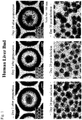
- Fig. 1 upper row shows photographs of a human liver bud prepared by three-dimensionally culturing human iPS cells, vascular endothelial cells and undifferentiated mesenchymal cells, which were taken with an inverted microscope at days 1, 7 and 15 after preparation. Even after 15 days from the preparation, the liver bud retains a spherical 3D structure.
- Fig. 1 bottom row shows photographs of the human liver bud infected with HBV, which were taken with an inverted microscope at days 10, 20 and 30 post infection. The morphology of the liver bud is retained up to 30 days post infection.
- Fig. 2 is a graph comparing HBV DNA copy numbers in culture supernatants between two cases where human iPS cell-derived hepatocytes and human liver buds were infected with HBV Markedly high HBV release was noted in human liver buds, compared with human iPS-derived hepatocytes.
- Fig. 3 is a graph comparing the intracellular HBV 3.5k RNA quantities between two cases where human iPS cell-derived hepatocytes and human liver buds were infected with HBV. Markedly high HBV 3.5k RNA was noted in cells of human liver buds, compared with human iPS-derived hepatocytes.
- Fig. 4 shows immunostaining of the inside of human liver buds with HBc antibody at days 10, 20 and 30 post infection. HBV infection was noted continuously at days 10, 20 and 30 post infection.
- Fig. 5 is a graph comparing HBV DNA copy numbers in culture supernatants of primary human hepatocytes (PXB cells) between two cases where PXB cells were infected with the culture supernatant of human liver buds at day 20 post infection and with a non-infected group. It was revealed that HBV-infected human liver buds are capable of producing infectious HBV.
- PXB cells primary human hepatocytes
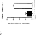
- Fig. 6 is a graph comparing intracellular HBV covalently closed circular DNA copy numbers between two cases where human iPS cell-derived hepatocytes and human liver buds were infected with HBV. A markedly great number of HBV covalently closed circular DNA copies were noted in human liver buds, compared with human iPS cell-derived hepatocytes.
- Fig. 7 is a set of electron micrographs comparing human liver bud at day 10 post infection with non-infected human liver bud.
- the HBV-infected human liver bud was observed to have fewer hepatocyte microvilli (indicated by arrow heads).
- Fig. 8 shows that infection of human liver bud with HBV is inhibited by various drugs.
- Con control group; IFN- ⁇ : interferon ⁇ 1000U/ml; IFN- ⁇ : interferon ⁇ 1000U/ml; PreS1: HBV surface antigen PreS1 peptide 100 nM; ETV: entecavir 1.8 ⁇ M. *p ⁇ 0.05, **p ⁇ 0.01, ***p ⁇ 0.0001 vs Con.
- the present invention is applicable to in vitro screenings in drug discovery.
Landscapes
- Health & Medical Sciences (AREA)
- Life Sciences & Earth Sciences (AREA)
- Engineering & Computer Science (AREA)
- Chemical & Material Sciences (AREA)
- Organic Chemistry (AREA)
- Biomedical Technology (AREA)
- Wood Science & Technology (AREA)
- Zoology (AREA)
- Bioinformatics & Cheminformatics (AREA)
- Biotechnology (AREA)
- Genetics & Genomics (AREA)
- Immunology (AREA)
- Microbiology (AREA)
- Biochemistry (AREA)
- General Health & Medical Sciences (AREA)
- General Engineering & Computer Science (AREA)
- Proteomics, Peptides & Aminoacids (AREA)
- Molecular Biology (AREA)
- Cell Biology (AREA)
- Analytical Chemistry (AREA)
- Physics & Mathematics (AREA)
- Virology (AREA)
- Biophysics (AREA)
- Hematology (AREA)
- Medicinal Chemistry (AREA)
- Urology & Nephrology (AREA)
- Communicable Diseases (AREA)
- Toxicology (AREA)
- Tropical Medicine & Parasitology (AREA)
- Food Science & Technology (AREA)
- General Physics & Mathematics (AREA)
- Pathology (AREA)
- Gastroenterology & Hepatology (AREA)
- Developmental Biology & Embryology (AREA)
- Transplantation (AREA)
- Micro-Organisms Or Cultivation Processes Thereof (AREA)
- Measuring Or Testing Involving Enzymes Or Micro-Organisms (AREA)
Description
- The present invention relates to a virus infection model, a method for preparing the same, and use of the same.
- Recently, methods of generating human functional cells useful for drug discovery screening and regenerative medicine by directed differentiation using pluripotent stem cells, such as iPS cells capable of differentiating into various functional cells, have been attracting attention. To date, the research group of the present inventors has established a method of reconstructing human tissues/organs which have well-organized three-dimensional structures composed of vascular endothelial cells and mesenchymal cells as seen in adult tissues (Non-Patent Document No. 1: Takebe T. et al., Nature (2013) Vol. 499, pp 481-485; Patent Document No. 1: Method for Producing Tissue and Organ
WO2013/047639 ). - On the other hand, infection models of hepatitis virus affecting humans have been conventionally prepared using primary cultures of human hepatocytes or various tumor cells (Non-Patent Document No. 2: Gripon P et al. Virol 1995; 213:292-299; Non-Patent Document No. 3: Watashi K et al., JBC. 2013 288: 31725-31727; Non-Patent Document No. 4: Yang D et al. PNAS 2014 E1264-E1273). However, the only system reported to date in which an in vitro reconstituted three-dimensional tissue is to be infected with hepatitis virus is one that uses tumor cells (Non-Patent Document No. 5: Molina-Jimenez F et al., Virology 2012 425; 31-9).
- Models in which primary cultures of human hepatocytes are infected with hepatitis virus (HBV: hepatitis B virus) have the following advantages and disadvantages.
- Susceptibility to HBV infection without over-expression of NTCP known as an HBV receptor, and so forth;
- Limited supply of hepatocytes; Non-proliferation of the hepatocytes;
- Low reproducibility (big difference between lots).
- Further, models in which hepatoma cell lines have been infected with hepatitis virus have the following advantages and disadvantages.
- The cells are capable of proliferation without lot-to-lot differences.
- A large quantity of virus (2,000-6,000 Geq/cell) is needed for establishment of infection.
- Hepatocyte-specific markers are not expressed because the cells are tumor cells.
- One problem with virus infection tests is that recapitulation of viral life cycle is difficult in the current culture system. Conventional primary cultures of hepatocytes are not susceptible to re-infection. As an infection model susceptible to re-infection, only human hepatocytes isolated from chimeric mice with a humanized liver have been reported (Non-Patent Document No. 6: Ishida Y, Am J Patho, Vol. 185, No.5, May 2015, pp.1275-1285). Other infection models are prepared by infecting tumor cells with virus (Non-Patent Document No. 7: Yang D, PNAS E1264-E1273 published online March 10, 2014; Non-Patent Document No. 8: Sai LT, JMII (2014)xx, 1-6).
- Japanese patent application publication no.
JP 2015-173635 -
- Non-Patent Document No. 1: Nature (2013) VOL.499, pp 481-485
- Non-Patent Document No. 2: Gripon P et al. Virol 1995; 213:292-299
- Non-Patent Document No. 3: Watashi K et al., JBC. 2013 288:31725-31727
- Non-Patent Document No. 4: Yang D et al. PNAS 2014 E1264-E1273
- Non-Patent Document No. 5: Molina-Jimenez F et al. Virology 2012 425; 31-9
- Non-Patent Document No. 6: Ishida Y, Am J Patho, Vol. 185, No.5, May 2015, pp.1275-1285
- Non-Patent Document No. 7: Yang D, PNAS E1264-E1273 published online March 10, 2014
- Non-Patent Document No. 8: Sai LT, JMII (2014)xx, 1-6
-
- Patent Document No. 1:
WO2013/047639 - Patent Document No. 2:
JP 2015-173635 - Conventional hepatitis virus infection models have the following drawbacks.
- The viral life cycle seen in hepatitis virus infection to humans, especially multi-round infection kinetics, could not be recapitulated.
- High concentrations of virus could not be released from the infected cells to the culture supernatant.
- Use of tumor cells made it impossible to examine functional disorders of the liver.
- The present invention aims at solving these problems.
- As a result of intensive and extensive efforts toward the solution of the above problems, the present inventors have found a technique for preparing a completely novel virus infection model using a human tissue/organ generated from pluripotent stem cells, etc. Briefly, the present inventors combined pluripotent stem cells (e.g., human iPS cells) with mesenchymal cells and vascular endothelial cells, to thereby reconstitute three-dimensional human liver buds; as a result, the multiplicity of infection of various organotropic viruses was outstandingly increased, leading to a successful construction of an infection model closely simulating an infection to the human liver. The present invention is expected to help advance the screenings in drug discovery and the elucidation of viral infection mechanism, eventually leading to the development of new drugs against various hepatitis virus infections and the elucidation of new mechanisms behind their pathogenesis.
- The gist of the present invention is as described below.
- (1) A method for constructing a hepatitis virus infection model capable of recapitulating a viral life cycle, by infecting a liver bud with hepatitis virus, wherein said liver bud is formed by co-culturing liver cells derived from human iPSC with human vascular endothelial cells and human mesenchymal cells.
- (2) The method of (1) above, wherein the vascular endothelial cells are human umbilical vein endothelial cells.
- (3) The method of (1) above, wherein the mesenchymal cells are human bone marrow-derived mesenchymal cell.
- (4) The method of (1) above, wherein the infecting hepatitis virus is hepatitis B virus, hepatitis C virus or hepatitis E virus.
- (5) A hepatitis virus infection model capable of recapitulating a viral life cycle, comprising a hepatitis virus-infected liver bud, wherein the liver bud is formed by co-culturing liver cells derived from human iPSC with human vascular endothelial cells and human mesenchymal cells.
- (6) The virus infection model of (5) above, wherein the infecting hepatitis virus is hepatitis B virus, hepatitis C virus or hepatitis E virus.
- (7) A method of screening for substances with antiviral activity, comprising using the virus infection model of (5) above.
- (8) A method of screening for substances with antiviral activity, comprising using the virus infection model of (6) above, wherein the infecting virus is hepatitis B virus, hepatitis C virus or hepatitis E virus.
- To date, the research group of the present inventors has succeeded in constructing a platform technology for preparing a human tissue/organ in which a vascular system is appropriately located in a way that has never been achieved by conventional techniques (
WO2013/047639 Method for Producing Tissue and Organ). - In the present invention, a human liver bud prepared by the above-described platform technology is infected with hepatitis virus and this enables the following to be achieved.
- To recapitulate the viral life cycle seen in hepatitis virus infection to humans, especially multi-round infection kinetics.
- To release high concentrations of virus from infected cells into the culture supernatant
- To examine functional disorders of the liver.
- It is anticipated that the present invention can be applied to in vitro screenings in drug discovery in a more convenient and efficient manner than conventionally performed.
- Since liver buds can be produced uniformly and in large quantities, the present invention can eliminate inter-lot variability as seen in conventional primary cultures of hepatocytes, making it possible to produce a large quantity of the virus-infected human liver tissue necessary for drug discovery and development.
- The present specification refers to the contents disclosed in the specification and/or the drawings of Japanese Patent Application No.
2015-249520 -
- [
Fig. 1 ] Upper row: photographs of a human liver bud prepared by three-dimensionally culturing human iPS cells, vascular endothelial cells and undifferentiated mesenchymal cells, as observed with an inverted optical microscope atdays
Bottom row: photographs of the human liver bud infected with HBV, as observed with an inverted optical microscope atdays - [
Fig. 2 ] A graph comparing HBV DNA copy numbers in culture supernatants between two cases where human iPS cell-derived hepatocytes and human liver buds were infected with HBV. - [
Fig. 3 ] A graph comparing the intracellular HBV 3.5k RNA quantities between two cases where human iPS cell-derived hepatocytes and human liver buds were infected with HBV - [
Fig. 4 ] This figure shows HBV infection inside the human liver bud. The liver bud atdays - [
Fig. 5 ] A graph comparing HBV DNA copy numbers in culture supernatants of primary human hepatocytes (PXB cells) between two cases where PXB cells were infected with the culture supernatant of human liver buds atday 20 post infection and with a non-infected group. - [
Fig. 6 ] A graph comparing intracellular HBV covalently closed circular DNA copy numbers between two cases where human iPS cell-derived hepatocytes and human liver buds were infected with HBV Compared to human iPS cell-derived hepatocytes, human liver buds had a noticeably greater number of HBV covalently closed circular DNA copies. - [
Fig. 7 ] Electron micrographs comparing human liver bud atday 10 post infection with non-infected human liver bud. The HBV-infected human liver bud was observed to have fewer hepatocyte microvilli (indicated by arrow heads). - [
Fig. 8 ] This figure shows that infection of human liver bud with HBV is inhibited by various drugs. Con: control group; IFN-α: interferon α 1000U/ml; IFN-γ: interferon γ 1000U/ml; PreS1: HBV surfaceantigen PreS1 peptide 100 nM; ETV: entecavir 1.8 µM. *p<0.05, **p<0.01, ***p<0.0001 vs Con. - Hereinbelow, the present invention will be described in detail.
- The present invention provides a method for constructing a hepatitis virus infection model capable of recapitulating a viral life cycle, by infecting a liver bud with hepatitis virus, wherein said liver bud is formed by co-culturing liver cells derived from human iPSC with human vascular endothelial cells and human mesenchymal cells.
- As disclosed herein, the term "cell condensate" refers to one that is formed by culturing a tissue or organ cell in vitro. Such a cell condensate is disclosed in, for example,
WO2015/129822 (Method for generating cell condensate for self-organization). In the cell condensate, cell-cell interactions take place in such a close manner that a biological environment as occurs in the womb is recapitulated. As a consequence, induction of early differentiation into organ progenitor cells occurs efficiently and this would improve the frequency and number of organ progenitor cells present. The cell condensate may be one in which cells adhere to each other so strongly that it can be collected in a non-destructive manner. - The cell condensate described above is of a concept that generally encompasses organ buds and organoids [organ bud (
WO2013/047639 ), liver bud, liver diverticula, liver organoid, pancreatic (dorsal or ventral) buds, pancreatic diverticula, pancreatic organoid, intestinal bud, intestinal diverticula, intestinal organoid (K. Matsumoto et al. Science. 19; 294 (5542): 559-63 (2001)]. While the cell condensate is independent of the types of constituent cells and the number of such types, organ buds correspond to cell condensates that are formed at an early stage of organogenesis and are in principle composed of the following three types of cells: functional cells that constitute organs or tissues (or undifferentiated cells which will differentiate into functional cells); vascular cells; and mesenchymal cells. Organoids are solely composed of cells that constitute epithelial tissues and they are basically of a small size (1 mm or less). - The cell condensate undergoes self-organization to form a three-dimensional tissue structure provided with higher structures, whereby progenitor cells can be directed to terminal differentiation. Self-organization may be performed either in vivo or in vitro. For example, when the cell condensate is transplanted into a living body, vascular networks are formed, blood perfusion is induced, and self-organization into a higher tissue with a complex structure occurs, enabling the preparation of tissues/organs that have a highly ordered tissue structure comparable to that of adult tissues. With such a cell condensate, it may be possible to prepare a higher tissue that is provided not only with a vascular network but also with additional higher structures such as ureteral structure, biliary structure, tracheal structure, etc. Further, there are many organs including the liver which, in order to exhibit their functions, essentially require the reconstitution of not only themselves but also associations with other organs; in the case of the liver, not only itself but also the junctions with the bile and pancreatic ducts as well as the connection to the duodenum must be reconstituted in order that it exhibits its function.
- The cell condensate may be formed by culturing a tissue or organ cell with vascular endothelial cells or mesenchymal cells in vitro. A method for preparing such a cell condensate is disclosed in
WO2015/129822 (Method for generating cell condensate for self-organization), but other preparation methods may also be used. - The cell condensate formed by culturing a tissue or organ cell in vitro may be an organ bud.
- As disclosed herein, the term "organ bud" means a structure capable of differentiating into an organ through maturing, the structure comprising tissue or organ cells, vascular endothelial cells and mesenchymal cells (undifferentiated mesenchymal cells or cells differentiated therefrom). Whether a structure is an organ bud or not can be determined, for example, by transplanting the structure into an organism and examining whether or not it is capable of differentiating into an organ of interest (the structure can be judged as organ bud if it has differentiated into the organ of interest); and/or by examining whether or not the structure comprises all of the above-described three types of cells, i.e., tissue or organ cells, vascular endothelial cells and mesenchymal cells (the structure can be judged as organ bud if it comprises all of the three types of cells). The organ bud may be one which differentiates into an organ such as kidney, heart, lung, spleen, esophagus, stomach, thyroid, parathyroid, thymus, gonad, brain, spinal cord or the like. Preferably, the organ bud is one which differentiates into an endodermal organ such as one which differentiates into liver (liver bud), one which differentiates into pancreas (pancreas bud), or one which differentiates into intestinal tract. Whether a certain structure is an organ bud which differentiates into an endodermal organ or not can be determined by examining the expression of marker proteins
- (if any one or more of the marker proteins described later are expressed, the organ bud can be judged as the organ bud of interest). For example, HHEX, SOX2, HNF4A, AFP, ALB and the like are markers for liver bud; PDX1, SOX17, SOX9 and the like are markers for pancreas bud; and CDX2, SOX9 and the like are markers for organ buds which differentiate into intestinal tract. Among the terms used by those skilled in the art, the following are included in the organ bud of the disclosure: liver bud, liver diverticula, liver organoid, pancreatic (dorsal or ventral) buds, pancreatic diverticula, pancreatic organoid, intestinal bud, intestinal diverticula, intestinal organoid (K. Matsumoto, et al. Science. 19; 294 (5542): 559-63 (2001)) and so on.
- An organ bud may be formed by culturing tissue or organ cells with vascular endothelial cells and mesenchymal cells in vitro. A method for preparing an organ bud is disclosed in
WO2013/047639 (Method for Producing Tissue and Organ), but other preparation methods may also be used. - As used herein, the term "tissue or organ cell" means functional cells constituting tissues or organs, or undifferentiated cells which differentiate into functional cells. Examples of "undifferentiated tissue or organ cell" include, but are not limited to, cells capable of differentiating into an organ such as kidney, heart, lung, spleen, esophagus, stomach, thyroid, parathyroid, thymus, gonad, brain or spinal cord; cells capable of differentiating into an ectodermal organ such as brain, spinal cord, adrenal medulla, epidermis, hair/nail/dermal gland, sensory organ, peripheral nerve or lens; cells capable of differentiating into a mesodermal organ such as kidney, urinary duct, heart, blood, gonad, adrenal cortex, muscle, skeleton, dermis, connective tissue or mesothelium; and cells capable of differentiating into an endodermal organ such as liver, pancreas, intestinal tract, lung, thyroid, parathyroid or urinary tract. Whether or not a cell is capable of differentiating into an ectodermal organ, mesodermal organ or endodermal organ can be determined by examining the expression of marker proteins (if any one or more of marker proteins are expressed, the cell can be judged as a cell capable of differentiating into an endodermal organ). For example, in cells capable of differentiating into liver, HHEX, SOX2, HNF4A, AFP, ALB and the like are markers; in cells capable of differentiating into pancreas, PDX1, SOX17, SOX9 and the like are markers; in cells capable of differentiating into intestinal tract, CDX2, SOX9 and the like are markers; in cells capable of differentiating into kidney, SIX2 and SALL1 are markers; in cells capable of differentiating into heart, NKX2-5, MYH6, ACTN2, MYL7 and HPPA are markers; in cells capable of differentiating into blood, C-KIT, SCA1, TER119 and HOXB4 are markers; and in cells capable of differentiating into brain or spinal cord, HNK1, AP2, NESTIN and the like are markers. Among the terms used by those skilled in the art, the following are included in the "undifferentiated tissue or organ cell" of the disclosure: hepatoblast, hepatic progenitor cells, hepatic precursor cells, pancreatoblast, pancreatic progenitors, pancreatic progenitor cells, pancreatic precursor cells, endocrine precursors, intestinal progenitor cells, intestinal precursor cells, intermediate mesoderm, metanephric mesenchymal precursor cells, multipotent nephron progenitor, renal progenitor cells, cardiac mesoderm, cardiovascular progenitor cells, cardiac progenitor cells (JR. Spence, et al. Nature.; 470(7332):105-9.(2011); Self, et al. EMBO J.; 25(21): 5214-5228.(2006); J. Zhang, et al. Circulation Research.; 104: e30-e41(2009); G. Lee, et al. )) and so on. Undifferentiated tissue or organ cells may be collected from tissues or organs, or may be prepared from pluripotent stem cells such as induced pluripotent stem cells (iPS cells) or embryonic stem cells (ES cells) according to known methods. Moreover, undifferentiated tissue or organ cells may be such cells as primitive gut endoderm cells (PGECs) (Japanese Patent No.
5777127 5777127 - In the present invention, the term "vascular endothelial cell" means cells constituting vascular endothelium or cells capable of differentiating into such cells. Whether a cell is vascular endothelial cell or not can be determined by examining the expression of marker proteins such as TIE2, VEGFR-1, VEGFR-2, VEGFR-3 and CD41 (if any one or more of the above-listed marker proteins are expressed, the cell can be judged as vascular endothelial cell). The vascular endothelial cell used in the present invention may be either differentiated or undifferentiated. Whether a vascular endothelial cell is a differentiated cell or not can be determined by means of CD31 and CD144. Among the terms used by those skilled in the art, the following are included in the "vascular endothelial cell" used in the present invention endothelial cells, umbilical vein endothelial cells, endothelial progenitor cells, endothelial precursor cells, vasculogenic progenitors, hemangioblast (HJ. Joo, et al. Blood. 25;118(8):2094-104 (2011)) and so on. Preferable vascular endothelial cells are those derived from umbilical vein. As vascular endothelial cells, human-derived cells are used.
- In the present invention, the term "mesenchymal cell" means connective tissue cells that are mainly located in mesoderm-derived connective tissues and which form support structures for cells that function in tissues. The "mesenchymal cell" is a concept that encompasses those cells which are destined to, but are yet to, differentiate into mesenchymal cells. Mesenchymal cells used in the present invention may be either differentiated or undifferentiated. Whether a certain cell is an undifferentiated mesenchymal cell or not may be determined by examining the expression of marker proteins such as Stro-1, CD29, CD44, CD73, CD90, CD105, CD133, CD271 or Nestin (if any one or more of the above-listed marker proteins are expressed, the cell can be judged as an undifferentiated mesenchymal cell). A mesenchymal cell in which none of the above-listed markers are expressed can be judged as a differentiated mesenchymal cell. Among the terms used by those skilled in the art, the following are included in the "mesenchymal cell" used in the present invention mesenchymal stem cells, mesenchymal progenitor cells, mesenchymal cells (R. Peters, et al. PLoS One. 30; 5(12):e15689 (2010)) and so on. Preferable mesenchymal cells are mesenchymal cells derived from bone marrow (especially, mesenchymal stem cells). As mesenchymal cells, human-derived cells are used.
- Culture ratios of three cell types in coculture are not particularly limited as long as they are within the range that enables the formation of liver buds. Preferable cell count ratio is 10 : 10-5 : 0.1-3 for liver cell : human vascular endothelial cell : human mesenchymal cell. Liver buds of approximately 50 µm to 3 mm in size may be formed by coculturing approx. 400,000 liver cells, approx. 200,000 to 400,000 human vascular endothelial cells and approx. 20,000 to 120,000 human mesenchymal cells.
- The medium used for culture may be any medium that enables the formation of cell condensates (preferably, organ buds) and examples that are preferably used include a medium for culturing vascular endothelial cells, a medium for culturing tissue or organ cells, and a mixture of these two media. As a medium for culturing vascular endothelial cells, any medium may be used but, preferably, a medium containing at least one of the following substances may be used: hEGF (recombinant human epidermal growth factor), VEGF (vascular endothelial growth factor), hydrocortisone, bFGF, ascorbic acid, IGF1, FBS, antibiotics (e.g., gentamycin or amphotericin B), heparin, L-glutamine, phenol red and BBE. Specific examples of media that may be used for culturing vascular endothelial cells include, but are not limited to, EGM-2 BulletKit (Lonza), EGM BulletKit (Lonza), VascuLife EnGS Comp Kit (LCT), Human Endothelial-SFM Basal Growth Medium (Invitrogen) and human microvascular endothelial cell growth medium (TOYOBO). As a medium for culturing tissue or organ cells, any medium may be used but in the case where the organ cell is a hepatocyte, a medium containing at least one of ascorbic acid, BSA-FAF, insulin, hydrocortisone and GA-1000 may preferably be used. As a medium for culturing hepatocytes, HCM BulletKit (Lonza) from which hEGF (recombinant human epidermal growth factor) has been removed or RPMI1640 (Sigma-Aldrich) to which 1% B27 Supplements (GIBCO) and 10 ng/mL hHGF (Sigma-Aldrich) have been added may typically be used. As regards the formation of human liver buds, a 1:1 mixture of GM BulletKit (Lonza) and HCM BulletKit (Lonza) from each of which hEGF has been removed and which are each supplemented with dexamethasone, oncostatin M and HGF has been found effective for maturation of liver buds.
- Although scaffold materials need not be used for culturing cells, a cell mixture may advantageously be cultured on a gel-like support that allows mesenchymal cells to contract.
- Contraction of mesenchymal cells may be confirmed, for example, by noting the formation of a 3D tissue morphologically (either under microscope or with the naked eye) or by showing that the tissue is strong enough to retain its shape as it is collected with a spatula or the like (Takebe et al. Nature 499 (7459), 481-484, 2013).
- The support may advantageously be a gel-like substrate having an appropriate stiffness [e.g., a Young's modulus of 200 kPa or less (in the case of a Matrigel-coated gel of a flat shape); however, the appropriate stiffness of the support may vary depending on the coating and shape]. Examples of such substrates include, but are not limited to, hydrogels (such as acrylamide gel, gelatin and Matrigel). The stiffness of the support need not be uniform and may vary with the shape, size and quantity of a cell condensate of interest so that it can be provided with a spatial/temporal gradient or can be patterned. In the case where the stiffness of the support is uniform, it is preferably 100 kPa or less, more preferably 1-50 kPa. The gel-like support may be planar, or alternatively, the side on which culture is to be performed may have a U- or V-shaped cross section. If the side of the gel-like support on which culture is to be performed has a U- or V-shaped cross section, cells tend to gather on the culture surface and a cell condensate can advantageously be formed from a smaller number of cells and/or tissues. Moreover, the support may be modified chemically or physically. Examples of modifying substances include, but are not limited to, Matrigel, laminin, entactin, collagen, fibronectin and vitronectin.
- One example of the gel-like culture support that is provided with a spatial gradient of stiffness is a gel-like culture support that is stiffer in the central part than in the peripheral part. The stiffness of the central part is appropriately 200 kPa or less and it suffices that the peripheral part is softer than the central part. Appropriate values for the stiffness of the central and peripheral parts of the substrate are variable with the coating and the shape. Another example of the gel-like culture support that is provided with a spatial gradient of stiffness is a gel-like culture support that is stiffer in the peripheral part than in the central part.
- One example of the patterned, gel-like culture support is a gel-like culture support having one or more patterns in which the central part is stiffer than the peripheral part. The stiffness of the central part is appropriately 200 kPa or less and it suffices that the peripheral part is softer than the central part. Appropriate values for the stiffness of the central and peripheral parts of the substrate are variable with the coating and the shape. Another example of the patterned, gel-like culture support is a gel-like culture support having one or more patterns in which the peripheral part is stiffer of than the central part. The stiffness of the peripheral part is appropriately 200 kPa or less and it suffices that the central part is softer than the peripheral part. Appropriate values for the stiffness of the central and peripheral parts of the substrate are variable with the coating and the shape.
- The temperature during culture is not particularly limited but it is preferably 30-40°C and more preferably 37°C.
- The culture period is not particularly limited but it is preferably 3-10 days and more preferably 6 days.
- A hepatitis virus infection model may be prepared by infecting a liver bud formed by the above-described method with a hepatitis virus. This model is capable of recapitulating a viral life cycle, especially multi-round infection kinetics. Moreover, this model achieves high concentrations of hepatitis virus release from infected cells to the culture supernatant. As will be shown in the Example described later, the level of intracellular HBV RNA in the virus infection model of the present invention (HBV-infected liver bud) increased up to
day 30 post infection, and HBV DNA was observed in the culture supernatant. Further, the virus released into the culture supernatant of the virus infection model of the present invention was capable of infecting other cells. In other words, the life cycle of the virus could be recapitulated. Further, a markedly high HBV closed circular DNA was noted in the virus infection model of the invention (HBV-infected liver bud). Still further, hepatocyte microvilli were reduced in the virus infection model of the present invention (HBV-infected liver bud). - The types of hepatitis virus which may be used in the present invention include, but are not limited to, hepatitis B virus, hepatitis C virus and hepatitis E virus.
- The liver bud to be infected with a hepatitis virus may advantageously be one prepared by mixing, for example, liver cells derived from human iPSC, human vascular endothelial cells and human mesenchymal cells, coculturing the resultant mixture in a medium containing DMSO for 7 days and further coculturing in a medium containing HGF, OSM and DEX for 7 days. For infection with HBV, the liver bud may, for example, be infected with about 500 copies of HBV per liver cell, and then cultured for about 30 days. A medium containing PEG8000 may be used for the culture.
- The present invention also provides a hepatitis virus infection model capable of recapitulating a viral life cycle, comprising a hepatitis virus-infected liver bud, wherein the liver bud is formed by co-culturing liver cells derived from human iPSC with human vascular endothelial cells and human mesenchymal cells.
- The liver bud, the virus and the virus-infected liver bud are as described above. The types of hepatitis virus which may be used in the present invention include, but are not limited to, hepatitis B virus, hepatitis C virus and hepatitis E virus.
- The present invention also provides a method of screening for substances with antiviral activity, comprising using the above-described hepatitis virus infection model. The types of hepatitis virus which may be used in the present invention include, but are not limited to, hepatitis B virus, hepatitis C virus and hepatitis E virus.
- One example of the method of screening for substances with antiviral activity will be described below. To begin with, the above-described liver bud is infected with a hepatitis virus, and then a test substance is supplied thereto. Alternatively, a test substance is mixed into a liver bud under preparation. Quantities of hepatitis virus in the liver bud culture supernatant or in the liver bud per se are compared between two groups, one supplied with the test substance and the other not supplied with the test substance. Alternatively, test substances which reduce the quantity of hepatitis virus in the culture supernatant or in the liver bud by 50% or more are identified. Test substances are not particularly limited and may include, but are not limited to, low molecular weight compounds, peptides, nucleic acids, extracts from natural products, and inorganic compounds. The quantities of hepatitis virus in liver bud culture supernatant or intracellular hepatitis virus from
day 1 today 30 after the supply of test substance are compared to the corresponding quantities in the control group which did not receive the test substance. It is desirable to select those test substances which reduce the quantity of hepatitis virus to 30% or less of the quantity in the control group not supplied with the test substance. This method enables the screening for hepatitis viral therapeutics or prophylactics. - Target substances for screening may be proteins, polypeptides, oligopeptides, nucleic acids (including natural and artificial nucleic acids), low molecular weight organic compounds, inorganic compounds, cell extracts, and extracts from animals or plants, soils and so forth. The substance may be either a natural product or a synthesized product.
- Hereinbelow, the present invention will be described in more detail with reference to the following Example.
- Human liver buds were prepared according to the "Method for Preparing Tissue and Organ" (
WO2013/047639 ). - (1) Preparation of human hepatic endoderm cells
Human iPS cells (for example, human skin-derived TkDA3 hiPSC clone (provided by the University of Tokyo)) were cultured in activin-, Wnt 3a- and NaB-supplemented serum-free medium to thereby obtain endodermal cells (definitive endoderms: DEs). - (2) Preparation of human liver buds
The endodermal cells obtained, vascular endothelial cells (human umbilical vein-derived endothelial cells) (Lonza, Basel, Switzerland) and undifferentiated mesenchymal cells (human bone marrow-derived mesenchymal stem cells) (Lonza, Basel, Switzerland) were mixed at 10:7:1 (400 cells: 280 cells: 40 cells per organ bud) to prepare human liver buds (120-150 µm in size). - (3) Infection of human liver buds with HBV
The human liver buds prepared in (2) above were cultured in DMSO-containing medium for 7 days and further cultured in HGF-, OSM- and DEX-containing medium for 7 days, followed by infection with HBV. HBV was recovered from culture supernatant of HepG2.2.15 cells which had been prepared by transferring the intact HBV genome (genotype D) into human hepatoma cell line HepG2 by lipofection. The liver buds were infected with approx. 500 copies of HBV per endodermal cell in 4% PEG8000-containing medium and cultured for about 30 days. - (4) Preparation of human iPS cell-derived hepatocytes and HBV infection
The endodermal cells obtained in (1) above were cultured in KO-DMEM-, KSR- and DMSO-containing medium for 7 days to thereby obtain immature hepatocytes (IHs). The resultant cells were then cultured in HGF-, OSM- and DEX-containing medium for another 7 days to thereby obtain human iPS cell-derived hepatocytes (mature hepatocytes: MHs). The human iPS cell-derived hepatocytes were infected with the same HBV as used in (3) above. Approx. 500 copies of HBV per cell and 4% PEG8000-containing medium were used for infection. - (5) Culture supernatants of human liver buds and human iPS-cell derived hepatocytes were recovered at
days - (6) Human liver buds and human iPS-cell derived hepatocytes were recovered at
days - (7) The inside of human liver buds was immunostained with HBc antibody at
days - (8) Human primary hepatocytes (PXB cells; Phoenix Bio) were infected with the culture supernatant of human liver buds at
day 20 post HBV infection. - (9) Human liver buds and human iPS-cell derived hepatocytes were recovered at
day 20 post HBV infection, followed by measurement of covalently closed circular DNA (cccDNA). - (10) Human liver buds at
day 10 post HBV infection and non-HBV-infected human liver buds in the corresponding period were fixed with paraformaldehyde and glutaraldehyde, and electron micrographs were taken. - (11) Human liver buds were infected with HBV (5,000 copies per cell). Interferon α (1000 U/ml), interferon γ (1000 U/ml), PreS1 peptide (100 nM), and entecavir (1.8 µM) were added to the medium, in which the liver buds were cultured for 10 days. Intracellular HBV 3.5k RNA and HBV DNA in culture supernatant were measured.
- The results are shown in
Figs. 1 to 8 . -
Fig. 1 , upper row shows photographs of a human liver bud prepared by three-dimensionally culturing human iPS cells, vascular endothelial cells and undifferentiated mesenchymal cells, which were taken with an inverted microscope atdays Fig. 1 , bottom row shows photographs of the human liver bud infected with HBV, which were taken with an inverted microscope atdays -
Fig. 2 is a graph comparing HBV DNA copy numbers in culture supernatants between two cases where human iPS cell-derived hepatocytes and human liver buds were infected with HBV Markedly high HBV release was noted in human liver buds, compared with human iPS-derived hepatocytes. -
Fig. 3 is a graph comparing the intracellular HBV 3.5k RNA quantities between two cases where human iPS cell-derived hepatocytes and human liver buds were infected with HBV. Markedly high HBV 3.5k RNA was noted in cells of human liver buds, compared with human iPS-derived hepatocytes. -
Fig. 4 shows immunostaining of the inside of human liver buds with HBc antibody atdays days -
Fig. 5 is a graph comparing HBV DNA copy numbers in culture supernatants of primary human hepatocytes (PXB cells) between two cases where PXB cells were infected with the culture supernatant of human liver buds atday 20 post infection and with a non-infected group. It was revealed that HBV-infected human liver buds are capable of producing infectious HBV. -
Fig. 6 is a graph comparing intracellular HBV covalently closed circular DNA copy numbers between two cases where human iPS cell-derived hepatocytes and human liver buds were infected with HBV. A markedly great number of HBV covalently closed circular DNA copies were noted in human liver buds, compared with human iPS cell-derived hepatocytes. -
Fig. 7 is a set of electron micrographs comparing human liver bud atday 10 post infection with non-infected human liver bud. The HBV-infected human liver bud was observed to have fewer hepatocyte microvilli (indicated by arrow heads). -
Fig. 8 shows that infection of human liver bud with HBV is inhibited by various drugs. Con: control group; IFN-α: interferon α 1000U/ml; IFN-γ: interferon γ 1000U/ml; PreS1: HBV surfaceantigen PreS1 peptide 100 nM; ETV: entecavir 1.8 µM. *p<0.05, **p<0.01, ***p<0.0001 vs Con. - The present invention is applicable to in vitro screenings in drug discovery.
Claims (8)
- A method for constructing a hepatitis virus infection model capable of recapitulating a viral life cycle, by infecting a liver bud with hepatitis virus, wherein said liver bud is formed by co-culturing liver cells derived from human iPSC with human vascular endothelial cells and human mesenchymal cells.
- The method of claim 1, wherein the vascular endothelial cells are human umbilical vein endothelial cells.
- The method of claim 1, wherein the mesenchymal cells are human bone marrow-derived mesenchymal cells.
- The method of claim 1, wherein the infecting hepatitis virus is hepatitis B virus, hepatitis C virus or hepatitis E virus.
- A hepatitis virus infection model capable of recapitulating a viral life cycle, comprising a hepatitis virus-infected liver bud, wherein the liver bud is formed by co-culturing liver cells derived from human iPSC with human vascular endothelial cells and human mesenchymal cells.
- The virus infection model of claim 5, wherein the infecting hepatitis virus is hepatitis B virus, hepatitis C virus or hepatitis E virus.
- A method of screening for substances with antiviral activity, comprising using the virus infection model of claim 5.
- A method of screening for substances with antiviral activity, comprising using the virus infection model of claim 6, wherein the infecting virus is hepatitis B virus, hepatitis C virus or hepatitis E virus.
Applications Claiming Priority (2)
| Application Number | Priority Date | Filing Date | Title |
|---|---|---|---|
| JP2015249520 | 2015-12-22 | ||
| PCT/JP2016/088220 WO2017110931A1 (en) | 2015-12-22 | 2016-12-21 | Virus infection model, preparation method therefor, and utilization thereof |
Publications (3)
| Publication Number | Publication Date |
|---|---|
| EP3395941A1 EP3395941A1 (en) | 2018-10-31 |
| EP3395941A4 EP3395941A4 (en) | 2019-07-17 |
| EP3395941B1 true EP3395941B1 (en) | 2021-03-10 |
Family
ID=59089533
Family Applications (1)
| Application Number | Title | Priority Date | Filing Date |
|---|---|---|---|
| EP16878818.0A Active EP3395941B1 (en) | 2015-12-22 | 2016-12-21 | Virus infection model, preparation method therefor, and utilization thereof |
Country Status (10)
| Country | Link |
|---|---|
| US (1) | US20190285619A1 (en) |
| EP (1) | EP3395941B1 (en) |
| JP (1) | JP6869543B2 (en) |
| KR (1) | KR20180095538A (en) |
| CN (1) | CN108350418B (en) |
| AU (1) | AU2016375418A1 (en) |
| BR (1) | BR112018010867A2 (en) |
| CA (1) | CA3008218A1 (en) |
| SG (1) | SG11201804754SA (en) |
| WO (1) | WO2017110931A1 (en) |
Families Citing this family (3)
| Publication number | Priority date | Publication date | Assignee | Title |
|---|---|---|---|---|
| CN111417716A (en) * | 2017-11-30 | 2020-07-14 | 公立大学法人横滨市立大学 | Construction of Stereoscopic Organs from Pluripotent Stem Cells |
| CN109731095B (en) * | 2019-03-14 | 2022-06-21 | 广东龙帆生物科技有限公司 | Medicine for preventing or treating hepatitis B virus infection and preparation method and application thereof |
| CN120391396B (en) * | 2025-07-04 | 2025-09-16 | 成都市第三人民医院 | A pulmonary vascular endothelial barrier injury model and its construction method and application |
Family Cites Families (7)
| Publication number | Priority date | Publication date | Assignee | Title |
|---|---|---|---|---|
| US6995299B2 (en) * | 1999-11-02 | 2006-02-07 | University Of Connecticut | Propagation of human hepatocytes in non-human animals |
| DE60131676T2 (en) * | 2000-12-01 | 2008-10-30 | Central Institute For Experimental Animals | PROCESS FOR MAKING A MOUSE SUITABLE FOR THE RECORDING, DIFFERENTIATION AND PROLIFERATION OF HETEROGENIC CELLS, THE MOUSE PRODUCED THEREOF AND THEIR USE |
| EP2762558B1 (en) * | 2011-09-27 | 2026-01-21 | Public University Corporation Yokohama City University | Method for producing tissue and organ |
| EP2890779B1 (en) * | 2012-08-31 | 2017-10-04 | The Governors of the University of Alberta | Methods for producing cells having a phenotype of primary human hepatocytes and compositions |
| SG11201606750UA (en) | 2014-02-27 | 2016-10-28 | Public Univ Corp Yokohama City | Method for generating cell condensate for self-organization |
| JP6418761B2 (en) * | 2014-03-14 | 2018-11-07 | 国立大学法人東京工業大学 | Hepatitis tissue, hepatitis virus infection method, hepatitis tissue production method, hepatitis virus propagation method, hepatitis vaccine production method, screening method, and kit |
| JP5777127B1 (en) | 2014-12-09 | 2015-09-09 | 公立大学法人横浜市立大学 | Primordial gut endoderm cells and method for producing the same |
-
2016
- 2016-12-21 JP JP2017558218A patent/JP6869543B2/en active Active
- 2016-12-21 SG SG11201804754SA patent/SG11201804754SA/en unknown
- 2016-12-21 WO PCT/JP2016/088220 patent/WO2017110931A1/en not_active Ceased
- 2016-12-21 BR BR112018010867-7A patent/BR112018010867A2/en not_active IP Right Cessation
- 2016-12-21 US US15/779,723 patent/US20190285619A1/en not_active Abandoned
- 2016-12-21 CA CA3008218A patent/CA3008218A1/en not_active Abandoned
- 2016-12-21 KR KR1020187017399A patent/KR20180095538A/en not_active Withdrawn
- 2016-12-21 CN CN201680068253.XA patent/CN108350418B/en not_active Expired - Fee Related
- 2016-12-21 EP EP16878818.0A patent/EP3395941B1/en active Active
- 2016-12-21 AU AU2016375418A patent/AU2016375418A1/en not_active Abandoned
Non-Patent Citations (1)
| Title |
|---|
| None * |
Also Published As
| Publication number | Publication date |
|---|---|
| SG11201804754SA (en) | 2018-07-30 |
| AU2016375418A1 (en) | 2018-07-05 |
| WO2017110931A1 (en) | 2017-06-29 |
| CN108350418B (en) | 2021-08-31 |
| HK1259971A1 (en) | 2019-12-13 |
| US20190285619A1 (en) | 2019-09-19 |
| KR20180095538A (en) | 2018-08-27 |
| EP3395941A1 (en) | 2018-10-31 |
| CA3008218A1 (en) | 2017-06-29 |
| JP6869543B2 (en) | 2021-05-12 |
| JPWO2017110931A1 (en) | 2018-10-11 |
| CN108350418A (en) | 2018-07-31 |
| EP3395941A4 (en) | 2019-07-17 |
| BR112018010867A2 (en) | 2018-11-21 |
Similar Documents
| Publication | Publication Date | Title |
|---|---|---|
| JP6990462B2 (en) | Organ bud | |
| US20240336898A1 (en) | Formation of three-dimensional organ from pluripotent stem cells, method for generating cell condensate for self-organization | |
| AU2015223798B2 (en) | Method for fabricating cell aggregate for self-organization | |
| KR102298147B1 (en) | Culture method and cell mass | |
| Li et al. | Engineering-derived approaches for iPSC preparation, expansion, differentiation and applications | |
| EP3395943B1 (en) | Method for manufacturing tissue/organ by using blood cells | |
| JP7228269B2 (en) | Cell mass fusion method | |
| CN103191445A (en) | Use of mesenchymal stem cell and preparation method thereof | |
| JP7158040B2 (en) | A construct that connects a structure and a cell mass | |
| EP3395941A1 (en) | Virus infection model, preparation method therefor, and utilization thereof | |
| HK1259971B (en) | Virus infection model, preparation method therefor, and utilization thereof | |
| JPWO2003038075A1 (en) | Method for inducing differentiation from mesoderm stem cells or ES cells, or immortalized mesoderm stem cells to neural cells | |
| WO2024034559A1 (en) | Method for producing cell aggregates | |
| Hosaka et al. | Exploration for Cell Sources for Liver Regenerative Medicine:“CLiP” as a Dawn of Cell Transplantation Therapy |
Legal Events
| Date | Code | Title | Description |
|---|---|---|---|
| STAA | Information on the status of an ep patent application or granted ep patent |
Free format text: STATUS: THE INTERNATIONAL PUBLICATION HAS BEEN MADE |
|
| PUAI | Public reference made under article 153(3) epc to a published international application that has entered the european phase |
Free format text: ORIGINAL CODE: 0009012 |
|
| STAA | Information on the status of an ep patent application or granted ep patent |
Free format text: STATUS: REQUEST FOR EXAMINATION WAS MADE |
|
| 17P | Request for examination filed |
Effective date: 20180719 |
|
| AK | Designated contracting states |
Kind code of ref document: A1 Designated state(s): AL AT BE BG CH CY CZ DE DK EE ES FI FR GB GR HR HU IE IS IT LI LT LU LV MC MK MT NL NO PL PT RO RS SE SI SK SM TR |
|
| AX | Request for extension of the european patent |
Extension state: BA ME |
|
| DAV | Request for validation of the european patent (deleted) | ||
| DAX | Request for extension of the european patent (deleted) | ||
| A4 | Supplementary search report drawn up and despatched |
Effective date: 20190619 |
|
| RIC1 | Information provided on ipc code assigned before grant |
Ipc: C12N 7/00 20060101ALI20190613BHEP Ipc: C12Q 1/70 20060101ALI20190613BHEP Ipc: C12N 5/071 20100101ALI20190613BHEP Ipc: C12N 5/16 20060101ALI20190613BHEP Ipc: C12N 5/00 20060101AFI20190613BHEP Ipc: C12N 5/0775 20100101ALI20190613BHEP Ipc: C12Q 1/02 20060101ALI20190613BHEP Ipc: C12N 5/074 20100101ALI20190613BHEP |
|
| REG | Reference to a national code |
Ref country code: HK Ref legal event code: DE Ref document number: 1259971 Country of ref document: HK |
|
| STAA | Information on the status of an ep patent application or granted ep patent |
Free format text: STATUS: EXAMINATION IS IN PROGRESS |
|
| 17Q | First examination report despatched |
Effective date: 20200416 |
|
| GRAP | Despatch of communication of intention to grant a patent |
Free format text: ORIGINAL CODE: EPIDOSNIGR1 |
|
| STAA | Information on the status of an ep patent application or granted ep patent |
Free format text: STATUS: GRANT OF PATENT IS INTENDED |
|
| RIC1 | Information provided on ipc code assigned before grant |
Ipc: C12Q 1/70 20060101ALI20201015BHEP Ipc: C12N 5/074 20100101ALI20201015BHEP Ipc: G01N 33/50 20060101ALI20201015BHEP Ipc: C12N 5/071 20100101ALI20201015BHEP Ipc: C12Q 1/02 20060101ALI20201015BHEP Ipc: C12N 7/00 20060101ALI20201015BHEP Ipc: C12N 5/0775 20100101ALI20201015BHEP Ipc: C12Q 1/18 20060101ALI20201015BHEP Ipc: C12N 5/16 20060101ALI20201015BHEP Ipc: C12N 5/00 20060101AFI20201015BHEP |
|
| INTG | Intention to grant announced |
Effective date: 20201030 |
|
| GRAS | Grant fee paid |
Free format text: ORIGINAL CODE: EPIDOSNIGR3 |
|
| GRAA | (expected) grant |
Free format text: ORIGINAL CODE: 0009210 |
|
| STAA | Information on the status of an ep patent application or granted ep patent |
Free format text: STATUS: THE PATENT HAS BEEN GRANTED |
|
| AK | Designated contracting states |
Kind code of ref document: B1 Designated state(s): AL AT BE BG CH CY CZ DE DK EE ES FI FR GB GR HR HU IE IS IT LI LT LU LV MC MK MT NL NO PL PT RO RS SE SI SK SM TR |
|
| REG | Reference to a national code |
Ref country code: GB Ref legal event code: FG4D |
|
| REG | Reference to a national code |
Ref country code: AT Ref legal event code: REF Ref document number: 1369830 Country of ref document: AT Kind code of ref document: T Effective date: 20210315 Ref country code: CH Ref legal event code: NV Representative=s name: ISLER AND PEDRAZZINI AG, CH Ref country code: CH Ref legal event code: EP |
|
| REG | Reference to a national code |
Ref country code: IE Ref legal event code: FG4D |
|
| REG | Reference to a national code |
Ref country code: DE Ref legal event code: R096 Ref document number: 602016054230 Country of ref document: DE |
|
| REG | Reference to a national code |
Ref country code: LT Ref legal event code: MG9D |
|
| PG25 | Lapsed in a contracting state [announced via postgrant information from national office to epo] |
Ref country code: NO Free format text: LAPSE BECAUSE OF FAILURE TO SUBMIT A TRANSLATION OF THE DESCRIPTION OR TO PAY THE FEE WITHIN THE PRESCRIBED TIME-LIMIT Effective date: 20210610 Ref country code: GR Free format text: LAPSE BECAUSE OF FAILURE TO SUBMIT A TRANSLATION OF THE DESCRIPTION OR TO PAY THE FEE WITHIN THE PRESCRIBED TIME-LIMIT Effective date: 20210611 Ref country code: HR Free format text: LAPSE BECAUSE OF FAILURE TO SUBMIT A TRANSLATION OF THE DESCRIPTION OR TO PAY THE FEE WITHIN THE PRESCRIBED TIME-LIMIT Effective date: 20210310 Ref country code: FI Free format text: LAPSE BECAUSE OF FAILURE TO SUBMIT A TRANSLATION OF THE DESCRIPTION OR TO PAY THE FEE WITHIN THE PRESCRIBED TIME-LIMIT Effective date: 20210310 Ref country code: LT Free format text: LAPSE BECAUSE OF FAILURE TO SUBMIT A TRANSLATION OF THE DESCRIPTION OR TO PAY THE FEE WITHIN THE PRESCRIBED TIME-LIMIT Effective date: 20210310 Ref country code: BG Free format text: LAPSE BECAUSE OF FAILURE TO SUBMIT A TRANSLATION OF THE DESCRIPTION OR TO PAY THE FEE WITHIN THE PRESCRIBED TIME-LIMIT Effective date: 20210610 |
|
| REG | Reference to a national code |
Ref country code: AT Ref legal event code: MK05 Ref document number: 1369830 Country of ref document: AT Kind code of ref document: T Effective date: 20210310 |
|
| REG | Reference to a national code |
Ref country code: NL Ref legal event code: MP Effective date: 20210310 |
|
| PG25 | Lapsed in a contracting state [announced via postgrant information from national office to epo] |
Ref country code: SE Free format text: LAPSE BECAUSE OF FAILURE TO SUBMIT A TRANSLATION OF THE DESCRIPTION OR TO PAY THE FEE WITHIN THE PRESCRIBED TIME-LIMIT Effective date: 20210310 Ref country code: LV Free format text: LAPSE BECAUSE OF FAILURE TO SUBMIT A TRANSLATION OF THE DESCRIPTION OR TO PAY THE FEE WITHIN THE PRESCRIBED TIME-LIMIT Effective date: 20210310 Ref country code: RS Free format text: LAPSE BECAUSE OF FAILURE TO SUBMIT A TRANSLATION OF THE DESCRIPTION OR TO PAY THE FEE WITHIN THE PRESCRIBED TIME-LIMIT Effective date: 20210310 |
|
| PG25 | Lapsed in a contracting state [announced via postgrant information from national office to epo] |
Ref country code: NL Free format text: LAPSE BECAUSE OF FAILURE TO SUBMIT A TRANSLATION OF THE DESCRIPTION OR TO PAY THE FEE WITHIN THE PRESCRIBED TIME-LIMIT Effective date: 20210310 |
|
| PG25 | Lapsed in a contracting state [announced via postgrant information from national office to epo] |
Ref country code: SM Free format text: LAPSE BECAUSE OF FAILURE TO SUBMIT A TRANSLATION OF THE DESCRIPTION OR TO PAY THE FEE WITHIN THE PRESCRIBED TIME-LIMIT Effective date: 20210310 Ref country code: AT Free format text: LAPSE BECAUSE OF FAILURE TO SUBMIT A TRANSLATION OF THE DESCRIPTION OR TO PAY THE FEE WITHIN THE PRESCRIBED TIME-LIMIT Effective date: 20210310 Ref country code: CZ Free format text: LAPSE BECAUSE OF FAILURE TO SUBMIT A TRANSLATION OF THE DESCRIPTION OR TO PAY THE FEE WITHIN THE PRESCRIBED TIME-LIMIT Effective date: 20210310 Ref country code: EE Free format text: LAPSE BECAUSE OF FAILURE TO SUBMIT A TRANSLATION OF THE DESCRIPTION OR TO PAY THE FEE WITHIN THE PRESCRIBED TIME-LIMIT Effective date: 20210310 |
|
| PG25 | Lapsed in a contracting state [announced via postgrant information from national office to epo] |
Ref country code: IS Free format text: LAPSE BECAUSE OF FAILURE TO SUBMIT A TRANSLATION OF THE DESCRIPTION OR TO PAY THE FEE WITHIN THE PRESCRIBED TIME-LIMIT Effective date: 20210710 Ref country code: SK Free format text: LAPSE BECAUSE OF FAILURE TO SUBMIT A TRANSLATION OF THE DESCRIPTION OR TO PAY THE FEE WITHIN THE PRESCRIBED TIME-LIMIT Effective date: 20210310 Ref country code: RO Free format text: LAPSE BECAUSE OF FAILURE TO SUBMIT A TRANSLATION OF THE DESCRIPTION OR TO PAY THE FEE WITHIN THE PRESCRIBED TIME-LIMIT Effective date: 20210310 Ref country code: PT Free format text: LAPSE BECAUSE OF FAILURE TO SUBMIT A TRANSLATION OF THE DESCRIPTION OR TO PAY THE FEE WITHIN THE PRESCRIBED TIME-LIMIT Effective date: 20210712 Ref country code: PL Free format text: LAPSE BECAUSE OF FAILURE TO SUBMIT A TRANSLATION OF THE DESCRIPTION OR TO PAY THE FEE WITHIN THE PRESCRIBED TIME-LIMIT Effective date: 20210310 |
|
| REG | Reference to a national code |
Ref country code: DE Ref legal event code: R097 Ref document number: 602016054230 Country of ref document: DE |
|
| PLBE | No opposition filed within time limit |
Free format text: ORIGINAL CODE: 0009261 |
|
| STAA | Information on the status of an ep patent application or granted ep patent |
Free format text: STATUS: NO OPPOSITION FILED WITHIN TIME LIMIT |
|
| PG25 | Lapsed in a contracting state [announced via postgrant information from national office to epo] |
Ref country code: DK Free format text: LAPSE BECAUSE OF FAILURE TO SUBMIT A TRANSLATION OF THE DESCRIPTION OR TO PAY THE FEE WITHIN THE PRESCRIBED TIME-LIMIT Effective date: 20210310 Ref country code: AL Free format text: LAPSE BECAUSE OF FAILURE TO SUBMIT A TRANSLATION OF THE DESCRIPTION OR TO PAY THE FEE WITHIN THE PRESCRIBED TIME-LIMIT Effective date: 20210310 Ref country code: ES Free format text: LAPSE BECAUSE OF FAILURE TO SUBMIT A TRANSLATION OF THE DESCRIPTION OR TO PAY THE FEE WITHIN THE PRESCRIBED TIME-LIMIT Effective date: 20210310 |
|
| 26N | No opposition filed |
Effective date: 20211213 |
|
| PG25 | Lapsed in a contracting state [announced via postgrant information from national office to epo] |
Ref country code: SI Free format text: LAPSE BECAUSE OF FAILURE TO SUBMIT A TRANSLATION OF THE DESCRIPTION OR TO PAY THE FEE WITHIN THE PRESCRIBED TIME-LIMIT Effective date: 20210310 |
|
| PG25 | Lapsed in a contracting state [announced via postgrant information from national office to epo] |
Ref country code: IT Free format text: LAPSE BECAUSE OF FAILURE TO SUBMIT A TRANSLATION OF THE DESCRIPTION OR TO PAY THE FEE WITHIN THE PRESCRIBED TIME-LIMIT Effective date: 20210310 |
|
| PG25 | Lapsed in a contracting state [announced via postgrant information from national office to epo] |
Ref country code: IS Free format text: LAPSE BECAUSE OF FAILURE TO SUBMIT A TRANSLATION OF THE DESCRIPTION OR TO PAY THE FEE WITHIN THE PRESCRIBED TIME-LIMIT Effective date: 20210710 |
|
| PG25 | Lapsed in a contracting state [announced via postgrant information from national office to epo] |
Ref country code: MC Free format text: LAPSE BECAUSE OF FAILURE TO SUBMIT A TRANSLATION OF THE DESCRIPTION OR TO PAY THE FEE WITHIN THE PRESCRIBED TIME-LIMIT Effective date: 20210310 |
|
| REG | Reference to a national code |
Ref country code: BE Ref legal event code: MM Effective date: 20211231 |
|
| PG25 | Lapsed in a contracting state [announced via postgrant information from national office to epo] |
Ref country code: LU Free format text: LAPSE BECAUSE OF NON-PAYMENT OF DUE FEES Effective date: 20211221 Ref country code: IE Free format text: LAPSE BECAUSE OF NON-PAYMENT OF DUE FEES Effective date: 20211221 |
|
| PG25 | Lapsed in a contracting state [announced via postgrant information from national office to epo] |
Ref country code: BE Free format text: LAPSE BECAUSE OF NON-PAYMENT OF DUE FEES Effective date: 20211231 |
|
| PGFP | Annual fee paid to national office [announced via postgrant information from national office to epo] |
Ref country code: GB Payment date: 20221222 Year of fee payment: 7 Ref country code: FR Payment date: 20221222 Year of fee payment: 7 Ref country code: DE Payment date: 20221213 Year of fee payment: 7 |
|
| PGFP | Annual fee paid to national office [announced via postgrant information from national office to epo] |
Ref country code: CH Payment date: 20230101 Year of fee payment: 7 |
|
| PG25 | Lapsed in a contracting state [announced via postgrant information from national office to epo] |
Ref country code: CY Free format text: LAPSE BECAUSE OF FAILURE TO SUBMIT A TRANSLATION OF THE DESCRIPTION OR TO PAY THE FEE WITHIN THE PRESCRIBED TIME-LIMIT Effective date: 20210310 |
|
| PG25 | Lapsed in a contracting state [announced via postgrant information from national office to epo] |
Ref country code: HU Free format text: LAPSE BECAUSE OF FAILURE TO SUBMIT A TRANSLATION OF THE DESCRIPTION OR TO PAY THE FEE WITHIN THE PRESCRIBED TIME-LIMIT; INVALID AB INITIO Effective date: 20161221 |
|
| PG25 | Lapsed in a contracting state [announced via postgrant information from national office to epo] |
Ref country code: MK Free format text: LAPSE BECAUSE OF FAILURE TO SUBMIT A TRANSLATION OF THE DESCRIPTION OR TO PAY THE FEE WITHIN THE PRESCRIBED TIME-LIMIT Effective date: 20210310 |
|
| REG | Reference to a national code |
Ref country code: DE Ref legal event code: R119 Ref document number: 602016054230 Country of ref document: DE |
|
| REG | Reference to a national code |
Ref country code: CH Ref legal event code: PL |
|
| GBPC | Gb: european patent ceased through non-payment of renewal fee |
Effective date: 20231221 |
|
| PG25 | Lapsed in a contracting state [announced via postgrant information from national office to epo] |
Ref country code: MT Free format text: LAPSE BECAUSE OF FAILURE TO SUBMIT A TRANSLATION OF THE DESCRIPTION OR TO PAY THE FEE WITHIN THE PRESCRIBED TIME-LIMIT Effective date: 20210310 |
|
| PG25 | Lapsed in a contracting state [announced via postgrant information from national office to epo] |
Ref country code: DE Free format text: LAPSE BECAUSE OF NON-PAYMENT OF DUE FEES Effective date: 20240702 |
|
| PG25 | Lapsed in a contracting state [announced via postgrant information from national office to epo] |
Ref country code: GB Free format text: LAPSE BECAUSE OF NON-PAYMENT OF DUE FEES Effective date: 20231221 |
|
| PG25 | Lapsed in a contracting state [announced via postgrant information from national office to epo] |
Ref country code: FR Free format text: LAPSE BECAUSE OF NON-PAYMENT OF DUE FEES Effective date: 20231231 |
|
| PG25 | Lapsed in a contracting state [announced via postgrant information from national office to epo] |
Ref country code: CH Free format text: LAPSE BECAUSE OF NON-PAYMENT OF DUE FEES Effective date: 20231231 |
|
| PG25 | Lapsed in a contracting state [announced via postgrant information from national office to epo] |
Ref country code: GB Free format text: LAPSE BECAUSE OF NON-PAYMENT OF DUE FEES Effective date: 20231221 Ref country code: FR Free format text: LAPSE BECAUSE OF NON-PAYMENT OF DUE FEES Effective date: 20231231 Ref country code: DE Free format text: LAPSE BECAUSE OF NON-PAYMENT OF DUE FEES Effective date: 20240702 Ref country code: CH Free format text: LAPSE BECAUSE OF NON-PAYMENT OF DUE FEES Effective date: 20231231 |
|
| PG25 | Lapsed in a contracting state [announced via postgrant information from national office to epo] |
Ref country code: TR Free format text: LAPSE BECAUSE OF FAILURE TO SUBMIT A TRANSLATION OF THE DESCRIPTION OR TO PAY THE FEE WITHIN THE PRESCRIBED TIME-LIMIT Effective date: 20210310 |