EP3386520B1 - Methods of treating an ocular disease or disorder - Google Patents
Methods of treating an ocular disease or disorder Download PDFInfo
- Publication number
- EP3386520B1 EP3386520B1 EP16873857.3A EP16873857A EP3386520B1 EP 3386520 B1 EP3386520 B1 EP 3386520B1 EP 16873857 A EP16873857 A EP 16873857A EP 3386520 B1 EP3386520 B1 EP 3386520B1
- Authority
- EP
- European Patent Office
- Prior art keywords
- conjugate
- sflt
- mvsflt
- vegf
- fold
- Prior art date
- Legal status (The legal status is an assumption and is not a legal conclusion. Google has not performed a legal analysis and makes no representation as to the accuracy of the status listed.)
- Active
Links
Images










Classifications
-
- A—HUMAN NECESSITIES
- A61—MEDICAL OR VETERINARY SCIENCE; HYGIENE
- A61K—PREPARATIONS FOR MEDICAL, DENTAL OR TOILETRY PURPOSES
- A61K47/00—Medicinal preparations characterised by the non-active ingredients used, e.g. carriers or inert additives; Targeting or modifying agents chemically bound to the active ingredient
- A61K47/50—Medicinal preparations characterised by the non-active ingredients used, e.g. carriers or inert additives; Targeting or modifying agents chemically bound to the active ingredient the non-active ingredient being chemically bound to the active ingredient, e.g. polymer-drug conjugates
- A61K47/51—Medicinal preparations characterised by the non-active ingredients used, e.g. carriers or inert additives; Targeting or modifying agents chemically bound to the active ingredient the non-active ingredient being chemically bound to the active ingredient, e.g. polymer-drug conjugates the non-active ingredient being a modifying agent
- A61K47/56—Medicinal preparations characterised by the non-active ingredients used, e.g. carriers or inert additives; Targeting or modifying agents chemically bound to the active ingredient the non-active ingredient being chemically bound to the active ingredient, e.g. polymer-drug conjugates the non-active ingredient being a modifying agent the modifying agent being an organic macromolecular compound, e.g. an oligomeric, polymeric or dendrimeric molecule
- A61K47/61—Medicinal preparations characterised by the non-active ingredients used, e.g. carriers or inert additives; Targeting or modifying agents chemically bound to the active ingredient the non-active ingredient being chemically bound to the active ingredient, e.g. polymer-drug conjugates the non-active ingredient being a modifying agent the modifying agent being an organic macromolecular compound, e.g. an oligomeric, polymeric or dendrimeric molecule the organic macromolecular compound being a polysaccharide or a derivative thereof
-
- A—HUMAN NECESSITIES
- A61—MEDICAL OR VETERINARY SCIENCE; HYGIENE
- A61K—PREPARATIONS FOR MEDICAL, DENTAL OR TOILETRY PURPOSES
- A61K38/00—Medicinal preparations containing peptides
- A61K38/16—Peptides having more than 20 amino acids; Gastrins; Somatostatins; Melanotropins; Derivatives thereof
-
- A—HUMAN NECESSITIES
- A61—MEDICAL OR VETERINARY SCIENCE; HYGIENE
- A61K—PREPARATIONS FOR MEDICAL, DENTAL OR TOILETRY PURPOSES
- A61K38/00—Medicinal preparations containing peptides
- A61K38/16—Peptides having more than 20 amino acids; Gastrins; Somatostatins; Melanotropins; Derivatives thereof
- A61K38/17—Peptides having more than 20 amino acids; Gastrins; Somatostatins; Melanotropins; Derivatives thereof from animals; from humans
- A61K38/177—Receptors; Cell surface antigens; Cell surface determinants
-
- A—HUMAN NECESSITIES
- A61—MEDICAL OR VETERINARY SCIENCE; HYGIENE
- A61K—PREPARATIONS FOR MEDICAL, DENTAL OR TOILETRY PURPOSES
- A61K38/00—Medicinal preparations containing peptides
- A61K38/16—Peptides having more than 20 amino acids; Gastrins; Somatostatins; Melanotropins; Derivatives thereof
- A61K38/17—Peptides having more than 20 amino acids; Gastrins; Somatostatins; Melanotropins; Derivatives thereof from animals; from humans
- A61K38/177—Receptors; Cell surface antigens; Cell surface determinants
- A61K38/1774—Immunoglobulin superfamily (e.g. CD2, CD4, CD8, ICAM molecules, B7 molecules, Fc-receptors, MHC-molecules)
-
- A—HUMAN NECESSITIES
- A61—MEDICAL OR VETERINARY SCIENCE; HYGIENE
- A61K—PREPARATIONS FOR MEDICAL, DENTAL OR TOILETRY PURPOSES
- A61K38/00—Medicinal preparations containing peptides
- A61K38/16—Peptides having more than 20 amino acids; Gastrins; Somatostatins; Melanotropins; Derivatives thereof
- A61K38/17—Peptides having more than 20 amino acids; Gastrins; Somatostatins; Melanotropins; Derivatives thereof from animals; from humans
- A61K38/177—Receptors; Cell surface antigens; Cell surface determinants
- A61K38/179—Receptors; Cell surface antigens; Cell surface determinants for growth factors; for growth regulators
-
- A—HUMAN NECESSITIES
- A61—MEDICAL OR VETERINARY SCIENCE; HYGIENE
- A61K—PREPARATIONS FOR MEDICAL, DENTAL OR TOILETRY PURPOSES
- A61K38/00—Medicinal preparations containing peptides
- A61K38/16—Peptides having more than 20 amino acids; Gastrins; Somatostatins; Melanotropins; Derivatives thereof
- A61K38/43—Enzymes; Proenzymes; Derivatives thereof
-
- A—HUMAN NECESSITIES
- A61—MEDICAL OR VETERINARY SCIENCE; HYGIENE
- A61K—PREPARATIONS FOR MEDICAL, DENTAL OR TOILETRY PURPOSES
- A61K39/00—Medicinal preparations containing antigens or antibodies
- A61K39/395—Antibodies; Immunoglobulins; Immune serum, e.g. antilymphocytic serum
-
- A—HUMAN NECESSITIES
- A61—MEDICAL OR VETERINARY SCIENCE; HYGIENE
- A61K—PREPARATIONS FOR MEDICAL, DENTAL OR TOILETRY PURPOSES
- A61K39/00—Medicinal preparations containing antigens or antibodies
- A61K39/395—Antibodies; Immunoglobulins; Immune serum, e.g. antilymphocytic serum
- A61K39/39533—Antibodies; Immunoglobulins; Immune serum, e.g. antilymphocytic serum against materials from animals
- A61K39/3955—Antibodies; Immunoglobulins; Immune serum, e.g. antilymphocytic serum against materials from animals against proteinaceous materials, e.g. enzymes, hormones, lymphokines
-
- A—HUMAN NECESSITIES
- A61—MEDICAL OR VETERINARY SCIENCE; HYGIENE
- A61K—PREPARATIONS FOR MEDICAL, DENTAL OR TOILETRY PURPOSES
- A61K47/00—Medicinal preparations characterised by the non-active ingredients used, e.g. carriers or inert additives; Targeting or modifying agents chemically bound to the active ingredient
- A61K47/30—Macromolecular organic or inorganic compounds, e.g. inorganic polyphosphates
- A61K47/32—Macromolecular compounds obtained by reactions only involving carbon-to-carbon unsaturated bonds, e.g. carbomers, poly(meth)acrylates, or polyvinyl pyrrolidone
-
- A—HUMAN NECESSITIES
- A61—MEDICAL OR VETERINARY SCIENCE; HYGIENE
- A61K—PREPARATIONS FOR MEDICAL, DENTAL OR TOILETRY PURPOSES
- A61K47/00—Medicinal preparations characterised by the non-active ingredients used, e.g. carriers or inert additives; Targeting or modifying agents chemically bound to the active ingredient
- A61K47/30—Macromolecular organic or inorganic compounds, e.g. inorganic polyphosphates
- A61K47/36—Polysaccharides; Derivatives thereof, e.g. gums, starch, alginate, dextrin, hyaluronic acid, chitosan, inulin, agar or pectin
-
- A—HUMAN NECESSITIES
- A61—MEDICAL OR VETERINARY SCIENCE; HYGIENE
- A61K—PREPARATIONS FOR MEDICAL, DENTAL OR TOILETRY PURPOSES
- A61K47/00—Medicinal preparations characterised by the non-active ingredients used, e.g. carriers or inert additives; Targeting or modifying agents chemically bound to the active ingredient
- A61K47/50—Medicinal preparations characterised by the non-active ingredients used, e.g. carriers or inert additives; Targeting or modifying agents chemically bound to the active ingredient the non-active ingredient being chemically bound to the active ingredient, e.g. polymer-drug conjugates
- A61K47/51—Medicinal preparations characterised by the non-active ingredients used, e.g. carriers or inert additives; Targeting or modifying agents chemically bound to the active ingredient the non-active ingredient being chemically bound to the active ingredient, e.g. polymer-drug conjugates the non-active ingredient being a modifying agent
- A61K47/56—Medicinal preparations characterised by the non-active ingredients used, e.g. carriers or inert additives; Targeting or modifying agents chemically bound to the active ingredient the non-active ingredient being chemically bound to the active ingredient, e.g. polymer-drug conjugates the non-active ingredient being a modifying agent the modifying agent being an organic macromolecular compound, e.g. an oligomeric, polymeric or dendrimeric molecule
- A61K47/58—Medicinal preparations characterised by the non-active ingredients used, e.g. carriers or inert additives; Targeting or modifying agents chemically bound to the active ingredient the non-active ingredient being chemically bound to the active ingredient, e.g. polymer-drug conjugates the non-active ingredient being a modifying agent the modifying agent being an organic macromolecular compound, e.g. an oligomeric, polymeric or dendrimeric molecule obtained by reactions only involving carbon-to-carbon unsaturated bonds, e.g. poly[meth]acrylate, polyacrylamide, polystyrene, polyvinylpyrrolidone, polyvinylalcohol or polystyrene sulfonic acid resin
-
- A—HUMAN NECESSITIES
- A61—MEDICAL OR VETERINARY SCIENCE; HYGIENE
- A61K—PREPARATIONS FOR MEDICAL, DENTAL OR TOILETRY PURPOSES
- A61K47/00—Medicinal preparations characterised by the non-active ingredients used, e.g. carriers or inert additives; Targeting or modifying agents chemically bound to the active ingredient
- A61K47/50—Medicinal preparations characterised by the non-active ingredients used, e.g. carriers or inert additives; Targeting or modifying agents chemically bound to the active ingredient the non-active ingredient being chemically bound to the active ingredient, e.g. polymer-drug conjugates
- A61K47/69—Medicinal preparations characterised by the non-active ingredients used, e.g. carriers or inert additives; Targeting or modifying agents chemically bound to the active ingredient the non-active ingredient being chemically bound to the active ingredient, e.g. polymer-drug conjugates the conjugate being characterised by physical or galenical forms, e.g. emulsion, particle, inclusion complex, stent or kit
- A61K47/6903—Medicinal preparations characterised by the non-active ingredients used, e.g. carriers or inert additives; Targeting or modifying agents chemically bound to the active ingredient the non-active ingredient being chemically bound to the active ingredient, e.g. polymer-drug conjugates the conjugate being characterised by physical or galenical forms, e.g. emulsion, particle, inclusion complex, stent or kit the form being semi-solid, e.g. an ointment, a gel, a hydrogel or a solidifying gel
-
- A—HUMAN NECESSITIES
- A61—MEDICAL OR VETERINARY SCIENCE; HYGIENE
- A61K—PREPARATIONS FOR MEDICAL, DENTAL OR TOILETRY PURPOSES
- A61K9/00—Medicinal preparations characterised by special physical form
- A61K9/0012—Galenical forms characterised by the site of application
- A61K9/0048—Eye, e.g. artificial tears
-
- A—HUMAN NECESSITIES
- A61—MEDICAL OR VETERINARY SCIENCE; HYGIENE
- A61K—PREPARATIONS FOR MEDICAL, DENTAL OR TOILETRY PURPOSES
- A61K9/00—Medicinal preparations characterised by special physical form
- A61K9/06—Ointments; Bases therefor; Other semi-solid forms, e.g. creams, sticks, gels
-
- A—HUMAN NECESSITIES
- A61—MEDICAL OR VETERINARY SCIENCE; HYGIENE
- A61P—SPECIFIC THERAPEUTIC ACTIVITY OF CHEMICAL COMPOUNDS OR MEDICINAL PREPARATIONS
- A61P27/00—Drugs for disorders of the senses
- A61P27/02—Ophthalmic agents
-
- A—HUMAN NECESSITIES
- A61—MEDICAL OR VETERINARY SCIENCE; HYGIENE
- A61P—SPECIFIC THERAPEUTIC ACTIVITY OF CHEMICAL COMPOUNDS OR MEDICINAL PREPARATIONS
- A61P27/00—Drugs for disorders of the senses
- A61P27/02—Ophthalmic agents
- A61P27/06—Antiglaucoma agents or miotics
-
- A—HUMAN NECESSITIES
- A61—MEDICAL OR VETERINARY SCIENCE; HYGIENE
- A61P—SPECIFIC THERAPEUTIC ACTIVITY OF CHEMICAL COMPOUNDS OR MEDICINAL PREPARATIONS
- A61P29/00—Non-central analgesic, antipyretic or antiinflammatory agents, e.g. antirheumatic agents; Non-steroidal antiinflammatory drugs [NSAID]
-
- A—HUMAN NECESSITIES
- A61—MEDICAL OR VETERINARY SCIENCE; HYGIENE
- A61P—SPECIFIC THERAPEUTIC ACTIVITY OF CHEMICAL COMPOUNDS OR MEDICINAL PREPARATIONS
- A61P9/00—Drugs for disorders of the cardiovascular system
- A61P9/10—Drugs for disorders of the cardiovascular system for treating ischaemic or atherosclerotic diseases, e.g. antianginal drugs, coronary vasodilators, drugs for myocardial infarction, retinopathy, cerebrovascula insufficiency, renal arteriosclerosis
-
- C—CHEMISTRY; METALLURGY
- C07—ORGANIC CHEMISTRY
- C07K—PEPTIDES
- C07K16/00—Immunoglobulins [IG], e.g. monoclonal or polyclonal antibodies
- C07K16/18—Immunoglobulins [IG], e.g. monoclonal or polyclonal antibodies against material from animals or humans
- C07K16/22—Immunoglobulins [IG], e.g. monoclonal or polyclonal antibodies against material from animals or humans against growth factors ; against growth regulators
-
- A—HUMAN NECESSITIES
- A61—MEDICAL OR VETERINARY SCIENCE; HYGIENE
- A61K—PREPARATIONS FOR MEDICAL, DENTAL OR TOILETRY PURPOSES
- A61K39/00—Medicinal preparations containing antigens or antibodies
- A61K2039/505—Medicinal preparations containing antigens or antibodies comprising antibodies
-
- C—CHEMISTRY; METALLURGY
- C07—ORGANIC CHEMISTRY
- C07K—PEPTIDES
- C07K2317/00—Immunoglobulins specific features
- C07K2317/20—Immunoglobulins specific features characterized by taxonomic origin
- C07K2317/22—Immunoglobulins specific features characterized by taxonomic origin from camelids, e.g. camel, llama or dromedary
-
- C—CHEMISTRY; METALLURGY
- C07—ORGANIC CHEMISTRY
- C07K—PEPTIDES
- C07K2317/00—Immunoglobulins specific features
- C07K2317/30—Immunoglobulins specific features characterized by aspects of specificity or valency
- C07K2317/35—Valency
-
- C—CHEMISTRY; METALLURGY
- C07—ORGANIC CHEMISTRY
- C07K—PEPTIDES
- C07K2317/00—Immunoglobulins specific features
- C07K2317/60—Immunoglobulins specific features characterized by non-natural combinations of immunoglobulin fragments
- C07K2317/62—Immunoglobulins specific features characterized by non-natural combinations of immunoglobulin fragments comprising only variable region components
- C07K2317/622—Single chain antibody (scFv)
Definitions
- the invention is defined in the appended claims.
- the present disclosure provides methods of treating an ocular disease or disorder.
- the methods involve direct administration into the eye of a conjugate comprising a biologically active polypeptide and a biocompatible polymer.
- polypeptide refers to a polymeric form of amino acids of any length, which can include coded and non-coded amino acids, chemically or biochemically modified or derivatized amino acids, and polypeptides having modified peptide backbones.
- polypeptide includes fusion proteins, including, but not limited to, fusion proteins with a heterologous amino acid sequence, fusions with heterologous and homologous leader sequences, with or without N-terminal methionine residues; immunologically tagged proteins; and the like.
- polypeptide includes polypeptides comprising one or more of a fatty acid moiety, a lipid moiety, a sugar moiety, and a carbohydrate moiety.
- polypeptides includes post-translationally modified polypeptides.
- antibodies and immunoglobulin include antibodies or immunoglobulins of any isotype, fragments of antibodies which retain specific binding to antigen, including, but not limited to, Fab, Fv, single chain Fv (scFv), and Fd fragments, chimeric antibodies, humanized antibodies, single-chain antibodies, single domain antibodies (VHH and VANR), and fusion proteins comprising an antigen-binding portion of an antibody and a non-antibody protein.
- Antibody fragments comprise a portion of an intact antibody, for example, the antigen binding or variable region of the intact antibody.
- antibody fragments include Fab, Fab', F(ab') 2 , and Fv fragments; diabodies; linear antibodies ( Zapata et al., Protein Eng. 8(10): 1057-1062 (1995 )); single-chain antibody molecules; single domain antibodies (e.g., camelid antibodies or "VHH" fragments (see, e.g., Harmsen and De Haard (2007) Appl. Microbiol. Biotechnol. 77:13 ); VNAR; and nanobodies; see, e.g., Wesolowski et al. (2009) Med. Microbiol. Immunol.
- Single-chain Fv or “sFv” antibody fragments comprise the V H and V L domains of antibody, wherein these domains are present in a single polypeptide chain.
- the Fv polypeptide further comprises a polypeptide linker between the V H and V L domains, which enables the sFv to form the desired structure for antigen binding.
- affinity refers to the equilibrium constant for the reversible binding of two agents and is expressed as a dissociation constant (Kd).
- Kd dissociation constant
- Affinity can be at least 1-fold greater, at least 2-fold greater, at least 3-fold greater, at least 4-fold greater, at least 5-fold greater, at least 6-fold greater, at least 7-fold greater, at least 8-fold greater, at least 9-fold greater, at least 10-fold greater, at least 20-fold greater, at least 30-fold greater, at least 40-fold greater, at least 50-fold greater, at least 60-fold greater, at least 70-fold greater, at least 80-fold greater, at least 90-fold greater, at least 100-fold greater, or at least 1000-fold greater, or more, than the affinity of an antibody for unrelated amino acid sequences.
- Affinity of an antibody to a target protein can be, for example, from about 100 nanomolar (nM) to about 0.1 nM, from about 100 nM to about 1 picomolar (pM), or from about 100 nM to about 1 femtomolar (fM) or more.
- nM nanomolar
- pM picomolar
- fM femtomolar
- the term “avidity” refers to the resistance of a complex of two or more agents to dissociation after dilution.
- the terms “immunoreactive” and “preferentially binds” are used interchangeably herein with respect to antibodies and/or antigen-binding fragments.
- binding refers to a direct association between two molecules, due to, for example, covalent, electrostatic, hydrophobic, and ionic and/or hydrogen-bond interactions, including interactions such as salt bridges and water bridges.
- Non-specific binding would refer to binding with an affinity of less than about 10 -7 M, e.g., binding with an affinity of 10 -6 M, 10 -5 M, 10 -4 M, etc.
- copolymer describes a polymer which contains more than one type of subunit.
- the term encompasses polymer which include two, three, four, five, or six types of subunits.
- Subjects and patients are used interchangeably herein to a member or members of any mammalian or non-mammalian species.
- Subjects and patients thus include, without limitation, humans, non-human primates, canines, felines, ungulates (e.g., equine, bovine, swine (e.g., pig)), avians, rodents (e.g., rats, mice), and other subjects.
- Non-human animal models particularly mammals, e.g. a non-human primate, a murine (e.g., a mouse, a rat), lagomorpha, etc. may be used for experimental investigations.
- Treating" or “treatment” of a condition or disease includes: (1) preventing at least one symptom of the condition, i.e., causing a clinical symptom to not significantly develop in a mammal that may be exposed to or predisposed to the disease but does not yet experience or display symptoms of the disease, (2) inhibiting the disease, i.e., arresting or reducing the development of the disease or its symptoms, or (3) relieving the disease, i.e., causing regression of the disease or its clinical symptoms.
- a “therapeutically effective amount” or “efficacious amount” means the amount of a conjugate that, when administered to a mammal or other subject for treating a disease, is sufficient, in combination with another agent, or alone in one or more doses, to effect such treatment for the disease.
- the “therapeutically effective amount” can vary depending on the conjugate, and depending on one or more other factors, such as the disease and its severity, the age, weight, etc., of the subject to be treated.
- unit dosage form refers to physically discrete units suitable as unitary dosages for human and animal subjects, each unit containing a predetermined quantity of a conjugate, calculated in an amount sufficient to produce the desired effect in association with a pharmaceutically acceptable diluent, carrier or vehicle.
- a “pharmaceutically acceptable excipient,” “pharmaceutically acceptable diluent,” “pharmaceutically acceptable carrier,” and “pharmaceutically acceptable adjuvant” means an excipient, diluent, carrier, and adjuvant that are useful in preparing a pharmaceutical composition that are generally safe, non-toxic and neither biologically nor otherwise undesirable, and include an excipient, diluent, carrier, and adjuvant that are acceptable for veterinary use as well as human use.
- “A pharmaceutically acceptable excipient, diluent, carrier and adjuvant” as used in the specification and claims includes one and more than one such excipient, diluent, carrier, and adjuvant.
- the present disclosure provides a method of treating an ocular disease or disorder in an individual.
- the method generally involves administering to an individual in need thereof an effective amount of a conjugate comprising a biologically active polypeptide and a biocompatible polymer, where the administration is directly into the eye, e.g., the administration is intravitreal administration. Methods of treatment per se do not form part of the claimed invention.
- the intravitreal injection route is the most efficient method of delivering drug products to structures in the eye that are located in the posterior chamber. Therefore, it is the preferred method of delivering drugs that act on the retina.
- each intravitreal injection carries a risk of causing retinal detachment from as a result of interfering with the integrity of the eye.
- Adding volume to the posterior chamber causes a rise in intraocular pressure, and there are risk associated with the eye not accommodating to relieve the added pressure.
- a drug that exhibits greater intravitreal residence time may be preferred by the patient as well relative to a drug product that must be administered more frequently for an equivalent therapeutic function. While the intravitreal injection is performed under topical anesthesia and is generally not regarded as painful, it is burdensome for the patient. It must be performed by a clinician, and thus an office visit is required for each administration of the drug. There is typically short-term irritation and blurred vision due to increased tearing. There may also be short tear changes to the appearance of the eye at the vicinity of the injection site. Thus, patients would likely exhibit a preference for an equivalent therapy that would require fewer intravitreal injections.
- the need for less frequent injections would also be preferable from the physician's perspective.
- the intravitreal injections must be performed by an ophthalmologist, and thus this procedure can occupy a substantial portion of their clinic time.
- the number of patients that are receiving the intravitreal therapy in their practice can be limited by the frequency that each patient must receive the intravitreal injections. Less frequent injection would increase the number patients that are able receive the method of therapy.
- a longer acting drug would also be preferable to a depot or long-term drug delivery device, as these typically require a longer implantation procedure and access to a procedure room, which may offset the benefits of less frequent administration for the clinician.
- a conjugate comprising a biologically active polypeptide and a biocompatible polymer exhibits a half-life in the vitreous that is greater than the half-life in the vitreous of the biologically active polypeptide not conjugated to the biocompatible polymer.
- the increased half-life of the conjugate in the vitreous confers certain advantages, including, e.g., reduced burden on the patient; reduced number and/or frequency of administrations; increased safety; decreased incidence of infection; increased patient compliance; and increased efficacy.
- a conjugate as described herein allows use of polypeptides for treatment of ocular disorders, which polypeptides would not, in unconjugated form, be retained in the eye for a time period suitable for therapy.
- an effective amount of a conjugate is an amount that is effective to inhibit pathological angiogenesis in the eye of the individual.
- an effective amount of a conjugate is an amount that, when administered in one or more doses, is effective to inhibit pathological angiogenesis in the eye of the individual by at least 10%, at least 15%, at least 20%, at least 25%, at least 30%, at least 40%, at least 50%, at least 60%, at least 70%, or at least 80%, or more than 80%, compared to the degree of pathological angiogenesis in the eye in the absence of treatment with the conjugate, or before treatment with the conjugate.
- an effective amount of a conjugate is an amount that is effective to reduce intraocular pressure in the eye of the individual.
- an effective amount of a conjugate is an amount that, when administered in one or more doses, is effective to reduce intraocular pressure by at least 10%, at least 15%, at least 20%, at least 25%, at least 30%, at least 40%, at least 50%, at least 60%, at least 70%, or at least 80%, or more than 80%, compared to the intraocular pressure in the eye in the absence of treatment with the conjugate, or before treatment with the conjugate.
- an effective amount of a conjugate is an amount that is effective to reduce macular edema in the eye of the individual.
- an effective amount of a conjugate is an amount that, when administered in one or more doses, is effective to reduce macular edema by at least 10%, at least 15%, at least 20%, at least 25%, at least 30%, at least 40%, at least 50%, at least 60%, at least 70%, or at least 80%, or more than 80%, compared to the level of macular edema in the eye in the absence of treatment with the conjugate, or before treatment with the conjugate.
- an effective amount of a conjugate is an amount that is effective to increase visual acuity in an eye of the individual.
- an effective amount of a conjugate is an amount that, when administered in one or more doses, is effective to increase visual acuity in an eye of the individual by at least at least 10%, at least 15%, at least 20%, at least 25%, at least 30%, at least 40%, at least 50%, at least 60%, at least 70%, at least 80%, at least 90%, at least 2-fold, at least 2.5-fold, at least 5-fold, or at least 10-fold, or more than 10-fold, compared to the visual acuity in the eye in the absence of treatment with the conjugate, or before treatment with the conjugate.
- an effective amount of a conjugate is an amount that is effective to inhibit progression of an ocular disease in an individual.
- an effective amount of a conjugate is an amount that, when administered in one or more doses, is effective to inhibit progression of an ocular disease in the individual by at least 10%, at least 15%, at least 20%, at least 25%, at least 30%, at least 40%, at least 50%, at least 60%, at least 70%, or more, compared to the progression in the absence of treatment with the conjugate, or before treatment with the conjugate.
- an effective amount of a conjugate is an amount that, when administered in one or more doses, is effective to inhibit progression of non-exudative ARMD to exudative ARMD or to inhibit progression of non-exudative ARMD to a more severe form.
- an effective amount of a conjugate is an amount that is effective to inhibit progression of early ARMD (AREDS 2) to intermediate ARMD (AREDS 3) or to advanced ARMD (AREDS 4).
- an effective amount of a conjugate is an amount that is effective to inhibit progression of intermediate ARMD (AREDS 3) to advanced ARMD (AREDS 4).
- an effective amount of a conjugate is an amount that is effective to enhance a biological activity of a retinal cell, e.g., where the retinal cell is a photoreceptor, a retinal ganglion cell, a Muller cell, a bipolar cell, an amacrine cell, a horizontal cell, or a retinal pigmented epithelium cell.
- a polypeptide-polymer conjugate (also referred to herein, for simplicity, as a "conjugate") suitable for use in a method of treatment is of the formula: X-(Y) n -Z , where X is a biologically active polypeptide;
- a conjugate comprising a biologically active polypeptide and a biocompatible polymer exhibits a half-life in the vitreous that is at least about 25%, at least about 50%, at least about 75%, at least about 2-fold, at least about 5-fold, at least about 10-fold, at least about 15-fold, at least about 20-fold, at least about 25-fold, at least about 30-fold, at least about 40-fold, at least about 50-fold, at least about 75-fold, at least about 100-fold, at least about 200-fold, at least about 500-fold, or at least about 1000-fold, or more than 1000-fold, greater than the half-life in the vitreous of the biologically active polypeptide not conjugated to the biocompatible polymer.
- a conjugate comprising a biologically active polypeptide and a biocompatible polymer exhibits a half-life in the vitreous that is 5-fold to 10-fold greater than the half-life in the vitreous of the biologically active polypeptide not conjugated to the biocompatible polymer.
- a conjugate comprising a biologically active polypeptide and a biocompatible polymer exhibits a half-life in the vitreous of from 12 hours to 24 hours, from 1 day to 3 days, from 3 days to 7 days, from one week to 2 weeks, from 2 weeks to 4 weeks, or from 1 month to 6 months.
- a conjugate comprising a biologically active polypeptide and a biocompatible polymer exhibits a therapeutically efficacious residence time in the vitreous of from 12 hours to 24 hours, from 1 day to 3 days, from 3 days to 7 days, from one week to 2 weeks, from 2 weeks to 4 weeks, from 1 month to 3 months, or from 3 months to 6 months.
- the biological activity of a polypeptide conjugated to the polymer substrate is enhanced relative to the activity of the polypeptide in soluble form, e.g., compared to the activity of the polypeptide not conjugated to the polymer.
- the biological activity of the polypeptide of a polypeptide-polymer conjugate is at least about 25%, at least about 50%, at least about 75%, at least about 2-fold, at least about 5-fold, at least about 10-fold, at least about 15-fold, at least about 20-fold, at least about 25-fold, at least about 30-fold, at least about 40-fold, at least about 50-fold, at least about 75-fold, at least about 100-fold, at least about 200-fold, at least about 500-fold, or at least about 1000-fold, or more than 1000-fold, greater than the biological activity of the polypeptide in soluble (unconjugated) form.
- the biological activity of the polypeptide of a suitable polypeptide-polymer conjugate is at least about 25%, at least about 50%, at least about 75%, at least about 2-fold, at least about 5-fold, at least about 10-fold, at least about 15-fold, at least about 20-fold, at least about 25-fold, at least about 30-fold, at least about 40-fold, at least about 50-fold, at least about 75-fold, at least about 100-fold, at least about 200-fold, at least about 500-fold, or at least about 1000-fold, or more than 1000-fold, greater than the biological activity of the polypeptide in when conjugated to the polymer at a 1:1 molar ratio.
- the biological activity of the polypeptide of a suitable polypeptide-polymer conjugate is at least about 25%, at least about 50%, at least about 75%, at least about 2-fold, at least about 5-fold, at least about 10-fold, at least about 15-fold, at least about 20-fold, at least about 25-fold, at least about 30-fold, at least about 40-fold, at least about 50-fold, at least about 75-fold, at least about 100-fold, at least about 200-fold, at least about 500-fold, or at least about 1000-fold, or more than 1000-fold, greater than the biological activity of the polypeptide when present in admixture with the polymer.
- the half-maximal effective concentration (EC 50 ) of the polypeptide of a subject polypeptide-polymer conjugate is at least about 10%, at least about 25%, at least about 50%, at least about 75%, at least about 2-fold, at least about 5-fold, at least about 10-fold, at least about 15-fold, at least about 20-fold, at least about 25-fold, at least about 30-fold, at least about 40-fold, at least about 50-fold, at least about 75-fold, at least about 100-fold, at least about 200-fold, at least about 500-fold, or at least about 1000-fold, or more than 1000-fold, lower than the EC 50 of the polypeptide in soluble (unconjugated form).
- the half-maximal inhibitory concentration (IC 50 ) of the polypeptide of a subject polypeptide-polymer conjugate is at least about 10%, at least about 25%, at least about 50%, at least about 75%, at least about 2-fold, at least about 5-fold, at least about 10-fold, at least about 15-fold, at least about 20-fold, at least about 25-fold, at least about 30-fold, at least about 40-fold, at least about 50-fold, at least about 75-fold, at least about 100-fold, at least about 200-fold, at least about 500-fold, or at least about 1000-fold, or more than 1000-fold, lower than the IC 50 of the polypeptide in soluble (unconjugated form).
- the molar ratio of the polypeptide to the polymer can vary from 5:1 to 100:1, e.g., from 5:1 to 7:1, from 7:1 to 10:1, from 10:1 to 12:1, from 12:1 to 15:1, from 15:1 to 20:1, from 20:1 to 25:1, from 25:1 to 30:1, from 30:1 to 35:1, from 35:1 to 40:1, from 40:1 to 45:1, from 45:1 to 50:1, from 50:1 to 60:1, from 60:1 to 70:1, from 70:1 to 80:1, from 80:1 to 90:1, or from 90:1 to 100:1.
- the anti-angiogenic polypeptide of a polypeptide-polymer conjugate inhibits angiogenesis by at least about 10%, at least about 15%, at least about 20%, at least about 25%, at least about 30%, at least about 40%, at least about 50%, at least about 60%, at least about 75%, at least about 2-fold, at least about 5-fold, at least about 10-fold, at least about 15-fold, at least about 20-fold, at least about 25-fold, at least about 30-fold, at least about 40-fold, at least about 50-fold, at least about 75-fold, at least about 100-fold, at least about 200-fold, at least about 500-fold, or at least about 1000-fold, or more than 1000-fold, or more, compared to the degree of inhibition of angiogenesis by the anti-angiogenic polypeptide when present in admixture with the polymer, when in soluble (unconjugated) form, or when conjugated to the polymer at a 1:1 molar ratio.
- Suitable polymers to which a biologically active polypeptide is conjugated include biocompatible polymers comprising from 50 to 100,000 subunits, e.g., from 50 subunits to 100 subunits, from 100 subunits to 500 subunits, from 500 subunits to 1,000 subunits, from 1,000 subunits to 5,000 subunits, from 5,000 subunits to 10,000 subunits, from 10,000 subunits to 25,000 subunits, from 25,000 subunits to 50,000 subunits, or from 50,000 subunits to 100,000 subunits.
- the linear polymer comprises more than 100,000 subunits.
- Suitable polymers to which a biologically active polypeptide is conjugated include biocompatible polymers having a molecular weight greater than 500 kDa.
- Suitable polymers to which a biologically active polypeptide is conjugated include biocompatible polymers having a molecular weight of from 500 kDa to 2 million Daltons (MDa).
- suitable polymers to which a biologically active polypeptide is conjugated include biocompatible polymers having a molecular weight of from 500 kDa to 750 kDa, from 750 kDa to 1 MDa, from 1 MDa to 1.5 MDa or from 1.5 MDa to 2 MDa.
- the biocompatible polymer is hyaluronic acid or, when the biologically active polypeptide is an antibody specific for VEGF, carboxymethyl cellulose.
- the size of the polypeptide ranges from 5 kDa to 250 kDa, e.g., from 5 kDa to 10 kDa, from 10 kDa to 25 kDa, from 25 kDa to 50 kDa, from 50 kDa to 100 kDa, from 100 kDa to 250 kDa.
- Biologically active polypeptides that are suitable for inclusion in a conjugate as described herein are a soluble vascular endothelial growth factor (VEGF) receptor and a VEGF-binding antibody.
- VEGF vascular endothelial growth factor
- the biologically active polypeptide is a soluble fms-like tyrosine kinase-1 (sFlt-1) polypeptide.
- the biologically active polypeptide is a single-domain camelid (VHH) anti-VEGF antibody (VHH anti-VEGF antibody).
- the biologically active polypeptide is a single chain Fv anti-VEGF antibody (scFv anti-VEGF antibody).
- Bioly active polypeptides that are suitable for inclusion in a conjugate as described herein include an antibody specific for VEGF.
- Suitable antibodies include, but are not limited to bevacizumab and ranibizumab.
- the biologically active polypeptide is a soluble fms-like tyrosine kinase-1 (sFlt-1) polypeptide.
- the biologically active polypeptide comprises an amino acid sequence having at least 80%, at least 85%, at least 90%, at least 95%, at least 98%, at least 99%, or 100%, amino acid sequence identity to a contiguous stretch of from 100 amino acids (aa) to 200 aa, from 200 aa to 300 aa, from 300 aa to 400 aa, from 400 aa to 500 aa, from 500 aa to 600 aa, from 600 aa to 700 aa, or from 700 aa to 755 aa, of the amino acid sequence depicted in Figure 16A .
- the biologically active polypeptide comprises an amino acid sequence having at least 80%, at least 85%, at least 90%, at least 95%, at least 98%, at least 99%, or 100%, amino acid sequence identity to the amino acid sequence depicted in Figure 16B . In some cases, the biologically active polypeptide comprises an amino acid sequence having at least 80%, at least 85%, at least 90%, at least 95%, at least 98%, at least 99%, or 100%, amino acid sequence identity to the amino acid sequence depicted in Figure 16C .
- the biologically active polypeptide comprises an amino acid sequence having at least 80%, at least 85%, at least 90%, at least 95%, at least 98%, at least 99%, or 100%, amino acid sequence identity to the amino acid sequence depicted in Figure 17 .
- the biologically active polypeptide comprises the amino acid sequence depicted in Figure 17 .
- An enterokinase cleavage site (DDDDK; SEQ ID NO:7) and a poly(His) tract (HHHHHH; SEQ ID NO:8) are present at the carboxyl terminus of the amino acid sequence depicted in Figure 17 .
- an sFlt polypeptide does not include an enterokinase cleavage site or a poly(His) tract.
- the biologically active polypeptide is an sFlt-1 polypeptide having a length of from 150 amino acids to 200 amino acids, from 200 to amino acids to 250 amino acids, from 250 amino acids to 300 amino acids, from 300 amino acids to 350 amino acids, or from 350 amino acids to 400 amino acids.
- the biologically active polypeptide is a scFv anti-VEGF antibody.
- Any suitable scFv anti-VEGF antibody can be used.
- a non-limiting example of an amino acid sequence of a scFv anti-VEGF antibody is provided in FIG. 18 .An enterokinase cleavage site (DDDDK; SEQ ID NO:7) and a poly(His) tract (HHHHHH; SEQ ID NO:8) are present at the carboxyl terminus of the scFv anti-VEGF antibody depicted in FIG. 18 .
- a scFv anti-VEGF antibody does not include an enterokinase cleavage site or a poly(His) tract.
- the biologically active polypeptide is a single domain camelid (VHH) anti-VEGF antibody.
- VHH anti-VEGF antibody Any suitable VHH anti-VEGF antibody can be used.
- a non-limiting example of an amino acid sequence of a VHH anti-VEGF antibody is provided in FIG. 19 .
- An enterokinase cleavage site (DDDDK; SEQ ID NO:7) and a poly(His) tract (HHHHHH; SEQ ID NO:8) are present at the carboxyl terminus of the VHH anti-VEGF antibody depicted in FIG. 19 .
- a VHH anti-VEGF antibody does not include an enterokinase cleavage site or a poly(His) tract.
- a suitable polypeptide-polymer conjugate comprises a linker group that links the polypeptide to the polymer.
- Suitable linkers include peptide linkers, and non-peptide linkers.
- a linker peptide may have any of a variety of amino acid sequences.
- Exemplary peptide linkers are between about 6 and about 40 amino acids in length, or between about 6 and about 25 amino acids in length.
- Exemplary linkers include poly(glycine) linkers (e.g., (Gly) n , where n is an integer from 2 to about 10); linkers comprising Gly and Ser; and the like.
- a variety of conjugation methods and chemistries can be used to conjugate a polypeptide to a polymer.
- Various zero-length, homo-bifunctional, and hetero-bifunctional crosslinking reagents can be used.
- Zero-length crosslinking reagents include direct conjugation of two intrinsic chemical groups with no introduction of extrinsic material. Agents that catalyze formation of a disulfide bond belong to this category.
- reagents that induce condensation of a carboxyl and a primary amino group to form an amide bond such as carbodiimides, ethylchloroformate, Woodward's reagent K (2-ethyl-5-phenylisoxazolium-3'-sulfonate), and carbonyldiimidazole.
- Homo- and hetero-bifunctional reagents generally contain two identical or two non-identical sites, respectively, which may be reactive with amino, sulfhydryl, guanidino, indole, or nonspecific groups.
- the polymer comprises an amino-reactive group for reacting with a primary amine group on the polypeptide, or on a linker.
- Suitable amino-reactive groups include, but are not limited to, N-hydroxysuccinimide (NHS) esters, imidoesters, isocyanates, acylhalides, arylazides, p-nitrophenyl esters, aldehydes, and sulfonyl chlorides.
- the polymer comprises a sulfhydryl-reactive group, e.g., for reacting with a cysteine residue in the polypeptide.
- Suitable sulfhydryl-reactive groups include, but are not limited to, maleimides, alkyl halides, pyridyl disulfides, and thiophthalimides.
- carbodiimides soluble in both water and organic solvent are used as carboxylreactive reagents. These compounds react with free carboxyl groups forming a pseudourea that can then couple to available amines, yielding an amide linkage.
- a polypeptide is conjugated to a polymer using a homobifunctional crosslinker.
- the homobifunctional crosslinker is reactive with primary amines.
- Homobifunctional crosslinkers that are reactive with primary amines include NHS esters, imidoesters, isothiocyanates, isocyanates, acylhalides, arylazides, p-nitrophenyl esters, aldehydes, and sulfonyl chlorides.
- Non-limiting examples of homobifunctional NHS esters include disuccinimidyl glutarate (DSG), disuccinimidyl suberate (DSS), bis(sulfosuccinimidyl) suberate (BS), disuccinimidyl tartarate (DST), disulfosuccinimidyl tartarate (sulfo-DST), bis-2-(succinimidooxycarbonyloxy)ethylsulfone (BSOCOES), bis-2-(sulfosuccinimidooxycarbonyloxy)ethylsulfone (sulfo-BSOCOES), ethylene glycolbis(succinimidylsuccinate) (EGS), ethylene glycolbis(sulfosuccinimidylsuccinate) (sulfo-EGS), dithiobis(succinimidylpropionate (DSP), and dithiobis(sulfosuccinimidyl
- Non-limiting examples of homobifunctional imidoesters include dimethyl malonimidate (DMM), dimethyl succinimidate (DMSC), dimethyl adipimidate (DMA), dimethyl pimelimidate (DMP), dimethyl suberimidate (DMS), dimethyl-3,3'-oxydipropionimidate (DODP), dimethyl-3,3'-(methylenedioxy)dipropionimidate (DMDP), dimethyl-,3'-(dimethylenedioxy)dipropionimidate (DDDP), dimethyl-3,3'-(tetramethylenedioxy)dipropionimidate (DTDP), and dimethyl-3,3'-dithiobispropionimidate (DTBP).
- DM malonimidate
- DMSC dimethyl succinimidate
- DMA dimethyl adipimidate
- DMP dimethyl pimelimidate
- DMS dimethyl suberimidate
- DODP dimethyl-3,3'-oxydipropionimidate
- DMDP dimethyl
- Non-limiting examples of homobifunctional isothiocyanates include: p-phenylenediisothiocyanate (DITC), and 4,4'-diisothiocyano-2,2'-disulfonic acid stilbene (DIDS).
- DITC p-phenylenediisothiocyanate
- DIDS 4,4'-diisothiocyano-2,2'-disulfonic acid stilbene
- Non-limiting examples of homobifunctional isocyanates include xylene-diisocyanate, toluene-2,4-diisocyanate, toluene-2-isocyanate-4-isothiocyanate, 3-methoxydiphenylmethane-4,4'-diisocyanate, 2,2'-dicarboxy-4,4'-azophenyldiisocyanate, and hexamethylenediisocyanate.
- Non-limiting examples of homobifunctional arylhalides include 1,5-difluoro-2,4-dinitrobenzene (DFDNB), and 4,4'-difluoro-3,3'-dinitrophenyl-sulfone.
- Non-limiting examples of homobifunctional aliphatic aldehyde reagents include glyoxal, malondialdehyde, and glutaraldehyde.
- Non-limiting examples of homobifunctional acylating reagents include nitrophenyl esters of dicarboxylic acids.
- Non-limiting examples of homobifunctional aromatic sulfonyl chlorides include phenol-2,4-disulfonyl chloride, and ⁇ -naphthol-2,4-disulfonyl chloride.
- Non-limiting examples of additional amino-reactive homobifunctional reagents include erythritolbiscarbonate, which reacts with amines to give biscarbamates.
- the homobifunctional crosslinker is reactive with free sulfhydryl groups.
- Homobifunctional crosslinkers reactive with free sulfhydryl groups include, e.g., maleimides, pyridyl disulfides, and alkyl halides.
- Non-limiting examples of homobifunctional maleimides include bismaleimidohexane (BMH), N,N'-(1,3-phenylene) bismaleimide, N,N'-(1,2-phenylene)bismaleimide, azophenyldimaleimide, and bis(N-maleimidomethyl)ether.
- Non-limiting examples of homobifunctional pyridyl disulfides include 1,4-di-3'-(2'-pyridyldithio)propionamidobutane (DPDPB).
- Non-limiting examples of homobifunctional alkyl halides include 2,2'-dicarboxy-4,4'-diiodoacetamidoazobenzene, ⁇ , ⁇ '-diiodo-p-xylenesulfonic acid, ⁇ , ⁇ '-dibromo-p-xylenesulfonic acid, N,N'-bis(b-bromoethyl)benzylamine, N,N'-di(bromoacetyl)phenylhydrazine, and 1,2-di(bromoacetyl)amino-3 -phenylpropane.
- a polypeptide is conjugated to a polymer using a heterobifunctional reagent.
- Suitable heterobifunctional reagents include amino-reactive reagents comprising a pyridyl disulfide moiety; amino-reactive reagents comprising a maleimide moiety; amino-reactive reagents comprising an alkyl halide moiety; and amino-reactive reagents comprising an alkyl dihalide moiety.
- Non-limiting examples of hetero-bifunctional reagents with a pyridyl disulfide moiety and an amino-reactive NHS ester include N-succinimidyl-3-(2-pyridyldithio)propionate (SPDP), succinimidyl 6-3-(2-pyridyldithio)propionamidohexanoate (LC-SPDP), sulfosuccinimidyl 6-3-(2-pyridyldithio)propionamidohexanoate (sulfo-LCSPDP), 4-succinimidyloxycarbonyl- ⁇ -methyl- ⁇ -(2-pyridyldithio)toluene (SMPT), divinyl sulfone (DVS), and sulfosuccinimidyl 6- ⁇ -methyl- ⁇ -(2-pyridyldithio)toluamidohexanoate (sulfo-LC
- Non-limiting examples of heterobifunctional reagents comprising a maleimide moiety and an amino-reactive NHS ester include succinimidyl maleimidylacetate (AMAS), succinimidyl 3-maleimidylpropionate (BMPS), N-.gamma.-maleimidobutyryloxysuccinimide ester (GMBS)N-.gamma.-maleimidobutyryloxysulfosuccinimide ester (sulfo-GMBS) succinimidyl 6-maleimidylhexanoate (EMCS), succinimidyl 3-maleimidylbenzoate (SMB), m-maleimidobenzoyl-N-hydroxysuccinimide ester (MBS), m-maleimidobenzoyl-N-hydroxysulfosuccinimide ester (sulfo-MBS), succinimidyl 4-(N-maleimidomethyl)cyclohexane-1-carboxylate
- Non-limiting examples of heterobifunctional reagents comprising an alkyl halide moiety and an amino-reactive NHS ester include N-succinimidyl-(4-iodoacetyl)aminobenzoate (SIAB), sulfosuccinimidyl-(4-iodoacetyl)aminobenzoate (sulfo-SIAB), succinimidyl-6-(iodoacetyl)aminohexanoate (SIAX), succinimidyl-6-(6-((iodoacetyl)-amino)hexanoylamino)hexanoate (SIAXX), succinimidyl-6-(((4-(iodoacetyl)-amino)methyl)-cyclohexane-1-carbonyl)aminohexanoate (SIACX), and succinimidyl-4((iodoacetyl)-
- a non-limiting example of a hetero-bifunctional reagent comprising an amino-reactive NHS ester and an alkyl dihalide moiety is N-hydroxysuccinimidyl 2,3-dibromopropionate (SDBP).
- SDBP N-hydroxysuccinimidyl 2,3-dibromopropionate
- a non-limiting example of a hetero-bifunctional reagent comprising an alkyl halide moiety and an amino-reactive p-nitrophenyl ester moiety include p-nitrophenyl iodoacetate (NPIA).
- compositions compositions, formulations, dosages, and routes of administration
- the polypeptide-polymer conjugate for use in a method of treatment according to the invention is homogeneous, e.g., all of the polypeptides of the polypeptide-polymer conjugate comprise the same amino acid sequence.
- a composition to be administered to an individual comprises a plurality of (e.g., multiple copies of) a polypeptide-polymer conjugate, where each polypeptide-polymer conjugate molecule comprises polypeptides that all have the same amino acid sequence.
- a composition comprising a polypeptide-polymer conjugate comprises two or more species of a polypeptide-polymer conjugate, e.g., a composition comprises a first polypeptide-polymer conjugate, where the first polypeptide-polymer conjugate comprises polypeptides of a first amino acid sequence; and at least a second polypeptide-polymer conjugate, wherein the second polypeptide-polymer conjugate comprises polypeptides of a second amino acid sequence that is different from the first amino acid sequence.
- a composition comprises a third or additional polypeptide-polymer conjugates.
- a first polypeptide-polymer conjugate comprises a first polypeptide that is an anti-angiogenic polypeptide; and a second polypeptide-polymer conjugate that comprises a second polypeptide that inhibits a cell signaling pathway.
- first, second, etc., polypeptides can be used.
- the ratio of the first polypeptide-polymer conjugate to the second polypeptide-polymer conjugate in a composition can be varied, e.g., from about 0:001 to 10 3 to about 10 3 to 0.001.
- a subject composition comprises a first, a second, and a third polypeptide-polymer conjugate
- the ratios of the first, second, and third polypeptide-polymer conjugates can be varied.
- a composition suitable for use in a method of the present disclosure can comprise, in addition to a polypeptide-polymer conjugate, one or more of: a salt, e.g., NaCl, MgCl 2 , KCl, MgSO 4 , etc.; a buffering agent, e.g., a Tris buffer, N-(2-Hydroxyethyl)piperazine-N'-(2-ethanesulfonic acid) (HEPES), 2-(N-Morpholino)ethanesulfonic acid (MES), 2-(N-Morpholino)ethanesulfonic acid sodium salt (MES), 3-(N-Morpholino)propanesulfonic acid (MOPS), N-tris[Hydroxymethyl]methyl-3-aminopropanesulfonic acid (TAPS), etc.; a solubilizing agent; a detergent, e.g., a non-ionic detergent such as Twe
- a composition suitable for use in a method of the present disclosure can comprise a polypeptide-polymer conjugate (as described above) and a pharmaceutically acceptable excipient.
- Suitable excipient vehicles are, for example, water, saline, dextrose, glycerol, ethanol, or the like, and combinations thereof.
- the vehicle may contain minor amounts of auxiliary substances such as wetting or emulsifying agents or pH buffering agents.
- auxiliary substances such as wetting or emulsifying agents or pH buffering agents.
- composition or formulation to be administered will, in any event, contain a quantity of the agent adequate to achieve the desired state in the subject being treated.
- pharmaceutically acceptable excipients such as vehicles, adjuvants, carriers or diluents, are readily available to the public.
- pharmaceutically acceptable auxiliary substances such as pH adjusting and buffering agents, tonicity adjusting agents, stabilizers, wetting agents and the like, are readily available to the public.
- the terms "pharmaceutically acceptable carrier” and “pharmaceutically acceptable excipient” are used interchangeably, and include any material, which when combined with a polypeptide-polymer conjugate does not substantially affect the biological activity of the conjugate, does not induce an immune response in a host, and does not have any substantial adverse physiological effect on the host.
- examples include, but are not limited to, any of the standard carriers such as a phosphate buffered saline solution, water, emulsions such as oil/water emulsion, and various types of wetting agents.
- Other carriers may also include sterile solutions, tablets including coated tablets and capsules.
- Such carriers typically contain excipients such as starch, milk, sugar, certain types of clay, gelatin, stearic acid or salts thereof, magnesium or calcium stearate, talc, vegetable fats or oils, gums, glycols, or other known excipients.
- excipients such as starch, milk, sugar, certain types of clay, gelatin, stearic acid or salts thereof, magnesium or calcium stearate, talc, vegetable fats or oils, gums, glycols, or other known excipients.
- Such carriers may also include flavor and color additives or other ingredients.
- Compositions comprising such carriers are formulated by well known conventional methods.
- compositions may be formulated for a selected manner of administration, including for example, intraocular, e.g., intravitreal administration.
- compositions comprising a conjugate can include an aqueous carrier, e.g., water, buffered water, saline, phosphate-buffered saline, and the like.
- aqueous carrier e.g., water, buffered water, saline, phosphate-buffered saline, and the like.
- the compositions may contain pharmaceutically acceptable auxiliary substances as required to approximate physiological conditions, such as pH adjusting and buffering agents, tonicity adjusting agents, wetting agents, detergents and the like.
- a composition can be sterilized by conventional sterilization techniques, or may be sterile filtered.
- the resulting aqueous solutions can be packaged for use as is, or lyophilized, the lyophilized preparation being combined with a sterile aqueous carrier prior to administration.
- the resulting aqueous solution can be packaged in a glass syringe.
- the pH of the preparations can range from 3 and 11, e.g., from about pH 5 to about pH 9, or from about pH 7 to about pH 8.
- Suitable doses of a conjugate, for use in a method of the present disclosure include from about 1 ⁇ g to about 10 mg, e.g., from about 1 ⁇ g to about 5 ⁇ g, from about 5 ⁇ g to about 10 ⁇ g, from about 10 ⁇ g to about 20 ⁇ g, from about 20 ⁇ g to about 25 ⁇ g, from about 25 ⁇ g to about 50 ⁇ g, from about 50 ⁇ g to about 100 ⁇ g, from about 100 ⁇ g to about 150 ⁇ g, from about 150 ⁇ g to about 250 ⁇ g, from about 250 ⁇ g to about 500 ⁇ g, from about 500 ⁇ g to about 750 ⁇ g, from about 750 ⁇ g to about 1 mg, from about 1 mg to about 5 mg, or from about 5 mg to about 10 mg, per dose.
- suitable doses of a conjugate for use in a method of the present disclosure, include from 10 mg to 100 mg, e.g., from 10 mg to 20 mg, from 20 mg to 25 mg, from 25 mg to 50 mg, from 50 mg to 75 mg, or from 75 mg to 100 mg, per dose.
- multiple doses of a conjugate are administered.
- the frequency of administration of a conjugate can vary depending on any of a variety of factors, e.g., severity of the symptoms, etc.
- a conjugate is administered once per month, twice per month, three times per month, every other week (qow), once per week (qw), twice per week (biw), three times per week (tiw), four times per week, five times per week, six times per week, every other day (qod), daily (qd), twice a day (qid), or three times a day (tid).
- a conjugate is administered once every two months, once every three months, once every 6 months, or once a year.
- a composition comprising a conjugate is administered by an intravitreal, transcleral, periocular, conjunctival, subtenon, intracameral, subretinal, subconjunctival, retrobulbar, or intracanalicular route of administration.
- a composition comprising a conjugate is administered intravitreally.
- the composition is delivered intravitreally or in close proximity to the posterior segment of the eye.
- the composition is administered intravitreally by injection.
- a composition comprising a conjugate is administered by intraocular injection.
- Ocular disorders that can be treated using a conjugate for the use of the present disclosure include, but are not limited to, macular degeneration, choroidal neovascularization, macular edema, retinal neovascularization, proliferative vitreoretinopathy, glaucoma, and ocular inflammation.
- Ocular diseases that can be treated using a conjugate for the use of the present disclosure include, but are not limited to, acute macular neuroretinopathy; Behcet's disease; choroidal neovascularization; diabetic uveitis; histoplasmosis; macular degeneration, such as acute macular degeneration, non-exudative age related macular degeneration and exudative age related macular degeneration; edema, such as macular edema, cystoid macular edema and diabetic macular edema; multifocal choroiditis; ocular trauma which affects a posterior ocular site or location; ocular tumors; retinal disorders, such as central retinal vein occlusion, diabetic retinopathy (including proliferative diabetic retinopathy and diabetic macular edema), proliferative vitreoretinopathy (PVR), retinal arterial occlusive disease, retinal detachment, uveitic retinal disease; sympathetic
- the ocular disease is glaucoma, retinitis pigmentosa, macular degeneration, retinoschisis, Leber's Congenital Amaurosis, diabetic retinopathy, achromotopsia, or color blindness.
- Subjects suitable for treatment with a conjugate for the use of the present disclosure include individuals who have been diagnosed as having an ocular disease or disorder, e.g., any of the above-listed ocular diseases or disorders.
- Subjects suitable for treatment with a conjugate for the use of the present disclosure include individuals who have been treated for an ocular disease or disorder, and who have failed to respond to the treatment.
- Individuals suitable for treatment with a conjugate for the use of the present disclosure include individuals with reduced visual acuity due to an ocular disease or disorder.
- Individuals suitable for treatment with a conjugate for the use of the present disclosure include individuals with abnormally high ocular pressure due to an ocular disease or disorder.
- Individuals suitable for treatment with a conjugate for the use of the present disclosure include individuals with pathological angiogenesis in an eye due to an ocular disease or disorder.
- Visual acuity can be measured using, for example, a Snellen chart, a Bailey-Lovie chart, a decimal progression chart, a Freiburg visual acuity test, a measurement of minimum angle of resolution (MAR) etc.
- Metamorphopsia (visual distortion) may be measured using an Amsler chart. Contrast sensitivity may be measured using a Pelli-Robson chart. Diagnostic studies include, but are not limited to, standard ophthalmologic examination of the fundus, stereo biomicroscopic examination of the macula, intravenous fundus fluorescein angiography, fundus photography, indocyanine green video-angiography, and optical coherence tomography.
- a subject displaying an abnormality on one or more of these diagnostic studies may be treated in accordance with the present disclosure.
- subjects may be classified as having early, intermediate, or advanced ARMD in accordance with the classification scheme used in the Age-Related Eye Diseases Study.
- a subject falling into any of the categories described therein, may be treated in accordance with a method of the present disclosure.
- Example 1 sFlt Multivalent Conjugates Inhibit Angiogenesis and Improve Half-life In Vivo
- conjugates comprise soluble fms-like tyrosine kinase-1 (sFlt) that is covalently grafted to chains of hyaluronic acid (HyA).
- HyA hyaluronic acid
- the conjugates are termed mvsFlt.
- the sFlt sequence for the first 3 Ig-like extracellular domains of sFlt-1 [13] was cloned into the pFastBac1 plasmid (Life Technologies) and then transformed into DH10Bac E.coli, which were plated on triple antibiotic plates containing kanamycin (50 ⁇ g/mL Sigma Aldrich), gentamicin (7 ⁇ g/mL, Sigma Aldrich), tetracycline (10 ⁇ g/mL, Sigma Aldrich), IPTG (40 ⁇ g/mL, Sigma Aldrich) and Bluo-gal (100 ⁇ g/mL, Thermo Fisher Scientific).
- the sFlt gene-containing bacmid was isolated from DH10Bac E.coli (Life Technologies) and transfected into SF9 insect cells for virus production (provided by the Tissue Culture Facility, UC Berkeley). Virus was then used to infect High Five insect cells (provided by the Tissue Culture Facility, UC Berkeley) to induce sFlt protein expression. After 3 days, protein was purified from the supernatant using Ni-NTA agarose beads (Qiagen Laboratories).
- Recombinant sFlt was eluted from the Ni-NTA beads using an imidazole (Sigma Aldrich) gradient and then concentrated and buffer exchanged with 10% glycerol/PBS using Amicon Ultra-15mL Centrifugal devices (EMD Millipore). The protein solution was sterile filtered and the concentration was determined by BCA assay (Thermo Fisher Scientific).
- HyA Conjugation of sFlt to HyA was carried out according to the schematic in Fig 1 , and as described previously ([12,14-16].
- EMCH 3,3'- N -( ⁇ -maleimidocaproic acid) hydrazide
- HOBt 1-hydroxybenzotriazole hydrate
- EDC 1-ethyl-3-(3-dimethylaminopropyl) carbodiimide hydrochloride
- EDC 1-ethyl-3-(3-dimethylaminopropyl) carbodiimide hydrochloride
- the solution was then dialyzed into pH 7.0 phosphate-buffered saline (PBS) containing 10% glycerol.
- PBS phosphate-buffered saline
- Recombinant sFlt was treated with 2-iminothiolane at 10 molar excess to create thiol groups for conjugation to the maleimide group on EMCH.
- Activated HyA-EMCH was then added to sFlt at a 1:10 molar ratio (HyA to sFlt) and allowed to react at 4 °C overnight to synthesize the final mvsFlt conjugate.
- the mvsFlt conjugate was dialyzed with 100 kDa molecular weight cut-off (MWCO) Float-A-Lyzer G2 (Spectrum Labs) in pH 7.0 PBS exhaustively to remove unreacted sFlt. The concentration of mvsFlt was measured using a BCA assay.
- MWCO molecular weight cut-off
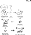
- Fig 1 Synthesis of mvsFlt schematic.
- mvsFlt bioconjugates were synthesized using a 3-step reaction in which HyA was reacted with EDC and EMCH to create a thiol reactive HyA-EMCH intermediate.
- sFlt was then treated with 2-iminothiolane and reacted with the HyA-EMCH intermediate for the synthesis of the final mvsFlt bioconjugate.
- mice All experiments were performed with wild-type 7 to 12-week old male and female littermate FVB/n mice. Mice were maintained under pathogen-free conditions in the UCSF barrier facility and conducted in accordance with procedures approved by the UCSF Institutional Animal Care and Use Committee (IACUC). All experiments were approved by the UCSF IACUC prior to work. Mice were anesthetized by isofluorane inhalation (Abbott Laboratories, Abbott Park, IL), 10mg/kg carprofren (Sigma, St. Louis, MO), and by topical application of 0.5% Proparacaine (Bausch & Lomb, Rochester, NY) placed on the cornea.
- IACUC Institutional Animal Care and Use Committee
- Corneas were blocked with 3% BSA and stained with DAPI, rabbit anti-mouse CD31 primary antibody (Santa Cruz Biotechnology) and goat anti-rabbit Alexa Fluor 488 secondary antibody (Life Technologies) for visualization and quantification of blood vessels.
- Corneas were cut into quadrants and flat-mounted onto glass slides using Fluoromount Mounting Medium (Sigma Aldrich). Imaging was carried out with an automated slide scanner, Zeiss Axioscan Z1 (Zeiss Instruments). Corneal blood vessel coverage was quantified using NIH ImageJ software by comparing the total cornea area to the corneal vascularized area.
- C t is the concentration at time t
- C 0 is the initial concentration
- k is the elimination constant given by Eq. 2:
- t 1/2 is the drug half-life. The values used for calculating t 1/2 were based on data from the 48-hour time point.
- Pregnant Brown Norway rats were obtained from Charles River Laboratories. All the animal experiments were performed in compliance with the ARVO statement for the Use of Animals in Ophthalmic and Vision Research and approved by the University of Oklahoma Institutional Animal Care and Use Committee. Newborn pups were assigned to PBS, sFlt or mvsFlt treatment groups. Light was cycled on a 12 hour on, 12 hour off schedule and room temperature was maintained at approximately 21C. Rat pups were exposed to hyperoxia (75% O 2 ) from postnatal day 7 (P7) to P12. The oxygen-treated rats were housed in an incubator connected to an Oxycler Model A4 (Redfield, NY) with oxygen and nitrogen, allowing for adjustment of oxygen concentration to 75/0 ⁇ 2%.
- Oxycler Model A4 Redfield, NY
- Rats at P17 were anesthetized and perfused with high-molecular weight FITC-dextran (2 ⁇ 10 6 ; Sigma-Aldrich, St. Louis MO) as described by Smith et al [17]. Retinas were dissected and flat-mounted and the vasculature was imaged using a fluorescence microscope (CKX41; Olympus). Vascular coverage at P17 was quantified using NIH ImageJ by comparing the total retinal area to the area of vascularization.
- the chemical injury-based corneal angiogenesis model was used to determine whether conjugation of sFlt to HyA reduced the bioactivity of mvsFlt to in comparison to sFlt in vivo.
- Corneas treated with PBS had 28.8 ⁇ 11.5% blood vessel coverage in contrast to corneas treated with sFlt and mvsFlt, which had 12.8 ⁇ 3.8% and 15.8 ⁇ 7.1% vascular coverage, respectively.
- FIG. 1 Fig 2A-2C : sFlt and mvsFlt equally inhibit corneal angiogenesis.
- C Quantification of corneal angiogenesis at day 10 following treatment. One-way ANOVA gives p value ⁇ ⁇ 0.01 (n.s.- not significant; ⁇ p ⁇ 0.05; ⁇ p ⁇ 0.01). Scale bars correspond to 20 ⁇ m.
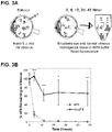
- Fig 3A-3B mvsFlt has longer residence time in the rat vitreous.
- Results are expressed as mean ⁇ SD ( ⁇ p ⁇ 0.05, ⁇ p ⁇ 0.01, ⁇ p ⁇ 0.001). ⁇ indicates a difference between the mvsFlt and sFlt at the given time point. Two-way ANOVA gives p-value ⁇ ⁇ 0.001.
- FIG. 6 Higher molecular weight dextran displays longer residence time in vivo.
- Validation experiment demonstrating the effect of size on the retention of fluorescently tagged dextrans.
- the 2 MDa dextran (solid line) has significantly improved residence time over the 40 kDa (dashed line) over 48 hours.
- the half-life of the 40 kDa and the 2 MDa dextrans is 3.2 and 5 hours, respectively.
- ⁇ indicates a difference between the 40 kDa and 2 MDa dextran at the given time point.
- Two-way ANOVA gives p-value ⁇ ⁇ 0.05 ( ⁇ corresponds to a P-value less than 0.01).
- mvsFlt is a more potent inhibitor of retinal neovascularization
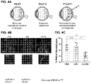
- Neovascular coverage was calculated by comparing the area of vascular coverage to total retinal area. After 5 days of treatment, retinal vascular coverage of PBS-injected eyes was 84.3 ⁇ 3.8% and retinas treated with intravitreal injections of sFlt were 85.4 ⁇ 6.1%. In contrast, retinas from rats treated with intravitreal injections of mvsFlt were significantly lower and had 72.9 ⁇ 3.4% retinal vascular coverage ( Fig 4 ).
- Fig 4A-4C mvsFlt inhibits retinal angiogenesis.
- both sFlt and mvsFlt have similar concentrations in the vitreous ( FIG 5A ). Over time, sFlt is small enough to be cleared from the vitreous leaving much lower concentrations of drug ( Fig 5B , top). This allows for an increase in the intravitreal VEGF concentration, which induces angiogenesis. In contrast, mvsFlt has a much longer intravitreal residence time and is thus able to act as a sponge for VEGF over time ( Fig 5B , bottom), inhibiting angiogenesis and maintaining the basal level of vascularization in the retina.
- Fig 5A-5B Schematic demonstrating the proposed mechanism of mvsFlt action.
- A) sFlt (red, unconjugated) and mvsFlt (red conjugated blue chain of HyA) are injected into a diabetic retina where there is a high concentration of VEGF (green circles).
- mvsFlt has a longer residence time in the vitreous and is able to bind and inhibit VEGF over much longer periods of time, leading to prolonged inhibition of retinal angiogenesis.
- the sFlt sequence for the first 3 Ig-like extracellular domains of sFlt-1 [15] was cloned into the pFastBac1 plasmid (Life Technologies) and then transformed into DH10Bac E.coli, which were plated on triple antibiotic plates containing kanamycin (50 ⁇ g/mL), gentamicin (7 ⁇ g/mL, Sigma Aldrich), tetracycline (10 ⁇ g/mL, Sigma Aldrich), IPTG (40 ⁇ g/mL, Sigma Aldrich) and Bluo-gal (100 ⁇ g/mL, Thermo Fisher Scientific).
- the sFlt gene-containing bacmid was isolated from DH10Bac E.coli (Life Technologies) and transfected into SF9 insect cells for virus production (provided by the Tissue Culture Facility, UC Berkeley). Virus was then used to infect High Five insect cells (provided by the Tissue Culture Facility, UC Berkeley) to induce sFlt protein expression. After 3 days, protein was purified from the supernatant using Ni-NTA agarose beads (Qiagen Laboratories). Recombinant sFlt was eluted from the Ni-NTA beads using an imidazole gradient and then concentrated and buffer exchanged with 10% glycerol/PBS using Amicon Ultra-15mL Centrifugal devices (EMD Millipore). The protein solution was sterile filtered and the concentration was determined using a BCA assay (Thermo Fisher Scientific).
- HyA Conjugation of sFlt to HyA was carried out according to the schematic in Figure 1A, as previously described [16-18].
- EMCH 3,3'- N -( ⁇ -maleimidocaproic acid) hydrazide
- HOBt 1-hydroxybenzotriazole hydrate
- EDC 1-ethyl-3-(3-dimethylaminopropyl) carbodiimide hydrochloride
- HyA Lifecore Biotechnology
- MES 2-( N -morpholino)ethanesulphonic acid
- the solution was then dialyzed into pH 7.0 PBS containing 10% glycerol.
- Recombinant sFlt was treated with 2-iminothiolane at 10 molar excess to create thiol groups for conjugation to the maleimide group on EMCH.
- sFlt was then added to activated HyA-EMCH at 10:1 and 30:1 molar ratios of sFlt to HyA to create the mvsFlt bioconjugates and allowed to react at 4 °C overnight.
- the mvsFlt reactions were dialyzed exhaustively using 100 kDa molecular weight cut-off (MWCO) Float-A-Lyzer G2 (Spectrum Labs) dialysis tubes in pH 7.0 PBS to remove unreacted sFlt.
- the BCA assay was used to measure the protein concentrations of the mvsFlt bioconjugates. Conjugates were defined with 10:1 sFlt to HyA molar feed ratios as ' low conjugation ratio' (LCR) mvsFlt and 30:1 molar feed ratios 'high conjugation ratio' (HCR) mvsFlt.
- SEC-MALS size exclusion chromatography with multiangle light scattering
- the mvsFlt conjugates were sterile filtered through a 0.45 ⁇ m filter and 200 ⁇ L was injected at HyA concentration between 0.2-0.5 mg/mL.
- the dn/dc values were determined using SEC-MALS to be 0.1447 and 0.185 for HyA-EMCH and sFlt, respectively.
- UV extinction coefficients used for HyA-EMCH and sFlt were also determined on SEC-MALS and were 0.022 and 0.894, respectively. Data analysis was carried out using the Astra software (Wyatt Technologies).
- a Brookhaven Goniometer & Laser Light Scattering system (BI-200SM, Brookhaven Instruments Corporation) was used to determine the hydrodynamic diameter of mvsFlt bioconjugates. Each sample was filtered at 0.45 ⁇ m and loaded into a 150 ⁇ L cuvette (BI-SVC, Brookhaven). Data acquisition was performed at 90 degrees with a 637nm laser for 2 minutes. Data analysis was carried out with BIC Dynamic light Scattering software (Brookhaven) using the BI-9000AT signal processor. The intensity average particle size was obtained using a nonnegative least squares (NNLS) analysis method.
- NLS nonnegative least squares
- VEGF 165 Quantikine Sandwich ELISA (R&D Systems) to examine the effect of HyA conjugation on sFlt inhibition of VEGF 165 .
- the assay was carried out according to the manufacturer's instructions. Briefly, VEGF 165 was added to PBS with varying concentrations of sFlt or mvsFlt. Free VEGF 165 that bound to the capture antibodies on the plate surface were detected using a horseradish peroxidase conjugated detection antibody and quantified using a spectrophotometer at 450 nm.
- HUVECs Human umbilical cord vein endothelial cells
- EBM-2 media Lido-Benoic acid
- a survival assay was carried out with HUVECs grown in the presence of VEGF 165 and mvsFlt conjugates. HUVECs were added to 96 well plates coated with 0.2% gelatin at 10,000 cell/well in M199 media.
- VEGF 165 vascular endothelial growth factor 165
- sFlt or mvsFlt 72 hours after plating, the media was aspirated and the cells were washed with PBS prior to freezing for analysis with CyQuant (Life Technologies). Total cell number per well was determined by reading fluorescence at 480 nm excitation and 520 nm emission using a fluorometer (Molecular Devices).
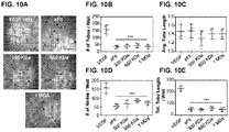
- HUVEC tube formation assays were carried out in 96-well plates coated with 80 ⁇ L of Matrigel (Corning, NY) and incubated at 37°C for 1 hour to allow gelation to occur. HUVECs were trypsinized and resuspended in M199 with 2% FBS and 20 ng/mL VEGF 165 and treated with mvsFlt LCR conjugates. Wells were imaged 18 hours after plating and tube formation was quantified using ImageJ software.
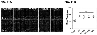
- HUVECs were added at 150,000 cells/well in EBM-2 and allowed to attach and spread overnight. Using a 1 mL pipette tip, crosses were scratched into the confluent layer of HUVECs. The wells were then washed with excess PBS to remove cell debris and media and replaced with media containing VEGF 165 and mvsFlt. Scratches were imaged at 0 and 24 hours post scratch and the area without cells was quantified using ImageJ and T-scratch software (CSE Laboratory software, ETH Zurich). The percent open wound area was calculated by comparing the open scratch area at 24 hours to the open scratch area at 0 hours.
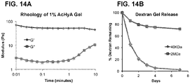
- acrylated HyA hydrogels were synthesized as described previously [20,21]. Briefly, adipic acid dihydrazide (ADH, Sigma Aldrich) was added in 30 molar excess to HyA in deionized water (DI). EDC (3 mmol) and HOBt (3 mmol) were dissolved in DMSO/water and added to the HyA solution. The solution was allowed to react for 24 hours and then dialyzed exhaustively against DI water. HyA-ADH was precipitated in 100% ethanol and reacted with acryloxysuccinimide to generate acrylate groups on the HyA. The resulting AcHyA was exhaustively dialyzed and lyophilized for storage. The presence of grafted acrylate groups on HyA chains was confirmed using H 1 -NMR.
- Cell culture inserts with 4 ⁇ m sized pores were added to wells of a 24-well plate and 70 ⁇ L of gel containing sFlt, mvsFlt or BSA was added to each insert.
- the gels were allowed to crosslink at 37°C for 1 hour before adding 150 ⁇ L of PBS to the wells and submerging the hydrogels.
- the well supernatant was collected and fully replaced at 0, 1, 2, 3, 7, 10 and 14 days. Samples were read with a fluorometer to detect the fluorescently tagged sFlt, mvsFlt and BSA in the supernatant.
- FRAP measurements were performed on 1% AcHyA hydrogels containing FITC labeled sFlt and 650 kDa LCR mvsFlt.
- Total fluorescence intensity of the hydrogels was acquired using a Zeiss LSM710 laser-scanning microscope (Carl Zeiss, Jena, Germany) with a 20X magnification objective and an argon laser set at 488 nm with 50% power. Photobleaching was done by exposing a 100 ⁇ 100- ⁇ m spot in the field of view to high intensity laser light. The area was monitored by 15 pre-bleach scanned images at low laser intensity (2%), then bleached to 75% of the starting fluorescence intensity at 100% laser power.
- the mobile fraction of fluorescent sFlt and mvsFlt molecules within the hydrogel was determined by comparing the fluorescence in the bleached region after full recovery ( F ⁇ ) with the fluorescence before bleaching ( F initial ) and just after bleaching ( F 0 ).
- the mobile fraction R was defined according to equation (1):
- Matrix metalloproteinase-7 has been previously shown to specifically degrade sFlt [22].
- MMP-7 matrix metalloproteinase-7
- sFlt and mvsFlt were treated with varying amounts of MMP-7 (EMD Millipore).
- MMP-7 EMD Millipore
- sFlt and mvsFlt were incubated with matrix metalloproteinase-7 (MMP-7) for 12 hours at 37C while shaking at high (1:1 molar ratio MMP-7:sFlt), medium (1:2), and low (1:4) molar ratios of MMP-7 to sFlt.
- the enzyme-treated sFlt and mvsFlt were then loaded into precast Mini-Protean TGX 4-20% gradient gels (Bio-Rad Laboratories) and run at 110 volts for 90 minutes. Gels were then stained using a silver staining kit (Thermo Fisher Scientific) according to the manufacturer's instructions. The degree of enzymatic degradation was assessed by quantifying the total amount of protein remaining in the gel following treatment with MMP-7. Each well was normalized to their respective no-MMP-7 well and background intensity was subtracted according to the blank well between the groups on the gel.
- the overall goal of this study was to synthesize protein-polymer bioconjugates to increase the residence time of anti-VEGF drugs in the vitreous for use in treating patients with DR.
- large multivalent protein bioconjugates with unperturbed affinity for VEGF 165 and good enzymatic stability were developed, which bioconjugates that show delayed diffusion and mobility in an in vitro model of the vitreous.
- FIGS 7A-7D Multivalent sFlt synthesis and schematics.
- A) mvsFlt bioconjugates were synthesized using a 3-step reaction in which HyA was reacted with EDC and EMCH to create a cysteine reactive HyA-EMCH intermediate. sFlt was then treated with 2-iminothiolane and then reacted with the HyA-EMCH intermediate for the synthesis of the final product.
- the ratio a:b represents the valency of sFlt molecules (a) covalently bound to a single chain of HyA (b).
- mvsFlt conjugates were carried out according to the schematics shown in Figure 7A-7D.
- mvsFlt was created at low (LCR, Figure 7C ) and high (HCR, Figure 7D ) conjugation ratios in order to determine whether a certain valency would provide an enhanced effect on VEGF binding.
- sFlt was successfully conjugated to HyA at several different molecular weights of HyA and valencies, which were significantly larger than the sFlt in its unconjugated form ( Figure 8A-8D ).
- the molecular weights of the protein and polymer components of the conjugates were characterized using SEC-MALS as shown in Table 1 and Figure 8A .
- Table 1 SEC-MALS analysis of all mvsFlt conjugates. 300 kDa LCR* 300 kDa HCR + 650 kDa LCR* 650 kDa HCR + 1 MDa LCR* 1 MDa HCR + HyA Mn a 2.7E5 2.1E5 5.9E5 6.4E5 9.7E5 1.0E6 HyA Mw b 3.1E5 3.4E5 7.1E5 7.1E5 1.2E6 1.1E6 HyA PDI c 1.1 1.6 1.2 1.1 1.3 1.1 sFlt: HyA d 7.9 15.4 5.3 17.1 5.2 14.1 a Number average molecular weight given in g/mol.
- the size of the mvsFlt conjugates was dependent on the HyA molecular weight for the 300 and 650 kDa conjugates; however, there was no significant difference between the 650 kDa and 1 MDa conjugates ( Figure 8D ).
- HCR conjugates of 300 and 650 kDa molecular weight had lower diameters than their respective LCR conjugates, which could be due to increased positive charge from sFlt on the negatively charged HyA backbone with increasing sFlt attachment causing the conjugate to fold in tighter around itself.
- Figure 8A-8D Characterizing mvsFlt conjugation efficiency and size.
- FIGS. 9A-9B All mvsFlt bioconjugates maintain their ability to inhibit VEGF 165 dependent activities in VEGF 165 ELISA and VEGF 165 -dependent HUVEC survival assays.
- FIGS. 10A-10E mvsFlt inhibits HUVEC tube formation.
- A) Representative images of inhibited HUVEC tube formation on Matrigel (BD Biosciences) when treated with 1 ⁇ g/mL of all LCR mvsFlt bioconjugates. Cells were seeded at 20,000 cells per well of a 96-well plate on 100 ⁇ L of matrigel and imaged at 18 hours. Scale bar 500 ⁇ m.
- B-E Quantification of total number of tubes per well (B), average tube length (C), total number of nodes (branching points, D), and total tube length per well (E) ( ⁇ p ⁇ 0.001).
- FIG 11A-11B mvsFlt inhibits VEGF 165 -driven HUVEC migration .
- HyA gels [20,21] were used to examine how diffusion conjugation of sFlt to HyA affects diffusion.
- This hydrogel was chosen as a model system for studying the vitreous in vitro based primarily on compositional similarity to the vitreous with respect to high HyA content.
- the stiffness of the HyA hydrogel ( Figure 14A ) was higher than published reports examining bovine and porcine vitreous stiffness [25], suggesting the predictions of drug clearance from the vitreous using this model may not be sufficient to estimate actual in vivo rates.
- it is anticipated that the diffusion of sFlt and mvsFlt through these gels will be instructive to predict the benefits of conjugation to HyA given the compositional similarities of the model with the vitreous.
- FIGS. 12A-12C sFlt conjugation to HyA decreases mvsFlt mobility and diffusion in HyA gels.
- F initial depicts mvsFlt in the gel prior to bleaching.
- F 0 is the fluorescence measurement immediately after 75% photobleaching;
- F ⁇ corresponds to the maximal recovery of fluorescence at the end of the experiment.
- FIG 14A-14B Characterizing the HyA hydrogel.
- A Rheological properties of the 1% HyA hydrogel.
- B Fluorescently tagged 40 kDa dextran encapsulated in HyA hydrogels diffused out significantly faster than the 2 MDa dextran after 7 days ( ⁇ p ⁇ 0.001).
- n is indicative of whether diffusion is Fickian and if n is equal to 0.5, the transport is Fickian.
- the n value for BSA, sFlt and mvsFlt release was determined to be 0.3-0.4 ( Fig. 15A -E ), indicating that the diffusion through the gel is not Fickian.
- BSA was depleted from the gel through rapid burst release due to the extremely small size of the protein and very low affinity to the HyA hydrogel due to charge repulsion, leading to non-Fickian diffusion.
- the sFlt and mvsFlt conjugates release slower in comparison due in part to ionic affinity with the HyA hydrogel and size.
- the sFlt and BSA are similar in size (50 kDa and 66 kDa, respectively), the sFlt has a much stronger ionic interaction with the matrix, which slows its diffusion from the gel, also resulting in non-Fickian diffusion due to this strong affinity.
- the 300 kDa mvsFlt conjugate is small enough in size ( ⁇ 150 nm, see Fig. 8D ) relative to the estimated hydrogel ⁇ (Table 3) to release as rapidly as sFlt.
- the two largest conjugates were close to the mesh size in diameter ( > 225 nm) and were significantly impeded in their release due to size, resulting in gel release that followed the reptation mechanism of diffusion [33].
- the characteristic diffusion time for mvsFlt (94.8 ⁇ 19.5 s) was significantly faster than sFlt (176 ⁇ 18.1 s). This difference may be due to ionic shielding of the positively charged sFlt by the negatively charged hyaluronic acid within the multivalent conjugate, which reduces the overall affinity of the conjugate for the gel allowing for faster diffusion.
- the unconjugated sFlt remains highly positively charged resulting in stronger ionic affinity within the HyA gel that slow its characteristic diffusion time.
- the mvsFlt in the hydrogel also recovered fluorescence to a significantly lower degree, 85.3 ⁇ 0.8%, in comparison to sFlt, which recovered to 91.6% ⁇ 2.4%.
- FIG. 13A-13B Conjugation to HyA reduces susceptibility to protease degradation by MMP-7.
- Figure 15A-15E Fits for data from gel release data to examine Fickian diffusion.
- A-E Graphs of fits made with Ritger et al. [13] equation (2) to determine the diffusional exponent, n, and the constant for characteristics of the macromolecular network system and the drug, k.
- Example 3 Generation of multivalent conjugates with anti-VEGF VHH or with single-chain variable fragment (scFv) anti-VEGF (anti-VEGF scFv)
- FIG. 20A -C The ability of each antibody conjugate to bind human 500 pg of VEGF-A 165 was compared to the corresponding unconjugated antibody using an ELISA assay. Both multivalent conjugates were made with 860 kDa hyaluronic acid (HyA). The valencies of scFv anti-VEGF antibody and VHH anti-VEGF antibody were 31 and 28, respectively. The data was fit using a four-parameter logistic curve and used to calculate the IC50 for each treatment.
- FIG. 20A shows percent unbound VEGF for unconjugated scFv anti-VEGF antibody and conjugated multivalent scFv anti-VEGF antibody.
- FIG. 20A shows percent unbound VEGF for unconjugated scFv anti-VEGF antibody and conjugated multivalent scFv anti-VEGF antibody.
- FIG. 20B shows percent unbound VEGF for unconjugated VHH anti-VEGF antibody and conjugated multivalent VHH anti-VEGF antibody.
- FIG. 20C shows the IC50 values for the conjugated antibodies of FIG. 20A-B compared to the unconjugated antibodies of FIG. 20A-B. Multivalent conjugation did not have a substantial effect on the IC50 values for the conjugates compared to the unconjugated antibody, as summarized in the FIG. 20C .
- Example 4 Anti-VEGF VHH multivalent (mvAnti-VEGF) conjugates made with HyA show increased half-life in vitro and in vivo
- the diffusion rates of unconjugated anti-VEGF antibody VHH and multivalent anti-VEGF antibody VHH were compared by fluorescently tagging the proteins and entrapping them within a 1% HyA-PEG hydrogel made from commercially available components (BioTime). Swelling ratio and the mean molecular weight between crosslinks was estimated. The average mesh diameter of the hydrogel was estimated to be approximately 80 nm.
- FIG. 21 The concentration of unconjugated protein and protein conjugate released from the hydrogels was measured every 2-3 days and the data were fit to an exponential decay curve to estimate their diffusion half-life. The half-life of the VHH anti-VEGF antibody conjugates was approximately 4-fold longer than that of the unconjugated VHH anti-VEGF antibody. The results shown in FIG. 21 are the mean of three independently repeated experiments.
- the in vivo residence times of the unconjugated VHH anti-VEGF antibody and multivalent VHH anti-VEGF antibody conjugates were compared after injection into rat eyes.
- all of the VHH anti-VEGF antibody was fluorescently tagged as a reporter for concentration measurements, and 4 independent batches of mvAnti-VEGF were used (860 kDa MW HyA and with valencies in the range of 24 to 45).
- mvAnti-VEGF 860 kDa MW HyA and with valencies in the range of 24 to 45.
- 5 ⁇ L of a 275 ⁇ g/mL solution of either VHH anti-VEGF antibody or multivalent VHH anti-VEGF antibody conjugate was injected.
- Each eye received a total of 1.375 ⁇ g VHH anti-VEGF antibody either unconjugated or conjugated at any valency.
- FIG. 22A shows the percent protein recovered for unconjugated VHH and multivalent VHH anti-VEGF antibody conjugates.
- FIG. 22B shows the half-life of the unconjugated VHH anti-VEGF antibody and the conjugated multivalent VHH anti-VEGF antibody. Results show that conjugated multivalent VHH anti-VEGF antibody had an in vivo half-life of 21.9 hours compared to 1.9 hours for the unconjugated VHH anti-VEGF antibody.
- FIG. 23A-B show generation of a multivalent conjugate made from ⁇ 17 repeats of a single-domain camelid (VHH) antibody and carboxymethylcellulose (CMC, ⁇ 700 kDa).
- VHH single-domain camelid
- CMC carboxymethylcellulose
- FIG. 23A The ability of this anti-VEGF antibody (anti-VEGF VHH-CMC) conjugate to bind human 500 pg of VEGF-A165 was compared to the corresponding unconjugated VHH anti-VEGF antibody using an ELISA assay as shown in FIG. 23A .
- the data were fit using a four-parameter logistic curve and used to calculate the IC50 for each treatment, as shown in FIG. 23B .
Landscapes
- Health & Medical Sciences (AREA)
- Life Sciences & Earth Sciences (AREA)
- Chemical & Material Sciences (AREA)
- Medicinal Chemistry (AREA)
- General Health & Medical Sciences (AREA)
- Veterinary Medicine (AREA)
- Public Health (AREA)
- Animal Behavior & Ethology (AREA)
- Pharmacology & Pharmacy (AREA)
- Epidemiology (AREA)
- Bioinformatics & Cheminformatics (AREA)
- Engineering & Computer Science (AREA)
- Immunology (AREA)
- Proteomics, Peptides & Aminoacids (AREA)
- Gastroenterology & Hepatology (AREA)
- Cell Biology (AREA)
- Zoology (AREA)
- Organic Chemistry (AREA)
- Ophthalmology & Optometry (AREA)
- General Chemical & Material Sciences (AREA)
- Chemical Kinetics & Catalysis (AREA)
- Nuclear Medicine, Radiotherapy & Molecular Imaging (AREA)
- Inorganic Chemistry (AREA)
- Microbiology (AREA)
- Mycology (AREA)
- Endocrinology (AREA)
- Molecular Biology (AREA)
- Genetics & Genomics (AREA)
- Biophysics (AREA)
- Biochemistry (AREA)
- Pain & Pain Management (AREA)
- Rheumatology (AREA)
- Urology & Nephrology (AREA)
- Vascular Medicine (AREA)
- Cardiology (AREA)
- Heart & Thoracic Surgery (AREA)
- Medicines That Contain Protein Lipid Enzymes And Other Medicines (AREA)
- Peptides Or Proteins (AREA)
- Medicinal Preparation (AREA)
Description
- Current anti-angiogenic polypeptide drugs for patients with diabetic retinopathy suffer from short residence time in the vitreous of the eye. In order to maintain biologically effective doses of drug for inhibiting retinal neovascularization, patients are required to receive regular monthly injections of drug, which often results in low patient compliance and progression of the disease.
- Altiok et al. (2016) Biomaterials 93:95 discusses the effect of multivalent hyaluronic acid bioconjugates on sFlt-1 activity in vitro.
- Shahangian et al. (2015) Int. J. Biol. Macromol. 77:222-234 describe generating VHHs for use in blocking the receptor binding site of VEGF and compete the VEGFR2.
- Yu et al. (2015) Transl. Vis. Sci. & Technol. 4:5 presents the results of a study of in vivo release of bevacizumab from a hyaluronic acid/dextran-based in situ hydrogel following intravitreal injection in the rabbit eye.
- The invention is defined in the appended claims. The present disclosure provides methods of treating an ocular disease or disorder. The methods involve direct administration into the eye of a conjugate comprising a biologically active polypeptide and a biocompatible polymer.
-
-
FIG. 1 is a schematic depiction of synthesis of the conjugate mvsFlt. -
FIG. 2A-2C depict inhibition of corneal angiogenesis by sFlt and mvsFlt. -
FIG. 3A-3B depict residence time of mvsFlt in the rat vitreous. -
FIG. 4A-4C depict inhibition of retinal angiogenesis by mvsFlt. -
FIG. 5A-5B provide a schematic depiction of a proposed mvsFlt mechanism of action. -
FIG. 6 depicts in vivo residence time of higher molecular weight dextran. -
FIG. 7A-7D depict multivalent sFlt synthesis and schematics. -
FIG. 8A-8D depict characterization of mvsFlt conjugation efficiency and size. -
FIG. 9A-9B depict the effect of mvsFlt bioconjugates on VEGF165-dependent activities in VEGF165 ELISA and VEGF165-dependent HUVEC survival assays. -
FIG. 10A-10E depict the effect of mvsFlt on HUVEC tube formation. -
FIG. 11A-11B depict the effect of mvsFlt on VEGF165-driven HUVEC migration. -
FIG. 12A-12C depict the effect of sFlt conjugation to HyA on mvsFlt mobility and diffusion in HyA gels. -
FIG. 13A-13B depict data showing that conjugation to HyA reduces susceptibility to protease degradation by MMP-7. -
FIG. 14A-14B depict characterization of a HyA hydrogel. -
FIG. 15A-15E depict fits for data from gel release data to Fickian diffusion. -
FIG. 16A-16C depict amino acid sequences of biologically active polypeptides. -
FIG. 17 depicts the amino acid sequence of an exemplary biologically active polypeptide sFlt. -
FIG. 18 depicts the amino acid sequence (SEQ ID NO:5) of an scFv anti-VEGF antibody. -
FIG. 19 depicts the amino acid sequence (SEQ ID NO:6) of a VHH anti-VEGF antibody. -
FIG. 20A-20C depict binding of unconjugated and conjugated anti-VEGF antibodies to VEGF-A165. -
FIG. 21 depicts the half-life of the conjugated multivalent VHH anti-VEGF antibody compared to the unconjugated VHH anti-VEGF antibody. -
FIG. 22A-22B depicts the in vivo residence time and percent protein recovered of the conjugated multivalent VHH anti-VEGF antibody and the unconjugated VHH anti-VEGF antibody after injection into rat eyes. -
FIG. 23A-23B compare the ability of conjugated multivalent VHH anti-VEGF antibody and conjugated multivalent VHH anti-VEGF antibody with carboxymethylcellulose (CMC) to bind to VEGF-A165 using an ELISA assay. - The terms "peptide," "polypeptide," and "protein" are used interchangeably herein, and refer to a polymeric form of amino acids of any length, which can include coded and non-coded amino acids, chemically or biochemically modified or derivatized amino acids, and polypeptides having modified peptide backbones. The term "polypeptide" includes fusion proteins, including, but not limited to, fusion proteins with a heterologous amino acid sequence, fusions with heterologous and homologous leader sequences, with or without N-terminal methionine residues; immunologically tagged proteins; and the like. The term "polypeptide" includes polypeptides comprising one or more of a fatty acid moiety, a lipid moiety, a sugar moiety, and a carbohydrate moiety. The term "polypeptides" includes post-translationally modified polypeptides.
- The terms "antibodies" and "immunoglobulin" include antibodies or immunoglobulins of any isotype, fragments of antibodies which retain specific binding to antigen, including, but not limited to, Fab, Fv, single chain Fv (scFv), and Fd fragments, chimeric antibodies, humanized antibodies, single-chain antibodies, single domain antibodies (VHH and VANR), and fusion proteins comprising an antigen-binding portion of an antibody and a non-antibody protein.
- "Antibody fragments" comprise a portion of an intact antibody, for example, the antigen binding or variable region of the intact antibody. Examples of antibody fragments include Fab, Fab', F(ab')2, and Fv fragments; diabodies; linear antibodies (Zapata et al., Protein Eng. 8(10): 1057-1062 (1995)); single-chain antibody molecules; single domain antibodies (e.g., camelid antibodies or "VHH" fragments (see, e.g., Harmsen and De Haard (2007) Appl. Microbiol. Biotechnol. 77:13); VNAR; and nanobodies; see, e.g., Wesolowski et al. (2009) Med. Microbiol. Immunol. 198:157); and multispecific antibodies formed from antibody fragments. Papain digestion of antibodies produces two identical antigen-binding fragments, called "Fab" fragments, each with a single antigen-binding site, and a residual "Fc" fragment, a designation reflecting the ability to crystallize readily. Pepsin treatment yields an F(ab')2 fragment that has two antigen combining sites and is still capable of cross-linking antigen.
- "Single-chain Fv" or "sFv" antibody fragments comprise the VH and VL domains of antibody, wherein these domains are present in a single polypeptide chain. In some embodiments, the Fv polypeptide further comprises a polypeptide linker between the VH and VL domains, which enables the sFv to form the desired structure for antigen binding. For a review of sFv, see Pluckthun in The Pharmacology of Monoclonal Antibodies, vol. 113, Rosenburg and Moore eds., Springer-Verlag, New York, pp. 269-315 (1994 ).
- As used herein, the term "affinity" refers to the equilibrium constant for the reversible binding of two agents and is expressed as a dissociation constant (Kd). Affinity can be at least 1-fold greater, at least 2-fold greater, at least 3-fold greater, at least 4-fold greater, at least 5-fold greater, at least 6-fold greater, at least 7-fold greater, at least 8-fold greater, at least 9-fold greater, at least 10-fold greater, at least 20-fold greater, at least 30-fold greater, at least 40-fold greater, at least 50-fold greater, at least 60-fold greater, at least 70-fold greater, at least 80-fold greater, at least 90-fold greater, at least 100-fold greater, or at least 1000-fold greater, or more, than the affinity of an antibody for unrelated amino acid sequences. Affinity of an antibody to a target protein can be, for example, from about 100 nanomolar (nM) to about 0.1 nM, from about 100 nM to about 1 picomolar (pM), or from about 100 nM to about 1 femtomolar (fM) or more. As used herein, the term "avidity" refers to the resistance of a complex of two or more agents to dissociation after dilution. The terms "immunoreactive" and "preferentially binds" are used interchangeably herein with respect to antibodies and/or antigen-binding fragments.
- The term "binding" refers to a direct association between two molecules, due to, for example, covalent, electrostatic, hydrophobic, and ionic and/or hydrogen-bond interactions, including interactions such as salt bridges and water bridges. Non-specific binding would refer to binding with an affinity of less than about 10-7 M, e.g., binding with an affinity of 10-6 M, 10-5 M, 10-4 M, etc.
- As used herein, the term "copolymer" describes a polymer which contains more than one type of subunit. The term encompasses polymer which include two, three, four, five, or six types of subunits.
- The terms "subject," "individual," "host," and "patient" are used interchangeably herein to a member or members of any mammalian or non-mammalian species. Subjects and patients thus include, without limitation, humans, non-human primates, canines, felines, ungulates (e.g., equine, bovine, swine (e.g., pig)), avians, rodents (e.g., rats, mice), and other subjects. Non-human animal models, particularly mammals, e.g. a non-human primate, a murine (e.g., a mouse, a rat), lagomorpha, etc. may be used for experimental investigations.
- "Treating" or "treatment" of a condition or disease includes: (1) preventing at least one symptom of the condition, i.e., causing a clinical symptom to not significantly develop in a mammal that may be exposed to or predisposed to the disease but does not yet experience or display symptoms of the disease, (2) inhibiting the disease, i.e., arresting or reducing the development of the disease or its symptoms, or (3) relieving the disease, i.e., causing regression of the disease or its clinical symptoms.
- A "therapeutically effective amount" or "efficacious amount" means the amount of a conjugate that, when administered to a mammal or other subject for treating a disease, is sufficient, in combination with another agent, or alone in one or more doses, to effect such treatment for the disease. The "therapeutically effective amount" can vary depending on the conjugate, and depending on one or more other factors, such as the disease and its severity, the age, weight, etc., of the subject to be treated.
- The term "unit dosage form," as used herein, refers to physically discrete units suitable as unitary dosages for human and animal subjects, each unit containing a predetermined quantity of a conjugate, calculated in an amount sufficient to produce the desired effect in association with a pharmaceutically acceptable diluent, carrier or vehicle.
- A "pharmaceutically acceptable excipient," "pharmaceutically acceptable diluent," "pharmaceutically acceptable carrier," and "pharmaceutically acceptable adjuvant" means an excipient, diluent, carrier, and adjuvant that are useful in preparing a pharmaceutical composition that are generally safe, non-toxic and neither biologically nor otherwise undesirable, and include an excipient, diluent, carrier, and adjuvant that are acceptable for veterinary use as well as human use. "A pharmaceutically acceptable excipient, diluent, carrier and adjuvant" as used in the specification and claims includes one and more than one such excipient, diluent, carrier, and adjuvant.
- The present disclosure provides a method of treating an ocular disease or disorder in an individual. The method generally involves administering to an individual in need thereof an effective amount of a conjugate comprising a biologically active polypeptide and a biocompatible polymer, where the administration is directly into the eye, e.g., the administration is intravitreal administration. Methods of treatment per se do not form part of the claimed invention.
- The intravitreal injection route is the most efficient method of delivering drug products to structures in the eye that are located in the posterior chamber. Therefore, it is the preferred method of delivering drugs that act on the retina. However, each intravitreal injection carries a risk of causing retinal detachment from as a result of interfering with the integrity of the eye. Adding volume to the posterior chamber causes a rise in intraocular pressure, and there are risk associated with the eye not accommodating to relieve the added pressure. There is also a risk of introducing an infection into the eye. These complications all carry a risk of impaired vision, which must be weighed against the possible benefits of administering a drug using the intravitreal route.
- Therefore, there is a strong motivation to improve the residence time of drugs that are designed to be routinely injected into the vitreous via the intravitreal route. Alternatively, it may be practical in some instances to chemically modify and existing drug in order to increase its residence time within the posterior chamber. This strategy has the potential to reduce the frequency of administration of the drug, and as a consequence, to reduce the overall risk of complication due to administering the drug overtime. Increasing the duration of bioactivity may also yield enhanced therapeutic outcomes for the drug as well.
- A drug that exhibits greater intravitreal residence time may be preferred by the patient as well relative to a drug product that must be administered more frequently for an equivalent therapeutic function. While the intravitreal injection is performed under topical anesthesia and is generally not regarded as painful, it is burdensome for the patient. It must be performed by a clinician, and thus an office visit is required for each administration of the drug. There is typically short-term irritation and blurred vision due to increased tearing. There may also be short tear changes to the appearance of the eye at the vicinity of the injection site. Thus, patients would likely exhibit a preference for an equivalent therapy that would require fewer intravitreal injections.
- The need for less frequent injections would also be preferable from the physician's perspective. The intravitreal injections must be performed by an ophthalmologist, and thus this procedure can occupy a substantial portion of their clinic time. The number of patients that are receiving the intravitreal therapy in their practice can be limited by the frequency that each patient must receive the intravitreal injections. Less frequent injection would increase the number patients that are able receive the method of therapy. A longer acting drug would also be preferable to a depot or long-term drug delivery device, as these typically require a longer implantation procedure and access to a procedure room, which may offset the benefits of less frequent administration for the clinician.
- A conjugate comprising a biologically active polypeptide and a biocompatible polymer exhibits a half-life in the vitreous that is greater than the half-life in the vitreous of the biologically active polypeptide not conjugated to the biocompatible polymer. The increased half-life of the conjugate in the vitreous confers certain advantages, including, e.g., reduced burden on the patient; reduced number and/or frequency of administrations; increased safety; decreased incidence of infection; increased patient compliance; and increased efficacy. In addition, a conjugate as described herein allows use of polypeptides for treatment of ocular disorders, which polypeptides would not, in unconjugated form, be retained in the eye for a time period suitable for therapy.
- In some cases, an effective amount of a conjugate is an amount that is effective to inhibit pathological angiogenesis in the eye of the individual. For example, in some cases, an effective amount of a conjugate is an amount that, when administered in one or more doses, is effective to inhibit pathological angiogenesis in the eye of the individual by at least 10%, at least 15%, at least 20%, at least 25%, at least 30%, at least 40%, at least 50%, at least 60%, at least 70%, or at least 80%, or more than 80%, compared to the degree of pathological angiogenesis in the eye in the absence of treatment with the conjugate, or before treatment with the conjugate.
- In some cases, an effective amount of a conjugate is an amount that is effective to reduce intraocular pressure in the eye of the individual. For example, in some cases, an effective amount of a conjugate is an amount that, when administered in one or more doses, is effective to reduce intraocular pressure by at least 10%, at least 15%, at least 20%, at least 25%, at least 30%, at least 40%, at least 50%, at least 60%, at least 70%, or at least 80%, or more than 80%, compared to the intraocular pressure in the eye in the absence of treatment with the conjugate, or before treatment with the conjugate.
- In some cases, an effective amount of a conjugate is an amount that is effective to reduce macular edema in the eye of the individual. For example, in some cases, an effective amount of a conjugate is an amount that, when administered in one or more doses, is effective to reduce macular edema by at least 10%, at least 15%, at least 20%, at least 25%, at least 30%, at least 40%, at least 50%, at least 60%, at least 70%, or at least 80%, or more than 80%, compared to the level of macular edema in the eye in the absence of treatment with the conjugate, or before treatment with the conjugate.
- In some cases, an effective amount of a conjugate is an amount that is effective to increase visual acuity in an eye of the individual. For example, in some cases, an effective amount of a conjugate is an amount that, when administered in one or more doses, is effective to increase visual acuity in an eye of the individual by at least at least 10%, at least 15%, at least 20%, at least 25%, at least 30%, at least 40%, at least 50%, at least 60%, at least 70%, at least 80%, at least 90%, at least 2-fold, at least 2.5-fold, at least 5-fold, or at least 10-fold, or more than 10-fold, compared to the visual acuity in the eye in the absence of treatment with the conjugate, or before treatment with the conjugate.
- In some cases, an effective amount of a conjugate is an amount that is effective to inhibit progression of an ocular disease in an individual. For example, in some cases, an effective amount of a conjugate is an amount that, when administered in one or more doses, is effective to inhibit progression of an ocular disease in the individual by at least 10%, at least 15%, at least 20%, at least 25%, at least 30%, at least 40%, at least 50%, at least 60%, at least 70%, or more, compared to the progression in the absence of treatment with the conjugate, or before treatment with the conjugate.
- For example, is in some cases, an effective amount of a conjugate is an amount that, when administered in one or more doses, is effective to inhibit progression of non-exudative ARMD to exudative ARMD or to inhibit progression of non-exudative ARMD to a more severe form. In some embodiments, an effective amount of a conjugate is an amount that is effective to inhibit progression of early ARMD (AREDS 2) to intermediate ARMD (AREDS 3) or to advanced ARMD (AREDS 4). In some embodiments, an effective amount of a conjugate is an amount that is effective to inhibit progression of intermediate ARMD (AREDS 3) to advanced ARMD (AREDS 4).
- In some cases, an effective amount of a conjugate is an amount that is effective to enhance a biological activity of a retinal cell, e.g., where the retinal cell is a photoreceptor, a retinal ganglion cell, a Muller cell, a bipolar cell, an amacrine cell, a horizontal cell, or a retinal pigmented epithelium cell.
- A polypeptide-polymer conjugate (also referred to herein, for simplicity, as a "conjugate") suitable for use in a method of treatment is of the formula:
X-(Y)n-Z
, where X is a biologically active polypeptide; - Y is an optional linker moiety, such that n is 0 or an integer from 1 to about 10; and
- Z is a biocompatible polymer comprising from about 50 subunits to 100,000 subunits, and/or having a molecular weight of from 10 kDa to 500 kDa.
- A conjugate comprising a biologically active polypeptide and a biocompatible polymer exhibits a half-life in the vitreous that is at least about 25%, at least about 50%, at least about 75%, at least about 2-fold, at least about 5-fold, at least about 10-fold, at least about 15-fold, at least about 20-fold, at least about 25-fold, at least about 30-fold, at least about 40-fold, at least about 50-fold, at least about 75-fold, at least about 100-fold, at least about 200-fold, at least about 500-fold, or at least about 1000-fold, or more than 1000-fold, greater than the half-life in the vitreous of the biologically active polypeptide not conjugated to the biocompatible polymer. A conjugate comprising a biologically active polypeptide and a biocompatible polymer exhibits a half-life in the vitreous that is 5-fold to 10-fold greater than the half-life in the vitreous of the biologically active polypeptide not conjugated to the biocompatible polymer.
- In some cases, a conjugate comprising a biologically active polypeptide and a biocompatible polymer exhibits a half-life in the vitreous of from 12 hours to 24 hours, from 1 day to 3 days, from 3 days to 7 days, from one week to 2 weeks, from 2 weeks to 4 weeks, or from 1 month to 6 months.
- In some cases, a conjugate comprising a biologically active polypeptide and a biocompatible polymer exhibits a therapeutically efficacious residence time in the vitreous of from 12 hours to 24 hours, from 1 day to 3 days, from 3 days to 7 days, from one week to 2 weeks, from 2 weeks to 4 weeks, from 1 month to 3 months, or from 3 months to 6 months.
- The biological activity of a polypeptide conjugated to the polymer substrate is enhanced relative to the activity of the polypeptide in soluble form, e.g., compared to the activity of the polypeptide not conjugated to the polymer. In some cases, the biological activity of the polypeptide of a polypeptide-polymer conjugate is at least about 25%, at least about 50%, at least about 75%, at least about 2-fold, at least about 5-fold, at least about 10-fold, at least about 15-fold, at least about 20-fold, at least about 25-fold, at least about 30-fold, at least about 40-fold, at least about 50-fold, at least about 75-fold, at least about 100-fold, at least about 200-fold, at least about 500-fold, or at least about 1000-fold, or more than 1000-fold, greater than the biological activity of the polypeptide in soluble (unconjugated) form.
- In some cases, the biological activity of the polypeptide of a suitable polypeptide-polymer conjugate is at least about 25%, at least about 50%, at least about 75%, at least about 2-fold, at least about 5-fold, at least about 10-fold, at least about 15-fold, at least about 20-fold, at least about 25-fold, at least about 30-fold, at least about 40-fold, at least about 50-fold, at least about 75-fold, at least about 100-fold, at least about 200-fold, at least about 500-fold, or at least about 1000-fold, or more than 1000-fold, greater than the biological activity of the polypeptide in when conjugated to the polymer at a 1:1 molar ratio.
- In some cases, the biological activity of the polypeptide of a suitable polypeptide-polymer conjugate is at least about 25%, at least about 50%, at least about 75%, at least about 2-fold, at least about 5-fold, at least about 10-fold, at least about 15-fold, at least about 20-fold, at least about 25-fold, at least about 30-fold, at least about 40-fold, at least about 50-fold, at least about 75-fold, at least about 100-fold, at least about 200-fold, at least about 500-fold, or at least about 1000-fold, or more than 1000-fold, greater than the biological activity of the polypeptide when present in admixture with the polymer.
- In some cases, the half-maximal effective concentration (EC50) of the polypeptide of a subject polypeptide-polymer conjugate is at least about 10%, at least about 25%, at least about 50%, at least about 75%, at least about 2-fold, at least about 5-fold, at least about 10-fold, at least about 15-fold, at least about 20-fold, at least about 25-fold, at least about 30-fold, at least about 40-fold, at least about 50-fold, at least about 75-fold, at least about 100-fold, at least about 200-fold, at least about 500-fold, or at least about 1000-fold, or more than 1000-fold, lower than the EC50 of the polypeptide in soluble (unconjugated form).
- In some cases, the half-maximal inhibitory concentration (IC50) of the polypeptide of a subject polypeptide-polymer conjugate is at least about 10%, at least about 25%, at least about 50%, at least about 75%, at least about 2-fold, at least about 5-fold, at least about 10-fold, at least about 15-fold, at least about 20-fold, at least about 25-fold, at least about 30-fold, at least about 40-fold, at least about 50-fold, at least about 75-fold, at least about 100-fold, at least about 200-fold, at least about 500-fold, or at least about 1000-fold, or more than 1000-fold, lower than the IC50 of the polypeptide in soluble (unconjugated form).
- Whether the biological activity of the polypeptide of a polypeptide-polymer conjugate is increased relative to the biological activity of the polypeptide in soluble (unconjugated) form is readily determined using an appropriate assay(s) for the biological activity.
- The molar ratio of the polypeptide to the polymer can vary from 5:1 to 100:1, e.g., from 5:1 to 7:1, from 7:1 to 10:1, from 10:1 to 12:1, from 12:1 to 15:1, from 15:1 to 20:1, from 20:1 to 25:1, from 25:1 to 30:1, from 30:1 to 35:1, from 35:1 to 40:1, from 40:1 to 45:1, from 45:1 to 50:1, from 50:1 to 60:1, from 60:1 to 70:1, from 70:1 to 80:1, from 80:1 to 90:1, or from 90:1 to 100:1.
- For example, the anti-angiogenic polypeptide of a polypeptide-polymer conjugate inhibits angiogenesis by at least about 10%, at least about 15%, at least about 20%, at least about 25%, at least about 30%, at least about 40%, at least about 50%, at least about 60%, at least about 75%, at least about 2-fold, at least about 5-fold, at least about 10-fold, at least about 15-fold, at least about 20-fold, at least about 25-fold, at least about 30-fold, at least about 40-fold, at least about 50-fold, at least about 75-fold, at least about 100-fold, at least about 200-fold, at least about 500-fold, or at least about 1000-fold, or more than 1000-fold, or more, compared to the degree of inhibition of angiogenesis by the anti-angiogenic polypeptide when present in admixture with the polymer, when in soluble (unconjugated) form, or when conjugated to the polymer at a 1:1 molar ratio.
- Suitable polymers to which a biologically active polypeptide is conjugated include biocompatible polymers comprising from 50 to 100,000 subunits, e.g., from 50 subunits to 100 subunits, from 100 subunits to 500 subunits, from 500 subunits to 1,000 subunits, from 1,000 subunits to 5,000 subunits, from 5,000 subunits to 10,000 subunits, from 10,000 subunits to 25,000 subunits, from 25,000 subunits to 50,000 subunits, or from 50,000 subunits to 100,000 subunits. In some embodiments, the linear polymer comprises more than 100,000 subunits.
- Suitable polymers to which a biologically active polypeptide is conjugated include biocompatible polymers having a molecular weight greater than 500 kDa. Suitable polymers to which a biologically active polypeptide is conjugated include biocompatible polymers having a molecular weight of from 500 kDa to 2 million Daltons (MDa). For example, suitable polymers to which a biologically active polypeptide is conjugated include biocompatible polymers having a molecular weight of from 500 kDa to 750 kDa, from 750 kDa to 1 MDa, from 1 MDa to 1.5 MDa or from 1.5 MDa to 2 MDa.
- The biocompatible polymer is hyaluronic acid or, when the biologically active polypeptide is an antibody specific for VEGF, carboxymethyl cellulose.
- The size of the polypeptide ranges from 5 kDa to 250 kDa, e.g., from 5 kDa to 10 kDa, from 10 kDa to 25 kDa, from 25 kDa to 50 kDa, from 50 kDa to 100 kDa, from 100 kDa to 250 kDa.
- Biologically active polypeptides that are suitable for inclusion in a conjugate as described herein are a soluble vascular endothelial growth factor (VEGF) receptor and a VEGF-binding antibody.
- In some cases, the biologically active polypeptide is a soluble fms-like tyrosine kinase-1 (sFlt-1) polypeptide. In some cases, the biologically active polypeptide is a single-domain camelid (VHH) anti-VEGF antibody (VHH anti-VEGF antibody). In some cases, the biologically active polypeptide is a single chain Fv anti-VEGF antibody (scFv anti-VEGF antibody).
- Biologically active polypeptides that are suitable for inclusion in a conjugate as described herein include an antibody specific for VEGF.
- Suitable antibodies include, but are not limited to bevacizumab and ranibizumab.
- In some cases, the biologically active polypeptide is a soluble fms-like tyrosine kinase-1 (sFlt-1) polypeptide. In some cases, the biologically active polypeptide comprises an amino acid sequence having at least 80%, at least 85%, at least 90%, at least 95%, at least 98%, at least 99%, or 100%, amino acid sequence identity to a contiguous stretch of from 100 amino acids (aa) to 200 aa, from 200 aa to 300 aa, from 300 aa to 400 aa, from 400 aa to 500 aa, from 500 aa to 600 aa, from 600 aa to 700 aa, or from 700 aa to 755 aa, of the amino acid sequence depicted in
Figure 16A . In some cases, the biologically active polypeptide comprises an amino acid sequence having at least 80%, at least 85%, at least 90%, at least 95%, at least 98%, at least 99%, or 100%, amino acid sequence identity to the amino acid sequence depicted inFigure 16B . In some cases, the biologically active polypeptide comprises an amino acid sequence having at least 80%, at least 85%, at least 90%, at least 95%, at least 98%, at least 99%, or 100%, amino acid sequence identity to the amino acid sequence depicted inFigure 16C . In some cases, the biologically active polypeptide comprises an amino acid sequence having at least 80%, at least 85%, at least 90%, at least 95%, at least 98%, at least 99%, or 100%, amino acid sequence identity to the amino acid sequence depicted inFigure 17 . In some cases, the biologically active polypeptide comprises the amino acid sequence depicted inFigure 17 . An enterokinase cleavage site (DDDDK; SEQ ID NO:7) and a poly(His) tract (HHHHHH; SEQ ID NO:8) are present at the carboxyl terminus of the amino acid sequence depicted inFigure 17 . In some cases, an sFlt polypeptide does not include an enterokinase cleavage site or a poly(His) tract. - In some cases, the biologically active polypeptide is an sFlt-1 polypeptide having a length of from 150 amino acids to 200 amino acids, from 200 to amino acids to 250 amino acids, from 250 amino acids to 300 amino acids, from 300 amino acids to 350 amino acids, or from 350 amino acids to 400 amino acids.
- In some cases, the biologically active polypeptide is a scFv anti-VEGF antibody. Any suitable scFv anti-VEGF antibody can be used. A non-limiting example of an amino acid sequence of a scFv anti-VEGF antibody is provided in
FIG. 18 .An enterokinase cleavage site (DDDDK; SEQ ID NO:7) and a poly(His) tract (HHHHHH; SEQ ID NO:8) are present at the carboxyl terminus of the scFv anti-VEGF antibody depicted inFIG. 18 . In some cases, a scFv anti-VEGF antibody does not include an enterokinase cleavage site or a poly(His) tract. - In some cases, the biologically active polypeptide is a single domain camelid (VHH) anti-VEGF antibody. Any suitable VHH anti-VEGF antibody can be used. A non-limiting example of an amino acid sequence of a VHH anti-VEGF antibody is provided in
FIG. 19 . An enterokinase cleavage site (DDDDK; SEQ ID NO:7) and a poly(His) tract (HHHHHH; SEQ ID NO:8) are present at the carboxyl terminus of the VHH anti-VEGF antibody depicted inFIG. 19 . In some cases, a VHH anti-VEGF antibody does not include an enterokinase cleavage site or a poly(His) tract. - As noted above, in some case, a suitable polypeptide-polymer conjugate comprises a linker group that links the polypeptide to the polymer. Suitable linkers include peptide linkers, and non-peptide linkers.
- A linker peptide may have any of a variety of amino acid sequences. Exemplary peptide linkers are between about 6 and about 40 amino acids in length, or between about 6 and about 25 amino acids in length. Exemplary linkers include poly(glycine) linkers (e.g., (Gly)n, where n is an integer from 2 to about 10); linkers comprising Gly and Ser; and the like.
- A variety of conjugation methods and chemistries can be used to conjugate a polypeptide to a polymer. Various zero-length, homo-bifunctional, and hetero-bifunctional crosslinking reagents can be used. Zero-length crosslinking reagents include direct conjugation of two intrinsic chemical groups with no introduction of extrinsic material. Agents that catalyze formation of a disulfide bond belong to this category. Another example is reagents that induce condensation of a carboxyl and a primary amino group to form an amide bond such as carbodiimides, ethylchloroformate, Woodward's reagent K (2-ethyl-5-phenylisoxazolium-3'-sulfonate), and carbonyldiimidazole. Homo- and hetero-bifunctional reagents generally contain two identical or two non-identical sites, respectively, which may be reactive with amino, sulfhydryl, guanidino, indole, or nonspecific groups.
- In some cases, the polymer comprises an amino-reactive group for reacting with a primary amine group on the polypeptide, or on a linker. Suitable amino-reactive groups include, but are not limited to, N-hydroxysuccinimide (NHS) esters, imidoesters, isocyanates, acylhalides, arylazides, p-nitrophenyl esters, aldehydes, and sulfonyl chlorides.
- In some cases, the polymer comprises a sulfhydryl-reactive group, e.g., for reacting with a cysteine residue in the polypeptide. Suitable sulfhydryl-reactive groups include, but are not limited to, maleimides, alkyl halides, pyridyl disulfides, and thiophthalimides.
- In other cases, carbodiimides soluble in both water and organic solvent, are used as carboxylreactive reagents. These compounds react with free carboxyl groups forming a pseudourea that can then couple to available amines, yielding an amide linkage.
- As noted above, in some cases, a polypeptide is conjugated to a polymer using a homobifunctional crosslinker.
- In some cases, the homobifunctional crosslinker is reactive with primary amines. Homobifunctional crosslinkers that are reactive with primary amines include NHS esters, imidoesters, isothiocyanates, isocyanates, acylhalides, arylazides, p-nitrophenyl esters, aldehydes, and sulfonyl chlorides.
- Non-limiting examples of homobifunctional NHS esters include disuccinimidyl glutarate (DSG), disuccinimidyl suberate (DSS), bis(sulfosuccinimidyl) suberate (BS), disuccinimidyl tartarate (DST), disulfosuccinimidyl tartarate (sulfo-DST), bis-2-(succinimidooxycarbonyloxy)ethylsulfone (BSOCOES), bis-2-(sulfosuccinimidooxycarbonyloxy)ethylsulfone (sulfo-BSOCOES), ethylene glycolbis(succinimidylsuccinate) (EGS), ethylene glycolbis(sulfosuccinimidylsuccinate) (sulfo-EGS), dithiobis(succinimidylpropionate (DSP), and dithiobis(sulfosuccinimidylpropionate(sulfo-DSP). Non-limiting examples of homobifunctional imidoesters include dimethyl malonimidate (DMM), dimethyl succinimidate (DMSC), dimethyl adipimidate (DMA), dimethyl pimelimidate (DMP), dimethyl suberimidate (DMS), dimethyl-3,3'-oxydipropionimidate (DODP), dimethyl-3,3'-(methylenedioxy)dipropionimidate (DMDP), dimethyl-,3'-(dimethylenedioxy)dipropionimidate (DDDP), dimethyl-3,3'-(tetramethylenedioxy)dipropionimidate (DTDP), and dimethyl-3,3'-dithiobispropionimidate (DTBP).
- Non-limiting examples of homobifunctional isothiocyanates include: p-phenylenediisothiocyanate (DITC), and 4,4'-diisothiocyano-2,2'-disulfonic acid stilbene (DIDS). Non-limiting examples of homobifunctional isocyanates include xylene-diisocyanate, toluene-2,4-diisocyanate, toluene-2-isocyanate-4-isothiocyanate, 3-methoxydiphenylmethane-4,4'-diisocyanate, 2,2'-dicarboxy-4,4'-azophenyldiisocyanate, and hexamethylenediisocyanate. Non-limiting examples of homobifunctional arylhalides include 1,5-difluoro-2,4-dinitrobenzene (DFDNB), and 4,4'-difluoro-3,3'-dinitrophenyl-sulfone. Non-limiting examples of homobifunctional aliphatic aldehyde reagents include glyoxal, malondialdehyde, and glutaraldehyde. Non-limiting examples of homobifunctional acylating reagents include nitrophenyl esters of dicarboxylic acids. Non-limiting examples of homobifunctional aromatic sulfonyl chlorides include phenol-2,4-disulfonyl chloride, and α-naphthol-2,4-disulfonyl chloride. Non-limiting examples of additional amino-reactive homobifunctional reagents include erythritolbiscarbonate, which reacts with amines to give biscarbamates.
- In some cases, the homobifunctional crosslinker is reactive with free sulfhydryl groups. Homobifunctional crosslinkers reactive with free sulfhydryl groups include, e.g., maleimides, pyridyl disulfides, and alkyl halides.
- Non-limiting examples of homobifunctional maleimides include bismaleimidohexane (BMH), N,N'-(1,3-phenylene) bismaleimide, N,N'-(1,2-phenylene)bismaleimide, azophenyldimaleimide, and bis(N-maleimidomethyl)ether. Non-limiting examples of homobifunctional pyridyl disulfides include 1,4-di-3'-(2'-pyridyldithio)propionamidobutane (DPDPB). Non-limiting examples of homobifunctional alkyl halides include 2,2'-dicarboxy-4,4'-diiodoacetamidoazobenzene, α, α'-diiodo-p-xylenesulfonic acid, α, α'-dibromo-p-xylenesulfonic acid, N,N'-bis(b-bromoethyl)benzylamine, N,N'-di(bromoacetyl)phenylhydrazine, and 1,2-di(bromoacetyl)amino-3 -phenylpropane.
- As noted above, in some cases, a polypeptide is conjugated to a polymer using a heterobifunctional reagent. Suitable heterobifunctional reagents include amino-reactive reagents comprising a pyridyl disulfide moiety; amino-reactive reagents comprising a maleimide moiety; amino-reactive reagents comprising an alkyl halide moiety; and amino-reactive reagents comprising an alkyl dihalide moiety.
- Non-limiting examples of hetero-bifunctional reagents with a pyridyl disulfide moiety and an amino-reactive NHS ester include N-succinimidyl-3-(2-pyridyldithio)propionate (SPDP), succinimidyl 6-3-(2-pyridyldithio)propionamidohexanoate (LC-SPDP), sulfosuccinimidyl 6-3-(2-pyridyldithio)propionamidohexanoate (sulfo-LCSPDP), 4-succinimidyloxycarbonyl-α-methyl-α-(2-pyridyldithio)toluene (SMPT), divinyl sulfone (DVS), and sulfosuccinimidyl 6-α-methyl-α-(2-pyridyldithio)toluamidohexanoate (sulfo-LC-SMPT).
- Non-limiting examples of heterobifunctional reagents comprising a maleimide moiety and an amino-reactive NHS ester include succinimidyl maleimidylacetate (AMAS), succinimidyl 3-maleimidylpropionate (BMPS), N-.gamma.-maleimidobutyryloxysuccinimide ester (GMBS)N-.gamma.-maleimidobutyryloxysulfosuccinimide ester (sulfo-GMBS) succinimidyl 6-maleimidylhexanoate (EMCS), succinimidyl 3-maleimidylbenzoate (SMB), m-maleimidobenzoyl-N-hydroxysuccinimide ester (MBS), m-maleimidobenzoyl-N-hydroxysulfosuccinimide ester (sulfo-MBS), succinimidyl 4-(N-maleimidomethyl)cyclohexane-1-carboxylate (SMCC), sulfosuccinimidyl 4-(N-maleimidomethyl)cyclohexane-1-carboxylate (sulfo-SMCC), succinimidyl 4-(p-maleimidophenyl)butyrate (SMPB), and sulfosuccinimidyl 4-(p-maleimidophenyl)butyrate (sulfo-SMPB).
- Non-limiting examples of heterobifunctional reagents comprising an alkyl halide moiety and an amino-reactive NHS ester include N-succinimidyl-(4-iodoacetyl)aminobenzoate (SIAB), sulfosuccinimidyl-(4-iodoacetyl)aminobenzoate (sulfo-SIAB), succinimidyl-6-(iodoacetyl)aminohexanoate (SIAX), succinimidyl-6-(6-((iodoacetyl)-amino)hexanoylamino)hexanoate (SIAXX), succinimidyl-6-(((4-(iodoacetyl)-amino)methyl)-cyclohexane-1-carbonyl)aminohexanoate (SIACX), and succinimidyl-4((iodoacetyl)-amino)methylcyclohexane-1-carboxylate (SIAC).
- A non-limiting example of a hetero-bifunctional reagent comprising an amino-reactive NHS ester and an alkyl dihalide moiety is N-
hydroxysuccinimidyl 2,3-dibromopropionate (SDBP). A non-limiting example of a hetero-bifunctional reagent comprising an alkyl halide moiety and an amino-reactive p-nitrophenyl ester moiety include p-nitrophenyl iodoacetate (NPIA). - In some cases, the polypeptide-polymer conjugate for use in a method of treatment according to the invention is homogeneous, e.g., all of the polypeptides of the polypeptide-polymer conjugate comprise the same amino acid sequence. For example, in some cases, a composition to be administered to an individual comprises a plurality of (e.g., multiple copies of) a polypeptide-polymer conjugate, where each polypeptide-polymer conjugate molecule comprises polypeptides that all have the same amino acid sequence.
- In some cases, a composition comprising a polypeptide-polymer conjugate comprises two or more species of a polypeptide-polymer conjugate, e.g., a composition comprises a first polypeptide-polymer conjugate, where the first polypeptide-polymer conjugate comprises polypeptides of a first amino acid sequence; and at least a second polypeptide-polymer conjugate, wherein the second polypeptide-polymer conjugate comprises polypeptides of a second amino acid sequence that is different from the first amino acid sequence. In some cases, a composition comprises a third or additional polypeptide-polymer conjugates. As one non-limiting example, a first polypeptide-polymer conjugate comprises a first polypeptide that is an anti-angiogenic polypeptide; and a second polypeptide-polymer conjugate that comprises a second polypeptide that inhibits a cell signaling pathway. Various other combinations of first, second, etc., polypeptides can be used. The ratio of the first polypeptide-polymer conjugate to the second polypeptide-polymer conjugate in a composition can be varied, e.g., from about 0:001 to 103 to about 103 to 0.001. Similarly, where a subject composition comprises a first, a second, and a third polypeptide-polymer conjugate, the ratios of the first, second, and third polypeptide-polymer conjugates can be varied.
- A composition suitable for use in a method of the present disclosure can comprise, in addition to a polypeptide-polymer conjugate, one or more of: a salt, e.g., NaCl, MgCl2, KCl, MgSO4, etc.; a buffering agent, e.g., a Tris buffer, N-(2-Hydroxyethyl)piperazine-N'-(2-ethanesulfonic acid) (HEPES), 2-(N-Morpholino)ethanesulfonic acid (MES), 2-(N-Morpholino)ethanesulfonic acid sodium salt (MES), 3-(N-Morpholino)propanesulfonic acid (MOPS), N-tris[Hydroxymethyl]methyl-3-aminopropanesulfonic acid (TAPS), etc.; a solubilizing agent; a detergent, e.g., a non-ionic detergent such as Tween-20, etc.; a protease inhibitor; and the like.
- A composition suitable for use in a method of the present disclosure can comprise a polypeptide-polymer conjugate (as described above) and a pharmaceutically acceptable excipient. Suitable excipient vehicles are, for example, water, saline, dextrose, glycerol, ethanol, or the like, and combinations thereof. In addition, if desired, the vehicle may contain minor amounts of auxiliary substances such as wetting or emulsifying agents or pH buffering agents. Actual methods of preparing such dosage forms are known, or will be apparent, to those skilled in the art. See, e.g., Remington's Pharmaceutical Sciences, Mack Publishing Company, Easton, Pa., 17th edition, 1985. The composition or formulation to be administered will, in any event, contain a quantity of the agent adequate to achieve the desired state in the subject being treated. The pharmaceutically acceptable excipients, such as vehicles, adjuvants, carriers or diluents, are readily available to the public. Moreover, pharmaceutically acceptable auxiliary substances, such as pH adjusting and buffering agents, tonicity adjusting agents, stabilizers, wetting agents and the like, are readily available to the public.
- As used herein, the terms "pharmaceutically acceptable carrier" and "pharmaceutically acceptable excipient" are used interchangeably, and include any material, which when combined with a polypeptide-polymer conjugate does not substantially affect the biological activity of the conjugate, does not induce an immune response in a host, and does not have any substantial adverse physiological effect on the host. Examples include, but are not limited to, any of the standard carriers such as a phosphate buffered saline solution, water, emulsions such as oil/water emulsion, and various types of wetting agents. Other carriers may also include sterile solutions, tablets including coated tablets and capsules. Typically such carriers contain excipients such as starch, milk, sugar, certain types of clay, gelatin, stearic acid or salts thereof, magnesium or calcium stearate, talc, vegetable fats or oils, gums, glycols, or other known excipients. Such carriers may also include flavor and color additives or other ingredients. Compositions comprising such carriers are formulated by well known conventional methods.
- The compositions may be formulated for a selected manner of administration, including for example, intraocular, e.g., intravitreal administration.
- A compositions comprising a conjugate can include an aqueous carrier, e.g., water, buffered water, saline, phosphate-buffered saline, and the like. The compositions may contain pharmaceutically acceptable auxiliary substances as required to approximate physiological conditions, such as pH adjusting and buffering agents, tonicity adjusting agents, wetting agents, detergents and the like.
- A composition can be sterilized by conventional sterilization techniques, or may be sterile filtered. The resulting aqueous solutions can be packaged for use as is, or lyophilized, the lyophilized preparation being combined with a sterile aqueous carrier prior to administration. The resulting aqueous solution can be packaged in a glass syringe. The pH of the preparations can range from 3 and 11, e.g., from about
pH 5 to about pH 9, or from about pH 7 to aboutpH 8. - Suitable doses of a conjugate, for use in a method of the present disclosure, include from about 1 µg to about 10 mg, e.g., from about 1 µg to about 5 µg, from about 5 µg to about 10 µg, from about 10 µg to about 20 µg, from about 20 µg to about 25 µg, from about 25 µg to about 50 µg, from about 50 µg to about 100 µg, from about 100 µg to about 150 µg, from about 150 µg to about 250 µg, from about 250 µg to about 500 µg, from about 500 µg to about 750 µg, from about 750 µg to about 1 mg, from about 1 mg to about 5 mg, or from about 5 mg to about 10 mg, per dose. In some cases, suitable doses of a conjugate, for use in a method of the present disclosure, include from 10 mg to 100 mg, e.g., from 10 mg to 20 mg, from 20 mg to 25 mg, from 25 mg to 50 mg, from 50 mg to 75 mg, or from 75 mg to 100 mg, per dose.
- In some embodiments, multiple doses of a conjugate are administered. The frequency of administration of a conjugate can vary depending on any of a variety of factors, e.g., severity of the symptoms, etc. For example, in some cases, a conjugate is administered once per month, twice per month, three times per month, every other week (qow), once per week (qw), twice per week (biw), three times per week (tiw), four times per week, five times per week, six times per week, every other day (qod), daily (qd), twice a day (qid), or three times a day (tid). In some embodiments, a conjugate is administered once every two months, once every three months, once every 6 months, or once a year.
- In some cases, a composition comprising a conjugate is administered by an intravitreal, transcleral, periocular, conjunctival, subtenon, intracameral, subretinal, subconjunctival, retrobulbar, or intracanalicular route of administration. In some cases, a composition comprising a conjugate is administered intravitreally. In some cases, the composition is delivered intravitreally or in close proximity to the posterior segment of the eye. In some cases, the composition is administered intravitreally by injection. In some cases, a composition comprising a conjugate is administered by intraocular injection.
- Ocular disorders that can be treated using a conjugate for the use of the present disclosure include, but are not limited to, macular degeneration, choroidal neovascularization, macular edema, retinal neovascularization, proliferative vitreoretinopathy, glaucoma, and ocular inflammation.
- Ocular diseases that can be treated using a conjugate for the use of the present disclosure include, but are not limited to, acute macular neuroretinopathy; Behcet's disease; choroidal neovascularization; diabetic uveitis; histoplasmosis; macular degeneration, such as acute macular degeneration, non-exudative age related macular degeneration and exudative age related macular degeneration; edema, such as macular edema, cystoid macular edema and diabetic macular edema; multifocal choroiditis; ocular trauma which affects a posterior ocular site or location; ocular tumors; retinal disorders, such as central retinal vein occlusion, diabetic retinopathy (including proliferative diabetic retinopathy and diabetic macular edema), proliferative vitreoretinopathy (PVR), retinal arterial occlusive disease, retinal detachment, uveitic retinal disease; sympathetic ophthalmia; Vogt Koyanagi-Harada (VKH) syndrome; uveal diffusion; a posterior ocular condition caused by or influenced by an ocular laser treatment; posterior ocular conditions caused by or influenced by a photodynamic therapy; photocoagulation, radiation retinopathy; epiretinal membrane disorders; branch retinal vein occlusion; anterior ischemic optic neuropathy; non-retinopathy diabetic retinal dysfunction; retinoschisis; retinitis pigmentosa; glaucoma; Usher syndrome, cone-rod dystrophy; Stargardt disease (fundus flavimaculatus); inherited macular degeneration; chorioretinal degeneration; Leber congenital amaurosis; congenital stationary night blindness; choroideremia; Bardet-Biedl syndrome; macular telangiectasia; Leber's hereditary optic neuropathy; retinopathy of prematurity; and disorders of color vision, including achromatopsia, protanopia, deuteranopia, and tritanopia.
- In some cases, the ocular disease is glaucoma, retinitis pigmentosa, macular degeneration, retinoschisis, Leber's Congenital Amaurosis, diabetic retinopathy, achromotopsia, or color blindness.
- Subjects suitable for treatment with a conjugate for the use of the present disclosure include individuals who have been diagnosed as having an ocular disease or disorder, e.g., any of the above-listed ocular diseases or disorders. Subjects suitable for treatment with a conjugate for the use of the present disclosure include individuals who have been treated for an ocular disease or disorder, and who have failed to respond to the treatment.
- Individuals suitable for treatment with a conjugate for the use of the present disclosure include individuals with reduced visual acuity due to an ocular disease or disorder. Individuals suitable for treatment with a conjugate for the use of the present disclosure include individuals with abnormally high ocular pressure due to an ocular disease or disorder. Individuals suitable for treatment with a conjugate for the use of the present disclosure include individuals with pathological angiogenesis in an eye due to an ocular disease or disorder.
- Visual acuity can be measured using, for example, a Snellen chart, a Bailey-Lovie chart, a decimal progression chart, a Freiburg visual acuity test, a measurement of minimum angle of resolution (MAR) etc. Metamorphopsia (visual distortion) may be measured using an Amsler chart. Contrast sensitivity may be measured using a Pelli-Robson chart. Diagnostic studies include, but are not limited to, standard ophthalmologic examination of the fundus, stereo biomicroscopic examination of the macula, intravenous fundus fluorescein angiography, fundus photography, indocyanine green video-angiography, and optical coherence tomography. A subject displaying an abnormality on one or more of these diagnostic studies (e.g., a subject that falls outside a range that is considered normal for a healthy eye) may be treated in accordance with the present disclosure. For example, subjects may be classified as having early, intermediate, or advanced ARMD in accordance with the classification scheme used in the Age-Related Eye Diseases Study. A subject falling into any of the categories described therein, may be treated in accordance with a method of the present disclosure.
- The following examples are put forth so as to provide those of ordinary skill in the art with a complete disclosure and description of how to make and use the present invention, and are not intended to limit the scope of the invention, which is defined by the claims.
- To improve the intravitreal residence time of anti-VEGF drugs, multivalent bioconjugates of an anti-VEGF protein were synthesized. The conjugates comprise soluble fms-like tyrosine kinase-1 (sFlt) that is covalently grafted to chains of hyaluronic acid (HyA). The conjugates are termed mvsFlt. Using a mouse corneal angiogenesis assay, it was demonstrated that covalent conjugation to HyA chains does not decrease the bioactivity of sFlt and that mvsFlt is equivalent to sFlt at inhibiting corneal angiogenesis. In a rat vitreous model, it was observed that mvsFlt had significantly increased intravitreal residence time compared to the unconjugated sFlt after 2 days. The calculated intravitreal half-lives for sFlt and mvsFlt were 3.3 and 35 hours, respectively. Furthermore, it was shown that mvsFlt is more effective than the unconjugated form at inhibiting retinal neovascularization in an oxygen-induced retinopathy model, an effect that is most likely due to the longer half-life of mvsFlt in the vitreous. Taken together, the results indicate that conjugation of sFlt to HyA does not affect its affinity for VEGF and this conjugation significantly improves drug half-life. These in vivo results indicate that multivalent conjugation can substantially improve upon drug half-life, and thus the efficacy of currently available drugs that are used in diseases such as diabetic retinopathy, thereby improving patient quality of life.
- The sFlt sequence for the first 3 Ig-like extracellular domains of sFlt-1 [13] was cloned into the pFastBac1 plasmid (Life Technologies) and then transformed into DH10Bac E.coli, which were plated on triple antibiotic plates containing kanamycin (50 µg/mL Sigma Aldrich), gentamicin (7 µg/mL, Sigma Aldrich), tetracycline (10 µg/mL, Sigma Aldrich), IPTG (40 µg/mL, Sigma Aldrich) and Bluo-gal (100 µg/mL, Thermo Fisher Scientific). The sFlt gene-containing bacmid was isolated from DH10Bac E.coli (Life Technologies) and transfected into SF9 insect cells for virus production (provided by the Tissue Culture Facility, UC Berkeley). Virus was then used to infect High Five insect cells (provided by the Tissue Culture Facility, UC Berkeley) to induce sFlt protein expression. After 3 days, protein was purified from the supernatant using Ni-NTA agarose beads (Qiagen Laboratories). Recombinant sFlt was eluted from the Ni-NTA beads using an imidazole (Sigma Aldrich) gradient and then concentrated and buffer exchanged with 10% glycerol/PBS using Amicon Ultra-15mL Centrifugal devices (EMD Millipore). The protein solution was sterile filtered and the concentration was determined by BCA assay (Thermo Fisher Scientific).
- Conjugation of sFlt to HyA was carried out according to the schematic in
Fig 1 , and as described previously ([12,14-16]. To make thiol-reactive HyA intermediates, 3,3'-N-(ε-maleimidocaproic acid) hydrazide (EMCH, Pierce, 1.2 mg/mL), 1-hydroxybenzotriazole hydrate (HOBt, Sigma, 0.3mg/mL) and 1-ethyl-3-(3-dimethylaminopropyl) carbodiimide hydrochloride (EDC, Pierce, 10 mg/mL) were added to a 3 mg/ml solution of 650 kDa HyA (Lifecore Biotechnology) in 0.1 M 2-(N-morpholino) ethanesulphonic acid (MES) (Sigma) buffer (pH 6.5) and allowed to react at 4 °C for 4 h. The solution was then dialyzed into pH 7.0 phosphate-buffered saline (PBS) containing 10% glycerol. Recombinant sFlt was treated with 2-iminothiolane at 10 molar excess to create thiol groups for conjugation to the maleimide group on EMCH. Activated HyA-EMCH was then added to sFlt at a 1:10 molar ratio (HyA to sFlt) and allowed to react at 4 °C overnight to synthesize the final mvsFlt conjugate. The mvsFlt conjugate was dialyzed with 100 kDa molecular weight cut-off (MWCO) Float-A-Lyzer G2 (Spectrum Labs) in pH 7.0 PBS exhaustively to remove unreacted sFlt. The concentration of mvsFlt was measured using a BCA assay. -
Fig 1 : Synthesis of mvsFlt schematic. mvsFlt bioconjugates were synthesized using a 3-step reaction in which HyA was reacted with EDC and EMCH to create a thiol reactive HyA-EMCH intermediate. sFlt was then treated with 2-iminothiolane and reacted with the HyA-EMCH intermediate for the synthesis of the final mvsFlt bioconjugate. - All experiments were performed with wild-type 7 to 12-week old male and female littermate FVB/n mice. Mice were maintained under pathogen-free conditions in the UCSF barrier facility and conducted in accordance with procedures approved by the UCSF Institutional Animal Care and Use Committee (IACUC). All experiments were approved by the UCSF IACUC prior to work. Mice were anesthetized by isofluorane inhalation (Abbott Laboratories, Abbott Park, IL), 10mg/kg carprofren (Sigma, St. Louis, MO), and by topical application of 0.5% Proparacaine (Bausch & Lomb, Rochester, NY) placed on the cornea. An alkaline burn was created by applying filter paper 2.5mm in diameter soaked in 0.1N NaOH (Sigma Aldrich) for 30 seconds to the central cornea followed by rinsing with 250 µL of PBS. After the chemical burn treatment, topical 0.5% proparacaine was added to the cornea for anesthesia. Mice were administered 5 µL subconjunctival injections with sFlt (150 µg/ml), mvsFlt (150 µg/ml), or PBS at
day 1 andday 3 after burn. Ten days after treatment, eyes were enucleated and the corneas were dissected and fixed in 4% paraformaldehyde overnight at 4°C. Corneas were blocked with 3% BSA and stained with DAPI, rabbit anti-mouse CD31 primary antibody (Santa Cruz Biotechnology) and goatanti-rabbit Alexa Fluor 488 secondary antibody (Life Technologies) for visualization and quantification of blood vessels. Corneas were cut into quadrants and flat-mounted onto glass slides using Fluoromount Mounting Medium (Sigma Aldrich). Imaging was carried out with an automated slide scanner, Zeiss Axioscan Z1 (Zeiss Instruments). Corneal blood vessel coverage was quantified using NIH ImageJ software by comparing the total cornea area to the corneal vascularized area. - All residence time experiments were performed on 8-week old Brown Norway rats obtained from Charles River Laboratories and treated in accordance with protocols approved by the Institutional Animal Care and Use Committee at UC Berkeley. All experiments were approved by the UC Berkeley IACUC prior to work. Rats were anesthetized using a mixture of ketamine and xylazine (50 mg and 10 mg/kg body weight, respectively) for the surgical procedure. Eyes were injected intravitreally 1 mm behind the limbus with 5 µl of PBS, sFlt or mvsFlt at 1 mg/mL using a 30-gauge Hamilton syringe and monitored daily for signs of inflammation. This concentration was selected to maximize fluorescence in the vitreous and remain in the detection limit of the fluorometer after 48 hours. Rats were sacrificed with CO2 asphyxiation in groups at 0, 4, 12, 24 and 48 hours post injection and eyes were immediately enucleated and placed on dry ice. Frozen vitreous was then extracted from the eye and immersed in 100 µL of RIPA buffer. After shaking on ice for 2 hours, each vitreous sample was homogenized with a Tissue Tearor (Bio Spec Products, Inc.) and the fluorescence measured using a fluorometer (Molecular Devices). Quantification was carried out by normalizing the fluorescence of vitreous samples to the 0 hour vitreous fluorescence readings within their respective group. The half-lives of sFlt and mvsFlt were calculated according to Eq. 1:

- where Ct is the concentration at time t, C 0 is the initial concentration and k is the elimination constant given by Eq. 2:

- Pregnant Brown Norway rats were obtained from Charles River Laboratories. All the animal experiments were performed in compliance with the ARVO statement for the Use of Animals in Ophthalmic and Vision Research and approved by the University of Oklahoma Institutional Animal Care and Use Committee. Newborn pups were assigned to PBS, sFlt or mvsFlt treatment groups. Light was cycled on a 12 hour on, 12 hour off schedule and room temperature was maintained at approximately 21C. Rat pups were exposed to hyperoxia (75% O2) from postnatal day 7 (P7) to P12. The oxygen-treated rats were housed in an incubator connected to an Oxycler Model A4 (Redfield, NY) with oxygen and nitrogen, allowing for adjustment of oxygen concentration to 75/0±2%. The rats were placed in the oxygen chamber with enough food and water to sustain them for 5 days. On P12, the animals were returned to room air and administered intravitreal injections with 2 µL per eye of PBS, sFlt or mvsFlt at 150 µg/mL. Rats at P17 were anesthetized and perfused with high-molecular weight FITC-dextran (2 × 10⁶; Sigma-Aldrich, St. Louis MO) as described by Smith et al [17]. Retinas were dissected and flat-mounted and the vasculature was imaged using a fluorescence microscope (CKX41; Olympus). Vascular coverage at P17 was quantified using NIH ImageJ by comparing the total retinal area to the area of vascularization.
- Values are expressed as means ± standard deviations (SD). Statistical analysis was performed with two-tailed t-tests to compare mean values. One-way (with Tukey post-hoc analysis) and two-way ANOVA (with Bonferroni posttest) were also used to compare treatment groups in the quantitative measurements where appropriate (Prism, GraphPad Software). A P-value of less than 0.05 was considered to be statistically significant.
- The chemical injury-based corneal angiogenesis model was used to determine whether conjugation of sFlt to HyA reduced the bioactivity of mvsFlt to in comparison to sFlt in vivo. Ten days-post corneal injury, all mice treated with sFlt and mvsFlt displayed similar inhibitory profiles of corneal angiogenesis (
Fig 2 ). Corneas treated with PBS had 28.8±11.5% blood vessel coverage in contrast to corneas treated with sFlt and mvsFlt, which had 12.8±3.8% and 15.8±7.1% vascular coverage, respectively. -
Fig 2A-2C : sFlt and mvsFlt equally inhibit corneal angiogenesis. A) Schematic depicting methods utilized for carrying out corneal burn model. Mice were treated twice with 5µl of PBS, sFlt or mvsFlt atday day 10 following treatment. One-way ANOVA gives p value ∗∗<0.01 (n.s.- not significant; ∗p<0.05; ∗∗p<0.01). Scale bars correspond to 20 µm. - It was confirmed that the vitreous of Brown Norway rats could be used to determine intravitreal residence time of different sized molecules using fluorescently tagged dextrans of varying sizes (
FIG. 6 ). Differences in residence time between sFlt and mvsFlt were immediately apparent beginning at 4 hours where only 18.2±7.3% of sFlt remained compared to 105.8±9.8% of mvsFlt (Fig 3 ). By 12 hours, only 2.6±1.9% of sFlt remained detectable compared to 62.9±14.1% of mvsFlt. By 2 days post injection, sFlt was almost undetectable (1.2±0.5%) whereas 66.2±28.6% of mvsFlt remained in the vitreous. The half-life of sFlt in the vitreous was calculated using Eq. 1 and Eq. 2 to be 3.3 hours compared to 35 hours for mvsFlt. -
Fig 3A-3B : mvsFlt has longer residence time in the rat vitreous. A) Schematic depicting methods used to determine the half-life of fluorescently tagged sFlt and mvsFlt in the rat vitreous. The vitreous was injected with 5µl of Alexa Fluor 488-tagged sFlt or mvsFlt. After 0, 4, 12, 24, and 48 hours, the rats were sacrificed and their eyes were enucleated and frozen for analysis. The vitreous was then removed, immersed in RIPA buffer and homogenized for subsequent fluorescence measurements. B) Conjugation to HyA significantly improves residence time of sFlt in the vitreous after 48 hours in comparison to sFlt. Results are expressed as mean ±SD (∗p<0.05, ∗∗p<0.01, ∗∗∗p<0.001). ∗ indicates a difference between the mvsFlt and sFlt at the given time point. Two-way ANOVA gives p-value ∗∗∗<0.001. -
FIG. 6 : Higher molecular weight dextran displays longer residence time in vivo. Validation experiment demonstrating the effect of size on the retention of fluorescently tagged dextrans. The 2 MDa dextran (solid line) has significantly improved residence time over the 40 kDa (dashed line) over 48 hours. The half-life of the 40 kDa and the 2 MDa dextrans is 3.2 and 5 hours, respectively. ∗ indicates a difference between the 40 kDa and 2 MDa dextran at the given time point. Two-way ANOVA gives p-value∗ <0.05 (∗∗ corresponds to a P-value less than 0.01). - An OIR model of retinal angiogenesis assay was used to examine the effect of HyA conjugation of sFlt inhibition of retinal angiogenesis. This short-term model allowed indirect examination of the effect of mvsFlt half-life on prolonged angiogenesis inhibition. Neovascular coverage was calculated by comparing the area of vascular coverage to total retinal area. After 5 days of treatment, retinal vascular coverage of PBS-injected eyes was 84.3±3.8% and retinas treated with intravitreal injections of sFlt were 85.4±6.1%. In contrast, retinas from rats treated with intravitreal injections of mvsFlt were significantly lower and had 72.9±3.4% retinal vascular coverage (
Fig 4 ). -
Fig 4A-4C : mvsFlt inhibits retinal angiogenesis. A) Schematic showing methods used for carrying out the OIR model. Newborn rat pups were housed in normoxic conditions (21% oxygen, room air) from post-natal day (P) 0-7 to allow for normal retinal vasculature development and then transferred to hyperoxic conditions from P7-P12, which induces vessel pruning. At P13, the pups are transferred back into normoxic conditions and treated with 2 µl of PBS, sFlt or mvsFlt and sacrificed at P17. B) Representative images of retinas treated with PBS, sFlt and mvsFlt. Green staining indicates CD31+ cells. Scale bar corresponds to 250 µm. Dashed boxes magnify that portion of tissue (scale bar corresponds to 100 µm). C) Quantified retinal vascularization after 5 days of treatment. Percent retinal vascularization was calculated by comparing the area of vascularization to the total retinal area in the image. One-way ANOVA gives p-value∗∗∗<0.001 (n.s.-not significant; ∗∗p<0.01). - At the time of injection, both sFlt and mvsFlt have similar concentrations in the vitreous (
Fig 5A ). Over time, sFlt is small enough to be cleared from the vitreous leaving much lower concentrations of drug (Fig 5B , top). This allows for an increase in the intravitreal VEGF concentration, which induces angiogenesis. In contrast, mvsFlt has a much longer intravitreal residence time and is thus able to act as a sponge for VEGF over time (Fig 5B , bottom), inhibiting angiogenesis and maintaining the basal level of vascularization in the retina. -
Fig 5A-5B : Schematic demonstrating the proposed mechanism of mvsFlt action. A) sFlt (red, unconjugated) and mvsFlt (red conjugated blue chain of HyA) are injected into a diabetic retina where there is a high concentration of VEGF (green circles). B) After a given time, t, the majority of the sFlt has been cleared from the vitreous and VEGF is thus able to induce blood vessel growth. mvsFlt has a longer residence time in the vitreous and is able to bind and inhibit VEGF over much longer periods of time, leading to prolonged inhibition of retinal angiogenesis. -
- 1. Shaw JE, Sicree RA, Zimmet PZ. Global estimates of the prevalence of diabetes for 2010 and 2030. Diabetes Res Clin Pract. 2010;87(1):4-14.
- 2. Antonetti DA, Klein R, Gardner TW. Diabetic retinopathy. N Engl J Med. 2012 Mar 29;366(13): 1227-39.
- 3. Engerman RL. Perspectives in Diabetes Pathogenesis of Diabetic Retinopathy. Diabetes. 1989;38:1203-6.
- 4. Fong, D.S., Aiello, L., Gardner, T.W., King, G.L., Blankenship, G., Cavallerano, J.D., Ferris, F.L., Klein R. Retinopathy in Diabetes. Diabetes Care. 2004 .
- 5. Boulton M, Foreman D, Williams G, McLeod D. VEGF localisation in diabetic retinopathy. Br J Ophthalmol. 1998 May;82(5):561-8.
- 6. Miller JW, Le Couter J, Strauss EC, Ferrara N. Vascular endothelial growth factor a in intraocular vascular disease. Ophthalmology. Elsevier Inc.; 2013 Jan;120(1):106-14.
- 7. Witmer A. Vascular endothelial growth factors and angiogenesis in eye disease. Prog Retin Eye Res. 2003 Jan;22(1):1-29.
- 8. Stewart MW, Rosenfeld PJ, Penha FM, Wang F, Yehoshua Z, Bueno-Lopez E, et al. Pharmacokinetic rationale for dosing every 2 weeks versus 4 weeks with intravitreal ranibizumab, bevacizumab, and aflibercept (vascular endothelial growth factor Trap-eye). Retina. 2012;32(3):434-57.
- 9. Pagliarini S, Beatty S, Lipkova B, Perez-Salvador Garcia E, Reynders S, Gekkieva M, et al. A 2-year, phase IV, multicentre, observational study of ranibizumab 0.5 mg in patients with neovascular age-related macular degeneration in routine clinical practice: The EPICOHORT study. J Ophthalmol. 2014;2014:1-9.
- 10. Cohen SY, Mimoun G, Oubraham H, Zourdani A, Malbrel C, Queré S, et al. Changes in visual acuity in patients with wet age-related macular degeneration treated with intravitreal ranibizumab in daily clinical practice: the LUMIERE study. Retina. 2013;33(3):474-81.
- 11. Tanaka K, Yamaguchi S, Sawano A, Shibuya M. Characterization of the extracellular domain in vascular endothelial growth factor receptor-1 (Flt-1 tyrosine kinase). JpnJCancer Res. 1997;88(9):867-76.
- 12. Altiok EI, Santiago-Ortiz JL, Zbinden A, Jha AK, Bhatnagar D, Jackson WM, et al. Multivalent Hyaluronic Acid Bioconjugates Improve sFlt Activity In Vitro. (under Rev. 2015;
- 13. Ma L, Wang X, Zhang Z, Zhou X, Chen A, Yao L. Identification of the ligand-binding domain of human vascular-endothelial-growth-factor receptor Flt-1. Biotechnol Appl Biochem. 2001;34:199-204.
- 14. Han BW, Layman H, Rode NA, Conway A, Schaffer D V., Boudreau N, et al. Multivalent conjugates of sonic hedgehog accelerate diabetic wound healing. Tissue Eng Part A. 2015;21:2366-78.
- 15. Conway A, Vazin T, Spelke DP, Rode NA, Healy KE, Kane RS, et al. Multivalent ligands control stem cell behaviour in vitro and in vivo. Nat Nanotechnol. Nature Publishing Group; 2013 .
- 16. Wall ST, Saha K, Ashton RS, Kam KR, Schaffer D V, Healy KE. Multivalency of Sonic hedgehog conjugated to linear polymer chains modulates protein potency. Bioconjug Chem. 2008 Apr;19(4):806-12.
- 17. Smith LEH, Wesolowski E, McLellan A, Kostyk SK, D'Amato R, Sullivan R, et al. Oxygen-induced retinopathy in the mouse. Investig Ophthalmol Vis Sci. 1994;35(1):101-11.
- 18. Ito T-K, Ishii G, Saito S, Yano K, Hoshino A, Suzuki T, et al. Degradation of soluble VEGF receptor-1 by MMP-7 allows VEGF access to endothelial cells. Blood. 2009;113(10):2363-9.
- 19. Chan MF, Li J, Bertrand A, Casbon A-J, Lin JH, Maltseva I, et al. Protective effects of matrix metalloproteinase-12 following corneal injury. J Cell Sci. 2013;126(Pt 17):3948-60.
- 20. Staton CA, Reed MWR, Brown NJ. A critical analysis of current in vitro and in vivo angiogenesis assays. Int J Exp Pathol. 2009;90(3):195-221.
- 21. Gaudana R, Ananthula HK, Parenky A, Mitra AK. Ocular drug delivery. AAPS J. 2010 Sep;12(3):348-60.
- 22. Hornof M, Toropainen E, Urtti A. Cell culture models of the ocular barriers. Eur J Pharm Biopharm. 2005;60(2):207-25.
- 23. Urtti A, Salminen L. Minimizing Systemic Absorption of Topically Administered Ophtalmic Drugs. Surv Ophtalmol. 1993;37(6):435-56.
- 24. Abrishami M, Zarei-Ghanavati S, Soroush D, Rouhbakhsh M, Jaafari MR, Malaekeh-Nikouei B. Preparation, characterization, and in vivo evaluation of nanoliposomes-encapsulated bevacizumab (avastin) for intravitreal administration. Retina. 2009;29(5):699-703.
- 25. Drolet DW, Nelson J, Tucker CE, Zack PM, Nixon K, Bolin R, et al. Pharmacokinetics and safety of an anti-vascular endothelial growth factor aptamer (NX1838) following injection into the vitreous humor of rhesus monkeys. Pharm Res. 2000;17(12):1503-10.
- 26. Nicholson BP, Schachat AP. A review of clinical trials of anti-VEGF agents for diabetic retinopathy. Graefes Arch Clin Exp Ophthalmol. 2010 Jul;248(7):915-30.
- 27. Ricci B. Oxygen-induced retinopathy in the rat model. Doc Ophthalmol. 1990;74(3):171-7.
- 28. Akula JD, Favazza TL, Mocko JA, Benador IY, Asturias AL, Kleinman MS, et al. The anatomy of the rat eye with oxygen-induced retinopathy. Doc Ophthalmol. 2010;120(1):41-50.
- 29. Tan LE, Orilla W, Hughes PM, Tsai S, Burke JA, Wilson CG. Effects of vitreous liquefaction on the intravitreal distribution of sodium fluorescein, fluorescein dextran, and fluorescent microparticles. Invest Ophthalmol Vis Sci. 2011;52(2):1111-8.
- 30. Maurice D. Review: practical issues in intravitreal drug delivery. J Ocul Pharmacol Ther. 2001 Aug;17(4):393-401.
- 31. Bakri SJ, Snyder MR, Reid JM, Pulido JS, Ezzat MK, Singh RJ. Pharmacokinetics of intravitreal ranibizumab (Lucentis). Ophthalmology. 2007 Dec;114(12):2179-82.
- 32. Gaudreault J, Fei D, Rusit J, Suboc P, Shiu V. Preclinical pharmacokinetics of Ranibizumab (rhuFabV2) after a single intravitreal administration. Invest Ophthalmol Vis Sci. 2005 Feb;46(2):726-33.
- 33. Lee SS, Robinson MR. Novel drug delivery systems for retinal diseases. A review. Ophthalmic Res. 2009 Jan;41(3):124-35.
- 34. Bakri SJ, Snyder MR, Reid JM, Pulido JS, Singh RJ. Pharmacokinetics of intravitreal bevacizumab (Avastin). Ophthalmology. 2007 May;114(5):855-9.
- 35. Moradi A, Sepah YJ, Sadiq MA, Nasir H, Kherani S, Sophie R, et al. Vascular endothelial growth factor trap-eye (Aflibercept) for the management of diabetic macular edema. World J Diabetes. 2013;4(6):303-9.
- 36. Stewart MW. What are the half-lives of ranibizumab and aflibercept (VEGF Trap-eye) in human eyes? Calculations with a mathematical model. Eye Reports. 2011 Jun 9;1(1):5-7.
- 37. Kane FE, Burdan J, Cutino A, Green KE. Iluvien: a new sustained delivery technology for posterior eye disease. Expert Opin Drug Deliv. 2008;5(9):1039-46.
- 38. Boyer DS, Yoon YH, Belfort R, Bandello F, Maturi RK, Augustin AJ, et al. Three-Year, Randomized, Sham-Controlled Trial of Dexamethasone Intravitreal Implant in Patients with Diabetic Macular Edema. Ophthalmology. 2014;
- 39. Mohammad DA, Sweet B V, Elner SG. Retisert: is the new advance in treatment of uveitis a good one? Ann Pharmacother. 2007;41(3):449-54.
- 40. Bochot A, Fattal E. Liposomes for intravitreal drug delivery: a state of the art. J Control Release. Elsevier B.V.; 2012 .
- 41. Sahoo SK, Dilnawaz F, Krishnakumar S. Nanotechnology in ocular drug delivery. Drug Discov Today. 2008 Feb;13(3-4):144-51.
- 42. Veronese FM. Peptide and protein PEGylation. Biomaterials. 2001 Mar;22(5):405-17.
- 43. Fishburn SC. The pharmacology of PEGylation: Balancing PD with PK to generate novel therapeutics. J Pharm Sci. 2010;97(10):4167-83.
- 44. Ng EWM, Shima DT, Calias P, Cunningham ET, Guyer DR, Adamis AP. Pegaptanib, a targeted anti-VEGF aptamer for ocular vascular disease. Nat Rev Drug Discov. 2006 Feb;5(2):123-32.
- 45. Necas J, Bartosikova L, Brauner P, Kolar J. Hyaluronic acid ( hyaluronan ): a review. Vet Med. 2008;2008(8):397-411.
- The sFlt sequence for the first 3 Ig-like extracellular domains of sFlt-1 [15] was cloned into the pFastBac1 plasmid (Life Technologies) and then transformed into DH10Bac E.coli, which were plated on triple antibiotic plates containing kanamycin (50 µg/mL), gentamicin (7 µg/mL, Sigma Aldrich), tetracycline (10 µg/mL, Sigma Aldrich), IPTG (40 µg/mL, Sigma Aldrich) and Bluo-gal (100 µg/mL, Thermo Fisher Scientific). The sFlt gene-containing bacmid was isolated from DH10Bac E.coli (Life Technologies) and transfected into SF9 insect cells for virus production (provided by the Tissue Culture Facility, UC Berkeley). Virus was then used to infect High Five insect cells (provided by the Tissue Culture Facility, UC Berkeley) to induce sFlt protein expression. After 3 days, protein was purified from the supernatant using Ni-NTA agarose beads (Qiagen Laboratories). Recombinant sFlt was eluted from the Ni-NTA beads using an imidazole gradient and then concentrated and buffer exchanged with 10% glycerol/PBS using Amicon Ultra-15mL Centrifugal devices (EMD Millipore). The protein solution was sterile filtered and the concentration was determined using a BCA assay (Thermo Fisher Scientific).
- Conjugation of sFlt to HyA was carried out according to the schematic in Figure 1A, as previously described [16-18]. To make thiol-reactive HyA intermediates, 3,3'-N-(ε-maleimidocaproic acid) hydrazide (EMCH, Thermo Fisher Scientific, 1.2 mg/mL), 1-hydroxybenzotriazole hydrate (HOBt, Sigma Aldrich, 0.3 mg/mL) and 1-ethyl-3-(3-dimethylaminopropyl) carbodiimide hydrochloride (EDC, Thermo Fisher Scientific, 10 mg/mL) were added to a 3 mg/mL solution of HyA (Lifecore Biotechnology) of various molecular weights in 0.1 M 2-(N-morpholino)ethanesulphonic acid (MES) (Sigma Aldrich) buffer (pH 6.5) and allowed to react at 4 °C for 4 hours. The solution was then dialyzed into pH 7.0 PBS containing 10% glycerol. Recombinant sFlt was treated with 2-iminothiolane at 10 molar excess to create thiol groups for conjugation to the maleimide group on EMCH. sFlt was then added to activated HyA-EMCH at 10:1 and 30:1 molar ratios of sFlt to HyA to create the mvsFlt bioconjugates and allowed to react at 4 °C overnight. The mvsFlt reactions were dialyzed exhaustively using 100 kDa molecular weight cut-off (MWCO) Float-A-Lyzer G2 (Spectrum Labs) dialysis tubes in pH 7.0 PBS to remove unreacted sFlt. The BCA assay was used to measure the protein concentrations of the mvsFlt bioconjugates. Conjugates were defined with 10:1 sFlt to HyA molar feed ratios as 'low conjugation ratio' (LCR) mvsFlt and 30:1 molar feed ratios 'high conjugation ratio' (HCR) mvsFlt.
- Protein conjugation was characterized using size exclusion chromatography with multiangle light scattering (SEC-MALS) as previous described [19]. Briefly, the SEC-MALS setup consisted of an
Agilent HPLC 1100 with a DAWN-HELEOS II multiangle laser light scattering detector and Optilab relative refractive interferometer (Wyatt Technology, Santa Barbara, CA). Refractive index change was measured differentially with 690nm laser and UV absorbance was measured with the diode array detector at 280 nm. Shodex OH pak SB-804 columns were used for separation (Phenomenex Inc.). Prior to analysis, the mvsFlt conjugates were sterile filtered through a 0.45 µm filter and 200 µL was injected at HyA concentration between 0.2-0.5 mg/mL. The dn/dc values were determined using SEC-MALS to be 0.1447 and 0.185 for HyA-EMCH and sFlt, respectively. UV extinction coefficients used for HyA-EMCH and sFlt were also determined on SEC-MALS and were 0.022 and 0.894, respectively. Data analysis was carried out using the Astra software (Wyatt Technologies). - Samples were prepared with 5X SDS dye loading buffer, 2-mercaptoethanol and boiled for 5 minutes at 95 °C. Precast Mini-Protean TGX 4-20% gradient gels (Bio-Rad Laboratories) were run for 90 minutes at 110 volts. Gels were then stained with Bio-Safe Coomassie Stain (Bio-Rad Laboratories) for 2 hours and then imaged using a BioRad Molecular Imager ChemiDoc XRS+. The intensities of protein in the stacking and gradient gel were analyzed using ImageJ to determine the amount of protein that was conjugated versus free unconjugated protein that did not dialyze out of solution following the conjugation reaction.
- A Brookhaven Goniometer & Laser Light Scattering system (BI-200SM, Brookhaven Instruments Corporation) was used to determine the hydrodynamic diameter of mvsFlt bioconjugates. Each sample was filtered at 0.45 µm and loaded into a 150 µL cuvette (BI-SVC, Brookhaven). Data acquisition was performed at 90 degrees with a 637nm laser for 2 minutes. Data analysis was carried out with BIC Dynamic light Scattering software (Brookhaven) using the BI-9000AT signal processor. The intensity average particle size was obtained using a nonnegative least squares (NNLS) analysis method.
- The mvsFlt conjugates were analyzed using a VEGF165 Quantikine Sandwich ELISA (R&D Systems) to examine the effect of HyA conjugation on sFlt inhibition of VEGF165. The assay was carried out according to the manufacturer's instructions. Briefly, VEGF165 was added to PBS with varying concentrations of sFlt or mvsFlt. Free VEGF165 that bound to the capture antibodies on the plate surface were detected using a horseradish peroxidase conjugated detection antibody and quantified using a spectrophotometer at 450 nm.
- Human umbilical cord vein endothelial cells (HUVECs) were purchased from ATCC and cultured in EBM-2 media (Lonza) in a humidified incubator at 37°C and 5% CO2. In order to examine the effect of HyA conjugation on the ability of sFlt to bind and inhibit VEGF165 activity in vitro, a survival assay was carried out with HUVECs grown in the presence of VEGF165 and mvsFlt conjugates. HUVECs were added to 96 well plates coated with 0.2% gelatin at 10,000 cell/well in M199 media. Cells were grown in 2% FBS and 20 ng/mL VEGF165 (R&D Systems) in the presence of sFlt or mvsFlt. 72 hours after plating, the media was aspirated and the cells were washed with PBS prior to freezing for analysis with CyQuant (Life Technologies). Total cell number per well was determined by reading fluorescence at 480 nm excitation and 520 nm emission using a fluorometer (Molecular Devices).
- HUVEC tube formation assays were carried out in 96-well plates coated with 80 µL of Matrigel (Corning, NY) and incubated at 37°C for 1 hour to allow gelation to occur. HUVECs were trypsinized and resuspended in M199 with 2% FBS and 20 ng/mL VEGF165 and treated with mvsFlt LCR conjugates. Wells were imaged 18 hours after plating and tube formation was quantified using ImageJ software.
- Wells of a 12-well plate were coated with 0.2% gelatin. HUVECs were added at 150,000 cells/well in EBM-2 and allowed to attach and spread overnight. Using a 1 mL pipette tip, crosses were scratched into the confluent layer of HUVECs. The wells were then washed with excess PBS to remove cell debris and media and replaced with media containing VEGF165 and mvsFlt. Scratches were imaged at 0 and 24 hours post scratch and the area without cells was quantified using ImageJ and T-scratch software (CSE Laboratory software, ETH Zurich). The percent open wound area was calculated by comparing the open scratch area at 24 hours to the open scratch area at 0 hours.
- To model the chemical and network structure of the vitreous, acrylated HyA (AcHyA) hydrogels were synthesized as described previously [20,21]. Briefly, adipic acid dihydrazide (ADH, Sigma Aldrich) was added in 30 molar excess to HyA in deionized water (DI). EDC (3 mmol) and HOBt (3 mmol) were dissolved in DMSO/water and added to the HyA solution. The solution was allowed to react for 24 hours and then dialyzed exhaustively against DI water. HyA-ADH was precipitated in 100% ethanol and reacted with acryloxysuccinimide to generate acrylate groups on the HyA. The resulting AcHyA was exhaustively dialyzed and lyophilized for storage. The presence of grafted acrylate groups on HyA chains was confirmed using H1-NMR.
- To make 1% AcHyA hydrogels, 8 mg of AcHyA was dissolved in 800 µL of triethanolamine-buffer (TEOA; 0.3 M, pH 8). Either 5 µg of
Alexafluor 488 5-SDP ester (Life Technologies) tagged sFlt, 650 KDa LCR mvsFlt or bovine serum albumin (BSA) were added in 50 µL volumes to the AcHyA solution prior to crosslinking.Thiolated 5 kDa-PEG crosslinker (Laysan Bio, Inc.) was dissolved in 100 µL of TEOA buffer and added to dissolved AcHyA. Cell culture inserts with 4 µm sized pores (Millipore Corporation, Billerica, MN) were added to wells of a 24-well plate and 70 µL of gel containing sFlt, mvsFlt or BSA was added to each insert. The gels were allowed to crosslink at 37°C for 1 hour before adding 150 µL of PBS to the wells and submerging the hydrogels. To determine release kinetics, the well supernatant was collected and fully replaced at 0, 1, 2, 3, 7, 10 and 14 days. Samples were read with a fluorometer to detect the fluorescently tagged sFlt, mvsFlt and BSA in the supernatant. - FRAP measurements were performed on 1% AcHyA hydrogels containing FITC labeled sFlt and 650 kDa LCR mvsFlt. Total fluorescence intensity of the hydrogels was acquired using a Zeiss LSM710 laser-scanning microscope (Carl Zeiss, Jena, Germany) with a 20X magnification objective and an argon laser set at 488 nm with 50% power. Photobleaching was done by exposing a 100×100-µm spot in the field of view to high intensity laser light. The area was monitored by 15 pre-bleach scanned images at low laser intensity (2%), then bleached to 75% of the starting fluorescence intensity at 100% laser power. A total of about 300 image scans of less than 1 second were collected for each sample. The mobile fraction of fluorescent sFlt and mvsFlt molecules within the hydrogel was determined by comparing the fluorescence in the bleached region after full recovery (F ∞) with the fluorescence before bleaching (F initial) and just after bleaching (F 0). The mobile fraction R was defined according to equation (1):

- Matrix metalloproteinase-7 (MMP-7) has been previously shown to specifically degrade sFlt [22]. To determine whether HyA conjugation shielded mvsFlt from degradation, sFlt and mvsFlt were treated with varying amounts of MMP-7 (EMD Millipore). sFlt and mvsFlt were incubated with matrix metalloproteinase-7 (MMP-7) for 12 hours at 37C while shaking at high (1:1 molar ratio MMP-7:sFlt), medium (1:2), and low (1:4) molar ratios of MMP-7 to sFlt. The enzyme-treated sFlt and mvsFlt were then loaded into precast Mini-Protean TGX 4-20% gradient gels (Bio-Rad Laboratories) and run at 110 volts for 90 minutes. Gels were then stained using a silver staining kit (Thermo Fisher Scientific) according to the manufacturer's instructions. The degree of enzymatic degradation was assessed by quantifying the total amount of protein remaining in the gel following treatment with MMP-7. Each well was normalized to their respective no-MMP-7 well and background intensity was subtracted according to the blank well between the groups on the gel.
- All quantitative experiments were performed in triplicate. Values are expressed as means ± standard deviations (SD). One-way ANOVA with Tukey post-hoc analysis was used to compare treatment groups in the quantitative measurements where appropriate and p<0.05 was used to assess statistical significance.
- The overall goal of this study was to synthesize protein-polymer bioconjugates to increase the residence time of anti-VEGF drugs in the vitreous for use in treating patients with DR. In contrast to drugs currently used for the treatment of DR that suffer from short half-lives, large multivalent protein bioconjugates with unperturbed affinity for VEGF165 and good enzymatic stability were developed, which bioconjugates that show delayed diffusion and mobility in an in vitro model of the vitreous.
-
Figures 7A-7D . Multivalent sFlt synthesis and schematics. A) mvsFlt bioconjugates were synthesized using a 3-step reaction in which HyA was reacted with EDC and EMCH to create a cysteine reactive HyA-EMCH intermediate. sFlt was then treated with 2-iminothiolane and then reacted with the HyA-EMCH intermediate for the synthesis of the final product. B) Schematic of protein conjugation to HyA and subsequent binding to VEGF165. The ratio a:b represents the valency of sFlt molecules (a) covalently bound to a single chain of HyA (b). C) Schematic of low conjugation ratio (LCR) mvsFlt conjugates which are synthesized by reacting 10 molecules of sFlt with 1 HyA chain. This reaction has 61% conjugation efficiency as determined by SEC-MALS (see Table 1). D) Schematic of high conjugation ratio (HCR) mvsFlt conjugates which are synthesized by reacting 30 molecules of sFlt with 1 HyA chain (same molecular weight of HyA as in (C)). This reaction has 52% conjugation efficiency as determined by SEC-MALS (see Table 1). - The synthesis of mvsFlt conjugates was carried out according to the schematics shown in
Figure 7A-7D. mvsFlt was created at low (LCR,Figure 7C ) and high (HCR,Figure 7D ) conjugation ratios in order to determine whether a certain valency would provide an enhanced effect on VEGF binding. sFlt was successfully conjugated to HyA at several different molecular weights of HyA and valencies, which were significantly larger than the sFlt in its unconjugated form (Figure 8A-8D ). The molecular weights of the protein and polymer components of the conjugates were characterized using SEC-MALS as shown in Table 1 andFigure 8A . Conjugates with 10:1 feed ratios (termed low conjugation ratio, LCR) of sFlt to HyA averaged 61.2±12.5% conjugation efficiency whereas conjugates with 30:1 sFlt to HyA feed ratios (termed high conjugation ratio, HCR) had conjugation efficiencies averaging 51.8±4.1%. SDS-PAGE of unbound sFlt exhibited a protein band at the predicted 50 kDa. Conversely, mvsFlt bioconjugates only migrated into the stacking portion of the gel, indicating inhibited mobility as a result of covalent attachment to the much larger multivalent conjugate (Figure 8B ). Gel analysis using ImageJ indicated that on average 76.4±6.7% of sFlt in the mvsFlt bioconjugates was covalently bound whereas the rest of the detected sFlt was presumably non-specifically interacting with the hyaluronic acid chain in solution and thus could not be removed by dialysis -
Table 1: SEC-MALS analysis of all mvsFlt conjugates. 300 kDa LCR* 300 kDa HCR +650 kDa LCR* 650 kDa HCR +1 MDa LCR* 1 MDa HCR+ HyA Mna 2.7E5 2.1E5 5.9E5 6.4E5 9.7E5 1.0E6 HyA Mwb 3.1E5 3.4E5 7.1E5 7.1E5 1.2E6 1.1E6 HyA PDIc 1.1 1.6 1.2 1.1 1.3 1.1 sFlt: HyAd 7.9 15.4 5.3 17.1 5.2 14.1 aNumber average molecular weight given in g/mol.
bWeight average molecular weight given in g/mol
cPolydispersity index given as Mw/Mn
dFinal stoichiometric ratio of sFlt to HyA calculated by dividing total attached protein Mw by sFlt MW (50 kDa)
∗LCR is low conjugation ratio conjugates (10:1 sFlt per HyA chain feed ratio)
+HCR is high conjugation ratio conjugates (30:1 sFlt per HyA chain feed ratio) - All mvsFlt conjugates were also characterized by DLS to determine conjugate hydrodynamic diameter in solution, as drug size is a critical factor that determines its mobility through biological hydrogels such as the vitreous. Unconjugated sFlt had a diameter of 22.6nm±3.1nm whereas mvsFlt conjugates made with HyA of molecular weights of 300 kDa, 650 kDa, and 1 MDa had diameters of 123.9±23.1 nm, 236.3±38.7 nm, and 223±13.9 nm, respectively (
Figure 8D ). The size of the mvsFlt conjugates was dependent on the HyA molecular weight for the 300 and 650 kDa conjugates; however, there was no significant difference between the 650 kDa and 1 MDa conjugates (Figure 8D ). Interestingly, HCR conjugates of 300 and 650 kDa molecular weight had lower diameters than their respective LCR conjugates, which could be due to increased positive charge from sFlt on the negatively charged HyA backbone with increasing sFlt attachment causing the conjugate to fold in tighter around itself. -
Figure 8A-8D . Characterizing mvsFlt conjugation efficiency and size. A) SEC-MALS chromatogram depicting cumulative weight fraction versus molar mass of 650 kDa LCR and HCR bioconjugates. Dotted line, dashed line and solid line represent total molar mass of all covalently attached sFlt proteins, HyA molar mass and total bioconjugate molar mass (all given as g/mol), respectively. B) 4-20% SDS-page gradient gel of sFlt and mvsFlt. Protein bands in the stacking gel indicate successful protein conjugation to HyA. Protein bands within gel represent the proportion of protein that was not covalently bound, but remained in solution after dialysis. C) Quantified protein band intensities of SDS-PAGE gel. Percent bound sFlt was determined by dividing the intensity of protein in the stacking gel by total protein intensity within the respective well. Free sFlt was determined by dividing the intensity of protein within the separating gel by the total protein intensity within the respective well. D) Dynamic light scattering analysis of conjugates. sFlt was significantly smaller than all mvsFlt bioconjugates (∗∗∗p<0.001). In the case of 300 and 650 kDa mvsFlt bioconjugates, the LCR mvsFlt was significantly larger than its respective HCR conjugate (∗∗p<0.01). Values are given as ±SD. -
Figure 9A-9B . All mvsFlt bioconjugates maintain their ability to inhibit VEGF165 dependent activities in VEGF165 ELISA and VEGF165-dependent HUVEC survival assays. A) Dose-dependent inhibition of VEGF165 binding to a capture antibody by mvsFlt bioconjugates. Inhibition was independent of whether the sFlt was bound to HyA or free in solution. There were no significant differences between any of the groups (Table 2). B) Dose-dependent inhibition of HUVEC survival with mvsFlt at different molecular weights and protein valencies in the presence of VEGF165. Inhibition was independent of whether the sFlt was bound to HyA or free in solution (Table 2). Values are given as means ±SD.Table 2: IC50 values from ELISA and HUVEC survival assays examining mvsFlt inhibition of VEGF165 ELISA (ng/mL) HUVEC Survival (ng/mL) sFlt unconjugated 3.8 ± 2.4 39.3 ± 4.4 300 kDa LCR 4.5 ± 1.5 46.9 ± 9.9 300 kDa HCR 4.7 ± 1.7 44.4 ± 2.6 650 kDa LCR 3.9 ± 2.6 41.7 ± 6.7 650 kDa HCR 2.0 ± 0.1 43.2 ± 13.6 1 MDa LCR 2.2 ± 0.1 44.9 ± 10.7 1 MDa HCR 3.3 ± 1.6 45.4 ± 2.2 - Several different assays were employed to determine whether conjugation of sFlt to HyA affected sFlt affinity VEGF165 and alter VEGF165-dependent cell function. Using a VEGF165 specific ELISA a dose-dependent response was observed; the response indicated conjugation of sFlt to HyA did not alter the ability of mvsFlt to bind VEGF165 (
Figure 9A ). ELISA results indicated that the IC50 value of sFlt was 3.8±2.4 ng/mL and the IC50 value of the various mvsFlt conjugates averaged to 3.4±1.1 ng/mL (Table 2). -
Figure 10A-10E . mvsFlt inhibits HUVEC tube formation. A) Representative images of inhibited HUVEC tube formation on Matrigel (BD Biosciences) when treated with 1 µg/mL of all LCR mvsFlt bioconjugates. Cells were seeded at 20,000 cells per well of a 96-well plate on 100 µL of matrigel and imaged at 18 hours. Scale bar = 500 µm. B-E) Quantification of total number of tubes per well (B), average tube length (C), total number of nodes (branching points, D), and total tube length per well (E) (∗∗∗p<0.001). - In survival assays with HUVECs, mvsFlt demonstrated a dose-dependent decrease in survival, and the effect was independent of conjugation to HyA similar to the ELISA results (
Figure 9B , Table 2). These results indicate that covalent conjugation of sFlt to HyA does not reduce the ability of sFlt to bind VEGF. This is extremely promising due to the fact that other conjugation technologies such as PEGylation, have previously reported that conjugation significantly reduces the bioactivity of the protein [23,24]. - For subsequent studies, only the LCR conjugates of different molecular weights were studied, since all conjugates performed equally well when examining in vitro VEGF165 inhibitory activity in ELISA and survival assays. In vitro tube formation and migration assays enabled examination of the effect of mvsFlt conjugates in vitro on two additional processes involved in angiogenesis in vivo, organization into tubes and cell migration. Similar to the survival data, sFlt and mvsFlt had similar inhibition profiles of VEGF165 in these two assays where addition of sFlt and mvsFlt equally inhibited organization into tubes (
Figure 10A-10E ) and closure of wounds (Figure 11A-11B ). Taken together, the ELISA and in vitro angiogenesis assays indicated that conjugation of sFlt to HyA did not affect the ability of sFlt to bind VEGF165 and that all mvsFlt conjugates maintained their ability to inhibit endothelial cell functions mediated by VEGF165 signaling. -
Figure 11A-11B . mvsFlt inhibits VEGF165-driven HUVEC migration. Representative images of inhibition of HUVEC migration with LCR mvsFlt bioconjugates of varying molecular weights. HUVECs were allowed to grow to confluence in 12-well plates prior to making a scratch and were treated with 20 ng/mL VEGF165 in the presence of 200 ng/mL mvsFlt. Cells were stained with CellTracker Green (Life Technologies) prior to seeding. Scale bar = 20 µm. B) Quantified HUVEC migration following treatment with LCR mvsFlt showing percent open wound area calculated by comparing open wound area at 24 hours to open wound area at time 0 (∗∗∗p<0.001). - Crosslinked HyA gels [20,21] were used to examine how diffusion conjugation of sFlt to HyA affects diffusion. This hydrogel was chosen as a model system for studying the vitreous in vitro based primarily on compositional similarity to the vitreous with respect to high HyA content. The stiffness of the HyA hydrogel (
Figure 14A ) was higher than published reports examining bovine and porcine vitreous stiffness [25], suggesting the predictions of drug clearance from the vitreous using this model may not be sufficient to estimate actual in vivo rates. However, it is anticipated that the diffusion of sFlt and mvsFlt through these gels will be instructive to predict the benefits of conjugation to HyA given the compositional similarities of the model with the vitreous. Several different models were used to characterize the mesh size of HyA hydrogels (Table 3). Preliminary experiments analyzing size dependent diffusion using 40 kDa and 2 MDa fluorescently tagged dextrans (Life Technologies) were used to confirm that this hydrogel system would be appropriate for examining molecular weight and size dependent diffusion (Figure 14B ). Using this system, the release kinetics of sFlt and all LCR mvsFlt bioconjugates from the HyA gels were analyzed. It was expected that the main effect of mvsFlt bioconjugates would be size-dependent decreases in mobility. Only conjugates of varying sizes were analyzed, while holding valency constant. After 14 days, only 30.8±1.9% of sFlt remained in the gel in comparison to 38.3%±2.2%, 63.8±0.5% and 62.8% ±0.4% of 300 kDa, 650 kDa, and 1 MDa LCR conjugates, respectively (Figure 12A ). In comparison, 100% of BSA released after 24 hours, a difference more likely due to significantly different isoelectric points (5.4 for BSA [26] and 9.5 for sFlt [27]) and very low protein affinity for HyA rather than protein size since BSA and sFlt have similar molecular weights.Table 3: M c and mesh size calculations based on swelling and rheological data.Model M c (g/mol)*ξ (nm)+ Peppas/Merrill [28] - Affine (Q)a 299,030 377 Erman [29] - Phantom (Q)a 388,940 430 Mooney [30] - Affine (G')b 247,750 343 Erman [29] - Phantom (G')b 123,880 242 ∗ M c - molecular weight between crosslinks
+ ξ - mesh size of 1% HyA gel calculated according to [31]
aM c calculated from mass swelling data
bM c calculated from rheology data -
Figure 12A-12C . sFlt conjugation to HyA decreases mvsFlt mobility and diffusion in HyA gels. A) Alexafluor 488-taggedLCR mvsFlt bioconjugates 650 kDa and 1 MDa encapsulated in 1% HyA hydrogels diffused out significantly slower than unconjugated sFlt and 300 kDa mvsFlt after day 1 (∗p<0.05) and persisted until the last time point, day 14 (∗∗∗p<0.001). B) Representative confocal images corresponding to FRAP experiment of FITC tagged 650 kDa LCR mvsFlt. Finitial depicts mvsFlt in the gel prior to bleaching. F0 is the fluorescence measurement immediately after 75% photobleaching; F∞ corresponds to the maximal recovery of fluorescence at the end of the experiment. C) Normalized fluorescence recovery [f(t)] of FITC labeled sFlt and 650 kDa LCR mvsFlt after photobleaching. -
Figure 14A-14B . Characterizing the HyA hydrogel. (A) Rheological properties of the 1% HyA hydrogel. (B) Fluorescently tagged 40 kDa dextran encapsulated in HyA hydrogels diffused out significantly faster than the 2 MDa dextran after 7 days (∗∗∗p<0.001). - To assess whether diffusion through the gel was Fickian, the curves in
Figure 12A were fit using equation (2) as described by Ritger et al. [32]:
- The diffusional exponent, n, is indicative of whether diffusion is Fickian and if n is equal to 0.5, the transport is Fickian. The n value for BSA, sFlt and mvsFlt release was determined to be 0.3-0.4 (
Fig. 15A-E ), indicating that the diffusion through the gel is not Fickian. BSA was depleted from the gel through rapid burst release due to the extremely small size of the protein and very low affinity to the HyA hydrogel due to charge repulsion, leading to non-Fickian diffusion. The sFlt and mvsFlt conjugates release slower in comparison due in part to ionic affinity with the HyA hydrogel and size. Although the sFlt and BSA are similar in size (50 kDa and 66 kDa, respectively), the sFlt has a much stronger ionic interaction with the matrix, which slows its diffusion from the gel, also resulting in non-Fickian diffusion due to this strong affinity. The 300 kDa mvsFlt conjugate is small enough in size ( < 150 nm, seeFig. 8D ) relative to the estimated hydrogel ξ (Table 3) to release as rapidly as sFlt. The two largest conjugates were close to the mesh size in diameter ( > 225 nm) and were significantly impeded in their release due to size, resulting in gel release that followed the reptation mechanism of diffusion [33]. - Based on data from the sFlt release studies, only the 650 kDa LCR bioconjugate was studied, using FRAP, since this conjugate displayed the highest difference in gel retention in comparison to unconjugated sFlt. The mobile fraction of sFlt in the gel was 73.8±4.4% whereas the mobile fraction of the mvsFlt within the gel was 48.3±3.0%, indicating that a significant portion of the mvsFlt bioconjugate was large enough to become immobile within the gel due to the similarity in diameter between the mvsFlt and the mesh size of the HyA gel. Experimental data in
Figure 12C was fit according to Soumpasis [34] to obtain characteristic diffusion times. Interestingly, the characteristic diffusion time for mvsFlt (94.8±19.5 s) was significantly faster than sFlt (176±18.1 s). This difference may be due to ionic shielding of the positively charged sFlt by the negatively charged hyaluronic acid within the multivalent conjugate, which reduces the overall affinity of the conjugate for the gel allowing for faster diffusion. In contrast, the unconjugated sFlt remains highly positively charged resulting in stronger ionic affinity within the HyA gel that slow its characteristic diffusion time. The mvsFlt in the hydrogel also recovered fluorescence to a significantly lower degree, 85.3±0.8%, in comparison to sFlt, which recovered to 91.6%±2.4%. Although the mvsFlt conjugate displayed faster diffusion, a much smaller percentage of the mvsFlt bioconjugate is actually mobile and the size significantly limited the total fluorescence recovery, results that are also supported by the gel release data inFigure 12A . It becomes clear that even though mvsFlt can diffuse faster as shown by FRAP inFigure 12B ,C a much smaller portion of this sample is able to move due to size and thus much less of it is released over time as evidenced by the gel release data inFigure 12A . Taken together, it is anticipated that the effect of size will be the strongest determinant of mvsFlt residence time in vivo. -
Figure 13A-13B . Conjugation to HyA reduces susceptibility to protease degradation by MMP-7. A) 4-20% SDS-page gradient gel of sFlt and mvsFlt (650 kDa LCR) following 12 hour treatment with MMP-7 at high, medium and low molar ratios of MMP-7 to sFlt, which correspond to 1:1, 1:2 and 1:4 molar ratios of MMP-7 to sFlt. Band intensity was normalized to the no MMP-7 treatment within each group and background was subtracted from each sample using the blank well. B) Quantification of 4-20% gradient gel of sFlt and mvsFlt treated with MMP-7 at high, medium, and low molar ratios of sFlt to MMP-7 for 12 hours (∗p<0.05; ∗∗p<0.01). -
Figure 15A-15E . Fits for data from gel release data to examine Fickian diffusion. (A-E) Graphs of fits made with Ritger et al. [13] equation (2) to determine the diffusional exponent, n, and the constant for characteristics of the macromolecular network system and the drug, k. - The effect of conjugation of sFlt to HyA on protease degradation of the sFlt protein was studied using MMP-7, a protease that has been shown to specifically degrade sFlt [22]. It was found that conjugation of sFlt to HyA shielded the degradation of sFlt at all molar ratios of MMP-7 to sFlt (
Figure 13A ). At high concentrations of protease (1:1 molar ratio of MMP-7 to sFlt), only 6.8±6.6% of sFlt remained detectable in comparison to 34.8±1.8% of mvsFlt (Figure 13B ). Decreasing the ratio of MMP-7 to sFlt from 1:1 to 1:4 still resulted in significant degradation of sFlt and degradation shielding of the conjugated form, where detectable sFlt increased to 34±2.7% in comparison to detectable mvsFlt at 74.8±4.9% (Figure 3B ). It is believed that the shielding effect of HyA provides for maintaining mvsFlt stability and bioavailability in vivo, aiding in the prolonged anti-angiogenic effect of mvsFlt bioconjugates. -
- [1] R. Fong, D.S., Aiello, L., Gardner, T.W., King, G.L., Blankenship, G., Cavallerano, J.D., Ferris, F.L., Klein, Retinopathy in Diabetes, Diabetes Care. 27 (2004) S84-87. doi:10.2337/diacare.27.2007.S84.
- [2] M. Boulton, D. Foreman, G. Williams, D. McLeod, VEGF localisation in diabetic retinopathy., Br. J. Ophthalmol. 82 (1998) 561-8. doi:10.1136/bjo.82.5.561.
- [3] J.W. Miller, J. Le Couter, E.C. Strauss, N. Ferrara, Vascular endothelial growth factor a in intraocular vascular disease., Ophthalmology. 120 (2013) 106-14. doi:10.1016/j.ophtha.2012.07.038.
- [4] A. Witmer, Vascular endothelial growth factors and angiogenesis in eye disease, Prog. Retin. Eye Res. 22 (2003) 1-29. doi:10.1016/S1350-9462(02)00043-5.
- [5] D. a Antonetti, R. Klein, T.W. Gardner, Diabetic retinopathy., N. Engl. J. Med. 366 (2012) 1227-39. doi:10.1056/NEJMra1005073.
- [6] J.G.F. Dowler, Laser management of diabetic retinopathy., J. R. Soc. Med. 96 (2003) 277-279. doi:10.1258/jrsm.96.6.277.
- [7] K. a M. Solaiman, M.M. Diab, S. a Dabour, Repeated intravitreal bevacizumab injection with and without macular grid photocoagulation for treatment of diffuse diabetic macular edema., Retina. 33 (2013) 1623-9. doi:10.1097/IAE.0b013e318285c99d.
- [8] P.L. Lip, F. Belgore, A.D. Blann, M.W. Hope-Ross, J.M. Gibson, G.Y.H. Lip, Plasma VEGF and soluble VEGF receptor FLT-1 in proliferative retinopathy: Relationship to endothelial dysfunction and laser treatment, Investig. Ophthalmol. Vis. Sci. 41 (2000) 2115-2119.
- [9] Q.D. Nguyen, D.M. Brown, D.M. Marcus, D.S. Boyer, S. Patel, L. Feiner, et al., Ranibizumab for diabetic macular edema: Results from 2 phase iii randomized trials: RISE and RIDE, Ophthalmology. 119 (2012) 789-801. doi:10.1016/j.ophtha.2011.12.039.
- [10] D.R.C.R. Network, A Phase II Randomized Clinical Trial of Intravitreal Bevacizumab for Diabetic Macular Edema, Ophthalmology. 114 (2007) 1860-1867. doi:10.1016/j.ophtha.2007.05.062.
- [11] D. V. Do, Q.D. Nguyen, D. Boyer, U. Schmidt-Erfurth, D.M. Brown, R. Vitti, et al., One-year outcomes of the da VINCI study of VEGF trap-eye in eyes with diabetic macular edema, Ophthalmology. 119 (2012) 1658-1665. doi:10.1016/j.ophtha.2012.02.010.
- [12] S. Pagliarini, S. Beatty, B. Lipkova, E. Perez-Salvador Garcia, S. Reynders, M. Gekkieva, et al., A 2-year, phase IV, multicentre, observational study of ranibizumab 0.5 mg in patients with neovascular age-related macular degeneration in routine clinical practice: The EPICOHORT study, J. Ophthalmol. 2014 (2014). doi:10.1155/2014/857148.
- [13] S.Y. Cohen, G. Mimoun, H. Oubraham, A. Zourdani, C. Malbrel, S. Queré, et al., Changes in visual acuity in patients with wet age-related macular degeneration treated with intravitreal ranibizumab in daily clinical practice: the LUMIERE study, RETINA. 33 (2013) 474-481. doi:10.1097/IAE.0b013e31827b6324.
- [14] J.E. Scott, The chemical morphology of the vitreous., Eye (Lond). 6 ( Pt 6) (1992) 553-5. doi:10.1038/eye.1992.120.
- [15] L. Ma, X. Wang, Z. Zhang, X. Zhou, a Chen, L. Yao, Identification of the ligand-binding domain of human vascular-endothelial-growth-factor receptor Flt-1., Biotechnol. Appl. Biochem. 34 (2001) 199-204. doi:10.1042/BA20010043.
- [16] S.T. Wall, K. Saha, R.S. Ashton, K.R. Kam, D. V Schaffer, K.E. Healy, Multivalency of Sonic hedgehog conjugated to linear polymer chains modulates protein potency., Bioconjug. Chem. 19 (2008) 806-12. doi:10.1021/bc700265k.
- [17] A. Conway, T. Vazin, D.P. Spelke, N. a Rode, K.E. Healy, R.S. Kane, et al., Multivalent ligands control stem cell behaviour in vitro and in vivo., Nat. Nanotechnol. (2013) 1-8. doi:10.1038/nnano.2013.205.
- [18] B.W. Han, H. Layman, N.A. Rode, A. Conway, D. V Schaffer, N. Boudreau, et al., Multivalent conjugates of sonic hedgehog accelerate diabetic wound healing., Tissue Eng. Part A. 21 (2015) 2366-2378. doi:10.1089/ten.TEA.2014.0281.
- [19] J.F. Pollock, R.S. Ashton, N. a Rode, D. V Schaffer, K.E. Healy, Molecular characterization of multivalent bioconjugates by size-exclusion chromatography with multiangle laser light scattering., Bioconjug. Chem. 23 (2012) 1794-801. doi:10.1021/bc3000595.
- [20] A.K. Jha, A. Mathur, F.L. Svedlund, J. Ye, Y. Yeghiazarians, K.E. Healy, Molecular Weight and Concentration of Heparin in Hyaluronic Acid-based Matrices Modulates Growth Factor Retention Kinetics and Stem Cell Fate, J. Control. Release. 209 (2015) 308-316. doi:10.1016/j.jconrel.2015.04.034.
- [21] A. Jha, K. Tharp, J. Ye, J. Santiago-Ortiz, W. Jackson, A. Stahl, et al., Growth factor sequestering and presenting hydrogels promote survival and engraftment of transplanted stem cells, Biomaterials. 47 (2015) 1-12. doi:10.1016/j.biomaterials.2014.12.043.
- [22] T.-K. Ito, G. Ishii, S. Saito, K. Yano, A. Hoshino, T. Suzuki, et al., Degradation of soluble VEGF receptor-1 by MMP-7 allows VEGF access to endothelial cells., Blood. 113 (2009) 2363-9. doi:10.1182/blood-2008-08-172742.
- [23] F.M. Veronese, Peptide and protein PEGylation, Biomaterials. 22 (2001) 405-417. doi:10.1016/S0142-9612(00)00193-9.
- [24] S.C. Fishburn, The pharmacology of PEGylation: Balancing PD with PK to generate novel therapeutics, J. Pharm. Sci. 97 (2010) 4167-4183. doi:10.1002/jps.
- [25] C.S. Nickerson, J. Park, J. a Kornfield, H. Karageozian, Rheological properties of the vitreous and the role of hyaluronic acid., J. Biomech. 41 (2008) 1840-6. doi:10.1016/jjbiomech2008.04.015.
- [26] J. Lu, T. Su, R. Thomas, Structural Conformation of Bovine Serum Albumin Layers at the Air-Water Interface Studied by Neutron Reflection., J. Colloid Interface Sci. 213 (1999) 426-437. doi:10.1006/jcis.1999.6157.
- [27] R. Thadhani, T. Kisner, H. Hagmann, V. Bossung, S. Noack, W. Schaarschmidt, et al., Pilot Study of Extracorporeal Removal of Soluble Fms-.
- [28] N. a. Peppas, E.W. Merrill, Crosslinked poly(vinyl alcohol) hydrogels as swollen elastic networks., J. Appl. Polym. Sci. 21 (1977) 1763-1770. doi:10.1002/app.1977.070210704.
- [29] J.E. Mark, B. Erman, Rubberlike Elasticity: A Molecular Primer, 1st ed., John Wiley & Sons, Toronto, 1988.
- [30] K.Y. Lee, J. a. Rowley, P. Eiselt, E.M. Moy, K.H. Bouhadir, D.J. Mooney, Controlling mechanical and swelling properties of alginate hydrogels independently by cross-linker type and cross-linking density, Macromolecules. 33 (2000) 4291-4294. doi:10.1021/ma9921347.
- [31] J. Baier Leach, K. a. Bivens, C.W. Patrick Jr., C.E. Schmidt, Photocrosslinked hyaluronic acid hydrogels: Natural, biodegradable tissue engineering scaffolds, Biotechnol. Bioeng. 82 (2003) 578-589. doi:10.1002/bit.10605.
- [32] P.L. Ritger, N. a. Peppas, A simple equation for description of solute release I. Fickian and non-Fickian release from non-swellable devices in the form of slabs, spheres, cylinders or discs, J. Control. Release. 5 (1987) 23-36. doi:10.1016/0168-3659(87)90035-6.
- [33] P.G. De Gennes, Conjectures on the transport of a melt through a gel, Macromolecules. 19 (1986) 1245-1249. doi:10.1021/ma00158a051.
- [34] D.M. Soumpasis, Theoretical analysis of fluorescence photobleaching recovery experiments, Biophys. J. 41 (1983) 95-97. doi:10.1016/S0006-3495(83)84410-5.
- [35] R. Gaudana, H.K. Ananthula, A. Parenky, A.K. Mitra, Ocular drug delivery., AAPS J. 12 (2010) 348-60. doi:10.1208/s12248-010-9183-3.
- [36] S.S. Lee, M.R. Robinson, Novel drug delivery systems for retinal diseases. A review., Ophthalmic Res. 41 (2009) 124-35. doi:10.1159/000209665.
- [37] H.F. Edelhauser, C.L. Rowe-Rendleman, M.R. Robinson, D.G. Dawson, G.J. Chader, H.E. Grossniklaus, et al., Ophthalmic drug delivery systems for the treatment of retinal diseases: basic research to clinical applications., Invest. Ophthalmol. Vis. Sci. 51 (2010) 5403-5420. doi:10.1167/iovs.10-5392.
- [38] E.M. Del Amo, A. Urtti, Current and future ophthalmic drug delivery systems. A shift to the posterior segment., Drug Discov. Today. 13 (2008) 135-43. doi:10.1016/j.drudis.2007.11.002.
- [39] T.R. Thrimawithana, S. Young, C.R. Bunt, C. Green, R.G. Alany, Drug delivery to the posterior segment of the eye., Drug Discov. Today. 16 (2011) 270-7. doi:10.1016/j.drudis.2010.12.004.
- [40] S.K. Sahoo, F. Dilnawaz, S. Krishnakumar, Nanotechnology in ocular drug delivery., Drug Discov. Today. 13 (2008) 144-51. doi:10.1016/j.drudis.2007.10.021.
- [41] D. Maurice, Review: practical issues in intravitreal drug delivery., J. Ocul. Pharmacol. Ther. 17 (2001) 393-401. doi:10.1089/108076801753162807.
- [42] R.D. Jager, L.P. Aiello, S.C. Patel, E.T. Cunningham, Risks of intravitreous injection: a comprehensive review., Retina. 24 (2004) 676-698. doi:10.1097/00006982-200410000-00002.
- [43] E.J. Oh, J.S. Choi, H. Kim, C.K. Joo, S.K. Hahn, Anti-Fltl peptide - Hyaluronate conjugate for the treatment of retinal neovascularization and diabetic retinopathy, Biomaterials. 32 (2011) 3115-3123. doi:10.1016/j.biomaterials.2011.01.003.
- [44] A. Bochot, E. Fattal, Liposomes for intravitreal drug delivery: a state of the art., J. Control. Release. 161 (2012) 628-34. doi:10.1016/j.jconrel.2012.01.019.
- [45] T. Yasukawa, Y. Ogura, Y. Tabata, H. Kimura, P. Wiedemann, Y. Honda, Drug delivery systems for vitreoretinal diseases., Prog. Retin. Eye Res. 23 (2004) 253-81. doi:10.1016/j .preteyeres.2004.02.003.
- [46] F.M. Veronese, G. Pasut, PEGylation, successful approach to drug delivery, Drug Discov. Today. 10 (2005) 1451-1458.
- [47] E.W.M. Ng, D.T. Shima, P. Calias, E.T. Cunningham, D.R. Guyer, A.P. Adamis, Pegaptanib, a targeted anti-VEGF aptamer for ocular vascular disease., Nat. Rev. Drug Discov. 5 (2006) 123-32. doi:10.1038/nrd1955.
- [48] P. Bishop, The biochemical structure of mammalian vitreous, Eye. 10 (1996) 664-670. doi:10.1038/eye.1996.159.
- [49] D. Park, Y. Kim, H. Kim, K. Kim, Y.-S. Lee, J. Choe, et al., Hyaluronic acid promotes angiogenesis by inducing RHAMM-TGFβ receptor interaction via CD44-PKCδ, Mol. Cells. 33 (2012) 563-574. doi:10.1007/s10059-012-2294-1.
- [50] U. Laurent, P. Törnquist, K. Granath, K. Lilja-Englind, D. Ytterberg, Molecular weight and concentration of hyaluronan in vitreous humour from diabetic patients, Acta Ophthalmol. 68 (1990) 109-112. doi:10.1111/j.1755-3768.1990.tb01972.x.
- [51] G.A. Lutty, D.C. Thompson, J.Y. Gallup, R.J. Mello, A. Patz, A. Fenselau, Vitreous : An Inhibitor of Retinal Extract-Induced Neovascularization, Investig. Ophthalmol. Vis. Sci. 24 (1983) 52-56.
- Two multivalent conjugates made from two different anti-VEGF antibody formats were generated: a single-chain variable fragment (scFv) anti-VEGF antibody and a single-domain camelid (VHH) anti-VEGF antibody.
-
Fig. 20A -C: The ability of each antibody conjugate to bind human 500 pg of VEGF-A165 was compared to the corresponding unconjugated antibody using an ELISA assay. Both multivalent conjugates were made with 860 kDa hyaluronic acid (HyA). The valencies of scFv anti-VEGF antibody and VHH anti-VEGF antibody were 31 and 28, respectively. The data was fit using a four-parameter logistic curve and used to calculate the IC50 for each treatment.FIG. 20A shows percent unbound VEGF for unconjugated scFv anti-VEGF antibody and conjugated multivalent scFv anti-VEGF antibody.FIG. 20B shows percent unbound VEGF for unconjugated VHH anti-VEGF antibody and conjugated multivalent VHH anti-VEGF antibody.FIG. 20C shows the IC50 values for the conjugated antibodies ofFIG. 20A-B compared to the unconjugated antibodies ofFIG. 20A-B. Multivalent conjugation did not have a substantial effect on the IC50 values for the conjugates compared to the unconjugated antibody, as summarized in theFIG. 20C . - The diffusion rates of unconjugated anti-VEGF antibody VHH and multivalent anti-VEGF antibody VHH (28 VHH per 860 kDa HyA) were compared by fluorescently tagging the proteins and entrapping them within a 1% HyA-PEG hydrogel made from commercially available components (BioTime). Swelling ratio and the mean molecular weight between crosslinks was estimated. The average mesh diameter of the hydrogel was estimated to be approximately 80 nm.
-
FIG. 21 : The concentration of unconjugated protein and protein conjugate released from the hydrogels was measured every 2-3 days and the data were fit to an exponential decay curve to estimate their diffusion half-life. The half-life of the VHH anti-VEGF antibody conjugates was approximately 4-fold longer than that of the unconjugated VHH anti-VEGF antibody. The results shown inFIG. 21 are the mean of three independently repeated experiments. - The in vivo residence times of the unconjugated VHH anti-VEGF antibody and multivalent VHH anti-VEGF antibody conjugates were compared after injection into rat eyes. In this experiment, all of the VHH anti-VEGF antibody was fluorescently tagged as a reporter for concentration measurements, and 4 independent batches of mvAnti-VEGF were used (860 kDa MW HyA and with valencies in the range of 24 to 45). In each eye, 5 µL of a 275 µg/mL solution of either VHH anti-VEGF antibody or multivalent VHH anti-VEGF antibody conjugate was injected. Each eye received a total of 1.375 µg VHH anti-VEGF antibody either unconjugated or conjugated at any valency. At 0.5, 4, 22 and 45 hours after injection, rats were euthanized and enucleated their eyes as follows: n=8 eyes (0.5 hr), n=8 eyes (4 hr), n=4 eyes (22 hr), and n=2 eyes (45 hr).
-
FIG. 22A shows the percent protein recovered for unconjugated VHH and multivalent VHH anti-VEGF antibody conjugates.FIG. 22B shows the half-life of the unconjugated VHH anti-VEGF antibody and the conjugated multivalent VHH anti-VEGF antibody. Results show that conjugated multivalent VHH anti-VEGF antibody had an in vivo half-life of 21.9 hours compared to 1.9 hours for the unconjugated VHH anti-VEGF antibody. -
FIG. 23A-B show generation of a multivalent conjugate made from ~17 repeats of a single-domain camelid (VHH) antibody and carboxymethylcellulose (CMC, ~700 kDa). The ability of this anti-VEGF antibody (anti-VEGF VHH-CMC) conjugate to bind human 500 pg of VEGF-A165 was compared to the corresponding unconjugated VHH anti-VEGF antibody using an ELISA assay as shown inFIG. 23A . The data were fit using a four-parameter logistic curve and used to calculate the IC50 for each treatment, as shown inFIG. 23B . -
- <110> Healy, Kevin E. Altiok, Eda I. Schaffer, David V. Jackson, Wesley A.
- <120> METHODS OF TREATING AN OCULAR DISEASE OR DISORDER
- <130> BERK-318WO
- <150>
US 62/265,293
<151> 2015-12-09 - <160> 8
- <170> PatentIn version 3.5
- <210> 1
<211> 755
<212> PRT
<213> Homo sapiens - <400> 1


- <210> 2
<211> 303
<212> PRT
<213> Homo sapiens - <400> 2

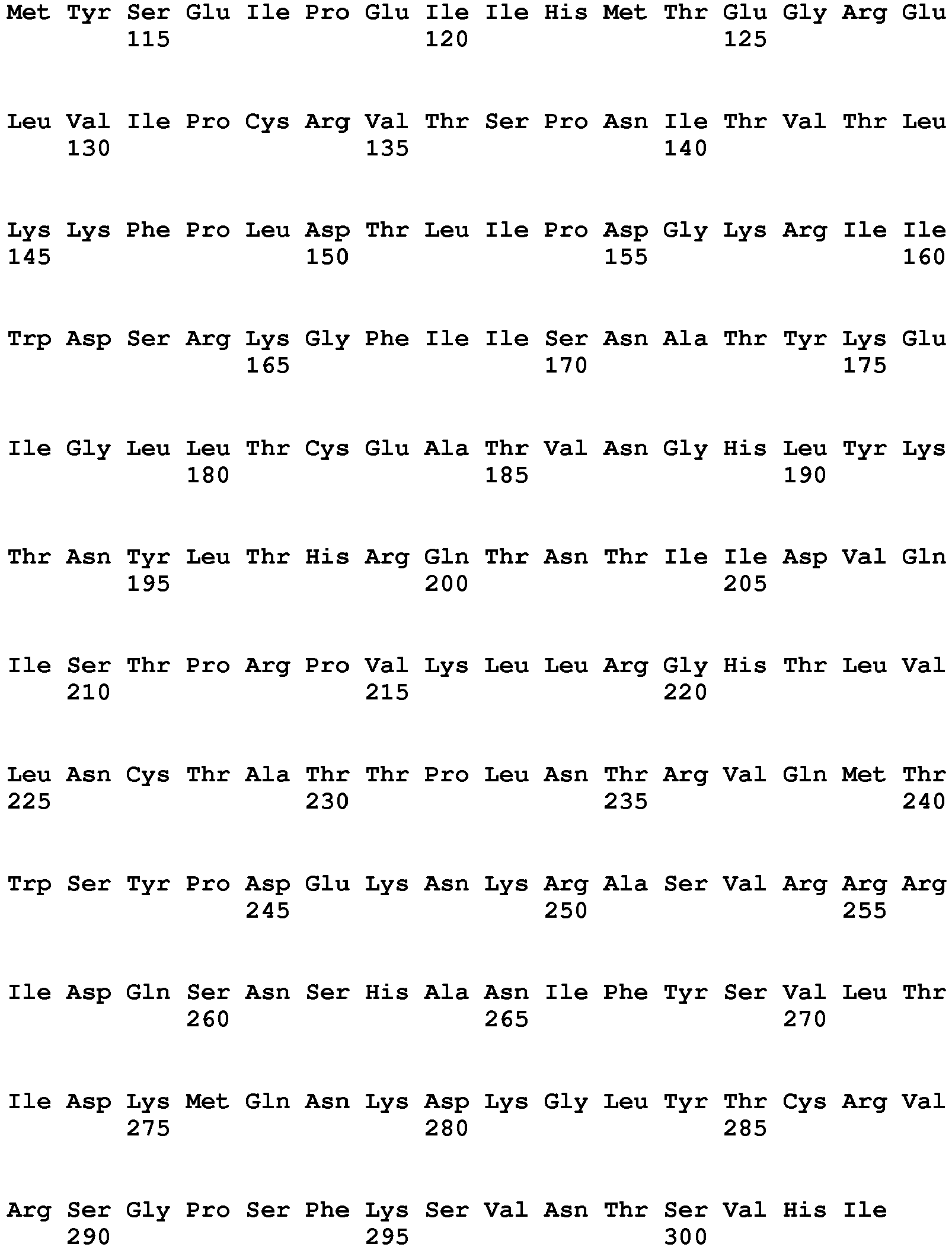
- <210> 3
<211> 187
<212> PRT
<213> Homo sapiens - <400> 3


- <210> 4
<211> 350
<212> PRT
<213> Artificial sequence - <220>
<223> Synthetic polypeptide - <400> 4


- <210> 5
<211> 280
<212> PRT
<213> Artificial sequence - <220>
<223> Synthetic polypeptide - <400> 5


- <210> 6
<211> 156
<212> PRT
<213> Artificial sequence - <220>
<223> Synthetic polypeptide - <400> 6


- <210> 7
<211> 5
<212> PRT
<213> Artificial sequence - <220>
<223> Synthetic polypeptide - <400> 7

- <210> 8
<211> 6
<212> PRT
<213> Artificial sequence - <220>
<223> Synthetic polypeptide - <400> 8

Claims (8)
- A conjugate for use in a method of treating an ocular disease or disorder in an individual, said conjugate comprising:a) a biologically active polypeptide having a molecular weight of from 5 kDa to 250 kDa, wherein the biologically active polypeptide is an inhibitor of angiogenesis; andb) a biocompatible polymer having a molecular weight of from 500 kDa to 2 MDa,wherein the polypeptide is covalently linked to the polymer directly or via a linker, wherein the molar ratio of the biologically active polypeptide to the polymer is at least about 10:1,wherein the method comprises administering to the individual an effective amount of said conjugate by intravitreal administration; and wherein:i) the biologically active polypeptide is a soluble vascular endothelial growth factor (VEGF) receptor, and the polymer is hyaluronic acid; orii) the biologically active polypeptide is an antibody specific for VEGF, and the polymer is carboxymethyl cellulose; oriii) the biologically active polypeptide is an antibody specific for VEGF, and the polymer is hyaluronic acid.
- The conjugate for use according to claim 1, wherein the molar ratio of the biologically active polypeptide to the polymer is:i) from 10: 1 to 25: 1; orii) from 25: 1 to 50: 1.
- The conjugate for use according to any one of the preceding claims, wherein the hyaluronic acid has a molecular weight of from 600 kDa to 700 kDa, or from 750 kDa to 1 MDa.
- The conjugate for use according to any one of the preceding claims, wherein the molar ratio of the VEGF receptor to the hyaluronic acid is 20: 1.
- The conjugate for use according to of any one of the preceding claims, wherein the individual is a human.
- The conjugate for use according to any one of the preceding claims, wherein the ocular disorder is macular degeneration, choroidal neovascularization, retinal neovascularization, proliferative vitreoretinopathy, glaucoma, or ocular inflammation.
- The conjugate for use according to any one of the preceding claims, wherein in the method the conjugate is administered once every two months, once every three months, once every 6 months, or once a year.
- The conjugate for use according to claim 1, whereina) the biologically active polypeptide is a VHH anti-VEGF antibody; andb) the biocompatible polymer comprises hyaluronic acid having a molecular weight of about 860kDa,wherein the polypeptide is covalently linked to the polymer via a linker, andwherein the molar ratio of the biologically active polypeptide to the polymer is from 24:1 to 45:1.
Applications Claiming Priority (2)
| Application Number | Priority Date | Filing Date | Title |
|---|---|---|---|
| US201562265293P | 2015-12-09 | 2015-12-09 | |
| PCT/US2016/065653 WO2017100470A1 (en) | 2015-12-09 | 2016-12-08 | Methods of treating an ocular disease or disorder |
Publications (3)
| Publication Number | Publication Date |
|---|---|
| EP3386520A1 EP3386520A1 (en) | 2018-10-17 |
| EP3386520A4 EP3386520A4 (en) | 2019-07-17 |
| EP3386520B1 true EP3386520B1 (en) | 2022-07-20 |
Family
ID=59014248
Family Applications (1)
| Application Number | Title | Priority Date | Filing Date |
|---|---|---|---|
| EP16873857.3A Active EP3386520B1 (en) | 2015-12-09 | 2016-12-08 | Methods of treating an ocular disease or disorder |
Country Status (7)
| Country | Link |
|---|---|
| US (3) | US10765759B2 (en) |
| EP (1) | EP3386520B1 (en) |
| KR (1) | KR102944785B1 (en) |
| CN (2) | CN113546178A (en) |
| CA (1) | CA3022905A1 (en) |
| ES (1) | ES2925248T3 (en) |
| WO (1) | WO2017100470A1 (en) |
Families Citing this family (15)
| Publication number | Priority date | Publication date | Assignee | Title |
|---|---|---|---|---|
| EP2268673B1 (en) | 2008-03-28 | 2017-10-25 | The Regents of The University of California | Polypeptide-polymer conjugates and methods of use thereof |
| JP6959912B2 (en) | 2015-09-23 | 2021-11-05 | ジェネンテック, インコーポレイテッド | Optimized variant of anti-VEGF antibody |
| WO2017100470A1 (en) | 2015-12-09 | 2017-06-15 | The Regents Of The University Of California | Methods of treating an ocular disease or disorder |
| KR20250057128A (en) | 2015-12-30 | 2025-04-28 | 코디악 사이언시스 인코포레이티드 | Antibodies and conjugates thereof |
| IL269506B2 (en) | 2017-03-22 | 2024-04-01 | Genentech Inc | Hyaluronic acid cross-linked hydrogel preparations and methods |
| CA3130653A1 (en) | 2018-03-09 | 2019-09-12 | Valitor, Inc. | Multivalent peptide conjugates for sustained intra-articular treatment of joint inflammation |
| WO2019246130A1 (en) * | 2018-06-19 | 2019-12-26 | Cella Therapeutics, Llc | Sustained-release drug delivery systems comprising an intraocular pressure lowering agent, a cnp compound, an npr-b compound, a tie-2 agonist, or neurotrophic agent for use for treating glaucoma or ocular hypertension |
| WO2020219497A1 (en) * | 2019-04-25 | 2020-10-29 | The University Of Massachusetts | Compositions and methods for treatment of angiogenesis related diseases |
| MA54325A (en) | 2019-06-05 | 2022-05-11 | Regeneron Pharma | DEVICES AND METHODS FOR ACCURATE DOSE DELIVERY |
| KR20220027215A (en) * | 2019-07-01 | 2022-03-07 | 발리터 인코포레이티드 | Hydrophilic linkers for multivalent peptide conjugates |
| AU2020364071A1 (en) * | 2019-10-10 | 2022-05-26 | Kodiak Sciences Inc. | Methods of treating an eye disorder |
| WO2024055995A1 (en) * | 2022-09-14 | 2024-03-21 | 寻济生物科技(北京)有限公司 | Anti-vegfa fusion protein, and preparation method therefor and use thereof |
| JP2025534252A (en) * | 2022-09-14 | 2025-10-15 | 尋済生物科技(北京)有限公司 | Anti-VEGFA antibodies or antigen-binding fragments thereof, and uses thereof |
| CN115671036A (en) * | 2022-11-15 | 2023-02-03 | 上海交通大学医学院附属第九人民医院 | Gel medicine for treating fundus and intraocular diseases |
| USD1120314S1 (en) | 2022-11-30 | 2026-03-24 | Regeneron Pharmaceuticals, Inc. | Dose delivery device |
Family Cites Families (37)
| Publication number | Priority date | Publication date | Assignee | Title |
|---|---|---|---|---|
| US5162218A (en) | 1988-11-18 | 1992-11-10 | The Regents Of The University Of California | Conjugated polypeptides and methods for their preparation |
| US7122636B1 (en) | 1997-02-21 | 2006-10-17 | Genentech, Inc. | Antibody fragment-polymer conjugates and uses of same |
| US6781030B1 (en) | 1998-11-02 | 2004-08-24 | Trustee Of Tufts College, Ballou Hall | Methods for cloning mammals using telophase oocytes |
| EP1025861A1 (en) | 1999-02-04 | 2000-08-09 | Roche Diagnostics GmbH | Pharmaceutical compositions of hydrophobically modified Hedgehog Proteins and their use |
| JP2003502292A (en) | 1999-06-01 | 2003-01-21 | バイオジェン インコーポレイテッド | Polymer conjugates and uses of hedgehog proteins |
| US20030236214A1 (en) | 1999-06-09 | 2003-12-25 | Wolff Jon A. | Charge reversal of polyion complexes and treatment of peripheral occlusive disease |
| US9522217B2 (en) | 2000-03-15 | 2016-12-20 | Orbusneich Medical, Inc. | Medical device with coating for capturing genetically-altered cells and methods for using same |
| US20040248801A1 (en) | 2000-03-21 | 2004-12-09 | Kiessling Laura L. | Methods and reagents for regulation of cellular responses in biological systems |
| CA2444483A1 (en) | 2001-04-26 | 2002-11-07 | Board Of Regents, The University Of Texas System | Diagnostic imaging compositions, their methods of synthesis and use |
| US6908963B2 (en) | 2001-10-09 | 2005-06-21 | Nektar Therapeutics Al, Corporation | Thioester polymer derivatives and method of modifying the N-terminus of a polypeptide therewith |
| US6964859B2 (en) | 2001-10-16 | 2005-11-15 | Massachusetts Institute Of Technology | Suppressor tRNA system |
| RU2005102604A (en) * | 2002-07-03 | 2005-09-10 | Перикор Сайенс, Инк. (Us) | COMPOSITION OF HYALURONIC ACID AND METHODS OF APPLICATION |
| US20040116348A1 (en) | 2002-09-23 | 2004-06-17 | Ying Chau | Polymer-linker-drug conjugates for targeted drug delivery |
| CA2521784C (en) | 2003-04-08 | 2012-03-27 | Yeda Research And Development Co. Ltd. | Reversible pegylated drugs |
| US8632994B2 (en) | 2003-12-01 | 2014-01-21 | Dako Denmark A/S | Methods and compositions for immuno-histochemical detection |
| MXPA06011965A (en) * | 2004-04-13 | 2007-04-17 | Osi Eyetech Inc | Enhanced biologically active conjugates. |
| US8591885B2 (en) | 2004-04-30 | 2013-11-26 | Allergan, Inc. | Carbonic anhydrase inhibitor sustained release intraocular drug delivery systems |
| WO2006105278A2 (en) | 2005-03-29 | 2006-10-05 | The Regents Of The University Of California | Controlling stem cell destiny destiny with tunable network |
| WO2009013563A2 (en) * | 2006-11-13 | 2009-01-29 | Yissum Research Development Company Of The Hebrew University Of Jerusalem | Use of lipid conjugates in the treatment of diseases or disorders of the eye |
| CA2705785A1 (en) * | 2006-11-14 | 2008-05-22 | Saul Yedgar | Use of lipid conjugates in the treatment of diseases or disorders of the eye |
| US11078262B2 (en) | 2007-04-30 | 2021-08-03 | Allergan, Inc. | High viscosity macromolecular compositions for treating ocular conditions |
| WO2009048280A2 (en) | 2007-10-09 | 2009-04-16 | Postech Academy-Industry Foundation | Long acting hyaluronic acid - peptide conjugate |
| EP2268673B1 (en) | 2008-03-28 | 2017-10-25 | The Regents of The University of California | Polypeptide-polymer conjugates and methods of use thereof |
| EP2360188B1 (en) | 2008-11-05 | 2014-09-17 | National University Corporation Tokyo Medical and Dental University | Hyaluronic acid derivative and pharmaceutical composition thereof |
| IT1399351B1 (en) | 2009-06-16 | 2013-04-16 | Fidia Farmaceutici | PROCEDURE FOR THE SYNTHESIS OF GLYCOSAMINOGLICAN CONJUGATES (GAG) WITH BIOLOGICALLY ACTIVE MOLECULES, POLYMERIC CONJUGATES AND RELATIVE USES |
| WO2011066417A2 (en) | 2009-11-24 | 2011-06-03 | Carnegie Mellon University | Antibodies and conjugates for modulators of angiogenesis |
| WO2012007880A2 (en) | 2010-07-16 | 2012-01-19 | Ablynx Nv | Modified single domain antigen binding molecules and uses thereof |
| KR101309566B1 (en) | 2010-12-10 | 2013-09-17 | 포항공과대학교 산학협력단 | Hyaluronic acid-protein conjugate, and preparation method thereof |
| WO2012140650A2 (en) | 2011-04-12 | 2012-10-18 | Hepacore Ltd. | Conjugates of carboxy polysaccharides with fibroblast growth factors and variants thereof |
| AU2012334825B2 (en) | 2011-11-09 | 2017-08-24 | Ascend Biopharmaceuticals Ltd | Immunomodulatory conjugates |
| US10231991B2 (en) | 2013-07-19 | 2019-03-19 | The Johns Hopkins University | Biomaterials comprising hyaluronic acid binding peptides and extracellular matrix binding peptides for hyaluronic acid retention and tissue engineering applications |
| JP6959912B2 (en) | 2015-09-23 | 2021-11-05 | ジェネンテック, インコーポレイテッド | Optimized variant of anti-VEGF antibody |
| CN108369611A (en) | 2015-10-07 | 2018-08-03 | 基因泰克公司 | The system and method for vitreum half-life period for predicted treatment agent-polymer conjugate |
| WO2017100470A1 (en) | 2015-12-09 | 2017-06-15 | The Regents Of The University Of California | Methods of treating an ocular disease or disorder |
| EP3600442A1 (en) | 2017-03-22 | 2020-02-05 | Genentech, Inc. | Optimized antibody compositions for treatment of ocular disorders |
| CA3130653A1 (en) | 2018-03-09 | 2019-09-12 | Valitor, Inc. | Multivalent peptide conjugates for sustained intra-articular treatment of joint inflammation |
| KR20220027215A (en) | 2019-07-01 | 2022-03-07 | 발리터 인코포레이티드 | Hydrophilic linkers for multivalent peptide conjugates |
-
2016
- 2016-12-08 WO PCT/US2016/065653 patent/WO2017100470A1/en not_active Ceased
- 2016-12-08 CA CA3022905A patent/CA3022905A1/en active Pending
- 2016-12-08 CN CN202110847440.0A patent/CN113546178A/en active Pending
- 2016-12-08 ES ES16873857T patent/ES2925248T3/en active Active
- 2016-12-08 KR KR1020187019422A patent/KR102944785B1/en active Active
- 2016-12-08 EP EP16873857.3A patent/EP3386520B1/en active Active
- 2016-12-08 US US15/781,395 patent/US10765759B2/en active Active
- 2016-12-08 CN CN201680078324.4A patent/CN108472310B/en active Active
-
2020
- 2020-08-05 US US16/985,522 patent/US11723982B2/en active Active
-
2023
- 2023-06-21 US US18/339,061 patent/US20240148882A1/en not_active Abandoned
Also Published As
| Publication number | Publication date |
|---|---|
| CN108472310B (en) | 2021-08-03 |
| US20180318431A1 (en) | 2018-11-08 |
| CN108472310A (en) | 2018-08-31 |
| KR20180100569A (en) | 2018-09-11 |
| US10765759B2 (en) | 2020-09-08 |
| KR102944785B1 (en) | 2026-03-30 |
| WO2017100470A1 (en) | 2017-06-15 |
| CN113546178A (en) | 2021-10-26 |
| US11723982B2 (en) | 2023-08-15 |
| EP3386520A4 (en) | 2019-07-17 |
| US20210113702A1 (en) | 2021-04-22 |
| CA3022905A1 (en) | 2017-06-15 |
| US20240148882A1 (en) | 2024-05-09 |
| ES2925248T3 (en) | 2022-10-14 |
| EP3386520A1 (en) | 2018-10-17 |
Similar Documents
| Publication | Publication Date | Title |
|---|---|---|
| US20240148882A1 (en) | Methods of treating an ocular disease or disorder | |
| CN103505718B (en) | Compstatin and analogs thereof for eye disorders | |
| JP5990511B2 (en) | Anti-CD160 specific antibodies for the treatment of ocular disorders based on angiogenesis | |
| AU2016241499B2 (en) | Pharmaceutical composition for preventing and treating eye diseases, containing, as active ingredient, fusion protein in which tissue-penetrating peptide and anti-vascular endothelial growth factor preparation are fused | |
| CN104066448A (en) | eye disease treatment | |
| US20230256107A1 (en) | Hyaluronic acid binding derivatives of versican (vg1) for long acting delivery of therapeutics | |
| WO2011112850A2 (en) | Humanized and chimeric anti-properdin antibodies | |
| US20220096656A1 (en) | Nanocarriers having surface conjugated peptides and uses thereof for sustained local release of drugs | |
| US20190167576A1 (en) | Use of high affinity monoclonal antibody product binders to increase the duration of action of therapeutic monoclonal antibody products in a biologic tissue | |
| Li et al. | Sustaining intravitreal residence with L-arginine peptide-conjugated nanocarriers | |
| HK1262517B (en) | Methods of treating an ocular disease or disorder | |
| HK1262517A1 (en) | Methods of treating an ocular disease or disorder | |
| RU2669787C2 (en) | Means for treatment of disease with macular oedema due to excessive vegf-a expression | |
| WO2023039206A2 (en) | Treatment of geographic atrophy | |
| Arlotta | Synthesis and Characterization of Therapeutic Antibody Conjugates | |
| Altiok | Improving Anti-VEGF Drugs in the Vitreous | |
| WO2018089465A1 (en) | Nanocarriers having surface conjugated peptides and uses thereof for sustained local release of drugs |
Legal Events
| Date | Code | Title | Description |
|---|---|---|---|
| STAA | Information on the status of an ep patent application or granted ep patent |
Free format text: STATUS: THE INTERNATIONAL PUBLICATION HAS BEEN MADE |
|
| PUAI | Public reference made under article 153(3) epc to a published international application that has entered the european phase |
Free format text: ORIGINAL CODE: 0009012 |
|
| STAA | Information on the status of an ep patent application or granted ep patent |
Free format text: STATUS: REQUEST FOR EXAMINATION WAS MADE |
|
| 17P | Request for examination filed |
Effective date: 20180702 |
|
| AK | Designated contracting states |
Kind code of ref document: A1 Designated state(s): AL AT BE BG CH CY CZ DE DK EE ES FI FR GB GR HR HU IE IS IT LI LT LU LV MC MK MT NL NO PL PT RO RS SE SI SK SM TR |
|
| AX | Request for extension of the european patent |
Extension state: BA ME |
|
| DAV | Request for validation of the european patent (deleted) | ||
| DAX | Request for extension of the european patent (deleted) | ||
| A4 | Supplementary search report drawn up and despatched |
Effective date: 20190618 |
|
| RIC1 | Information provided on ipc code assigned before grant |
Ipc: A61K 31/715 20060101AFI20190607BHEP Ipc: A61K 47/18 20170101ALI20190607BHEP Ipc: A61K 38/17 20060101ALI20190607BHEP Ipc: A61K 38/16 20060101ALI20190607BHEP |
|
| REG | Reference to a national code |
Ref country code: HK Ref legal event code: DE Ref document number: 1262517 Country of ref document: HK |
|
| STAA | Information on the status of an ep patent application or granted ep patent |
Free format text: STATUS: EXAMINATION IS IN PROGRESS |
|
| 17Q | First examination report despatched |
Effective date: 20200129 |
|
| GRAP | Despatch of communication of intention to grant a patent |
Free format text: ORIGINAL CODE: EPIDOSNIGR1 |
|
| STAA | Information on the status of an ep patent application or granted ep patent |
Free format text: STATUS: GRANT OF PATENT IS INTENDED |
|
| INTG | Intention to grant announced |
Effective date: 20220203 |
|
| GRAS | Grant fee paid |
Free format text: ORIGINAL CODE: EPIDOSNIGR3 |
|
| GRAA | (expected) grant |
Free format text: ORIGINAL CODE: 0009210 |
|
| STAA | Information on the status of an ep patent application or granted ep patent |
Free format text: STATUS: THE PATENT HAS BEEN GRANTED |
|
| AK | Designated contracting states |
Kind code of ref document: B1 Designated state(s): AL AT BE BG CH CY CZ DE DK EE ES FI FR GB GR HR HU IE IS IT LI LT LU LV MC MK MT NL NO PL PT RO RS SE SI SK SM TR |
|
| REG | Reference to a national code |
Ref country code: CH Ref legal event code: EP |
|
| REG | Reference to a national code |
Ref country code: DE Ref legal event code: R096 Ref document number: 602016073696 Country of ref document: DE |
|
| REG | Reference to a national code |
Ref country code: AT Ref legal event code: REF Ref document number: 1505110 Country of ref document: AT Kind code of ref document: T Effective date: 20220815 |
|
| REG | Reference to a national code |
Ref country code: IE Ref legal event code: FG4D |
|
| REG | Reference to a national code |
Ref country code: SE Ref legal event code: TRGR |
|
| REG | Reference to a national code |
Ref country code: NO Ref legal event code: T2 Effective date: 20220720 |
|
| REG | Reference to a national code |
Ref country code: NL Ref legal event code: FP |
|
| REG | Reference to a national code |
Ref country code: ES Ref legal event code: FG2A Ref document number: 2925248 Country of ref document: ES Kind code of ref document: T3 Effective date: 20221014 |
|
| REG | Reference to a national code |
Ref country code: LT Ref legal event code: MG9D |
|
| PG25 | Lapsed in a contracting state [announced via postgrant information from national office to epo] |
Ref country code: RS Free format text: LAPSE BECAUSE OF FAILURE TO SUBMIT A TRANSLATION OF THE DESCRIPTION OR TO PAY THE FEE WITHIN THE PRESCRIBED TIME-LIMIT Effective date: 20220720 Ref country code: PT Free format text: LAPSE BECAUSE OF FAILURE TO SUBMIT A TRANSLATION OF THE DESCRIPTION OR TO PAY THE FEE WITHIN THE PRESCRIBED TIME-LIMIT Effective date: 20221121 Ref country code: LV Free format text: LAPSE BECAUSE OF FAILURE TO SUBMIT A TRANSLATION OF THE DESCRIPTION OR TO PAY THE FEE WITHIN THE PRESCRIBED TIME-LIMIT Effective date: 20220720 Ref country code: LT Free format text: LAPSE BECAUSE OF FAILURE TO SUBMIT A TRANSLATION OF THE DESCRIPTION OR TO PAY THE FEE WITHIN THE PRESCRIBED TIME-LIMIT Effective date: 20220720 Ref country code: FI Free format text: LAPSE BECAUSE OF FAILURE TO SUBMIT A TRANSLATION OF THE DESCRIPTION OR TO PAY THE FEE WITHIN THE PRESCRIBED TIME-LIMIT Effective date: 20220720 |
|
| REG | Reference to a national code |
Ref country code: AT Ref legal event code: MK05 Ref document number: 1505110 Country of ref document: AT Kind code of ref document: T Effective date: 20220720 |
|
| PG25 | Lapsed in a contracting state [announced via postgrant information from national office to epo] |
Ref country code: PL Free format text: LAPSE BECAUSE OF FAILURE TO SUBMIT A TRANSLATION OF THE DESCRIPTION OR TO PAY THE FEE WITHIN THE PRESCRIBED TIME-LIMIT Effective date: 20220720 Ref country code: IS Free format text: LAPSE BECAUSE OF FAILURE TO SUBMIT A TRANSLATION OF THE DESCRIPTION OR TO PAY THE FEE WITHIN THE PRESCRIBED TIME-LIMIT Effective date: 20221120 Ref country code: HR Free format text: LAPSE BECAUSE OF FAILURE TO SUBMIT A TRANSLATION OF THE DESCRIPTION OR TO PAY THE FEE WITHIN THE PRESCRIBED TIME-LIMIT Effective date: 20220720 Ref country code: GR Free format text: LAPSE BECAUSE OF FAILURE TO SUBMIT A TRANSLATION OF THE DESCRIPTION OR TO PAY THE FEE WITHIN THE PRESCRIBED TIME-LIMIT Effective date: 20221021 |
|
| REG | Reference to a national code |
Ref country code: DE Ref legal event code: R097 Ref document number: 602016073696 Country of ref document: DE |
|
| PG25 | Lapsed in a contracting state [announced via postgrant information from national office to epo] |
Ref country code: SM Free format text: LAPSE BECAUSE OF FAILURE TO SUBMIT A TRANSLATION OF THE DESCRIPTION OR TO PAY THE FEE WITHIN THE PRESCRIBED TIME-LIMIT Effective date: 20220720 Ref country code: RO Free format text: LAPSE BECAUSE OF FAILURE TO SUBMIT A TRANSLATION OF THE DESCRIPTION OR TO PAY THE FEE WITHIN THE PRESCRIBED TIME-LIMIT Effective date: 20220720 Ref country code: DK Free format text: LAPSE BECAUSE OF FAILURE TO SUBMIT A TRANSLATION OF THE DESCRIPTION OR TO PAY THE FEE WITHIN THE PRESCRIBED TIME-LIMIT Effective date: 20220720 Ref country code: CZ Free format text: LAPSE BECAUSE OF FAILURE TO SUBMIT A TRANSLATION OF THE DESCRIPTION OR TO PAY THE FEE WITHIN THE PRESCRIBED TIME-LIMIT Effective date: 20220720 Ref country code: AT Free format text: LAPSE BECAUSE OF FAILURE TO SUBMIT A TRANSLATION OF THE DESCRIPTION OR TO PAY THE FEE WITHIN THE PRESCRIBED TIME-LIMIT Effective date: 20220720 |
|
| PLBE | No opposition filed within time limit |
Free format text: ORIGINAL CODE: 0009261 |
|
| STAA | Information on the status of an ep patent application or granted ep patent |
Free format text: STATUS: NO OPPOSITION FILED WITHIN TIME LIMIT |
|
| PG25 | Lapsed in a contracting state [announced via postgrant information from national office to epo] |
Ref country code: SK Free format text: LAPSE BECAUSE OF FAILURE TO SUBMIT A TRANSLATION OF THE DESCRIPTION OR TO PAY THE FEE WITHIN THE PRESCRIBED TIME-LIMIT Effective date: 20220720 Ref country code: EE Free format text: LAPSE BECAUSE OF FAILURE TO SUBMIT A TRANSLATION OF THE DESCRIPTION OR TO PAY THE FEE WITHIN THE PRESCRIBED TIME-LIMIT Effective date: 20220720 |
|
| P01 | Opt-out of the competence of the unified patent court (upc) registered |
Effective date: 20230506 |
|
| 26N | No opposition filed |
Effective date: 20230421 |
|
| PG25 | Lapsed in a contracting state [announced via postgrant information from national office to epo] |
Ref country code: AL Free format text: LAPSE BECAUSE OF FAILURE TO SUBMIT A TRANSLATION OF THE DESCRIPTION OR TO PAY THE FEE WITHIN THE PRESCRIBED TIME-LIMIT Effective date: 20220720 |
|
| PG25 | Lapsed in a contracting state [announced via postgrant information from national office to epo] |
Ref country code: SI Free format text: LAPSE BECAUSE OF FAILURE TO SUBMIT A TRANSLATION OF THE DESCRIPTION OR TO PAY THE FEE WITHIN THE PRESCRIBED TIME-LIMIT Effective date: 20220720 Ref country code: LU Free format text: LAPSE BECAUSE OF NON-PAYMENT OF DUE FEES Effective date: 20221208 |
|
| PG25 | Lapsed in a contracting state [announced via postgrant information from national office to epo] |
Ref country code: HU Free format text: LAPSE BECAUSE OF FAILURE TO SUBMIT A TRANSLATION OF THE DESCRIPTION OR TO PAY THE FEE WITHIN THE PRESCRIBED TIME-LIMIT; INVALID AB INITIO Effective date: 20161208 |
|
| PG25 | Lapsed in a contracting state [announced via postgrant information from national office to epo] |
Ref country code: CY Free format text: LAPSE BECAUSE OF FAILURE TO SUBMIT A TRANSLATION OF THE DESCRIPTION OR TO PAY THE FEE WITHIN THE PRESCRIBED TIME-LIMIT Effective date: 20220720 |
|
| PG25 | Lapsed in a contracting state [announced via postgrant information from national office to epo] |
Ref country code: MK Free format text: LAPSE BECAUSE OF FAILURE TO SUBMIT A TRANSLATION OF THE DESCRIPTION OR TO PAY THE FEE WITHIN THE PRESCRIBED TIME-LIMIT Effective date: 20220720 |
|
| PG25 | Lapsed in a contracting state [announced via postgrant information from national office to epo] |
Ref country code: MC Free format text: LAPSE BECAUSE OF FAILURE TO SUBMIT A TRANSLATION OF THE DESCRIPTION OR TO PAY THE FEE WITHIN THE PRESCRIBED TIME-LIMIT Effective date: 20220720 |
|
| PG25 | Lapsed in a contracting state [announced via postgrant information from national office to epo] |
Ref country code: TR Free format text: LAPSE BECAUSE OF FAILURE TO SUBMIT A TRANSLATION OF THE DESCRIPTION OR TO PAY THE FEE WITHIN THE PRESCRIBED TIME-LIMIT Effective date: 20220720 Ref country code: MC Free format text: LAPSE BECAUSE OF FAILURE TO SUBMIT A TRANSLATION OF THE DESCRIPTION OR TO PAY THE FEE WITHIN THE PRESCRIBED TIME-LIMIT Effective date: 20220720 |
|
| PG25 | Lapsed in a contracting state [announced via postgrant information from national office to epo] |
Ref country code: BG Free format text: LAPSE BECAUSE OF FAILURE TO SUBMIT A TRANSLATION OF THE DESCRIPTION OR TO PAY THE FEE WITHIN THE PRESCRIBED TIME-LIMIT Effective date: 20220720 |
|
| PG25 | Lapsed in a contracting state [announced via postgrant information from national office to epo] |
Ref country code: MT Free format text: LAPSE BECAUSE OF FAILURE TO SUBMIT A TRANSLATION OF THE DESCRIPTION OR TO PAY THE FEE WITHIN THE PRESCRIBED TIME-LIMIT Effective date: 20220720 |
|
| REG | Reference to a national code |
Ref country code: CH Ref legal event code: U11 Free format text: ST27 STATUS EVENT CODE: U-0-0-U10-U11 (AS PROVIDED BY THE NATIONAL OFFICE) Effective date: 20260101 |
|
| PGFP | Annual fee paid to national office [announced via postgrant information from national office to epo] |
Ref country code: GB Payment date: 20251229 Year of fee payment: 10 |
|
| PGFP | Annual fee paid to national office [announced via postgrant information from national office to epo] |
Ref country code: NO Payment date: 20251231 Year of fee payment: 10 |
|
| PGFP | Annual fee paid to national office [announced via postgrant information from national office to epo] |
Ref country code: IT Payment date: 20251219 Year of fee payment: 10 |
|
| PGFP | Annual fee paid to national office [announced via postgrant information from national office to epo] |
Ref country code: FR Payment date: 20251226 Year of fee payment: 10 Ref country code: NL Payment date: 20251226 Year of fee payment: 10 |
|
| PGFP | Annual fee paid to national office [announced via postgrant information from national office to epo] |
Ref country code: BE Payment date: 20251229 Year of fee payment: 10 |
|
| PGFP | Annual fee paid to national office [announced via postgrant information from national office to epo] |
Ref country code: SE Payment date: 20251227 Year of fee payment: 10 |
|
| PGFP | Annual fee paid to national office [announced via postgrant information from national office to epo] |
Ref country code: IE Payment date: 20251229 Year of fee payment: 10 |
|
| PGFP | Annual fee paid to national office [announced via postgrant information from national office to epo] |
Ref country code: ES Payment date: 20260102 Year of fee payment: 10 |
|
| PGFP | Annual fee paid to national office [announced via postgrant information from national office to epo] |
Ref country code: DE Payment date: 20251229 Year of fee payment: 10 |
|
| PGFP | Annual fee paid to national office [announced via postgrant information from national office to epo] |
Ref country code: CH Payment date: 20260101 Year of fee payment: 10 |