EP3383423B1 - Polypeptides and analogues thereof for use in the treatment of diabetes and bone disorders - Google Patents
Polypeptides and analogues thereof for use in the treatment of diabetes and bone disorders Download PDFInfo
- Publication number
- EP3383423B1 EP3383423B1 EP16809694.9A EP16809694A EP3383423B1 EP 3383423 B1 EP3383423 B1 EP 3383423B1 EP 16809694 A EP16809694 A EP 16809694A EP 3383423 B1 EP3383423 B1 EP 3383423B1
- Authority
- EP
- European Patent Office
- Prior art keywords
- gip
- xenin
- bone
- mice
- dala2
- Prior art date
- Legal status (The legal status is an assumption and is not a legal conclusion. Google has not performed a legal analysis and makes no representation as to the accuracy of the status listed.)
- Active
Links
Images








Classifications
-
- A—HUMAN NECESSITIES
- A61—MEDICAL OR VETERINARY SCIENCE; HYGIENE
- A61K—PREPARATIONS FOR MEDICAL, DENTAL OR TOILETRY PURPOSES
- A61K38/00—Medicinal preparations containing peptides
- A61K38/16—Peptides having more than 20 amino acids; Gastrins; Somatostatins; Melanotropins; Derivatives thereof
- A61K38/17—Peptides having more than 20 amino acids; Gastrins; Somatostatins; Melanotropins; Derivatives thereof from animals; from humans
- A61K38/22—Hormones
-
- A—HUMAN NECESSITIES
- A61—MEDICAL OR VETERINARY SCIENCE; HYGIENE
- A61P—SPECIFIC THERAPEUTIC ACTIVITY OF CHEMICAL COMPOUNDS OR MEDICINAL PREPARATIONS
- A61P19/00—Drugs for skeletal disorders
- A61P19/08—Drugs for skeletal disorders for bone diseases, e.g. rachitism, Paget's disease
- A61P19/10—Drugs for skeletal disorders for bone diseases, e.g. rachitism, Paget's disease for osteoporosis
-
- A—HUMAN NECESSITIES
- A61—MEDICAL OR VETERINARY SCIENCE; HYGIENE
- A61P—SPECIFIC THERAPEUTIC ACTIVITY OF CHEMICAL COMPOUNDS OR MEDICINAL PREPARATIONS
- A61P3/00—Drugs for disorders of the metabolism
- A61P3/08—Drugs for disorders of the metabolism for glucose homeostasis
- A61P3/10—Drugs for disorders of the metabolism for glucose homeostasis for hyperglycaemia, e.g. antidiabetics
-
- C—CHEMISTRY; METALLURGY
- C07—ORGANIC CHEMISTRY
- C07K—PEPTIDES
- C07K14/00—Peptides having more than 20 amino acids; Gastrins; Somatostatins; Melanotropins; Derivatives thereof
- C07K14/435—Peptides having more than 20 amino acids; Gastrins; Somatostatins; Melanotropins; Derivatives thereof from animals; from humans
- C07K14/46—Peptides having more than 20 amino acids; Gastrins; Somatostatins; Melanotropins; Derivatives thereof from animals; from humans from vertebrates
- C07K14/47—Peptides having more than 20 amino acids; Gastrins; Somatostatins; Melanotropins; Derivatives thereof from animals; from humans from vertebrates from mammals
-
- C—CHEMISTRY; METALLURGY
- C07—ORGANIC CHEMISTRY
- C07K—PEPTIDES
- C07K14/00—Peptides having more than 20 amino acids; Gastrins; Somatostatins; Melanotropins; Derivatives thereof
- C07K14/435—Peptides having more than 20 amino acids; Gastrins; Somatostatins; Melanotropins; Derivatives thereof from animals; from humans
- C07K14/575—Hormones
- C07K14/605—Glucagons
-
- A—HUMAN NECESSITIES
- A61—MEDICAL OR VETERINARY SCIENCE; HYGIENE
- A61K—PREPARATIONS FOR MEDICAL, DENTAL OR TOILETRY PURPOSES
- A61K38/00—Medicinal preparations containing peptides
Definitions
- the present invention relates to polypeptides and analogues thereof for use in the treatment of diabetes and bone disorders. Also disclosed are pharmaceutical compositions comprising the polypeptides and analogues thereof, and methods for the treatment of diabetes and bone disorders.
- GLP-1 glucagon-like peptide-1
- GIP glucose-dependent insulinotropic peptide
- diabetes mellitus It is estimated that 382 million people are living with diabetes mellitus in the world, which is approximately 8.5% of the population. Both major forms of diabetes mellitus, type-1 and type-2, are associated with increased occurrence of bone fragility fractures. Importantly, this increased risk is irrespective of the normal measure of bone fracture likelihood in non-diabetic subjects. Increased body weight can reduce the risk of developing bone disorders due to enhanced mechanical loading of bone. However, with type-2 diabetes mellitus, bone fracture risk is increased despite elevated body weight. Conservative estimates suggest subjects suffering from type-2 diabetes mellitus patients are more than twice as likely to suffer a bone fracture, compared to the normal population.
- bisphosphonates are the first-line treatment. Although bisphosphonates are effective in reducing further deterioration of the bone mass and micro-architecture, they do not reconstruct the skeleton. Anabolic treatments that stimulate bone formation have also been developed. A recombinant form of parathyroid hormone (teriparatide) has been shown to be an effective osteoporotic treatment and is now approved. However, there are still many unanswered questions concerning teriparatide therapy.
- Bone is comprised mainly of collagen, calcium-phosphate hydroxyapatite and water.
- bone is a highly complex, dynamic organ that undergoes continuous remodelling throughout life through the sequential activity of osteoblasts and osteoclasts.
- Bone resorption biomarkers experience a rapid change shortly after food ingestion.
- the incretin hormones have emerged as major player that regulates bone metabolism and plays a crucial role in maintaining bone quality.
- functional GIP receptors are present in bone. Indeed, previous studies reported that GIP-R knockout mice have reduced osteoblasts numbers and increased osteoclast activity, resulting in increased bone fracture risk.
- double-incretin knockout mice exhibit reduced cortical bone mass and cortical bone strength. GIP was also recently shown to reduce osteoclast differentiation and resorption. Further to this, beneficial effects of a stable GIP receptor agonist on tissue-level bone material properties has been reported in rats. However, little attention has been given to the role of GIP on bone.
- GIT gastrointestinal tract
- GIP glucose-dependent insulinotropic polypeptide
- a polypeptide comprising the amino acid sequence YAEGTFISDYSIAMHPQQPWIL.
- polypeptide or analogue thereof further comprises the amino acid sequence GAADDDDDD.
- the polypeptide or analogue thereof comprises the amino acid sequence YAEGTFISDYSIAMHPQQPWILGAADDDDDD.
- polypeptide or analogue thereof comprises the amino acid sequence GAAEEEEEE.
- polypeptide or analogue thereof comprises the amino acid sequence YAEGTFISDYSIAMHPQQPWILGAAEEEEEE.
- any or each amino acid of the polypeptide is a D- or L-amino acid.
- any or each amino acid of the polypeptide is a D-amino acid.
- any or each alanine (A) amino acid of the polypeptide is a D-amino acid.
- the alanine (A) amino acid at position 2 of the polypeptide is a D-amino acid.
- the alanine (A) amino acid at position 2 of the polypeptide is D-alanine.
- a polypeptide according to a first aspect of the present invention or an analogue thereof for use in the treatment of diabetes mellitus is provided.
- polypeptide or analogue thereof is for use in the treatment of type-2 diabetes mellitus.
- a polypeptide according to a first aspect of the present invention or an analogue thereof for use in the treatment of obesity is provided.
- a polypeptide according to a first aspect of the present invention or an analogue thereof for use in the treatment of metabolic syndrome is provided.
- a polypeptide according to a first aspect of the present invention or an analogue thereof for use in the treatment of bone disorders is provided.
- the or each bone disorder is selected from Osteogenesis imperfecta, Paget's disease of bone, osteomalacia, and osteoporosis.
- the polypeptide or analogue thereof is for use in reducing bone fragility.
- polypeptide or analogue thereof is for use in reducing bone fragility in a subject suffering from diabetes mellitus.
- polypeptide or analogue thereof is for use in reducing bone fragility in a subject suffering from type-2 diabetes mellitus.
- polypeptide or analogue thereof is for use in increasing bone strength.
- polypeptide or analogue thereof is for use in increasing bone mass.
- the polypeptide or analogue thereof is for use in increasing any one or more of bone density, bone volume density, bone mineral density, and bone mineral content.
- polypeptide or analogue thereof is for use in increasing bone strength, increasing bone mass, and/or increasing bone density in a subject suffering from diabetes mellitus.
- polypeptide or analogue thereof is for use in increasing bone strength, increasing bone mass, and/or increasing bone density in a subject suffering from type-2 diabetes mellitus.
- the polypeptide or analogue thereof is for use in increasing cAMP levels in bone cells, optionally osteoblastic cells.
- the polypeptide or analogue thereof is for use in increasing TGF-beta levels in or from bone cells, optionally osteoblastic cells.
- the polypeptide or analogue thereof is for use in increasing IGF-1 levels in or from bone cells, optionally osteoblastic cells.
- polypeptide or analogue thereof does not affect any one or more of body weight, fat mass, lean mass, and food intake/appetite.
- a pharmaceutical composition comprising at least one polypeptide according to a first aspect of the present invention or analogue thereof.
- the pharmaceutical composition comprises at least one polypeptide according to a first aspect of the present invention or analogue thereof, and a pharmaceutically acceptable excipient.
- polypeptides were purchased from GL Biochem Ltd (Shanghai, China, greater than 95% purity). Polypeptides were characterised using HPLC and MALDI-TOF mass spectrometry as described by Martin et al. 2013 (Biochim Biophys Acta. 2013 Jun;1830(6):3407-13 ). The experimental mass for all peptides corresponded closely to their theoretical values, confirming structural identity.
- peptide samples (1 ⁇ g) were mixed with ⁇ -cyano-4-hydroxycinnamic acid (1 ⁇ g) and applied to a Voyager-DE BioSpectrometry Workstation and mass-to-charge (m/z) ratio verses peak intensity recorded by MALDI-TOF mass spectrometry.
- DPP-4 half-life was assessed by incubating peptides (37°C in 50 mmol/L TEA-HCI; pH 7.8) with 5 mU purified porcine DPP-4 for 0, 1, 2, 4, 8 and 12 h.
- Degradation profiles were obtained using rp-HPLC analysis and HPLC peak area data used to calculate percentage intact peptide remaining at each time point.
- Peptide DPP-4 degradation profiles were assessed as described by Martin et al. 2013 (Biochim Biophys Acta. 2013 Jun;1830(6):3407-13 ). Briefly, polypeptides (20 ug) were incubated with 5 ⁇ l of purified DPP-4 (5 mU, Sigma-Aldrich, Dorset, UK) in 50 mM TEA-HCl (pH 7.8; final volume 450 ⁇ l) at 37°C for 0, 1, 4 and 12 hours. Enzymatic reactions were terminated by the addition of 50 ⁇ l TFA/H2O (10% v/v, Sigma-Aldrich, Dorset, UK). Reaction products were then separated by HPLC and analysed using MALDI-TOF.
- BRIN-BD11 cells were used to assess the insulin releasing activity of polypeptides as described by McClenaghan et al., 1996 (Diabetes. 1996 Aug;45(8):1132-40 ).
- Cells were cultured in RPMI-1640 growth media supplemented with 10% (v/v) foetal bovine serum (FBS) and 1% (v/v) antibiotics (penicillin (100 U/ml), streptomycin (0.1 mg/l)), in 75 cm2 sterile tissue culture flasks (Greiner bio-one, UK) maintained at 37°C and 5% CO2 in a LEEC incubator (Laboratory technical engineering, Nottingham, UK).
- FBS foetal bovine serum
- antibiotics penicillin (100 U/ml), streptomycin (0.1 mg/l)
- BRIN-BD11 cells were then seeded at a density of 150,000 cells/well in 24-well plates (Nunc, Roskilde, Denmark) and allowed to attach overnight at 37°C. Culture medium was removed and cells were pre-incubated in Krebs-Ringer bicarbonate buffer (KRBB) (115 mmol/l NaCl, 4.7 mmol/l KCl, 1.2 mmol/l MgSO4, 1.28 mmol/l CaCl2, 1.2 mmol/l KH2PO4, 25 mmol/l HEPES and 8.4% NaHCO3, containing 0.5% (w/v) BSA , pH 7.4) supplemented with 1.1 mmol/l glucose for 40 min at 37°C.
- KRBB Krebs-Ringer bicarbonate buffer
- experiments were performed in presence of glucose (5.6 or 16.7 mmol/l) with a range of concentrations of polypeptides (10 -12 to 10 -6 mol/l) for 20 min at 37°C. After test incubations, aliquots of assay buffer were collected from each well and stored at -20°C prior to measurement of insulin by radioimmunoassay as described by Flatt & Bailey, 1981 (Diabetologia, 1981; 20:573-577 ).
- mice For food intake studies, fasted (18 h) mice were given intraperitoneal (i.p) injections of polypeptides at a dose of 25, 100 or 250 nmol/kg bw. Mice were then allowed free access to normal chow for 180 mins and cumulative food intake measured.
- i.p intraperitoneal
- blood glucose and plasma insulin were measured immediately prior to and 15, 30, 60 and 105 min after i.p. administration of glucose alone (18 mmol/kg bw) or in combination with the polypeptides (each at 25 nmol/kg bw) in non-fasted mice.
- polypeptides each at 25 nmol/kg bw or saline vehicle (0.9% w/v NaCl) were injected 4 h before a glucose load (18 mmol/kg bw) in non-fasted mice and blood glucose and plasma insulin measured at 0, 15, 30 and 60 min post glucose injection.
- HOMA-IR fasting glucose (mmol/l) x fasting insulin (mU/l)/22.5. All test solutions were administered in a final volume of 5 ml/kg body weight, at 10:00 h without previous 09:30 h peptide injection. Terminal analysis included measurement of total body fat and lean mass by DEXA scanning (Piximus Densitometer, Inside Outside Sales, Madison WI, USA) and extraction of pancreatic tissue for analysis as detailed below. Immunohistochemistry Pancreatic tissue was excised, divided longitudinally and either immediately snap frozen for subsequent extraction of insulin using acid ethanol extraction (5 ml/g) as described by Irwin et al., 2015 (Diabetes.
- pancreatic tissues were fixed in 4% parafolmaldehyde at 4°C for 48 h. Pancreata were then embedded in paraffin wax and processed using an automated tissue processor (Leica TP1020, Leica Microsystems, Nussloch, Germany). Immunohistochemistry was performed as described by Vasu et al., 2014 (PLoS One. 2014 Jun 26;9(6):e101005 ).
- tissue sections were deparaffinised, rehydrated and probed with primary antibodies: mouse anti-insulin antibody (1:500; Abcam, ab6995) or guinea-pig anti-glucagon antibody (PCA2/4, 1:400; raised in-house). The sections were then incubated with secondary antibody, Alexa Fluor 488 goat anti-guinea pig IgG (1:400) or Alexa Fluor 594 goat anti-mouse IgG (1:400); respectively. The slides were viewed under a FITC (488 nm) or TRITC filter (594 nm) using a fluorescent microscope (Olympus system microscope, model BX51) and photographed using a DP70 camera adapter system. Islet parameters were analysed using Cell ⁇ F image analysis software (Olympus Soft Imaging Solutions, GmbH).
- Blood samples were collected from the cut tip on the tail vein of conscious mice into chilled fluoride/heparin glucose micro-centrifuge tubes (Sarstedt, Numbrecht, Germany) at the time points indicated in the Drawings. Blood glucose was measured directly using a hand-held Ascencia Contour blood glucose meter (Bayer Healthcare, Newbury, Berkshire, UK). Blood samples were centrifuged using a Beckman microcentrifuge (Beckman Instruments, Galway, Ireland) for 1-5 mins at 13,000 x g and stored at -20 °C. Plasma and pancreatic insulin was assayed by a modified dextran-coated charcoal radioimmunoassay as described by Flatt & Bailey, 1981 (Diabetologia, 1981; 20:573-577 ).
- Human osteoblast SaOS-2 cells [sourced from The European Collection of Cell Cultures (ECACC)] were seeded at a density of 2e5 cells/well in 24 well plates [Nunc, Roskilde, Denmark]. The cells were incubated in media [Life Technologies] supplemented with 10% FBS. Before adding test peptides, the media were changed with fresh 0.1% FBS-containing media 24 h previously.
- GIP/Xenin On the day of experimentation, media were removed and 1 ml of respective peptides GIP/Xenin, GIP/Xenin Asp and GIP/Xenin Glu [(DAla2)GIP/xenin-8-Gln, (DAIa2)GIP/xenin-8-Gln(L-Asp), and (DAla2)GIP/xenin-8-Gln(L-Glu) respectively] (1e-12 - 1e-6 M) were added to each well. The plates were then incubated at 37°C for a period of 8 hours.
- TGF- ⁇ Immunoassay kit Quantikine, R&D Systems
- concentration of TGF- ⁇ was calculated from a standard curve of recombinant human TGF- ⁇ ranging from 0 to 2000 pg/ml.
- SaOS-2 cells were seeded at a density of 2e5 cells/well in 6-well plates in 10% FBS containing media. Media were changed 24 hours prior to experimentation with 0.1% FBS. Media was then removed, followed by an addition of 1 ml of test peptides (1e-12 - 1e-6 M). Plates were incubated at 37°C for 8 hours. After incubation, media was collected and IGF-1 released in the supernatant was measured using IGF-1 Immunoassay kit (Quantikine, R&D Systems) according to the manufacturer's instructions. The concentration of IGF-1 in the samples was determined from a standard curve of recombinant human IGF-1 in the range of 0 - 60 ng/ml.
- SaOS-2 cells were seeded at a density of 5e4 cells in 96-well plates.
- Cells were cultured in ⁇ -MEM 1X media supplemented with 10% FBS for 24 hours in order to allow attachment to the plate.
- HBSS Hank's Balanced Salt Solution
- cells were then incubated with various concentrations (1e-12 - 1e-6 M) of test peptides supplemented with 200 ⁇ M of 3-isobutyl-1-methylxanthine (IBMX). Plates were then incubated for 60 minutes at 37°C. Following this step, cells were washed 3 times with 150 ⁇ l PBS.
- Cell lysis was performed with 150 ⁇ l cell lysis buffer (R&D Systems) and a freeze(-20°C)-thaw(37°C) cycle was carried out. The contents were then collected into 500 ⁇ l tubes and centrifuged at 600 rpm for 10 min (4°C). The supernatant was collected and cAMP was measured using a cAMP assay kit (R&D Systems) according to the manufacturer's instructions. The concentration of cAMP was calculated from a standard curve of cAMP ranging from 0 to 240 pmol/ml.
- the DEXA (dual energy X-ray absorptiometry) analysis [Lunar PIXImus, Inside Outside Sales] was performed on unconscious mice and the whole body was scanned.
- Various parameters like bone mineral density (BMD), bone mineral content (BMC), lean mass, fat mass and percentage of total fat were measured according to the manufacturer's instructions.
- Femur and tibia bones were extracted at the end of the experiment using a scalpel and fine scissors, soft tissue removed and stored in 70% ethanol. Three-point bending strength was measured with a constant span length of 10 mm. Femurs were positioned horizontally with the anterior surface facing upwards, centered on the support and the pressing force was applied vertically to the midshaft of the bone. Each bone was tested with a loading speed of 2 mm/min until failure with a 90 N load cell. The load-time curve obtained was converted into a load-displacement curve by the MTS testSuite TW software (MTS, Cruteil, France). Ultimate load and ultimate displacement were respectively defined as the maximum load and maximum displacement recorded before break-down of the bone.
- Stiffness was calculated as the slope of the elastic deformation of the bone.
- the total absorbed energy was defined as the total area under the load-displacement curve and represents the total energy absorbed by the midshaft femur.
- the yield was defined as the load necessary to initiate the transformation from elastic to plastic deformation.
- the post-yield energy was defined as the area under the load-displacement curve from yield until failure and represents the energy absorbed by bone during plastic deformation.
- the tibia was used to investigate trabecular and cortical bone microstructural morphology by microCT analysis with a Skyscan 1172 microtomograph (Bruker, Kontich, Belgium) equipped with an X-ray tube working at 69 kV/100 ⁇ A.
- the pixel size was fixed at 8.3 ⁇ m, the rotation step at 0.25° and exposure was performed with a 0.5-mm aluminum filter.
- Bone mass and morphometry parameters were calculated with the CTan software (release 1.11.4.2, Bruker).
- the trabecular parameters were assessed at the volume of interest (VOI), located 1 mm below the growth plate and spanning 2.5 mm in height.
- Trabecular bone volume (BV/TV, in %), trabecular number (Tb.N, in 1/mm) and trabecular separation (Tb.Sp, in ⁇ m) were measured according to guidelines and nomenclature proposed by the American Society for Bone and Mineral Research. Cortical parameters were determined at the midshaft tibia with a lab-made routine using ImageJ. ⁇ arrow area (Ma.Ar), cortical area (Ct.Ar), total area (Tt.Ar), cortical thickness (Ct.Th, in ⁇ m) and cross-sectional moment of inertia (J in mm4) were also measured according to guidelines and nomenclature on bone histomorphometry proposed by the American Society for Bone and Mineral Research.
- (DAla2)GIP/xenin-8-Gln and (DAla2)GIP remained fully intact when incubated in the presence of DPP-4 for up to 12 hs. All polypeptides, including xenin, xenin-8, xenin-8-Gln, (DAla2)GIP and (DAla2)GIP/xenin-8-Gln, significantly (P ⁇ 0.05 to P ⁇ 0.001) stimulated insulin secretion from BRIN-BD11 cells at a concentration of 10 -6 mol/l when compared to 5.6 and 16.7 mmol/l glucose control ( Figure 1A,B ).
- Non-fasting blood glucose levels progressively declined in (DAIa2)GIP/xenin-8-Gln treated high fat fed mice over the 21 days, being significantly (P ⁇ 0.05 to P ⁇ 0.01) reduced on observation days 3, 6 and 15 when compared to saline control high fat mice ( Figure 3D ).
- both (DAla2)GIP and (DAla2)GIP/xenin-8-Gln treated mice had glucose levels that were not significantly different from lean control mice from day 12 onwards ( Figure 3D ).
- (DAla2)GIP and (DAla2)GIP/xenin-8-Gln treatment significantly (P ⁇ 0.05) improved the glucose lowering and insulin releasing actions of native GIP.
- the beneficial effects of both (DAla2)GIP and (DAla2)GIP/xenin-8-Gln treatment were evident from overall blood glucose AUC values, which were significantly (P ⁇ 0.05) decreased by 36% and 30%, respectively, compared to saline treated control mice.
- AUC glycaemic values were not different in (DAla2)GIP or (DAla2)GIP/xenin-8-Gln treated high fat mice compared to saline treated lean controls in response to administration of GIP in combination with glucose.
- Tests were conducted after twice daily treatment with saline vehicle (0.9% (w/v) NaCl), (DAIa 2 )GIP and (DAIa 2 )GIP/xenin-8-Gln (each at 25 nmol/kg bw) for 21 days.
- saline vehicle (0.9% (w/v) NaCl
- blood glucose and plasma insulin concentrations were measured before and 15, 30, 60 and 105 min after i.p injection of glucose (18 mmol/kg bw) in combination with GIP (25 nmol/kg bw). All studies were conducted in 18 h fasted mice.
- AUC values (0-105 min) for blood glucose and plasma insulin are shown. Values represent means ⁇ SEM for 6-8 mice. *P ⁇ 0.05, **P ⁇ 0.01, **P ⁇ 0.01 compared to high fat controls. ⁇ P ⁇ 0.05, ⁇ P ⁇ 0.01, ⁇ P ⁇ 0.001 compared to lean controls.
- Pancreatic insulin content of (DAla2)GIP/xenin-8-Gln treated mice was augmented (P ⁇ 0.05) compared to lean controls, but not significantly different when compared to high fat control mice ( Figure 4D ).
- (DAla2)GIP treated mice had similar pancreatic insulin content as lean control mice ( Figure 4D ).
- pancreatic islets from lean control, high fat control, (DAla2)GIP and (DAIa2)GIP/xenin-8-Gln treated mice are shown in Figures 5A-D .
- High fat fed mice had significantly (P ⁇ 0.01) increased islet area compared to lean controls ( Figure 5E ), giving rise to significant (P ⁇ 0.01) increases in both beta and alpha cell areas ( Figure 5F , G ).
- Treatment with (DAla2)GIP had no significant effect on overall pancreatic area or beta cell area, but decreased (P ⁇ 0.001) alpha cell area compared high fat controls ( Figure 5E-G ).
- the peptides significantly stimulated cAMP production in SaOS-2 cells compared to controls at a concentration of 1e-6 M.
- the lowest effective concentration of GIP/Xenin was 1e-10 M (P ⁇ 0.05) while for GIP/Xenin Asp, and GIP/Xenin Glu, it was 1e-8 M and 1e-6 M; respectively.
- GIP/Xenin, GIP/Xenin Asp, and GIP/Xenin Glu dose-dependently stimulated TGF- ⁇ ( Figure 8 ) and IGF-1 ( Figure 9 ) release from SaOS-2 cells.
- GIP/Xenin significantly induced TGF- ⁇ release at concentrations ranging from (1e-12 - 1e-8 M, P ⁇ 0.01), while GIP/Xenin Asp, and GIP/Xenin Glu, required higher concentrations (1e-8 - 1e-6 M, P ⁇ 0.01) to induce a significant effect ( Figure 8 ).
- the release of IGF-1 by GIP/Xenin, GIP/Xenin Asp, and GIP/Xenin Glu followed a similar pattern as TFG- ⁇ release ( Figure 9 ).
- Glucose tolerance test was carried out at the end of the 42-day treatment period. Following a glucose challenge (18 mmol/kg bw), blood glucose levels were significantly (P ⁇ 0.01 to P ⁇ 0.001) in GIP/Xenin Asp treated mice at 30, 60 and 105 min post-injection ( Figure 14A ). This corresponded to a significant (P ⁇ 0.05) reduction in overall AUC measures in these mice ( Figure 14B). GIP/Xenin and GIP/Xenin Glu also reduced blood glucose concentrations, but not as effectively as GIP/Xenin Asp ( Figure 14 ). During insulin tolerance tests, GIP/Xenin Glu treated mice were shown to have a more pronounced hypoglycaemic response to exogenous insulin administration ( Figure 15 ). Overall effects of insulin injection were similar in saline control, GIP/Xenin and GIP/Xenin Asp high fat mice ( Figure 15 ).
- Example 13 DEXA analysis of femur, tibia and lumbar spine
- the present invention provides polypeptides and analogues thereof for use in the treatment of diabetes and bone disorders.
- the present invention demonstrates the biological actions and therapeutic utility of a novel polypeptide (DAla2)GIP/xenin-8-Gln.
- Pancreatic islet (P ⁇ 0.001) and beta-cell (P ⁇ 0.001) area, as well as pancreatic insulin content (P ⁇ 0.01) were augmented in (DAla2)GIP/xenin-8-Gln treated mice, whereas (DAla2)GIP treatment evoked increases (P ⁇ 0.01) in islet number.
- GIP GIP forms
- the GIP forms which have been generated are highly specific and help to target the drug towards bone.
- the inventors have designed similar bone-targeting GIP/xenin hybrid molecules.
- GIP/Xenin significantly induced TGF-alpha release at all concentrations examined.
- GIP/Xenin Asp and GIP/Xenin Glu had similar, albeit slightly less potent, effects, which is encouraging as it suggests retained bioactivity of the C-terminally modified peptide analogues.
- GIP/Xenin Asp and GIP/Xenin Glu augmented IGF-1 levels confirming biological efficacy.
- activation of the GIP receptor stimulated adenylyl cyclase and caused an increase in intracellular cAMP levels.
- all peptides enhanced cAMP generation in SaOS2 cells.
- GIP/Xenin Glu and GIP/Xenin Asp on bone properties in type 2 diabetes the inventors have employed a once daily injection regimen of the peptides in high fat fed mice.
- high fat fed mice represent an excellent model of human type 2 diabetes that is fuelled by excessive calorie intake.
- the peptides had no effect on body weight or food intake, which agrees well with the biological actions of GIP.
- all peptides improved glucose tolerance and insulin sensitivity.
Landscapes
- Health & Medical Sciences (AREA)
- Life Sciences & Earth Sciences (AREA)
- Chemical & Material Sciences (AREA)
- Organic Chemistry (AREA)
- Medicinal Chemistry (AREA)
- General Health & Medical Sciences (AREA)
- Gastroenterology & Hepatology (AREA)
- Bioinformatics & Cheminformatics (AREA)
- Diabetes (AREA)
- Engineering & Computer Science (AREA)
- Pharmacology & Pharmacy (AREA)
- Proteomics, Peptides & Aminoacids (AREA)
- Physical Education & Sports Medicine (AREA)
- Animal Behavior & Ethology (AREA)
- Zoology (AREA)
- Public Health (AREA)
- Veterinary Medicine (AREA)
- Endocrinology (AREA)
- Toxicology (AREA)
- Genetics & Genomics (AREA)
- Chemical Kinetics & Catalysis (AREA)
- General Chemical & Material Sciences (AREA)
- Nuclear Medicine, Radiotherapy & Molecular Imaging (AREA)
- Orthopedic Medicine & Surgery (AREA)
- Molecular Biology (AREA)
- Biochemistry (AREA)
- Biophysics (AREA)
- Rheumatology (AREA)
- Epidemiology (AREA)
- Immunology (AREA)
- Emergency Medicine (AREA)
- Hematology (AREA)
- Obesity (AREA)
- Medicines That Contain Protein Lipid Enzymes And Other Medicines (AREA)
- Peptides Or Proteins (AREA)
Description
- The present invention relates to polypeptides and analogues thereof for use in the treatment of diabetes and bone disorders. Also disclosed are pharmaceutical compositions comprising the polypeptides and analogues thereof, and methods for the treatment of diabetes and bone disorders.
- A defect in the postprandial insulin secretory incretin response, mediated by the gut hormones glucagon-like peptide-1 (GLP-1) and glucose-dependent insulinotropic peptide (GIP), is a specific and important pathophysiological characteristic of type-2 diabetes mellitus. Thus, numerous stable GLP-1 mimetics have now been approved for use in the treatment of diabetes. In contrast, pharmacological augmentation of circulating GIP levels fails to evoke an effective increase in insulin secretion in subjects suffering from type-2 diabetes mellitus. As such, it seems unlikely that standalone GIP-based drugs would have therapeutic value. Notwithstanding this, strategies to overcome defective GIP action in type-2 diabetes mellitus would be of considerable interest and represent a new and exciting, physiologically relevant, approach for the treatment of type-2 diabetes mellitus.
- It is estimated that 382 million people are living with diabetes mellitus in the world, which is approximately 8.5% of the population. Both major forms of diabetes mellitus, type-1 and type-2, are associated with increased occurrence of bone fragility fractures. Importantly, this increased risk is irrespective of the normal measure of bone fracture likelihood in non-diabetic subjects. Increased body weight can reduce the risk of developing bone disorders due to enhanced mechanical loading of bone. However, with type-2 diabetes mellitus, bone fracture risk is increased despite elevated body weight. Conservative estimates suggest subjects suffering from type-2 diabetes mellitus patients are more than twice as likely to suffer a bone fracture, compared to the normal population.
- In terms of medications for the treatment of bone disorders, bisphosphonates are the first-line treatment. Although bisphosphonates are effective in reducing further deterioration of the bone mass and micro-architecture, they do not reconstruct the skeleton. Anabolic treatments that stimulate bone formation have also been developed. A recombinant form of parathyroid hormone (teriparatide) has been shown to be an effective osteoporotic treatment and is now approved. However, there are still many unanswered questions concerning teriparatide therapy.
- Bone is comprised mainly of collagen, calcium-phosphate hydroxyapatite and water. In addition, bone is a highly complex, dynamic organ that undergoes continuous remodelling throughout life through the sequential activity of osteoblasts and osteoclasts. Bone resorption biomarkers experience a rapid change shortly after food ingestion. In terms of bone remodelling, the incretin hormones have emerged as major player that regulates bone metabolism and plays a crucial role in maintaining bone quality. As such, functional GIP receptors are present in bone. Indeed, previous studies reported that GIP-R knockout mice have reduced osteoblasts numbers and increased osteoclast activity, resulting in increased bone fracture risk. Moreover, double-incretin knockout mice (DIRKO) exhibit reduced cortical bone mass and cortical bone strength. GIP was also recently shown to reduce osteoclast differentiation and resorption. Further to this, beneficial effects of a stable GIP receptor agonist on tissue-level bone material properties has been reported in rats. However, little attention has been given to the role of GIP on bone.
- Given that both major forms of diabetes mellitus are associated with increased occurrence of bone fragility fractures, and the gastrointestinal tract (GIT) has been shown to be involved in bone metabolism, and preservation of bone quality; the class of polypeptides termed the 'incretin' hormones has emerged as a major regulator of both metabolic state and bone quality. Of the two incretin hormones, glucagon-like peptide-1 (GLP-1) and glucose-dependent insulinotropic polypeptide (GIP), functional GIP receptors are evidenced on the surface of bone cells, but a direct effect of GLP-1 on bone has been questioned.
- According to a first aspect of the present invention, there is provided a polypeptide comprising the amino acid sequence YAEGTFISDYSIAMHPQQPWIL.
- Optionally, the polypeptide or analogue thereof further comprises the amino acid sequence GAADDDDDD.
- Optionally, the polypeptide or analogue thereof comprises the amino acid sequence YAEGTFISDYSIAMHPQQPWILGAADDDDDD.
- Optionally, the polypeptide or analogue thereof comprises the amino acid sequence GAAEEEEEE.
- Optionally, the polypeptide or analogue thereof comprises the amino acid sequence YAEGTFISDYSIAMHPQQPWILGAAEEEEEE.
- Optionally, any or each amino acid of the polypeptide is a D- or L-amino acid.
- Further optionally, any or each amino acid of the polypeptide is a D-amino acid.
- Still further optionally, any or each alanine (A) amino acid of the polypeptide is a D-amino acid.
- Still further optionally, the alanine (A) amino acid at
position 2 of the polypeptide is a D-amino acid. - Still further optionally, the alanine (A) amino acid at
position 2 of the polypeptide is D-alanine. - According to a second aspect of the present invention there is provided a polypeptide according to a first aspect of the present invention or an analogue thereof for use in the treatment of diabetes mellitus.
- Optionally, the polypeptide or analogue thereof is for use in the treatment of type-2 diabetes mellitus.
- According to a third aspect of the present invention there is provided a polypeptide according to a first aspect of the present invention or an analogue thereof for use in the treatment of obesity.
- According to a fourth aspect of the present invention there is provided a polypeptide according to a first aspect of the present invention or an analogue thereof for use in the treatment of metabolic syndrome.
- According to a fifth aspect of the present invention there is provided a polypeptide according to a first aspect of the present invention or an analogue thereof for use in the treatment of bone disorders.
- Optionally, the or each bone disorder is selected from Osteogenesis imperfecta, Paget's disease of bone, osteomalacia, and osteoporosis.
- Optionally, the polypeptide or analogue thereof is for use in reducing bone fragility.
- Further optionally, the polypeptide or analogue thereof is for use in reducing bone fragility in a subject suffering from diabetes mellitus.
- Still further optionally, the polypeptide or analogue thereof is for use in reducing bone fragility in a subject suffering from type-2 diabetes mellitus.
- Optionally or additionally, the polypeptide or analogue thereof is for use in increasing bone strength.
- Optionally or additionally, the polypeptide or analogue thereof is for use in increasing bone mass.
- Optionally or additionally, the polypeptide or analogue thereof is for use in increasing any one or more of bone density, bone volume density, bone mineral density, and bone mineral content.
- Further optionally, the polypeptide or analogue thereof is for use in increasing bone strength, increasing bone mass, and/or increasing bone density in a subject suffering from diabetes mellitus.
- Still further optionally, the polypeptide or analogue thereof is for use in increasing bone strength, increasing bone mass, and/or increasing bone density in a subject suffering from type-2 diabetes mellitus.
- Optionally, the polypeptide or analogue thereof is for use in increasing cAMP levels in bone cells, optionally osteoblastic cells.
- Optionally or additionally, the polypeptide or analogue thereof is for use in increasing TGF-beta levels in or from bone cells, optionally osteoblastic cells.
- Optionally or additionally, the polypeptide or analogue thereof is for use in increasing IGF-1 levels in or from bone cells, optionally osteoblastic cells.
- Optionally or additionally, the polypeptide or analogue thereof does not affect any one or more of body weight, fat mass, lean mass, and food intake/appetite.
- According to a sixth aspect of the present invention there is provided a pharmaceutical composition comprising at least one polypeptide according to a first aspect of the present invention or analogue thereof.
- Optionally, the pharmaceutical composition comprises at least one polypeptide according to a first aspect of the present invention or analogue thereof, and a pharmaceutically acceptable excipient.
SEQ ID NO:1 HPXXPWIL SEQ ID NO:2 HPKRPWIL SEQ ID NO:3 HPQQPWIL SEQ ID NO:4 MLTKFETKSARVKGLSF SEQ ID NO:5 MLTKFETKSARVKGLSFHPKRPWIL SEQ ID NO:6 YAEGTFISDYSIAM SEQ ID NO:7 YAEGTFISDYSIAMHPQQPWIL SEQ ID NO:8 GAADDDDDD SEQ ID NO:9 HPQQPWILGAADDDDDD SEQ ID NO:10 YAEGTFISDYSIAMHPQQPWILGAADDDDDD SEQ ID NO:11 GAAEEEEEE SEQ ID NO:12 HPQQPWILGAAEEEEEE SEQ ID NO:13 YAEGTFISDYSIAMHPQQPWILGAAEEEEEE SEQ ID NO:14 DKIHQQDFVNWLLAQKGKKNDWKHNITQ SEQ ID NO:15 YAEGTFISDYSIAMDKIHQQDFVNWLLAQKGKKNDWKHNITQ - Embodiments of the present invention will now be described with reference to the accompanying drawings in which:
-
Figure 1 illustrates the effects of xenin(25), xenin-8, xenin-8-Gln, (DAla2)GIP and (DAla2)GIP/Xenin-8-Gln on (A,B) insulin release from BRIN BD11 cells and (C-E) cumulative food intake in lean control mice. (A,B) BRIN BD11 cells were incubated (20 min) with a range of concentrations (10-12 to 10-6 M) of test peptides in the presence of (A) 5.6 and (B) 16.7 mmol/l glucose, and insulin was measured using radioimmunoassay. (C-E) Cumulative food intake was measured after i.p. injection of xenin, xenin-8, xenin-8-Gln, (DAla2)GIP and (DAla2)GIP/xenin-8-Gln at (C) 25, (D) 100 and (E) 250 nmol/kg bw in overnight (18 h) fasted mice. Values represent means ± SEM (n=8). *P<0.05, **P<0.01 and ***P<0.001 compared to respective (A,B) glucose or (C-E) saline controls; -
Figure 2 illustrates acute (A,B) and persistent (C,D) glucose lowering and insulin releasing effects of (DAla2)GIP, xenin-8-Gln and (DAla2)GIP/xenin-8-Gln in lean control mice. (A) Blood glucose and (B) plasma insulin concentrations were measured immediately before and after intraperitoneal injection of glucose alone (18 mmol/kg bw), or in combination with (DAla2)GIP, xenin-8-Gln or (DAla2)GIP/xenin-8-Gln hybrid (each at 25 nmol/kg bw) in non-fasted mice. (C) Blood glucose and (D) plasma insulin concentrations were measured following an i.p. glucose load (18 mmol/kg bw) in non-fasted mice injected with saline vehicle (0.9% w/v NaCl), (DAla2)GIP, xenin-8-Gln or (DAla2)GIP/xenin-8-Gln (each at 25 nmol/kg bw) 4 h previously. (A-D) Blood glucose and plasma insulin AUC values are also shown in insets. Values represent means ± SEM for 7-8 mice. *P<0.05 compared to (A,B) glucose alone or (C,D) saline control; -
Figure 3 illustrates the effects of twice-daily administration of (DAla2)GIP and (DAla2)GIP/xenin-8-Gln on (A) body weight, (B) body composition, (C) energy intake, (D) non fasting blood glucose, (E) 24 h blood glucose profile and (F) non fasting plasma insulin in high fat fed mice. (A,C,D,F) Parameters were measured for 3 days before and 21 days during (indicated by black horizontal bar) twice daily treatment with saline vehicle (0.9% (w/v) NaCl), (DAla2)GIP and (DAla2)GIP/xenin-8-Gln hybrid (each at 25 nmol/kg bw). (B) Total fat and lean masses, as assessed by DXA scanning at 21 days. (E) 24 h glucose profile was assessed onday 21, arrows indicate timing of normal twice daily injections. Values represent means ± SEM for 6-8 mice. *P<0.05, **P<0.01 and ***P<0.001 compared to high fat controls. ΔP<0.05, ΔΔP<0.01, ΔΔΔP<0.01 compared to lean controls; -
Figure 4 illustrates the effects of twice-daily administration of (DAla2)GIP and (DAla2)GIP/xenin-8-Gln on (A,B) insulin sensitivity, (C) HOMA-IR and (D) pancreatic insulin content in high fat fed mice. (A,B) Insulin (15 U/kg bw) was injected i.p. (t=0) in non-fasted mice following 21 days treatment with saline vehicle (0.9% (w/v) NaCl), (DAla2)GIP or (DAla2)GIP/xenin-8-Gln hybrid (each at 25 nmol/kg bw). (B) Blood glucose AAC values for 0-60 min are also shown. (C) HOMA-IR was calculated from fasting glucose and insulin concentrations. (D) Pancreatic insulin content was measured by RIA following hormone extraction by acid/ethanol. Values represent means ± SEM for 6-8 mice. *P<0.05, **P<0.01, ***P<0.001 compared to high fat controls. ΔP<0.05 compared to lean controls; -
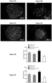
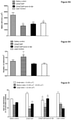
Figure 5 illustrates the effects of twice-daily administration of (DAla2)GIP and (DAla2)GIP/xenin-8-Gln on pancreatic histology. (A-D) Representative islets images showing insulin (red) and glucagon (green) immunoreactivity from pancreatic tissues extracted from (A) high fat saline control, (B) (DAla2)GIP, (C) (DAla2)GIP/xenin-8-Gln treated high mice and (D) lean control mice. (E-I) Parameters were assessed using Cell^F image analysis software after 21 days twice daily i.p injections of saline vehicle (0.9% (w/v) NaCl) or peptides (each at 25 nmol/kg) in high fat mice. Saline treated lean control mice are shown for comparative purposes. Values are mean ±SEM of 6-8 mice. *P<0.05, **P<0.01, ***P<0.001 compared to high fat controls. ΔP<0.05, ΔΔΔP<0.001 compared to lean controls. φφP<0.01, φφφP<0.001 compared to (DAla2)GIP group; -
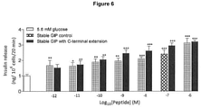
Figure 6 illustrates dose-dependent (10-12 - 10-6 M) insulin secretory (20 min; n=8) effects of C-terminally extended GIP on insulin secretion from BRIN BD11 beta-cells. Values represent means ± SEM. *p<0.05, **p<0.01, ***p<0.001 compared to 5.6 mM glucose alone control; -
Figure 7 illustrates cAMP release measured using a commercially available cAMP assay kit. Values are mean ± SEM for n=4-5. *P < 0.05, **P < 0.01 vs control; -
Figure 8 illustrates TGF-β levels measured using recombinant human TGF-β Immunoassay kit. Values are mean ± SEM for n=4. **P < 0.05, **P < 0.01, ***P < 0.001 compared with control group; -
Figure 9 illustrates IGF-1 levels measured using recombinant human IGF-1 Immunoassay kit. Values are mean ± SEM for n=4. **P < 0.05, **P < 0.01, ***P < 0.001 compared with control group; -
Figure 10 illustrates body weight as measured for 3 days before and 42 days during (indicated by black horizontal bar) once daily treatment with saline vehicle (0.9%w/v NaCl), GIP/Xenin hybrid, GIP/Xenin hybrid Asp and GIP/Xenin hybrid Glu (each peptide at 25 nmol/kg bw). Values represent means ± SEM for 5 mice; -
Figure 11 illustrates (A) %Fat mass and (B) %Lean mass as measured by DEXA scanning in high fat fed mice following 42 days treatment with GIP/Xenin hybrid, GIP/Xenin hybrid Asp and GIP/Xenin hybrid Glu. Values represent means ± SEM for 3-5 mice; -
Figure 12 illustrates cumulative energy intake as measured for 3 days before and 42 days (indicated by black horizontal bar) during once daily treatment with saline vehicle (0.9%w/v NaCl), GIP/Xenin hybrid, GIP/Xenin hybrid Asp and GIP/Xenin hybrid Glu (each peptide at 25 nmol/kg bw). Values represent means ± SEM for 3-5 mice. ΔP < 0.05, ΔΔP < 0.01, ΔΔΔP < 0.001 compared to saline control; -
Figure 13 illustrates non fasting blood glucose as measured for 3 days before and 42 days during (indicated by black horizontal bar) once daily treatment with saline vehicle (0.9%w/v NaCl), GIP/Xenin hybrid, GIP/Xenin hybrid Asp and GIP/Xenin hybrid Glu (each peptide at 25 nmol/kg bw). Values represent means ± SEM for 5 mice; -
Figure 14 illustrates (A) Blood glucose before and after i.p injection of glucose (18 mmol/kg bw) alone in 18 hr fasted mice. AUC values for blood glucose for 0-105 min are shown in insets. Values represent means ± SEM for 6-7 mice. *P < 0.05, **P < 0.01 and ***P < 0.001 compared to saline control; -
Figure 15 illustrates Blood glucose and blood glucose AUC values for 0-105 min (shown in inset) following 42 days treatment with saline, GIP/Xenin hybrid, GIP/Xenin hybrid Asp and GIP/Xenin hybrid Glu (each peptide at 25 nmol/kg bw), and subsequent insulin (25 U/kg bw) injection intraperitoneally (at t=0) in non-fasted mice. Values represent means ± SEM for 5 mice. *P < 0.05, **P < 0.01 and ***P < 0.001 compared to saline control; -
Figure 16 illustrates (A) Bone mineral density (BMD) and (B) Bone mineral content (BMC) as measured by DEXA scanning in high fat fed mice following 42 days treatment with GIP/Xenin hybrid, GIP/Xenin hybrid Asp and GIP/Xenin hybrid Glu. Values represent means ± SEM for 3-5 mice; -
Figure 17 illustrates, in the Femur, (A) Bone mineral density (BMD) and (B) bone mineral content (BMC) as measured by DEXA scanning in high fat fed mice following 42 days treatment with GIP/Xenin hybrid, GIP/Xenin hybrid Asp and GIP/Xenin hybrid Glu. Values represent means ± SEM for 7 mice; -
Figure 18 illustrates, in the Lumbar Spine, (A) Bone mineral density (BMD) and (B) bone mineral content (BMC) as measured by DEXA scanning in high fat fed mice following 42 days treatment with GIP/Xenin hybrid, GIP/Xenin hybrid Asp and GIP/Xenin hybrid Glu. Values represent means ± SEM for 7 mice. *P < 0.05 compared to saline control; -
Figure 19 illustrates, in the Tibia, (A) Bone mineral density (BMD) and (B) bone mineral content (BMC) as measured by DEXA scanning in high fat fed mice following 42 days treatment with GIP/Xenin hybrid, GIP/Xenin hybrid Asp and GIP/Xenin hybrid Glu. Values represent means ± SEM for 7 mice. *P < 0.05, **P < 0.01 compared to saline control; -
Figure 20 illustrates three-point bending parameters in saline control and high fat treated mice treated for 42 days with GIP/Xenin hybrid, GIP/Xenin hybrid Asp and GIP/Xenin hybrid Glu. Values represent means ± SEM for 7 mice. *P < 0.05, ***P < 0.001 compared to saline controls; -
Figure 21 illustrates microcomputed tomography (MicorCT) parameters of trabecular bone in saline control and high fat treated mice treated for 42 days with GIP/Xenin hybrid, GIP/Xenin hybrid Asp and GIP/Xenin hybrid Glu. Values represent means ± SEM for 7 mice. *P < 0.05, **P < 0.01 compared to saline controls; and -
Figure 22 illustrates micorCT parameters of cortical bone in saline control and high fat treated mice treated for 42 days with GIP/Xenin hybrid, GIP/Xenin hybrid Asp and GIP/Xenin hybrid Glu. Values represent means ± SEM for 7 mice. *P < 0.05, **P < 0.01 compared to saline controls. - All polypeptides were purchased from GL Biochem Ltd (Shanghai, China, greater than 95% purity). Polypeptides were characterised using HPLC and MALDI-TOF mass spectrometry as described by Martin et al. 2013 (Biochim Biophys Acta. 2013 Jun;1830(6):3407-13). The experimental mass for all peptides corresponded closely to their theoretical values, confirming structural identity.
Table 1 - Amino acid sequence of xenin, xenin-8, xenin-8-Gln, (DAla2)GIP and (DAla2)GIP/xenin-8-Gln hybrid Polypeptide Amino acid sequence SEQ ID Xenin(25) MET-LEU-THR-LYS-PHE-GLU-THR-LYS-SER-ALA-ARG-VAL-LYS-GLY-LEU-SER-PHE-HIS-PRO-LYS-ARG-PRO-TRP-ILE-LEU SEQ ID NO:5 Xenin-8 HIS-PRO-LYS-ARG-PRO-TRP-ILE-LEU SEQ ID NO:2 Xenin-8-Gln HIS-PRO-GLN-GLN-PRO-TRP-ILE-LEU SEQ ID NO:3 (DAIa2)GIP TYR-(d)ALA-GLU-GLY-THR-PHE-ILE-SER-ASP-TYR-SER-ILE-ALA-MET-ASP-LYS-ILE-HIS-GLN-GLN-ASP-PHE-VAL-ASN-TRP-LEU-LEU-ALA-GLN-LYS-GLY-LYS-LYS-ASN-ASP-TRP-LYS-HIS-ASN-ILE-THR-GLN SEQ ID NO:15 (DAIa2)GIP/xenin-8-Gln TYR-(d)ALA-GLU-GLY-THR-PHE-ILE-SER-ASP-TYR-SER-ILE-ALA-MET-H IS-PRO-GLN-GLN-PRO- TRP-ILE-LEU SEQ ID NO:7 (DAIa2)GIP/xenin-8-Gln(L-Asp) TYR-(d)ALA-GLU-GLY-THR-PHE-ILE-SER-ASP-TYR-SER-ILE-ALA-MET-HIS-PRO-GLN-GLN-PRO-TRP-ILE-LEU-GLY-ALA-ALA-ASP-ASP-ASP-ASP-ASP-ASP SEQ ID NO:10 (DAla2)GIP/xenin-8-Gln(L-Glu) TYR-(d)ALA-GLU-GLY-THR-PHE-ILE-SER-ASP-TYR-SER-ILE-ALA-MET-HIS-PRO-GLN-GLN-PRO- TRP-ILE-LEU-GLY-ALA-ALA-GLU-GLU-GLU-GLU-GLU-GLU SEQ ID NO:13 GIP(L-Glu) HIS-PRO-GLN-GLN-PRO-TRP-ILE-LEU-GLY-ALA-ALA-GLU-GLU-GLU-GLU-GLU-GLU SEQ ID NO:12 Table 2 - Theoretical and experimental molecular masses and DPP-4 half-lives of test peptides Peptide Theoretical molecular mass (Da) Experimental molecular mass (Da) In vitro DPP-4 half-life (h) Native GIP 4982.4 4986.1 <1 (DAIa2)GIP 4982.4 4982.4 >12 Xenin-25 2971.5 2970.8 - Xenin-8 1046.3 1046.1 - Xenin-8-Gln 1018.2 1018.9 - (DAla2)GIP/ xenin-8-Gln 2567.9 2565.3 >12 - For calculation of experimental molecular masses, peptide samples (1 µg) were mixed with α-cyano-4-hydroxycinnamic acid (1 µg) and applied to a Voyager-DE BioSpectrometry Workstation and mass-to-charge (m/z) ratio verses peak intensity recorded by MALDI-TOF mass spectrometry. DPP-4 half-life was assessed by incubating peptides (37°C in 50 mmol/L TEA-HCI; pH 7.8) with 5 mU purified porcine DPP-4 for 0, 1, 2, 4, 8 and 12 h. Degradation profiles were obtained using rp-HPLC analysis and HPLC peak area data used to calculate percentage intact peptide remaining at each time point.
- Peptide DPP-4 degradation profiles were assessed as described by Martin et al. 2013 (Biochim Biophys Acta. 2013 Jun;1830(6):3407-13). Briefly, polypeptides (20 ug) were incubated with 5 µl of purified DPP-4 (5 mU, Sigma-Aldrich, Dorset, UK) in 50 mM TEA-HCl (pH 7.8; final volume 450 µl) at 37°C for 0, 1, 4 and 12 hours. Enzymatic reactions were terminated by the addition of 50 µl TFA/H2O (10% v/v, Sigma-Aldrich, Dorset, UK). Reaction products were then separated by HPLC and analysed using MALDI-TOF.
- BRIN-BD11 cells were used to assess the insulin releasing activity of polypeptides as described by McClenaghan et al., 1996 (Diabetes. 1996 Aug;45(8):1132-40). Cells were cultured in RPMI-1640 growth media supplemented with 10% (v/v) foetal bovine serum (FBS) and 1% (v/v) antibiotics (penicillin (100 U/ml), streptomycin (0.1 mg/l)), in 75 cm2 sterile tissue culture flasks (Greiner bio-one, UK) maintained at 37°C and 5% CO2 in a LEEC incubator (Laboratory technical engineering, Nottingham, UK). BRIN-BD11 cells were then seeded at a density of 150,000 cells/well in 24-well plates (Nunc, Roskilde, Denmark) and allowed to attach overnight at 37°C. Culture medium was removed and cells were pre-incubated in Krebs-Ringer bicarbonate buffer (KRBB) (115 mmol/l NaCl, 4.7 mmol/l KCl, 1.2 mmol/l MgSO4, 1.28 mmol/l CaCl2, 1.2 mmol/l KH2PO4, 25 mmol/l HEPES and 8.4% NaHCO3, containing 0.5% (w/v) BSA , pH 7.4) supplemented with 1.1 mmol/l glucose for 40 min at 37°C. Following the pre-incubation, experiments (n=8) were performed in presence of glucose (5.6 or 16.7 mmol/l) with a range of concentrations of polypeptides (10-12 to 10-6 mol/l) for 20 min at 37°C. After test incubations, aliquots of assay buffer were collected from each well and stored at -20°C prior to measurement of insulin by radioimmunoassay as described by Flatt & Bailey, 1981 (Diabetologia, 1981; 20:573-577).
- Acute animal studies were conducted in male NIH Swiss mice (12-14 weeks old, Harlan Ltd, UK) maintained on a standard rodent maintenance diet (10% fat, 30% protein and 60% carbohydrate, Trouw Nutrition, Cheshire, UK). Prior to commencement of longer term studies, all animals, except lean controls that continued on rodent maintenance diet, were maintained on a high fat diet (45% fat, 35% carbohydrate and 20% protein, Special Diet Services, Essex, UK) for 12-15 weeks. This diet resulted in progressive body weight gain and hyperglycaemia compared with age-matched controls. All animals were housed individually in an air-conditioned room at 22 ± 2°C with a 12 h light:12 h dark cycle and had free access to food and water. All animal experiments were carried out in accordance with the UK Animals (Scientific Procedures) Act 1986.
- For food intake studies, fasted (18 h) mice were given intraperitoneal (i.p) injections of polypeptides at a dose of 25, 100 or 250 nmol/kg bw. Mice were then allowed free access to normal chow for 180 mins and cumulative food intake measured. For glucose homeostasis and insulin secretory studies, blood glucose and plasma insulin were measured immediately prior to and 15, 30, 60 and 105 min after i.p. administration of glucose alone (18 mmol/kg bw) or in combination with the polypeptides (each at 25 nmol/kg bw) in non-fasted mice. In second series of experiments, polypeptides (each at 25 nmol/kg bw) or saline vehicle (0.9% w/v NaCl) were injected 4 h before a glucose load (18 mmol/kg bw) in non-fasted mice and blood glucose and plasma insulin measured at 0, 15, 30 and 60 min post glucose injection.
- Twice daily (09:30 and 17:30 h) i.p. injections of saline vehicle (0.9% w/v NaCl), polypeptides (at 25 nmol/kg bw) were administered for 21 days in high fat mice. Energy intake and body weight were monitored daily, and non-fasting blood glucose and plasma insulin concentrations were assessed at 3 day intervals. On
day 21, a non-fasted 24 h glucose profile was conducted. At the end of the treatment period, i.p. glucose tolerance (18 mmol/kg bw), biological response to GIP (18 mmol/kg glucose in combination with native GIP (25 nmol/kg); i.p.) and insulin sensitivity (15 U/kg bw; i.p.) tests were performed. HOMA-IR was determined using the equation HOMA-IR = fasting glucose (mmol/l) x fasting insulin (mU/l)/22.5. All test solutions were administered in a final volume of 5 ml/kg body weight, at 10:00 h without previous 09:30 h peptide injection. Terminal analysis included measurement of total body fat and lean mass by DEXA scanning (Piximus Densitometer, Inside Outside Sales, Madison WI, USA) and extraction of pancreatic tissue for analysis as detailed below. Immunohistochemistry Pancreatic tissue was excised, divided longitudinally and either immediately snap frozen for subsequent extraction of insulin using acid ethanol extraction (5 ml/g) as described by Irwin et al., 2015 (Diabetes. 2015 Aug;64(8):2996-3009), or processed for immunohistochemical examination. Briefly, pancreatic tissues were fixed in 4% parafolmaldehyde at 4°C for 48 h. Pancreata were then embedded in paraffin wax and processed using an automated tissue processor (Leica TP1020, Leica Microsystems, Nussloch, Germany). Immunohistochemistry was performed as described by Vasu et al., 2014 (PLoS One. 2014 Jun 26;9(6):e101005). Succinctly, tissue sections were deparaffinised, rehydrated and probed with primary antibodies: mouse anti-insulin antibody (1:500; Abcam, ab6995) or guinea-pig anti-glucagon antibody (PCA2/4, 1:400; raised in-house). The sections were then incubated with secondary antibody, Alexa Fluor 488 goat anti-guinea pig IgG (1:400) or Alexa Fluor 594 goat anti-mouse IgG (1:400); respectively. The slides were viewed under a FITC (488 nm) or TRITC filter (594 nm) using a fluorescent microscope (Olympus system microscope, model BX51) and photographed using a DP70 camera adapter system. Islet parameters were analysed using Cell^F image analysis software (Olympus Soft Imaging Solutions, GmbH). - Blood samples were collected from the cut tip on the tail vein of conscious mice into chilled fluoride/heparin glucose micro-centrifuge tubes (Sarstedt, Numbrecht, Germany) at the time points indicated in the Drawings. Blood glucose was measured directly using a hand-held Ascencia Contour blood glucose meter (Bayer Healthcare, Newbury, Berkshire, UK). Blood samples were centrifuged using a Beckman microcentrifuge (Beckman Instruments, Galway, Ireland) for 1-5 mins at 13,000 x g and stored at -20 °C. Plasma and pancreatic insulin was assayed by a modified dextran-coated charcoal radioimmunoassay as described by Flatt & Bailey, 1981 (Diabetologia, 1981; 20:573-577).
- Human osteoblast SaOS-2 cells [sourced from The European Collection of Cell Cultures (ECACC)] were seeded at a density of 2e5 cells/well in 24 well plates [Nunc, Roskilde, Denmark]. The cells were incubated in media [Life Technologies] supplemented with 10% FBS. Before adding test peptides, the media were changed with fresh 0.1% FBS-containing media 24 h previously. On the day of experimentation, media were removed and 1 ml of respective peptides GIP/Xenin, GIP/Xenin Asp and GIP/Xenin Glu [(DAla2)GIP/xenin-8-Gln, (DAIa2)GIP/xenin-8-Gln(L-Asp), and (DAla2)GIP/xenin-8-Gln(L-Glu) respectively] (1e-12 - 1e-6 M) were added to each well. The plates were then incubated at 37°C for a period of 8 hours. After incubation, media was collected and TGF-β released in the supernatant measured using a TGF-β Immunoassay kit (Quantikine, R&D Systems) according to the manufacturer's instructions. The concentration of TGF-β was calculated from a standard curve of recombinant human TGF-β ranging from 0 to 2000 pg/ml.
- SaOS-2 cells were seeded at a density of 2e5 cells/well in 6-well plates in 10% FBS containing media. Media were changed 24 hours prior to experimentation with 0.1% FBS. Media was then removed, followed by an addition of 1 ml of test peptides (1e-12 - 1e-6 M). Plates were incubated at 37°C for 8 hours. After incubation, media was collected and IGF-1 released in the supernatant was measured using IGF-1 Immunoassay kit (Quantikine, R&D Systems) according to the manufacturer's instructions. The concentration of IGF-1 in the samples was determined from a standard curve of recombinant human IGF-1 in the range of 0 - 60 ng/ml.
- SaOS-2 cells were seeded at a density of 5e4 cells in 96-well plates. Cells were cultured in α-MEM 1X media supplemented with 10% FBS for 24 hours in order to allow attachment to the plate. Before the experiment, cells were washed with Hank's Balanced Salt Solution (HBSS). Cells were then incubated with various concentrations (1e-12 - 1e-6 M) of test peptides supplemented with 200 µM of 3-isobutyl-1-methylxanthine (IBMX). Plates were then incubated for 60 minutes at 37°C. Following this step, cells were washed 3 times with 150 µl PBS. Cell lysis was performed with 150 µl cell lysis buffer (R&D Systems) and a freeze(-20°C)-thaw(37°C) cycle was carried out. The contents were then collected into 500 µl tubes and centrifuged at 600 rpm for 10 min (4°C). The supernatant was collected and cAMP was measured using a cAMP assay kit (R&D Systems) according to the manufacturer's instructions. The concentration of cAMP was calculated from a standard curve of cAMP ranging from 0 to 240 pmol/ml.
- The DEXA (dual energy X-ray absorptiometry) analysis [Lunar PIXImus, Inside Outside Sales] was performed on unconscious mice and the whole body was scanned. Various parameters like bone mineral density (BMD), bone mineral content (BMC), lean mass, fat mass and percentage of total fat were measured according to the manufacturer's instructions.
- Femur and tibia bones were extracted at the end of the experiment using a scalpel and fine scissors, soft tissue removed and stored in 70% ethanol. Three-point bending strength was measured with a constant span length of 10 mm. Femurs were positioned horizontally with the anterior surface facing upwards, centered on the support and the pressing force was applied vertically to the midshaft of the bone. Each bone was tested with a loading speed of 2 mm/min until failure with a 90 N load cell. The load-time curve obtained was converted into a load-displacement curve by the MTS testSuite TW software (MTS, Créteil, France). Ultimate load and ultimate displacement were respectively defined as the maximum load and maximum displacement recorded before break-down of the bone. Stiffness was calculated as the slope of the elastic deformation of the bone. The total absorbed energy was defined as the total area under the load-displacement curve and represents the total energy absorbed by the midshaft femur. The yield was defined as the load necessary to initiate the transformation from elastic to plastic deformation. The post-yield energy was defined as the area under the load-displacement curve from yield until failure and represents the energy absorbed by bone during plastic deformation.
- The tibia was used to investigate trabecular and cortical bone microstructural morphology by microCT analysis with a Skyscan 1172 microtomograph (Bruker, Kontich, Belgium) equipped with an X-ray tube working at 69 kV/100 µA. The pixel size was fixed at 8.3 µm, the rotation step at 0.25° and exposure was performed with a 0.5-mm aluminum filter. Bone mass and morphometry parameters were calculated with the CTan software (release 1.11.4.2, Bruker). The trabecular parameters were assessed at the volume of interest (VOI), located 1 mm below the growth plate and spanning 2.5 mm in height. Trabecular bone volume (BV/TV, in %), trabecular number (Tb.N, in 1/mm) and trabecular separation (Tb.Sp, in µm) were measured according to guidelines and nomenclature proposed by the American Society for Bone and Mineral Research. Cortical parameters were determined at the midshaft tibia with a lab-made routine using ImageJ. <arrow area (Ma.Ar), cortical area (Ct.Ar), total area (Tt.Ar), cortical thickness (Ct.Th, in µm) and cross-sectional moment of inertia (J in mm4) were also measured according to guidelines and nomenclature on bone histomorphometry proposed by the American Society for Bone and Mineral Research.
- Statistical analysis was performed using GraphPad PRISM (Version 5). Results are expressed as means ± SEM and data compared using repeated measures ANOVA followed by the Student-Newman-Keuls post-hoc test or two-tailed t-test. The data are expressed as mean ± S.E.M and a P value <0.05 was considered statistically significant.
- In contrast to native GIP which was used as a positive control, (DAla2)GIP/xenin-8-Gln and (DAla2)GIP remained fully intact when incubated in the presence of DPP-4 for up to 12 hs. All polypeptides, including xenin, xenin-8, xenin-8-Gln, (DAla2)GIP and (DAla2)GIP/xenin-8-Gln, significantly (P<0.05 to P<0.001) stimulated insulin secretion from BRIN-BD11 cells at a concentration of 10-6 mol/l when compared to 5.6 and 16.7 mmol/l glucose control (
Figure 1A,B ). Only (DAla2)GIP evoked a significant (P<0.001 and P<0.01; respectively) increases in insulin release at 10-8 M when compared to respective glucose controls (Figure 1A,B ). Further in vivo observations were made using (DAla2)GIP/xenin-8-Gln and its two parent peptides. - At a dose of 25 nmol/kg, (DAla2)GIP/xenin-8-Gln induced a significant (P<0.05) reduction in food intake at 30 min post-injection in overnight fasted mice when compared to saline controls (
Figure 1C ). All other treatments had no significant effect on re-feeding at a dose of 25 nmol/kg (Figure 1C ). However, none of the peptides had significant appetite suppressive effects when administered at a dose of 100 nmol/kg (Figure 1D ). At a supraphysiological dose of 250 nmol/kg, all peptides, barring (DAla2)GIP, induced significant (P<0.05 to P<0.01) reductions in cumulative food intake at 180 min post injection compared to saline treated controls (Figure 1E ). In addition, at 250 nmol/kg, the appetite suppressive effect of xenin was significantly (P<0.05) superior to control mice at 90 min post-injection, whilst xenin-8-Gln and (DAla2)GIP/xenin-8-Gln also evoked significant (P<0.05) reductions in cumulative food intake at 120 min post-injection (Figure 1E ). - Administration of xenin-8-Gln, (DAla2)GIP or (DAla2)GIP/xenin-8-Gln concomitantly with glucose resulted in significantly (P<0.05) lowered blood glucose values at 30 min post injection, culminating in significantly (P<0.05) decreased overall 0-105 min AUC blood glucose values when compared to glucose alone controls (
Figure 2A ). (DAla2)GIP also induced a significant (P<0.05) reduction inblood glucose levels 15 min post-injection (Figure 2A ). Corresponding glucose-induced plasma insulin concentrations were not altered between groups in terms of individual values at 15, 30, 60, 90 and 105 min post-injection (Figure 2B ). However, overall glucose-stimulated plasma insulin levels, as assessed by AUC measures, were significantly (P<0.05) increased in all treatment groups compared to controls (Figure 2B ). When administered 4 h prior to glucose load, xenin-8-Gln was devoid of any beneficial glucose lowering or insulin releasing effects (Figure 2C,D ). However, when administered 4 hs previously, both (DAla2)GIP and (DAla2)GIP/xenin-8-Gln significantly reduced overall AUC blood glucose levels (P<0.05) and increased plasma insulin AUC values (P<0.05) following a glucose challenge (Figure 2C,D ). These two peptides were carried forward to evaluate sub-chronic effects in high fat fed mice. - Twice daily administration of (DAla2)GIP and (DAla2)GIP/xenin-8-Gln for 21 days to high fat fed mice had no significant effect on energy intake or body weight compared to high fat controls (
Figure 3A,C ). In addition, total body fat and lean mass was also unaltered after the 21 day regimen (Figure 3B ). However, all high fat fed mice exhibited significantly increased (P<0.05 to P<0.001) body weight and energy intake when compared to lean controls (Figure 3A,C ). Non-fasting blood glucose levels progressively declined in (DAIa2)GIP/xenin-8-Gln treated high fat fed mice over the 21 days, being significantly (P<0.05 to P<0.01) reduced onobservation days Figure 3D ). In addition, both (DAla2)GIP and (DAla2)GIP/xenin-8-Gln treated mice had glucose levels that were not significantly different from lean control mice fromday 12 onwards (Figure 3D ). In agreement, analysis of non-fasting 24 h glucose profile onday 21 revealed that (DAla2)GIP/xenin-8-Gln treated mice had similar blood glucose levels as lean controls at each observation point and significantly (P<0.05) reduced compared to saline controls at 17:00 h (Figure 3E ). Blood glucose levels in (DAla2)GIP treated mice were also reduced, but not to the same extent as in (DAla2)GIP/xenin-8-Gln mice (Figure 3E ). Circulating plasma insulin levels were generally elevated in all high fat fed mice when compared to lean controls (Figure 3F ). However, mice treated twice daily with either (DAla2)GIP or (DAla2)GIP/xenin-8-Gln did have a tendency for reduced plasma insulin levels, and indeed were not significantly different from lean control mice on day 21 (Figure 3F ). - Treatment with (DAIa2)GIP or (DAla2)GIP/xenin-8-Gln for 21 days had a strong tendency to reduce overall AUC blood glucose levels of high fat fed mice following a glucose load, but this failed to reach significance compared to saline controls. Similarly, glucose-stimulated plasma insulin AUC concentrations were reduced, albeit non-significantly, by (DAla2)GIP or (DAla2)GIP/xenin-8-Gln treatment when compared to high fat controls (able 2). All high fat mice had a significantly (P<0.05 to P<0<001) increased glycaemic excursion and overall insulin secretory response compared to lean controls (Table 3). As illustrated in Table 3, (DAla2)GIP and (DAla2)GIP/xenin-8-Gln treatment significantly (P<0.05) improved the glucose lowering and insulin releasing actions of native GIP. As such, the beneficial effects of both (DAla2)GIP and (DAla2)GIP/xenin-8-Gln treatment were evident from overall blood glucose AUC values, which were significantly (P<0.05) decreased by 36% and 30%, respectively, compared to saline treated control mice. Moreover, AUC glycaemic values were not different in (DAla2)GIP or (DAla2)GIP/xenin-8-Gln treated high fat mice compared to saline treated lean controls in response to administration of GIP in combination with glucose. Corresponding GIP-induced elevations of plasma insulin concentrations were also significantly (P<0.05 to P<0.01) augmented in (DAIa2)GIP and (DAla2)GIP/xenin-8-Gln high fat mice compared to high fat controls, as revealed from AUC measures.
Table 3 - Effects of twice-daily administration of (DAIa2)GIP and (DAIa2)GIP/xenin-8-Gln on glucose tolerance, glucose stimulated insulin and GIP-mediated glucose-lowering and insulin-secretory actions in high fat fed mice Treatment group Glucose tolerance te est Metabolic respon se to GIP Blood glucose AUC (mmol/l.min) Plasma insulin AUC (ng/ml.min) Blood glucose AUC (mmol/l.min) Plasma insulin AUC (ng/ml.min) High fat saline control 2062.0±121.2 95.2±17.8 1618.9±148.1 56.8±11.8 (DAIa2)GIP 1714.7±138.6Δ 65.4±9.2Δ 1029.5±138.0* 142.3±19.2**,ΔΔΔ (DAla2)GIP/xenin-8-Gln 1757.3±154.6Δ 49.0±6.5Δ 1130.1±143.9* 103.1±23.3*ΔΔ Lean control 1080.5±85.7*** 24.6±1.4* 787.4±54.2** 21.0±2.4 - Tests were conducted after twice daily treatment with saline vehicle (0.9% (w/v) NaCl), (DAIa2)GIP and (DAIa2)GIP/xenin-8-Gln (each at 25 nmol/kg bw) for 21 days. For glucose tolerance test, blood glucose and plasma insulin concentrations were measured before and 15, 30, 60 and 105 min after i.p injection of glucose (18 mmol/kg bw). For metabolic response to GIP, blood glucose and plasma insulin concentrations were measured before and 15, 30, 60 and 105 min after i.p injection of glucose (18 mmol/kg bw) in combination with GIP (25 nmol/kg bw). All studies were conducted in 18 h fasted mice. AUC values (0-105 min) for blood glucose and plasma insulin are shown. Values represent means ± SEM for 6-8 mice. *P<0.05, **P<0.01, **P<0.01 compared to high fat controls. ΔP<0.05, ΔΔP<0.01, ΔΔΔP<0.001 compared to lean controls.
- Individual blood glucose levels were reduced, albeit non-significantly, in (DAla2)GIP and (DAla2)GIP/xenin-8-Gln treated mice following administration of exogenous insulin on
day 21 when compared to high fat controls (Figure 4A ). However, the overall glucose lowering effect of insulin was significantly (P<0.001) improved in (DAla2)GIP and (DAla2)GIP/xenin-8-Gln mice compared to saline control high fat mice (Figure 4B ). The improved hypoglycaemic action of insulin was confirmed by HOMA-IR calculations, revealing that both peptide treatment groups had significantly (P<0.05) improved insulin sensitivity compared with high fat controls, and not significantly different from lean control mice (Figure 4C ). Pancreatic insulin content of (DAla2)GIP/xenin-8-Gln treated mice was augmented (P<0.05) compared to lean controls, but not significantly different when compared to high fat control mice (Figure 4D ). In contrast, (DAla2)GIP treated mice had similar pancreatic insulin content as lean control mice (Figure 4D ). - Representative images of pancreatic islets from lean control, high fat control, (DAla2)GIP and (DAIa2)GIP/xenin-8-Gln treated mice are shown in
Figures 5A-D . High fat fed mice had significantly (P<0.01) increased islet area compared to lean controls (Figure 5E ), giving rise to significant (P<0.01) increases in both beta and alpha cell areas (Figure 5F ,G ). Treatment with (DAla2)GIP had no significant effect on overall pancreatic area or beta cell area, but decreased (P<0.001) alpha cell area compared high fat controls (Figure 5E-G ). Similarly, 21 days twice daily treatment with (DAla2)GIP/xenin-8-Gln significantly (P<0.001) reduced pancreatic alpha cell area compared to high fat controls (Figure 5G ) and increased (P<0.001) pancreatic islet and beta cell area compared to lean control mice (Figure 5E,F ). The number of islets per mm2 was significantly (P<0.05 to P<0.01) augmented in (DAla2)GIP treated mice compared to all other groups of mice (Figure 5H ), which appeared to be related to an increase in number of small sized islets (Figure 5I ). Thus, high fat feeding significantly (P<0.001) decreased the number of small islets, and increased the number of larger sized islets, compared to lean control mice (Figure 5I ). (DAla2)GIP/xenin-8-Gln treated high fat mice also had a decreased (P<0.001) number of small and medium sized islets, with increased (P<0.001) numbers of larger islets, when compared to lean control mice (Figure 5I ). - C-terminally modified forms of (DAla2)GIP/xenin-8-Gln and of GIP have been designed and synthesised for site-specific delivery to bone. These analogues have six acidic amino acid (L-Asp or L-Glu) residues at the C-terminus to enhance binding to hydroxyapatite, the major component of bone. The structural modification does not adversely affect biological activity of GIP at the level of the pancreatic beta-cell. Dose-dependent (10-12 - 10-6 M) insulin secretory (20 min; n=8) effects of GIP(L-Glu) were measured in BRIN BD11 beta-cells by insulin RIA. Values represent means ± SEM. *p<0.05, **p<0.01, ***p<0.001 compared to 5.6 mM glucose alone control. These observations confirm that the specific C-terminal modification does not hinder biological activity at the cellular level.
- As shown in
Figure 7 , the peptides significantly stimulated cAMP production in SaOS-2 cells compared to controls at a concentration of 1e-6 M. The lowest effective concentration of GIP/Xenin was 1e-10 M (P < 0.05) while for GIP/Xenin Asp, and GIP/Xenin Glu, it was 1e-8 M and 1e-6 M; respectively. - GIP/Xenin, GIP/Xenin Asp, and GIP/Xenin Glu dose-dependently stimulated TGF-β (
Figure 8 ) and IGF-1 (Figure 9 ) release from SaOS-2 cells. GIP/Xenin significantly induced TGF-β release at concentrations ranging from (1e-12 - 1e-8 M, P < 0.01), while GIP/Xenin Asp, and GIP/Xenin Glu, required higher concentrations (1e-8 - 1e-6 M, P < 0.01) to induce a significant effect (Figure 8 ). The release of IGF-1 by GIP/Xenin, GIP/Xenin Asp, and GIP/Xenin Glu followed a similar pattern as TFG- β release (Figure 9 ). - There was no significant change in body weight (
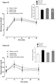
Figure 10 ), fat and lean mass (Figure 11 ) as well as food intake (Figure 12 ) after daily administration of GIP /Xenin, GIP/Xenin Asp, and GIP/Xenin Glu peptides in high fat fed mice. Circulating blood glucose levels were also not significantly different between groups during the 42 day treatment period (Figure 13 ). - Glucose tolerance test was carried out at the end of the 42-day treatment period. Following a glucose challenge (18 mmol/kg bw), blood glucose levels were significantly (P<0.01 to P<0.001) in GIP/Xenin Asp treated mice at 30, 60 and 105 min post-injection (
Figure 14A ). This corresponded to a significant (P<0.05) reduction in overall AUC measures in these mice (Figure 14B). GIP/Xenin and GIP/Xenin Glu also reduced blood glucose concentrations, but not as effectively as GIP/Xenin Asp (Figure 14 ). During insulin tolerance tests, GIP/Xenin Glu treated mice were shown to have a more pronounced hypoglycaemic response to exogenous insulin administration (Figure 15 ). Overall effects of insulin injection were similar in saline control, GIP/Xenin and GIP/Xenin Asp high fat mice (Figure 15 ). - DEXA analysis revealed no difference in whole body (
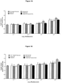
Figure 16 ) and femur (Figure 17 ) bone mineral density and bone mineral content between groups of mice. However, assessment of lumbar spine BMC (Figure 18A ) showed a reduction (P<0.05) in bone mineral content. In addition, tibia bone mineral density was reduced (P<0.05 and P<0.01; respectively) in GIP/Xenin Asp and GIP/Xenin Glu mice (Figure 19B ). - Treatment with GIP/Xenin Glu appeared to have negative effects (P<0.05 to P<0.001) on bone ultimate load and stiffness (
Figure 20 ). All other three-point bending parameters were similar between groups (Figure 20 ). In terms of tibia structural properties, GIP/Xenin improved bone volume density (P<0.01) and increased trabecular number (P<0.05) compared to saline controls (Figure 21 ). All other structural properties of trabecular bone were similar between groups (Figure 21 ). Finally, cortical bone area, thickness and moment of inertia were reduced by 42 days treatment with GIP/Xenin Glu (Figure 22 ), with all other treatments having no significant effect on the structural properties of cortical bone (Figure 22 ). - Accordingly, the present invention provides polypeptides and analogues thereof for use in the treatment of diabetes and bone disorders.
- The present invention demonstrates the biological actions and therapeutic utility of a novel polypeptide (DAla2)GIP/xenin-8-Gln.
- All tested polypeptides significantly (P<0.05 to P<0.001) enhanced in vitro insulin secretion from pancreatic clonal BRIN-BD11 cells at 5.6 and 16.7 mmol/l glucose. Administration of (DAla2)GIP or (DAla2)GIP/xenin-8-Gln in combination with glucose significantly (P<0.05) lowered blood glucose and increased plasma insulin in mice, with a protracted response of up to 4 hours. Certain treatments elicited appetite suppressive effects (P<0.05 to P<0.01), but particularly (DAla2)GIP/xenin-8-Gln and xenin-8-Gln at elevated doses of 250 nmol/kg. Twice-daily administration of (DAla2)GIP/xenin-8-Gln or (DAla2)GIP for 21 days to high fat fed mice returned circulating blood glucose to lean control levels. In addition, (DAla2)GIP/xenin-8-Gln treatment significantly (P<0.05) reduced glycaemic levels during a more detailed 24 hour glucose profile assessment. There was no significant effect of either treatment regimen on body weight, energy intake or circulating insulin concentrations. However, glucose tolerance (P<0.05) and insulin sensitivity (P<0.001) were improved by both treatments. Interestingly, GIP-mediated glucose-lowering and insulin-releasing effects were substantially (P<0.05 to P<0.001) improved by (DAla2)GIP and (DAla2)GIP/xenin-8-Gln treatment. Pancreatic islet (P<0.001) and beta-cell (P<0.001) area, as well as pancreatic insulin content (P<0.01) were augmented in (DAla2)GIP/xenin-8-Gln treated mice, whereas (DAla2)GIP treatment evoked increases (P<0.01) in islet number.
- In order to exploit the potential of GIP, the inventors have generated GIP forms, through addition of six C-terminal acidic L-Asp amino acid residues that encourage binding to hydroxyapatite which is the major component of bone. The GIP forms which have been generated are highly specific and help to target the drug towards bone. To further increase utility, the inventors have designed similar bone-targeting GIP/xenin hybrid molecules. As compared to control, GIP/Xenin significantly induced TGF-alpha release at all concentrations examined. GIP/Xenin Asp and GIP/Xenin Glu had similar, albeit slightly less potent, effects, which is encouraging as it suggests retained bioactivity of the C-terminally modified peptide analogues. Similar to TGF-alpha release, GIP/Xenin Asp and GIP/Xenin Glu augmented IGF-1 levels confirming biological efficacy. In full agreement with this, we confirmed that activation of the GIP receptor stimulated adenylyl cyclase and caused an increase in intracellular cAMP levels. Thus, all peptides enhanced cAMP generation in SaOS2 cells.
- To assess the impact of GIP/Xenin, GIP/Xenin Glu and GIP/Xenin Asp on bone properties in
type 2 diabetes, the inventors have employed a once daily injection regimen of the peptides in high fat fed mice. Importantly, high fat fed mice represent an excellent model ofhuman type 2 diabetes that is fuelled by excessive calorie intake. The peptides had no effect on body weight or food intake, which agrees well with the biological actions of GIP. Moreover, similar to previous observations with GIP and xenin, all peptides improved glucose tolerance and insulin sensitivity. - In terms of bone-specific effects of the peptides of the present invention, it is notable that spine bone mineral content was augmented by the GIP/xenin hybrid peptides, whereas tibia bone mineral content was reduced. This was particularly evident for both the bone-targeting forms of the GIP/xenin hybrids. Although perhaps not as anticipated, it is important to note that measures of BMD and BMC are known to be altered in
type 2 diabetes - thus, simple DEXA scanning may not be sufficient to assess fracture risk intype 2 diabetes, so the inventors also assessed trabecular and cortical properties of bone by microcomputed tomography. Interestingly, positive effects of GIP/xenin were noted in relation to trabecular bone, whilst GIP/Xenin Glu, but not GIP/Xenin Asp, evoked negative effects on trabecular bone material properties. This was borne out in resistance to fracture studies, where mice treated with GIP/Xenin Glu had reduced ultimate load bearing capabilities and decreased bone stiffness. It is intriguing that GIP/Xenin Glu, but not GIP/Xenin Asp, had detrimental effects of the assessed bone-specific parameters, especially since both modifications should encourage deposition of the peptide in bone. Without being bound by theory, it could be that the L-Glu residues create stearic hindrance in relation to activation of either GIP or xenin related cellular pathways. Taken together, it is apparent that GIP/Xenin, and particularly, GIP/Xenin Asp, have beneficial metabolic and bone-related effects in high fat fed mice. -
- <110> UNIVERSITY OF ULSTER
- <120> Polypeptides and analogues thereof for use in the treatment of diabetes and bone disorders
- <130> pg13700pc00
- <150>
GB1521442.2
<151> 2015-12-04 - <160> 15
- <170> PatentIn version 3.5
- <210> 1
<211> 8
<212> PRT
<213> Homo sapiens - <220>
<221> VARIANT
<222> (3)..(4)
<223> X is independently selected from K, R, and Q - <400> 1

- <210> 2
<211> 8
<212> PRT
<213> Homo sapiens - <400> 2

- <210> 3
<211> 8
<212> PRT
<213> Homo sapiens - <400> 3

- <210> 4
<211> 17
<212> PRT
<213> Homo sapiens - <400> 4


- <210> 5
<211> 25
<212> PRT
<213> Homo sapiens - <400> 5

- <210> 6
<211> 14
<212> PRT
<213> Homo sapiens - <400> 6

- <210> 7
<211> 22
<212> PRT
<213> Homo sapiens - <400> 7

- <210> 8
<211> 9
<212> PRT
<213> Homo sapiens - <400> 8

- <210> 9
<211> 17
<212> PRT
<213> Homo sapiens - <400> 9

- <210> 10
<211> 31
<212> PRT
<213> Homo sapiens - <400> 10

- <210> 11
<211> 9
<212> PRT
<213> Homo sapiens - <400> 11

- <210> 12
<211> 17
<212> PRT
<213> Homo sapiens - <400> 12

- <210> 13
<211> 31
<212> PRT
<213> Homo sapiens - <400> 13


- <210> 14
<211> 28
<212> PRT
<213> Homo sapiens - <400> 14

- <210> 15
<211> 42
<212> PRT
<213> Homo sapiens - <400> 15

Claims (11)
- A polypeptide comprising the amino acid sequence YAEGTFISDYSIAMHPQQPWIL (SEQ ID NO:7).
- A polypeptide according to claim 1 consisting of the amino acid sequence YAEGTFISDYSIAMHPQQPWIL (SEQ ID NO:7).
- A polypeptide according to claim 1 further comprising the amino acid sequence GAADDDDDD (SEQ ID NO:8).
- A polypeptide according to claim 3 consisting of the amino acid sequence YAEGTFISDYSIAMHPQQPWILGAADDDDDD (SEQ ID NO:10).
- A polypeptide according to claim 1 further comprising the amino acid sequence GAAEEEEEE (SEQ ID NO:11).
- A polypeptide according to claim 5 consisting of the amino acid sequence YAEGTFISDYSIAMHPQQPWILGAAEEEEEE (SEQ ID NO:13).
- A polypeptide according to any of claims 1-6 for use in the treatment of diabetes mellitus.
- A polypeptide according to any of claims 1-6 for use in the treatment of obesity.
- A polypeptide according to any of claims 1-6 for use in the treatment of metabolic syndrome.
- A polypeptide according to any of claims 1-6 for use in the treatment of bone disorders.
- A polypeptide for use according to claim 10 in a subject suffering from diabetes mellitus.
Applications Claiming Priority (2)
| Application Number | Priority Date | Filing Date | Title |
|---|---|---|---|
| GBGB1521442.2A GB201521442D0 (en) | 2015-12-04 | 2015-12-04 | Polytpeptides and analogues thereof for use in the treatment of diabetes and bone disorders |
| PCT/EP2016/079748 WO2017093562A1 (en) | 2015-12-04 | 2016-12-05 | Polypeptides and analogues thereof for use in the treatment of diabetes and bone disorders |
Publications (2)
| Publication Number | Publication Date |
|---|---|
| EP3383423A1 EP3383423A1 (en) | 2018-10-10 |
| EP3383423B1 true EP3383423B1 (en) | 2022-03-09 |
Family
ID=55234432
Family Applications (1)
| Application Number | Title | Priority Date | Filing Date |
|---|---|---|---|
| EP16809694.9A Active EP3383423B1 (en) | 2015-12-04 | 2016-12-05 | Polypeptides and analogues thereof for use in the treatment of diabetes and bone disorders |
Country Status (4)
| Country | Link |
|---|---|
| US (2) | US10888604B2 (en) |
| EP (1) | EP3383423B1 (en) |
| GB (1) | GB201521442D0 (en) |
| WO (1) | WO2017093562A1 (en) |
Families Citing this family (1)
| Publication number | Priority date | Publication date | Assignee | Title |
|---|---|---|---|---|
| GB201521442D0 (en) | 2015-12-04 | 2016-01-20 | Univ Ulster | Polytpeptides and analogues thereof for use in the treatment of diabetes and bone disorders |
Family Cites Families (2)
| Publication number | Priority date | Publication date | Assignee | Title |
|---|---|---|---|---|
| US20150025002A1 (en) * | 2012-01-12 | 2015-01-22 | University Of Ulster | Peptides and peptide derivatives based on xenin |
| GB201521442D0 (en) | 2015-12-04 | 2016-01-20 | Univ Ulster | Polytpeptides and analogues thereof for use in the treatment of diabetes and bone disorders |
-
2015
- 2015-12-04 GB GBGB1521442.2A patent/GB201521442D0/en not_active Ceased
-
2016
- 2016-12-05 US US15/781,355 patent/US10888604B2/en active Active
- 2016-12-05 WO PCT/EP2016/079748 patent/WO2017093562A1/en not_active Ceased
- 2016-12-05 EP EP16809694.9A patent/EP3383423B1/en active Active
-
2021
- 2021-01-11 US US17/145,714 patent/US11738065B2/en active Active
Also Published As
| Publication number | Publication date |
|---|---|
| US11738065B2 (en) | 2023-08-29 |
| US20190091294A1 (en) | 2019-03-28 |
| EP3383423A1 (en) | 2018-10-10 |
| US10888604B2 (en) | 2021-01-12 |
| WO2017093562A1 (en) | 2017-06-08 |
| GB201521442D0 (en) | 2016-01-20 |
| US20210379157A1 (en) | 2021-12-09 |
Similar Documents
| Publication | Publication Date | Title |
|---|---|---|
| TWI850611B (en) | Co-agonists of the glp-1 and amylin receptors | |
| JP4124734B2 (en) | Nasal absorption pharmaceutical composition | |
| EP1988100B1 (en) | Derivatives of glucagon-like peptide-2 and their therapeutic use | |
| TWI489992B (en) | Amide based glucagon superfamily peptide prodrugs | |
| US7371721B2 (en) | Use of GLP-2 and related compounds for the treatment, prevention, diagnosis, and prognosis of bone-related disorders and calcium homeostasis related syndromes | |
| CA3178366A1 (en) | Gip/glp1 co-agonist compounds | |
| JP2022509568A (en) | Modified GIP peptide analogs | |
| US20030158101A1 (en) | Glucagon-like peptide-2 and its therapeutic use | |
| US7521528B2 (en) | Conformationally constrained parathyroid hormone (PTH) analogs with lactam bridges | |
| PL192359B1 (en) | Glp-1 derivatives | |
| JP2004508410A (en) | Methods of using GLP-1 and GLP-2 peptides | |
| JP2017537894A (en) | Compositions and peptides having GLP-1R and GLP-2R dual agonist activity | |
| CN104870009A (en) | Functionalized exendin-4 derivatives | |
| US20080312133A1 (en) | Unacylated ghrelin as therapeutic agent in the treatment of metabolic disorders | |
| EP2201960A1 (en) | Conformationally constrained parathyroid hormones with alpha-helix stabilizers | |
| WO2003009804A2 (en) | Conformationally constrained parathyroid hormone (pth) analogs | |
| AU2002339843A1 (en) | Conformationally constrained parathyroid hormone (PTH) analogs | |
| US11738065B2 (en) | Polypeptides and analogues thereof for use in the treatment of diabetes and bone disorders | |
| TWI364292B (en) | Glp-1 pharmaceutical compositions | |
| CN101516399A (en) | Therapeutic agent for acceleration of spinal nerve repair comprising desacyl ghrelin or derivative thereof as active ingredient | |
| US20240247080A1 (en) | Glp1/gip/npy2 receptor triple agonists | |
| TW202346324A (en) | Prodrugs and uses thereof | |
| Chunxiao et al. | Pharmacological effects of a recombinant hPTH (1− 34) derived peptide on ovariectomized rats | |
| KR20260010743A (en) | Polypeptide derivatives for oral delivery | |
| WO2026078181A1 (en) | Antagonists of human fgf23 |
Legal Events
| Date | Code | Title | Description |
|---|---|---|---|
| STAA | Information on the status of an ep patent application or granted ep patent |
Free format text: STATUS: UNKNOWN |
|
| STAA | Information on the status of an ep patent application or granted ep patent |
Free format text: STATUS: THE INTERNATIONAL PUBLICATION HAS BEEN MADE |
|
| PUAI | Public reference made under article 153(3) epc to a published international application that has entered the european phase |
Free format text: ORIGINAL CODE: 0009012 |
|
| STAA | Information on the status of an ep patent application or granted ep patent |
Free format text: STATUS: REQUEST FOR EXAMINATION WAS MADE |
|
| 17P | Request for examination filed |
Effective date: 20180629 |
|
| AK | Designated contracting states |
Kind code of ref document: A1 Designated state(s): AL AT BE BG CH CY CZ DE DK EE ES FI FR GB GR HR HU IE IS IT LI LT LU LV MC MK MT NL NO PL PT RO RS SE SI SK SM TR |
|
| AX | Request for extension of the european patent |
Extension state: BA ME |
|
| DAV | Request for validation of the european patent (deleted) | ||
| DAX | Request for extension of the european patent (deleted) | ||
| STAA | Information on the status of an ep patent application or granted ep patent |
Free format text: STATUS: EXAMINATION IS IN PROGRESS |
|
| 17Q | First examination report despatched |
Effective date: 20190917 |
|
| GRAP | Despatch of communication of intention to grant a patent |
Free format text: ORIGINAL CODE: EPIDOSNIGR1 |
|
| STAA | Information on the status of an ep patent application or granted ep patent |
Free format text: STATUS: GRANT OF PATENT IS INTENDED |
|
| INTG | Intention to grant announced |
Effective date: 20211022 |
|
| GRAS | Grant fee paid |
Free format text: ORIGINAL CODE: EPIDOSNIGR3 |
|
| GRAA | (expected) grant |
Free format text: ORIGINAL CODE: 0009210 |
|
| STAA | Information on the status of an ep patent application or granted ep patent |
Free format text: STATUS: THE PATENT HAS BEEN GRANTED |
|
| AK | Designated contracting states |
Kind code of ref document: B1 Designated state(s): AL AT BE BG CH CY CZ DE DK EE ES FI FR GB GR HR HU IE IS IT LI LT LU LV MC MK MT NL NO PL PT RO RS SE SI SK SM TR |
|
| REG | Reference to a national code |
Ref country code: GB Ref legal event code: FG4D |
|
| REG | Reference to a national code |
Ref country code: CH Ref legal event code: EP Ref country code: AT Ref legal event code: REF Ref document number: 1473577 Country of ref document: AT Kind code of ref document: T Effective date: 20220315 |
|
| REG | Reference to a national code |
Ref country code: DE Ref legal event code: R096 Ref document number: 602016069889 Country of ref document: DE |
|
| REG | Reference to a national code |
Ref country code: IE Ref legal event code: FG4D |
|
| REG | Reference to a national code |
Ref country code: LT Ref legal event code: MG9D |
|
| REG | Reference to a national code |
Ref country code: NL Ref legal event code: MP Effective date: 20220309 |
|
| PG25 | Lapsed in a contracting state [announced via postgrant information from national office to epo] |
Ref country code: SE Free format text: LAPSE BECAUSE OF FAILURE TO SUBMIT A TRANSLATION OF THE DESCRIPTION OR TO PAY THE FEE WITHIN THE PRESCRIBED TIME-LIMIT Effective date: 20220309 Ref country code: RS Free format text: LAPSE BECAUSE OF FAILURE TO SUBMIT A TRANSLATION OF THE DESCRIPTION OR TO PAY THE FEE WITHIN THE PRESCRIBED TIME-LIMIT Effective date: 20220309 Ref country code: NO Free format text: LAPSE BECAUSE OF FAILURE TO SUBMIT A TRANSLATION OF THE DESCRIPTION OR TO PAY THE FEE WITHIN THE PRESCRIBED TIME-LIMIT Effective date: 20220609 Ref country code: LT Free format text: LAPSE BECAUSE OF FAILURE TO SUBMIT A TRANSLATION OF THE DESCRIPTION OR TO PAY THE FEE WITHIN THE PRESCRIBED TIME-LIMIT Effective date: 20220309 Ref country code: HR Free format text: LAPSE BECAUSE OF FAILURE TO SUBMIT A TRANSLATION OF THE DESCRIPTION OR TO PAY THE FEE WITHIN THE PRESCRIBED TIME-LIMIT Effective date: 20220309 Ref country code: BG Free format text: LAPSE BECAUSE OF FAILURE TO SUBMIT A TRANSLATION OF THE DESCRIPTION OR TO PAY THE FEE WITHIN THE PRESCRIBED TIME-LIMIT Effective date: 20220609 |
|
| REG | Reference to a national code |
Ref country code: AT Ref legal event code: MK05 Ref document number: 1473577 Country of ref document: AT Kind code of ref document: T Effective date: 20220309 |
|
| PG25 | Lapsed in a contracting state [announced via postgrant information from national office to epo] |
Ref country code: LV Free format text: LAPSE BECAUSE OF FAILURE TO SUBMIT A TRANSLATION OF THE DESCRIPTION OR TO PAY THE FEE WITHIN THE PRESCRIBED TIME-LIMIT Effective date: 20220309 Ref country code: GR Free format text: LAPSE BECAUSE OF FAILURE TO SUBMIT A TRANSLATION OF THE DESCRIPTION OR TO PAY THE FEE WITHIN THE PRESCRIBED TIME-LIMIT Effective date: 20220610 Ref country code: FI Free format text: LAPSE BECAUSE OF FAILURE TO SUBMIT A TRANSLATION OF THE DESCRIPTION OR TO PAY THE FEE WITHIN THE PRESCRIBED TIME-LIMIT Effective date: 20220309 |
|
| PG25 | Lapsed in a contracting state [announced via postgrant information from national office to epo] |
Ref country code: NL Free format text: LAPSE BECAUSE OF FAILURE TO SUBMIT A TRANSLATION OF THE DESCRIPTION OR TO PAY THE FEE WITHIN THE PRESCRIBED TIME-LIMIT Effective date: 20220309 |
|
| PG25 | Lapsed in a contracting state [announced via postgrant information from national office to epo] |
Ref country code: SM Free format text: LAPSE BECAUSE OF FAILURE TO SUBMIT A TRANSLATION OF THE DESCRIPTION OR TO PAY THE FEE WITHIN THE PRESCRIBED TIME-LIMIT Effective date: 20220309 Ref country code: SK Free format text: LAPSE BECAUSE OF FAILURE TO SUBMIT A TRANSLATION OF THE DESCRIPTION OR TO PAY THE FEE WITHIN THE PRESCRIBED TIME-LIMIT Effective date: 20220309 Ref country code: RO Free format text: LAPSE BECAUSE OF FAILURE TO SUBMIT A TRANSLATION OF THE DESCRIPTION OR TO PAY THE FEE WITHIN THE PRESCRIBED TIME-LIMIT Effective date: 20220309 Ref country code: PT Free format text: LAPSE BECAUSE OF FAILURE TO SUBMIT A TRANSLATION OF THE DESCRIPTION OR TO PAY THE FEE WITHIN THE PRESCRIBED TIME-LIMIT Effective date: 20220711 Ref country code: ES Free format text: LAPSE BECAUSE OF FAILURE TO SUBMIT A TRANSLATION OF THE DESCRIPTION OR TO PAY THE FEE WITHIN THE PRESCRIBED TIME-LIMIT Effective date: 20220309 Ref country code: EE Free format text: LAPSE BECAUSE OF FAILURE TO SUBMIT A TRANSLATION OF THE DESCRIPTION OR TO PAY THE FEE WITHIN THE PRESCRIBED TIME-LIMIT Effective date: 20220309 Ref country code: CZ Free format text: LAPSE BECAUSE OF FAILURE TO SUBMIT A TRANSLATION OF THE DESCRIPTION OR TO PAY THE FEE WITHIN THE PRESCRIBED TIME-LIMIT Effective date: 20220309 Ref country code: AT Free format text: LAPSE BECAUSE OF FAILURE TO SUBMIT A TRANSLATION OF THE DESCRIPTION OR TO PAY THE FEE WITHIN THE PRESCRIBED TIME-LIMIT Effective date: 20220309 |
|
| PG25 | Lapsed in a contracting state [announced via postgrant information from national office to epo] |
Ref country code: PL Free format text: LAPSE BECAUSE OF FAILURE TO SUBMIT A TRANSLATION OF THE DESCRIPTION OR TO PAY THE FEE WITHIN THE PRESCRIBED TIME-LIMIT Effective date: 20220309 Ref country code: IS Free format text: LAPSE BECAUSE OF FAILURE TO SUBMIT A TRANSLATION OF THE DESCRIPTION OR TO PAY THE FEE WITHIN THE PRESCRIBED TIME-LIMIT Effective date: 20220709 Ref country code: AL Free format text: LAPSE BECAUSE OF FAILURE TO SUBMIT A TRANSLATION OF THE DESCRIPTION OR TO PAY THE FEE WITHIN THE PRESCRIBED TIME-LIMIT Effective date: 20220309 |
|
| REG | Reference to a national code |
Ref country code: DE Ref legal event code: R097 Ref document number: 602016069889 Country of ref document: DE |
|
| PLBE | No opposition filed within time limit |
Free format text: ORIGINAL CODE: 0009261 |
|
| STAA | Information on the status of an ep patent application or granted ep patent |
Free format text: STATUS: NO OPPOSITION FILED WITHIN TIME LIMIT |
|
| PG25 | Lapsed in a contracting state [announced via postgrant information from national office to epo] |
Ref country code: DK Free format text: LAPSE BECAUSE OF FAILURE TO SUBMIT A TRANSLATION OF THE DESCRIPTION OR TO PAY THE FEE WITHIN THE PRESCRIBED TIME-LIMIT Effective date: 20220309 |
|
| 26N | No opposition filed |
Effective date: 20221212 |
|
| PG25 | Lapsed in a contracting state [announced via postgrant information from national office to epo] |
Ref country code: SI Free format text: LAPSE BECAUSE OF FAILURE TO SUBMIT A TRANSLATION OF THE DESCRIPTION OR TO PAY THE FEE WITHIN THE PRESCRIBED TIME-LIMIT Effective date: 20220309 |
|
| PG25 | Lapsed in a contracting state [announced via postgrant information from national office to epo] |
Ref country code: IT Free format text: LAPSE BECAUSE OF FAILURE TO SUBMIT A TRANSLATION OF THE DESCRIPTION OR TO PAY THE FEE WITHIN THE PRESCRIBED TIME-LIMIT Effective date: 20220309 |
|
| REG | Reference to a national code |
Ref country code: CH Ref legal event code: PL |
|
| REG | Reference to a national code |
Ref country code: BE Ref legal event code: MM Effective date: 20221231 |
|
| PG25 | Lapsed in a contracting state [announced via postgrant information from national office to epo] |
Ref country code: LU Free format text: LAPSE BECAUSE OF NON-PAYMENT OF DUE FEES Effective date: 20221205 |
|
| PG25 | Lapsed in a contracting state [announced via postgrant information from national office to epo] |
Ref country code: LI Free format text: LAPSE BECAUSE OF NON-PAYMENT OF DUE FEES Effective date: 20221231 Ref country code: CH Free format text: LAPSE BECAUSE OF NON-PAYMENT OF DUE FEES Effective date: 20221231 |
|
| PG25 | Lapsed in a contracting state [announced via postgrant information from national office to epo] |
Ref country code: BE Free format text: LAPSE BECAUSE OF NON-PAYMENT OF DUE FEES Effective date: 20221231 |
|
| PG25 | Lapsed in a contracting state [announced via postgrant information from national office to epo] |
Ref country code: HU Free format text: LAPSE BECAUSE OF FAILURE TO SUBMIT A TRANSLATION OF THE DESCRIPTION OR TO PAY THE FEE WITHIN THE PRESCRIBED TIME-LIMIT; INVALID AB INITIO Effective date: 20161205 |
|
| PG25 | Lapsed in a contracting state [announced via postgrant information from national office to epo] |
Ref country code: CY Free format text: LAPSE BECAUSE OF FAILURE TO SUBMIT A TRANSLATION OF THE DESCRIPTION OR TO PAY THE FEE WITHIN THE PRESCRIBED TIME-LIMIT Effective date: 20220309 |
|
| PG25 | Lapsed in a contracting state [announced via postgrant information from national office to epo] |
Ref country code: MK Free format text: LAPSE BECAUSE OF FAILURE TO SUBMIT A TRANSLATION OF THE DESCRIPTION OR TO PAY THE FEE WITHIN THE PRESCRIBED TIME-LIMIT Effective date: 20220309 |
|
| PG25 | Lapsed in a contracting state [announced via postgrant information from national office to epo] |
Ref country code: MC Free format text: LAPSE BECAUSE OF FAILURE TO SUBMIT A TRANSLATION OF THE DESCRIPTION OR TO PAY THE FEE WITHIN THE PRESCRIBED TIME-LIMIT Effective date: 20220309 |
|
| PG25 | Lapsed in a contracting state [announced via postgrant information from national office to epo] |
Ref country code: TR Free format text: LAPSE BECAUSE OF FAILURE TO SUBMIT A TRANSLATION OF THE DESCRIPTION OR TO PAY THE FEE WITHIN THE PRESCRIBED TIME-LIMIT Effective date: 20220309 Ref country code: MC Free format text: LAPSE BECAUSE OF FAILURE TO SUBMIT A TRANSLATION OF THE DESCRIPTION OR TO PAY THE FEE WITHIN THE PRESCRIBED TIME-LIMIT Effective date: 20220309 |
|
| PG25 | Lapsed in a contracting state [announced via postgrant information from national office to epo] |
Ref country code: MT Free format text: LAPSE BECAUSE OF FAILURE TO SUBMIT A TRANSLATION OF THE DESCRIPTION OR TO PAY THE FEE WITHIN THE PRESCRIBED TIME-LIMIT Effective date: 20220309 |
|
| REG | Reference to a national code |
Ref country code: DE Ref legal event code: R082 Ref document number: 602016069889 Country of ref document: DE Representative=s name: HGF EUROPE LLP, DE Ref country code: DE Ref legal event code: R081 Ref document number: 602016069889 Country of ref document: DE Owner name: DIA BETA LABS LTD., GB Free format text: FORMER OWNER: UNIVERSITY OF ULSTER, LONDONDERRY, GB |
|
| PGFP | Annual fee paid to national office [announced via postgrant information from national office to epo] |
Ref country code: DE Payment date: 20251210 Year of fee payment: 10 |
|
| PGFP | Annual fee paid to national office [announced via postgrant information from national office to epo] |
Ref country code: GB Payment date: 20251126 Year of fee payment: 10 |
|
| PGFP | Annual fee paid to national office [announced via postgrant information from national office to epo] |
Ref country code: FR Payment date: 20251205 Year of fee payment: 10 |
|
| PGFP | Annual fee paid to national office [announced via postgrant information from national office to epo] |
Ref country code: IE Payment date: 20251217 Year of fee payment: 10 |