EP3382642A1 - Highly integrated annotation and segmentation system for medical imaging - Google Patents
Highly integrated annotation and segmentation system for medical imaging Download PDFInfo
- Publication number
- EP3382642A1 EP3382642A1 EP18162624.3A EP18162624A EP3382642A1 EP 3382642 A1 EP3382642 A1 EP 3382642A1 EP 18162624 A EP18162624 A EP 18162624A EP 3382642 A1 EP3382642 A1 EP 3382642A1
- Authority
- EP
- European Patent Office
- Prior art keywords
- segmentation
- measurement
- corrections
- image
- annotation
- Prior art date
- Legal status (The legal status is an assumption and is not a legal conclusion. Google has not performed a legal analysis and makes no representation as to the accuracy of the status listed.)
- Granted
Links
Images


Classifications
-
- G—PHYSICS
- G06—COMPUTING OR CALCULATING; COUNTING
- G06T—IMAGE DATA PROCESSING OR GENERATION, IN GENERAL
- G06T7/00—Image analysis
- G06T7/10—Segmentation; Edge detection
- G06T7/11—Region-based segmentation
-
- G—PHYSICS
- G06—COMPUTING OR CALCULATING; COUNTING
- G06N—COMPUTING ARRANGEMENTS BASED ON SPECIFIC COMPUTATIONAL MODELS
- G06N3/00—Computing arrangements based on biological models
- G06N3/02—Neural networks
- G06N3/04—Architecture, e.g. interconnection topology
- G06N3/0464—Convolutional networks [CNN, ConvNet]
-
- G—PHYSICS
- G06—COMPUTING OR CALCULATING; COUNTING
- G06N—COMPUTING ARRANGEMENTS BASED ON SPECIFIC COMPUTATIONAL MODELS
- G06N3/00—Computing arrangements based on biological models
- G06N3/02—Neural networks
- G06N3/04—Architecture, e.g. interconnection topology
- G06N3/0475—Generative networks
-
- G—PHYSICS
- G06—COMPUTING OR CALCULATING; COUNTING
- G06N—COMPUTING ARRANGEMENTS BASED ON SPECIFIC COMPUTATIONAL MODELS
- G06N3/00—Computing arrangements based on biological models
- G06N3/02—Neural networks
- G06N3/08—Learning methods
- G06N3/09—Supervised learning
-
- G—PHYSICS
- G06—COMPUTING OR CALCULATING; COUNTING
- G06N—COMPUTING ARRANGEMENTS BASED ON SPECIFIC COMPUTATIONAL MODELS
- G06N3/00—Computing arrangements based on biological models
- G06N3/02—Neural networks
- G06N3/08—Learning methods
- G06N3/094—Adversarial learning
-
- G—PHYSICS
- G06—COMPUTING OR CALCULATING; COUNTING
- G06T—IMAGE DATA PROCESSING OR GENERATION, IN GENERAL
- G06T11/00—Two-dimensional [2D] image generation
- G06T11/60—Creating or editing images; Combining images with text
-
- G—PHYSICS
- G06—COMPUTING OR CALCULATING; COUNTING
- G06T—IMAGE DATA PROCESSING OR GENERATION, IN GENERAL
- G06T7/00—Image analysis
- G06T7/0002—Inspection of images, e.g. flaw detection
- G06T7/0012—Biomedical image inspection
-
- G—PHYSICS
- G06—COMPUTING OR CALCULATING; COUNTING
- G06V—IMAGE OR VIDEO RECOGNITION OR UNDERSTANDING
- G06V10/00—Arrangements for image or video recognition or understanding
- G06V10/98—Detection or correction of errors, e.g. by rescanning the pattern or by human intervention; Evaluation of the quality of the acquired patterns
- G06V10/987—Detection or correction of errors, e.g. by rescanning the pattern or by human intervention; Evaluation of the quality of the acquired patterns with the intervention of an operator
-
- G—PHYSICS
- G16—INFORMATION AND COMMUNICATION TECHNOLOGY [ICT] SPECIALLY ADAPTED FOR SPECIFIC APPLICATION FIELDS
- G16H—HEALTHCARE INFORMATICS, i.e. INFORMATION AND COMMUNICATION TECHNOLOGY [ICT] SPECIALLY ADAPTED FOR THE HANDLING OR PROCESSING OF MEDICAL OR HEALTHCARE DATA
- G16H30/00—ICT specially adapted for the handling or processing of medical images
- G16H30/40—ICT specially adapted for the handling or processing of medical images for processing medical images, e.g. editing
-
- G—PHYSICS
- G06—COMPUTING OR CALCULATING; COUNTING
- G06T—IMAGE DATA PROCESSING OR GENERATION, IN GENERAL
- G06T2207/00—Indexing scheme for image analysis or image enhancement
- G06T2207/20—Special algorithmic details
- G06T2207/20081—Training; Learning
-
- G—PHYSICS
- G06—COMPUTING OR CALCULATING; COUNTING
- G06T—IMAGE DATA PROCESSING OR GENERATION, IN GENERAL
- G06T2207/00—Indexing scheme for image analysis or image enhancement
- G06T2207/20—Special algorithmic details
- G06T2207/20084—Artificial neural networks [ANN]
-
- G—PHYSICS
- G06—COMPUTING OR CALCULATING; COUNTING
- G06T—IMAGE DATA PROCESSING OR GENERATION, IN GENERAL
- G06T2207/00—Indexing scheme for image analysis or image enhancement
- G06T2207/20—Special algorithmic details
- G06T2207/20092—Interactive image processing based on input by user
Definitions
- the present invention relates generally to methods and systems for automating the annotation and segmentation of medical image data.
- the techniques described herein may generally be applied to any medical imaging modality including, without limitation, Magnetic Resonance (MR), Ultrasound, and Computed Tomography (CT) images.
- MR Magnetic Resonance
- CT Computed Tomography
- a segmentation of an image is produced by portioning an image into different segments.
- these segments may correspond to biologically relevant structures such as organs, blood vessels, pathologies, etc.
- one of the biggest limitations of Deep Learning is that large amounts of labeled data are necessary to get good results without overfitting.
- Medical images are difficult to annotate compared to ordinary photographs and videos. For example, different image modalities may introduce artifacts that are not readily identifiable by one without medical training. Moreover, reliable detection of organs and other relevant anatomical structures, as well as identification of relevant diseases and abnormalities, will be difficult, if not impossible unless the annotator has medical training. This makes medical image annotation more costly to obtain as the number of people able to perform this task is limited.
- Embodiments of the present invention address and overcome one or more of the above shortcomings and drawbacks, by providing an integrated system of manual annotation and automatic segmentation for medical imaging tasks.
- the techniques described herein build upon machine learning techniques previously applied to object classification and semantic labeling problems to automate the segmentation correction process.
- the techniques described herein offer improvements to various computer-related technologies. For example, the disclosed techniques using computing systems to enable the automation of specific image annotation and segmentation tasks that previously could not be automated.
- a method for training a segmentation correction model includes performing an iterative model training process over a plurality of iterations. During each iteration, an initial segmentation estimate for an image is provided to a human annotators via an annotation interface. The initial segmentation estimate identifies one or more anatomical areas of interest within the image. Interactions with the annotation interface are automatically monitored to record annotation information comprising one or more of (i) segmentation corrections made to the initial segmentation estimate by the annotators via the annotation interface, and (ii) interactions with the annotation interface performed by the annotators while making the corrections.
- a base segmentation machine learning model is trained to automatically create a base segmentation based on the image. Additionally, a segmentation correction machine learning model is trained to automatically perform the segmentation corrections based on the image.
- the annotation information further comprises an effort measurement indicative of an amount of effort expended by the annotators in making the corrections.
- This effort measurement can be used to determine when to terminate the training process. For example, if the effort measurement is equal to the convergence value, the iterative model training process may be terminated. Conversely, if the effort measurement is not equal to the convergence value, the base segmentation and segmentation correction machine learning models may be used to determine the initial segmentation estimate for a new image. Then, the iterative model training process can continue to the next iteration.
- the effort measurement is a time-based measurement and the convergence value is equal to a predetermined time value.
- the effort measurement is a measurement of time spent by the annotators in making the corrections and number of interface motions made in making the corrections.
- the image comprises a plurality of slices/volumes and the effort measurement includes a measurement of time spent in scrolling through the plurality of slices/volumes.
- the effort measurement is a measurement of a number of mouse motions and the convergence value is equal to a predetermined number of mouse motions.
- the effort measurement can also be used in model training. For example, if the segmentation correction machine learning model is a convolutional neural network may be used to set one or more training weights used by the convolutional neural network.
- a method for training a landmark location correction model includes performing an iterative model training process in a manner similar to the other methods discussed above.
- initial landmark location estimates are provided for an image to a plurality of human annotators via an annotation interface.
- Each initial landmark location estimate identifies an anatomical landmark within the image.
- a system for training a segmentation correction model includes an annotation system and a parallel computing platform.
- the annotation system is configured to provide an initial segmentation estimate for an image to a plurality of human annotators via an annotation interface.
- the initial segmentation estimate identifies one or more anatomical areas of interest within the image.
- the annotation system also automatically monitors interactions with the annotation interface to record annotation information comprising (i) segmentation corrections made to the initial segmentation estimate by the annotators via the annotation interface, and (ii) interactions with the annotation interface performed by the annotators while making the corrections.
- the parallel computing platform is configured to train a base segmentation machine learning model to automatically create a base segmentation based on the image.
- the platform trains a segmentation correction machine learning model to automatically perform the segmentation corrections based on the image.
- the annotation information further comprises an effort measurement indicative of an amount of effort expended by the annotators in making the corrections.
- the segmentation correction machine learning model can be a convolutional neural network and the effort measurement can be used to set one or more training weights used by the convolutional neural network.
- an annotation system is used to collect an initial segmentation estimate.
- This initial segmentation estimate is then presented to one or more human annotators which make corrections to the initial segmentation estimate.
- a correction model and a segmentation model are trained.
- the system described herein actively improves the segmentation model, which differs from training the model with cumulative annotated data.
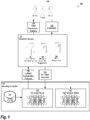
- FIG. 1 is a view of a system for automating image segmentation, according to some embodiments.
- An Initial Segmentation Estimate 110 is provided to Annotators 105 via an Annotation System 115.
- this Initial Segmentation Estimate 110 is generated using an already developed segmentation method that needs improvement.
- one or more conventional techniques generally known in the art may be used to provide a fast, but inaccurate segmentation which can be used as the Initial Segmentation Estimate 110.
- the Initial Segmentation Estimate 110 can come from a simple ellipse placed by the user in the location of the object to be annotated.
- the initial segmentation estimate can be defined by labels (or "seeds") placed by the user. For example, the user may label certain pixels as "object” or "background.” Once the labels have been provided, a generic seed-based algorithm such as the random walker algorithm may be applied to perform the segmentation.
- the Annotation System 115 includes a system for presenting a graphical user interface (GUI) which allows the user to perform Interactions 112 (e.g., via mouse movements) which correct the Initial Segmentation Estimate 110.
- GUI graphical user interface
- This GUI is referred to herein as an "annotation interface.”
- the computing technology supporting this annotation interface can be implemented using any technique known in the art.
- the Annotation System 115 is installed as software on computing devices used by the Annotators 105.
- General purpose computing devices can be used in these embodiments, or specialized devices with additional software or hardware to facilitate image annotation.
- the Annotation System 115 can be cloud based.
- FIG. 2 provides an example Interface 200 that may be employed by the Annotation System 115.
- an upper body anatomy is presented in a plurality of views and the user is presented with a variety of tools (located on the left hand side of the Interface 200) for adjusting the initial segmentation (indicated by segmentation lines 210A, 210B, 210C, and 210D in FIG 2 ).
- the Annotation System 115 stores Corrections 120 that describe the refinement of the segmentation from initial estimate to ground truth. These Corrections 120 indicate one or more changes to the Initial Segmentation Estimate 110.
- the Initial Segmentation Estimate 110 is presented as a series of points around the outer edge of the anatomy to be segmented.
- the Annotators 105 may adjust the Initial Segmentation Estimate 110 by clicking and dragging these points until the boundary of the segmentation accurately reflects the edge of the anatomical area of interest.
- the Corrections 120 could comprise the coordinates of the points in the new segmentation or adjustments to the points in the Initial Segmentation Estimate 110.
- just the shape of the new segmentation may be included in the Corrections 120.
- the annotation system records their Motions 125 to adjust the initial estimate.
- the corrections may be thought of moving the contour inward in the case of over-segmentation, or moving the contour outward in the case of under-segmentation. These inward or outward motions, along with the places where they are performed, serve as input to a classifier, as described below.
- the annotation system may record an Effort Measurement 130 which indicates the amount of effort expended by the annotators to perform the corrections.
- the Effort Measurement 130 provides a measure of how close the initial result was to "perfect.”
- Amount of effort may include, for example, overall time, number of mouse motions, amount of scrolling through slices for multi-slice images, etc.
- the effort measurement may be used, for example, to give larger weights to such cases during training, and to determine whether the overall system has converged.
- the approach described above is not limited to segmentation of objects, but may also be used in other applications such as landmark or object detection.
- the input is an initial guess of the landmark location, and the actions of the annotators to move the location to the correct location are recorded and used in the machine learning model described below.
- the amount of effort required may be recorded.
- the Initial Segmentation Estimate 110 is combined with the Interactions 112, the Motions 125, and the Effort Measurement 130 to form an Annotated Correction 135 which is presented to a Modeling Computer 140.
- the Annotated Correction 135 further includes the image data which is being analyzed.
- the Annotated Correction 135 only includes an identifier (e.g., filename) of the image data which can then be used to retrieve the image data from the Annotation System 115, either locally at the Modeling Computer 140 or on another system (not shown in FIG. 1 ).
- the Modeling Computer 140 is assumed to be connected to the Annotation System 115 via one or more networks (e.g., the Internet) not shown in FIG. 1 ; however, in some embodiments, the Modeling Computer 140 and the Annotation System 115 can be combined in a single computing system.
- the data included in the Annotated Correction 135 may be transmitted to the Modeling Computer 140 using any technique known in the art.
- the Annotated Correction 135 comprise a data file containing the Initial Segmentation Estimate 110 and a second data file describing the Interactions 112, the Motions 125, and the Effort Measurement 130 in a structured data language such as Extensible Markup Language (XML).
- XML Extensible Markup Language
- a Segmentation Model 147 (i.e., classifier) is also learned from the Annotated Correction 135. More specifically, the Segmentation Model 147 is trained to perform the segmentation provided in the Segmentation Model 147 when presented with corresponding image data. Thus, once trained the Segmentation Model 147 is capable of automatically segmenting an image without any need for manual annotation. The accuracy of the segmentation will be dependent on the level of training provided to the Segmentation Model 147. In some embodiments, the Segmentation Model 147 simply outputs a segmentation, but additional information may also be provided such as the accuracy of the segmentation (based on modeling results). Furthermore, in some embodiments, the Segmentation Model 147 may suggest more than one segmentation based on modeling results and a clinician can select the preferred segmentation based on manual inspection of the data.
- the Modeling Computer 140 includes a Ground Truth Database 145 which stores the ground truth for each image presented to the Annotators 105 for segmentation.
- a Correction Model 150 i.e., classifier
- the Effort Measurement 130 included in the Annotated Correction 135 is used to adjust the training weights so that the learning evolves faster when the estimate is far from the ground truth, and slows down when the estimate is close to the ground truth. Note that the learning step may occur after a certain amount of annotations have been performed or immediately after each annotation.
- the Segmentation Model 147 and the Correction Model 150 may generally be any classifier known in the art. In one embodiment, the Segmentation Model 147 and the Correction Model 150 are organized as a recursive convolutional neural network. In another embodiment, the Segmentation Model 147 and the Correction Model 150 are organized as a generative adversarial neural network. Combinations of recursive convolutional and generative adversarial neural networks can also be used as well as other deep learning architectures.
- the Segmentation Model 147 When applied to an image, the Segmentation Model 147 generates a segmentation, referred to herein as the "base segmentation,” for the image.
- the output of the Correction Model 147, when applied to the image, is referred to herein as the “segmentation correction" for thee image.
- the base segmentation and the segmentation correction are combined as the Updated Segmentation Estimate 155 and input into the Annotation System 115.
- This Updated Segmentation Estimation 155 is provided to the Annotators 105 via the Annotation System 115. In this way, the work load on the Annotators 105 is systematically reduced as the Segmentation Model 147 and the Correction Model 150 become better at automating segmentation.
- the process of presenting a segmentation and using annotated corrections to train the models may be repeated until the system converges at which point training is complete.
- the Modeling Computer 140 can use a fully convolutional neural network to learn the base segmentation and the correction provided by the annotators.
- the system continues to update both the segmentation and correction models along with new incoming annotations until the effort of correction by the annotators is close to or at zero, or meets the accuracy required by the task.
- the system collects only as much training data as is needed to achieve the goal.
- the method described above more efficiently limits the amount of data collected to what is needed.
- the amount of annotation resources required to support algorithm development can be reduced.
- the efforts of the annotators are continuously reduced on subsequent passes. Since most annotators are paid by the hour, this will result in a considerable saving in the overall cost of developing new algorithms.
- FIG. 3 illustrates and example of the overall annotation/segmentation process, with the actions of the annotator on top and the actions of the segmentation algorithm on bottom.
- the images depict an organ to be segmented.
- the segmentation result for an organ is shown as crosshatch pattern, the white outlined clear sections indicate the areas of over-segmentation, and white filled sections indicate the areas of under-segmentation.
- the actions of the annotators to correct these areas are used as inputs to the training algorithm.
- the amount of effort required to make the correction is used to adjust the training weights and detect convergence.
- FIG. 4 provides an example of a parallel computing platform 400 that may be utilized to implement the modeling computer 140 shown FIG. 1 , according to some embodiments of the present invention.
- This platform 400 may be used in embodiments of the present invention where NVIDIA CUDATM (or a similar parallel computing platform) is used.
- the architecture includes a host computing unit (“host") 405 and a graphics processing unit (GPU) device (“device”) 410 connected via a bus 415 (e.g., a PCIe bus).
- the host 405 includes the central processing unit, or "CPU” (not shown in FIG. 4 ), and host memory 425 accessible to the CPU.
- the device 410 includes the graphics processing unit (GPU) and its associated memory 420, referred to herein as device memory.
- the device memory 420 may include various types of memory, each optimized for different memory usages. For example, in some embodiments, the device memory includes global memory, constant memory, and texture memory.
- Parallel portions of a big data platform and/or big simulation platform may be executed on the platform 400 as “device kernels" or simply “kernels.”
- a kernel comprises parameterized code configured to perform a particular function.
- the parallel computing platform is configured to execute these kernels in an optimal manner across the platform 400 based on parameters, settings, and other selections provided by the user. Additionally, in some embodiments, the parallel computing platform may include additional functionality to allow for automatic processing of kernels in an optimal manner with minimal input provided by the user.
- each kernel is performed by grid of thread blocks (described in greater detail below).
- the platform 400 of FIG. 4 may be used to parallelize portions of the model based operations performed in training the Correction Model 150 or the Segmentation Model 147 shown in FIG. 1
- the device 410 includes one or more thread blocks 430 which represent the computation unit of the device 410.
- the term thread block refers to a group of threads that can cooperate via shared memory and synchronize their execution to coordinate memory accesses. For example, in FIG. 4 , threads 440, 445 and 450 operate in thread block 430 and access shared memory 435.
- thread blocks may be organized in a grid structure. A computation or series of computations may then be mapped onto this grid. For example, in embodiments utilizing CUDA, computations may be mapped on one-, two-, or three-dimensional grids. Each grid contains multiple thread blocks, and each thread block contains multiple threads. For example, in FIG.
- the thread blocks 430 are organized in a two dimensional grid structure with m +1 rows and n+1 columns.
- threads in different thread blocks of the same grid cannot communicate or synchronize with each other.
- thread blocks in the same grid can run on the same multiprocessor within the GPU at the same time.
- the number of threads in each thread block may be limited by hardware or software constraints.
- registers 455, 460, and 465 represent the fast memory available to thread block 430. Each register is only accessible by a single thread. Thus, for example, register 455 may only be accessed by thread 440. Conversely, shared memory is allocated per thread block, so all threads in the block have access to the same shared memory. Thus, shared memory 435 is designed to be accessed, in parallel, by each thread 440, 445, and 450 in thread block 430. Threads can access data in shared memory 435 loaded from device memory 420 by other threads within the same thread block (e.g., thread block 430). The device memory 420 is accessed by all blocks of the grid and may be implemented using, for example, Dynamic Random-Access Memory (DRAM).
- DRAM Dynamic Random-Access Memory
- Each thread can have one or more levels of memory access.
- each thread may have three levels of memory access.
- Second, each thread 440, 445, 450 in thread block 430, may read and write data to the shared memory 435 corresponding to that block 430.
- the time required for a thread to access shared memory exceeds that of register access due to the need to synchronize access among all the threads in the thread block.
- the shared memory is typically located close to the multiprocessor executing the threads.
- the third level of memory access allows all threads on the device 410 to read and/or write to the device memory.
- Device memory requires the longest time to access because access must be synchronized across the thread blocks operating on the device.
- the processing of each individual annotation in the Annotated Correction 135 is coded such that it primarily utilizes registers and shared memory and only utilizes device memory as necessary to move data in and out of a thread block.
- the embodiments of the present disclosure may be implemented with any combination of hardware and software.
- standard computing platforms e.g., servers, desktop computer, etc.
- the embodiments of the present disclosure may be included in an article of manufacture (e.g., one or more computer program products) having, for example, computer-readable, non-transitory media.
- the media may have embodied therein computer readable program code for providing and facilitating the mechanisms of the embodiments of the present disclosure.
- the article of manufacture can be included as part of a computer system or sold separately.
- An executable application comprises code or machine readable instructions for conditioning the processor to implement predetermined functions, such as those of an operating system, a context data acquisition system or other information processing system, for example, in response to user command or input.
- An executable procedure is a segment of code or machine readable instruction, sub-routine, or other distinct section of code or portion of an executable application for performing one or more particular processes. These processes may include receiving input data and/or parameters, performing operations on received input data and/or performing functions in response to received input parameters, and providing resulting output data and/or parameters.
- a graphical user interface comprises one or more display images, generated by a display processor and enabling user interaction with a processor or other device and associated data acquisition and processing functions.
- the GUI also includes an executable procedure or executable application.
- the executable procedure or executable application conditions the display processor to generate signals representing the GUI display images. These signals are supplied to a display device which displays the image for viewing by the user.
- the processor under control of an executable procedure or executable application, manipulates the GUI display images in response to signals received from the input devices. In this way, the user may interact with the display image using the input devices, enabling user interaction with the processor or other device.
- An activity performed automatically is performed in response to one or more executable instructions or device operation without user direct initiation of the activity.
Landscapes
- Engineering & Computer Science (AREA)
- Physics & Mathematics (AREA)
- Theoretical Computer Science (AREA)
- General Physics & Mathematics (AREA)
- Health & Medical Sciences (AREA)
- General Health & Medical Sciences (AREA)
- Evolutionary Computation (AREA)
- Molecular Biology (AREA)
- Software Systems (AREA)
- Mathematical Physics (AREA)
- General Engineering & Computer Science (AREA)
- Computing Systems (AREA)
- Data Mining & Analysis (AREA)
- Computational Linguistics (AREA)
- Biophysics (AREA)
- Life Sciences & Earth Sciences (AREA)
- Artificial Intelligence (AREA)
- Biomedical Technology (AREA)
- Computer Vision & Pattern Recognition (AREA)
- Quality & Reliability (AREA)
- Radiology & Medical Imaging (AREA)
- Nuclear Medicine, Radiotherapy & Molecular Imaging (AREA)
- Medical Informatics (AREA)
- Multimedia (AREA)
- Public Health (AREA)
- Primary Health Care (AREA)
- Epidemiology (AREA)
- Image Analysis (AREA)
Abstract
Description
- The present invention relates generally to methods and systems for automating the annotation and segmentation of medical image data. The techniques described herein may generally be applied to any medical imaging modality including, without limitation, Magnetic Resonance (MR), Ultrasound, and Computed Tomography (CT) images.
- Automation of medical imaging requires algorithms to learn how to perform a particular task, and these algorithms require "ground truth" data for training and validation. This ground truth data comes from human experts annotating the data, but such annotations are time-consuming and expensive to obtain. Key problems include how to obtain annotation data efficiently, with minimal effort from the human experts, and how to obtain the right amount of labeled data without paying for more than is actually needed. For machine learning algorithms an additional challenge is knowing when a sufficiently accurate result has been achieved. Finally, the entire cycle of annotation, testing, and validation is slow, limiting the overall pace of innovation.
- There have been many machine algorithms trained with data annotated by human experts. In a typical development cycle, researchers guess how much training data will be needed and then employ human experts to provide it. Prior research focused on how best to train given a set of annotated data.
- Recently, Deep Learning has emerged as a popular and highly effective method for performing image segmentation. A segmentation of an image is produced by portioning an image into different segments. For medical images, these segments may correspond to biologically relevant structures such as organs, blood vessels, pathologies, etc. However one of the biggest limitations of Deep Learning is that large amounts of labeled data are necessary to get good results without overfitting.
- Medical images are difficult to annotate compared to ordinary photographs and videos. For example, different image modalities may introduce artifacts that are not readily identifiable by one without medical training. Moreover, reliable detection of organs and other relevant anatomical structures, as well as identification of relevant diseases and abnormalities, will be difficult, if not impossible unless the annotator has medical training. This makes medical image annotation more costly to obtain as the number of people able to perform this task is limited.
- Current practices involve a sequential approach of first obtaining the annotations followed by algorithm development. Any benefits from creating the algorithm do not enhance the annotation acquisition. In this disclosure, we describe how the twin needs for segmentation algorithm development and segmentation training data can be combined into a single process for a more efficient development cycle. Improvements in the algorithm development will speed up the annotation, whereas at the same time the actions of the annotators are used to synchronously drive the learning algorithm.
- Embodiments of the present invention address and overcome one or more of the above shortcomings and drawbacks, by providing an integrated system of manual annotation and automatic segmentation for medical imaging tasks. The techniques described herein build upon machine learning techniques previously applied to object classification and semantic labeling problems to automate the segmentation correction process. The techniques described herein offer improvements to various computer-related technologies. For example, the disclosed techniques using computing systems to enable the automation of specific image annotation and segmentation tasks that previously could not be automated.
- According to some embodiments, a method for training a segmentation correction model includes performing an iterative model training process over a plurality of iterations. During each iteration, an initial segmentation estimate for an image is provided to a human annotators via an annotation interface. The initial segmentation estimate identifies one or more anatomical areas of interest within the image. Interactions with the annotation interface are automatically monitored to record annotation information comprising one or more of (i) segmentation corrections made to the initial segmentation estimate by the annotators via the annotation interface, and (ii) interactions with the annotation interface performed by the annotators while making the corrections. A base segmentation machine learning model is trained to automatically create a base segmentation based on the image. Additionally, a segmentation correction machine learning model is trained to automatically perform the segmentation corrections based on the image.
- In some embodiments of the aforementioned method, the annotation information further comprises an effort measurement indicative of an amount of effort expended by the annotators in making the corrections. This effort measurement can be used to determine when to terminate the training process. For example, if the effort measurement is equal to the convergence value, the iterative model training process may be terminated. Conversely, if the effort measurement is not equal to the convergence value, the base segmentation and segmentation correction machine learning models may be used to determine the initial segmentation estimate for a new image. Then, the iterative model training process can continue to the next iteration.
- In some embodiments of the aforementioned method, the effort measurement is a time-based measurement and the convergence value is equal to a predetermined time value. In other embodiments, the effort measurement is a measurement of time spent by the annotators in making the corrections and number of interface motions made in making the corrections. In one embodiment, the image comprises a plurality of slices/volumes and the effort measurement includes a measurement of time spent in scrolling through the plurality of slices/volumes. In another embodiment, the effort measurement is a measurement of a number of mouse motions and the convergence value is equal to a predetermined number of mouse motions. The effort measurement can also be used in model training. For example, if the segmentation correction machine learning model is a convolutional neural network may be used to set one or more training weights used by the convolutional neural network.
- According to another aspect of the present invention, a method for training a landmark location correction model includes performing an iterative model training process in a manner similar to the other methods discussed above. However, rather than relying on an initial segmentation estimate, initial landmark location estimates are provided for an image to a plurality of human annotators via an annotation interface. Each initial landmark location estimate identifies an anatomical landmark within the image.
- According to other embodiments, a system for training a segmentation correction model includes an annotation system and a parallel computing platform. The annotation system is configured to provide an initial segmentation estimate for an image to a plurality of human annotators via an annotation interface. The initial segmentation estimate identifies one or more anatomical areas of interest within the image. The annotation system also automatically monitors interactions with the annotation interface to record annotation information comprising (i) segmentation corrections made to the initial segmentation estimate by the annotators via the annotation interface, and (ii) interactions with the annotation interface performed by the annotators while making the corrections. The parallel computing platform is configured to train a base segmentation machine learning model to automatically create a base segmentation based on the image. Additionally, the platform trains a segmentation correction machine learning model to automatically perform the segmentation corrections based on the image. In some embodiments of the aforementioned system, the annotation information further comprises an effort measurement indicative of an amount of effort expended by the annotators in making the corrections. The segmentation correction machine learning model can be a convolutional neural network and the effort measurement can be used to set one or more training weights used by the convolutional neural network.
- Additional features and advantages of the invention will be made apparent from the following detailed description of illustrative embodiments that proceeds with reference to the accompanying drawings.
- The foregoing and other aspects of the present invention are best understood from the following detailed description when read in connection with the accompanying drawings. For the purpose of illustrating the invention, there is shown in the drawings embodiments that are presently preferred, it being understood, however, that the invention is not limited to the specific instrumentalities disclosed. Included in the drawings are the following figures:
-
FIG. 1 is a view of a system for automating image segmentation, according to some embodiments; -
FIG. 2 shows an example interface used by an annotation system, according to some embodiments; -
FIG. 3 illustrates and example of the overall annotation/segmentation process, according to some embodiments; and -
FIG. 4 provides an example of a parallel processing memory architecture that may be utilized to implement one or more components shown inFIG. 1 , according to some embodiments of the present invention. - The following disclosure describes the present invention according to several embodiments directed at methods, systems, and apparatuses related to automated manual annotation and automatic segmentation for medical imaging tasks. Briefly, an annotation system is used to collect an initial segmentation estimate. This initial segmentation estimate is then presented to one or more human annotators which make corrections to the initial segmentation estimate. Based on the corrections made by the annotators, and interactions made the annotators in making the corrections, a correction model and a segmentation model are trained. By learning the corrective actions taken by annotators to refine a segmentation result, the system described herein actively improves the segmentation model, which differs from training the model with cumulative annotated data.
-
FIG. 1 is a view of a system for automating image segmentation, according to some embodiments. AnInitial Segmentation Estimate 110 is provided toAnnotators 105 via anAnnotation System 115. In some embodiments, thisInitial Segmentation Estimate 110 is generated using an already developed segmentation method that needs improvement. For example, one or more conventional techniques generally known in the art may be used to provide a fast, but inaccurate segmentation which can be used as theInitial Segmentation Estimate 110. In other embodiments, theInitial Segmentation Estimate 110 can come from a simple ellipse placed by the user in the location of the object to be annotated. In yet another alternative, the initial segmentation estimate can be defined by labels (or "seeds") placed by the user. For example, the user may label certain pixels as "object" or "background." Once the labels have been provided, a generic seed-based algorithm such as the random walker algorithm may be applied to perform the segmentation. - In general, the
Annotation System 115 includes a system for presenting a graphical user interface (GUI) which allows the user to perform Interactions 112 (e.g., via mouse movements) which correct theInitial Segmentation Estimate 110. This GUI is referred to herein as an "annotation interface." The computing technology supporting this annotation interface can be implemented using any technique known in the art. For example, in some embodiments, theAnnotation System 115 is installed as software on computing devices used by theAnnotators 105. General purpose computing devices can be used in these embodiments, or specialized devices with additional software or hardware to facilitate image annotation. In other embodiments, theAnnotation System 115 can be cloud based. Thus, theAnnotators 105 interact through a browser or some other thin client interface to interact with the annotation interface.FIG. 2 provides anexample Interface 200 that may be employed by theAnnotation System 115. Here, an upper body anatomy is presented in a plurality of views and the user is presented with a variety of tools (located on the left hand side of the Interface 200) for adjusting the initial segmentation (indicated bysegmentation lines FIG 2 ). - Based on the
Interactions 112 shown inFIG. 1 , theAnnotation System 115stores Corrections 120 that describe the refinement of the segmentation from initial estimate to ground truth. TheseCorrections 120 indicate one or more changes to theInitial Segmentation Estimate 110. For example, in some embodiments, theInitial Segmentation Estimate 110 is presented as a series of points around the outer edge of the anatomy to be segmented. TheAnnotators 105 may adjust theInitial Segmentation Estimate 110 by clicking and dragging these points until the boundary of the segmentation accurately reflects the edge of the anatomical area of interest. In this case theCorrections 120 could comprise the coordinates of the points in the new segmentation or adjustments to the points in theInitial Segmentation Estimate 110. In other embodiments, rather than storing theCorrections 120 as points, just the shape of the new segmentation may be included in theCorrections 120. - As the annotators perform the
Interactions 112 with theAnnotation System 115, the annotation system records theirMotions 125 to adjust the initial estimate. Broadly speaking, the corrections may be thought of moving the contour inward in the case of over-segmentation, or moving the contour outward in the case of under-segmentation. These inward or outward motions, along with the places where they are performed, serve as input to a classifier, as described below. In addition to theMotions 125, the annotation system may record anEffort Measurement 130 which indicates the amount of effort expended by the annotators to perform the corrections. Effectively, theEffort Measurement 130 provides a measure of how close the initial result was to "perfect." Amount of effort may include, for example, overall time, number of mouse motions, amount of scrolling through slices for multi-slice images, etc. The effort measurement may be used, for example, to give larger weights to such cases during training, and to determine whether the overall system has converged. - It should be noted that the approach described above is not limited to segmentation of objects, but may also be used in other applications such as landmark or object detection. In these applications, the input is an initial guess of the landmark location, and the actions of the annotators to move the location to the correct location are recorded and used in the machine learning model described below. In addition, the amount of effort required may be recorded.
- The
Initial Segmentation Estimate 110 is combined with theInteractions 112, theMotions 125, and theEffort Measurement 130 to form anAnnotated Correction 135 which is presented to aModeling Computer 140. In some embodiments, theAnnotated Correction 135 further includes the image data which is being analyzed. In other embodiments, theAnnotated Correction 135 only includes an identifier (e.g., filename) of the image data which can then be used to retrieve the image data from theAnnotation System 115, either locally at theModeling Computer 140 or on another system (not shown inFIG. 1 ). - The
Modeling Computer 140 is assumed to be connected to theAnnotation System 115 via one or more networks (e.g., the Internet) not shown inFIG. 1 ; however, in some embodiments, theModeling Computer 140 and theAnnotation System 115 can be combined in a single computing system. The data included in theAnnotated Correction 135 may be transmitted to theModeling Computer 140 using any technique known in the art. For example, in one embodiment, theAnnotated Correction 135 comprise a data file containing theInitial Segmentation Estimate 110 and a second data file describing theInteractions 112, theMotions 125, and theEffort Measurement 130 in a structured data language such as Extensible Markup Language (XML). - A Segmentation Model 147 (i.e., classifier) is also learned from the
Annotated Correction 135. More specifically, theSegmentation Model 147 is trained to perform the segmentation provided in theSegmentation Model 147 when presented with corresponding image data. Thus, once trained theSegmentation Model 147 is capable of automatically segmenting an image without any need for manual annotation. The accuracy of the segmentation will be dependent on the level of training provided to theSegmentation Model 147. In some embodiments, theSegmentation Model 147 simply outputs a segmentation, but additional information may also be provided such as the accuracy of the segmentation (based on modeling results). Furthermore, in some embodiments, theSegmentation Model 147 may suggest more than one segmentation based on modeling results and a clinician can select the preferred segmentation based on manual inspection of the data. - The
Modeling Computer 140 includes aGround Truth Database 145 which stores the ground truth for each image presented to theAnnotators 105 for segmentation. A Correction Model 150 (i.e., classifier) is learned from the difference between theInitial Segmentation Estimate 110 and theAnnotated Correction 135 using a machine learning algorithm. TheEffort Measurement 130 included in theAnnotated Correction 135 is used to adjust the training weights so that the learning evolves faster when the estimate is far from the ground truth, and slows down when the estimate is close to the ground truth. Note that the learning step may occur after a certain amount of annotations have been performed or immediately after each annotation. - The
Segmentation Model 147 and theCorrection Model 150 may generally be any classifier known in the art. In one embodiment, theSegmentation Model 147 and theCorrection Model 150 are organized as a recursive convolutional neural network. In another embodiment, theSegmentation Model 147 and theCorrection Model 150 are organized as a generative adversarial neural network. Combinations of recursive convolutional and generative adversarial neural networks can also be used as well as other deep learning architectures. - When applied to an image, the
Segmentation Model 147 generates a segmentation, referred to herein as the "base segmentation," for the image. The output of theCorrection Model 147, when applied to the image, is referred to herein as the "segmentation correction" for thee image. The base segmentation and the segmentation correction are combined as theUpdated Segmentation Estimate 155 and input into theAnnotation System 115. ThisUpdated Segmentation Estimation 155 is provided to theAnnotators 105 via theAnnotation System 115. In this way, the work load on theAnnotators 105 is systematically reduced as theSegmentation Model 147 and theCorrection Model 150 become better at automating segmentation. The process of presenting a segmentation and using annotated corrections to train the models may be repeated until the system converges at which point training is complete. - As an example implementation of the techniques described above with reference to
FIG. 1 , consider a task of 3D liver segmentation on CT. TheModeling Computer 140 can use a fully convolutional neural network to learn the base segmentation and the correction provided by the annotators. The system continues to update both the segmentation and correction models along with new incoming annotations until the effort of correction by the annotators is close to or at zero, or meets the accuracy required by the task. - By taking this approach, the system collects only as much training data as is needed to achieve the goal. In contrast to traditional learning approaches, where researchers must guess how much training data is required by a task, the method described above more efficiently limits the amount of data collected to what is needed. As a result, the amount of annotation resources required to support algorithm development can be reduced. Moreover by continuously incorporating improvements in the algorithm from what is learned by earlier corrections, the efforts of the annotators are continuously reduced on subsequent passes. Since most annotators are paid by the hour, this will result in a considerable saving in the overall cost of developing new algorithms.
-
FIG. 3 illustrates and example of the overall annotation/segmentation process, with the actions of the annotator on top and the actions of the segmentation algorithm on bottom. The images depict an organ to be segmented. The segmentation result for an organ is shown as crosshatch pattern, the white outlined clear sections indicate the areas of over-segmentation, and white filled sections indicate the areas of under-segmentation. The actions of the annotators to correct these areas are used as inputs to the training algorithm. The amount of effort required to make the correction is used to adjust the training weights and detect convergence. -
FIG. 4 provides an example of aparallel computing platform 400 that may be utilized to implement themodeling computer 140 shownFIG. 1 , according to some embodiments of the present invention. Thisplatform 400 may be used in embodiments of the present invention where NVIDIA CUDA™ (or a similar parallel computing platform) is used. The architecture includes a host computing unit ("host") 405 and a graphics processing unit (GPU) device ("device") 410 connected via a bus 415 (e.g., a PCIe bus). Thehost 405 includes the central processing unit, or "CPU" (not shown inFIG. 4 ), and host memory 425 accessible to the CPU. Thedevice 410 includes the graphics processing unit (GPU) and its associatedmemory 420, referred to herein as device memory. Thedevice memory 420 may include various types of memory, each optimized for different memory usages. For example, in some embodiments, the device memory includes global memory, constant memory, and texture memory. - Parallel portions of a big data platform and/or big simulation platform (see
FIG. 4 ) may be executed on theplatform 400 as "device kernels" or simply "kernels." A kernel comprises parameterized code configured to perform a particular function. The parallel computing platform is configured to execute these kernels in an optimal manner across theplatform 400 based on parameters, settings, and other selections provided by the user. Additionally, in some embodiments, the parallel computing platform may include additional functionality to allow for automatic processing of kernels in an optimal manner with minimal input provided by the user. - The processing required for each kernel is performed by grid of thread blocks (described in greater detail below). Using concurrent kernel execution, streams, and synchronization with lightweight events, the
platform 400 ofFIG. 4 (or similar architectures) may be used to parallelize portions of the model based operations performed in training theCorrection Model 150 or theSegmentation Model 147 shown inFIG. 1 - The
device 410 includes one or more thread blocks 430 which represent the computation unit of thedevice 410. The term thread block refers to a group of threads that can cooperate via shared memory and synchronize their execution to coordinate memory accesses. For example, inFIG. 4 ,threads thread block 430 and access sharedmemory 435. Depending on the parallel computing platform used, thread blocks may be organized in a grid structure. A computation or series of computations may then be mapped onto this grid. For example, in embodiments utilizing CUDA, computations may be mapped on one-, two-, or three-dimensional grids. Each grid contains multiple thread blocks, and each thread block contains multiple threads. For example, inFIG. 4 , the thread blocks 430 are organized in a two dimensional grid structure with m+1 rows and n+1 columns. Generally, threads in different thread blocks of the same grid cannot communicate or synchronize with each other. However, thread blocks in the same grid can run on the same multiprocessor within the GPU at the same time. The number of threads in each thread block may be limited by hardware or software constraints. - Continuing with reference to
FIG. 4 , registers 455, 460, and 465 represent the fast memory available tothread block 430. Each register is only accessible by a single thread. Thus, for example, register 455 may only be accessed bythread 440. Conversely, shared memory is allocated per thread block, so all threads in the block have access to the same shared memory. Thus, sharedmemory 435 is designed to be accessed, in parallel, by eachthread thread block 430. Threads can access data in sharedmemory 435 loaded fromdevice memory 420 by other threads within the same thread block (e.g., thread block 430). Thedevice memory 420 is accessed by all blocks of the grid and may be implemented using, for example, Dynamic Random-Access Memory (DRAM). - Each thread can have one or more levels of memory access. For example, in the
platform 400 ofFIG. 4 , each thread may have three levels of memory access. First, eachthread corresponding registers thread thread block 430, may read and write data to the sharedmemory 435 corresponding to thatblock 430. Generally, the time required for a thread to access shared memory exceeds that of register access due to the need to synchronize access among all the threads in the thread block. However, like the registers in the thread block, the shared memory is typically located close to the multiprocessor executing the threads. The third level of memory access allows all threads on thedevice 410 to read and/or write to the device memory. Device memory requires the longest time to access because access must be synchronized across the thread blocks operating on the device. Thus, in some embodiments, the processing of each individual annotation in theAnnotated Correction 135 is coded such that it primarily utilizes registers and shared memory and only utilizes device memory as necessary to move data in and out of a thread block. - The embodiments of the present disclosure may be implemented with any combination of hardware and software. For example, aside from parallel processing architecture presented in
FIG. 4 , standard computing platforms (e.g., servers, desktop computer, etc.) may be specially configured to perform the techniques discussed herein. In addition, the embodiments of the present disclosure may be included in an article of manufacture (e.g., one or more computer program products) having, for example, computer-readable, non-transitory media. The media may have embodied therein computer readable program code for providing and facilitating the mechanisms of the embodiments of the present disclosure. The article of manufacture can be included as part of a computer system or sold separately. - While various aspects and embodiments have been disclosed herein, other aspects and embodiments will be apparent to those skilled in the art. The various aspects and embodiments disclosed herein are for purposes of illustration and are not intended to be limiting, with the true scope and spirit being indicated by the following claims.
- An executable application, as used herein, comprises code or machine readable instructions for conditioning the processor to implement predetermined functions, such as those of an operating system, a context data acquisition system or other information processing system, for example, in response to user command or input. An executable procedure is a segment of code or machine readable instruction, sub-routine, or other distinct section of code or portion of an executable application for performing one or more particular processes. These processes may include receiving input data and/or parameters, performing operations on received input data and/or performing functions in response to received input parameters, and providing resulting output data and/or parameters.
- A graphical user interface (GUI), as used herein, comprises one or more display images, generated by a display processor and enabling user interaction with a processor or other device and associated data acquisition and processing functions. The GUI also includes an executable procedure or executable application. The executable procedure or executable application conditions the display processor to generate signals representing the GUI display images. These signals are supplied to a display device which displays the image for viewing by the user. The processor, under control of an executable procedure or executable application, manipulates the GUI display images in response to signals received from the input devices. In this way, the user may interact with the display image using the input devices, enabling user interaction with the processor or other device.
- The functions and process steps herein may be performed automatically or wholly or partially in response to user command. An activity (including a step) performed automatically is performed in response to one or more executable instructions or device operation without user direct initiation of the activity.
- The system and processes of the figures are not exclusive. Other systems, processes and menus may be derived in accordance with the principles of the invention to accomplish the same objectives. Although this invention has been described with reference to particular embodiments, it is to be understood that the embodiments and variations shown and described herein are for illustration purposes only. Modifications to the current design may be implemented by those skilled in the art, without departing from the scope of the invention. As described herein, the various systems, subsystems, agents, managers and processes can be implemented using hardware components, software components, and/or combinations thereof. No claim element herein is to be construed under the provisions of 35 U.S.C. 112, sixth paragraph, unless the element is expressly recited using the phrase "means for."
Claims (18)
- A method for training a segmentation correction model, the method comprising:
performing an iterative model training process over a plurality of iterations, wherein each iteration comprises:providing an initial segmentation estimate for an image to a plurality of human annotators via an annotation interface, wherein the initial segmentation estimate identifies one or more anatomical areas of interest within the image;automatically monitoring interactions with the annotation interface to record annotation information comprising one or more of (i) segmentation corrections made to the initial segmentation estimate by the annotators via the annotation interface, and (ii) interactions with the annotation interface performed by the annotators while making the corrections;training a base segmentation machine learning model to automatically create a base segmentation based on the image; andtraining a segmentation correction machine learning model to automatically perform the segmentation corrections based on the image. - The method according to claim 1, wherein the annotation information further comprises an effort measurement indicative of an amount of effort expended by the annotators in making the corrections.
- The method according to claim 2, wherein each iteration further comprises:determining whether the effort measurement is equal to a convergence value;if the effort measurement is equal to the convergence value, terminating the iterative model training process; andif the effort measurement is not equal to the convergence value, using the base segmentation and segmentation correction machine learning models to determine the initial segmentation estimate for a new image and continuing to a next iteration of the iterative model training process.
- The method according to claim 3, wherein the effort measurement is a time-based measurement and the convergence value is equal to a predetermined time value.
- The method according to claim 4, wherein the effort measurement is a measurement of time spent by the annotators in making the corrections and number of interface motions made in making the corrections.
- The method according to claim 4, wherein the image comprises a plurality of slices/volumes and the effort measurement includes a measurement of time spent in scrolling through the plurality of slices/volumes.
- The method according to claim 3, wherein the effort measurement is a measurement of a number of mouse motions and the convergence value is equal to a predetermined number of mouse motions.
- The method according to claim 2, wherein the segmentation correction machine learning model is a convolutional neural network.
- The method according to claim 8, wherein the effort measurement is used to set one or more training weights used by the convolutional neural network.
- A method for training a landmark location correction model, the method comprising:
performing an iterative model training process over a plurality of iterations, wherein each iteration comprises:providing initial landmark location estimates for an image to a plurality of human annotators via an annotation interface, wherein each initial landmark location estimate identifies an anatomical landmark within the image;automatically monitoring interactions with the annotation interface to record annotation information comprising (i) corrections to the landmark location estimates made by the annotators via the annotation interface, (ii) interactions with the annotation interface performed by the annotators while making the corrections, and (iii) an effort measurement indicative of an amount of effort expended by the annotators in making the corrections;training a landmark location machine learning model to automatically identify landmark locations in the image based on the annotation information;training a location correction machine learning model to automatically perform the corrections to the initial landmark location estimates;if the effort measurement is equal to a convergence value, terminating the iterative model training process; andif the effort measurement is not equal to the convergence value, using the landmark location and the location correction machine learning model to determine the initial landmark location estimate for a new image and continuing to a next iteration of the iterative model training process. - The method according to claim 10, wherein the effort measurement is a time-based measurement and the convergence value is equal to a predetermined time value
and/or
wherein the effort measurement is a measurement of time spent by the annotators in making the corrections and number of interface motions made in making the corrections. - The method according to claims 10 or 11, wherein the image comprises a plurality of slices and the effort measurement is a measurement of time spent in scrolling through the plurality of slices.
- The method according to claim 10, wherein the effort measurement is a measurement of a number of mouse motions and the convergence value is equal to a predetermined number of mouse motions.
- The method according to any of the claims 10 to 13, wherein the correction machine learning model is a convolutional neural network.
- The method according to claim 14, wherein the effort measurement is used to set one or more training weights used by the convolutional neural network.
- A system for training a segmentation correction model, the system comprising:an annotation system configured to:provide an initial segmentation estimate for an image to a plurality of human annotators via an annotation interface, wherein the initial segmentation estimate identifies one or more anatomical areas of interest within the image;automatically monitor interactions with the annotation interface to record annotation information comprising (i) segmentation corrections made to the initial segmentation estimate by the annotators via the annotation interface, and (ii) interactions with the annotation interface performed by the annotators while making the corrections; anda parallel computing platform configured to:train a base segmentation machine learning model to automatically create a base segmentation based on the image; andtrain a segmentation correction machine learning model to automatically perform the segmentation corrections based on the image.
- The system according to claim 16, wherein the annotation information further comprises an effort measurement indicative of an amount of effort expended by the annotators in making the corrections.
- The system of claim 17, wherein the segmentation correction machine learning model is a convolutional neural network and the effort measurement is used to set one or more training weights used by the convolutional neural network.
Applications Claiming Priority (1)
| Application Number | Priority Date | Filing Date | Title |
|---|---|---|---|
| US15/470,360 US10366490B2 (en) | 2017-03-27 | 2017-03-27 | Highly integrated annotation and segmentation system for medical imaging |
Publications (2)
| Publication Number | Publication Date |
|---|---|
| EP3382642A1 true EP3382642A1 (en) | 2018-10-03 |
| EP3382642B1 EP3382642B1 (en) | 2020-04-29 |
Family
ID=61768058
Family Applications (1)
| Application Number | Title | Priority Date | Filing Date |
|---|---|---|---|
| EP18162624.3A Active EP3382642B1 (en) | 2017-03-27 | 2018-03-19 | Highly integrated annotation and segmentation system for medical imaging |
Country Status (3)
| Country | Link |
|---|---|
| US (1) | US10366490B2 (en) |
| EP (1) | EP3382642B1 (en) |
| CN (1) | CN108665462B (en) |
Cited By (1)
| Publication number | Priority date | Publication date | Assignee | Title |
|---|---|---|---|---|
| EP4485351A1 (en) * | 2023-06-29 | 2025-01-01 | Supersonic Imagine | A method of refining a segmentation mask |
Families Citing this family (47)
| Publication number | Priority date | Publication date | Assignee | Title |
|---|---|---|---|---|
| US10672122B2 (en) * | 2015-11-19 | 2020-06-02 | Koninklijke Philips N.V. | Optimizing user interactions in segmentation |
| GB201617507D0 (en) | 2016-10-14 | 2016-11-30 | Axial3D Limited | Axial3D UK |
| US11138790B2 (en) | 2016-10-14 | 2021-10-05 | Axial Medical Printing Limited | Method for generating a 3D physical model of a patient specific anatomic feature from 2D medical images |
| EP3635683B1 (en) | 2017-05-09 | 2022-07-06 | HeartFlow, Inc. | Systems and methods for anatomic structure segmentation in image analysis |
| WO2018209057A1 (en) * | 2017-05-11 | 2018-11-15 | The Research Foundation For The State University Of New York | System and method associated with predicting segmentation quality of objects in analysis of copious image data |
| US11468286B2 (en) * | 2017-05-30 | 2022-10-11 | Leica Microsystems Cms Gmbh | Prediction guided sequential data learning method |
| KR102117050B1 (en) * | 2017-09-08 | 2020-05-29 | 삼성전자주식회사 | Electronic device and method for human segmentation in image |
| WO2020028352A1 (en) | 2018-07-31 | 2020-02-06 | Washington University | Methods and systems for segmenting organs in images using a cnn-based correction network |
| WO2020096098A1 (en) | 2018-11-09 | 2020-05-14 | 주식회사 루닛 | Method for managing annotation work, and apparatus and system supporting same |
| GB201900437D0 (en) | 2019-01-11 | 2019-02-27 | Axial Medical Printing Ltd | Axial3d big book 2 |
| US11068694B2 (en) * | 2019-01-23 | 2021-07-20 | Molecular Devices, Llc | Image analysis system and method of using the image analysis system |
| JP7164008B2 (en) * | 2019-03-13 | 2022-11-01 | 日本電気株式会社 | Data generation method, data generation device and program |
| CA3141859A1 (en) * | 2019-04-11 | 2020-10-15 | Agilent Technologies, Inc. | User interface configured to facilitate user annotation for instance segmentation within biological sample |
| CN110895968B (en) * | 2019-04-24 | 2023-12-15 | 苏州图灵微生物科技有限公司 | Artificial intelligence medical image automatic diagnosis system and method |
| US11195277B2 (en) * | 2019-04-25 | 2021-12-07 | GE Precision Healthcare LLC | Systems and methods for generating normative imaging data for medical image processing using deep learning |
| US10997466B2 (en) * | 2019-06-21 | 2021-05-04 | Straxciro Pty. Ltd. | Method and system for image segmentation and identification |
| US11954610B2 (en) | 2019-08-09 | 2024-04-09 | GE Precision Healthcare LLC | Active surveillance and learning for machine learning model authoring and deployment |
| EP3813013A1 (en) * | 2019-10-23 | 2021-04-28 | Koninklijke Philips N.V. | Methods and systems for segmenting images |
| EP4073752A4 (en) * | 2019-12-09 | 2024-01-03 | Janssen Biotech, Inc. | METHOD FOR DETERMINING THE SEVERITY OF A SKIN DISEASE BASED ON A PERCENTAGE OF BODY SURFACE COVERED BY LESSIONS |
| US11526689B2 (en) * | 2019-12-19 | 2022-12-13 | Robert Bosch Gmbh | Few-shot learning of repetitive human tasks |
| CN111260659A (en) * | 2020-01-13 | 2020-06-09 | 镇江优瞳智能科技有限公司 | Image interactive segmentation method based on initial annotation point guidance |
| EP3866107A1 (en) * | 2020-02-14 | 2021-08-18 | Koninklijke Philips N.V. | Model-based image segmentation |
| EP3893245A1 (en) * | 2020-04-10 | 2021-10-13 | Koninklijke Philips N.V. | Correction of automated label data for tomographic medical imaging systems |
| US12573029B2 (en) * | 2020-04-24 | 2026-03-10 | Nvidia Corporation | Image annotation using one or more neural networks |
| CN111553613B (en) * | 2020-05-11 | 2023-04-28 | 中石化石油工程技术服务有限公司 | Intelligent grading evaluation method and system for quality of seismic acquisition data |
| CN111598900B (en) | 2020-05-18 | 2022-08-09 | 腾讯医疗健康(深圳)有限公司 | Image region segmentation model training method, segmentation method and device |
| US20210374467A1 (en) * | 2020-05-29 | 2021-12-02 | Fei Company | Correlated slice and view image annotation for machine learning |
| JP7294275B2 (en) * | 2020-08-25 | 2023-06-20 | トヨタ自動車株式会社 | Image processing device, image processing program and image processing method |
| US12361561B2 (en) * | 2020-10-30 | 2025-07-15 | Shanghai United Imaging Healthcare Co., Ltd. | Methods and systems for medical image segmentation |
| US11587237B2 (en) * | 2020-11-30 | 2023-02-21 | Robert Bosch Gmbh | Image segmention via efficient semidefinate-programming based inference for binary and multi-class Markov Random Fields |
| US12182671B2 (en) | 2021-01-26 | 2024-12-31 | International Business Machines Corporation | Optimizing a machine learning system |
| CN112862789B (en) * | 2021-02-10 | 2023-06-13 | 上海大学 | Interactive image segmentation method based on machine learning |
| US12444506B2 (en) | 2021-02-11 | 2025-10-14 | Axial Medical Printing Limited | Systems and methods for automated segmentation of patient specific anatomies for pathology specific measurements |
| GB202101908D0 (en) | 2021-02-11 | 2021-03-31 | Axial Medical Printing Ltd | Axial3D pathology |
| US12094190B2 (en) | 2021-02-19 | 2024-09-17 | Arizona Board Of Regents On Behalf Of Arizona State University | Systems, methods, and apparatuses for implementing medical image segmentation using interactive refinement |
| JP7674913B2 (en) * | 2021-05-24 | 2025-05-12 | キヤノンメディカルシステムズ株式会社 | Machine learning device, machine learning method, machine learning program, and inference device |
| CN115423736A (en) | 2021-05-31 | 2022-12-02 | 通用电气精准医疗有限责任公司 | Imaging system and method |
| EP4349267A4 (en) * | 2021-06-03 | 2025-06-04 | Furuno Electric Co., Ltd. | COMPUTER PROGRAM, LEARNING MODEL GENERATION METHOD, IMAGE PROCESSING METHOD, AND IMAGE PROCESSING DEVICE |
| EP4105817A1 (en) * | 2021-06-16 | 2022-12-21 | Volkswagen Aktiengesellschaft | Surveillance system and method for automatically executing a security function and system and method for generating a synthetic training data set |
| US12567150B2 (en) * | 2021-08-17 | 2026-03-03 | Siemens Healthineers Ag | Editing presegmented images and volumes using deep learning |
| CN113903433B (en) * | 2021-10-14 | 2022-11-15 | 数坤(北京)网络科技股份有限公司 | Image processing method and device and electronic equipment |
| US20230305845A1 (en) * | 2022-03-22 | 2023-09-28 | Nvidia Corporation | Techniques to selectively store data |
| CN114494275B (en) * | 2022-04-15 | 2022-08-05 | 北京美摄网络科技有限公司 | Method and device for training image segmentation model of mobile terminal |
| US12423938B2 (en) | 2022-07-25 | 2025-09-23 | Dell Products L.P. | System and method for identifying auxiliary areas of interest for image based on focus indicators |
| US12373955B2 (en) * | 2022-07-25 | 2025-07-29 | Dell Products L.P. | System and method for storage management of images |
| US12488578B1 (en) | 2022-09-16 | 2025-12-02 | Nvidia Corporation | Neural network training technique |
| EP4343687A1 (en) * | 2022-09-26 | 2024-03-27 | Koninklijke Philips N.V. | Image segmentation with improved expert guidance |
Citations (1)
| Publication number | Priority date | Publication date | Assignee | Title |
|---|---|---|---|---|
| US20100322489A1 (en) * | 2009-06-18 | 2010-12-23 | Omisa Inc. | System and method for image segmentation |
Family Cites Families (28)
| Publication number | Priority date | Publication date | Assignee | Title |
|---|---|---|---|---|
| US7257769B2 (en) * | 2003-06-05 | 2007-08-14 | Siemens Communications, Inc. | System and method for indicating an annotation for a document |
| EP1815430A1 (en) * | 2004-11-19 | 2007-08-08 | Koninklijke Philips Electronics N.V. | System and method for automated detection and segmentation of tumor boundaries within medical imaging data |
| US7693717B2 (en) * | 2006-04-12 | 2010-04-06 | Custom Speech Usa, Inc. | Session file modification with annotation using speech recognition or text to speech |
| US8189880B2 (en) * | 2007-05-29 | 2012-05-29 | Microsoft Corporation | Interactive photo annotation based on face clustering |
| US9785858B2 (en) * | 2008-09-26 | 2017-10-10 | Siemens Healthcare Gmbh | Method and system for hierarchical parsing and semantic navigation of full body computed tomography data |
| US8600143B1 (en) * | 2010-05-20 | 2013-12-03 | Kla-Tencor Corporation | Method and system for hierarchical tissue analysis and classification |
| US9715754B2 (en) * | 2010-08-05 | 2017-07-25 | Koninklijke Philips N.V. | In-plane and interactive surface mesh adaptation |
| US11074495B2 (en) * | 2013-02-28 | 2021-07-27 | Z Advanced Computing, Inc. (Zac) | System and method for extremely efficient image and pattern recognition and artificial intelligence platform |
| US9700276B2 (en) * | 2012-02-28 | 2017-07-11 | Siemens Healthcare Gmbh | Robust multi-object tracking using sparse appearance representation and online sparse appearance dictionary update |
| EP2672396A1 (en) * | 2012-06-08 | 2013-12-11 | ETH Zürich | Method for annotating images |
| US9058317B1 (en) * | 2012-11-01 | 2015-06-16 | Digital Reasoning Systems, Inc. | System and method for machine learning management |
| US20140324808A1 (en) * | 2013-03-15 | 2014-10-30 | Sumeet Sandhu | Semantic Segmentation and Tagging and Advanced User Interface to Improve Patent Search and Analysis |
| US9348815B1 (en) * | 2013-06-28 | 2016-05-24 | Digital Reasoning Systems, Inc. | Systems and methods for construction, maintenance, and improvement of knowledge representations |
| EP3846176B1 (en) * | 2013-09-25 | 2024-09-04 | HeartFlow, Inc. | Systems and methods for validating and correcting automated medical image annotations |
| US9589349B2 (en) * | 2013-09-25 | 2017-03-07 | Heartflow, Inc. | Systems and methods for controlling user repeatability and reproducibility of automated image annotation correction |
| EP3077956B1 (en) * | 2013-12-03 | 2023-10-11 | Children's National Medical Center | Method and system for wound assessment and management |
| US9552549B1 (en) * | 2014-07-28 | 2017-01-24 | Google Inc. | Ranking approach to train deep neural nets for multilabel image annotation |
| US9508341B1 (en) * | 2014-09-03 | 2016-11-29 | Amazon Technologies, Inc. | Active learning for lexical annotations |
| CN105425777B (en) * | 2015-12-11 | 2017-12-15 | 渤海大学 | A kind of chemical process failure monitoring method based on Active Learning |
| US9928875B2 (en) * | 2016-03-22 | 2018-03-27 | Nec Corporation | Efficient video annotation with optical flow based estimation and suggestion |
| US9799120B1 (en) * | 2016-05-09 | 2017-10-24 | Siemens Healthcare Gmbh | Method and apparatus for atlas/model-based segmentation of magnetic resonance images with weakly supervised examination-dependent learning |
| US20180012359A1 (en) * | 2016-07-06 | 2018-01-11 | Marinko Venci Sarunic | Systems and Methods for Automated Image Classification and Segmentation |
| EP3246873B1 (en) * | 2016-07-15 | 2018-07-11 | Siemens Healthcare GmbH | Method and data processing unit for segmenting an object in a medical image |
| US10452899B2 (en) * | 2016-08-31 | 2019-10-22 | Siemens Healthcare Gmbh | Unsupervised deep representation learning for fine-grained body part recognition |
| US10582907B2 (en) * | 2016-10-31 | 2020-03-10 | Siemens Healthcare Gmbh | Deep learning based bone removal in computed tomography angiography |
| JP2018081545A (en) * | 2016-11-17 | 2018-05-24 | パナソニック インテレクチュアル プロパティ コーポレーション オブ アメリカPanasonic Intellectual Property Corporation of America | Image data extraction device and image data extraction method |
| US11449985B2 (en) * | 2016-12-02 | 2022-09-20 | Regents Of The University Of Minnesota | Computer vision for cancerous tissue recognition |
| US10699412B2 (en) * | 2017-03-23 | 2020-06-30 | Petuum Inc. | Structure correcting adversarial network for chest X-rays organ segmentation |
-
2017
- 2017-03-27 US US15/470,360 patent/US10366490B2/en active Active
-
2018
- 2018-03-19 EP EP18162624.3A patent/EP3382642B1/en active Active
- 2018-03-27 CN CN201810257794.8A patent/CN108665462B/en active Active
Patent Citations (1)
| Publication number | Priority date | Publication date | Assignee | Title |
|---|---|---|---|---|
| US20100322489A1 (en) * | 2009-06-18 | 2010-12-23 | Omisa Inc. | System and method for image segmentation |
Non-Patent Citations (2)
| Title |
|---|
| MARTIN RAJCHL ET AL: "DeepCut: Object Segmentation From Bounding Box Annotations Using Convolutional Neural Networks", IEEE TRANSACTIONS ON MEDICAL IMAGING., vol. 36, no. 2, 1 February 2017 (2017-02-01), US, pages 674 - 683, XP055488970, ISSN: 0278-0062, DOI: 10.1109/TMI.2016.2621185 * |
| XU NING ET AL: "Deep Interactive Object Selection", 2016 IEEE CONFERENCE ON COMPUTER VISION AND PATTERN RECOGNITION (CVPR), IEEE, 27 June 2016 (2016-06-27), pages 373 - 381, XP033021213, DOI: 10.1109/CVPR.2016.47 * |
Cited By (1)
| Publication number | Priority date | Publication date | Assignee | Title |
|---|---|---|---|---|
| EP4485351A1 (en) * | 2023-06-29 | 2025-01-01 | Supersonic Imagine | A method of refining a segmentation mask |
Also Published As
| Publication number | Publication date |
|---|---|
| US10366490B2 (en) | 2019-07-30 |
| US20180276815A1 (en) | 2018-09-27 |
| EP3382642B1 (en) | 2020-04-29 |
| CN108665462A (en) | 2018-10-16 |
| CN108665462B (en) | 2022-11-22 |
Similar Documents
| Publication | Publication Date | Title |
|---|---|---|
| EP3382642B1 (en) | Highly integrated annotation and segmentation system for medical imaging | |
| US12161451B2 (en) | Medical imaging and efficient sharing of medical imaging information | |
| EP3117771B1 (en) | Direct computation of image-derived biomarkers | |
| EP3657437B1 (en) | Automatic detection and quantification of the aorta from medical images | |
| US11423554B2 (en) | Registering a two-dimensional image with a three-dimensional image | |
| CN109949300B (en) | Method, system and computer readable medium for anatomical tree structure analysis | |
| US20230310085A1 (en) | Systems and methods for estimation of blood flow using response surface and reduced order modeling | |
| CN110648358B (en) | Adaptive nonlinear optimization of shape parameters for object localization in 3D medical images | |
| CN114913174B (en) | Method, apparatus and storage medium for vascular system variation detection | |
| JP7007469B2 (en) | Medical document creation support devices, methods and programs, trained models, and learning devices, methods and programs | |
| Chernyshov et al. | Automated segmentation and quantification of the right ventricle in 2-D echocardiography | |
| Akan et al. | ViViEchoformer: deep video regressor predicting ejection fraction | |
| US11200976B2 (en) | Tracking method and apparatus | |
| US11494956B2 (en) | Computer-implemented method for the reconstruction of medical image data | |
| EP4083650A1 (en) | Controlling a scanning operation of a medical imaging device | |
| KR102865582B1 (en) | A method of predicting a risk of brain disease and a training method of model for analyzing brain disease risk | |
| CN121127926A (en) | Artificial deep neural network training methods for estimating hemodynamic parameters, hemodynamic parameter estimation methods, computer program products, and computer systems. | |
| CN120689263A (en) | AI-assisted detection of vascular anomalies in medical images |
Legal Events
| Date | Code | Title | Description |
|---|---|---|---|
| PUAI | Public reference made under article 153(3) epc to a published international application that has entered the european phase |
Free format text: ORIGINAL CODE: 0009012 |
|
| STAA | Information on the status of an ep patent application or granted ep patent |
Free format text: STATUS: REQUEST FOR EXAMINATION WAS MADE |
|
| 17P | Request for examination filed |
Effective date: 20180319 |
|
| AK | Designated contracting states |
Kind code of ref document: A1 Designated state(s): AL AT BE BG CH CY CZ DE DK EE ES FI FR GB GR HR HU IE IS IT LI LT LU LV MC MK MT NL NO PL PT RO RS SE SI SK SM TR |
|
| AX | Request for extension of the european patent |
Extension state: BA ME |
|
| RBV | Designated contracting states (corrected) |
Designated state(s): AL AT BE BG CH CY CZ DE DK EE ES FI FR GB GR HR HU IE IS IT LI LT LU LV MC MK MT NL NO PL PT RO RS SE SI SK SM TR |
|
| GRAP | Despatch of communication of intention to grant a patent |
Free format text: ORIGINAL CODE: EPIDOSNIGR1 |
|
| STAA | Information on the status of an ep patent application or granted ep patent |
Free format text: STATUS: GRANT OF PATENT IS INTENDED |
|
| INTG | Intention to grant announced |
Effective date: 20191111 |
|
| GRAS | Grant fee paid |
Free format text: ORIGINAL CODE: EPIDOSNIGR3 |
|
| GRAA | (expected) grant |
Free format text: ORIGINAL CODE: 0009210 |
|
| STAA | Information on the status of an ep patent application or granted ep patent |
Free format text: STATUS: THE PATENT HAS BEEN GRANTED |
|
| AK | Designated contracting states |
Kind code of ref document: B1 Designated state(s): AL AT BE BG CH CY CZ DE DK EE ES FI FR GB GR HR HU IE IS IT LI LT LU LV MC MK MT NL NO PL PT RO RS SE SI SK SM TR |
|
| REG | Reference to a national code |
Ref country code: GB Ref legal event code: FG4D |
|
| REG | Reference to a national code |
Ref country code: CH Ref legal event code: EP |
|
| REG | Reference to a national code |
Ref country code: DE Ref legal event code: R096 Ref document number: 602018004076 Country of ref document: DE |
|
| REG | Reference to a national code |
Ref country code: AT Ref legal event code: REF Ref document number: 1264446 Country of ref document: AT Kind code of ref document: T Effective date: 20200515 |
|
| REG | Reference to a national code |
Ref country code: IE Ref legal event code: FG4D |
|
| REG | Reference to a national code |
Ref country code: NL Ref legal event code: MP Effective date: 20200429 |
|
| REG | Reference to a national code |
Ref country code: LT Ref legal event code: MG4D |
|
| PG25 | Lapsed in a contracting state [announced via postgrant information from national office to epo] |
Ref country code: FI Free format text: LAPSE BECAUSE OF FAILURE TO SUBMIT A TRANSLATION OF THE DESCRIPTION OR TO PAY THE FEE WITHIN THE PRESCRIBED TIME-LIMIT Effective date: 20200429 Ref country code: NO Free format text: LAPSE BECAUSE OF FAILURE TO SUBMIT A TRANSLATION OF THE DESCRIPTION OR TO PAY THE FEE WITHIN THE PRESCRIBED TIME-LIMIT Effective date: 20200729 Ref country code: PT Free format text: LAPSE BECAUSE OF FAILURE TO SUBMIT A TRANSLATION OF THE DESCRIPTION OR TO PAY THE FEE WITHIN THE PRESCRIBED TIME-LIMIT Effective date: 20200831 Ref country code: IS Free format text: LAPSE BECAUSE OF FAILURE TO SUBMIT A TRANSLATION OF THE DESCRIPTION OR TO PAY THE FEE WITHIN THE PRESCRIBED TIME-LIMIT Effective date: 20200829 Ref country code: GR Free format text: LAPSE BECAUSE OF FAILURE TO SUBMIT A TRANSLATION OF THE DESCRIPTION OR TO PAY THE FEE WITHIN THE PRESCRIBED TIME-LIMIT Effective date: 20200730 Ref country code: SE Free format text: LAPSE BECAUSE OF FAILURE TO SUBMIT A TRANSLATION OF THE DESCRIPTION OR TO PAY THE FEE WITHIN THE PRESCRIBED TIME-LIMIT Effective date: 20200429 Ref country code: LT Free format text: LAPSE BECAUSE OF FAILURE TO SUBMIT A TRANSLATION OF THE DESCRIPTION OR TO PAY THE FEE WITHIN THE PRESCRIBED TIME-LIMIT Effective date: 20200429 |
|
| REG | Reference to a national code |
Ref country code: AT Ref legal event code: MK05 Ref document number: 1264446 Country of ref document: AT Kind code of ref document: T Effective date: 20200429 |
|
| PG25 | Lapsed in a contracting state [announced via postgrant information from national office to epo] |
Ref country code: HR Free format text: LAPSE BECAUSE OF FAILURE TO SUBMIT A TRANSLATION OF THE DESCRIPTION OR TO PAY THE FEE WITHIN THE PRESCRIBED TIME-LIMIT Effective date: 20200429 Ref country code: LV Free format text: LAPSE BECAUSE OF FAILURE TO SUBMIT A TRANSLATION OF THE DESCRIPTION OR TO PAY THE FEE WITHIN THE PRESCRIBED TIME-LIMIT Effective date: 20200429 Ref country code: BG Free format text: LAPSE BECAUSE OF FAILURE TO SUBMIT A TRANSLATION OF THE DESCRIPTION OR TO PAY THE FEE WITHIN THE PRESCRIBED TIME-LIMIT Effective date: 20200729 Ref country code: RS Free format text: LAPSE BECAUSE OF FAILURE TO SUBMIT A TRANSLATION OF THE DESCRIPTION OR TO PAY THE FEE WITHIN THE PRESCRIBED TIME-LIMIT Effective date: 20200429 |
|
| PG25 | Lapsed in a contracting state [announced via postgrant information from national office to epo] |
Ref country code: NL Free format text: LAPSE BECAUSE OF FAILURE TO SUBMIT A TRANSLATION OF THE DESCRIPTION OR TO PAY THE FEE WITHIN THE PRESCRIBED TIME-LIMIT Effective date: 20200429 Ref country code: AL Free format text: LAPSE BECAUSE OF FAILURE TO SUBMIT A TRANSLATION OF THE DESCRIPTION OR TO PAY THE FEE WITHIN THE PRESCRIBED TIME-LIMIT Effective date: 20200429 |
|
| PG25 | Lapsed in a contracting state [announced via postgrant information from national office to epo] |
Ref country code: ES Free format text: LAPSE BECAUSE OF FAILURE TO SUBMIT A TRANSLATION OF THE DESCRIPTION OR TO PAY THE FEE WITHIN THE PRESCRIBED TIME-LIMIT Effective date: 20200429 Ref country code: EE Free format text: LAPSE BECAUSE OF FAILURE TO SUBMIT A TRANSLATION OF THE DESCRIPTION OR TO PAY THE FEE WITHIN THE PRESCRIBED TIME-LIMIT Effective date: 20200429 Ref country code: AT Free format text: LAPSE BECAUSE OF FAILURE TO SUBMIT A TRANSLATION OF THE DESCRIPTION OR TO PAY THE FEE WITHIN THE PRESCRIBED TIME-LIMIT Effective date: 20200429 Ref country code: SM Free format text: LAPSE BECAUSE OF FAILURE TO SUBMIT A TRANSLATION OF THE DESCRIPTION OR TO PAY THE FEE WITHIN THE PRESCRIBED TIME-LIMIT Effective date: 20200429 Ref country code: IT Free format text: LAPSE BECAUSE OF FAILURE TO SUBMIT A TRANSLATION OF THE DESCRIPTION OR TO PAY THE FEE WITHIN THE PRESCRIBED TIME-LIMIT Effective date: 20200429 Ref country code: DK Free format text: LAPSE BECAUSE OF FAILURE TO SUBMIT A TRANSLATION OF THE DESCRIPTION OR TO PAY THE FEE WITHIN THE PRESCRIBED TIME-LIMIT Effective date: 20200429 Ref country code: RO Free format text: LAPSE BECAUSE OF FAILURE TO SUBMIT A TRANSLATION OF THE DESCRIPTION OR TO PAY THE FEE WITHIN THE PRESCRIBED TIME-LIMIT Effective date: 20200429 Ref country code: CZ Free format text: LAPSE BECAUSE OF FAILURE TO SUBMIT A TRANSLATION OF THE DESCRIPTION OR TO PAY THE FEE WITHIN THE PRESCRIBED TIME-LIMIT Effective date: 20200429 |
|
| REG | Reference to a national code |
Ref country code: DE Ref legal event code: R097 Ref document number: 602018004076 Country of ref document: DE |
|
| PG25 | Lapsed in a contracting state [announced via postgrant information from national office to epo] |
Ref country code: PL Free format text: LAPSE BECAUSE OF FAILURE TO SUBMIT A TRANSLATION OF THE DESCRIPTION OR TO PAY THE FEE WITHIN THE PRESCRIBED TIME-LIMIT Effective date: 20200429 Ref country code: SK Free format text: LAPSE BECAUSE OF FAILURE TO SUBMIT A TRANSLATION OF THE DESCRIPTION OR TO PAY THE FEE WITHIN THE PRESCRIBED TIME-LIMIT Effective date: 20200429 |
|
| PLBE | No opposition filed within time limit |
Free format text: ORIGINAL CODE: 0009261 |
|
| STAA | Information on the status of an ep patent application or granted ep patent |
Free format text: STATUS: NO OPPOSITION FILED WITHIN TIME LIMIT |
|
| 26N | No opposition filed |
Effective date: 20210201 |
|
| PG25 | Lapsed in a contracting state [announced via postgrant information from national office to epo] |
Ref country code: SI Free format text: LAPSE BECAUSE OF FAILURE TO SUBMIT A TRANSLATION OF THE DESCRIPTION OR TO PAY THE FEE WITHIN THE PRESCRIBED TIME-LIMIT Effective date: 20200429 |
|
| PG25 | Lapsed in a contracting state [announced via postgrant information from national office to epo] |
Ref country code: MC Free format text: LAPSE BECAUSE OF FAILURE TO SUBMIT A TRANSLATION OF THE DESCRIPTION OR TO PAY THE FEE WITHIN THE PRESCRIBED TIME-LIMIT Effective date: 20200429 |
|
| REG | Reference to a national code |
Ref country code: CH Ref legal event code: PL |
|
| REG | Reference to a national code |
Ref country code: BE Ref legal event code: MM Effective date: 20210331 |
|
| PG25 | Lapsed in a contracting state [announced via postgrant information from national office to epo] |
Ref country code: LI Free format text: LAPSE BECAUSE OF NON-PAYMENT OF DUE FEES Effective date: 20210331 Ref country code: LU Free format text: LAPSE BECAUSE OF NON-PAYMENT OF DUE FEES Effective date: 20210319 Ref country code: IE Free format text: LAPSE BECAUSE OF NON-PAYMENT OF DUE FEES Effective date: 20210319 Ref country code: CH Free format text: LAPSE BECAUSE OF NON-PAYMENT OF DUE FEES Effective date: 20210331 |
|
| PG25 | Lapsed in a contracting state [announced via postgrant information from national office to epo] |
Ref country code: BE Free format text: LAPSE BECAUSE OF NON-PAYMENT OF DUE FEES Effective date: 20210331 |
|
| PG25 | Lapsed in a contracting state [announced via postgrant information from national office to epo] |
Ref country code: CY Free format text: LAPSE BECAUSE OF FAILURE TO SUBMIT A TRANSLATION OF THE DESCRIPTION OR TO PAY THE FEE WITHIN THE PRESCRIBED TIME-LIMIT Effective date: 20200429 |
|
| PG25 | Lapsed in a contracting state [announced via postgrant information from national office to epo] |
Ref country code: HU Free format text: LAPSE BECAUSE OF FAILURE TO SUBMIT A TRANSLATION OF THE DESCRIPTION OR TO PAY THE FEE WITHIN THE PRESCRIBED TIME-LIMIT; INVALID AB INITIO Effective date: 20180319 |
|
| REG | Reference to a national code |
Ref country code: DE Ref legal event code: R081 Ref document number: 602018004076 Country of ref document: DE Owner name: SIEMENS HEALTHINEERS AG, DE Free format text: FORMER OWNER: SIEMENS HEALTHCARE GMBH, MUENCHEN, DE |
|
| PG25 | Lapsed in a contracting state [announced via postgrant information from national office to epo] |
Ref country code: MK Free format text: LAPSE BECAUSE OF FAILURE TO SUBMIT A TRANSLATION OF THE DESCRIPTION OR TO PAY THE FEE WITHIN THE PRESCRIBED TIME-LIMIT Effective date: 20200429 |
|
| PG25 | Lapsed in a contracting state [announced via postgrant information from national office to epo] |
Ref country code: TR Free format text: LAPSE BECAUSE OF FAILURE TO SUBMIT A TRANSLATION OF THE DESCRIPTION OR TO PAY THE FEE WITHIN THE PRESCRIBED TIME-LIMIT Effective date: 20200429 |
|
| PG25 | Lapsed in a contracting state [announced via postgrant information from national office to epo] |
Ref country code: MT Free format text: LAPSE BECAUSE OF FAILURE TO SUBMIT A TRANSLATION OF THE DESCRIPTION OR TO PAY THE FEE WITHIN THE PRESCRIBED TIME-LIMIT Effective date: 20200429 |
|
| PGFP | Annual fee paid to national office [announced via postgrant information from national office to epo] |
Ref country code: DE Payment date: 20250520 Year of fee payment: 8 |
|
| PGFP | Annual fee paid to national office [announced via postgrant information from national office to epo] |
Ref country code: GB Payment date: 20250403 Year of fee payment: 8 |
|
| PGFP | Annual fee paid to national office [announced via postgrant information from national office to epo] |
Ref country code: FR Payment date: 20260316 Year of fee payment: 9 |