EP3381443A1 - Sustained action formulation of cyclosporin form 2 - Google Patents
Sustained action formulation of cyclosporin form 2 Download PDFInfo
- Publication number
- EP3381443A1 EP3381443A1 EP18158837.7A EP18158837A EP3381443A1 EP 3381443 A1 EP3381443 A1 EP 3381443A1 EP 18158837 A EP18158837 A EP 18158837A EP 3381443 A1 EP3381443 A1 EP 3381443A1
- Authority
- EP
- European Patent Office
- Prior art keywords
- formulation
- cyclosporin
- eye
- conjunctiva
- needle
- Prior art date
- Legal status (The legal status is an assumption and is not a legal conclusion. Google has not performed a legal analysis and makes no representation as to the accuracy of the status listed.)
- Withdrawn
Links
Images



Classifications
-
- A—HUMAN NECESSITIES
- A61—MEDICAL OR VETERINARY SCIENCE; HYGIENE
- A61K—PREPARATIONS FOR MEDICAL, DENTAL OR TOILETRY PURPOSES
- A61K38/00—Medicinal preparations containing peptides
- A61K38/04—Peptides having up to 20 amino acids in a fully defined sequence; Derivatives thereof
- A61K38/12—Cyclic peptides, e.g. bacitracins; Polymyxins; Gramicidins S, C; Tyrocidins A, B or C
- A61K38/13—Cyclosporins
-
- A—HUMAN NECESSITIES
- A61—MEDICAL OR VETERINARY SCIENCE; HYGIENE
- A61K—PREPARATIONS FOR MEDICAL, DENTAL OR TOILETRY PURPOSES
- A61K9/00—Medicinal preparations characterised by special physical form
-
- A—HUMAN NECESSITIES
- A61—MEDICAL OR VETERINARY SCIENCE; HYGIENE
- A61K—PREPARATIONS FOR MEDICAL, DENTAL OR TOILETRY PURPOSES
- A61K47/00—Medicinal preparations characterised by the non-active ingredients used, e.g. carriers or inert additives; Targeting or modifying agents chemically bound to the active ingredient
- A61K47/30—Macromolecular organic or inorganic compounds, e.g. inorganic polyphosphates
- A61K47/32—Macromolecular compounds obtained by reactions only involving carbon-to-carbon unsaturated bonds, e.g. carbomers, poly(meth)acrylates, or polyvinyl pyrrolidone
-
- A—HUMAN NECESSITIES
- A61—MEDICAL OR VETERINARY SCIENCE; HYGIENE
- A61K—PREPARATIONS FOR MEDICAL, DENTAL OR TOILETRY PURPOSES
- A61K47/00—Medicinal preparations characterised by the non-active ingredients used, e.g. carriers or inert additives; Targeting or modifying agents chemically bound to the active ingredient
- A61K47/30—Macromolecular organic or inorganic compounds, e.g. inorganic polyphosphates
- A61K47/36—Polysaccharides; Derivatives thereof, e.g. gums, starch, alginate, dextrin, hyaluronic acid, chitosan, inulin, agar or pectin
- A61K47/38—Cellulose; Derivatives thereof
-
- A—HUMAN NECESSITIES
- A61—MEDICAL OR VETERINARY SCIENCE; HYGIENE
- A61K—PREPARATIONS FOR MEDICAL, DENTAL OR TOILETRY PURPOSES
- A61K9/00—Medicinal preparations characterised by special physical form
- A61K9/0012—Galenical forms characterised by the site of application
- A61K9/0019—Injectable compositions; Intramuscular, intravenous, arterial, subcutaneous administration; Compositions to be administered through the skin in an invasive manner
-
- A—HUMAN NECESSITIES
- A61—MEDICAL OR VETERINARY SCIENCE; HYGIENE
- A61K—PREPARATIONS FOR MEDICAL, DENTAL OR TOILETRY PURPOSES
- A61K9/00—Medicinal preparations characterised by special physical form
- A61K9/0012—Galenical forms characterised by the site of application
- A61K9/0048—Eye, e.g. artificial tears
-
- A—HUMAN NECESSITIES
- A61—MEDICAL OR VETERINARY SCIENCE; HYGIENE
- A61K—PREPARATIONS FOR MEDICAL, DENTAL OR TOILETRY PURPOSES
- A61K9/00—Medicinal preparations characterised by special physical form
- A61K9/10—Dispersions; Emulsions
-
- A—HUMAN NECESSITIES
- A61—MEDICAL OR VETERINARY SCIENCE; HYGIENE
- A61P—SPECIFIC THERAPEUTIC ACTIVITY OF CHEMICAL COMPOUNDS OR MEDICINAL PREPARATIONS
- A61P27/00—Drugs for disorders of the senses
- A61P27/02—Ophthalmic agents
-
- A—HUMAN NECESSITIES
- A61—MEDICAL OR VETERINARY SCIENCE; HYGIENE
- A61P—SPECIFIC THERAPEUTIC ACTIVITY OF CHEMICAL COMPOUNDS OR MEDICINAL PREPARATIONS
- A61P27/00—Drugs for disorders of the senses
- A61P27/02—Ophthalmic agents
- A61P27/04—Artificial tears; Irrigation solutions
-
- C—CHEMISTRY; METALLURGY
- C07—ORGANIC CHEMISTRY
- C07K—PEPTIDES
- C07K1/00—General methods for the preparation of peptides, i.e. processes for the organic chemical preparation of peptides or proteins of any length
- C07K1/14—Extraction; Separation; Purification
- C07K1/30—Extraction; Separation; Purification by precipitation
- C07K1/306—Extraction; Separation; Purification by precipitation by crystallization
-
- C—CHEMISTRY; METALLURGY
- C07—ORGANIC CHEMISTRY
- C07K—PEPTIDES
- C07K7/00—Peptides having 5 to 20 amino acids in a fully defined sequence; Derivatives thereof
- C07K7/64—Cyclic peptides containing only normal peptide links
- C07K7/645—Cyclosporins; Related peptides
Definitions
- Cyclosporin A is the active Ingredient in Restasis®, a topical ocular formulation approved in the United States for the treatment of dry eye. Topical cyclosporin is also used in the management of other ophthalmic conditions such as restoration of corneal sensitivity following refractive surgery, conjunctival and corneal inflammation, keratoconjuntivitis, graft versus host disease, post-transplant glaucoma, corneal transplants, mycotic keratitis, meibomian gland dysfunction, Thygeson's superficial punctate keratitis, uveitis, and Theodore's superior limbic keratoconjunctivitis.
- a single implantation or injection which delivers high levels of cyclosporin constantly over a long period (weeks or months) is desirable.
- the use of multi-month biodegradable or bioerodible delivery system which delivers cyclosporin to targeted anterior ocular tissues - at equal or greater concentration as Restasis - is clearly desirable.
- Biodegradable implants which can be delivered through a 22 gauge needle, do not deliver sufficient drug over a sufficient period of time to meet the need. Biodegradable gel-forming suspensions perform better but, still do not meet the need.
- the formulations of the invention when delivered to the subconjunctival space of the eye through a 22 or smaller gauge needle, deliver cyclosporin A to most anterior ocular tissue for a sustained period of time.
- Cyclosporin A is a cyclic peptide having the following chemical structure: Its chemical name is cyclo[[(E)-(2S,3R,4R)-3-hydroxy-4-methyl-2-(methylamino)-6-octenoyl]-L-2-aminobutyryl- N -methylglycyl- N -methyl-Lleucyl-L-valyl- N -methyl-L-leucyl-L-alanyl-D-alanyl- N -methyl-L-leucyl- N -methyl-L-leucyl- N methyl-L-valyl].
- cyclosporine It is also known by the names cyclosporine, cyclosporine A, ciclosporin, and cyclosporin A. It is the active ingredient in Restasis® (Allergan, Inc., Irvine, California), an emulsion comprising 0.05% (w/v) cyclosporin. Restasis® is approved in the United States to increase tear production in patients whose tear production is presumed to be suppressed due to ocular inflammation associated with keratoconjunctivitis sicca.
- Cyclosporin A is known to exist in an amorphous form, liquid crystal form, tetragonal crystalline form (form 1), and an orthorhombic form (form 3).
- a new crystalline form, cyclosporin A form 2 has recently been discovered.
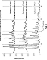
- the XRPD pattern of CsA Form 2 differs significantly from the tetragonal form and orthorhombic form ( FIG. 1 ).
- These major peaks are defined as those being unique to Form 2 relative to the orthorhombic or tetragonal forms; as well as, peaks having an intensity greater than 5 times the background.
- the new crystalline form (Form 2) of CsA is a nonstoichiometric hydrate of Cyclosporin A.
- the crystalline Form 2 is represented by the formula: wherein X is the number of molecules of water and varies from 0 to 3. In one embodiment, X in the above formula is 2.
- Form 2 appears to be a kinetically stable form of CsA in aqueous suspensions. Suspensions containing Form 2 show no conversion to other known polymorphic or pseudomorphic forms upon storage. It has been found that Form 1 and the amorphous form convert to Form 2 in the presence of water.
- the asymmetric unit of this CsA Form 2 contains one cyclosporine A molecule and two water molecules. It is possible that any small molecule that can hydrogen bond to water could play the role of space filler, which would give a range of potential structures running from the orthorhombic dihydrate to distorted monoclinic dihydrate
- the XRPD pattern calculated from the single-crystal structure is shown in Figure 8 and it matches the experimental pattern shown in Figure 2 . These matching patterns further corroborate that Form 2 is a unique and pure crystalline form of cyclosporine A.
- thermogravimetric analysis combined with KF titration and vapor sorption desorption analysis suggest that CsA Form 2 is a non-stoichiometric hydrate of CsA.
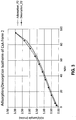
- the vapor sorption analysis of Cyclosporine Form 2 indicates that water content in the new crystal form reversibly varies with relative humidity as shown in Fig. 3 .
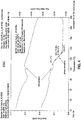
- the new CsA form undergoes a phase transition to a liquid crystal or amorphous form at 124.4 °C prior to melting as indicated by the modulated differential calorimetric (MDSC) analysis ( Figure 4 ).
- MDSC modulated differential calorimetric
- Cyclosporin A Form 2 may be obtained by suspending amorphous 0.05 % cyclosporin A (w/v) in 1% Polysorbate 80, heating the solution to 65 °C, holding it at that temperature for 24 hours, and then recovering the precipitate by vacuum filtration. One can then use the cyclosporin A Form 2 thus obtained to generate additional amounts, using Cyclosporin A Form 2 as a seed crystal; in this method, one suspends about 30 g cyclosporin A in a solution of 900 ml water containing 1% (w/v) Polysorbate 80, heats the solution to 65 °C, and then seeds it with 0.2 g of cyclosporin A Form 2 at a temperature of 52 °C. The solution is then stirred for about 22 hours at a temperature of between about 61 °C and 65 °C, and then recovers the precipitate that results.
- CsA Form 2 may be found in U.S. Patent Application No. 13/480,710 , the entire contents of which are incorporated by reference herein.
- compositions of the invention are ophthalmically acceptable formulations of Cyclosporin A form 2.
- ophthalmically acceptable the inventors mean that the suspensions are formulated in such a way as to be non-irritating when administered to the eye of a mammal, such as a human.
- the formulations of the invention comprise cyclosporin A form 2 and a hydrogel such as hyaluronic acid, hydroxypropyl cellulose, hydroxypropylmethyl cellulose, hydroxyethylcellulose, polyvinyl pyrolidone, and carboxymethylcellulose.
- a hydrogel such as hyaluronic acid, hydroxypropyl cellulose, hydroxypropylmethyl cellulose, hydroxyethylcellulose, polyvinyl pyrolidone, and carboxymethylcellulose.
- the formulation comprises cyclosporin A form 2 at a concentration of about 0.001% to about 10% (w/v).
- the suspension comprises cyclosporin A form 2 at a concentration of about 0.001% (w/v) to about 0.01%, about 0.001% (w/v) to about 0.04% (w/v), about 0.001% (w/v) to about 0.03% (w/v), about 0.001% (w/v) to about 0.02% (w/v), or about 0.001% (w/v) to about 0.01 % (w/v).
- the suspension comprises cyclosporin A form 2 at a concentration of about 0.01% (w/v) to about 0.05%, about 0.01% (w/v) to about 0.04% (w/v), about 0.01% (w/v) to about 0.03% (w/v), about 0.01% (w/v) to about 0.02% (w/v), or about 0.01% (w/v) to about 0.01% (w/v).
- the suspension comprises cyclosporin A form 2 at a concentration of about 0.01% (w/v) to about 0.1%, about 0.1% (w/v) to about 0.5% (w/v), about 0.01% (w/v) to about 1% (w/v), or about 1% (w/v) to about 10%.
- the formulations may comprise about 0.001% (w/v), about 0.002% (w/v), about 0.003% (w/v), about 0.004% (w/v), about 0.005% (w/v), about 0.006% (w/v), about 0.007% (w/v), about 0.008% (w/v), about 0.009% (w/v), about 0.01% (w/v), about 0.015% (w/v), about 0.02% (w/v), about 0.025% (w/v), about 0.03% (w/v), about 0.035% (w/v), about 0.04% (w/v), about 0.045% (w/v), about 0.05% (w/v), about 0.055% (w/v), about 0.06% (w/v), about 0.065% (w/v), about 0.07% (w/v), about 0.075% (w/v), about 0.08% (w/v), about 0.085% (w/v), about 0.09% (w/v), about 0.095% (w/v), about 0.1% (
- Formulations of the invention may be used to treat any condition of the eye which is known to be amenable to topical treatment with cyclosporin A (such as with Restasis®) at the concentrations stated here.
- compositions of the invention may be used to treat patients suffering from dry eye, to treat blepharitis and meibomian gland disease, to restore corneal sensitivity that has been impaired due to refractive surgery on the eye, to treat allergic conjunctivitis and atopic and vernal keratoconjunctivitis, and to treat ptyregia, conjunctival and corneal inflammation, keratoconjuntivitis, graft versus host disease, post-transplant glaucoma, corneal transplants, mycotic keratitis, Thygeson's superficial punctate keratitis, uveitis, and Theodore's superior limbic keratoconjunctivitis, among other conditions.
- the International Dry Eye Workshop defines dry eye as "a multifactorial disease of the tears and ocular surface that results in symptoms of discomfort, visual disturbance, and tear film instability with potential damage to the ocular surface, accompanied by increased osmolarity of the tear film and inflammation of the ocular surface.” It includes those conditions, such as keratoconjunctivitis sicca, that are caused by tear deficiency or excessive evaporation of tears.
- Blepharitis is a chronic disorder producing inflammation of the anterior and posterior lid margin, with involvement of skin and its related structures (hairs and sebaceous glands), the mucocutaneous junction, and the meibomian glands. It can also affect the conjunctiva, tear film, and the corneal surface in advanced stages and may be associated with dry eye. Blepharitis is commonly classified into anterior or posterior blepharitis, with anterior affecting the lash bearing region of the lids, and posterior primarily affecting the meibomian gland orifices.
- Meibomian gland disease most often occurs as one of three forms: primary meibomitis, secondary meibomitis, and meibomian seborrhea.
- Meibomian seborrhea is characterized by excessive meibomian secretion in the absence of inflammation (hypersecretory meibomian gland disease).
- Primary meibomitis by contrast, is distinguished by stagnant and inspissated meibomian secretions (obstructive hypersecretory meibomian gland disease).
- Secondary meibomitis represents a localized inflammatory response in which the meibomian glands are secondarily inflamed in a spotty fashion from an anterior lid margin blepharitis.
- Impaired corneal sensitivity often occurs after refractive surgery, such as photorefractive keratectomy, laser assisted sub-epithelium keratomileusis (LASEK), EPI-LASEK, customized transepithelial non-contact ablation, or other procedures in which the corneal nerves are severed. Impaired corneal sensitivity may also occur after viral infection, such as by HSV-1, HSV-2, and VZV viruses. Patients with impaired corneal sensitivity often complain that their eyes feel dry, even though tear production and evaporation may be normal, suggesting that "dryness" in such patients is actually a form of corneal neuropathy that results when corneal nerves are severed by surgery or inflamed after viral infection.
- Allergic conjunctivitis is an inflammation of the conjunctiva resulting from hypersensitivity to one or more allergens. It may be acute, intermittent, or chronic. It occurs seasonally, that is, at only certain time of the year, or it occurs perennially, that is, chronically throughout the year. Symptoms of seasonal and perennial allergic conjunctivitis include, in addition to inflammation of the conjunctiva, lacrimation, tearing, conjunctival vascular dilation, itching, papillary hyperlasia, chemosis, eyelid edema, and discharge from the eye. The discharge may form a crust over the eyes after a night's sleep.
- Atopic keratoconjunctivitis is a chronic, severe form of allergic conjunctivitis that often leads to visual impairment. Symptoms include itching, burning, pain, redness, foreign body sensation, light sensitivity and blurry vision. There is often a discharge, especially on awakening from a night's sleep; the discharge may be stringy, ropy, and mucoid. The lower conjunctiva is often more prominently affected than the upper conjunctiva. The conjunctiva may range from pale, edematous, and featureless to having the characteristics of advanced disease, including papillary hypertrophy, subepithelial fibrosis, formix foreshortening, trichiasis, entropion, and madurosis.
- the disease progresses to punctate epithelial erosions, corneal neovascularization, and other features of keratopathy which may impair vision.
- CD25+T lymphocytes, macrophages, and dendritic cells are significantly elevated in the substantia intestinal.
- vernal keratoconjunctivitis is a severe form of allergic conjunctivitis, but it tends to affect the upper conjunctiva more prominently than the lower. It occurs in two forms. In the palpebral form, square, hard, flattened, closely packed papillae are present; in the bulbar (limbal) form, the circumcorneal conjunctiva becomes hypertrophied and grayish. Both forms are often accompanied by a mucoid discharge. Corneal epithelium loss may occur, accompanied by pain and photophobia, as may central corneal plaques and Trantas' dots.
- Formulations of the invention are administered to the subconjunctival space.
- the "subconjunctival space” refers to any of the following: (1) the potential space between the bulbar conjunctiva and Tenon's capsule and extending from the limbus to the fornix; (2) the potential space between the palpebral conjunctiva and the tarsus and extending from the eye lid margin (mucocutaneous junction of the eyelid) to the fornix; and (3) the potential space just beneath the forniceal conjunctiva at the junctional bay or fornix.
- the subconjunctival space is therefore the potential space just beneath the conjunctiva from the limbus, around the fornix, to the eye lid margin.
- the subconjunctival space around the eye can be divided into four quadrants: the superior, nasal, inferior, and temporal. These quadrants may be further subdivided into sub-quadrants, such as the superotemporal, superonasal, inferior nasal, and inferior temporal, and so on.
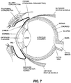
- the formulations of the invention may be administered, for example, to the superotemporal quadrant of the bulbar subconjunctival space, or to any one or more of the inferior, superior, nasal, or temporal quadrants of the bulbar, palpepral, or forniceal subconjunctival spaces, or to the superotemporal, superonasal, inferior temporal, or inferior nasal bulbar, palpepral, or forniceal subconjunctival spaces ( Figs. 6 and 7 ).
- administration into the "subconjunctival space" of the eye refers to administration into any of the bulbar, palpepral, and/or forniceal subconjunctival spaces in the eye in any one or more of the four quadrants of the eye (superior, nasal, temporal, and inferior), or any one or more of the possible sub-quadrants of the eye, including the supertemporal, superonasal, inferior temporal, or inferior nasal regions of the bulbar, palpepral, or forniceal subconjunctival spaces.
- administration of the formulations of the invention excludes their topical delivery (such as by eye drops) to the surface of the eye, or more specifically the cornea, since the subconjunctival space does not include any region or space immediately over, under, or within the cornea.
- administration to the subconjunctival space further excludes a delivery method that penetrates or perforates Tenon's capsule or the sclera, that penetrates or extends through the sclera into the vitreous body, or that is delivered beneath the sclera.
- the "subconjunctival space”, as used herein, is not equivalent to the "sub-tenon space.”
- Tenon's capsule is a thin, fibrous, somewhat elastic membrane, beneath the conjunctiva, that envelopes the eyeball from the edge of the cornea (limbus) to the optic nerve. It attaches loosely to the sclera. Thus, the sub-tenon space is between Tenon's capsule and the sclera.
- Sub-tenon administration of a composition to an eye generally involves elevating both the conjunctiva and Tenons'capsule using forceps and then introducing the composition into the space between the sclera and Tenon's capsule. A needle and syringe may be used to pierce Tenon's capsule and inject the composition into the sub-tenon space.
- the device for administering the formulations of the invention to the subconjunctival space of an eye is a syringe equipped with a needle or a cannula.
- the needle or cannula used to inject a ophthalmic composition into the subconjunctival space of an eye can be a 20 gauge or higher gauge needle or cannula.
- the needle or cannula may have a blunt or beveled end.
- the gauge of the needle or cannula used in the method is a 25 gauge or higher needle or cannula, or is specifically a 22, 23, 24, 25, 26, 27, 28, 29, 30, 31, 32, 33, or 34 gauge needle or cannula.
- a formulation of the invention is injected into the subconjunctival space of the eye (i.e., administered) according to the following procedure.
- the practitioner uses a thumb (or device such as forceps) to retract the upper or lower eye lid, depending on whether injection into the subconjunctival space is to take place in the superior (e.g., superotemporal) or inferior quadrants, respectively. See Figure 2 , for example.
- the practitioner positions the needle (attached to a syringe containing the composition) tangential to the eye globe with the needle bevel facing toward the globe (i.e., with the needle bevel facing down). In certain instances, it may be preferable to orient the needle with the bevel facing up (i.e., away from the globe). Keeping the needle tangential to the globe and using a gentle stabbing motion, the practitioner then directs the needle posteriorly and just beneath the conjunctiva to introduce the needle into the subconjunctival space. The desired volume of composition is then injected into the subconjunctival space. Depending on the volume injected, the composition may appear as an elevation or small bulge in the conjunctiva. The needle is then withdrawn. Pressure may then be applied to the eye with a gauze pad to help distribute and diffuse the composition and stem possible bleeding.
- the same procedure may be used to introduce anesthetic into the subconjunctival space of the eye prior to injecting the formulation of the invention.
- the anesthetic solution may appear as an elevation or bulge in the conjunctiva. If this procedure is followed, and once the anesthetic has taken effect, the composition is then injected into the elevated portion of the conjunctiva but at a site other than that used to inject the anesthetic.
- an additional dose of formulation of the invention may be injected into the nasal and/or inferior (e.g., the inferior forniceal) subconjunctival space using the same procedure outlined above.
- an ocular anesthetic may be applied topically to one or each eye prior to injection of the composition into the subconjunctival space of the eye.
- Analgesic agents may also be administered following subconjunctival administration of the composition.
- Analgesic agents include triple antibiotic ointment or atropine drops or ointment.
- biodegradable implants containing cyclosporin A when delivered through a 22 gauge needle, do not deliver sufficient drug over a sufficient period of time to effectively treat those conditions of the eye for which topical cyclosporin A is prescribed or known to be effective in treating.
- the inventors prepared the following formulations, and injected them into rabbit eyes.
- Emb. 1 A formulation comprising cyclosporin A form 2 and a hydrogel.
- Emb. 2 The formulation of emb. 1, wherein the hydrogel is selected from hyaluronic acid, hydroxypropyl cellulose, hydroxypropylmethyl cellulose, hydroxyethylcellulose, polyvinyl pyrolidone, carboxymethylcellulose hydroxyethyl cellulose, and polyvinylpyrrolidone.
- Emb. 3 The formulation of emb. 2, wherein the cyclosporin A form 2 is at a concentration of about 0.01% to about 10%.
- Emb. 4 A method of treating a condition selected from dry eye, blepharitis, meibomian gland disease, impaired corneal sensitivity, allergic conjunctivitis, atopic keratoconjunctivitis, vernal keratoconjunctivitis, and ptyregia, the method comprising the step of administering to a patient having such a condition the formulation of any of the preceding claims.
- Emb. 5 The method of emb. 4, wherein the formulation is administered to the subconjunctival space.
- Emb. 6 The method of emb. 5, wherein the formulation is administered via a needle of a gauge no larger than 22.
Landscapes
- Health & Medical Sciences (AREA)
- Chemical & Material Sciences (AREA)
- Life Sciences & Earth Sciences (AREA)
- Medicinal Chemistry (AREA)
- General Health & Medical Sciences (AREA)
- Animal Behavior & Ethology (AREA)
- Pharmacology & Pharmacy (AREA)
- Public Health (AREA)
- Veterinary Medicine (AREA)
- Epidemiology (AREA)
- Inorganic Chemistry (AREA)
- Organic Chemistry (AREA)
- Proteomics, Peptides & Aminoacids (AREA)
- Bioinformatics & Cheminformatics (AREA)
- Engineering & Computer Science (AREA)
- Ophthalmology & Optometry (AREA)
- Gastroenterology & Hepatology (AREA)
- Immunology (AREA)
- Biochemistry (AREA)
- Genetics & Genomics (AREA)
- Biophysics (AREA)
- Molecular Biology (AREA)
- Dermatology (AREA)
- Nuclear Medicine, Radiotherapy & Molecular Imaging (AREA)
- Chemical Kinetics & Catalysis (AREA)
- General Chemical & Material Sciences (AREA)
- Crystallography & Structural Chemistry (AREA)
- Analytical Chemistry (AREA)
- Dispersion Chemistry (AREA)
- Medicines That Contain Protein Lipid Enzymes And Other Medicines (AREA)
- Medicinal Preparation (AREA)
- Pharmaceuticals Containing Other Organic And Inorganic Compounds (AREA)
Abstract
Description
- This patent application claims priority to
U.S. Provisional Patent Application No. 61/559,838, filed November 15, 2011 U.S. Provisional Patent Application No. 61/563,199, filed November 23, 2011 - Cyclosporin A is the active Ingredient in Restasis®, a topical ocular formulation approved in the United States for the treatment of dry eye. Topical cyclosporin is also used in the management of other ophthalmic conditions such as restoration of corneal sensitivity following refractive surgery, conjunctival and corneal inflammation, keratoconjuntivitis, graft versus host disease, post-transplant glaucoma, corneal transplants, mycotic keratitis, meibomian gland dysfunction, Thygeson's superficial punctate keratitis, uveitis, and Theodore's superior limbic keratoconjunctivitis.
- Patient compliance is extremely important but many patients fail to comply with ocular medications. Moreover, some treatments for conditions other than dry eye dose more frequently than the twice-a-day dosing approved for Restasis, and concentrations of cyclosporin A higher in tissue than Restasis can deliver after multiple dosing would be beneficial to certain patients with severe conditions, such as graft vs. host disease.
Some ocular diseases may require long-term therapy. For example, the National Eye Institute (NEI) has completed a study to evaluate 1 and 2 mg biodegradable implants of cyclosporin to treat uveitis. The NEI is also studying the use of non-degradable cyclosporin implants for treatment of graft vs. host disease. However, it should be noted that removing non-degradable implants after the dose has been depleted requires a second surgery. Some other relevant studies also have been conducted by the NEI or are in the process of recruiting patients for studies. Results for these NEI studies have not been published. - A single implantation or injection, which delivers high levels of cyclosporin constantly over a long period (weeks or months) is desirable. The use of multi-month biodegradable or bioerodible delivery system, which delivers cyclosporin to targeted anterior ocular tissues - at equal or greater concentration as Restasis - is clearly desirable. It is also desirable to have an injectable formulation, which would avoid surgery. A formulation which could pass through a 22 gauge needle (or smaller) and avoid the ocular tissue damage which may occur with trocars, larger needles, or surgical implantation, is also desirable.
- The reported success of injecting cyclosporin formulations into the conjunctiva for
long-duration therapy has been highly limited: while short-term efficacy has been shown with some microspheres and other formulations, no formulation has been demonstrated to be equivalent or superior in delivering cyclosporin to anterior tissue,
when compared to Restasis for a period of at least 3 months. - Biodegradable implants, which can be delivered through a 22 gauge needle, do not deliver sufficient drug over a sufficient period of time to meet the need. Biodegradable gel-forming suspensions perform better but, still do not meet the need.
- The inventors have surprisingly found that the formulations of the invention, when delivered to the subconjunctival space of the eye through a 22 or smaller gauge needle, deliver cyclosporin A to most anterior ocular tissue for a sustained period of time.
-
-
Figure 1 depicts characteristic X-ray powder diffraction (XRPD) patterns of CsA in a new crystalline form (designated asForm 2 herein), tetragonal form (designated asForm 1 herein), and orthorhombic form (designated asForm 3 herein). -
Figure 2 depicts the XRPD diffractogram of CsAcrystalline Form 2. -
Figure 3 depicts the water sorption/desorption profile ofCsA Form 2. -
Figure 4 depicts MDSC analysis ofCsA Form 2 recovered from 0.04% formulation with 1 % PS80. -
Figure 5 shows the front of the eye and the different quadrants of the conjunctiva relative to the limbus and cornea. -
Figure 6 illustrates injection of a steroid compound into the superotemporal quadrant of the subconjunctival space of the eye. The patient looks down while the thumb of one hand is used to gently retract the upper lid. The syringe containing the steroid-containing composition is placed tangential to the globe and inserted through the bulbar conjunctiva thereby introducing the needle into the subconjunctival space. -
Figure 7 shows a cross section of the eye and the location of the three zones of the conjunctiva (heavy black line)-palpebral, forniceal, and bulbar-relative to other anatomical regions in the eye. -
Figure 8 shows the simulated XRPD pattern of cyclosporine A forms. - Cyclosporin A (CsA) is a cyclic peptide having the following chemical structure:

- Cyclosporin A is known to exist in an amorphous form, liquid crystal form, tetragonal crystalline form (form 1), and an orthorhombic form (form 3). A new crystalline form,
cyclosporin A form 2, has recently been discovered. - The XRPD pattern of
CsA Form 2 differs significantly from the tetragonal form and orthorhombic form (FIG. 1 ). The major crystalline peaks forCsA form 2 appear at (2θ) when scanned by an X-ray diffractometer with X-ray source as Cu Kα radiation, λ = 1.54 Å, at 30 kV /15 mA: 7.5, 8.8, 10.2, 11.3, 12.7, 13.8, 14.5, 15.6 and 17.5 (d-spacing in crystal lattice at about 11.8, 10.0, 8.7, 7.8, 7.0, 6.4, 6.1, 5.6 and 5.1Ǻ, respectively,Fig. 2 ). These major peaks are defined as those being unique toForm 2 relative to the orthorhombic or tetragonal forms; as well as, peaks having an intensity greater than 5 times the background. - In one embodiment, the new crystalline form (Form 2) of CsA is a nonstoichiometric hydrate of Cyclosporin A. In another embodiment, the
crystalline Form 2 is represented by the formula:
-
Form 2 appears to be a kinetically stable form of CsA in aqueous suspensions.Suspensions containing Form 2 show no conversion to other known polymorphic or pseudomorphic forms upon storage. It has been found thatForm 1 and the amorphous form convert toForm 2 in the presence of water. - The single crystal structure of the hydrate form of
CsA Form 2 has been determined and the crystal structure parameters are listed in Table 2. These results indicate thatForm 2 is unique compared to other known crystalline forms of cyclosporine A.Table 1: Crystal data and data collection parameters of crystal structure solution of CsA Form 2.formula CszH215N11O14 formula weight 1238.67 space group P 21 21 21 (No. 18) a (Å) 12.6390(5) b (Å) 19.7582(8) c (Å) 29.568(2) volume (Å3) 7383.8(7) Z 4 dcalc (g·cm-3) 1.114 crystal dimensions (mm) 0.27 x 0.18 x 0.12 temperature (K) 150 radiation (wavelength in Å) Cu K2 (1.54184) monochromator confocal optics linear abs coef (mm-1) 0.640 absorption correction applied empirical2 transmission factors (min, max) 0.80, 0.93 diffractometer Rigaku RAPID-II h, k, l range -13 to 13 -21 to 21 -32 to 21 28 range (deg) 5.38-115.00 mosaicity (deg) 1.31 programs used SHELXTL F000 2704.0 weighting 1/[o2(Fo2)+(0.0845P)2+0.0000P] where P=(Fo2+2Fc2)/3 data collected 37360 unique data 8964 Rint 0.077 data used in refinement 9984 cutoff used in R-factor calculations Fo3>2.0s(Fo2) data with 1>2.0s(1) 6597 number of variables 834 largest shift/esd in final cycle 0.00 R(F0) 0.061 R0(F0 2) 0.0145 goodness of fit 1.037 absolute structure determination Flack parameter2 (0.0(3)) - The asymmetric unit of this
CsA Form 2 contains one cyclosporine A molecule and two water molecules. It is possible that any small molecule that can hydrogen bond to water could play the role of space filler, which would give a range of potential structures running from the orthorhombic dihydrate to distorted monoclinic dihydrate The XRPD pattern calculated from the single-crystal structure is shown inFigure 8 and it matches the experimental pattern shown inFigure 2 . These matching patterns further corroborate thatForm 2 is a unique and pure crystalline form of cyclosporine A. - Without wishing to be bound by theory, thermogravimetric analysis combined with KF titration and vapor sorption desorption analysis (VSA) suggest that
CsA Form 2 is a non-stoichiometric hydrate of CsA. The vapor sorption analysis of CyclosporineForm 2 indicates that water content in the new crystal form reversibly varies with relative humidity as shown inFig. 3 . Similar to the tetragonal form, the new CsA form undergoes a phase transition to a liquid crystal or amorphous form at 124.4 °C prior to melting as indicated by the modulated differential calorimetric (MDSC) analysis (Figure 4 ). - Cyclosporin A
Form 2 may be obtained by suspending amorphous 0.05 % cyclosporin A (w/v) in 1%Polysorbate 80, heating the solution to 65 °C, holding it at that temperature for 24 hours, and then recovering the precipitate by vacuum filtration. One can then use thecyclosporin A Form 2 thus obtained to generate additional amounts, using Cyclosporin AForm 2 as a seed crystal; in this method, one suspends about 30 g cyclosporin A in a solution of 900 ml water containing 1% (w/v)Polysorbate 80, heats the solution to 65 °C, and then seeds it with 0.2 g ofcyclosporin A Form 2 at a temperature of 52 °C. The solution is then stirred for about 22 hours at a temperature of between about 61 °C and 65 °C, and then recovers the precipitate that results. - Further details regarding
CsA Form 2 may be found inU.S. Patent Application No. 13/480,710 , the entire contents of which are incorporated by reference herein. - Compositions of the invention are ophthalmically acceptable formulations of Cyclosporin
A form 2. By "ophthalmically acceptable," the inventors mean that the suspensions are formulated in such a way as to be non-irritating when administered to the eye of a mammal, such as a human. - The formulations of the invention comprise
cyclosporin A form 2 and a hydrogel such as hyaluronic acid, hydroxypropyl cellulose, hydroxypropylmethyl cellulose, hydroxyethylcellulose, polyvinyl pyrolidone, and carboxymethylcellulose. - In one embodiment, the formulation comprises
cyclosporin A form 2 at a concentration of about 0.001% to about 10% (w/v). In one embodiment, the suspension comprisescyclosporin A form 2 at a concentration of about 0.001% (w/v) to about 0.01%, about 0.001% (w/v) to about 0.04% (w/v), about 0.001% (w/v) to about 0.03% (w/v), about 0.001% (w/v) to about 0.02% (w/v), or about 0.001% (w/v) to about 0.01 % (w/v). In another embodiment, the suspension comprisescyclosporin A form 2 at a concentration of about 0.01% (w/v) to about 0.05%, about 0.01% (w/v) to about 0.04% (w/v), about 0.01% (w/v) to about 0.03% (w/v), about 0.01% (w/v) to about 0.02% (w/v), or about 0.01% (w/v) to about 0.01% (w/v). In another embodiment, the suspension comprisescyclosporin A form 2 at a concentration of about 0.01% (w/v) to about 0.1%, about 0.1% (w/v) to about 0.5% (w/v), about 0.01% (w/v) to about 1% (w/v), or about 1% (w/v) to about 10%. - For example, the formulations may comprise about 0.001% (w/v), about 0.002% (w/v), about 0.003% (w/v), about 0.004% (w/v), about 0.005% (w/v), about 0.006% (w/v), about 0.007% (w/v), about 0.008% (w/v), about 0.009% (w/v), about 0.01% (w/v), about 0.015% (w/v), about 0.02% (w/v), about 0.025% (w/v), about 0.03% (w/v), about 0.035% (w/v), about 0.04% (w/v), about 0.045% (w/v), about 0.05% (w/v), about 0.055% (w/v), about 0.06% (w/v), about 0.065% (w/v), about 0.07% (w/v), about 0.075% (w/v), about 0.08% (w/v), about 0.085% (w/v), about 0.09% (w/v), about 0.095% (w/v), about 0.1% (w/v), about 0.15% (w/v), about 0.2% (w/v), about 0.25% (w/v), about 0.3% (w/v), about 0.35% (w/v), about 0.4% (w/v), about 0.45% (w/v), about 0.5% (w/v), about 0.55% (w/v), about 0.6% (w/v), about 0.65% (w/v), about 0.7% (w/v), about 0.75% (w/v), about 0.8% (w/v), about 0.85% (w/v), about 0.9% (w/v), about 0.95% (w/v), or about 1.0% (w/v) cyclosporin A form 2.
- Formulations of the invention may be used to treat any condition of the eye which is known to be amenable to topical treatment with cyclosporin A (such as with Restasis®) at the concentrations stated here. For example, compositions of the invention may be used to treat patients suffering from dry eye, to treat blepharitis and meibomian gland disease, to restore corneal sensitivity that has been impaired due to refractive surgery on the eye, to treat allergic conjunctivitis and atopic and vernal keratoconjunctivitis, and to treat ptyregia, conjunctival and corneal inflammation, keratoconjuntivitis, graft versus host disease, post-transplant glaucoma, corneal transplants, mycotic keratitis, Thygeson's superficial punctate keratitis, uveitis, and Theodore's superior limbic keratoconjunctivitis, among other conditions.
- The International Dry Eye Workshop (DEWS) defines dry eye as "a multifactorial disease of the tears and ocular surface that results in symptoms of discomfort, visual disturbance, and tear film instability with potential damage to the ocular surface, accompanied by increased osmolarity of the tear film and inflammation of the ocular surface." It includes those conditions, such as keratoconjunctivitis sicca, that are caused by tear deficiency or excessive evaporation of tears.
- Blepharitis is a chronic disorder producing inflammation of the anterior and posterior lid margin, with involvement of skin and its related structures (hairs and sebaceous glands), the mucocutaneous junction, and the meibomian glands. It can also affect the conjunctiva, tear film, and the corneal surface in advanced stages and may be associated with dry eye. Blepharitis is commonly classified into anterior or posterior blepharitis, with anterior affecting the lash bearing region of the lids, and posterior primarily affecting the meibomian gland orifices.
- Meibomian gland disease most often occurs as one of three forms: primary meibomitis, secondary meibomitis, and meibomian seborrhea. Meibomian seborrhea is characterized by excessive meibomian secretion in the absence of inflammation (hypersecretory meibomian gland disease). Primary meibomitis, by contrast, is distinguished by stagnant and inspissated meibomian secretions (obstructive hypersecretory meibomian gland disease). Secondary meibomitis represents a localized inflammatory response in which the meibomian glands are secondarily inflamed in a spotty fashion from an anterior lid margin blepharitis.
- Impaired corneal sensitivity often occurs after refractive surgery, such as photorefractive keratectomy, laser assisted sub-epithelium keratomileusis (LASEK), EPI-LASEK, customized transepithelial non-contact ablation, or other procedures in which the corneal nerves are severed. Impaired corneal sensitivity may also occur after viral infection, such as by HSV-1, HSV-2, and VZV viruses. Patients with impaired corneal sensitivity often complain that their eyes feel dry, even though tear production and evaporation may be normal, suggesting that "dryness" in such patients is actually a form of corneal neuropathy that results when corneal nerves are severed by surgery or inflamed after viral infection.
- Allergic conjunctivitis is an inflammation of the conjunctiva resulting from hypersensitivity to one or more allergens. It may be acute, intermittent, or chronic. It occurs seasonally, that is, at only certain time of the year, or it occurs perennially, that is, chronically throughout the year. Symptoms of seasonal and perennial allergic conjunctivitis include, in addition to inflammation of the conjunctiva, lacrimation, tearing, conjunctival vascular dilation, itching, papillary hyperlasia, chemosis, eyelid edema, and discharge from the eye. The discharge may form a crust over the eyes after a night's sleep.
- Atopic keratoconjunctivitis is a chronic, severe form of allergic conjunctivitis that often leads to visual impairment. Symptoms include itching, burning, pain, redness, foreign body sensation, light sensitivity and blurry vision. There is often a discharge, especially on awakening from a night's sleep; the discharge may be stringy, ropy, and mucoid. The lower conjunctiva is often more prominently affected than the upper conjunctiva. The conjunctiva may range from pale, edematous, and featureless to having the characteristics of advanced disease, including papillary hypertrophy, subepithelial fibrosis, formix foreshortening, trichiasis, entropion, and madurosis. In some patients the disease progresses to punctate epithelial erosions, corneal neovascularization, and other features of keratopathy which may impair vision. There is typically goblet cell proliferation in the conjunctiva, epithelial pseudotubular formation, and an increased number of degranulating eosinophils and mast cells in the epithelium. CD25+T lymphocytes, macrophages, and dendritic cells (HLA-DR.sup.+, HLA-CD1+) are significantly elevated in the substantia propria.
- Like atopic keratoconjunctivitis, vernal keratoconjunctivitis is a severe form of allergic conjunctivitis, but it tends to affect the upper conjunctiva more prominently than the lower. It occurs in two forms. In the palpebral form, square, hard, flattened, closely packed papillae are present; in the bulbar (limbal) form, the circumcorneal conjunctiva becomes hypertrophied and grayish. Both forms are often accompanied by a mucoid discharge. Corneal epithelium loss may occur, accompanied by pain and photophobia, as may central corneal plaques and Trantas' dots.
- Formulations of the invention are administered to the subconjunctival space. As used herein, the "subconjunctival space" refers to any of the following: (1) the potential space between the bulbar conjunctiva and Tenon's capsule and extending from the limbus to the fornix; (2) the potential space between the palpebral conjunctiva and the tarsus and extending from the eye lid margin (mucocutaneous junction of the eyelid) to the fornix; and (3) the potential space just beneath the forniceal conjunctiva at the junctional bay or fornix. The subconjunctival space is therefore the potential space just beneath the conjunctiva from the limbus, around the fornix, to the eye lid margin.
- Referring to
Figure 5 , the subconjunctival space around the eye can be divided into four quadrants: the superior, nasal, inferior, and temporal. These quadrants may be further subdivided into sub-quadrants, such as the superotemporal, superonasal, inferior nasal, and inferior temporal, and so on. Hence, the formulations of the invention may be administered, for example, to the superotemporal quadrant of the bulbar subconjunctival space, or to any one or more of the inferior, superior, nasal, or temporal quadrants of the bulbar, palpepral, or forniceal subconjunctival spaces, or to the superotemporal, superonasal, inferior temporal, or inferior nasal bulbar, palpepral, or forniceal subconjunctival spaces (Figs. 6 and7 ). Therefore, unless further delimited, administration into the "subconjunctival space" of the eye refers to administration into any of the bulbar, palpepral, and/or forniceal subconjunctival spaces in the eye in any one or more of the four quadrants of the eye (superior, nasal, temporal, and inferior), or any one or more of the possible sub-quadrants of the eye, including the supertemporal, superonasal, inferior temporal, or inferior nasal regions of the bulbar, palpepral, or forniceal subconjunctival spaces. - In one embodiment, administration of the formulations of the invention excludes their topical delivery (such as by eye drops) to the surface of the eye, or more specifically the cornea, since the subconjunctival space does not include any region or space immediately over, under, or within the cornea. In another embodiment, administration to the subconjunctival space further excludes a delivery method that penetrates or perforates Tenon's capsule or the sclera, that penetrates or extends through the sclera into the vitreous body, or that is delivered beneath the sclera. The "subconjunctival space", as used herein, is not equivalent to the "sub-tenon space." Tenon's capsule is a thin, fibrous, somewhat elastic membrane, beneath the conjunctiva, that envelopes the eyeball from the edge of the cornea (limbus) to the optic nerve. It attaches loosely to the sclera. Thus, the sub-tenon space is between Tenon's capsule and the sclera. Sub-tenon administration of a composition to an eye generally involves elevating both the conjunctiva and Tenons'capsule using forceps and then introducing the composition into the space between the sclera and Tenon's capsule. A needle and syringe may be used to pierce Tenon's capsule and inject the composition into the sub-tenon space.
- In one embodiment, the device for administering the formulations of the invention to the subconjunctival space of an eye is a syringe equipped with a needle or a cannula. The needle or cannula used to inject a ophthalmic composition into the subconjunctival space of an eye can be a 20 gauge or higher gauge needle or cannula. The needle or cannula may have a blunt or beveled end. In preferred embodiments, the gauge of the needle or cannula used in the method is a 25 gauge or higher needle or cannula, or is specifically a 22, 23, 24, 25, 26, 27, 28, 29, 30, 31, 32, 33, or 34 gauge needle or cannula.
- In one embodiment of the present method for treating an ocular surface inflammation or inflammation of the ocular surface adnexa in a subject in need thereof, a formulation of the invention is injected into the subconjunctival space of the eye (i.e., administered) according to the following procedure. With the subject facing the practitioner, the practitioner uses a thumb (or device such as forceps) to retract the upper or lower eye lid, depending on whether injection into the subconjunctival space is to take place in the superior (e.g., superotemporal) or inferior quadrants, respectively. See
Figure 2 , for example. With the other hand, the practitioner positions the needle (attached to a syringe containing the composition) tangential to the eye globe with the needle bevel facing toward the globe (i.e., with the needle bevel facing down). In certain instances, it may be preferable to orient the needle with the bevel facing up (i.e., away from the globe). Keeping the needle tangential to the globe and using a gentle stabbing motion, the practitioner then directs the needle posteriorly and just beneath the conjunctiva to introduce the needle into the subconjunctival space. The desired volume of composition is then injected into the subconjunctival space. Depending on the volume injected, the composition may appear as an elevation or small bulge in the conjunctiva. The needle is then withdrawn. Pressure may then be applied to the eye with a gauze pad to help distribute and diffuse the composition and stem possible bleeding. - In certain embodiments, the same procedure may be used to introduce anesthetic into the subconjunctival space of the eye prior to injecting the formulation of the invention. The anesthetic solution may appear as an elevation or bulge in the conjunctiva. If this procedure is followed, and once the anesthetic has taken effect, the composition is then injected into the elevated portion of the conjunctiva but at a site other than that used to inject the anesthetic. In some embodiments, an additional dose of formulation of the invention may be injected into the nasal and/or inferior (e.g., the inferior forniceal) subconjunctival space using the same procedure outlined above.
- Alternatively, an ocular anesthetic may be applied topically to one or each eye prior to injection of the composition into the subconjunctival space of the eye. Analgesic agents may also be administered following subconjunctival administration of the composition. Analgesic agents include triple antibiotic ointment or atropine drops or ointment.
- The invention is further illustrated by the following examples.
- The inventors' work suggests that biodegradable implants containing cyclosporin A, when delivered through a 22 gauge needle, do not deliver sufficient drug over a sufficient period of time to effectively treat those conditions of the eye for which topical cyclosporin A is prescribed or known to be effective in treating.
- The inventors prepared the following formulations, and injected them into rabbit eyes.

- Emb. 1: A formulation comprising
cyclosporin A form 2 and a hydrogel. - Emb. 2: The formulation of emb. 1, wherein the hydrogel is selected from hyaluronic acid, hydroxypropyl cellulose, hydroxypropylmethyl cellulose, hydroxyethylcellulose, polyvinyl pyrolidone, carboxymethylcellulose hydroxyethyl cellulose, and polyvinylpyrrolidone.
- Emb. 3: The formulation of emb. 2, wherein the
cyclosporin A form 2 is at a concentration of about 0.01% to about 10%. - Emb. 4: A method of treating a condition selected from dry eye, blepharitis, meibomian gland disease, impaired corneal sensitivity, allergic conjunctivitis, atopic keratoconjunctivitis, vernal keratoconjunctivitis, and ptyregia, the method comprising the step of administering to a patient having such a condition the formulation of any of the preceding claims.
- Emb. 5: The method of emb. 4, wherein the formulation is administered to the subconjunctival space.
- Emb. 6: The method of emb. 5, wherein the formulation is administered via a needle of a gauge no larger than 22.
Claims (7)
- A formulation comprising cyclosporin A form 2 and a hydrogel, wherein the hydrogel is selected from hyaluronic acid, hydroxypropyl cellulose, hydroxypropylmethyl cellulose, hydroxyethylcellulose, polyvinyl pyrolidone, carboxymethylcellulose hydroxyethyl cellulose, and polyvinylpyrrolidone, wherein the cyclosporin A form 2 is at a concentration of about 0.01% to about 10%.
- The formulation according to claim 1, wherein the cyclosporin A form 2 is at a concentration of about 5% (w/v) or about 10% (w/v).
- The formulation according to claim 2, wherein the cyclosporin A form 2 is at a concentration of about 5% (w/v).
- The formulation according to any of the preceding claims, wherein the formulation is free of other crystalline forms of cyclosporin A.
- The formulation according to any of the previous claims for use in a method of treating a condition selected from dry eye, blepharitis, meibomian gland disease, impaired corneal sensitivity, allergic conjunctivitis, atopic keratoconjunctivitis, vernal keratoconjunctivitis, and ptyregia, the method comprising the step of administering the formulation to a patient having such a condition.
- The formulation for use according to claim 5, wherein the formulation is administered to the subconjunctival space.
- The formulation for use according to claim 6, wherein the formulation is administered via a needle of a gauge no larger than 22.
Applications Claiming Priority (4)
| Application Number | Priority Date | Filing Date | Title |
|---|---|---|---|
| US201161559838P | 2011-11-15 | 2011-11-15 | |
| US201161563199P | 2011-11-23 | 2011-11-23 | |
| PCT/US2012/064988 WO2013074610A1 (en) | 2011-11-15 | 2012-11-14 | Sustained action formulation of cyclosporin form 2 |
| EP12788092.0A EP2779992B1 (en) | 2011-11-15 | 2012-11-14 | Sustained action formulation of cyclosporin form 2 |
Related Parent Applications (1)
| Application Number | Title | Priority Date | Filing Date |
|---|---|---|---|
| EP12788092.0A Division EP2779992B1 (en) | 2011-11-15 | 2012-11-14 | Sustained action formulation of cyclosporin form 2 |
Publications (1)
| Publication Number | Publication Date |
|---|---|
| EP3381443A1 true EP3381443A1 (en) | 2018-10-03 |
Family
ID=47192236
Family Applications (2)
| Application Number | Title | Priority Date | Filing Date |
|---|---|---|---|
| EP18158837.7A Withdrawn EP3381443A1 (en) | 2011-11-15 | 2012-11-14 | Sustained action formulation of cyclosporin form 2 |
| EP12788092.0A Not-in-force EP2779992B1 (en) | 2011-11-15 | 2012-11-14 | Sustained action formulation of cyclosporin form 2 |
Family Applications After (1)
| Application Number | Title | Priority Date | Filing Date |
|---|---|---|---|
| EP12788092.0A Not-in-force EP2779992B1 (en) | 2011-11-15 | 2012-11-14 | Sustained action formulation of cyclosporin form 2 |
Country Status (10)
| Country | Link |
|---|---|
| US (3) | US8785394B2 (en) |
| EP (2) | EP3381443A1 (en) |
| JP (2) | JP6231486B2 (en) |
| KR (1) | KR102029513B1 (en) |
| CN (2) | CN104039309B (en) |
| AU (3) | AU2012339695B2 (en) |
| BR (1) | BR112014011744A2 (en) |
| CA (1) | CA2856044C (en) |
| RU (1) | RU2639393C2 (en) |
| WO (1) | WO2013074610A1 (en) |
Families Citing this family (9)
| Publication number | Priority date | Publication date | Assignee | Title |
|---|---|---|---|---|
| CN103251375B (en) | 2009-04-01 | 2016-09-28 | 眼泪科学公司 | For imaging and the instrument of the measurement one or more thickness of tear film layer |
| JP6363506B2 (en) * | 2011-11-15 | 2018-07-25 | アラーガン、インコーポレイテッドAllergan,Incorporated | Cyclosporine A2 suspension |
| WO2013181339A2 (en) | 2012-06-01 | 2013-12-05 | Allergan, Inc. | Cyclosporin a analogs |
| WO2014179795A2 (en) | 2013-05-03 | 2014-11-06 | Tearscience, Inc. | Eyelid illumination systems and methods for imaging meibomian glands for meibomian gland analysis |
| US20170112936A1 (en) * | 2014-05-23 | 2017-04-27 | Imprimis Pharmaceuticals, Inc. | Pharmaceutical compositions comprising gels and methods for fabricating thereof |
| RU2713953C2 (en) | 2014-07-18 | 2020-02-11 | Аллерган, Инк. | Suspension compositions of cyclosporine a for subconjunctival and periocular injection |
| TW201639873A (en) | 2015-01-08 | 2016-11-16 | 歐樂根公司 | Cyclosporin derivatives wherein the MEBMT sidechain has been cyclized |
| US9820954B2 (en) | 2015-08-19 | 2017-11-21 | Jenivision Inc. | Quantitative peri-orbital application of ophthalmology drugs |
| CN118159282A (en) * | 2021-10-26 | 2024-06-07 | 兆科(香港)眼科药物有限公司 | Methods of using cyclosporine A eye gel to treat moderate to severe dry eyes |
Citations (3)
| Publication number | Priority date | Publication date | Assignee | Title |
|---|---|---|---|---|
| WO2005072701A1 (en) * | 2004-01-20 | 2005-08-11 | Allergan, Inc. | Compositions for localized therapy of the eye, comprising preferably triamcinolone acetonide and hyaluronic acid |
| WO2011053841A1 (en) * | 2009-10-30 | 2011-05-05 | Aton Pharma, Inc. | Ocular drug delivery devices |
| WO2012166610A1 (en) * | 2011-05-27 | 2012-12-06 | Allergan, Inc. | A crystalline form of cyclosporine a, methods of preparation, and methods for use thereof |
Family Cites Families (9)
| Publication number | Priority date | Publication date | Assignee | Title |
|---|---|---|---|---|
| WO1989001772A1 (en) | 1987-09-03 | 1989-03-09 | University Of Georgia Research Foundation, Inc. | Ocular cyclosporin composition |
| HU203564B (en) * | 1987-12-21 | 1991-08-28 | Sandoz Ag | Process for producing new orthorombos cyclosporin without solvatation |
| US5294604A (en) * | 1989-12-20 | 1994-03-15 | The Government Of The United States Of America As Represented By The Secretary Of The Department Of Health And Human Services | Method of treating ocular diseases by periocular administration of cyclosporine A or G |
| US7732404B2 (en) | 1999-12-30 | 2010-06-08 | Dexcel Ltd | Pro-nanodispersion for the delivery of cyclosporin |
| PT1142566E (en) * | 2000-04-07 | 2004-02-27 | Medidom Lab | Topical Formulin Formulation in the Form of a Aqueous Solution and Use of a Cyclosporine in Association with Hyaluronic Acid or One of Its Salts and with Polysorbate 80 |
| US20050192264A1 (en) * | 2004-02-04 | 2005-09-01 | Penfold Philip L. | Slow release steroid composition |
| WO2006039558A2 (en) * | 2004-10-09 | 2006-04-13 | Formurex, Inc. | Ocular agent delivery systems |
| US10137083B2 (en) | 2006-03-07 | 2018-11-27 | SGN Nanopharma Inc | Ophthalmic preparations |
| US20080044473A1 (en) * | 2006-08-17 | 2008-02-21 | Milan Radojicic | Methods of reducing nervous system glial scar formation using cyclosporine components |
-
2012
- 2012-11-14 KR KR1020147016154A patent/KR102029513B1/en not_active Expired - Fee Related
- 2012-11-14 CN CN201280066567.8A patent/CN104039309B/en not_active Expired - Fee Related
- 2012-11-14 WO PCT/US2012/064988 patent/WO2013074610A1/en not_active Ceased
- 2012-11-14 CA CA2856044A patent/CA2856044C/en active Active
- 2012-11-14 JP JP2014542397A patent/JP6231486B2/en not_active Expired - Fee Related
- 2012-11-14 BR BR112014011744A patent/BR112014011744A2/en not_active Application Discontinuation
- 2012-11-14 EP EP18158837.7A patent/EP3381443A1/en not_active Withdrawn
- 2012-11-14 AU AU2012339695A patent/AU2012339695B2/en not_active Ceased
- 2012-11-14 CN CN201810127698.1A patent/CN108175742B/en not_active Expired - Fee Related
- 2012-11-14 EP EP12788092.0A patent/EP2779992B1/en not_active Not-in-force
- 2012-11-14 US US13/676,551 patent/US8785394B2/en active Active
- 2012-11-14 RU RU2014123469A patent/RU2639393C2/en not_active Application Discontinuation
-
2014
- 2014-06-16 US US14/305,883 patent/US20140296158A1/en not_active Abandoned
-
2017
- 2017-10-19 JP JP2017202536A patent/JP6559753B2/en not_active Expired - Fee Related
- 2017-12-12 AU AU2017276175A patent/AU2017276175A1/en not_active Abandoned
-
2019
- 2019-07-02 AU AU2019204743A patent/AU2019204743B2/en not_active Ceased
-
2020
- 2020-10-05 US US17/062,972 patent/US20210236590A1/en not_active Abandoned
Patent Citations (3)
| Publication number | Priority date | Publication date | Assignee | Title |
|---|---|---|---|---|
| WO2005072701A1 (en) * | 2004-01-20 | 2005-08-11 | Allergan, Inc. | Compositions for localized therapy of the eye, comprising preferably triamcinolone acetonide and hyaluronic acid |
| WO2011053841A1 (en) * | 2009-10-30 | 2011-05-05 | Aton Pharma, Inc. | Ocular drug delivery devices |
| WO2012166610A1 (en) * | 2011-05-27 | 2012-12-06 | Allergan, Inc. | A crystalline form of cyclosporine a, methods of preparation, and methods for use thereof |
Also Published As
| Publication number | Publication date |
|---|---|
| WO2013074610A1 (en) | 2013-05-23 |
| US20130123195A1 (en) | 2013-05-16 |
| AU2019204743B2 (en) | 2021-02-11 |
| US20140296158A1 (en) | 2014-10-02 |
| CN104039309A (en) | 2014-09-10 |
| AU2012339695B2 (en) | 2017-10-05 |
| CN108175742A (en) | 2018-06-19 |
| BR112014011744A2 (en) | 2017-05-02 |
| JP6231486B2 (en) | 2017-11-15 |
| RU2014123469A (en) | 2015-12-27 |
| US20210236590A1 (en) | 2021-08-05 |
| KR102029513B1 (en) | 2019-10-07 |
| KR20140100966A (en) | 2014-08-18 |
| AU2019204743A1 (en) | 2019-07-18 |
| AU2012339695A1 (en) | 2014-06-05 |
| JP2014533301A (en) | 2014-12-11 |
| CN108175742B (en) | 2021-10-22 |
| CA2856044A1 (en) | 2013-05-23 |
| EP2779992A1 (en) | 2014-09-24 |
| RU2639393C2 (en) | 2017-12-21 |
| CN104039309B (en) | 2018-03-06 |
| JP2018048168A (en) | 2018-03-29 |
| JP6559753B2 (en) | 2019-08-14 |
| AU2017276175A1 (en) | 2018-01-18 |
| HK1202079A1 (en) | 2015-09-18 |
| CA2856044C (en) | 2016-11-29 |
| US8785394B2 (en) | 2014-07-22 |
| EP2779992B1 (en) | 2018-02-28 |
Similar Documents
| Publication | Publication Date | Title |
|---|---|---|
| AU2019204743B2 (en) | Sustained action formulation of cyclosporin Form 2 | |
| US20210269484A1 (en) | Crystalline form of cyclosporin a, methods of preparation, and methods for use thereof | |
| US10206971B2 (en) | Suspensions of cyclosporin A form 2 | |
| EP3403645A1 (en) | Autoclavable suspensions of cyclosporin a form 2 | |
| HK1202079B (en) | Sustained action formulation of cyclosporin form 2 | |
| HK1202066B (en) | Suspensions of cyclosporin a form 2 |
Legal Events
| Date | Code | Title | Description |
|---|---|---|---|
| PUAI | Public reference made under article 153(3) epc to a published international application that has entered the european phase |
Free format text: ORIGINAL CODE: 0009012 |
|
| STAA | Information on the status of an ep patent application or granted ep patent |
Free format text: STATUS: REQUEST FOR EXAMINATION WAS MADE |
|
| 17P | Request for examination filed |
Effective date: 20180227 |
|
| AC | Divisional application: reference to earlier application |
Ref document number: 2779992 Country of ref document: EP Kind code of ref document: P |
|
| AK | Designated contracting states |
Kind code of ref document: A1 Designated state(s): AL AT BE BG CH CY CZ DE DK EE ES FI FR GB GR HR HU IE IS IT LI LT LU LV MC MK MT NL NO PL PT RO RS SE SI SK SM TR |
|
| STAA | Information on the status of an ep patent application or granted ep patent |
Free format text: STATUS: EXAMINATION IS IN PROGRESS |
|
| 17Q | First examination report despatched |
Effective date: 20210910 |
|
| P01 | Opt-out of the competence of the unified patent court (upc) registered |
Effective date: 20230331 |
|
| REG | Reference to a national code |
Ref country code: DE Ref legal event code: R079 Free format text: PREVIOUS MAIN CLASS: A61K0009000000 Ipc: A61K0009100000 |
|
| GRAP | Despatch of communication of intention to grant a patent |
Free format text: ORIGINAL CODE: EPIDOSNIGR1 |
|
| STAA | Information on the status of an ep patent application or granted ep patent |
Free format text: STATUS: GRANT OF PATENT IS INTENDED |
|
| RIC1 | Information provided on ipc code assigned before grant |
Ipc: C07K 7/64 20060101ALI20241010BHEP Ipc: C07K 1/30 20060101ALI20241010BHEP Ipc: A61P 27/04 20060101ALI20241010BHEP Ipc: A61P 27/02 20060101ALI20241010BHEP Ipc: A61K 47/36 20060101ALI20241010BHEP Ipc: A61K 47/32 20060101ALI20241010BHEP Ipc: A61K 47/38 20060101ALI20241010BHEP Ipc: A61K 38/13 20060101ALI20241010BHEP Ipc: A61K 9/10 20060101AFI20241010BHEP |
|
| INTG | Intention to grant announced |
Effective date: 20241021 |
|
| STAA | Information on the status of an ep patent application or granted ep patent |
Free format text: STATUS: THE APPLICATION IS DEEMED TO BE WITHDRAWN |
|
| 18D | Application deemed to be withdrawn |
Effective date: 20250222 |