EP3369809A1 - Method for obtaining leydig cells by carrying out induced differentiation on human induced pluripotent stem cells, and use of leydig cells - Google Patents
Method for obtaining leydig cells by carrying out induced differentiation on human induced pluripotent stem cells, and use of leydig cells Download PDFInfo
- Publication number
- EP3369809A1 EP3369809A1 EP16868053.6A EP16868053A EP3369809A1 EP 3369809 A1 EP3369809 A1 EP 3369809A1 EP 16868053 A EP16868053 A EP 16868053A EP 3369809 A1 EP3369809 A1 EP 3369809A1
- Authority
- EP
- European Patent Office
- Prior art keywords
- cells
- culture medium
- hncscs
- differentiation
- volume percent
- Prior art date
- Legal status (The legal status is an assumption and is not a legal conclusion. Google has not performed a legal analysis and makes no representation as to the accuracy of the status listed.)
- Granted
Links
Images



Classifications
-
- A—HUMAN NECESSITIES
- A61—MEDICAL OR VETERINARY SCIENCE; HYGIENE
- A61K—PREPARATIONS FOR MEDICAL, DENTAL OR TOILETRY PURPOSES
- A61K35/00—Medicinal preparations containing materials or reaction products thereof with undetermined constitution
- A61K35/12—Materials from mammals; Compositions comprising non-specified tissues or cells; Compositions comprising non-embryonic stem cells; Genetically modified cells
- A61K35/48—Reproductive organs
- A61K35/52—Sperm; Prostate; Seminal fluid; Leydig cells of testes
-
- A—HUMAN NECESSITIES
- A61—MEDICAL OR VETERINARY SCIENCE; HYGIENE
- A61K—PREPARATIONS FOR MEDICAL, DENTAL OR TOILETRY PURPOSES
- A61K45/00—Medicinal preparations containing active ingredients not provided for in groups A61K31/00 - A61K41/00
-
- A—HUMAN NECESSITIES
- A61—MEDICAL OR VETERINARY SCIENCE; HYGIENE
- A61P—SPECIFIC THERAPEUTIC ACTIVITY OF CHEMICAL COMPOUNDS OR MEDICINAL PREPARATIONS
- A61P5/00—Drugs for disorders of the endocrine system
- A61P5/24—Drugs for disorders of the endocrine system of the sex hormones
- A61P5/26—Androgens
-
- C—CHEMISTRY; METALLURGY
- C12—BIOCHEMISTRY; BEER; SPIRITS; WINE; VINEGAR; MICROBIOLOGY; ENZYMOLOGY; MUTATION OR GENETIC ENGINEERING
- C12N—MICROORGANISMS OR ENZYMES; COMPOSITIONS THEREOF; PROPAGATING, PRESERVING, OR MAINTAINING MICROORGANISMS; MUTATION OR GENETIC ENGINEERING; CULTURE MEDIA
- C12N5/00—Undifferentiated human, animal or plant cells, e.g. cell lines; Tissues; Cultivation or maintenance thereof; Culture media therefor
- C12N5/06—Animal cells or tissues; Human cells or tissues
- C12N5/0602—Vertebrate cells
- C12N5/0603—Embryonic cells ; Embryoid bodies
- C12N5/0606—Pluripotent embryonic cells, e.g. embryonic stem cells [ES]
-
- C—CHEMISTRY; METALLURGY
- C12—BIOCHEMISTRY; BEER; SPIRITS; WINE; VINEGAR; MICROBIOLOGY; ENZYMOLOGY; MUTATION OR GENETIC ENGINEERING
- C12N—MICROORGANISMS OR ENZYMES; COMPOSITIONS THEREOF; PROPAGATING, PRESERVING, OR MAINTAINING MICROORGANISMS; MUTATION OR GENETIC ENGINEERING; CULTURE MEDIA
- C12N5/00—Undifferentiated human, animal or plant cells, e.g. cell lines; Tissues; Cultivation or maintenance thereof; Culture media therefor
- C12N5/06—Animal cells or tissues; Human cells or tissues
- C12N5/0602—Vertebrate cells
- C12N5/0681—Cells of the genital tract; Non-germinal cells from gonads
- C12N5/0683—Cells of the male genital tract, e.g. prostate, epididymis; Non-germinal cells from testis, e.g. Leydig cells, Sertoli cells
-
- A—HUMAN NECESSITIES
- A61—MEDICAL OR VETERINARY SCIENCE; HYGIENE
- A61K—PREPARATIONS FOR MEDICAL, DENTAL OR TOILETRY PURPOSES
- A61K38/00—Medicinal preparations containing peptides
- A61K38/16—Peptides having more than 20 amino acids; Gastrins; Somatostatins; Melanotropins; Derivatives thereof
- A61K38/17—Peptides having more than 20 amino acids; Gastrins; Somatostatins; Melanotropins; Derivatives thereof from animals; from humans
- A61K38/18—Growth factors; Growth regulators
- A61K38/1808—Epidermal growth factor [EGF] urogastrone
-
- A—HUMAN NECESSITIES
- A61—MEDICAL OR VETERINARY SCIENCE; HYGIENE
- A61K—PREPARATIONS FOR MEDICAL, DENTAL OR TOILETRY PURPOSES
- A61K38/00—Medicinal preparations containing peptides
- A61K38/16—Peptides having more than 20 amino acids; Gastrins; Somatostatins; Melanotropins; Derivatives thereof
- A61K38/17—Peptides having more than 20 amino acids; Gastrins; Somatostatins; Melanotropins; Derivatives thereof from animals; from humans
- A61K38/18—Growth factors; Growth regulators
- A61K38/1825—Fibroblast growth factor [FGF]
-
- C—CHEMISTRY; METALLURGY
- C12—BIOCHEMISTRY; BEER; SPIRITS; WINE; VINEGAR; MICROBIOLOGY; ENZYMOLOGY; MUTATION OR GENETIC ENGINEERING
- C12N—MICROORGANISMS OR ENZYMES; COMPOSITIONS THEREOF; PROPAGATING, PRESERVING, OR MAINTAINING MICROORGANISMS; MUTATION OR GENETIC ENGINEERING; CULTURE MEDIA
- C12N2500/00—Specific components of cell culture medium
- C12N2500/30—Organic components
- C12N2500/32—Amino acids
-
- C—CHEMISTRY; METALLURGY
- C12—BIOCHEMISTRY; BEER; SPIRITS; WINE; VINEGAR; MICROBIOLOGY; ENZYMOLOGY; MUTATION OR GENETIC ENGINEERING
- C12N—MICROORGANISMS OR ENZYMES; COMPOSITIONS THEREOF; PROPAGATING, PRESERVING, OR MAINTAINING MICROORGANISMS; MUTATION OR GENETIC ENGINEERING; CULTURE MEDIA
- C12N2500/00—Specific components of cell culture medium
- C12N2500/30—Organic components
- C12N2500/44—Thiols, e.g. mercaptoethanol
-
- C—CHEMISTRY; METALLURGY
- C12—BIOCHEMISTRY; BEER; SPIRITS; WINE; VINEGAR; MICROBIOLOGY; ENZYMOLOGY; MUTATION OR GENETIC ENGINEERING
- C12N—MICROORGANISMS OR ENZYMES; COMPOSITIONS THEREOF; PROPAGATING, PRESERVING, OR MAINTAINING MICROORGANISMS; MUTATION OR GENETIC ENGINEERING; CULTURE MEDIA
- C12N2500/00—Specific components of cell culture medium
- C12N2500/99—Serum-free medium
-
- C—CHEMISTRY; METALLURGY
- C12—BIOCHEMISTRY; BEER; SPIRITS; WINE; VINEGAR; MICROBIOLOGY; ENZYMOLOGY; MUTATION OR GENETIC ENGINEERING
- C12N—MICROORGANISMS OR ENZYMES; COMPOSITIONS THEREOF; PROPAGATING, PRESERVING, OR MAINTAINING MICROORGANISMS; MUTATION OR GENETIC ENGINEERING; CULTURE MEDIA
- C12N2501/00—Active agents used in cell culture processes, e.g. differentation
- C12N2501/10—Growth factors
- C12N2501/105—Insulin-like growth factors [IGF]
-
- C—CHEMISTRY; METALLURGY
- C12—BIOCHEMISTRY; BEER; SPIRITS; WINE; VINEGAR; MICROBIOLOGY; ENZYMOLOGY; MUTATION OR GENETIC ENGINEERING
- C12N—MICROORGANISMS OR ENZYMES; COMPOSITIONS THEREOF; PROPAGATING, PRESERVING, OR MAINTAINING MICROORGANISMS; MUTATION OR GENETIC ENGINEERING; CULTURE MEDIA
- C12N2501/00—Active agents used in cell culture processes, e.g. differentation
- C12N2501/10—Growth factors
- C12N2501/11—Epidermal growth factor [EGF]
-
- C—CHEMISTRY; METALLURGY
- C12—BIOCHEMISTRY; BEER; SPIRITS; WINE; VINEGAR; MICROBIOLOGY; ENZYMOLOGY; MUTATION OR GENETIC ENGINEERING
- C12N—MICROORGANISMS OR ENZYMES; COMPOSITIONS THEREOF; PROPAGATING, PRESERVING, OR MAINTAINING MICROORGANISMS; MUTATION OR GENETIC ENGINEERING; CULTURE MEDIA
- C12N2501/00—Active agents used in cell culture processes, e.g. differentation
- C12N2501/10—Growth factors
- C12N2501/115—Basic fibroblast growth factor (bFGF, FGF-2)
-
- C—CHEMISTRY; METALLURGY
- C12—BIOCHEMISTRY; BEER; SPIRITS; WINE; VINEGAR; MICROBIOLOGY; ENZYMOLOGY; MUTATION OR GENETIC ENGINEERING
- C12N—MICROORGANISMS OR ENZYMES; COMPOSITIONS THEREOF; PROPAGATING, PRESERVING, OR MAINTAINING MICROORGANISMS; MUTATION OR GENETIC ENGINEERING; CULTURE MEDIA
- C12N2501/00—Active agents used in cell culture processes, e.g. differentation
- C12N2501/10—Growth factors
- C12N2501/135—Platelet-derived growth factor [PDGF]
-
- C—CHEMISTRY; METALLURGY
- C12—BIOCHEMISTRY; BEER; SPIRITS; WINE; VINEGAR; MICROBIOLOGY; ENZYMOLOGY; MUTATION OR GENETIC ENGINEERING
- C12N—MICROORGANISMS OR ENZYMES; COMPOSITIONS THEREOF; PROPAGATING, PRESERVING, OR MAINTAINING MICROORGANISMS; MUTATION OR GENETIC ENGINEERING; CULTURE MEDIA
- C12N2501/00—Active agents used in cell culture processes, e.g. differentation
- C12N2501/30—Hormones
-
- C—CHEMISTRY; METALLURGY
- C12—BIOCHEMISTRY; BEER; SPIRITS; WINE; VINEGAR; MICROBIOLOGY; ENZYMOLOGY; MUTATION OR GENETIC ENGINEERING
- C12N—MICROORGANISMS OR ENZYMES; COMPOSITIONS THEREOF; PROPAGATING, PRESERVING, OR MAINTAINING MICROORGANISMS; MUTATION OR GENETIC ENGINEERING; CULTURE MEDIA
- C12N2501/00—Active agents used in cell culture processes, e.g. differentation
- C12N2501/30—Hormones
- C12N2501/31—Pituitary sex hormones, e.g. follicle-stimulating hormone [FSH], luteinising hormone [LH]; Chorionic gonadotropins
-
- C—CHEMISTRY; METALLURGY
- C12—BIOCHEMISTRY; BEER; SPIRITS; WINE; VINEGAR; MICROBIOLOGY; ENZYMOLOGY; MUTATION OR GENETIC ENGINEERING
- C12N—MICROORGANISMS OR ENZYMES; COMPOSITIONS THEREOF; PROPAGATING, PRESERVING, OR MAINTAINING MICROORGANISMS; MUTATION OR GENETIC ENGINEERING; CULTURE MEDIA
- C12N2501/00—Active agents used in cell culture processes, e.g. differentation
- C12N2501/30—Hormones
- C12N2501/38—Hormones with nuclear receptors
- C12N2501/395—Thyroid hormones
-
- C—CHEMISTRY; METALLURGY
- C12—BIOCHEMISTRY; BEER; SPIRITS; WINE; VINEGAR; MICROBIOLOGY; ENZYMOLOGY; MUTATION OR GENETIC ENGINEERING
- C12N—MICROORGANISMS OR ENZYMES; COMPOSITIONS THEREOF; PROPAGATING, PRESERVING, OR MAINTAINING MICROORGANISMS; MUTATION OR GENETIC ENGINEERING; CULTURE MEDIA
- C12N2501/00—Active agents used in cell culture processes, e.g. differentation
- C12N2501/70—Enzymes
- C12N2501/72—Transferases [EC 2.]
- C12N2501/727—Kinases (EC 2.7.)
-
- C—CHEMISTRY; METALLURGY
- C12—BIOCHEMISTRY; BEER; SPIRITS; WINE; VINEGAR; MICROBIOLOGY; ENZYMOLOGY; MUTATION OR GENETIC ENGINEERING
- C12N—MICROORGANISMS OR ENZYMES; COMPOSITIONS THEREOF; PROPAGATING, PRESERVING, OR MAINTAINING MICROORGANISMS; MUTATION OR GENETIC ENGINEERING; CULTURE MEDIA
- C12N2506/00—Differentiation of animal cells from one lineage to another; Differentiation of pluripotent cells
- C12N2506/45—Differentiation of animal cells from one lineage to another; Differentiation of pluripotent cells from artificially induced pluripotent stem cells
-
- C—CHEMISTRY; METALLURGY
- C12—BIOCHEMISTRY; BEER; SPIRITS; WINE; VINEGAR; MICROBIOLOGY; ENZYMOLOGY; MUTATION OR GENETIC ENGINEERING
- C12N—MICROORGANISMS OR ENZYMES; COMPOSITIONS THEREOF; PROPAGATING, PRESERVING, OR MAINTAINING MICROORGANISMS; MUTATION OR GENETIC ENGINEERING; CULTURE MEDIA
- C12N2513/00—3D culture
-
- C—CHEMISTRY; METALLURGY
- C12—BIOCHEMISTRY; BEER; SPIRITS; WINE; VINEGAR; MICROBIOLOGY; ENZYMOLOGY; MUTATION OR GENETIC ENGINEERING
- C12N—MICROORGANISMS OR ENZYMES; COMPOSITIONS THEREOF; PROPAGATING, PRESERVING, OR MAINTAINING MICROORGANISMS; MUTATION OR GENETIC ENGINEERING; CULTURE MEDIA
- C12N2533/00—Supports or coatings for cell culture, characterised by material
- C12N2533/30—Synthetic polymers
- C12N2533/32—Polylysine, polyornithine
-
- C—CHEMISTRY; METALLURGY
- C12—BIOCHEMISTRY; BEER; SPIRITS; WINE; VINEGAR; MICROBIOLOGY; ENZYMOLOGY; MUTATION OR GENETIC ENGINEERING
- C12N—MICROORGANISMS OR ENZYMES; COMPOSITIONS THEREOF; PROPAGATING, PRESERVING, OR MAINTAINING MICROORGANISMS; MUTATION OR GENETIC ENGINEERING; CULTURE MEDIA
- C12N2533/00—Supports or coatings for cell culture, characterised by material
- C12N2533/50—Proteins
- C12N2533/52—Fibronectin; Laminin
-
- C—CHEMISTRY; METALLURGY
- C12—BIOCHEMISTRY; BEER; SPIRITS; WINE; VINEGAR; MICROBIOLOGY; ENZYMOLOGY; MUTATION OR GENETIC ENGINEERING
- C12N—MICROORGANISMS OR ENZYMES; COMPOSITIONS THEREOF; PROPAGATING, PRESERVING, OR MAINTAINING MICROORGANISMS; MUTATION OR GENETIC ENGINEERING; CULTURE MEDIA
- C12N2533/00—Supports or coatings for cell culture, characterised by material
- C12N2533/50—Proteins
- C12N2533/54—Collagen; Gelatin
Definitions
- the present application relates to the technical field of stem cells and tissue engineering, and more particularly relates to a method of commited differentiation of human induced pluripotent stem cells into Leydig cells (LCs) and application of the LCs.
- LCs Leydig cells
- Leydig cells are distributed on loose connective tissues between testicular spermatogenic tubules , and testosterone secreted by the LCs is a main source of testosterone in adult males.
- LOH late-onset hypogonadism
- a main therapy for late-onset hypogonadism adopts an exogenous androgen replacement therapy by injecting medicines, but it has the defects that the supplemental dose is hard to control, and it is still difficult to mimic physiological testosterone secretion and charicteristics.
- LOH patients have to take androgen drugs for a long time, which may cause various complications and side effects.
- LCs transplantation is the best way to treat testosterone deficiency-related diseases such as the LOH, and it can better simulate the physiological characteristics of human testosterone secretion, effectively increase the serum and intratesticular testosterone concentration, achieve a better therapeutic effect and avoid negative effects produced in a therapeutic process as much as possible.
- the unresolved issues including unclear development origin, difficulty in obtaining human tissues for primary culture, small number, and difficulty in expansion greatly restrain the clinical translation of the LCs.
- a conventional separation method of the LCs adopts a density gradient centrifugation method ( Ge RS, Dong Q, Sottas CM, Papadopoulos V, Zirkin BR, Hardy MP.
- a density gradient centrifugation method Ge RS, Dong Q, Sottas CM, Papadopoulos V, Zirkin BR, Hardy MP.
- Induced pluripotent stem (iPS) cells have self-renewal and multi-directional differentiation potentials similar to that of embryonic stem cells; in addition. As generated from autologous adult cells, the iPS cells avoid many of the restrictions including immunogenic rejection and ethical concerns that have hampered the clinical applications of embryonic stem cells, and become important cell sources for discovering pathogenesis and developing new cell replacement treatment of human diseases.
- Patient-derived cells are transduced with transcription factors (KLF4, SOX2, OCT4 and c-MYC) and then are reprogrammed into pluripotent stem cells, thereby making auto-transplantation possible.
- the iPS cells are used as "seed" cells, which can be greatly amplified in vitro and are subjected to differentiated into specific tissue cells.
- Possible sources of the LCs include adrenal-gonadal primordium, neural crests, mesonephros or coelomic epithelium [ Barsoum, I.B. and H.H. Yao, Fetal Leydig cells: progenitor cell maintenance and differentiation. J Androl, 2010. 31(1): p. 11-5 .].
- NCSCs neural crest stem cells
- the present application is directed to provide an in-vitro committed differentiation method for differentiating human hiPS cells into NCSCs, the latter of which are then induced to differentiate to the LCs, and the function of hiPS-derived LCs is verified by an animal model to test whether hiPS-derived LCs have the capacity of regenerating senile or injured LCs, so that a new therapy for testosterone supplement can be provided for patients suffering from hypogonadism, particularly for patients suffering from LOH.
- a method of committed differentiation of hiPS cells into LCs (hiPS-hNCSCs-LCs) is provided according to one aspect of the present application.
- the above-mentioned method is mainly that the hiPS cells are used as "seed" cells; firstly, the hiPS cells are induced to differentiate into obtain specific tissue cells; the specific tissue cells may be further directionally differentiated into LCs in vitro; and more concrete , the specific tissue cells are human NCSCs.
- a method for inducing human-induced pluripotent stem cells (hiPSCs) differentiate to Leydig cells specifically includes:
- the step (1) includes: inoculating the human induced pluripotent stem cells (hiPSCs) into a culture dish with low attachment surface for culture, and a Petri culture dish is preferred.
- hiPSCs human induced pluripotent stem cells
- the step (1) includes: performing committed differentiation of the human induced pluripotent stem cells (hiPSCs) in a neural differentiation culture medium to obtain the human neural crest stem cells (hiPS-hNCSCs).
- hiPSCs human induced pluripotent stem cells
- hiPS-hNCSCs human neural crest stem cells
- the step (1) includes: culturing the human induced pluripotent stem cells (hiPSCs) in the neural differentiation culture medium to form embryoid bodies (EBs), and then performing adherent culture of EBs with a neutral crest stem cell culture medium.
- hiPSCs human induced pluripotent stem cells
- the step (1) of the committed differentiation method of the human induced pluripotent stem cells (hiPSCs) to the Leydig cells (hiPS-hNCSCs-LCs) includes: digesting and then resuspending the human induced pluripotent stem cells (hiPSCs), inoculating suspension cells into the culture dish with low attachment surface, preferably inoculating the cells into the Petri culture dish, performing suspension culture by the neural differentiation culture medium to form the embryoid bodies, then inoculating the embryoid bodies into a fibronectin-coated culture plate, performing adherent culture by using the neural crest stem cell culture medium, and sorting P75/HNK1 double-positive cells through a flow cytometry from differentiated cell population subjected to the adherent culture, and the P75+/HNK1+ cells are the human neural crest stem cells (hiPS-hNCSCs).
- the human induced pluripotent stem cells are resuspended by a ROCK inhibitor-containing mTeSR culture medium.
- the neural differentiation culture medium may adopt the one which is known in this field.
- the neural differentiation culture medium contains 50 to 80 percent (V/V) of Knockout DMEM, 5 to 20 percent (V/V) of Knockout SR, 0.5 to 5 percent (V/V) of Penicillin-Streptomycin solution, 0.5 to 5 mM of L-glutamine, and 0.05 to 0.5 mM of ⁇ -mercaptoethanol.
- neural differentiation culture medium contains 80 percent (V/V) of KnockoutTM DMEM, 18 percent (V/V) of KnockoutTM SR, 1 percent (V/V) of Penicillin-Streptomycin solution, 1 mM of L-glutamine, and 0.1 mM of ⁇ -mercaptoethanol.
- the neural crest stem cell culture medium may adopt the one which is known in this field.
- the neural crest stem cell culture medium is obtained by mixing a DMEM-F12 culture medium with a Neurobasal culture medium in a ratio of 1:0.1-1.
- the neural crest stem cell culture medium is obtained by mixing the DMEM-F12 culture medium with the Neurobasal culture medium in a ratio of 1:1.
- the neural crest stem cell culture medium contains 0.1 to 5 percent (V/V) of N2, 0.5 to 10 percent (V/V) of B27, 0.5 to 5 percent (V/V) of Penicillin-Streptomycin solution, 0.5 to 5 mM of L-glutamine, and 0.05 to 0.5 mM of ⁇ -mercaptoethanol, and is further added with 1 to 100 ng/mL of basic fibroblast growth factor (bFGF) and 1 to 100 ng/mL of epidermal growth factor (EGF).
- V/V basic fibroblast growth factor
- EGF epidermal growth factor
- the neural crest stem cell culture medium contains 1 percent (V/V) of N2, 2 percent (V/V) of B27, 1 percent (V/V) of mycillin mixed medium, 1 mM of L-glutamine, and 0.1 mM of ⁇ -mercaptoethanol, and is further added with 10 ng/mL of the bFGF and 10 ng/mL of the EGF.
- the neural crest stem cell culture medium is obtained by mixing the DMEM-F12 culture medium with the Neurobasal culture medium in a ratio of 1:0.1-1, adding 0.1 to 5 percent (V/V) of N2, 0.5 to 10 percent (V/V) of B27, 0.5 to 5 percent (V/V) of Penicillin-Streptomycin solution, 0.5 to 5 mM of L-glutamine, and 0.05 to 0.5 mM of ⁇ -mercaptoethanol, and further adding 1 to 100 ng/mL of the bFGF and 1 to 100 ng/mL of the EGF.
- the neural crest stem cell culture medium is obtained by mixing the DMEM-F12 culture medium with the Neurobasal culture medium in the ratio of 1:1, adding 1 percent (V/V) of N2, 2 percent (V/V) of B27, 1 percent (V/V) of Penicillin-Streptomycin solution, 1 mM of L-glutamine, and 0.1 mM of ⁇ -mercaptoethanol, and further adding 10 ng/mL of the bFGF and 10 ng/mL of the EGF.
- the embryoid bodies after being formed for 5 days, are inoculated into the fibronectin-coated culture plate.
- the fibronectin-coated culture plate is coated by polylysine/gelatin/fibronectin.
- the cells subjected to the adherent culture are sorted by using the flow cytometry.
- the step (2) of the committed differentiation method of the human-induced pluripotent stem cells (hiPSCs) to the Leydig cells (hiPS-hNCSCs-LCs) includes: amplifying the human neural crest stem cells (hiPS-hNCSCs) obtained in the step (1), and then replacing the culture medium by a Leydig cells (LCs) differentiation culture medium to obtain the Leydig cells (hiPS-hNCSCs-LCs).
- the culture medium is replaced by the LC differentiation culture medium.
- the LCs differentiation culture medium is obtained by adding 0.1 to 20 percent (V/V) of fetal calf serum (FCS), 0.1 to 10 nM of triiodothyronine (T3), 0.1 to 20 ng/ml of luteinizing hormone (LH), 5 to 100 ng/ml of an insulin-like growth factor (IGF-I), and 1 to 50 ng/ml of a platelet-derived growth factor BB (PDGFBB) into the DMEM-F12 culture medium.
- V/V fetal calf serum
- T3 triiodothyronine
- LH luteinizing hormone
- IGF-I insulin-like growth factor
- PDGFBB platelet-derived growth factor BB
- the LCs differentiation culture medium is obtained by adding 2 percent (V/V) of FCS, 1 nM of T3, 1 ng/ml of LH, 70 ng/ml of the IGF-I, and 10 ng/ml of the PDGF-BB into the DMEM-F12 culture medium.
- the induction time in the step (2) is 14 days.
- Leydig cells hiPS-hNCSCs-LCs
- Leydig cells (hiPS-hNCSCs-LCs) of the present application may increase the level of serum testosterone.
- hNCSCs human neural crest stem cells (hNCSCs) obtained by the committed differentiation of human-induced pluripotent stem cells (hiPSCs).
- hiPS-hNCSCs-LCs or "hNCSCs-LCs” used in the present application means Leydig cells (LCs) obtained by the induced differentiation of the human neural crest stem cells (hNCSCs) obtained by performing the differentiation on the human-induced pluripotent stem cells (hiPSCs).
- LCs Leydig cells
- hNCSCs human neural crest stem cells
- hiPSCs human-induced pluripotent stem cells
- a human iPS cell line is induced to differentiat into the neural crest stem cells (hNCSCs) which are further subjected to differentiate into the Leydig cells (LCs), with specific steps of:
- the step (2) may include: Expansion the neural crest stem cells (hNCSCs) obtained in the step (1), and performing differentiation in a Leydig cells (LCs) differentiation culture medium for 14 days, thus obtaining the Leydig cells (NCSCs-LCs), wherein the obtained neural crest stem cells (hNCSCs) may be expansioned in the neural crest stem cell culture medium. Further, when the hNCSCs are expansioned to reach a density of 60% confluence, the neural crest stem cell culture medium is replaced by the LCs differentiation culture medium.
- LCs Leydig cells
- the LCs differentiation culture medium may be obtained by adding 0.1 to 20 (V/V) percent of fetal calf serum (FCS), 0.1 to 10 nM of triiodothyronine (T3), 0.1 to 20 ng/ml of luteinizing hormone (LH), 5 to 100 ng/ml of an insulin-like growth factor (IGF-I), and 1 to 50 ng/ml of a platelet-derived growth factor BB (PDGFBB) into the DMEM-F12 culture medium.
- the obtained hNCSCs-LCs express 3 ⁇ -HSD, P450C17, StAR and SF-1, and secrete testosterone.
- the Leydig cells (hiPS-hNCSCs-LCs) obtained by the method of the present application are transplanted to a Leydig cell-removed rat animal model (which is a Leydig cell apoptosis model established by applying a specific apoptosis inducer ethane dimethyl sulfonate (EDS) for the Leydig cells of a rat, namely an EDS model) to evaluate the effect of the cells in a testicular microenvironment.
- a Leydig cell-removed rat animal model which is a Leydig cell apoptosis model established by applying a specific apoptosis inducer ethane dimethyl sulfonate (EDS) for the Leydig cells of a rat, namely an EDS model
- a result shows that after the cells are transplanted, the concentration of serum testosterone can be increased, so that the Leydig cells (hiPS-hNCSCs-LCs) obtained by the method of the present application may be used as the medicines for treating the relevant diseases caused by low testosterone level.
- the human iPS cells are subjected to neural crest differentiation and the cell sorting to obtain the neural crest stem cells, and the neural crest stem cells are then subjected to the leydig cell differentiation in the in-vitro culture medium to obtain the Leydig cells with the function of secreting testosterone.
- the Leydig cells (hiPS-hNCSCs-LCs) of the present application are transplanted into the rat model in which the Leydig cells are eliminated by the EDS, so that the serum testosterone level of the model animal may be increased.
- V/V volume percent of each component in the culture medium.
- hiPSCs human induced pluripotent stem cells
- LCs Leydig cells
- the defects of using a conventional exogenous androgen drug including the difficulty in simulating the physiological conditions of testosterone of organisms, uncontrolled supplemental dose, long-term intakes, and various complications and side effects are overcome, and on the other hand, the defects of unclear LC developmental origin, difficulty in obtaining human tissues for primary cultire, small number, and difficulty in expansion are also overcome.
- Purified hNCSCs might be obtained through cell sorting, and the adherent culture was performed according to 5x10 4 to 1x10 5 cell/cm 2 .
- the cells in the adherent culture were relatively uniform in shape.
- the hNCSCs were inoculated into a low-attachment culture plate.
- the cells formed neural spheres with relatively uniform sizes.
- the hNCSCs obtained by cell sorting were subjected to immunofluorescence detection.
- Fig. 2D it can be seen that the cells maintained the expression of the specific markers of NCSCs including P75, Sox10. It indicates that under the existing culture and expansion conditions, the cells may sustain the characteristics of the NCSCs.
- Biological characterization was performed on the hNCSCs. After the hNCSCs were inoculated into a culture plate coated with polylysine/gelatin/fibronectin, the culture medium was replaced by a induction medium for further differentiation. As shown in Fig. 3A , after the NCSCs were subjected to the peripheral neurons differentiation for 2 weeks, it could be seen that the cells had obvious changes in their morphology: cell bodies were rounded, and had neuronal fibers. After 3 to 4 weeks, the cells were used for immunostaining. As shown in Figs. 3B and 3C , it can be seen that there were peripherin+/Tuj1+ peripheral neurons and TH+/Tuj1+ sympathetic neurons.
- the hNCSCs were subjected to differentiation in schwann cell induction medium, and then were detected by immunostaining after 4 weeks. As shown in Fig. 3D , it can be seen that there were GFAP+/S100b+ schwann cells.
- a mesenchymal stem cell (MSC) culture medium low-glucose DMEM, 10 percent of FBS
- MSC mesenchymal stem cell
- Fig. 4A it can be seen that the cells turned into spindle-shaped and were in swirling growth.
- the multidirectional differentiation capacity of the MSCs (hNCSCs-MSC) induced from the hNCSCs was further tested.
- Fig. 4B Alizarin Red S staining showed the formation of calcium nodules which verified that the hNCSCs-MSC were differentiated into osteocytes.
- Tzindine Blue staining verified that the hNCSCs-MSC had been differentiated into chondrocytes, and formation of Oil Red O positive lipid droplets showed that the hNCSCs-MSC had been differentiated to adipocytes.
- ⁇ SMA staining showed that the hNCSCs-MSC had been differentiated into smooth muscle cells. It indicates that the cultured hNCSCs have the multidirectional differentiation capacity.
- LCs Leydig cells
- FCS fetal calf serum
- T3 triiodothyronine
- LH luteinizing hormone
- IGF-I insulin-like growth factor
- PDGF-BB platelet-derived growth factor BB
- LCs-related markers including 3 ⁇ -HSD, P450c17, steroidogenic acute regulatory protein (StAR), and steroidogenic factor 1 (SF-1) were detected via immunofluorescence.
- immunostaining analysis showed that cells (hNCSCs-LCs) differentiated from the hNCSCs expressed 3 ⁇ -HSD, P450c17, StAR and SF-1.
- the testosterone level in the culture supernatant was tested via testosterone enzyme-linked immuno sorbent assay (ELISA) detection.
- ELISA enzyme-linked immuno sorbent assay
- the LCs differentiation culture medium is not limited to the above-mentioned formula, and may still achieve the effects as shown in Figs. 5 and 6 when adopting any one of specific combinations of the LCs differentiation culture medium obtained by adding 0.1 to 20 volume percent of FCS, 0.1 to 10 nM of T3, 0.1 to 20 ng/ml of LH, 5 to 100 ng/ml of the IGF-I, and 1 to 50 ng/ml of the PDGFBB into the DMEM-F12 culture medium.
- EDS 75 mg/kg weight
- 20 ⁇ l of normal saline (10 ⁇ l/unilateral testicle) was injected intraperitoneally on the day 4.
- EDS 75 mg/kg weight
- the hNCSCs-LCs 1.5x10⁶ resuspended in 10 ⁇ l of PBS/unilateral testicle cultured in an LC culture medium for 5 to 7 days were transplanted into the testicles of the rats on the day 4.
- the serum testosterone concentration was tested. The result showed that the transplantion of hNCSCs-LCs can increase the serum testosterone level, as shown in Fig. 7 .
Landscapes
- Health & Medical Sciences (AREA)
- Life Sciences & Earth Sciences (AREA)
- Engineering & Computer Science (AREA)
- Biomedical Technology (AREA)
- Chemical & Material Sciences (AREA)
- Reproductive Health (AREA)
- Biotechnology (AREA)
- Zoology (AREA)
- General Health & Medical Sciences (AREA)
- Cell Biology (AREA)
- Bioinformatics & Cheminformatics (AREA)
- Organic Chemistry (AREA)
- Genetics & Genomics (AREA)
- Wood Science & Technology (AREA)
- Public Health (AREA)
- Veterinary Medicine (AREA)
- Medicinal Chemistry (AREA)
- Pharmacology & Pharmacy (AREA)
- Animal Behavior & Ethology (AREA)
- Developmental Biology & Embryology (AREA)
- Epidemiology (AREA)
- General Engineering & Computer Science (AREA)
- Microbiology (AREA)
- Biochemistry (AREA)
- Immunology (AREA)
- Virology (AREA)
- Gynecology & Obstetrics (AREA)
- Diabetes (AREA)
- Endocrinology (AREA)
- Chemical Kinetics & Catalysis (AREA)
- General Chemical & Material Sciences (AREA)
- Nuclear Medicine, Radiotherapy & Molecular Imaging (AREA)
- Micro-Organisms Or Cultivation Processes Thereof (AREA)
- Medicines Containing Material From Animals Or Micro-Organisms (AREA)
Abstract
Description
- This present application claims priority to China Patent Application No.
CN201510845021.8, filed on November 27, 2015 - The present application relates to the technical field of stem cells and tissue engineering, and more particularly relates to a method of commited differentiation of human induced pluripotent stem cells into Leydig cells (LCs) and application of the LCs.
- Leydig cells (LCs) are distributed on loose connective tissues between testicular spermatogenic tubules , and testosterone secreted by the LCs is a main source of testosterone in adult males. At present, a main therapy for late-onset hypogonadism (LOH) adopts an exogenous androgen replacement therapy by injecting medicines, but it has the defects that the supplemental dose is hard to control, and it is still difficult to mimic physiological testosterone secretion and charicteristics. LOH patients have to take androgen drugs for a long time, which may cause various complications and side effects. Therefore, olny a small number of patients with androgen deficiency receiving exogenous testosterone therapy, so researchers have sought to develop alternative treatment methods for the long-lasting delivery of androgens. LCs transplantation is the best way to treat testosterone deficiency-related diseases such as the LOH, and it can better simulate the physiological characteristics of human testosterone secretion, effectively increase the serum and intratesticular testosterone concentration, achieve a better therapeutic effect and avoid negative effects produced in a therapeutic process as much as possible. However, the unresolved issues including unclear development origin, difficulty in obtaining human tissues for primary culture, small number, and difficulty in expansion greatly restrain the clinical translation of the LCs. A conventional separation method of the LCs adopts a density gradient centrifugation method (Ge RS, Dong Q, Sottas CM, Papadopoulos V, Zirkin BR, Hardy MP. In search of rat stem Leydig cells: identification, isolation, and lineage-specific development. Proc Natl Acad Sci U S A 2006; 103: 2719-2724; Stanley E, Lin CY, Jin S, Liu J, Sottas CM, Ge R, et al. Identification, proliferation, and differentiation of adult Leydig stem cells. Endocrinology 2012; 153: 5002-5010.).
- Induced pluripotent stem (iPS) cells have self-renewal and multi-directional differentiation potentials similar to that of embryonic stem cells; in addition. As generated from autologous adult cells, the iPS cells avoid many of the restrictions including immunogenic rejection and ethical concerns that have hampered the clinical applications of embryonic stem cells, and become important cell sources for discovering pathogenesis and developing new cell replacement treatment of human diseases. Patient-derived cells are transduced with transcription factors (KLF4, SOX2, OCT4 and c-MYC) and then are reprogrammed into pluripotent stem cells, thereby making auto-transplantation possible. The iPS cells are used as "seed" cells, which can be greatly amplified in vitro and are subjected to differentiated into specific tissue cells.
- Possible sources of the LCs include adrenal-gonadal primordium, neural crests, mesonephros or coelomic epithelium [Barsoum, I.B. and H.H. Yao, Fetal Leydig cells: progenitor cell maintenance and differentiation. J Androl, 2010. 31(1): p. 11-5.]. The research of Davidoff have found that progenitor leydig cells express neural stem cell markers Nestin and pericyte markers NG2, which further verifies that the LCs may be derived from neural crest [Davidoff, M.S., Middendorff, R., Enikolopov, G., Riethmacher, D., Holstein, A.F., Müller, D., Progenitor cells of the testosterone-producing Leydig cells revealed. J Cell Biol, 2004. 167(5): p. 935-44.].
- The inventor finds that iPS cells can be induced to differentiate into mature LCs through a intermediate stage of neural crest stem cells (NCSCs) in vitro. The present application is directed to provide an in-vitro committed differentiation method for differentiating human hiPS cells into NCSCs, the latter of which are then induced to differentiate to the LCs, and the function of hiPS-derived LCs is verified by an animal model to test whether hiPS-derived LCs have the capacity of regenerating senile or injured LCs, so that a new therapy for testosterone supplement can be provided for patients suffering from hypogonadism, particularly for patients suffering from LOH.
- The present application is implemented by the following technical solution:
A method of committed differentiation of hiPS cells into LCs (hiPS-hNCSCs-LCs) is provided according to one aspect of the present application. - In a further implementation solution, the above-mentioned method is mainly that the hiPS cells are used as "seed" cells; firstly, the hiPS cells are induced to differentiate into obtain specific tissue cells; the specific tissue cells may be further directionally differentiated into LCs in vitro; and more concrete , the specific tissue cells are human NCSCs.
- In a further implementation solution, a method for inducing human-induced pluripotent stem cells (hiPSCs) differentiate to Leydig cells (hiPS-hNCSCs-LCs) specifically includes:
- (1) performing committed differentiation of the human-induced pluripotent stem cells (hiPSCs) to obtain human neural crest stem cells (hiPS-hNCSCs);
- (2) performing committed differentiation of the human neural crest stem cells (hiPS-hNCSCs) obtained in the step (1) to obtain the Leydig cells (hiPS-hNCSCs-LCs).
- In a further implementation solution, the step (1) includes: inoculating the human induced pluripotent stem cells (hiPSCs) into a culture dish with low attachment surface for culture, and a Petri culture dish is preferred.
- In a further implementation solution, the step (1) includes: performing committed differentiation of the human induced pluripotent stem cells (hiPSCs) in a neural differentiation culture medium to obtain the human neural crest stem cells (hiPS-hNCSCs).
- In a further implementation solution, the step (1) includes: culturing the human induced pluripotent stem cells (hiPSCs) in the neural differentiation culture medium to form embryoid bodies (EBs), and then performing adherent culture of EBs with a neutral crest stem cell culture medium.
- In one specific implementation solution, the step (1) of the committed differentiation method of the human induced pluripotent stem cells (hiPSCs) to the Leydig cells (hiPS-hNCSCs-LCs) includes: digesting and then resuspending the human induced pluripotent stem cells (hiPSCs), inoculating suspension cells into the culture dish with low attachment surface, preferably inoculating the cells into the Petri culture dish, performing suspension culture by the neural differentiation culture medium to form the embryoid bodies, then inoculating the embryoid bodies into a fibronectin-coated culture plate, performing adherent culture by using the neural crest stem cell culture medium, and sorting P75/HNK1 double-positive cells through a flow cytometry from differentiated cell population subjected to the adherent culture, and the P75+/HNK1+ cells are the human neural crest stem cells (hiPS-hNCSCs).
- In one specific implementation solution, the human induced pluripotent stem cells (hiPSCs) are resuspended by a ROCK inhibitor-containing mTeSR culture medium.
- The neural differentiation culture medium may adopt the one which is known in this field. In one preferred implementation solution, the neural differentiation culture medium contains 50 to 80 percent (V/V) of Knockout DMEM, 5 to 20 percent (V/V) of Knockout SR, 0.5 to 5 percent (V/V) of Penicillin-Streptomycin solution, 0.5 to 5 mM of L-glutamine, and 0.05 to 0.5 mM of β-mercaptoethanol. In a further preferred implementation solution, neural differentiation culture medium contains 80 percent (V/V) of KnockoutTM DMEM, 18 percent (V/V) of KnockoutTM SR, 1 percent (V/V) of Penicillin-Streptomycin solution, 1 mM of L-glutamine, and 0.1 mM of β-mercaptoethanol.
- The neural crest stem cell culture medium may adopt the one which is known in this field. In one preferred implementation solution, the neural crest stem cell culture medium is obtained by mixing a DMEM-F12 culture medium with a Neurobasal culture medium in a ratio of 1:0.1-1. In a further preferred implementation solution, the neural crest stem cell culture medium is obtained by mixing the DMEM-F12 culture medium with the Neurobasal culture medium in a ratio of 1:1.
- In a further preferred implementation solution, the neural crest stem cell culture medium contains 0.1 to 5 percent (V/V) of N2, 0.5 to 10 percent (V/V) of B27, 0.5 to 5 percent (V/V) of Penicillin-Streptomycin solution, 0.5 to 5 mM of L-glutamine, and 0.05 to 0.5 mM of β-mercaptoethanol, and is further added with 1 to 100 ng/mL of basic fibroblast growth factor (bFGF) and 1 to 100 ng/mL of epidermal growth factor (EGF). Preferably, the neural crest stem cell culture medium contains 1 percent (V/V) of N2, 2 percent (V/V) of B27, 1 percent (V/V) of mycillin mixed medium, 1 mM of L-glutamine, and 0.1 mM of β-mercaptoethanol, and is further added with 10 ng/mL of the bFGF and 10 ng/mL of the EGF.
- In a further preferred implementation solution, the neural crest stem cell culture medium is obtained by mixing the DMEM-F12 culture medium with the Neurobasal culture medium in a ratio of 1:0.1-1, adding 0.1 to 5 percent (V/V) of N2, 0.5 to 10 percent (V/V) of B27, 0.5 to 5 percent (V/V) of Penicillin-Streptomycin solution, 0.5 to 5 mM of L-glutamine, and 0.05 to 0.5 mM of β-mercaptoethanol, and further adding 1 to 100 ng/mL of the bFGF and 1 to 100 ng/mL of the EGF.
- In one specific implementation solution, the neural crest stem cell culture medium is obtained by mixing the DMEM-F12 culture medium with the Neurobasal culture medium in the ratio of 1:1, adding 1 percent (V/V) of N2, 2 percent (V/V) of B27, 1 percent (V/V) of Penicillin-Streptomycin solution, 1 mM of L-glutamine, and 0.1 mM of β-mercaptoethanol, and further adding 10 ng/mL of the bFGF and 10 ng/mL of the EGF.
- In one specific implementation solution, after being formed for 5 days, the embryoid bodies are inoculated into the fibronectin-coated culture plate. In one preferred implementation solution, the fibronectin-coated culture plate is coated by polylysine/gelatin/fibronectin.
- In one specific implementation solution, after being adhered for 5 to 7 days, the cells subjected to the adherent culture are sorted by using the flow cytometry.
- In one specific implementation solution, the step (2) of the committed differentiation method of the human-induced pluripotent stem cells (hiPSCs) to the Leydig cells (hiPS-hNCSCs-LCs) includes: amplifying the human neural crest stem cells (hiPS-hNCSCs) obtained in the step (1), and then replacing the culture medium by a Leydig cells (LCs) differentiation culture medium to obtain the Leydig cells (hiPS-hNCSCs-LCs).
- In one specific implementation solution, in the step (2), when the human neural crest stem cells (hiPS-hNCSCs) are amplified to reach a density of 60% confluence, the culture medium is replaced by the LC differentiation culture medium.
- In a preferred implementation solution, the LCs differentiation culture medium is obtained by adding 0.1 to 20 percent (V/V) of fetal calf serum (FCS), 0.1 to 10 nM of triiodothyronine (T3), 0.1 to 20 ng/ml of luteinizing hormone (LH), 5 to 100 ng/ml of an insulin-like growth factor (IGF-I), and 1 to 50 ng/ml of a platelet-derived growth factor BB (PDGFBB) into the DMEM-F12 culture medium.
- In a preferred implementation solution, the LCs differentiation culture medium is obtained by adding 2 percent (V/V) of FCS, 1 nM of T3, 1 ng/ml of LH, 70 ng/ml of the IGF-I, and 10 ng/ml of the PDGF-BB into the DMEM-F12 culture medium.
- In one specific implementation solution, the induction time in the step (2) is 14 days.
- Application of the Leydig cells (hiPS-hNCSCs-LCs) of the present application in preparation of a drug capable of increasing the testosterone level, and application in preparation of a drug for treating relevant diseases caused by low testosterone level are further provided according to another aspect of the present application.
- In one specific implementation solution, Leydig cells (hiPS-hNCSCs-LCs) of the present application may increase the level of serum testosterone.
- The "hNCSCs" or "hiPS-hNCSCs" used in the present application means human neural crest stem cells (hNCSCs) obtained by the committed differentiation of human-induced pluripotent stem cells (hiPSCs).
- The "hiPS-hNCSCs-LCs" or "hNCSCs-LCs" used in the present application means Leydig cells (LCs) obtained by the induced differentiation of the human neural crest stem cells (hNCSCs) obtained by performing the differentiation on the human-induced pluripotent stem cells (hiPSCs).
- In this solution of the present application, a human iPS cell line is induced to differentiat into the neural crest stem cells (hNCSCs) which are further subjected to differentiate into the Leydig cells (LCs), with specific steps of:
- (1) performing committed differentiation on the human iPS cell line (hiPSCs) to obtain the neural crest stem cells (hNCSCs or hiPS-hNCSCs):
- a. preparation of cell suspension: digesting the iPS cell line (hiPSCs) into small masses, and resuspending them;
wherein cells may be resuspended by a ROCK inhibitor-containing mTeSR culture medium. - b. Committed differentiation: inoculating the suspension cells into a culture dish with low attachment surface, and culturing the cells by using a neural differentiation culture medium to form embryoid bodies;
wherein the neural differentiation culture medium may for example contain 80 percent of KnockoutTM DMEM, 18 percent of KnockoutTM SR, 1 percent (V/V) of a dual antibody, 1 mM of L-glutamine, and 0.1 mM of β-mercaptoethanol. - c. Adherent culture: inoculating the formed embryoid bodies into a fibronectin-coated culture plate for adherent culture;
Embryoid bodies were cultured for 5 days in ultra-lowattachment culture dishes with daily changes of medium. The culture plate may be coated with polylysine/gelatin/fibronectin. The neural crest stem cell culture medium may be added into the culture plate. The neural crest stem cell culture medium may be obtained by mixing a DMEM/F12 culture medium with a Neurobasal culture medium in a ratio of 1:1, is added with 1 percent (V/V) of N2, 2 percent (V/V) of B27, 1 percent (V/V) of a Penicillin-Streptomycin solution, 1 mM of L-glutamine, and 0.1 mM of β-mercaptoethanol, and is further added with 10 ng/mL of a basic fibroblast growth factor (bFGF) and 10 ng/mL of an epidermal growth factor (EGF). - d. Flow sorting: sorting cells of P75+/HNK1+ by using flow cytometry, wherein the cells are the neural crest stem cells (hNCSCs or hiPS-hNCSCs).
The neural crest stem cell culture medium may be obtained by mixing the DMEM/F12 culture medium with the Neurobasal culture medium in the ratio of 1:1, is added with 1 percent (V/V) of N2, 2 percent (V/V) of B27, 1 percent (V/V) of the Penicillin-Streptomycin solution, 1 mM of L-glutamine, and 0.1 mM of β-mercaptoethanol, and is further added with 10 ng/mL of the bFGF and 10 ng/mL of the EGF.
- a. preparation of cell suspension: digesting the iPS cell line (hiPSCs) into small masses, and resuspending them;
- (2) performing committed differentiation on the neural crest stem cells (hNCSCs or hiPS-hNCSCs) obtained in the step (1) to the Leydig cells (hiPS-hNCSCs-LCs or hNCSCs-LCs).
- The step (2) may include: Expansion the neural crest stem cells (hNCSCs) obtained in the step (1), and performing differentiation in a Leydig cells (LCs) differentiation culture medium for 14 days, thus obtaining the Leydig cells (NCSCs-LCs), wherein the obtained neural crest stem cells (hNCSCs) may be expansioned in the neural crest stem cell culture medium. Further, when the hNCSCs are expansioned to reach a density of 60% confluence, the neural crest stem cell culture medium is replaced by the LCs differentiation culture medium. The LCs differentiation culture medium may be obtained by adding 0.1 to 20 (V/V) percent of fetal calf serum (FCS), 0.1 to 10 nM of triiodothyronine (T3), 0.1 to 20 ng/ml of luteinizing hormone (LH), 5 to 100 ng/ml of an insulin-like growth factor (IGF-I), and 1 to 50 ng/ml of a platelet-derived growth factor BB (PDGFBB) into the DMEM-F12 culture medium. The obtained hNCSCs-LCs express 3β-HSD, P450C17, StAR and SF-1, and secrete testosterone.
- The Leydig cells (hiPS-hNCSCs-LCs) obtained by the method of the present application are transplanted to a Leydig cell-removed rat animal model (which is a Leydig cell apoptosis model established by applying a specific apoptosis inducer ethane dimethyl sulfonate (EDS) for the Leydig cells of a rat, namely an EDS model) to evaluate the effect of the cells in a testicular microenvironment. A result shows that after the cells are transplanted, the concentration of serum testosterone can be increased, so that the Leydig cells (hiPS-hNCSCs-LCs) obtained by the method of the present application may be used as the medicines for treating the relevant diseases caused by low testosterone level.
- In the present application, the human iPS cells are subjected to neural crest differentiation and the cell sorting to obtain the neural crest stem cells, and the neural crest stem cells are then subjected to the leydig cell differentiation in the in-vitro culture medium to obtain the Leydig cells with the function of secreting testosterone. The Leydig cells (hiPS-hNCSCs-LCs) of the present application are transplanted into the rat model in which the Leydig cells are eliminated by the EDS, so that the serum testosterone level of the model animal may be increased.
- The "V/V" described in this text is volume percent of each component in the culture medium.
- Through long-term and intensive study by researchers of the present application, a new idea is provided for the treatment of testosterone deficiency diseases such as the LOH, namely the human induced pluripotent stem cells (hiPSCs) are used as cell sources of the Leydig cells (LCs), and an experiment shows that the hiPS-derived LCs have the capacity of regenerating senile or injured LCs, and can better simulate the physiological characteristic of human testosterone secretion, so that the serum and intratesticular testosterone concentration are effectively restored. A better therapeutic effect is achieved, and negative effects produced in a therapeutic process can be avoided as much as possible. Therefore, on one hand, the defects of using a conventional exogenous androgen drug including the difficulty in simulating the physiological conditions of testosterone of organisms, uncontrolled supplemental dose, long-term intakes, and various complications and side effects are overcome, and on the other hand, the defects of unclear LC developmental origin, difficulty in obtaining human tissues for primary cultire, small number, and difficulty in expansion are also overcome.
-
-
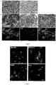
Fig. 1 shows phase contrast photos of formation of an embryoid body in suspension culture in an the differentiation process of human-induced Pluripotent stem cells (hiPSCs) to human neural crest stem cells (hNCSCs) and immunofluorescent staining graphs of differentiated NCSCs markers, wherein pictures from A to C are the phase contrast photos: A. hiPSCs subjected to expansion culture, B. the embryoid body subjected to suspension culture, C. the embryoid body subjected to adherent culture, which forms a neural rossette structure and cells is migrated out, D. expressions (immunofluorescent staining) of NCSCs specific markers in the differentiated cells, wherein Scale bar is equal to 100 µm. -
Fig. 2 shows phase contrast and fluorescent staining photos of biological characteristic analysis of hNCSCs. A. expressions of HNK1 and P75 of NCSCs detected by a flow cytometry in hiPSCs; B. a phase contrast photo of hNCSCs subjected to adherent culture after flow cytometry; C. a phase contrast photo of hNCSCs after suspension culture; D. expression of specific markers P75 and Sox10 of NCSCs in hNCSCs subjected to expansion culture, wherein Scale bar is equal to 100 µm. -
Fig. 3 shows a graph of and immunofluorescent staining photos of hNCSCs differentiated to peripheral neurons and schwann cells. A. cell shapes, observed under a phase contrast microscope, of hNCSCs before and after differentiation to the peripheral neurons, and B to D are immunofluorescent staining results: B. hNCSCs differentiated into peripherin+/Tuj1+ peripheral neurons, C. hNCSCs differentiated into TH+/Tuj1+ peripheral sympathetic neurons, and D. hNCSCs differentiated into GFAP+/S100B+ schwann cells, wherein Scale bar is equal to 100 µm. -
Fig. 4 shows phase contrast and histochemical staining photos of hNCSCs differentiated into hNCSCs-MSC. A. cell shapes, observed under a phase contrast microscope, of hNCSCs before and after differentiation to hNCSCs-MSC; B. an Alizarin Red S staining graph, a Tuluidine Blue staining graph and an Oil Red O staining graph which are obtained by culturing hNCSCs-MSC in a proper differentiation medium for a period of time by using a chemical staining method; C. an αSMA staining graph obtained by culturing hNCSCs-MSC in a proper differentiation medium for a period of time, wherein Scale bar is equal to 100 µm. -
Fig. 5 shows a diagram of expression of Leydig cell (LC) markers in hNCSCs-LCs. -
Fig. 6 is a diagram of testosterone secretion by hNCSCs-LCs under an in-vitro culture condition. -
Fig. 7 is a diagram for describing the influence of hNCSCs-LCs transplantation on the serum testosterone level of an ethane dimethyl sulfonate (EDS) model rat. - It can be understood that specific implementation modes described herein are only expressed by means of examples, but not deemed as limitations to the prevent application. Main features of the present application can be used for various implementation modes without departing from the scope of the present application. Persons skilled in the art will realize or can confirm that many equivalents may be all applied into the specific steps described in this text only by using conventional experiments. These equivalents are deemed as falling within the scope of the present application, and are covered by claims.
- For the purpose of making objectives, technical solutions and advantages of the present application clearer and more understandable, a further detailed description will be made below to the present application in combination with specific implementation modes and with reference to accompanying drawings. It should be understood that these descriptions are only examples, but not intended to limit the scope of the present application. In addition, in the descriptions below, descriptions of known structures and technologies are omitted to avoid unnecessary confusion of the concept of the present application.
-
- (1) preparation of cell suspension: with an established human iPS cell line (hiPSCs, Hum Mol Genet. 2013,22(11): 2221 -33), human induced pluripotent stem cells (hiPSCs) grew in a flat cloning form when subjected to expansion culture on Matrigel, and were closely arrayed, as shown in
Fig. 1A . The hiPSCs were digested into small masses with EDTA at the concentration of 0.5 mmol/L, and were resuspended by a ROCK inhibitor-containing mTeSR culture medium, wherein a used ROCK inhibitor is Y27632 (Calbiochem, San Diego, CA). - (2) committed differentiation: suspension cells were collected, then inoculated in a Petri culture dish, and subjected to suspension culture by a neural differentiation culture medium (80 percent of KnockoutTM DMEM, 18 percent of KnockoutTM SR, 1 percent (V/V) of Penicillin-Streptomycin solution, 1 mM of L-glutamine, and 0.1 mM of β-mercaptoethanol), thus forming brightly spherical embryoid bodies, as shown in
Fig. 1B (the neural differentiation culture medium in this embodiment is not limited to the above-mentioned formula, and may still achieve the effect as shown inFig. 1B when adopting any one of specific combinations of the neural differentiation culture medium containing 50 to 80 volume percent of Knockout DMEM, 5 to 20 volume percent of Knockout SR, 0.5 to 5 volume percent of Penicillin-Streptomycin solution, 0.5 to 5 mM of L-glutamine, and 0.05 to 0.5 mM of β-mercaptoethanol of the present application), wherein KnockoutTM DMEM and KnockoutTM SR are both purchased from Invitrogen, Carlsbad, CA. - (3) adherent culture: 5 days after the embryoid body was formed by the suspension culture, the spherical embryoid body was inoculated into a culture plate coated with polylysine/gelatin/fibronectin for adherent culture; a neural crest stem cell culture medium (obtained by mixing a DMEM-F12 culture medium and a Neurobasal culture medium according to a ratio of 1:1, adding 1 percent (V/V) of N2, 2 percent (V/V) of B27, 1 percent (V/V) of Penicillin-Streptomycin solution, 1 mM of L-glutamine, and 0.1 mM of β-mercaptoethanol, and further adding 10 ng/mL of a basic fibroblast growth factor (bFGF) (Invitrogen, 13256029) and 10 ng/mL of an epidermal growth factor (EGF) (PeproTech, NO. 62253-63-8)) were used, and then changed every other day. After the adherent culture was carried out for 2 days, it could be seen that obvious neural rossette structures appeared at the central parts of cell masses, and the cells were migrated out, as shown in
Fig. 1C . (The neural crest stem cell culture medium in this embodiment is not limited to the above-mentioned formula, and may still achieve the effect as shown inFig. 1C when adopting any one of specific combinations of the neural crest stem cell culture medium which is obtained by mixing the DMEM-F12 culture medium with the Neurobasal culture medium in a ratio of 1:0.1-1, adding 0.1 to 5 volume percent of N2, 0.5 to 10 volume percent of B27, 0.5 to 5 volume percent of Penicillin-Streptomycin solution, 0.5 to 5 mM of L-glutamine, and 0.05 to 0.5 mM of β-mercaptoethanol, and further adding 1 to 100 ng/mL of the bFGF and 1 to 100 ng/mL of the EGF.)
Immunofluorescent staining detection carried out on the cells subjected to the adherent culture found that as shown inFig. 1D , markers such as Pax6 and Sox2 were mainly expressed in cells at the central parts of neural rosette structures , and specific markers such as AP2α, Sox10, P75 and HNK1 of neural crest stem cells were mainly expressed in outwards migrated cells, thus indicating that after the embryoid body culture stage and then the adherent culture, the hiPSCs might become hNCSCs through differentiation. - (4) flow cytometry cell sorting: after the adherent culture was carried out for 5 days, the embryoid body was digested into single cells, the cells were labeled with anti-P75 and -HNK1 antibodies, and then P75+/HNK1+ double positive cells was isolated by flow cytometry cell sorting. During cell sorting, the culture medium was removed by a sucker; the cells were washed with PBS twice, and then Accutase was added to digest the differentiated hiPSCs for 3 to 5 minutes in 37°C. it was observed that the cells turned into round and bright; a culture medium was added to stop the digestion, and the cells were resuspended; then the cells were filtered by a nylon sieve, and were centrifugated at 1,500 rmp for 5 min; supernatant was abandoned, and 1mL of PBS was added and the cells were resuspended uniformly; and 20 µL of cell suspension was used for cell counting. The residual cells were divided into four groups for antibody labeling: an IgG negative control group, a P75 antibody single-label group, an HNK1 antibody single-label group and a P75+/HNK1+ antibody sample group, and 20 µL of antibody was added to label every 106 cells. A flow cytometry (BD influx cell sorter) was firstly used for performing flow sample injection on the cell suspension in the IgG negative control group to sort out a negative fluorescence signal region serving as a negative control, and cells having the fluorescence intensity which was 10 or higher times that of the negative control were collected. Flow detection analysis showed that after the differentiation, about 80 to 90 percent of hiPSCs expressed the specific markers HNK1 and P75 (
Fig. 2A ) of NCSC, thus indicating that most cells have been differentiated into hNCSCs after the differentiation. - Purified hNCSCs might be obtained through cell sorting, and the adherent culture was performed according to 5x10⁴ to 1x10⁵ cell/cm2. As shown in Fig. 2B, the cells in the adherent culture were relatively uniform in shape. After being subjected to digestion and passage, the hNCSCs were inoculated into a low-attachment culture plate. As shown in Fig. 2C, the cells formed neural spheres with relatively uniform sizes. The hNCSCs obtained by cell sorting were subjected to immunofluorescence detection. As shown in Fig. 2D, it can be seen that the cells maintained the expression of the specific markers of NCSCs including P75, Sox10. It indicates that under the existing culture and expansion conditions, the cells may sustain the characteristics of the NCSCs.
- Biological characterization was performed on the hNCSCs. After the hNCSCs were inoculated into a culture plate coated with polylysine/gelatin/fibronectin, the culture medium was replaced by a induction medium for further differentiation. As shown in
Fig. 3A , after the NCSCs were subjected to the peripheral neurons differentiation for 2 weeks, it could be seen that the cells had obvious changes in their morphology: cell bodies were rounded, and had neuronal fibers. After 3 to 4 weeks, the cells were used for immunostaining. As shown inFigs. 3B and 3C , it can be seen that there were peripherin+/Tuj1+ peripheral neurons and TH+/Tuj1+ sympathetic neurons. The hNCSCs were subjected to differentiation in schwann cell induction medium, and then were detected by immunostaining after 4 weeks. As shown inFig. 3D , it can be seen that there were GFAP+/S100b+ schwann cells. - After being subjected to differentiation in a mesenchymal stem cell (MSC) culture medium (low-glucose DMEM, 10 percent of FBS) for 7 days, the hNCSCs were differentiated into MSCs. As shown in
Fig. 4A , it can be seen that the cells turned into spindle-shaped and were in swirling growth. The multidirectional differentiation capacity of the MSCs (hNCSCs-MSC) induced from the hNCSCs was further tested. After the cells were cultured in a proper induction medium for certain time, as shown inFig. 4B , Alizarin Red S staining showed the formation of calcium nodules which verified that the hNCSCs-MSC were differentiated into osteocytes. Tuluidine Blue staining verified that the hNCSCs-MSC had been differentiated into chondrocytes, and formation of Oil Red O positive lipid droplets showed that the hNCSCs-MSC had been differentiated to adipocytes. As shown inFig. 4C , after the cells were cultured in a proper differentiation medium for certain time, αSMA staining showed that the hNCSCs-MSC had been differentiated into smooth muscle cells. It indicates that the cultured hNCSCs have the multidirectional differentiation capacity. - When the hNCSCs obtained by expansion reached the density of 60 percent confluence, the culture medium was replaced by a Leydig cells (LCs) differentiation culture medium (obtained by adding 2 volume percent of fetal calf serum (FCS), 1 nM of triiodothyronine (T3) (Sigma, T2877), 1 ng/ml of luteinizing hormone (LH) (Sigma, L6420), 70 ng/ml of an insulin-like growth factor (IGF-I) (PeproTech, 100-11), and 10 ng/ml of a platelet-derived growth factor BB (PDGF-BB) (PeproTech, 500-P47) into a DMEM-F12 culture medium (Hyclone, SH30023.018) for induction for 14 days and for cell differentiation, then cellular supernatant was collected, and cells were fixed by 4% PFA. Expression of mature LCs-related markers including 3β-HSD, P450c17, steroidogenic acute regulatory protein (StAR), and steroidogenic factor 1 (SF-1) were detected via immunofluorescence. As shown in
Fig. 5 , immunostaining analysis showed that cells (hNCSCs-LCs) differentiated from the hNCSCs expressed 3β-HSD, P450c17, StAR and SF-1. The testosterone level in the culture supernatant was tested via testosterone enzyme-linked immuno sorbent assay (ELISA) detection. As shown inFig. 6 , it revealed that the in vitro differentiated hNCSCs-LCs gradually secreted testosterone, which showed that the hNCSCs could be differentiated into mature LCs. (The LCs differentiation culture medium is not limited to the above-mentioned formula, and may still achieve the effects as shown inFigs. 5 and6 when adopting any one of specific combinations of the LCs differentiation culture medium obtained by adding 0.1 to 20 volume percent of FCS, 0.1 to 10 nM of T3, 0.1 to 20 ng/ml of LH, 5 to 100 ng/ml of the IGF-I, and 1 to 50 ng/ml of the PDGFBB into the DMEM-F12 culture medium.) - The previous research showed that the Leydig cells in a rat could be exhausted 4 days after treated by a specific apoptosis inducer ethane dimethyl sulfonate (EDS), so that the EDS was injected to the abdominal cavity of the rat to establish an EDS model. Adult rats were randomly divided into three groups, including a normal control group, an EDS-control group and a cell transplantation group. For the rats in the normal control group, normal saline with the same volume was injected intraperitoneally respectively on the
day 1 and the day 4. For the rats in the EDS-control group, EDS (75 mg/kg weight) was injected intraperitoneally on the first day, and 20 µl of normal saline (10 µl/unilateral testicle) was injected intraperitoneally on the day 4. For the rats in the cell group, EDS (75 mg/kg weight) was injected intraperitoneally on theday 1, and the hNCSCs-LCs (1.5x10⁶ resuspended in 10 µl of PBS/unilateral testicle) cultured in an LC culture medium for 5 to 7 days were transplanted into the testicles of the rats on the day 4. On the day 10 of the transplantation, the serum testosterone concentration was tested. The result showed that the transplantion of hNCSCs-LCs can increase the serum testosterone level, as shown inFig. 7 . - It should be understood that the above-mentioned specific implementation modes of the present application are merely exemplarily description or explanation of the principle of the present application, but not deemed as limiting the present application. Therefore, any modifications, equivalent replacements and improvements that are made without departing from the spirit and scope of the present application shall all fall within the scope of protection of the present application. In addition, the attached claims of the present application aim at covering all changes and modifications, which fall within the scope and the boundary of the attached claims or within equivalent forms of this scope and boundary.
Claims (19)
- A method of induced committed differentiation of human-induced pluripotent stem cells (hiPSCs) to Leydig cells (hiPS-hNCSCs-LCs).
- The method of committed differentiation of the human induced pluripotent stem cells (hiPSCs) to the Leydig cells (hiPS-hNCSCs-LCs) according to claim 1, characterized in that:(1) performing differentiation on the human-induced pluripotent stem cells (hiPSCs) to obtain human neural crest stem cells (hiPS-hNCSCs);(2) performing differentiation on the human neural crest stem cells (hiPS-hNCSCs) obtained in the step (1) to obtain the Leydig cells (hiPS-hNCSCs-LCs).
- The method according to claim 2, characterized in that the step (1) comprises inoculating the human-induced pluripotent stem cells (hiPSCs) into a culture dish with low attachment surface for culture.
- The method according to claim 2, characterized in that the step (1) comprises performing differentiation on the human induced pluripotent stem cells (hiPSCs) in a neural differentiation culture medium to obtain the human neural crest stem cells (hiPS-hNCSCs).
- The method according to claim 2, characterized in that the step (1) comprises culturing the human induced pluripotent stem cells (hiPSCs) in the neural differentiation culture medium to form embryoid bodies, and then performing adherent culture with a neural crest stem cell culture medium.
- The method according to claim 2, characterized in that the step (1) comprises digesting and then resuspending the human induced pluripotent stem cells (hiPSCs), inoculating suspended cells into the culture dish with low attachment surface, performing suspension culture in the neural differentiation culture medium to form the embryoid body, then inoculating the embryoid body into a fibronectin-coated culture plate, performing adherent culture by using the neural crest stem cell culture medium, and sorting P75+/HNK1+ double positive cells from cells subjected to the adherent culture through flow cytometry, wherein the double positive cells are the human neural crest stem cells (hiPS-hNCSCs).
- The method according to claim 3 or claim 6, characterized in that the culture dish with low attachment surface is a Petri culture dish.
- The method according to any one of claims 4 to 7, characterized in that the neural differentiation culture medium contains 50 to 80 volume percent of Knockout DMEM, 5 to 20 volume percent of Knockout SR, 0.5 to 5 volume percent of Penicillin-Streptomycin solution, 0.5 to 5 mM of L-glutamine, and 0.05 to 0.5 mM of β-mercaptoethanol.
- The method according to any one of claims 4 to 7, characterized in that the neural differentiation culture medium contains 80 volume percent of Knockout™ DMEM, 18 volume percent of Knockout™ SR, 1 volume percent of Penicillin-Streptomycin solution, 1 mM of L-glutamine, and 0.1 mM of β-mercaptoethanol.
- The method according to any one of claims 5 to 6, characterized in that the neural crest stem cell culture medium contains 0.1 to 5 volume percent of N2, 0.5 to 10 volume percent of B27, 0.5 to 5 volume percent of Penicillin-Streptomycin solution, 0.5 to 5 mM of L-glutamine, and 0.05 to 0.5 mM of β-mercaptoethanol, and is further added with 1 to 100 ng/mL of a basic fibroblast growth factor (bFGF) and 1 to 100 ng/mL of an epidermal growth factor (EGF).
- The method according to any one of claims 5 to 6, characterized in that the neural crest stem cell culture medium contains 1 volume percent of N2, 2 volume percent of B27, 1 volume percent of Penicillin-Streptomycin solution, 1 mM of L-glutamine, and 0.1 mM of β-mercaptoethanol, and is further added with 10 ng/mL of the bFGF and 10 ng/mL of the EGF.
- The method according to any one of claims 5 to 6, characterized in that the neural crest stem cell culture medium is obtained by mixing a DMEM-F12 culture medium with a Neurobasal culture medium according to the ratio of 1:0.1-1, adding 0.1 to 5 volume percent of N2, 0.5 to 10 volume percent of B27, 0.5 to 5 volume percent of Penicillin-Streptomycin solution, 0.5 to 5 mM of L-glutamine, and 0.05 to 0.5 mM of β-mercaptoethanol, and further adding 1 to 100 ng/mL of the bFGF and 1 to 100 ng/mL of the EGF.
- The method according to any one of claims 5 to 6, characterized in that the neural crest stem cell culture medium is obtained by mixing the DMEM-F12 culture medium with the Neurobasal culture medium according to the ratio of 1:1, adding 1 volume percent of N2, 2 volume percent of B27, 1 volume percent of Penicillin-Streptomycin solution, 1 mM of L-glutamine, and 0.1 mM of β-mercaptoethanol, and further adding 10 ng/mL of the bFGF and 10 ng/mL of the EGF.
- The method according to claim 2, characterized in that the step (2) comprises expension the hiPS-hNCSCs obtained in the step (1), and then performing differentiation on hiPS-hNCSCs in a Leydig cells (LCs) differentiation culture medium to obtain the Leydig cells (hiPS-hNCSCs-LCs).
- The method according to claim 14, characterized in that the LCs differentiation culture medium is obtained by adding 0.1 to 20 volume percent of fetal calf serum (FCS), 0.1 to 10 nM of triiodothyronine (T3), 0.1 to 20 ng/ml of luteinizing hormone (LH), 5 to 100 ng/ml of an insulin-like growth factor (IGF-I), and 1 to 50 ng/ml of a platelet-derived growth factor BB (PDGF-BB) into the DMEM-F12 culture medium.
- The method according to claim 14, characterized in that the LCs differentiation culture medium is obtained by adding 2 volume percent of fetal calf serum (FCS), 1 nM of triiodothyronine (T3), 1 ng/ml of luteinizing hormone (LH), 70 ng/ml of the IGF-I, and 10 ng/ml of the PDGF-BB into the DMEM-F12 culture medium.
- Application of the Leydig cells (hiPS-hNCSCs-LCs) obtained by the method according to any one of claims 1 to 16 in preparation of a drug capable of increasing the testosterone level.
- Application of the Leydig cells (hiPS-hNCSCs-LCs) obtained by the method according to any one of claims 1 to 16 in preparation of a drug for treating relevant diseases caused by low testosterone level.
- The method according to claim 15 or claim 16, wherein the testosterone level is the serum testosterone level.
Applications Claiming Priority (2)
| Application Number | Priority Date | Filing Date | Title |
|---|---|---|---|
| CN201510845021.8A CN105255826B (en) | 2015-11-27 | 2015-11-27 | Method of inducing differentiation and application thereof of the people iPS cell to interstitial glands |
| PCT/CN2016/107361 WO2017088830A1 (en) | 2015-11-27 | 2016-11-25 | Method for obtaining leydig cells by carrying out induced differentiation on human induced pluripotent stem cells, and use of leydig cells |
Publications (3)
| Publication Number | Publication Date |
|---|---|
| EP3369809A1 true EP3369809A1 (en) | 2018-09-05 |
| EP3369809A4 EP3369809A4 (en) | 2018-12-05 |
| EP3369809B1 EP3369809B1 (en) | 2021-02-24 |
Family
ID=55095757
Family Applications (1)
| Application Number | Title | Priority Date | Filing Date |
|---|---|---|---|
| EP16868053.6A Not-in-force EP3369809B1 (en) | 2015-11-27 | 2016-11-25 | Method for obtaining leydig cells by carrying out induced differentiation on human induced pluripotent stem cells, and use of leydig cells |
Country Status (6)
| Country | Link |
|---|---|
| US (1) | US11052120B2 (en) |
| EP (1) | EP3369809B1 (en) |
| JP (1) | JP6824267B2 (en) |
| KR (1) | KR102269485B1 (en) |
| CN (1) | CN105255826B (en) |
| WO (1) | WO2017088830A1 (en) |
Families Citing this family (9)
| Publication number | Priority date | Publication date | Assignee | Title |
|---|---|---|---|---|
| CN105255826B (en) * | 2015-11-27 | 2019-01-08 | 中山大学 | Method of inducing differentiation and application thereof of the people iPS cell to interstitial glands |
| JP6979702B2 (en) * | 2016-11-10 | 2021-12-15 | 国立大学法人神戸大学 | Method for producing Leydig-like cells derived from human pluripotent stem cells and Leydig-like cells derived from human pluripotent stem cells |
| UA129013C2 (en) | 2017-11-30 | 2024-12-25 | Кіото Юніверсіті | Method for culture of cells |
| CN108524485A (en) * | 2018-02-24 | 2018-09-14 | 中山大学 | A kind of monkey testosterone lacks the method for building up of model |
| CN108795843B (en) * | 2018-06-01 | 2021-10-08 | 温州医科大学附属第二医院、温州医科大学附属育英儿童医院 | A method for the differentiation of pluripotent stem cells into Leydig cells induced by small molecules |
| CN108949688B (en) * | 2018-08-07 | 2022-03-29 | 中山大学 | Neural crest lineage pericyte from pluripotent stem cells and induced differentiation method thereof |
| WO2023007943A1 (en) * | 2021-07-30 | 2023-02-02 | 株式会社リコー | Cell culture method, cell culture container, method for producing cell culture container, and cell-containing structure |
| CN114250199B (en) * | 2021-11-18 | 2024-05-24 | 中山大学 | Spinal column-spinal cord organoid culture method and culture medium |
| CN117431209B (en) * | 2023-12-22 | 2024-07-26 | 上海元戊医学技术有限公司 | Method for preparing mesenchymal stem cells through neural crest cell line and application as osteoarthritis medicine |
Family Cites Families (9)
| Publication number | Priority date | Publication date | Assignee | Title |
|---|---|---|---|---|
| WO2005085425A1 (en) * | 2004-03-03 | 2005-09-15 | Japan Science And Technology Agency | Method of differentiating mesenchymal stem cells into steroid-producing cells |
| US20070280907A1 (en) * | 2006-04-07 | 2007-12-06 | Los Angeles Biomedical Research Institute At Harbor-Ucla Medical Center | Adult bone marrow cell transplantation to testes creation of transdifferentiated testes germ cells, leydig cells and sertoli cells |
| JP5700301B2 (en) * | 2009-06-03 | 2015-04-15 | 国立大学法人大阪大学 | Method for inducing differentiation of neural crest cells from pluripotent stem cells |
| CN101892190B (en) * | 2010-06-24 | 2013-03-27 | 浙江大学 | Construction and application of model for differentiating stem cells into testicular interstitial cells |
| CN102174468A (en) * | 2011-02-24 | 2011-09-07 | 暨南大学 | Method and application for inducing human umbilical cord mesenchyme stem cells to be differentiated into testicular interstitial cells |
| US8951798B2 (en) * | 2011-10-13 | 2015-02-10 | Wisconsin Alumni Research Foundation | Generation of cardiomyocytes from human pluripotent stem cells |
| CN102952777B (en) * | 2012-11-29 | 2015-01-21 | 山东大学 | Inducing method for directionally differentiating human embryonic stem cells to corneal endothelial cells |
| CN103861087B (en) * | 2012-12-14 | 2016-04-20 | 广州暨南大学医药生物技术研究开发中心 | Nerve growth factor is for the preparation of the purposes in treatment middle-aging male sexual disorder syndromic medicine |
| CN105255826B (en) * | 2015-11-27 | 2019-01-08 | 中山大学 | Method of inducing differentiation and application thereof of the people iPS cell to interstitial glands |
-
2015
- 2015-11-27 CN CN201510845021.8A patent/CN105255826B/en not_active Expired - Fee Related
-
2016
- 2016-11-25 WO PCT/CN2016/107361 patent/WO2017088830A1/en not_active Ceased
- 2016-11-25 EP EP16868053.6A patent/EP3369809B1/en not_active Not-in-force
- 2016-11-25 JP JP2018525578A patent/JP6824267B2/en active Active
- 2016-11-25 KR KR1020187014620A patent/KR102269485B1/en not_active Expired - Fee Related
-
2018
- 2018-05-24 US US15/987,953 patent/US11052120B2/en active Active
Also Published As
| Publication number | Publication date |
|---|---|
| EP3369809B1 (en) | 2021-02-24 |
| CN105255826B (en) | 2019-01-08 |
| KR102269485B1 (en) | 2021-06-28 |
| CN105255826A (en) | 2016-01-20 |
| JP6824267B2 (en) | 2021-02-03 |
| US20180311286A1 (en) | 2018-11-01 |
| WO2017088830A1 (en) | 2017-06-01 |
| US11052120B2 (en) | 2021-07-06 |
| KR20180081530A (en) | 2018-07-16 |
| EP3369809A4 (en) | 2018-12-05 |
| JP2018534934A (en) | 2018-11-29 |
Similar Documents
| Publication | Publication Date | Title |
|---|---|---|
| US11052120B2 (en) | Method of committed differentiation of human induced pluripotent stem cells into Leydig cells and application of Leydig cells | |
| Techawattanawisal et al. | Isolation of multipotent stem cells from adult rat periodontal ligament by neurosphere-forming culture system | |
| CN109844102B (en) | Methods to differentiate pluripotent cells | |
| DK1758986T3 (en) | Neural stem cells | |
| JP5843775B2 (en) | Manipulation of osmolality to differentiate stem cells | |
| JP6097076B2 (en) | Progeny of differentiated pluripotent stem cells that exclude excluded phenotypes | |
| WO2010075500A1 (en) | Target populations of oligodendrocyte precursor cells and methods of making and using same | |
| EA034058B1 (en) | Culture method to obtain and maintain a pure or enriched population of mammalian neural stem cells and/or neural progenitor cells that are prone to differentiate into oligodendrocyte-lineage cells in vitro | |
| WO2017088829A1 (en) | Methods for separating and culturing human testicular mesenchymal stem cells, and use of human testicular mesenchymal stem cells | |
| Lu et al. | Isolation and culture of human oligodendrocyte precursor cells from neurospheres | |
| Chatzi et al. | Derivation of homogeneous GABAergic neurons from mouse embryonic stem cells | |
| Zhang et al. | Isolation of neural crest‐derived stem cells from rat embryonic mandibular processes | |
| Major et al. | Derivation of telencephalic oligodendrocyte progenitors from human pluripotent stem cells | |
| Yamasaki et al. | Long-term serial cultivation of mouse induced pluripotent stem cells in serum-free and feeder-free defined medium | |
| Maciaczyk et al. | Restricted spontaneous in vitro differentiation and region-specific migration of long-term expanded fetal human neural precursor cells after transplantation into the adult rat brain | |
| KR20180067330A (en) | Method for increasing the rate of inducing differentiation of human pluripotent stem cells to mesenchymal stem cells, and mesenchymal stem cells produced by thereof | |
| Li et al. | Transplantable neural progenitor populations derived from rhesus monkey embryonic stem cells | |
| Wang et al. | Differentiation of neuronal cells from NIH/3T3 fibroblasts under defined conditions | |
| Liu et al. | Microcapsule co-culture system enhances neural differentiation of mesenchymal stem cells | |
| WO2025190925A1 (en) | Retinal cell 3d culture | |
| Fan et al. | In vitro culture of human embryonic striatum-derived neural stem cells | |
| Hyysalo et al. | Directed Differentiation of Human PSC into Oligodendrocytes |
Legal Events
| Date | Code | Title | Description |
|---|---|---|---|
| STAA | Information on the status of an ep patent application or granted ep patent |
Free format text: STATUS: THE INTERNATIONAL PUBLICATION HAS BEEN MADE |
|
| PUAI | Public reference made under article 153(3) epc to a published international application that has entered the european phase |
Free format text: ORIGINAL CODE: 0009012 |
|
| STAA | Information on the status of an ep patent application or granted ep patent |
Free format text: STATUS: REQUEST FOR EXAMINATION WAS MADE |
|
| 17P | Request for examination filed |
Effective date: 20180531 |
|
| AK | Designated contracting states |
Kind code of ref document: A1 Designated state(s): AL AT BE BG CH CY CZ DE DK EE ES FI FR GB GR HR HU IE IS IT LI LT LU LV MC MK MT NL NO PL PT RO RS SE SI SK SM TR |
|
| AX | Request for extension of the european patent |
Extension state: BA ME |
|
| RIC1 | Information provided on ipc code assigned before grant |
Ipc: C12N 5/071 20100101ALI20181018BHEP Ipc: A61K 45/00 20060101ALI20181018BHEP Ipc: A61P 5/26 20060101ALI20181018BHEP Ipc: A61K 35/52 20150101ALI20181018BHEP Ipc: C12N 5/077 20100101AFI20181018BHEP |
|
| A4 | Supplementary search report drawn up and despatched |
Effective date: 20181030 |
|
| DAV | Request for validation of the european patent (deleted) | ||
| DAX | Request for extension of the european patent (deleted) | ||
| STAA | Information on the status of an ep patent application or granted ep patent |
Free format text: STATUS: EXAMINATION IS IN PROGRESS |
|
| 17Q | First examination report despatched |
Effective date: 20200114 |
|
| REG | Reference to a national code |
Ref country code: DE Ref legal event code: R079 Ref document number: 602016053340 Country of ref document: DE Free format text: PREVIOUS MAIN CLASS: C12N0005077000 Ipc: A61K0038180000 |
|
| GRAP | Despatch of communication of intention to grant a patent |
Free format text: ORIGINAL CODE: EPIDOSNIGR1 |
|
| STAA | Information on the status of an ep patent application or granted ep patent |
Free format text: STATUS: GRANT OF PATENT IS INTENDED |
|
| RIC1 | Information provided on ipc code assigned before grant |
Ipc: C12N 5/071 20100101ALI20200901BHEP Ipc: A61P 5/26 20060101ALI20200901BHEP Ipc: A61K 35/52 20150101ALI20200901BHEP Ipc: A61K 38/18 20060101AFI20200901BHEP Ipc: A61K 45/00 20060101ALI20200901BHEP |
|
| INTG | Intention to grant announced |
Effective date: 20200930 |
|
| GRAS | Grant fee paid |
Free format text: ORIGINAL CODE: EPIDOSNIGR3 |
|
| GRAA | (expected) grant |
Free format text: ORIGINAL CODE: 0009210 |
|
| STAA | Information on the status of an ep patent application or granted ep patent |
Free format text: STATUS: THE PATENT HAS BEEN GRANTED |
|
| AK | Designated contracting states |
Kind code of ref document: B1 Designated state(s): AL AT BE BG CH CY CZ DE DK EE ES FI FR GB GR HR HU IE IS IT LI LT LU LV MC MK MT NL NO PL PT RO RS SE SI SK SM TR |
|
| REG | Reference to a national code |
Ref country code: CH Ref legal event code: EP |
|
| REG | Reference to a national code |
Ref country code: AT Ref legal event code: REF Ref document number: 1363635 Country of ref document: AT Kind code of ref document: T Effective date: 20210315 |
|
| REG | Reference to a national code |
Ref country code: IE Ref legal event code: FG4D |
|
| REG | Reference to a national code |
Ref country code: DE Ref legal event code: R096 Ref document number: 602016053340 Country of ref document: DE |
|
| REG | Reference to a national code |
Ref country code: LT Ref legal event code: MG9D |
|
| REG | Reference to a national code |
Ref country code: NL Ref legal event code: MP Effective date: 20210224 |
|
| PG25 | Lapsed in a contracting state [announced via postgrant information from national office to epo] |
Ref country code: HR Free format text: LAPSE BECAUSE OF FAILURE TO SUBMIT A TRANSLATION OF THE DESCRIPTION OR TO PAY THE FEE WITHIN THE PRESCRIBED TIME-LIMIT Effective date: 20210224 Ref country code: GR Free format text: LAPSE BECAUSE OF FAILURE TO SUBMIT A TRANSLATION OF THE DESCRIPTION OR TO PAY THE FEE WITHIN THE PRESCRIBED TIME-LIMIT Effective date: 20210525 Ref country code: FI Free format text: LAPSE BECAUSE OF FAILURE TO SUBMIT A TRANSLATION OF THE DESCRIPTION OR TO PAY THE FEE WITHIN THE PRESCRIBED TIME-LIMIT Effective date: 20210224 Ref country code: PT Free format text: LAPSE BECAUSE OF FAILURE TO SUBMIT A TRANSLATION OF THE DESCRIPTION OR TO PAY THE FEE WITHIN THE PRESCRIBED TIME-LIMIT Effective date: 20210624 Ref country code: NO Free format text: LAPSE BECAUSE OF FAILURE TO SUBMIT A TRANSLATION OF THE DESCRIPTION OR TO PAY THE FEE WITHIN THE PRESCRIBED TIME-LIMIT Effective date: 20210524 Ref country code: BG Free format text: LAPSE BECAUSE OF FAILURE TO SUBMIT A TRANSLATION OF THE DESCRIPTION OR TO PAY THE FEE WITHIN THE PRESCRIBED TIME-LIMIT Effective date: 20210524 Ref country code: LT Free format text: LAPSE BECAUSE OF FAILURE TO SUBMIT A TRANSLATION OF THE DESCRIPTION OR TO PAY THE FEE WITHIN THE PRESCRIBED TIME-LIMIT Effective date: 20210224 |
|
| REG | Reference to a national code |
Ref country code: AT Ref legal event code: MK05 Ref document number: 1363635 Country of ref document: AT Kind code of ref document: T Effective date: 20210224 |
|
| PG25 | Lapsed in a contracting state [announced via postgrant information from national office to epo] |
Ref country code: NL Free format text: LAPSE BECAUSE OF FAILURE TO SUBMIT A TRANSLATION OF THE DESCRIPTION OR TO PAY THE FEE WITHIN THE PRESCRIBED TIME-LIMIT Effective date: 20210224 Ref country code: RS Free format text: LAPSE BECAUSE OF FAILURE TO SUBMIT A TRANSLATION OF THE DESCRIPTION OR TO PAY THE FEE WITHIN THE PRESCRIBED TIME-LIMIT Effective date: 20210224 Ref country code: PL Free format text: LAPSE BECAUSE OF FAILURE TO SUBMIT A TRANSLATION OF THE DESCRIPTION OR TO PAY THE FEE WITHIN THE PRESCRIBED TIME-LIMIT Effective date: 20210224 Ref country code: LV Free format text: LAPSE BECAUSE OF FAILURE TO SUBMIT A TRANSLATION OF THE DESCRIPTION OR TO PAY THE FEE WITHIN THE PRESCRIBED TIME-LIMIT Effective date: 20210224 Ref country code: SE Free format text: LAPSE BECAUSE OF FAILURE TO SUBMIT A TRANSLATION OF THE DESCRIPTION OR TO PAY THE FEE WITHIN THE PRESCRIBED TIME-LIMIT Effective date: 20210224 |
|
| PG25 | Lapsed in a contracting state [announced via postgrant information from national office to epo] |
Ref country code: IS Free format text: LAPSE BECAUSE OF FAILURE TO SUBMIT A TRANSLATION OF THE DESCRIPTION OR TO PAY THE FEE WITHIN THE PRESCRIBED TIME-LIMIT Effective date: 20210624 |
|
| PG25 | Lapsed in a contracting state [announced via postgrant information from national office to epo] |
Ref country code: SM Free format text: LAPSE BECAUSE OF FAILURE TO SUBMIT A TRANSLATION OF THE DESCRIPTION OR TO PAY THE FEE WITHIN THE PRESCRIBED TIME-LIMIT Effective date: 20210224 Ref country code: AT Free format text: LAPSE BECAUSE OF FAILURE TO SUBMIT A TRANSLATION OF THE DESCRIPTION OR TO PAY THE FEE WITHIN THE PRESCRIBED TIME-LIMIT Effective date: 20210224 Ref country code: CZ Free format text: LAPSE BECAUSE OF FAILURE TO SUBMIT A TRANSLATION OF THE DESCRIPTION OR TO PAY THE FEE WITHIN THE PRESCRIBED TIME-LIMIT Effective date: 20210224 Ref country code: EE Free format text: LAPSE BECAUSE OF FAILURE TO SUBMIT A TRANSLATION OF THE DESCRIPTION OR TO PAY THE FEE WITHIN THE PRESCRIBED TIME-LIMIT Effective date: 20210224 |
|
| REG | Reference to a national code |
Ref country code: DE Ref legal event code: R097 Ref document number: 602016053340 Country of ref document: DE |
|
| PG25 | Lapsed in a contracting state [announced via postgrant information from national office to epo] |
Ref country code: SK Free format text: LAPSE BECAUSE OF FAILURE TO SUBMIT A TRANSLATION OF THE DESCRIPTION OR TO PAY THE FEE WITHIN THE PRESCRIBED TIME-LIMIT Effective date: 20210224 Ref country code: RO Free format text: LAPSE BECAUSE OF FAILURE TO SUBMIT A TRANSLATION OF THE DESCRIPTION OR TO PAY THE FEE WITHIN THE PRESCRIBED TIME-LIMIT Effective date: 20210224 Ref country code: DK Free format text: LAPSE BECAUSE OF FAILURE TO SUBMIT A TRANSLATION OF THE DESCRIPTION OR TO PAY THE FEE WITHIN THE PRESCRIBED TIME-LIMIT Effective date: 20210224 |
|
| PLBE | No opposition filed within time limit |
Free format text: ORIGINAL CODE: 0009261 |
|
| STAA | Information on the status of an ep patent application or granted ep patent |
Free format text: STATUS: NO OPPOSITION FILED WITHIN TIME LIMIT |
|
| PG25 | Lapsed in a contracting state [announced via postgrant information from national office to epo] |
Ref country code: ES Free format text: LAPSE BECAUSE OF FAILURE TO SUBMIT A TRANSLATION OF THE DESCRIPTION OR TO PAY THE FEE WITHIN THE PRESCRIBED TIME-LIMIT Effective date: 20210224 Ref country code: AL Free format text: LAPSE BECAUSE OF FAILURE TO SUBMIT A TRANSLATION OF THE DESCRIPTION OR TO PAY THE FEE WITHIN THE PRESCRIBED TIME-LIMIT Effective date: 20210224 |
|
| 26N | No opposition filed |
Effective date: 20211125 |
|
| PG25 | Lapsed in a contracting state [announced via postgrant information from national office to epo] |
Ref country code: SI Free format text: LAPSE BECAUSE OF FAILURE TO SUBMIT A TRANSLATION OF THE DESCRIPTION OR TO PAY THE FEE WITHIN THE PRESCRIBED TIME-LIMIT Effective date: 20210224 |
|
| PG25 | Lapsed in a contracting state [announced via postgrant information from national office to epo] |
Ref country code: IS Free format text: LAPSE BECAUSE OF FAILURE TO SUBMIT A TRANSLATION OF THE DESCRIPTION OR TO PAY THE FEE WITHIN THE PRESCRIBED TIME-LIMIT Effective date: 20210624 |
|
| PG25 | Lapsed in a contracting state [announced via postgrant information from national office to epo] |
Ref country code: MC Free format text: LAPSE BECAUSE OF FAILURE TO SUBMIT A TRANSLATION OF THE DESCRIPTION OR TO PAY THE FEE WITHIN THE PRESCRIBED TIME-LIMIT Effective date: 20210224 |
|
| REG | Reference to a national code |
Ref country code: CH Ref legal event code: PL |
|
| PG25 | Lapsed in a contracting state [announced via postgrant information from national office to epo] |
Ref country code: LU Free format text: LAPSE BECAUSE OF NON-PAYMENT OF DUE FEES Effective date: 20211125 Ref country code: BE Free format text: LAPSE BECAUSE OF NON-PAYMENT OF DUE FEES Effective date: 20211130 |
|
| REG | Reference to a national code |
Ref country code: BE Ref legal event code: MM Effective date: 20211130 |
|
| PG25 | Lapsed in a contracting state [announced via postgrant information from national office to epo] |
Ref country code: LI Free format text: LAPSE BECAUSE OF NON-PAYMENT OF DUE FEES Effective date: 20211130 Ref country code: CH Free format text: LAPSE BECAUSE OF NON-PAYMENT OF DUE FEES Effective date: 20211130 |
|
| PG25 | Lapsed in a contracting state [announced via postgrant information from national office to epo] |
Ref country code: HU Free format text: LAPSE BECAUSE OF FAILURE TO SUBMIT A TRANSLATION OF THE DESCRIPTION OR TO PAY THE FEE WITHIN THE PRESCRIBED TIME-LIMIT; INVALID AB INITIO Effective date: 20161125 |
|
| PG25 | Lapsed in a contracting state [announced via postgrant information from national office to epo] |
Ref country code: CY Free format text: LAPSE BECAUSE OF FAILURE TO SUBMIT A TRANSLATION OF THE DESCRIPTION OR TO PAY THE FEE WITHIN THE PRESCRIBED TIME-LIMIT Effective date: 20210224 |
|
| PGFP | Annual fee paid to national office [announced via postgrant information from national office to epo] |
Ref country code: IT Payment date: 20230828 Year of fee payment: 8 |
|
| PGFP | Annual fee paid to national office [announced via postgrant information from national office to epo] |
Ref country code: FR Payment date: 20230829 Year of fee payment: 8 |
|
| PGFP | Annual fee paid to national office [announced via postgrant information from national office to epo] |
Ref country code: GB Payment date: 20231123 Year of fee payment: 8 |
|
| PGFP | Annual fee paid to national office [announced via postgrant information from national office to epo] |
Ref country code: IE Payment date: 20231117 Year of fee payment: 8 Ref country code: DE Payment date: 20230920 Year of fee payment: 8 |
|
| PG25 | Lapsed in a contracting state [announced via postgrant information from national office to epo] |
Ref country code: MK Free format text: LAPSE BECAUSE OF FAILURE TO SUBMIT A TRANSLATION OF THE DESCRIPTION OR TO PAY THE FEE WITHIN THE PRESCRIBED TIME-LIMIT Effective date: 20210224 |
|
| PG25 | Lapsed in a contracting state [announced via postgrant information from national office to epo] |
Ref country code: MT Free format text: LAPSE BECAUSE OF FAILURE TO SUBMIT A TRANSLATION OF THE DESCRIPTION OR TO PAY THE FEE WITHIN THE PRESCRIBED TIME-LIMIT Effective date: 20210224 |
|
| REG | Reference to a national code |
Ref country code: DE Ref legal event code: R119 Ref document number: 602016053340 Country of ref document: DE |
|
| GBPC | Gb: european patent ceased through non-payment of renewal fee |
Effective date: 20241125 |
|
| PG25 | Lapsed in a contracting state [announced via postgrant information from national office to epo] |
Ref country code: DE Free format text: LAPSE BECAUSE OF NON-PAYMENT OF DUE FEES Effective date: 20250603 |
|
| PG25 | Lapsed in a contracting state [announced via postgrant information from national office to epo] |
Ref country code: IT Free format text: LAPSE BECAUSE OF NON-PAYMENT OF DUE FEES Effective date: 20241125 |
|
| PG25 | Lapsed in a contracting state [announced via postgrant information from national office to epo] |
Ref country code: GB Free format text: LAPSE BECAUSE OF NON-PAYMENT OF DUE FEES Effective date: 20241125 |
|
| PG25 | Lapsed in a contracting state [announced via postgrant information from national office to epo] |
Ref country code: FR Free format text: LAPSE BECAUSE OF NON-PAYMENT OF DUE FEES Effective date: 20241130 |
|
| PG25 | Lapsed in a contracting state [announced via postgrant information from national office to epo] |
Ref country code: IE Free format text: LAPSE BECAUSE OF NON-PAYMENT OF DUE FEES Effective date: 20241125 |
|
| PG25 | Lapsed in a contracting state [announced via postgrant information from national office to epo] |
Ref country code: TR Free format text: LAPSE BECAUSE OF FAILURE TO SUBMIT A TRANSLATION OF THE DESCRIPTION OR TO PAY THE FEE WITHIN THE PRESCRIBED TIME-LIMIT Effective date: 20210224 |