EP3336175A1 - Three-dimensional model of depigmenting disorder - Google Patents
Three-dimensional model of depigmenting disorder Download PDFInfo
- Publication number
- EP3336175A1 EP3336175A1 EP16306718.4A EP16306718A EP3336175A1 EP 3336175 A1 EP3336175 A1 EP 3336175A1 EP 16306718 A EP16306718 A EP 16306718A EP 3336175 A1 EP3336175 A1 EP 3336175A1
- Authority
- EP
- European Patent Office
- Prior art keywords
- tnfα
- model
- ifnγ
- melanocytes
- cellular model
- Prior art date
- Legal status (The legal status is an assumption and is not a legal conclusion. Google has not performed a legal analysis and makes no representation as to the accuracy of the status listed.)
- Granted
Links
Images




Classifications
-
- C—CHEMISTRY; METALLURGY
- C12—BIOCHEMISTRY; BEER; SPIRITS; WINE; VINEGAR; MICROBIOLOGY; ENZYMOLOGY; MUTATION OR GENETIC ENGINEERING
- C12N—MICROORGANISMS OR ENZYMES; COMPOSITIONS THEREOF; PROPAGATING, PRESERVING, OR MAINTAINING MICROORGANISMS; MUTATION OR GENETIC ENGINEERING; CULTURE MEDIA
- C12N5/00—Undifferentiated human, animal or plant cells, e.g. cell lines; Tissues; Cultivation or maintenance thereof; Culture media therefor
- C12N5/06—Animal cells or tissues; Human cells or tissues
- C12N5/0602—Vertebrate cells
- C12N5/0625—Epidermal cells, skin cells; Cells of the oral mucosa
- C12N5/0626—Melanocytes
-
- C—CHEMISTRY; METALLURGY
- C12—BIOCHEMISTRY; BEER; SPIRITS; WINE; VINEGAR; MICROBIOLOGY; ENZYMOLOGY; MUTATION OR GENETIC ENGINEERING
- C12N—MICROORGANISMS OR ENZYMES; COMPOSITIONS THEREOF; PROPAGATING, PRESERVING, OR MAINTAINING MICROORGANISMS; MUTATION OR GENETIC ENGINEERING; CULTURE MEDIA
- C12N5/00—Undifferentiated human, animal or plant cells, e.g. cell lines; Tissues; Cultivation or maintenance thereof; Culture media therefor
- C12N5/06—Animal cells or tissues; Human cells or tissues
- C12N5/0602—Vertebrate cells
- C12N5/0625—Epidermal cells, skin cells; Cells of the oral mucosa
- C12N5/0629—Keratinocytes; Whole skin
-
- C—CHEMISTRY; METALLURGY
- C12—BIOCHEMISTRY; BEER; SPIRITS; WINE; VINEGAR; MICROBIOLOGY; ENZYMOLOGY; MUTATION OR GENETIC ENGINEERING
- C12N—MICROORGANISMS OR ENZYMES; COMPOSITIONS THEREOF; PROPAGATING, PRESERVING, OR MAINTAINING MICROORGANISMS; MUTATION OR GENETIC ENGINEERING; CULTURE MEDIA
- C12N5/00—Undifferentiated human, animal or plant cells, e.g. cell lines; Tissues; Cultivation or maintenance thereof; Culture media therefor
- C12N5/06—Animal cells or tissues; Human cells or tissues
- C12N5/0697—Artificial constructs associating cells of different lineages, e.g. tissue equivalents
- C12N5/0698—Skin equivalents
-
- G—PHYSICS
- G01—MEASURING; TESTING
- G01N—INVESTIGATING OR ANALYSING MATERIALS BY DETERMINING THEIR CHEMICAL OR PHYSICAL PROPERTIES
- G01N33/00—Investigating or analysing materials by specific methods not covered by groups G01N1/00 - G01N31/00
- G01N33/48—Biological material, e.g. blood, urine; Haemocytometers
- G01N33/50—Chemical analysis of biological material, e.g. blood, urine; Testing involving biospecific ligand binding methods; Immunological testing
- G01N33/5005—Chemical analysis of biological material, e.g. blood, urine; Testing involving biospecific ligand binding methods; Immunological testing involving human or animal cells
- G01N33/5008—Chemical analysis of biological material, e.g. blood, urine; Testing involving biospecific ligand binding methods; Immunological testing involving human or animal cells for testing or evaluating the effect of chemical or biological compounds, e.g. drugs, cosmetics
- G01N33/5082—Supracellular entities, e.g. tissue, organisms
- G01N33/5088—Supracellular entities, e.g. tissue, organisms of vertebrates
-
- C—CHEMISTRY; METALLURGY
- C12—BIOCHEMISTRY; BEER; SPIRITS; WINE; VINEGAR; MICROBIOLOGY; ENZYMOLOGY; MUTATION OR GENETIC ENGINEERING
- C12N—MICROORGANISMS OR ENZYMES; COMPOSITIONS THEREOF; PROPAGATING, PRESERVING, OR MAINTAINING MICROORGANISMS; MUTATION OR GENETIC ENGINEERING; CULTURE MEDIA
- C12N2501/00—Active agents used in cell culture processes, e.g. differentation
- C12N2501/10—Growth factors
- C12N2501/15—Transforming growth factor beta (TGF-β)
-
- C—CHEMISTRY; METALLURGY
- C12—BIOCHEMISTRY; BEER; SPIRITS; WINE; VINEGAR; MICROBIOLOGY; ENZYMOLOGY; MUTATION OR GENETIC ENGINEERING
- C12N—MICROORGANISMS OR ENZYMES; COMPOSITIONS THEREOF; PROPAGATING, PRESERVING, OR MAINTAINING MICROORGANISMS; MUTATION OR GENETIC ENGINEERING; CULTURE MEDIA
- C12N2501/00—Active agents used in cell culture processes, e.g. differentation
- C12N2501/20—Cytokines; Chemokines
- C12N2501/23—Interleukins [IL]
- C12N2501/2301—Interleukin-1 (IL-1)
-
- C—CHEMISTRY; METALLURGY
- C12—BIOCHEMISTRY; BEER; SPIRITS; WINE; VINEGAR; MICROBIOLOGY; ENZYMOLOGY; MUTATION OR GENETIC ENGINEERING
- C12N—MICROORGANISMS OR ENZYMES; COMPOSITIONS THEREOF; PROPAGATING, PRESERVING, OR MAINTAINING MICROORGANISMS; MUTATION OR GENETIC ENGINEERING; CULTURE MEDIA
- C12N2501/00—Active agents used in cell culture processes, e.g. differentation
- C12N2501/20—Cytokines; Chemokines
- C12N2501/23—Interleukins [IL]
- C12N2501/2317—Interleukin-17 (IL-17)
-
- C—CHEMISTRY; METALLURGY
- C12—BIOCHEMISTRY; BEER; SPIRITS; WINE; VINEGAR; MICROBIOLOGY; ENZYMOLOGY; MUTATION OR GENETIC ENGINEERING
- C12N—MICROORGANISMS OR ENZYMES; COMPOSITIONS THEREOF; PROPAGATING, PRESERVING, OR MAINTAINING MICROORGANISMS; MUTATION OR GENETIC ENGINEERING; CULTURE MEDIA
- C12N2501/00—Active agents used in cell culture processes, e.g. differentation
- C12N2501/20—Cytokines; Chemokines
- C12N2501/24—Interferons [IFN]
-
- C—CHEMISTRY; METALLURGY
- C12—BIOCHEMISTRY; BEER; SPIRITS; WINE; VINEGAR; MICROBIOLOGY; ENZYMOLOGY; MUTATION OR GENETIC ENGINEERING
- C12N—MICROORGANISMS OR ENZYMES; COMPOSITIONS THEREOF; PROPAGATING, PRESERVING, OR MAINTAINING MICROORGANISMS; MUTATION OR GENETIC ENGINEERING; CULTURE MEDIA
- C12N2501/00—Active agents used in cell culture processes, e.g. differentation
- C12N2501/20—Cytokines; Chemokines
- C12N2501/25—Tumour necrosing factors [TNF]
-
- C—CHEMISTRY; METALLURGY
- C12—BIOCHEMISTRY; BEER; SPIRITS; WINE; VINEGAR; MICROBIOLOGY; ENZYMOLOGY; MUTATION OR GENETIC ENGINEERING
- C12N—MICROORGANISMS OR ENZYMES; COMPOSITIONS THEREOF; PROPAGATING, PRESERVING, OR MAINTAINING MICROORGANISMS; MUTATION OR GENETIC ENGINEERING; CULTURE MEDIA
- C12N2513/00—3D culture
-
- G—PHYSICS
- G01—MEASURING; TESTING
- G01N—INVESTIGATING OR ANALYSING MATERIALS BY DETERMINING THEIR CHEMICAL OR PHYSICAL PROPERTIES
- G01N2800/00—Detection or diagnosis of diseases
- G01N2800/20—Dermatological disorders
- G01N2800/207—Pigmentation disorders
Definitions
- the present invention relates to the field of tissue engineering, dermatology and cosmetic.
- the invention more specifically relates to a three-dimensional cellular model of depigmenting disorders as well as to products and methods for preparing such a model. It further relates to uses of the model in experimental research, typically for studying pigmentation/depigmentation mechanisms and for the screening of molecules of interest in the prevention or treatment of pathological conditions wherein a depigmenting disorder is observed, in particular vitiligo as well as other inflammatory dermatoses.
- Vitiligo is the most common skin depigmenting disorder. The estimated prevalence of vitiligo is around 0.5 % to 1% of the world population. The two sexes are equally affected, and there are no apparent differences in rates of occurrence according to skin type or race. It is clinically characterized by symmetric and bilateral white macules, resulting from a loss of epidermal melanocytes, the pigment-producing cells.
- Vitiligo is described as an acquired chronic depigmenting disorder of the skin which is associated with autoimmune components involving both CD4 + and CD8 + T cells.
- TEM effector memory T
- Depigmentation can also occur in several inflammatory skin diseases/dermatoses such as psoriasis, atopic dermatitis, scleroderma, typically cutaneous scleroderma, hypomelanose and leukotrichia.
- Vitiligo in particular is a stigmatizing disease with major social and psychological impact. Often seen as purely aesthetic, this disorder is indeed responsible for a major impairment of quality of life sometimes comparable to other chronic diseases such as depression.
- patients with vitiligo often display other chronic inflammatory disorders such as autoimmune thyroiditis, rheumatoid arthritis and inflammatory bowel diseases.
- no specific curative intervention has been reported so far. To date, no treatment has demonstrated efficacy against depigmentation.
- the present invention provides for the first time a three-dimensional model of depigmenting disorder allowing the specific study of molecular mechanisms leading to the modulation of pigmentation, typically to depigmentation, and the development of methods for the screening of modulatory molecules in order to identify new drugs of interest and prepare preventive as well as therapeutic compositions, typically compositions capable of preventing or of attenuating depigmentation, in particular of preventing or of attenuating the loss of epidermal melanocytes, or of inducing a repigmentation of the skin.
- a first object herein described is thus a cellular model of depigmenting disorder wherein the model i) comprises melanocytes, and ii) has been exposed to interferon gamma (IFN ⁇ ) and Tumor necrosis factor alpha (TNF ⁇ ).
- IFN ⁇ interferon gamma
- TNF ⁇ Tumor necrosis factor alpha
- a second object herein described is a three-dimensional cellular model of depigmenting disorder wherein the model i) comprises keratinocytes and melanocytes, and ii) has been exposed to interferon gamma (IFN ⁇ ) and Tumor necrosis factor alpha (TNF ⁇ ).
- IFN ⁇ interferon gamma
- TNF ⁇ Tumor necrosis factor alpha
- kits comprising IFN ⁇ and TNF ⁇ , and its use for preparing in vitro or ex vivo a three-dimensional cellular model of depigmenting disorder.
- a method of preparing in vitro, ex vivo or in vivo a cellular model of depigmenting disorder comprising a step of exposing, simultaneously or sequentially, a population of cells comprising at least melanocytes, preferably keratinocytes and melanocytes, or a skin sample, to IFN ⁇ and TNF ⁇ .
- a cellular model of depigmenting disorder according to the invention for studying in vitro or ex vivo for example pigmentation/depigmentation mechanisms and potential modulators of these mechanisms.
- the cellular model can also be used in a method for screening, typically in vitro or ex vivo, pharmaceutically active molecules suitable for preventing, treating or attenuating a depigmenting disorder, disease or dysfunctional state in a subject in need thereof.
- a method for screening, typically in vitro or ex vivo, pharmaceutically active molecules suitable for preventing, treating or attenuating a depigmenting disorder in a subject in need thereof comprises a step of exposing a cellular model of depigmenting disorder according to the invention to a molecule to be tested and a step of evaluating the ability of the tested molecule to inhibit MMP9 expression or function, to inhibit both MMP9 and Janus kinases (JAK) expression or function, in particular the expression or function of a JAK selected from JAK1, JAK2, JAK3, TYK2 and any combination thereof to induce melanin production and/or melanocyte function, to cleave E-cadherin, to increase soluble E-cadherin expression, to increase cytoplasmic redistribution of E-cadherin, to relocalize membrane bound E-cadherin, and/or to stabilize melanocytes to the basement membrane of the epidermis, said ability(ies) indicating that the tested
- the skin is a large and complex tissue providing a protective interface between an organism and its environment.
- Epidermis forms its external surface, and is mainly constituted of multiple layers of specialized epithelial cells named keratinocytes.
- Melanocytes are melanin-producing cells located in the bottom layer of the epidermis and are responsible for the production of melanin: the pigment primarily responsible for skin color.
- Skin can be injured by many different causes, including microorganisms, chemicals, behaviours, physical injury, ageing, U.V. irradiation, cancer, autoimmune or inflammatory diseases.
- the present invention advantageously provides new means for studying and understanding specific mechanisms leading to a disorder of skin pigmentation, typically to a depigmenting disorder, i.e. to a disorder inducing a depigmentation.
- a loss of epidermal melanocytes and/or the detection of functional abnormalities of epidermal melanocytes are the pathogenic hallmark of disorders associated with skin depigmentation.
- Vitiligo the most common depigmenting disease is associated with complete loss of melanocytes. Factors involved in the initiation of vitiligo remain largely unknown, but likely include genetic predisposition and environmental triggers (such as friction). However, the mechanisms leading to the loss of melanocytes are still in debate. Several theories have been proposed.
- melanocytes could result from cell destruction through melanocyte-specific cytotoxic immune responses (mainly through CD8 + cells) and/or melanocyte detachment through a defective adhesion system, also called melanocytorrhagy.
- E-cadherin which is the major mediator of human melanocytes adhesion in the epidermis, was recently shown to be absent or discontinuously distributed across melanocyte membranes of vitiligo patients. Such alteration is responsible for melanocytes detachment from the basal layer of the epidermis and their subsequent loss.
- TNF ⁇ has been largely described for its dual role as both a pro-apoptotic and a pro-inflammatory cytokine.
- Anti-TNF therapies have proven efficacy in a number of autoimmune and inflammatory disorders, in particular in the field of dermatology, rheumatology, and gastroenterology.
- Conflicting results have been reported regarding a deregulation of TNF ⁇ production in peripheral mononuclear cells and sera from vitiligo patients (Camara-Lemarroy et al .).
- TNF ⁇ secreting cells in patients cytotoxic CD8 + and CD4 + T cells have been described as potent producers; yet keratinocytes, fibroblasts, and even melanocytes could be other sources of this cytokine under inflammatory conditions.
- TNF ⁇ inhibits the expression of microphtalmia associated transcription factor (MITF), essential for melanocyte development, survival, and function, of tyrosinase and tyrosinase related protein 1 (TRP-1), two enzymes involved in melanogenesis.
- TNF ⁇ could enhance T cell trafficking to the skin through upregulation of intercellular adhesion molecule-1 (ICAM-1) expression both in melanocytes and keratinocytes.
- ICAM-1 intercellular adhesion molecule-1 is important in the initiation of inflammation and has been detected in melanocytes around active vitiligo patches.
- IFN ⁇ is the most important cytokine related to the Th1 immune response. Binding of this cytokine to its receptor induces the Janus kinase (JAK) / signal transducer and activator of transcription (STAT) pathway, in particular JAK1, JAK2 and STAT1 activation, and regulates the expression of genes/proteins involved in a number of processes, such as defense against pathogens, proliferation, survival/apoptosis, immunomodulation, and leukocyte trafficking. As herein demonstrated, the expression of IFN ⁇ is increased in the skin of patients with vitiligo. Comparison of the transcriptional profile of lesional vitiligo skin with healthy skin revealed an IFN ⁇ specific signature in vitiligo. During disease, IFN ⁇ is mainly produced by cytotoxic CD8 + and CD4 + T cells.
- TNF ⁇ and IFN ⁇ are cytokines that have discovered the surprising synergistic action of two cytokines, namely TNF ⁇ and IFN ⁇ , in i) downregulating the expression of genes involved in pigmentation signaling, such as in particular MITF, the melanocyte master transcription factor, tyrosinase (TYR), and dopachrome tautomerase (DCT), ii) upregulating the expression of inflammatory genes in melanocytes, including IL-6, TNF ⁇ , and intercellular adhesion molecule-1 (ICAM-1), and iii) inducing the production of the CXCR3 ligands CXCL9 and CXCL10.
- MITF melanocyte master transcription factor
- TNF ⁇ tyrosinase
- DCT dopachrome tautomerase
- ICM-1 intercellular adhesion molecule-1
- Models of the invention comprise at least melanocytes, preferably at least melanocytes and keratinocytes, for example melanocytes, keratinocytes and fibroblasts, and advantageously reproduce features typical of a skin suffering from a depigmenting disorder, for example an inflammatory depigmenting disorder.
- Preferred features are those whose manipulation is desirable for purposes of research and development of new pharmacological molecules or of new combinations of pharmacological molecules for preventing, treating or alleviating a depigmenting disorder, disease or dysfunctional state, for example an inflammatory depigmenting disorder, disease or dysfunctional state.
- a typically preferred feature of the model of the invention relates to the presence in said model of "detached" melanocytes, i.e. of melanocytes which are detached from the basal layer of the epidermis, and/or from neighboring/adjacent keratinocytes.
- "detached" melanocytes represent the majority of melanocytes of the cellular model, i.e. more than 50%, for example at least 60%, 70%, 80%, 90%, 95%, 96%, 97%, 98% or 99% of melanocytes.
- the number of melanocytes can be easily counted and the proportion of melanocytes detached from the basal layer can be easily determined by the skilled person using conventional microscopy and marking/labeling methods of the art.
- the model of the invention typically exhibits in addition several (at least two) or all of the herein below described features.
- Another typical feature of the model of the invention relates to the presence therein of dead (or apoptotic) melanocytes, typically of disrupted melanocytes.
- a further feature of the model of the invention relates to the analysis of cell death through investigation of the expression of caspase 3 or DNA fragmentation ("TUNEL” assay - Methods Mol Biol. 2002; 203:21-30 . TUNEL assay. An overview of techniques. Loo DT).
- Another feature of the model of the invention relates to a decreased expression of melanin (the pigment produced by melanocyte responsible for the skin coloration) when compared to a portion of the cell structure used to prepare the model which has not been exposed to the combination of TNF ⁇ and IFN ⁇ .
- melanin the pigment produced by melanocyte responsible for the skin coloration
- An additional feature of the model of the invention relates to an increase of soluble (cleaved) E-cadherin or of altered membranous E-cadherin expression (concentration) and/or to an increase of membranous E-cadherin cytoplasmic relocation, when compared to a portion of the cell structure used to prepare the model which has not been exposed to the combination of TNF ⁇ and IFN ⁇ .
- Soluble E-cadherin levels can be typically assessed by ELISA in cell free culture supernatants (for example, with a kit from R&D systems).
- Membrane full length E-cadherin expression can be assessed by immunofluorescence.
- a further feature of the model of the invention relates to the inhibition of the expression, or to the decreased expression, of Melanocyte Master Transcription Factor (MITF), Tyrosinase (TYR), Cadherin-1 also known as E-cadherin (CDH1), Dopachrome tautomerase (DCT), Tyrosinase Related Protein 1 (TRP1), connective tissue growth factor (CCN2), nephroblastoma overexpressed (CCN3) and Discoidin Domain Receptor family, member 1 (DDR1) genes and/or proteins, preferably of the DCT gene or protein, when compared to a portion of the cell structure used to prepare the model which has not been exposed to the combination of TNF ⁇ and IFN ⁇ .
- MITF Melanocyte Master Transcription Factor
- TYR Tyrosinase
- Cadherin-1 also known as E-cadherin (CDH1)
- DCT Dopachrome tautomerase
- TRP1 Tyrosinase Related Protein 1
- Another feature of the model of the invention relates to the stimulation of the expression of interleukin-6 (IL6), tumor necrosis factor (TNF), in particular TNF ⁇ , and intercellular adhesion molecule-1 (ICAM-1) genes and/or proteins, when compared to a portion of the cell structure used to prepare the model which has not been exposed to the combination of TNF ⁇ and IFN ⁇ .
- IL6 interleukin-6
- TNF tumor necrosis factor
- IFN-1 intercellular adhesion molecule-1
- a further feature of the model of the invention relates to the increased expression of C-X-C Motif Chemokine Receptor 3 (CXCR3) ligands such as C-X-C Motif Chemokine ligand 9 (CXCL9) and C-X-C Motif Chemokine ligand 10 (CXCL10).
- CXCR3 C-X-C Motif Chemokine Receptor 3
- CXCL9 C-X-C Motif Chemokine ligand 9
- CXCL10 C-X-C Motif Chemokine ligand 10
- Chemokines levels can also be assessed by ELISA in cell-free supernatants.
- a typical model is a three-dimensional cellular model of depigmenting disorder wherein the model i) comprises keratinocytes and melanocytes and ii) has been exposed to interferon (IFN), in particular interferon gamma (IFN ⁇ ), and Tumor necrosis factor alpha (TNF ⁇ ).
- the model can further comprise fibroblasts.
- the model typically the in vitro three-dimensional model containing both melanocytes, keratinocytes, and fibroblasts, or the ex vivo model ( ex vivo culture of a skin biopsy or plasty), can also comprise a cutaneous/skin immune system, i.e. immune cells such as CD8 + and CD4 + T cells.
- a typical cellular model of the invention is an in vitro or ex vivo three-dimensional cellular model.
- Such a cellular model is typically a skin model, an epidermis model, or a tissue model comprising an epidermis and a dermis.
- the cellular model of the invention is typically prepared from mammal cells, for example from non-human primate (monkeys) cells, from domesticated animals cells (e.g., cows, sheep, pigs, rabbits cats, dogs, and horses cells), or from rodent cells (such as rat or mouse cells).
- the cellular model of the invention is preferably prepared from human cells.
- the cellular model of the invention can be prepared from cells from a healthy subject or from a patient suffering from a depigmenting disorder or from an inflammatory skin disease, typically an inflammatory skin disease where depigmentation is occurring or associated with depigmentation.
- the "subject” is an animal, typically a mammal.
- mammals include humans and non-human animals such as, without limitation, domesticated animals (e.g., cows, sheep, pigs, rabbits cats, dogs, and horses), non-human primates (such as monkeys), and rodents (e.g., mice and rats).
- domesticated animals e.g., cows, sheep, pigs, rabbits cats, dogs, and horses
- non-human primates such as monkeys
- rodents e.g., mice and rats.
- the "subject” is preferably a human being, whatever its gender, age, race or sex.
- the cellular model of the invention is prepared from cells from a patient suffering from, or diagnosed or identified as suffering from, a depigmenting disorder, disease or dysfunctional state, an inflammatory skin disease, or a depigmenting disorder associated to an inflammatory skin disease (the depigmenting disorder being typically a symptom of the inflammatory skin disease).
- the depigmenting disorder, disease or dysfunctional state is not a depigmenting disorder, disease or dysfunctional state of genetic or infectious origin.
- the cells used to prepare the model are preferably healthy cells from the patient or in other words cells from a biopsy performed in an unaffected area of the patient, typically a pigmented area of the skin patient.
- the cellular model of the invention is prepared from cells from a patient suffering from an inflammatory skin disease (also herein identified as "inflammatory dermatose") selected from vitiligo, psoriasis, atopic dermatitis, scleroderma, typically systemic sclerosis, preferably cutaneous scleroderma, hypomelanose and leukotrichia.
- an inflammatory skin disease also herein identified as "inflammatory dermatose”
- the patient suffering from the inflammatory skin disease typically has or is at risk of developing a depigmenting disorder.
- Psoriasis is a chronic inflammatory multisystem disease, with predominantly skin and joint manifestations, affecting approximately 2% of the population. Almost 90% of affected individuals have psoriasis vulgaris. The major manifestation of psoriasis is chronic inflammation of the skin. It is characterised by disfiguring, scaling and erythematous plaques that may be painful or often severely pruritic. Psoriasis is a chronic disease that waxes and wanes during a patient's life time. Plaque psoriasis is the most common form, affecting approximately 80% to 90% of patients. Plaque psoriasis manifests as well-defined, sharply demarcated, erythematous plaques varying in size from 1 cm to several centimeters.
- vitiligo patients may have involvement ranging from only few plaques to numerous lesions covering almost the entire body surface.
- the inflammatory response developed in psoriasis can lead to skin depigmentation according to the same ranging. This manifestation alters the patients' life quality in the same way vitiligo does, the unaesthetic manifestations leading to depressive disorder or social withdrawal.
- spontaneously occurring vitiligo can be associated with psoriasis.
- Atopic dermatitis is a chronic relapsing inflammatory skin disorder that affects approximately 2% to 5% of the adult population in the western world.
- the disease involves complex combinatorial pathogenic effects, multiple susceptibility genes, environmental triggers and disruption of the epidermal barrier.
- AD can result in impairment of skin function, with intense and bothering itching leading to considerable loss of sleep and poor quality of life that is usually associated with disease severity.
- the inflammatory response developed in Atopic Dermatitis can lead to skin depigmentation according to the same ranging. This manifestation alters the patients' life quality in the same way vitiligo does, the unaesthetic manifestations leading to depressive disorder or social withdrawal.
- spontaneously occurring vitiligo can be associated with atopic dermatitis.
- Scleroderma is an inflammatory disease.
- morphea localized scleroderma
- systemic scleroderma also called “systemic sclerosis” comprising two subgroups based on the extent of skin involvement (herein generally identified as “cutaneous scleroderma”): the subgroups are diffuse cutaneous scleroderma and limited cutaneous scleroderma.
- Systemic sclerosis can be associated with progressive visceral organs defects, including the kidneys, heart, lungs and gastrointestinal tract. Skin manifestations are progressive skin tightness and induration and prominent skin pigmentary changes (both hyperpigmentation and hypopigmentation). Those skin manifestations, as discussed with psoriasis and vitiligo, are responsible for a major alteration of patients' quality of life.
- the cellular model of the invention is prepared from healthy cells from a healthy patient.
- the cellular model of the invention is typically prepared from a population of cells from a subject as herein defined who is either a healthy subject or a patient suffering from a disease or disorder as herein described.
- the population of cells comprises at least melanocytes, preferably at least melanocytes and keratinocytes, and can also contain fibroblasts and/or immune cells from the cutaneous/skin immune system such as CD8 + and CD4 + T cells.
- the population of cells can be prepared from cells extracted from a biological sample (biopsy or plasty) of the subject.
- the biological sample is typically a (healthy) skin sample obtained by surgery.
- the healthy skin sample is preferably a foreskin sample, but it can also be an abdominal or mammary/breast skin sample.
- the skin sample is typically a healthy skin sample obtained by surgery.
- a skin biopsy can also be obtained from a patient suffering from an inflammatory depigmenting disorder.
- the skin sample may vary from 1 to 50 cm 2 in size.
- the skin biopsy is preferably from 3 to 6 mm in diameter.
- the population of cells can be used to prepare a reconstructed cellular structure such as a skin, an epidermis or a tissue-like or skin-like structure containing an epidermis and a dermis. Such a population of cells or reconstructed cellular/cell structure is typically exposed to a culture medium.
- Culture medium usable in the context of the present invention are standard culture medium classically used by the skilled person such as Epilife® Medium, CnT-Prime 3D Barrier® Medium (CELLnTEC) and 3D-Skin Medium® (PrimCells).
- a culture medium usable in the context of the invention is for example a complete EpiLife® Medium, such as for example the cascade biologics® base medium reference M-EPI-500-CA.
- the culture medium is typically supplemented for example with insulin, vitamin C, keratinocyte growth factor (KGF) and/or CaCl 2 .
- the culture medium is supplemented with 5 ⁇ g/ml of bovine insulin, 50 ⁇ g/ml of vitamin C, 3 ng/ml of keratinocyte growth factor (KGF), and 1,5 mM of CaCl 2 .
- the culture medium comprises, or consists in, 20% pituaray extract, 5 ⁇ g/ml bovin transferrin bovin, 0.2ng/ml recombinant human EGF (supplements Epilife), 5 ⁇ g/ml of bovine insulin, 50 ⁇ g/ml of vitamin C, 3 ng/ml of keratinocyte growth factor (KGF), and 1,5 mM of CaCl 2 .
- the culture medium comprises DMEM, fetal calf serum, typically 10% of fetal calf serum, and hydrocortisone, typically 0,4 ⁇ g/ml of hydrocortisone (such as a Bioalternatives® culture medium).
- IFN ⁇ interferon gamma
- TNF ⁇ Tumor necrosis factor alpha
- composition comprising IFN ⁇ and TNF ⁇ can further comprise a pharmaceutically acceptable carrier or support, typically a culture medium as herein above described.
- Culture media usable in the context of the present invention for a skin sample are standard culture media classically used by the skilled person such as Dulbecco's minimal essential medium (DMEM) supplemented or not with serum.
- a particular kit comprising IFN ⁇ and TNF ⁇ further comprises a culture medium as herein described and optional supplements thereof such as for example epidermal growth factor (EGF), transforming growth factor- ⁇ (TGF ⁇ ), keratinocyte growth factor (KGF), the medium and each of the supplements being present in distinct containers.
- Another particular kit comprising IFN ⁇ and TNF ⁇ further comprises a culture medium as herein described and optional supplements thereof such as for example Epilife® Medium supplements and/or insulin, the medium and each of the supplements being preferably present in distinct containers.
- kits as herein described comprising IFN ⁇ and TNF ⁇ for preparing in vitro or ex vivo a cellular model of depigmenting disorder, in particular a three-dimensional cellular model.
- Inventors further herein provide a method of preparing, typically in vitro or ex vivo, a cellular model of depigmenting disorder, in particular a three-dimensional cellular model of depigmenting disorder.
- This method comprises a step of exposing a population of cells or cell structure, typically a reconstructed pigmented/melanised epidermis (containing or not dermis), comprising at least melanocytes, preferably at least keratinocytes and melanocytes, but could also contain melanocytes, keratinocytes and fibroblasts, or a (whole) skin sample, to IFN ⁇ and TNF ⁇ .
- Cells or cell structures typically cultured cells or cultured cell structures, used to prepare a model of the invention can be exposed to IFN ⁇ and TNF ⁇ simultaneously or sequentially and in any order, i.e. IFN ⁇ before TNF ⁇ or TNF ⁇ before IFN ⁇ . They are typically exposed to IFN ⁇ and TNF ⁇ at a concentration and for a time sufficient, preferably about 24 hours, to allow the cell structure or biopsy to acquire features (such as anyone of those described previously) of a skin suffering from a depigmenting disorder, typically from vitiligo, preferably at a concentration and for a time sufficient for the cell structure to mimic a depigmenting disorder, typically vitiligo.
- a preferred acquired feature is "melanocytes detachment".
- the method of the invention typically comprises a step of exposing cells to a culture or culture medium, wherein the medium comprises IFN ⁇ and TNF ⁇ .
- the medium comprises IFN ⁇ and TNF ⁇ .
- IFN ⁇ and TNF ⁇ are respectively present in a concentration ranging from about 0.1 to 50 ng/ml of medium.
- IFN ⁇ and TNF ⁇ are each present at a concentration of about 10 ng/ml of medium.
- cells are exposed to a concentration of IFN ⁇ ranging from about 0.1 to 50 ng/ml of medium, typically from about 1 to 50 ng/ml of medium, from about 5 to 30 ng/ml of medium, from about 5 to 20 ng/ml of medium, from about 5 to 15 ng/ml or from about 8 to 12 ng/ml of medium, for example to a concentration of IFN ⁇ of about 1, 2, 3, 4, 5, 6, 7, 8, 9, 10, 11, 12, 13, 14, 15, 16, 17, 18, 19, 20, 21, 22, 23, 24 or 25 ng/ml of medium, and to a concentration of TNF ⁇ ranging from about 0.1 to 50 ng/ml of medium, typically from about 5 to 30 ng/ml of medium, from about 5 to 20 ng/ml of medium, from about 5 to 15 ng/ml or from about 8 to 12 ng/ml of medium, for example to a concentration of TNF ⁇ of about 1, 2, 3, 4, 5, 6, 7, 8, 9, 10, 11, 12, 13, 14, 15, 16, 17, 18, 19, 20, 21, 22, 23, 24 or 25 ng/
- Cells are typically exposed to each of IFN ⁇ and TNF ⁇ for a period varying between about one hour and 6, 12, 24, 48, 72, 96 or 120 hours, preferably 24, 48 or 72 hours.
- cells are exposed to each of IFN ⁇ and TNF ⁇ for a period of 24h.
- the model of depigmenting disorder of the invention can be used advantageously for studying, typically for studying in vitro or ex vivo, pigmentation/depigmentation mechanisms as well as (potential) modulators of these mechanisms.
- the term "modulator" is defined as any molecule able to modulate (activate or inhibit) skin pigmentation or depigmentation.
- the modulator is typically a pharmaceutically active molecule.
- the model of depigmenting disorder of the invention is used in a method for screening and studying in vitro or ex vivo pharmaceutically active molecules suitable for preventing, treating or attenuating a depigmenting disorder in a subject in need thereof.
- a method for screening, typically for screening in vitro or ex vivo, pharmaceutically active molecules suitable for preventing, treating or attenuating a depigmenting disorder in a subject in need thereof is further herein described.
- This method comprises a step of exposing a cellular model of depigmenting disorder according to the invention to a molecule to be tested and a step of evaluating the ability of the tested molecule to inhibit MMP9 expression or function, to inhibit both MMP9 and Janus kinases (JAK) expression or function, in particular the expression or function of a JAK selected from JAK1, JAK2, JAK3, TYK2 and any combination thereof to induce melanin production and/or melanocyte function, to cleave E-cadherin, to increase soluble E-cadherin expression, to increase cytoplasmic redistribution of E-cadherin, to relocalize membrane bound E-cadherin, and/or to stabilize melanocytes to the basement membrane of the epidermis, said ability(ies
- the candidate (test) pharmacological molecules/agents can be derived for example from combinatorial peptide libraries, combinatorial chemical compound libraries, and natural products libraries.
- Molecules which can be tested on the cellular model of the invention are preferably selected for example from monoclonal antibodies, aptamers, spiegelmers, inhibitory nucleic acid sequences, small molecules. In a particular example, these molecules are directed against MMP9. Convenient reagents for such assays are known in the art.
- the molecule to be tested can be introduced, typically diluted, in the culture medium into which the model is maintained, for example immersed, or directly applied on the upper cell layer of the model, typically on the epidermis or dermis.
- the molecule effects are typically studied in order to evaluate its potential therapeutic, typically dermatological, or cosmetic effect.
- the molecule to be tested can be introduced before, at the same time, or after the introduction of TNF ⁇ and IFN ⁇ .
- TNF ⁇ and IFN ⁇ the localization of melanocyte within the epidermis can be assessed, typically the detached (from the basal layer of the epidermis) melanocytes and attached melanocytes present in the epidermis, can be respectively quantified, and compared to the condition where only TNF ⁇ and IFN ⁇ are present (positive control of depigmentation).
- E-cadherin expression in a skin sample is classically assessed by immunofluorescence, immunohistochemistry, or western blot.
- Soluble E-cadherin levels are classically determined by ELISA
- pluralities of assay mixtures are run in parallel with different agent concentrations to obtain a different response to the various concentrations.
- one of these concentrations serves as a negative control, i.e., at zero concentration of agent or at a concentration of agent below the limits of assay detection.
- Inventors extracted T cells from perilesional skin of vitiligo patients with a stable or a progressive disease in order to analyze their propensity to produce inflammatory cytokines, in particular TNF ⁇ and IFN ⁇ , two cytokines previously identified as potentially involved in disease development (reviewed in Boniface et al. G. Ital. Dermatol. Venereol 2016 ). Results were compared to those obtained from healthy individuals ( Figure 1 ). They show that CD4 and CD8 T cells extracted from the skin of vitiligo patients with active disease produce higher levels of the type-1 related cytokine IFN ⁇ and TNF ⁇ , also known to be secreted by most of T cell subsets, suggesting their involvement in melanocyte loss.
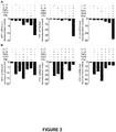
- Inventors next assessed the biological activities of pro- or anti-inflammatory cytokines on the expression of genes involved in melanocyte function, such as microphthalmia associated transcription factor (MITF, the melanocyte master transcription factor) and tyrosinase (TYR, involved in melanogenesis), as well as E-cadherin encoding gene (CDH1), a protein critical for melanocyte adhesion to keratinocytes and to the epidermis basal membrane. More precisely, they evaluated the effects of TNF ⁇ , IFN ⁇ , IL-17, IL-1 ⁇ , and TGF ⁇ , alone or in combination, on primary cultures of human epidermal melanocytes ( Figure 2 ).
- MITF microphthalmia associated transcription factor
- TGF E-cadherin encoding gene
- TNF ⁇ or TNF ⁇ inhibited the expression of genes involved in melanocyte differentiation, function, melanin synthesis, and melanocyte adhesion ( Figure 2A ); TGF ⁇ was also able to downregulate melanocyte function, although at a weaker level, whereas IL-17 and IL-1 ⁇ had no or little effect. Strikingly, the combination of the 5 cytokines resulted in a robust inhibition of the studied genes. Inventors more specifically identified TNF ⁇ and IFN ⁇ as the most potent cytokines that inhibit melanocyte function and adhesiveness ( Figure 2B ).
- TNF ⁇ and IFN ⁇ act synergistically on melanocytes to inhibit pigmentation and induce inflammatory mediators
- TNF ⁇ and IFN ⁇ synergized to downregulate the expression of genes involved in pigmentation signaling, such as MITF, the melanocyte master transcription factor, tyrosinase (TYR), and dopachrome tautomerase (DCT).
- TNF ⁇ and IFN ⁇ also synergized to upregulate the expression of inflammatory genes in melanocytes, including IL-6, TNF ⁇ , and intercellular adhesion molecule-1 (ICAM-1), and they also induced production of the CXCR3 ligands CXCL9 and CXCL10 ( Figure 3B ).
- E-cadherin which is the major mediator of human melanocytes adhesion, was recently shown to be discontinuously distributed across melanocyte membranes of vitiligo patients ( Wagner et al. J Invest Dermatol. 2015 ).
- E-cadherin is a transmembrane protein existing in two forms: the full length E-cadherin and soluble E-cadherin ( David et al. Cancer Res 2012 ).
- the soluble form has been shown to be upregulated in the sera of patients with autoimmune diseases ( Jin et al. J Rheumatol 2013 ; Matsuoyoshi et al. Br J Dermatol 1995 ).
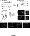
- Inventors determined the levels of soluble E-cadherin in sera of vitiligo patients. As shown in Figure 4C , levels of soluble E-cadherin were significantly higher in vitiligo patients compared to healthy controls. In addition, such levels tend to correlate with disease activity indicate that levels of soluble E-cadherin can be used as a marker for disease activity.
- MMP9 matrix metalloproteinase
- Inventors showed that MMP9 levels were significantly higher in serum of patients with vitiligo than in healthy controls ( Figure 8 ). MMP9 levels were significantly higher in patients with active disease compared to stable vitiligo. Strikingly, they observed a positive correlation between MMP9 and soluble E-cadherin levels in vitiligo patients.
- inventors prepared an in vitro 3D cellular model of containing both melanocytes and keratinocytes. Strikingly, they demonstrated that the combined activities of TNF ⁇ and IFN ⁇ induce melanocyte detachment from the model basal layer, at least in part mediated through alteration of E-cadherin expression ( Figure 4E ). A decrease in DCT, a melanogenesis-associated gene, was also observed ( Figure 7 ).
- TNF ⁇ and IFN ⁇ induce MMP9 gene expression in melanocyte.
- SB3CT a non-selective MMP2 and MMP3 inhibitor
- ab142180 a selective MMP9 inhibitor
- Pigmented epidermis are reconstructed from normal human keratinocytes (NHEK) and from normal human melanocytes (NHEM), previously isolated from preputial samples.
- RHEm human pigmented epidermis
- composition of the culture medium without hydrocortisone complete EpiLife® Medium (cascade biologics ® base medium reference M-EPI-500-CA + supplements reference S-001-K among which IGF-1 and hydrocortisone aliquots have not been used) supplemented with:
- the inserts containing the reconstructed epidermis are collected with pliers, left on a towel in order to remove residual liquid under the insert, then the reconstructed epidermis were cut from the support with a scalpel, put in a formaldehyde solution before dehydration, and then embedded in paraffin.
Landscapes
- Health & Medical Sciences (AREA)
- Engineering & Computer Science (AREA)
- Life Sciences & Earth Sciences (AREA)
- Biomedical Technology (AREA)
- Chemical & Material Sciences (AREA)
- Biotechnology (AREA)
- Bioinformatics & Cheminformatics (AREA)
- Organic Chemistry (AREA)
- Zoology (AREA)
- Genetics & Genomics (AREA)
- Wood Science & Technology (AREA)
- Cell Biology (AREA)
- General Health & Medical Sciences (AREA)
- Biochemistry (AREA)
- Microbiology (AREA)
- General Engineering & Computer Science (AREA)
- Immunology (AREA)
- Dermatology (AREA)
- Urology & Nephrology (AREA)
- Molecular Biology (AREA)
- Hematology (AREA)
- Tropical Medicine & Parasitology (AREA)
- Pathology (AREA)
- General Physics & Mathematics (AREA)
- Analytical Chemistry (AREA)
- Physics & Mathematics (AREA)
- Medicinal Chemistry (AREA)
- Food Science & Technology (AREA)
- Toxicology (AREA)
- Measuring Or Testing Involving Enzymes Or Micro-Organisms (AREA)
- Medicines That Contain Protein Lipid Enzymes And Other Medicines (AREA)
- Cosmetics (AREA)
Abstract
Description
- The present invention relates to the field of tissue engineering, dermatology and cosmetic. The invention more specifically relates to a three-dimensional cellular model of depigmenting disorders as well as to products and methods for preparing such a model. It further relates to uses of the model in experimental research, typically for studying pigmentation/depigmentation mechanisms and for the screening of molecules of interest in the prevention or treatment of pathological conditions wherein a depigmenting disorder is observed, in particular vitiligo as well as other inflammatory dermatoses.
- Vitiligo is the most common skin depigmenting disorder. The estimated prevalence of vitiligo is around 0.5 % to 1% of the world population. The two sexes are equally affected, and there are no apparent differences in rates of occurrence according to skin type or race. It is clinically characterized by symmetric and bilateral white macules, resulting from a loss of epidermal melanocytes, the pigment-producing cells.
- Several mechanisms have been implicated to explain melanocyte disappearance, including genetic predisposition, environmental triggers (such as friction), metabolic alteration, and altered inflammatory and immune responses. Vitiligo is described as an acquired chronic depigmenting disorder of the skin which is associated with autoimmune components involving both CD4+ and CD8+ T cells. However the phenotype and contribution of effector memory T (TEM) cell subsets remain to date unclear and controversial due to the lack of extensive analysis in models of the disease.
- Depigmentation can also occur in several inflammatory skin diseases/dermatoses such as psoriasis, atopic dermatitis, scleroderma, typically cutaneous scleroderma, hypomelanose and leukotrichia. Vitiligo in particular is a stigmatizing disease with major social and psychological impact. Often seen as purely aesthetic, this disorder is indeed responsible for a major impairment of quality of life sometimes comparable to other chronic diseases such as depression. Moreover patients with vitiligo often display other chronic inflammatory disorders such as autoimmune thyroiditis, rheumatoid arthritis and inflammatory bowel diseases. However, despite its prevalence and impact, no specific curative intervention has been reported so far. To date, no treatment has demonstrated efficacy against depigmentation. In addition, there is no treatment allowing the blockade of depigmentation or skin repigmentation, preferably definitive skin repigmentation. Current treatments have overall limited efficacy and include topical steroids or calcineurin inhibitors, systemic steroids and/or UV-light, all of which have potential long-term side-effects.
- Despite significant unmet treatment needs, there is currently no cellular model, in particular no ex vivo or in vitro three-dimensional cellular model, on the market for the specific study of depigmentation mechanisms, in particular depigmentation's pathogenesis, or for the testing of molecules that may be of interest for the prevention, treatment and/or alleviation of symptoms of a depigmenting disorder.
- The present invention provides for the first time a three-dimensional model of depigmenting disorder allowing the specific study of molecular mechanisms leading to the modulation of pigmentation, typically to depigmentation, and the development of methods for the screening of modulatory molecules in order to identify new drugs of interest and prepare preventive as well as therapeutic compositions, typically compositions capable of preventing or of attenuating depigmentation, in particular of preventing or of attenuating the loss of epidermal melanocytes, or of inducing a repigmentation of the skin.
- Inventors indeed discovered the synergistic ability of the particular combination of TNFα and IFNγ to modify a cellular structure comprising a pigmented/melanised epidermis (containing both keratinocytes and melanocytes) in order for it to mimic defects observed in skin of patients affected by a depigmenting disorder, in particular to induce melanocytes detachment from the basal layer of an epidermis. Based on this discovery they have developed a method for producing in vitro, ex vivo or in vivo cellular models, in particular three-dimensional cellular models, mimicking a depigmenting disorder, and herein describe such models.
- A first object herein described is thus a cellular model of depigmenting disorder wherein the model i) comprises melanocytes, and ii) has been exposed to interferon gamma (IFNγ) and Tumor necrosis factor alpha (TNFα).
- A second object herein described is a three-dimensional cellular model of depigmenting disorder wherein the model i) comprises keratinocytes and melanocytes, and ii) has been exposed to interferon gamma (IFNγ) and Tumor necrosis factor alpha (TNFα).
- Another object herein described is a kit comprising IFNγ and TNFα, and its use for preparing in vitro or ex vivo a three-dimensional cellular model of depigmenting disorder.
- Also herein described is the in vitro, ex vivo or in vivo use of a combination of IFNγ and TNFα for preparing a cellular model of depigmenting disorder, in particular a three-dimensional cellular model.
- Further herein described is a method of preparing in vitro, ex vivo or in vivo a cellular model of depigmenting disorder, in particular a three-dimensional cellular model, comprising a step of exposing, simultaneously or sequentially, a population of cells comprising at least melanocytes, preferably keratinocytes and melanocytes, or a skin sample, to IFNγ and TNFα. Also described is the use of a cellular model of depigmenting disorder according to the invention for studying in vitro or ex vivo for example pigmentation/depigmentation mechanisms and potential modulators of these mechanisms. The cellular model can also be used in a method for screening, typically in vitro or ex vivo, pharmaceutically active molecules suitable for preventing, treating or attenuating a depigmenting disorder, disease or dysfunctional state in a subject in need thereof.
- A method for screening, typically in vitro or ex vivo, pharmaceutically active molecules suitable for preventing, treating or attenuating a depigmenting disorder in a subject in need thereof is also described. This method comprises a step of exposing a cellular model of depigmenting disorder according to the invention to a molecule to be tested and a step of evaluating the ability of the tested molecule to inhibit MMP9 expression or function, to inhibit both MMP9 and Janus kinases (JAK) expression or function, in particular the expression or function of a JAK selected from JAK1, JAK2, JAK3, TYK2 and any combination thereof to induce melanin production and/or melanocyte function, to cleave E-cadherin, to increase soluble E-cadherin expression, to increase cytoplasmic redistribution of E-cadherin, to relocalize membrane bound E-cadherin, and/or to stabilize melanocytes to the basement membrane of the epidermis, said ability(ies) indicating that the tested molecule is a pharmaceutically active molecule suitable for preventing, treating or attenuating a depigmenting disorder in a subject in need thereof.
- The skin is a large and complex tissue providing a protective interface between an organism and its environment. Epidermis forms its external surface, and is mainly constituted of multiple layers of specialized epithelial cells named keratinocytes. Melanocytes are melanin-producing cells located in the bottom layer of the epidermis and are responsible for the production of melanin: the pigment primarily responsible for skin color. Skin can be injured by many different causes, including microorganisms, chemicals, behaviours, physical injury, ageing, U.V. irradiation, cancer, autoimmune or inflammatory diseases.
- The present invention advantageously provides new means for studying and understanding specific mechanisms leading to a disorder of skin pigmentation, typically to a depigmenting disorder, i.e. to a disorder inducing a depigmentation.
- A loss of epidermal melanocytes and/or the detection of functional abnormalities of epidermal melanocytes (such as abnormalities of melanocyte adhesion to the basement membrane of epidermis, melanocytes proliferation's abnormalities, increase of inflammatory proteins secretion, abnormalities of melanin and/or melanosomes transfer to keratinocytes) are the pathogenic hallmark of disorders associated with skin depigmentation. Vitiligo, the most common depigmenting disease is associated with complete loss of melanocytes. Factors involved in the initiation of vitiligo remain largely unknown, but likely include genetic predisposition and environmental triggers (such as friction). However, the mechanisms leading to the loss of melanocytes are still in debate. Several theories have been proposed. Melanocyte-intrinsic abnormalities in vitiligo leading to impaired melanocyte degeneration and/or proliferation support the hypothesis that the disease could be due to a primary defect of melanocytes. However, other observations strongly support the role of the autoimmune system, particularly in the chronic and progressive phases of vitiligo. Genome wide association studies have shown that 90% of vitiligo susceptibility loci encode components of the immune system. Furthermore, vitiligo is often associated with autoimmune diseases, and both innate and adaptive immune cells are found in patients' affected skin. Loss of melanocytes could result from cell destruction through melanocyte-specific cytotoxic immune responses (mainly through CD8+ cells) and/or melanocyte detachment through a defective adhesion system, also called melanocytorrhagy. E-cadherin, which is the major mediator of human melanocytes adhesion in the epidermis, was recently shown to be absent or discontinuously distributed across melanocyte membranes of vitiligo patients. Such alteration is responsible for melanocytes detachment from the basal layer of the epidermis and their subsequent loss.
- TNFα has been largely described for its dual role as both a pro-apoptotic and a pro-inflammatory cytokine. Anti-TNF therapies have proven efficacy in a number of autoimmune and inflammatory disorders, in particular in the field of dermatology, rheumatology, and gastroenterology. Conflicting results have been reported regarding a deregulation of TNFα production in peripheral mononuclear cells and sera from vitiligo patients (Camara-Lemarroy et al.). Regarding TNFα secreting cells in patients, cytotoxic CD8+ and CD4+ T cells have been described as potent producers; yet keratinocytes, fibroblasts, and even melanocytes could be other sources of this cytokine under inflammatory conditions. Regarding the possible involvement of TNFα in vitiligo pathophysiology, in vitro studies revealed direct effects of this pro-inflammatory cytokine on melanocyte function. Indeed, TNFα inhibits the expression of microphtalmia associated transcription factor (MITF), essential for melanocyte development, survival, and function, of tyrosinase and tyrosinase related protein 1 (TRP-1), two enzymes involved in melanogenesis. Furthermore, TNFα could enhance T cell trafficking to the skin through upregulation of intercellular adhesion molecule-1 (ICAM-1) expression both in melanocytes and keratinocytes. ICAM-1 is important in the initiation of inflammation and has been detected in melanocytes around active vitiligo patches.
If data from the literature tends to support a role of TNFα in vitiligo, the limited available evaluation of anti-TNF therapies in vitiligo suggests that blocking TNFα is effective at best in halting disease progression but does not induce significant repigmentation. - IFNγ is the most important cytokine related to the Th1 immune response. Binding of this cytokine to its receptor induces the Janus kinase (JAK) / signal transducer and activator of transcription (STAT) pathway, in particular JAK1, JAK2 and STAT1 activation, and regulates the expression of genes/proteins involved in a number of processes, such as defense against pathogens, proliferation, survival/apoptosis, immunomodulation, and leukocyte trafficking. As herein demonstrated, the expression of IFNγ is increased in the skin of patients with vitiligo. Comparison of the transcriptional profile of lesional vitiligo skin with healthy skin revealed an IFNγ specific signature in vitiligo. During disease, IFNγ is mainly produced by cytotoxic CD8+ and CD4+ T cells.
- In the experimental part of the present description, inventors have demonstrated the infiltration of skin inflammatory T cells expressing TNFα and IFNγ in progressive vitiligo and the involvement of TNFα and IFNγ in melanocytes loss through alteration of E-cadherin expression and induction matrix metalloproteinase-9 (MMP9), an enzyme described to cleave E-cadherin (Symowicz et al. Cancer Res 2007). In addition, they have discovered the surprising synergistic action of two cytokines, namely TNFα and IFNγ, in i) downregulating the expression of genes involved in pigmentation signaling, such as in particular MITF, the melanocyte master transcription factor, tyrosinase (TYR), and dopachrome tautomerase (DCT), ii) upregulating the expression of inflammatory genes in melanocytes, including IL-6, TNFα, and intercellular adhesion molecule-1 (ICAM-1), and iii) inducing the production of the CXCR3 ligands CXCL9 and CXCL10. Inventors further demonstrated that TNFα and IFNγ synergistically downregulate the expression of CDH1 transcripts in melanocytes but not in keratinocytes. They showed that TNFα upregulated expression of MMP9 gene in melanocytes and that this effect was also surprisingly potentiated in the presence of IFNγ. In addition, they showed that TNFα upregulated expression of MMP9 gene in keratinocytes.
- Inventors herein describe for the first time three-dimensional cellular models of depigmenting disorder. Models of the invention comprise at least melanocytes, preferably at least melanocytes and keratinocytes, for example melanocytes, keratinocytes and fibroblasts, and advantageously reproduce features typical of a skin suffering from a depigmenting disorder, for example an inflammatory depigmenting disorder. Preferred features are those whose manipulation is desirable for purposes of research and development of new pharmacological molecules or of new combinations of pharmacological molecules for preventing, treating or alleviating a depigmenting disorder, disease or dysfunctional state, for example an inflammatory depigmenting disorder, disease or dysfunctional state.
- A typically preferred feature of the model of the invention relates to the presence in said model of "detached" melanocytes, i.e. of melanocytes which are detached from the basal layer of the epidermis, and/or from neighboring/adjacent keratinocytes. In a typical model, "detached" melanocytes represent the majority of melanocytes of the cellular model, i.e. more than 50%, for example at least 60%, 70%, 80%, 90%, 95%, 96%, 97%, 98% or 99% of melanocytes.
- The number of melanocytes can be easily counted and the proportion of melanocytes detached from the basal layer can be easily determined by the skilled person using conventional microscopy and marking/labeling methods of the art.
- The model of the invention typically exhibits in addition several (at least two) or all of the herein below described features.
- Another typical feature of the model of the invention relates to the presence therein of dead (or apoptotic) melanocytes, typically of disrupted melanocytes.
- A further feature of the model of the invention relates to the analysis of cell death through investigation of the expression of
caspase 3 or DNA fragmentation ("TUNEL" assay - Methods Mol Biol. 2002; 203:21-30. TUNEL assay. An overview of techniques. Loo DT). - Another feature of the model of the invention relates to a decreased expression of melanin (the pigment produced by melanocyte responsible for the skin coloration) when compared to a portion of the cell structure used to prepare the model which has not been exposed to the combination of TNFα and IFNγ.
- An additional feature of the model of the invention relates to an increase of soluble (cleaved) E-cadherin or of altered membranous E-cadherin expression (concentration) and/or to an increase of membranous E-cadherin cytoplasmic relocation, when compared to a portion of the cell structure used to prepare the model which has not been exposed to the combination of TNFα and IFNγ. Soluble E-cadherin levels can be typically assessed by ELISA in cell free culture supernatants (for example, with a kit from R&D systems). Membrane full length E-cadherin expression can be assessed by immunofluorescence.
- A further feature of the model of the invention relates to the inhibition of the expression, or to the decreased expression, of Melanocyte Master Transcription Factor (MITF), Tyrosinase (TYR), Cadherin-1 also known as E-cadherin (CDH1), Dopachrome tautomerase (DCT), Tyrosinase Related Protein 1 (TRP1), connective tissue growth factor (CCN2), nephroblastoma overexpressed (CCN3) and Discoidin Domain Receptor family, member 1 (DDR1) genes and/or proteins, preferably of the DCT gene or protein, when compared to a portion of the cell structure used to prepare the model which has not been exposed to the combination of TNFα and IFNγ.
- Another feature of the model of the invention relates to the stimulation of the expression of interleukin-6 (IL6), tumor necrosis factor (TNF), in particular TNFα, and intercellular adhesion molecule-1 (ICAM-1) genes and/or proteins, when compared to a portion of the cell structure used to prepare the model which has not been exposed to the combination of TNFα and IFNγ.
- A further feature of the model of the invention relates to the increased expression of C-X-C Motif Chemokine Receptor 3 (CXCR3) ligands such as C-X-C Motif Chemokine ligand 9 (CXCL9) and C-X-C Motif Chemokine ligand 10 (CXCL10).
- Assessment of the expression of the herein above identified proteins or genes can be easily performed by the skilled person respectively by immunohistochemistry/immunofluorescence and real time PCR. Chemokines levels can also be assessed by ELISA in cell-free supernatants.
- A typical model is a three-dimensional cellular model of depigmenting disorder wherein the model i) comprises keratinocytes and melanocytes and ii) has been exposed to interferon (IFN), in particular interferon gamma (IFNγ), and Tumor necrosis factor alpha (TNFα). The model can further comprise fibroblasts. The model, typically the in vitro three-dimensional model containing both melanocytes, keratinocytes, and fibroblasts, or the ex vivo model (ex vivo culture of a skin biopsy or plasty), can also comprise a cutaneous/skin immune system, i.e. immune cells such as CD8+ and CD4+ T cells.
- A typical cellular model of the invention is an in vitro or ex vivo three-dimensional cellular model. Such a cellular model is typically a skin model, an epidermis model, or a tissue model comprising an epidermis and a dermis.
- The cellular model of the invention is typically prepared from mammal cells, for example from non-human primate (monkeys) cells, from domesticated animals cells (e.g., cows, sheep, pigs, rabbits cats, dogs, and horses cells), or from rodent cells (such as rat or mouse cells). The cellular model of the invention is preferably prepared from human cells.
- The cellular model of the invention can be prepared from cells from a healthy subject or from a patient suffering from a depigmenting disorder or from an inflammatory skin disease, typically an inflammatory skin disease where depigmentation is occurring or associated with depigmentation.
- In the context of the invention, the "subject" is an animal, typically a mammal. Examples of mammals include humans and non-human animals such as, without limitation, domesticated animals (e.g., cows, sheep, pigs, rabbits cats, dogs, and horses), non-human primates (such as monkeys), and rodents (e.g., mice and rats). The "subject" is preferably a human being, whatever its gender, age, race or sex.
- Typically, the cellular model of the invention is prepared from cells from a patient suffering from, or diagnosed or identified as suffering from, a depigmenting disorder, disease or dysfunctional state, an inflammatory skin disease, or a depigmenting disorder associated to an inflammatory skin disease (the depigmenting disorder being typically a symptom of the inflammatory skin disease). Preferably, the depigmenting disorder, disease or dysfunctional state is not a depigmenting disorder, disease or dysfunctional state of genetic or infectious origin.
- In such a context, the cells used to prepare the model are preferably healthy cells from the patient or in other words cells from a biopsy performed in an unaffected area of the patient, typically a pigmented area of the skin patient.
- In a particular example, the cellular model of the invention is prepared from cells from a patient suffering from an inflammatory skin disease (also herein identified as "inflammatory dermatose") selected from vitiligo, psoriasis, atopic dermatitis, scleroderma, typically systemic sclerosis, preferably cutaneous scleroderma, hypomelanose and leukotrichia. The patient suffering from the inflammatory skin disease typically has or is at risk of developing a depigmenting disorder.
- Psoriasis is a chronic inflammatory multisystem disease, with predominantly skin and joint manifestations, affecting approximately 2% of the population. Almost 90% of affected individuals have psoriasis vulgaris. The major manifestation of psoriasis is chronic inflammation of the skin. It is characterised by disfiguring, scaling and erythematous plaques that may be painful or often severely pruritic. Psoriasis is a chronic disease that waxes and wanes during a patient's life time. Plaque psoriasis is the most common form, affecting approximately 80% to 90% of patients. Plaque psoriasis manifests as well-defined, sharply demarcated, erythematous plaques varying in size from 1 cm to several centimeters. Patients may have involvement ranging from only few plaques to numerous lesions covering almost the entire body surface. The inflammatory response developed in psoriasis can lead to skin depigmentation according to the same ranging. This manifestation alters the patients' life quality in the same way vitiligo does, the unaesthetic manifestations leading to depressive disorder or social withdrawal. Moreover spontaneously occurring vitiligo can be associated with psoriasis.
- Atopic dermatitis (AD) is a chronic relapsing inflammatory skin disorder that affects approximately 2% to 5% of the adult population in the western world. The disease involves complex combinatorial pathogenic effects, multiple susceptibility genes, environmental triggers and disruption of the epidermal barrier. Clinically, AD can result in impairment of skin function, with intense and bothering itching leading to considerable loss of sleep and poor quality of life that is usually associated with disease severity. The inflammatory response developed in Atopic Dermatitis can lead to skin depigmentation according to the same ranging. This manifestation alters the patients' life quality in the same way vitiligo does, the unaesthetic manifestations leading to depressive disorder or social withdrawal. Moreover spontaneously occurring vitiligo can be associated with atopic dermatitis.
- Scleroderma is an inflammatory disease. There are two forms of the disease: i) localized scleroderma (called "morphea"), involves isolated patches of hardened skin with no internal involvement and ii) systemic scleroderma (also called "systemic sclerosis") comprising two subgroups based on the extent of skin involvement (herein generally identified as "cutaneous scleroderma"): the subgroups are diffuse cutaneous scleroderma and limited cutaneous scleroderma. Systemic sclerosis can be associated with progressive visceral organs defects, including the kidneys, heart, lungs and gastrointestinal tract. Skin manifestations are progressive skin tightness and induration and prominent skin pigmentary changes (both hyperpigmentation and hypopigmentation). Those skin manifestations, as discussed with psoriasis and vitiligo, are responsible for a major alteration of patients' quality of life.
In a preferred embodiment, the cellular model of the invention is prepared from healthy cells from a healthy patient. - The cellular model of the invention is typically prepared from a population of cells from a subject as herein defined who is either a healthy subject or a patient suffering from a disease or disorder as herein described. The population of cells comprises at least melanocytes, preferably at least melanocytes and keratinocytes, and can also contain fibroblasts and/or immune cells from the cutaneous/skin immune system such as CD8+ and CD4+ T cells. The population of cells can be prepared from cells extracted from a biological sample (biopsy or plasty) of the subject. The biological sample is typically a (healthy) skin sample obtained by surgery. The healthy skin sample is preferably a foreskin sample, but it can also be an abdominal or mammary/breast skin sample. The skin sample is typically a healthy skin sample obtained by surgery. A skin biopsy can also be obtained from a patient suffering from an inflammatory depigmenting disorder. The skin sample may vary from 1 to 50 cm2 in size. The skin biopsy is preferably from 3 to 6 mm in diameter.
- The population of cells can be used to prepare a reconstructed cellular structure such as a skin, an epidermis or a tissue-like or skin-like structure containing an epidermis and a dermis. Such a population of cells or reconstructed cellular/cell structure is typically exposed to a culture medium.
Culture medium usable in the context of the present invention are standard culture medium classically used by the skilled person such as Epilife® Medium, CnT-Prime 3D Barrier® Medium (CELLnTEC) and 3D-Skin Medium® (PrimCells). - A culture medium usable in the context of the invention is for example a complete EpiLife® Medium, such as for example the cascade biologics® base medium reference M-EPI-500-CA.
- The culture medium is typically supplemented for example with insulin, vitamin C, keratinocyte growth factor (KGF) and/or CaCl2.
Preferably, the culture medium is supplemented with 5 µg/ml of bovine insulin, 50 µg/ml of vitamin C, 3 ng/ml of keratinocyte growth factor (KGF), and 1,5 mM of CaCl2. Even more preferably, the culture medium comprises, or consists in, 20% pituaray extract, 5µg/ml bovin transferrin bovin, 0.2ng/ml recombinant human EGF (supplements Epilife), 5 µg/ml of bovine insulin, 50 µg/ml of vitamin C, 3 ng/ml of keratinocyte growth factor (KGF), and 1,5 mM of CaCl2. - In a particular embodiment, when the model is a skin model, typically an ex vivo skin model, the culture medium comprises DMEM, fetal calf serum, typically 10% of fetal calf serum, and hydrocortisone, typically 0,4 µg/ml of hydrocortisone (such as a Bioalternatives® culture medium).
- Whatever their origins, inventors herein reveal for the first time that cells or cell structures used to prepare the model of the invention are to be exposed specifically to both interferon gamma (IFNγ) and Tumor necrosis factor alpha (TNFα).
The use of a combination of IFNγ and TNFα for preparing a cellular model of depigmenting disorder, in particular a three-dimensional cellular model, is thus herein described, in particular the in vitro or ex vivo use of such a combination. IFNγ and TNFα can for example be introduced into the culture medium.
As well, a composition and a kit comprising IFNγ and TNFα are herein disclosed.
The composition comprising IFNγ and TNFα can further comprise a pharmaceutically acceptable carrier or support, typically a culture medium as herein above described.
Culture media usable in the context of the present invention for a skin sample are standard culture media classically used by the skilled person such as Dulbecco's minimal essential medium (DMEM) supplemented or not with serum.
A particular kit comprising IFNγ and TNFα further comprises a culture medium as herein described and optional supplements thereof such as for example epidermal growth factor (EGF), transforming growth factor-α (TGFα), keratinocyte growth factor (KGF), the medium and each of the supplements being present in distinct containers.
Another particular kit comprising IFNγ and TNFα further comprises a culture medium as herein described and optional supplements thereof such as for example Epilife® Medium supplements and/or insulin, the medium and each of the supplements being preferably present in distinct containers. - Also disclosed is the use of a kit as herein described comprising IFNγ and TNFα for preparing in vitro or ex vivo a cellular model of depigmenting disorder, in particular a three-dimensional cellular model.
- Inventors further herein provide a method of preparing, typically in vitro or ex vivo, a cellular model of depigmenting disorder, in particular a three-dimensional cellular model of depigmenting disorder. This method comprises a step of exposing a population of cells or cell structure, typically a reconstructed pigmented/melanised epidermis (containing or not dermis), comprising at least melanocytes, preferably at least keratinocytes and melanocytes, but could also contain melanocytes, keratinocytes and fibroblasts, or a (whole) skin sample, to IFNγ and TNFα.
- Cells or cell structures, typically cultured cells or cultured cell structures, used to prepare a model of the invention can be exposed to IFNγ and TNFα simultaneously or sequentially and in any order, i.e. IFNγ before TNFα or TNFα before IFNγ. They are typically exposed to IFNγ and TNFα at a concentration and for a time sufficient, preferably about 24 hours, to allow the cell structure or biopsy to acquire features (such as anyone of those described previously) of a skin suffering from a depigmenting disorder, typically from vitiligo, preferably at a concentration and for a time sufficient for the cell structure to mimic a depigmenting disorder, typically vitiligo. A preferred acquired feature is "melanocytes detachment".
The method of the invention typically comprises a step of exposing cells to a culture or culture medium, wherein the medium comprises IFNγ and TNFα. In a particular embodiment, IFNγ and TNFα are respectively present in a concentration ranging from about 0.1 to 50 ng/ml of medium. Preferably, IFNγ and TNFα are each present at a concentration of about 10 ng/ml of medium. - Typically, cells are exposed to a concentration of IFNγ ranging from about 0.1 to 50 ng/ml of medium, typically from about 1 to 50 ng/ml of medium, from about 5 to 30 ng/ml of medium, from about 5 to 20 ng/ml of medium, from about 5 to 15 ng/ml or from about 8 to 12 ng/ml of medium, for example to a concentration of IFNγ of about 1, 2, 3, 4, 5, 6, 7, 8, 9, 10, 11, 12, 13, 14, 15, 16, 17, 18, 19, 20, 21, 22, 23, 24 or 25 ng/ml of medium, and to a concentration of TNFα ranging from about 0.1 to 50 ng/ml of medium, typically from about 5 to 30 ng/ml of medium, from about 5 to 20 ng/ml of medium, from about 5 to 15 ng/ml or from about 8 to 12 ng/ml of medium, for example to a concentration of TNFα of about 1, 2, 3, 4, 5, 6, 7, 8, 9, 10, 11, 12, 13, 14, 15, 16, 17, 18, 19, 20, 21, 22, 23, 24 or 25 ng/ml of medium. Cytokines are diluted in the medium which is typically a culture medium or a culture medium as herein described.
In another particular embodiment, the medium comprises IFNγ, TNFα and insulin. - Cells are typically exposed to each of IFNγ and TNFα for a period varying between about one hour and 6, 12, 24, 48, 72, 96 or 120 hours, preferably 24, 48 or 72 hours. Preferably, cells are exposed to each of IFNγ and TNFα for a period of 24h.
- The model of depigmenting disorder of the invention can be used advantageously for studying, typically for studying in vitro or ex vivo, pigmentation/depigmentation mechanisms as well as (potential) modulators of these mechanisms.
As used herein, the term "modulator" is defined as any molecule able to modulate (activate or inhibit) skin pigmentation or depigmentation. The modulator is typically a pharmaceutically active molecule.
In a preferred embodiment, the model of depigmenting disorder of the invention is used in a method for screening and studying in vitro or ex vivo pharmaceutically active molecules suitable for preventing, treating or attenuating a depigmenting disorder in a subject in need thereof. - A method for screening, typically for screening in vitro or ex vivo, pharmaceutically active molecules suitable for preventing, treating or attenuating a depigmenting disorder in a subject in need thereof is further herein described. This method comprises a step of exposing a cellular model of depigmenting disorder according to the invention to a molecule to be tested and a step of evaluating the ability of the tested molecule to inhibit MMP9 expression or function, to inhibit both MMP9 and Janus kinases (JAK) expression or function, in particular the expression or function of a JAK selected from JAK1, JAK2, JAK3, TYK2 and any combination thereof to induce melanin production and/or melanocyte function, to cleave E-cadherin, to increase soluble E-cadherin expression, to increase cytoplasmic redistribution of E-cadherin, to relocalize membrane bound E-cadherin, and/or to stabilize melanocytes to the basement membrane of the epidermis, said ability(ies) indicating that the tested molecule is a pharmaceutically active molecule suitable for preventing, treating or attenuating a depigmenting disorder in a subject in need thereof.
- The candidate (test) pharmacological molecules/agents can be derived for example from combinatorial peptide libraries, combinatorial chemical compound libraries, and natural products libraries. Molecules which can be tested on the cellular model of the invention are preferably selected for example from monoclonal antibodies, aptamers, spiegelmers, inhibitory nucleic acid sequences, small molecules. In a particular example, these molecules are directed against MMP9. Convenient reagents for such assays are known in the art.
The molecule to be tested can be introduced, typically diluted, in the culture medium into which the model is maintained, for example immersed, or directly applied on the upper cell layer of the model, typically on the epidermis or dermis.
The molecule effects are typically studied in order to evaluate its potential therapeutic, typically dermatological, or cosmetic effect.
The molecule to be tested can be introduced before, at the same time, or after the introduction of TNFα and IFNγ.
To evaluate the potential therapeutic effect of a test molecule, the localization of melanocyte within the epidermis can be assessed, typically the detached (from the basal layer of the epidermis) melanocytes and attached melanocytes present in the epidermis, can be respectively quantified, and compared to the condition where only TNFα and IFNγ are present (positive control of depigmentation). Evaluation/quantification of soluble E-cadherin levels and/or membrane bound E-cadherin expression and/or quantification of melanin content can also be assessed and compared to a positive control. E-cadherin expression in a skin sample is classically assessed by immunofluorescence, immunohistochemistry, or western blot. Soluble E-cadherin levels are classically determined by ELISA
Typically, pluralities of assay mixtures are run in parallel with different agent concentrations to obtain a different response to the various concentrations. Typically, one of these concentrations serves as a negative control, i.e., at zero concentration of agent or at a concentration of agent below the limits of assay detection. - The examples, which follow, and their corresponding figures illustrate the invention without limiting the scope thereof.
-
-
Figure 1 . Prominent infiltration of skin inflammatory T cells expressing TNFα and IFNγ in progressive vitiligo. T cells were isolated from perilesional skin of patients with a progressive (n=10) or a stable vitiligo (n=6). Results were compared to healthy controls (n=6). Analysis of IFNγ, TNFα, or IFNγ/TNFα producing skin infiltrating CD4 and CD8 T cells by flow cytometry. Each symbol represents one specimen. The mean + s.e.m. is shown . *p<0.05, **p<0.01, using a Mann Whitney test -
Figure 2 . TNFα and IFNγ: two major cytokines involved in melanocyte inhibition
A-B. Primary cultures of normal human epidermal melanocytes were stimulated in the presence or absence of the indicated cytokines for 24h (IL-17, IL-1β, TNFα, IFNγ: 20ng/ml; TGFβ: 5ng/ml). Real-time analysis of the indicated gene expression. Results are expressed as a fold decrease below control culture (no stimulation). GAPDH was used as a housekeeping gene. Data are representative of three independent experiments. -
Figure 3 . Involvement of TNFα and IFNγ in the inflammatory response and depigmentation.
A-B. Primary cultures of normal human epidermal melanocytes were stimulated in the presence or absence of 20ng/ml TNFα and/or IFNγ for 24h (A) or 48h (B). A. Real-time analysis of the indicated gene expression. Results are expressed as a fold increase above control culture (no stimulation) for upregulated genes (upper panel) or fold decrease below control culture for downregulated genes (lower panel). GAPDH was used as a housekeeping gene. B. Measurement of CXCL9 and CXCL10 secretion in cell-free supernatants by ELISA. -
Figure 4 . Involvement of TNFα and IFNγ in the inflammatory response and melanocyte loss.
A-B. Primary cultures of normal human epidermal melanocytes (A) or keratinocytes (B) were stimulated in the presence or absence of 20ng/ml TNFα and/or IFNγ for the indicated time. Real-time analysis of CDH1 gene expression. GAPDH was used as a housekeeping gene. C. Assessment of soluble E-cadherin levels in sera from healthy controls (n=20) or patients with a progressive (n=40) or a stable (n=40) vitiligo. Data are representative of three (A-D) or two (E) independent experiments. D. Real-time analysis of MMP9 gene expression in melanocytes treated 24h with or without 20ng/ml of TNFα and/or IFNγ. GAPDH was used as a housekeeping gene. E. Reconstructed human epidermis containing melanocytes were stimulated in the presence or absence of 20ng/ml of TNFα and IFNγ for 24h. Immunofluorescence microscopy analysis of melan-A (red) and E-cadherin (green) expression, Magnification 400x. E. Immunofluorescence microscopy analysis of melan-A (red) and E-cadherin (green) expression in healthy skin, perilesional skin of patients with stable or progressive vitiligo, and lesional psosiatic skin. Magnification 400x. White arrows show melanocytes that are detached from the basal layer of the epidermis. -
Figure 5 . TNFα and IFNγ -induced melanocyte loss is mediated through MMP9. Reconstructed human epidermis containing melanocytes were stimulated 24h with or without 20ng/ml of TNFα and IFNγ in the presence or absence of 10µM of MMP9 inhibitor ab 142180 or SB-CT. Immunofluorescence microscopy analysis of melan-A (red) and E-cadherin (green) expression, Magnification 400x. White arrows show melanocytes that are detached from the basal layer of the epidermis. -
Figure 6 . Involvement of TNFα and IFNγ in melanocyte loss.
Primary cultures of melanocytes were stimulated in the presence or absence of 20ng/ml of TNFα and IFNγ. Analysis using quantitative RT-PCR of the expression of the indicated genes after 24h of stimulation. The gene encoding GAPDH is used as a control gene. Results show the expression ratio between stimulated cells and control cells in the absence of cytokines [mean + standard error of the mean (SEM) of 3 experiments performed on 3 distinct samples]. Inventors observed a synergistic effect of the TNFα and IFNγ cocktail in the inhibition of genes encoding proteins involved in melanocyte function, melanogenesis and melanocytes adhesion. -
Figure 7 . Involvement of TNFα and IFNγ in melanocyte loss.
Reconstructed human epidermis containing melanocytes were stimulated in the presence or absence of 20ng/ml of TNFα and IFNγ. Analysis using quantitative RT-PCR of the DCT gene expression after 24h of stimulation. The genes encoding GAPDH and β-glucuronidase are used as control genes. Results show the expression ratio between the stimulated epidermis and the control epidermis without cytokines [mean + standard error of the mean (SME) of 2 experiments performed on 2 different samples]. Inventors observe a DCT gene inhibition in presence of TNFα. This effect is not modified by addition of IFNγ. -
Figure 8 . Cell death is not a primary mechanism involved in melanocyte loss.
(A-B). Reconstructed human epidermis containing melanocytes were stimulated in the presence or absence of 20ng/ml of TNFα and IFNγ for 24h. Analysis of Melan-A expression (red color reveals melanocytes) using immunofluorescence and detection of apoptotic cells (green) using TUNEL technique (results representative of 2 experiments performed on 2 distinct samples). Results show that TNFα and/or IFNγ have no effect on melanocytes apoptosis. A slight pro-apoptotic effect of TNFα on keratinocytes is observed. (B) Analysis of Melan-A expression (red color reveals melanocytes) using immunofluorescence and detection of apoptotic cells (green) using TUNEL technique in healthy skin (control), perilesional skin of patients with stable or progressive vitiligo, lesional skin of patients with psoriasis or lupus. Magnification 400x. TEN is a positive control for apoptosis. Results show that in vitiligo patients (with a stable or active disease), detached melanocytes do not enter apoptosis, emphasizing that melanocyte death is not the primary mechanism involved in their loss. -
Figure 9 . MMP9 and soluble E-cadherin levels in vitiligo patients' sera.
(A-B) Levels of soluble E-cadherin (A) and MMP9 (B) were determined by ELISA in the serum of patients with vitiligo (stable and progressive) or psoriasis, and in the serum of healthy controls. (C) Serum levels of MMP9 were correlated to serum levels of soluble E-cad in vitiligo patients (Spearman test p<0,05). Inventors show a significant increase of soluble E-cadherin and MMP9 levels in active and stable vitiligo patient's serum compared to healthy controls. Levels of MMP9 were significantly higher in vitiligo patients with active disease. In addition, a positive correlation was observed between MMP9 and soluble E-cadherin levels in vitiligo patients. - Inventors extracted T cells from perilesional skin of vitiligo patients with a stable or a progressive disease in order to analyze their propensity to produce inflammatory cytokines, in particular TNFα and IFNγ, two cytokines previously identified as potentially involved in disease development (reviewed in Boniface et al. G. Ital. Dermatol. Venereol 2016). Results were compared to those obtained from healthy individuals (
Figure 1 ). They show that CD4 and CD8 T cells extracted from the skin of vitiligo patients with active disease produce higher levels of the type-1 related cytokine IFNγ and TNFα, also known to be secreted by most of T cell subsets, suggesting their involvement in melanocyte loss. - Inventors next assessed the biological activities of pro- or anti-inflammatory cytokines on the expression of genes involved in melanocyte function, such as microphthalmia associated transcription factor (MITF, the melanocyte master transcription factor) and tyrosinase (TYR, involved in melanogenesis), as well as E-cadherin encoding gene (CDH1), a protein critical for melanocyte adhesion to keratinocytes and to the epidermis basal membrane. More precisely, they evaluated the effects of TNFα, IFNγ, IL-17, IL-1β, and TGFβ, alone or in combination, on primary cultures of human epidermal melanocytes (
Figure 2 ). IFNγ or TNFα inhibited the expression of genes involved in melanocyte differentiation, function, melanin synthesis, and melanocyte adhesion (Figure 2A ); TGFβ was also able to downregulate melanocyte function, although at a weaker level, whereas IL-17 and IL-1β had no or little effect. Strikingly, the combination of the 5 cytokines resulted in a robust inhibition of the studied genes. Inventors more specifically identified TNFα and IFNγ as the most potent cytokines that inhibit melanocyte function and adhesiveness (Figure 2B ). - Inventors evaluated the capability of TNFα and IFNγ to act in concert to modify melanocyte function. As shown in
Figure 3 andFigure 6 , TNFα and IFNγ synergized to downregulate the expression of genes involved in pigmentation signaling, such as MITF, the melanocyte master transcription factor, tyrosinase (TYR), and dopachrome tautomerase (DCT). TNFα and IFNγ also synergized to upregulate the expression of inflammatory genes in melanocytes, including IL-6, TNFα, and intercellular adhesion molecule-1 (ICAM-1), and they also induced production of the CXCR3 ligands CXCL9 and CXCL10 (Figure 3B ). These results reveal for the first time that a melanocyte can become an inflammatory cell under the influence of type-1-related cytokines and could amplify the inflammatory response observed in vitiligo. - Inventors showed that TNFα and IFNγ have a synergistic effect to downregulate the expression of CDH1 transcripts in melanocytes (
Figure 4A ), but not in keratinocytes (Figure 4B ). Such effects are of interest as E-cadherin, which is the major mediator of human melanocytes adhesion, was recently shown to be discontinuously distributed across melanocyte membranes of vitiligo patients (Wagner et al. J Invest Dermatol. 2015). E-cadherin is a transmembrane protein existing in two forms: the full length E-cadherin and soluble E-cadherin (David et al. Cancer Res 2012). The soluble form has been shown to be upregulated in the sera of patients with autoimmune diseases (Jin et al. J Rheumatol 2013; Matsuoyoshi et al. Br J Dermatol 1995). Inventors determined the levels of soluble E-cadherin in sera of vitiligo patients. As shown inFigure 4C , levels of soluble E-cadherin were significantly higher in vitiligo patients compared to healthy controls. In addition, such levels tend to correlate with disease activity indicate that levels of soluble E-cadherin can be used as a marker for disease activity. Several enzymes can cleave full length E-cadh and lead to the release of its soluble form, including matrix metalloproteinase MMP9 (aka gelatinase) (Symowicz et al. Cancer Res 2007). Inventors showed that MMP9 levels were significantly higher in serum of patients with vitiligo than in healthy controls (Figure 8 ). MMP9 levels were significantly higher in patients with active disease compared to stable vitiligo. Strikingly, they observed a positive correlation between MMP9 and soluble E-cadherin levels in vitiligo patients. Importantly, inventors showed that TNFα upregulated expression of MMP9 gene in melanocytes (Figure 4D ); such effect was potentiated in the presence of IFNγ. These results indicate that MMP9 is involved in the up-regulation of soluble E-cadherin in vitiligo. - Next, to better characterize the biological activities of TNFα and IFNγ on melanocyte adhesion, inventors prepared an in vitro 3D cellular model of containing both melanocytes and keratinocytes. Strikingly, they demonstrated that the combined activities of TNFα and IFNγ induce melanocyte detachment from the model basal layer, at least in part mediated through alteration of E-cadherin expression (
Figure 4E ). A decrease in DCT, a melanogenesis-associated gene, was also observed (Figure 7 ). Such phenotype clearly resembles the one observed in perilesional skin of vitiligo patients (with a stable or a progressive disease), where melanocytes are detached from the basal layer of the epidermis (Figure 4F ). Interestingly, such phenomenon is also observed in psoriasis (Figure 4F ), emphasizing that melanocyte adhesion defect can be extended to other skin inflammatory conditions like psoriasis. Moreover, the inventors showed that TNFα and IFNγ, alone or in combination, have little or no effect on melanocyte apoptosis (Figure 8A ). These data are in line with the observation that melanocytes do not undergo apoptosis in vitiligo patients perilesional skin (Figure 8B ), emphasizing that melanocytes' detachment is the primary mechanism involved in melanocyte loss in vitiligo. - Altogether, these results are of major interest and identify a new mechanism leading to melanocyte loss, with a critical role of TNFα and IFNγ. In addition, this in vitro model of depigmentation can advantageously be used to test new therapeutic targets in vitiligo patients, as well as in patients with depigmenting disorders associated with inflammatory skin diseases.
- As discussed above, TNFα and IFNγ induce MMP9 gene expression in melanocyte. Inventors used two commercially available synthetic MMP9 inhibitors, SB3CT (a non-selective MMP2 and MMP3 inhibitor) and a selective MMP9 inhibitor (ab142180), to assess whether such inhibition could inhibit TNFα and IFNγ effects on depigmentation. In vitro reconstructed human epidermis containing melanocytes were stimulated with these two cytokines in the presence or absence of the two different MMP9 inhibitors for 24h (
Figure 5 ). The combination of TNFα and IFNγ induced melanocyte detachment and E-cadherin inhibition. Importantly, such effect was strongly inhibited in the presence of either MMP9 inhibitor, as MMP9 inhibition led to melanocyte stabilisation to the basal membrane of the epidermis (Figure 5 ). These results are the first to demonstrate that IFNγ and TNFα effect on melanocyte adhesion is mediated through MMP9. - All together, these results support the concept that inhibiting MMP9 represent an attractive and novel strategy to prevent melanocyte loss in vitiligo, but also in depigmentation occurring in skin inflammatory disorders such as psoriasis, atopic dermatitis, scleroderma, hypomelanose or leukotrichia.
- Pigmented epidermis (RHEm) are reconstructed from normal human keratinocytes (NHEK) and from normal human melanocytes (NHEM), previously isolated from preputial samples.
- After 10 days of reconstruction (D10), reconstructed human pigmented epidermis (RHEm) are placed in 6 wells plate (NUNC reference 140675) in 2 ml of culture medium without hydrocortisone to which 10 ng/ml of interferon (IFN)-γ (R&D systems) and 10 ng/ml of "tumor necrosis factor (TNF)-α" (R&D systems) are added. A control condition without cytokines addition is performed. Each experimental condition is performed in duplicate or triplicate.
- Composition of the culture medium without hydrocortisone: complete EpiLife® Medium (cascade biologics ® base medium reference M-EPI-500-CA + supplements reference S-001-K among which IGF-1 and hydrocortisone aliquots have not been used) supplemented with:
- 5 µg/ml of bovine insulin (Sigma Aldrich I1882)
- 50 µg/ml of vitamin C (Sigma Aldrich A4403)
- 3 ng/ml of keratinocyte growth factor (KGF) (Millipore GF008)
- 1,5 mM of CaCl2 (Sigma Aldrich C7902).
- After a 24h incubation period, the inserts containing the reconstructed epidermis are collected with pliers, left on a towel in order to remove residual liquid under the insert, then the reconstructed epidermis were cut from the support with a scalpel, put in a formaldehyde solution before dehydration, and then embedded in paraffin.
- 5 µm sections of paraffin inclusions are deposited on slides for labelling by immunofluorescence to reveal MelanA and E-Cadherin according to the following protocol:
-
Placing the slides on a hot plate at 70° C 10 min Successive xylene baths 3 x 5 min -
Ethanol 100°1 min Ethanol 100° 1 min Ethanol 95° 1 min Ethanol 70° 1 min Ethanol 50° 1 min Distilled water 1 min PBS or TBS 5 min - Use of the Decloacking Chamber device BIOCARE Medical
Place the slides into the device at pH8
Launch the program (98°C - 20 min)
Wash with TBS (Tris buffer saline) 0.1% Tween at a temperature equal to cooling temperature (60-70°C).
Allow to come back to room temperature - 4- Wash with TBS 0.1% tween 3 x 5 min
- 5- Delimit the deposition areas with a hydrophobic pen
- 6- Put the primary anti-MELANAA antibody (1) at 1/100 dilution and the anti-E-CADHERIN antibody (2) at 1/100 dilution in the buffer (3)
Incubate overnight at 4°C - 7- The day after: wash with TBS 0.1% tween 3 x 5 min
Put the secondary antibody A555 (4) at 1/100 dilution in the buffer (3)
+ A 488 (5) at 1/100 dilution in the buffer (3) 60 min - 8- Wash with TBS 0.1% tween 3 x 5 min
- Assemble slide with medium assembly with "DAPI" (6) away from light.
- (1): DAKO Melan-A Clone A103 Isotype IgG1, Kappa (ref: M 7196)
- (2): Abcam rabbit anti-E-cadherin antibody (ref: ab 15148)
- (3): DAKO Antibody Diluent with background reducing components (ref: S3022)
- (4): Invitrogen Alexa 555 goat anti mouse IgG (ref: A21422)
- (5): Invitrogen Alexa 488 rabbit IgG(H+L) (ref: A21441) or Novus (ref: NBP 1-72944)
- (6): Molecular Probes Prolong gold antifade reagent with DAPI (ref: P36935)
- Conclusion: Inventors developed in vitro and ex vivo human 3D models that mimic depigmentation observed in inflammatory depigmenting disorders, in particular vitiligo, but also in skin inflammatory disorders where depigmentation can occur, such as psoriasis, atopic dermatitis, scleroderma, hypomelanose and leukotrichia. These in vitro and/or ex vivo models can be used to model inflammatory depigmentation to study the involved mechanisms and/or to test the potential of pharmaceutically active molecules to prevent, treat or attenuate a depigmenting disorder.
-
- Boniface K, Taieb A, Seneschal J. New insights into immune mechanisms of vitiligo. G. Ital. Dermatol. Venereol. 2016;151(1):44-54.
- Camara-Lemarroy CR, Salas-Alanis JC. The role of tumor necrosis factor-α in the pathogenesis of vitiligo. Am J Clin Dermatol. 2013;14:343-50.
- David JM, Rajasekaran AK. Dishonorable discharge: the oncogenic roles of cleaved E-cadherin fragments. Cancer Res 2012; 72: 2917-23
- Ezzedine K et al. Vitiligo is not a cosmetic disease. JAAD 2015; 73(5):883-5
- Jin T et al. Soluble E-cadherin in systemic lupus erythematosus. J Rheumatol 2013; 40: 1677-82.
- Matsuyoshi N et al. Soluble E-cadherin: a novel cutaneous disease marker. Br J Dermatol 1995; 132: 745-9.
- Natarajan VT et al. IFN-γ signaling maintains skin pigmentation homeostasis through regulation of melanosome maturation. Proc Natl Acad Sci USA. 2014;111:2301-6.
- Symowicz, J. et al. Engagement of collagen-binding integrins promotes matrix metalloproteinase-9-dependent E-cadherin ectodomain shedding in ovarian carcinoma cells. Cancer Res. 67, 2030-2039 (2007).
- Taïeb A, Picardo M. Clinical practice. Vitiligo. N Engl J Med. 2009;360:160-9.
- Wagner RY, et al. Altered E-Cadherin Levels and Distribution in Melanocytes Precede Clinical Manifestations of Vitiligo. J Invest Dermatol. 2015 Jul;135(7):1810-9
- Wang CQF et al. IL-17 and TNF synergistically modulate cytokine expression while suppressing melanogenesis: potential relevance to psoriasis. J Invest Dermatol. 2013;133:2741-52.
- Wang S et al. Interferon-γ induces senescence in normal human melanocytes. PloS One. 2014;9:e93232.
- Webb KC et al. Tumour necrosis factor-α inhibition can stabilize disease in progressive vitiligo. Br J Dermatol. 2015;173:641-50.
- Whitton, M. E. et al. Interventions for vitiligo. Cochrane Database Syst Rev CD003263 (2010).
- Yang L et al. Interferon-gamma Inhibits Melanogenesis and Induces Apoptosis in Melanocytes: A Pivotal Role of CD8+ Cytotoxic T Lymphocytes in Vitiligo. Acta Derm Venereol. 24 juin 2015;95(6):664-70.
Claims (15)
- Three-dimensional cellular model of depigmenting disorder wherein the model i) comprises keratinocytes and melanocytes and ii) has been exposed to interferon gamma (IFNγ) and Tumor necrosis factor alpha (TNFα).
- The cellular model of claim 1, wherein the cellular model is prepared from human cells.
- The cellular model of claim 1 or 2, wherein the cellular model is prepared from cells from a healthy subject or from a patient suffering from a depigmenting disorder or from an inflammatory skin disease where depigmentation is occurring.
- The cellular model of any one of claims 1 to 3, wherein the model is a skin model, an epidermis model, or a tissue model comprising an epidermis and a dermis and optionally cells of the skin immune system.
- The cellular model of any one of claims 1 to 4, wherein the depigmenting disorder is associated with an inflammatory skin disease.
- The cellular model of claim 5, wherein the inflammatory skin disease is selected from vitiligo, psoriasis, atopic dermatitis, scleroderma, hypomelanose and leukotrichia.
- The cellular model of any one of claims 1 to 6, wherein the depigmenting disorder is vitiligo.
- In vitro or ex vivo use of a combination of IFNγ and TNFα for preparing a three-dimensional cellular model of depigmenting disorder.
- Use of a kit comprising IFNγ and TNFα for preparing in vitro or ex vivo a three-dimensional cellular model of depigmenting disorder.
- A method of preparing in vitro or ex vivo a three-dimensional cellular model of depigmenting disorder comprising a step of exposing simultaneously or sequentially a population of cells comprising keratinocytes and melanocytes or skin sample, to IFNγ and TNFα.
- The method of claim 10, wherein the method comprises a step of exposing cells to a culture medium, wherein the medium comprises IFNγ and TNFα.
- The method of claim 10 or 11, wherein IFNγ and TNFα are respectively present in a concentration ranging from 1 to 50 ng/ml of medium, preferably are each present at a concentration of 10 ng/ml of medium.
- Use of a cellular model of depigmenting disorder according to any one of claims 1 to 7 for studying in vitro or ex vivo pigmentation/depigmentation mechanisms and modulators of these mechanisms.
- Use of a cellular model of depigmenting disorder according to any one of claims 1 to 7 in a method for screening in vitro or ex vivo pharmaceutically active molecules suitable for preventing, treating or attenuating a depigmenting disorder in a subject in need thereof.
- A method for screening in vitro or ex vivo pharmaceutically active molecules suitable for preventing, treating or attenuating a depigmenting disorder in a subject in need thereof, wherein the method comprises a step of exposing a cellular model of depigmenting disorder according to any one of claims 1 to 7 to a molecule to be tested and a step of evaluating the ability of the tested molecule to inhibit MMP9 expression or function, to inhibit both MMP9 and Janus kinases (JAK) expression or function, in particular the expression or function of a JAK selected from JAK1, JAK2, JAK3, TYK2 and any combination thereof to induce melanin production and/or melanocyte function, to cleave E-cadherin, to increase soluble E-cadherin expression, to increase cytoplasmic redistribution of E-cadherin, to relocalize membrane bound E-cadherin, and/or to stabilize melanocytes to the basement membrane of the epidermis, said ability(ies) indicating that the tested molecule is a pharmaceutically active molecule suitable for preventing, treating or attenuating a depigmenting disorder in a subject in need thereof.
Priority Applications (2)
| Application Number | Priority Date | Filing Date | Title |
|---|---|---|---|
| EP16306718.4A EP3336175B1 (en) | 2016-12-16 | 2016-12-16 | Three-dimensional model of depigmenting disorder |
| ES16306718T ES2960918T3 (en) | 2016-12-16 | 2016-12-16 | Three-dimensional model of depigmentation disorder |
Applications Claiming Priority (1)
| Application Number | Priority Date | Filing Date | Title |
|---|---|---|---|
| EP16306718.4A EP3336175B1 (en) | 2016-12-16 | 2016-12-16 | Three-dimensional model of depigmenting disorder |
Publications (3)
| Publication Number | Publication Date |
|---|---|
| EP3336175A1 true EP3336175A1 (en) | 2018-06-20 |
| EP3336175B1 EP3336175B1 (en) | 2023-08-30 |
| EP3336175C0 EP3336175C0 (en) | 2023-08-30 |
Family
ID=57714494
Family Applications (1)
| Application Number | Title | Priority Date | Filing Date |
|---|---|---|---|
| EP16306718.4A Active EP3336175B1 (en) | 2016-12-16 | 2016-12-16 | Three-dimensional model of depigmenting disorder |
Country Status (2)
| Country | Link |
|---|---|
| EP (1) | EP3336175B1 (en) |
| ES (1) | ES2960918T3 (en) |
Cited By (3)
| Publication number | Priority date | Publication date | Assignee | Title |
|---|---|---|---|---|
| CN111081325A (en) * | 2019-12-27 | 2020-04-28 | 医渡云(北京)技术有限公司 | Medical data processing method and device |
| JP2023096623A (en) * | 2021-12-27 | 2023-07-07 | 花王株式会社 | Method for evaluating or selecting agent for preventing or improving vitiligo |
| JP2023184306A (en) * | 2022-06-17 | 2023-12-28 | 株式会社 資生堂 | Cosmetic method by controlling E-cadherin expression and method for predicting age spots based on E-cadherin expression |
Citations (4)
| Publication number | Priority date | Publication date | Assignee | Title |
|---|---|---|---|---|
| US5861153A (en) * | 1996-01-23 | 1999-01-19 | Societe L'oreal S.A. | Skin equivalent comprising langerhans' cells |
| WO2007061168A1 (en) * | 2005-11-25 | 2007-05-31 | Modern Cell & Tissue Technologies Inc. | Pharmaceutical compositions for cell therapy of pigmentation disorders |
| US20140348788A1 (en) * | 2011-05-16 | 2014-11-27 | Council Of Scientific & Industrial Research | Method to modulate pigmentation process in the melanocytes of skin |
| FR3007037A1 (en) * | 2013-06-18 | 2014-12-19 | Clarins Lab | RECONSTRUCTED EPIDERM MODEL HAVING CHARACTERISTICS OF OLD-AGE TASKS |
-
2016
- 2016-12-16 ES ES16306718T patent/ES2960918T3/en active Active
- 2016-12-16 EP EP16306718.4A patent/EP3336175B1/en active Active
Patent Citations (4)
| Publication number | Priority date | Publication date | Assignee | Title |
|---|---|---|---|---|
| US5861153A (en) * | 1996-01-23 | 1999-01-19 | Societe L'oreal S.A. | Skin equivalent comprising langerhans' cells |
| WO2007061168A1 (en) * | 2005-11-25 | 2007-05-31 | Modern Cell & Tissue Technologies Inc. | Pharmaceutical compositions for cell therapy of pigmentation disorders |
| US20140348788A1 (en) * | 2011-05-16 | 2014-11-27 | Council Of Scientific & Industrial Research | Method to modulate pigmentation process in the melanocytes of skin |
| FR3007037A1 (en) * | 2013-06-18 | 2014-12-19 | Clarins Lab | RECONSTRUCTED EPIDERM MODEL HAVING CHARACTERISTICS OF OLD-AGE TASKS |
Non-Patent Citations (26)
| Title |
|---|
| BONIFACE ET AL., G. ITAL. DERMATOL. VENEREOL, 2016 |
| BONIFACE K; TAIEB A; SENESCHAL J: "New insights into immune mechanisms of vitiligo", G. ITAL. DERMATOL. VENEREOL., vol. 151, no. 1, 2016, pages 44 - 54 |
| CAMARA-LEMARROY CR; SALAS-ALANIS JC: "The role of tumor necrosis factor-a in the pathogenesis of vitiligo", AM J CLIN DERMATOL., vol. 14, 2013, pages 343 - 50, XP055359684, DOI: doi:10.1007/s40257-013-0039-3 |
| CARLOS R. CAMARA-LEMARROY ET AL: "The Role of Tumor Necrosis Factor-[alpha] in the Pathogenesis of Vitiligo", AMERICAN JOURNAL OF CLINICAL DERMATOLOGY, vol. 14, no. 5, 1 October 2013 (2013-10-01), US, pages 343 - 350, XP055359684, ISSN: 1175-0561, DOI: 10.1007/s40257-013-0039-3 * |
| DAVID ET AL., CANCER RES, 2012 |
| DAVID JM; RAJASEKARAN AK: "Dishonorable discharge: the oncogenic roles of cleaved E-cadherin fragments", CANCER RES, vol. 72, 2012, pages 2917 - 23 |
| EZZEDINE K ET AL.: "Vitiligo is not a cosmetic disease", JAAD, vol. 73, no. 5, 2015, pages 883 - 5 |
| JIN ET AL., J RHEUMATOL, 2013 |
| JIN T ET AL.: "Soluble E-cadherin in systemic lupus erythematosus", J RHEUMATOL, vol. 40, 2013, pages 1677 - 82 |
| LEI T C ET AL: "A Melanocyte-Keratinocyte Coculture Model to Assess Regulators of Pigmentation in Vitro", ANALYTICAL BIOCHEMISTRY, ELSEVIER, AMSTERDAM, NL, vol. 305, no. 2, 15 June 2002 (2002-06-15), pages 260 - 268, XP027227027, ISSN: 0003-2697, [retrieved on 20020615] * |
| LI P ET AL: "Interleukin-33 affects cytokine production by keratinocytes in vitiligo.", CLINICAL AND EXPERIMENTAL DERMATOLOGY MAR 2015, vol. 40, no. 2, March 2015 (2015-03-01), pages 163 - 170, XP009193970, ISSN: 1365-2230 * |
| MATSUOYOSHI ET AL., BR J DERMATOL, 1995 |
| MATSUYOSHI N ET AL.: "Soluble E-cadherin: a novel cutaneous disease marker", BR J DERMATOL, vol. 132, 1995, pages 745 - 9, XP009160546, DOI: doi:10.1111/j.1365-2133.1995.tb00720.x |
| METHODS MOL BIOL., vol. 203, 2002, pages 21 - 30 |
| NATARAJAN VT ET AL.: "IFN-? signaling maintains skin pigmentation homeostasis through regulation of melanosome maturation", PROC NATL ACAD SCI USA., vol. 111, 2014, pages 2301 - 6 |
| RICHMOND JILLIAN M ET AL: "Keratinocyte-Derived Chemokines Orchestrate T-Cell Positioning in the Epidermis during Vitiligo and May Serve as Biomarkers of Disease", JOURNAL OF INVESTIGATIVE DERMATOLOGY, NATURE PUBLISHING GROUP, US, vol. 137, no. 2, 18 November 2016 (2016-11-18), pages 350 - 358, XP009193975, ISSN: 1523-1747, DOI: 10.1016/J.JID.2016.09.016 * |
| SYMOWICZ ET AL., CANCER RES, 2007 |
| SYMOWICZ, J. ET AL.: "Engagement of collagen-binding integrins promotes matrix metalloproteinase-9-dependent E-cadherin ectodomain shedding in ovarian carcinoma cells", CANCER RES., vol. 67, 2007, pages 2030 - 2039, XP003026572, DOI: doi:10.1158/0008-5472.CAN-06-2808 |
| TAÏEB A; PICARDO M: "Clinical practice. Vitiligo", N ENGL J MED., vol. 360, 2009, pages 160 - 9 |
| WAGNER ET AL., J INVEST DERMATOL., 2015 |
| WAGNER RY ET AL.: "Altered E-Cadherin Levels and Distribution in Melanocytes Precede Clinical Manifestations of Vitiligo", J INVEST DERMATOL., vol. 135, no. 7, July 2015 (2015-07-01), pages 1810 - 9 |
| WANG CQF ET AL.: "IL-17 and TNF synergistically modulate cytokine expression while suppressing melanogenesis: potential relevance to psoriasis", J INVEST DERMATOL., vol. 133, 2013, pages 2741 - 52 |
| WANG S ET AL.: "Interferon-? induces senescence in normal human melanocytes", PLOS ONE., vol. 9, 2014, pages E93232 |
| WEBB KC ET AL.: "Tumour necrosis factor-a inhibition can stabilize disease in progressive vitiligo", BR J DERMATOL., vol. 173, 2015, pages 641 - 50 |
| WHITTON, M. E. ET AL.: "Interventions for vitiligo", COCHRANE DATABASE SYST REV CD003263, 2010 |
| YANG L ET AL.: "Interferon-gamma Inhibits Melanogenesis and Induces Apoptosis in Melanocytes: A Pivotal Role of CD8+ Cytotoxic T Lymphocytes in Vitiligo", ACTA DERM VENEREOL., vol. 95, no. 6, 24 June 2015 (2015-06-24), pages 664 - 70 |
Cited By (4)
| Publication number | Priority date | Publication date | Assignee | Title |
|---|---|---|---|---|
| CN111081325A (en) * | 2019-12-27 | 2020-04-28 | 医渡云(北京)技术有限公司 | Medical data processing method and device |
| CN111081325B (en) * | 2019-12-27 | 2023-12-12 | 医渡云(北京)技术有限公司 | Medical data processing method and device |
| JP2023096623A (en) * | 2021-12-27 | 2023-07-07 | 花王株式会社 | Method for evaluating or selecting agent for preventing or improving vitiligo |
| JP2023184306A (en) * | 2022-06-17 | 2023-12-28 | 株式会社 資生堂 | Cosmetic method by controlling E-cadherin expression and method for predicting age spots based on E-cadherin expression |
Also Published As
| Publication number | Publication date |
|---|---|
| ES2960918T3 (en) | 2024-03-07 |
| EP3336175B1 (en) | 2023-08-30 |
| EP3336175C0 (en) | 2023-08-30 |
Similar Documents
| Publication | Publication Date | Title |
|---|---|---|
| Vinaik et al. | NLRP3 inflammasome activity is required for wound healing after burns | |
| Kotobuki et al. | Dysregulation of melanocyte function by Th17‐related cytokines: significance of Th17 cell infiltration in autoimmune vitiligo vulgaris | |
| Sundnes et al. | Epidermal expression and regulation of interleukin-33 during homeostasis and inflammation: strong species differences | |
| D’Esposito et al. | Adipose microenvironment promotes triple negative breast cancer cell invasiveness and dissemination by producing CCL5 | |
| Syn et al. | Osteopontin is induced by hedgehog pathway activation and promotes fibrosis progression in nonalcoholic steatohepatitis | |
| Krautbauer et al. | Chemerin is highly expressed in hepatocytes and is induced in non-alcoholic steatohepatitis liver | |
| Lee et al. | Changes in transepidermal water loss and skin hydration according to expression of aquaporin-3 in psoriasis | |
| Murase et al. | The essential role of p53 in hyperpigmentation of the skin via regulation of paracrine melanogenic cytokine receptor signaling | |
| Thibaut de Ménonville et al. | Topical treatment of rosacea with ivermectin inhibits gene expression of cathelicidin innate immune mediators, LL-37 and KLK5, in reconstructed and ex vivo skin models | |
| US20180344680A1 (en) | Compositions and methods for treatment, amelioration, and prevention of diabetes-related skin ulcers | |
| Sun et al. | Adiponectin receptor agonist AdipoRon blocks skin inflamm‐ageing by regulating mitochondrial dynamics | |
| US10894837B2 (en) | MMP9 inhibitors and uses thereof in the prevention or treatment of a depigmenting disorder | |
| Akagi et al. | Prostaglandin I2 induces apoptosis via upregulation of Fas ligand in pulmonary artery smooth muscle cells from patients with idiopathic pulmonary arterial hypertension | |
| Bin et al. | Fibronectin-containing extracellular vesicles protect melanocytes against ultraviolet radiation-induced cytotoxicity | |
| Li et al. | Aging-conferred SIRT7 decline inhibits rosacea-like skin inflammation by modulating toll-like receptor 2‒NF-κB signaling | |
| Huang et al. | Cartilage decellularized matrix hydrogel loaded with protocatechualdehyde for targeted epiphycan treatment of osteoarthritis | |
| EP3336175B1 (en) | Three-dimensional model of depigmenting disorder | |
| Elewa et al. | Age‐associated skin changes in innate immunity markers reflect a complex interaction between aging mechanisms in the sebaceous gland | |
| Kasamatsu et al. | Production of the soluble form of KIT, s-KIT, abolishes stem cell factor-induced melanogenesis in human melanocytes | |
| TW202320781A (en) | Method for preventing or treating skin disorders and conditions | |
| Chajra et al. | Opioid receptor delta as a global modulator of skin differentiation and barrier function repair | |
| Longmate et al. | Keratinocyte integrin α3β1 induces expression of the macrophage stimulating factor, CSF-1, through a YAP/TEAD-dependent mechanism. | |
| Lecoutre et al. | Beta-hydroxybutyrate dampens adipose progenitors’ profibrotic activation through canonical Tgfβ signaling and non-canonical ZFP36-dependent mechanisms | |
| Chen et al. | Keratinocytes take part in the regulation of substance P in melanogenesis through the HPA axis | |
| US12061189B2 (en) | Models of reconstructed sensitive skin |
Legal Events
| Date | Code | Title | Description |
|---|---|---|---|
| PUAI | Public reference made under article 153(3) epc to a published international application that has entered the european phase |
Free format text: ORIGINAL CODE: 0009012 |
|
| STAA | Information on the status of an ep patent application or granted ep patent |
Free format text: STATUS: THE APPLICATION HAS BEEN PUBLISHED |
|
| AK | Designated contracting states |
Kind code of ref document: A1 Designated state(s): AL AT BE BG CH CY CZ DE DK EE ES FI FR GB GR HR HU IE IS IT LI LT LU LV MC MK MT NL NO PL PT RO RS SE SI SK SM TR |
|
| AX | Request for extension of the european patent |
Extension state: BA ME |
|
| STAA | Information on the status of an ep patent application or granted ep patent |
Free format text: STATUS: REQUEST FOR EXAMINATION WAS MADE |
|
| 17P | Request for examination filed |
Effective date: 20181012 |
|
| RBV | Designated contracting states (corrected) |
Designated state(s): AL AT BE BG CH CY CZ DE DK EE ES FI FR GB GR HR HU IE IS IT LI LT LU LV MC MK MT NL NO PL PT RO RS SE SI SK SM TR |
|
| RAP1 | Party data changed (applicant data changed or rights of an application transferred) |
Owner name: CENTRE HOSPITALIER UNIVERSITAIRE DE BORDEAUX Owner name: INSERM (INSTITUT NATIONAL DE LA SANTE ET DE LA REC Owner name: UNIVERSITE DE BORDEAUX |
|
| STAA | Information on the status of an ep patent application or granted ep patent |
Free format text: STATUS: EXAMINATION IS IN PROGRESS |
|
| 17Q | First examination report despatched |
Effective date: 20191105 |
|
| GRAP | Despatch of communication of intention to grant a patent |
Free format text: ORIGINAL CODE: EPIDOSNIGR1 |
|
| STAA | Information on the status of an ep patent application or granted ep patent |
Free format text: STATUS: GRANT OF PATENT IS INTENDED |
|
| INTG | Intention to grant announced |
Effective date: 20230321 |
|
| GRAS | Grant fee paid |
Free format text: ORIGINAL CODE: EPIDOSNIGR3 |
|
| GRAA | (expected) grant |
Free format text: ORIGINAL CODE: 0009210 |
|
| STAA | Information on the status of an ep patent application or granted ep patent |
Free format text: STATUS: THE PATENT HAS BEEN GRANTED |
|
| AK | Designated contracting states |
Kind code of ref document: B1 Designated state(s): AL AT BE BG CH CY CZ DE DK EE ES FI FR GB GR HR HU IE IS IT LI LT LU LV MC MK MT NL NO PL PT RO RS SE SI SK SM TR |
|
| REG | Reference to a national code |
Ref country code: GB Ref legal event code: FG4D |
|
| REG | Reference to a national code |
Ref country code: CH Ref legal event code: EP |
|
| REG | Reference to a national code |
Ref country code: DE Ref legal event code: R096 Ref document number: 602016082270 Country of ref document: DE |
|
| REG | Reference to a national code |
Ref country code: IE Ref legal event code: FG4D |
|
| U01 | Request for unitary effect filed |
Effective date: 20230925 |
|
| U07 | Unitary effect registered |
Designated state(s): AT BE BG DE DK EE FI FR IT LT LU LV MT NL PT SE SI Effective date: 20230928 |
|
| PG25 | Lapsed in a contracting state [announced via postgrant information from national office to epo] |
Ref country code: GR Free format text: LAPSE BECAUSE OF FAILURE TO SUBMIT A TRANSLATION OF THE DESCRIPTION OR TO PAY THE FEE WITHIN THE PRESCRIBED TIME-LIMIT Effective date: 20231201 |
|
| PG25 | Lapsed in a contracting state [announced via postgrant information from national office to epo] |
Ref country code: IS Free format text: LAPSE BECAUSE OF FAILURE TO SUBMIT A TRANSLATION OF THE DESCRIPTION OR TO PAY THE FEE WITHIN THE PRESCRIBED TIME-LIMIT Effective date: 20231230 |
|
| U20 | Renewal fee for the european patent with unitary effect paid |
Year of fee payment: 8 Effective date: 20231218 |
|
| PG25 | Lapsed in a contracting state [announced via postgrant information from national office to epo] |
Ref country code: RS Free format text: LAPSE BECAUSE OF FAILURE TO SUBMIT A TRANSLATION OF THE DESCRIPTION OR TO PAY THE FEE WITHIN THE PRESCRIBED TIME-LIMIT Effective date: 20230830 Ref country code: NO Free format text: LAPSE BECAUSE OF FAILURE TO SUBMIT A TRANSLATION OF THE DESCRIPTION OR TO PAY THE FEE WITHIN THE PRESCRIBED TIME-LIMIT Effective date: 20231130 Ref country code: IS Free format text: LAPSE BECAUSE OF FAILURE TO SUBMIT A TRANSLATION OF THE DESCRIPTION OR TO PAY THE FEE WITHIN THE PRESCRIBED TIME-LIMIT Effective date: 20231230 Ref country code: HR Free format text: LAPSE BECAUSE OF FAILURE TO SUBMIT A TRANSLATION OF THE DESCRIPTION OR TO PAY THE FEE WITHIN THE PRESCRIBED TIME-LIMIT Effective date: 20230830 Ref country code: GR Free format text: LAPSE BECAUSE OF FAILURE TO SUBMIT A TRANSLATION OF THE DESCRIPTION OR TO PAY THE FEE WITHIN THE PRESCRIBED TIME-LIMIT Effective date: 20231201 |
|
| PG25 | Lapsed in a contracting state [announced via postgrant information from national office to epo] |
Ref country code: PL Free format text: LAPSE BECAUSE OF FAILURE TO SUBMIT A TRANSLATION OF THE DESCRIPTION OR TO PAY THE FEE WITHIN THE PRESCRIBED TIME-LIMIT Effective date: 20230830 |
|
| REG | Reference to a national code |
Ref country code: ES Ref legal event code: FG2A Ref document number: 2960918 Country of ref document: ES Kind code of ref document: T3 Effective date: 20240307 |
|
| PG25 | Lapsed in a contracting state [announced via postgrant information from national office to epo] |
Ref country code: SM Free format text: LAPSE BECAUSE OF FAILURE TO SUBMIT A TRANSLATION OF THE DESCRIPTION OR TO PAY THE FEE WITHIN THE PRESCRIBED TIME-LIMIT Effective date: 20230830 Ref country code: RO Free format text: LAPSE BECAUSE OF FAILURE TO SUBMIT A TRANSLATION OF THE DESCRIPTION OR TO PAY THE FEE WITHIN THE PRESCRIBED TIME-LIMIT Effective date: 20230830 Ref country code: CZ Free format text: LAPSE BECAUSE OF FAILURE TO SUBMIT A TRANSLATION OF THE DESCRIPTION OR TO PAY THE FEE WITHIN THE PRESCRIBED TIME-LIMIT Effective date: 20230830 Ref country code: SK Free format text: LAPSE BECAUSE OF FAILURE TO SUBMIT A TRANSLATION OF THE DESCRIPTION OR TO PAY THE FEE WITHIN THE PRESCRIBED TIME-LIMIT Effective date: 20230830 |
|
| REG | Reference to a national code |
Ref country code: DE Ref legal event code: R097 Ref document number: 602016082270 Country of ref document: DE |
|
| PLBE | No opposition filed within time limit |
Free format text: ORIGINAL CODE: 0009261 |
|
| STAA | Information on the status of an ep patent application or granted ep patent |
Free format text: STATUS: NO OPPOSITION FILED WITHIN TIME LIMIT |
|
| REG | Reference to a national code |
Ref country code: CH Ref legal event code: PL |
|
| 26N | No opposition filed |
Effective date: 20240603 |
|
| PG25 | Lapsed in a contracting state [announced via postgrant information from national office to epo] |
Ref country code: MC Free format text: LAPSE BECAUSE OF FAILURE TO SUBMIT A TRANSLATION OF THE DESCRIPTION OR TO PAY THE FEE WITHIN THE PRESCRIBED TIME-LIMIT Effective date: 20230830 |
|
| PG25 | Lapsed in a contracting state [announced via postgrant information from national office to epo] |
Ref country code: MC Free format text: LAPSE BECAUSE OF FAILURE TO SUBMIT A TRANSLATION OF THE DESCRIPTION OR TO PAY THE FEE WITHIN THE PRESCRIBED TIME-LIMIT Effective date: 20230830 |
|
| REG | Reference to a national code |
Ref country code: IE Ref legal event code: MM4A |
|
| PG25 | Lapsed in a contracting state [announced via postgrant information from national office to epo] |
Ref country code: IE Free format text: LAPSE BECAUSE OF NON-PAYMENT OF DUE FEES Effective date: 20231216 |
|
| PG25 | Lapsed in a contracting state [announced via postgrant information from national office to epo] |
Ref country code: CH Free format text: LAPSE BECAUSE OF NON-PAYMENT OF DUE FEES Effective date: 20231231 |
|
| PG25 | Lapsed in a contracting state [announced via postgrant information from national office to epo] |
Ref country code: IE Free format text: LAPSE BECAUSE OF NON-PAYMENT OF DUE FEES Effective date: 20231216 Ref country code: CH Free format text: LAPSE BECAUSE OF NON-PAYMENT OF DUE FEES Effective date: 20231231 |
|
| U20 | Renewal fee for the european patent with unitary effect paid |
Year of fee payment: 9 Effective date: 20241218 |
|
| PG25 | Lapsed in a contracting state [announced via postgrant information from national office to epo] |
Ref country code: CY Free format text: LAPSE BECAUSE OF FAILURE TO SUBMIT A TRANSLATION OF THE DESCRIPTION OR TO PAY THE FEE WITHIN THE PRESCRIBED TIME-LIMIT; INVALID AB INITIO Effective date: 20161216 |
|
| PG25 | Lapsed in a contracting state [announced via postgrant information from national office to epo] |
Ref country code: HU Free format text: LAPSE BECAUSE OF FAILURE TO SUBMIT A TRANSLATION OF THE DESCRIPTION OR TO PAY THE FEE WITHIN THE PRESCRIBED TIME-LIMIT; INVALID AB INITIO Effective date: 20161216 |
|
| PG25 | Lapsed in a contracting state [announced via postgrant information from national office to epo] |
Ref country code: TR Free format text: LAPSE BECAUSE OF FAILURE TO SUBMIT A TRANSLATION OF THE DESCRIPTION OR TO PAY THE FEE WITHIN THE PRESCRIBED TIME-LIMIT Effective date: 20230830 |
|
| PGFP | Annual fee paid to national office [announced via postgrant information from national office to epo] |
Ref country code: GB Payment date: 20251229 Year of fee payment: 10 |
|
| U20 | Renewal fee for the european patent with unitary effect paid |
Year of fee payment: 10 Effective date: 20251217 |
|
| PGFP | Annual fee paid to national office [announced via postgrant information from national office to epo] |
Ref country code: ES Payment date: 20260119 Year of fee payment: 10 |