EP3290438A1 - Food allergen extracts and methods of producing and using the same - Google Patents
Food allergen extracts and methods of producing and using the same Download PDFInfo
- Publication number
- EP3290438A1 EP3290438A1 EP16382413.9A EP16382413A EP3290438A1 EP 3290438 A1 EP3290438 A1 EP 3290438A1 EP 16382413 A EP16382413 A EP 16382413A EP 3290438 A1 EP3290438 A1 EP 3290438A1
- Authority
- EP
- European Patent Office
- Prior art keywords
- food
- allergen
- ige
- type
- amyloid
- Prior art date
- Legal status (The legal status is an assumption and is not a legal conclusion. Google has not performed a legal analysis and makes no representation as to the accuracy of the status listed.)
- Withdrawn
Links
- 239000013568 food allergen Substances 0.000 title claims abstract description 47
- 238000000034 method Methods 0.000 title claims abstract description 45
- 229940074608 allergen extract Drugs 0.000 title claims abstract description 38
- 206010016946 Food allergy Diseases 0.000 claims abstract description 36
- 208000004262 Food Hypersensitivity Diseases 0.000 claims abstract description 34
- 238000003745 diagnosis Methods 0.000 claims abstract description 34
- 235000020932 food allergy Nutrition 0.000 claims abstract description 34
- 230000003993 interaction Effects 0.000 claims abstract description 32
- 210000002966 serum Anatomy 0.000 claims abstract description 4
- 102000004169 proteins and genes Human genes 0.000 claims description 39
- 108090000623 proteins and genes Proteins 0.000 claims description 39
- 230000008569 process Effects 0.000 claims description 18
- KCXVZYZYPLLWCC-UHFFFAOYSA-N EDTA Chemical compound OC(=O)CN(CC(O)=O)CCN(CC(O)=O)CC(O)=O KCXVZYZYPLLWCC-UHFFFAOYSA-N 0.000 claims description 13
- 101000992159 Homo sapiens Oncomodulin-1 Proteins 0.000 claims description 12
- 102100031945 Oncomodulin-1 Human genes 0.000 claims description 12
- 102000035195 Peptidases Human genes 0.000 claims description 8
- 108091005804 Peptidases Proteins 0.000 claims description 8
- 239000004365 Protease Substances 0.000 claims description 8
- 239000012472 biological sample Substances 0.000 claims description 7
- 201000005318 fish allergy Diseases 0.000 claims description 6
- 230000001900 immune effect Effects 0.000 claims description 5
- 238000006116 polymerization reaction Methods 0.000 claims description 5
- 238000000338 in vitro Methods 0.000 claims description 3
- 239000002253 acid Substances 0.000 claims description 2
- 238000004519 manufacturing process Methods 0.000 abstract description 5
- 230000027455 binding Effects 0.000 description 54
- 239000013566 allergen Substances 0.000 description 39
- 206010002022 amyloidosis Diseases 0.000 description 38
- 235000018102 proteins Nutrition 0.000 description 35
- 108090000765 processed proteins & peptides Proteins 0.000 description 34
- 239000000178 monomer Substances 0.000 description 26
- 206010020751 Hypersensitivity Diseases 0.000 description 25
- 102000004196 processed proteins & peptides Human genes 0.000 description 24
- 230000003941 amyloidogenesis Effects 0.000 description 21
- 229960004784 allergens Drugs 0.000 description 20
- 208000026935 allergic disease Diseases 0.000 description 20
- 230000007815 allergy Effects 0.000 description 18
- JADVWWSKYZXRGX-UHFFFAOYSA-M thioflavine T Chemical compound [Cl-].C1=CC(N(C)C)=CC=C1C1=[N+](C)C2=CC=C(C)C=C2S1 JADVWWSKYZXRGX-UHFFFAOYSA-M 0.000 description 17
- 241000251468 Actinopterygii Species 0.000 description 15
- 235000019688 fish Nutrition 0.000 description 14
- 238000010181 skin prick test Methods 0.000 description 13
- FAPWRFPIFSIZLT-UHFFFAOYSA-M Sodium chloride Chemical compound [Na+].[Cl-] FAPWRFPIFSIZLT-UHFFFAOYSA-M 0.000 description 11
- 208000010668 atopic eczema Diseases 0.000 description 11
- 230000000694 effects Effects 0.000 description 11
- 241000276438 Gadus morhua Species 0.000 description 10
- 241001676635 Lepidorhombus whiffiagonis Species 0.000 description 10
- 230000001070 adhesive effect Effects 0.000 description 10
- 238000004458 analytical method Methods 0.000 description 10
- 235000013305 food Nutrition 0.000 description 10
- 230000009257 reactivity Effects 0.000 description 10
- QKNYBSVHEMOAJP-UHFFFAOYSA-N 2-amino-2-(hydroxymethyl)propane-1,3-diol;hydron;chloride Chemical compound Cl.OCC(N)(CO)CO QKNYBSVHEMOAJP-UHFFFAOYSA-N 0.000 description 9
- 108090000284 Pepsin A Proteins 0.000 description 9
- 102000057297 Pepsin A Human genes 0.000 description 9
- 238000010790 dilution Methods 0.000 description 9
- 239000012895 dilution Substances 0.000 description 9
- 239000012634 fragment Substances 0.000 description 9
- 229940111202 pepsin Drugs 0.000 description 9
- 239000000243 solution Substances 0.000 description 9
- 238000002983 circular dichroism Methods 0.000 description 8
- 239000000853 adhesive Substances 0.000 description 7
- 238000006243 chemical reaction Methods 0.000 description 7
- 230000001419 dependent effect Effects 0.000 description 7
- 238000011534 incubation Methods 0.000 description 7
- 239000012528 membrane Substances 0.000 description 7
- 230000000172 allergic effect Effects 0.000 description 6
- 238000003556 assay Methods 0.000 description 6
- 210000004027 cell Anatomy 0.000 description 6
- 238000002296 dynamic light scattering Methods 0.000 description 6
- 235000013601 eggs Nutrition 0.000 description 6
- 230000035772 mutation Effects 0.000 description 6
- 235000014571 nuts Nutrition 0.000 description 6
- 210000003491 skin Anatomy 0.000 description 6
- 238000012360 testing method Methods 0.000 description 6
- 238000012286 ELISA Assay Methods 0.000 description 5
- 238000003491 array Methods 0.000 description 5
- 238000003776 cleavage reaction Methods 0.000 description 5
- 230000029087 digestion Effects 0.000 description 5
- 238000002474 experimental method Methods 0.000 description 5
- 230000006870 function Effects 0.000 description 5
- 238000004949 mass spectrometry Methods 0.000 description 5
- 238000005259 measurement Methods 0.000 description 5
- 239000011780 sodium chloride Substances 0.000 description 5
- 108010094108 Amyloid Proteins 0.000 description 4
- 102000001049 Amyloid Human genes 0.000 description 4
- LFQSCWFLJHTTHZ-UHFFFAOYSA-N Ethanol Chemical compound CCO LFQSCWFLJHTTHZ-UHFFFAOYSA-N 0.000 description 4
- NTYJJOPFIAHURM-UHFFFAOYSA-N Histamine Chemical compound NCCC1=CN=CN1 NTYJJOPFIAHURM-UHFFFAOYSA-N 0.000 description 4
- 241001417096 Merluccius vulgaris Species 0.000 description 4
- 239000006180 TBST buffer Substances 0.000 description 4
- 238000004132 cross linking Methods 0.000 description 4
- 235000013399 edible fruits Nutrition 0.000 description 4
- 238000010438 heat treatment Methods 0.000 description 4
- 230000001965 increasing effect Effects 0.000 description 4
- 238000012552 review Methods 0.000 description 4
- 230000007017 scission Effects 0.000 description 4
- QTBSBXVTEAMEQO-UHFFFAOYSA-N Acetic acid Chemical compound CC(O)=O QTBSBXVTEAMEQO-UHFFFAOYSA-N 0.000 description 3
- 102000000119 Beta-lactoglobulin Human genes 0.000 description 3
- UXVMQQNJUSDDNG-UHFFFAOYSA-L Calcium chloride Chemical compound [Cl-].[Cl-].[Ca+2] UXVMQQNJUSDDNG-UHFFFAOYSA-L 0.000 description 3
- 102000011632 Caseins Human genes 0.000 description 3
- 108010076119 Caseins Proteins 0.000 description 3
- 108010060630 Lactoglobulins Proteins 0.000 description 3
- 102000016943 Muramidase Human genes 0.000 description 3
- 108010014251 Muramidase Proteins 0.000 description 3
- 108010062010 N-Acetylmuramoyl-L-alanine Amidase Proteins 0.000 description 3
- 108010058846 Ovalbumin Proteins 0.000 description 3
- 102000001675 Parvalbumin Human genes 0.000 description 3
- 108060005874 Parvalbumin Proteins 0.000 description 3
- 239000000654 additive Substances 0.000 description 3
- 230000002009 allergenic effect Effects 0.000 description 3
- 108010064539 amyloid beta-protein (1-42) Proteins 0.000 description 3
- 208000003455 anaphylaxis Diseases 0.000 description 3
- 238000013459 approach Methods 0.000 description 3
- 230000000712 assembly Effects 0.000 description 3
- 238000000429 assembly Methods 0.000 description 3
- 238000000089 atomic force micrograph Methods 0.000 description 3
- 230000015572 biosynthetic process Effects 0.000 description 3
- 239000000872 buffer Substances 0.000 description 3
- 239000001110 calcium chloride Substances 0.000 description 3
- 229910001628 calcium chloride Inorganic materials 0.000 description 3
- 239000003153 chemical reaction reagent Substances 0.000 description 3
- 238000011161 development Methods 0.000 description 3
- 230000003292 diminished effect Effects 0.000 description 3
- 210000003630 histaminocyte Anatomy 0.000 description 3
- 230000028993 immune response Effects 0.000 description 3
- 239000004325 lysozyme Substances 0.000 description 3
- 229960000274 lysozyme Drugs 0.000 description 3
- 235000010335 lysozyme Nutrition 0.000 description 3
- 239000000463 material Substances 0.000 description 3
- 230000001404 mediated effect Effects 0.000 description 3
- 229940092253 ovalbumin Drugs 0.000 description 3
- 239000008194 pharmaceutical composition Substances 0.000 description 3
- 239000012071 phase Substances 0.000 description 3
- 229920000642 polymer Polymers 0.000 description 3
- 235000014102 seafood Nutrition 0.000 description 3
- 241000894007 species Species 0.000 description 3
- 238000001228 spectrum Methods 0.000 description 3
- 238000001262 western blot Methods 0.000 description 3
- AFVLVVWMAFSXCK-UHFFFAOYSA-N α-cyano-4-hydroxycinnamic acid Chemical compound OC(=O)C(C#N)=CC1=CC=C(O)C=C1 AFVLVVWMAFSXCK-UHFFFAOYSA-N 0.000 description 3
- 108050001786 Alpha-s2 casein Proteins 0.000 description 2
- 206010002198 Anaphylactic reaction Diseases 0.000 description 2
- 238000009010 Bradford assay Methods 0.000 description 2
- 241000283707 Capra Species 0.000 description 2
- 238000002965 ELISA Methods 0.000 description 2
- 108010001336 Horseradish Peroxidase Proteins 0.000 description 2
- 206010061218 Inflammation Diseases 0.000 description 2
- 230000002776 aggregation Effects 0.000 description 2
- 238000004220 aggregation Methods 0.000 description 2
- 230000036783 anaphylactic response Effects 0.000 description 2
- 230000000890 antigenic effect Effects 0.000 description 2
- 208000010216 atopic IgE responsiveness Diseases 0.000 description 2
- 230000008901 benefit Effects 0.000 description 2
- 238000005119 centrifugation Methods 0.000 description 2
- 230000008859 change Effects 0.000 description 2
- 238000001142 circular dichroism spectrum Methods 0.000 description 2
- 230000007423 decrease Effects 0.000 description 2
- 238000004925 denaturation Methods 0.000 description 2
- 230000036425 denaturation Effects 0.000 description 2
- 238000005516 engineering process Methods 0.000 description 2
- 230000002708 enhancing effect Effects 0.000 description 2
- 230000007613 environmental effect Effects 0.000 description 2
- 239000000284 extract Substances 0.000 description 2
- 238000013467 fragmentation Methods 0.000 description 2
- 238000006062 fragmentation reaction Methods 0.000 description 2
- 230000002496 gastric effect Effects 0.000 description 2
- 229960001340 histamine Drugs 0.000 description 2
- 230000004054 inflammatory process Effects 0.000 description 2
- 239000003446 ligand Substances 0.000 description 2
- 239000011159 matrix material Substances 0.000 description 2
- 238000000074 matrix-assisted laser desorption--ionisation tandem time-of-flight detection Methods 0.000 description 2
- 235000013336 milk Nutrition 0.000 description 2
- 239000008267 milk Substances 0.000 description 2
- 210000004080 milk Anatomy 0.000 description 2
- 239000000203 mixture Substances 0.000 description 2
- 230000004048 modification Effects 0.000 description 2
- 238000012986 modification Methods 0.000 description 2
- 239000013642 negative control Substances 0.000 description 2
- 238000005457 optimization Methods 0.000 description 2
- 230000008520 organization Effects 0.000 description 2
- 230000036961 partial effect Effects 0.000 description 2
- 239000002243 precursor Substances 0.000 description 2
- 238000000159 protein binding assay Methods 0.000 description 2
- 230000002829 reductive effect Effects 0.000 description 2
- 238000001179 sorption measurement Methods 0.000 description 2
- 239000000758 substrate Substances 0.000 description 2
- 208000024891 symptom Diseases 0.000 description 2
- 230000009885 systemic effect Effects 0.000 description 2
- 210000001519 tissue Anatomy 0.000 description 2
- 230000007704 transition Effects 0.000 description 2
- 230000001960 triggered effect Effects 0.000 description 2
- 235000021246 κ-casein Nutrition 0.000 description 2
- UCTWMZQNUQWSLP-VIFPVBQESA-N (R)-adrenaline Chemical compound CNC[C@H](O)C1=CC=C(O)C(O)=C1 UCTWMZQNUQWSLP-VIFPVBQESA-N 0.000 description 1
- 229930182837 (R)-adrenaline Natural products 0.000 description 1
- BYEAHWXPCBROCE-UHFFFAOYSA-N 1,1,1,3,3,3-hexafluoropropan-2-ol Chemical compound FC(F)(F)C(O)C(F)(F)F BYEAHWXPCBROCE-UHFFFAOYSA-N 0.000 description 1
- 101800004226 12 kDa calcium-binding protein Proteins 0.000 description 1
- JKMHFZQWWAIEOD-UHFFFAOYSA-N 2-[4-(2-hydroxyethyl)piperazin-1-yl]ethanesulfonic acid Chemical compound OCC[NH+]1CCN(CCS([O-])(=O)=O)CC1 JKMHFZQWWAIEOD-UHFFFAOYSA-N 0.000 description 1
- GOJUJUVQIVIZAV-UHFFFAOYSA-N 2-amino-4,6-dichloropyrimidine-5-carbaldehyde Chemical group NC1=NC(Cl)=C(C=O)C(Cl)=N1 GOJUJUVQIVIZAV-UHFFFAOYSA-N 0.000 description 1
- IVLXQGJVBGMLRR-UHFFFAOYSA-N 2-aminoacetic acid;hydron;chloride Chemical compound Cl.NCC(O)=O IVLXQGJVBGMLRR-UHFFFAOYSA-N 0.000 description 1
- FWMNVWWHGCHHJJ-SKKKGAJSSA-N 4-amino-1-[(2r)-6-amino-2-[[(2r)-2-[[(2r)-2-[[(2r)-2-amino-3-phenylpropanoyl]amino]-3-phenylpropanoyl]amino]-4-methylpentanoyl]amino]hexanoyl]piperidine-4-carboxylic acid Chemical compound C([C@H](C(=O)N[C@H](CC(C)C)C(=O)N[C@H](CCCCN)C(=O)N1CCC(N)(CC1)C(O)=O)NC(=O)[C@H](N)CC=1C=CC=CC=1)C1=CC=CC=C1 FWMNVWWHGCHHJJ-SKKKGAJSSA-N 0.000 description 1
- 206010027654 Allergic conditions Diseases 0.000 description 1
- 108010048112 Amyloidogenic Proteins Proteins 0.000 description 1
- 102000009091 Amyloidogenic Proteins Human genes 0.000 description 1
- 208000028185 Angioedema Diseases 0.000 description 1
- 235000017060 Arachis glabrata Nutrition 0.000 description 1
- 244000105624 Arachis hypogaea Species 0.000 description 1
- 235000010777 Arachis hypogaea Nutrition 0.000 description 1
- 235000018262 Arachis monticola Nutrition 0.000 description 1
- 241000972773 Aulopiformes Species 0.000 description 1
- 208000009079 Bronchial Spasm Diseases 0.000 description 1
- 208000014181 Bronchial disease Diseases 0.000 description 1
- 206010006482 Bronchospasm Diseases 0.000 description 1
- BVKZGUZCCUSVTD-UHFFFAOYSA-L Carbonate Chemical compound [O-]C([O-])=O BVKZGUZCCUSVTD-UHFFFAOYSA-L 0.000 description 1
- 108090000317 Chymotrypsin Proteins 0.000 description 1
- 108010026206 Conalbumin Proteins 0.000 description 1
- 241000252233 Cyprinus carpio Species 0.000 description 1
- 206010015150 Erythema Diseases 0.000 description 1
- 241000276478 Gadus morhua callarias Species 0.000 description 1
- 108010001515 Galectin 4 Proteins 0.000 description 1
- 102100039556 Galectin-4 Human genes 0.000 description 1
- 235000010469 Glycine max Nutrition 0.000 description 1
- 208000001718 Immediate Hypersensitivity Diseases 0.000 description 1
- 239000012901 Milli-Q water Substances 0.000 description 1
- 239000000020 Nitrocellulose Substances 0.000 description 1
- 108091034117 Oligonucleotide Proteins 0.000 description 1
- 206010068355 Oral allergy syndrome Diseases 0.000 description 1
- 240000007594 Oryza sativa Species 0.000 description 1
- 235000007164 Oryza sativa Nutrition 0.000 description 1
- 102000003992 Peroxidases Human genes 0.000 description 1
- 229920001213 Polysorbate 20 Polymers 0.000 description 1
- 239000004793 Polystyrene Substances 0.000 description 1
- 239000012614 Q-Sepharose Substances 0.000 description 1
- 102000007056 Recombinant Fusion Proteins Human genes 0.000 description 1
- 108010008281 Recombinant Fusion Proteins Proteins 0.000 description 1
- 241000277263 Salmo Species 0.000 description 1
- 241000736084 Scomber japonicus Species 0.000 description 1
- 101710172711 Structural protein Proteins 0.000 description 1
- QAOWNCQODCNURD-UHFFFAOYSA-N Sulfuric acid Chemical compound OS(O)(=O)=O QAOWNCQODCNURD-UHFFFAOYSA-N 0.000 description 1
- 241000269841 Thunnus albacares Species 0.000 description 1
- 241000209140 Triticum Species 0.000 description 1
- 235000021307 Triticum Nutrition 0.000 description 1
- 108090000631 Trypsin Proteins 0.000 description 1
- 102000004142 Trypsin Human genes 0.000 description 1
- 206010045240 Type I hypersensitivity Diseases 0.000 description 1
- XSQUKJJJFZCRTK-UHFFFAOYSA-N Urea Chemical compound NC(N)=O XSQUKJJJFZCRTK-UHFFFAOYSA-N 0.000 description 1
- 208000024780 Urticaria Diseases 0.000 description 1
- 206010047700 Vomiting Diseases 0.000 description 1
- 241000269959 Xiphias gladius Species 0.000 description 1
- JLCPHMBAVCMARE-UHFFFAOYSA-N [3-[[3-[[3-[[3-[[3-[[3-[[3-[[3-[[3-[[3-[[3-[[5-(2-amino-6-oxo-1H-purin-9-yl)-3-[[3-[[3-[[3-[[3-[[3-[[5-(2-amino-6-oxo-1H-purin-9-yl)-3-[[5-(2-amino-6-oxo-1H-purin-9-yl)-3-hydroxyoxolan-2-yl]methoxy-hydroxyphosphoryl]oxyoxolan-2-yl]methoxy-hydroxyphosphoryl]oxy-5-(5-methyl-2,4-dioxopyrimidin-1-yl)oxolan-2-yl]methoxy-hydroxyphosphoryl]oxy-5-(6-aminopurin-9-yl)oxolan-2-yl]methoxy-hydroxyphosphoryl]oxy-5-(6-aminopurin-9-yl)oxolan-2-yl]methoxy-hydroxyphosphoryl]oxy-5-(6-aminopurin-9-yl)oxolan-2-yl]methoxy-hydroxyphosphoryl]oxy-5-(6-aminopurin-9-yl)oxolan-2-yl]methoxy-hydroxyphosphoryl]oxyoxolan-2-yl]methoxy-hydroxyphosphoryl]oxy-5-(5-methyl-2,4-dioxopyrimidin-1-yl)oxolan-2-yl]methoxy-hydroxyphosphoryl]oxy-5-(4-amino-2-oxopyrimidin-1-yl)oxolan-2-yl]methoxy-hydroxyphosphoryl]oxy-5-(5-methyl-2,4-dioxopyrimidin-1-yl)oxolan-2-yl]methoxy-hydroxyphosphoryl]oxy-5-(5-methyl-2,4-dioxopyrimidin-1-yl)oxolan-2-yl]methoxy-hydroxyphosphoryl]oxy-5-(6-aminopurin-9-yl)oxolan-2-yl]methoxy-hydroxyphosphoryl]oxy-5-(6-aminopurin-9-yl)oxolan-2-yl]methoxy-hydroxyphosphoryl]oxy-5-(4-amino-2-oxopyrimidin-1-yl)oxolan-2-yl]methoxy-hydroxyphosphoryl]oxy-5-(4-amino-2-oxopyrimidin-1-yl)oxolan-2-yl]methoxy-hydroxyphosphoryl]oxy-5-(4-amino-2-oxopyrimidin-1-yl)oxolan-2-yl]methoxy-hydroxyphosphoryl]oxy-5-(6-aminopurin-9-yl)oxolan-2-yl]methoxy-hydroxyphosphoryl]oxy-5-(4-amino-2-oxopyrimidin-1-yl)oxolan-2-yl]methyl [5-(6-aminopurin-9-yl)-2-(hydroxymethyl)oxolan-3-yl] hydrogen phosphate Polymers Cc1cn(C2CC(OP(O)(=O)OCC3OC(CC3OP(O)(=O)OCC3OC(CC3O)n3cnc4c3nc(N)[nH]c4=O)n3cnc4c3nc(N)[nH]c4=O)C(COP(O)(=O)OC3CC(OC3COP(O)(=O)OC3CC(OC3COP(O)(=O)OC3CC(OC3COP(O)(=O)OC3CC(OC3COP(O)(=O)OC3CC(OC3COP(O)(=O)OC3CC(OC3COP(O)(=O)OC3CC(OC3COP(O)(=O)OC3CC(OC3COP(O)(=O)OC3CC(OC3COP(O)(=O)OC3CC(OC3COP(O)(=O)OC3CC(OC3COP(O)(=O)OC3CC(OC3COP(O)(=O)OC3CC(OC3COP(O)(=O)OC3CC(OC3COP(O)(=O)OC3CC(OC3COP(O)(=O)OC3CC(OC3COP(O)(=O)OC3CC(OC3CO)n3cnc4c(N)ncnc34)n3ccc(N)nc3=O)n3cnc4c(N)ncnc34)n3ccc(N)nc3=O)n3ccc(N)nc3=O)n3ccc(N)nc3=O)n3cnc4c(N)ncnc34)n3cnc4c(N)ncnc34)n3cc(C)c(=O)[nH]c3=O)n3cc(C)c(=O)[nH]c3=O)n3ccc(N)nc3=O)n3cc(C)c(=O)[nH]c3=O)n3cnc4c3nc(N)[nH]c4=O)n3cnc4c(N)ncnc34)n3cnc4c(N)ncnc34)n3cnc4c(N)ncnc34)n3cnc4c(N)ncnc34)O2)c(=O)[nH]c1=O JLCPHMBAVCMARE-UHFFFAOYSA-N 0.000 description 1
- 239000004480 active ingredient Substances 0.000 description 1
- 239000002671 adjuvant Substances 0.000 description 1
- 230000002411 adverse Effects 0.000 description 1
- 238000001042 affinity chromatography Methods 0.000 description 1
- 238000013019 agitation Methods 0.000 description 1
- 150000001413 amino acids Chemical class 0.000 description 1
- 230000003942 amyloidogenic effect Effects 0.000 description 1
- 230000009830 antibody antigen interaction Effects 0.000 description 1
- 238000004630 atomic force microscopy Methods 0.000 description 1
- 210000003651 basophil Anatomy 0.000 description 1
- 238000013378 biophysical characterization Methods 0.000 description 1
- 230000000903 blocking effect Effects 0.000 description 1
- 210000004899 c-terminal region Anatomy 0.000 description 1
- 239000004202 carbamide Substances 0.000 description 1
- BECPQYXYKAMYBN-UHFFFAOYSA-N casein, tech. Chemical compound NCCCCC(C(O)=O)N=C(O)C(CC(O)=O)N=C(O)C(CCC(O)=N)N=C(O)C(CC(C)C)N=C(O)C(CCC(O)=O)N=C(O)C(CC(O)=O)N=C(O)C(CCC(O)=O)N=C(O)C(C(C)O)N=C(O)C(CCC(O)=N)N=C(O)C(CCC(O)=N)N=C(O)C(CCC(O)=N)N=C(O)C(CCC(O)=O)N=C(O)C(CCC(O)=O)N=C(O)C(COP(O)(O)=O)N=C(O)C(CCC(O)=N)N=C(O)C(N)CC1=CC=CC=C1 BECPQYXYKAMYBN-UHFFFAOYSA-N 0.000 description 1
- 235000021240 caseins Nutrition 0.000 description 1
- 229940021722 caseins Drugs 0.000 description 1
- 230000001364 causal effect Effects 0.000 description 1
- 238000004587 chromatography analysis Methods 0.000 description 1
- 229960002376 chymotrypsin Drugs 0.000 description 1
- 238000000978 circular dichroism spectroscopy Methods 0.000 description 1
- 230000007012 clinical effect Effects 0.000 description 1
- 230000000052 comparative effect Effects 0.000 description 1
- 230000000295 complement effect Effects 0.000 description 1
- 230000009260 cross reactivity Effects 0.000 description 1
- 230000002939 deleterious effect Effects 0.000 description 1
- 238000013461 design Methods 0.000 description 1
- 235000021245 dietary protein Nutrition 0.000 description 1
- 239000003085 diluting agent Substances 0.000 description 1
- 208000037265 diseases, disorders, signs and symptoms Diseases 0.000 description 1
- 208000035475 disorder Diseases 0.000 description 1
- 238000010494 dissociation reaction Methods 0.000 description 1
- 230000005593 dissociations Effects 0.000 description 1
- 230000009977 dual effect Effects 0.000 description 1
- 239000012636 effector Substances 0.000 description 1
- 239000003256 environmental substance Substances 0.000 description 1
- 210000002615 epidermis Anatomy 0.000 description 1
- 229960005139 epinephrine Drugs 0.000 description 1
- 210000000981 epithelium Anatomy 0.000 description 1
- 230000005284 excitation Effects 0.000 description 1
- 230000002349 favourable effect Effects 0.000 description 1
- 239000000835 fiber Substances 0.000 description 1
- 238000000684 flow cytometry Methods 0.000 description 1
- 230000037406 food intake Effects 0.000 description 1
- 210000005095 gastrointestinal system Anatomy 0.000 description 1
- 238000004128 high performance liquid chromatography Methods 0.000 description 1
- HNDVDQJCIGZPNO-UHFFFAOYSA-N histidine Natural products OC(=O)C(N)CC1=CN=CN1 HNDVDQJCIGZPNO-UHFFFAOYSA-N 0.000 description 1
- 125000000487 histidyl group Chemical group [H]N([H])C(C(=O)O*)C([H])([H])C1=C([H])N([H])C([H])=N1 0.000 description 1
- 230000036571 hydration Effects 0.000 description 1
- 238000006703 hydration reaction Methods 0.000 description 1
- 230000009610 hypersensitivity Effects 0.000 description 1
- 230000000774 hypoallergenic effect Effects 0.000 description 1
- 230000008105 immune reaction Effects 0.000 description 1
- 210000000987 immune system Anatomy 0.000 description 1
- 238000003018 immunoassay Methods 0.000 description 1
- 238000003364 immunohistochemistry Methods 0.000 description 1
- 230000001771 impaired effect Effects 0.000 description 1
- 238000001727 in vivo Methods 0.000 description 1
- 230000005764 inhibitory process Effects 0.000 description 1
- 239000002198 insoluble material Substances 0.000 description 1
- 235000021374 legumes Nutrition 0.000 description 1
- 231100000518 lethal Toxicity 0.000 description 1
- 230000001665 lethal effect Effects 0.000 description 1
- 230000000670 limiting effect Effects 0.000 description 1
- 150000002632 lipids Chemical class 0.000 description 1
- 238000001294 liquid chromatography-tandem mass spectrometry Methods 0.000 description 1
- 238000002595 magnetic resonance imaging Methods 0.000 description 1
- 238000000816 matrix-assisted laser desorption--ionisation Methods 0.000 description 1
- 230000007246 mechanism Effects 0.000 description 1
- 239000010445 mica Substances 0.000 description 1
- 229910052618 mica group Inorganic materials 0.000 description 1
- 238000002493 microarray Methods 0.000 description 1
- 210000003097 mucus Anatomy 0.000 description 1
- 238000002703 mutagenesis Methods 0.000 description 1
- 231100000350 mutagenesis Toxicity 0.000 description 1
- 238000010844 nanoflow liquid chromatography Methods 0.000 description 1
- 230000007383 nerve stimulation Effects 0.000 description 1
- 230000004770 neurodegeneration Effects 0.000 description 1
- 208000015122 neurodegenerative disease Diseases 0.000 description 1
- 230000007935 neutral effect Effects 0.000 description 1
- 229920001220 nitrocellulos Polymers 0.000 description 1
- 230000006911 nucleation Effects 0.000 description 1
- 238000010899 nucleation Methods 0.000 description 1
- 230000003287 optical effect Effects 0.000 description 1
- 231100000822 oral exposure Toxicity 0.000 description 1
- 239000002245 particle Substances 0.000 description 1
- 230000001717 pathogenic effect Effects 0.000 description 1
- 235000020232 peanut Nutrition 0.000 description 1
- 239000008188 pellet Substances 0.000 description 1
- 108040007629 peroxidase activity proteins Proteins 0.000 description 1
- 238000002823 phage display Methods 0.000 description 1
- 230000000144 pharmacologic effect Effects 0.000 description 1
- 238000005191 phase separation Methods 0.000 description 1
- 235000010486 polyoxyethylene sorbitan monolaurate Nutrition 0.000 description 1
- 239000000256 polyoxyethylene sorbitan monolaurate Substances 0.000 description 1
- 229920002223 polystyrene Polymers 0.000 description 1
- 239000011148 porous material Substances 0.000 description 1
- 239000013641 positive control Substances 0.000 description 1
- 238000011533 pre-incubation Methods 0.000 description 1
- 238000002360 preparation method Methods 0.000 description 1
- 238000004321 preservation Methods 0.000 description 1
- 239000003755 preservative agent Substances 0.000 description 1
- 239000000047 product Substances 0.000 description 1
- 230000001681 protective effect Effects 0.000 description 1
- 239000012460 protein solution Substances 0.000 description 1
- 230000005180 public health Effects 0.000 description 1
- 239000010453 quartz Substances 0.000 description 1
- 239000011541 reaction mixture Substances 0.000 description 1
- 230000008521 reorganization Effects 0.000 description 1
- 210000002345 respiratory system Anatomy 0.000 description 1
- 230000004044 response Effects 0.000 description 1
- 235000009566 rice Nutrition 0.000 description 1
- 235000019515 salmon Nutrition 0.000 description 1
- 239000000523 sample Substances 0.000 description 1
- 230000028327 secretion Effects 0.000 description 1
- 235000015170 shellfish Nutrition 0.000 description 1
- VYPSYNLAJGMNEJ-UHFFFAOYSA-N silicon dioxide Inorganic materials O=[Si]=O VYPSYNLAJGMNEJ-UHFFFAOYSA-N 0.000 description 1
- 238000002741 site-directed mutagenesis Methods 0.000 description 1
- 230000016160 smooth muscle contraction Effects 0.000 description 1
- 239000007787 solid Substances 0.000 description 1
- 239000007790 solid phase Substances 0.000 description 1
- 238000000371 solid-state nuclear magnetic resonance spectroscopy Methods 0.000 description 1
- 239000002904 solvent Substances 0.000 description 1
- 125000006850 spacer group Chemical group 0.000 description 1
- 230000006641 stabilisation Effects 0.000 description 1
- 238000011105 stabilization Methods 0.000 description 1
- 230000003335 steric effect Effects 0.000 description 1
- 238000006467 substitution reaction Methods 0.000 description 1
- 239000012134 supernatant fraction Substances 0.000 description 1
- 235000021335 sword fish Nutrition 0.000 description 1
- 238000004448 titration Methods 0.000 description 1
- 238000000954 titration curve Methods 0.000 description 1
- 239000012588 trypsin Substances 0.000 description 1
- 229960001322 trypsin Drugs 0.000 description 1
- 108020005087 unfolded proteins Proteins 0.000 description 1
- 230000024883 vasodilation Effects 0.000 description 1
- 230000008673 vomiting Effects 0.000 description 1
- 238000005406 washing Methods 0.000 description 1
- 238000002424 x-ray crystallography Methods 0.000 description 1
- DGVVWUTYPXICAM-UHFFFAOYSA-N β‐Mercaptoethanol Chemical compound OCCS DGVVWUTYPXICAM-UHFFFAOYSA-N 0.000 description 1
Images






Classifications
-
- C—CHEMISTRY; METALLURGY
- C07—ORGANIC CHEMISTRY
- C07K—PEPTIDES
- C07K14/00—Peptides having more than 20 amino acids; Gastrins; Somatostatins; Melanotropins; Derivatives thereof
- C07K14/435—Peptides having more than 20 amino acids; Gastrins; Somatostatins; Melanotropins; Derivatives thereof from animals; from humans
- C07K14/46—Peptides having more than 20 amino acids; Gastrins; Somatostatins; Melanotropins; Derivatives thereof from animals; from humans from vertebrates
- C07K14/461—Peptides having more than 20 amino acids; Gastrins; Somatostatins; Melanotropins; Derivatives thereof from animals; from humans from vertebrates from fish
-
- C—CHEMISTRY; METALLURGY
- C07—ORGANIC CHEMISTRY
- C07K—PEPTIDES
- C07K14/00—Peptides having more than 20 amino acids; Gastrins; Somatostatins; Melanotropins; Derivatives thereof
- C07K14/435—Peptides having more than 20 amino acids; Gastrins; Somatostatins; Melanotropins; Derivatives thereof from animals; from humans
- C07K14/76—Albumins
Definitions
- This invention belongs to the field of immunology and food allergy diagnosis methods. Specifically, it refers to a method for producing improved food allergen extracts, the food allergen extracts produced and their use for the diagnosis of type I food allergy.
- Allergy is an acquired hypersensitivity disorder of the immune system and is triggered by exposure to harmless environmental substances known as allergens.
- a type I hypersensitivity reaction is characteristic of allergic reactions and results in the production of excessive amounts of IgE antibodies which in turn activate basophils and mast cells causing an inflammatory reaction.
- the effects may be systemic such as vasodilation, mucus secretion, nerve stimulation and smooth muscle contraction causing an anaphylaxis reaction, or confined to a particular area of the body, for example the respiratory system.
- Food allergy is an emerging major public health problem that affects around 6% of school children and approximately 4% of adults and can have severe consequences, including fatal anaphylactic reactions. Allergy can therefore have a significant impact on psychosocial aspects of quality of life extending beyond the immediate clinical effects of the patient's allergic condition and the daily activities of families. At present the standard of care for this type of allergy includes strict avoidance of the offending allergens and treatment with epinephrine.
- Type I food allergy such as fish allergy
- IgE hypersensitivity reaction in which IgE is involved.
- the intake of the allergen in hypersensitive patients leads to a symptomatology that may be lethal.
- Tests in the skin of the patient, together with the medical record, are the main diagnosis tools. These tests, named Skin Prick Tests (SPT), asses the IgE reactivity linked to the patient mast cells.
- SPT Skin Prick Tests
- the skin test consists of eliciting a reaction in the patient skin by adding a small allergen amount.
- a small allergen amount is deposited in the arm of the patient, afterwards an epidermis puncture is made so that the allergen is allowed to infiltrate in deeper layers of the skin.
- Reactives used in these SPT diagnosis methods are extracts containing standardized and diluted allergens Besides the contact with the allergen, positive (an histamine solution) and negative (a saline solution) controls are usually performed.
- Amyloids are polymeric structural protein states formed from locally or totally unfolded protein chains that permit surface reorganizations, stability enhancements and interaction properties that are absent in the precursor monomers.
- Amyloids are defined by a cross- ⁇ sheet backbone, in which ⁇ -sheets that are mostly formed from ⁇ -strands of different molecules pack through their side chains ( Goldschmidt, L., et al. Proceedings of the National Academy of Sciences of the United States of America 107, 3487-3492, (2010 )).
- ⁇ -sheets that are mostly formed from ⁇ -strands of different molecules pack through their side chains.
- Amyloids exhibit a dual (soluble/insoluble) nature and a characteristic structural repetitiveness, allowing phase separations and interactions with enhanced affinity and specificity compared to the precursor monomer ( Greenwald, J. & Riek, R. Structure 18, 1244-1260, (2010 )).
- ⁇ -parvalbumins represent the major allergens of IgE-mediated fish hypersensitive patients ( Sharp, M. F. & Lopata, A. L. Clinical reviews in allergy & immunology 46, 258-271, (2014 )).
- ⁇ -Parvalbumins are 12 kDa calcium-binding proteins with three EF hand motifs; a nonfunctional AB motif, followed by the CD and EF Ca 2+ -binding motifs ( Berchtold, M. W., Brinkmeier, H. & Sloner, M. Physiological reviews 80, 1215-1265 (2000 )).
- ⁇ -parvalbumins display a high IgE cross-reactivity, supporting a pattern of recognition other than the sequence identity ( Van Do, T., et al. J Allergy Clin Immun 116, 1314-1320, (2005 )).
- amyloid state that confers a polymeric trait required for multivalent interactions through the spine structure and growth properties.
- Amyloid formation is a sequence-dependent process and has been described for distinct food allergens, such as Bos d 5 ( ⁇ -lactoglobulin), Bos d 10 ( ⁇ -casein), Bos d 12 ( ⁇ s2-casein), Gal d 2 (ovalbumin), Gal d 4 (lysozyme) and Gad m 1 ( ⁇ -parvalbumin) among others (( Martinez, J. et al. Swiss Med Wkly 145, (2015 )).
- the present invention provides a method for producing improved food allergen extracts, the food allergen extracts produced and their use for the diagnosis of type I food allergy.
- the problem mentioned above is solved by the present invention through the use of food allergen extracts comprising a polymerized and insoluble food allergen, i.e . the food allergen to be used in the diagnosis method of the invention is in an amyloid state.
- the food allergen extracts of the invention are used as a reactive in type I food allergy diagnosis methods the number of false negatives decreases compared to other methods in which monomeric allergens are used, as the food allergen of the invention in an amyloid state has significantly increased IgE affinity.
- the amyloid fold of the food allergen extracts of the invention governs IgE binding.
- Food allergens such as ⁇ -Parvalbumin which is the major allergen in fish allergy, form amyloids that are recognized by IgE in the patient sera.
- Gad m1 was used as the fish ⁇ -parvalbumin model and a combination of approaches, including peptide arrays, recombinant wt and mutant chains, biophysical characterizations, protease digestions, mass spectrometry, dot-blot and ELISA assays was performed to gain insights into the role of amyloids in the IgE interaction.
- Gad m1 immunoreactive regions behave as sequence-dependent conformational epitopes that provide a 1000-fold increase in affinity and the structural repetitiveness required for optimal IgE binding and cross-linking upon folding into amyloids.
- the allergen chains that were not in an amyloid state share a diminished IgE interaction, which leads to a less effective diagnosis method when said non-amyloid allergens are used.
- Both allergen monomers and amyloids bind IgE, but with significantly different affinities.
- the interaction of sera IgE with allergen amyloids reveals a 1000-fold enhancement of the interaction strength compared to the monomer.
- the folding of the epitope into a cross- ⁇ sheet may optimize the number and nature of contacts for optimal antibody recognition.
- the polymer nature of amyloids permits the saturation of the two IgE binding arms, whereas a monomer does not permit saturation.
- the presentation of the allergen as an amyloid allows the development of a single IgE molecule and the optimization by enhancing the probability of the cross-linking step.
- phase transition accompanying amyloid formation has therefore major implications in allergy diagnosis. Clearing amyloids (either by their physical removal or by using additives that prevent their formation) from solutions used for skin prick tests may result in false negatives compared with non-risk-free oral food challenges.
- This strategy proposed by the present invention is of particular relevance for cases in which amyloids are preferentially formed after the partial digestion of food allergens. Therefore, in light of these results, a food allergy diagnosis should be implemented for the consideration of allergen folds displaying insolubility.
- an aspect of the present invention refers to a process for producing an allergen extract, hereinafter “the process of the invention”, comprising:
- allergen extract may be derived from any source material comprising natural allergens known to illicit an IgE mediated immune reaction in an individual, i.e. the solution comprising the type I food allergen as defined in the process of the invention may be any source material comprising one or more natural type I food allergens.
- a "type I food allergen” is any food protein, glycosylated or not, that produces an IgE mediated hypersensitive adverse systemic reaction via the gastrointestinal system and generally exhibits heat, low pH and protease resistance.
- Type I food allergen may be the complete protein sequence or a relevant fragment thereof, both isolated from natural sources or produced either recombinantly or synthetically.
- the "relevant fragment thereof” is preferably any immunoreactive protein fragment or any other fragment capable of folding into an amyloid state.
- type I food allergens are, without limitation, milk allergens such as ⁇ -lactoglobulin (Bos d 5) or caseins (Bos d 12, etc), peanut allergens (Ara h 1, Ara h 2, etc.), egg allergens such as lysozyme (Gal 4 d), ovalbumin (Gal d 2), and ovotransferrin (Gal d 3), shellfish allergens, fish allergens such ⁇ -parvalbumins (Gad m 1, Gad c 1, The c 1, Sal s 1, Cyp c 1, Car ca 1, and others in the allergome database), and nut, soy, wheat or rice allergens.
- milk allergens such as ⁇ -lactoglobulin (Bos d 5) or caseins (Bos d 12, etc)
- peanut allergens Ara h 1, Ara h 2, etc.
- egg allergens such as lysozyme (Gal 4 d
- the type I food allergen is a fish ⁇ -Parvalbumin protein, preferably the Gad m1 protein which is, for example, any sequence selected from the list consisting of: Q90YK9, Q90YL0 (UniProtKB/Swiss-Prot), A5I873, A5I874 (GeneBank) and their variants or relevant fragments.
- Examples of immunoreactive and amyloid forming regions in the Gad m 1 chain that can be used, alone or in combination, in the process of the invention as relevant fragments of the type I food allergen Gad m 1 are any of those identified in the Figures of the present application, specifically in Figs. 2a and 5d .
- the conditions that allow protein polymerization or protein amyloid formation are selected from the list consisting of: the presence of EDTA, the presence of an acid pH (preferably pH ⁇ 1.9), the presence of proteases (such as pepsin, chymotrypsin, trypsin or any combination thereof), temperature changes (including heating at different temperatures, changes in the heating time and the use or not of agitation processes), lipid content variations or their combinations.
- the conditions will depend on the type I food allergen present in the solution.
- the type I food allergen is a ⁇ -Parvalbumin protein, preferably the Gad m1 protein
- the conditions are the presence of EDTA and preferably an incubation time of at least 70h at 37 °C.
- the "amyloid or polymerized state” is that state in which the monomeric type I food allergen molecules are grouped together so that they produce a molecule with a higher molecular weight, which includes aggregation states.
- allergen extract of the invention refers to an allergen extract that comprises a type I food allergen in an amyloid (or polymerized), and preferably in an insoluble, state.
- allergen extract of the invention comprises a type I food allergen in an amyloid (or polymerized), and preferably in an insoluble, state.
- the allergen extract of the invention is that obtainable by the process of the invention.
- the type I food allergen comprised in the allergen extract of the invention is a ⁇ -Parvalbumin protein, preferably the Gad m1 protein.
- Another aspect of the invention refers to a pharmaceutical composition, preferably a diagnostic pharmaceutical composition for type I food allergy, which comprises as the active ingredient a diagnostically effective amount of the allergen extract of the invention.
- This pharmaceutical composition may further comprise one or more adjuvants, diluents, preservatives or mixtures thereof.
- kits hereinafter the "kit of the invention", comprising the allergen extract of the invention.
- the allergen extract of the invention is labelled or immobilized in the kit of the invention.
- This kit of the invention may additionally comprise other components needed for the in vivo or in vitro diagnosis of type I food allergy, such as negative and/or positive controls of an immunological response or medical instruments or devices for putting in contact the allergen extract of the invention with patient tissue/s or with an isolated biological sample derived from the patient.
- the kit of the invention is a kit suitable for the diagnosis of type I food allergy, more preferably for the diagnosis of type I food allergy by the skin prick test (SPT).
- SPT skin prick test
- Another aspect of the invention refers to the use of the allergen extract of the invention or the kit of the invention as a reactive for the diagnosis of type I food allergy.
- this aspect of the invention refers to the use of the allergen extract of the invention or the kit of the invention for the manufacture of a reactive for the diagnosis of type I food allergy.
- the diagnosis of type I food allergy is performed by SPT.
- the type I food allergy is fish allergy.
- Another aspect of the invention refers to a method for the diagnosis of type I food allergy comprising putting in contact the skin of a subject with the allergen extract of the invention or the kit of the invention, analysing the presence of an immunological response in the subject and assigning the subject to the group of patients suffering from a type I food allergy when the immunological response is observed.
- diagnosis method of the invention for the in vitro diagnosis of type I food allergy comprising:
- the type I food allergy is fish allergy.
- the isolated biological sample is serum or epithelial tissue preferably from the skin.
- the "presence of immunological interactions" as defined in the diagnosis method of the invention may be analysed by any method known in the art for detecting interactions between molecules, specifically antigen-antibody interactions. Examples of such methods are, but without limitations, magnetic resonance imaging, flow cytometry, immunohistochemistry, ELISA, and/or Western Blot.
- This peptide length and surface density (10 nmol/spot) ensures the preservation of amyloid formation (which is conventionally limited to 6 residues), and differs from previous microarrays assays using a 15-mer with an offset of 3 ( Perez-Gordo, M. et al. Mol Nutr Food Res 57, 1283-1290, (2013 )).
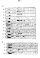
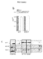
- the peptide membranes were assayed using the sera from ten patients who are allergic to fish ( Figs. 1 , 2 ).
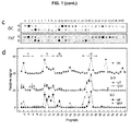
- the IgE binding intensity was variable among the tested sera; however, it was possible to identify four major binding regions (I-IV) and two major groups of sera (S-I: S2, S3, S4, S7 and S8; S-II: S1, S5, S6, S12 and S13).
- Region I was only recognized by the sera group S-II and covers peptides 1-2 (SEQ ID NO: 61, FAGILNDAD common core) and 6-7 (SEQ ID NO: 62, TAALAACKAE common core).
- Region II which was constituted of peptides 12-16, with SEQ ID NO: 63, FTKV as the common core, was identified by all sera with a high relative signal.
- region III formed by peptides 18-19 (SEQ ID NO: 64, AAKSSADIKK common core) displayed variable recognition by the distinct sera.
- Region IV covering the peptides 31-34 (SEQ ID NO: 65, FLQNFS common core) displayed high intensity, but varied between the two sera groups.
- region I overlaps the sequence predicted as the cross- ⁇ sheet forming (adhesive) segment in helix A
- regions III and IV overlap the immunologically reactive sites located on the junctions between the AB and CD domains (residues 33-44) and between the CD and EF domains (residues 65-74) ( Martinez, J. et al. Swiss Med Wkly 145, (2015 )).
- the reactivity of the Ca 2+ -binding loop of the EF domain (residues 88-96) and of the C-terminal (residues 95-109) regions was not detected by these sera, as probed by both IgE and IgG4 binding ( Fig.
- amyloid folds was then assayed using the conformation-selective OC antibody, which detects oligomer and fibril assemblies produced during the protein fibrillization process and the amyloid probe thioflavin T (ThT) ( Kayed, R. et al. Molecular neurodegeneration 2, 18, (2007 )).
- Figure 1 c shows that OC immunoreactivity covers peptides 2 (SEQ ID NO: 2, FAGILNDADITA), 12-16 (preferentially sequences containing SEQ ID NO: 63, FTKV, as the common core), 24 (SEQ ID NO: 24, VFEIIDQDKSDF), 34 (SEQ ID NO: 34, FLQNFSAGARAL), 41 (SEQ ID NO: 41, AETKVFLKAGDS) and 49 (SEQ ID NO: 49, KIGVDEFGAMIK). Of these regions, peptides 2 and 41 overlap the adhesive segments in helix A and E, predicted by the ZipperDB algorithm ( Goldschmidt, L., et al.
- peptides 12-16 and 34 lie over the IgE-binding regions III and IV, peptide 24 contains part of the Ca 2+ -binding site of domain CD, and peptide 49 corresponds to a previously described epitope groups ( Perez-Gordo, M. et al. Mol Nutr Food Res 57, 1283-1290, (2013 )) ( Fig. 2 and Fig. 7 ). Similar to the OC reactivity, probing the membranes with ThT shows increased fluorescence emission for peptides 2, 12, 24, 34 and 41, confirming the presence of amyloid folds ( Eisenberg, D. & Jucker, M. Cell 148, 1188-1203, (2012 )). It must be noted that peptides containing the helix C sequence, which was predicted to be an adhesive segment, were not detected by either the OC or ThT binding assays.
- Gad m 1 chain contains two major IgE binding sites (regions II and IV), which are flanked and overlapped by sequences with amyloid-forming capacity ( FIG. 2a ).
- regions II and IV sequences with amyloid-forming capacity
- regions A, C and E sequences with a predicted amyloid-forming capacity identified by the ZipperDB analysis as regions A, C and E to uncouple amyloid formation from IgE binding without disrupting the linear epitopes.
- Figs. 2a , 3 We searched for different sequences in the A, C and E regions of ⁇ -parvalbumins from other fish species with commercial added value and tested the effect of substitutions by a second round of ZipperDB analysis to preserve the native fold ( Martinez, J. et al.
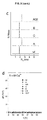
- Figure 2b shows that the I12V-T13K-A14T-A17E mutations in region A, A42D-V47A mutations in region C, and E82K-F86A mutations in region E abrogate the predicted adhesive properties of each of the segments of the wt chain. All of the chains containing the single or combined mutations in regions A, C and E were produced as recombinant proteins, and their conformations were characterized. All chains yielded Ca 2+ -bound folds with predominant ⁇ -helical secondary structures and a highly stable cooperative fold with a denaturation temperature of approximately 85 °C ( Fig. 3a and 3b ).
- mutant E showed slightly faster fibrillization
- mutants C and CE showed reduced fibrillization
- fibrillization of mutant A was abolished ( Fig. 3e ).
- the impaired amyloid formation observed in the A mutant was preserved upon cleavage of the N-terminal histidine tail, excluding tag effects, as shown for the wt chain ( Martinez, J. et al. Swiss Med Wkly 145, (2015 )).
- the ACE mutant displays a largely retarded (lag phase of approximately 24 h) but highly cooperative fibrillization at approximately 60 h ( Fig. 3e ).
- the adhesive region A largely governs rGad m 1 amyloid formation and its modification provides a non-amyloid-forming chain (A mutant) and a highly retarded amyloid-forming form (ACE mutant) as function of the sequences in the regions C and E.
- the dot-blot assays support the hypothesis that amyloid assembly endows rGad m 1 with enhanced IgE-binding activity.
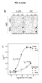
- the monomers and amyloid states of the distinct rGad m 1 chains were immobilized at varying concentrations and their IgE binding was determined using a sera pool ( Fig. 4c ). Under the conditions used, both the monomers and amyloids displayed similar adsorption levels, as shown by the protein concentration determinations, allowing the assignment of signal differences to the binding process.
- Amyloids prepared from rGad m 1 wt and the ACE mutant yielded I 50 values of approximately 10 -6.4 M, whereas titration of the distinct monomers (wt, A and ACE in 5 mM Ca 2+ ) resulted in I 50 values of approximately 10 -3.6 M.
- I 50 as the apparent dissociation constant (K Dapp )
- K Dapp K Dapp
- amyloid fibrils formed by wt and ACE chains and the ACE monomers were incubated with the peptide arrays and binding was determined using the anti-6XHis antibody, which recognizes the tag present in the recombinant chains.
- Fig. 5c shows that the amyloid fibrils differentially bound to peptides 12-15 compared to the monomers, indicating that the SEQ ID NO: 66, GSFDHKAFFTKVGLAAKS sequence, is a reactive segment for amyloid growth. Because this segment matches the IgE-binding region III ( Fig. 1 ), these results support the sequence overlap of the antigenic and amyloid-folding activities.
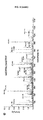
- Figure 5d shows that among the peptides detected, the sequences SEQ ID NO: 56 AACKAEGSFDHKAFF, SEQ ID NO: 57 FTKVGLAAKSSADIKKVF, SEQ ID NO: 58 KLFLQNF, SEQ ID NO: 34 FLQNFSAGARAL and SEQ ID NO: 67 SAGARALSDAETKVFL contain protected pepsin-cleavage sites and match the IgE-binding regions III and IV. Therefore, these data support the hypothesis that the sequential IgE epitopes of Gad m 1 encrypt an amyloid fold, indicating their functions as sequence-dependent conformational epitopes.
- amyloid state that confers a polymeric trait required for multivalent interactions through the spine structure and growth properties.
- Amyloid formation is a sequence-dependent process and has been described for distinct food allergens, such as Bos d 5 ( ⁇ -lactoglobulin), Bos d 10 ( ⁇ -casein), Bos d 12 ( ⁇ s2-casein), Gad m 1 ( ⁇ -parvalbumin), Gal d 2 (ovalbumin), and Gal d 4 (lysozyme), among others ( Martinez, J. et al.
- region A plays a role in the initial steps of the polymerization process (namely, the nucleation step) with an outcome that is dependent on the sequences of the C and E regions.
- regions identified as the two major IgE linear epitopes that formed part of pepsin-resistant fragments and displayed OC binding properties escaped the ZipperDB prediction analysis.
- Gad m 1 amyloids are formed from the apo form and their IgE reactivity argues against the well-established Ca 2+ -binding dependence of ⁇ -parvalbumins-IgE interaction ( Martinez, J. et al. Swiss Med Wkly 145, (2015 )).
- this contradiction is apparent and both facts converge when the IgE interaction is analyzed in terms of the physical state of the allergen.
- the results obtained here using the wt, A and ACE chains of Gad m 1 show that the structural impact of Ca 2+ removal is highly dependent on the chain sequence, as amyloid formation was triggered in wt and the ACE mutant, but not in the A mutant. Therefore, these data indicate that not all apo forms will form amyloids.
- hypoallergenic mrCyp c 1 with the two mutated Ca 2+ binding sites exhibits a highly stable native fold that argues against its capacity to polymerize ( Zuidmeer-Jongejan, L. et al. International archives of allergy and immunology 166, 41-51, (2015 )). Therefore, ⁇ -parvalbumin chains that are unable to form amyloids share a diminished IgE interaction.
- the overlap of epitopes and amyloid folds in the AB motif which is a hot spot for conformational exchange and lacks Ca 2+ -binding properties, suggests that amyloids could also form in the presence of Ca 2+ as a function of the chain sequence ( Moraes, A. H.
- ⁇ -parvalbumin solutions may contain traces of amyloid species that, as function of their analytical use, could mislead reactivity assignments.
- the folding of the epitope into a cross- ⁇ sheet may optimize the number and nature of contacts for optimal antibody recognition.
- the polymer nature of amyloids permits the saturation of the two IgE binding arms, whereas a monomer does not permit saturation.
- binding of the first IgE arm consists of an intermolecular reaction, whereas the occupancy of the second site involves an intramolecular reaction.
- allergens require the existence of at least two IgE epitopes for their deleterious IgE cross-linking on the surface of effector cells. This requirement presumes the development of either two different IgE molecules for monomeric allergens or single IgE molecules for an oligomeric allergen ( Sharp, M. F. & Lopata, A. L. Clinical reviews in allergy & immunology 46, 258-271, (2014 )).
- the presentation of the allergen as an amyloid allows the development of a single IgE molecule and the optimization by enhancing the probability of the cross-linking step.
- phase transition accompanying amyloid formation has also major implications in allergy diagnosis. Clearing amyloids (either by their physical removal or by using additives that prevent their formation) from solutions used for skin prick tests may result in false negatives compared with non-risk-free oral food challenges ( Sharp, M. F. & Lopata, A. L. Clinical reviews in allergy & immunology 46, 258-271, (2014 )). This strategy is of particular relevance for cases in which amyloids are preferentially formed after the partial digestion of food allergens ( Martinez, J. et al. Swiss Med Wkly 145, (2015 )). Therefore, in light of these results, a food allergy diagnosis should be implemented for the consideration of allergen folds displaying insolubility.
- OAS oral allergy syndrome
- U urticaria
- AE angioedema
- V vomiting
- BS bronchospasm
- AN anaphylaxis.
- c Cod Godus morhuo
- Hake Merluccius merluccius
- Megrim Lepidorhombus whiffiagonis
- Tuna Thunnus albacares.
- SPT serine proteosus
- rGad m 1 (recombinant Gad m 1) was produced from a pET15b construct containing the synthetic ORF of Atlantic cod parvalbumin A51874 ( Martinez, J. et al. Swiss Med Wkly 145, (2015 )). The mutants were prepared using Quick-Change protocols and a pair of complementary oligonucleotides (Table 2). Table 2. List of primers used in this study for rGad m 1 mutant generation. PV refers to the synthetic ORF of Atlantic cod parvalbumin A51874. SEQ ID NOs: 51-55 are the sequences of the primers in the order listed below.
- the proteins were produced in BL21(DE3) cells and purified from the soluble fraction by Ni 2+ -affinity chromatography (GE Healthcare Life Sciences), followed by Q-Sepharose chromatography (GE Healthcare Life Sciences) ( Martinez, J. et al. Swiss Med Wkly 145, (2015 )).
- the eluted fractions were filtered using 30 kDa-pore size Amicon Ultra-15 (Merck Millipore) and extensively dialyzed against 5 mM Hepes, pH 7.5, containing 0.1 mM CaCl 2 ( Martinez, J. et al. Swiss Med Wkly 145, (2015 )). Before use, the proteins were centrifuged at 16,000xg for 20 min at 4 °C to remove any insoluble material.
- Peptide arrays SPOT. Dodecapeptides spanning the whole sequence of Gad m 1, with an overlap of ten residues, were solid-phase synthesized and immobilized ( ⁇ 10 nmol per spot) on an Amino-PEG500-UC540 sheet at the National Centre for Biotechnology (CNB-CSIC, Madrid). Before use, the membranes were rinsed with ethanol, washed three times with TBS (25 mM Tris-HCl, pH 7.5, containing 137 mM NaCl and 2.7 mM KCI), and incubated in TBS containing 1% BSA (w/v) and 2 mM EDTA for 1 h.
- TBS 25 mM Tris-HCl, pH 7.5, containing 137 mM NaCl and 2.7 mM KCI
- the membranes were then probed for 2 h with sera from patients who are allergic to fish (1/10 dilution) and the anti-amyloid fibril OC antibody (AB2286 Merck Millipore, 1/2000 dilution), prepared in TBST (TBS containing 0.05% Tween-20) supplemented with 0.5% BSA (w/v) or with rGad m 1 chains (2.5 ⁇ M) in TBS.
- TBST TBS containing 0.05% Tween-20
- BSA w/v
- rGad m 1 chains 2.5 ⁇ M
- the membranes were regenerated by sequential incubation with TBS containing 8M urea, 1% SDS and 0.5% ⁇ -mercaptoethanol for 30 min at 55 °C and three times with acetic acid/ethanol/Milli-Q water (10:50:40). Regions were considered major allergenic epitopes when at least two overlapping peptides were involved. Signals resulting from the binding of secondary antibodies in the absence of primary antibodies were negligible under the conditions tested.
- Circular Dichroism Spectroscopy Circular Dichroism (CD) experiments were performed using a Jasco J-820 spectropolarimeter equipped with a Peltier-controlled thermostatted cell holder. Far UV CD spectra were recorded for a 25 ⁇ M protein concentration in 50 mM Tris-HCl, pH 7.5, supplemented with either 1 mM EDTA or 1 mM CaCl 2 . Thermal denaturation experiments were performed to follow the ellipticity changes at 222 nm upon heating from 15 °C to 90 °C at a 1 degree/min heating rate. Both the spectra and thermal unfolding curves were analyzed as previously described ( Martinez, J. et al. Swiss Med Wkly 145, (2015 )).
- Dynamic Light Scattering Dynamic light scattering (DLS) measurements were performed using a DynaPro spectroscatter (Wyatt Technology) with a 1.5-mm path length and a 12 ⁇ l quartz cuvette. The average of 20-25 acquisitions of buffers and protein solutions (160 ⁇ M protein concentrations) were filtered using a 0.2 ⁇ m Whatman Anodisc-3 filter. The hydrodynamic radii (R H ) and mass proportions (%) of the species were derived as previously described ( Martinez, J. et al. Swiss Med Wkly 145, (2015 )). The experiments were performed in duplicate using two different protein preparations.
- DLS Dynamic light scattering
- the theoretical hydrodynamic radius R H T for spherical rGad m 1 was calculated as 1.81 nm using 0.73 cm 3 g -1 and 0.35 g of H 2 O (g protein) -1 for the particle-specific volume and hydration ( Martinez, J. et al. Swiss Med Wkly 145, (2015 )).
- Amyloid formation assays The amyloid propensity and location of the segments in ⁇ -parvalbumin chains with adhesive properties was theoretically evaluated using the ZipperDB algorithm, as previously described ( Martinez, J. et al. Swiss Med Wkly 145, (2015 )).
- rGad m 1 wt and the mutants were prepared at a concentration of 150 ⁇ M in 50 mM Glycine-HCl, pH 1.6, with 100 mM NaCl or 50 mM Tris-HCl, pH 7.5, with 100 mM NaCl, both of which were supplemented with either 5 mM EDTA or 5 mM CaCl 2 .
- the kinetics of thioflavin T (ThT) binding was monitored by bottom reading the fluorescence intensity in a POLARstar microplate reader (BMG Labtech), as previously described ( Martinez, J. et al. Swiss Med Wkly 145, (2015 )). The measurements were performed using 0.18 mL samples containing 10 ⁇ M ThT and 450 nm excitation and 480 nm emission filters. The measurement program consisted of 10 flashes, a reading collected every 15 min, 0.5-min of orbital shaking at 100 rpm, and the temperature controller was set to 37 °C. All measurements were collected in duplicate, and the experiments were repeated at least twice using two different protein batches.
- the fibers were harvested from the reaction mixtures by centrifugation at 100,000xg for 1 h using an OptimaTm Max Beckman ultracentrifuge. The resulting pellet and supernatant fractions were used for the protein and ThT determinations.
- Atomic force microscopy rGad m 1 wt fibrils were deposited onto freshly cleaved mica surfaces for 10 min, washed with H 2 O, and dried with a stream of N 2 .
- AFM images were recorded in a MultiMode Veeco microscope and analyzed using WSxM (Nanotec), as previously described ( Martinez, J. et al. Swiss Med Wkly 145, (2015 )).
- Dot-blot analysis The immunoreactivity of the rGad m 1 wt and mutant species was assessed by dot-blot analysis using the anti-amyloid fibril OC antibody (AB2286 Merck Millipore, 1:2,000 dilution) and sera from patients who are allergic to fish (1:10 dilution). Briefly, aliquots containing 50-100 ng of protein in the different states were spotted in triplicate on a nitrocellulose membrane.
- Immunodetection was performed by incubating the membranes with the primary antibodies for 1 h, followed by extensive washes and 30 min incubation with horseradish peroxidase-labeled either mouse monoclonal B3102E8 anti-human IgE (Abcam, diluted 1:2,000) or goat anti-rabbit IgG (diluted 1:5,000; Sigma).
- the signal was developed using the ECL-Western-blotting reagent (Bio-Rad) and detected with a ChemiDoc XRS instrument ( Martinez, J. et al. Swiss Med Wkly 145, (2015 )).
- Polystyrene 96-well plates (Costar 3590SA) were coated with 100 ⁇ l of rGad m1 chains in 0.2 M carbonate buffer, pH 9.4, by varying the concentrations from 0.1-500 ⁇ g/ml for 2 h at 37 °C.
- the coated wells were blocked with 1% BSA in TBS for 30 min and then incubated with 100 ⁇ l of a 1:10 dilution of the patients' serum in TBST containing 1% BSA for 2 h at room temperature. After washing with TBST, the wells were incubated with peroxidase-labeled anti human IgE (Abcam, diluted 1:2,000) for 1.5 h at room temperature.
- the plates were washed again and then developed with 100 ⁇ l of TMB-turbo ELISA substrate (Thermo Scientific). The reaction was stopped after 30 min with 10 ⁇ l of 2N H 2 SO 4 and the optical density (OD) was measured at 450 nm using a microplate reader (Bio-Rad 3550). The assays were performed in duplicate using blocking buffer as negative control and were statistically analyzed and fitted using Origin software. A parallel analysis using the Bradford protein assay (Bio-Rad) indicated similar extent of adsorption for all proteins used.
- Protease digestion and mass spectrometry Typically, 50 ⁇ l of rGad m 1 fibrils (160 ⁇ M) were digested in 10 mM Gly-HCl, pH 1.3, with pepsin (1:50 w:w protease/substrate) at 37 °C for 1 h. The reactions were stopped by the addition of 1 M Tris-HCl, pH 8.
- the hydrolyzed products were analyzed by mass spectrometry using a CHCA matrix and a MALDI-TOF/TOF 4800 (SCIEX, reflector mode) for identification and HPLC (Eksigent Technologies nanoLC Ultra 1D plus, SCIEX) coupled to a Triple TOF 5600 (SCIEX) for fragmentation at the CNB Proteomics Facility (CNB-CSIC, Madrid).
- SCIEX MALDI-TOF/TOF 4800
- HPLC Eksigent Technologies nanoLC Ultra 1D plus, SCIEX
- SCIEX Triple TOF 5600
Landscapes
- Chemical & Material Sciences (AREA)
- Health & Medical Sciences (AREA)
- Life Sciences & Earth Sciences (AREA)
- Organic Chemistry (AREA)
- Zoology (AREA)
- General Health & Medical Sciences (AREA)
- Biochemistry (AREA)
- Biophysics (AREA)
- Gastroenterology & Hepatology (AREA)
- Genetics & Genomics (AREA)
- Medicinal Chemistry (AREA)
- Molecular Biology (AREA)
- Proteomics, Peptides & Aminoacids (AREA)
- Toxicology (AREA)
- Peptides Or Proteins (AREA)
- Medicines Containing Antibodies Or Antigens For Use As Internal Diagnostic Agents (AREA)
Abstract
The present invention refers to a method for producing improved food allergen extracts, the food allergen extracts produced and their use for the diagnosis of type I food allergy. Particularly, the food allergen extracts of the invention comprise a type I food allergen in an amyloid and insoluble state which strengths its interaction with the IgE present in the patient serum, allowing thus to diagnose the food allergy in a more reliable manner, reducing the number of false negatives obtained by other diagnosis methods that use monomeric food allergens.
Description
- This invention belongs to the field of immunology and food allergy diagnosis methods. Specifically, it refers to a method for producing improved food allergen extracts, the food allergen extracts produced and their use for the diagnosis of type I food allergy.
- Allergy is an acquired hypersensitivity disorder of the immune system and is triggered by exposure to harmless environmental substances known as allergens. A type I hypersensitivity reaction is characteristic of allergic reactions and results in the production of excessive amounts of IgE antibodies which in turn activate basophils and mast cells causing an inflammatory reaction. The effects may be systemic such as vasodilation, mucus secretion, nerve stimulation and smooth muscle contraction causing an anaphylaxis reaction, or confined to a particular area of the body, for example the respiratory system.
- Food allergy is an emerging major public health problem that affects around 6% of school children and approximately 4% of adults and can have severe consequences, including fatal anaphylactic reactions. Allergy can therefore have a significant impact on psychosocial aspects of quality of life extending beyond the immediate clinical effects of the patient's allergic condition and the daily activities of families. At present the standard of care for this type of allergy includes strict avoidance of the offending allergens and treatment with epinephrine.
- Type I food allergy, such as fish allergy, is a hypersensitivity reaction in which IgE is involved. The intake of the allergen in hypersensitive patients leads to a symptomatology that may be lethal. Tests in the skin of the patient, together with the medical record, are the main diagnosis tools. These tests, named Skin Prick Tests (SPT), asses the IgE reactivity linked to the patient mast cells. The skin test consists of eliciting a reaction in the patient skin by adding a small allergen amount. Thus, a small allergen amount is deposited in the arm of the patient, afterwards an epidermis puncture is made so that the allergen is allowed to infiltrate in deeper layers of the skin. If the patient is allergic, a histamine release from the skin mast cells will be observed as a consequence of the allergic response, and inflammation and redness will be detected. Reactives used in these SPT diagnosis methods are extracts containing standardized and diluted allergens Besides the contact with the allergen, positive (an histamine solution) and negative (a saline solution) controls are usually performed.
- Additionally, a definitive test for the diagnosis of food allergies can be also performed, which consists of the oral exposure to the causal food (Oral Food Challenge). However, this test is highly risky.
- There is, therefore, a need to further improve the efficacy of allergy diagnosis methods to ensure that allergic patients are properly identified, eliminating or lowering the number of false negatives detected with current diagnosis methods. In this sense, molecular basis of the interactions between the allergen and IgE should be studied further in order to design improved allergen extracts capable of increasing their interaction with the IgE. These improved allergen extracts would allow a more reliable diagnosis when used in SPT solutions as a reactive.
- Amyloids are polymeric structural protein states formed from locally or totally unfolded protein chains that permit surface reorganizations, stability enhancements and interaction properties that are absent in the precursor monomers.
- Amyloids are defined by a cross-β sheet backbone, in which β-sheets that are mostly formed from β-strands of different molecules pack through their side chains (Goldschmidt, L., et al. Proceedings of the National Academy of Sciences of the United States of America 107, 3487-3492, (2010)). Despite their initial association with proteins involved in neurodegenerative disorders, an increasing number of proteins that form functional and pathogenic amyloids have been reported. Indeed, protein chains containing sequences with cross-β sheet folding traits under conditions that cause their solvent exposure enter this state when they are present at sufficient concentrations to overcome the entropy that opposes fibril ordering. Amyloids exhibit a dual (soluble/insoluble) nature and a characteristic structural repetitiveness, allowing phase separations and interactions with enhanced affinity and specificity compared to the precursor monomer (Greenwald, J. & Riek, R. )).
- Fish β-parvalbumins represent the major allergens of IgE-mediated fish hypersensitive patients (Sharp, M. F. & Lopata, A. L. Clinical reviews in allergy & )). β-Parvalbumins are 12 kDa calcium-binding proteins with three EF hand motifs; a nonfunctional AB motif, followed by the CD and EF Ca2+-binding motifs (Berchtold, M. W., Brinkmeier, H. & Muntener, M. )). Despite the variations in sequence identity between proteins from the same and different fish species, β-parvalbumins display a high IgE cross-reactivity, supporting a pattern of recognition other than the sequence identity (Van Do, T., et al. J Allergy Clin Immun 116, 1314-1320, (2005)). The use of a variety of approaches, including protease digests, phage display libraries, site-directed mutagenesis and arrays of overlapping peptides with β-parvalbumins from Baltic cod (Gad c 1) (Lapteva, Y. S., et al. Bba-Proteins Proteom 1834, 1607-1614, (2013)), Atlantic cod (Gad m 1) (Perez-Gordo, M. et al. Mol Nutr Food Res 57, 1283-1290, (2013)), Common carp (Cyp c 1) (Swoboda, I. et al. J Immunol 178, 6290-6296 (2007)), Chub mackerel (Sco j 1) (Yoshida, S., Ichimura, A. & Shiomi, K. Food Chem 111, 857-861, (2008)) and Atlantic salmon (Sal s 1) (Perez-Gordo, M. et al. Int Arch Allergy Imm 157, 31-40, (2012)), has shown distinct IgE epitopes of both linear and conformational types. For Gad
c 1 and Sal s 1, the IgE binding sites have been located at the spacers between the AB and CD motifs (residues 28-45) and CD and EF motifs (residues 65-74), whereas for Gad m1, the dominant IgE binding region was found at the C-terminus (segment 95-109) (Perez-Gordo, M. et al. Int Arch Allergy Imm 157, 31-40, (2012)). On the other hand, disruption of the Ca2+ binding sites by mutagenesis in bothCyp c 1 and Scoj 1 resulted in soluble chains with reduced IgE-binding signals (Swoboda, I. et al. J Immunol 178, 6290-6296 (2007)). Conversely, stabilization of apo-Gad m 1 produces highly stable amyloids with enhanced IgE reactivity (Martinez, J. et al. Swiss Med Wkly 145, (2015)). - Despite the large number of sequences and 3D structures of allergens elucidated in the last few decades, the knowledge of how allergens recognize and cross-link cell-bound IgEs is still limited (Gieras, A. et al. The Journal of allergy and clinical immunology 119, 384-390, (2007)). This lack of knowledge is largely due to the complexity of allergen epitopes and their incomplete interpretation, based on the use of structures obtained from stable and ligand-bound states (Perez-Gordo, M. et al. Mol Nutr Food Res 57, 1283-1290, (2013)). For food allergens that undergo drastic environmental changes during gastrointestinal transit, these structural templates are merely snapshots of the protein life and other nonnative structures can be formed and change their interaction repertoire. One such structural state is the amyloid state that confers a polymeric trait required for multivalent interactions through the spine structure and growth properties. Amyloid formation is a sequence-dependent process and has been described for distinct food allergens, such as Bos d 5 (β-lactoglobulin), Bos d 10 (κ-casein), Bos d 12 (αs2-casein), Gal d 2 (ovalbumin), Gal d 4 (lysozyme) and Gad m 1 (β-parvalbumin) among others ((Martinez, J. et al. Swiss Med Wkly 145, (2015)).
- In summary, improved methods for the diagnosis of type I food allergies are required, as current standardized allergen extracts used in these methods as reagents render a significant number of false negatives due to their weak interaction of the IgE present in the samples/tissues of the hypersensitive patients. Thus, improved methods for the diagnosis of type I food allergies are needed. These methods should lead to a more reliable diagnosis and should render a lower number of false negatives.
- The present invention provides a method for producing improved food allergen extracts, the food allergen extracts produced and their use for the diagnosis of type I food allergy.
- The problem mentioned above is solved by the present invention through the use of food allergen extracts comprising a polymerized and insoluble food allergen, i.e. the food allergen to be used in the diagnosis method of the invention is in an amyloid state. When these food allergen extracts of the invention are used as a reactive in type I food allergy diagnosis methods the number of false negatives decreases compared to other methods in which monomeric allergens are used, as the food allergen of the invention in an amyloid state has significantly increased IgE affinity.
- The amyloid fold of the food allergen extracts of the invention governs IgE binding. Food allergens, such as β-Parvalbumin which is the major allergen in fish allergy, form amyloids that are recognized by IgE in the patient sera. In the examples of the present invention, Gad m1 was used as the fish β-parvalbumin model and a combination of approaches, including peptide arrays, recombinant wt and mutant chains, biophysical characterizations, protease digestions, mass spectrometry, dot-blot and ELISA assays was performed to gain insights into the role of amyloids in the IgE interaction. It was found that Gad m1 immunoreactive regions behave as sequence-dependent conformational epitopes that provide a 1000-fold increase in affinity and the structural repetitiveness required for optimal IgE binding and cross-linking upon folding into amyloids. These findings support the amyloid state of food allergens as a key entity in type I food allergy.
- Thus, in the present invention it has been addressed the role of food allergen amyloids with respect to both its IgE-reactive properties and epitope architecture. It was found that when the relevant antigenic regions of the food allergens fold into amyloids they optimize their IgE interaction, which allows for more reliable food allergy diagnosis methods.
- According to the examples of the application, the allergen chains that were not in an amyloid state share a diminished IgE interaction, which leads to a less effective diagnosis method when said non-amyloid allergens are used. Both allergen monomers and amyloids bind IgE, but with significantly different affinities. The interaction of sera IgE with allergen amyloids reveals a 1000-fold enhancement of the interaction strength compared to the monomer.
- From a structural point of view, the folding of the epitope into a cross-β sheet may optimize the number and nature of contacts for optimal antibody recognition. From the stoichiometry point of view, the polymer nature of amyloids permits the saturation of the two IgE binding arms, whereas a monomer does not permit saturation. The presentation of the allergen as an amyloid allows the development of a single IgE molecule and the optimization by enhancing the probability of the cross-linking step.
- The phase transition accompanying amyloid formation has therefore major implications in allergy diagnosis. Clearing amyloids (either by their physical removal or by using additives that prevent their formation) from solutions used for skin prick tests may result in false negatives compared with non-risk-free oral food challenges. This strategy proposed by the present invention is of particular relevance for cases in which amyloids are preferentially formed after the partial digestion of food allergens. Therefore, in light of these results, a food allergy diagnosis should be implemented for the consideration of allergen folds displaying insolubility.
- Therefore, an aspect of the present invention refers to a process for producing an allergen extract, hereinafter "the process of the invention", comprising:
- a. incubating a solution comprising a type I food allergen under conditions that allow protein polymerization or protein amyloid formation, and
- b. recovering the solution comprising the type I food allergen in an amyloid (or polymerized), and preferably in an insoluble, state after step (a).
- An "allergen extract" may be derived from any source material comprising natural allergens known to illicit an IgE mediated immune reaction in an individual, i.e. the solution comprising the type I food allergen as defined in the process of the invention may be any source material comprising one or more natural type I food allergens.
- A "type I food allergen" is any food protein, glycosylated or not, that produces an IgE mediated hypersensitive adverse systemic reaction via the gastrointestinal system and generally exhibits heat, low pH and protease resistance. Type I food allergen may be the complete protein sequence or a relevant fragment thereof, both isolated from natural sources or produced either recombinantly or synthetically. The "relevant fragment thereof" is preferably any immunoreactive protein fragment or any other fragment capable of folding into an amyloid state. Examples of type I food allergens are, without limitation, milk allergens such as β-lactoglobulin (Bos d 5) or caseins (
Bos d 12, etc), peanut allergens (Ara h 1,Ara h 2, etc.), egg allergens such as lysozyme (Gal 4 d), ovalbumin (Gal d 2), and ovotransferrin (Gal d 3), shellfish allergens, fish allergens such β-parvalbumins (Gad m 1,Gad c 1, Thec 1, Sal s 1,Cyp c 1,Car ca 1, and others in the allergome database), and nut, soy, wheat or rice allergens. - In a preferred embodiment of the process of the invention, the type I food allergen is a fish β-Parvalbumin protein, preferably the Gad m1 protein which is, for example, any sequence selected from the list consisting of: Q90YK9, Q90YL0 (UniProtKB/Swiss-Prot), A5I873, A5I874 (GeneBank) and their variants or relevant fragments.
- Examples of immunoreactive and amyloid forming regions in the
Gad m 1 chain that can be used, alone or in combination, in the process of the invention as relevant fragments of the type I food allergenGad m 1 are any of those identified in the Figures of the present application, specifically inFigs. 2a and5d . - In a more preferred embodiment of the process of the invention, the conditions that allow protein polymerization or protein amyloid formation are selected from the list consisting of: the presence of EDTA, the presence of an acid pH (preferably pH<1.9), the presence of proteases (such as pepsin, chymotrypsin, trypsin or any combination thereof), temperature changes (including heating at different temperatures, changes in the heating time and the use or not of agitation processes), lipid content variations or their combinations. These conditions will depend on the type I food allergen present in the solution. When the type I food allergen is a β-Parvalbumin protein, preferably the Gad m1 protein, the conditions are the presence of EDTA and preferably an incubation time of at least 70h at 37 °C.
- The "amyloid or polymerized state" is that state in which the monomeric type I food allergen molecules are grouped together so that they produce a molecule with a higher molecular weight, which includes aggregation states.
- Another aspect of the invention refers to an allergen extract that comprises a type I food allergen in an amyloid (or polymerized), and preferably in an insoluble, state. Hereinafter, this allergen extract will be referred to as "allergen extract of the invention".
- In a preferred embodiment, the allergen extract of the invention is that obtainable by the process of the invention.
- In a more preferred embodiment, the type I food allergen comprised in the allergen extract of the invention is a β-Parvalbumin protein, preferably the Gad m1 protein.
- Another aspect of the invention refers to a pharmaceutical composition, preferably a diagnostic pharmaceutical composition for type I food allergy, which comprises as the active ingredient a diagnostically effective amount of the allergen extract of the invention. This pharmaceutical composition may further comprise one or more adjuvants, diluents, preservatives or mixtures thereof.
- Another aspect of the invention refers to a kit, hereinafter the "kit of the invention", comprising the allergen extract of the invention. Preferably, the allergen extract of the invention is labelled or immobilized in the kit of the invention. This kit of the invention may additionally comprise other components needed for the in vivo or in vitro diagnosis of type I food allergy, such as negative and/or positive controls of an immunological response or medical instruments or devices for putting in contact the allergen extract of the invention with patient tissue/s or with an isolated biological sample derived from the patient. Preferably, the kit of the invention is a kit suitable for the diagnosis of type I food allergy, more preferably for the diagnosis of type I food allergy by the skin prick test (SPT).
- Another aspect of the invention refers to the use of the allergen extract of the invention or the kit of the invention as a reactive for the diagnosis of type I food allergy. Alternative, this aspect of the invention refers to the use of the allergen extract of the invention or the kit of the invention for the manufacture of a reactive for the diagnosis of type I food allergy. Preferably, the diagnosis of type I food allergy is performed by SPT.
- In a preferred embodiment of this aspect of the invention, the type I food allergy is fish allergy.
- Another aspect of the invention refers to a method for the diagnosis of type I food allergy comprising putting in contact the skin of a subject with the allergen extract of the invention or the kit of the invention, analysing the presence of an immunological response in the subject and assigning the subject to the group of patients suffering from a type I food allergy when the immunological response is observed.
- Another aspect of the invention refers to a method, hereinafter "the diagnosis method of the invention", for the in vitro diagnosis of type I food allergy comprising:
- a. Putting in contact the allergen extract of the invention or the kit of the invention with an isolated biological sample of a subject,
- b. analysing the presence of immunological interactions between the IgE comprised in the isolated biological sample and the type I food allergen comprised in the allergen extract or in the kit, and
- c. assigning the subject to the group of patients suffering from a type I food allergy when the immunological interactions of step (b) are observed.
- In a preferred embodiment, the type I food allergy is fish allergy.
- In a more preferred embodiment, the isolated biological sample is serum or epithelial tissue preferably from the skin.
- The "presence of immunological interactions" as defined in the diagnosis method of the invention may be analysed by any method known in the art for detecting interactions between molecules, specifically antigen-antibody interactions. Examples of such methods are, but without limitations, magnetic resonance imaging, flow cytometry, immunohistochemistry, ELISA, and/or Western Blot.
- Unless otherwise defined, all technical and scientific terms used herein have the same meaning as commonly understood by one of ordinary skilled in the art to which this invention belongs. Methods and materials similar or equivalent to those described herein can be used in the practice of the present invention. Throughout the description and claims the word "comprise" and its variations are not intended to exclude other technical features, additives, components, or steps. Additional objects, advantages and features of the invention will become apparent to those skilled in the art upon examination of the description or may be learned by practice of the invention. The following examples, drawings and sequence listing are provided by way of illustration and are not intended to be limiting of the present invention.
-
-
Figure 1 . Reactivity ofGad m 1 chain. (a) Regions binding IgE in sera from fish-allergic patients. (b) Regions binding IgG4 in sera from fish-allergic patients. (c) Sequences with anti-amyloid OC antibody reactivity and ThT binding properties. (d) Relative signal of the binding of IgE and IgG4 from the sera of fish-allergic patients and the anti-amyloid OC antibody to the distinct overlapping peptides representing theGad m 1 sequence. Signals corresponding to the IgE and IgG4 binding are displayed as the average and standard deviations of the signals of sera groups S-I (S2, S3, S4, S7, and S8) and S-II (S1, S5, S6, S12, and S13) are shown. Peptides are indicated by numbers on the top (row A: peptides SEQ ID NO: 1-30, row B: peptides SEQ ID NO: 31-50) and their sequences are displayed inFig. 6 . The sera are depicted as Si, where i is a number (Table 1). -
Figure 2 . Location and relation of the immunoreactive and amyloid forming regions in theGad m 1 chain (a) Sequence (SEQ ID NO: 60) and location of the following regions in theGad m 1 chain: i) IgE-binding epitopes found in sera groups S-I (S-I black and underlined) and S-II (S-II black and underlined), ii) IgG4-binding epitopes found in S-I (S-I, IgG4 grey) and S-II (S-II, IgG4 grey), iii) OC-binding segments (OC, grey), iv) adhesive regions identified by the ZipperDB algorithm (ZipperDB, grey), and v) sequence changes in the A, C and E regions (ACE, grey). The sequence changes were based on the sequences of the following β-parvalbumins: i) Q91482, Q90YK8 and E0WDA2 for A, ii) Q90YL0, Q91482, Q91483, E0WDA2 and Q90YK8 for C, and iii) C6GKU7 for E (Martinez, J. et al. Swiss Med Wkly 145, (2015)). (b) Comparative ZipperDB analysis of the Gad m1 wt and ACE mutant chains. The longest bars in boxes in the wt indicate the N-terminus of a hexapeptide with adhesive properties. The effects of the adhesive segments are depicted by rectangles and were conserved in the analysis of chains containing single A, C and E modifications. -
Figure 3 . Conformational features ofrGad m 1 wt and its ACE mutants. (a) Far-V Circular Dichroism spectra of the distinct proteins obtained in 50 mM Tris-HCl, pH 7.5, containing 2 mM Ca2+ at 20 µM protein concentrations. (b) Variation of the unfolded fraction ofrGad m 1 wt and mutants with temperature. (c) DLS determination of the hydrodynamic radius (RH) ofrGad m 1 wt and the mutants. The measurements were performed in 50 mM Tris-HCl, pH 7.5, containing 2 mM Ca2+ at 150 µM protein concentrations. Amyloid formation was monitored by the time-dependence of ThT binding ofrGad m 1 wt in 50 mM Tris-HCl, pH 7.5, in the presence of (d) 5 mM Ca2+ or (e) 5 mM EDTA. -
Figure 4 .rGad m 1 amyloids displayed enhanced interaction with the IgE in the sera from fish-allergic patients. (a) Dot-blots of rGad m wt and its non-amyloidogenic A mutant probed with the sera pools S-I and S-II and with the anti-amyloid OC antibody. Typically, 50 ng of proteins that were incubated for 50 h under monomer-preserving (5 mM Ca2+) and amyloid-forming (5 mM EDTA) conditions were spotted. (b) Dot-blots of the ACE mutant at 0, 12 and 50 h of incubation in 50 mM Tris-HCl, pH 7.5, containing 0.1 M NaCl and either 5 mM Ca2+ or 5 mM EDTA were probed with a sera pool (S-I/II) and the anti-amyloid OC antibody. (c) Titration curves ofrGad m 1 wt and of its A and ACE mutants stabilized in the monomer (M) and amyloid (F) states with sera IgE generated by ELISA assays and their best fits to sigmoidal curves. The data are displayed as the mean average value of two experiments performed in duplicate and their standard deviations. (d) AFM image of a typical amyloid assembly ofrGad m 1 and its z-axis dimension. -
Figure 5 .Gad m 1 sequences that are recognized by IgE in patient sera display amyloid folds. (a) The effects of patient sera (S-I/II) and the anti-amyloid OC antibody on the amyloid formation kinetics of rGad wt, its ACE mutant and Aβ42 were monitored by ThT-binding in 50 mM Tris-HCl, pH 7.5, containing 0.1 M NaCl and 5 mM EDTA. (b) Effects of the anti-amyloid OC antibody andGad m 1 monomers on recognition of the wt and ACE amyloid fibrils by the IgE in patient sera, as determined by the ELISA assay. Amyloid fibrils (100 ng/well) were probed with the patient sera pool that had been preadsorbed with 1 µg of BSA (SF), 1 µg of OC (OC-preadsorbed S-I/II) and 1 µg of the corresponding monomer chain (M-preadsorbed S-I/II). (c) Gad m1 peptide arrays were probed with amyloids (wt-F and ACE-F) and monomers (ACE-M) and developed with the anti-His tag antibody to detect binding. (d) Sequence coverage ofrGad m 1 wt (SEQ ID NO: 60) obtained by LC-MS/MS analysis of the peptide mixture after pepsin digestion of the amyloids. The pepsin cleavage sites are displayed in the sequence in grey, the predicted adhesive regions are shown as continuous lines above, and the IgE binding regions are indicated with discontinuous-continuous-discontinuous lines above (solid in the common core, dotted for all peptides). The sequences of the overlapping IgE-binding regions in the peptides are depicted with continuous lines below and their mass/charge ratio (m/z) is indicated nearby. -
Figure 6 . Sequences and chain location of the 50 overlapping synthetic peptides (12 amino acid lengths, 2 amino acids offset, SEQ ID NO: 1-50) used for the identification reactive regions inGad m 1. Peptide numbering (1 to 50) was performed from the N- to the C-terminus of the chain sequence. Helices (A,B,C,D,E and F) forming the three EF hands are represented by rectangles, and the Ca2+ binding sites depicted as spheres. -
Figure 7 . Typical MALDI-TOF/TOF spectra of pepsin-hydrolyzedrGad m 1 amyloids. Mass spectrometry analysis was performed using α-cyano-4-hydroxycinnamic acid (CHCA) matrix. Peak features have been removed for clarity reasons. -
Figure 8 . Identification of pepsin-resistant peptides ofrGad m 1 amyloids. Fragmentation spectra were obtained using nano-LC coupled to MALDI Triple TOF. Panels (a-e) depict the distinct sequences (SEQ ID NO: 56, 57, 58, 34 and 59) that contain undigested pepsin cleavage sites. - It was first characterized the sequence elements to address how amyloid formation contributes to the IgE reactivity of
Gad m 1. A set of 12-mer overlapping synthetic peptides with an offset of 2 that correspond to the sequence ofGad m 1 was used in an array-based immunoassay (Fig. 6 ). - This peptide length and surface density (10 nmol/spot) ensures the preservation of amyloid formation (which is conventionally limited to 6 residues), and differs from previous microarrays assays using a 15-mer with an offset of 3 (Perez-Gordo, M. et al. Mol Nutr Food Res 57, 1283-1290, (2013)).
- The peptide membranes were assayed using the sera from ten patients who are allergic to fish (
Figs. 1 ,2 ). The IgE binding intensity was variable among the tested sera; however, it was possible to identify four major binding regions (I-IV) and two major groups of sera (S-I: S2, S3, S4, S7 and S8; S-II: S1, S5, S6, S12 and S13). Region I was only recognized by the sera group S-II and covers peptides 1-2 (SEQ ID NO: 61, FAGILNDAD common core) and 6-7 (SEQ ID NO: 62, TAALAACKAE common core). Region II, which was constituted of peptides 12-16, with SEQ ID NO: 63, FTKV as the common core, was identified by all sera with a high relative signal. In contrast, region III formed by peptides 18-19 (SEQ ID NO: 64, AAKSSADIKK common core) displayed variable recognition by the distinct sera. Region IV covering the peptides 31-34 (SEQ ID NO: 65, FLQNFS common core) displayed high intensity, but varied between the two sera groups. Of these four regions, region I overlaps the sequence predicted as the cross-β sheet forming (adhesive) segment in helix A, whereas regions III and IV overlap the immunologically reactive sites located on the junctions between the AB and CD domains (residues 33-44) and between the CD and EF domains (residues 65-74) (Martinez, J. et al. Swiss Med Wkly 145, (2015)). Nevertheless, the reactivity of the Ca2+-binding loop of the EF domain (residues 88-96) and of the C-terminal (residues 95-109) regions was not detected by these sera, as probed by both IgE and IgG4 binding (Fig. 1d ) (Perez-Gordo, M. et al. Int Arch Allergy Imm 157, 31-40, (2012)). It merits comment that IgG4 reactivity, which is considered as a protective response in allergy, is largely detected in region IV for both sera groups (Perez-Gordo, M. et al. Int Arch Allergy Imm 157, 31-40, (2012)). - The presence of amyloid folds was then assayed using the conformation-selective OC antibody, which detects oligomer and fibril assemblies produced during the protein fibrillization process and the amyloid probe thioflavin T (ThT) (Kayed, R. et al. )).
Figure 1 c shows that OC immunoreactivity covers peptides 2 (SEQ ID NO: 2, FAGILNDADITA), 12-16 (preferentially sequences containing SEQ ID NO: 63, FTKV, as the common core), 24 (SEQ ID NO: 24, VFEIIDQDKSDF), 34 (SEQ ID NO: 34, FLQNFSAGARAL), 41 (SEQ ID NO: 41, AETKVFLKAGDS) and 49 (SEQ ID NO: 49, KIGVDEFGAMIK). Of these regions,peptides 2 and 41 overlap the adhesive segments in helix A and E, predicted by the ZipperDB algorithm (Goldschmidt, L., et al. Proceedings of the National Academy of Sciences of the United States of America 107, 3487-3492, (2010)), peptides 12-16 and 34 lie over the IgE-binding regions III and IV,peptide 24 contains part of the Ca2+-binding site of domain CD, and peptide 49 corresponds to a previously described epitope groups (Perez-Gordo, M. et al. Mol Nutr Food Res 57, 1283-1290, (2013)) (Fig. 2 andFig. 7 ). Similar to the OC reactivity, probing the membranes with ThT shows increased fluorescence emission forpeptides - Taken together, these results indicate that the
Gad m 1 chain contains two major IgE binding sites (regions II and IV), which are flanked and overlapped by sequences with amyloid-forming capacity (Fig. 2a ). We modified the sequences with a predicted amyloid-forming capacity identified by the ZipperDB analysis as regions A, C and E to uncouple amyloid formation from IgE binding without disrupting the linear epitopes (Figs. 2a ,3 ). We searched for different sequences in the A, C and E regions of β-parvalbumins from other fish species with commercial added value and tested the effect of substitutions by a second round of ZipperDB analysis to preserve the native fold (Martinez, J. et al. Swiss Med Wkly 145, (2015)).Figure 2b shows that the I12V-T13K-A14T-A17E mutations in region A, A42D-V47A mutations in region C, and E82K-F86A mutations in region E abrogate the predicted adhesive properties of each of the segments of the wt chain. All of the chains containing the single or combined mutations in regions A, C and E were produced as recombinant proteins, and their conformations were characterized. All chains yielded Ca2+-bound folds with predominant α-helical secondary structures and a highly stable cooperative fold with a denaturation temperature of approximately 85 °C (Fig. 3a and 3b ). These data indicate that the considered single (A, C, and E mutants) and combined (ACE mutant) mutations in regions A, C and E preserve the conformation and stability of the wt protein, in agreement with their chimeric-like trend. However, the DLS analysis of their hydrodynamic features shows that the wt C and E chains yielded an RH value of 1.9 ± 0.02 nm, similar to the RH T of a spherical monomer, whereas mutants A and ACE displayed an RH value of 2.4 ± 0.04 nm, indicating a less compact fold (Fig. 3c ). - Based on this conformational pattern, the capacity of the different chains to form amyloid aggregates was analyzed using the ThT binding assay (
Fig. 3d and3e ). As previously shown, incubating 150µM rGad m 1 at pH 7.5 in the presence of EDTA produces an increase in ThT fluorescence as a result of fibrillization, whereas in neutral media and in the presence of Ca2+, the fluorescence of ThT remains unaltered (Martinez, J. et al. Swiss Med Wkly 145, (2015)). Similar torGad m 1 wt, the ThT fluorescence readings of all mutant chains at pH 7.5 in the presence of Ca2+ remained unaltered for at least 70 h (Fig. 3d ). In contrast, in the presence of EDTA, thedistinct rGad m 1 mutants showed altered fibrillization: mutant E showed slightly faster fibrillization, mutants C and CE showed reduced fibrillization, and fibrillization of mutant A was abolished (Fig. 3e ). The impaired amyloid formation observed in the A mutant was preserved upon cleavage of the N-terminal histidine tail, excluding tag effects, as shown for the wt chain (Martinez, J. et al. Swiss Med Wkly 145, (2015)). Interestingly, the ACE mutant displays a largely retarded (lag phase of approximately 24 h) but highly cooperative fibrillization at approximately 60 h (Fig. 3e ). Therefore, the adhesive region A largely governsrGad m 1 amyloid formation and its modification provides a non-amyloid-forming chain (A mutant) and a highly retarded amyloid-forming form (ACE mutant) as function of the sequences in the regions C and E. - We first used dot-blot assays to analyze the binding features of the different states to test the effect of amyloid formation on the IgE interaction (
Fig. 4a and4b ). Amyloids obtained after 70 h of incubation ofrGad m 1 wt in 5 mM EDTA exhibited an at least 50-fold enhancement of IgE binding compared to the Ca2+-bound monomer. This enhancement was not observed with the amyloid-prone A mutant under similar conditions, and it was rescued in the ACE mutant at long incubations under the assembly conditions (Fig. 4a and4b ). Therefore, the dot-blot assays support the hypothesis that amyloid assembly endowsrGad m 1 with enhanced IgE-binding activity. We developed an ELISA assay to quantify the observed differences in binding. In this assay, the monomers and amyloid states of thedistinct rGad m 1 chains were immobilized at varying concentrations and their IgE binding was determined using a sera pool (Fig. 4c ). Under the conditions used, both the monomers and amyloids displayed similar adsorption levels, as shown by the protein concentration determinations, allowing the assignment of signal differences to the binding process. Amyloids prepared fromrGad m 1 wt and the ACE mutant yielded I50 values of approximately 10-6.4 M, whereas titration of the distinct monomers (wt, A and ACE in 5 mM Ca2+) resulted in I50 values of approximately 10-3.6 M. Using I50 as the apparent dissociation constant (KDapp), these values then yield KDapp of 4x10⁻⁷ M and 2x10⁻⁴ M for the amyloid and monomer forms, respectively. These data reveal that amyloid assembly provokes a 1000-fold enhancement of IgE affinity. On the other hand, using the dimensions of therGad m 1 fibrils from the AFM images (2 nm height, 15 nm diameter and an average length of 350 nm) (Fig. 4d ), the RH value of the monomer from DLS measurements (Fig. 3c ) and by applying simple geometrical considerations, it can be calculated that the average number of monomers per aggregate is 370. Using this aggregation number, the amyloid KDapp amounts to 1x10⁹ M, supporting a tight interaction. - The differences observed in the KDapp of the sera IgE binding to monomers and amyloids cannot simply be explained in terms of the polymer structural repetitiveness. We assayed the effect of sera on amyloid formation to analyze whether amyloid formation sculpts the architecture of nonnative epitopes, accounting for the high IgE affinity.
Fig. 5a shows that sera, like the anti-amyloid OC antibody, disrupt amyloid formation in bothrGad m 1 wt and ACE, as judged from the decrease in the final ThT fluorescence intensity. Sera inhibition is specific forGad m 1 chains because the fibrillization of Aβ42 control is only diminished by the anti-amyloid OC but remains unaffected by the presence of sera. Similarly, pre-incubation of sera with OC, but not with monomers, abrogates the binding of IgE torGad m 1 wt and ACE amyloids (Fig. 5b ). Therefore, both results indicate that sera IgE and OC compete for the recognition ofGad m 1 amyloids, which agrees with the proximity and overlap of the epitopes (Fig. 2a ). We exploited the properties of the amyloid formation mechanism and searched forGad m 1 sequences involved in fibril growth to separate the contributions from proximity and overlap and rule out steric effects (Tessier, P. M. & Lindquist, S. Nature 447, 556-561, (2007)). For this purpose, amyloid fibrils formed by wt and ACE chains and the ACE monomers were incubated with the peptide arrays and binding was determined using the anti-6XHis antibody, which recognizes the tag present in the recombinant chains.Fig. 5c shows that the amyloid fibrils differentially bound to peptides 12-15 compared to the monomers, indicating that the SEQ ID NO: 66, GSFDHKAFFTKVGLAAKS sequence, is a reactive segment for amyloid growth. Because this segment matches the IgE-binding region III (Fig. 1 ), these results support the sequence overlap of the antigenic and amyloid-folding activities. - We took advantage of the protease resistance of amyloids to isolate and identify the assembly core and strengthen the observed functional overlap (Martinez, J. et al. Swiss Med Wkly 145, (2015)). Given the presence of several pepsin cleavage sites at the IgE-binding regions and the stability of the assemblies at pH 1.3 (Martinez, J. et al. Swiss Med Wkly 145, (2015)), the
rGad m 1 wt amyloids were extensively digested with pepsin and the resulting fragments were analyzed by mass spectrometry (Figs. 7 and8 ).Figure 5d shows that among the peptides detected, the sequences SEQ ID NO: 56 AACKAEGSFDHKAFF, SEQ ID NO: 57 FTKVGLAAKSSADIKKVF, SEQ ID NO: 58 KLFLQNF, SEQ ID NO: 34 FLQNFSAGARAL and SEQ ID NO: 67 SAGARALSDAETKVFL contain protected pepsin-cleavage sites and match the IgE-binding regions III and IV. Therefore, these data support the hypothesis that the sequential IgE epitopes ofGad m 1 encrypt an amyloid fold, indicating their functions as sequence-dependent conformational epitopes. - Despite the large number of sequences and 3D structures of allergens elucidated in the last few decades, the knowledge of how allergens recognize and cross-link cell-bound IgEs is still limited (Gieras, A. et al. The Journal of allergy and clinical immunology 119, 384-390, (2007)). This lack of knowledge is largely due to the complexity of allergen epitopes and their incomplete interpretation, based on the use of structures obtained from stable and ligand-bound states (Perez-Gordo, M. et al. Mol Nutr Food Res 57, 1283-1290, (2013)). For food allergens that undergo drastic environmental changes during gastrointestinal transit, these structural templates are merely snapshots of the protein life and other nonnative structures can be formed and change their interaction repertoire. One such structural state is the amyloid state that confers a polymeric trait required for multivalent interactions through the spine structure and growth properties. Amyloid formation is a sequence-dependent process and has been described for distinct food allergens, such as Bos d 5 (β-lactoglobulin), Bos d 10 (κ-casein), Bos d 12 (αs2-casein), Gad m 1 (β-parvalbumin), Gal d 2 (ovalbumin), and Gal d 4 (lysozyme), among others (Martinez, J. et al. Swiss Med Wkly 145, (2015)). Of these processes, only
Gad m 1 amyloidogenesis was studied from the allergenic point of view, revealing the IgE binding capacity of the assemblies (Martinez, J. et al. Swiss Med Wkly 145, (2015)). Here, we have found that the amyloid state ofGad m 1 is essential for its ability to bind IgE; the results provided both the architecture of the epitopes and the repetitiveness required to optimize the interaction parameters, such as affinity and multivalence. This finding identifies allergens with a previously unconsidered sequence-dependent reactive conformation and adds a novel discrete function for the amyloid state. - The search for regions coding
Gad m 1 amyloid assembly showed a complex chain organization. On the one hand, the ZipperDB algorithm putatively identified three regions (A, C and E) with favorable energetic fits for steric zipper formation. However, of these regions, only A and E exhibit OC-antibody binding as peptides. According to the ThT binding kinetics, region A plays a role in the initial steps of the polymerization process (namely, the nucleation step) with an outcome that is dependent on the sequences of the C and E regions. On the other hand, the regions identified as the two major IgE linear epitopes that formed part of pepsin-resistant fragments and displayed OC binding properties escaped the ZipperDB prediction analysis. The failure of the ZipperDB prediction suggests that these segments form cross-β spines that are different than the model steric zipper used as a reference in the algorithm. In fact, spines consisting of parallel β-turns resulting from β-hairpins that are distinct from the steric zipper model have been described for other amyloids (Mompean, M. et al. The journal of )). In addition, the interdependence of the assembly process on the A, C and E sequences suggests a role for long-range effects and the possibility of cross-β spines formed by distinct β-sheets, which are not considered in the algorithm (Goldschmidt, L., et al. Proceedings of the National Academy of Sciences of the United States of America 107, 3487-3492, (2010)). -
Gad m 1 amyloids are formed from the apo form and their IgE reactivity argues against the well-established Ca2+-binding dependence of β-parvalbumins-IgE interaction (Martinez, J. et al. Swiss Med Wkly 145, (2015)). However, this contradiction is apparent and both facts converge when the IgE interaction is analyzed in terms of the physical state of the allergen. First, the results obtained here using the wt, A and ACE chains ofGad m 1 show that the structural impact of Ca2+ removal is highly dependent on the chain sequence, as amyloid formation was triggered in wt and the ACE mutant, but not in the A mutant. Therefore, these data indicate that not all apo forms will form amyloids. Second, thehypoallergenic mrCyp c 1 with the two mutated Ca2+ binding sites exhibits a highly stable native fold that argues against its capacity to polymerize (Zuidmeer-Jongejan, L. et al. International archives of allergy and immunology 166, 41-51, (2015)). Therefore, β-parvalbumin chains that are unable to form amyloids share a diminished IgE interaction. Third, the overlap of epitopes and amyloid folds in the AB motif, which is a hot spot for conformational exchange and lacks Ca2+-binding properties, suggests that amyloids could also form in the presence of Ca2+ as a function of the chain sequence (Moraes, A. H. et al. Proteins 82, 3032-3042, (2014)). Therefore, unless specifically removed by centrifugation, β-parvalbumin solutions may contain traces of amyloid species that, as function of their analytical use, could mislead reactivity assignments. - The finding of amyloid folds for
Gad m 1 IgE epitopes agrees with the unusual structural features of theBos d 5/Fab immunocomplex (Niemi, M. et al. )). In this complex, the epitope of the milkallergen Bos d 5 was observed as a flat β-sheet resembling a monomer unit of a cross-β sheet (Niemi, M. et al. )). Nevertheless, the determination of the structure of the allergenic cross-β sheet motif will require the use of specific approaches, such as X-ray crystallography of short fragments and solid state NMR, among others (Xiao, Y. et al. Nature structural & )). Such structures will provide the identities of the β-strand segments, the number and organization of the strands in the β-sheets, and the conformations of the non-β-strand segments. This information will be essential for the identification of as yet unconsidered targets for a potential pharmacological intervention. - Both
Gad m 1 monomers and amyloids bind IgE, but with significantly different affinities. The binding of sera IgE to monomers features a high KDapp (mM range). The weakness of this interaction agrees with the loose contacts observed in theBos d 5/Fab immunocomplex structure (Niemi, M. et al. )). In contrast, the interaction of sera IgE withGad m 1 amyloids is featured by a KDapp in the µM range, revealing a 1000-fold enhancement of the interaction strength compared to the monomer. This tightening of the interaction can be explained by several factors. From a structural point of view, the folding of the epitope into a cross-β sheet may optimize the number and nature of contacts for optimal antibody recognition. From the stoichiometry point of view, the polymer nature of amyloids permits the saturation of the two IgE binding arms, whereas a monomer does not permit saturation. Moreover, binding of the first IgE arm consists of an intermolecular reaction, whereas the occupancy of the second site involves an intramolecular reaction. - On the other hand, allergens require the existence of at least two IgE epitopes for their deleterious IgE cross-linking on the surface of effector cells. This requirement presumes the development of either two different IgE molecules for monomeric allergens or single IgE molecules for an oligomeric allergen (Sharp, M. F. & Lopata, A. L. Clinical reviews in allergy & )). Thus, the presentation of the allergen as an amyloid allows the development of a single IgE molecule and the optimization by enhancing the probability of the cross-linking step.
- The phase transition accompanying amyloid formation has also major implications in allergy diagnosis. Clearing amyloids (either by their physical removal or by using additives that prevent their formation) from solutions used for skin prick tests may result in false negatives compared with non-risk-free oral food challenges (Sharp, M. F. & Lopata, A. L. Clinical reviews in allergy & )). This strategy is of particular relevance for cases in which amyloids are preferentially formed after the partial digestion of food allergens (Martinez, J. et al. Swiss Med Wkly 145, (2015)). Therefore, in light of these results, a food allergy diagnosis should be implemented for the consideration of allergen folds displaying insolubility.
- Ethics statement. Approval from the Ethics Committee (PI1950) of Hospital Universitario La Paz (Madrid, Spain) was obtained. Parents signed written informed consent, and in the case of children aged 12 or older, assent from the children was also obtained. All methods were performed in accordance with the guidelines of the Hospital Universitario La Paz.
- Patient sera. Sera samples from 10 fish-allergic patients (mean age: 9.8 years, 7 boys) from the Hospital Universitario La Paz with specific IgE antibodies to cod parvalbumin were selected. All patients had a history of symptoms suggestive of immediate hypersensitivity elicited by eating fish, with positive skin prick tests to fish extracts: cod and tuna (1000 IC/mL; Alyostal, Stallergenes, France); swordfish and salmon (1 mg/mL; Bial Aristegui); hake (1.25 mg/mL; Laboratorios LETI S.L. ) and megrim (1 mg/mL; Laboratorios LETI S.L.), as well as specific IgE antibodies to fish, as determined by the CAP-System FEIA™ (ThermoFisher) (Table 1).
Table 1. Clinical data and sera features of the 10 patients allergic to fish. Patient Age (years) Sexa Symptoms after fish ingestionb Offending fishc Other food allergies Cod SPT (mean diameter, mm)d Total IgE (kU/I) Cod slgE (kU/l) S1 12.5 M OAS Hake, megrim 8 776 12.2 S2 9.1 M U, OAS Hake, tuna Seafood, tree nuts 12 1020 13.9 S3 9.4 F U Megrim 0 1843 3.4 S4 10.3 F U, AE, OAS, V Hake, megrim Fruits 5 851 8.4 S5 8.6 M U, V Hake, cod Egg, fruits, tree nuts 4 1054 3.7 S6 8.5 M U, AE, BS Hake, megrim Egg, seafood, fruits, tree nuts 5.5 4277 7.5 S7 10.3 M V Hake, cod, megrim Egg, seafood, legumes, tree nuts 6 2223 8.0 S8 4.6 M AE, U Hake Egg 15 901 15.3 S12 16.4 F AN Hake, megrim Fruits, tree nuts 10.5 516 10.1 S13 8.5 M OAS Hake, megrim Egg 14.5 354 27.6 a M: male; F: female.
b OAS: oral allergy syndrome; U: urticaria; AE: angioedema; V: vomiting; BS: bronchospasm; AN: anaphylaxis.
c Cod: Godus morhuo; Hake: Merluccius merluccius; Megrim: Lepidorhombus whiffiagonis; Tuna: Thunnus albacares.
d SPT:skin prick test. - Proteins and peptides. rGad m 1 (recombinant Gad m 1) was produced from a pET15b construct containing the synthetic ORF of Atlantic cod parvalbumin A51874 (Martinez, J. et al. Swiss Med Wkly 145, (2015)). The mutants were prepared using Quick-Change protocols and a pair of complementary oligonucleotides (Table 2).
Table 2. List of primers used in this study for rGad m 1 mutant generation. PV refers to the synthetic ORF of Atlantic cod parvalbumin A51874. SEQ ID NOs: 51-55 are the sequences of the primers in the order listed below.Mutation Template Primer (forward) A pET15b-PV 5'-GCGGATGTGAAGACGGCGCTGGAGGCGTG-3' C pET15b-PV 5'-CGAAAAGCAGCGATGATATTAAGAAGGCGTTTGAAATTATTG-3' E pET15b-PV 5'-GAGCGATAAGGAAACCAAGGCGTTTCTG-3' CE pET15b-PVC 5'-GAGCGATAAGGAAACCAAGGCGTTTCTG-3' ACE pET15b-PVCE 5'-GCGGATGTGAAGACGGCGCTGGAGGCGTG-3' - The proteins were produced in BL21(DE3) cells and purified from the soluble fraction by Ni2+-affinity chromatography (GE Healthcare Life Sciences), followed by Q-Sepharose chromatography (GE Healthcare Life Sciences) (Martinez, J. et al. Swiss Med Wkly 145, (2015)). The eluted fractions were filtered using 30 kDa-pore size Amicon Ultra-15 (Merck Millipore) and extensively dialyzed against 5 mM Hepes, pH 7.5, containing 0.1 mM CaCl2 (Martinez, J. et al. Swiss Med Wkly 145, (2015)). Before use, the proteins were centrifuged at 16,000xg for 20 min at 4 °C to remove any insoluble material. Protein concentrations were determined using the Bradford protein assay (Bio-Rad) calibrated with BSA (Sigma, A8806). Aβ42 was obtained from GenScript and used as previously described (Martinez, J. et al. PloS one 6, e27999, doi:10.1371/journal.pone.0027999 (2011)).
- Peptide arrays (SPOT). Dodecapeptides spanning the whole sequence of
Gad m 1, with an overlap of ten residues, were solid-phase synthesized and immobilized (≈10 nmol per spot) on an Amino-PEG500-UC540 sheet at the National Centre for Biotechnology (CNB-CSIC, Madrid). Before use, the membranes were rinsed with ethanol, washed three times with TBS (25 mM Tris-HCl, pH 7.5, containing 137 mM NaCl and 2.7 mM KCI), and incubated in TBS containing 1% BSA (w/v) and 2 mM EDTA for 1 h. The membranes were then probed for 2 h with sera from patients who are allergic to fish (1/10 dilution) and the anti-amyloid fibril OC antibody (AB2286 Merck Millipore, 1/2000 dilution), prepared in TBST (TBS containing 0.05% Tween-20) supplemented with 0.5% BSA (w/v) or withrGad m 1 chains (2.5 µM) in TBS. After extensive washes with TBST, a 30 min incubation with either horseradish peroxidase-labeled anti-human IgE (Abcam ab99806, 1/2000 dilution), anti-human IgG4 (Abcam ab99823, 1/4000 dilution), goat anti-rabbit IgG (Sigma, 1/5000 dilution) or anti-6X His tag® antibody (Abcam ab18184,dilution 1/1000) was performed. The signal was developed with the ECL-Western-blotting reagent (Bio-Rad) and detected with a ChemiDoc XRS instrument (Martinez, J. et al. Swiss Med Wkly 145, (2015)). When required, the membranes were regenerated by sequential incubation with TBS containing 8M urea, 1% SDS and 0.5% β-mercaptoethanol for 30 min at 55 °C and three times with acetic acid/ethanol/Milli-Q water (10:50:40). Regions were considered major allergenic epitopes when at least two overlapping peptides were involved. Signals resulting from the binding of secondary antibodies in the absence of primary antibodies were negligible under the conditions tested. - Circular Dichroism Spectroscopy. Circular dichroism (CD) experiments were performed using a Jasco J-820 spectropolarimeter equipped with a Peltier-controlled thermostatted cell holder. Far UV CD spectra were recorded for a 25 µM protein concentration in 50 mM Tris-HCl, pH 7.5, supplemented with either 1 mM EDTA or 1 mM CaCl2. Thermal denaturation experiments were performed to follow the ellipticity changes at 222 nm upon heating from 15 °C to 90 °C at a 1 degree/min heating rate. Both the spectra and thermal unfolding curves were analyzed as previously described (Martinez, J. et al. Swiss Med Wkly 145, (2015)).
- Dynamic Light Scattering. Dynamic light scattering (DLS) measurements were performed using a DynaPro spectroscatter (Wyatt Technology) with a 1.5-mm path length and a 12 µl quartz cuvette. The average of 20-25 acquisitions of buffers and protein solutions (160 µM protein concentrations) were filtered using a 0.2 µm Whatman Anodisc-3 filter. The hydrodynamic radii (RH) and mass proportions (%) of the species were derived as previously described (Martinez, J. et al. Swiss Med Wkly 145, (2015)). The experiments were performed in duplicate using two different protein preparations. The theoretical hydrodynamic radius RH T for
spherical rGad m 1 was calculated as 1.81 nm using 0.73 cm3 g-1 and 0.35 g of H2O (g protein)-1 for the particle-specific volume and hydration (Martinez, J. et al. Swiss Med Wkly 145, (2015)). - Amyloid formation assays. The amyloid propensity and location of the segments in β-parvalbumin chains with adhesive properties was theoretically evaluated using the ZipperDB algorithm, as previously described (Martinez, J. et al. Swiss Med Wkly 145, (2015)).
rGad m 1 wt and the mutants were prepared at a concentration of 150 µM in 50 mM Glycine-HCl, pH 1.6, with 100 mM NaCl or 50 mM Tris-HCl, pH 7.5, with 100 mM NaCl, both of which were supplemented with either 5 mM EDTA or 5 mM CaCl2. The kinetics of thioflavin T (ThT) binding was monitored by bottom reading the fluorescence intensity in a POLARstar microplate reader (BMG Labtech), as previously described (Martinez, J. et al. Swiss Med Wkly 145, (2015)). The measurements were performed using 0.18 mL samples containing 10 µM ThT and 450 nm excitation and 480 nm emission filters. The measurement program consisted of 10 flashes, a reading collected every 15 min, 0.5-min of orbital shaking at 100 rpm, and the temperature controller was set to 37 °C. All measurements were collected in duplicate, and the experiments were repeated at least twice using two different protein batches. When required, the fibers were harvested from the reaction mixtures by centrifugation at 100,000xg for 1 h using an OptimaTmMax Beckman ultracentrifuge. The resulting pellet and supernatant fractions were used for the protein and ThT determinations. - Atomic force microscopy.
rGad m 1 wt fibrils were deposited onto freshly cleaved mica surfaces for 10 min, washed with H2O, and dried with a stream of N2. AFM images were recorded in a MultiMode Veeco microscope and analyzed using WSxM (Nanotec), as previously described (Martinez, J. et al. Swiss Med Wkly 145, (2015)). - Dot-blot analysis. The immunoreactivity of the
rGad m 1 wt and mutant species was assessed by dot-blot analysis using the anti-amyloid fibril OC antibody (AB2286 Merck Millipore, 1:2,000 dilution) and sera from patients who are allergic to fish (1:10 dilution). Briefly, aliquots containing 50-100 ng of protein in the different states were spotted in triplicate on a nitrocellulose membrane. Immunodetection was performed by incubating the membranes with the primary antibodies for 1 h, followed by extensive washes and 30 min incubation with horseradish peroxidase-labeled either mouse monoclonal B3102E8 anti-human IgE (Abcam, diluted 1:2,000) or goat anti-rabbit IgG (diluted 1:5,000; Sigma). The signal was developed using the ECL-Western-blotting reagent (Bio-Rad) and detected with a ChemiDoc XRS instrument (Martinez, J. et al. Swiss Med Wkly 145, (2015)). - ELISA assays. Polystyrene 96-well plates (Costar 3590SA) were coated with 100 µl of rGad m1 chains in 0.2 M carbonate buffer, pH 9.4, by varying the concentrations from 0.1-500 µg/ml for 2 h at 37 °C. The coated wells were blocked with 1% BSA in TBS for 30 min and then incubated with 100 µl of a 1:10 dilution of the patients' serum in TBST containing 1% BSA for 2 h at room temperature. After washing with TBST, the wells were incubated with peroxidase-labeled anti human IgE (Abcam, diluted 1:2,000) for 1.5 h at room temperature. The plates were washed again and then developed with 100 µl of TMB-turbo ELISA substrate (Thermo Scientific). The reaction was stopped after 30 min with 10 µl of 2N H2SO4 and the optical density (OD) was measured at 450 nm using a microplate reader (Bio-Rad 3550). The assays were performed in duplicate using blocking buffer as negative control and were statistically analyzed and fitted using Origin software. A parallel analysis using the Bradford protein assay (Bio-Rad) indicated similar extent of adsorption for all proteins used.
- Protease digestion and mass spectrometry. Typically, 50 µl of
rGad m 1 fibrils (160 µM) were digested in 10 mM Gly-HCl, pH 1.3, with pepsin (1:50 w:w protease/substrate) at 37 °C for 1 h. The reactions were stopped by the addition of 1 M Tris-HCl,pH 8. After disaggregation with hexafluorisopropanol (HIFP), the hydrolyzed products were analyzed by mass spectrometry using a CHCA matrix and a MALDI-TOF/TOF 4800 (SCIEX, reflector mode) for identification and HPLC (Eksigent Technologies nanoLC Ultra 1D plus, SCIEX) coupled to a Triple TOF 5600 (SCIEX) for fragmentation at the CNB Proteomics Facility (CNB-CSIC, Madrid).
Claims (12)
- A process for producing an allergen extract comprising:a. incubating a solution comprising a type I food allergen under conditions that allow protein polymerization, andb. recovering the solution comprising the type I food allergen in an amyloid state after step (a).
- The process according to claim 1, wherein the type I food allergen is a β-Parvalbumin protein, preferably the Gad m1 protein.
- The process according to claims 1 or 2, wherein the conditions that allow protein polymerization are selected from: the presence of EDTA, the presence of an acid pH, the presence of proteases or temperature changes.
- An allergen extract that comprises a type I food allergen in an amyloid state.
- The allergen extract according to claim 4 obtainable by the process according to any one of claims 1 to 3.
- The allergen extract according to claims 4 or 5, wherein the type I food allergen is a β-Parvalbumin protein, preferably the Gad m1 protein.
- A kit comprising the allergen extract according to any one of claims 4 to 6.
- Use of the allergen extract according to any one of claims 4 to 6 or the kit according to claim 7 as a reactive for the diagnosis of type I food allergy.
- Use according to claim 8, wherein the type I food allergy is fish allergy.
- A method for the in vitro diagnosis of type I food allergy comprising:a. Putting in contact the allergen extract according to any one of claims 4 to 6 or the kit according to claim 7 with an isolated biological sample of a subject,b. analysing the presence of immunological interactions between the IgE comprised in the isolated biological sample and the type I food allergen comprised in the allergen extract or in the kit, andc. assigning the subject to the group of patients suffering from a type I food allergy when the immunological interactions of step (b) are observed.
- The method according to claim 10, wherein the type I food allergy is fish allergy.
- The method according to claims 10 or 11, wherein the isolated biological sample is serum.
Priority Applications (1)
| Application Number | Priority Date | Filing Date | Title |
|---|---|---|---|
| EP16382413.9A EP3290438A1 (en) | 2016-09-06 | 2016-09-06 | Food allergen extracts and methods of producing and using the same |
Applications Claiming Priority (1)
| Application Number | Priority Date | Filing Date | Title |
|---|---|---|---|
| EP16382413.9A EP3290438A1 (en) | 2016-09-06 | 2016-09-06 | Food allergen extracts and methods of producing and using the same |
Publications (1)
| Publication Number | Publication Date |
|---|---|
| EP3290438A1 true EP3290438A1 (en) | 2018-03-07 |
Family
ID=56920675
Family Applications (1)
| Application Number | Title | Priority Date | Filing Date |
|---|---|---|---|
| EP16382413.9A Withdrawn EP3290438A1 (en) | 2016-09-06 | 2016-09-06 | Food allergen extracts and methods of producing and using the same |
Country Status (1)
| Country | Link |
|---|---|
| EP (1) | EP3290438A1 (en) |
Cited By (1)
| Publication number | Priority date | Publication date | Assignee | Title |
|---|---|---|---|---|
| CN111091877A (en) * | 2018-10-24 | 2020-05-01 | 中国农业大学 | Bioinformatics-based analysis of linear epitopes of plant food allergens |
-
2016
- 2016-09-06 EP EP16382413.9A patent/EP3290438A1/en not_active Withdrawn
Non-Patent Citations (25)
| Title |
|---|
| ALICIA ARIAS COELLO ET AL: "Evaluación de la calidad del Máster en Gestión de la Documentación, Bibliotecas y Archivos de la Universidad Complutense de Madrid", INVESTIGACIÓN BIBLIOTECOLÓGICA: ARCHIVONOMÍA, BIBLIOTECOLOGÍA E INFORMACIÓN, 1 September 2016 (2016-09-01), pages 61 - 82, XP055335173, Retrieved from the Internet <URL:http://eprints.ucm.es/36314/1/T36938.pdf> DOI: 10.1016/j.ibbai.2016.10.004 * |
| BERCHTOLD, M. W.; BRINKMEIER, H.; MUNTENER, M, PHYSIOLOGICAL REVIEWS, vol. 80, 2000, pages 1215 - 1265 |
| DANIEL W O'NEILL ET AL: "Stability of the Bet v1 cross-reactive allergens Api g and Dau c1. A biophysical approach.", ECOLOGICAL ECONOMICS, 1 December 2012 (2012-12-01), pages 221 - 231, XP055335254, Retrieved from the Internet <URL:https://core.ac.uk/download/pdf/29253455.pdf> DOI: 10.1016/j.ecolecon.2011.05.020 * |
| EISENBERG, D.; JUCKER, M., CELL, vol. 148, 2012, pages 1188 - 1203 |
| GIERAS, A. ET AL., THE JOURNAL OF ALLERGY AND CLINICAL IMMUNOLOGY, vol. 119, 2007, pages 384 - 390 |
| GOLDSCHMIDT, L. ET AL., PROCEEDINGS OF THE NATIONAL ACADEMY OF SCIENCES OF THE UNITED STATES OF AMERICA, vol. 107, 2010, pages 3487 - 3492 |
| GREENWALD, J.; RIEK, R., STRUCTURE, vol. 18, 2010, pages 1244 - 1260 |
| KAYED, R. ET AL., MOLECULAR NEURODEGENERATION, vol. 2, 2007, pages 18 |
| LAPTEVA, Y. S. ET AL., BBA-PROTEINS PROTEOM, vol. 1834, 2013, pages 1607 - 1614 |
| MARTINEZ, J. ET AL., PLOS ONE, vol. 6, 2011, pages E27999 |
| MARTINEZ, J. ET AL., SWISS MED WKLY, vol. 145, 2015 |
| MARTINEZ: "Fish [beta]-parvalbumin acquires allergenic properties by amyloid assembly", SWISS MEDICAL WEEKLY, 1 January 2015 (2015-01-01), XP055334826 * |
| MOMPEAN, M. ET AL., THE JOURNAL OF PHYSICAL CHEMISTRY LETTERS, vol. 6, 2015, pages 2608 - 2615 |
| MORAES, A. H. ET AL., PROTEINS, vol. 82, 2014, pages 3032 - 3042 |
| NIEMI, M. ET AL., STRUCTURE, vol. 15, 2007, pages 1413 - 1421 |
| PEREZ-GORDO, M. ET AL., INT ARCH ALLERGY IMM, vol. 157, 2012, pages 31 - 40 |
| PEREZ-GORDO, M. ET AL., MOL NUTR FOOD RES, vol. 57, 2013, pages 1283 - 1290 |
| ROSA SÁNCHEZ ET AL: "The amyloid fold of Gad m 1 epitopes governs IgE binding", SCIENTIFIC REPORTS, vol. 6, 6 September 2016 (2016-09-06), pages 32801, XP055334821, DOI: 10.1038/srep32801 * |
| SHARP, M. F.; LOPATA, A. L, CLINICAL REVIEWS IN ALLERGY & IMMUNOLOGY, vol. 46, 2014, pages 258 - 271 |
| SWOBODA, I. ET AL., J IMMUNOL, vol. 178, 2007, pages 6290 - 6296 |
| TESSIER, P. M.; LINDQUIST, S, NATURE, vol. 447, 2007, pages 556 - 561 |
| VAN DO, T. ET AL., J ALLERGY CLIN IMMUN, vol. 116, 2005, pages 1314 - 1320 |
| XIAO, Y. ET AL., NATURE STRUCTURAL & MOLECULAR BIOLOGY, vol. 22, 2015, pages 499 - 505 |
| YOSHIDA, S.; ICHIMURA, A.; SHIOMI, K, FOOD CHEM, vol. 111, 2008, pages 857 - 861 |
| ZUIDMEER-JONGEJAN, L. ET AL., INTERNATIONAL ARCHIVES OF ALLERGY AND IMMUNOLOGY, vol. 166, 2015, pages 41 - 51 |
Cited By (2)
| Publication number | Priority date | Publication date | Assignee | Title |
|---|---|---|---|---|
| CN111091877A (en) * | 2018-10-24 | 2020-05-01 | 中国农业大学 | Bioinformatics-based analysis of linear epitopes of plant food allergens |
| CN111091877B (en) * | 2018-10-24 | 2023-12-22 | 中国农业大学 | Method for analyzing linear epitope of plant food allergen based on bioinformatics |
Similar Documents
| Publication | Publication Date | Title |
|---|---|---|
| Köhler et al. | Component resolution reveals additional major allergens in patients with honeybee venom allergy | |
| Maruyama et al. | Measurement of specific IgE antibodies to Ses i 1 improves the diagnosis of sesame allergy | |
| Sánchez et al. | The amyloid fold of Gad m 1 epitopes governs IgE binding | |
| Perez‐Gordo et al. | Epitope mapping of the major allergen from Atlantic cod in Spanish population reveals different IgE‐binding patterns | |
| Sun et al. | Prediction and characterization of the linear IgE epitopes for the major soybean allergen β-conglycinin using immunoinformatics tools | |
| Elsayed et al. | Evaluation of the allergenicity and antigenicity of bovine‐milk αs1‐casein using extensively purified synthetic peptides | |
| Asturias et al. | Engineering of major house dust mite allergens Der p 1 and Der p 2 for allergen‐specific immunotherapy | |
| JP6940427B2 (en) | Improved assay for diagnosing peanut allergies | |
| Pérez-Tavarez et al. | Reconstruction of fish allergenicity from the content and structural traits of the component β-parvalbumin isoforms | |
| JP7592412B2 (en) | Methods and products for diagnosing seafood allergies | |
| TWI690537B (en) | Novel macadamia allergen | |
| CN101280010B (en) | Shrimp allergen antishrimp allergen antibody and use thereof | |
| CN110317255A (en) | αs1The monoclonal antibody and milk allergen detection method that the epitope of casein is prepared | |
| EP3244212A1 (en) | An improved assay for the diagnosis of peanut allergy | |
| US20160320407A1 (en) | Detection of nut allergies | |
| Lee et al. | Reactivity of German cockroach allergen, Bla g 2, peptide fragments to IgE antibodies in patients' sera | |
| Gruber et al. | Development of an epitope‐specific analytical tool for the major peanut allergen Ara h 2 using a high‐density multiple‐antigenic peptide strategy | |
| Ćirković et al. | The influence of a residual group in low‐molecular‐weight allergoids of Artemisia vulgaris pollen on their allergenicity, IgE‐and IgG‐binding properties | |
| Pérez-Tavarez et al. | Animal food allergens share the formation of IgE-binding amyloids | |
| Duan et al. | Localization of G3A1b Destroyed by Heat Treatment and Identification of Allergenic Amino Acids | |
| HK1241893B (en) | A novel macadamia allergen | |
| HK1263291B (en) | An improved assay for the diagnosis of peanut allergy | |
| HK1241893A1 (en) | A novel macadamia allergen | |
| BR102017026619B1 (en) | METHOD, DIAGNOSTIC VEHICLE, KIT, USE OF THE SAME AND OF POLYPEPTIDES | |
| de Paula PhDb | Bet v 1–a Trojan horse for small ligands boosting allergic sensitization? |
Legal Events
| Date | Code | Title | Description |
|---|---|---|---|
| PUAI | Public reference made under article 153(3) epc to a published international application that has entered the european phase |
Free format text: ORIGINAL CODE: 0009012 |
|
| STAA | Information on the status of an ep patent application or granted ep patent |
Free format text: STATUS: THE APPLICATION HAS BEEN PUBLISHED |
|
| AK | Designated contracting states |
Kind code of ref document: A1 Designated state(s): AL AT BE BG CH CY CZ DE DK EE ES FI FR GB GR HR HU IE IS IT LI LT LU LV MC MK MT NL NO PL PT RO RS SE SI SK SM TR |
|
| AX | Request for extension of the european patent |
Extension state: BA ME |
|
| STAA | Information on the status of an ep patent application or granted ep patent |
Free format text: STATUS: THE APPLICATION IS DEEMED TO BE WITHDRAWN |
|
| 18D | Application deemed to be withdrawn |
Effective date: 20180908 |