EP3289107B1 - Biomarkers for the detection of acute rejection in heart transplantation - Google Patents
Biomarkers for the detection of acute rejection in heart transplantation Download PDFInfo
- Publication number
- EP3289107B1 EP3289107B1 EP16789373.4A EP16789373A EP3289107B1 EP 3289107 B1 EP3289107 B1 EP 3289107B1 EP 16789373 A EP16789373 A EP 16789373A EP 3289107 B1 EP3289107 B1 EP 3289107B1
- Authority
- EP
- European Patent Office
- Prior art keywords
- nucleic acid
- markers
- subject
- rejection
- allograft
- Prior art date
- Legal status (The legal status is an assumption and is not a legal conclusion. Google has not performed a legal analysis and makes no representation as to the accuracy of the status listed.)
- Active
Links
Images

Classifications
-
- G—PHYSICS
- G01—MEASURING; TESTING
- G01N—INVESTIGATING OR ANALYSING MATERIALS BY DETERMINING THEIR CHEMICAL OR PHYSICAL PROPERTIES
- G01N33/00—Investigating or analysing materials by specific methods not covered by groups G01N1/00 - G01N31/00
- G01N33/48—Biological material, e.g. blood, urine; Haemocytometers
- G01N33/50—Chemical analysis of biological material, e.g. blood, urine; Testing involving biospecific ligand binding methods; Immunological testing
- G01N33/68—Chemical analysis of biological material, e.g. blood, urine; Testing involving biospecific ligand binding methods; Immunological testing involving proteins, peptides or amino acids
- G01N33/6893—Chemical analysis of biological material, e.g. blood, urine; Testing involving biospecific ligand binding methods; Immunological testing involving proteins, peptides or amino acids related to diseases not provided for elsewhere
-
- C—CHEMISTRY; METALLURGY
- C12—BIOCHEMISTRY; BEER; SPIRITS; WINE; VINEGAR; MICROBIOLOGY; ENZYMOLOGY; MUTATION OR GENETIC ENGINEERING
- C12Q—MEASURING OR TESTING PROCESSES INVOLVING ENZYMES, NUCLEIC ACIDS OR MICROORGANISMS; COMPOSITIONS OR TEST PAPERS THEREFOR; PROCESSES OF PREPARING SUCH COMPOSITIONS; CONDITION-RESPONSIVE CONTROL IN MICROBIOLOGICAL OR ENZYMOLOGICAL PROCESSES
- C12Q1/00—Measuring or testing processes involving enzymes, nucleic acids or microorganisms; Compositions therefor; Processes of preparing such compositions
- C12Q1/68—Measuring or testing processes involving enzymes, nucleic acids or microorganisms; Compositions therefor; Processes of preparing such compositions involving nucleic acids
- C12Q1/6876—Nucleic acid products used in the analysis of nucleic acids, e.g. primers or probes
- C12Q1/6883—Nucleic acid products used in the analysis of nucleic acids, e.g. primers or probes for diseases caused by alterations of genetic material
-
- G—PHYSICS
- G01—MEASURING; TESTING
- G01N—INVESTIGATING OR ANALYSING MATERIALS BY DETERMINING THEIR CHEMICAL OR PHYSICAL PROPERTIES
- G01N33/00—Investigating or analysing materials by specific methods not covered by groups G01N1/00 - G01N31/00
- G01N33/48—Biological material, e.g. blood, urine; Haemocytometers
- G01N33/50—Chemical analysis of biological material, e.g. blood, urine; Testing involving biospecific ligand binding methods; Immunological testing
- G01N33/5005—Chemical analysis of biological material, e.g. blood, urine; Testing involving biospecific ligand binding methods; Immunological testing involving human or animal cells
- G01N33/5091—Chemical analysis of biological material, e.g. blood, urine; Testing involving biospecific ligand binding methods; Immunological testing involving human or animal cells for testing the pathological state of an organism
-
- G—PHYSICS
- G01—MEASURING; TESTING
- G01N—INVESTIGATING OR ANALYSING MATERIALS BY DETERMINING THEIR CHEMICAL OR PHYSICAL PROPERTIES
- G01N33/00—Investigating or analysing materials by specific methods not covered by groups G01N1/00 - G01N31/00
- G01N33/48—Biological material, e.g. blood, urine; Haemocytometers
- G01N33/50—Chemical analysis of biological material, e.g. blood, urine; Testing involving biospecific ligand binding methods; Immunological testing
- G01N33/68—Chemical analysis of biological material, e.g. blood, urine; Testing involving biospecific ligand binding methods; Immunological testing involving proteins, peptides or amino acids
- G01N33/6887—Chemical analysis of biological material, e.g. blood, urine; Testing involving biospecific ligand binding methods; Immunological testing involving proteins, peptides or amino acids from muscle, cartilage or connective tissue
-
- C—CHEMISTRY; METALLURGY
- C12—BIOCHEMISTRY; BEER; SPIRITS; WINE; VINEGAR; MICROBIOLOGY; ENZYMOLOGY; MUTATION OR GENETIC ENGINEERING
- C12Q—MEASURING OR TESTING PROCESSES INVOLVING ENZYMES, NUCLEIC ACIDS OR MICROORGANISMS; COMPOSITIONS OR TEST PAPERS THEREFOR; PROCESSES OF PREPARING SUCH COMPOSITIONS; CONDITION-RESPONSIVE CONTROL IN MICROBIOLOGICAL OR ENZYMOLOGICAL PROCESSES
- C12Q2600/00—Oligonucleotides characterized by their use
- C12Q2600/158—Expression markers
-
- G—PHYSICS
- G01—MEASURING; TESTING
- G01N—INVESTIGATING OR ANALYSING MATERIALS BY DETERMINING THEIR CHEMICAL OR PHYSICAL PROPERTIES
- G01N2800/00—Detection or diagnosis of diseases
- G01N2800/24—Immunology or allergic disorders
- G01N2800/245—Transplantation related diseases, e.g. graft versus host disease
-
- G—PHYSICS
- G01—MEASURING; TESTING
- G01N—INVESTIGATING OR ANALYSING MATERIALS BY DETERMINING THEIR CHEMICAL OR PHYSICAL PROPERTIES
- G01N2800/00—Detection or diagnosis of diseases
- G01N2800/32—Cardiovascular disorders
- G01N2800/325—Heart failure or cardiac arrest, e.g. cardiomyopathy, congestive heart failure
-
- G—PHYSICS
- G01—MEASURING; TESTING
- G01N—INVESTIGATING OR ANALYSING MATERIALS BY DETERMINING THEIR CHEMICAL OR PHYSICAL PROPERTIES
- G01N2800/00—Detection or diagnosis of diseases
- G01N2800/50—Determining the risk of developing a disease
-
- G—PHYSICS
- G01—MEASURING; TESTING
- G01N—INVESTIGATING OR ANALYSING MATERIALS BY DETERMINING THEIR CHEMICAL OR PHYSICAL PROPERTIES
- G01N2800/00—Detection or diagnosis of diseases
- G01N2800/52—Predicting or monitoring the response to treatment, e.g. for selection of therapy based on assay results in personalised medicine; Prognosis
Definitions
- Transplantation is considered the primary therapy for patients with end-stage vital organ failure. While the availability of immunosuppressants such as cyclosporine and Tacrolimus has improved allograft recipient, e.g., heart transplant recipient, survival and wellbeing, identification of rejection of the allograft as early and as accurately as possible, and effective monitoring and adjusting immunosuppressive medication doses is still of primary importance to the continuing survival of the allograft recipient.
- immunosuppressants such as cyclosporine and Tacrolimus
- Rejection of an allograft may be generally described as the result of recipient's immune response to nonself antigens expressed by the donor tissues. Acute rejection may occur within days or weeks of the transplant, while chronic rejection may be a slower process, occurring months or years following the transplant.
- invasive biopsies such as endomyocardial, liver core, and renal fine-needle aspiration biopsies
- endomyocardial, liver core, and renal fine-needle aspiration biopsies are widely regarded as the gold standard for the surveillance and diagnosis of allograft rejections, but are invasive procedures which carry risks of their own (e.g. Mehra MR, et al. Curr.Opin.Cardiol. 2002 Mar; 17(2): 131-136 .).
- Biopsy results may also be subject to reproducibility and interpretation issues due to sampling errors and inter-observer variabilities, despite the availability of international guidelines such as the Banff schema for grading liver allograft rejection ( Ormonde et al 1999.
- the severity of allograft rejection as determined by biopsy may be graded to provide standardized reference indicia.
- the International Society for Heart and Lung Transplantation scale (ISHLT) provides a means of grading biopsy samples for heart transplant subjects (Table 1) Table 1: International Society for Heart and Lung Transplantation grading of heart transplant rejection for histopathologic biopsy analysis. Grade Comment 0R No acute cellular rejection: No evidence of mononuclear inflammation or myocyte damage or necrosis. 1R Mild, low-grade, acute cellular rejection: Mononuclear cells are present and there may be limited myocyte damage and necrosis. 2R Moderate, intermediate-grade, acute cellular rejection: Two or more foci of mononuclear cells with associated myocyte damage and necrosis are present.
- the damage may be found in the same biopsy, or two separate biopsies. Eosinophils may be present. 3R Severe, high-grade, acute cellular rejection: Widespread, diffuse myocyte damage and necrosis, and disruption of normal architecture across several biopsies. Edema, interstitial hemorrhage and vasculitis may be present. The infiltrate may be polymorphous.
- Indicators of allograft rejection may include a heightened and localized immune response as indicated by one or more of localized or systemic inflammation, tissue injury, allograft infiltration of immune cells, altered composition and concentration of tissue- and blood- derived proteins, differential oxygenation of allograft tissue, edema, thickening of the endothelium, increased collagen content, altered intramyocardial blood flow, infection, necrosis of the allograft and/or surrounding tissue, and the like.
- Allograft rejection maybe described as 'acute' or 'chronic'.
- Acute rejection is generally considered to be rejection of a tissue or organ allograft within ⁇ 6 months of the subject receiving the allograft.
- Acute rejection may be characterized by cellular and humoral insults on the donor tissue, leading to rapid graft dysfunction and failure of the tissue or organ.
- Chronic rejection is generally considered to be reject of a tissue or organ allograft beyond 6 months, and may be several years after receiving the allograft.
- Chronic rejection may be characterized by progressive tissue remodeling triggered by the alloimmune response may lead to gradual neointimal formation within arteries, contributing to obliterative vasculopathy, parenchymal fibrosis and consequently, failure and loss of the graft.
- progressive tissue remodeling triggered by the alloimmune response may lead to gradual neointimal formation within arteries, contributing to obliterative vasculopathy, parenchymal fibrosis and consequently, failure and loss of the graft.
- the CARGO study (Cardiac Allograft Rejection Gene Expression Observation) ( Deng et al., 2006. Am J. Transplantation 6:150-160 ) used custom microarray analysis of - 7300 genes and RT-PCR to examine gene expression profile in subjects exhibiting an ISHLT score of 3 A or greater in samples taken 6 months or more post-transplant.
- Immune cells that have a role in recognizing may be useful as indicators of allograft rejection.
- WO 2005/05721 describes methods for distinguishing immunoreactive T-lymphocytes that bind specifically to donor antigen presenting cells, providing a population of T-lymphocytes that are specifically immunoreactive to the donor antigens. Again however, particular markers that may be useful in assessing or diagnosing allograft rejection remain to be determined.
- Traum et al., 2005 provides a general overview of transplantation proteomics. Exploration of biomarkers directly in the plasma proteome presents two main challenges - the dynamic range of protein concentrations extends from 10"6 to 103 micro g/ mL ( Anderson et al. 2004. MoI Cell Proteomics 3:311-326 ), with many of the proteins of potential interest existing at very low concentrations and the most abundant plasma proteins comprising as much as 99 percent of the total protein mass.
- Borozdenkova et al. 2004 J. Proteome Research 3:282-288 ) discloses that alpha B-crystallin and tropmyosin were elevated in a set of cardiac transplant subjects.
- ADIPOQ may have a role in cardiac transplantation
- Nakano Transplant Immunology 2007 17:130-136
- upregulation of ADIPOQ may be necessary for overcoming rejection in liver transplant subjects.
- Antibodies that bind SHBG ( PCT Publication WO 2007/024715 ) and FlO ( PCT Publication WO 2005/020927 ) are suggested as being useful in preventing graft rejection.
- SERPINFl and CIQ are disclosed as biomarkers associated with an increased risk of a cardiovascular event; the biomarkers maybe detected in a sample of an atherosclerotic plaque from a subject ( PCT Publication WO 2009/017405 ); sequences for SERPINFl may also be useful in an assay to select optimal blood vessel graft ( US Publication 2006/0003338 ).
- Complement is also known to have a role in rejection of allografts - Csencits et al., 2008 (Am J. Transplantation 8:1622-1630 ) summarizes past studies on various complement components and observes an accelerated humoral immune response in ClQ-/- mice allograft recipients.
- PCT Publications WO2006/083986 , WO206/122407 , US Publications 2008/0153092 , 2006/0141493 and US7235358 disclose methods for using panels of biomarkers (proteomic or genomic) for diagnosing or detecting various disease states ranging from cancer to organ transplantation
- the invention provides a method of determining the acute rejection status of a heart transplant in a subject using a biomarker panel comprising nucleic acid markers, the method comprising the steps of:
- the disclosure further provides a kit for determining the acute rejection status of heart transplant in a patient, comprising a plurality of detection reagents for detecting the nucleic acid expression of nucleic acid markers, wherein the nucleic acid markers comprise or consists of CD177, CNTNAP3, CPA3, HEBP1, ORM1 and VNN1.
- the nucleic acid expression profile is determined by PCR, HTG EdgeSeq or NanoString nCounter.
- the biomarker panel has an AUC of at least 0.6, and/or a sensitivity of at least 80%, and/or a specificity of at least 15%, and/or a positive predictive value (PPV) of at least 4%, and/or a negative predictive value (NPV) of at least 98%, in predicting the status of acute rejection of heart transplant.
- the assays using the biomarker panel described herein show comparable performance (e.g., NPV and PPV) to commercially available tests, such as the AlloMap assay by CareDx.
- the assays described herein can be used in the first 2 months post-transplant where commercial tests have not demonstrated utility. For example, in some embodiments, assays using the biomarkers described herein achieve a PPV of 5% and a NPV of 98-100% for samples obtained during the first 2 months after the heart transplant.
- the present disclosure provides nucleic acidexpression profiles related to the assessment, prediction or diagnosis of allograft rejection in a subject, as defined in the claims.
- the specific combination of the altered expression levels (increased or decreased relative to a control) of specific sets of genomic markers comprise a novel combination useful for assessment, prediction or diagnosis of allograft rejection in a subject.
- An allograft is an organ or tissue transplanted between two genetically different subjects of the same species.
- the subject receiving the allograft is the 'recipient', while the subject providing the allograft is the 'donor'.
- a tissue or organ allograft may alternately be referred to as a 'transplant', a 'graft', an 'allograft', a 'donor tissue' or 'donor organ', or similar terms.
- a transplant between two subjects of different species is a xenograft.
- Subjects may present with a variety of symptoms or clinical variables well-known in the literature, however none of these of itself is a predictive or diagnostic of allograft rejection.
- a myriad of clinical variables may be used in assessing a subject having, or suspected of having, allograft rejection, in addition to biopsy of the allograft. The information gleaned from these clinical variables is then used by a clinician, physician, veterinarian or other practitioner in a clinical field in attempts to determine if rejection is occurring, and how rapidly it progresses, to allow for modification of the immunosuppressive drug therapy of the subject. Examples of clinical variables are described in Table 2. Table 2. Clinical variables for possible use in assessment of allograft rejection.
- Clinical variables are not always able to cleanly differentiate between an AR (an "acute rejector”; ISHLT grade 2 R or higher) and an NR (a "mild or non-rejector”; ISHLT grade 0R or 1R) subject. While the extreme left and right subjects are correctly classified as AR or NR, the bulk of the subjects are represented in the middle range and their status is unclear. This does not negate the value of the clinical variables in the assessment of allograft rejection, but instead indicates their limitation when used in the absence of other methods.
- allograft rejection prediction, diagnosis and assessment is considered in the art to exclude the possibility of a single biomarker that meets even one of the needs of prediction, diagnosis or assessment of allograft rejection.
- Strategies involving a plurality of markers may take into account this multifactorial nature.
- a plurality of markers may be assessed in combination with clinical variables that are less invasive (e.g. a biopsy not required) to tailor the prediction, diagnosis and/or assessment of allograft rejection in a subject.
- Applying a plurality of mathematical and/or statistical analytical methods to a nucleic acid expression dataset may indicate varying subsets of significant markers, leading to uncertainty as to which method is 'best' or 'more accurate'. Regardless of the mathematics, the underlying biology is the same in a dataset.
- a plurality of mathematical and/or statistical methods to a microarray dataset and assessing the statistically significant subsets of each for common markers, uncertainty may be reduced, and clinically relevant core group of markers may be identified.
- Markers may be used interchangeably and refer generally to detectable (and in some cases quantifiable) molecules or compounds in a biological sample.
- a marker may be down-regulated (decreased), up-regulated (increased) or effectively unchanged in a subject following transplantation of an allograft.
- Markers may include nucleic acids (DNA or RNA), a gene, or a transcript, or a portion or fragment of a transcript in reference to 'genomic' markers (alternately referred to as "nucleic acid markers").
- these terms may reference the level or quantity of a nucleic acid or polynucleotide (in absolute terms or relative to another sample or standard value) or the ratio between the levels of two polynucleotidesin a subject's biological sample.
- the level may be expressed as a concentration, for example micrograms per milliliter; as a colorimetric intensity, for example 0.0 being transparent and 1.0 being opaque at a particular wavelength of light, with the experimental sample ranked accordingly and receiving a numerical score based on transmission or absorption of light at a particular wavelength; or as relevant for other means for quantifying a marker, such as are known in the art.
- a ratio may be expressed as a unitless value.
- a "marker” may also reference to a ratio, or a net value following subtraction of a baseline value.
- a marker may also be represented as a 'fold-change', with or without an indicator of directionality (increase or decrease/ up or down).
- the increase or decrease in expression of a marker may also be referred to as 'down-regulation' or 'up-regulation', or similar indicators of an increase or decrease in response to a stimulus, physiological event, or condition of the subject.
- a marker may be present in a first biological sample, and absent in a second biological sample; alternately the marker may be present in both, with a statistically significant difference between the two. Expression of the presence, absence or relative levels of a marker in a biological sample may be dependent on the nature of the assay used to quantify or assess the marker, and the manner of such expression will be familiar to those skilled in the art.
- a marker may be described as being differentially expressed when the level of expression in a subject who is rejecting an allograft is significantly different from that of a subject or sample taken from a non-rejecting subject.
- a differentially expressed marker may be overexpressed or underexpressed as compared to the expression level of a normal or control sample.
- a “profile” is a set of one or more markers and their presence, absence, relative level or abundance (relative to one or more controls).
- a genomic or nucleic acid profile a dataset of the presence, absence, relative level or abundance of expressed nucleic acids (e.g. transcripts, mRNA, EST or the like).
- a profile may alternately be referred to as an expression profile.
- the increase or decrease, or quantification of the markers in the biological sample may be determined by any of several methods known in the art for measuring the presence and/or relative abundance of a gene product or transcript, or a nucleic acid molecule comprising a particular sequence or the like.
- the level of the markers may be determined as an absolute value, or relative to a baseline value, and the level of the subject's markers compared to a cutoff index (e.g. a non-rejection cutoff index).
- a cutoff index e.g. a non-rejection cutoff index
- the relative abundance of the marker may be determined relative to a control.
- the control may be a clinically normal subject (e.g. one who has not received an allograft) or may be an allograft recipient that has not previously demonstrated rejection.
- control may be an autologous control, for example a sample or profile obtained from the subject before undergoing allograft transplantation.
- profile obtained at one time point may be compared to one or more than one profiles obtained previously from the same subject.
- a composite profile illustrating marker level or expression over time may be provided. Sequential samples can also be obtained from the subject and a profile obtained for each, to allow the course of increase or decrease in one or more markers to be followed over time.
- an initial sample or samples may be taken before the transplantation, with subsequent samples being taken weekly, biweekly, monthly, bimonthly or at another suitable, regular interval and compared with profiles from samples taken previously. Samples may also be taken before, during and after administration of a course of a drug, for example an immunosuppressive drug.
- One of skill in the art when provided with the set of markers to be identified, will be capable of selecting the appropriate assay (for example, a PCR based or a microarray based assay for nucleic acid markers, an ELISA, protein or antibody microarray or similar immunologic assay, or in some examples, use of an MRM, iTRAQ, iCAT or SELDI proteomic mass spectrometric based method) for performing the methods disclosed herein.
- the appropriate assay for example, a PCR based or a microarray based assay for nucleic acid markers, an ELISA, protein or antibody microarray or similar immunologic assay, or in some examples, use of an MRM, iTRAQ, iCAT or SELDI proteomic mass spectrometric based method
- the present disclosure provides nucleic acid expression profiles related to the assessment, prediction or diagnosis of allograft rejection in a subject.
- detection or determination, and in some cases quantification, of a nucleic acid may be accomplished by any one of a number methods or assays employing recombinant DNA technologies known in the art, including but not limited to, as sequence-specific hybridization, polymerase chain reaction (PCR), RT-PCR, microarrays and the like.
- assays may include sequence-specific hybridization, primer extension, or invasive cleavage.
- methods for analyzing/detecting the products of each type of reaction for example, fluorescence, luminescence, mass measurement, electrophoresis, etc.
- reactions can occur in solution or on a solid support such as a glass slide, a chip, a bead, or the like.
- a subject's rejection status may be described as an "acute rejector” (ISHLT grade 2R of higher; AR) or as a “non-rejector” (ISHLT grade 0R or 1R; NR) and is determined by comparison of the concentration of the markers to that of a non-rejector cutoff index.
- a "non-rejector cutoff index” is a numerical value or score, beyond or outside of which a subject is categorized as having an AR rejection status.
- the non- rejector cutoff index maybe alternately referred to as a 'control value', a 'control index', or simply as a 'control'.
- a non-rejector cutoff-index maybe the concentration of individual markers in a control subject population and considered separately for each marker measured; alternately the non-rejector cutoff index may be a combination of the concentration of the markers, and compared to a combination of the concentration of the markers in the subject's sample provided for diagnosing.
- the control subject population may be a normal or healthy control population, or may be an allograft recipient population that has not, or is not, rejecting the allograft.
- the control maybe a single subject, and for some embodiments, maybe an autologous control.
- a control, or pool of controls may be constant e.g. represented by a static value, or may be cumulative, in that the sample population used to obtain it may change from site to site, or over time and incorporate additional data points.
- a central data repository such as a centralized healthcare information system, may receive and store data obtained at various sites (hospitals, clinical laboratories or the like) and provide this cumulative data set for use with the methods of the invention at a single hospital, community clinic, for access by an end user (i.e. an individual medical practitioner, medical clinic or center, or the like).
- the non-rejector cutoff index may be alternately referred to as a 'control value', a 'control index' or simply as a 'control'.
- the cutoff index may be further characterized as being a genomic cutoff index (for genomic expression profiling of subjects), or the like.
- a “biological sample” refers generally to body fluid or tissue or organ sample from a subject.
- the biological sample may a body fluid such as blood, plasma, lymph fluid, serum, urine or saliva.
- a tissue or organ sample such as a non-liquid tissue sample maybe digested, extracted or otherwise rendered to a liquid form - examples of such tissues or organs include cultured cells, blood cells, skin, liver, heart, kidney, pancreas, islets of Langerhans, bone marrow, blood, blood vessels, heart valve, lung, intestine, bowel, spleen, bladder, penis, face, hand, bone, muscle, fat, cornea or the like.
- a plurality of biological samples may be collected at any one time.
- a biological sample or samples may be taken from a subject at any time, including before allograft transplantation, at the time of translation or at anytime following transplantation.
- a biological sample may comprise nucleic acid, such as deoxyribonucleic acid or ribonucleic acid, or a combination thereof, in either single or double- stranded form.
- nucleic acid such as deoxyribonucleic acid or ribonucleic acid, or a combination thereof, in either single or double- stranded form.
- the spleen of the donor or a part of it may be kept as a biological sample from which to obtain donor T-cells.
- a blood sample may be taken, from which donor T-cells may be obtained.
- Alloreactive T-cells may be isolated by exploiting their specific interaction with antigens (including the MHC complexes) of the allograft. Methods to enable specific isolation of alloreactive T-cells are described in, for example PCT Publication WO 2005/05721 .
- a lymphocyte is nucleated or 'white' blood cell (leukocyte) of lymphoid stem cell origin. Lymphocytes include T-cells, B-cells natural killer cells and the like, and other immune regulatory cells.
- a "T-cell” is a class of lymphocyte responsible for cell-mediated immunity, and for stimulating B-cells. A stimulated B-cell produces antibodies for specific antigens. Both B- cells and T-cells function to recognize non-self antigens in a subject. Non-self antigens include those of viruses, bacteria and other infectious agents as well as allografts.
- subject generally refers to mammals and other animals including humans and other primates, companion animals, zoo, and farm animals, including, but not limited to, cats, dogs, rodents, rats, mice, hamsters, rabbits, horses, cows, sheep, pigs, goats, poultry, etc.
- a subject includes one who is to be tested, or has been tested for prediction, assessment or diagnosis of allograft rejection.
- the subject may have been previously assessed or diagnosed using other methods, such as those described herein or those in current clinical practice, or maybe selected as part of a general population (a control subject).
- a fold-change of a marker in a subject, relative to a control maybe at least 0.1, 0.2, 0.3, 0.4, 0.5, 0.6, 0.7, 0.8, 0.9, 1.0, 1.1, 1.2, 1.3, 1.4, 1.5, 1.6, 1.7, 1.8, 1.9, 2.0, 2.1, 2.2, 2.3, 2.4, 2.5, 2.6, 2.7, 2.8, 2.9, 3.0, 3.1, 3.2, 3.3, 3.4, 3.5, 3.6, 3.7, 3.8, 3.9, 4.0, 4.1, 4.2, 4.3, 4.4, 4.5, 4.6, 4.7, 4.8, 4.9, 5.0 or more, or any amount therebetween.
- the fold change may represent a decrease, or an increase, compared to the control value.
- One or more than one includes 1, 2,3, 4, 5, 6, 7, 8, 9, 10, 11, 12, 13, 14, 15, 16 or more.
- Down-regulation or 'down-regulated may be used interchangeably and refer to a decrease in the level of a marker, such as a gene, nucleic acid, transcript, protein or polypeptide.
- Up-regulation or “up-regulated” may be used interchangeably and refer to an increase in the level of a marker, such as a gene, nucleic acid or transcript.
- a patient has treatable acute rejection status to heart transplant if he or she had response that fits into the "2R” or “3R” category according to the International Society for heart and Lung transplantation standard (Table 1).
- a patient has non rejection status if the response to heart transplant fit into the "0R” category and moderate rejection status if the response fits into the "1R” category according to the above standard.
- therapeutic measures may be implemented to alter the subject's immune response to the allograft.
- the subject may undergo additional monitoring of clinical values more frequently, or using more sensitive monitoring methods.
- the subject may be administered immunosuppressive medicaments to decrease or increase the subject's immune response. Even though a subject's immune response needs to be suppressed to prevent rejection of the allograft, a suitable level of immune function is also needed to protect against opportunistic infection.
- Various medicaments that maybe administered to a subject are known; see for example, Goodman and Gilman's The Pharmacological Basis of Therapeutics 11th edition.
- a method of diagnosing acute allograft rejection in a subject comprises 1) determining the expression profile of nucleic acid markers in a biological sample from the subject, wherein the nucleic acid markers comprise or consist of the nucleic acid markers listed in Table 7; 2) comparing the expression profile of the nucleic acid markers to a non-rejector profile; and 3) determining whether the expression level of the nucleic acid markers is up-regulated or down- regulated relative to the control profile, wherein up-regulation or down-regulation of the nucleic acid markers is indicative of the rejection status.
- the disclosure also provides for a method of predicting, assessing or diagnosing allograft rejection in a subject comprising 1) measuring the increase or decrease of the nucleic acid markers listed in Table 7 and 2) determining the 'rejection status' of the subject, wherein the determination of 'rejection status' of the subject is based on comparison of the subject's nucleic acid marker expression profile to a control nucleic acid marker expression profile.
- gene expression data refers to information regarding the relative or absolute level of expression of a gene or set of genes in a biological sample.
- the level of expression of a gene may be determined based on the level of a nucleic acid such as RNA including mRNA, transcribed from or encoded by the gene.
- a “polynucleotide”, “oligonucleotide”, “nucleic acid” or “nucleotide polymer” as used herein may include synthetic or mixed polymers of nucleic acids, including RNA, DNA or both RNA and DNA, both sense and antisense strands, and may be chemically or biochemically modified or may contain non- natural or derivatized nucleotide bases, as will be readily appreciated by those skilled in the art.
- Such modifications include, for example, labels, methylation, substitution of one or more of the naturally occurring nucleotides with an analog, internucleotide modifications such as uncharged linkages (e.g., methyl phosphonates, phosphotriesters, phosphoamidates, carbamates, etc.), charged linkages (e. g., phosphorothioates, phosphorodithioates, etc.), pendent moieties (e.g., polypeptides), and modified linkages (e.g., alpha anomeric polynucleotides, etc.).
- uncharged linkages e.g., methyl phosphonates, phosphotriesters, phosphoamidates, carbamates, etc.
- charged linkages e. g., phosphorothioates, phosphorodithioates, etc.
- pendent moieties e.g., polypeptides
- modified linkages e.g., alpha anomeric polynucleo
- An oligonucleotide includes variable length nucleic acids, which may be useful as probes, primers and in the manufacture of microarrays (arrays) for the detection and/or amplification of specific nucleic acids.
- Oligonucleotides may comprise DNA, RNA, PNA or other polynucleotide moieties as described in, for example, US 5,948,902 .
- DNA, RNA or oligonucleotide strands may be synthesized by the sequential addition (5 '-3' or 3'-5') of activated monomers to a growing chain which may be linked to an insoluble support.
- oligonucleotides are synthesized through the stepwise addition of activated and protected monomers under a variety of conditions depending on the method being used. Subsequently, specific protecting groups may be removed to allow for further elongation and subsequently and once synthesis is complete all the protecting groups may be removed and the oligonucleotides removed from their solid supports for purification of the complete chains if so desired.
- a “gene” is an ordered sequence of nucleotides located in a particular position on a particular chromosome that encodes a specific functional product and may include untranslated and untranscribed sequences in proximity to the coding regions (5' and 3' to the coding sequence). Such non-coding sequences may contain regulatory sequences needed for transcription and translation of the sequence or splicing of introns, for example, or may as yet to have any function attributed to them beyond the occurrence of the mutation of interest.
- a gene may also include one or more promoters, enhancers, transcription factor binding sites, termination signals or other regulatory elements.
- a gene may be generally referred to as 'nucleic acid'.
- microarray refers to a plurality of defined nucleic acid probes coupled to the surface of a substrate in defined locations.
- the substrate may be preferably solid.
- Microarrays, their methods of manufacture, use and analysis have been generally described in the art in, for example, U.S. Patent Nos. 5,143,854 (Pirrung ), 5,424,186 (Fodor ), 5,445,934 (Fodor ), 5,677,195 (Winkler ), 5,744,305 (Fodor ), 5,800,992 (Fodor ), 6,040,193 (Winkler ), and Fodor et al. 1991. Science, 251 -.161-111 .
- Hybridization includes a reaction in which one or more polynucleotides and/or oligonucleotides interact in an ordered manner (sequence-specific) to form a complex that is stabilized by hydrogen bonding - also referred to as 'Watson-Crick' base pairing.
- Variant base- pairing may also occur through non-canonical hydrogen bonding includes Hoogsteen base pairing. Under some thermodynamic, ionic or pH conditions, triple helices may occur, particularly with ribonucleic acids.
- Hybridization reactions can be performed under conditions of different "stringency".
- the stringency of a hybridization reaction includes the difficulty with which any two nucleic acid molecules will hybridize to one another. Stringency may be increased, for example, by increasing the temperature at which hybridization occurs, by decreasing the ionic concentration at which hybridization occurs, or a combination thereof.
- nucleic acid molecules at least 60 percent, 65 percent, 70 percent, 75 percent or more identical to each other remain hybridized to each other, whereas molecules with low percent identity cannot remain hybridized.
- An example of stringent hybridization conditions are hybridization in 6x sodium chloride/sodium citrate (SSC) at about 44-45 degrees centigrade, followed by one or more washes in 0.2xSSC, 0.1 percent SDS at 5ODegrees centigrade 55Degrees centigrade 60Degrees centigrade 65 degrees centigrade, or at a temperature therebetween.
- SSC sodium chloride/sodium citrate
- Hybridization between two nucleic acids may occur in an antiparallel configuration - this is referred to as 'annealing', and the paired nucleic acids are described as complementary.
- a double-stranded polynucleotide may be "complementary", if hybridization can occur between one of the strands of the first polynucleotide and the second.
- the degree of which one polynucleotide is complementary with another is referred to as homology, and is quantifiable in terms of the proportion of bases in opposing strands that are expected to hydrogen bond with each other, according to generally accepted base-pairing rules.
- sequence-specific hybridization involves a hybridization probe, which is capable of specifically hybridizing to a defined sequence.
- probes may be designed to differentiate between sequences varying in only one or a few nucleotides, thus providing a high degree of specificity.
- a strategy which couples detection and sequence discrimination is the use of a "molecular beacon", whereby the hybridization probe (molecular beacon) has 3' and 5' reporter and quencher molecules and 3' and 5' sequences which are complementary such that absent an adequate binding target for the intervening sequence the probe will form a hairpin loop.
- the hairpin loop keeps the reporter and quencher in close proximity resulting in quenching of the fluorophor (reporter) which reduces fluorescence emissions.
- the molecular beacon hybridizes to the target the fluorophor and the quencher are sufficiently separated to allow fluorescence to be emitted from the fluorophor.
- Probes used in hybridization may include double-stranded DNA, single-stranded DNA and RNA oligonucleotides, and peptide nucleic acids.
- Hybridization conditions and methods for identifying markers that hybridize to a specific probe are described in the art - see, for example, Brown, T. "Hybridization Analysis of DNA Blots" in Current Protocols in Molecular Biology. FM Ausubel et al, editors. Wiley and Sons, 2003. doi: 10.1002/0471142727.mb0210s21 .
- Suitable hybridization probes for use in accordance with the invention include oligonucleotides, polynucleotides or modified nucleic acids from about 10 to about 400 nucleotides, alternatively from about 20 to about 200 nucleotides, or from about 30 to about 100 nucleotides in length.
- Specific sequences may be identified by hybridization with a primer or a probe, and this hybridization subsequently detected.
- a “primer” includes a short polynucleotide, generally with a free 3'-OH group that binds to a target or “template” present in a sample of interest by hybridizing with the target, and thereafter promoting polymerization of a polynucleotide complementary to the target.
- a “polymerase chain reaction” (“PCR”) is a reaction in which replicate copies are made of a target polynucleotide using a "pair of primers” or “set of primers” consisting of "upstream” and a “downstream” primer, and a catalyst of polymerization, such as a DNA polymerase, and typically a thermally-stable polymerase enzyme.
- PCR Methods for PCR are well known in the art, and are taught, for example, in Beverly, SM. Enzymatic Amplification of RNA by PCR (RT-PCR) in Current Protocols in Molecular Biology. FM Ausubel et al, editors. Wiley and Sons, 2003. doi: 10.1002/0471142727.mb 1505s56 . Synthesis of the replicate copies may include incorporation of a nucleotide having a label or tag, for example, a fluorescent molecule, biotin, or a radioactive molecule. The replicate copies may subsequently be detected via these tags, using conventional methods.
- a nucleotide having a label or tag for example, a fluorescent molecule, biotin, or a radioactive molecule.
- a primer may also be used as a probe in hybridization reactions, such as Southern or Northern blot analyses (see, e.g., Sambrook, J., Fritsh, E. F., and Maniatis, T. Molecular Cloning: A Laboratory Manual. 2nd, ed., Cold Spring Harbor Laboratory, Cold Spring Harbor Laboratory Press, Cold Spring Harbor, N.Y., 1989 ).
- a "probe set” refers to a group of oligonucleotides that may be used to detect one or more expressed nucleic acids, or expressed genes. Detection may be, for example, through amplification as in PCR and RT-PCR, or through hybridization, as on a microarray, or through selective destruction and protection, as in assays based on the selective enzymatic degradation of single or double stranded nucleic acids. Probes in a probe set may be labeled with one or more fluorescent, radioactive or other detectable moieties (including enzymes).
- Probes may be any size so long as the probe is sufficiently large to selectively detect the desired gene - generally a size range from about 15 to about 25, or to about 30 nucleotides is of sufficient size.
- a probe set maybe in solution, e.g. for use in multiplex PCR. Alternately, a probe set may be adhered to a solid surface, as in an array or microarray.
- a probe set for detection of nucleic acids markers wherein the nucleic acid markers comprise or consist of CD177, CPA3, HEBP1, ORM1, VNN 1 and CNTNAP3 is provided.
- a probe set may be useful for determining the rejection status of a subject.
- the probe set may comprise one or more pairs of primers for specific amplification (e.g. PCR or RT- PCR) of nucleic acid sequences.
- the probe set is part of a microarray.
- any numerical designations of nucleotides or amino acids within a sequence are relative to the specific sequence. Also, the same positions may be assigned different numerical designations depending on the way in which the sequence is numbered and the sequence chosen. Furthermore, sequence variations such as insertions or deletions, may change the relative position and subsequently the numerical designations of particular nucleotides or amino acids at or around a mutational site.
- probes, primers or probe sets for specific detection of expression of any gene of interest is within the ability of one of skill in the relevant art, when provided with one or more nucleic acid sequences of the gene of interest. Further, any of several probes, primers or probe sets, or a plurality of probes, primers or probe sets may be used to detect a gene of interest, for example, an array may include multiple probes for a single gene transcript - the aspects of the invention as described herein are not limited to any specific probes exemplified.
- Sequence identity or sequence similarity may be determined using a nucleotide sequence comparison program (for DNA or RNA sequences, or fragments or portions thereof) or an amino acid sequence comparison program (for protein, polypeptide or peptide sequences, or fragments or portions thereof), such as that provided within DNASIS (for example, but not limited to, using the following parameters: GAP penalty 5, #of top diagonals 5, fixed GAP penalty 10, k-tuple 2, floating gap 10, and window size 5).
- GAP penalty 5 #of top diagonals 5
- fixed GAP penalty 10 k-tuple 2, floating gap 10, and window size 5
- other methods of alignment of sequences for comparison are well-known in the art for example the algorithms of Smith and Waterman (1981, Adv. Appl. Math. 2:482 ), Needleman and Wunsch (J. MoI. Biol. 48:443, 1970 ), Pearson and Lipman (1988, Proc. Nat'l. Acad. Sci. USA 85:2444 ), and by computerized implementations of these algorithms (e.g
- nucleic acid or gene, polypeptide or sequence of interest is identified and a portion or fragment of the sequence (or sequence of the gene polypeptide or the like) is provided, other sequences that are similar, or substantially similar may be identified using the programs exemplified above.
- the sequence and location are known, such that if a microarray experiment identifies a 'hit' (the probe at a particular location hybridizes with one or more nucleic acids in a sample, the sequence of the probe will be known (either by the manufacturer or producer of the microarray, or from a database provided by the manufacturer - for example the NetAffx databases of Affymetrix, the manufacturer of the Human Genome U133 Plus 2.0 Array). If the identity of the sequence source is not provided, it may be determined by using the sequence of the probe in a sequence-based search of one or more databases.
- sequence of the peptide or fragment may be used to query databases of amino acid sequences as described above. Examples of such a database include those maintained by the National Centre for Biotechnology Information, or those maintained by the European Bioinformatics Institute.
- a protein or polypeptide, nucleic acid or fragment or portion thereof may be considered to be specifically identified when its sequence may be differentiated from others found in the same phylogenetic Species, Genus, Family or Order. Such differentiation may be identified by comparison of sequences. Comparisons of a sequence or sequences may be done using a BLAST algorithm ( Altschul et al. 1009. J. MoI Biol 215:403-410 ). A BLAST search allows for comparison of a query sequence with a specific sequence or group of sequences, or with a larger library or database (e.g. GenBank or GenPept) of sequences, and identify not only sequences that exhibit 100 percent identity, but also those with lesser degrees of identity.
- an isoform may be specifically identified when it is differentiated from other isoforms from the same or a different species, by specific detection of a structure, sequence or motif that is present on one isoform and is absent, or not detectable on one or more other isoforms.
- Access to the methods of the invention may be provided to an end user by, for example, a clinical laboratory or other testing facility performing the individual marker tests - the biological samples are provided to the facility where the individual tests and analyses are performed and the predictive method applied; alternately, a medical practitioner may receive the marker values from a clinical laboratory and use a local implementation or an internet-based implementation to access the predictive methods of the invention.
- Mathematical and statistical analysis of nucleic acid profiles may accomplish several things - identification of groups of genes that demonstrate coordinate regulation in a pathway or a domain of a biological system, identification of similarities and differences between two or more biological samples, identification of features of a gene expression profile that differentiate between specific events or processes in a subject, or the like. This may include assessing the efficacy of a therapeutic regimen or a change in a therapeutic regimen, monitoring or detecting the development of a particular pathology, differentiating between two otherwise clinically similar (or almost identical) pathologies, or the like.
- Clustering methods are known and have been applied to microarray datasets, for example, hierarchical clustering, self-organizing maps, k-means or deterministic annealing.
- Such methods may be useful to identify groups of genes in a gene expression profile that demonstrate coordinate regulation, and also useful for the identification of novel genes of otherwise unknown function that are likely to participate in the same pathway or system as the others demonstrating coordinate regulation.
- the pattern of nucleic acid in a biological sample may also provide a distinctive and accessible molecular picture of its functional state and identity (DeRisi 1997; Cho 1998; Chu 1998; Holstege 1998; Spellman 1998). Two different samples that have related gene expression patterns are therefore likely to be biologically and functionally similar to one another, conversely two samples that demonstrate significant differences may not only be differentiated by the complex expression pattern displayed, but may indicate a diagnostic subset of gene products or transcripts that are indicative of a specific pathological state or other physiological condition, such as allograft rejection.
- the methods of the present invention use a core group of markers to determine the acute rejection status of a heart transplant in a subject, which comprises or consists of CD177, CPA3, HEBP1, ORM1, VNN 1 and CNTNAP3.
- the sensitivity of the assay to determine the acute rejection status of a heart transplant in a subject using panels of nucleic acid markers described herein may be at least 80%, at least 85%, at least 90%, at least 91%, at least 92%, at least 93%, at least 94%, at least 95%, at least 96%, at least 97%, at least 98%, at least 99%, or 100%.
- the specificity of the assay using the panels of nucleic acid markers may be at least 10%, at least 15%, at least 22%, at least 30%, at least 40%, at least 50%, at least 60%, at least 70%, at least 74%, at least 80%, at least 90%, at least 91%, or at least 95%.
- the PPV of the assay using the panels of the nucleic acid markers may be at least 2%, at least 4%, at least 5%, at least 7%, at least 14%, or at least 32%.
- the disclosure also provides for a kit for determining the acute rejection status of heart transplant in a patient, comprising a plurality of detection reagents for detecting the nucleic acid expression of nucleic acid markers, wherein the nucleic acid markers comprise or consists of CD177, CNTNAP3, CPA3, HEBP1, ORM1 and VNN1, along with instructions for the use of such reagents and methods for analyzing the resulting data.
- the kit may be used alone for determining the acute rejection status of heart transplant in a patient, or it may be used in conjunction with other methods for determining clinical variables, or other assays that may be deemed appropriate.
- the kit may include, for example, one or more labelled oligonucleotides capable of selectively hybridizing to the marker.
- the kit may further include, for example, one or more oligonucleotides operable to amplify a region of the marker (e.g. by PCR). Instructions or other information useful to combine the kit results with those of other assays to provide a non-rejection cutoff index for the prediction or diagnosis of a subject's rejection status may also be provided.
- Subjects were enrolled according to Biomarkers in Transplantation standard operating procedures. Subjects waiting for a cardiac transplant at the St. Paul's Hospital, Vancouver, BC were approached by the research coordinators and consented subjects were enrolled in the study. All heart transplants are overseen by the British Columbia Transplant (BCT) and all subjects receive standard immunosuppressive therapy consisting of cyclosporine, prednisone and mycophenolate mofetil. Cyclosporine was replaced by tacrolimus for women and by sirolimus in cases of renal impairment. Blood samples from consented subjects were taken before transplant (baseline) and collected in PAXGene tubes, placed in an ice bath for delivery, frozen at -20°C for one day and then stored at -80°C until RNA extraction for nucleic acid marker analysis.
- BCT British Columbia Transplant
- RNA extraction was performed on thawed samples using the PAXgene TM Blood RNA Kit [Cat #762134] to isolate total RNA. Between 4 and 10 micro g of RNA was routinely isolated from 2.5 ml whole blood and the RNA quality confirmed using the Agilent BioAnalyzer. Samples with 1.5 micro g of RNA, an RIN number >5, and A240/A280 > 1.9 were packaged on dry ice and shipped by Federal Express to the Microarray Core (MAC) Laboratory, Children's Hospital, Los Angeles, CA for Affymetrix microarray analysis. The microarray analysis was performed by a single technician at the CAP/CLIA accredited MAC laboratory. Nascent RNA was used for double stranded cDNA synthesis.
- MAC Microarray Core
- the cDNA was then labeled with biotin, fragmented, mixed with hybridization cocktail and hybridized onto GeneChip Human Genome U133 Plus 2.0 Arrays.
- the arrays were scanned with the Affymetrix System in batches of 48 with an internal RNA control made from pooled normal whole blood. Microarrays were checked for quality issues using Affymetrix version 1.16.0 and affyPLM version 1.14.0 BioConductor packages ( Bolstad, B., Low Level Analysis of High-density Oligonucleotide Array Data: Background, Normalization and Summarization. 2004, University of California, Berkeley ; Irizarry et al. 2003. Biostatistics 4(2): 249-64 ). The arrays with lower quality were repeated with a different RNA aliquot from the same time point.
- the Affymetrix TM NetAffx TM Annotation database Update Release 25 (March 2008) was used for identification and analysis of microarray results.
- nucleic acid markers can also be confirmed by RT-PCR or NanoString nCounter technology.
- the expression of these markers can also be detected and validated using more clinically-amenable technologies, e.g., the HTG Molecular qNPA (quantitative nuclease protection assay) platform.
- the HTG Edge System is a desired platform for clinical assay development because it is fully-automated, which greatly simplifies laboratory workflow, requires small sample input and minimal hands-on time.
- One or more housekeeping genes can be used in these assay platforms, for example, ACTB, ANT, B2M, OAZ1, RPL11, or SDHA.
- Applying a plurality of mathematical and/or statistical analytical methods to a microarray dataset may indicate varying subsets of significant markers, leading to uncertainty as to which method is 'best' or 'more accurate'. Regardless of the mathematics, the underlying biology is the same in a dataset.
- Exemplar statistical models that can be used include a robust moderated t-test (eBayes - Smyth GK) for the evaluation of differential protein expression levels, and linear models and empirical Bayes methods for assessing differential expression in microarray experiments.
- eBayes - Smyth GK moderated t-test
- linear models and empirical Bayes methods for assessing differential expression in microarray experiments StatAppl Genet MoI Biol. 2004;3:Article3 (Berkeley Electronic Press ).
- Classification methods such as elaticnet, random forest, Linear Discriminant Analysis (LDA), regression, and others were applied to identify a subset of the markers to be included in the mRNA panel.
- LDA Linear Discriminant Analysis
- AUC area under the curve
- ROC receiver-operator characteristic
- Sensitivity is defined as the number of actually acute rejectors divided by the sum of the numbers of the actually acute rejecters and the number of non rejectors that has been false diagnosed as acute rejectors.
- a sensitivity of 1 means that the test recognizes all acute rejectors. This says nothing about how reliably the test recognizes non-rejectors.
- an AUC value of 1 means that all samples have been assigned correctly (specificity and sensitivity of 1)
- an AUC value of 0.5 means that the samples have been assigned with guesswork probability and the parameter thus has no significance.
- the panel of biomarkers employed to determine the acute rejection status in patients has an AUC value that is greater than 0.6, preferably greater than 0.7.
- the sensitivity of the panels is equal to or greater than 91%, and the specificity of the panels is equal to or greater than 15%.
- PPV Positive predictive value
- TP/(TP+FP) the true positive fraction of all positive test results. It is inherently impacted by the prevalence of the disease and pre-test probability of the population intended to be tested.
- Negative predictive value or “NPV” is calculated by TN/(TN + FN) or the true negative fraction of all negative test results. It also is inherently impacted by the prevalence of the disease and pre-test probability of the population intended to be tested.
- the PPV of the panels of markers used to determine the acute rejection status in patients is equal to or greater than 4% and the NPV of the panels is equal to or greater than 98%.
- biomarker panel in determining the acute rejection status of a patient involves three phases: a biomarker discovery phase, a biomarker replication phase, and an assay migration and validation phase.
- the biomarker discovery phase 65 heart transplant patients were recruited from a single site (Vancouver, Canada). Nucleic acid expression of over 36,000 nucleic acid markers were analyzed using Affymetrix microarrays, HTG EdgeSeq, and NanoString nCounter technology. Over 200 proteomic markers in plasma were analyzed using mass spectrometry and ELISA. Panels of nucleic acid markers or proteomic markers with an area under the receiver operating characteristics curve (AUC) above 0.8 were moved to the biomarker replication phase.
- AUC receiver operating characteristics curve
- mRNA markers identified herein participate in a range of biological processes: cellular and humoral immune responses, acute phase inflammatory pathways, proliferation, chemotaxis, development, cell adhesion, apoptosis, signal transduction, cell cycle, and reproduction. See Figure 1 .
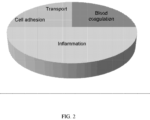
- proteomic markers (Table 4), originally identified by MS technologies, were also confirmed by immunoassays (ELISAs) to be suitable as markers for determining the acute rejection status. The performance of the protein panel comprising these six proteomic markers is described in Example 2. These proteomic markers participate in a range of biological processes, including cell adhesion, transport, blood coagulation, and inflammation. See Figure 2 . These proteomic markers, along with housekeeping genes, will be migrated onto a multiplexed, immuno-based microfluidics point-of-care platform for further testing and validation. Table 3. 40 nucleic acid markers.
- the assay which employs a panel comprising the 10 nucleic acid markers to determine the acute rejection status in a patient, had a sensitivity of 100%, a specificity of 74%.
- NR non rejectors
- MR moderate rejectors
- PPV positive predictive value
- NPV negative predictive value
- the panel comprising the six proteomic markers had a sensitivity of 100% and a specificity of 48%.
- the PPV for the panel was 7%, and NPV was 100%.
- the AUC for the panel was 0.62.
- the result also shows that the a biomarker panel combining the 6 proteomic markers and the 10 nucleic acid markers through computational methods improved the specificity of the HTG assay using the 10 nucleic acid markers alone, from 74% to 91%. See Table 6. Table 6. Performance of the diagnostic biomarker panels.
- a panel consisting of the 6 nucleic acid markers in Table 7 was tested in two different cohorts using the NanoString nCounter technology.
- the first is the recalibration cohort, in which the 6 nucleic acid marker panel was tested on samples from 38 subjects. 15 subjects had acute rejection and 23 had no rejection or moderate rejection to heart transplant.
- the second is the replication cohort, in which the panel of the 6 nucleic acid makers was tested on samples from 126 subjects, of which 22 had acute rejection and 104 had no rejection or moderate rejection.
Landscapes
- Life Sciences & Earth Sciences (AREA)
- Health & Medical Sciences (AREA)
- Chemical & Material Sciences (AREA)
- Engineering & Computer Science (AREA)
- Immunology (AREA)
- Molecular Biology (AREA)
- Proteomics, Peptides & Aminoacids (AREA)
- Biomedical Technology (AREA)
- Analytical Chemistry (AREA)
- Urology & Nephrology (AREA)
- Hematology (AREA)
- Organic Chemistry (AREA)
- Pathology (AREA)
- Biotechnology (AREA)
- Physics & Mathematics (AREA)
- Microbiology (AREA)
- Biochemistry (AREA)
- General Health & Medical Sciences (AREA)
- Zoology (AREA)
- Wood Science & Technology (AREA)
- Genetics & Genomics (AREA)
- Cell Biology (AREA)
- General Physics & Mathematics (AREA)
- Medicinal Chemistry (AREA)
- Food Science & Technology (AREA)
- Biophysics (AREA)
- Bioinformatics & Cheminformatics (AREA)
- General Engineering & Computer Science (AREA)
- Tropical Medicine & Parasitology (AREA)
- Physiology (AREA)
- Measuring Or Testing Involving Enzymes Or Micro-Organisms (AREA)
Description
- Transplantation is considered the primary therapy for patients with end-stage vital organ failure. While the availability of immunosuppressants such as cyclosporine and Tacrolimus has improved allograft recipient, e.g., heart transplant recipient, survival and wellbeing, identification of rejection of the allograft as early and as accurately as possible, and effective monitoring and adjusting immunosuppressive medication doses is still of primary importance to the continuing survival of the allograft recipient.
- Rejection of an allograft may be generally described as the result of recipient's immune response to nonself antigens expressed by the donor tissues. Acute rejection may occur within days or weeks of the transplant, while chronic rejection may be a slower process, occurring months or years following the transplant.
- At present, invasive biopsies, such as endomyocardial, liver core, and renal fine-needle aspiration biopsies, are widely regarded as the gold standard for the surveillance and diagnosis of allograft rejections, but are invasive procedures which carry risks of their own (e.g. Mehra MR, et al. Curr.Opin.Cardiol. 2002 Mar; 17(2): 131-136.). Biopsy results may also be subject to reproducibility and interpretation issues due to sampling errors and inter-observer variabilities, despite the availability of international guidelines such as the Banff schema for grading liver allograft rejection (Ormonde et al 1999. Liver Transplantation 5:261-268) or the Revised ISHLT transplantation scale (Stewart et al. 2005. J Heart Lung Transplant, 2005; 24: 1710-20). Although less invasive (imaging) techniques have been developed such as angiography and IVUS for monitoring chronic heart rejection, these alternatives are also susceptible to limitations similar to those associated with biopsies.
- The severity of allograft rejection as determined by biopsy may be graded to provide standardized reference indicia. The International Society for Heart and Lung Transplantation scale (ISHLT) provides a means of grading biopsy samples for heart transplant subjects (Table 1)
Table 1: International Society for Heart and Lung Transplantation grading of heart transplant rejection for histopathologic biopsy analysis. Grade Comment 0R No acute cellular rejection: No evidence of mononuclear inflammation or myocyte damage or necrosis. 1R Mild, low-grade, acute cellular rejection: Mononuclear cells are present and there may be limited myocyte damage and necrosis. 2R Moderate, intermediate-grade, acute cellular rejection: Two or more foci of mononuclear cells with associated myocyte damage and necrosis are present. The damage may be found in the same biopsy, or two separate biopsies. Eosinophils may be present. 3R Severe, high-grade, acute cellular rejection: Widespread, diffuse myocyte damage and necrosis, and disruption of normal architecture across several biopsies. Edema, interstitial hemorrhage and vasculitis may be present. The infiltrate may be polymorphous. - Indicators of allograft rejection may include a heightened and localized immune response as indicated by one or more of localized or systemic inflammation, tissue injury, allograft infiltration of immune cells, altered composition and concentration of tissue- and blood- derived proteins, differential oxygenation of allograft tissue, edema, thickening of the endothelium, increased collagen content, altered intramyocardial blood flow, infection, necrosis of the allograft and/or surrounding tissue, and the like.
- Allograft rejection maybe described as 'acute' or 'chronic'. Acute rejection is generally considered to be rejection of a tissue or organ allograft within ~6 months of the subject receiving the allograft. Acute rejection may be characterized by cellular and humoral insults on the donor tissue, leading to rapid graft dysfunction and failure of the tissue or organ. Chronic rejection is generally considered to be reject of a tissue or organ allograft beyond 6 months, and may be several years after receiving the allograft. Chronic rejection may be characterized by progressive tissue remodeling triggered by the alloimmune response may lead to gradual neointimal formation within arteries, contributing to obliterative vasculopathy, parenchymal fibrosis and consequently, failure and loss of the graft. Depending on the nature and severity of the rejection, there may be overlap in the indicators or clinical variables observed in a subject undergoing, or suspected of undergoing, allograft rejection - either chronic or acute.
- Attempts have been made to reduce the number of biopsies per patient, but may be generally unsuccessful, due in part to the difficulty in pinpointing the sites where rejection starts or progresses, and also to the difficulty in assessing tissue without performing the actual biopsy. Noninvasive surveillance techniques have been investigated, and may provide a reasonable negative prediction of allograft rejection, but may be of less practical utility in a clinical setting (Mehra et al., supra).
- The scientific and patent literature is replete with reports of this marker or that being important for identification/diagnosis/prediction/treatment of every medical condition that can be named. Even within the field of allograft rejection, a myriad of markers are recited (frequently singly), and conflicting results may be presented. This conflict in the literature, added to the complexity of the genome (estimates range upwards of 30,000 transcriptional units), the variety of cell types (estimates range upwards of 200), organs and tissues, and expressed proteins or polypeptides (estimates range upwards of 80,000) in the human body, renders the number of possible nucleic acid sequences, genes, proteins or combinations thereof useful for diagnosing acute organ rejection is staggering. Variation between individuals presents additional obstacles, as well as the dynamic range of protein concentration in plasma (ranging from 10"6 to 103 micro g/ mL) with many of the proteins of potential interest existing at very low concentrations) and the overwhelming quantities of the few, most abundant plasma proteins (constituting ~ 99 percent of the total protein mass.
- The CARGO study (Cardiac Allograft Rejection Gene Expression Observation) (Deng et al., 2006. Am J. Transplantation 6:150-160) used custom microarray analysis of - 7300 genes and RT-PCR to examine gene expression profile in subjects exhibiting an ISHLT score of 3 A or greater in samples taken 6 months or more post-transplant.
- Immune cells that have a role in recognizing may be useful as indicators of allograft rejection.
WO 2005/05721 - Traum et al., 2005 (Pediatr. Transplant 9(6):700-711) provides a general overview of transplantation proteomics. Exploration of biomarkers directly in the plasma proteome presents two main challenges - the dynamic range of protein concentrations extends from 10"6 to 103 micro g/ mL (Anderson et al. 2004. MoI Cell Proteomics 3:311-326), with many of the proteins of potential interest existing at very low concentrations and the most abundant plasma proteins comprising as much as 99 percent of the total protein mass.
- Maintenance or measurement of B2M serum levels in heart transplant patients was suggested as helpful in managing long-term immunosuppressive therapy (Erez et al., 1998. J Heart Lung Transplant 17:538-541).
PCT Publication WO 2009/003142 disclose that B2M, along with another protein may be useful as biomarkers for peripheral artery disease. - Borozdenkova et al. 2004 (J. Proteome Research 3:282-288) discloses that alpha B-crystallin and tropmyosin were elevated in a set of cardiac transplant subjects.
- Ishihara, 2008 (J. Mol Cell Cardiology 45:S33) discloses that ADIPOQ may have a role in cardiac transplantation, and Nakano (Transplant Immunology 2007 17:130-136) suggests that upregulation of ADIPOQ may be necessary for overcoming rejection in liver transplant subjects.
- Antibodies that bind SHBG (
PCT Publication WO 2007/024715 ) and FlO (PCT Publication WO 2005/020927 ) are suggested as being useful in preventing graft rejection. - SERPINFl and CIQ are disclosed as biomarkers associated with an increased risk of a cardiovascular event; the biomarkers maybe detected in a sample of an atherosclerotic plaque from a subject (
PCT Publication WO 2009/017405 ); sequences for SERPINFl may also be useful in an assay to select optimal blood vessel graft (US Publication 2006/0003338 ). - Complement is also known to have a role in rejection of allografts - Csencits et al., 2008 (Am J. Transplantation 8:1622-1630) summarizes past studies on various complement components and observes an accelerated humoral immune response in ClQ-/- mice allograft recipients.
-
PCT Publications WO2006/083986 ,WO206/122407 US Publications 2008/0153092 ,2006/0141493 andUS7235358 disclose methods for using panels of biomarkers (proteomic or genomic) for diagnosing or detecting various disease states ranging from cancer to organ transplantation - Alakulppi et al, 2007 (Transplantation 83:791-798) discloses the diagnosis of acute renal allograft rejection using RT-PCR for eight markers.
- A review by Fildes et al 2008 (Transplant Immunology 19:1-11) discusses the role of cell types in immune processes following lung transplantation, and discloses that AICL (CLEC2B) interaction with NK cell proteins may have a role in acute and chronic rejection
- Integration of multiple platforms (proteomics, genomics) has been suggested for diagnosis and monitoring of various cancers, however discordance between protein and mRNA expression is identified in the field (Chen et al., 2002.Mol Cell Proteomicsl:304-313; Nishizuka et al., 2003 Cancer Research 63:5243-5250). Previous studies have reported low correlations between genomic and proteomic data (Gygi SP et al. 1999. Mol Cell Biol.\9?12Q-\ 730; Huber et al., 2004 Mol Cell Proteomics 3:43-55). Hollander et al. 2010 discusses whole blood biomarkers of acute cardiac allograft rejection. Cohen Freue et al. 2013 discusses computational biomarker pipeline from discovery to clinical implementation, including plasma proteomic biomarkers for cardiac transplantation. Kasamatsu et al. 2005 discusses the identification of candidate genes associated with salivary adenoid cystic carcinomas using combined comparative genomic hybridization and oligonucleotide microarray analyses. Thach et al.2005 discusses the surveillance of transcriptomes in basic military trainees with normal, febrile respiratory illness, and convalescent phenotypes. Lin et al. 2009 discusses whole blood genomic biomarkers of acute cardiac allograft rejection.
- In a first aspect, the invention provides a method of determining the acute rejection status of a heart transplant in a subject using a biomarker panel comprising nucleic acid markers, the method comprising the steps of:
- a. determining the nucleic acid expression profile of nucleic acid markers in a biological sample from the subject, wherein the nucleic acid markers comprise or consist of CD177, CPA3, HEBP1, ORM1, VNN 1 and CNTNAP3,
- b. comparing the nucleic acid expression profile to a control profile,
- c. determining whether expression of the nucleic acid markers is increased or decreased relative to the control profile, wherein the increased or decreased expression of the nucleic acid markers is indicative of the acute rejection status of the subject.
- The disclosure further provides a kit for determining the acute rejection status of heart transplant in a patient, comprising a plurality of detection reagents for detecting the nucleic acid expression of nucleic acid markers, wherein the nucleic acid markers comprise or consists of CD177, CNTNAP3, CPA3, HEBP1, ORM1 and VNN1.
- In some embodiments, the nucleic acid expression profile is determined by PCR, HTG EdgeSeq or NanoString nCounter.
- In some embodiments, the biomarker panel has an AUC of at least 0.6, and/or a sensitivity of at least 80%, and/or a specificity of at least 15%, and/or a positive predictive value (PPV) of at least 4%, and/or a negative predictive value (NPV) of at least 98%, in predicting the status of acute rejection of heart transplant. In some embodiments, the assays using the biomarker panel described herein show comparable performance (e.g., NPV and PPV) to commercially available tests, such as the AlloMap assay by CareDx. In some embodiments, the assays described herein can be used in the first 2 months post-transplant where commercial tests have not demonstrated utility. For example, in some embodiments, assays using the biomarkers described herein achieve a PPV of 5% and a NPV of 98-100% for samples obtained during the first 2 months after the heart transplant.
-
-
Figure 1 shows a range of biological processes in which the nucleic acid markers disclosed herein participate. -
Figure 2 shows a range of biological processes in which the proteomic markers disclosed herein participate. - In the description that follows, a number of terms are used extensively, the following definitions are provided to facilitate understanding of various aspects of the invention. Use of examples in the specification, including examples of terms, is for illustrative purposes only and is not intended to limit the scope and meaning of the embodiments of the invention herein. Numeric ranges are inclusive of the numbers defining the range. In the specification, the word "comprising" is used as an open-ended term, substantially equivalent to the phrase "including, but not limited to," and the word "comprises" has a corresponding meaning.
- The present disclosure provides nucleic acidexpression profiles related to the assessment, prediction or diagnosis of allograft rejection in a subject, as defined in the claims. The specific combination of the altered expression levels (increased or decreased relative to a control) of specific sets of genomic markers comprise a novel combination useful for assessment, prediction or diagnosis of allograft rejection in a subject.
- An allograft is an organ or tissue transplanted between two genetically different subjects of the same species. The subject receiving the allograft is the 'recipient', while the subject providing the allograft is the 'donor'. A tissue or organ allograft may alternately be referred to as a 'transplant', a 'graft', an 'allograft', a 'donor tissue' or 'donor organ', or similar terms. A transplant between two subjects of different species is a xenograft.
- Subjects may present with a variety of symptoms or clinical variables well-known in the literature, however none of these of itself is a predictive or diagnostic of allograft rejection. A myriad of clinical variables may be used in assessing a subject having, or suspected of having, allograft rejection, in addition to biopsy of the allograft. The information gleaned from these clinical variables is then used by a clinician, physician, veterinarian or other practitioner in a clinical field in attempts to determine if rejection is occurring, and how rapidly it progresses, to allow for modification of the immunosuppressive drug therapy of the subject. Examples of clinical variables are described in Table 2.
Table 2. Clinical variables for possible use in assessment of allograft rejection. Clinical Variable Name Renal/Heart/Liver/ All Variable Explanation Primary Diagnosis All Diagnosis leading to transplant Secondary Diagnosis All Diagnosis leading to transplant "Transplant Procedure - Living related, Living unrelated, or cadaveric" Blood Type All Blood Type Blood Rh All Blood Rh Height (cm) All Height (cm) Weight (kg) All Weight (kg) BMI All Calculation: Weight / (Height)2 Liver Ascites All HLA A1 All HLA A2 All HLA B1 All HLA B2 All HLA DR1 All HLA DR2 All CMV All Viral Status CMV Date All Date of viral status HIV All Viral Status HBV All Viral Status HBV Date All Date of viral status HbsAb All Viral Status HbcAb (Total) All Viral Status HBvDNA All Viral Status HCV All Viral Status HCV Genotype All Hepatitis C genotype HCV Genotype Sub All "Hepatitis C genotype, subtype" EBV All Viral Status Zoster All Viral Status Dialysis Start Date All Dialysis Start Date Dialysis Type All Dialysis Type Cytoxicity Current Level All Cytoxicity Current Date All Cytoxicity Peak Level All Cytoxicity Peak Date All Flush Soln All Type of Flush Solution used at transplant Cold Time 1 All Cold Time 2 All Re-Warm Time 1 All Re-Warm Time 2 All HTLV 1 All HTLV 2 All HCV RNA All 24hr Urine All 24 Hour urine output Systolic Blood Pressure All Blood Pressure reading Diastolic Blood Pressure All Blood Pressure reading 24 Hr Urine All 24 hour urine Sodium All Blood test Potassium All Blood test Chloride All Blood test Total CO2 All Blood test Albumin All Blood test Protein All Blood test Calcium All Blood test Inorganic Phosphate All Blood test Magnesium All Blood test Uric Acid All Blood test Glucose All Blood test Hemoglobin A1C All Blood test CPK All Blood test Parathyroid Hormone All Blood test Homocysteine All Blood test Urine Protein All Urine test Creatinine All Blood test BUN All Blood test Hemaglobin All Blood test Platelet Count All Blood test WBC Count All Blood test Prothrombin Time All Blood test Partial Thromboplastin Time All Blood test INR All Blood test Gamma GT All Blood test AST All Blood test Alkaline Phosphatase All Blood test Amylase All Blood test Total Bilirubin All Blood test Direct Bilirubin All Blood test LDH All Blood test ALT All Blood test Triglycerides All Blood test Cholesterol All Blood test HDL Cholesterol All Blood test LDL Cholesterol All Blood test FEV1 All Lung function test FVC All Lung function test Total Ferritin All Blood test TIBC All Blood test Transferrin Saturated All Blood test Ferritin All Blood test Angiography Heart Heart function test Intravascular ultrasound Heart Heart function test Dobutamine Stress Echocardiography Heart Heart function test Cyclosporine WB All Immunosuppressive levels Cyclosporine 2 hr All Immunosuppressive levels Tacrolimus WB All Immunosuppressive levels Sirolimus WB All Immunosuppressive total daily dose Solumedrol All Immunosuppressive total daily dose Prednisone All Immunosuppressive total daily dose Prednisone ALT All Immunosuppressive total daily dose Tacrolimus All Immunosuppressive total daily dose Cyclosporine All Immunosuppressive total daily dose Imuran All Immunosuppressive total daily dose Mycophonelate Mofetil All Immunosuppressive total daily dose Sirolimus All Immunosuppressive total daily dose OKT3 All Immunosuppressive total daily dose ATG All Immunosuppressive total daily dose ALG All Immunosuppressive total daily dose Basiliximab All Immunosuppressive total daily dose Daclizumab All Immunosuppressive total daily dose Ganciclovir All Anti-viral total daily dose Lamivudine All Anti-viral total daily dose Riboviron All Anti-viral total daily dose Interferon All Anti-viral total daily dose Hepatisis C Virus RNA All test for presence of HCV values () CMV Antigenemia All Antiviral and Virus Valganciclovir All Anti-viral total daily dose Neutrophil Number All Blood test C Peptide All Blood test Peg Interferon All Anti-viral total daily dose GFR All Glomerular Filtration Rate Complication Events All Complication Type Biopsy Scores Renal Borderline, 1A, 1B, 2A, 2B, 3, Hyperacute Biopsy Scores Liver Portal inflammation, Bile duct inflammation damage, Venous endothelial inflammation each scored from 1 to 3 Donor Blood Type All Donor Blood Type Donor Blood Rh All Donor Rh Donor HLA A1 All Donor HLA A1 Donor HLA A2 All Donor HLA A2 Donor HLA B 1 All Donor HLA B 1 Donor HLA B2 All Donor HLA B2 Donor HLA DR1 All Donor HLA DR1 Donor HLA DR2 All Donor HLA DR2 Donor CMV All Donor CMV Donor HIV All Donor HIV Donor HBV All Donor HBV Donor HbsAb All Donor HbsAb Donor HbcAb (total) All Donor HbcAb (total) Donor Hbdna All Donor Hbdna Donor HCV All Donor HCV Donor EBV All Donor EBV - Clinical variables (optionally accompanied by biopsy), while currently the only practical tools available to a clinician in mainstream medical practice, are not always able to cleanly differentiate between an AR (an "acute rejector"; ISHLT grade 2 R or higher) and an NR (a "mild or non-rejector"; ISHLT grade 0R or 1R) subject. While the extreme left and right subjects are correctly classified as AR or NR, the bulk of the subjects are represented in the middle range and their status is unclear. This does not negate the value of the clinical variables in the assessment of allograft rejection, but instead indicates their limitation when used in the absence of other methods.
- The multifactorial nature of allograft rejection prediction, diagnosis and assessment is considered in the art to exclude the possibility of a single biomarker that meets even one of the needs of prediction, diagnosis or assessment of allograft rejection. Strategies involving a plurality of markers may take into account this multifactorial nature. Alternately, a plurality of markers may be assessed in combination with clinical variables that are less invasive (e.g. a biopsy not required) to tailor the prediction, diagnosis and/or assessment of allograft rejection in a subject.
- Regardless of the methods used for prediction, diagnosis and assessment of allograft rejection, earlier is better - from the viewpoint of preserving organ or tissue function and preventing more systemic detrimental effects. There is no 'cure' for allograft rejection, only maintenance of the subject at a suitably immunosuppressed state, or in some cases, replacement of the organ if rejection has progressed too rapidly or is too severe to correct with immunosuppressive drug intervention therapy.
- Applying a plurality of mathematical and/or statistical analytical methods to a nucleic acid expression dataset may indicate varying subsets of significant markers, leading to uncertainty as to which method is 'best' or 'more accurate'. Regardless of the mathematics, the underlying biology is the same in a dataset. By applying a plurality of mathematical and/or statistical methods to a microarray dataset and assessing the statistically significant subsets of each for common markers, uncertainty may be reduced, and clinically relevant core group of markers may be identified.
- "Markers", "biological markers" or "biomarkers" may be used interchangeably and refer generally to detectable (and in some cases quantifiable) molecules or compounds in a biological sample. A marker may be down-regulated (decreased), up-regulated (increased) or effectively unchanged in a subject following transplantation of an allograft. Markers may include nucleic acids (DNA or RNA), a gene, or a transcript, or a portion or fragment of a transcript in reference to 'genomic' markers (alternately referred to as "nucleic acid markers"). In some usages, these terms may reference the level or quantity of a nucleic acid or polynucleotide (in absolute terms or relative to another sample or standard value) or the ratio between the levels of two polynucleotidesin a subject's biological sample. The level may be expressed as a concentration, for example micrograms per milliliter; as a colorimetric intensity, for example 0.0 being transparent and 1.0 being opaque at a particular wavelength of light, with the experimental sample ranked accordingly and receiving a numerical score based on transmission or absorption of light at a particular wavelength; or as relevant for other means for quantifying a marker, such as are known in the art. In some examples, a ratio may be expressed as a unitless value. A "marker" may also reference to a ratio, or a net value following subtraction of a baseline value. A marker may also be represented as a 'fold-change', with or without an indicator of directionality (increase or decrease/ up or down). The increase or decrease in expression of a marker may also be referred to as 'down-regulation' or 'up-regulation', or similar indicators of an increase or decrease in response to a stimulus, physiological event, or condition of the subject. A marker may be present in a first biological sample, and absent in a second biological sample; alternately the marker may be present in both, with a statistically significant difference between the two. Expression of the presence, absence or relative levels of a marker in a biological sample may be dependent on the nature of the assay used to quantify or assess the marker, and the manner of such expression will be familiar to those skilled in the art.
- A marker may be described as being differentially expressed when the level of expression in a subject who is rejecting an allograft is significantly different from that of a subject or sample taken from a non-rejecting subject. A differentially expressed marker may be overexpressed or underexpressed as compared to the expression level of a normal or control sample.
- A "profile" is a set of one or more markers and their presence, absence, relative level or abundance (relative to one or more controls). A genomic or nucleic acid profile a dataset of the presence, absence, relative level or abundance of expressed nucleic acids (e.g. transcripts, mRNA, EST or the like). A profile may alternately be referred to as an expression profile.
- The increase or decrease, or quantification of the markers in the biological sample may be determined by any of several methods known in the art for measuring the presence and/or relative abundance of a gene product or transcript, or a nucleic acid molecule comprising a particular sequence or the like. The level of the markers may be determined as an absolute value, or relative to a baseline value, and the level of the subject's markers compared to a cutoff index (e.g. a non-rejection cutoff index). Alternately the relative abundance of the marker may be determined relative to a control. The control may be a clinically normal subject (e.g. one who has not received an allograft) or may be an allograft recipient that has not previously demonstrated rejection.
- In some embodiments, the control may be an autologous control, for example a sample or profile obtained from the subject before undergoing allograft transplantation. In some embodiments, the profile obtained at one time point (before, after or before and after transplantation) may be compared to one or more than one profiles obtained previously from the same subject. By repeatedly sampling the same biological sample from the same subject over time, a composite profile, illustrating marker level or expression over time may be provided. Sequential samples can also be obtained from the subject and a profile obtained for each, to allow the course of increase or decrease in one or more markers to be followed over time. For example, an initial sample or samples may be taken before the transplantation, with subsequent samples being taken weekly, biweekly, monthly, bimonthly or at another suitable, regular interval and compared with profiles from samples taken previously. Samples may also be taken before, during and after administration of a course of a drug, for example an immunosuppressive drug.
- Techniques, methods, tools, algorithms, reagents and other necessary aspects of assays that may be employed to detect and/or quantify a particular marker or set of markers are varied. Of significance is not so much the particular method used to detect the marker or set of markers, but what markers to detect. As is reflected in the literature, tremendous variation is possible. Once the marker or set of markers to be detected or quantified is identified, any of several techniques may be well suited, with the provision of appropriate reagents. One of skill in the art, when provided with the set of markers to be identified, will be capable of selecting the appropriate assay (for example, a PCR based or a microarray based assay for nucleic acid markers, an ELISA, protein or antibody microarray or similar immunologic assay, or in some examples, use of an MRM, iTRAQ, iCAT or SELDI proteomic mass spectrometric based method) for performing the methods disclosed herein.
- The present disclosure provides nucleic acid expression profiles related to the assessment, prediction or diagnosis of allograft rejection in a subject.
- For example, detection or determination, and in some cases quantification, of a nucleic acid may be accomplished by any one of a number methods or assays employing recombinant DNA technologies known in the art, including but not limited to, as sequence-specific hybridization, polymerase chain reaction (PCR), RT-PCR, microarrays and the like. Such assays may include sequence-specific hybridization, primer extension, or invasive cleavage. Furthermore, there are numerous methods for analyzing/detecting the products of each type of reaction (for example, fluorescence, luminescence, mass measurement, electrophoresis, etc.). Furthermore, reactions can occur in solution or on a solid support such as a glass slide, a chip, a bead, or the like.
- Methods of designing and selecting probes for use in microarrays or biochips, or for selecting or designing primers for use in PCR-based assays are known in the art. Once the marker or markers are identified and the sequence of the nucleic acid determined by, for example, querying a database comprising such sequences, or by having an appropriate sequence provided (for example, a sequence listing as provided herein), one of skill in the art will be able to use such information to select appropriate probes or primers and perform the selected assay.
- Standard reference works setting forth the general principles of recombinant DNA technologies known to those of skill in the art include, for example: Ausubel et al, Current Protocols In Molecular Biology, John Wiley and Sons, New York (1998 and Supplements to 2001); Sambrook et al, Molecular Cloning: A Laboratory Manual, 2d Ed., Cold Spring Harbor Laboratory Press, Plainview, New York (1989); Kaufman et al, Eds., Handbook Of Molecular And Cellular Methods In Biology And Medicine, CRC Press, Boca Raton (1995); McPherson, Ed., Directed Mutagenesis: A Practical Approach, IRL Press, Oxford (1991).
- A subject's rejection status may be described as an "acute rejector" (ISHLT grade 2R of higher; AR) or as a "non-rejector" (ISHLT grade 0R or 1R; NR) and is determined by comparison of the concentration of the markers to that of a non-rejector cutoff index. A "non-rejector cutoff index" is a numerical value or score, beyond or outside of which a subject is categorized as having an AR rejection status. The non- rejector cutoff index maybe alternately referred to as a 'control value', a 'control index', or simply as a 'control'. A non-rejector cutoff-index maybe the concentration of individual markers in a control subject population and considered separately for each marker measured; alternately the non-rejector cutoff index may be a combination of the concentration of the markers, and compared to a combination of the concentration of the markers in the subject's sample provided for diagnosing. The control subject population may be a normal or healthy control population, or may be an allograft recipient population that has not, or is not, rejecting the allograft. The control maybe a single subject, and for some embodiments, maybe an autologous control. A control, or pool of controls, may be constant e.g. represented by a static value, or may be cumulative, in that the sample population used to obtain it may change from site to site, or over time and incorporate additional data points. For example, a central data repository, such as a centralized healthcare information system, may receive and store data obtained at various sites (hospitals, clinical laboratories or the like) and provide this cumulative data set for use with the methods of the invention at a single hospital, community clinic, for access by an end user (i.e. an individual medical practitioner, medical clinic or center, or the like).
- The non-rejector cutoff index may be alternately referred to as a 'control value', a 'control index' or simply as a 'control'. In some embodiments the cutoff index may be further characterized as being a genomic cutoff index (for genomic expression profiling of subjects), or the like.
- A "biological sample" refers generally to body fluid or tissue or organ sample from a subject. For example, the biological sample may a body fluid such as blood, plasma, lymph fluid, serum, urine or saliva. A tissue or organ sample, such as a non-liquid tissue sample maybe digested, extracted or otherwise rendered to a liquid form - examples of such tissues or organs include cultured cells, blood cells, skin, liver, heart, kidney, pancreas, islets of Langerhans, bone marrow, blood, blood vessels, heart valve, lung, intestine, bowel, spleen, bladder, penis, face, hand, bone, muscle, fat, cornea or the like. A plurality of biological samples may be collected at any one time. A biological sample or samples may be taken from a subject at any time, including before allograft transplantation, at the time of translation or at anytime following transplantation. A biological sample may comprise nucleic acid, such as deoxyribonucleic acid or ribonucleic acid, or a combination thereof, in either single or double- stranded form. When an organ is removed from a donor, the spleen of the donor or a part of it may be kept as a biological sample from which to obtain donor T-cells. When an organ is removed from a living donor, a blood sample may be taken, from which donor T-cells may be obtained. Alloreactive T-cells may be isolated by exploiting their specific interaction with antigens (including the MHC complexes) of the allograft. Methods to enable specific isolation of alloreactive T-cells are described in, for example
PCT Publication WO 2005/05721 - A lymphocyte is nucleated or 'white' blood cell (leukocyte) of lymphoid stem cell origin. Lymphocytes include T-cells, B-cells natural killer cells and the like, and other immune regulatory cells. A "T-cell" is a class of lymphocyte responsible for cell-mediated immunity, and for stimulating B-cells. A stimulated B-cell produces antibodies for specific antigens. Both B- cells and T-cells function to recognize non-self antigens in a subject. Non-self antigens include those of viruses, bacteria and other infectious agents as well as allografts.
- The term "subject" or "patient" generally refers to mammals and other animals including humans and other primates, companion animals, zoo, and farm animals, including, but not limited to, cats, dogs, rodents, rats, mice, hamsters, rabbits, horses, cows, sheep, pigs, goats, poultry, etc. A subject includes one who is to be tested, or has been tested for prediction, assessment or diagnosis of allograft rejection. The subject may have been previously assessed or diagnosed using other methods, such as those described herein or those in current clinical practice, or maybe selected as part of a general population (a control subject).
- A fold-change of a marker in a subject, relative to a control maybe at least 0.1, 0.2, 0.3, 0.4, 0.5, 0.6, 0.7, 0.8, 0.9, 1.0, 1.1, 1.2, 1.3, 1.4, 1.5, 1.6, 1.7, 1.8, 1.9, 2.0, 2.1, 2.2, 2.3, 2.4, 2.5, 2.6, 2.7, 2.8, 2.9, 3.0, 3.1, 3.2, 3.3, 3.4, 3.5, 3.6, 3.7, 3.8, 3.9, 4.0, 4.1, 4.2, 4.3, 4.4, 4.5, 4.6, 4.7, 4.8, 4.9, 5.0 or more, or any amount therebetween. The fold change may represent a decrease, or an increase, compared to the control value.
- One or more than one includes 1, 2,3, 4, 5, 6, 7, 8, 9, 10, 11, 12, 13, 14, 15, 16 or more.
- "Down-regulation" or 'down-regulated may be used interchangeably and refer to a decrease in the level of a marker, such as a gene, nucleic acid, transcript, protein or polypeptide. "Up-regulation" or "up-regulated" may be used interchangeably and refer to an increase in the level of a marker, such as a gene, nucleic acid or transcript.
- For the purpose of this invention, a patient has treatable acute rejection status to heart transplant if he or she had response that fits into the "2R" or "3R" category according to the International Society for heart and Lung transplantation standard (Table 1). A patient has non rejection status if the response to heart transplant fit into the "0R" category and moderate rejection status if the response fits into the "1R" category according to the above standard.
- Once a subject is identified as an acute rejector, or at risk for becoming an acute rejector by any method (genomic, proteomic, or a combination thereof), therapeutic measures may be implemented to alter the subject's immune response to the allograft. The subject may undergo additional monitoring of clinical values more frequently, or using more sensitive monitoring methods. Additionally the subject may be administered immunosuppressive medicaments to decrease or increase the subject's immune response. Even though a subject's immune response needs to be suppressed to prevent rejection of the allograft, a suitable level of immune function is also needed to protect against opportunistic infection. Various medicaments that maybe administered to a subject are known; see for example, Goodman and Gilman's The Pharmacological Basis of Therapeutics 11th edition. Ch 52, pp 1405-1431 and references therein; LL Brunton, JS Lazo, KL Parker editors. Standard reference works setting forth the general principles of medical physiology and pharmacology known to those of skill in the art include: Fauci et al., Eds., Harrison's Principles Of Internal Medicine, 14th Ed., McGraw-Hill Companies, Inc. (1998). Other preventative and therapeutic strategies are reviewed in the medical literature- see, for example Kobashigawa et al. 2006. Nature Clinical Practice. Cardiovascular Medicine 3:203-21.
- A method of diagnosing acute allograft rejection in a subject comprises 1) determining the expression profile of nucleic acid markers in a biological sample from the subject, wherein the nucleic acid markers comprise or consist of the nucleic acid markers listed in Table 7; 2) comparing the expression profile of the nucleic acid markers to a non-rejector profile; and 3) determining whether the expression level of the nucleic acid markers is up-regulated or down- regulated relative to the control profile, wherein up-regulation or down-regulation of the nucleic acid markers is indicative of the rejection status.
- Therefore, the disclosure also provides for a method of predicting, assessing or diagnosing allograft rejection in a subject comprising 1) measuring the increase or decrease of the nucleic acid markers listed in Table 7 and 2) determining the 'rejection status' of the subject, wherein the determination of 'rejection status' of the subject is based on comparison of the subject's nucleic acid marker expression profile to a control nucleic acid marker expression profile.
- The phrase "gene expression data", "gene expression profile" "nucleic acid expression profile" or "marker expression profile" as used herein refers to information regarding the relative or absolute level of expression of a gene or set of genes in a biological sample. The level of expression of a gene may be determined based on the level of a nucleic acid such as RNA including mRNA, transcribed from or encoded by the gene.
- A "polynucleotide", "oligonucleotide", "nucleic acid" or "nucleotide polymer" as used herein may include synthetic or mixed polymers of nucleic acids, including RNA, DNA or both RNA and DNA, both sense and antisense strands, and may be chemically or biochemically modified or may contain non- natural or derivatized nucleotide bases, as will be readily appreciated by those skilled in the art. Such modifications include, for example, labels, methylation, substitution of one or more of the naturally occurring nucleotides with an analog, internucleotide modifications such as uncharged linkages (e.g., methyl phosphonates, phosphotriesters, phosphoamidates, carbamates, etc.), charged linkages (e. g., phosphorothioates, phosphorodithioates, etc.), pendent moieties (e.g., polypeptides), and modified linkages (e.g., alpha anomeric polynucleotides, etc.). Also included are synthetic molecules that mimic polynucleotides in their ability to bind to a designated sequence via hydrogen bonding and other chemical interactions.
- An oligonucleotide includes variable length nucleic acids, which may be useful as probes, primers and in the manufacture of microarrays (arrays) for the detection and/or amplification of specific nucleic acids. Oligonucleotides may comprise DNA, RNA, PNA or other polynucleotide moieties as described in, for example,
US 5,948,902 . Such DNA, RNA or oligonucleotide strands may be synthesized by the sequential addition (5 '-3' or 3'-5') of activated monomers to a growing chain which may be linked to an insoluble support. Numerous methods are known in the art for synthesizing oligonucleotides for subsequent individual use or as a part of the insoluble support, for example in arrays (BERNFIELD MR. and ROTTMAN FM. J. Biol. Chem. (1967) 242(18):4134-43; SULSTON J. et al. PNAS (1968) 60(2):409-415; GILLAM S. et al. Nucleic Acid Res.(1975) 2(5):613-624; BONORA GM. et al. Nucleic Acid Res.(1990) 18(11):3155-9; LASHKARI DA. et al. PNAS (1995) 92(17):7912-5; MCGALL G. et al. PNAS (1996) 93(24):13555-60; ALBERT TJ. et al. Nucleic Acid Res.(2003) 31(7):e35; GAO X. et al. Biopolymers (2004) 73(5):579-96; and MOORCROFT MJ. et al. Nucleic Acid Res.(2005) 33(8):e75). In general, oligonucleotides are synthesized through the stepwise addition of activated and protected monomers under a variety of conditions depending on the method being used. Subsequently, specific protecting groups may be removed to allow for further elongation and subsequently and once synthesis is complete all the protecting groups may be removed and the oligonucleotides removed from their solid supports for purification of the complete chains if so desired. - A "gene" is an ordered sequence of nucleotides located in a particular position on a particular chromosome that encodes a specific functional product and may include untranslated and untranscribed sequences in proximity to the coding regions (5' and 3' to the coding sequence). Such non-coding sequences may contain regulatory sequences needed for transcription and translation of the sequence or splicing of introns, for example, or may as yet to have any function attributed to them beyond the occurrence of the mutation of interest. A gene may also include one or more promoters, enhancers, transcription factor binding sites, termination signals or other regulatory elements. A gene may be generally referred to as 'nucleic acid'.
- The term "microarray," "array," or "chip" refers to a plurality of defined nucleic acid probes coupled to the surface of a substrate in defined locations. The substrate may be preferably solid. Microarrays, their methods of manufacture, use and analysis have been generally described in the art in, for example,
U.S. Patent Nos. 5,143,854 (Pirrung ),5,424,186 (Fodor ),5,445,934 (Fodor ),5,677,195 (Winkler ),5,744,305 (Fodor ),5,800,992 (Fodor ),6,040,193 (Winkler ), and Fodor et al. 1991. Science, 251 -.161-111. - "Hybridization" includes a reaction in which one or more polynucleotides and/or oligonucleotides interact in an ordered manner (sequence-specific) to form a complex that is stabilized by hydrogen bonding - also referred to as 'Watson-Crick' base pairing. Variant base- pairing may also occur through non-canonical hydrogen bonding includes Hoogsteen base pairing. Under some thermodynamic, ionic or pH conditions, triple helices may occur, particularly with ribonucleic acids. These and other variant hydrogen bonding or base-pairing are known in the art, and may be found in, for example, Lehninger - Principles of Biochemistry, 3rd edition (Nelson and Cox, eds. Worth Publishers, New York.).
- Hybridization reactions can be performed under conditions of different "stringency". The stringency of a hybridization reaction includes the difficulty with which any two nucleic acid molecules will hybridize to one another. Stringency may be increased, for example, by increasing the temperature at which hybridization occurs, by decreasing the ionic concentration at which hybridization occurs, or a combination thereof. Under stringent conditions, nucleic acid molecules at least 60 percent, 65 percent, 70 percent, 75 percent or more identical to each other remain hybridized to each other, whereas molecules with low percent identity cannot remain hybridized. An example of stringent hybridization conditions are hybridization in 6x sodium chloride/sodium citrate (SSC) at about 44-45 degrees centigrade, followed by one or more washes in 0.2xSSC, 0.1 percent SDS at 5ODegrees centigrade 55Degrees centigrade 60Degrees centigrade 65 degrees centigrade, or at a temperature therebetween.
- Hybridization between two nucleic acids may occur in an antiparallel configuration - this is referred to as 'annealing', and the paired nucleic acids are described as complementary. A double-stranded polynucleotide may be "complementary", if hybridization can occur between one of the strands of the first polynucleotide and the second. The degree of which one polynucleotide is complementary with another is referred to as homology, and is quantifiable in terms of the proportion of bases in opposing strands that are expected to hydrogen bond with each other, according to generally accepted base-pairing rules.
- In general, sequence-specific hybridization involves a hybridization probe, which is capable of specifically hybridizing to a defined sequence. Such probes may be designed to differentiate between sequences varying in only one or a few nucleotides, thus providing a high degree of specificity. A strategy which couples detection and sequence discrimination is the use of a "molecular beacon", whereby the hybridization probe (molecular beacon) has 3' and 5' reporter and quencher molecules and 3' and 5' sequences which are complementary such that absent an adequate binding target for the intervening sequence the probe will form a hairpin loop. The hairpin loop keeps the reporter and quencher in close proximity resulting in quenching of the fluorophor (reporter) which reduces fluorescence emissions. However, when the molecular beacon hybridizes to the target the fluorophor and the quencher are sufficiently separated to allow fluorescence to be emitted from the fluorophor.
- Probes used in hybridization may include double-stranded DNA, single-stranded DNA and RNA oligonucleotides, and peptide nucleic acids. Hybridization conditions and methods for identifying markers that hybridize to a specific probe are described in the art - see, for example, Brown, T. "Hybridization Analysis of DNA Blots" in Current Protocols in Molecular Biology. FM Ausubel et al, editors. Wiley and Sons, 2003. doi: 10.1002/0471142727.mb0210s21. Suitable hybridization probes for use in accordance with the invention include oligonucleotides, polynucleotides or modified nucleic acids from about 10 to about 400 nucleotides, alternatively from about 20 to about 200 nucleotides, or from about 30 to about 100 nucleotides in length.
- Specific sequences may be identified by hybridization with a primer or a probe, and this hybridization subsequently detected.
- A "primer" includes a short polynucleotide, generally with a free 3'-OH group that binds to a target or "template" present in a sample of interest by hybridizing with the target, and thereafter promoting polymerization of a polynucleotide complementary to the target. A "polymerase chain reaction" ("PCR") is a reaction in which replicate copies are made of a target polynucleotide using a "pair of primers" or "set of primers" consisting of "upstream" and a "downstream" primer, and a catalyst of polymerization, such as a DNA polymerase, and typically a thermally-stable polymerase enzyme. Methods for PCR are well known in the art, and are taught, for example, in Beverly, SM. Enzymatic Amplification of RNA by PCR (RT-PCR) in Current Protocols in Molecular Biology. FM Ausubel et al, editors. Wiley and Sons, 2003. doi: 10.1002/0471142727.mb 1505s56. Synthesis of the replicate copies may include incorporation of a nucleotide having a label or tag, for example, a fluorescent molecule, biotin, or a radioactive molecule. The replicate copies may subsequently be detected via these tags, using conventional methods.
- A primer may also be used as a probe in hybridization reactions, such as Southern or Northern blot analyses (see, e.g., Sambrook, J., Fritsh, E. F., and Maniatis, T. Molecular Cloning: A Laboratory Manual. 2nd, ed., Cold Spring Harbor Laboratory, Cold Spring Harbor Laboratory Press, Cold Spring Harbor, N.Y., 1989).
- A "probe set" (or 'primer set') as used herein refers to a group of oligonucleotides that may be used to detect one or more expressed nucleic acids, or expressed genes. Detection may be, for example, through amplification as in PCR and RT-PCR, or through hybridization, as on a microarray, or through selective destruction and protection, as in assays based on the selective enzymatic degradation of single or double stranded nucleic acids. Probes in a probe set may be labeled with one or more fluorescent, radioactive or other detectable moieties (including enzymes). Probes may be any size so long as the probe is sufficiently large to selectively detect the desired gene - generally a size range from about 15 to about 25, or to about 30 nucleotides is of sufficient size. A probe set maybe in solution, e.g. for use in multiplex PCR. Alternately, a probe set may be adhered to a solid surface, as in an array or microarray.
- In the invention, a probe set for detection of nucleic acids markers, wherein the nucleic acid markers comprise or consist of CD177, CPA3, HEBP1, ORM1, VNN 1 and CNTNAP3 is provided. Such a probe set may be useful for determining the rejection status of a subject. The probe set may comprise one or more pairs of primers for specific amplification (e.g. PCR or RT- PCR) of nucleic acid sequences. In another embodiment of the invention, the probe set is part of a microarray.
- It will be appreciated that numerous other methods for sequence discrimination and detection are known in the art and some of which are described in further detail below. It will also be appreciated that reactions such as arrayed primer extension mini sequencing, tag microarrays and sequence-specific extension could be performed on a microarray. One such array based genotyping platform is the microsphere based tag-it high throughput array (BORTOLINS. et al. 2004 Clinical Chemistry 50: 2028-36). This method amplifies genomic DNA by PCR followed by sequence-specific primer extension with universally tagged primers. The products are then sorted on a Tag-It array and detected using the Luminex xMAP system.
- It will be appreciated by a person of skill in the art that any numerical designations of nucleotides or amino acids within a sequence are relative to the specific sequence. Also, the same positions may be assigned different numerical designations depending on the way in which the sequence is numbered and the sequence chosen. Furthermore, sequence variations such as insertions or deletions, may change the relative position and subsequently the numerical designations of particular nucleotides or amino acids at or around a mutational site.
- Selection and/or design of probes, primers or probe sets for specific detection of expression of any gene of interest is within the ability of one of skill in the relevant art, when provided with one or more nucleic acid sequences of the gene of interest. Further, any of several probes, primers or probe sets, or a plurality of probes, primers or probe sets may be used to detect a gene of interest, for example, an array may include multiple probes for a single gene transcript - the aspects of the invention as described herein are not limited to any specific probes exemplified.
- Sequence identity or sequence similarity may be determined using a nucleotide sequence comparison program (for DNA or RNA sequences, or fragments or portions thereof) or an amino acid sequence comparison program (for protein, polypeptide or peptide sequences, or fragments or portions thereof), such as that provided within DNASIS (for example, but not limited to, using the following parameters: GAP penalty 5, #of top diagonals 5, fixed GAP penalty 10, k-tuple 2, floating gap 10, and window size 5). However, other methods of alignment of sequences for comparison are well-known in the art for example the algorithms of Smith and Waterman (1981, Adv. Appl. Math. 2:482), Needleman and Wunsch (J. MoI. Biol. 48:443, 1970), Pearson and Lipman (1988, Proc. Nat'l. Acad. Sci. USA 85:2444), and by computerized implementations of these algorithms (e.g. GAP, BESTFIT, FASTA, and BLAST), or by manual alignment and visual inspection.
- If a nucleic acid or gene, polypeptide or sequence of interest is identified and a portion or fragment of the sequence (or sequence of the gene polypeptide or the like) is provided, other sequences that are similar, or substantially similar may be identified using the programs exemplified above. For example, when constructing a microarray or probe sequences, the sequence and location are known, such that if a microarray experiment identifies a 'hit' (the probe at a particular location hybridizes with one or more nucleic acids in a sample, the sequence of the probe will be known (either by the manufacturer or producer of the microarray, or from a database provided by the manufacturer - for example the NetAffx databases of Affymetrix, the manufacturer of the Human Genome U133 Plus 2.0 Array). If the identity of the sequence source is not provided, it may be determined by using the sequence of the probe in a sequence-based search of one or more databases. For peptide or peptide fragments identified by proteomics assays, for example iTRAQ, the sequence of the peptide or fragment may be used to query databases of amino acid sequences as described above. Examples of such a database include those maintained by the National Centre for Biotechnology Information, or those maintained by the European Bioinformatics Institute.
- A protein or polypeptide, nucleic acid or fragment or portion thereof may be considered to be specifically identified when its sequence may be differentiated from others found in the same phylogenetic Species, Genus, Family or Order. Such differentiation may be identified by comparison of sequences. Comparisons of a sequence or sequences may be done using a BLAST algorithm (Altschul et al. 1009. J. MoI Biol 215:403-410). A BLAST search allows for comparison of a query sequence with a specific sequence or group of sequences, or with a larger library or database (e.g. GenBank or GenPept) of sequences, and identify not only sequences that exhibit 100 percent identity, but also those with lesser degrees of identity. For example, regarding a protein with multiple isoforms (either resulting from, for example, separate genes or variant splicing of the nucleic acid transcript from the gene, or post translational processing), an isoform may be specifically identified when it is differentiated from other isoforms from the same or a different species, by specific detection of a structure, sequence or motif that is present on one isoform and is absent, or not detectable on one or more other isoforms.
- Access to the methods of the invention may be provided to an end user by, for example, a clinical laboratory or other testing facility performing the individual marker tests - the biological samples are provided to the facility where the individual tests and analyses are performed and the predictive method applied; alternately, a medical practitioner may receive the marker values from a clinical laboratory and use a local implementation or an internet-based implementation to access the predictive methods of the invention.
- Determination of statistical parameters such as multiples of the median, standard error, standard deviation and the like, as well as other statistical analyses as described herein are known and within the skill of one versed in the relevant art. Use of a particular coefficient, value or index is exemplary only and is not intended to constrain the limits of the various aspects of the invention as disclosed herein.
- Interpretation of the large body of gene expression data obtained from, for example, microarray experiments, or complex RT-PCR experiments may be a formidable task, but is greatly facilitated through use of algorithms and statistical tools designed to organize the data in a way that highlights systematic features. Visualization tools are also of value to represent differential expression by, for example, varying intensity and hue of colour (Eisen et al. 1998. Proc Natl Acad Sci 95:14863-14868). The algorithm and statistical tools available have increased in sophistication with the increase in complexity of arrays and the resulting datasets, and with the increase in processing speed, computer memory, and the relative decrease in cost of these.
- Mathematical and statistical analysis of nucleic acid profiles may accomplish several things - identification of groups of genes that demonstrate coordinate regulation in a pathway or a domain of a biological system, identification of similarities and differences between two or more biological samples, identification of features of a gene expression profile that differentiate between specific events or processes in a subject, or the like. This may include assessing the efficacy of a therapeutic regimen or a change in a therapeutic regimen, monitoring or detecting the development of a particular pathology, differentiating between two otherwise clinically similar (or almost identical) pathologies, or the like.
- Clustering methods are known and have been applied to microarray datasets, for example, hierarchical clustering, self-organizing maps, k-means or deterministic annealing. (Eisen et al, 1998 Proc Natl Acad Sci USA 95:14863- 14868; Tamayo, P., et al. 1999. Proc Natl Acad Sci USA 96:2907-2912; Tavazoie, S., et al. 1999. Nat Genet 22:281-285; Alon, U., et al. 1999. Proc Natl Acad Sci USA 96:6745-6750). Such methods may be useful to identify groups of genes in a gene expression profile that demonstrate coordinate regulation, and also useful for the identification of novel genes of otherwise unknown function that are likely to participate in the same pathway or system as the others demonstrating coordinate regulation.
- The pattern of nucleic acid in a biological sample may also provide a distinctive and accessible molecular picture of its functional state and identity (DeRisi 1997; Cho 1998; Chu 1998; Holstege 1998; Spellman 1998). Two different samples that have related gene expression patterns are therefore likely to be biologically and functionally similar to one another, conversely two samples that demonstrate significant differences may not only be differentiated by the complex expression pattern displayed, but may indicate a diagnostic subset of gene products or transcripts that are indicative of a specific pathological state or other physiological condition, such as allograft rejection.
- The methods of the present invention use a core group of markers to determine the acute rejection status of a heart transplant in a subject, which comprises or consists of CD177, CPA3, HEBP1, ORM1, VNN 1 and CNTNAP3.
- The sensitivity of the assay to determine the acute rejection status of a heart transplant in a subject using panels of nucleic acid markers described herein may be at least 80%, at least 85%, at least 90%, at least 91%, at least 92%, at least 93%, at least 94%, at least 95%, at least 96%, at least 97%, at least 98%, at least 99%, or 100%. The specificity of the assay using the panels of nucleic acid markers may be at least 10%, at least 15%, at least 22%, at least 30%, at least 40%, at least 50%, at least 60%, at least 70%, at least 74%, at least 80%, at least 90%, at least 91%, or at least 95%. The PPV of the assay using the panels of the nucleic acid markers may be at least 2%, at least 4%, at least 5%, at least 7%, at least 14%, or at least 32%.
- The disclosure also provides for a kit for determining the acute rejection status of heart transplant in a patient, comprising a plurality of detection reagents for detecting the nucleic acid expression of nucleic acid markers, wherein the nucleic acid markers comprise or consists of CD177, CNTNAP3, CPA3, HEBP1, ORM1 and VNN1, along with instructions for the use of such reagents and methods for analyzing the resulting data. The kit may be used alone for determining the acute rejection status of heart transplant in a patient, or it may be used in conjunction with other methods for determining clinical variables, or other assays that may be deemed appropriate. The kit may include, for example, one or more labelled oligonucleotides capable of selectively hybridizing to the marker. The kit may further include, for example, one or more oligonucleotides operable to amplify a region of the marker (e.g. by PCR). Instructions or other information useful to combine the kit results with those of other assays to provide a non-rejection cutoff index for the prediction or diagnosis of a subject's rejection status may also be provided.
- Subjects were enrolled according to Biomarkers in Transplantation standard operating procedures. Subjects waiting for a cardiac transplant at the St. Paul's Hospital, Vancouver, BC were approached by the research coordinators and consented subjects were enrolled in the study. All heart transplants are overseen by the British Columbia Transplant (BCT) and all subjects receive standard immunosuppressive therapy consisting of cyclosporine, prednisone and mycophenolate mofetil. Cyclosporine was replaced by tacrolimus for women and by sirolimus in cases of renal impairment. Blood samples from consented subjects were taken before transplant (baseline) and collected in PAXGene tubes, placed in an ice bath for delivery, frozen at -20°C for one day and then stored at -80°C until RNA extraction for nucleic acid marker analysis.
- RNA extraction was performed on thawed samples using the PAXgene™ Blood RNA Kit [Cat #762134] to isolate total RNA. Between 4 and 10 micro g of RNA was routinely isolated from 2.5 ml whole blood and the RNA quality confirmed using the Agilent BioAnalyzer. Samples with 1.5 micro g of RNA, an RIN number >5, and A240/A280 > 1.9 were packaged on dry ice and shipped by Federal Express to the Microarray Core (MAC) Laboratory, Children's Hospital, Los Angeles, CA for Affymetrix microarray analysis. The microarray analysis was performed by a single technician at the CAP/CLIA accredited MAC laboratory. Nascent RNA was used for double stranded cDNA synthesis. The cDNA was then labeled with biotin, fragmented, mixed with hybridization cocktail and hybridized onto GeneChip Human Genome U133 Plus 2.0 Arrays. The arrays were scanned with the Affymetrix System in batches of 48 with an internal RNA control made from pooled normal whole blood. Microarrays were checked for quality issues using Affymetrix version 1.16.0 and affyPLM version 1.14.0 BioConductor packages (Bolstad, B., Low Level Analysis of High-density Oligonucleotide Array Data: Background, Normalization and Summarization. 2004, University of California, Berkeley; Irizarry et al. 2003. Biostatistics 4(2): 249-64). The arrays with lower quality were repeated with a different RNA aliquot from the same time point. The Affymetrix™ NetAffx™ Annotation database Update Release 25 (March 2008) was used for identification and analysis of microarray results.
- The expression profile of nucleic acid markers can also be confirmed by RT-PCR or NanoString nCounter technology. The expression of these markers can also be detected and validated using more clinically-amenable technologies, e.g., the HTG Molecular qNPA (quantitative nuclease protection assay) platform. The HTG Edge System is a desired platform for clinical assay development because it is fully-automated, which greatly simplifies laboratory workflow, requires small sample input and minimal hands-on time. One or more housekeeping genes can be used in these assay platforms, for example, ACTB, ANT, B2M, OAZ1, RPL11, or SDHA.
- Applying a plurality of mathematical and/or statistical analytical methods to a microarray dataset may indicate varying subsets of significant markers, leading to uncertainty as to which method is 'best' or 'more accurate'. Regardless of the mathematics, the underlying biology is the same in a dataset. By applying a plurality of mathematical and/or statistical methods to a microarray dataset or the mass spectrometry dataset and assessing the statistically significant subsets of each for common markers to all, the uncertainty is reduced, and clinically relevant core group of markers is identified.
- Exemplar statistical models that can be used include a robust moderated t-test (eBayes - Smyth GK) for the evaluation of differential protein expression levels, and linear models and empirical Bayes methods for assessing differential expression in microarray experiments. StatAppl Genet MoI Biol. 2004;3:Article3 (Berkeley Electronic Press).
- Classification methods such as elaticnet, random forest, Linear Discriminant Analysis (LDA), regression, and others were applied to identify a subset of the markers to be included in the mRNA panel.
- Various parameters are employed to evaluate the performance of panels of biomarkers used in determining acute rejection status in patients. AUC, "area under the curve", which is examined within the scope of ROC (receiver-operator characteristic) analysis and which is a measure of the quality of the individual parameter (biomarker) or a combination of biomarkers, based on the cases examined. Thus, the sensitivity on the ordinate is plotted against specificity on the abscissa in the diagram. Specificity is defined as the number of actually negative samples divided by the sum of the numbers of the actually negative and false positive samples. A specificity of 1 means that a test recognizes all acute rejectors as acute rejectors, i.e., no non-rejector is identified as being an acute rejector. This says nothing about how reliably the test recognizes acute rejectors. Sensitivity is defined as the number of actually acute rejectors divided by the sum of the numbers of the actually acute rejecters and the number of non rejectors that has been false diagnosed as acute rejectors. A sensitivity of 1 means that the test recognizes all acute rejectors. This says nothing about how reliably the test recognizes non-rejectors. Thus, an AUC value of 1 means that all samples have been assigned correctly (specificity and sensitivity of 1), an AUC value of 0.5 means that the samples have been assigned with guesswork probability and the parameter thus has no significance.
- In a preferred embodiment the panel of biomarkers employed to determine the acute rejection status in patients has an AUC value that is greater than 0.6, preferably greater than 0.7. In another preferred embodiment the sensitivity of the panels is equal to or greater than 91%, and the specificity of the panels is equal to or greater than 15%.
- "Positive predictive value" or "PPV" is calculated by TP/(TP+FP) or the true positive fraction of all positive test results. It is inherently impacted by the prevalence of the disease and pre-test probability of the population intended to be tested. "Negative predictive value" or "NPV" is calculated by TN/(TN + FN) or the true negative fraction of all negative test results. It also is inherently impacted by the prevalence of the disease and pre-test probability of the population intended to be tested. In one preferred embodiment of the invention, the PPV of the panels of markers used to determine the acute rejection status in patients is equal to or greater than 4% and the NPV of the panels is equal to or greater than 98%.
- The present invention is described by reference to the following Examples. Standard techniques well known in the art or the techniques specifically described below were utilized.
- The development of the biomarker panel in determining the acute rejection status of a patient involves three phases: a biomarker discovery phase, a biomarker replication phase, and an assay migration and validation phase. In the biomarker discovery phase: 65 heart transplant patients were recruited from a single site (Vancouver, Canada). Nucleic acid expression of over 36,000 nucleic acid markers were analyzed using Affymetrix microarrays, HTG EdgeSeq, and NanoString nCounter technology. Over 200 proteomic markers in plasma were analyzed using mass spectrometry and ELISA. Panels of nucleic acid markers or proteomic markers with an area under the receiver operating characteristics curve (AUC) above 0.8 were moved to the biomarker replication phase.
- In the biomarker replication phase: 84 heart transplant patients were recruited from eight enrolling sites across Canada. Nucleic acid expression and proteomic expression were performed on the markers identified in the discovery phase with the same technologies. Over 99% negative predictive value (NPV) was achieved for panels of nucleic acid markers and panels of proteomic panels. The best performing panels were selected for development in the assay migration and validation phase.
- In the assay migration and validation phase, panels of markers identified in previous phases were migrated into clinically-amenable technologies, e.g., the HTG Molecular qNPA (quantitative nuclease protection assay) platform for detection of nucleic acid expression. The HTG Edge System is a desired platform for clinical assay development because it is fully-automated, which greatly simplifies laboratory workflow; and it requires small sample input and minimal hands-on time. Over 100 patients (and 350 samples) were collected through the 8 pan-Canadian sites for testing in this stage, in which 40 mRNA markers (Table 3) were tested. See Table 3. In the initial testing on the multiplex HTG study, a panel of 10 mRNA markers (Table 5) was identified and its performance in determining the acute rejection status is discussed in Example 2. The mRNA markers identified herein participate in a range of biological processes: cellular and humoral immune responses, acute phase inflammatory pathways, proliferation, chemotaxis, development, cell adhesion, apoptosis, signal transduction, cell cycle, and reproduction. See
Figure 1 . - Six proteomic markers (Table 4), originally identified by MS technologies, were also confirmed by immunoassays (ELISAs) to be suitable as markers for determining the acute rejection status. The performance of the protein panel comprising these six proteomic markers is described in Example 2. These proteomic markers participate in a range of biological processes, including cell adhesion, transport, blood coagulation, and inflammation. See
Figure 2 . These proteomic markers, along with housekeeping genes, will be migrated onto a multiplexed, immuno-based microfluidics point-of-care platform for further testing and validation.Table 3. 40 nucleic acid markers. Symbol Gene Name CD177 CD177 molecule CPA3 carboxypeptidase A3 (mast cell) HEBP 1 heme binding protein 1 ORM 1 orosomucoid 1 VNN1 vanin 1 CNTNAP3 contactin associated protein-like 3 ADD2 adducin 2 (beta) AKAP12 A kinase (PRKA) anchor protein 12 APOBEC3C apolipoprotein B mRNA editing enzyme, catalytic polypeptide-like 3C CTSE cathepsin E KEL Kell blood groUp, metallo-endopeptidase LTB lymphotoxin-beta isoform a RNF5 ring finger protein 5 UBL7 ubiquitin-like 7 (bone marrow stromal cell-derived) HCLS 1 hematopoietic cell-specific Lyn substrate 1 PSTPIP1 proline-serine-threonine phosphatase interacting protein 1 ENO1 BH3 interacting domain death agonist IL16 Interleukin 16 (lymphocyte chemoattractant factor) LOC284454 Hypothetical protein LOC284454 RAB37 RAB37, member RAS oncogene family TIMP1 TIMP metallopeptidase inhibitor 1 C3 complement component 3 CD44 CD44 molecule (Indian blood group) FCER1G Fc fragment of IgE, high affinity I, receptor for; gamma polypeptide IFIT2 interferon-induced protein with tetratricopeptide repeats 2 IKZF 1 IKAROS family zinc finger 1 (Ikaros) IL4R interleukin 4 receptor LILRA5 leukocyte immunoglobulin-like receptor, subfamily A (with TM domain), member 5 NFKB1 nuclear factor of kappa light polypeptide gene enhancer in B-cells 1 SERPING1 serpin peptidase inhibitor, clade G (C1 inhibitor), member 1 SOCS3 suppressor of cytokine signaling 3 STAT5B signal transducer and activator of transcription 5B BTK Bruton agammaglobulinemia tyrosine kinase CD99 CD99 molecule CTNNB1 catenin (cadherin-associated protein), beta 1, 88kDa IRF7 interferon regulatory factor 7 LTBR Lymphotoxin Beta Receptor NFKBIZ nuclear factor of kappa light polypeptide gene enhancer in B-cells inhibitor, zeta STAT5A signal transducer and activator of transcription 5A SYK spleen tyrosine kinase Symbol Protein Name FXIII Factor XIII FV Factor V CFI Complement factor I ADP Adiponectin PLTP Phospholipid transfer protein PROCR Endothelial protein C receptor Table 4. Proteomics markers. FXIII Factor XIII FV Factor V CFI Complement factor I ADP Adiponectin PLTP Phospholipid transfer protein PROCR Endothelial protein C receptor - 37 banked samples were used in the initial assay migration and validation phase study. 14 of them were previously diagnosed with acute rejection status (AR), and 23 with no rejection status (NR). The panel of 10 nucleic acid markers in Table 5 was assayed using the multiplex HTG mRNA assay and the panel of six proteomic markers in Table 4 were assayed using ELISA kits.
Table 5. 10 nucleic acid markers. HEBP 1 Heme binding protein 1 ORM 1 Orosomucoid 1 IL4R Interleukin 4 receptor CD44 CD44 molecule (Indian blood group) SERPING1 serpin peptidase inhibitor, clade G (C1 inhibitor), member 1 FCER1G Fc fragment of IgE, high affinity I, receptor for; gamma polypeptide C3 complement component 3 NFKB1 nuclear factor of kappa light polypeptide gene enhancer in B-cells 1 LTBR Lymphotoxin Beta Receptor BTK Bruton agammaglobulinemia tyrosine kinase - The results show that the assay, which employs a panel comprising the 10 nucleic acid markers to determine the acute rejection status in a patient, had a sensitivity of 100%, a specificity of 74%. This indicates that by using only 10 mRNA measurements on the HTG assay, those samples from patients without acute rejection, i.e. non rejectors (NR) and moderate rejectors (MR), can be identified 100% of the time; and samples from patients who had acute rejection, i.e. acute rejectors, can be identified 74% of the time. The assay using the panel showed a positive predictive value (PPV) of 14%, a negative predictive value (NPV) of 100%, and an AUC of 0.85. The panel comprising the six proteomic markers had a sensitivity of 100% and a specificity of 48%. The PPV for the panel was 7%, and NPV was 100%. The AUC for the panel was 0.62. The result also shows that the a biomarker panel combining the 6 proteomic markers and the 10 nucleic acid markers through computational methods improved the specificity of the HTG assay using the 10 nucleic acid markers alone, from 74% to 91%. See Table 6.
Table 6. Performance of the diagnostic biomarker panels. Panel sensitivity Specificity PPV NPV AUC 10 mRNAs 100% 74% 14% 100% 0.85 6 proteins 100% 48% 7% 100% 0.62 combined 100% 91% 32% 100% 0.91 - A panel consisting of the 6 nucleic acid markers in Table 7 was tested in two different cohorts using the NanoString nCounter technology. The first is the recalibration cohort, in which the 6 nucleic acid marker panel was tested on samples from 38 subjects. 15 subjects had acute rejection and 23 had no rejection or moderate rejection to heart transplant. The second is the replication cohort, in which the panel of the 6 nucleic acid makers was tested on samples from 126 subjects, of which 22 had acute rejection and 104 had no rejection or moderate rejection.
- The results (Table 8) show that the assay used in the recalibration cohort had a sensitivity of 100%, a specificity of 22%, a PPV of 5%, and a NPV of 100%. The assay used in the replication cohort had a sensitivity of 91%, a specificity of 15%, a PPV of 4%, and a NPV of 98%.
Table 7. 6 nucleic acid markers. Gene Symbol Gene name HEBP1 Heme binding protein 1 CD 177 CD 177 molecule CPA3 Carboxypeptidase A3 (Mast Cell) VNN1 Vanin 1 ORM1 Orosomucoid 1 CNTNAP3 Contactin associated protein-like 3 Table 8. The performance of the 6 nucleic acid markers. Panel Cohort Sensitivity Specificity PPV NPV 6 mRNAs Recalibration 100% 22% 5% 100% Replication 91% 15% 4% 98%
Claims (6)
- A method of determining the acute rejection status of a heart transplant in a subject using a biomarker panel comprising nucleic acid markers, the method comprising the steps of:a. determining the nucleic acid expression profile of nucleic acid markers in a biological sample from the subject, wherein the nucleic acid markers comprise or consist of CD177, CPA3, HEBP1, ORM1, VNN 1 and CNTNAP3,b. comparing the nucleic acid expression profile to a control profile,c. determining whether expression of the nucleic acid markers is increased or decreased relative to the control profile, wherein the increased or decreased expression of the nucleic acid markers is indicative of the acute rejection status of the subject.
- The method of claim 1, wherein the step (a) further comprises determining the nucleic acid expression profile of housekeeping genes, wherein the housekeeping genes are selected from a group consisting of ACTB, ANT, B2M, OAZ1, RPL11, and SDHA.
- The method of claim 2, wherein the housekeeping genes are ACTB, B2M, and OAZ 1.
- The method of any one of claims 1 to 3, wherein step (b) comprises comparing a concentration of the nucleic acid markers to a non-rejector cutoff index.
- The method of any one of claims 1 to 4, wherein the biomarker panel has a sensitivity of 90%, a specificity of at least 15%, a positive predictive value (PPV) of at least 2%, and a negative predictive value (NPV) of at least 95%, in determining the acute rejection status of heart transplant of the subject.
- The method of any one of claims 1 to 5, wherein the biological sample is blood.
Applications Claiming Priority (2)
| Application Number | Priority Date | Filing Date | Title |
|---|---|---|---|
| US201562156134P | 2015-05-01 | 2015-05-01 | |
| PCT/IB2016/052402 WO2016178121A1 (en) | 2015-05-01 | 2016-04-28 | Biomarkers for the detection of acute rejection in heart transplantation |
Publications (4)
| Publication Number | Publication Date |
|---|---|
| EP3289107A1 EP3289107A1 (en) | 2018-03-07 |
| EP3289107A4 EP3289107A4 (en) | 2018-09-12 |
| EP3289107C0 EP3289107C0 (en) | 2024-12-11 |
| EP3289107B1 true EP3289107B1 (en) | 2024-12-11 |
Family
ID=57217575
Family Applications (1)
| Application Number | Title | Priority Date | Filing Date |
|---|---|---|---|
| EP16789373.4A Active EP3289107B1 (en) | 2015-05-01 | 2016-04-28 | Biomarkers for the detection of acute rejection in heart transplantation |
Country Status (4)
| Country | Link |
|---|---|
| US (1) | US10989716B2 (en) |
| EP (1) | EP3289107B1 (en) |
| CA (1) | CA2984589A1 (en) |
| WO (1) | WO2016178121A1 (en) |
Families Citing this family (1)
| Publication number | Priority date | Publication date | Assignee | Title |
|---|---|---|---|---|
| EP3289107B1 (en) | 2015-05-01 | 2024-12-11 | The University of British Columbia | Biomarkers for the detection of acute rejection in heart transplantation |
Family Cites Families (25)
| Publication number | Priority date | Publication date | Assignee | Title |
|---|---|---|---|---|
| US5225539A (en) | 1986-03-27 | 1993-07-06 | Medical Research Council | Recombinant altered antibodies and methods of making altered antibodies |
| US4946778A (en) | 1987-09-21 | 1990-08-07 | Genex Corporation | Single polypeptide chain binding molecules |
| US5744101A (en) | 1989-06-07 | 1998-04-28 | Affymax Technologies N.V. | Photolabile nucleoside protecting groups |
| US5424186A (en) | 1989-06-07 | 1995-06-13 | Affymax Technologies N.V. | Very large scale immobilized polymer synthesis |
| US5800992A (en) | 1989-06-07 | 1998-09-01 | Fodor; Stephen P.A. | Method of detecting nucleic acids |
| US5143854A (en) | 1989-06-07 | 1992-09-01 | Affymax Technologies N.V. | Large scale photolithographic solid phase synthesis of polypeptides and receptor binding screening thereof |
| US5677195A (en) | 1991-11-22 | 1997-10-14 | Affymax Technologies N.V. | Combinatorial strategies for polymer synthesis |
| US5948902A (en) | 1997-11-20 | 1999-09-07 | South Alabama Medical Science Foundation | Antisense oligonucleotides to human serine/threonine protein phosphatase genes |
| FR2780284B1 (en) | 1998-06-29 | 2001-10-05 | Goemar Lab Sa | USE OF ISOOSMOTIC SALINE SOLUTIONS, THEIR PREPARATION METHOD AND CERUMENOLYTIC DRUGS BASED ON SUCH SOLUTIONS |
| EP1329513B1 (en) | 1998-08-25 | 2011-12-07 | University of Washington | Rapid quantitative analysis of proteins or protein function in complex mixtures |
| US7235358B2 (en) | 2001-06-08 | 2007-06-26 | Expression Diagnostics, Inc. | Methods and compositions for diagnosing and monitoring transplant rejection |
| US20060141493A1 (en) | 2001-11-09 | 2006-06-29 | Duke University Office Of Science And Technology | Atherosclerotic phenotype determinative genes and methods for using the same |
| US7473535B2 (en) | 2002-08-20 | 2009-01-06 | The Institute For Systems Biology | Chemical reagents and methods for detection and quantification of proteins in complex mixtures |
| US20050003724A1 (en) | 2003-07-02 | 2005-01-06 | Fitzpatrick Keith | Substrate for endless belt for use in papermaking applications |
| AU2004268648A1 (en) | 2003-08-29 | 2005-03-10 | Centocor, Inc. | Method of promoting graft survival with anti-tissue factor antibodies |
| US20060003338A1 (en) | 2004-06-30 | 2006-01-05 | Deng David X | System and methods for the management and treatment of vascular graft disease |
| AU2006210794A1 (en) | 2005-02-01 | 2006-08-10 | Government Of The U.S.A., As Represented By The Secretary, Department Of Health & Human Services | Biomarkers for tissue status |
| US20060269948A1 (en) | 2005-05-16 | 2006-11-30 | Halloran Philip F | Tissue rejection |
| JP5364870B2 (en) | 2005-08-19 | 2013-12-11 | アッヴィ・インコーポレイテッド | Dual variable domain immunoglobulins and uses thereof |
| GB0617429D0 (en) | 2006-09-05 | 2006-10-18 | Electrophoretics Ltd | Markers of renal transplant rejection and renal damage |
| WO2009003142A1 (en) | 2007-06-27 | 2008-12-31 | The Board Of Trustees Of The Leland, Stanford Junior University | Beta2-microglobulin and c reactive protein (crp) as biomarkers for peripheral artery disease |
| EP2019318A1 (en) | 2007-07-27 | 2009-01-28 | Erasmus University Medical Center Rotterdam | Protein markers for cardiovascular events |
| JP2011517939A (en) | 2008-04-09 | 2011-06-23 | ザ・ユニバーシティ・オブ・ブリティッシュ・コロンビア | Methods for diagnosing acute cardiac allograft rejection |
| CA2631309C (en) | 2008-04-10 | 2009-10-13 | Hubert Sabourin | Method and system for treating polluted gases |
| EP3289107B1 (en) | 2015-05-01 | 2024-12-11 | The University of British Columbia | Biomarkers for the detection of acute rejection in heart transplantation |
-
2016
- 2016-04-28 EP EP16789373.4A patent/EP3289107B1/en active Active
- 2016-04-28 WO PCT/IB2016/052402 patent/WO2016178121A1/en not_active Ceased
- 2016-04-28 US US15/571,019 patent/US10989716B2/en active Active
- 2016-04-28 CA CA2984589A patent/CA2984589A1/en active Pending
Also Published As
| Publication number | Publication date |
|---|---|
| EP3289107C0 (en) | 2024-12-11 |
| EP3289107A1 (en) | 2018-03-07 |
| US10989716B2 (en) | 2021-04-27 |
| US20180164326A1 (en) | 2018-06-14 |
| EP3289107A4 (en) | 2018-09-12 |
| CA2984589A1 (en) | 2016-11-10 |
| WO2016178121A1 (en) | 2016-11-10 |
Similar Documents
| Publication | Publication Date | Title |
|---|---|---|
| US7811767B2 (en) | Methods and compositions for assessing acute rejection | |
| US20110189680A1 (en) | Methods of Diagnosing Rejection of a Kidney Allograft Using Genomic or Proteomic Expression Profiling | |
| US20110171645A1 (en) | Methods of diagnosing acute cardiac allograft rejection | |
| CN102439172A (en) | Biomarker Panel for Diagnosis and Prediction of Graft Rejection | |
| AU2006210794A1 (en) | Biomarkers for tissue status | |
| US20110190151A1 (en) | Methods of diagnosing chronic cardiac allograft rejection | |
| US11021750B2 (en) | Biomarkers for predicting risk of acute ischemic stroke and methods of use thereof | |
| CN118755816B (en) | Molecular markers of aortic dissection and their applications | |
| CN118726571B (en) | Molecular markers of aortic dissection and their applications | |
| US20210087629A1 (en) | Methods for Treating and Detecting Sepsis in Humans | |
| US10989716B2 (en) | Biomarkers for the detection of acute rejection in heart transplantation | |
| WO2023060245A1 (en) | Methods for predicting drug responsiveness in heart failure subjects | |
| CN118726570B (en) | Molecular markers of aortic dissection and their applications | |
| US20200172975A1 (en) | Cell-Free microRNA Signatures of Pancreatic Islet Beta Cell Death | |
| US20160265058A1 (en) | Diagnostic methods and kits for determining a personalized treatment regimen for a subject suffering from a pathologic disorder | |
| CN121109575A (en) | Application of MNDA in the diagnosis of multiple sclerosis | |
| WO2003016476A2 (en) | Gene expression profiles in glomerular diseases | |
| WO2025041146A1 (en) | Markers of resistance and systemic inflammation and uses thereof | |
| CN113862360A (en) | Diagnosis, prevention and treatment of lung cancer |
Legal Events
| Date | Code | Title | Description |
|---|---|---|---|
| STAA | Information on the status of an ep patent application or granted ep patent |
Free format text: STATUS: THE INTERNATIONAL PUBLICATION HAS BEEN MADE |
|
| PUAI | Public reference made under article 153(3) epc to a published international application that has entered the european phase |
Free format text: ORIGINAL CODE: 0009012 |
|
| STAA | Information on the status of an ep patent application or granted ep patent |
Free format text: STATUS: REQUEST FOR EXAMINATION WAS MADE |
|
| 17P | Request for examination filed |
Effective date: 20171116 |
|
| AK | Designated contracting states |
Kind code of ref document: A1 Designated state(s): AL AT BE BG CH CY CZ DE DK EE ES FI FR GB GR HR HU IE IS IT LI LT LU LV MC MK MT NL NO PL PT RO RS SE SI SK SM TR |
|
| AX | Request for extension of the european patent |
Extension state: BA ME |
|
| DAV | Request for validation of the european patent (deleted) | ||
| DAX | Request for extension of the european patent (deleted) | ||
| A4 | Supplementary search report drawn up and despatched |
Effective date: 20180809 |
|
| RIC1 | Information provided on ipc code assigned before grant |
Ipc: G01N 33/50 20060101ALI20180804BHEP Ipc: C40B 30/04 20060101ALI20180804BHEP Ipc: C12Q 1/68 20180101AFI20180804BHEP Ipc: G01N 33/68 20060101ALI20180804BHEP Ipc: G01N 33/53 20060101ALI20180804BHEP Ipc: C12Q 1/6883 20180101ALI20180804BHEP Ipc: G01N 33/48 20060101ALI20180804BHEP |
|
| STAA | Information on the status of an ep patent application or granted ep patent |
Free format text: STATUS: EXAMINATION IS IN PROGRESS |
|
| 17Q | First examination report despatched |
Effective date: 20190722 |
|
| P01 | Opt-out of the competence of the unified patent court (upc) registered |
Effective date: 20230717 |
|
| GRAP | Despatch of communication of intention to grant a patent |
Free format text: ORIGINAL CODE: EPIDOSNIGR1 |
|
| STAA | Information on the status of an ep patent application or granted ep patent |
Free format text: STATUS: GRANT OF PATENT IS INTENDED |
|
| INTG | Intention to grant announced |
Effective date: 20240130 |
|
| GRAJ | Information related to disapproval of communication of intention to grant by the applicant or resumption of examination proceedings by the epo deleted |
Free format text: ORIGINAL CODE: EPIDOSDIGR1 |
|
| STAA | Information on the status of an ep patent application or granted ep patent |
Free format text: STATUS: EXAMINATION IS IN PROGRESS |
|
| INTC | Intention to grant announced (deleted) | ||
| GRAP | Despatch of communication of intention to grant a patent |
Free format text: ORIGINAL CODE: EPIDOSNIGR1 |
|
| STAA | Information on the status of an ep patent application or granted ep patent |
Free format text: STATUS: GRANT OF PATENT IS INTENDED |
|
| INTG | Intention to grant announced |
Effective date: 20240704 |
|
| GRAS | Grant fee paid |
Free format text: ORIGINAL CODE: EPIDOSNIGR3 |
|
| GRAA | (expected) grant |
Free format text: ORIGINAL CODE: 0009210 |
|
| STAA | Information on the status of an ep patent application or granted ep patent |
Free format text: STATUS: THE PATENT HAS BEEN GRANTED |
|
| AK | Designated contracting states |
Kind code of ref document: B1 Designated state(s): AL AT BE BG CH CY CZ DE DK EE ES FI FR GB GR HR HU IE IS IT LI LT LU LV MC MK MT NL NO PL PT RO RS SE SI SK SM TR |
|
| REG | Reference to a national code |
Ref country code: GB Ref legal event code: FG4D |
|
| REG | Reference to a national code |
Ref country code: CH Ref legal event code: EP |
|
| REG | Reference to a national code |
Ref country code: DE Ref legal event code: R096 Ref document number: 602016090595 Country of ref document: DE |
|
| REG | Reference to a national code |
Ref country code: IE Ref legal event code: FG4D |
|
| P04 | Withdrawal of opt-out of the competence of the unified patent court (upc) registered |
Free format text: CASE NUMBER: APP_2461/2025 Effective date: 20250115 |
|
| U01 | Request for unitary effect filed |
Effective date: 20250113 |
|
| U07 | Unitary effect registered |
Designated state(s): AT BE BG DE DK EE FI FR IT LT LU LV MT NL PT RO SE SI Effective date: 20250117 |
|
| PG25 | Lapsed in a contracting state [announced via postgrant information from national office to epo] |
Ref country code: HR Free format text: LAPSE BECAUSE OF FAILURE TO SUBMIT A TRANSLATION OF THE DESCRIPTION OR TO PAY THE FEE WITHIN THE PRESCRIBED TIME-LIMIT Effective date: 20241211 |
|
| U20 | Renewal fee for the european patent with unitary effect paid |
Year of fee payment: 10 Effective date: 20250310 |
|
| PG25 | Lapsed in a contracting state [announced via postgrant information from national office to epo] |
Ref country code: ES Free format text: LAPSE BECAUSE OF FAILURE TO SUBMIT A TRANSLATION OF THE DESCRIPTION OR TO PAY THE FEE WITHIN THE PRESCRIBED TIME-LIMIT Effective date: 20241211 |
|
| PG25 | Lapsed in a contracting state [announced via postgrant information from national office to epo] |
Ref country code: NO Free format text: LAPSE BECAUSE OF FAILURE TO SUBMIT A TRANSLATION OF THE DESCRIPTION OR TO PAY THE FEE WITHIN THE PRESCRIBED TIME-LIMIT Effective date: 20250311 |
|
| PG25 | Lapsed in a contracting state [announced via postgrant information from national office to epo] |
Ref country code: GR Free format text: LAPSE BECAUSE OF FAILURE TO SUBMIT A TRANSLATION OF THE DESCRIPTION OR TO PAY THE FEE WITHIN THE PRESCRIBED TIME-LIMIT Effective date: 20250312 |
|
| PG25 | Lapsed in a contracting state [announced via postgrant information from national office to epo] |
Ref country code: RS Free format text: LAPSE BECAUSE OF FAILURE TO SUBMIT A TRANSLATION OF THE DESCRIPTION OR TO PAY THE FEE WITHIN THE PRESCRIBED TIME-LIMIT Effective date: 20250311 |
|
| PG25 | Lapsed in a contracting state [announced via postgrant information from national office to epo] |
Ref country code: SM Free format text: LAPSE BECAUSE OF FAILURE TO SUBMIT A TRANSLATION OF THE DESCRIPTION OR TO PAY THE FEE WITHIN THE PRESCRIBED TIME-LIMIT Effective date: 20241211 |
|
| PG25 | Lapsed in a contracting state [announced via postgrant information from national office to epo] |
Ref country code: PL Free format text: LAPSE BECAUSE OF FAILURE TO SUBMIT A TRANSLATION OF THE DESCRIPTION OR TO PAY THE FEE WITHIN THE PRESCRIBED TIME-LIMIT Effective date: 20241211 |
|
| PG25 | Lapsed in a contracting state [announced via postgrant information from national office to epo] |
Ref country code: IS Free format text: LAPSE BECAUSE OF FAILURE TO SUBMIT A TRANSLATION OF THE DESCRIPTION OR TO PAY THE FEE WITHIN THE PRESCRIBED TIME-LIMIT Effective date: 20250411 |
|
| PG25 | Lapsed in a contracting state [announced via postgrant information from national office to epo] |
Ref country code: SK Free format text: LAPSE BECAUSE OF FAILURE TO SUBMIT A TRANSLATION OF THE DESCRIPTION OR TO PAY THE FEE WITHIN THE PRESCRIBED TIME-LIMIT Effective date: 20241211 |
|
| PG25 | Lapsed in a contracting state [announced via postgrant information from national office to epo] |
Ref country code: CZ Free format text: LAPSE BECAUSE OF FAILURE TO SUBMIT A TRANSLATION OF THE DESCRIPTION OR TO PAY THE FEE WITHIN THE PRESCRIBED TIME-LIMIT Effective date: 20241211 |
|
| PLBE | No opposition filed within time limit |
Free format text: ORIGINAL CODE: 0009261 |
|
| STAA | Information on the status of an ep patent application or granted ep patent |
Free format text: STATUS: NO OPPOSITION FILED WITHIN TIME LIMIT |
|
| REG | Reference to a national code |
Ref country code: CH Ref legal event code: L10 Free format text: ST27 STATUS EVENT CODE: U-0-0-L10-L00 (AS PROVIDED BY THE NATIONAL OFFICE) Effective date: 20251022 |
|
| 26N | No opposition filed |
Effective date: 20250912 |
|
| REG | Reference to a national code |
Ref country code: CH Ref legal event code: H13 Free format text: ST27 STATUS EVENT CODE: U-0-0-H10-H13 (AS PROVIDED BY THE NATIONAL OFFICE) Effective date: 20251125 |
|
| PG25 | Lapsed in a contracting state [announced via postgrant information from national office to epo] |
Ref country code: MC Free format text: LAPSE BECAUSE OF FAILURE TO SUBMIT A TRANSLATION OF THE DESCRIPTION OR TO PAY THE FEE WITHIN THE PRESCRIBED TIME-LIMIT Effective date: 20241211 |
|
| GBPC | Gb: european patent ceased through non-payment of renewal fee |
Effective date: 20250428 |
|
| PG25 | Lapsed in a contracting state [announced via postgrant information from national office to epo] |
Ref country code: GB Free format text: LAPSE BECAUSE OF NON-PAYMENT OF DUE FEES Effective date: 20250428 |
|
| PG25 | Lapsed in a contracting state [announced via postgrant information from national office to epo] |
Ref country code: CH Free format text: LAPSE BECAUSE OF NON-PAYMENT OF DUE FEES Effective date: 20250430 |
|
| PG25 | Lapsed in a contracting state [announced via postgrant information from national office to epo] |
Ref country code: IE Free format text: LAPSE BECAUSE OF NON-PAYMENT OF DUE FEES Effective date: 20250428 |
|
| U20 | Renewal fee for the european patent with unitary effect paid |
Year of fee payment: 11 Effective date: 20260310 |