EP3287828A1 - Endoscope objective optical system - Google Patents
Endoscope objective optical system Download PDFInfo
- Publication number
- EP3287828A1 EP3287828A1 EP16783064.5A EP16783064A EP3287828A1 EP 3287828 A1 EP3287828 A1 EP 3287828A1 EP 16783064 A EP16783064 A EP 16783064A EP 3287828 A1 EP3287828 A1 EP 3287828A1
- Authority
- EP
- European Patent Office
- Prior art keywords
- lens
- optical system
- objective optical
- denotes
- endoscope objective
- Prior art date
- Legal status (The legal status is an assumption and is not a legal conclusion. Google has not performed a legal analysis and makes no representation as to the accuracy of the status listed.)
- Withdrawn
Links
- 230000003287 optical effect Effects 0.000 title claims abstract description 81
- 230000014509 gene expression Effects 0.000 claims abstract description 81
- 238000006243 chemical reaction Methods 0.000 claims abstract description 10
- 230000004075 alteration Effects 0.000 description 48
- 201000009310 astigmatism Diseases 0.000 description 24
- 238000010586 diagram Methods 0.000 description 11
- 230000005499 meniscus Effects 0.000 description 11
- JNDMLEXHDPKVFC-UHFFFAOYSA-N aluminum;oxygen(2-);yttrium(3+) Chemical compound [O-2].[O-2].[O-2].[Al+3].[Y+3] JNDMLEXHDPKVFC-UHFFFAOYSA-N 0.000 description 7
- 239000006059 cover glass Substances 0.000 description 7
- 229910019901 yttrium aluminum garnet Inorganic materials 0.000 description 7
- 239000011248 coating agent Substances 0.000 description 6
- 238000000576 coating method Methods 0.000 description 6
- 239000004065 semiconductor Substances 0.000 description 6
- 238000004513 sizing Methods 0.000 description 6
- 230000000694 effects Effects 0.000 description 4
- 230000015572 biosynthetic process Effects 0.000 description 2
- 230000015556 catabolic process Effects 0.000 description 2
- 238000006731 degradation reaction Methods 0.000 description 2
- 210000001747 pupil Anatomy 0.000 description 2
- QIVUCLWGARAQIO-OLIXTKCUSA-N (3s)-n-[(3s,5s,6r)-6-methyl-2-oxo-1-(2,2,2-trifluoroethyl)-5-(2,3,6-trifluorophenyl)piperidin-3-yl]-2-oxospiro[1h-pyrrolo[2,3-b]pyridine-3,6'-5,7-dihydrocyclopenta[b]pyridine]-3'-carboxamide Chemical compound C1([C@H]2[C@H](N(C(=O)[C@@H](NC(=O)C=3C=C4C[C@]5(CC4=NC=3)C3=CC=CN=C3NC5=O)C2)CC(F)(F)F)C)=C(F)C=CC(F)=C1F QIVUCLWGARAQIO-OLIXTKCUSA-N 0.000 description 1
- 206010010071 Coma Diseases 0.000 description 1
- 206010073261 Ovarian theca cell tumour Diseases 0.000 description 1
- AYOOGWWGECJQPI-NSHDSACASA-N n-[(1s)-1-(5-fluoropyrimidin-2-yl)ethyl]-3-(3-propan-2-yloxy-1h-pyrazol-5-yl)imidazo[4,5-b]pyridin-5-amine Chemical compound N1C(OC(C)C)=CC(N2C3=NC(N[C@@H](C)C=4N=CC(F)=CN=4)=CC=C3N=C2)=N1 AYOOGWWGECJQPI-NSHDSACASA-N 0.000 description 1
- XULSCZPZVQIMFM-IPZQJPLYSA-N odevixibat Chemical compound C12=CC(SC)=C(OCC(=O)N[C@@H](C(=O)N[C@@H](CC)C(O)=O)C=3C=CC(O)=CC=3)C=C2S(=O)(=O)NC(CCCC)(CCCC)CN1C1=CC=CC=C1 XULSCZPZVQIMFM-IPZQJPLYSA-N 0.000 description 1
- 230000002093 peripheral effect Effects 0.000 description 1
- 230000002250 progressing effect Effects 0.000 description 1
- 208000001644 thecoma Diseases 0.000 description 1
Images




Classifications
-
- G—PHYSICS
- G02—OPTICS
- G02B—OPTICAL ELEMENTS, SYSTEMS OR APPARATUS
- G02B23/00—Telescopes, e.g. binoculars; Periscopes; Instruments for viewing the inside of hollow bodies; Viewfinders; Optical aiming or sighting devices
- G02B23/24—Instruments or systems for viewing the inside of hollow bodies, e.g. fibrescopes
- G02B23/2407—Optical details
- G02B23/2423—Optical details of the distal end
- G02B23/243—Objectives for endoscopes
-
- A—HUMAN NECESSITIES
- A61—MEDICAL OR VETERINARY SCIENCE; HYGIENE
- A61B—DIAGNOSIS; SURGERY; IDENTIFICATION
- A61B1/00—Instruments for performing medical examinations of the interior of cavities or tubes of the body by visual or photographical inspection, e.g. endoscopes; Illuminating arrangements therefor
- A61B1/00064—Constructional details of the endoscope body
- A61B1/00071—Insertion part of the endoscope body
- A61B1/0008—Insertion part of the endoscope body characterised by distal tip features
- A61B1/00096—Optical elements
-
- A—HUMAN NECESSITIES
- A61—MEDICAL OR VETERINARY SCIENCE; HYGIENE
- A61B—DIAGNOSIS; SURGERY; IDENTIFICATION
- A61B1/00—Instruments for performing medical examinations of the interior of cavities or tubes of the body by visual or photographical inspection, e.g. endoscopes; Illuminating arrangements therefor
- A61B1/00163—Optical arrangements
-
- G—PHYSICS
- G02—OPTICS
- G02B—OPTICAL ELEMENTS, SYSTEMS OR APPARATUS
- G02B13/00—Optical objectives specially designed for the purposes specified below
- G02B13/04—Reversed telephoto objectives
-
- G—PHYSICS
- G02—OPTICS
- G02B—OPTICAL ELEMENTS, SYSTEMS OR APPARATUS
- G02B9/00—Optical objectives characterised both by the number of the components and their arrangements according to their sign, i.e. + or -
- G02B9/12—Optical objectives characterised both by the number of the components and their arrangements according to their sign, i.e. + or - having three components only
-
- G—PHYSICS
- G02—OPTICS
- G02B—OPTICAL ELEMENTS, SYSTEMS OR APPARATUS
- G02B23/00—Telescopes, e.g. binoculars; Periscopes; Instruments for viewing the inside of hollow bodies; Viewfinders; Optical aiming or sighting devices
- G02B23/24—Instruments or systems for viewing the inside of hollow bodies, e.g. fibrescopes
- G02B23/26—Instruments or systems for viewing the inside of hollow bodies, e.g. fibrescopes using light guides
Definitions
- the present invention relates to an endoscope objective optical system such as an endoscope objective optical system that is applicable to an endoscope apparatus used in fields such as a medical field and an industrial field.
- a visual-field direction converting member such as a prism is disposed in an optical system.
- Examples of such optical systems for oblique observation have been proposed (refer to patent literature 1 to patent literature 10 for example).
- a visual-field direction converting element such as a prism is disposed in an objective optical system. Consequently, even when the image pickup element is made small-sized, there are cases in which, a spatial distance for disposing the visual-field direction converting member has to be secured appropriately long.
- a case, in which the spatial distance is to be secured is for example, a case in which a mechanical component that holds the image pickup element, or the image pickup element and the objective optical system is / are disposed in the objective optical system.
- a size of an optical system can be scaled down to be small-sized, or in other words, to be reduced.
- the image pickup element is simply scaled down to be small-sized, there are cases in which the visual-field direction converting member cannot be scaled down to be small-sized to the same scale.
- an image pickup element in which shading characteristics have been optimized according to a light ray with a large angle of emergence from the objective optical system has been provided.
- Patent Literatures 1, 2, and 3 when an optical system is scaled down to be small-sized simply in accordance with the size of an image pickup element, a space for disposing a visual-field direction converting member becomes small. Consequently, it becomes difficult to achieve a small-sized endoscope for oblique viewing.
- Patent Literature 6 an arrangement in order of a negative lens, a positive lens, a prism, and a cemented lens from an object side has been disclosed.
- a radius of curvature of a surface on an image side of a negative first lens being small, an effect of a relative decentering with a positive first lens, on a performance is substantial, and it is difficult to achieve further small-sizing and a superior image quality.
- Patent Literature 7 In an arrangement of Patent Literature 7, a cemented lens is not provided. Consequently, the chromatic blurring is susceptible to occur, and it is difficult to achieve a superior image quality.
- Patent Literatures 8 and 9 in arrangements except in an example 3 of Patent Literature 9, an angle of view is not more than 90° and it is difficult to achieve an adequate field-of-view range. Moreover, in an arrangement of the example 3 of Patent Literature 9, an angle of emergence of an objective optical system is small. Consequently, it is not preferable for an image pickup element in which shading characteristics have been optimized, and it is difficult to achieve an endoscope with further small-sizing and superior image quality.
- Patent Literature 10 in an arrangement except in an example 5, a cemented lens in a second group is cemented in order of a negative lens and a positive lens. Consequently, the capacity of correcting the chromatic aberration of magnification is low. Moreover, in an arrangement of the example 5 of the Patent Literature 10, a cemented lens in a second group is cemented in order of a positive lens and a negative lens, but a space for disposing a visual-field direction converting member is small. Furthermore, in all the examples of Patent Literature 10, a refractive power of a second group is large. Consequently, correction of a curvature of field tends to be inadequate, and it is difficult to achieve an endoscope for oblique viewing with a small size and a superior image quality.
- the present invention has been made in view of the abovementioned circumstances, and an object of the present invention is to provide an endoscope objective optical system with a small size and a high image quality, and in which, an adequate space for disposing a visual-field direction converting element is secured.
- an endoscope objective optical system is characterized by including in order from an object side, a first group having a negative refractive power, an aperture stop, and a second group having a positive refractive power, wherein the first group includes in order from the object side, a first lens having a negative refractive power, and a visual-field direction converting member, and the second group includes in order from the object side, a second lens having a biconvex shape, and a cemented lens in which a third lens having a positive refractive power and a fourth lens having a negative refractive power are cemented, and the endoscope objective optical system satisfies the following conditional expressions (1), (2), and (3) 2.4 ⁇ d 1 / f ⁇ 4.6 1.85 ⁇ f 2 / f ⁇ 2.6 ⁇ 50 ⁇ r 21 / r 22 ⁇ 0.4 where,
- an effect is shown that it is possible to achieve an endoscope objective optical system with a small size and a superior image quality, and in which, an adequate space for disposing a visual-field direction converting element is provided.
- Fig. 1 is a cross-sectional view showing an arrangement of the endoscope objective optical system according to the present embodiment.
- the endoscope objective optical system includes in order from an object side, a first group G1 having a negative refractive power, an aperture stop S, and a second group G2 having a positive refractive power
- the first group includes in order from the object side, a first lens L1 having a negative refractive power and a visual-field direction converting member P
- the second group G2 includes in order from the object side, a second lens L2 having a biconvex shape, and a cemented lens in which a third lens L3 having a positive refractive power and a fourth lens L4 having a negative refractive power are cemented in this order.
- the endoscope objective optical system satisfies the following conditional expressions (1), (2), and (3). 2.4 ⁇ d 1 / f ⁇ 4.6 1.85 ⁇ f 2 / f ⁇ 2.6 ⁇ 50 ⁇ r 21 / r 22 ⁇ ⁇ 0.4 where,
- the first lens L1 having a planoconcave shape is disposed nearest to object in the first group G1, and a negative refractive power is secured. Accordingly, it is possible to adopt an arrangement of a retro-focus type. Moreover, by disposing the visual-field direction converting member P on an image side of the first lens L1, an oblique observation is made possible.
- Fig. 1 a diagram in which a visual-field direction converting member such as a prism is unfolded is shown. Therefore, the prism has been depicted as a plane parallel plate.
- the positive lens L2 which contributes mainly to image formation is disposed on the object side of the second group G2.
- the cemented lens in which positive lens L3 and the negative lens L4 are cemented in this order is disposed at a position at which a height of a marginal ray from an optical axis becomes high on the image side of the positive lens L2.
- a chromatic aberration of magnification is corrected by the cemented lens. Contribution of a position where the height of the marginal ray from the optical axis is high is substantial to the correction of the chromatic aberration of magnification. Therefore, in the cemented lens, the positive lens L3 and the negative lens L4 are cemented in order from the object side so as to correct the chromatic aberration of magnification at a position where an off-axis light ray becomes higher.
- Conditional expression (1) regulates the space (air conversion length) in which the visual-field direction converting member P is disposed and the focal length of the overall system.
- conditional expression (1) When an upper limit value of conditional expression (1) is exceeded, the space for disposing the visual-field direction converting member P becomes large. Consequently, when an attempt is made to arrange the optical system with a diameter of the first lens maintained as it has been, a refractive power of the first lens L1 becomes large, and all aberrations are deteriorated. When a value falls below a lower limit value of conditional expression (1), the space for disposing the visual-field direction converting member P becomes small. Consequently, it becomes difficult to achieve a small-sized endoscope for oblique viewing.
- conditional expression (1') instead of conditional expression (1).
- conditional expression (1") instead of conditional expression (1).
- conditional expression (2) When an upper limit value of conditional expression (2) is exceeded, the refractive power of the second group G2 becomes small, and correction of the curvature of field becomes excessive, and therefore it is not preferable. When a value falls below a lower limit value of conditional expression (2), the refractive power of the second group G2 becomes large, and correction of the curvature of field becomes inadequate, and therefore it is not preferable.
- conditional expression (2) 1.85 ⁇ f 2 / f ⁇ 2.5
- conditional expression (2) 1.9 ⁇ f 2 / f ⁇ 2.4
- the second lens L2 corrects the spherical aberration and a coma aberration while maintaining the appropriate positive refractive power. For this reason, it is desirable to satisfy the following conditional expression (3). ⁇ 50 ⁇ r 21 / r 22 ⁇ ⁇ 0.4 where,
- conditional expression (3) When an upper limit value of conditional expression (3) is exceeded, or when a value falls below a lower limit value of conditional expression (3), the spherical aberration and the coma aberration become substantial, and therefore it is not preferable.
- conditional expression (3) It is preferable to satisfy the following conditional expression (3') instead of conditional expression (3). ⁇ 44 ⁇ r 21 / r 22 ⁇ ⁇ 0.4
- conditional expression (3) ⁇ 38 ⁇ r 21 / r 22 ⁇ ⁇ 0.5
- conditional expression (4) When an upper limit value of conditional expression (4) is exceeded, the refractive power of the third lens L3 becomes small, and the angle of emergence of the light ray becomes excessively large, and it is susceptible to degradation of image quality due to shading.
- conditional expression (4) When a value falls below a lower limit value of conditional expression (4), the refractive power of the third lens L3 becomes large, and the angle of emergence becomes excessively small. In other words, since the diameter of the lens is susceptible to become large, it is not preferable. Furthermore, it is unfavorable for an image pickup element in which the shading characteristics are optimized, and the image quality is susceptible to be degraded.
- conditional expression (4') 1.7 ⁇ f 03 / f ⁇ 2.4
- conditional expression (4") instead of conditional expression (4).
- conditional expression (5) When an upper limit value of conditional expression (5) is exceeded, the negative refractive power of the first lens L1 becomes large, and all aberrations are deteriorated. When a value falls below a lower limit value of conditional expression (5), the refractive power of the first lens L1 becomes small, and the diameter of the first lens L1 becomes large, and therefore it is not preferable.
- conditional expression (5) 2.0 ⁇ f 01 / f ⁇ ⁇ 1.1
- conditional expression (5) ⁇ 1.8 ⁇ f 01 / f ⁇ ⁇ 1.1
- the optical system is compact in a longitudinal direction of the endoscope while securing the space for disposing the long visual-field direction converting member P.
- conditional expression (6) When an upper limit value of conditional expression (6) is exceeded, the space for disposing the visual-field direction converting member P becomes excessively large, and the diameter of the first lens L1 becomes large. When a value falls below a lower limit value of conditional expression (6), the optical system becomes excessively large in the longitudinal direction of the endoscope, and therefore it is not preferable.
- conditional expression (6) 0.55 ⁇ d 1 / d 2 ⁇ 1.15
- conditional expression (6") 0.6 ⁇ d 1 / d 2 ⁇ 1.1
- conditional expression (7) When an upper limit value of conditional expression (7) is exceeded, the refractive power of the second lens L2 becomes excessively small. Consequently, the curvature of field is corrected excessively, and it is not preferable. When a value falls below a lower limit value of conditional expression (7), the refractive power of the second lens L2 becomes large. Consequently, correction of the spherical aberration becomes inadequate, and therefore it is not preferable.
- conditional expression (7') it is preferable to satisfy the following conditional expression (7') instead of conditional expression (7).
- conditional expression (7") instead of conditional expression (7).
- conditional expression (8) When an upper limit value of conditional expression (8) is exceeded, the refractive power of the fourth lens L4 becomes large. Consequently, the curvature of field is corrected excessively, and therefore it is not preferable. When a value falls below a lower limit value of conditional expression (8), the refractive power of the fourth lens L4 becomes small. Consequently, correction of the spherical aberration is inadequate, and therefore it is not preferable.
- conditional expression (8') ⁇ 2.7 ⁇ f 04 / f ⁇ ⁇ 1.5
- conditional expression (8) ⁇ 2.6 ⁇ f 04 / f ⁇ ⁇ 1.8
- conditional expression (9) When an upper limit value of conditional expression (9) is exceeded, since the diameter of the first lens L1 having a negative refractive power becomes large, it is not preferable. When a value falls below a lower limit value of conditional expression (9), the refractive power of the first lens L1 having a negative refractive power becomes large, and therefore it is not preferable.
- conditional expression (9') 0.64 ⁇ en / f ⁇ 1.1
- conditional expression (9) 0.68 ⁇ en / f ⁇ 1.05
- conditional expression (10) When an upper limit value of conditional expression (10) is exceeded, an angle of emergence of a light ray becomes large. In other words, the refractive power of the second lens L2 becomes large, and the spherical aberration is deteriorated. When a value falls below a lower limit value of conditional expression (10), the angle of emergence of a light ray becomes small. Consequently, it is unfavorable for an image pickup element in which the shading characteristics are optimized.
- conditional expression (10') it is preferable to satisfy the following conditional expression (10). ⁇ 5.8 ⁇ ex / f ⁇ ⁇ 3.3
- conditional expression (10) ⁇ 5.1 ⁇ ex / f ⁇ ⁇ 3.8
- FIG. 2A is a cross-sectional view of the endoscope objective optical system according to the present example.
- a visual-field direction converting member P such as a prism is shown to be in an unfolded state. Therefore, the visual-field direction converting member P is illustrated as a plane parallel plate having an optical path length equivalent to that of prism.
- the endoscope objective optical system includes in order from an object side, a first group G1 having a negative refractive power, an aperture stop S, and a second group G2 having a positive refractive power.
- the first group G1 includes in order from the object side, a planoconcave negative lens L1 and a visual-field direction converting member P.
- the second group G2 includes a biconvex positive lens L2, a biconvex positive lens L3, a negative meniscus lens L4 having a convex surface directed toward an image side, and a plane parallel plate F1.
- the positive lens L3 and the negative meniscus lens L4 are cemented.
- a plane parallel plate F2 and a cover glass CG are disposed on the image side of the second group G2.
- the plane parallel plate F1 is a filter with a coating applied thereto for cutting specific wavelengths such as 1060 nm of YAG (yttrium aluminum garnet) laser and 810 nm of semiconductor laser, or an infra-red region.
- YAG yttrium aluminum garnet
- Fig. 2B, Fig. 2C, Fig. 2D, and Fig. 2E show a spherical aberration (SA), an astigmatism (AS), a distortion (DT), and a chromatic aberration of magnification (CC) respectively of the present example.
- SA spherical aberration
- AS astigmatism
- DT distortion
- CC chromatic aberration of magnification
- Fig. 3A is a cross-sectional view of an endoscope objective optical system according to an example 2.
- the endoscope objective optical system includes in order from an object side, a first group G1 having a negative refractive power, an aperture stop S, and a second group G2 having a positive refractive power.
- the first group G1 includes in order from an object side, a planoconcave negative lens L1 and a visual-field direction converting member P.
- the second group G2 includes a biconvex positive lens L2, a plane parallel plate F1, a biconvex positive lens L3, and a negative meniscus lens L4 having a convex surface directed toward an image side.
- the positive lens L3 and the negative meniscus lens L4 are cemented.
- a plane parallel plate F2 and a cover glass CG are disposed on the image side of the second group G2.
- the plane parallel plate F1 is a filter with a coating applied thereto for cutting specific wavelengths such as 1060 nm YAG laser and 810 nm of semiconductor laser, or an infra-red region.
- Fig. 3B, Fig. 3C, Fig. 3D, and Fig. 3E show a spherical aberration (SA), an astigmatism (AS), a distortion (DT), and a chromatic aberration of magnification (CC) respectively of the present example.
- SA spherical aberration
- AS astigmatism
- DT distortion
- CC chromatic aberration of magnification
- the image height is 0.5 mm
- Fno is 6.104
- the half angle of view is 50.4°.
- Fig. 4A is a cross-sectional view of an endoscope objective optical system according to an example 3.
- the endoscope objective optical system includes in order from an object side, a first group G1 having a negative refractive power, an aperture stop S, and a second group G2 having a positive refractive power.
- the first group G1 includes in order from an object side, a planoconcave negative lens L1 and a visual-field direction converting member P.
- the second group G2 includes a biconvex positive lens L2, a biconvex positive lens L3, a negative meniscus lens L4 having a convex surface directed toward an image side, and a plane parallel plate F1.
- the positive lens L3 and the negative meniscus lens L4 are cemented.
- a plane parallel plate F2 and a cover glass CG are disposed on the image side of the second group G2.
- the plane parallel plate F1 is a filter with a coating applied thereto for cutting specific wavelengths such as 1060 nm of YAG laser and 810 nm of semiconductor laser, or an infra-red region.
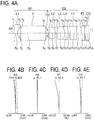
- Fig. 4B, Fig. 4C, Fig. 4D, and Fig. 4E show a spherical aberration (SA), an astigmatism (AS), a distortion (DT), and a chromatic aberration of magnification (CC) respectively of the present example.
- SA spherical aberration
- AS astigmatism
- DT distortion
- CC chromatic aberration of magnification
- the image height is 0.5 mm
- Fno is 5.802
- the half angle of view is 50.1°.
- Fig. 5A is a cross-sectional view of an endoscope objective optical system according to an example 4.
- the endoscope objective optical system includes in order from an object side, a first group G1 having a negative refractive power, an aperture stop S, and a second group G2 having a positive refractive power.
- the first group G1 includes in order from the object side, a planoconcave negative lens L1 and a visual-field direction converting member P.
- the second group G2 includes in order from the object side, a biconvex positive lens L2, a biconvex positive lens L3, a negative meniscus lens L4 having a convex surface directed toward an image side, and a plane parallel plate F1.
- the biconvex positive lens L3 and the negative meniscus lens L4 are cemented.
- a plane parallel plate F2 and a cover glass CG are disposed on the image side of the second group G2.
- the plane parallel plate F1 is a filter with a coating applied thereto for cutting specific wavelengths such as 1060 nm of YAG laser, and 810 nm of semiconductor laser, or an infra-red region.
- Fig. 5B, Fig. 5C, Fig. 5D, and Fig. 5E show a spherical aberration (SA), an astigmatism (AS), a distortion (DT), and a chromatic aberration of magnification (CC) respectively of the present example.
- SA spherical aberration
- AS astigmatism
- DT distortion
- CC chromatic aberration of magnification
- the image height is 0.5 mm
- Fno is 5.483
- the half angle of view is 50.8°.
- Fig. 6A is a cross-sectional view of an endoscope objective optical system according to an example 5.
- the endoscope objective optical system includes in order from an object side, a first group G1 having a negative refractive power, an aperture stop S, and a second group G2 having a positive refractive power.
- the first group G1 includes in order from the object side, a planoconcave negative lens L1 and a visual-field direction converting member P.
- the second group G2 includes a biconvex positive lens L2, a plane parallel plate F1, a biconvex positive lens L3, and a negative meniscus lens L4 having a convex surface directed toward an image side.
- a plane parallel plate F2 and a cover glass CG are disposed on the image side of the second group G2.
- the plane parallel plate F1 is a filter with a coating applied thereto for cutting specific wavelengths such as 1060 nm of YAG laser, and 810 nm of semiconductor laser, or an infra-red region.
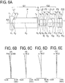
- Fig. 6B, Fig. 6C, Fig. 6D, and Fig. 6E show a spherical aberration (SA), an astigmatism (AS), a distortion (DT), and a chromatic aberration of magnification (CC) respectively of the present example.
- SA spherical aberration
- AS astigmatism
- DT distortion
- CC chromatic aberration of magnification
- the image height is 0.5 mm
- Fno is 5.588
- the half angle of view is 50.2°.
- Fig. 7A is a cross-sectional view of an endoscope objective optical system according to an example 6.
- the endoscope objective optical system includes in order from an object side, a first group G1 having a negative refractive power, an aperture stop S, and a second group G2 having a positive refractive power.
- the first group G1 includes in order from the object side, a planoconcave negative lens L1 and a visual-field direction converting member P.
- the second group G2 includes a biconvex positive lens L2, a plane parallel plate F1, a biconvex positive lens L3, and a negative meniscus lens L4 having a convex surface directed toward an image side.
- the positive lens L3 and the negative meniscus lens L4 are cemented.
- a plane parallel plate F2 and a cover glass CG are disposed on the image side of second group G2.
- the plane parallel plate F1 is a filter with a coating applied thereto for cutting specific wavelengths such as 1060 nm of YAG laser, and 810 nm of semiconductor laser, or an infra-red region.
- Fig. 7B, Fig. 7C, Fig. 7D, and Fig. 7E show a spherical aberration (SA), an astigmatism (AS), a distortion (DT), and a chromatic aberration of magnification (CC) respectively of the present example.
- SA spherical aberration
- AS astigmatism
- DT distortion
- CC chromatic aberration of magnification
- the image height is 0.5 mm
- Fno is 5.539
- the half angle of view is 49.8°.
- r denotes a radius of curvature of each lens surface
- d denotes a distance between two lens surfaces
- nd denotes a refractive index of each lens for an e-line
- vd denotes Abbe's number for each lens
- Fn denotes an F-number.
- Example2 Example3 (1) d 1 /f 2.68 2.77 2.64 (2) f 2 /f 1.92 1.95 2.03 (3) r 21 /r 22 -1.00 -36.97 -1.16 (4) f 03 /f 2.01 2.27 2.10 (5) f 01 /f -1.18 -1.39 -1.22 (6) d 1 /d 2 0.66 0.67 0.61 (7) f 02 /f 2.04 2.19 2.32 (8) f 04 /f -1.89 -2.25 -2.53 (9) en/f 0.75 0.82 0.76 (10) ex/f -4.06 -4.13 -4.78 Conditional Expression Example4 Example5 Example6 (1) d 1 /f 3.78 3.79 3.94 (2) f 2 /f 2.28 2.15 2.36 (3) r 21 /r 22 -0.66 -0.52 -0.90 (4) f 03 /f 2.13 1.81 2.17 (5) f 01
- the present invention is useful for an endoscope objective optical system and particularly to an endoscope objective optical system for oblique viewing.
Landscapes
- Physics & Mathematics (AREA)
- Health & Medical Sciences (AREA)
- Life Sciences & Earth Sciences (AREA)
- Optics & Photonics (AREA)
- Surgery (AREA)
- General Physics & Mathematics (AREA)
- Biomedical Technology (AREA)
- General Health & Medical Sciences (AREA)
- Pathology (AREA)
- Engineering & Computer Science (AREA)
- Nuclear Medicine, Radiotherapy & Molecular Imaging (AREA)
- Heart & Thoracic Surgery (AREA)
- Medical Informatics (AREA)
- Molecular Biology (AREA)
- Animal Behavior & Ethology (AREA)
- Radiology & Medical Imaging (AREA)
- Public Health (AREA)
- Veterinary Medicine (AREA)
- Biophysics (AREA)
- Astronomy & Astrophysics (AREA)
- Lenses (AREA)
- Instruments For Viewing The Inside Of Hollow Bodies (AREA)
- Endoscopes (AREA)
Abstract


d1 denotes an air conversion length from a surface on an image side of the first lens up to a surface of the aperture stop,
f denotes a focal length of the overall endoscope objective optical system,
f2 denotes a focal length of the second group,
r21 denotes a radius of curvature of a surface on the object side of the second lens, and
r22 denotes a radius of curvature of a surface on the image side of the second lens.
Description
- The present invention relates to an endoscope objective optical system such as an endoscope objective optical system that is applicable to an endoscope apparatus used in fields such as a medical field and an industrial field.
- In an endoscope objective optical system, sometimes, for carrying out oblique observation, a visual-field direction converting member such as a prism is disposed in an optical system. Examples of such optical systems for oblique observation have been proposed (refer to
patent literature 1 to patent literature 10 for example). -
- Patent Literature 1: Japanese Patent Application Laid-open Publication No.
2008-83316 - Patent Literature 2: Japanese Patent Application Laid-open Publication No.
Hei 9-269450 - Patent Literature 3: Japanese Patent No.
3574484 - Patent Literature 4: Japanese Patent No.
4439184 - Patent Literature 5: Japanese Patent No.
3742484 - Patent Literature 6: Japanese Patent No.
558058 - Patent Literature 7: Japanese Patent No.
4274602 - Patent Literature 8: Japanese Patent Publication after Examination No.
Sho 53-36787 - Patent Literature 9: Japanese Patent Application Laid-open Publication No.
Sho 51-62053 - Patent Literature 10: Japanese Patent Application Laid-open Publication No.
Hei 7-294806 - In recent years, small-sizing of image pickup elements has been progressing as small-sizing of endoscopes has been sought. Here, in an endoscope for oblique viewing, a visual-field direction converting element such as a prism is disposed in an objective optical system. Consequently, even when the image pickup element is made small-sized, there are cases in which, a spatial distance for disposing the visual-field direction converting member has to be secured appropriately long. A case, in which the spatial distance is to be secured, is for example, a case in which a mechanical component that holds the image pickup element, or the image pickup element and the objective optical system is / are disposed in the objective optical system.
- Generally, a size of an optical system can be scaled down to be small-sized, or in other words, to be reduced. Here, when the image pickup element is simply scaled down to be small-sized, there are cases in which the visual-field direction converting member cannot be scaled down to be small-sized to the same scale. Furthermore, in recent years, for realizing even further small-sizing, an image pickup element in which shading characteristics have been optimized according to a light ray with a large angle of emergence from the objective optical system, has been provided.
- In arrangements of
Patent Literatures 1, 2, and 3, when an optical system is scaled down to be small-sized simply in accordance with the size of an image pickup element, a space for disposing a visual-field direction converting member becomes small. Consequently, it becomes difficult to achieve a small-sized endoscope for oblique viewing. - In arrangements of Patent Literatures 4 and 5, a cemented lens in a second group is cemented in order of a negative lens and a positive lens. As a result of this, a capacity of correcting a chromatic aberration of magnification becomes low. Consequently, a chromatic blurring is susceptible to occur in a peripheral portion of an image field, and it is difficult to achieve an endoscope image of a superior quality.
- In Patent Literature 6, an arrangement in order of a negative lens, a positive lens, a prism, and a cemented lens from an object side has been disclosed. Here, a radius of curvature of a surface on an image side of a negative first lens being small, an effect of a relative decentering with a positive first lens, on a performance is substantial, and it is difficult to achieve further small-sizing and a superior image quality.
- In an arrangement of
Patent Literature 7, a cemented lens is not provided. Consequently, the chromatic blurring is susceptible to occur, and it is difficult to achieve a superior image quality. - In
Patent Literatures 8 and 9, in arrangements except in an example 3 ofPatent Literature 9, an angle of view is not more than 90° and it is difficult to achieve an adequate field-of-view range. Moreover, in an arrangement of the example 3 ofPatent Literature 9, an angle of emergence of an objective optical system is small. Consequently, it is not preferable for an image pickup element in which shading characteristics have been optimized, and it is difficult to achieve an endoscope with further small-sizing and superior image quality. - In Patent Literature 10, in an arrangement except in an example 5, a cemented lens in a second group is cemented in order of a negative lens and a positive lens. Consequently, the capacity of correcting the chromatic aberration of magnification is low. Moreover, in an arrangement of the example 5 of the Patent Literature 10, a cemented lens in a second group is cemented in order of a positive lens and a negative lens, but a space for disposing a visual-field direction converting member is small. Furthermore, in all the examples of Patent Literature 10, a refractive power of a second group is large. Consequently, correction of a curvature of field tends to be inadequate, and it is difficult to achieve an endoscope for oblique viewing with a small size and a superior image quality.
- The present invention has been made in view of the abovementioned circumstances, and an object of the present invention is to provide an endoscope objective optical system with a small size and a high image quality, and in which, an adequate space for disposing a visual-field direction converting element is secured.
- To solve the abovementioned issues and to achieve the object, an endoscope objective optical system according to the present invention is characterized by including in order from an object side, a first group having a negative refractive power, an aperture stop, and a second group having a positive refractive power, wherein the first group includes in order from the object side, a first lens having a negative refractive power, and a visual-field direction converting member, and the second group includes in order from the object side, a second lens having a biconvex shape, and a cemented lens in which a third lens having a positive refractive power and a fourth lens having a negative refractive power are cemented, and the endoscope objective optical system satisfies the following conditional expressions (1), (2), and (3)


- d1 denotes an air conversion length from a surface on an image side of the first lens up to a surface of the aperture stop,
- f denotes a focal length of the overall endoscope objective optical system,
- f2 denotes a focal length of the second group,
- r21 denotes a radius of curvature of a surface on the object side of the second lens,
- r22 denotes a radius of curvature of a surface on the image side of the second lens.
- According to the present invention, an effect is shown that it is possible to achieve an endoscope objective optical system with a small size and a superior image quality, and in which, an adequate space for disposing a visual-field direction converting element is provided.
-
-
Fig. 1 is a cross-sectional view showing an arrangement of an endoscope objective optical system according to an embodiment of the present invention; -
Fig. 2A is a cross-sectional view showing an arrangement of an endoscope objective optical system according to an example 1 of the present invention, andFig. 2B, Fig. 2C, Fig. 2D, and Fig. 2E are aberration diagrams showing a spherical aberration (SA), an astigmatism (AS), a distortion (DT), and a chromatic aberration of magnification (CC) respectively of the example 1; -
Fig. 3A is a cross-sectional view showing an arrangement of an endoscope objective optical system according to an example 2 of the present invention, andFig. 3B, Fig. 3C, Fig. 3D, and Fig. 3E are aberration diagrams showing a spherical aberration (SA), an astigmatism (AS), a distortion (DT), and a chromatic aberration of magnification (CC) respectively of the example 2; -
Fig. 4A is a cross-sectional view showing an arrangement of an endoscope objective optical system according to an example 3 of the present invention, andFig. 4B, Fig. 4C, Fig. 4D, and Fig. 4E are aberration diagrams showing a spherical aberration (SA), an astigmatism (AS), a distortion (DT), and a chromatic aberration of magnification (CC) respectively of the example 3; -
Fig. 5A is a cross-sectional view showing an arrangement of an endoscope objective optical system according to an example 4 of the present invention, andFig. 5B, Fig. 5C, Fig. 5D, and Fig. 5E are aberration diagrams showing a spherical aberration (SA), an astigmatism (AS), a distortion (DT), and a chromatic aberration of magnification (CC) respectively of the example 4; -
Fig. 6A is a cross-sectional view showing an arrangement of an endoscope objective optical system according to an example 5 of the present invention, andFig. 6B, Fig. 6C, Fig. 6D, and Fig. 6E are aberration diagrams showing a spherical aberration (SA), an astigmatism (AS), a distortion (DT), and a chromatic aberration of magnification (CC) respectively of the example 5; and -
Fig. 7A is a cross-sectional view showing an arrangement of an endoscope objective optical system according to an example 6 of the present invention, andFig. 7B, Fig. 7C, Fig. 7D, and Fig. 7E are aberration diagrams showing a spherical aberration (SA), an astigmatism (AS), a distortion (DT), and a chromatic aberration of magnification (CC) respectively of the example 6. - Reasons for and effects of adopting such arrangement for an endoscope objective optical system according to the present embodiment will be described below by using the accompanying diagrams. However, the present invention is not limited to embodiments described below.
-
Fig. 1 is a cross-sectional view showing an arrangement of the endoscope objective optical system according to the present embodiment. - The endoscope objective optical system according to the present embodiment includes in order from an object side, a first group G1 having a negative refractive power, an aperture stop S, and a second group G2 having a positive refractive power, and the first group includes in order from the object side, a first lens L1 having a negative refractive power and a visual-field direction converting member P, and the second group G2 includes in order from the object side, a second lens L2 having a biconvex shape, and a cemented lens in which a third lens L3 having a positive refractive power and a fourth lens L4 having a negative refractive power are cemented in this order.
- Moreover, the endoscope objective optical system satisfies the following conditional expressions (1), (2), and (3).


- d1 denotes an air conversion length from a surface on an image side of the first lens up to a surface of the aperture stop,
- f denotes a focal length of the overall endoscope objective optical system,
- f2 denotes a focal length of the second group,
- r21 denotes a radius of curvature of a surface on the object side of the second lens,
- r22 denotes a radius of curvature of a surface on the image side of the second lens.
- In such manner, in the present embodiment, for letting it to be an endoscope objective optical system having a small size and wide angle of view that can be used in an endoscope, the first lens L1 having a planoconcave shape is disposed nearest to object in the first group G1, and a negative refractive power is secured. Accordingly, it is possible to adopt an arrangement of a retro-focus type. Moreover, by disposing the visual-field direction converting member P on an image side of the first lens L1, an oblique observation is made possible. In
Fig. 1 , a diagram in which a visual-field direction converting member such as a prism is unfolded is shown. Therefore, the prism has been depicted as a plane parallel plate. - The positive lens L2 which contributes mainly to image formation is disposed on the object side of the second group G2. The cemented lens in which positive lens L3 and the negative lens L4 are cemented in this order is disposed at a position at which a height of a marginal ray from an optical axis becomes high on the image side of the positive lens L2. A chromatic aberration of magnification is corrected by the cemented lens. Contribution of a position where the height of the marginal ray from the optical axis is high is substantial to the correction of the chromatic aberration of magnification. Therefore, in the cemented lens, the positive lens L3 and the negative lens L4 are cemented in order from the object side so as to correct the chromatic aberration of magnification at a position where an off-axis light ray becomes higher.
- Here, in an endoscope for oblique viewing, there are cases in which, a space for disposing the visual-field direction converting member has to be secured appropriately long. In this case, in a scaled down optical system in which the image pickup element is simply made small-sized, since the space for disposing the visual-field direction converting member P also becomes short, it is not preferable. In other words, it is necessary to secure a relatively long space for disposing the visual-field direction converting element with respect to a focal length of the optical system. For this reason, it is desirable to satisfy the following conditional expression (1).

- d1 denotes the air conversion length from the surface on the image side of the first lens up to the surface of the aperture stop, and
- f denotes the focal length of the overall endoscope objective optical system
- Conditional expression (1) regulates the space (air conversion length) in which the visual-field direction converting member P is disposed and the focal length of the overall system.
- When an upper limit value of conditional expression (1) is exceeded, the space for disposing the visual-field direction converting member P becomes large. Consequently, when an attempt is made to arrange the optical system with a diameter of the first lens maintained as it has been, a refractive power of the first lens L1 becomes large, and all aberrations are deteriorated. When a value falls below a lower limit value of conditional expression (1), the space for disposing the visual-field direction converting member P becomes small. Consequently, it becomes difficult to achieve a small-sized endoscope for oblique viewing.
- It is preferable to satisfy the following conditional expression (1') instead of conditional expression (1).

- Furthermore, it is more preferable to satisfy the following conditional expression (1") instead of conditional expression (1).

- Moreover, when a refractive power of the second group G2 having a positive refractive power is not set appropriately, an amount of a spherical aberration and a curvature of field that occur becomes large, thereby leading to susceptibility of degradation of image quality. For this reason, it is desirable to satisfy the following conditional expression (2).

- f2 denotes the focal length of the second group, and
- f denotes the focal length of the overall endoscope objective optical system.
- When an upper limit value of conditional expression (2) is exceeded, the refractive power of the second group G2 becomes small, and correction of the curvature of field becomes excessive, and therefore it is not preferable. When a value falls below a lower limit value of conditional expression (2), the refractive power of the second group G2 becomes large, and correction of the curvature of field becomes inadequate, and therefore it is not preferable.
- It is preferable to satisfy the following conditional expression (2') instead of conditional expression (2).

- Furthermore, it is more preferable to satisfy the following conditional expression (2") instead of conditional expression (2).

- Moreover, in order to secure a large angle of emergence and the positive refractive power for image formation, it is necessary that the second lens L2 corrects the spherical aberration and a coma aberration while maintaining the appropriate positive refractive power. For this reason, it is desirable to satisfy the following conditional expression (3).

- r21 denotes the radius of curvature of the surface on the object side of the second lens,
- r22 denotes the radius of curvature of the surface on the image side of the second lens.
- When an upper limit value of conditional expression (3) is exceeded, or when a value falls below a lower limit value of conditional expression (3), the spherical aberration and the coma aberration become substantial, and therefore it is not preferable.
- It is preferable to satisfy the following conditional expression (3') instead of conditional expression (3).

- Furthermore, it is more preferable to satisfy the following conditional expression (3") instead of conditional expression (3).

- Moreover, when a refractive power of the third lens L3 is excessively large, the optical system becomes close to a telecentric optical system. Consequently, it becomes difficult to secure a large angle of emergence. As a result, a height of a light ray at a lens near an image plane I becomes high, and a diameter of the lens is susceptible to becomes large. Therefore, it is preferable arrange the optical system such that the refractive power of the third lens L3 is set to be appropriate, and the angle of emergence becomes large. For this, it is desirable to satisfy conditional expression (4).

- f03 denotes a focal length of the third lens, and
- f denotes the focal length of the overall endoscope objective optical system.
- When an upper limit value of conditional expression (4) is exceeded, the refractive power of the third lens L3 becomes small, and the angle of emergence of the light ray becomes excessively large, and it is susceptible to degradation of image quality due to shading. When a value falls below a lower limit value of conditional expression (4), the refractive power of the third lens L3 becomes large, and the angle of emergence becomes excessively small. In other words, since the diameter of the lens is susceptible to become large, it is not preferable. Furthermore, it is unfavorable for an image pickup element in which the shading characteristics are optimized, and the image quality is susceptible to be degraded.
- It is preferable to satisfy the following conditional expression (4') instead of conditional expression (4).

- Furthermore, it is more preferable to satisfy the following conditional expression (4") instead of conditional expression (4).

- Moreover, for achieving an endoscope for oblique viewing having a small size and a superior image quality, it is necessary to set appropriately the refractive power of the first lens L1, and to maintain balance of a lens diameter and an optical performance. For this, it is desirable to satisfy the following conditional expression (5).

- f01 denotes a focal length of the first lens, and
- f denotes the focal length of the overall endoscope objective optical system.
- When an upper limit value of conditional expression (5) is exceeded, the negative refractive power of the first lens L1 becomes large, and all aberrations are deteriorated. When a value falls below a lower limit value of conditional expression (5), the refractive power of the first lens L1 becomes small, and the diameter of the first lens L1 becomes large, and therefore it is not preferable.
- It is preferable to satisfy the following conditional expression (5') instead of conditional expression (5).

- Furthermore, it is more preferable to satisfy the following conditional expression (5") instead of conditional expression (5).

- Moreover, it is desirable that the optical system is compact in a longitudinal direction of the endoscope while securing the space for disposing the long visual-field direction converting member P. For this, it is desirable to satisfy the following conditional expression (6).

- d1 denotes the air conversion length from the surface on the image side of the first lens up to the surface of the aperture stop, and
- d2 denotes an air conversion length from the surface of the aperture stop up to an image plane.
- When an upper limit value of conditional expression (6) is exceeded, the space for disposing the visual-field direction converting member P becomes excessively large, and the diameter of the first lens L1 becomes large. When a value falls below a lower limit value of conditional expression (6), the optical system becomes excessively large in the longitudinal direction of the endoscope, and therefore it is not preferable.
- It is preferable to satisfy the following conditional expression (6') instead of conditional expression (6).

- Furthermore, it is more preferable to satisfy the following conditional expression (6") instead of conditional expression (6).

- Moreover, it is desirable that the present embodiment satisfies the following conditional expression (7).

- f02 denotes a focal length of the second lens L2, and
- f denotes the focal length of the overall endoscope objective optical system.
- When an upper limit value of conditional expression (7) is exceeded, the refractive power of the second lens L2 becomes excessively small. Consequently, the curvature of field is corrected excessively, and it is not preferable. When a value falls below a lower limit value of conditional expression (7), the refractive power of the second lens L2 becomes large. Consequently, correction of the spherical aberration becomes inadequate, and therefore it is not preferable.
- It is preferable to satisfy the following conditional expression (7') instead of conditional expression (7).

- Furthermore, it is more preferable to satisfy the following conditional expression (7") instead of conditional expression (7).

- Moreover, for achieving an endoscope image of a superior quality, it is desirable to correct the inadequately corrected spherical aberration and the curvature of field occurring in the second lens L2 having a positive refractive power and the third lens L3 having a positive refractive power, by a fourth lens L4 having an appropriate negative refractive power. For this, it is desirable to satisfy the following conditional expression (8).

- f04 denotes a focal length of the fourth lens L4, and
- f denotes the focal length of the overall endoscope objective optical system.
- When an upper limit value of conditional expression (8) is exceeded, the refractive power of the fourth lens L4 becomes large. Consequently, the curvature of field is corrected excessively, and therefore it is not preferable. When a value falls below a lower limit value of conditional expression (8), the refractive power of the fourth lens L4 becomes small. Consequently, correction of the spherical aberration is inadequate, and therefore it is not preferable.
- It is preferable to satisfy the following conditional expression (8') instead of conditional expression (8).

- Furthermore, it is more preferable to satisfy the following conditional expression (8") instead of conditional expression (8).

- Moreover, for small-sizing, it is desirable to make a diameter of the first group G1 small. For this, it is desirable to satisfy the following conditional expression (9).

- en denotes a position of a light ray with the maximum image height at an entrance pupil, and
- f denotes the focal length of the overall endoscope objective optical system.
- When an upper limit value of conditional expression (9) is exceeded, since the diameter of the first lens L1 having a negative refractive power becomes large, it is not preferable. When a value falls below a lower limit value of conditional expression (9), the refractive power of the first lens L1 having a negative refractive power becomes large, and therefore it is not preferable.
- It is preferable to satisfy the following conditional expression (9') instead of conditional expression (9).

- Furthermore, it is more preferable to satisfy the following conditional expression (9") instead of conditional expression (9).

- Moreover, it is desirable that the present embodiment satisfies the following conditional expression (10).

- ex denotes a position of a light ray having the maximum image height at an exit pupil, and
- f denotes the focal length of the overall endoscope objective optical system.
- When an upper limit value of conditional expression (10) is exceeded, an angle of emergence of a light ray becomes large. In other words, the refractive power of the second lens L2 becomes large, and the spherical aberration is deteriorated. When a value falls below a lower limit value of conditional expression (10), the angle of emergence of a light ray becomes small. Consequently, it is unfavorable for an image pickup element in which the shading characteristics are optimized.
- It is preferable to satisfy the following conditional expression (10') instead of conditional expression (10).

- Furthermore, it is more preferable to satisfy the following conditional expression (10") instead of conditional expression (10).

- An endoscope objective optical system according to an example 1 will be described below.
Fig. 2A is a cross-sectional view of the endoscope objective optical system according to the present example. In all examples that follow, a visual-field direction converting member P such as a prism is shown to be in an unfolded state. Therefore, the visual-field direction converting member P is illustrated as a plane parallel plate having an optical path length equivalent to that of prism. - In the present example, the endoscope objective optical system includes in order from an object side, a first group G1 having a negative refractive power, an aperture stop S, and a second group G2 having a positive refractive power.
- The first group G1 includes in order from the object side, a planoconcave negative lens L1 and a visual-field direction converting member P. The second group G2 includes a biconvex positive lens L2, a biconvex positive lens L3, a negative meniscus lens L4 having a convex surface directed toward an image side, and a plane parallel plate F1. Here, the positive lens L3 and the negative meniscus lens L4 are cemented. A plane parallel plate F2 and a cover glass CG are disposed on the image side of the second group G2.
- The plane parallel plate F1 is a filter with a coating applied thereto for cutting specific wavelengths such as 1060 nm of YAG (yttrium aluminum garnet) laser and 810 nm of semiconductor laser, or an infra-red region.
-
Fig. 2B, Fig. 2C, Fig. 2D, and Fig. 2E show a spherical aberration (SA), an astigmatism (AS), a distortion (DT), and a chromatic aberration of magnification (CC) respectively of the present example. These aberration diagrams are shown for wavelengths of 546.07 nm (e-line), 435.84 nm (g-line), 486.13 nm (F-line), and 656.27 nm (C-line). Moreover, in each diagram, Fno denotes an F-number and ω denotes a half angle of view. It is similar for the aberration diagrams. Furthermore, the image height is 0.5 mm, Fno is 6.375, and the half angle of view is 50.0°. -
Fig. 3A is a cross-sectional view of an endoscope objective optical system according to an example 2. In the present example, the endoscope objective optical system includes in order from an object side, a first group G1 having a negative refractive power, an aperture stop S, and a second group G2 having a positive refractive power. - The first group G1 includes in order from an object side, a planoconcave negative lens L1 and a visual-field direction converting member P. The second group G2 includes a biconvex positive lens L2, a plane parallel plate F1, a biconvex positive lens L3, and a negative meniscus lens L4 having a convex surface directed toward an image side. Here, the positive lens L3 and the negative meniscus lens L4 are cemented. A plane parallel plate F2 and a cover glass CG are disposed on the image side of the second group G2.
- The plane parallel plate F1 is a filter with a coating applied thereto for cutting specific wavelengths such as 1060 nm YAG laser and 810 nm of semiconductor laser, or an infra-red region.
-
Fig. 3B, Fig. 3C, Fig. 3D, and Fig. 3E show a spherical aberration (SA), an astigmatism (AS), a distortion (DT), and a chromatic aberration of magnification (CC) respectively of the present example. The image height is 0.5 mm, Fno is 6.104, and the half angle of view is 50.4°. -
Fig. 4A is a cross-sectional view of an endoscope objective optical system according to an example 3. In the present example, the endoscope objective optical system includes in order from an object side, a first group G1 having a negative refractive power, an aperture stop S, and a second group G2 having a positive refractive power. - The first group G1 includes in order from an object side, a planoconcave negative lens L1 and a visual-field direction converting member P. The second group G2 includes a biconvex positive lens L2, a biconvex positive lens L3, a negative meniscus lens L4 having a convex surface directed toward an image side, and a plane parallel plate F1. Here, the positive lens L3 and the negative meniscus lens L4 are cemented. A plane parallel plate F2 and a cover glass CG are disposed on the image side of the second group G2.
- The plane parallel plate F1 is a filter with a coating applied thereto for cutting specific wavelengths such as 1060 nm of YAG laser and 810 nm of semiconductor laser, or an infra-red region.
-
Fig. 4B, Fig. 4C, Fig. 4D, and Fig. 4E show a spherical aberration (SA), an astigmatism (AS), a distortion (DT), and a chromatic aberration of magnification (CC) respectively of the present example. The image height is 0.5 mm, Fno is 5.802, and the half angle of view is 50.1°. -
Fig. 5A is a cross-sectional view of an endoscope objective optical system according to an example 4. In the present example, the endoscope objective optical system includes in order from an object side, a first group G1 having a negative refractive power, an aperture stop S, and a second group G2 having a positive refractive power. - The first group G1 includes in order from the object side, a planoconcave negative lens L1 and a visual-field direction converting member P. The second group G2 includes in order from the object side, a biconvex positive lens L2, a biconvex positive lens L3, a negative meniscus lens L4 having a convex surface directed toward an image side, and a plane parallel plate F1. Here, the biconvex positive lens L3 and the negative meniscus lens L4 are cemented. A plane parallel plate F2 and a cover glass CG are disposed on the image side of the second group G2.
- The plane parallel plate F1 is a filter with a coating applied thereto for cutting specific wavelengths such as 1060 nm of YAG laser, and 810 nm of semiconductor laser, or an infra-red region.
-
Fig. 5B, Fig. 5C, Fig. 5D, and Fig. 5E show a spherical aberration (SA), an astigmatism (AS), a distortion (DT), and a chromatic aberration of magnification (CC) respectively of the present example. The image height is 0.5 mm, Fno is 5.483, and the half angle of view is 50.8°. -
Fig. 6A is a cross-sectional view of an endoscope objective optical system according to an example 5. In the present example, the endoscope objective optical system includes in order from an object side, a first group G1 having a negative refractive power, an aperture stop S, and a second group G2 having a positive refractive power. - The first group G1 includes in order from the object side, a planoconcave negative lens L1 and a visual-field direction converting member P. The second group G2 includes a biconvex positive lens L2, a plane parallel plate F1, a biconvex positive lens L3, and a negative meniscus lens L4 having a convex surface directed toward an image side. A plane parallel plate F2 and a cover glass CG are disposed on the image side of the second group G2.
- The plane parallel plate F1 is a filter with a coating applied thereto for cutting specific wavelengths such as 1060 nm of YAG laser, and 810 nm of semiconductor laser, or an infra-red region.
-
Fig. 6B, Fig. 6C, Fig. 6D, and Fig. 6E show a spherical aberration (SA), an astigmatism (AS), a distortion (DT), and a chromatic aberration of magnification (CC) respectively of the present example. The image height is 0.5 mm, Fno is 5.588, and the half angle of view is 50.2°. -
Fig. 7A is a cross-sectional view of an endoscope objective optical system according to an example 6. In the present example, the endoscope objective optical system includes in order from an object side, a first group G1 having a negative refractive power, an aperture stop S, and a second group G2 having a positive refractive power. - The first group G1 includes in order from the object side, a planoconcave negative lens L1 and a visual-field direction converting member P. The second group G2 includes a biconvex positive lens L2, a plane parallel plate F1, a biconvex positive lens L3, and a negative meniscus lens L4 having a convex surface directed toward an image side. Here, the positive lens L3 and the negative meniscus lens L4 are cemented. A plane parallel plate F2 and a cover glass CG are disposed on the image side of second group G2.
- The plane parallel plate F1 is a filter with a coating applied thereto for cutting specific wavelengths such as 1060 nm of YAG laser, and 810 nm of semiconductor laser, or an infra-red region.
-
Fig. 7B, Fig. 7C, Fig. 7D, and Fig. 7E show a spherical aberration (SA), an astigmatism (AS), a distortion (DT), and a chromatic aberration of magnification (CC) respectively of the present example. The image height is 0.5 mm, Fno is 5.539, and the half angle of view is 49.8°. - Numerical data for each example is shown below. Regarding symbols, r denotes a radius of curvature of each lens surface, d denotes a distance between two lens surfaces, nd denotes a refractive index of each lens for an e-line, vd denotes Abbe's number for each lens, and Fn denotes an F-number.
-
Surface data Surface no. r d nd νd Object plane ∞ 4.3605 1 ∞ 0.2907 1.88815 40.76 2 0.6463 0.3101 3 ∞ 2.2674 1.88815 40.76 4 ∞ 0.1453 5(Stop) ∞ 0.126 6 1.9264 0.5136 1.81078 40.92 7 -1.9264 0.1744 8 2.2926 0.6395 1.51977 52.43 9 -0.814 0.2907 1.93429 18.90 10 -3.7607 0.2326 11 ∞ 0.3876 1.523 65.13 12 ∞ 0.3886 13 ∞ 0.3198 1.51825 64.14 14 ∞ 0.0097 1.515 64.00 15 ∞ 0.3876 1.507 63.26 Image plane ∞ Various data focal length 0.6187 Fno. 6.375 -
Surface data Surface no. r d nd νd Object plane ∞ 5.4264 1 ∞ 0.2955 1.88815 40.76 2 0.7592 0.2907 3 ∞ 2.2481 1.88815 40.76 4 ∞ 0.2229 5(Stop) ∞ 0.0969 6 39.5292 0.4464 1.77621 49.60 7 -1.0693 0.2958 8 ∞ 0.2907 1.523 65.13 9 ∞ 0.3147 10 2.7791 0.6343 1.73234 54.68 11 -1.4607 0.3018 1.97189 17.47 12 18.6587 0.4059 13 ∞ 0.3198 1.51825 64.14 14 ∞ 0.0097 1.515 64.00 15 ∞ 0.3876 1.507 63.26 Image plane ∞ Various data focal length 0.6148 Fno. 6.104 -
Surface data Surface no. r d nd νd Object plane ∞ 5.1357 1 ∞ 0.2907 1.88815 40.76 2 0.6741 0.2904 3 ∞ 2.2774 1.88815 40.76 4 ∞ 0.1453 5(Stop) ∞ 0.1316 6 2.3270 0.5463 1.79012 44.20 7 -2.0048 0.1708 8 2.3145 0.7102 1.51825 64.14 9 -0.8563 0.2907 1.93429 18.90 10 -2.3840 0.1870 11 ∞ 0.5814 1.523 65.13 12 ∞ 0.4016 13 ∞ 0.3198 1.51825 64.14 14 ∞ 0.0097 1.515 64.00 15 ∞ 0.3876 1.507 63.26 Image plane ∞ Various data focal length 0.6225 Fno. 5.802 -
Surface data Surface no. r d nd νd Object plane ∞ 6.1047 1 ∞ 0.2955 1.88815 40.76 2 0.9467 0.3246 3 ∞ 3.3794 1.88815 40.76 4 ∞ 0.2229 5 (Stop) ∞ 0.0775 6 2.1943 1.2617 1.88815 40.76 7 -3.316 0.1163 8 2.2462 0.6810 1.59143 61.14 9 -1.0563 0.2821 1.93429 18.90 10 -5.2068 0.0484 11 ∞ 0.3876 1.523 65.13 12 ∞ 0.4244 13 ∞ 0.3198 1.51825 64.14 14 ∞ 0.0097 1.515 64.00 15 ∞ 0.3876 1.507 63.26 Image plane ∞ Various data focal length 0.6189 Fno. 5.483 -
Surface data Surface no. r d nd νd Object plane ∞ 5.4264 1 ∞ 0.2955 1.88815 40.76 2 0.9620 0.3246 3 ∞ 3.3794 1.88815 40.76 4 ∞ 0.2229 5(Stop) ∞ 0.0775 6 1.7223 0.4823 1.73234 54.68 7 -3.3188 0.1367 8 ∞ 0.2907 1.523 65.13 9 ∞ 0.2422 10 1.4769 0.7630 1.5343 48.84 11 -0.8247 0.2907 1.93429 18.90 12 -3.8931 0.3604 13 ∞ 0.3198 1.51825 64.14 14 ∞ 0.0097 1.515 64.00 15 ∞ 0.3876 1.507 63.26 Image plane ∞ Various data focal length 0.6170 Fno. 5.588 -
Surface data Surface no. r d nd νd Object plane ∞ 5.4264 1 ∞ 0.2955 1.88815 40.76 2 0.9467 0.3246 3 ∞ 3.3794 1.88815 40.76 4 ∞ 0.3682 5(Stop) ∞ 0.0969 6 2.5285 0.8326 1.79196 47.37 7 -2.8131 0.1268 8 ∞ 0.3876 1.523 65.13 9 ∞ 0.1223 10 2.0400 0.7674 1.59143 61.14 11 -1.1548 0.2907 1.93429 18.90 12 -6.1764 0.6617 13 ∞ 0.3198 1.51825 64.14 14 ∞ 0.0097 1.515 64.00 15 ∞ 0.3876 1.507 63.26 Image plane ∞ Various data focal length 0.6300 Fno. 5.539 - Values of each of the conditional expressions are shown below;
Conditional Expression Example1 Example2 Example3 (1) d1/f 2.68 2.77 2.64 (2) f2/f 1.92 1.95 2.03 (3) r21/r22 -1.00 -36.97 -1.16 (4) f03/f 2.01 2.27 2.10 (5) f01/f -1.18 -1.39 -1.22 (6) d1/d2 0.66 0.67 0.61 (7) f02/f 2.04 2.19 2.32 (8) f04/f -1.89 -2.25 -2.53 (9) en/f 0.75 0.82 0.76 (10) ex/f -4.06 -4.13 -4.78 Conditional Expression Example4 Example5 Example6 (1) d1/f 3.78 3.79 3.94 (2) f2/f 2.28 2.15 2.36 (3) r21/r22 -0.66 -0.52 -0.90 (4) f03/f 2.13 1.81 2.17 (5) f01/f -1.72 -1.76 -1.69 (6) d1/d2 0.89 0.97 0.88 (7) f02/f 2.69 2.62 2.87 (8) f04/f -2.37 -1.90 -2.48 (9) en/f 0.99 1.02 1.01 (10) ex/f -4.76 -4.00 -4.95 - As described above, the present invention is useful for an endoscope objective optical system and particularly to an endoscope objective optical system for oblique viewing.
-
- G1
- first group
- G2
- second group
- S
- aperture stop
- L1, L2, L3, L4
- lens
- F1, F2
- plane parallel plate
- CG
- cover glass
Claims (4)
- An endoscope objective optical system, comprising in order from an object side:a first group having a negative refractive power;an aperture stop; anda second group having a positive refractive power; whereinthe first group includes in order from the object side, a first lens having a negative refractive power, and a visual-field direction converting member, andthe second group includes in order from the object side, a second lens having a biconvex shape, and a cemented lens in which a third lens having a positive refractive power and a fourth lens having a negative refractive power are cemented in this order, andthe endoscope objective optical system satisfies the following conditional expressions (1), (2), and (3)


 d1 denotes an air conversion length from a surface on an image side of the first lens up to a surface of the aperture stop,f denotes a focal length of the overall endoscope objective optical system,f2 denotes a focal length of the second group,r21 denotes a radius of curvature of a surface on the object side of the second lens, andr22 denotes a radius of curvature of a surface on the image side of the second lens.
 - The endoscope objective optical system according to claim 1, wherein the endoscope objective optical system satisfies the following conditional expression (4)
 f03 denotes a focal length of the third lens, andf denotes the focal length of the overall endoscope objective optical system.
 - The endoscope objective optical system according to claim 1, wherein the endoscope objective optical system satisfies the following conditional expression (5)
 f01 denotes a focal length of the first lens, andf denotes the focal length of the overall endoscope objective optical system.
 - The endoscope objective optical system according to claim 1, wherein the endoscope objective optical system satisfies the following conditional expression (6)
 d1 denotes the air conversion length from the surface on the image side of the first lens up to the surface of the aperture stop, andd2 denotes an air conversion length from the surface of the aperture stop up to an image plane.
Applications Claiming Priority (2)
| Application Number | Priority Date | Filing Date | Title |
|---|---|---|---|
| JP2015089661 | 2015-04-24 | ||
| PCT/JP2016/061828 WO2016171043A1 (en) | 2015-04-24 | 2016-04-12 | Endoscope objective optical system |
Publications (1)
| Publication Number | Publication Date |
|---|---|
| EP3287828A1 true EP3287828A1 (en) | 2018-02-28 |
Family
ID=57144435
Family Applications (1)
| Application Number | Title | Priority Date | Filing Date |
|---|---|---|---|
| EP16783064.5A Withdrawn EP3287828A1 (en) | 2015-04-24 | 2016-04-12 | Endoscope objective optical system |
Country Status (5)
| Country | Link |
|---|---|
| US (1) | US9804381B2 (en) |
| EP (1) | EP3287828A1 (en) |
| JP (1) | JP6062137B1 (en) |
| CN (1) | CN107076965B (en) |
| WO (1) | WO2016171043A1 (en) |
Families Citing this family (4)
| Publication number | Priority date | Publication date | Assignee | Title |
|---|---|---|---|---|
| WO2018124248A1 (en) * | 2016-12-28 | 2018-07-05 | オリンパス株式会社 | Endoscope objective optical system |
| JP6865303B2 (en) | 2018-01-26 | 2021-04-28 | オリンパス株式会社 | Endoscope objective optical system, endoscope imaging device and endoscope |
| CN108563010B (en) * | 2018-03-15 | 2023-09-08 | 安徽长庚光学科技有限公司 | rigid endoscope |
| CN120335143B (en) * | 2025-06-19 | 2025-09-19 | 浙江华诺康科技有限公司 | Imaging system and endoscope |
Family Cites Families (9)
| Publication number | Priority date | Publication date | Assignee | Title |
|---|---|---|---|---|
| JP2552861B2 (en) * | 1987-05-26 | 1996-11-13 | オリンパス光学工業株式会社 | Zoom lens |
| JP2596755B2 (en) * | 1987-08-14 | 1997-04-02 | オリンパス光学工業株式会社 | Focusing system with two focal length switching type lens system |
| JPH01191820A (en) * | 1988-01-28 | 1989-08-01 | Olympus Optical Co Ltd | Variable power lens |
| US4999007A (en) | 1988-01-28 | 1991-03-12 | Olympus Optical Co., Ltd. | Vari-focal lens system |
| JP3574484B2 (en) * | 1994-12-22 | 2004-10-06 | ペンタックス株式会社 | Objective optical system for endoscope |
| US5774279A (en) | 1995-10-19 | 1998-06-30 | Konica Corporation | Retrofoucus photographic lens |
| JPH09113799A (en) * | 1995-10-19 | 1997-05-02 | Konica Corp | Retrofocus type photographic lens |
| JPH09269450A (en) * | 1996-03-29 | 1997-10-14 | Olympus Optical Co Ltd | Objective lens for endoscope |
| JPH11237549A (en) * | 1998-02-19 | 1999-08-31 | Fuji Photo Optical Co Ltd | Simple wide angle zoom lens |
-
2016
- 2016-04-12 WO PCT/JP2016/061828 patent/WO2016171043A1/en not_active Ceased
- 2016-04-12 CN CN201680003203.3A patent/CN107076965B/en active Active
- 2016-04-12 EP EP16783064.5A patent/EP3287828A1/en not_active Withdrawn
- 2016-04-12 JP JP2016561874A patent/JP6062137B1/en active Active
-
2017
- 2017-05-03 US US15/585,579 patent/US9804381B2/en active Active
Also Published As
| Publication number | Publication date |
|---|---|
| JP6062137B1 (en) | 2017-01-18 |
| JPWO2016171043A1 (en) | 2017-05-18 |
| CN107076965B (en) | 2019-05-17 |
| WO2016171043A1 (en) | 2016-10-27 |
| CN107076965A (en) | 2017-08-18 |
| US9804381B2 (en) | 2017-10-31 |
| US20170235122A1 (en) | 2017-08-17 |
Similar Documents
| Publication | Publication Date | Title |
|---|---|---|
| EP3214475A1 (en) | Objective optical system for endoscope | |
| CN106066533B (en) | Objective lens for endoscope and endoscope | |
| CN105892037B (en) | Objective lens for endoscope and endoscope | |
| EP3037858A1 (en) | Endoscope objective optical system | |
| EP2980624A1 (en) | Optical system for endoscope | |
| EP2784563B1 (en) | Endoscope objective optical system | |
| EP3312652A1 (en) | Endoscope objective optical system | |
| EP3316016A1 (en) | Optical system of object for endoscope | |
| EP3312653A1 (en) | Endoscope objective optical system | |
| EP2990847A1 (en) | Wide-angle objective optical system | |
| EP2905644A1 (en) | Endoscope objective optical system | |
| EP3168668A1 (en) | Objective optical system | |
| US11543647B2 (en) | Objective optical system for endoscope, endoscope, and image pickup unit | |
| EP3401718A1 (en) | Objective optical system | |
| EP3159725A1 (en) | Objective optical system for endoscope | |
| US11576564B2 (en) | Objective optical system for endoscope | |
| EP3508900A1 (en) | Objective optical system for endoscope | |
| EP2648026B1 (en) | Endoscope objective optical system | |
| US10775604B2 (en) | Objective optical system for endoscope | |
| US10564406B2 (en) | Objective optical system for endoscope | |
| EP2662718A1 (en) | Optical system for endoscope | |
| EP3287828A1 (en) | Endoscope objective optical system | |
| US20190107706A1 (en) | Objective optical system for endoscope | |
| US20220026702A1 (en) | Endoscope objective optical system and endoscope | |
| JP2019148675A (en) | Endoscope objective optical system |
Legal Events
| Date | Code | Title | Description |
|---|---|---|---|
| STAA | Information on the status of an ep patent application or granted ep patent |
Free format text: STATUS: THE INTERNATIONAL PUBLICATION HAS BEEN MADE |
|
| PUAI | Public reference made under article 153(3) epc to a published international application that has entered the european phase |
Free format text: ORIGINAL CODE: 0009012 |
|
| STAA | Information on the status of an ep patent application or granted ep patent |
Free format text: STATUS: REQUEST FOR EXAMINATION WAS MADE |
|
| 17P | Request for examination filed |
Effective date: 20171117 |
|
| AK | Designated contracting states |
Kind code of ref document: A1 Designated state(s): AL AT BE BG CH CY CZ DE DK EE ES FI FR GB GR HR HU IE IS IT LI LT LU LV MC MK MT NL NO PL PT RO RS SE SI SK SM TR |
|
| AX | Request for extension of the european patent |
Extension state: BA ME |
|
| DAV | Request for validation of the european patent (deleted) | ||
| DAX | Request for extension of the european patent (deleted) | ||
| STAA | Information on the status of an ep patent application or granted ep patent |
Free format text: STATUS: THE APPLICATION IS DEEMED TO BE WITHDRAWN |
|
| 18D | Application deemed to be withdrawn |
Effective date: 20181101 |