EP3272857A1 - Method for preparing dendritic cell by non-adhesive culture using ifn - Google Patents
Method for preparing dendritic cell by non-adhesive culture using ifn Download PDFInfo
- Publication number
- EP3272857A1 EP3272857A1 EP16765008.4A EP16765008A EP3272857A1 EP 3272857 A1 EP3272857 A1 EP 3272857A1 EP 16765008 A EP16765008 A EP 16765008A EP 3272857 A1 EP3272857 A1 EP 3272857A1
- Authority
- EP
- European Patent Office
- Prior art keywords
- cells
- dcs
- monocytes
- dendritic cells
- ifn
- Prior art date
- Legal status (The legal status is an assumption and is not a legal conclusion. Google has not performed a legal analysis and makes no representation as to the accuracy of the status listed.)
- Granted
Links
Images














Classifications
-
- A—HUMAN NECESSITIES
- A61—MEDICAL OR VETERINARY SCIENCE; HYGIENE
- A61K—PREPARATIONS FOR MEDICAL, DENTAL OR TOILETRY PURPOSES
- A61K35/00—Medicinal preparations containing materials or reaction products thereof with undetermined constitution
- A61K35/12—Materials from mammals; Compositions comprising non-specified tissues or cells; Compositions comprising non-embryonic stem cells; Genetically modified cells
- A61K35/14—Blood; Artificial blood
- A61K35/15—Cells of the myeloid line, e.g. granulocytes, basophils, eosinophils, neutrophils, leucocytes, monocytes, macrophages or mast cells; Myeloid precursor cells; Antigen-presenting cells, e.g. dendritic cells
-
- A—HUMAN NECESSITIES
- A61—MEDICAL OR VETERINARY SCIENCE; HYGIENE
- A61K—PREPARATIONS FOR MEDICAL, DENTAL OR TOILETRY PURPOSES
- A61K35/00—Medicinal preparations containing materials or reaction products thereof with undetermined constitution
- A61K35/12—Materials from mammals; Compositions comprising non-specified tissues or cells; Compositions comprising non-embryonic stem cells; Genetically modified cells
- A61K35/14—Blood; Artificial blood
- A61K35/17—Lymphocytes; B-cells; T-cells; Natural killer cells; Interferon-activated or cytokine-activated lymphocytes
-
- A—HUMAN NECESSITIES
- A61—MEDICAL OR VETERINARY SCIENCE; HYGIENE
- A61K—PREPARATIONS FOR MEDICAL, DENTAL OR TOILETRY PURPOSES
- A61K39/00—Medicinal preparations containing antigens or antibodies
- A61K39/0005—Vertebrate antigens
- A61K39/0011—Cancer antigens
- A61K39/001136—Cytokines
- A61K39/001139—Colony stimulating factors [CSF]
-
- A—HUMAN NECESSITIES
- A61—MEDICAL OR VETERINARY SCIENCE; HYGIENE
- A61K—PREPARATIONS FOR MEDICAL, DENTAL OR TOILETRY PURPOSES
- A61K39/00—Medicinal preparations containing antigens or antibodies
- A61K39/0005—Vertebrate antigens
- A61K39/0011—Cancer antigens
- A61K39/001136—Cytokines
- A61K39/001141—Interferons [IFN]
-
- A—HUMAN NECESSITIES
- A61—MEDICAL OR VETERINARY SCIENCE; HYGIENE
- A61K—PREPARATIONS FOR MEDICAL, DENTAL OR TOILETRY PURPOSES
- A61K40/00—Cellular immunotherapy
- A61K40/10—Cellular immunotherapy characterised by the cell type used
- A61K40/19—Dendritic cells
-
- A—HUMAN NECESSITIES
- A61—MEDICAL OR VETERINARY SCIENCE; HYGIENE
- A61K—PREPARATIONS FOR MEDICAL, DENTAL OR TOILETRY PURPOSES
- A61K40/00—Cellular immunotherapy
- A61K40/20—Cellular immunotherapy characterised by the effect or the function of the cells
- A61K40/24—Antigen-presenting cells [APC]
-
- A—HUMAN NECESSITIES
- A61—MEDICAL OR VETERINARY SCIENCE; HYGIENE
- A61K—PREPARATIONS FOR MEDICAL, DENTAL OR TOILETRY PURPOSES
- A61K40/00—Cellular immunotherapy
- A61K40/40—Cellular immunotherapy characterised by antigens that are targeted or presented by cells of the immune system
- A61K40/41—Vertebrate antigens
- A61K40/42—Cancer antigens
-
- A—HUMAN NECESSITIES
- A61—MEDICAL OR VETERINARY SCIENCE; HYGIENE
- A61K—PREPARATIONS FOR MEDICAL, DENTAL OR TOILETRY PURPOSES
- A61K40/00—Cellular immunotherapy
- A61K40/40—Cellular immunotherapy characterised by antigens that are targeted or presented by cells of the immune system
- A61K40/41—Vertebrate antigens
- A61K40/42—Cancer antigens
- A61K40/4271—Melanoma antigens
- A61K40/4272—Melan-A/MART
-
- C—CHEMISTRY; METALLURGY
- C12—BIOCHEMISTRY; BEER; SPIRITS; WINE; VINEGAR; MICROBIOLOGY; ENZYMOLOGY; MUTATION OR GENETIC ENGINEERING
- C12N—MICROORGANISMS OR ENZYMES; COMPOSITIONS THEREOF; PROPAGATING, PRESERVING, OR MAINTAINING MICROORGANISMS; MUTATION OR GENETIC ENGINEERING; CULTURE MEDIA
- C12N5/00—Undifferentiated human, animal or plant cells, e.g. cell lines; Tissues; Cultivation or maintenance thereof; Culture media therefor
- C12N5/06—Animal cells or tissues; Human cells or tissues
- C12N5/0602—Vertebrate cells
- C12N5/0634—Cells from the blood or the immune system
- C12N5/0639—Dendritic cells, e.g. Langherhans cells in the epidermis
-
- A—HUMAN NECESSITIES
- A61—MEDICAL OR VETERINARY SCIENCE; HYGIENE
- A61K—PREPARATIONS FOR MEDICAL, DENTAL OR TOILETRY PURPOSES
- A61K35/00—Medicinal preparations containing materials or reaction products thereof with undetermined constitution
- A61K35/12—Materials from mammals; Compositions comprising non-specified tissues or cells; Compositions comprising non-embryonic stem cells; Genetically modified cells
- A61K35/13—Tumour cells, irrespective of tissue of origin
-
- C—CHEMISTRY; METALLURGY
- C12—BIOCHEMISTRY; BEER; SPIRITS; WINE; VINEGAR; MICROBIOLOGY; ENZYMOLOGY; MUTATION OR GENETIC ENGINEERING
- C12N—MICROORGANISMS OR ENZYMES; COMPOSITIONS THEREOF; PROPAGATING, PRESERVING, OR MAINTAINING MICROORGANISMS; MUTATION OR GENETIC ENGINEERING; CULTURE MEDIA
- C12N2501/00—Active agents used in cell culture processes, e.g. differentation
- C12N2501/20—Cytokines; Chemokines
- C12N2501/23—Interleukins [IL]
-
- C—CHEMISTRY; METALLURGY
- C12—BIOCHEMISTRY; BEER; SPIRITS; WINE; VINEGAR; MICROBIOLOGY; ENZYMOLOGY; MUTATION OR GENETIC ENGINEERING
- C12N—MICROORGANISMS OR ENZYMES; COMPOSITIONS THEREOF; PROPAGATING, PRESERVING, OR MAINTAINING MICROORGANISMS; MUTATION OR GENETIC ENGINEERING; CULTURE MEDIA
- C12N5/00—Undifferentiated human, animal or plant cells, e.g. cell lines; Tissues; Cultivation or maintenance thereof; Culture media therefor
- C12N5/06—Animal cells or tissues; Human cells or tissues
- C12N5/0602—Vertebrate cells
- C12N5/0634—Cells from the blood or the immune system
- C12N5/0636—T lymphocytes
Definitions
- the present invention relates to a method for preparing dendritic cells from monocytes.
- DCs Dendritic cells
- B cells B cells
- NK cells NK cells
- NKT cells NKT cells
- cytokines are known as substances that are approved to be capable of inducing dendritic cell differentiation from blood progenitor cells.
- many reports have been made concerning induction of DC differentiation with the use of GM-CSF in combination with IL-4 ( Akagawa, K. S. et al., Blood, Vol. 88, No. 10, November 15, 1996: pp. 4029-4039 ).
- substances capable of inducing DC differentiation alone or in combination with other cytokines have been reported ( O'Neill D. W. et al., Blood, Vol. 104, No. 8, October 15, 2004, pp. 2235-2246 ).
- TNF- ⁇ TNF- ⁇ , IL-2, IL-3, IL-6, IL-7, IL-12, IL-13, IL-15, HGF (hepatocyte growth factor), CD40 ligand, M-CSF, Flt3 ligand, c-kit ligand, and TGF- ⁇ have been reported.
- HGF hepatocyte growth factor
- CD40 ligand M-CSF
- Flt3 ligand Flt3 ligand
- c-kit ligand TGF- ⁇
- DC differentiation is induced with the use of GM-CSF in combination with IL-4 via adhesive culture, mononuclear cells (monocytes and lymphocytes) are inoculated into a culture dish, lymphocytes are washed, and the adhered monocytes are used for culture. Culture is conducted in the presence of GM-CSF/IL-4 for 5 to 7 days, cells are washed with a medium and collected via scraping (physical peeling), the medium is exchanged with a fresh medium containing an adjuvant (an immunopotentiating agent, OK432), and mature DCs are thus prepared.
- mononuclear cells mononuclear cells (monocytes and lymphocytes) are inoculated into a culture dish, lymphocytes are washed, and the adhered monocytes are used for culture.
- Culture is conducted in the presence of GM-CSF/IL-4 for 5 to 7 days, cells are washed with a medium and collected via scraping (physical peeling), the medium is exchanged with a fresh medium
- the present invention provides a method for preparing dendritic cells from monocytes via non-adhesive culture using interferon ⁇ .
- DCs dendritic cells
- monocytes and lymphocytes mononuclear cells
- monocytes and lymphocytes were inoculated into a culture dish, the lymphocytes and included platelets were washed, the adhered monocytes were cultured in the presence of GM-CSF and IL-4 for 5 days, the cells were washed with a medium and collected via scraping (physical peeling), the medium was exchanged with a fresh medium containing an adjuvant (an immunopotentiating agent) OK432, and mature DCs were thus prepared.
- an adjuvant an immunopotentiating agent
- the present inventors had conducted concentrated studies in order to enhance a DC yield and to prepare highly functional DCs. As a result, they discovered that optimized DCs could be prepared within a short period of time with the use of GM-CSF and pegylated interferon ⁇ (PEG-IFN- ⁇ ) instead of GM-CSF and IL-4, monocytes are isolated and refined from the peripheral blood, and DCs are then prepared via non-adhesive culture; i.e., float culture. In addition, they discovered that an yield for DC preparation would be enhanced, and the resulting DCs would have a strong cytotoxic activity. This has led to the completion of the present invention.
- PEG-IFN- ⁇ pegylated interferon ⁇
- the present invention is as follows.
- the method for preparing dendritic cells (DCs) of the present invention comprises subjecting the isolated and refined monocytes to non-adhesive culture in the presence of GM-CSF, pegylated interferon (IFN)- ⁇ (PEG-IFN- ⁇ ), prostaglandin E2 (PGE2), and OK432.
- IFN interferon
- PEG-IFN- ⁇ pegylated interferon
- PGE2 prostaglandin E2
- the present invention concerns a method for preparing cytotoxic dendritic cells (DCs) from monocytes.
- DCs cytotoxic dendritic cells
- dendritic cells were induced to differentiate from monocytes by subjecting monocytes to adhesive culture in the presence of the granulocyte macrophage colony-stimulating factors (GM-CSF) and interleukin 4 (IL-4).
- monocytes are subjected to non-adhesive culture with the use of GM-CSF and pegylated interferon (IFN)- ⁇ (PEG-IFN- ⁇ ) for induction of dendritic cell differentiation.
- IFN pegylated interferon
- PEG-IFN- ⁇ pegylated interferon
- Such immature dendritic cells are further subjected to non-adhesive culture in the presence of GM-CSF, PEG-IFN- ⁇ , prostaglandin E2 (PGE2), and OK432, so that mature DCs having cytotoxicity can be obtained.
- PEG-IFN- ⁇ is composed of polyethylene glycol (PEG) bound to IFN- ⁇ .
- PEG polyethylene glycol
- a preferable PEG-IFN- ⁇ is PEG-IFN- ⁇ -2b.
- PEG-IFN- ⁇ a commercially available PEG-IFN formulation can be used.
- An example of a commercially available PEG-IFN- ⁇ formulation is a PEG-IFN- ⁇ -2b formulation (i.e., PEG-Intron®; generic name: PEG interferon ⁇ -2b (genetic recombination)).
- PEG-Intron® is represented by a structural formula: H 3 C-(O-CH 2 CH 2 )n-OCO-Interferon alfa-2b, which is composed of a molecule of methoxy polyethylene glycol (average molecular weight: about 12,000) covalently bound to a single site of amino acid residues (Cys1, His7, Lys31, His34, Lys49, Lys83, Lys112, Lys121, Tyr129, Lys131, Lys133, Lys134, Ser163, and Lys164) of interferon ⁇ -2b (genetic recombination) (molecular weight: 19268.91) through a carbonyl group.
- the molecular weight thereof is about 32,000, and the molecular formula thereof is C 86 0H 1353 N 229 O 255 S 9 .
- the CAS registry number is 215647-85-1.
- OK432 is a lyophilized powder of Streptococcus pyogenes (A group, type 3) Su strain cells treated with penicillin, which is a lyophilized cell formulation prepared by treating hemolytic streptococcus Streptococcus pyogenes (A group, type 3) Su strain cells in the presence of benzylpenicillin potassium under certain conditions.
- OK432 is used as an adjuvant.
- the CAS registry number is 39325-01-4.
- OK-432 is a clinical trial number, and commercially available Picibanil® can be used.
- monocytes that were purified and refined in advance are used.
- monocytes include peripheral blood-derived monocytes, bone marrow-derived monocytes, spleen cell-derived monocytes, and umbilical cord blood-derived monocytes, with the peripheral blood-derived monocytes being particularly preferable.
- Monocytes are positive for CD14.
- monocytes can be isolated on the basis of the presence of CD14 as the indicator with the use of a fluorescent activated cell sorter (FACS), a flow cytometer, a magnetic separator, or the like, and the isolated can then be refined.
- FACS fluorescent activated cell sorter
- monocytes can be concentrated and refined with the use of an apparatus for apheresis (blood component collection).
- monocytes can be isolated and refined via density gradient centrifugation with the use of Ficoll® or the like.
- Animal species from which monocytes are derived are not limited.
- mammalian animals such as mice, rats, guinea pigs, hamsters, rabbits, cats, dogs, sheep, pigs, cows, horses, goats, monkeys, and humans, can be used.
- FACS and a flow cytometer for example, FACS vantage (Becton, Dickinson and Company) and FACS Calibur (Becton, Dickinson and Company) can be used.
- An example of a magnetic separator that can be used is autoMACS® (Miltenyi Biotec).
- CD14 expression as the indicator for example, monocytes can be isolated from peripheral mononuclear cells (PBMCs) with the use of CD14 microbeads to which CD14 has been bound via techniques involving the use of autoMACS® and CliniMACS®.
- PBMCs peripheral mononuclear cells
- Non-adhesive culture may be conducted with the use of a culture vessel, such as a non-adhesive plate, dish, or flask.
- a non-adhesive culture vessel is prepared by coating a culture dish surface with a compound, such as a superhydrophilic polymer, phospholipid polymer, or MPC polymer.
- a culture dish surface is made hydrophilic without the use of a coating agent, so as to prevent cells from adhering to the culture vessel.
- a low-adhesive culture dish such as FlydroCellTM (CellSeed), EZ-BindShut® II (Iwaki), NunclonTM Vita (NOF Corporation), or Lipidure®-Coat (NOF Corporation), can be used.
- FlydroCellTM CellSeed
- EZ-BindShut® II Iwaki
- NunclonTM Vita NOF Corporation
- Lipidure®-Coat Lipidure®-Coat
- culture media examples include RPMI1640, DMEM, AIM-V®, and X-VIVO5®, and adequate antibiotics, animal blood sera, human albumin, human blood sera, or the like may be added to such basal media, and culture may be conducted therein.
- the concentration of GM-CSF used for culture is 100 U/ml to 10,000 U/ml, preferably 500 U/ml to 2,000 U/ml, more preferably 800 to 1,200 U/ml, and particularly preferably 1,000 U/ml.
- concentration is 10 ng/ml to 1,000 ng/ml, preferably 20 ng/ml to 200 ng/ml, and more preferably 20 ng/ml to 100 ng/ml.
- the PEG-IFN- ⁇ concentration is 100 ng/ml to 10 ⁇ g/ml, preferably 500 ng/ml to 5 ⁇ g/ml, and more preferably 500 ng/ml to 2 ⁇ g/ml.
- the PG2E concentration is 1 ng/ml to 100 ng/ml, preferably 5 ng/ml to 50 ng/ml, and more preferably 5 ng/ml to 20 ng/ml.
- the OK432 concentration is 1 ⁇ g/ml to 100 ⁇ g/ml, preferably 5 ⁇ g/ml to 50 ⁇ g/ml, and more preferably 5 ⁇ g/ml to 20 ⁇ g/ml.
- Culture is conducted in the presence of GM-CSF and PEG-IFN- ⁇ for 2 to 5 days, preferably for 3 to 4 days, and more preferably for 3 days.
- immature DCs are obtained.
- Such immature DCs are subjected to non-adhesive culture in the presence of GM-CSF, PEG-IFN- ⁇ , prostaglandin E2, and OK432 for an additional 1 or 2 days, and preferably 1 day.
- cytotoxic DCs can be obtained.
- OK-432 and PGE2 may be added by supplementing the medium containing GM-CSF and PEG-IFN- ⁇ with a supplemental medium containing OK-432 and PGE2.
- a total culture period is for 3 to 7 days, preferably 4 to 6 days, more preferably 4 to 5 days, and particularly preferably 4 days.
- monocyte or DC surface antigens may be inspected via FACS or the like, so that the concentration at which cells with the degree of differentiation of interest are obtained can adequately be determined.
- the isolated and refined monocytes are cultured in the presence of GM-CSF and PEG-IFN- ⁇ and further cultured in the presence of GM-CSF, PEG-IFN- ⁇ , PGE2, and OK432.
- cytotoxic DCs are differentiated and induced.
- the DCs obtained in such a manner are occasionally referred to as "IFN-OK-DCs" or "mIFN-DCs.”
- the DCs prepared in such a manner of the present invention are characterized in that they have dendrites in morphological sense.
- DCs By analyzing the DCs via FACS or other means, in addition, such DCs are found to be positive for CD11c, CD14, CD40, CD56, CD80, CD86, HLA-ABC, and HLA-DR and negative for CD83 and CD197 as surface antigens.
- CD56 which is an indicator of NK cell
- CD86 and HLA-DR which are the minimal standards of DCs are expressed.
- Whether or not the cells are positive or negative for such surface antigens can be determined via microscopic observation as to whether or not the cells are stained with the antibodies reacting with such antigens, which have been labeled with a color enzyme, a fluorescent compound, or the like.
- cells may be subjected to immunostaining with the use of such antibodies, so as to determine the presence or absence of surface antigens.
- magnetic beads comprising the antibodies bound thereto may be used.
- the presence or absence of surface antigens can be determined with the use of FACS or a flow cytometer.
- the expression "negative for surface antigens” means that the cells are not sorted as positive cells when the cells are analyzed with the use of FACS as described above, and expression is not observed when expression is analyzed via immunostaining. When the expression level is lower than the lower detection limit of such technique, the cells are determined to be negative for surface antigens.
- the cytotoxicity of the DCs prepared by the method of the present invention is correlated with the expression level of CD56 as an NK cell marker.
- the cytotoxicity of the DCs prepared by the method of the present invention is 30% or higher, preferably 32% or higher, and more preferably 35% or higher, when measured as the proportion of the target cells as dead cells after the effector cells (DCs) and the target cells (i.e., the chronic myeloid leukemia cell line K562) are cultured at the proportion of 50:1 for 18 hours in accordance with the description of M. Korthals et al. Journal of Translational Medicine 2007, 5: 46 .
- the DCs prepared by the method of the present invention exert cytotoxicity with the aid of the tumor necrosis factor (TNF)-related apoptosis-inducing ligand (TRAIL) or a FAS ligand (FasL).
- TNF tumor necrosis factor
- TRAIL and FasL are referred to as "cytotoxic factors,” and they are reported to function in certain types of DCs or lymphocytes when attacking cancer cells.
- TRAIL and FasL are referred to as "cytotoxic factors,” and they are reported to function in certain types of DCs or lymphocytes when attacking cancer cells.
- TRAIL and FasL are referred to as "cytotoxic factors,” and they are reported to function in certain types of DCs or lymphocytes when attacking cancer cells.
- TRAIL or Fas ligand also reacts. Accordingly, the DCs prepared by the method of the present invention can effectively exert cytotoxicity on cancer cells expressing TRAIL receptors or FAS, although the targets are not limited thereto.
- DCs are prepared from monocytes via non-adhesive culture. Accordingly, the percentage of viable cells and the yield of DCs are also high.
- the percentage of viable cells of the obtained DCs is 70% or higher, preferably 80% or higher, more preferably 90% or higher, further preferably 95% or higher, and still further preferably 97% or higher, which is the standard designated by the National Institutes of Health (NIH).
- the DC recovery rate i.e., the proportion of the number of the viable DCs obtained relative to the number of the monocytes inoculated
- the DC recovery rate is 15% or higher, and preferably 20% or higher.
- IL-4-OK DCs While preparation of IL-4-OK DCs requires about 7 days, specifically, the IFN-OK DCs of the present invention can be prepared within a short period of time (about 4 days). While the percentage of viable IL-4-OK DCs is about 82%, that of the IFN-OK DCs according to the present invention is as high as about 95% or higher. While the yield of IL-4-OK DC preparation is about 10% (10% to 15%), in addition, that of IFN-OK DC preparation is high; that is, about 16% or higher (e.g., 10% to 25% or higher). While the cytotoxicity of IL-4-OK DCs is 10% or lower, further, that of IFN-OK DC is as high as about 30% or higher.
- IL-4-OK DC is CD11c-positive, CD14-negative, CD40-positive, CD56-negative, CD80-positive, CD83-positive, CD86-positive, CD197-positive, HLA-ABC-positive, and HLA-DR-positive.
- IFN-OK DC is CD11c-positive, CD14-positive, CD40-positive, CD56-positive, CD80-positive, CD83-negative, CD86-positive, CD197-negative, HLA-ABC-positive, and HLA-DR-positive.
- the cytotoxic level of IFN-OK DC is correlated with the CD56 expression level.
- IFN-OK DCs are found to exert cytotoxicity through TRAIL or a FAS ligand. This indicates that IFN-OK DCs exert the cytotoxicity on cancer cells expressing TRAIL or a FAS ligand.
- the DCs of the present invention have antigen phagocytic activity and degrading activity equivalent to those of the DCs prepared with the use of mGM-CSF and IL-4 and excellent antigen presenting activity as with the DCs prepared with the use of mGM-CSF and IL-4.
- the DCs of the present invention have the cytokine-producing ability and killer activity superior to those of the DCs prepared with the use of mGM-CSF and IL-4.
- the present invention also concerns an agent for inducing and differentiating DC from monocytes comprising GM-CSF and PEG-IFN- ⁇ .
- agent for inducing and differentiating DC can also be referred to as an "agent for DC preparation.”
- the agent for inducing and differentiating DC may further comprise PGE2 and OK432.
- the agent for inducing and differentiating DC may be composed of a first reagent comprising GM-CSF and PEG-IFN- ⁇ and a second reagent comprising PGE2 and OK432.
- the present invention also concerns a kit for inducing and differentiating DC comprising the first reagent and the second reagent.
- the first reagent comprising GM-CSF and PEG-IFN- ⁇ is used for inducing and differentiating immature DCs and the second reagent comprising PGE2 and OK432 is used for maturation of immature DCs.
- DCs are induced as mature DCs.
- the present invention concerns DCs prepared by the method of the present invention and a cell population comprising such DCs.
- Such cell population includes 10% or more, 30% or more, 50% or more, 70% or more, 90% or more, or 95% or more DCs.
- the DCs prepared by the method of the present invention can be used for dendritic cell therapy.
- An example of dendritic cell therapy is cancer immunotherapy that is known as dendritic cell vaccine therapy.
- dendritic cells are prepared from monocytes obtained from a subject by the method of the present invention, and the prepared dendritic cells are returned into the subject' body.
- dendritic cells can be used for treatment or prevention of cancer.
- the prepared dendritic cells can act regardless of cancer type and can exert cancer therapeutic effects.
- culture may be conducted with the addition of cancer-specific antigens that are specific to a particular type of cancer.
- dendritic cells exhibiting cancer-type-specific anticancer immunoreactivity can be prepared.
- such cells can be used for treatment of bacterial or viral infections.
- DCs prepared from monocytes via non-adhesive culture in the presence of GM-CSF, PEG-IFN- ⁇ , PGE2, and OK432 by the method of the present invention are useful.
- the prepared dendritic cells may be administered to a subject via the intradermal, subcutaneous, intravenous, intra-nodal, or other route. The amount and the time of administration can be adequately determined in accordance with the disease type, the disease severity, and the conditions of the subject.
- peripheral blood mononuclear cells PBMCs
- DCs dendritic cells
- IL-4 DCs obtained via culture of monocytes in the presence of the granulocyte macrophage colony-stimulating factor (GM-CSF) and interleukin-4 (IL-4), OK432 (Picibanil®) prepared via treatment of a spontaneous mutant (Su strain) of group A Streptococcus pyogenes with penicillin, and prostaglandin E2 (PGE2), mature IL-4-OK DCs were prepared via a conventional adhesive culture technique.
- GM-CSF granulocyte macrophage colony-stimulating factor
- IL-4 interleukin-4
- OK432 Picibanil®
- PGE2 prostaglandin E2
- PBMCs were suspended in the AIM-V medium (Invitrogen Life Technologies) at the monocyte density of 2 to 4 ⁇ 10 6 cells/ml, the cell suspension was inoculated into an adhesive cell culture dish (BD PrimariaTM, BD Bioscience), non-adherent cells were removed 30 minutes later, the medium was exchanged with the AIM-V medium supplemented with GM-CSF (50 ng/ml; Gentaur) and IL-4 (50 ng/ml; R&D Systems) on the following day, the adhered monocytes were cultured for 5 days, and immature IL-4 DCs were then recovered with the use of a scraper.
- AIM-V medium Invitrogen Life Technologies
- the cells were suspended in a fresh AIM-V medium containing 10 ⁇ g/ml of OK432 (streptococcal preparation, Chugai Pharmaceutical Co, Ltd, Tokyo, Japan) and 50 ng/ml of PGE2 (Daiichi Fine Chemical Co, Ltd, Toyama, Japan), and culture was conducted for an additional 16 to 24 hours to prepare dendritic cells.
- the dendritic cells prepared in such a manner are referred to as "IL-4-OK DCs.”
- IFN DCs obtained via culture of monocytes in the presence of interferon (IFN)- ⁇ (IFN- ⁇ ) and GM-CSF and IFN-OK DCs matured with the aid of OK432 and PGE2 were prepared with the use of the monocytes refined from PBMCs with the aid of CD14 microbeads (Miltenyi Biotec).
- the cells were suspended in the AIM-V medium containing GM-CSF (1,000 U/ml; Miltenyi Biotec) and a pegylated interferon ⁇ -2b formulation Pegintron® (1 ⁇ g/ml; MSD) at 2 to 4 ⁇ 10 6 cells/ml, the cell suspension was inoculated into a non-adhesive culture dish (HydroCell, CellSeed), and culture was conducted for 3 days to prepare IFN DCs.
- AIM-V medium containing GM-CSF (1,000 U/ml; Miltenyi Biotec) and a pegylated interferon ⁇ -2b formulation Pegintron® (1 ⁇ g/ml; MSD) at 2 to 4 ⁇ 10 6 cells/ml
- the cell suspension was inoculated into a non-adhesive culture dish (HydroCell, CellSeed), and culture was conducted for 3 days to prepare IFN DCs.
- the IFN DC culture solution was supplemented with the maturation medium 4 days after the initiation of culture (after the culture had been conducted for 3 days), and culture was conducted for an additional 16 to 24 hours in the culture medium containing OK432 (final concentration: 10 ⁇ g/ml) and PGE2 (10 ng/ml) to prepare IFN-OK DCs.
- the dendritic cells prepared in such a manner are referred to as "IFN-OK DCs.”
- dendritic cells were prepared in the same manner except for the use of an interferon ⁇ -2b formulation (Intron® A, 1,000 U/ml; MSD).
- Fig. 1 schematically shows a method for IL-4-OK DC preparation ( Fig. 1A ) and a method for IFN-OK DC preparation ( Fig. 1B ).
- the dendritic cells prepared with the use of IL-4 alone instead of IL-4 and OK432 and the dendritic cells prepared with the use of IFN alone instead of IFN and OK432 were examined, in addition to IL-4-OK DCs and IFN-OK DC.
- Such dendritic cells are referred to as IL-4 DC and IFN DC, respectively.
- the type of IFN- ⁇ used, the concentration of INF- ⁇ added, the culture period, and the PGE2 concentration were examined.
- the chronic myeloid leukemia strain K562 (ATCC) or the human pancreatic cancer-derived cell strain MIA PaCa2 (RIKEN BRC) was suspended in PBS containing 0.1% FCS supplemented with carboxyfluorescein succinimidyl ester (CFSE) (5 ⁇ m; Molecular Probes), and the cell suspension was subjected to the reaction at 37°C for 10 minutes, followed by washing in the AIM-V medium.
- CFSE carboxyfluorescein succinimidyl ester
- the cells were subjected to the reaction at 37°C for 18 hours, washed in a FACSFlow solution, and then stained with 2 ⁇ g/ml of propidium iodide (PI), so as to detect dead cells via FACS.
- PI propidium iodide
- the number of CFSE-positive and PI-positive cells was divided by the number of CFSE-positive cells, the resulting value was evaluated as the proportion of cytotoxicity.
- unstained cancer cells were designated as effector cells, the effector cells were mixed with the CFSE-stained cancer cells at a ratio E:T of 50:1, and the cells were subjected to the reaction in the same manner as described above.
- the inhibition test using antibodies was carried out by subjecting 10 ⁇ g/ml anti-TRAIL antibodies or anti-FAS ligand antibodies (eBioscience) and DCs to incubation for 1 hour in advance, mixing the CFSE-labeled cancer cells therewith, and then conducting the reaction.
- IFN DCs were prepared with the use of two types of IFN- ⁇ formulations (i.e., GM-CSF and PEGINTRON (1 ⁇ g/ml) or Intron A (1,000 U/ml)), and cytotoxicity was examined with or without the addition of OK432.
- the CFSE-labeled chronic myeloid leukemia strain K562 was designated as the target, DCs were mixed with the target at various proportions, and the percentage of dead cells was determined 18 hours later to determine the cytotoxicity (%).
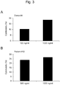
- Fig. 3A shows the results concerning Donor # 4
- Fig. 3B shows the results concerning Patient # 12.
- the concentration 1,000 ng/ml (1 ⁇ g/ml) at which a high degree of cytotoxicity was observed in the healthy volunteer and in the cancer patient was employed. At a concentration higher than the aforementioned level, cells died, and measurement could not be performed.
- Monocytes were cultured for 1 to 6 days with the use of PEGINTRON and OK432, the cytotoxicity relative to the K562 cells as the target and the cytotoxicity relative to MIA PaCa2 as the target were determined.
- the results are shown in Fig. 4 .
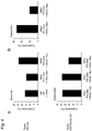
- the upper charts shown in Fig. 4 demonstrate the results attained with the use of the K562 cells as the target cells, (A) demonstrates the results concerning Donor # 4, and (B) demonstrates the results concerning Patient # 12.
- the lower chart shown in Fig. 4 demonstrates the results concerning Patient # 69 attained with the use of MIA PaCa2 as the target cell.
- a high degree of cytotoxicity was observed 4 days and 5 days after the initiation of culture on MIA PaCa2. From the viewpoint of a shorter culture period, a culture period of 4 days (culture with IFN for 3 days, followed by culture with OK432 for 1 day) was employed. The cytotoxicity on K562 and MIA PaCa2 was also confirmed.
- Fig. 5 A process of culture comprising adding the maturation medium on Day 4 of (4) IFN DC culture in the presence of 10 ⁇ g/ml (final concentration) of OK432 and 10 ng/ml of PGE2, which would provide the best results, was employed.
- the prepared DCs were treated with the FcR Blocking Reagent (Miltenyi Biotec) for 10 minutes, and the cells were stained with the antibodies serving as indicators for the dendritic cells labeled with FITC (fluorescein isothiocyanate) or PE (phycoerythrin) (i.e., CD11c, CD80, CD86, and HLA-ABC (BD Pharmingen), CD14, CD40, CD83, and HLA-DR (eBioscience), and CD197 (R&D Systems)).
- FITC fluorescein isothiocyanate
- PE phycoerythrin
- CD11c, CD80, CD86, and HLA-ABC (BD Pharmingen) CD14, CD40, CD83, and HLA-DR (eBioscience), and CD197 (R&D Systems)
- APC-conjugated CD56 (BD Pharmingen) was used.
- dead cells were stained with 2 ⁇ g/ml of propidium iod
- Fig. 6A demonstrates the results concerning IL-4-OK DC, and, from left to right, the results attained on Day 1, those on Day 7, and those on Day 8.
- Fig. 6B demonstrates the results concerning IFN-OK DC C, and, from left to right, the results attained on Day 1, those on Day 4, and those on Day 5.
- an adjuvant an immunopotentiating agent
- OK432 apparent cluster formation (cell aggregation) was observed in IL-4-OK DC on Day 8 and in IFN-OK DC on Day 5.
- a black arrow shown in Fig. 6A indicates cell adhesion. The application of physical damage at the time of recovery with a scraper was considered to lower the percentage of viable cells and the yield.
- IFN-OK DCs prepared via non-adhesive culture did not adhere to a non-adhesive HydroCell dish throughout the culture period.
- Cell surface marker expression was inspected via FACS analysis with the use of the recovered DCs.
- expression of CD86 and HLA-DR as dendritic cell markers was observed, and differences were observed in expression of CD14, CD56, CD83, and CD197.
- Expression of CD56 serving as the NK cell marker known as a killer cell was observed in IFN-OK DC.
- a black scale bar represents 50 ⁇ m.
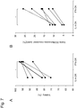
- Fig. 7 shows the results of inspection of the percentage of viable cells concerning the prepared DCs via trypan blue staining.
- the percentage of viable IFN-OK DCs was found to be significantly higher than that of IL-4-OK DCs ( Fig. 7A ).
- the yield (viable DCs/inoculated monocytes) was calculated.
- the percentage of viable IFN-OK DCs was found to be significantly higher than that of IL-4-OK DCs ( Fig. 7B ).
- DQ ovalbumin (Molecular Probes) was added at 10 ⁇ g/ml to the AIM-V medium containing 10% FCS, the reaction was carried out at 37°C for 30 minutes, and the reaction product was washed two times in the AIM-V medium, followed by the measurement using FACS CantoII.
- the cells subjected to the reaction at 4°C were used as negative controls, the values ( ⁇ MFI) subtracted from the mean fluorescent intensity (MFI) obtained at 37°C were evaluated as the antigen phagocytic activity and the antigen degrading ability.
- Fig. 8A shows the antigen phagocytic activity
- AIM medium supplemented with IL-2 (Imunace, 2.5 U/ml; Shionogi), IL-7 (5 ng/ml; R&D Systems), and IL-15 (10 ng/ml; PeproTech)
- 1 ⁇ 10 6 DCs were mixed with PBL at 1:10, and culture was conducted on a 12-well plate for 3 to 5 days.
- the AIM-V medium containing 10% FCS was added, and culture was conducted for an additional 2 or 3 days to recover cells.
- the recovered cells were stained with CD8-FITC (Beckman Coulter), CD3-APC (eBioscience), and T-select HLA-A*02:01 MART-1 tetramer-ELAGIGILTV-PE (MBL), and MART-1-specific CTL was detected.
- DCs were prepared from monocytes obtained from a HLA-A*02:01 cancer patient, the MART-1 antigens were pulsed, PBL was mixed with DCs at 10:1, and culture was conducted for 1 week. Thereafter, cytotoxic T lymphocytes (CTLs) were detected using the MART tetramer. The results are shown in Fig. 9 . Numerical values shown in the panels indicates the proportion of MART tetramers + in CD3 + cells and CD8 + cells induced by relevant DCs. The panel indicated with the term "Before" demonstrates the proportion of MART-specific CTLs in PBL before the initiation of induction.
- CTLs cytotoxic T lymphocytes
- the density of the DCs was adjusted to 2 ⁇ 10 5 cells/ml in the AIM-V medium, the resultant was inoculated into a 24-well plate, the culture supernatant was recovered 24 hours later, and the cytokine was then measured.
- IL-12 p70 interleukin-12
- IFN- ⁇ interferon-y
- TNF- ⁇ tumor necrosis factor- ⁇
- IL-6 interleukin-6
- IL-10 ELISA kit BD Bioscience
- Fig. 10 The results are shown in Fig. 10 .
- A, B, C, D, and E each show IL-12-, IFN- ⁇ -, TNF- ⁇ -, IL-10-, and IL-6-producing ability, respectively.
- OK432 As shown in Fig. 10 , the cytokine-producing ability tended to elevate, and no significant differences were observed in terms of cytokine production between IFN-OK DC and IL-4-OK DC. Significant differences were tested by the Wilcoxon signed rank test.
- the cytotoxicity test was carried out in accordance with the method described in 1.
- the DCs prepared from cancer patients and the chronic myeloid leukemia cells (K562) as the target cells were subjected to the reaction at E:T of 50:1 for 18 hours, and cytotoxicity was then examined. The results are shown in Fig. 11 .
- IFN DC was found to exhibit cytotoxicity, and the experimentation system was found to sufficiently function.
- stimulation of IFN DC with OK432 (IFN-OK DC) was found to significantly elevate the cytotoxicity, and IFN-OK DC was found to exhibit cytotoxicity superior to that of IL-4-OK DC to a significant extent.
- the prepared DCs were used to inspect the CD56 expression via FACS.
- K562 was designated as the target, IFN DC and IFN-OK DC were designated as the effectors, these cells were subjected to the reaction at E:T of 50:1 for 18 hours, and measurement was then carried out.
- the correlation between cytotoxicity and CD56 expression ( ⁇ MFI) was evaluated using the Spearman's rank correlation coefficient, and significant differences were tested. The results are shown in Fig. 12.
- Fig. 12A demonstrates CD56 expression ( ⁇ MFI) of the dendritic cells prepared by various methods, Fig.
- Fig. 12B demonstrates the correlation between CD56 expression and cytotoxicity in IFN DC
- Fig. 12C demonstrates the correlation between CD56 expression and cytotoxicity in IFN-OK DC.
- IFN DC R is 0.637
- p is 0.173
- n is 6.
- IFN-OK DC R is 0.714
- p is 0.136
- n is 6.
- the cytotoxicity of IFN-OK DC was found to be highly correlated with CD56 expression.
- IFN DCs and IFN-OK DCs were suspended in the AIM medium containing 10% FCS, TRAIL-sensitive K562 cells were mixed with FasL (Fas ligand)-sensitive MIA PaCa2 at E:T of 50:1 one hour after the antibodies were added, and the cytotoxicity was examined via FACS 18 hours later.
- the results are shown in Fig. 13.
- Fig. 13A demonstrates the results concerning the K562 cells and Fig. 13B demonstrates the results concerning MIA PaCa2.
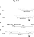
- the CD14-positive cells sorted from the PBMCs were suspended in a medium containing GM-CSF (1,000 U/ml; Miltenyi Biotec) and a pegylated interferon ⁇ -2b formulation (PEGINTRON®, 1 ⁇ g/ml; MSD) at 2 to 4 ⁇ 10 6 cells/ml, and culture was conducted in a non-adhesive culture dish for 3 days to prepare IFN-DCs ( Fig. 15-1A ).
- GM-CSF 1,000 U/ml; Miltenyi Biotec
- PEGINTRON® 1 ⁇ g/ml
- MSD pegylated interferon ⁇ -2b formulation
- PBMCs were inoculated into an adhesive culture dish, non-adherent cells were removed, culture was conducted in a medium supplemented with GM-CSF (50 ng/ml; Gentaur) and IL-4 (50 ng/ml; R&D Systems) for 5 days to prepare imDCs ( Fig. 15-1C ), maturation was further carried out in a medium supplemented with OK432 (10 ⁇ g/ml) and PGE2 (50 ng/ml) for 18-24 hours, and mIL-4-DCs were then recovered ( Fig. 15-1D ). The DCs were observed under the phase contrast microscope at the time of recovery. The results are shown in Fig. 15-2A to Fig. 15-2D .
- a black scale bar represents 50 mm.
- Fig. 15-2A to Fig. 15-2D in IFN-DCs dispersed in a culture dish ( Fig. 15-2A ), cell aggregation (cluster formation) was observed upon stimulation with OK432 (mIFN-DC, Fig. 15-2B ), and a typical phenotype of mature DCs was observed.
- Cluster formation of mIFN-DC Fig. 15-2A
- Fig. 15-2D tended to be similar to that of mIL-4-DC
- a phenotype of mature DCs was observed.
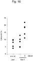
- the culture solution of IFN-DC (supplemented with PEGINTRON) shown in Fig. 1 was supplemented with a medium supplemented with a cytokine cocktail prepared with or without the addition of OK432 (?/+) 4 days after the initiation of culture (Day 4), the cells were recovered on Day 5, and cytotoxic activity was assayed.
- the results are shown in Fig. 16 .
- the cytotoxicity (%) was significantly enhanced with the addition of OK432.
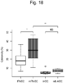
- the results are shown in Figs. 17 and 18 .
- Notations concerning DCs used in Fig. 17 correspond to those used in Fig. 15-1 .
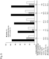
- Numerical values in the charts represent the percentage of the damaged K562 cells, and such values were determined on the basis of the percentage of PI-positive cells among the CFSE-stained K562 cells. As shown in Fig.
- CD56 expression of the prepared DCs was inspected via FACS.
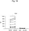
- the mean fluorescence density (MFI) of CD56 of DCs is shown in the form of a dot plot and compared on the basis of maturation with OK432. The results are shown in Fig. 19 (*, p ⁇ 0.05). Notations concerning DCs used in Fig. 19 correspond to those used in Fig. 15-1 .
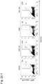
- CFSE-labeled K562 cells were mixed with the PKH26-stained IFN-DCs or IFN-DCs at 1:1, the cells were subjected to the reaction for 18 hours, and the cells were further subjected to the reaction with DAPI (blue) for 5 minutes. Thereafter, the cells were observed under a fluorescence microscope.
- Fig. 21 shows microscopic images. White arrows indicate DAPI-positive dead cells and a white scale bar indicates 50 ⁇ m. As shown in Fig. 21 , clusters formed by IFN-DCs and the K562 cells were small (the upper image, Fig. 21 ), cells were dispersed, and the number of dead cells was very small.
- clusters formed by the mIFN-DCs and the K562 cells were large and apparent (the lower image, Fig. 21 ), and many dead cells were observed in the K562 cells within the cluster.
- the results suggest that cluster formation; that is, intercellular contact, is critical for induction of cell damage.
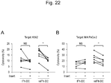
- cell contact may be critical for enhanced cell damage caused by OK432-stimulated mIFN-DC. Since such phenomenon was not limited to the K562 cells but was also observed in MIA PaCa-2 ( Fig. 22B ), such phenomenon was considered to be a common mechanism of cell damage.
- the results are shown in Fig. 24 .
- variations in expression were precisely evaluated on the basis of MFI, and statistically significant differences were examined.
- stimulation with OK432 was found to significantly increase TRAIL and Fas ligand expression levels of mIFN-DC and mIL-4-DC.
- the TRAIL expression level in mIFN-DC was significantly higher. Accordingly, a high TRAIL expression level in IFN-DC was considered to be involved with such phenomenon. It was thus suggested that inhibition of TRAIL would inhibit cell damage and a high TRAIL expression level would affect cell damage in mIFN-DC.
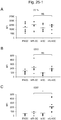
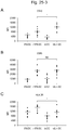
- Fig. 25-1 shows values concerning CD11c ( Fig. 25-1A ), CD80 ( Fig. 25-1B ), and CCR7 ( Fig. 25-1C )
- Fig. 25-2 shows values concerning CD14 ( Fig. 25-2A ), CD83 ( Fig. 25-2B ), and HLA-ABC ( Fig. 25-2C )
- Fig. 25-3 shows values concerning CD40 ( Fig. 25-3A ), CD86 ( Fig. 25-3B ), and HLA-DR ( Fig. 25-3C ).
- the antigen phagocytic activity was inspected based on FITC-dextran uptake and the antigen degrading ability was confirmed based on the fluorescence intensity of DQ-Ovalbumin.
- Fig. 27 compared with IFN-DC and imDC, the amount of FITC-dextran uptake into OK432-stimulated mIFN-DC and mIL-4-DC decreased to a significant extent, and similar activity was observed ( Fig. 27A ). Specifically, the antigen phagocytic activity of mIFN-DC was similar to that of mIL4-DC. As a result of fluorescence intensity measurement based on DQ-ovalbumin degradation, the antigen degrading ability was found to significantly decrease upon stimulation with OK432, and the antigen degrading ability of mIFN-DC was found to be similar to that of mIL-4-DC ( Fig. 27B ). Thus, mIFN-DC and mIL-4-DC were found to exhibit similar antigen phagocytic activity and antigen presenting activity of mature DCs.
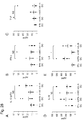
- cytokines IL-12, IFN- ⁇ , TNF- ⁇ , IL-10, and IL-6
- the cytokine IL-12 (p70) and IFN- ⁇ that would enhance CTL induction elevated upon stimulation with OK432 were also observed in mIFN-DC as well as in mIL-4-DC. No variation was observed in TNF- ⁇ , which is a soluble tumor necrosis factor.
- the production level for IL-10 that would inhibit CTL induction was similar between mIFN-DC and mIL-4-DC.
- the production level for IL-6 that would exert inhibitory functions on regulatory T cells that would lower the CTL inducing capacity was significantly higher in mIFN-DC than in mIL-4-DC, compared with mIl-4-DC.
- the results demonstrate that, except for IL-6 production, mIFN-DC has the similar cytokine producing ability of mature DC as with mIL-4DC.
- the dendritic cells prepared by the method of the present invention can be used for dendritic cell therapy.
Landscapes
- Health & Medical Sciences (AREA)
- Life Sciences & Earth Sciences (AREA)
- General Health & Medical Sciences (AREA)
- Immunology (AREA)
- Animal Behavior & Ethology (AREA)
- Public Health (AREA)
- Veterinary Medicine (AREA)
- Epidemiology (AREA)
- Engineering & Computer Science (AREA)
- Chemical & Material Sciences (AREA)
- Biomedical Technology (AREA)
- Biotechnology (AREA)
- Cell Biology (AREA)
- Zoology (AREA)
- Hematology (AREA)
- Medicinal Chemistry (AREA)
- Pharmacology & Pharmacy (AREA)
- Microbiology (AREA)
- Virology (AREA)
- Bioinformatics & Cheminformatics (AREA)
- Genetics & Genomics (AREA)
- Developmental Biology & Embryology (AREA)
- Wood Science & Technology (AREA)
- Organic Chemistry (AREA)
- Oncology (AREA)
- Mycology (AREA)
- Biochemistry (AREA)
- General Engineering & Computer Science (AREA)
- Micro-Organisms Or Cultivation Processes Thereof (AREA)
- Medicines Containing Material From Animals Or Micro-Organisms (AREA)
Abstract
Description
- The present invention relates to a method for preparing dendritic cells from monocytes.
- Dendritic cells (DCs) are potent antigen presenting cells in vivo and they are known to induce immune responses by presenting antigens to T cells. DCs are also known to directly react with, for example, B cells, NK cells, and NKT cells, in addition to T cells, and play a key role in immune responses. When immature DCs are stimulated by antigens, expression levels of CD40, CD80, CD86, and the like are elevated. DCs acquire a high degree of T cell stimulating capacity, migrate to peripheral lymph tissue, and activate T cells specific for the antigens incorporated therein. Thus, immune responses are induced.
- In general, several types of cytokines are known as substances that are approved to be capable of inducing dendritic cell differentiation from blood progenitor cells. For example, many reports have been made concerning induction of DC differentiation with the use of GM-CSF in combination with IL-4 (Akagawa, K. S. et al., Blood, Vol. 88, No. 10, November 15, 1996: pp. 4029-4039). In addition, substances capable of inducing DC differentiation alone or in combination with other cytokines have been reported (O'Neill D. W. et al., Blood, Vol. 104, No. 8, October 15, 2004, pp. 2235-2246). For example, TNF-α, IL-2, IL-3, IL-6, IL-7, IL-12, IL-13, IL-15, HGF (hepatocyte growth factor), CD40 ligand, M-CSF, Flt3 ligand, c-kit ligand, and TGF-β have been reported.
- DC differentiation is induced with the use of GM-CSF in combination with IL-4 via adhesive culture, mononuclear cells (monocytes and lymphocytes) are inoculated into a culture dish, lymphocytes are washed, and the adhered monocytes are used for culture. Culture is conducted in the presence of GM-CSF/IL-4 for 5 to 7 days, cells are washed with a medium and collected via scraping (physical peeling), the medium is exchanged with a fresh medium containing an adjuvant (an immunopotentiating agent, OK432), and mature DCs are thus prepared.
- The present invention provides a method for preparing dendritic cells from monocytes via non-adhesive culture using interferon α.
- In the past, dendritic cells (DCs) were prepared from monocytes in the peripheral blood via adhesive culture with the use of GM-CSF and IL-4. Specifically, mononuclear cells (i.e., monocytes and lymphocytes) were inoculated into a culture dish, the lymphocytes and included platelets were washed, the adhered monocytes were cultured in the presence of GM-CSF and IL-4 for 5 days, the cells were washed with a medium and collected via scraping (physical peeling), the medium was exchanged with a fresh medium containing an adjuvant (an immunopotentiating agent) OK432, and mature DCs were thus prepared.
- According to such technique, however, a DC yield was not sufficient. In order to use DCs for cancer therapy, in addition, DCs having activity such as cytotoxicity, in addition to a strong antigen presenting activity and phagocytic activity, have been desired.
- The present inventors had conducted concentrated studies in order to enhance a DC yield and to prepare highly functional DCs. As a result, they discovered that optimized DCs could be prepared within a short period of time with the use of GM-CSF and pegylated interferon α (PEG-IFN-α) instead of GM-CSF and IL-4, monocytes are isolated and refined from the peripheral blood, and DCs are then prepared via non-adhesive culture; i.e., float culture. In addition, they discovered that an yield for DC preparation would be enhanced, and the resulting DCs would have a strong cytotoxic activity. This has led to the completion of the present invention.
- Specifically, the present invention is as follows.
- [1] A method for preparing cytotoxic dendritic cells from monocytes comprising subjecting monocytes which were isolated from the peripheral blood to non-adhesive culture in the presence of GM-CSF and pegylated interferon α and further conducting non-adhesive culture with the addition of prostaglandin E2 and OK432.
- [2] The method for preparing dendritic cells from monocytes according to [1], wherein, following the non-adhesive culture conducted for 2 to 5 days in the presence of GM-CSF and pegylated interferon α, non-adhesive culture is conducted for an additional 1 or 2 days with the addition of prostaglandin E2 and OK432.
- [3] The method for preparing dendritic cells from monocytes according to [1] or [2], wherein the monocytes are cultured with the use of 100 U/ml to 10,000 U/ml of GM-CSF, 500 ng/ml to 5 µg/ml of pegylated interferon α, 5 ng/ml to 50 ng/ml of prostaglandin E2, and 5 µg/ml to 50 µg/ml of OK432.
- [4] The method for preparing dendritic cells from monocytes according to any of [1] to [3], wherein the percentage of viable dendritic cells is 90% or higher and the yield indicating the proportion of the number of the obtained dendritic cells relative to the number of monocytes at the time of culture is 15% or higher.
- [5] The method for preparing dendritic cells from monocytes according to any of [1] to [4], wherein the obtained dendritic cells are positive for CD11c, CD14, CD40, CD56, CD80, CD86, HLA-ABC, and HLA-DR and negative for CD83 and CD197.
- [6] The method for preparing dendritic cells from monocytes according to any of [1] to [5], wherein, when effector cells (DCs) and target cells (the chronic myeloid leukemia cell line K562) are subjected to culture at a ratio of 50:1 for 18 hours, the cytotoxicity of the dendritic cells measured as the proportion of the target cells as dead cells is 30% or higher.
- [7] Dendritic cells prepared by the method for preparing dendritic cells from monocytes according to any of [1] to [6].
- [8] Dendritic cells, which are positive for CD11c, CD14, CD40, CD56, CD80, CD86, HLA-ABC, and HLA-DR and negative for CD83 and CD197, when effector cells (DCs) and target cells (the chronic myeloid leukemia cell line K562) are subjected to culture at a ratio of 50:1 for 18 hours, the cytotoxicity of the dendritic cells measured as the proportion of the target cells as dead cells is 30% or higher.
- [9] A pharmaceutical composition comprising the dendritic cells according to [8].
- [10] The pharmaceutical composition according to [9], which has the anticancer immunoreactivity and is used for cancer therapy.
- [11] An agent for inducing and differentiating cytotoxic dendritic cell (DC) from monocytes comprising GM-CSF, pegylated interferon α, prostaglandin E2, and OK432.
- [12] The agent for inducing cytotoxic dendritic cell differentiation from monocytes according to [11], which comprises an agent for inducing and differentiating immature dendritic cell comprising GM-CSF and pegylated interferon α and an agent for dendritic cell maturation comprising prostaglandin E2 and OK432.
- This description includes part or all of the disclosure of Japanese Patent Application No.
2015-053804 - The method for preparing dendritic cells (DCs) of the present invention comprises subjecting the isolated and refined monocytes to non-adhesive culture in the presence of GM-CSF, pegylated interferon (IFN)-α (PEG-IFN-α), prostaglandin E2 (PGE2), and OK432. According to such method, DCs with high cytotoxicity can be prepared within a short period of time with a high yield. The obtained DCs can be preferably used for cancer immunotherapy.
-
-
Fig. 1 schematically shows a method for preparing IL-4-OK DC and a method for preparing IFN-OK DC. -
Fig. 2 shows the effects of an IFN-α formulation and OK432 used for dendritic cell preparation on cytotoxicity. -
Fig. 3 shows the results of examination of the concentration of PEGINTRON used for dendritic cell preparation. -
Fig. 4 shows cytotoxicity of the dendritic cells prepared via culture of monocytes for 1 to 6 days on K562 cells and MIA PaCa2. -
Fig. 5 shows the effects of the PGE2 concentration when preparing dendritic cells. -
Fig. 6 shows the phenotype of the prepared dendritic cells. -
Fig. 7 shows the percentage of viable cells (Fig. 7A ) and the yield (Fig. 7B ) concerning the prepared dendritic cells. -
Fig. 8 shows the antigen phagocytic activity (Fig. 8A ) and the antigen degrading ability (Fig. 8B ) concerning the prepared dendritic cells. -
Fig. 9 shows in vitro CTL induction of the prepared dendritic cells. -
Fig. 10 shows the capacity of the prepared dendritic cells for cytokine production. -
Fig. 11 shows the cytotoxicity of the prepared dendritic cells that attacks cancer cells. -
Fig. 12 shows the correlation between CD56 expression and cytotoxicity of the prepared dendritic cells.Fig. 12A shows CD56 expression (ΔMFI) of the dendritic cells prepared by various techniques,Fig. 12B shows the correlation between CD56 expression and cytotoxicity in IFN DC, andFig. 12C shows the correlation between CD56 expression and cytotoxicity in IFN-OK DC. -
Fig. 13 shows the cytotoxic mechanism of IFN-OK DC. -
Fig. 14 shows a comparison between IL-4-OK DC and IFN-OK-DC. -
Fig. 15-1 shows a procedure for preparing dendritic cells under various conditions. -
Fig. 15-2 shows phase contrast microscopic images of the dendritic cells prepared under various conditions. -
Fig. 16 shows the effects of OK432 on cytotoxicity. -
Fig. 17 shows induction of cell damage caused by OK432. -
Fig. 18 shows induction of cell damage caused by OK432 in the form of a box plot. -
Fig. 19 shows the mean fluorescence intensity (MFI) of CD56 of DCs. -
Fig. 20 shows the correlation between MFI of CD56 and cytotoxicity in mIFN-DC. -
Fig. 21 shows the cluster formation by IFN-DC and K562 cells (the upper diagram) and the cluster formation by mIFN-DC and K562 cells (the lower diagram). -
Fig. 22 shows K562 cell damage (Fig. 22A ) and MIA PaCa-2 cell damage (Fig. 22B ) caused by IFN-DC or mIFN-DC. -
Fig. 23 shows TNF-α, TRAIL, and Fas ligand expression on the DC cell surface. -
Fig. 24 shows DMFI of TRAIL (N = 9) (A) and Fas ligand (N = 8) (B) on the DC cell surface. -
Fig. 25-1 shows the results of marker analysis of DCs prepared from PBMCs obtained from a cancer patient (CD11c (A), CD80 (B), and CCR7 (C)). -
Fig. 25-2 shows the results of marker analysis of DCs prepared from PBMCs obtained from a cancer patient (CD14 (A), CD83 (B), and HLA-ABC (C)). -
Fig. 25-3 shows the results of marker analysis of DCs prepared from PBMCs obtained from a cancer patient (CD40 (A), CD86 (B), and HLA-DR (C)). -
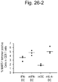
Fig. 26-1 shows the antigen presenting activity of mIFN-DC and that of mIL-4-DC based on Mart-1-specific CTL induction. -
Fig. 26-2 shows the antigen presenting activity of mIFN-DC and that of mIL-4-DC based on Mart-1-specific CTL induction (a dot plot). -
Fig. 27 shows the antigen phagocytic activity and the antigen presenting activity of DCs. -
Fig. 28 shows the cytokine-producing capacity of DCs. - Hereafter, the present invention is described in detail.
- The present invention concerns a method for preparing cytotoxic dendritic cells (DCs) from monocytes.
- It has heretofore been known that dendritic cells were induced to differentiate from monocytes by subjecting monocytes to adhesive culture in the presence of the granulocyte macrophage colony-stimulating factors (GM-CSF) and interleukin 4 (IL-4). In the present invention, monocytes are subjected to non-adhesive culture with the use of GM-CSF and pegylated interferon (IFN)-α (PEG-IFN-α) for induction of dendritic cell differentiation. By conducting culture in the presence of GM-CSF and PEG-IFN-α, immature dendritic cells are prepared. Such immature dendritic cells are further subjected to non-adhesive culture in the presence of GM-CSF, PEG-IFN-α, prostaglandin E2 (PGE2), and OK432, so that mature DCs having cytotoxicity can be obtained.
- PEG-IFN-α is composed of polyethylene glycol (PEG) bound to IFN-α. A preferable PEG-IFN-α is PEG-IFN-α-2b. As PEG-IFN-α, a commercially available PEG-IFN formulation can be used. An example of a commercially available PEG-IFN-α formulation is a PEG-IFN-α-2b formulation (i.e., PEG-Intron®; generic name: PEG interferon α-2b (genetic recombination)).
- PEG-Intron® is represented by a structural formula: H3C-(O-CH2CH2)n-OCO-Interferon alfa-2b, which is composed of a molecule of methoxy polyethylene glycol (average molecular weight: about 12,000) covalently bound to a single site of amino acid residues (Cys1, His7, Lys31, His34, Lys49, Lys83, Lys112, Lys121, Tyr129, Lys131, Lys133, Lys134, Ser163, and Lys164) of interferon α-2b (genetic recombination) (molecular weight: 19268.91) through a carbonyl group. The molecular weight thereof is about 32,000, and the molecular formula thereof is C860H1353N229O255S9. The CAS registry number is 215647-85-1.
- OK432 is a lyophilized powder of Streptococcus pyogenes (A group, type 3) Su strain cells treated with penicillin, which is a lyophilized cell formulation prepared by treating hemolytic streptococcus Streptococcus pyogenes (A group, type 3) Su strain cells in the presence of benzylpenicillin potassium under certain conditions. OK432 is used as an adjuvant. The CAS registry number is 39325-01-4. OK-432 is a clinical trial number, and commercially available Picibanil® can be used.
- In the method of the present invention, monocytes that were purified and refined in advance are used. Examples of monocytes include peripheral blood-derived monocytes, bone marrow-derived monocytes, spleen cell-derived monocytes, and umbilical cord blood-derived monocytes, with the peripheral blood-derived monocytes being particularly preferable. Monocytes are positive for CD14. When monocytes are to be collected from organisms, accordingly, monocytes can be isolated on the basis of the presence of CD14 as the indicator with the use of a fluorescent activated cell sorter (FACS), a flow cytometer, a magnetic separator, or the like, and the isolated can then be refined. Also, monocytes can be concentrated and refined with the use of an apparatus for apheresis (blood component collection). In addition, monocytes can be isolated and refined via density gradient centrifugation with the use of Ficoll® or the like. Animal species from which monocytes are derived are not limited. For example, mammalian animals, such as mice, rats, guinea pigs, hamsters, rabbits, cats, dogs, sheep, pigs, cows, horses, goats, monkeys, and humans, can be used. As FACS and a flow cytometer, for example, FACS vantage (Becton, Dickinson and Company) and FACS Calibur (Becton, Dickinson and Company) can be used. An example of a magnetic separator that can be used is autoMACS® (Miltenyi Biotec). On the basis of CD14 expression as the indicator, for example, monocytes can be isolated from peripheral mononuclear cells (PBMCs) with the use of CD14 microbeads to which CD14 has been bound via techniques involving the use of autoMACS® and CliniMACS®.
- The isolated and refined monocytes are subjected to non-adhesive culture; that is, float culture. Non-adhesive culture may be conducted with the use of a culture vessel, such as a non-adhesive plate, dish, or flask. A non-adhesive culture vessel is prepared by coating a culture dish surface with a compound, such as a superhydrophilic polymer, phospholipid polymer, or MPC polymer. Alternatively, a culture dish surface is made hydrophilic without the use of a coating agent, so as to prevent cells from adhering to the culture vessel. For example, a low-adhesive culture dish, such as FlydroCell™ (CellSeed), EZ-BindShut® II (Iwaki), Nunclon™ Vita (NOF Corporation), or Lipidure®-Coat (NOF Corporation), can be used.
- Examples of culture media that can be used include RPMI1640, DMEM, AIM-V®, and X-VIVO5®, and adequate antibiotics, animal blood sera, human albumin, human blood sera, or the like may be added to such basal media, and culture may be conducted therein.
- When monocytes are used at the concentration of 104 to 107 cells/ml, for example, the concentration of GM-CSF used for culture is 100 U/ml to 10,000 U/ml, preferably 500 U/ml to 2,000 U/ml, more preferably 800 to 1,200 U/ml, and particularly preferably 1,000 U/ml. Alternatively, such concentration is 10 ng/ml to 1,000 ng/ml, preferably 20 ng/ml to 200 ng/ml, and more preferably 20 ng/ml to 100 ng/ml. The PEG-IFN-α concentration is 100 ng/ml to 10 µg/ml, preferably 500 ng/ml to 5 µg/ml, and more preferably 500 ng/ml to 2 µg/ml. The PG2E concentration is 1 ng/ml to 100 ng/ml, preferably 5 ng/ml to 50 ng/ml, and more preferably 5 ng/ml to 20 ng/ml. The OK432 concentration is 1 µg/ml to 100 µg/ml, preferably 5 µg/ml to 50 µg/ml, and more preferably 5 µg/ml to 20 µg/ml.
- Culture is conducted in the presence of GM-CSF and PEG-IFN-α for 2 to 5 days, preferably for 3 to 4 days, and more preferably for 3 days. By conducting culture in the presence of GM-CSF and PEG-IFN-α, immature DCs are obtained. Such immature DCs are subjected to non-adhesive culture in the presence of GM-CSF, PEG-IFN-α, prostaglandin E2, and OK432 for an additional 1 or 2 days, and preferably 1 day. Thus, cytotoxic DCs can be obtained. OK-432 and PGE2 may be added by supplementing the medium containing GM-CSF and PEG-IFN-α with a supplemental medium containing OK-432 and PGE2. A total culture period is for 3 to 7 days, preferably 4 to 6 days, more preferably 4 to 5 days, and particularly preferably 4 days.
- Expression of monocyte or DC surface antigens may be inspected via FACS or the like, so that the concentration at which cells with the degree of differentiation of interest are obtained can adequately be determined.
- By the method described above, the isolated and refined monocytes are cultured in the presence of GM-CSF and PEG-IFN-α and further cultured in the presence of GM-CSF, PEG-IFN-α, PGE2, and OK432. Thus, cytotoxic DCs are differentiated and induced. The DCs obtained in such a manner are occasionally referred to as "IFN-OK-DCs" or "mIFN-DCs." The DCs prepared in such a manner of the present invention are characterized in that they have dendrites in morphological sense. By analyzing the DCs via FACS or other means, in addition, such DCs are found to be positive for CD11c, CD14, CD40, CD56, CD80, CD86, HLA-ABC, and HLA-DR and negative for CD83 and CD197 as surface antigens. In particular, CD56 which is an indicator of NK cell and CD86 and HLA-DR which are the minimal standards of DCs are expressed.
- Whether or not the cells are positive or negative for such surface antigens can be determined via microscopic observation as to whether or not the cells are stained with the antibodies reacting with such antigens, which have been labeled with a color enzyme, a fluorescent compound, or the like. For example, cells may be subjected to immunostaining with the use of such antibodies, so as to determine the presence or absence of surface antigens. Further, magnetic beads comprising the antibodies bound thereto may be used. Furthermore, the presence or absence of surface antigens can be determined with the use of FACS or a flow cytometer. The expression "negative for surface antigens" means that the cells are not sorted as positive cells when the cells are analyzed with the use of FACS as described above, and expression is not observed when expression is analyzed via immunostaining. When the expression level is lower than the lower detection limit of such technique, the cells are determined to be negative for surface antigens.
- The cytotoxicity of the DCs prepared by the method of the present invention is correlated with the expression level of CD56 as an NK cell marker. The cytotoxicity of the DCs prepared by the method of the present invention is 30% or higher, preferably 32% or higher, and more preferably 35% or higher, when measured as the proportion of the target cells as dead cells after the effector cells (DCs) and the target cells (i.e., the chronic myeloid leukemia cell line K562) are cultured at the proportion of 50:1 for 18 hours in accordance with the description of M. Korthals et al. Journal of Translational Medicine 2007, 5: 46.
- The DCs prepared by the method of the present invention exert cytotoxicity with the aid of the tumor necrosis factor (TNF)-related apoptosis-inducing ligand (TRAIL) or a FAS ligand (FasL). TRAIL and FasL are referred to as "cytotoxic factors," and they are reported to function in certain types of DCs or lymphocytes when attacking cancer cells. When the DCs prepared by the method of the present invention exert cytotoxicity, specifically, TRAIL or a Fas ligand also reacts. Accordingly, the DCs prepared by the method of the present invention can effectively exert cytotoxicity on cancer cells expressing TRAIL receptors or FAS, although the targets are not limited thereto.
- In the method of the present invention, in addition, DCs are prepared from monocytes via non-adhesive culture. Accordingly, the percentage of viable cells and the yield of DCs are also high. The percentage of viable cells of the obtained DCs is 70% or higher, preferably 80% or higher, more preferably 90% or higher, further preferably 95% or higher, and still further preferably 97% or higher, which is the standard designated by the National Institutes of Health (NIH). The DC recovery rate (i.e., the proportion of the number of the viable DCs obtained relative to the number of the monocytes inoculated) is 15% or higher, and preferably 20% or higher.
- For example, there are characteristic differences as shown in
Fig. 14 between DCs prepared via adhesive culture with the use of GM-CSF, IL-4, PGE2, and OK432 (IL-4-OK DC) and DCs prepared via non-adhesive culture with the use of GM-CSF, PEG-IFN, PGE2, and OK432 (IFN-OK DC). - While preparation of IL-4-OK DCs requires about 7 days, specifically, the IFN-OK DCs of the present invention can be prepared within a short period of time (about 4 days). While the percentage of viable IL-4-OK DCs is about 82%, that of the IFN-OK DCs according to the present invention is as high as about 95% or higher. While the yield of IL-4-OK DC preparation is about 10% (10% to 15%), in addition, that of IFN-OK DC preparation is high; that is, about 16% or higher (e.g., 10% to 25% or higher). While the cytotoxicity of IL-4-OK DCs is 10% or lower, further, that of IFN-OK DC is as high as about 30% or higher.
- Regarding the antigens expressed on the cell surface, IL-4-OK DC is CD11c-positive, CD14-negative, CD40-positive, CD56-negative, CD80-positive, CD83-positive, CD86-positive, CD197-positive, HLA-ABC-positive, and HLA-DR-positive. In contrast, IFN-OK DC is CD11c-positive, CD14-positive, CD40-positive, CD56-positive, CD80-positive, CD83-negative, CD86-positive, CD197-negative, HLA-ABC-positive, and HLA-DR-positive. The cytotoxic level of IFN-OK DC is correlated with the CD56 expression level. In addition, IFN-OK DCs are found to exert cytotoxicity through TRAIL or a FAS ligand. This indicates that IFN-OK DCs exert the cytotoxicity on cancer cells expressing TRAIL or a FAS ligand.
- In addition, the DCs of the present invention have antigen phagocytic activity and degrading activity equivalent to those of the DCs prepared with the use of mGM-CSF and IL-4 and excellent antigen presenting activity as with the DCs prepared with the use of mGM-CSF and IL-4.
- The DCs of the present invention have the cytokine-producing ability and killer activity superior to those of the DCs prepared with the use of mGM-CSF and IL-4.
- The present invention also concerns an agent for inducing and differentiating DC from monocytes comprising GM-CSF and PEG-IFN-α. Such agent for inducing and differentiating DC can also be referred to as an "agent for DC preparation." The agent for inducing and differentiating DC may further comprise PGE2 and OK432. The agent for inducing and differentiating DC may be composed of a first reagent comprising GM-CSF and PEG-IFN-α and a second reagent comprising PGE2 and OK432. The present invention also concerns a kit for inducing and differentiating DC comprising the first reagent and the second reagent. The first reagent comprising GM-CSF and PEG-IFN-α is used for inducing and differentiating immature DCs and the second reagent comprising PGE2 and OK432 is used for maturation of immature DCs.
- By the method of the present invention, DCs are induced as mature DCs. In addition, the present invention concerns DCs prepared by the method of the present invention and a cell population comprising such DCs. Such cell population includes 10% or more, 30% or more, 50% or more, 70% or more, 90% or more, or 95% or more DCs.
- The DCs prepared by the method of the present invention can be used for dendritic cell therapy. An example of dendritic cell therapy is cancer immunotherapy that is known as dendritic cell vaccine therapy. For example, dendritic cells are prepared from monocytes obtained from a subject by the method of the present invention, and the prepared dendritic cells are returned into the subject' body. Thus, dendritic cells can be used for treatment or prevention of cancer. In such a case, the prepared dendritic cells can act regardless of cancer type and can exert cancer therapeutic effects. When preparing dendritic cells, alternatively, culture may be conducted with the addition of cancer-specific antigens that are specific to a particular type of cancer. Thus, dendritic cells exhibiting cancer-type-specific anticancer immunoreactivity can be prepared. Also, such cells can be used for treatment of bacterial or viral infections. For the treatment of infections, DCs prepared from monocytes via non-adhesive culture in the presence of GM-CSF, PEG-IFN-α, PGE2, and OK432 by the method of the present invention are useful. The prepared dendritic cells may be administered to a subject via the intradermal, subcutaneous, intravenous, intra-nodal, or other route. The amount and the time of administration can be adequately determined in accordance with the disease type, the disease severity, and the conditions of the subject.
- The present invention is described in greater detail with reference to the following examples, although the technical scope of the present invention is not limited to these examples.
- After informed consent was obtained, the peripheral blood mononuclear cells (PBMCs) were obtained from cancer patients via blood component collection (apheresis), and dendritic cells (DCs) were prepared using the obtained PBMCs as raw materials (Medical ethics approval number: 2107; date of approval: September 4, 2012). With the use of IL-4 DCs obtained via culture of monocytes in the presence of the granulocyte macrophage colony-stimulating factor (GM-CSF) and interleukin-4 (IL-4), OK432 (Picibanil®) prepared via treatment of a spontaneous mutant (Su strain) of group A Streptococcus pyogenes with penicillin, and prostaglandin E2 (PGE2), mature IL-4-OK DCs were prepared via a conventional adhesive culture technique. PBMCs were suspended in the AIM-V medium (Invitrogen Life Technologies) at the monocyte density of 2 to 4 × 10⁶ cells/ml, the cell suspension was inoculated into an adhesive cell culture dish (BD Primaria™, BD Bioscience), non-adherent cells were removed 30 minutes later, the medium was exchanged with the AIM-V medium supplemented with GM-CSF (50 ng/ml; Gentaur) and IL-4 (50 ng/ml; R&D Systems) on the following day, the adhered monocytes were cultured for 5 days, and immature IL-4 DCs were then recovered with the use of a scraper. For maturation, the cells were suspended in a fresh AIM-V medium containing 10 µg/ml of OK432 (streptococcal preparation, Chugai Pharmaceutical Co, Ltd, Tokyo, Japan) and 50 ng/ml of PGE2 (Daiichi Fine Chemical Co, Ltd, Toyama, Japan), and culture was conducted for an additional 16 to 24 hours to prepare dendritic cells. The dendritic cells prepared in such a manner are referred to as "IL-4-OK DCs."
- IFN DCs obtained via culture of monocytes in the presence of interferon (IFN)-α (IFN-α) and GM-CSF and IFN-OK DCs matured with the aid of OK432 and PGE2 were prepared with the use of the monocytes refined from PBMCs with the aid of CD14 microbeads (Miltenyi Biotec). With the use of the monocytes refined at the purity of 95% or higher on a daily basis, the cells were suspended in the AIM-V medium containing GM-CSF (1,000 U/ml; Miltenyi Biotec) and a pegylated interferon α-2b formulation Pegintron® (1 µg/ml; MSD) at 2 to 4 × 10⁶ cells/ml, the cell suspension was inoculated into a non-adhesive culture dish (HydroCell, CellSeed), and culture was conducted for 3 days to prepare IFN DCs. In addition, the IFN DC culture solution was supplemented with the
maturation medium 4 days after the initiation of culture (after the culture had been conducted for 3 days), and culture was conducted for an additional 16 to 24 hours in the culture medium containing OK432 (final concentration: 10 µg/ml) and PGE2 (10 ng/ml) to prepare IFN-OK DCs. The dendritic cells prepared in such a manner are referred to as "IFN-OK DCs." In some experiments, dendritic cells were prepared in the same manner except for the use of an interferon α-2b formulation (Intron® A, 1,000 U/ml; MSD). -
Fig. 1 schematically shows a method for IL-4-OK DC preparation (Fig. 1A ) and a method for IFN-OK DC preparation (Fig. 1B ). - In this example, the dendritic cells prepared with the use of IL-4 alone instead of IL-4 and OK432 and the dendritic cells prepared with the use of IFN alone instead of IFN and OK432 were examined, in addition to IL-4-OK DCs and IFN-OK DC. Such dendritic cells are referred to as IL-4 DC and IFN DC, respectively.
- The type of IFN-α used, the concentration of INF-α added, the culture period, and the PGE2 concentration were examined.
- Cytotoxicity of the prepared dendritic cells was examined in the manner described below.
- The chronic myeloid leukemia strain K562 (ATCC) or the human pancreatic cancer-derived cell strain MIA PaCa2 (RIKEN BRC) was suspended in PBS containing 0.1% FCS supplemented with carboxyfluorescein succinimidyl ester (CFSE) (5 µm; Molecular Probes), and the cell suspension was subjected to the reaction at 37°C for 10 minutes, followed by washing in the AIM-V medium. With the use of the AIM medium containing 10% FCS, 5 × 10⁵ DCs (effector cells) were mixed with CFSE-stained cancer cells (target cells) at a ratio (effector cells:target cells (E:T)) of 50:1. The cells were subjected to the reaction at 37°C for 18 hours, washed in a FACSFlow solution, and then stained with 2 µg/ml of propidium iodide (PI), so as to detect dead cells via FACS. The number of CFSE-positive and PI-positive cells was divided by the number of CFSE-positive cells, the resulting value was evaluated as the proportion of cytotoxicity. As a negative control, unstained cancer cells were designated as effector cells, the effector cells were mixed with the CFSE-stained cancer cells at a ratio E:T of 50:1, and the cells were subjected to the reaction in the same manner as described above.
- The inhibition test using antibodies was carried out by subjecting 10 µg/ml anti-TRAIL antibodies or anti-FAS ligand antibodies (eBioscience) and DCs to incubation for 1 hour in advance, mixing the CFSE-labeled cancer cells therewith, and then conducting the reaction.
- IFN DCs were prepared with the use of two types of IFN-α formulations (i.e., GM-CSF and PEGINTRON (1 µg/ml) or Intron A (1,000 U/ml)), and cytotoxicity was examined with or without the addition of OK432. The CFSE-labeled chronic myeloid leukemia strain K562 was designated as the target, DCs were mixed with the target at various proportions, and the percentage of dead cells was determined 18 hours later to determine the cytotoxicity (%). An error bar represents "mean ± standard error," and experiments were carried out 3 times independently (representative data (n = 3), the significant difference was tested by the Tukey-Kramer test, * p < 0.01, ** p < 0.001).
- The results are shown in
Fig. 2 . As shown inFig. 2 , OK432 elevates cytotoxicity, and IFN DC using PEGINTRON exhibits higher cytotoxicity than Intron A. Based on such results, PEGINTRON was used as the IFN-α formulation. - With the use of monocytes obtained from a healthy volunteer (Donor #4) and those obtained from a cancer patient (Patient #12), the optimal concentration of PEGINTRON was examined based on the cytotoxicity. Culture was conducted for 3 days at different concentrations, maturation with the aid of OK432 was conducted for 1 day, and IFN-OK DCs were then collected 4 days after the initiation of culture.
- The results are shown in
Fig. 3. Fig. 3A shows theresults concerning Donor #4 andFig. 3B shows theresults concerning Patient #12. As shown inFig. 3 , the concentration (1,000 ng/ml (1 µg/ml)) at which a high degree of cytotoxicity was observed in the healthy volunteer and in the cancer patient was employed. At a concentration higher than the aforementioned level, cells died, and measurement could not be performed. - Monocytes were cultured for 1 to 6 days with the use of PEGINTRON and OK432, the cytotoxicity relative to the K562 cells as the target and the cytotoxicity relative to MIA PaCa2 as the target were determined. The results are shown in
Fig. 4 . The upper charts shown inFig. 4 demonstrate the results attained with the use of the K562 cells as the target cells, (A) demonstrates theresults concerning Donor #4, and (B) demonstrates theresults concerning Patient #12. The lower chart shown inFig. 4 demonstrates theresults concerning Patient #69 attained with the use of MIA PaCa2 as the target cell. - When the K562 cells were used as the target cells, a high degree of cell damage was observed after culture had been conducted for 4 days (culture with IFN for 3 days, followed by culture with OK432 for 1 day) in both the healthy volunteer and the cancer patient.
- A high degree of cytotoxicity was observed 4 days and 5 days after the initiation of culture on MIA PaCa2. From the viewpoint of a shorter culture period, a culture period of 4 days (culture with IFN for 3 days, followed by culture with OK432 for 1 day) was employed. The cytotoxicity on K562 and MIA PaCa2 was also confirmed.
- The influence of PGE2 imposed on the percentage of viable cells, the recovery rate, and the cytotoxicity was inspected. The results are shown in
Fig. 5 . A process of culture comprising adding the maturation medium onDay 4 of (4) IFN DC culture in the presence of 10 µg/ml (final concentration) of OK432 and 10 ng/ml of PGE2, which would provide the best results, was employed. - The prepared DCs were treated with the FcR Blocking Reagent (Miltenyi Biotec) for 10 minutes, and the cells were stained with the antibodies serving as indicators for the dendritic cells labeled with FITC (fluorescein isothiocyanate) or PE (phycoerythrin) (i.e., CD11c, CD80, CD86, and HLA-ABC (BD Pharmingen), CD14, CD40, CD83, and HLA-DR (eBioscience), and CD197 (R&D Systems)). As the indicator for the NK cell phenotype, APC-conjugated CD56 (BD Pharmingen) was used. Immediately before FACS analysis, dead cells were stained with 2 µg/ml of propidium iodide (PI, Sigma-Aldrich), and the cells were then analyzed using FACS CantoII (BD Bioscience).
- The results are shown in
Fig. 6. Fig. 6A demonstrates the results concerning IL-4-OK DC, and, from left to right, the results attained onDay 1, those onDay 7, and those onDay 8.Fig. 6B demonstrates the results concerning IFN-OK DC C, and, from left to right, the results attained onDay 1, those onDay 4, and those onDay 5. After the addition of an adjuvant (an immunopotentiating agent) OK432, apparent cluster formation (cell aggregation) was observed in IL-4-OK DC onDay 8 and in IFN-OK DC onDay 5. A black arrow shown inFig. 6A indicates cell adhesion. The application of physical damage at the time of recovery with a scraper was considered to lower the percentage of viable cells and the yield. In contrast, IFN-OK DCs prepared via non-adhesive culture did not adhere to a non-adhesive HydroCell dish throughout the culture period. Cell surface marker expression was inspected via FACS analysis with the use of the recovered DCs. As a result, expression of CD86 and HLA-DR as dendritic cell markers was observed, and differences were observed in expression of CD14, CD56, CD83, and CD197. Expression of CD56 serving as the NK cell marker known as a killer cell was observed in IFN-OK DC. A black scale bar represents 50 µm. -
Fig. 7 shows the results of inspection of the percentage of viable cells concerning the prepared DCs via trypan blue staining. The percentage of viable IFN-OK DCs was found to be significantly higher than that of IL-4-OK DCs (Fig. 7A ). On the basis of the percentage of viable DCs obtained vial culture relative to the number of inoculated monocytes, the yield (viable DCs/inoculated monocytes) was calculated. As a result, the percentage of viable IFN-OK DCs was found to be significantly higher than that of IL-4-OK DCs (Fig. 7B ). In this example, DCs were prepared from monocytes obtained from cancer patients and analyzed (n = 6). Significant difference was tested by the Wilcoxon signed rank test. The symbol "*" indicates P < 0.05. - In order to inspect the antigen phagocytic activity, FITC-labeled dextran (M.W. = 40,000, Molecular Probes) was adjusted to 250 µg/ml using the AIM-V medium containing 10% fetal calf serum (FCS, Thermo Scientific), the prepared cells were mixed therein at 1 × 10⁶ cells/ml, and the resultant was then subjected to the reaction at 37°C for 3 hours. After the cells were washed two times in the AIM-V medium, the activity was measured using FACS CantoII. In order to elucidate the antigen degrading ability, DQ ovalbumin (Molecular Probes) was added at 10 µg/ml to the AIM-V medium containing 10% FCS, the reaction was carried out at 37°C for 30 minutes, and the reaction product was washed two times in the AIM-V medium, followed by the measurement using FACS CantoII. In the measurement of the antigen phagocytic activity and that of the antigen degrading ability, the cells subjected to the reaction at 4°C were used as negative controls, the values (ΔMFI) subtracted from the mean fluorescent intensity (MFI) obtained at 37°C were evaluated as the antigen phagocytic activity and the antigen degrading ability.
-
Fig. 8A shows the antigen phagocytic activity andFig. 8B shows the antigen degrading ability represented by dot-plotting ΔMFI obtained by inspecting FITC-dextran uptake and the DQ ovalbumin (DQ-OVA) degrading ability with the use of the DCs prepared from monocytes obtained from cancer patients (n = 6). After the cells were stimulated with OK432, the antigen phagocytic activity and the antigen degrading ability of IFN DCs and IL-4 DCs lowered, and these DCs developed into mature DCs. No significant difference was observed between IFN-OK DCs and IL-4-OK DCs (NS = not significant). Significant difference was tested by the Wilcoxon signed rank test. - With the use of the DCs and the peripheral blood lymphocytes (PBL) prepared from a HLA-A*02:01 cancer patient (Patient #29), in vitro CTL induction was performed. Synthetic peptides MART-1 and 26-35 A27L (ELAGIGILTV) were adjusted to 20 µg/ml in the AIM medium, and the resultant was mixed with DCs. The resultant was pulsed at 37°C for 1 hour, treated with mitomycin C (MMC, 25 µg/ml; Kyowa Hakko Kogyo) for 1 hour, and washed two times in the AIM medium to prepare DCs. With the use of the AIM medium supplemented with IL-2 (Imunace, 2.5 U/ml; Shionogi), IL-7 (5 ng/ml; R&D Systems), and IL-15 (10 ng/ml; PeproTech), 1×10⁶ DCs were mixed with PBL at 1:10, and culture was conducted on a 12-well plate for 3 to 5 days. In accordance with the cell growth, the AIM-V medium containing 10% FCS was added, and culture was conducted for an additional 2 or 3 days to recover cells. The recovered cells were stained with CD8-FITC (Beckman Coulter), CD3-APC (eBioscience), and T-select HLA-A*02:01 MART-1 tetramer-ELAGIGILTV-PE (MBL), and MART-1-specific CTL was detected.
- DCs were prepared from monocytes obtained from a HLA-A*02:01 cancer patient, the MART-1 antigens were pulsed, PBL was mixed with DCs at 10:1, and culture was conducted for 1 week. Thereafter, cytotoxic T lymphocytes (CTLs) were detected using the MART tetramer. The results are shown in
Fig. 9 . Numerical values shown in the panels indicates the proportion of MART tetramers+ in CD3+ cells and CD8+ cells induced by relevant DCs. The panel indicated with the term "Before" demonstrates the proportion of MART-specific CTLs in PBL before the initiation of induction. By stimulating IFN DCs or IL-4 DCs with OK432, the CTL-inducing ability was elevated, and no significant difference was observed therebetween (representative data (n = 3), significant difference is being tested while increasing the number n). The results demonstrate that stimulation with OK432 leads to the elevated antigen presenting activity and the antigen presenting activity of IFN-OK DC is equivalent to that of IL-4-OK DC. - The density of the DCs was adjusted to 2 × 10⁵ cells/ml in the AIM-V medium, the resultant was inoculated into a 24-well plate, the culture supernatant was recovered 24 hours later, and the cytokine was then measured. With the use of interleukin-12 (IL-12 p70) and interferon-y (IFN-γ) (R&D Systems), a tumor necrosis factor-α (TNF-α), interleukin-6 (IL-6), interleukin-10, and the IL-10 ELISA kit (BD Bioscience), the reaction was allowed to proceed in accordance with the instructions, and measurement was then carried out using a spectrophotometer (MULTISKAN FC, Thermo Scientific).
- The results are shown in
Fig. 10 . InFig. 10 , A, B, C, D, and E each show IL-12-, IFN-γ-, TNF-α-, IL-10-, and IL-6-producing ability, respectively. After the addition of OK432, as shown inFig. 10 , the cytokine-producing ability tended to elevate, and no significant differences were observed in terms of cytokine production between IFN-OK DC and IL-4-OK DC. Significant differences were tested by the Wilcoxon signed rank test. - The cytotoxicity test was carried out in accordance with the method described in 1.
- The DCs prepared from cancer patients and the chronic myeloid leukemia cells (K562) as the target cells were subjected to the reaction at E:T of 50:1 for 18 hours, and cytotoxicity was then examined. The results are shown in
Fig. 11 . As already reported, IFN DC was found to exhibit cytotoxicity, and the experimentation system was found to sufficiently function. Interestingly, stimulation of IFN DC with OK432 (IFN-OK DC) was found to significantly elevate the cytotoxicity, and IFN-OK DC was found to exhibit cytotoxicity superior to that of IL-4-OK DC to a significant extent. Significant difference was tested by the Wilcoxon signed rank test with the application of Bonfferoni correction (n = 8, NS = not significant, the symbol "*" indicates p < 0.05). - The prepared DCs were used to inspect the CD56 expression via FACS. K562 was designated as the target, IFN DC and IFN-OK DC were designated as the effectors, these cells were subjected to the reaction at E:T of 50:1 for 18 hours, and measurement was then carried out. Significant difference of IFN DC and IFN-OK DC was tested by the Wilcoxon signed rank test (NS: not significant; n = 6). The correlation between cytotoxicity and CD56 expression (ΔMFI) was evaluated using the Spearman's rank correlation coefficient, and significant differences were tested. The results are shown in
Fig. 12. Fig. 12A demonstrates CD56 expression (ΔMFI) of the dendritic cells prepared by various methods,Fig. 12B demonstrates the correlation between CD56 expression and cytotoxicity in IFN DC, andFig. 12C demonstrates the correlation between CD56 expression and cytotoxicity in IFN-OK DC. Regarding IFN DC, R is 0.637, p is 0.173, and n is 6. Regarding IFN-OK DC, R is 0.714, p is 0.136, and n is 6. Thus, the cytotoxicity of IFN-OK DC was found to be highly correlated with CD56 expression. - IFN DCs and IFN-OK DCs were suspended in the AIM medium containing 10% FCS, TRAIL-sensitive K562 cells were mixed with FasL (Fas ligand)-sensitive MIA PaCa2 at E:T of 50:1 one hour after the antibodies were added, and the cytotoxicity was examined via FACS 18 hours later. The results are shown in
Fig. 13. Fig. 13A demonstrates the results concerning the K562 cells andFig. 13B demonstrates the results concerning MIA PaCa2. Through treatment with the anti-TRAIL antibody and the anti-FasL antibody, the cytotoxicity on the K562 cells and that on MIA PaCa2 were found to be suppressed to a significant extent (n = 7, significant differences were tested by the Wilcoxon signed rank test, the symbol "*" indicates p < 0.05). The results demonstrate the involvement of TRAIL and FasL with the mechanism of cell damage by IFN-OK DC. - While IFN DC was reported to exert cytotoxicity on K562 with the aid of TRAIL, such cytotoxicity was not observed in this example. The influence of TRAIL and FasL imposed on IFN-OK DC stimulated with OK432 was reported for the first time.
- In this example, statistical analysis was carried out using the statistic software R, significant differences were tested by the Wilcoxon signed rank test and the Tukey-Kramer test, and the correlational efficient and the significant differences were examined using Spearman's rank-order correlation.
- The CD14-positive cells sorted from the PBMCs were suspended in a medium containing GM-CSF (1,000 U/ml; Miltenyi Biotec) and a pegylated interferon α-2b formulation (PEGINTRON®, 1 µg/ml; MSD) at 2 to 4 × 10⁶ cells/ml, and culture was conducted in a non-adhesive culture dish for 3 days to prepare IFN-DCs (
Fig. 15-1A ). Culture was conducted in a maturation medium containing 10 µg/ml of OK432 (streptococcal preparation, Chugai Pharmaceutical Co, Ltd, Tokyo, Japan) and 50 ng/ml of PGE2 (Daiichi Fine Chemical Co, Ltd, Toyama, Japan), in addition to GM-CSF (1,000 U/ml; Miltenyi Biotec) for 1 day, and mIFN-DCs were then recovered (Fig. 15-1B ). Separately, PBMCs were inoculated into an adhesive culture dish, non-adherent cells were removed, culture was conducted in a medium supplemented with GM-CSF (50 ng/ml; Gentaur) and IL-4 (50 ng/ml; R&D Systems) for 5 days to prepare imDCs (Fig. 15-1C ), maturation was further carried out in a medium supplemented with OK432 (10 µg/ml) and PGE2 (50 ng/ml) for 18-24 hours, and mIL-4-DCs were then recovered (Fig. 15-1D ). The DCs were observed under the phase contrast microscope at the time of recovery. The results are shown inFig. 15-2A to Fig. 15-2D . A black scale bar represents 50 mm. As shown inFig. 15-2A to Fig. 15-2D , in IFN-DCs dispersed in a culture dish (Fig. 15-2A ), cell aggregation (cluster formation) was observed upon stimulation with OK432 (mIFN-DC,Fig. 15-2B ), and a typical phenotype of mature DCs was observed. Cluster formation of mIFN-DC (Fig. 15-2A ) tended to be similar to that of mIL-4-DC (Fig. 15-2D ), and a phenotype of mature DCs was observed. - The culture solution of IFN-DC (supplemented with PEGINTRON) shown in
Fig. 1 was supplemented with a medium supplemented with a cytokine cocktail prepared with or without the addition of OK432 (?/+) 4 days after the initiation of culture (Day 4), the cells were recovered onDay 5, and cytotoxic activity was assayed. The results are shown inFig. 16 . As shown inFig. 16 , the cytotoxicity (%) was significantly enhanced with the addition of OK432. - DCs were used as the effector cells (E), CFSE-stained K562 cells were used as the target cells (T), they were mixed at E:T of 50 : 1, the cells were stained with PI 18 hours later, and cell damage caused by DCs was examined (representative data (N = 11)). The results are shown in
Figs. 17 and18 . Notations concerning DCs used inFig. 17 correspond to those used inFig. 15-1 . Numerical values in the charts represent the percentage of the damaged K562 cells, and such values were determined on the basis of the percentage of PI-positive cells among the CFSE-stained K562 cells. As shown inFig. 17 , the highest cytotoxicity was observed in mIFN-DC prepared with the use of PEGINTRON and OK-432.Fig. 18 shows the cell damage imposed on K562 in the form of a box plot (N = 11). (*p < 0.05, **p < 0.01, NS: not significant). - CD56 expression of the prepared DCs was inspected via FACS.
- The mean fluorescence density (MFI) of CD56 of DCs is shown in the form of a dot plot and compared on the basis of maturation with OK432. The results are shown in
Fig. 19 (*, p < 0.05). Notations concerning DCs used inFig. 19 correspond to those used inFig. 15-1 .Fig. 20 shows the correlation between MFI and cytotoxicity of CD56 in mIFN-DC (N = 12). As shown inFigs. 19 and20 , the cytotoxicity of the mIFN-DC prepared with the use of PEGINTRON and OK-432 was correlated with the CD56 expression level. - The CFSE-labeled K562 cells were mixed with the PKH26-stained IFN-DCs or IFN-DCs at 1:1, the cells were subjected to the reaction for 18 hours, and the cells were further subjected to the reaction with DAPI (blue) for 5 minutes. Thereafter, the cells were observed under a fluorescence microscope.
Fig. 21 shows microscopic images. White arrows indicate DAPI-positive dead cells and a white scale bar indicates 50 µm. As shown inFig. 21 , clusters formed by IFN-DCs and the K562 cells were small (the upper image,Fig. 21 ), cells were dispersed, and the number of dead cells was very small. In contrast, clusters formed by the mIFN-DCs and the K562 cells were large and apparent (the lower image,Fig. 21 ), and many dead cells were observed in the K562 cells within the cluster. The results suggest that cluster formation; that is, intercellular contact, is critical for induction of cell damage. - Subsequently, K562 or MIA PaCa-2 was used as the target, and cytotoxicity caused by IFN-DC or mIFN-DC was inspected based on the presence or absence of transwell membranes (0.4 mm) (N = 6). The results are shown in
Fig. 22 . As shown inFig. 22 , when the K562 cells were the target cells (Fig. 22A ), no difference was observed in cell damage caused by IFN-DC in terms of the presence or absence of transwell membranes (Inserts) inhibiting cell contact. In contrast, cell damage caused by mIFN-DC was found to decrease to a significant extent when cell contact was inhibited (Insert+). That is, cell contact may be critical for enhanced cell damage caused by OK432-stimulated mIFN-DC. Since such phenomenon was not limited to the K562 cells but was also observed in MIA PaCa-2 (Fig. 22B ), such phenomenon was considered to be a common mechanism of cell damage. - Subsequently, ΔMFI of TRAIL (N = 9) and that of the Fas ligand (N = 8) on the DC cell surface were compared (i.e., MFI determined by subtracting an isotype control from the relevant specific antibody). The results are shown in
Fig. 24 . As shown inFig. 24 , variations in expression were precisely evaluated on the basis of MFI, and statistically significant differences were examined. As a result, stimulation with OK432 was found to significantly increase TRAIL and Fas ligand expression levels of mIFN-DC and mIL-4-DC. Compared with mIL-4-DC, the TRAIL expression level in mIFN-DC was significantly higher. Accordingly, a high TRAIL expression level in IFN-DC was considered to be involved with such phenomenon. It was thus suggested that inhibition of TRAIL would inhibit cell damage and a high TRAIL expression level would affect cell damage in mIFN-DC. - DCs were prepared from PBMCs obtained from cancer patients (N = 8), typical DC markers were analyzed via flow cytometry, and the mean fluorescence intensity (MFI) was measured. MFIs are shown in the form of a dot plot in
Fig. 25-1 to Fig. 25-3 .Fig. 25-1 shows values concerning CD11c (Fig. 25-1A ), CD80 (Fig. 25-1B ), and CCR7 (Fig. 25-1C ),Fig. 25-2 shows values concerning CD14 (Fig. 25-2A ), CD83 (Fig. 25-2B ), and HLA-ABC (Fig. 25-2C ), andFig. 25-3 shows values concerning CD40 (Fig. 25-3A ), CD86 (Fig. 25-3B ), and HLA-DR (Fig. 25-3C ). - DCs pulsed with Mart antigens (i.e., cancer antigens specific to human melanoma cells) were mixed with PBL, culture was conducted for 1 week, CD3-positive lymphocytes were subjected to gating, and the results were plotted as CD8-FITC along the horizontal axis and as MART-1 tetramer-PE. Thus, Mart-1-specific CTL induction was inspected. Numerical values in the panels represent the percentage of MART-specific CTL among CD3- and CD8-positive cells. The results are shown in
Fig. 26-1 andFig. 26-2 (a dot plot). As shown inFig. 26-1 andFig. 26-2 , no differences were observed between mIFN-DC and mIL-4-DC in respect of the antigen presenting activity. - The antigen phagocytic activity was inspected based on FITC-dextran uptake and the antigen degrading ability was confirmed based on the fluorescence intensity of DQ-Ovalbumin. The value obtained for the negative control subjected to the reaction at 4°C was subtracted from the value obtained for the sample subjected to the reaction at 37°C to determine ΔMFI, and the obtained ΔMFI is shown in the form of a dot plot (n = 6). The results are shown in
Fig. 27 . As shown inFig. 27 , compared with IFN-DC and imDC, the amount of FITC-dextran uptake into OK432-stimulated mIFN-DC and mIL-4-DC decreased to a significant extent, and similar activity was observed (Fig. 27A ). Specifically, the antigen phagocytic activity of mIFN-DC was similar to that of mIL4-DC. As a result of fluorescence intensity measurement based on DQ-ovalbumin degradation, the antigen degrading ability was found to significantly decrease upon stimulation with OK432, and the antigen degrading ability of mIFN-DC was found to be similar to that of mIL-4-DC (Fig. 27B ). Thus, mIFN-DC and mIL-4-DC were found to exhibit similar antigen phagocytic activity and antigen presenting activity of mature DCs. - DCs (2 × 10⁵ cells/ml) were inoculated on a 24-well plate, culture was conducted in the AIM medium for 24 hours, the resulting supernatant was used to inspect production of cytokines (IL-12, IFN-γ, TNF-α, IL-10, and IL-6) via ELISA (N = 8). The results are shown in
Fig. 28 . As shown inFig. 28 , the cytokine IL-12 (p70) and IFN-γ that would enhance CTL induction elevated upon stimulation with OK432 were also observed in mIFN-DC as well as in mIL-4-DC. No variation was observed in TNF-α, which is a soluble tumor necrosis factor. The production level for IL-10 that would inhibit CTL induction was similar between mIFN-DC and mIL-4-DC. The production level for IL-6 that would exert inhibitory functions on regulatory T cells that would lower the CTL inducing capacity was significantly higher in mIFN-DC than in mIL-4-DC, compared with mIl-4-DC. The results demonstrate that, except for IL-6 production, mIFN-DC has the similar cytokine producing ability of mature DC as with mIL-4DC. - All publications, patents, and patent applications cited herein are incorporated herein by reference in their entirety.
- The dendritic cells prepared by the method of the present invention can be used for dendritic cell therapy.
Claims (12)
- A method for preparing cytotoxic dendritic cells from monocytes comprising subjecting monocytes which were isolated from the peripheral blood to non-adhesive culture in the presence of GM-CSF and pegylated interferon α and further conducting non-adhesive culture with the addition of prostaglandin E2 and OK432.
- The method for preparing dendritic cells from monocytes according to claim 1, wherein, following the non-adhesive culture conducted for 2 to 5 days in the presence of GM-CSF and pegylated interferon α, non-adhesive culture is conducted for an additional 1 or 2 days with the addition of prostaglandin E2 and OK432.
- The method for preparing dendritic cells from monocytes according to claim 1 or 2, wherein the monocytes are cultured with the use of 100 U/ml to 10,000 U/ml of GM-CSF, 500 ng/ml to 5 µg/ml of pegylated interferon α, 5 ng/ml to 50 ng/ml of prostaglandin E2, and 5 µg/ml to 50 µg/ml of OK432.
- The method for preparing dendritic cells from monocytes according to any one of claims 1 to 3, wherein the percentage of viable dendritic cells is 90% or higher and the yield indicating the proportion of the number of the obtained dendritic cells relative to the number of monocytes at the time of culture is 15% or higher.
- The method for preparing dendritic cells from monocytes according to any one of claims 1 to 4, wherein the obtained dendritic cells are positive for CD11c, CD14, CD40, CD56, CD80, CD86, HLA-ABC, and HLA-DR and negative for CD83 and CD197.
- The method for preparing dendritic cells from monocytes according to any one of claims 1 to 5, wherein, when effector cells (DCs) and target cells (the chronic myeloid leukemia cell line K562) are subjected to culture at a ratio of 50:1 for 18 hours, the cytotoxicity of the dendritic cells measured as the proportion of the target cells as dead cells is 30% or higher.
- Dendritic cells prepared by the method for preparing dendritic cells from monocytes according to any one of claims 1 to 6.
- Dendritic cells, which are positive for CD11c, CD14, CD40, CD56, CD80, CD86, HLA-ABC, and HLA-DR and negative for CD83 and CD197, when effector cells (DCs) and target cells (the chronic myeloid leukemia cell line K562) are subjected to culture at a ratio of 50:1 for 18 hours, the cytotoxicity of the dendritic cells measured as the proportion of the target cells as dead cells is 30% or higher.
- A pharmaceutical composition comprising the dendritic cells according to claim 8.
- The pharmaceutical composition according to claim 9, which has the anticancer immunoreactivity and is used for cancer therapy.
- An agent for inducing and differentiating cytotoxic dendritic cell (DC) from monocytes comprising GM-CSF, pegylated interferon α, prostaglandin E2, and OK432.
- The agent for inducing cytotoxic dendritic cell differentiation from monocytes according to claim 11, which comprises an agent for inducing and differentiating immature dendritic cell comprising GM-CSF and pegylated interferon α and an agent for dendritic cell maturation comprising prostaglandin E2 and OK432.
Applications Claiming Priority (2)
| Application Number | Priority Date | Filing Date | Title |
|---|---|---|---|
| JP2015053804 | 2015-03-17 | ||
| PCT/JP2016/058295 WO2016148179A1 (en) | 2015-03-17 | 2016-03-16 | Method for preparing dendritic cell by non-adhesive culture using ifn |
Publications (3)
| Publication Number | Publication Date |
|---|---|
| EP3272857A1 true EP3272857A1 (en) | 2018-01-24 |
| EP3272857A4 EP3272857A4 (en) | 2018-10-24 |
| EP3272857B1 EP3272857B1 (en) | 2020-08-12 |
Family
ID=56920197
Family Applications (1)
| Application Number | Title | Priority Date | Filing Date |
|---|---|---|---|
| EP16765008.4A Not-in-force EP3272857B1 (en) | 2015-03-17 | 2016-03-16 | Method for preparing dendritic cell by non-adhesive culture using ifn |
Country Status (6)
| Country | Link |
|---|---|
| US (1) | US20180066229A1 (en) |
| EP (1) | EP3272857B1 (en) |
| JP (1) | JP6334810B2 (en) |
| CN (1) | CN107532146A (en) |
| TW (1) | TWI721969B (en) |
| WO (1) | WO2016148179A1 (en) |
Families Citing this family (5)
| Publication number | Priority date | Publication date | Assignee | Title |
|---|---|---|---|---|
| JP6782503B1 (en) * | 2019-12-12 | 2020-11-11 | 康基 土方 | A method for producing a cell composition for treating cancer, a cell composition for treating cancer produced thereby, and a method for treating cancer using the cell composition for treating cancer. |
| CN116322714A (en) * | 2020-11-04 | 2023-06-23 | 株式会社阿尔卑斯再生医学研究所 | Method for preparing dendritic cells using platelet lysate |
| JP7708411B2 (en) * | 2020-12-09 | 2025-07-15 | 株式会社Npt | Method for preparing dendritic cells by cluster-controlled culture |
| CN112852732A (en) * | 2021-03-09 | 2021-05-28 | 四川省人民医院 | DC cell culture method, culture medium, drug based on DC treatment strategy and application of tyrosine kinase inhibitor in preparation of drug |
| CN113186160A (en) * | 2021-04-25 | 2021-07-30 | 大连大学 | Continuous cell line of porcine DC cells and establishment method and application thereof |
Family Cites Families (7)
| Publication number | Priority date | Publication date | Assignee | Title |
|---|---|---|---|---|
| US5382657A (en) * | 1992-08-26 | 1995-01-17 | Hoffmann-La Roche Inc. | Peg-interferon conjugates |
| WO2004076474A2 (en) * | 2003-02-26 | 2004-09-10 | Intermune, Inc. | Polyethylene glycol modified interferon compositions and methods of use thereof |
| EP2630968A1 (en) * | 2006-06-30 | 2013-08-28 | Baylor Research Institute | Dendritic cells generated using GM-CSF and interferon alpha and loaded with heat-treated and killed cancer cells |
| JP2008220357A (en) * | 2007-03-15 | 2008-09-25 | Cellseed Inc | Method for producing mature induced dendritic cells for transplantation |
| CA2698173C (en) * | 2007-09-04 | 2015-02-03 | Biosteed Gene Expression Tech. Co., Ltd. | Interferon alpha 2b modified by polyethylene glycol, the preparation and use thereof |
| JP6343755B2 (en) * | 2013-02-15 | 2018-06-20 | 国立大学法人信州大学 | Method for preparing dendritic cells using G-CSF |
| WO2014136845A1 (en) * | 2013-03-06 | 2014-09-12 | タカラバイオ株式会社 | Method for producing mature dendritic cell population |
-
2016
- 2016-03-16 WO PCT/JP2016/058295 patent/WO2016148179A1/en not_active Ceased
- 2016-03-16 EP EP16765008.4A patent/EP3272857B1/en not_active Not-in-force
- 2016-03-16 CN CN201680028109.3A patent/CN107532146A/en active Pending
- 2016-03-16 JP JP2017506580A patent/JP6334810B2/en active Active
- 2016-03-16 US US15/558,755 patent/US20180066229A1/en not_active Abandoned
- 2016-03-17 TW TW105108339A patent/TWI721969B/en active
Also Published As
| Publication number | Publication date |
|---|---|
| JP6334810B2 (en) | 2018-05-30 |
| CN107532146A (en) | 2018-01-02 |
| EP3272857A4 (en) | 2018-10-24 |
| WO2016148179A1 (en) | 2016-09-22 |
| JPWO2016148179A1 (en) | 2018-01-11 |
| EP3272857B1 (en) | 2020-08-12 |
| TW201639955A (en) | 2016-11-16 |
| US20180066229A1 (en) | 2018-03-08 |
| TWI721969B (en) | 2021-03-21 |
Similar Documents
| Publication | Publication Date | Title |
|---|---|---|
| US20250152630A1 (en) | Methods for the production of tcr gamma delta + t cells | |
| EP2749639B1 (en) | Proliferating agent for monocyte, culture medium for proliferating monocyte, method for producing monocyte, method for producing dendritic cell, and method for producing dendritic cell vaccine | |
| KR101643165B1 (en) | Method for enrichment and expansion of natural killer cells derived from peripheral blood mononuclear cells | |
| JP5954918B2 (en) | Compositions and methods for inducing activation of immature monocytic dendritic cells | |
| EP3272857B1 (en) | Method for preparing dendritic cell by non-adhesive culture using ifn | |
| JP2009518045A5 (en) | ||
| US12178832B2 (en) | Methods for the production of TCR gamma delta + t cells | |
| US20240010978A1 (en) | Method for preparing dendritic cell using platelet lysate | |
| TWI764896B (en) | Efficient NKT cell activation technology | |
| CN112972491B (en) | Cell composition for treating or preventing cancer and method for producing same | |
| JP2022533012A (en) | Systems and methods for regulating cell phenotype | |
| HK40051623A (en) | Compositions and methods for inducing the activation of immature monocytic dendritic cells | |
| EA043082B1 (en) | METHODS FOR PRODUCING TCR GAMMA DELTA+ T-CELLS | |
| HK1193842A (en) | Compositions and methods for inducing the activation of immature monocytic dendritic cells |
Legal Events
| Date | Code | Title | Description |
|---|---|---|---|
| STAA | Information on the status of an ep patent application or granted ep patent |
Free format text: STATUS: THE INTERNATIONAL PUBLICATION HAS BEEN MADE |
|
| PUAI | Public reference made under article 153(3) epc to a published international application that has entered the european phase |
Free format text: ORIGINAL CODE: 0009012 |
|
| STAA | Information on the status of an ep patent application or granted ep patent |
Free format text: STATUS: REQUEST FOR EXAMINATION WAS MADE |
|
| 17P | Request for examination filed |
Effective date: 20171013 |
|
| AK | Designated contracting states |
Kind code of ref document: A1 Designated state(s): AL AT BE BG CH CY CZ DE DK EE ES FI FR GB GR HR HU IE IS IT LI LT LU LV MC MK MT NL NO PL PT RO RS SE SI SK SM TR |
|
| AX | Request for extension of the european patent |
Extension state: BA ME |
|
| DAV | Request for validation of the european patent (deleted) | ||
| DAX | Request for extension of the european patent (deleted) | ||
| A4 | Supplementary search report drawn up and despatched |
Effective date: 20180926 |
|
| RIC1 | Information provided on ipc code assigned before grant |
Ipc: A61K 35/15 20150101ALI20180920BHEP Ipc: C12N 5/0784 20100101AFI20180920BHEP Ipc: A61P 35/00 20060101ALI20180920BHEP Ipc: A61K 39/00 20060101ALI20180920BHEP Ipc: A61P 43/00 20060101ALI20180920BHEP Ipc: A61P 35/02 20060101ALI20180920BHEP Ipc: A61P 37/02 20060101ALI20180920BHEP |
|
| STAA | Information on the status of an ep patent application or granted ep patent |
Free format text: STATUS: EXAMINATION IS IN PROGRESS |
|
| 17Q | First examination report despatched |
Effective date: 20190621 |
|
| GRAP | Despatch of communication of intention to grant a patent |
Free format text: ORIGINAL CODE: EPIDOSNIGR1 |
|
| STAA | Information on the status of an ep patent application or granted ep patent |
Free format text: STATUS: GRANT OF PATENT IS INTENDED |
|
| INTG | Intention to grant announced |
Effective date: 20200227 |
|
| RAP1 | Party data changed (applicant data changed or rights of an application transferred) |
Owner name: SHIMODAIRA, SHIGETAKA |
|
| GRAS | Grant fee paid |
Free format text: ORIGINAL CODE: EPIDOSNIGR3 |
|
| GRAA | (expected) grant |
Free format text: ORIGINAL CODE: 0009210 |
|
| STAA | Information on the status of an ep patent application or granted ep patent |
Free format text: STATUS: THE PATENT HAS BEEN GRANTED |
|
| AK | Designated contracting states |
Kind code of ref document: B1 Designated state(s): AL AT BE BG CH CY CZ DE DK EE ES FI FR GB GR HR HU IE IS IT LI LT LU LV MC MK MT NL NO PL PT RO RS SE SI SK SM TR |
|
| REG | Reference to a national code |
Ref country code: CH Ref legal event code: EP |
|
| REG | Reference to a national code |
Ref country code: IE Ref legal event code: FG4D |
|
| REG | Reference to a national code |
Ref country code: DE Ref legal event code: R096 Ref document number: 602016041890 Country of ref document: DE |
|
| REG | Reference to a national code |
Ref country code: AT Ref legal event code: REF Ref document number: 1301578 Country of ref document: AT Kind code of ref document: T Effective date: 20200915 |
|
| REG | Reference to a national code |
Ref country code: LT Ref legal event code: MG4D |
|
| REG | Reference to a national code |
Ref country code: NL Ref legal event code: MP Effective date: 20200812 |
|
| PG25 | Lapsed in a contracting state [announced via postgrant information from national office to epo] |
Ref country code: BG Free format text: LAPSE BECAUSE OF FAILURE TO SUBMIT A TRANSLATION OF THE DESCRIPTION OR TO PAY THE FEE WITHIN THE PRESCRIBED TIME-LIMIT Effective date: 20201112 Ref country code: SE Free format text: LAPSE BECAUSE OF FAILURE TO SUBMIT A TRANSLATION OF THE DESCRIPTION OR TO PAY THE FEE WITHIN THE PRESCRIBED TIME-LIMIT Effective date: 20200812 Ref country code: NO Free format text: LAPSE BECAUSE OF FAILURE TO SUBMIT A TRANSLATION OF THE DESCRIPTION OR TO PAY THE FEE WITHIN THE PRESCRIBED TIME-LIMIT Effective date: 20201112 Ref country code: HR Free format text: LAPSE BECAUSE OF FAILURE TO SUBMIT A TRANSLATION OF THE DESCRIPTION OR TO PAY THE FEE WITHIN THE PRESCRIBED TIME-LIMIT Effective date: 20200812 Ref country code: FI Free format text: LAPSE BECAUSE OF FAILURE TO SUBMIT A TRANSLATION OF THE DESCRIPTION OR TO PAY THE FEE WITHIN THE PRESCRIBED TIME-LIMIT Effective date: 20200812 Ref country code: GR Free format text: LAPSE BECAUSE OF FAILURE TO SUBMIT A TRANSLATION OF THE DESCRIPTION OR TO PAY THE FEE WITHIN THE PRESCRIBED TIME-LIMIT Effective date: 20201113 Ref country code: LT Free format text: LAPSE BECAUSE OF FAILURE TO SUBMIT A TRANSLATION OF THE DESCRIPTION OR TO PAY THE FEE WITHIN THE PRESCRIBED TIME-LIMIT Effective date: 20200812 |
|
| REG | Reference to a national code |
Ref country code: AT Ref legal event code: MK05 Ref document number: 1301578 Country of ref document: AT Kind code of ref document: T Effective date: 20200812 |
|
| PG25 | Lapsed in a contracting state [announced via postgrant information from national office to epo] |
Ref country code: NL Free format text: LAPSE BECAUSE OF FAILURE TO SUBMIT A TRANSLATION OF THE DESCRIPTION OR TO PAY THE FEE WITHIN THE PRESCRIBED TIME-LIMIT Effective date: 20200812 Ref country code: LV Free format text: LAPSE BECAUSE OF FAILURE TO SUBMIT A TRANSLATION OF THE DESCRIPTION OR TO PAY THE FEE WITHIN THE PRESCRIBED TIME-LIMIT Effective date: 20200812 Ref country code: PL Free format text: LAPSE BECAUSE OF FAILURE TO SUBMIT A TRANSLATION OF THE DESCRIPTION OR TO PAY THE FEE WITHIN THE PRESCRIBED TIME-LIMIT Effective date: 20200812 Ref country code: RS Free format text: LAPSE BECAUSE OF FAILURE TO SUBMIT A TRANSLATION OF THE DESCRIPTION OR TO PAY THE FEE WITHIN THE PRESCRIBED TIME-LIMIT Effective date: 20200812 Ref country code: IS Free format text: LAPSE BECAUSE OF FAILURE TO SUBMIT A TRANSLATION OF THE DESCRIPTION OR TO PAY THE FEE WITHIN THE PRESCRIBED TIME-LIMIT Effective date: 20201212 |
|
| PG25 | Lapsed in a contracting state [announced via postgrant information from national office to epo] |
Ref country code: EE Free format text: LAPSE BECAUSE OF FAILURE TO SUBMIT A TRANSLATION OF THE DESCRIPTION OR TO PAY THE FEE WITHIN THE PRESCRIBED TIME-LIMIT Effective date: 20200812 Ref country code: SM Free format text: LAPSE BECAUSE OF FAILURE TO SUBMIT A TRANSLATION OF THE DESCRIPTION OR TO PAY THE FEE WITHIN THE PRESCRIBED TIME-LIMIT Effective date: 20200812 Ref country code: RO Free format text: LAPSE BECAUSE OF FAILURE TO SUBMIT A TRANSLATION OF THE DESCRIPTION OR TO PAY THE FEE WITHIN THE PRESCRIBED TIME-LIMIT Effective date: 20200812 Ref country code: CZ Free format text: LAPSE BECAUSE OF FAILURE TO SUBMIT A TRANSLATION OF THE DESCRIPTION OR TO PAY THE FEE WITHIN THE PRESCRIBED TIME-LIMIT Effective date: 20200812 Ref country code: DK Free format text: LAPSE BECAUSE OF FAILURE TO SUBMIT A TRANSLATION OF THE DESCRIPTION OR TO PAY THE FEE WITHIN THE PRESCRIBED TIME-LIMIT Effective date: 20200812 |
|
| REG | Reference to a national code |
Ref country code: DE Ref legal event code: R097 Ref document number: 602016041890 Country of ref document: DE |
|
| PG25 | Lapsed in a contracting state [announced via postgrant information from national office to epo] |
Ref country code: ES Free format text: LAPSE BECAUSE OF FAILURE TO SUBMIT A TRANSLATION OF THE DESCRIPTION OR TO PAY THE FEE WITHIN THE PRESCRIBED TIME-LIMIT Effective date: 20200812 Ref country code: AL Free format text: LAPSE BECAUSE OF FAILURE TO SUBMIT A TRANSLATION OF THE DESCRIPTION OR TO PAY THE FEE WITHIN THE PRESCRIBED TIME-LIMIT Effective date: 20200812 Ref country code: AT Free format text: LAPSE BECAUSE OF FAILURE TO SUBMIT A TRANSLATION OF THE DESCRIPTION OR TO PAY THE FEE WITHIN THE PRESCRIBED TIME-LIMIT Effective date: 20200812 |
|
| PLBE | No opposition filed within time limit |
Free format text: ORIGINAL CODE: 0009261 |
|
| STAA | Information on the status of an ep patent application or granted ep patent |
Free format text: STATUS: NO OPPOSITION FILED WITHIN TIME LIMIT |
|
| PG25 | Lapsed in a contracting state [announced via postgrant information from national office to epo] |
Ref country code: SK Free format text: LAPSE BECAUSE OF FAILURE TO SUBMIT A TRANSLATION OF THE DESCRIPTION OR TO PAY THE FEE WITHIN THE PRESCRIBED TIME-LIMIT Effective date: 20200812 |
|
| 26N | No opposition filed |
Effective date: 20210514 |
|
| PG25 | Lapsed in a contracting state [announced via postgrant information from national office to epo] |
Ref country code: IT Free format text: LAPSE BECAUSE OF FAILURE TO SUBMIT A TRANSLATION OF THE DESCRIPTION OR TO PAY THE FEE WITHIN THE PRESCRIBED TIME-LIMIT Effective date: 20200812 |
|
| PG25 | Lapsed in a contracting state [announced via postgrant information from national office to epo] |
Ref country code: SI Free format text: LAPSE BECAUSE OF FAILURE TO SUBMIT A TRANSLATION OF THE DESCRIPTION OR TO PAY THE FEE WITHIN THE PRESCRIBED TIME-LIMIT Effective date: 20200812 |
|
| PG25 | Lapsed in a contracting state [announced via postgrant information from national office to epo] |
Ref country code: MC Free format text: LAPSE BECAUSE OF FAILURE TO SUBMIT A TRANSLATION OF THE DESCRIPTION OR TO PAY THE FEE WITHIN THE PRESCRIBED TIME-LIMIT Effective date: 20200812 |
|
| REG | Reference to a national code |
Ref country code: CH Ref legal event code: PL |
|
| REG | Reference to a national code |
Ref country code: BE Ref legal event code: MM Effective date: 20210331 |
|
| PG25 | Lapsed in a contracting state [announced via postgrant information from national office to epo] |
Ref country code: CH Free format text: LAPSE BECAUSE OF NON-PAYMENT OF DUE FEES Effective date: 20210331 Ref country code: LU Free format text: LAPSE BECAUSE OF NON-PAYMENT OF DUE FEES Effective date: 20210316 Ref country code: LI Free format text: LAPSE BECAUSE OF NON-PAYMENT OF DUE FEES Effective date: 20210331 Ref country code: FR Free format text: LAPSE BECAUSE OF NON-PAYMENT OF DUE FEES Effective date: 20210331 Ref country code: IE Free format text: LAPSE BECAUSE OF NON-PAYMENT OF DUE FEES Effective date: 20210316 |
|
| PGFP | Annual fee paid to national office [announced via postgrant information from national office to epo] |
Ref country code: GB Payment date: 20220321 Year of fee payment: 7 Ref country code: DE Payment date: 20220322 Year of fee payment: 7 |
|
| PG25 | Lapsed in a contracting state [announced via postgrant information from national office to epo] |
Ref country code: BE Free format text: LAPSE BECAUSE OF NON-PAYMENT OF DUE FEES Effective date: 20210331 |
|
| PG25 | Lapsed in a contracting state [announced via postgrant information from national office to epo] |
Ref country code: PT Free format text: LAPSE BECAUSE OF FAILURE TO SUBMIT A TRANSLATION OF THE DESCRIPTION OR TO PAY THE FEE WITHIN THE PRESCRIBED TIME-LIMIT Effective date: 20201214 |
|
| PG25 | Lapsed in a contracting state [announced via postgrant information from national office to epo] |
Ref country code: CY Free format text: LAPSE BECAUSE OF FAILURE TO SUBMIT A TRANSLATION OF THE DESCRIPTION OR TO PAY THE FEE WITHIN THE PRESCRIBED TIME-LIMIT Effective date: 20200812 |
|
| PG25 | Lapsed in a contracting state [announced via postgrant information from national office to epo] |
Ref country code: HU Free format text: LAPSE BECAUSE OF FAILURE TO SUBMIT A TRANSLATION OF THE DESCRIPTION OR TO PAY THE FEE WITHIN THE PRESCRIBED TIME-LIMIT; INVALID AB INITIO Effective date: 20160316 |
|
| REG | Reference to a national code |
Ref country code: DE Ref legal event code: R119 Ref document number: 602016041890 Country of ref document: DE |
|
| GBPC | Gb: european patent ceased through non-payment of renewal fee |
Effective date: 20230316 |
|
| PG25 | Lapsed in a contracting state [announced via postgrant information from national office to epo] |
Ref country code: GB Free format text: LAPSE BECAUSE OF NON-PAYMENT OF DUE FEES Effective date: 20230316 |
|
| PG25 | Lapsed in a contracting state [announced via postgrant information from national office to epo] |
Ref country code: GB Free format text: LAPSE BECAUSE OF NON-PAYMENT OF DUE FEES Effective date: 20230316 Ref country code: DE Free format text: LAPSE BECAUSE OF NON-PAYMENT OF DUE FEES Effective date: 20231003 |
|
| PG25 | Lapsed in a contracting state [announced via postgrant information from national office to epo] |
Ref country code: MK Free format text: LAPSE BECAUSE OF FAILURE TO SUBMIT A TRANSLATION OF THE DESCRIPTION OR TO PAY THE FEE WITHIN THE PRESCRIBED TIME-LIMIT Effective date: 20200812 |
|
| PG25 | Lapsed in a contracting state [announced via postgrant information from national office to epo] |
Ref country code: TR Free format text: LAPSE BECAUSE OF FAILURE TO SUBMIT A TRANSLATION OF THE DESCRIPTION OR TO PAY THE FEE WITHIN THE PRESCRIBED TIME-LIMIT Effective date: 20200812 |
|
| PG25 | Lapsed in a contracting state [announced via postgrant information from national office to epo] |
Ref country code: MT Free format text: LAPSE BECAUSE OF FAILURE TO SUBMIT A TRANSLATION OF THE DESCRIPTION OR TO PAY THE FEE WITHIN THE PRESCRIBED TIME-LIMIT Effective date: 20200812 |