EP3266876A1 - Method for identifying a bacterial infection - Google Patents
Method for identifying a bacterial infection Download PDFInfo
- Publication number
- EP3266876A1 EP3266876A1 EP17156558.3A EP17156558A EP3266876A1 EP 3266876 A1 EP3266876 A1 EP 3266876A1 EP 17156558 A EP17156558 A EP 17156558A EP 3266876 A1 EP3266876 A1 EP 3266876A1
- Authority
- EP
- European Patent Office
- Prior art keywords
- gram
- bacterial infection
- peritoneal
- individual
- amount
- Prior art date
- Legal status (The legal status is an assumption and is not a legal conclusion. Google has not performed a legal analysis and makes no representation as to the accuracy of the status listed.)
- Granted
Links
- 238000000034 method Methods 0.000 title claims abstract description 60
- 208000035143 Bacterial infection Diseases 0.000 title claims abstract description 44
- 208000022362 bacterial infectious disease Diseases 0.000 title claims abstract description 22
- 208000015181 infectious disease Diseases 0.000 claims abstract description 65
- 210000001744 T-lymphocyte Anatomy 0.000 claims description 120
- 239000000523 sample Substances 0.000 claims description 51
- 102100025248 C-X-C motif chemokine 10 Human genes 0.000 claims description 33
- 101000858088 Homo sapiens C-X-C motif chemokine 10 Proteins 0.000 claims description 33
- 102000003814 Interleukin-10 Human genes 0.000 claims description 29
- 108090000174 Interleukin-10 Proteins 0.000 claims description 29
- 210000001616 monocyte Anatomy 0.000 claims description 27
- 102100030703 Interleukin-22 Human genes 0.000 claims description 21
- 208000027096 gram-negative bacterial infections Diseases 0.000 claims description 21
- 108010074109 interleukin-22 Proteins 0.000 claims description 21
- 238000011282 treatment Methods 0.000 claims description 19
- MZOFCQQQCNRIBI-VMXHOPILSA-N (3s)-4-[[(2s)-1-[[(2s)-1-[[(1s)-1-carboxy-2-hydroxyethyl]amino]-4-methyl-1-oxopentan-2-yl]amino]-5-(diaminomethylideneamino)-1-oxopentan-2-yl]amino]-3-[[2-[[(2s)-2,6-diaminohexanoyl]amino]acetyl]amino]-4-oxobutanoic acid Chemical compound OC[C@@H](C(O)=O)NC(=O)[C@H](CC(C)C)NC(=O)[C@H](CCCN=C(N)N)NC(=O)[C@H](CC(O)=O)NC(=O)CNC(=O)[C@@H](N)CCCCN MZOFCQQQCNRIBI-VMXHOPILSA-N 0.000 claims description 16
- 108060008682 Tumor Necrosis Factor Proteins 0.000 claims description 16
- 102000006354 HLA-DR Antigens Human genes 0.000 claims description 14
- 108010058597 HLA-DR Antigens Proteins 0.000 claims description 14
- 230000001225 therapeutic effect Effects 0.000 claims description 10
- 239000013068 control sample Substances 0.000 claims description 9
- 239000012530 fluid Substances 0.000 claims description 9
- 239000003242 anti bacterial agent Substances 0.000 claims description 8
- 238000000502 dialysis Methods 0.000 claims description 8
- 239000003112 inhibitor Substances 0.000 claims description 8
- 210000000265 leukocyte Anatomy 0.000 claims description 8
- 150000003952 β-lactams Chemical class 0.000 claims description 8
- 229940088710 antibiotic agent Drugs 0.000 claims description 7
- -1 lincosamides Chemical class 0.000 claims description 7
- 239000004098 Tetracycline Substances 0.000 claims description 5
- 229940126575 aminoglycoside Drugs 0.000 claims description 5
- 239000003120 macrolide antibiotic agent Substances 0.000 claims description 5
- 229940041033 macrolides Drugs 0.000 claims description 5
- 150000007660 quinolones Chemical class 0.000 claims description 5
- JQXXHWHPUNPDRT-WLSIYKJHSA-N rifampicin Chemical compound O([C@](C1=O)(C)O/C=C/[C@@H]([C@H]([C@@H](OC(C)=O)[C@H](C)[C@H](O)[C@H](C)[C@@H](O)[C@@H](C)\C=C\C=C(C)/C(=O)NC=2C(O)=C3C([O-])=C4C)C)OC)C4=C1C3=C(O)C=2\C=N\N1CC[NH+](C)CC1 JQXXHWHPUNPDRT-WLSIYKJHSA-N 0.000 claims description 5
- 229960001225 rifampicin Drugs 0.000 claims description 5
- 235000019364 tetracycline Nutrition 0.000 claims description 5
- 150000003522 tetracyclines Chemical class 0.000 claims description 5
- 229940040944 tetracyclines Drugs 0.000 claims description 5
- 210000001519 tissue Anatomy 0.000 claims description 5
- 230000015572 biosynthetic process Effects 0.000 claims description 4
- 229960005091 chloramphenicol Drugs 0.000 claims description 4
- WIIZWVCIJKGZOK-RKDXNWHRSA-N chloramphenicol Chemical compound ClC(Cl)C(=O)N[C@H](CO)[C@H](O)C1=CC=C([N+]([O-])=O)C=C1 WIIZWVCIJKGZOK-RKDXNWHRSA-N 0.000 claims description 4
- 230000003247 decreasing effect Effects 0.000 claims description 4
- 229940014144 folate Drugs 0.000 claims description 4
- OVBPIULPVIDEAO-LBPRGKRZSA-N folic acid Chemical compound C=1N=C2NC(N)=NC(=O)C2=NC=1CNC1=CC=C(C(=O)N[C@@H](CCC(O)=O)C(O)=O)C=C1 OVBPIULPVIDEAO-LBPRGKRZSA-N 0.000 claims description 4
- 235000019152 folic acid Nutrition 0.000 claims description 4
- 239000011724 folic acid Substances 0.000 claims description 4
- 229960000308 fosfomycin Drugs 0.000 claims description 4
- YMDXZJFXQJVXBF-STHAYSLISA-N fosfomycin Chemical compound C[C@@H]1O[C@@H]1P(O)(O)=O YMDXZJFXQJVXBF-STHAYSLISA-N 0.000 claims description 4
- GJXWDTUCERCKIX-UHFFFAOYSA-N fosmidomycin Chemical compound O=CN(O)CCCP(O)(O)=O GJXWDTUCERCKIX-UHFFFAOYSA-N 0.000 claims description 4
- 229950006501 fosmidomycin Drugs 0.000 claims description 4
- 229940041028 lincosamides Drugs 0.000 claims description 4
- 210000004303 peritoneum Anatomy 0.000 claims description 4
- 238000003786 synthesis reaction Methods 0.000 claims description 4
- MINDHVHHQZYEEK-UHFFFAOYSA-N (E)-(2S,3R,4R,5S)-5-[(2S,3S,4S,5S)-2,3-epoxy-5-hydroxy-4-methylhexyl]tetrahydro-3,4-dihydroxy-(beta)-methyl-2H-pyran-2-crotonic acid ester with 9-hydroxynonanoic acid Natural products CC(O)C(C)C1OC1CC1C(O)C(O)C(CC(C)=CC(=O)OCCCCCCCCC(O)=O)OC1 MINDHVHHQZYEEK-UHFFFAOYSA-N 0.000 claims description 3
- 108010013198 Daptomycin Proteins 0.000 claims description 3
- 108010015899 Glycopeptides Proteins 0.000 claims description 3
- 102000002068 Glycopeptides Human genes 0.000 claims description 3
- 229960005484 daptomycin Drugs 0.000 claims description 3
- DOAKLVKFURWEDJ-QCMAZARJSA-N daptomycin Chemical compound C([C@H]1C(=O)O[C@H](C)[C@@H](C(NCC(=O)N[C@@H](CCCN)C(=O)N[C@@H](CC(O)=O)C(=O)N[C@H](C)C(=O)N[C@@H](CC(O)=O)C(=O)NCC(=O)N[C@H](CO)C(=O)N[C@H](C(=O)N1)[C@H](C)CC(O)=O)=O)NC(=O)[C@H](CC(O)=O)NC(=O)[C@@H](CC(N)=O)NC(=O)[C@H](CC=1C2=CC=CC=C2NC=1)NC(=O)CCCCCCCCC)C(=O)C1=CC=CC=C1N DOAKLVKFURWEDJ-QCMAZARJSA-N 0.000 claims description 3
- 229960003907 linezolid Drugs 0.000 claims description 3
- TYZROVQLWOKYKF-ZDUSSCGKSA-N linezolid Chemical compound O=C1O[C@@H](CNC(=O)C)CN1C(C=C1F)=CC=C1N1CCOCC1 TYZROVQLWOKYKF-ZDUSSCGKSA-N 0.000 claims description 3
- VAOCPAMSLUNLGC-UHFFFAOYSA-N metronidazole Chemical compound CC1=NC=C([N+]([O-])=O)N1CCO VAOCPAMSLUNLGC-UHFFFAOYSA-N 0.000 claims description 3
- 229960000282 metronidazole Drugs 0.000 claims description 3
- 229960003128 mupirocin Drugs 0.000 claims description 3
- 229930187697 mupirocin Natural products 0.000 claims description 3
- DDHVILIIHBIMQU-YJGQQKNPSA-L mupirocin calcium hydrate Chemical compound O.O.[Ca+2].C[C@H](O)[C@H](C)[C@@H]1O[C@H]1C[C@@H]1[C@@H](O)[C@@H](O)[C@H](C\C(C)=C\C(=O)OCCCCCCCCC([O-])=O)OC1.C[C@H](O)[C@H](C)[C@@H]1O[C@H]1C[C@@H]1[C@@H](O)[C@@H](O)[C@H](C\C(C)=C\C(=O)OCCCCCCCCC([O-])=O)OC1 DDHVILIIHBIMQU-YJGQQKNPSA-L 0.000 claims description 3
- NXFQHRVNIOXGAQ-YCRREMRBSA-N nitrofurantoin Chemical compound O1C([N+](=O)[O-])=CC=C1\C=N\N1C(=O)NC(=O)C1 NXFQHRVNIOXGAQ-YCRREMRBSA-N 0.000 claims description 3
- 229960000564 nitrofurantoin Drugs 0.000 claims description 3
- 229960005442 quinupristin Drugs 0.000 claims description 3
- WTHRRGMBUAHGNI-LCYNINFDSA-N quinupristin Chemical compound N([C@@H]1C(=O)N[C@@H](C(N2CCC[C@H]2C(=O)N(C)[C@@H](CC=2C=CC(=CC=2)N(C)C)C(=O)N2C[C@@H](CS[C@H]3C4CCN(CC4)C3)C(=O)C[C@H]2C(=O)N[C@H](C(=O)O[C@@H]1C)C=1C=CC=CC=1)=O)CC)C(=O)C1=NC=CC=C1O WTHRRGMBUAHGNI-LCYNINFDSA-N 0.000 claims description 3
- 108700028429 quinupristin Proteins 0.000 claims description 3
- ONUMZHGUFYIKPM-MXNFEBESSA-N telavancin Chemical compound O1[C@@H](C)[C@@H](O)[C@](NCCNCCCCCCCCCC)(C)C[C@@H]1O[C@H]1[C@H](OC=2C3=CC=4[C@H](C(N[C@H]5C(=O)N[C@H](C(N[C@@H](C6=CC(O)=C(CNCP(O)(O)=O)C(O)=C6C=6C(O)=CC=C5C=6)C(O)=O)=O)[C@H](O)C5=CC=C(C(=C5)Cl)O3)=O)NC(=O)[C@H](CC(N)=O)NC(=O)[C@H](NC(=O)[C@@H](CC(C)C)NC)[C@H](O)C3=CC=C(C(=C3)Cl)OC=2C=4)O[C@H](CO)[C@@H](O)[C@@H]1O ONUMZHGUFYIKPM-MXNFEBESSA-N 0.000 claims description 3
- 229960005240 telavancin Drugs 0.000 claims description 3
- 108010089019 telavancin Proteins 0.000 claims description 3
- 229930186147 Cephalosporin Natural products 0.000 claims description 2
- 229930182555 Penicillin Natural products 0.000 claims description 2
- 239000003781 beta lactamase inhibitor Substances 0.000 claims description 2
- 229940126813 beta-lactamase inhibitor Drugs 0.000 claims description 2
- 229940041011 carbapenems Drugs 0.000 claims description 2
- 229940124587 cephalosporin Drugs 0.000 claims description 2
- 150000001780 cephalosporins Chemical class 0.000 claims description 2
- 229940124307 fluoroquinolone Drugs 0.000 claims description 2
- 229940041009 monobactams Drugs 0.000 claims description 2
- 150000002960 penicillins Chemical class 0.000 claims description 2
- 229940124530 sulfonamide Drugs 0.000 claims description 2
- 150000003456 sulfonamides Chemical class 0.000 claims description 2
- 102000000852 Tumor Necrosis Factor-alpha Human genes 0.000 claims 1
- 229940041022 streptomycins Drugs 0.000 claims 1
- 230000001413 cellular effect Effects 0.000 abstract description 8
- 206010034674 peritonitis Diseases 0.000 description 42
- 239000000090 biomarker Substances 0.000 description 21
- 210000004027 cell Anatomy 0.000 description 17
- 230000001900 immune effect Effects 0.000 description 17
- 102100040247 Tumor necrosis factor Human genes 0.000 description 16
- 210000000440 neutrophil Anatomy 0.000 description 12
- 241000894006 Bacteria Species 0.000 description 10
- 230000003115 biocidal effect Effects 0.000 description 9
- 108010074328 Interferon-gamma Proteins 0.000 description 8
- 210000002540 macrophage Anatomy 0.000 description 8
- 102100037850 Interferon gamma Human genes 0.000 description 7
- 102000000422 Matrix Metalloproteinase 3 Human genes 0.000 description 7
- 108010016160 Matrix Metalloproteinase 3 Proteins 0.000 description 7
- 230000035945 sensitivity Effects 0.000 description 7
- 238000012360 testing method Methods 0.000 description 7
- 101001055222 Homo sapiens Interleukin-8 Proteins 0.000 description 6
- 108050003558 Interleukin-17 Proteins 0.000 description 6
- 102000013691 Interleukin-17 Human genes 0.000 description 6
- 108010002350 Interleukin-2 Proteins 0.000 description 6
- 102000000588 Interleukin-2 Human genes 0.000 description 6
- 102000004889 Interleukin-6 Human genes 0.000 description 6
- 108090001005 Interleukin-6 Proteins 0.000 description 6
- 102100026236 Interleukin-8 Human genes 0.000 description 6
- 238000004458 analytical method Methods 0.000 description 6
- 238000001514 detection method Methods 0.000 description 6
- 238000003745 diagnosis Methods 0.000 description 6
- 238000009629 microbiological culture Methods 0.000 description 6
- 244000052769 pathogen Species 0.000 description 6
- 208000024891 symptom Diseases 0.000 description 6
- 102000004127 Cytokines Human genes 0.000 description 5
- 108090000695 Cytokines Proteins 0.000 description 5
- 238000007477 logistic regression Methods 0.000 description 5
- 210000004379 membrane Anatomy 0.000 description 5
- 239000012528 membrane Substances 0.000 description 5
- 230000004044 response Effects 0.000 description 5
- 102100035248 Alpha-(1,3)-fucosyltransferase 4 Human genes 0.000 description 4
- 101001022185 Homo sapiens Alpha-(1,3)-fucosyltransferase 4 Proteins 0.000 description 4
- 101000946889 Homo sapiens Monocyte differentiation antigen CD14 Proteins 0.000 description 4
- 102100035877 Monocyte differentiation antigen CD14 Human genes 0.000 description 4
- 210000000683 abdominal cavity Anatomy 0.000 description 4
- 238000013459 approach Methods 0.000 description 4
- 238000007796 conventional method Methods 0.000 description 4
- 230000014509 gene expression Effects 0.000 description 4
- 210000002443 helper t lymphocyte Anatomy 0.000 description 4
- 230000001717 pathogenic effect Effects 0.000 description 4
- 208000004998 Abdominal Pain Diseases 0.000 description 3
- 241000192125 Firmicutes Species 0.000 description 3
- 206010061218 Inflammation Diseases 0.000 description 3
- 108091008874 T cell receptors Proteins 0.000 description 3
- 102000016266 T-Cell Antigen Receptors Human genes 0.000 description 3
- 230000001154 acute effect Effects 0.000 description 3
- 201000010099 disease Diseases 0.000 description 3
- 208000037265 diseases, disorders, signs and symptoms Diseases 0.000 description 3
- 210000002865 immune cell Anatomy 0.000 description 3
- 230000004054 inflammatory process Effects 0.000 description 3
- 230000028709 inflammatory response Effects 0.000 description 3
- 244000005700 microbiome Species 0.000 description 3
- 238000000491 multivariate analysis Methods 0.000 description 3
- 102000039446 nucleic acids Human genes 0.000 description 3
- 108020004707 nucleic acids Proteins 0.000 description 3
- 150000007523 nucleic acids Chemical class 0.000 description 3
- UCSJYZPVAKXKNQ-HZYVHMACSA-N streptomycin Chemical class CN[C@H]1[C@H](O)[C@@H](O)[C@H](CO)O[C@H]1O[C@@H]1[C@](C=O)(O)[C@H](C)O[C@H]1O[C@@H]1[C@@H](NC(N)=N)[C@H](O)[C@@H](NC(N)=N)[C@H](O)[C@H]1O UCSJYZPVAKXKNQ-HZYVHMACSA-N 0.000 description 3
- 230000004083 survival effect Effects 0.000 description 3
- 238000002560 therapeutic procedure Methods 0.000 description 3
- 208000019206 urinary tract infection Diseases 0.000 description 3
- 238000003794 Gram staining Methods 0.000 description 2
- 102000004887 Transforming Growth Factor beta Human genes 0.000 description 2
- 108090001012 Transforming Growth Factor beta Proteins 0.000 description 2
- 102000046299 Transforming Growth Factor beta1 Human genes 0.000 description 2
- 101800002279 Transforming growth factor beta-1 Proteins 0.000 description 2
- 206010066901 Treatment failure Diseases 0.000 description 2
- 206010000059 abdominal discomfort Diseases 0.000 description 2
- 239000000427 antigen Substances 0.000 description 2
- 210000000612 antigen-presenting cell Anatomy 0.000 description 2
- 108091007433 antigens Proteins 0.000 description 2
- 102000036639 antigens Human genes 0.000 description 2
- 238000003556 assay Methods 0.000 description 2
- 230000001580 bacterial effect Effects 0.000 description 2
- 230000008901 benefit Effects 0.000 description 2
- 238000004364 calculation method Methods 0.000 description 2
- 238000012512 characterization method Methods 0.000 description 2
- 239000013078 crystal Substances 0.000 description 2
- 230000001186 cumulative effect Effects 0.000 description 2
- 210000001151 cytotoxic T lymphocyte Anatomy 0.000 description 2
- 238000011161 development Methods 0.000 description 2
- 238000002405 diagnostic procedure Methods 0.000 description 2
- 230000009977 dual effect Effects 0.000 description 2
- 238000005516 engineering process Methods 0.000 description 2
- 210000000987 immune system Anatomy 0.000 description 2
- 238000003018 immunoassay Methods 0.000 description 2
- 230000002458 infectious effect Effects 0.000 description 2
- 230000002757 inflammatory effect Effects 0.000 description 2
- 238000001325 log-rank test Methods 0.000 description 2
- 230000007774 longterm Effects 0.000 description 2
- 238000005259 measurement Methods 0.000 description 2
- 230000002906 microbiologic effect Effects 0.000 description 2
- 238000012543 microbiological analysis Methods 0.000 description 2
- 238000012544 monitoring process Methods 0.000 description 2
- 230000008569 process Effects 0.000 description 2
- 239000000092 prognostic biomarker Substances 0.000 description 2
- 238000011160 research Methods 0.000 description 2
- 238000010186 staining Methods 0.000 description 2
- 238000007619 statistical method Methods 0.000 description 2
- 238000002626 targeted therapy Methods 0.000 description 2
- ZRKFYGHZFMAOKI-QMGMOQQFSA-N tgfbeta Chemical compound C([C@H](NC(=O)[C@H](C(C)C)NC(=O)CNC(=O)[C@H](CCC(O)=O)NC(=O)[C@H](CCCNC(N)=N)NC(=O)[C@H](CC(N)=O)NC(=O)[C@H](CC(C)C)NC(=O)[C@H]([C@@H](C)O)NC(=O)[C@H](CCC(O)=O)NC(=O)[C@H]([C@@H](C)O)NC(=O)[C@H](CC(C)C)NC(=O)CNC(=O)[C@H](C)NC(=O)[C@H](CO)NC(=O)[C@H](CCC(N)=O)NC(=O)[C@@H](NC(=O)[C@H](C)NC(=O)[C@H](C)NC(=O)[C@@H](NC(=O)[C@H](CC(C)C)NC(=O)[C@@H](N)CCSC)C(C)C)[C@@H](C)CC)C(=O)N[C@@H]([C@@H](C)O)C(=O)N[C@@H](C(C)C)C(=O)N[C@@H](CC=1C=CC=CC=1)C(=O)N[C@@H](C)C(=O)N1[C@@H](CCC1)C(=O)N[C@@H]([C@@H](C)O)C(=O)N[C@@H](CC(N)=O)C(=O)N[C@@H](CCC(O)=O)C(=O)N[C@@H](C)C(=O)N[C@@H](CC=1C=CC=CC=1)C(=O)N[C@@H](CCCNC(N)=N)C(=O)N[C@@H](C)C(=O)N[C@@H](CC(C)C)C(=O)N1[C@@H](CCC1)C(=O)N1[C@@H](CCC1)C(=O)N[C@@H](CCCNC(N)=N)C(=O)N[C@@H](CCC(O)=O)C(=O)N[C@@H](CCCNC(N)=N)C(=O)N[C@@H](CO)C(=O)N[C@@H](CCCNC(N)=N)C(=O)N[C@@H](CC(C)C)C(=O)N[C@@H](CC(C)C)C(O)=O)C1=CC=C(O)C=C1 ZRKFYGHZFMAOKI-QMGMOQQFSA-N 0.000 description 2
- 238000007473 univariate analysis Methods 0.000 description 2
- LOGFVTREOLYCPF-KXNHARMFSA-N (2s,3r)-2-[[(2r)-1-[(2s)-2,6-diaminohexanoyl]pyrrolidine-2-carbonyl]amino]-3-hydroxybutanoic acid Chemical compound C[C@@H](O)[C@@H](C(O)=O)NC(=O)[C@H]1CCCN1C(=O)[C@@H](N)CCCCN LOGFVTREOLYCPF-KXNHARMFSA-N 0.000 description 1
- XMTQQYYKAHVGBJ-UHFFFAOYSA-N 3-(3,4-DICHLOROPHENYL)-1,1-DIMETHYLUREA Chemical compound CN(C)C(=O)NC1=CC=C(Cl)C(Cl)=C1 XMTQQYYKAHVGBJ-UHFFFAOYSA-N 0.000 description 1
- 241000589291 Acinetobacter Species 0.000 description 1
- 241000186046 Actinomyces Species 0.000 description 1
- 241000607534 Aeromonas Species 0.000 description 1
- 241000193830 Bacillus <bacterium> Species 0.000 description 1
- 241000606660 Bartonella Species 0.000 description 1
- 241000588807 Bordetella Species 0.000 description 1
- 241000589562 Brucella Species 0.000 description 1
- 241001453380 Burkholderia Species 0.000 description 1
- 108010074051 C-Reactive Protein Proteins 0.000 description 1
- 102100032752 C-reactive protein Human genes 0.000 description 1
- 108050006947 CXC Chemokine Proteins 0.000 description 1
- 102000019388 CXC chemokine Human genes 0.000 description 1
- 241000589876 Campylobacter Species 0.000 description 1
- 108010001857 Cell Surface Receptors Proteins 0.000 description 1
- 102000000844 Cell Surface Receptors Human genes 0.000 description 1
- 241000606161 Chlamydia Species 0.000 description 1
- 241001647372 Chlamydia pneumoniae Species 0.000 description 1
- 208000035086 Chlamydophila Infections Diseases 0.000 description 1
- 241000611330 Chryseobacterium Species 0.000 description 1
- 241000588923 Citrobacter Species 0.000 description 1
- 241000193163 Clostridioides difficile Species 0.000 description 1
- 241000193403 Clostridium Species 0.000 description 1
- 241000186216 Corynebacterium Species 0.000 description 1
- 238000002965 ELISA Methods 0.000 description 1
- 238000008157 ELISA kit Methods 0.000 description 1
- 102100025137 Early activation antigen CD69 Human genes 0.000 description 1
- 241000588914 Enterobacter Species 0.000 description 1
- 241000194033 Enterococcus Species 0.000 description 1
- 241000588722 Escherichia Species 0.000 description 1
- 206010016654 Fibrosis Diseases 0.000 description 1
- 241000589601 Francisella Species 0.000 description 1
- 241000605909 Fusobacterium Species 0.000 description 1
- WQZGKKKJIJFFOK-GASJEMHNSA-N Glucose Natural products OC[C@H]1OC(O)[C@H](O)[C@@H](O)[C@@H]1O WQZGKKKJIJFFOK-GASJEMHNSA-N 0.000 description 1
- 102000003886 Glycoproteins Human genes 0.000 description 1
- 108090000288 Glycoproteins Proteins 0.000 description 1
- 102000004269 Granulocyte Colony-Stimulating Factor Human genes 0.000 description 1
- 108010017080 Granulocyte Colony-Stimulating Factor Proteins 0.000 description 1
- 241000606790 Haemophilus Species 0.000 description 1
- 241000588731 Hafnia Species 0.000 description 1
- 241000589989 Helicobacter Species 0.000 description 1
- 241000282412 Homo Species 0.000 description 1
- 101000934374 Homo sapiens Early activation antigen CD69 Proteins 0.000 description 1
- 101001033233 Homo sapiens Interleukin-10 Proteins 0.000 description 1
- 102000008070 Interferon-gamma Human genes 0.000 description 1
- 102000000589 Interleukin-1 Human genes 0.000 description 1
- 108010002352 Interleukin-1 Proteins 0.000 description 1
- 102000003777 Interleukin-1 beta Human genes 0.000 description 1
- 108090000193 Interleukin-1 beta Proteins 0.000 description 1
- 102000010781 Interleukin-6 Receptors Human genes 0.000 description 1
- 108010038501 Interleukin-6 Receptors Proteins 0.000 description 1
- 241000588748 Klebsiella Species 0.000 description 1
- 238000001276 Kolmogorov–Smirnov test Methods 0.000 description 1
- 241000186660 Lactobacillus Species 0.000 description 1
- 241000194036 Lactococcus Species 0.000 description 1
- 241000589248 Legionella Species 0.000 description 1
- 208000007764 Legionnaires' Disease Diseases 0.000 description 1
- 241000192132 Leuconostoc Species 0.000 description 1
- 241000186781 Listeria Species 0.000 description 1
- 102000043131 MHC class II family Human genes 0.000 description 1
- 108091054438 MHC class II family Proteins 0.000 description 1
- 102000007651 Macrophage Colony-Stimulating Factor Human genes 0.000 description 1
- 108010046938 Macrophage Colony-Stimulating Factor Proteins 0.000 description 1
- 238000000585 Mann–Whitney U test Methods 0.000 description 1
- 241000192041 Micrococcus Species 0.000 description 1
- 241000588621 Moraxella Species 0.000 description 1
- 241000588771 Morganella <proteobacterium> Species 0.000 description 1
- 101100422580 Mus musculus Stil gene Proteins 0.000 description 1
- 241000186359 Mycobacterium Species 0.000 description 1
- 241000588653 Neisseria Species 0.000 description 1
- 108020005187 Oligonucleotide Probes Proteins 0.000 description 1
- 241000520272 Pantoea Species 0.000 description 1
- 206010034133 Pathogen resistance Diseases 0.000 description 1
- 241000191992 Peptostreptococcus Species 0.000 description 1
- 208000012896 Peritoneal disease Diseases 0.000 description 1
- 241000607568 Photobacterium Species 0.000 description 1
- 241000186429 Propionibacterium Species 0.000 description 1
- 241000588769 Proteus <enterobacteria> Species 0.000 description 1
- 241000588768 Providencia Species 0.000 description 1
- 241000589516 Pseudomonas Species 0.000 description 1
- 208000001647 Renal Insufficiency Diseases 0.000 description 1
- 206010057190 Respiratory tract infections Diseases 0.000 description 1
- 241000607142 Salmonella Species 0.000 description 1
- 241000607720 Serratia Species 0.000 description 1
- 241000607768 Shigella Species 0.000 description 1
- 241000191940 Staphylococcus Species 0.000 description 1
- 241000122971 Stenotrophomonas Species 0.000 description 1
- 241000194017 Streptococcus Species 0.000 description 1
- 241000187747 Streptomyces Species 0.000 description 1
- 238000000692 Student's t-test Methods 0.000 description 1
- 230000006044 T cell activation Effects 0.000 description 1
- 230000024932 T cell mediated immunity Effects 0.000 description 1
- 210000000662 T-lymphocyte subset Anatomy 0.000 description 1
- 102000009270 Tumour necrosis factor alpha Human genes 0.000 description 1
- 108050000101 Tumour necrosis factor alpha Proteins 0.000 description 1
- 229940127174 UCHT1 Drugs 0.000 description 1
- 241001148134 Veillonella Species 0.000 description 1
- 241000607598 Vibrio Species 0.000 description 1
- 241000607734 Yersinia <bacteria> Species 0.000 description 1
- 230000004913 activation Effects 0.000 description 1
- 230000010398 acute inflammatory response Effects 0.000 description 1
- 239000000654 additive Substances 0.000 description 1
- 230000004075 alteration Effects 0.000 description 1
- 230000002924 anti-infective effect Effects 0.000 description 1
- 230000003110 anti-inflammatory effect Effects 0.000 description 1
- 230000000845 anti-microbial effect Effects 0.000 description 1
- 238000009635 antibiotic susceptibility testing Methods 0.000 description 1
- 229960005475 antiinfective agent Drugs 0.000 description 1
- 244000052616 bacterial pathogen Species 0.000 description 1
- 239000013060 biological fluid Substances 0.000 description 1
- 239000003795 chemical substances by application Substances 0.000 description 1
- 238000000546 chi-square test Methods 0.000 description 1
- 208000020832 chronic kidney disease Diseases 0.000 description 1
- 230000004186 co-expression Effects 0.000 description 1
- 150000001875 compounds Chemical group 0.000 description 1
- 238000012790 confirmation Methods 0.000 description 1
- 238000011109 contamination Methods 0.000 description 1
- 230000001934 delay Effects 0.000 description 1
- 210000004443 dendritic cell Anatomy 0.000 description 1
- 230000001627 detrimental effect Effects 0.000 description 1
- 239000000385 dialysis solution Substances 0.000 description 1
- 238000003748 differential diagnosis Methods 0.000 description 1
- 230000004069 differentiation Effects 0.000 description 1
- 238000009792 diffusion process Methods 0.000 description 1
- 238000009826 distribution Methods 0.000 description 1
- 230000008030 elimination Effects 0.000 description 1
- 238000003379 elimination reaction Methods 0.000 description 1
- 210000002889 endothelial cell Anatomy 0.000 description 1
- 230000007717 exclusion Effects 0.000 description 1
- 210000002950 fibroblast Anatomy 0.000 description 1
- 230000004761 fibrosis Effects 0.000 description 1
- 238000000684 flow cytometry Methods 0.000 description 1
- 239000008103 glucose Substances 0.000 description 1
- 210000003714 granulocyte Anatomy 0.000 description 1
- CJNBYAVZURUTKZ-UHFFFAOYSA-N hafnium(IV) oxide Inorganic materials O=[Hf]=O CJNBYAVZURUTKZ-UHFFFAOYSA-N 0.000 description 1
- 230000036541 health Effects 0.000 description 1
- 230000028996 humoral immune response Effects 0.000 description 1
- 238000009396 hybridization Methods 0.000 description 1
- 238000013095 identification testing Methods 0.000 description 1
- 230000007365 immunoregulation Effects 0.000 description 1
- 230000006698 induction Effects 0.000 description 1
- 239000012678 infectious agent Substances 0.000 description 1
- 230000037456 inflammatory mechanism Effects 0.000 description 1
- 230000004941 influx Effects 0.000 description 1
- 230000015788 innate immune response Effects 0.000 description 1
- 230000010354 integration Effects 0.000 description 1
- 229960003130 interferon gamma Drugs 0.000 description 1
- 229940076144 interleukin-10 Drugs 0.000 description 1
- 238000011835 investigation Methods 0.000 description 1
- 210000003734 kidney Anatomy 0.000 description 1
- 201000006370 kidney failure Diseases 0.000 description 1
- 229940039696 lactobacillus Drugs 0.000 description 1
- 239000003550 marker Substances 0.000 description 1
- 239000000463 material Substances 0.000 description 1
- 230000000813 microbial effect Effects 0.000 description 1
- 239000000203 mixture Substances 0.000 description 1
- 239000002751 oligonucleotide probe Substances 0.000 description 1
- 230000003204 osmotic effect Effects 0.000 description 1
- 210000005259 peripheral blood Anatomy 0.000 description 1
- 239000011886 peripheral blood Substances 0.000 description 1
- 210000003200 peritoneal cavity Anatomy 0.000 description 1
- 230000035699 permeability Effects 0.000 description 1
- 230000004983 pleiotropic effect Effects 0.000 description 1
- 230000002035 prolonged effect Effects 0.000 description 1
- 238000002331 protein detection Methods 0.000 description 1
- 102000004169 proteins and genes Human genes 0.000 description 1
- 108090000623 proteins and genes Proteins 0.000 description 1
- 230000007115 recruitment Effects 0.000 description 1
- 230000000306 recurrent effect Effects 0.000 description 1
- 230000001105 regulatory effect Effects 0.000 description 1
- 208000020029 respiratory tract infectious disease Diseases 0.000 description 1
- 238000005070 sampling Methods 0.000 description 1
- 210000002966 serum Anatomy 0.000 description 1
- 230000011664 signaling Effects 0.000 description 1
- 241000894007 species Species 0.000 description 1
- 238000004611 spectroscopical analysis Methods 0.000 description 1
- 238000010561 standard procedure Methods 0.000 description 1
- 238000000528 statistical test Methods 0.000 description 1
- 239000000126 substance Chemical group 0.000 description 1
- 230000008685 targeting Effects 0.000 description 1
- 238000011285 therapeutic regimen Methods 0.000 description 1
- 230000008719 thickening Effects 0.000 description 1
- 239000003053 toxin Substances 0.000 description 1
- 231100000765 toxin Toxicity 0.000 description 1
- 108700012359 toxins Proteins 0.000 description 1
- 238000011269 treatment regimen Methods 0.000 description 1
- 201000008827 tuberculosis Diseases 0.000 description 1
- 238000010200 validation analysis Methods 0.000 description 1
- 230000001018 virulence Effects 0.000 description 1
- XLYOFNOQVPJJNP-UHFFFAOYSA-N water Substances O XLYOFNOQVPJJNP-UHFFFAOYSA-N 0.000 description 1
Images


Classifications
-
- G—PHYSICS
- G01—MEASURING; TESTING
- G01N—INVESTIGATING OR ANALYSING MATERIALS BY DETERMINING THEIR CHEMICAL OR PHYSICAL PROPERTIES
- G01N33/00—Investigating or analysing materials by specific methods not covered by groups G01N1/00 - G01N31/00
- G01N33/48—Biological material, e.g. blood, urine; Haemocytometers
- G01N33/50—Chemical analysis of biological material, e.g. blood, urine; Testing involving biospecific ligand binding methods; Immunological testing
- G01N33/53—Immunoassay; Biospecific binding assay; Materials therefor
- G01N33/569—Immunoassay; Biospecific binding assay; Materials therefor for microorganisms, e.g. protozoa, bacteria, viruses
- G01N33/56966—Animal cells
- G01N33/56972—White blood cells
-
- A—HUMAN NECESSITIES
- A61—MEDICAL OR VETERINARY SCIENCE; HYGIENE
- A61P—SPECIFIC THERAPEUTIC ACTIVITY OF CHEMICAL COMPOUNDS OR MEDICINAL PREPARATIONS
- A61P31/00—Antiinfectives, i.e. antibiotics, antiseptics, chemotherapeutics
- A61P31/04—Antibacterial agents
-
- C—CHEMISTRY; METALLURGY
- C12—BIOCHEMISTRY; BEER; SPIRITS; WINE; VINEGAR; MICROBIOLOGY; ENZYMOLOGY; MUTATION OR GENETIC ENGINEERING
- C12Q—MEASURING OR TESTING PROCESSES INVOLVING ENZYMES, NUCLEIC ACIDS OR MICROORGANISMS; COMPOSITIONS OR TEST PAPERS THEREFOR; PROCESSES OF PREPARING SUCH COMPOSITIONS; CONDITION-RESPONSIVE CONTROL IN MICROBIOLOGICAL OR ENZYMOLOGICAL PROCESSES
- C12Q1/00—Measuring or testing processes involving enzymes, nucleic acids or microorganisms; Compositions therefor; Processes of preparing such compositions
- C12Q1/02—Measuring or testing processes involving enzymes, nucleic acids or microorganisms; Compositions therefor; Processes of preparing such compositions involving viable microorganisms
- C12Q1/04—Determining presence or kind of microorganism; Use of selective media for testing antibiotics or bacteriocides; Compositions containing a chemical indicator therefor
-
- G—PHYSICS
- G01—MEASURING; TESTING
- G01N—INVESTIGATING OR ANALYSING MATERIALS BY DETERMINING THEIR CHEMICAL OR PHYSICAL PROPERTIES
- G01N33/00—Investigating or analysing materials by specific methods not covered by groups G01N1/00 - G01N31/00
- G01N33/48—Biological material, e.g. blood, urine; Haemocytometers
- G01N33/50—Chemical analysis of biological material, e.g. blood, urine; Testing involving biospecific ligand binding methods; Immunological testing
- G01N33/68—Chemical analysis of biological material, e.g. blood, urine; Testing involving biospecific ligand binding methods; Immunological testing involving proteins, peptides or amino acids
- G01N33/6863—Cytokines, i.e. immune system proteins modifying a biological response such as cell growth proliferation or differentiation, e.g. TNF, CNF, GM-CSF, lymphotoxin, MIF or their receptors
-
- G—PHYSICS
- G01—MEASURING; TESTING
- G01N—INVESTIGATING OR ANALYSING MATERIALS BY DETERMINING THEIR CHEMICAL OR PHYSICAL PROPERTIES
- G01N33/00—Investigating or analysing materials by specific methods not covered by groups G01N1/00 - G01N31/00
- G01N33/48—Biological material, e.g. blood, urine; Haemocytometers
- G01N33/50—Chemical analysis of biological material, e.g. blood, urine; Testing involving biospecific ligand binding methods; Immunological testing
- G01N33/68—Chemical analysis of biological material, e.g. blood, urine; Testing involving biospecific ligand binding methods; Immunological testing involving proteins, peptides or amino acids
- G01N33/6863—Cytokines, i.e. immune system proteins modifying a biological response such as cell growth proliferation or differentiation, e.g. TNF, CNF, GM-CSF, lymphotoxin, MIF or their receptors
- G01N33/6869—Interleukin
-
- G—PHYSICS
- G01—MEASURING; TESTING
- G01N—INVESTIGATING OR ANALYSING MATERIALS BY DETERMINING THEIR CHEMICAL OR PHYSICAL PROPERTIES
- G01N2333/00—Assays involving biological materials from specific organisms or of a specific nature
- G01N2333/435—Assays involving biological materials from specific organisms or of a specific nature from animals; from humans
- G01N2333/52—Assays involving cytokines
- G01N2333/525—Tumor necrosis factor [TNF]
-
- G—PHYSICS
- G01—MEASURING; TESTING
- G01N—INVESTIGATING OR ANALYSING MATERIALS BY DETERMINING THEIR CHEMICAL OR PHYSICAL PROPERTIES
- G01N2333/00—Assays involving biological materials from specific organisms or of a specific nature
- G01N2333/435—Assays involving biological materials from specific organisms or of a specific nature from animals; from humans
- G01N2333/52—Assays involving cytokines
- G01N2333/54—Interleukins [IL]
-
- G—PHYSICS
- G01—MEASURING; TESTING
- G01N—INVESTIGATING OR ANALYSING MATERIALS BY DETERMINING THEIR CHEMICAL OR PHYSICAL PROPERTIES
- G01N2333/00—Assays involving biological materials from specific organisms or of a specific nature
- G01N2333/435—Assays involving biological materials from specific organisms or of a specific nature from animals; from humans
- G01N2333/52—Assays involving cytokines
- G01N2333/54—Interleukins [IL]
- G01N2333/5428—IL-10
-
- G—PHYSICS
- G01—MEASURING; TESTING
- G01N—INVESTIGATING OR ANALYSING MATERIALS BY DETERMINING THEIR CHEMICAL OR PHYSICAL PROPERTIES
- G01N2333/00—Assays involving biological materials from specific organisms or of a specific nature
- G01N2333/435—Assays involving biological materials from specific organisms or of a specific nature from animals; from humans
- G01N2333/52—Assays involving cytokines
- G01N2333/54—Interleukins [IL]
- G01N2333/545—IL-1
-
- G—PHYSICS
- G01—MEASURING; TESTING
- G01N—INVESTIGATING OR ANALYSING MATERIALS BY DETERMINING THEIR CHEMICAL OR PHYSICAL PROPERTIES
- G01N2333/00—Assays involving biological materials from specific organisms or of a specific nature
- G01N2333/435—Assays involving biological materials from specific organisms or of a specific nature from animals; from humans
- G01N2333/705—Assays involving receptors, cell surface antigens or cell surface determinants
- G01N2333/70503—Immunoglobulin superfamily, e.g. VCAMs, PECAM, LFA-3
- G01N2333/70514—CD4
-
- G—PHYSICS
- G01—MEASURING; TESTING
- G01N—INVESTIGATING OR ANALYSING MATERIALS BY DETERMINING THEIR CHEMICAL OR PHYSICAL PROPERTIES
- G01N2333/00—Assays involving biological materials from specific organisms or of a specific nature
- G01N2333/435—Assays involving biological materials from specific organisms or of a specific nature from animals; from humans
- G01N2333/705—Assays involving receptors, cell surface antigens or cell surface determinants
- G01N2333/70503—Immunoglobulin superfamily, e.g. VCAMs, PECAM, LFA-3
- G01N2333/70532—B7 molecules, e.g. CD80, CD86
Definitions
- the present application relates to at least one method for identifying the nature of a bacterial infection from a peritoneal sample, and in particular whether the infection is caused by Gram-positive and/or Gram-negative bacteria. More specifically, said method involves identifying the relative amount of one or more cellular and/or humoral markers in said sample. Further, said invention concerns a method of treatment involving the use of said method, ideally to allow informed decision making in the selection of treatment regimens and/or of monitoring successful treatment outcome.
- Bacterial infection remains a leading cause of morbidity and mortality worldwide, not least due to the unprecedented spread of antibiotic-resistant pathogens. Moreover, effective infection control is hampered by the poor performance of standard diagnostics and hence inadequate choices are being made about the use of effective treatments. This alarming situation is posing an enormous challenge for clinical practice, public healthcare and biomedical research.
- pathogen identification is significantly hampered by the inherent delay associated with a reliance on conventional culture techniques. It often takes 2 days, or even longer, before the results are available to the treating doctor, to enable specific and targeted therapy. In some diseases, such as tuberculosis, this can take up to two months. Most importantly, in many cases bacteria cannot be cultured by traditional methods, meaning that the cause underlying the patient's clinical symptoms remains unknown.
- PD Peritoneal dialysis
- Therapy involves the introduction of specialised dialysis fluids into the peritoneal cavity, via a Tenckhoff tube, with the aim of removing toxins that accumulate on kidney failure by diffusion and also water by the osmotic gradient created by the fluid. Both processes occur contemporaneously across the semipermeable peritoneal membrane. Infection of the glucose rich dialysis fluid, leading to peritonitis, is a major complication of PD that has significant morbidity and mortality implications.
- Peritonitis can have a detrimental effect on both short and long term patient health.
- severe peritonitis episodes can result in the need to immediately remove the Tenckhoff tube and, therefore, treatment failure.
- recurrent or severe peritonitis can result in thickening (fibrosis) of the peritoneal membrane and significant alterations in membrane permeability / function that lead to treatment failure.
- fibrosis thickening of the peritoneal membrane and significant alterations in membrane permeability / function that lead to treatment failure.
- Identification of the causative micro-organism using standard techniques can be difficult due to the relatively low bacterial numbers and the length of time required to culture them (often many days).
- the present disclosure relates to methods of rapid diagnosis of infection in peritoneal disease.
- a detailed immunological and microbiological analysis in PD patients on the first day of presentation with acute peritonitis Key to this technology is the discovery that the induction of complex cellular and humoral immune responses rapidly follows exposure of the immune system to an infectious agent. Surprisingly, this response is apparent within hours of the exposure and can be measured in the draining effluent of peritoneal dialysis patients with acute clinical symptoms, at the time of presenting with a characteristic but not diagnostic 'cloudy bag'. As such, these results have far-reaching implications for differential diagnosis of patients with suspected infections and may help guide patient management through faster biomarker-based diagnostics, better predictive risk modelling and improved targeting of a therapy and its ultimate efficacy.
- a method for identifying a Gram-negative bacterial infection in a peritoneal sample which method comprises:
- Gram-negative bacteria infection concerns infection with a bacterium that does not retain crystal violet dye in the Gram staining protocol.
- Gram-negative bacteria include, but are not limited to, the genera Acinetobacter, Aeromonas, Bactero ⁇ des, Bartonella, Bordetella, Brucella, Burkholderia, Campylobacter, Chlamydia, Chryseobacterium, Citrobacter, Enterobacter, Escherichia, Francisella, Fusobacterium, Haemophilus, Hafnia, Helicobacter, Klebsiella , Legionella, Moraxella, Morganella, Neisseria, Pantoea, Photobacterium , Proteus, Providencia, Pseudomonas, Salmonella, Serratia, Shigella , Stenotrophomonas, Veillonella, Vibrio, Yersinia.
- peritoneal sample refers to a sample of fluid from the abdominal cavity, including peritoneal effluent, typically from a PD patient, or tissue removed from the membrane lining the abdominal cavity (peritoneum).
- Reference herein to a control sample from an individual not having a Gram-negative bacterial infection is reference to a sample that has been shown not to have a Gram-negative bacterial infection using any one or more conventional techniques for identifying same.
- examining said peritoneal sample from an individual in order to determine the amount of V ⁇ 2 + T cells within the total T cell population of said sample may be undertaken directly or indirectly.
- a direct examination of said peritoneal sample from an individual in order to determine the amount of V ⁇ 2 + T cells within the total T cell population of said sample is undertaken by measuring the amount of V ⁇ 2 + T cells within the total T cell population of said sample.
- An indirect examination of said peritoneal sample from an individual in order to determine the amount of V ⁇ 2 + T cells within the total T cell population of said sample is undertaken by measuring the amount of other T cells within the total T cell population of said sample, measuring the total T cell population of said sample and then performing a subtraction to determine the amount of V ⁇ 2 + T cells within the total T cell population of said sample.
- indirect examination of said peritoneal sample from an individual in order to determine the amount of V ⁇ 2 + T cells within the total T cell population of said sample is undertaken by assaying for surrogate markers, i.e. markers that are representative of V ⁇ 2 + T cells, e.g.
- V ⁇ 9 + T cells pan- ⁇ + T cells
- DN double-negative
- said peritoneal sample is fluid from the peritoneal space, which as appreciated by those skilled in the art, may be removed by numerous means such as, but not limited to, a needle and syringe.
- said sample may be obtained from peritoneal dialysate of those undergoing peritoneal dialysis treatment i.e. , ideally, peritoneal effluent.
- control sample ideally refers to a sample taken from the same site as the test but from an individual not infected (so-called “stable patients").
- An individual not having a Gram-negative bacterial infection may or may not have symptoms of infection such as elevated temperature, abdominal pain or discomfort or cloudy effluent or, in the case of a patient undergoing PD, the presence of a non-cloudy dialysate when viewed by the eye. Additionally, or alternatively, said individual not having an infection is identified by being negative for a bacterial infection, particularly a Gram-negative infection, when exposed to a culture test to try and grow and so identify a bacterial infection or when exposed to other conventional techniques to identify a bacterial infection.
- V ⁇ 2 + T cells are a distinct subset of CD3 + T cells that are characterized by positive expression of the TCR-V ⁇ 2 chain and typically by co-expression of the V ⁇ 9 chain (V ⁇ 2 according to an alternative nomenclature). These V ⁇ 9/V ⁇ 2 T cells are commonly found in peripheral blood and inflamed tissues and play an important role in immune processes during microbial infections.
- said method of the invention may be performed using steps a-c) and any one or more of steps b)-f) including any preferred combination thereof.
- Interleukin 10 also known as human cytokine synthesis inhibitory factor (CSIF)
- CCF human cytokine synthesis inhibitory factor
- IL-10 is known to have pleiotropic effects in immunoregulation and inflammation.
- IL-1 ⁇ is a member of the interleukin 1 cytokine family produced by activated immune cells, and is known to be an important mediator of the inflammatory response.
- TNF- ⁇ is a member of the TNF cytokine family and produced by many different cell types, and is known to be an important mediator of the inflammatory response.
- HLA-DR is a MHC class II cell surface receptor encoded by the human leukocyte antigen complex, and is known to be up-regulated in response to signaling. Specifically, in response to an infection, bacterial antigens bind a DR molecule and are presented to T-cell receptors found on T-helper cells.
- Cluster of Differentiation 86 (also known as CD86 and B7-2) is a protein expressed on antigen-presenting cells that provides co-stimulatory signals necessary for T cell activation and survival.
- a method for identifying a Gram-positive bacterial infection in a peritoneal sample which method comprises:
- CXCL10 is a cytokine belonging to the CXC chemokine family, and is secreted by several cell types in response to IFN- ⁇ . These cell types include monocytes, T cells, endothelial cells and fibroblasts. CXCL10 is known to be increased in response to many infections.
- Gram-positive bacteria infection concerns an infection with a bacterium that does retain crystal violet dye in the Gram staining protocol.
- Gram-positive bacteria include, but are not limited to, the genera Actinomyces, Bacillus, Clostridium, Corynebacterium, Enterococcus, Lactobacillus, Lactococcus, Leuconostoc, Listeria, Micrococcus, Mycobacterium , Peptostreptococcus, Propionibacterium, Staphylococcus, Streptococcus, Streptomyces.
- peritoneal sample refers to a sample of fluid from the abdominal cavity, including peritoneal effluent, typically from a PD patient, or tissue removed from the membrane lining the abdominal cavity (peritoneum).
- Reference herein to a control sample from an individual not having a Gram-positive bacterial infection is reference to a sample that has been shown not to have a Gram-positive bacterial infection using any one or more conventional techniques for identifying same.
- said peritoneal sample is fluid from the peritoneal space, which as appreciated by those skilled in the art, may be removed by numerous means such as, but not limited to, a needle and syringe.
- said sample may be obtained from peritoneal dialysate of those undergoing peritoneal dialysis treatment i.e ., ideally, peritoneal effluent.
- control sample ideally refers to a sample taken from the same site as the test but from an individual not infected (so-called “stable patients").
- An individual not having a Gram-positive bacterial infection may or may not have symptoms of infection such as elevated temperature, abdominal pain or discomfort or cloudy effluent or, in the case of a patient undergoing PD, the presence of a non-cloudy dialysate when viewed by the eye. Additionally, or alternatively, said individual not having an infection is identified by being negative for a bacterial infection, particularly a Gram-positive infection, when exposed to a culture test to try and grow and so identify a bacterial infection or when exposed to other conventional techniques to identify a bacterial infection.
- CD4 + T helper cells area sub-division of white blood cells that are an essential part of the human immune system, characterized by cell surface expression of the CD4 glycoprotein.
- CD4 is a co-receptor that assists the T cell receptor (TCR) in communicating with an antigen-presenting cell.
- IL-22 is a cytokine that is a member of a group of cytokines called the IL-10 family or IL-10 superfamily, which are known to be mediators of cellular inflammatory responses.
- IL-22 is produced by activated dendritic cells and T cells and initiates innate immune responses against bacterial pathogens
- said method of the second aspect of the invention may be performed using step a) and any one or more of steps d)-f) including any preferred combination thereof.
- said method for identifying a Gram-negative bacterial infection is carried out after, or alternatively prior to, said method for identifying a Gram-positive bacterial infection, or vice versa. Alternatively, said methods may be carried out simultaneously.
- a method for treating an individual suffering from a bacterial infection as identified by any one of more of the above methods which further comprises administering to said individual a therapeutic for treating a Gram-negative and/or Gram-positive bacterial infection depending upon the nature of the bacterial infection identified by said above methods.
- said method for treating an individual suffering from a bacterial infection further comprises periodically repeating any one of more of the above methods for identifying a Gram-negative and/or Gram-positive infection and, where it is concluded that said individual is still infected, continuing to administer said therapeutic or changing the therapeutic regimen.
- said therapeutic includes, but is not limited to, an antibiotic such as those belonging to the families of penicillins and beta-lactamase inhibitors, cephalosporins, macrolides and lincosamines, quinolones and fluoroquinolones, carbapenems, monobactams, aminoglycosides, tetracyclines, sulfonamides, rifampin, oxazolidonones, and derivatives thereof.
- an antibiotic such as those belonging to the families of penicillins and beta-lactamase inhibitors, cephalosporins, macrolides and lincosamines, quinolones and fluoroquinolones, carbapenems, monobactams, aminoglycosides, tetracyclines, sulfonamides, rifampin, oxazolidonones, and derivatives thereof.
- said antibiotic may be broad spectrum and efficacious against both Gram-positive and/or Gram-negative bacteria, such as but not limited to, all beta-lactams including betalactam/inhibitor combinations, all macrolides, all tetracyclines, all aminoglycosides, all polymixins, all quinolones, all glycopeptides, all lincosamides, all streptomycin derivatives, and all antibiotics not covered by generic classes such as chloramphenicol, rifampicin, fosfomycin, fosmidomycin, nitrofurantoin, metronidazole, daptomycin, quinupristin, linezolid, telavancin, mupirocin, and folate synthesis inhibitors, as well as structural analogues and derivates thereof.
- all beta-lactams including betalactam/inhibitor combinations
- all macrolides all tetracyclines, all aminoglycosides, all polymixins
- said antibiotic is specific and efficacious against Gram-negative bacteria, such as but not limited to, all beta-lactams including betalactam/inhibitor combinations, all macrolides, all tetracyclines, all aminoglycosides, all polymixins, all quinolones, all lincosamides, all streptomycin derivatives, and all antibiotics not covered by generic classes such as chloramphenicol, rifampicin, fosfomycin, fosmidomycin, nitrofurantoin, metronidazole, and folate synthesis inhibitors, as well as structural analogues and derivates thereof.
- said antibiotic is specific and efficacious against Gram-positive bacteria, such as but not limited to, all beta-lactams including betalactam/inhibitor combinations, all macrolides, all tetracyclines, all aminoglycosides, all quinolones, all glycopeptides, all lincosamides, all streptomycin derivatives, and all antibiotics not covered by generic classes such as chloramphenicol, rifampicin, fosfomycin, fosmidomycin daptomycin, quinupristin, linezolid, telavancin, mupirocin, and folate synthesis inhibitors, as well as structural analogues and derivates thereof.
- any feature disclosed herein may be replaced by an alternative feature serving the same or a similar purpose.
- Peritoneal cells were acquired on an eight-color FACSCanto II (BD Biosciences) and analyzed with FloJo (Tree Star), using monoclonal antibodies against CD3 (UCHT1), CD4 (SK3), CD8 (RPA-T8), CD15 (HI98), CD69 (FN50), CD86 (2331-FUN1), HLA-DR (L234) and TCR-V ⁇ 2 (B6.1) from BD Biosciences; CD14 (61D3) from eBioscience; and TCR-V ⁇ 9 (Immu360) from Beckman Coulter, together with appropriate isotype controls.
- Leukocyte populations were identified based on their appearance in side scatter and forward scatter area/height, exclusion of live/dead staining (fixable Aqua; Invitrogen), and surface staining: CD15 + neutrophils, CD14 + monocytes/macrophages and CD3 + T cells.
- T cell subsets were identified as CD4 + helper T cells, CD8 + cytotoxic T cells and V ⁇ 2 + ⁇ T cells.
- IL-17A, IL-22, CXCL10 and MMP-3 (R&D Systems) as well as TGF- ⁇ 1 (eBioscience) were measured in duplicate on a Dynex MRX II reader, using conventional ELISA kits.
- Patients presenting with acute peritonitis were characterized by a significant peritoneal influx of immune cells, predominantly CD15 + neutrophils, CD14 + monocytes/macrophages and CD3 + T cells (Table 1). This was true for culture-negative episodes of peritonitis as well as cases of confirmed infection by Gram-negative or Gram-positive bacteria ( Figure 1 ). While peritoneal leukocytes in stable patients comprised mainly of monocytes/macrophages and T cells, acute peritonitis was dominated by a massive recruitment of neutrophils, at times reaching >95% of all peritoneal cells and >10 11 cells in total.
- Soluble mediators significantly increased in acute peritonitis included interleukin-1 ⁇ (IL-1 ⁇ ), IL-6, soluble IL-6 receptor (sIL-6R), IL-10, IL-22, CXCL8, CXCL10, tumour necrosis factor- ⁇ (TNF- ⁇ ), transforming growth factor- ⁇ (TGF- ⁇ ) and matrix metalloproteinase-3 (MMP-3); at the same time levels of cytokines such as IL-2, IL-12p70, IL-17 and granulocyte/macrophage colony-stimulating factor (GM-CSF) were not elevated above baseline or, in the case of interferon- ⁇ (IFN- ⁇ ), not significantly ( Figure 2 , Table 1). Taken together, these measurements identify a broad range of humoral and cellular biomarkers that indicate acute inflammatory responses in PD patients, some of which might be of diagnostic value.
- IL-1 ⁇ interleukin-1 ⁇
- sIL-6R soluble IL-6 receptor
- IL-10 IL-22
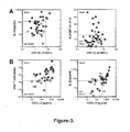
- peritoneal monocytes/macrophages expressed lower levels of CD86 in Gram-negative infections, compared to the rest of the patients.
- Inflammatory markers significantly increased in Gram-negative infections included IL-1 ⁇ , IL-10 and TNF- ⁇ ( Figure 3A ; Table 2).
- AUROC calculations identified the combination of V ⁇ 2 + T cell frequencies and peritoneal levels of IL-10 as excellent discriminator to predict Gram-negative infections (Table 3). This combination also had the highest overall correctness, with 100% sensitivity and 93% specificity for the correct prediction of Gram-negative peritonitis (Table 4).
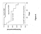
- V ⁇ 2 + T cell frequency has any indirect predictive power as to clinical outcome from episodes of acute peritonitis.
- FIG 4 there was a clear difference between patients with a relatively low and a relatively high proportion of peritoneal V ⁇ 2 + T cells on the day of presentation. While patients with V ⁇ 2 + T cell levels below the cut-off value of 4.3% had very low rates of technique failure over the following 3 months, patients with V ⁇ 2 + T cell levels higher than 4.3% had a significantly elevated risk of technique failure, confirming V ⁇ 2 + T cells as having both diagnostic and prognostic value in PD patients with acute peritonitis.
- CD4 + T cell counts as well as V ⁇ 2 + T cell frequencies and CXCL10 levels had prognostic value as confirmed by univariate analyses. Multivariate analysis of these parameters alone and in combination identified the combination of CD4 + T cell counts and CXCL10 levels as independent biomarker for the prediction of Gram-positive peritonitis in all patients presenting with a cloudy bag on day 1 (Table 9).
- T cells (% of total) 17.5 ⁇ 6.8 1.0 ⁇ 0.3 *** CD4 + (% of T cells) 52.2 ⁇ 3.3 50.6 ⁇ 2.1 n.s. CD8 + (% of T cells) 40.0 ⁇ 3.8 37.6 ⁇ 2.1 n.s. V ⁇ 2 + (% of T cells) 1.3 ⁇ 0.3 2.9 ⁇ 0.4 * IL-1 ⁇ (pg/ml) 3.7 ⁇ 1.6 45.1 ⁇ 12.6 *** IL-2 (pg/ml) 6.2 ⁇ 2.2 11.4 ⁇ 2.1 n . s .
- IL-6 (pg/ml) 37.1 ⁇ 7.3 3,249 ⁇ 623.6 *** sIL-6R (pg/ml) 360.1 ⁇ 50.8 639.6 ⁇ 50.3 ** IL-10 (pg/ml) 10.9 ⁇ 3.8 85.6 ⁇ 19.4 *** IL-12p70 (pg/ml) 4.2 ⁇ 1.5 4.5 ⁇ 1.0 n.s . IL-17 (pg/ml) 1.5 ⁇ 1.0 6.7 ⁇ 2.7 n.s .
- IL-22 (pg/ml) 13.9 ⁇ 7.7 121.8 ⁇ 36.5 ** CXCL8 (pg/ml) 18.1 ⁇ 3.2 699.3 ⁇ 191.7 *** CXCL10 (pg/ml) 43.2 ⁇ 22.5 514.1 ⁇ 82.7 *** IFN- ⁇ (pg/ml) 52.7 ⁇ 16.6 123.0 ⁇ 39.7 n.s . TNF- ⁇ (pg/ml) 21.8 ⁇ 8.4 86.8 ⁇ 18.8 * GM-CSF (pg/ml) 11.3 ⁇ 4.4 15.5 ⁇ 3.8 n . s .
- Monocytes (% of total) 15.1 ⁇ 2.3 5.8 ⁇ 1.0 * T cells (% of total) 1.3 ⁇ 0.3 0.2 ⁇ 0.03 * CD4 + (% of T cells) 52.7 ⁇ 2.3 43.7 ⁇ 4.6 n . s . CD8 + (% of T cells) 37.8 ⁇ 2.4 36.6 ⁇ 5.0 n.s.
- IL-10 (pg/ml) 49.4 ⁇ 10.6 208.6 ⁇ 65.5 ** IL-12p70 (pg/ml) 4.6 ⁇ 1.3 4.1 ⁇ 1.4 n.s . IL-17 (pg/ml) 8.4 ⁇ 3.7 2.4 ⁇ 1.3 n.s . IL-22 (pg/ml) 139.8 ⁇ 45.9 56.8 ⁇ 22.9 n.s.
- CXCL8 (pg/ml) 742.6 ⁇ 237.5 552.3 ⁇ 257.8 n.s .
- CXCL10 (pg/ml) 570.1 ⁇ 97.7 306.9 ⁇ 130.2 n.s.
- IFN- ⁇ 144.5 ⁇ 50.9 50.0 ⁇ 11.6 n.s.
- TNF- ⁇ (pg/ml) 65.3 ⁇ 17.9 159.7 ⁇ 51.8 * GM-CSF (pg/ml) 16.6 ⁇ 4.8 12.1 ⁇ 3.6 n.s .
- MMP-3 (pg/ml) 2,804 ⁇ 359.2 3,765 ⁇ 1,429 n.s. Table 3 .
- CD4 + T cells ( ⁇ 10 6 ) 6.8 ⁇ 1.9 60.2 ⁇ 32.2 ** CD8 + T cells ( ⁇ 10 6 ) 4.9 ⁇ 1.5 46.1 ⁇ 24.5 * V ⁇ 2 + T cells ( ⁇ 10 6 ) 0.6 ⁇ 0.2 1.6 ⁇ 0.7 n . s .
- Neutrophils (% of total) 77.2 ⁇ 3.8 83.7 ⁇ 2.1 n.s.
- Monocytes (% of total) 15.4 ⁇ 3.3 10.3 ⁇ 1.5 n . s .
- T cells (% of total) 0.9 ⁇ 0.4 1.1 ⁇ 0.3 n.s.
- CD4 + (% of T cells) 49.3 ⁇ 3.2 51.9 ⁇ 2.9 n.s.
Landscapes
- Life Sciences & Earth Sciences (AREA)
- Health & Medical Sciences (AREA)
- Engineering & Computer Science (AREA)
- Chemical & Material Sciences (AREA)
- Immunology (AREA)
- Molecular Biology (AREA)
- Cell Biology (AREA)
- Hematology (AREA)
- Biomedical Technology (AREA)
- Urology & Nephrology (AREA)
- General Health & Medical Sciences (AREA)
- Proteomics, Peptides & Aminoacids (AREA)
- Analytical Chemistry (AREA)
- Biotechnology (AREA)
- Microbiology (AREA)
- Biochemistry (AREA)
- Physics & Mathematics (AREA)
- Organic Chemistry (AREA)
- Medicinal Chemistry (AREA)
- Zoology (AREA)
- Food Science & Technology (AREA)
- General Physics & Mathematics (AREA)
- Pathology (AREA)
- Wood Science & Technology (AREA)
- Tropical Medicine & Parasitology (AREA)
- Virology (AREA)
- Biophysics (AREA)
- Toxicology (AREA)
- Bioinformatics & Cheminformatics (AREA)
- General Engineering & Computer Science (AREA)
- Genetics & Genomics (AREA)
- Animal Behavior & Ethology (AREA)
- Public Health (AREA)
- Communicable Diseases (AREA)
- Veterinary Medicine (AREA)
- Oncology (AREA)
- Pharmacology & Pharmacy (AREA)
- Nuclear Medicine, Radiotherapy & Molecular Imaging (AREA)
- General Chemical & Material Sciences (AREA)
- Chemical Kinetics & Catalysis (AREA)
Abstract
Description
- The present application relates to at least one method for identifying the nature of a bacterial infection from a peritoneal sample, and in particular whether the infection is caused by Gram-positive and/or Gram-negative bacteria. More specifically, said method involves identifying the relative amount of one or more cellular and/or humoral markers in said sample. Further, said invention concerns a method of treatment involving the use of said method, ideally to allow informed decision making in the selection of treatment regimens and/or of monitoring successful treatment outcome.
- Bacterial infection remains a leading cause of morbidity and mortality worldwide, not least due to the unprecedented spread of antibiotic-resistant pathogens. Moreover, effective infection control is hampered by the poor performance of standard diagnostics and hence inadequate choices are being made about the use of effective treatments. This alarming situation is posing an enormous challenge for clinical practice, public healthcare and biomedical research.
- Current diagnosis and treatment of suspected infections depends largely on the positive identification of the likely microbiological pathogen, a concept that was introduced by Robert Koch more than a century ago but has not changed since then. Positive identification of the causative pathogen of a bacterial infection is not only crucial to guide and refine patient management with respect to the choice of antibiotic treatment, but it also provides us with important clues as to the underlying inflammatory mechanisms and how these can be manipulated to help resolve infection.
- Presently, detection and characterisation of infectious organisms requires classical microbiological culture techniques, spectrometry based assays and/or highly complex nucleic acid identification methods. Mohan et al. 2011 describe the combined use of nucleic acid detection and immunoassay detection e.g. "oligonucleotide probes optimized for hybridization at 37°C to facilitate integration with the immunoassay" to diagnose urinary tract infections [5]. Podsiadly et al. 2005 describe detection of levels of specific Chlamydia pneumoniae IgM, IgG and !gA serum antibodies to diagnose urinary infections [6]. These latter assays are considered unreliable.
- Moreover, pathogen identification is significantly hampered by the inherent delay associated with a reliance on conventional culture techniques. It often takes 2 days, or even longer, before the results are available to the treating doctor, to enable specific and targeted therapy. In some diseases, such as tuberculosis, this can take up to two months. Most importantly, in many cases bacteria cannot be cultured by traditional methods, meaning that the cause underlying the patient's clinical symptoms remains unknown.
- Typically, infection is only apparent when significant clinical symptoms appear. Effective and specific therapeutic intervention depends on early detection and identification of the causative organism. Conventionally, patient management is determined on the basis of symptoms, initial clinical findings, and other basic laboratory markers of inflammation such as C-reactive protein. This approach is often sub-optimal being too general and lacking in specificity. Increased specificity is generally only attained by microbiological identification and antibiotic sensitivity testing, and this can be associated with delay in the optimal management of the infection. The tendency to use broad spectrum antibiotics to cover a number of possible aetiological agents encourages the development of other serious problems such as C. difficile infection, and also bacterial resistance. Thus, there is a clear clinical need to achieve earlier and better detection and characterisation of the infection in order to 1) improve patient outcomes; 2) improve the use of specific anti-infectives; and 3) reduce antimicrobial resistance and the development of other serious infections. Technologies capable of achieving this should enhance the patient experience, and ensure a more sustainable, cost-effective approach to patient management.
- Peritoneal dialysis (PD) is a treatment for patients with severe chronic kidney disease. Therapy involves the introduction of specialised dialysis fluids into the peritoneal cavity, via a Tenckhoff tube, with the aim of removing toxins that accumulate on kidney failure by diffusion and also water by the osmotic gradient created by the fluid. Both processes occur contemporaneously across the semipermeable peritoneal membrane. Infection of the glucose rich dialysis fluid, leading to peritonitis, is a major complication of PD that has significant morbidity and mortality implications.
- Peritonitis can have a detrimental effect on both short and long term patient health. In the short term, severe peritonitis episodes can result in the need to immediately remove the Tenckhoff tube and, therefore, treatment failure. In the long term, recurrent or severe peritonitis can result in thickening (fibrosis) of the peritoneal membrane and significant alterations in membrane permeability / function that lead to treatment failure. Early recognition of the onset of peritonitis coupled with rapid investigation, diagnosis, and effective treatment is imperative for successful outcome. Identification of the causative micro-organism using standard techniques can be difficult due to the relatively low bacterial numbers and the length of time required to culture them (often many days). This leads to the phenomenon of 'culture negative' peritonitis, i.e. all clinical and laboratory parameters confirm the diagnosis but no organism is ever identified, in 0-50 % of cases [3]. Consequently, given the delays in micro-organism confirmation, all patients are started on dual antibiotic treatment to cover both Gram-positive and Gram-negative bacteria. Only when (and if) the organism is identified is the antibiotic regimen refined. If the PD fluid specimen remains culture negative, then the patient is required to undergo prolonged dual, broad spectrum, antibiotic treatment.
- As mentioned above, whilst molecular tests that identify microorganisms in biological fluids are available, they often suffer from a lack of specificity, unacceptably high rates of false positivity and the frequent identification of non-pathogenic species that are due to contamination of the sample or asymptomatic carriage.
- It follows from the above that point-of-care methods that direct therapy, especially in cases where organism species or virulence is a determinant of outcome, are urgently required.
- The present disclosure relates to methods of rapid diagnosis of infection in peritoneal disease. In the present study we performed a detailed immunological and microbiological analysis in PD patients on the first day of presentation with acute peritonitis. Key to this technology is the discovery that the induction of complex cellular and humoral immune responses rapidly follows exposure of the immune system to an infectious agent. Surprisingly, this response is apparent within hours of the exposure and can be measured in the draining effluent of peritoneal dialysis patients with acute clinical symptoms, at the time of presenting with a characteristic but not diagnostic 'cloudy bag'. As such, these results have far-reaching implications for differential diagnosis of patients with suspected infections and may help guide patient management through faster biomarker-based diagnostics, better predictive risk modelling and improved targeting of a therapy and its ultimate efficacy.
- According to a first aspect of the invention there is provided a method for identifying a Gram-negative bacterial infection in a peritoneal sample, which method comprises:
- (a) examining said peritoneal sample from an individual in order to determine the amount of Vδ2+ T cells within the total T cell population of said sample, and;
- (b) where the amount of Vδ2+ T cells within the total T cell population is increased relative to the amount of Vδ2+ T cells within the total T cell population in a control sample from an individual not having a Gram-negative bacterial infection;
- (c) concluding that the individual from whom the peritoneal sample has been taken has a Gram-negative bacterial infection.
- As will be appreciated by those skilled in the art, reference herein to Gram-negative bacterial infection concerns infection with a bacterium that does not retain crystal violet dye in the Gram staining protocol. Examples of Gram-negative bacteria include, but are not limited to, the genera Acinetobacter, Aeromonas, Bacteroïdes, Bartonella, Bordetella, Brucella, Burkholderia, Campylobacter, Chlamydia, Chryseobacterium, Citrobacter, Enterobacter, Escherichia, Francisella, Fusobacterium, Haemophilus, Hafnia, Helicobacter, Klebsiella, Legionella, Moraxella, Morganella, Neisseria, Pantoea, Photobacterium, Proteus, Providencia, Pseudomonas, Salmonella, Serratia, Shigella, Stenotrophomonas, Veillonella, Vibrio, Yersinia.
- Reference herein to peritoneal sample refers to a sample of fluid from the abdominal cavity, including peritoneal effluent, typically from a PD patient, or tissue removed from the membrane lining the abdominal cavity (peritoneum).
- Reference herein to a control sample from an individual not having a Gram-negative bacterial infection is reference to a sample that has been shown not to have a Gram-negative bacterial infection using any one or more conventional techniques for identifying same.
- In a preferred embodiment of the invention examining said peritoneal sample from an individual in order to determine the amount of Vδ2+ T cells within the total T cell population of said sample may be undertaken directly or indirectly. A direct examination of said peritoneal sample from an individual in order to determine the amount of Vδ2+ T cells within the total T cell population of said sample is undertaken by measuring the amount of Vδ2+ T cells within the total T cell population of said sample. An indirect examination of said peritoneal sample from an individual in order to determine the amount of Vδ2+ T cells within the total T cell population of said sample is undertaken by measuring the amount of other T cells within the total T cell population of said sample, measuring the total T cell population of said sample and then performing a subtraction to determine the amount of Vδ2+ T cells within the total T cell population of said sample. Alternatively, indirect examination of said peritoneal sample from an individual in order to determine the amount of Vδ2+ T cells within the total T cell population of said sample is undertaken by assaying for surrogate markers, i.e. markers that are representative of Vδ2+ T cells, e.g. by measuring Vγ9+ T cells, pan-γδ+ T cells, pan-αβ- (alpha/beta negative T cells) or CD4-CD8- ("double-negative" or DN) T cells, where typically all or at least the majority of cells will be Vδ2+ T cells. Those skilled in the art will appreciate the nature of the cells to look for in order to perform this indirect measurement.
- Preferably, said peritoneal sample is fluid from the peritoneal space, which as appreciated by those skilled in the art, may be removed by numerous means such as, but not limited to, a needle and syringe. Alternatively, and more preferably, said sample may be obtained from peritoneal dialysate of those undergoing peritoneal dialysis treatment i.e., ideally, peritoneal effluent.
- The control sample ideally refers to a sample taken from the same site as the test but from an individual not infected (so-called "stable patients").
- An individual not having a Gram-negative bacterial infection may or may not have symptoms of infection such as elevated temperature, abdominal pain or discomfort or cloudy effluent or, in the case of a patient undergoing PD, the presence of a non-cloudy dialysate when viewed by the eye. Additionally, or alternatively, said individual not having an infection is identified by being negative for a bacterial infection, particularly a Gram-negative infection, when exposed to a culture test to try and grow and so identify a bacterial infection or when exposed to other conventional techniques to identify a bacterial infection.
- As known by those skilled in the art, Vδ2+ T cells are a distinct subset of CD3+ T cells that are characterized by positive expression of the TCR-Vδ2 chain and typically by co-expression of the Vγ9 chain (Vγ2 according to an alternative nomenclature). These Vγ9/Vδ2 T cells are commonly found in peripheral blood and inflamed tissues and play an important role in immune processes during microbial infections.
- In yet a further preferred embodiment of the invention at least one additional study is undertaken to determine the amount of anyone or more of the following in the peritoneal sample relative to said control:
- d) IL-10;
- e) IL-1β;
- f) TNF-α;
- g) Vδ2+ T cells expressing HLA-DR; and/or
- h) monocytes within the total leukocyte population expressing CD86; and where the amount of any one or more of d)-g) is increased relative to the amount of same in said control or in the case of h) is decreased relative to the amount of same in said control,
concluding that the individual from whom the peritoneal sample has been taken has a Gram-negative bacterial infection. - In a preferred embodiment of the invention said method of the invention may be performed using steps a-c) and any one or more of steps b)-f) including any preferred combination thereof.
- Interleukin 10 (IL-10 or IL10), also known as human cytokine synthesis inhibitory factor (CSIF), is an anti-inflammatory cytokine. In humans, IL-10 is known to have pleiotropic effects in immunoregulation and inflammation.
- IL-1β is a member of the
interleukin 1 cytokine family produced by activated immune cells, and is known to be an important mediator of the inflammatory response. - TNF-α is a member of the TNF cytokine family and produced by many different cell types, and is known to be an important mediator of the inflammatory response.
- HLA-DR is a MHC class II cell surface receptor encoded by the human leukocyte antigen complex, and is known to be up-regulated in response to signaling. Specifically, in response to an infection, bacterial antigens bind a DR molecule and are presented to T-cell receptors found on T-helper cells.
- Cluster of Differentiation 86 (also known as CD86 and B7-2) is a protein expressed on antigen-presenting cells that provides co-stimulatory signals necessary for T cell activation and survival.
- According to a second aspect of the invention there is provided a method for identifying a Gram-positive bacterial infection in a peritoneal sample, which method comprises:
- (a) examining said peritoneal sample from an individual in order to determine the amount of CXCL10 and;
- (b) where the amount of CXCL10 is increased relative to the amount of CXCL10 in a control sample from an individual not having a Gram-positive bacterial infection;
- (c) concluding that the individual from whom the peritoneal sample has been taken has a Gram-positive bacterial infection.
- CXCL10 is a cytokine belonging to the CXC chemokine family, and is secreted by several cell types in response to IFN-γ. These cell types include monocytes, T cells, endothelial cells and fibroblasts. CXCL10 is known to be increased in response to many infections.
- As will be appreciated by those skilled in the art, reference herein to Gram-positive bacterial infection concerns an infection with a bacterium that does retain crystal violet dye in the Gram staining protocol. Examples of Gram-positive bacteria include, but are not limited to, the genera Actinomyces, Bacillus, Clostridium, Corynebacterium, Enterococcus, Lactobacillus, Lactococcus, Leuconostoc, Listeria, Micrococcus, Mycobacterium, Peptostreptococcus, Propionibacterium, Staphylococcus, Streptococcus, Streptomyces.
- Reference herein to peritoneal sample refers to a sample of fluid from the abdominal cavity, including peritoneal effluent, typically from a PD patient, or tissue removed from the membrane lining the abdominal cavity (peritoneum).
- Reference herein to a control sample from an individual not having a Gram-positive bacterial infection is reference to a sample that has been shown not to have a Gram-positive bacterial infection using any one or more conventional techniques for identifying same.
- Preferably, said peritoneal sample is fluid from the peritoneal space, which as appreciated by those skilled in the art, may be removed by numerous means such as, but not limited to, a needle and syringe. Alternatively, and more preferably, said sample may be obtained from peritoneal dialysate of those undergoing peritoneal dialysis treatment i.e., ideally, peritoneal effluent.
- The control sample ideally refers to a sample taken from the same site as the test but from an individual not infected (so-called "stable patients").
- An individual not having a Gram-positive bacterial infection may or may not have symptoms of infection such as elevated temperature, abdominal pain or discomfort or cloudy effluent or, in the case of a patient undergoing PD, the presence of a non-cloudy dialysate when viewed by the eye. Additionally, or alternatively, said individual not having an infection is identified by being negative for a bacterial infection, particularly a Gram-positive infection, when exposed to a culture test to try and grow and so identify a bacterial infection or when exposed to other conventional techniques to identify a bacterial infection.
- In yet a further preferred embodiment of the second aspect of the invention at least one additional study is undertaken to determine the amount of any one or more of the following in the peritoneal sample relative to said control:
- (d) Vδ2+ T cells within the total T cell population;
- (e) total numbers of CD4+ T cells; or
- (f) IL-22;
- CD4+ T helper cells area sub-division of white blood cells that are an essential part of the human immune system, characterized by cell surface expression of the CD4 glycoprotein. CD4 is a co-receptor that assists the T cell receptor (TCR) in communicating with an antigen-presenting cell.
- IL-22 is a cytokine that is a member of a group of cytokines called the IL-10 family or IL-10 superfamily, which are known to be mediators of cellular inflammatory responses. IL-22 is produced by activated dendritic cells and T cells and initiates innate immune responses against bacterial pathogens In a preferred embodiment of the invention said method of the second aspect of the invention may be performed using step a) and any one or more of steps d)-f) including any preferred combination thereof.
- According to a third aspect of the invention, or a preferred embodiment of the first and/or second aspect of the invention, said method for identifying a Gram-negative bacterial infection is carried out after, or alternatively prior to, said method for identifying a Gram-positive bacterial infection, or vice versa. Alternatively, said methods may be carried out simultaneously.
- According to a fourth aspect of the invention there is provided a method for treating an individual suffering from a bacterial infection as identified by any one of more of the above methods, which further comprises administering to said individual a therapeutic for treating a Gram-negative and/or Gram-positive bacterial infection depending upon the nature of the bacterial infection identified by said above methods.
- According to a preferred embodiment of the fourth aspect of the invention, said method for treating an individual suffering from a bacterial infection further comprises periodically repeating any one of more of the above methods for identifying a Gram-negative and/or Gram-positive infection and, where it is concluded that said individual is still infected, continuing to administer said therapeutic or changing the therapeutic regimen.
- According to a preferred embodiment of the fourth aspect of the invention, said therapeutic includes, but is not limited to, an antibiotic such as those belonging to the families of penicillins and beta-lactamase inhibitors, cephalosporins, macrolides and lincosamines, quinolones and fluoroquinolones, carbapenems, monobactams, aminoglycosides, tetracyclines, sulfonamides, rifampin, oxazolidonones, and derivatives thereof.
- As will be appreciated by those skilled in the art, said antibiotic may be broad spectrum and efficacious against both Gram-positive and/or Gram-negative bacteria, such as but not limited to, all beta-lactams including betalactam/inhibitor combinations, all macrolides, all tetracyclines, all aminoglycosides, all polymixins, all quinolones, all glycopeptides, all lincosamides, all streptomycin derivatives, and all antibiotics not covered by generic classes such as chloramphenicol, rifampicin, fosfomycin, fosmidomycin, nitrofurantoin, metronidazole, daptomycin, quinupristin, linezolid, telavancin, mupirocin, and folate synthesis inhibitors, as well as structural analogues and derivates thereof.
- More ideally, said antibiotic is specific and efficacious against Gram-negative bacteria, such as but not limited to, all beta-lactams including betalactam/inhibitor combinations, all macrolides, all tetracyclines, all aminoglycosides, all polymixins, all quinolones, all lincosamides, all streptomycin derivatives, and all antibiotics not covered by generic classes such as chloramphenicol, rifampicin, fosfomycin, fosmidomycin, nitrofurantoin, metronidazole, and folate synthesis inhibitors, as well as structural analogues and derivates thereof.
- Alternatively, said antibiotic is specific and efficacious against Gram-positive bacteria, such as but not limited to, all beta-lactams including betalactam/inhibitor combinations, all macrolides, all tetracyclines, all aminoglycosides, all quinolones, all glycopeptides, all lincosamides, all streptomycin derivatives, and all antibiotics not covered by generic classes such as chloramphenicol, rifampicin, fosfomycin, fosmidomycin daptomycin, quinupristin, linezolid, telavancin, mupirocin, and folate synthesis inhibitors, as well as structural analogues and derivates thereof.
- Throughout the description and claims of this specification, the words "comprise" and "contain" and variations of the words, for example "comprising" and "comprises", mean "including but not limited to" and do not exclude other moieties, additives, components, integers or steps. Throughout the description and claims of this specification, the singular encompasses the plural unless the context otherwise requires. In particular, where the indefinite article is used, the specification is to be understood as contemplating plurality as well as singularity, unless the context requires otherwise.
- All references, including any patent or patent application, cited in this specification are hereby incorporated by reference. No admission is made that any reference constitutes prior art. Further, no admission is made that any of the prior art constitutes part of the common general knowledge in the art.
- Preferred features of each aspect of the invention may be as described in connection with any of the other aspects.
- Other features of the present invention will become apparent from the following examples. Generally speaking, the invention extends to any novel one, or any novel combination, of the features disclosed in this specification (including the accompanying claims and drawings). Thus, features, integers, characteristics, compounds or chemical moieties described in conjunction with a particular aspect, embodiment or example of the invention are to be understood to be applicable to any other aspect, embodiment or example described herein, unless incompatible therewith.
- Moreover, unless stated otherwise, any feature disclosed herein may be replaced by an alternative feature serving the same or a similar purpose.
- The Invention will now be described in greater detail with reference to the Examples below and to the drawings in which:
-
Figure 1 . Relates to cellular indicators.(A) Total number and relative frequency (%) of peritoneal neutrophils (CD15+), monocytes/macrophages (CD14+) and T cells (CD3+) in stable and infected PD patients onday 1 of acute peritonitis. (B) Total number and proportion of helper T cells (CD4+), cytotoxic T cells (CD8+) and γδ T cells (Vδ2+) within the peritoneal CD3+ T cell population in stable and infected PD patients onday 1 of acute peritonitis. Infected patients were grouped according to the microbiological culture results into patients with culture-negative (n/c) or confirmed Gram-positive or Gram-negative infection. Data points represent individual patients, horizontal lines mean values per group. -
Figure 2 . Relates to humoral indicators. Peritoneal levels (in ng/ml) of IL-1β, IL-2, IL-6,sIL 6R, IL-10, IL-12p70, IL-17A, IL-22, CXCL8, CXCL10, IFN-γ, TNF-α, GM-CSF, TGF-β1 and MMP-3 in stable and infected PD patients onday 1 of acute peritonitis. Infected patients were grouped according to the microbiological culture results into patients with culture-negative (n/c) or confirmed Gram-positive or Gram-negative infection. Data points represent individual patients, horizontal lines mean values per group. -
Figure 3 . Relates to humoral and cellular indicators. Examples of immunological biomarkers in patients presenting with a 'cloudy bag' onday 1, depending on the microbiological culture results. (A) Discrimination between Gram-negative and non-Gram-negative infections. (B) Discrimination between Gram-positive and non-Gram-positive infections. Data points represent individual episodes (white, culture-negative; blue, confirmed Gram-positive infection; red, confirmed Gram-negative infection. Dashed lines indicate calculated cut-off values for positive or negative discrimination. -
Figure 4 . Kaplan-Meier plot showing cumulative technique survival of patients with acute peritonitis, depending on the frequency of Vδ2+ T cells among all peritoneal T cells onday 1. The cut-off value was determined by ROC analysis. AUROC for 30th technique failure prediction: 0.917 ± 0.048, 95% confidence interval: 0.823-1.000. Youden Index: 0.85, sensitivity: 100%, specificity: 85%. The statistical difference between the two curves was analysed by log rank test. -
Figure 5 . Flow chart of proposed PD peritonitis treatment as guided by immune fingerprint-based diagnostic tests, depending on the test result. - Table 1. Immunological biomarkers in stable patients and patients presenting with a 'cloudy bag' on day 1 (mean ± SEM). n.s., not significant.
- Table 2. Immunological biomarkers in patients presenting with a 'cloudy bag' on
day 1, depending on the absence or presence of a confirmed Gram-negative infection (mean ± SEM). n.s., not significant. - Table 3. Ability of immunological biomarkers to discriminate between Gram-negative and non-Gram-negative episodes of peritonitis on
day 1. - Table 4. Prediction of Gram-negative infection on the first day of presentation with acute peritonitis.
- Table 5. Immunological biomarkers showing diagnostic significance for prediction of Gram-negative episodes of peritonitis on
day 1. - Table 6. Immunological biomarkers in patients presenting with a 'cloudy bag' on
day 1, depending on the absence or presence of a confirmed Gram-positive infection (mean ± SEM). n.s., not significant. - Table 7. Ability of immunological biomarkers to discriminate between Gram-positive and non-Gram-positive episodes of peritonitis on
day 1. - Table 8. Prediction of Gram-positive infection on the first day of presentation with acute peritonitis.
- Table 9. Immunological biomarkers showing diagnostic-significance for prediction of Gram-positive episodes of peritonitis on
day 1. - Table 10. Biomarkers of potential diagnostic value in patients presenting with a 'cloudy bag' on
day 1, depending on the microbiological culture results. - We recruited 52 adult patients who were receiving PD at the University Hospital of Wales, Cardiff, UK, and were admitted on
day 1 of acute peritonitis between 1 September 2008 and 31 January 2012. 15 stable patients with no infection in the previous 3 months were included in this study as non-infected controls. Sampling of PD effluent was approved by the South East Wales Local Ethics Committee (04WSE04/27), and conducted according to the principles expressed in the Declaration of Helsinki. All patients provided written informed consent. Diagnosis of acute peritonitis was based on the presence of abdominal pain and cloudy peritoneal effluent with >100 WBC/mm3. Infections were grouped into culture-negative, Gram-positive and Gram-negative episodes, according to the result of the microbiological analysis of the effluent, which was conducted at the central diagnostic laboratories of the University Hospital of Wales. - Peritoneal cells were acquired on an eight-color FACSCanto II (BD Biosciences) and analyzed with FloJo (Tree Star), using monoclonal antibodies against CD3 (UCHT1), CD4 (SK3), CD8 (RPA-T8), CD15 (HI98), CD69 (FN50), CD86 (2331-FUN1), HLA-DR (L234) and TCR-Vδ2 (B6.1) from BD Biosciences; CD14 (61D3) from eBioscience; and TCR-Vγ9 (Immu360) from Beckman Coulter, together with appropriate isotype controls. Leukocyte populations were identified based on their appearance in side scatter and forward scatter area/height, exclusion of live/dead staining (fixable Aqua; Invitrogen), and surface staining: CD15+ neutrophils, CD14+ monocytes/macrophages and CD3+ T cells. T cell subsets were identified as CD4+ helper T cells, CD8+ cytotoxic T cells and Vδ2+ γδ T cells.
- Cell-free peritoneal effluents were analyzed for TNF-α, GM-CSF, IFN-γ, IL-1β, IL-2, IL-6, IL-10, IL-12p70, CXCL8 and sIL-6R on a SECTOR Imager 6000 (Meso Scale Discovery). IL-17A, IL-22, CXCL10 and MMP-3 (R&D Systems) as well as TGF-β1 (eBioscience) were measured in duplicate on a Dynex MRX II reader, using conventional ELISA kits.
- Statistical analyses were performed using SPSS 16.0 and GraphPad Prism 4.0 software. All variables were tested for normal distributions using the Kolmogorov-Smirnov test. Differences between patient groups were analyzed using Student's t-tests for normally distributed data or Mann-Whitney U-tests for non-parametric data. Categorical data were tested using the Chi-square test. Predictive biomarkers were assessed using univariate analysis; statistically significant (p<0.05) variables from the univariate analysis were included in a multivariate analysis. Multiple logistic regression analyses were conducted based on forward and/or backward elimination of data, as indicated in the tables. Discrimination was assessed using AUROC curves and compared using non-parametric approaches; AUROC analyses were also used to calculate cut-off values, sensitivity and specificity. Cumulative survival curves as a function of time were generated using the Kaplan-Meier approach and compared using the log rank test. All statistical tests were two-tailed; differences were considered statistically significant as indicated in the figures and tables: *, p<0.05; **, p<0.01; ***, p<0.001.
- Patients presenting with acute peritonitis were characterized by a significant peritoneal influx of immune cells, predominantly CD15+ neutrophils, CD14+ monocytes/macrophages and CD3+ T cells (Table 1). This was true for culture-negative episodes of peritonitis as well as cases of confirmed infection by Gram-negative or Gram-positive bacteria (
Figure 1 ). While peritoneal leukocytes in stable patients comprised mainly of monocytes/macrophages and T cells, acute peritonitis was dominated by a massive recruitment of neutrophils, at times reaching >95% of all peritoneal cells and >1011 cells in total. Detailed flow cytometrical analyses of the peritoneal leukocyte population revealed further striking changes in the composition of the local immune cell infiltrate in patients with acute peritonitis. As such, there was a preferential increase in the frequency of Vδ2+ T cells within the T cell population in acute peritonitis, while the percentages of CD4+ and CD8+ T cells remained virtually unchanged (Table 1). - Soluble mediators significantly increased in acute peritonitis included interleukin-1β (IL-1β), IL-6, soluble IL-6 receptor (sIL-6R), IL-10, IL-22, CXCL8, CXCL10, tumour necrosis factor-α (TNF-α), transforming growth factor-β (TGF-β) and matrix metalloproteinase-3 (MMP-3); at the same time levels of cytokines such as IL-2, IL-12p70, IL-17 and granulocyte/macrophage colony-stimulating factor (GM-CSF) were not elevated above baseline or, in the case of interferon-γ (IFN-γ), not significantly (
Figure 2 , Table 1). Taken together, these measurements identify a broad range of humoral and cellular biomarkers that indicate acute inflammatory responses in PD patients, some of which might be of diagnostic value. - We next tested whether pathogen-specific immune fingerprints exist that could predict infections by certain groups of bacteria. Given the importance of Gram-negative bacteria in the clinic and their association with worse outcomes we initially concentrated on the prediction of Gram-negative infections. Amongst all patients presenting with a cloudy bag on
day 1, patients with confirmed Gram-negative infections displayed larger numbers of infiltrating neutrophils and consequently lower proportions of monocytes/macrophages and T cells than the rest of the patients, i.e. individuals with culture-negative or Gram-positive infections (Table 2). Within the T cell population, Vδ2+ T cells were significantly increased and expressed higher levels of the activation marker HLA-DR in Gram-negative infections. In turn, peritoneal monocytes/macrophages expressed lower levels of CD86 in Gram-negative infections, compared to the rest of the patients. Inflammatory markers significantly increased in Gram-negative infections included IL-1β, IL-10 and TNF-α (Figure 3A ; Table 2). AUROC calculations identified the combination of Vδ2+ T cell frequencies and peritoneal levels of IL-10 as excellent discriminator to predict Gram-negative infections (Table 3). This combination also had the highest overall correctness, with 100% sensitivity and 93% specificity for the correct prediction of Gram-negative peritonitis (Table 4). The prognostic value of the proportion of monocytes/macrophages amongst all cells (≤ 10.7%) and their expression of CD86 (≤ 67.9%), the frequency of Vδ2+ T cells with the T cell population (≥ 6.3%) and their expression of HLA-DR (≥ 16.1%), and peritoneal IL-10 levels (≥ 110.1 pg/ml) were all confirmed by univariate analyses. Multivariate analysis of these parameters identified the Vδ2+ T cell frequency as independent prognostic biomarker for the prediction of Gram-negative peritonitis in all patients presenting with a cloudy bag on day 1 (Table 5). As Gram-negative infections are associated with higher rates of technique failure and mortality, we finally tested whether the peritoneal Vδ2+ T cell frequency has any indirect predictive power as to clinical outcome from episodes of acute peritonitis. As shown inFigure 4 , there was a clear difference between patients with a relatively low and a relatively high proportion of peritoneal Vδ2+ T cells on the day of presentation. While patients with Vδ2+ T cell levels below the cut-off value of 4.3% had very low rates of technique failure over the following 3 months, patients with Vδ2+ T cell levels higher than 4.3% had a significantly elevated risk of technique failure, confirming Vδ2+ T cells as having both diagnostic and prognostic value in PD patients with acute peritonitis. - In an analogous way, we next attempted to identify predictors of Gram-positive infections. Amongst all patients presenting with a cloudy bag on
day 1, patients with confirmed Gram-positive infections displayed larger numbers of infiltrating CD4+ and CD8+ T cells than the rest of the patients, i.e. individuals with culture-negative or Gram-negative infections (Table 6), while the proportion of Vδ2+ T cells within the T cell population was significantly lower in Gram-positive infections. Inflammatory markers significantly increased in Gram-positive infections included IL-22 and CXCL10 (Figure 3B ; Table 6). AUROC calculations identified the combination of CXCL10 (≥301.2 pg/ml) and IL-22 levels (≥ 54.3 pg/ml) and Vδ2+ T cell frequencies (≤ 2.7%) as excellent discriminator to predict Gram-positive infections (Table 7). This combination also had the highest overall correctness, with 89% sensitivity and 67% specificity for the correct prediction of Gram-positive peritonitis. In turn, CXCL10 levels (≥ 301.2 pg/ml) combined with total CD4+ T cell counts (≥ 15.2•106) had a sensitivity of only 53% yet at a specificity of 95% for the prediction of Gram-positive peritonitis (Table 8). CD4+ T cell counts as well as Vδ2+ T cell frequencies and CXCL10 levels had prognostic value as confirmed by univariate analyses. Multivariate analysis of these parameters alone and in combination identified the combination of CD4+ T cell counts and CXCL10 levels as independent biomarker for the prediction of Gram-positive peritonitis in all patients presenting with a cloudy bag on day 1 (Table 9). - Taken together, our findings indicate that immune fingerprint-based diagnostic tests are able to guide treatment of patients with acute infections (
Figure 5 ). Depending on the test result, patients might be diagnosed as having a culture-negative episode (i.e. no bacterial infection present) of peritonitis that is associated with relatively benign outcome. Such patients might benefit from shorter course treatments as outpatients (and might not need any antibiotics at all if the episode is of non-infectious origin). Patients diagnosed with Gram-positive or Gram-negative infections might benefit from better targeted therapy, and especially in the case of Gram-negative infections that are associated with high rates of technique failure and mortality, from close monitoring and improved management. In the case of unknown bacterial infection, patients might receive the conventional treatment with broad-spectrum antibiotics. - Our research highlights the importance of combining humoral and cellular parameters to establish accurate 'immune fingerprints'. Particularly promising humoral and cellular parameters in the definition of disease-specific immune fingerprints include local levels of IL-1β, IL-10, IL-22, TNF-α and CXCL10, as well as the relative proportion of neutrophils and monocytes/macrophages among total peritoneal cells and the frequency of γδ T cells within the peritoneal T cell population (Table 10).
- Consequently, it has therefore been shown that immune fingerprints of bacterial infections exist, and that characteristic immunological biomarkers have the potential to distinguish and predict early bacterial infection and so direct appropriate modes of treatment.
- 3. Fahim M, Hawley CM, McDonald SP, Brown FG, Rosman JB, Wiggins KJ, Bannister KM, Johnson DW: Culture-negative peritonitis in peritoneal dialysis patients in Australia: predictors, treatment, and outcomes in 435 cases. Am J Kidney Dis 55: 690-697, 2010
5. Mohan R, Mach KE, Bercovici M, Pan Y, Dhulipala L, Wong PK, Liao JC: Clinical validation of integrated nucleic acid and protein detection on an electrochemical biosensor array for urinary tract infection diagnosis.PLoS One 6(10): e26846, 2011
6. Podsiady E, Fracka B, Szmigielska A, Tylewska-Wierzbanowska S: Seroepidemiological studies of Chlamydia pneumoniae infections in 1-36 months old children with respiratory tract infections and other diseases in Poland.Pol J Microbiol. 54(3):215-9, 2005.
Table 1. Immunological biomarkers in stable patients and patients presenting with a 'cloudy bag' on day 1 (mean ± SEM). n.s., not significant. Stable PD Cloudy bag p Gender (male/female) ' 9/6 32/20 n.s. Age (years) 60.4 ± 4.9 65.5 ± 2.0 n.s. Days on PD (days) 1,058 ± 219.2 1,142 ± 131.1 n.s. Neutrophils (·106) 0.6 ± 0.3 7,048 ± 1,384 *** Monocytes (·106) 2.8 ± 1.5 753.9 ± 166.0 *** CD4+ T cells (·106) 0.3 ± 0.2 32.3 ± 15.7 *** CD8+ T cells (·106) 0.3 ± 0.2 24.5 ± 12.0 *** Vδ2+ T cells (·106) 0.01 ± 0.003 1.1 ± 0.4 *** Neutrophils (% of total) 9.0 ± 4.2 80.3 ± 2.2 *** Monocytes (% of total) 28.9 ± 9.4 12.9 ± 1.9 n.s. T cells (% of total) 17.5 ± 6.8 1.0 ± 0.3 *** CD4+ (% of T cells) 52.2 ± 3.3 50.6 ± 2.1 n.s. CD8+ (% of T cells) 40.0 ± 3.8 37.6 ± 2.1 n.s. Vδ2+ (% of T cells) 1.3 ± 0.3 2.9 ± 0.4 * IL-1β (pg/ml) 3.7 ± 1.6 45.1 ± 12.6 *** IL-2 (pg/ml) 6.2 ± 2.2 11.4 ± 2.1 n.s. IL-6 (pg/ml) 37.1 ± 7.3 3,249 ± 623.6 *** sIL-6R (pg/ml) 360.1 ± 50.8 639.6 ± 50.3 ** IL-10 (pg/ml) 10.9 ± 3.8 85.6 ± 19.4 *** IL-12p70 (pg/ml) 4.2 ± 1.5 4.5 ± 1.0 n.s. IL-17 (pg/ml) 1.5 ± 1.0 6.7 ± 2.7 n.s. IL-22 (pg/ml) 13.9 ± 7.7 121.8 ± 36.5 ** CXCL8 (pg/ml) 18.1 ± 3.2 699.3 ± 191.7 *** CXCL10 (pg/ml) 43.2 ± 22.5 514.1 ± 82.7 *** IFN-γ (pg/ml) 52.7 ± 16.6 123.0 ± 39.7 n.s. TNF-α (pg/ml) 21.8 ± 8.4 86.8 ± 18.8 * GM-CSF (pg/ml) 11.3 ± 4.4 15.5 ± 3.8 n.s. MMP-3 (pg/ml) 1,540 ± 184.9 3,029 ± 427.3 ** Table 2. Immunological biomarkers in patients presenting with a 'cloudy bag' on day 1, depending on the absence or presence of a confirmed Gram-negative infection (mean ± SEM). n.s., not significant.Other Gram-negative p Gender (male/female) 25/15 7/5 n.s. Age (years) 64.1 ± 2.4 70.1 ± 2.9 n.s. Days on PD (days) 1,234 ± 156.2 899.3 ± 248.3 n.s. Neutrophils (·106) 5,875 ± 1,580 10,779 ± 2,686 * Monocytes (·106) 754.7 ± 206.1 751.1 ± 242.9 n.s. CD4+ T cells (·106) 39.3 ± 20.5 9.9 ± 3.4 n.s. CD8+ T cells (·106) 29.5 ± 15.6 8.6 ± 2.8 n.s. Vδ2+ T cells (·106) 1.1 ± 0.4 1.1 ± 0.4 n.s. Neutrophils (% of total) 78.1 ± 2.7 87.7 ± 2.3 n.s. Monocytes (% of total) 15.1 ± 2.3 5.8 ± 1.0 * T cells (% of total) 1.3 ± 0.3 0.2 ± 0.03 * CD4+ (% of T cells) 52.7 ± 2.3 43.7 ± 4.6 n.s. CD8+ (% of T cells) 37.8 ± 2.4 36.6 ± 5.0 n.s. Vδ2+ (% of T cells) 2.0 ± 0.2 5.0 1.4 * HLA-DR+ (% of γδT cells) 10.8 ± 1.8 22.5 ± 4.7 * CD86+ (% of monocytes) 72.6 ± 3.6 53.1 ± 7.2 * IL-1β (pg/ml) 35.0 ± 12.5 79.5 ± 34.7 * IL-2 (pg/ml) 12.2 ± 2.6 8.8 ± 1.9 n.s. IL-6 (pg/ml) 2,561 ± 538.8 5,586 ± 1,942 n.s. sIL-6R (pg/ml) 606.3 ± 57.6 751.4 ± 100.9 n.s. IL-10 (pg/ml) 49.4 ± 10.6 208.6 ± 65.5 ** IL-12p70 (pg/ml) 4.6 ± 1.3 4.1 ± 1.4 n.s. IL-17 (pg/ml) 8.4 ± 3.7 2.4 ± 1.3 n.s. IL-22 (pg/ml) 139.8 ± 45.9 56.8 ± 22.9 n.s. CXCL8 (pg/ml) 742.6 ± 237.5 552.3 ± 257.8 n.s. CXCL10 (pg/ml) 570.1 ± 97.7 306.9 ± 130.2 n.s. IFN-γ (pg/ml) 144.5 ± 50.9 50.0 ± 11.6 n.s. TNF-α (pg/ml) 65.3 ± 17.9 159.7 ± 51.8 * GM-CSF (pg/ml) 16.6 ± 4.8 12.1 ± 3.6 n.s. MMP-3 (pg/ml) 2,804 ± 359.2 3,765 ± 1,429 n.s. Table 3. Ability of immunological biomarkers to discriminate between Gram-negative and non-Gram-negative episodes of peritonitis on day 1.AUROC ± SEM 95% CI p For Gram-negative prediction Monocytes (% of total) 0.745 ± 0.075 0.597 - 0.893 * Vδ2+ (% of T cells) 0.710 ± 0.106 0.501 - 0.918 * HLA-DR+ (% of γδT cells) 0.720 ± 0.097 0.530 - 0.911 * CD86+ (% of monocytes) 0.740 ± 0.083 0.578 - 0.903 * IL-10 (pg/ml) 0.803 ± 0.092 0.622 - 0.984 ** IL-1β (pg/ml) 0.738 ± 0.086 0.569 - 0.907 * TNF-α (pg/ml) 0.735 ± 0.091 0.557 - 0.914 * IL-10 + Vδ2+ 0.976 ± 0.022 0.934 - 1.000 *** IL-10 + Vδ2+ + HLA-DR+ 0.959 ± 0.031 0.899 - 1.000 *** IL-1β + IL-10 + TNF-α 0.844 ± 0.077 0.693 - 0.995 ** Table 4. Prediction of Gram-negative infection on the first day of presentation with acute peritonitis. Cut-off Point Youden Index Sensitivity (%) Specificity (%) For Gram-negative prediction Monocytes (% of total) 10.7 0.50 50 100 Vδ2+ (% of T cells) 6.3 0.46 46 100 HLA-DR+ (% of γδT cells) 16.1 0.48 64 84 CD86+ (% of monocytes) 67.9 0.47 74 73 IL-10 (pg/ml) 110.1 0.61 70 91 IL-1β (pg/ml) 14.0 0.55 90 65 TNF-α (pg/ml) 32.8 0.45 80 65 IL-10 + Vδ2+ 0.5 0.93 100 93 IL-10 + Vδ2+ + HLA-DR+ 0.5 0.78 100 78 IL-1β + IL-10 + TNF-α 2.5 0.61 70 91 Table 5. Immunological biomarkers showing diagnostic significance for prediction of Gram-negative episodes of peritonitis on day 1.β Coefficient Standard error Odds ratio (95%CI) p Univariate logistic regression Monocytes (% of total) -0.189 0.093 0.828 (0.690-0.994) * Vδ2+ (% of T cells) 0.385 0.159 1.469 (1.075-2.008) * HLA-DR+ (% of γδT cells) 0.072 0.029 1.075 (1.014-1.139) * CD86+ (% of monocytes) -0.037 0.016 0.964 (0.934-0.995) * IL-10 0.012 0.005 1.012 (1.003-1.022) * Multivariate logistic regression (backward) Vδ2+ (% of T cells) 0.571 0.246 1.770 (1.093 - 2.866) * Constant -4.591 1.376 0.010 - Table 6. Immunological biomarkers in patients presenting with a 'cloudy bag' on day 1, depending on the absence or presence of a confirmed Gram-positive infection (mean ± SEM). n.s., not significant.Other Gram-positive p Gender (male/female) 17/10 15/10 n.s. Age (years) 68.2 ± 2.4 62.4 ± 3.1 n.s. Days on PD (days) 1,058 ± 167.9 1,266 ± 213.9 n.s. Neutrophils (·106) 5,604 ± 1,610 8,623 ± 2,293 n.s. Monocytes (·106) 477.2 ± 128.0 1,056 ± 309.1 n.s. CD4+ T cells (·106) 6.8 ± 1.9 60.2 ± 32.2 ** CD8+ T cells (·106) 4.9 ± 1.5 46.1 ± 24.5 * Vδ2+ T cells (·106) 0.6 ± 0.2 1.6 ± 0.7 n.s. Neutrophils (% of total) 77.2 ± 3.8 83.7 ± 2.1 n.s. Monocytes (% of total) 15.4 ± 3.3 10.3 ± 1.5 n.s. T cells (% of total) 0.9 ± 0.4 1.1 ± 0.3 n.s. CD4+ (% of T cells) 49.3 ± 3.2 51.9 ± 2.9 n.s. CD8+ (% of T cells) 36.6 ± 3.0 38.6 ± 3.1 n.s. Vδ2+ (% of T cells) 3.6 0.7 1.8 0.3 * HLA-DR+ (% of γδT cells) 17.2 ± 3.0 10.3 ± 2.1 n.s. CD86+ (% of monocytes) 62.9 ± 5.3 73.0 ± 4.1 n.s. IL-1β (pg/ml) 37.3 ± 16.0 54.5 ± 20.1 n.s. IL-2 (pg/ml) 7.6 ± 1.4 16.0 ± 4.0 n.s. IL-6 (pg/ml) 3,204 ± 970.8 3,303 ± 753.0 n.s. sIL-6R (pg/ml) 607.2 ± 60.0 674.8 ± 83.2 n.s. IL-10 (pg/ml) 96.5 ± 33.1 72.5 ± 16.0 n.s. IL-12p70 (pg/ml) 3.1 ± 0.7 6.2 ± 2.0 n.s. IL-17 (pg/ml) 3.7 ± 1.9 12.1 ± 6.5 n.s. IL-22 (pg/ml) 55.8 ± 16.8 193.8 ± 71.8 ** CXCL8 (pg/ml) 504.3 ± 199.9 933.3 ± 346.0 n.s. CXCL10 (pg/ml) 316.6 ± 88.8 738.4 ± 131.4 ** IFN-γ (pg/ml) 55.5 ± 12.7 204.0 ± 83.6 n.s. TNF-α (pg/ml) 76.0 ± 25.8 99.7 ± 28.0 n.s. GM-CSF (pg/ml) 10.1 ± 2.2 22.0 ± 7.8 n.s. MMP-3 (pg/ml) 2,760 ± 658.1 3,336 ± 532.7 n.s. Table 7. Ability of immunological biomarkers to discriminate between Gram-positive and non-Gram-positive episodes of peritonitis on day 1.AUROC ± SEM 95% Cl p For Gram-positive prediction CD4+ T cells (·106) 0.741 ± 0.079 0.587 - 0.895 ** CD8+ T cells (·106) 0.723 ± 0.083 0.561 - 0.885 * Vδ2+ (% of T cells) 0.699 ± 0.079 0.543 - 0.854 * CXCL10 (pg/ml) 0.744 ± 0.075 0.596 - 0.891 ** IL-22 (pg/ml) 0.736 ± 0.074 0.591 - 0.881 ** CXCL10 + Vδ2+ 0.821 ± 0.066 0.691-0.951 *** CXCL10 + IL-22 + Vδ2+ 0.816 ± 0.069 0.681-0.951 ** CXCL10 + CD4+ 0.797 ± 0.073 0.653 - 0.941 ** CXCL10 + IL-22 0.794 ± 0.068 0.661 - 0.926 ** Table 8. Prediction of Gram-positive infection on the first day of presentation with acute peritonitis. Cut-off Point Youden Index Sensitivity (%) Specificity (%) For Gram-positive prediction CD4+ T cells (·106) 15.2 0.46 55 91 CD8+ T cells (·106) 7.8 0.46 60 86 CXCL10 (pg/ml) 301.2 0.49 77 72 IL-22 (pg/ml) 54.3 0.39 73 67 CXCL10 + IL-22 + Vδ2+ 1.5 0.561 89 67 CXCL10 + Vδ2+ 0.5 0.524 100 52 CXCL10 + CD4+ 1.5 0.48 53 95 CXCL10 + IL-22 0.5 0.49 91 55 For non-Gram-positive Prediction Vδ2+ (% of T cells) 2.7 0.375 57 81 Table 9. Immunological biomarker showing diagnostic significance for prediction of Gram-positive episodes of peritonitis on day 1.βCoefficient Standard error Odds ratio (95%CI) p Univariate logistic regression CD4+ T cells (·106) 0.062 0.026 1.064 (1.011 - 1.119) * CD8+ T cells (·106) 0.077 0.035 1.080 (1.008 - 1.156) * Vδ2+ (% of T cells) -0.388 0.185 0.679 (0.472 - 0.976) * CXCL10 (pg/ml) 0.002 0.001 1.002 (1.000 - 1.003) * Multivariate logistic regression (forward) CXCL10 + CD4+ 1.972 0.597 7.186 (2.229 - 23.170) ** Constant -1.543 0.581 0.214 - Table 10. Biomarkers of potential diagnostic value in patients presenting with a 'cloudy bag' on day 1, depending on the microbiological culture results.Culture-negative Culture-positive Gram-positive Gram-negative Monocytes (% of total) ≥19.1 < 19.1 < 10.7 CD4+ T cells (·106) ≥ 15.2 Vδ2+ (% of T cells) ≤ 2.7 ≥ 6.3 HLA-DR+ (% of yδT cells) ≥ 16.1 CD86+ (% of monocytes) < 67.9 IL-1β (pg/ml) <4.1 ≥ 4.1 ≥ 14.0 IL-10 (pg/ml) < 23.3 ≥ 23.3 ≥ 110.1 IL-22 (pg/ml) ≥ 54.3 CXCL10 (pg/ml) ≥ 301.2 TNF-α (pg/ml) < 19.5 ≥ 19.5 ≥ 32.8
concluding that the individual from whom the peritoneal sample has been taken has a Gram-positive bacterial infection.
Claims (13)
- A method for identifying a Gram-positive bacterial infection in a peritoneal sample, which method comprises:(a) examining said peritoneal sample from an individual in order to determine the amount of CXCL10 and the amount of total CD4+ T cell numbers; and(b) where the amount of CXCL10 is increased relative to the amount of CXCL10 in a control sample from an individual not having a Gram-positive bacterial infection and the amount of total CD4+ T cell numbers is increased relative to the amount of CD4+ T cell numbers in a control sample from an individual not having a Gram-positive bacterial infection;(c) concluding that the individual from whom the peritoneal sample has been taken has a Gram-positive bacterial infection.
- The method according to claim 1 wherein said peritoneal sample is fluid from the peritoneal space or from a peritoneal dialysate of an individual undergoing peritoneal dialysis treatment or tissue from the peritoneum.
- The method according to claim 1 or 2 wherein at least one additional study is undertaken to determine the amount of any one or more of the following in the peritoneal sample relative to said control:(d) Vδ2+ T cells within the total T cell population; or(e) IL-22; and
where the amount of d) Vδ2+T cells within the total T cell population is decreased relative to the amount of same in said control and the amount of IL-22 is increased relative to the amount of same in said control;
concluding that the individual from whom the peritoneal sample has been taken has a Gram-positive bacterial infection. - The method according to claim 3 wherein said method is performed using steps (a)-(c) of claim 1 and any one or more of steps (d)-(e) of claim 3 including a combination thereof.
- The method according to any one of claims 1-4 wherein a further method is undertaken for identifying a Gram-negative bacterial infection in a peritoneal sample, which method comprises:(a) examining said peritoneal sample from an individual in order to determine the amount of Vδ2+ T cells within the total T cell population of said sample, and;(b) where the amount of Vδ2+ T cells within the total T cell population is increased relative to the amount of Vδ2+ T cells within the total T cell population in a control sample from an individual not having a Gram-negative bacterial infection;(c) concluding that the individual from whom the peritoneal sample has been taken has a Gram-negative bacterial infection.
- The method according to claim 5 wherein said peritoneal sample is fluid from the peritoneal space or from a peritoneal dialysate of an individual undergoing peritoneal dialysis treatment or tissue from the peritoneum.
- The method according to claim 5 or 6 wherein at least one additional study is undertaken to determine the amount of any one or more of the following in the peritoneal sample relative to said control:(d) IL-10;(e) Vδ2+ T cells expressing HLA-DR;(f) TNF-α;(g) IL-1β; and/or(h) monocytes within the peritoneal leukocyte population expressing CD86; and
where the amount of any one or more of (d)-(g) is increased relative to the amount of same in said control or in the case of (h) decreased relative to the amount of same in said control,
concluding that the individual from whom the peritoneal sample has been taken has a Gram-negative bacterial infection. - The method according to claim 7 wherein said method is performed using steps (a)-(c) of claim 5 and any one or more of steps (d)-(h) of claim 7, including any combination of (d)-(h).
- The method according to any preceding claim wherein said method for identifying a Gram-positive bacterial infection according to any one of claims 1-4 is carried out after, or alternatively prior to, said methods for identifying a Gram-negative bacterial infection according to claims 5-8, or vice versa.
- the method according to any one of claims 5-8 wherein said methods for identifying a Gram-positive bacterial infection according to any one of claims 1-4 and said methods for identifying a Gram-negative bacterial infection according to claims 5-8 are carried out simultaneously.
- A therapeutic for use in a method for treating an individual suffering from a bacterial infection comprising undertaking a method according to any preceding claim to determine whether the individual is suffering from a Gram-negative and/or Gram-positive bacterial infection and then administering to said individual a therapeutic for treating a Gram-negative and/or Gram-positive bacterial infection according to the nature of the bacterial infection identified by said methods.
- A therapeutic for use in a method for treating an individual suffering from a bacterial infection comprising periodically repeating any one of more of the methods for identifying a Gram-negative and/or Gram-positive infection according to claims 1-10 and, where it is concluded that said individual is still suffering from a Gram-negative and/or Gram-positive bacterial infection continuing to administer to said individual a therapeutic for treating a Gram-negative and/or Gram-positive bacterial infection according to the nature of the bacterial infection identified by said methods.
- A therapeutic for use in treating a Gram-negative or Gram-positive bacterial infection according to claim 11 or 12 wherein said therapeutic is selected form the list comprising: penicillins, beta-lactamase inhibitors, cephalosporins, macrolides, lincosamides, quinolones, fluoroquinolones, carbapenems, monobactams, aminoglycosides, tetracyclines, sulfonamides, oxazolidonones, all beta-lactams including beta-lactam/inhibitor combinations, polymixins, glycopeptides, streptomycins, and all antibiotics not covered by generic classes such as chloramphenicol, rifampicin, fosfomycin, fosmidomycin, nitrofurantoin, metronidazole, daptomycin, quinupristin, linezolid, telavancin, mupirocin, and folate synthesis inhibitors, as well as structural analogues and derivates thereof.
Applications Claiming Priority (3)
| Application Number | Priority Date | Filing Date | Title |
|---|---|---|---|
| GBGB1304626.3A GB201304626D0 (en) | 2013-03-14 | 2013-03-14 | Method of identifying a bacterial infection |
| PCT/GB2014/050720 WO2014140558A1 (en) | 2013-03-14 | 2014-03-11 | Method for identifying a bacterial infection |
| EP14710372.5A EP2971061B1 (en) | 2013-03-14 | 2014-03-11 | Method for identifying a bacterial infection |
Related Parent Applications (3)
| Application Number | Title | Priority Date | Filing Date |
|---|---|---|---|
| PCT/GB2014/050720 Previously-Filed-Application WO2014140558A1 (en) | 2013-03-14 | 2014-03-11 | Method for identifying a bacterial infection |
| EP14710372.5A Division EP2971061B1 (en) | 2013-03-14 | 2014-03-11 | Method for identifying a bacterial infection |
| EP14710372.5A Division-Into EP2971061B1 (en) | 2013-03-14 | 2014-03-11 | Method for identifying a bacterial infection |
Publications (2)
| Publication Number | Publication Date |
|---|---|
| EP3266876A1 true EP3266876A1 (en) | 2018-01-10 |
| EP3266876B1 EP3266876B1 (en) | 2018-08-15 |
Family
ID=48226338
Family Applications (2)
| Application Number | Title | Priority Date | Filing Date |
|---|---|---|---|
| EP14710372.5A Not-in-force EP2971061B1 (en) | 2013-03-14 | 2014-03-11 | Method for identifying a bacterial infection |
| EP17156558.3A Not-in-force EP3266876B1 (en) | 2013-03-14 | 2014-03-11 | Method for identifying a bacterial infection |
Family Applications Before (1)
| Application Number | Title | Priority Date | Filing Date |
|---|---|---|---|
| EP14710372.5A Not-in-force EP2971061B1 (en) | 2013-03-14 | 2014-03-11 | Method for identifying a bacterial infection |
Country Status (8)
| Country | Link |
|---|---|
| US (2) | US10209255B2 (en) |
| EP (2) | EP2971061B1 (en) |
| JP (1) | JP6406551B2 (en) |
| CN (2) | CN105102628B (en) |
| GB (1) | GB201304626D0 (en) |
| HK (1) | HK1248770B (en) |
| MX (2) | MX365360B (en) |
| WO (1) | WO2014140558A1 (en) |
Families Citing this family (3)
| Publication number | Priority date | Publication date | Assignee | Title |
|---|---|---|---|---|
| CN105388291B (en) * | 2015-11-24 | 2017-04-12 | 中山大学 | Gamma delta T cell surface activation molecule and kit for quickly diagnosing active tuberculosis |
| US11280788B2 (en) * | 2019-01-31 | 2022-03-22 | Fresenius Medical Care Holdings, Inc. | Rapid diagnosis of peritonitis in peritoneal dialysis patients |
| WO2020190682A1 (en) * | 2019-03-15 | 2020-09-24 | Research Institute At Nationwide Children's Hospital | Compositions and methods for diagnosing acute infectious peritonitis |
Family Cites Families (5)
| Publication number | Priority date | Publication date | Assignee | Title |
|---|---|---|---|---|
| EP1401463B1 (en) * | 2001-07-02 | 2005-09-21 | Purecell Technologies Inc. | Compositions comprising thylakoids useful in the modulation of the inflammation process |
| CN1751129B (en) * | 2002-12-19 | 2014-07-23 | 生命科技公司 | Identification, monitoring and treatment of infectious disease and characterization of inflammatory conditions related to infectious disease using gene expression profiles |
| AU2009256014A1 (en) * | 2008-06-06 | 2009-12-10 | Baylor Research Institute | Respiratory syncytial virus renders dendritic cells tolerogenic |
| PT2710123T (en) * | 2011-05-19 | 2017-10-13 | Inst De Medicina Molecular | CELL LINE OF LYMPHOCYTES UNDERSTANDING GAMA-DELTA T-CELLS, COMPOSITION AND PRODUCTION METHOD RESULTING FROM THE SAME |
| CN103498006A (en) * | 2013-10-24 | 2014-01-08 | 唐余龙 | IL-10 gene real-time fluorescence PCR detection primer and kit |
-
2013
- 2013-03-14 GB GBGB1304626.3A patent/GB201304626D0/en not_active Ceased
-
2014
- 2014-03-11 EP EP14710372.5A patent/EP2971061B1/en not_active Not-in-force
- 2014-03-11 CN CN201480015332.5A patent/CN105102628B/en not_active Expired - Fee Related
- 2014-03-11 MX MX2015012330A patent/MX365360B/en active IP Right Grant
- 2014-03-11 CN CN201910628141.0A patent/CN110229867A/en not_active Withdrawn
- 2014-03-11 WO PCT/GB2014/050720 patent/WO2014140558A1/en not_active Ceased
- 2014-03-11 EP EP17156558.3A patent/EP3266876B1/en not_active Not-in-force
- 2014-03-11 JP JP2015562304A patent/JP6406551B2/en not_active Expired - Fee Related
- 2014-03-11 US US14/775,738 patent/US10209255B2/en active Active
-
2015
- 2015-09-11 MX MX2018010657A patent/MX2018010657A/en unknown
-
2018
- 2018-06-15 HK HK18107792.5A patent/HK1248770B/en not_active IP Right Cessation
- 2018-11-08 US US16/184,499 patent/US10564161B2/en not_active Expired - Fee Related
Non-Patent Citations (7)
| Title |
|---|
| A. G. CUENCA ET AL: "Critical Role for CXC Ligand 10/CXC Receptor 3 Signaling in the Murine Neonatal Response to Sepsis", INFECTION AND IMMUNITY, vol. 79, no. 7, 1 July 2011 (2011-07-01), pages 2746 - 2754, XP055135201, ISSN: 0019-9567, DOI: 10.1128/IAI.01291-10 * |
| FAHIM M; HAWLEY CM; MCDONALD SP; BROWN FG; ROSMAN JB; WIGGINS KJ; BANNISTER KM; JOHNSON DW: "Culture-negative peritonitis in peritoneal dialysis patients in Australia: predictors, treatment, and outcomes in 435 cases.", AM J KIDNEY DIS, vol. 55, 2010, pages 690 - 697 |
| LIN CY ET AL: "Pathogen-specific local immune fingerprints diagnose bacterial infection in peritoneal dialysis patients", JOURNAL OF THE AMERICAN SOCIETY OF NEPHROLOGY, WILLIAMS AND WILKINS, BALTIMORE, MD, US, vol. 24, no. 12, 1 December 2013 (2013-12-01), pages 2002 - 2009, XP008169729, ISSN: 1046-6673, [retrieved on 20131031], DOI: 10.1681/ASN.2013040332 * |
| MARTIN S. DAVEY ET AL: "Human Neutrophil Clearance of Bacterial Pathogens Triggers Anti-Microbial [gamma][delta] T Cell Responses in Early Infection", PLOS PATHOGENS, vol. 7, no. 5, 12 May 2011 (2011-05-12), pages e1002040, XP055120605, DOI: 10.1371/journal.ppat.1002040 * |
| MATTHIAS EBERL ET AL: "A Rapid Crosstalk of Human [gamma][delta] T Cells and Monocytes Drives the Acute Inflammation in Bacterial Infections", PLOS PATHOGENS, vol. 5, no. 2, 20 February 2009 (2009-02-20), pages e1000308, XP055120604, ISSN: 1553-7366, DOI: 10.1371/journal.ppat.1000308 * |
| MOHAN R; MACH KE; BERCOVICI M; PAN Y; DHULIPALA L; WONG PK; LIAO JC: "Clinical validation of integrated nucleic acid and protein detection on an electrochemical biosensor array for urinary tract infection diagnosis.", PLOS ONE, vol. 6, no. 10, 2011, pages e26846 |
| PODSIADTY E; FRACKA B; SZMIGIELSKA A; TYLEWSKA-WIERZBANOWSKA S: "Seroepidemiological studies of Chlamydia pneumoniae infections in 1-36 months old children with respiratory tract infections and other diseases in Poland.", POL J MICROBIOL., vol. 54, no. 3, 2005, pages 215 - 9 |
Also Published As
| Publication number | Publication date |
|---|---|
| JP2016518582A (en) | 2016-06-23 |
| HK1219510A1 (en) | 2017-04-07 |
| EP3266876B1 (en) | 2018-08-15 |
| MX365360B (en) | 2019-05-30 |
| MX2018010657A (en) | 2021-05-27 |
| MX2015012330A (en) | 2016-06-21 |
| WO2014140558A1 (en) | 2014-09-18 |
| GB201304626D0 (en) | 2013-05-01 |
| US20160025725A1 (en) | 2016-01-28 |
| CN105102628B (en) | 2019-08-23 |
| CN110229867A (en) | 2019-09-13 |
| US10209255B2 (en) | 2019-02-19 |
| HK1248770B (en) | 2019-08-02 |
| JP6406551B2 (en) | 2018-10-17 |
| EP2971061B1 (en) | 2017-11-29 |
| CN105102628A (en) | 2015-11-25 |
| US20190120839A1 (en) | 2019-04-25 |
| EP2971061A1 (en) | 2016-01-20 |
| US10564161B2 (en) | 2020-02-18 |
Similar Documents
| Publication | Publication Date | Title |
|---|---|---|
| Zeng et al. | Alterations in urobiome in patients with bladder cancer and implications for clinical outcome: a single-institution study | |
| O’Dwyer et al. | Lung microbiota contribute to pulmonary inflammation and disease progression in pulmonary fibrosis | |
| Araújo et al. | End stage renal disease as a modifier of the periodontal microbiome | |
| US12486545B2 (en) | Diagnostic and treatment of chronic pathologies such as Lyme disease | |
| Ichiki et al. | Clinicopathological analysis of 320 cases of diffuse large B-cell lymphoma using the Hans classifier | |
| US10564161B2 (en) | Method for identifying a bacterial infection | |
| Shaikh et al. | Host and bacterial markers that differ in children with cystitis and pyelonephritis | |
| HK1248770A1 (en) | Method for identifying a bacterial infection | |
| Li et al. | Clinical value of droplet digital PCR in the diagnosis and dynamic monitoring of suspected bacterial bloodstream infections | |
| Engelbrecht et al. | The antimicrobial susceptibility and gene based resistance of Streptococcus agalactiae (Group B Streptococcus), in pregnant women in Windhoek (Khomas region), Namibia | |
| HK1219510B (en) | Method for identifying a bacterial infection | |
| US20240036060A1 (en) | Optimal combination of early biomarkers for infection and sepsis diagnosis in emergency department | |
| Furuuchi et al. | Mycobacterium avium complex pleuritis with elevated anti-glycopeptidolipid-core IgA antibody levels in pleural effusion | |
| Nguyen et al. | Diagnostic values of different cytokines in identifying tuberculous pleural effusion. | |
| Fujita et al. | Exploratory Bronchoalveolar lavage fluid analyses reveal a high diversity of Microbiome in NTM lung disease and a low diversity of Microbiome in bronchiectasis | |
| Sakai et al. | Transcriptomic profiling in Mycobacterium avium complex pulmonary disease identifies clusters linked to disease severity and neutrophil activation | |
| Mangioni | A MULTIDISCIPLINARY APPROACH AGAINST ANTIMICROBIAL RESISTANCE: INTEGRATING IMMUNITY, MICROBIOLOGICAL FACTORS AND CLINICAL CARE TO FACE THE THREAT OF KLEBSIELLA PNEUMONIAE-CARBAPENEMASE (KPC)-PRODUCING KLEBSIELLA PNEUMONIAE | |
| Conway-Klaassena et al. | The use of biomarkers in the diagnosis and management of sepsis | |
| Fukushima et al. | A novel method for rapid detection of Streptococcus pneumoniae antigens in blood | |
| Hommood | Novel Biomarkers for Early Detection of Bacterial Meningitis: Penanda Biologis Baru untuk Deteksi Dini Meningitis Bakteri | |
| 渕之上裕 et al. | Usefulness of cerebrospinal fluid presepsin (soluble CD14 subtype) as a new marker in the diagnosis of neurosurgical postoperative meningitis | |
| LEWIS | Identifying Host and Bacterial Biomarkers to Predict Sepsis | |
| MARS Consortium | Blood leukocyte transcriptomes in gram-positive and gram-negative community-acquired pneumonia | |
| Ahmad et al. | 2025. Procalcitonin Reference Cutoff and Specimen-Type Validation | |
| Schaenman et al. | DNA Methylation Age Is More Closely Associated With Infection Risk Than |
Legal Events
| Date | Code | Title | Description |
|---|---|---|---|
| PUAI | Public reference made under article 153(3) epc to a published international application that has entered the european phase |
Free format text: ORIGINAL CODE: 0009012 |
|
| STAA | Information on the status of an ep patent application or granted ep patent |
Free format text: STATUS: REQUEST FOR EXAMINATION WAS MADE |
|
| 17P | Request for examination filed |
Effective date: 20170216 |
|
| AC | Divisional application: reference to earlier application |
Ref document number: 2971061 Country of ref document: EP Kind code of ref document: P |
|
| AK | Designated contracting states |
Kind code of ref document: A1 Designated state(s): AL AT BE BG CH CY CZ DE DK EE ES FI FR GB GR HR HU IE IS IT LI LT LU LV MC MK MT NL NO PL PT RO RS SE SI SK SM TR |
|
| GRAP | Despatch of communication of intention to grant a patent |
Free format text: ORIGINAL CODE: EPIDOSNIGR1 |
|
| STAA | Information on the status of an ep patent application or granted ep patent |
Free format text: STATUS: GRANT OF PATENT IS INTENDED |
|
| INTG | Intention to grant announced |
Effective date: 20180508 |
|
| GRAS | Grant fee paid |
Free format text: ORIGINAL CODE: EPIDOSNIGR3 |
|
| GRAA | (expected) grant |
Free format text: ORIGINAL CODE: 0009210 |
|
| STAA | Information on the status of an ep patent application or granted ep patent |
Free format text: STATUS: THE PATENT HAS BEEN GRANTED |
|
| AC | Divisional application: reference to earlier application |
Ref document number: 2971061 Country of ref document: EP Kind code of ref document: P |
|
| AK | Designated contracting states |
Kind code of ref document: B1 Designated state(s): AL AT BE BG CH CY CZ DE DK EE ES FI FR GB GR HR HU IE IS IT LI LT LU LV MC MK MT NL NO PL PT RO RS SE SI SK SM TR |
|
| REG | Reference to a national code |
Ref country code: CH Ref legal event code: EP Ref country code: GB Ref legal event code: FG4D Ref country code: AT Ref legal event code: REF Ref document number: 1029813 Country of ref document: AT Kind code of ref document: T Effective date: 20180815 |
|
| REG | Reference to a national code |
Ref country code: IE Ref legal event code: FG4D |
|
| REG | Reference to a national code |
Ref country code: DE Ref legal event code: R096 Ref document number: 602014030699 Country of ref document: DE |
|
| REG | Reference to a national code |
Ref country code: HK Ref legal event code: DE Ref document number: 1248770 Country of ref document: HK |
|
| REG | Reference to a national code |
Ref country code: NL Ref legal event code: MP Effective date: 20180815 |
|
| REG | Reference to a national code |
Ref country code: LT Ref legal event code: MG4D |
|
| REG | Reference to a national code |
Ref country code: AT Ref legal event code: MK05 Ref document number: 1029813 Country of ref document: AT Kind code of ref document: T Effective date: 20180815 |
|
| PG25 | Lapsed in a contracting state [announced via postgrant information from national office to epo] |
Ref country code: GR Free format text: LAPSE BECAUSE OF FAILURE TO SUBMIT A TRANSLATION OF THE DESCRIPTION OR TO PAY THE FEE WITHIN THE PRESCRIBED TIME-LIMIT Effective date: 20181116 Ref country code: FI Free format text: LAPSE BECAUSE OF FAILURE TO SUBMIT A TRANSLATION OF THE DESCRIPTION OR TO PAY THE FEE WITHIN THE PRESCRIBED TIME-LIMIT Effective date: 20180815 Ref country code: AT Free format text: LAPSE BECAUSE OF FAILURE TO SUBMIT A TRANSLATION OF THE DESCRIPTION OR TO PAY THE FEE WITHIN THE PRESCRIBED TIME-LIMIT Effective date: 20180815 Ref country code: IS Free format text: LAPSE BECAUSE OF FAILURE TO SUBMIT A TRANSLATION OF THE DESCRIPTION OR TO PAY THE FEE WITHIN THE PRESCRIBED TIME-LIMIT Effective date: 20181215 Ref country code: NO Free format text: LAPSE BECAUSE OF FAILURE TO SUBMIT A TRANSLATION OF THE DESCRIPTION OR TO PAY THE FEE WITHIN THE PRESCRIBED TIME-LIMIT Effective date: 20181115 Ref country code: RS Free format text: LAPSE BECAUSE OF FAILURE TO SUBMIT A TRANSLATION OF THE DESCRIPTION OR TO PAY THE FEE WITHIN THE PRESCRIBED TIME-LIMIT Effective date: 20180815 Ref country code: LT Free format text: LAPSE BECAUSE OF FAILURE TO SUBMIT A TRANSLATION OF THE DESCRIPTION OR TO PAY THE FEE WITHIN THE PRESCRIBED TIME-LIMIT Effective date: 20180815 Ref country code: NL Free format text: LAPSE BECAUSE OF FAILURE TO SUBMIT A TRANSLATION OF THE DESCRIPTION OR TO PAY THE FEE WITHIN THE PRESCRIBED TIME-LIMIT Effective date: 20180815 Ref country code: SE Free format text: LAPSE BECAUSE OF FAILURE TO SUBMIT A TRANSLATION OF THE DESCRIPTION OR TO PAY THE FEE WITHIN THE PRESCRIBED TIME-LIMIT Effective date: 20180815 Ref country code: BG Free format text: LAPSE BECAUSE OF FAILURE TO SUBMIT A TRANSLATION OF THE DESCRIPTION OR TO PAY THE FEE WITHIN THE PRESCRIBED TIME-LIMIT Effective date: 20181115 |
|
| PG25 | Lapsed in a contracting state [announced via postgrant information from national office to epo] |
Ref country code: LV Free format text: LAPSE BECAUSE OF FAILURE TO SUBMIT A TRANSLATION OF THE DESCRIPTION OR TO PAY THE FEE WITHIN THE PRESCRIBED TIME-LIMIT Effective date: 20180815 Ref country code: AL Free format text: LAPSE BECAUSE OF FAILURE TO SUBMIT A TRANSLATION OF THE DESCRIPTION OR TO PAY THE FEE WITHIN THE PRESCRIBED TIME-LIMIT Effective date: 20180815 Ref country code: HR Free format text: LAPSE BECAUSE OF FAILURE TO SUBMIT A TRANSLATION OF THE DESCRIPTION OR TO PAY THE FEE WITHIN THE PRESCRIBED TIME-LIMIT Effective date: 20180815 |
|
| PG25 | Lapsed in a contracting state [announced via postgrant information from national office to epo] |
Ref country code: RO Free format text: LAPSE BECAUSE OF FAILURE TO SUBMIT A TRANSLATION OF THE DESCRIPTION OR TO PAY THE FEE WITHIN THE PRESCRIBED TIME-LIMIT Effective date: 20180815 Ref country code: ES Free format text: LAPSE BECAUSE OF FAILURE TO SUBMIT A TRANSLATION OF THE DESCRIPTION OR TO PAY THE FEE WITHIN THE PRESCRIBED TIME-LIMIT Effective date: 20180815 Ref country code: CZ Free format text: LAPSE BECAUSE OF FAILURE TO SUBMIT A TRANSLATION OF THE DESCRIPTION OR TO PAY THE FEE WITHIN THE PRESCRIBED TIME-LIMIT Effective date: 20180815 Ref country code: PL Free format text: LAPSE BECAUSE OF FAILURE TO SUBMIT A TRANSLATION OF THE DESCRIPTION OR TO PAY THE FEE WITHIN THE PRESCRIBED TIME-LIMIT Effective date: 20180815 Ref country code: EE Free format text: LAPSE BECAUSE OF FAILURE TO SUBMIT A TRANSLATION OF THE DESCRIPTION OR TO PAY THE FEE WITHIN THE PRESCRIBED TIME-LIMIT Effective date: 20180815 Ref country code: IT Free format text: LAPSE BECAUSE OF FAILURE TO SUBMIT A TRANSLATION OF THE DESCRIPTION OR TO PAY THE FEE WITHIN THE PRESCRIBED TIME-LIMIT Effective date: 20180815 |
|
| REG | Reference to a national code |
Ref country code: DE Ref legal event code: R097 Ref document number: 602014030699 Country of ref document: DE |
|
| PG25 | Lapsed in a contracting state [announced via postgrant information from national office to epo] |
Ref country code: DK Free format text: LAPSE BECAUSE OF FAILURE TO SUBMIT A TRANSLATION OF THE DESCRIPTION OR TO PAY THE FEE WITHIN THE PRESCRIBED TIME-LIMIT Effective date: 20180815 Ref country code: SM Free format text: LAPSE BECAUSE OF FAILURE TO SUBMIT A TRANSLATION OF THE DESCRIPTION OR TO PAY THE FEE WITHIN THE PRESCRIBED TIME-LIMIT Effective date: 20180815 Ref country code: SK Free format text: LAPSE BECAUSE OF FAILURE TO SUBMIT A TRANSLATION OF THE DESCRIPTION OR TO PAY THE FEE WITHIN THE PRESCRIBED TIME-LIMIT Effective date: 20180815 |
|
| PLBE | No opposition filed within time limit |
Free format text: ORIGINAL CODE: 0009261 |
|
| STAA | Information on the status of an ep patent application or granted ep patent |
Free format text: STATUS: NO OPPOSITION FILED WITHIN TIME LIMIT |
|
| 26N | No opposition filed |
Effective date: 20190516 |
|
| PG25 | Lapsed in a contracting state [announced via postgrant information from national office to epo] |
Ref country code: SI Free format text: LAPSE BECAUSE OF FAILURE TO SUBMIT A TRANSLATION OF THE DESCRIPTION OR TO PAY THE FEE WITHIN THE PRESCRIBED TIME-LIMIT Effective date: 20180815 |
|
| PG25 | Lapsed in a contracting state [announced via postgrant information from national office to epo] |
Ref country code: MC Free format text: LAPSE BECAUSE OF FAILURE TO SUBMIT A TRANSLATION OF THE DESCRIPTION OR TO PAY THE FEE WITHIN THE PRESCRIBED TIME-LIMIT Effective date: 20180815 |
|
| REG | Reference to a national code |
Ref country code: CH Ref legal event code: PL |
|
| PG25 | Lapsed in a contracting state [announced via postgrant information from national office to epo] |
Ref country code: LU Free format text: LAPSE BECAUSE OF NON-PAYMENT OF DUE FEES Effective date: 20190311 |
|
| REG | Reference to a national code |
Ref country code: BE Ref legal event code: MM Effective date: 20190331 |
|
| PG25 | Lapsed in a contracting state [announced via postgrant information from national office to epo] |
Ref country code: LI Free format text: LAPSE BECAUSE OF NON-PAYMENT OF DUE FEES Effective date: 20190331 Ref country code: IE Free format text: LAPSE BECAUSE OF NON-PAYMENT OF DUE FEES Effective date: 20190311 Ref country code: CH Free format text: LAPSE BECAUSE OF NON-PAYMENT OF DUE FEES Effective date: 20190331 |
|
| PG25 | Lapsed in a contracting state [announced via postgrant information from national office to epo] |
Ref country code: BE Free format text: LAPSE BECAUSE OF NON-PAYMENT OF DUE FEES Effective date: 20190331 |
|
| PG25 | Lapsed in a contracting state [announced via postgrant information from national office to epo] |
Ref country code: TR Free format text: LAPSE BECAUSE OF FAILURE TO SUBMIT A TRANSLATION OF THE DESCRIPTION OR TO PAY THE FEE WITHIN THE PRESCRIBED TIME-LIMIT Effective date: 20180815 |
|
| PG25 | Lapsed in a contracting state [announced via postgrant information from national office to epo] |
Ref country code: PT Free format text: LAPSE BECAUSE OF FAILURE TO SUBMIT A TRANSLATION OF THE DESCRIPTION OR TO PAY THE FEE WITHIN THE PRESCRIBED TIME-LIMIT Effective date: 20181215 Ref country code: MT Free format text: LAPSE BECAUSE OF NON-PAYMENT OF DUE FEES Effective date: 20190311 |
|
| PG25 | Lapsed in a contracting state [announced via postgrant information from national office to epo] |
Ref country code: CY Free format text: LAPSE BECAUSE OF FAILURE TO SUBMIT A TRANSLATION OF THE DESCRIPTION OR TO PAY THE FEE WITHIN THE PRESCRIBED TIME-LIMIT Effective date: 20180815 |
|
| PG25 | Lapsed in a contracting state [announced via postgrant information from national office to epo] |
Ref country code: HU Free format text: LAPSE BECAUSE OF FAILURE TO SUBMIT A TRANSLATION OF THE DESCRIPTION OR TO PAY THE FEE WITHIN THE PRESCRIBED TIME-LIMIT; INVALID AB INITIO Effective date: 20140311 |
|
| PGFP | Annual fee paid to national office [announced via postgrant information from national office to epo] |
Ref country code: GB Payment date: 20220310 Year of fee payment: 9 Ref country code: DE Payment date: 20220309 Year of fee payment: 9 |
|
| PGFP | Annual fee paid to national office [announced via postgrant information from national office to epo] |
Ref country code: FR Payment date: 20220309 Year of fee payment: 9 |
|
| PG25 | Lapsed in a contracting state [announced via postgrant information from national office to epo] |
Ref country code: MK Free format text: LAPSE BECAUSE OF FAILURE TO SUBMIT A TRANSLATION OF THE DESCRIPTION OR TO PAY THE FEE WITHIN THE PRESCRIBED TIME-LIMIT Effective date: 20180815 |
|
| REG | Reference to a national code |
Ref country code: DE Ref legal event code: R119 Ref document number: 602014030699 Country of ref document: DE |
|
| GBPC | Gb: european patent ceased through non-payment of renewal fee |
Effective date: 20230311 |
|
| PG25 | Lapsed in a contracting state [announced via postgrant information from national office to epo] |
Ref country code: GB Free format text: LAPSE BECAUSE OF NON-PAYMENT OF DUE FEES Effective date: 20230311 |
|
| PG25 | Lapsed in a contracting state [announced via postgrant information from national office to epo] |
Ref country code: GB Free format text: LAPSE BECAUSE OF NON-PAYMENT OF DUE FEES Effective date: 20230311 Ref country code: FR Free format text: LAPSE BECAUSE OF NON-PAYMENT OF DUE FEES Effective date: 20230331 Ref country code: DE Free format text: LAPSE BECAUSE OF NON-PAYMENT OF DUE FEES Effective date: 20231003 |