EP3264088A1 - Method of early diagnosis immune-mediated inflammatory disease - Google Patents
Method of early diagnosis immune-mediated inflammatory disease Download PDFInfo
- Publication number
- EP3264088A1 EP3264088A1 EP16177673.7A EP16177673A EP3264088A1 EP 3264088 A1 EP3264088 A1 EP 3264088A1 EP 16177673 A EP16177673 A EP 16177673A EP 3264088 A1 EP3264088 A1 EP 3264088A1
- Authority
- EP
- European Patent Office
- Prior art keywords
- vitronectin
- fragment
- fragments
- serum
- analysis
- Prior art date
- Legal status (The legal status is an assumption and is not a legal conclusion. Google has not performed a legal analysis and makes no representation as to the accuracy of the status listed.)
- Withdrawn
Links
- 238000000034 method Methods 0.000 title claims abstract description 55
- 230000001404 mediated effect Effects 0.000 title claims abstract description 9
- 208000027866 inflammatory disease Diseases 0.000 title claims abstract description 7
- 238000013399 early diagnosis Methods 0.000 title description 2
- 239000012634 fragment Substances 0.000 claims abstract description 114
- 108010031318 Vitronectin Proteins 0.000 claims abstract description 97
- 102100035140 Vitronectin Human genes 0.000 claims abstract description 97
- 238000004458 analytical method Methods 0.000 claims abstract description 21
- 238000004885 tandem mass spectrometry Methods 0.000 claims abstract description 14
- 108010041513 complement C3f Proteins 0.000 claims abstract description 9
- 239000007857 degradation product Substances 0.000 claims abstract description 6
- 238000000338 in vitro Methods 0.000 claims abstract 2
- 201000008482 osteoarthritis Diseases 0.000 claims description 79
- 210000002966 serum Anatomy 0.000 claims description 44
- 238000004811 liquid chromatography Methods 0.000 claims description 15
- 210000004369 blood Anatomy 0.000 claims description 5
- 239000008280 blood Substances 0.000 claims description 5
- 238000000534 ion trap mass spectrometry Methods 0.000 claims description 4
- 210000001179 synovial fluid Anatomy 0.000 claims description 4
- 210000002381 plasma Anatomy 0.000 claims description 2
- 210000002700 urine Anatomy 0.000 claims description 2
- 125000002924 primary amino group Chemical group [H]N([H])* 0.000 claims 2
- WEVYAHXRMPXWCK-UHFFFAOYSA-N Acetonitrile Chemical compound CC#N WEVYAHXRMPXWCK-UHFFFAOYSA-N 0.000 description 42
- 108090000765 processed proteins & peptides Proteins 0.000 description 33
- 206010039073 rheumatoid arthritis Diseases 0.000 description 31
- DTQVDTLACAAQTR-UHFFFAOYSA-N Trifluoroacetic acid Chemical compound OC(=O)C(F)(F)F DTQVDTLACAAQTR-UHFFFAOYSA-N 0.000 description 26
- 102000004196 processed proteins & peptides Human genes 0.000 description 24
- OKKJLVBELUTLKV-UHFFFAOYSA-N Methanol Chemical compound OC OKKJLVBELUTLKV-UHFFFAOYSA-N 0.000 description 21
- 238000001294 liquid chromatography-tandem mass spectrometry Methods 0.000 description 20
- 239000000090 biomarker Substances 0.000 description 19
- BDAGIHXWWSANSR-UHFFFAOYSA-N methanoic acid Natural products OC=O BDAGIHXWWSANSR-UHFFFAOYSA-N 0.000 description 16
- 239000012071 phase Substances 0.000 description 15
- 238000001514 detection method Methods 0.000 description 14
- 239000000523 sample Substances 0.000 description 14
- 208000037265 diseases, disorders, signs and symptoms Diseases 0.000 description 13
- 238000000605 extraction Methods 0.000 description 13
- 239000000243 solution Substances 0.000 description 13
- 238000012784 weak cation exchange Methods 0.000 description 13
- 230000014509 gene expression Effects 0.000 description 12
- 230000008569 process Effects 0.000 description 12
- 150000002500 ions Chemical class 0.000 description 11
- 238000013459 approach Methods 0.000 description 10
- 201000010099 disease Diseases 0.000 description 10
- 150000001413 amino acids Chemical class 0.000 description 9
- OSWFIVFLDKOXQC-UHFFFAOYSA-N 4-(3-methoxyphenyl)aniline Chemical compound COC1=CC=CC(C=2C=CC(N)=CC=2)=C1 OSWFIVFLDKOXQC-UHFFFAOYSA-N 0.000 description 8
- 206010061218 Inflammation Diseases 0.000 description 8
- 235000019253 formic acid Nutrition 0.000 description 8
- 230000004054 inflammatory process Effects 0.000 description 8
- 102000004169 proteins and genes Human genes 0.000 description 8
- 108090000623 proteins and genes Proteins 0.000 description 8
- 208000025747 Rheumatic disease Diseases 0.000 description 7
- 210000000845 cartilage Anatomy 0.000 description 7
- 238000010200 validation analysis Methods 0.000 description 7
- 230000002757 inflammatory effect Effects 0.000 description 6
- 239000011159 matrix material Substances 0.000 description 6
- 238000004445 quantitative analysis Methods 0.000 description 6
- 238000011160 research Methods 0.000 description 6
- USFZMSVCRYTOJT-UHFFFAOYSA-N Ammonium acetate Chemical compound N.CC(O)=O USFZMSVCRYTOJT-UHFFFAOYSA-N 0.000 description 5
- VHUUQVKOLVNVRT-UHFFFAOYSA-N Ammonium hydroxide Chemical compound [NH4+].[OH-] VHUUQVKOLVNVRT-UHFFFAOYSA-N 0.000 description 5
- 208000008558 Osteophyte Diseases 0.000 description 5
- 210000000988 bone and bone Anatomy 0.000 description 5
- 230000015556 catabolic process Effects 0.000 description 5
- 238000006731 degradation reaction Methods 0.000 description 5
- 230000000694 effects Effects 0.000 description 5
- 238000005516 engineering process Methods 0.000 description 5
- 238000004949 mass spectrometry Methods 0.000 description 5
- 238000001228 spectrum Methods 0.000 description 5
- IJGRMHOSHXDMSA-UHFFFAOYSA-N Atomic nitrogen Chemical compound N#N IJGRMHOSHXDMSA-UHFFFAOYSA-N 0.000 description 4
- 108010037362 Extracellular Matrix Proteins Proteins 0.000 description 4
- 102000010834 Extracellular Matrix Proteins Human genes 0.000 description 4
- 208000034189 Sclerosis Diseases 0.000 description 4
- 210000004899 c-terminal region Anatomy 0.000 description 4
- 238000013375 chromatographic separation Methods 0.000 description 4
- 238000004587 chromatography analysis Methods 0.000 description 4
- 230000002596 correlated effect Effects 0.000 description 4
- 238000011161 development Methods 0.000 description 4
- 210000002744 extracellular matrix Anatomy 0.000 description 4
- 230000015788 innate immune response Effects 0.000 description 4
- 238000012417 linear regression Methods 0.000 description 4
- 229940021182 non-steroidal anti-inflammatory drug Drugs 0.000 description 4
- 238000002414 normal-phase solid-phase extraction Methods 0.000 description 4
- 230000007170 pathology Effects 0.000 description 4
- 239000002243 precursor Substances 0.000 description 4
- 238000011084 recovery Methods 0.000 description 4
- XLYOFNOQVPJJNP-UHFFFAOYSA-N water Substances O XLYOFNOQVPJJNP-UHFFFAOYSA-N 0.000 description 4
- 102000004127 Cytokines Human genes 0.000 description 3
- 108090000695 Cytokines Proteins 0.000 description 3
- 206010003246 arthritis Diseases 0.000 description 3
- 238000011088 calibration curve Methods 0.000 description 3
- 230000001925 catabolic effect Effects 0.000 description 3
- 238000003795 desorption Methods 0.000 description 3
- 208000035475 disorder Diseases 0.000 description 3
- 230000008482 dysregulation Effects 0.000 description 3
- 238000002347 injection Methods 0.000 description 3
- 239000007924 injection Substances 0.000 description 3
- 206010025135 lupus erythematosus Diseases 0.000 description 3
- 230000014759 maintenance of location Effects 0.000 description 3
- 239000000463 material Substances 0.000 description 3
- 230000037361 pathway Effects 0.000 description 3
- 238000000746 purification Methods 0.000 description 3
- 238000000926 separation method Methods 0.000 description 3
- 210000001258 synovial membrane Anatomy 0.000 description 3
- 238000001269 time-of-flight mass spectrometry Methods 0.000 description 3
- 239000005695 Ammonium acetate Substances 0.000 description 2
- 206010002556 Ankylosing Spondylitis Diseases 0.000 description 2
- IYMAXBFPHPZYIK-BQBZGAKWSA-N Arg-Gly-Asp Chemical group NC(N)=NCCC[C@H](N)C(=O)NCC(=O)N[C@@H](CC(O)=O)C(O)=O IYMAXBFPHPZYIK-BQBZGAKWSA-N 0.000 description 2
- 208000017667 Chronic Disease Diseases 0.000 description 2
- 108010035532 Collagen Proteins 0.000 description 2
- 102000008186 Collagen Human genes 0.000 description 2
- 102400000632 Complement C3f fragment Human genes 0.000 description 2
- 101800001454 Complement C3f fragment Proteins 0.000 description 2
- 102000016550 Complement Factor H Human genes 0.000 description 2
- 108010053085 Complement Factor H Proteins 0.000 description 2
- HTTJABKRGRZYRN-UHFFFAOYSA-N Heparin Chemical compound OC1C(NC(=O)C)C(O)OC(COS(O)(=O)=O)C1OC1C(OS(O)(=O)=O)C(O)C(OC2C(C(OS(O)(=O)=O)C(OC3C(C(O)C(O)C(O3)C(O)=O)OS(O)(=O)=O)C(CO)O2)NS(O)(=O)=O)C(C(O)=O)O1 HTTJABKRGRZYRN-UHFFFAOYSA-N 0.000 description 2
- 208000022559 Inflammatory bowel disease Diseases 0.000 description 2
- 208000012659 Joint disease Diseases 0.000 description 2
- 206010029113 Neovascularisation Diseases 0.000 description 2
- 108010067787 Proteoglycans Proteins 0.000 description 2
- 102000016611 Proteoglycans Human genes 0.000 description 2
- 208000006045 Spondylarthropathies Diseases 0.000 description 2
- 201000009594 Systemic Scleroderma Diseases 0.000 description 2
- 206010042953 Systemic sclerosis Diseases 0.000 description 2
- 230000004913 activation Effects 0.000 description 2
- 229940043376 ammonium acetate Drugs 0.000 description 2
- 235000019257 ammonium acetate Nutrition 0.000 description 2
- 239000000908 ammonium hydroxide Substances 0.000 description 2
- 210000001188 articular cartilage Anatomy 0.000 description 2
- 230000001363 autoimmune Effects 0.000 description 2
- 239000013060 biological fluid Substances 0.000 description 2
- 239000012888 bovine serum Substances 0.000 description 2
- 230000008355 cartilage degradation Effects 0.000 description 2
- 239000007795 chemical reaction product Substances 0.000 description 2
- 210000001612 chondrocyte Anatomy 0.000 description 2
- 230000001684 chronic effect Effects 0.000 description 2
- 229920001436 collagen Polymers 0.000 description 2
- 239000012141 concentrate Substances 0.000 description 2
- 230000006378 damage Effects 0.000 description 2
- 238000003745 diagnosis Methods 0.000 description 2
- 238000004090 dissolution Methods 0.000 description 2
- 229940079593 drug Drugs 0.000 description 2
- 239000003814 drug Substances 0.000 description 2
- 201000010934 exostosis Diseases 0.000 description 2
- 238000002474 experimental method Methods 0.000 description 2
- 239000003102 growth factor Substances 0.000 description 2
- 230000036541 health Effects 0.000 description 2
- 229960002897 heparin Drugs 0.000 description 2
- 229920000669 heparin Polymers 0.000 description 2
- 230000013632 homeostatic process Effects 0.000 description 2
- 238000003384 imaging method Methods 0.000 description 2
- 210000003127 knee Anatomy 0.000 description 2
- 238000011068 loading method Methods 0.000 description 2
- 239000000203 mixture Substances 0.000 description 2
- 208000015122 neurodegenerative disease Diseases 0.000 description 2
- 229910052757 nitrogen Inorganic materials 0.000 description 2
- 239000000041 non-steroidal anti-inflammatory agent Substances 0.000 description 2
- 238000005457 optimization Methods 0.000 description 2
- 238000000575 proteomic method Methods 0.000 description 2
- 230000005180 public health Effects 0.000 description 2
- 238000011002 quantification Methods 0.000 description 2
- 238000000611 regression analysis Methods 0.000 description 2
- 230000008439 repair process Effects 0.000 description 2
- 230000004044 response Effects 0.000 description 2
- 238000005316 response function Methods 0.000 description 2
- 230000035945 sensitivity Effects 0.000 description 2
- 239000002594 sorbent Substances 0.000 description 2
- 201000004595 synovitis Diseases 0.000 description 2
- 230000009885 systemic effect Effects 0.000 description 2
- 239000003643 water by type Substances 0.000 description 2
- 241000283690 Bos taurus Species 0.000 description 1
- 208000030275 Chondronectin Diseases 0.000 description 1
- 208000011231 Crohn disease Diseases 0.000 description 1
- 206010061818 Disease progression Diseases 0.000 description 1
- 238000002965 ELISA Methods 0.000 description 1
- 108010067306 Fibronectins Proteins 0.000 description 1
- 102000016359 Fibronectins Human genes 0.000 description 1
- 206010016803 Fluid overload Diseases 0.000 description 1
- 108060003393 Granulin Proteins 0.000 description 1
- 108010003272 Hyaluronate lyase Proteins 0.000 description 1
- 238000012313 Kruskal-Wallis test Methods 0.000 description 1
- 241000124008 Mammalia Species 0.000 description 1
- 102000005741 Metalloproteases Human genes 0.000 description 1
- 108010006035 Metalloproteases Proteins 0.000 description 1
- 241000288906 Primates Species 0.000 description 1
- VYPSYNLAJGMNEJ-UHFFFAOYSA-N Silicium dioxide Chemical compound O=[Si]=O VYPSYNLAJGMNEJ-UHFFFAOYSA-N 0.000 description 1
- 108060008245 Thrombospondin Proteins 0.000 description 1
- 102000002938 Thrombospondin Human genes 0.000 description 1
- 102000002689 Toll-like receptor Human genes 0.000 description 1
- 108020000411 Toll-like receptor Proteins 0.000 description 1
- 238000011000 absolute method Methods 0.000 description 1
- 229940035676 analgesics Drugs 0.000 description 1
- 239000000730 antalgic agent Substances 0.000 description 1
- 238000003556 assay Methods 0.000 description 1
- 208000006673 asthma Diseases 0.000 description 1
- 238000002306 biochemical method Methods 0.000 description 1
- 238000010876 biochemical test Methods 0.000 description 1
- 239000012472 biological sample Substances 0.000 description 1
- 238000001574 biopsy Methods 0.000 description 1
- 230000010072 bone remodeling Effects 0.000 description 1
- 230000003846 cartilage breakdown Effects 0.000 description 1
- 125000002091 cationic group Chemical group 0.000 description 1
- 210000004027 cell Anatomy 0.000 description 1
- 230000021164 cell adhesion Effects 0.000 description 1
- 238000012512 characterization method Methods 0.000 description 1
- 239000003153 chemical reaction reagent Substances 0.000 description 1
- 208000037976 chronic inflammation Diseases 0.000 description 1
- 230000006020 chronic inflammation Effects 0.000 description 1
- 230000000295 complement effect Effects 0.000 description 1
- 230000003750 conditioning effect Effects 0.000 description 1
- 210000002808 connective tissue Anatomy 0.000 description 1
- 238000011437 continuous method Methods 0.000 description 1
- 230000000875 corresponding effect Effects 0.000 description 1
- 238000007405 data analysis Methods 0.000 description 1
- 230000007423 decrease Effects 0.000 description 1
- 230000003247 decreasing effect Effects 0.000 description 1
- 230000006735 deficit Effects 0.000 description 1
- 230000005750 disease progression Effects 0.000 description 1
- 238000010494 dissociation reaction Methods 0.000 description 1
- 230000005593 dissociations Effects 0.000 description 1
- 239000012738 dissolution medium Substances 0.000 description 1
- 238000002651 drug therapy Methods 0.000 description 1
- 238000001035 drying Methods 0.000 description 1
- 238000000132 electrospray ionisation Methods 0.000 description 1
- 238000010828 elution Methods 0.000 description 1
- 230000002708 enhancing effect Effects 0.000 description 1
- 238000001704 evaporation Methods 0.000 description 1
- 230000008020 evaporation Effects 0.000 description 1
- 239000000284 extract Substances 0.000 description 1
- 239000000835 fiber Substances 0.000 description 1
- 238000013467 fragmentation Methods 0.000 description 1
- 238000006062 fragmentation reaction Methods 0.000 description 1
- 239000007789 gas Substances 0.000 description 1
- 208000035474 group of disease Diseases 0.000 description 1
- 239000001307 helium Substances 0.000 description 1
- 229910052734 helium Inorganic materials 0.000 description 1
- SWQJXJOGLNCZEY-UHFFFAOYSA-N helium atom Chemical compound [He] SWQJXJOGLNCZEY-UHFFFAOYSA-N 0.000 description 1
- 108700020610 human chondronectin Proteins 0.000 description 1
- 102000043667 human chondronectin Human genes 0.000 description 1
- 208000013403 hyperactivity Diseases 0.000 description 1
- 230000028993 immune response Effects 0.000 description 1
- 230000008595 infiltration Effects 0.000 description 1
- 238000001764 infiltration Methods 0.000 description 1
- 238000001802 infusion Methods 0.000 description 1
- 102000006495 integrins Human genes 0.000 description 1
- 108010044426 integrins Proteins 0.000 description 1
- 230000003993 interaction Effects 0.000 description 1
- 238000005040 ion trap Methods 0.000 description 1
- 239000007788 liquid Substances 0.000 description 1
- 238000004895 liquid chromatography mass spectrometry Methods 0.000 description 1
- 210000004698 lymphocyte Anatomy 0.000 description 1
- 210000002540 macrophage Anatomy 0.000 description 1
- 238000007726 management method Methods 0.000 description 1
- 238000004519 manufacturing process Methods 0.000 description 1
- 208000030159 metabolic disease Diseases 0.000 description 1
- 238000012544 monitoring process Methods 0.000 description 1
- 239000002547 new drug Substances 0.000 description 1
- 238000007474 nonparametric Mann- Whitney U test Methods 0.000 description 1
- 210000004789 organ system Anatomy 0.000 description 1
- 230000002018 overexpression Effects 0.000 description 1
- 230000001717 pathogenic effect Effects 0.000 description 1
- 230000000144 pharmacologic effect Effects 0.000 description 1
- 238000010149 post-hoc-test Methods 0.000 description 1
- 230000002028 premature Effects 0.000 description 1
- 230000035755 proliferation Effects 0.000 description 1
- 238000012797 qualification Methods 0.000 description 1
- 238000012552 review Methods 0.000 description 1
- 238000007619 statistical method Methods 0.000 description 1
- 150000003431 steroids Chemical class 0.000 description 1
- 210000005065 subchondral bone plate Anatomy 0.000 description 1
- 239000000126 substance Substances 0.000 description 1
- 230000008961 swelling Effects 0.000 description 1
- 230000001839 systemic circulation Effects 0.000 description 1
- 238000012360 testing method Methods 0.000 description 1
- WROMPOXWARCANT-UHFFFAOYSA-N tfa trifluoroacetic acid Chemical compound OC(=O)C(F)(F)F.OC(=O)C(F)(F)F WROMPOXWARCANT-UHFFFAOYSA-N 0.000 description 1
- 230000001225 therapeutic effect Effects 0.000 description 1
- 238000002560 therapeutic procedure Methods 0.000 description 1
- 210000001519 tissue Anatomy 0.000 description 1
- 230000001960 triggered effect Effects 0.000 description 1
- 230000007306 turnover Effects 0.000 description 1
- 238000002525 ultrasonication Methods 0.000 description 1
Images









Classifications
-
- G—PHYSICS
- G01—MEASURING; TESTING
- G01N—INVESTIGATING OR ANALYSING MATERIALS BY DETERMINING THEIR CHEMICAL OR PHYSICAL PROPERTIES
- G01N33/00—Investigating or analysing materials by specific methods not covered by groups G01N1/00 - G01N31/00
- G01N33/48—Biological material, e.g. blood, urine; Haemocytometers
- G01N33/50—Chemical analysis of biological material, e.g. blood, urine; Testing involving biospecific ligand binding methods; Immunological testing
- G01N33/53—Immunoassay; Biospecific binding assay; Materials therefor
- G01N33/564—Immunoassay; Biospecific binding assay; Materials therefor for pre-existing immune complex or autoimmune disease, i.e. systemic lupus erythematosus, rheumatoid arthritis, multiple sclerosis, rheumatoid factors or complement components C1-C9
-
- G—PHYSICS
- G01—MEASURING; TESTING
- G01N—INVESTIGATING OR ANALYSING MATERIALS BY DETERMINING THEIR CHEMICAL OR PHYSICAL PROPERTIES
- G01N33/00—Investigating or analysing materials by specific methods not covered by groups G01N1/00 - G01N31/00
- G01N33/48—Biological material, e.g. blood, urine; Haemocytometers
- G01N33/50—Chemical analysis of biological material, e.g. blood, urine; Testing involving biospecific ligand binding methods; Immunological testing
- G01N33/68—Chemical analysis of biological material, e.g. blood, urine; Testing involving biospecific ligand binding methods; Immunological testing involving proteins, peptides or amino acids
- G01N33/6803—General methods of protein analysis not limited to specific proteins or families of proteins
- G01N33/6848—Methods of protein analysis involving mass spectrometry
-
- G—PHYSICS
- G01—MEASURING; TESTING
- G01N—INVESTIGATING OR ANALYSING MATERIALS BY DETERMINING THEIR CHEMICAL OR PHYSICAL PROPERTIES
- G01N2800/00—Detection or diagnosis of diseases
- G01N2800/10—Musculoskeletal or connective tissue disorders
- G01N2800/105—Osteoarthritis, e.g. cartilage alteration, hypertrophy of bone
Definitions
- the present invention relates to a method of diagnosing early-stage Immune-mediated inflammatory diseases, particularly osteoarthritis.
- IMID Immune-mediated inflammatory disease
- RA rheumatoid arthritis
- AS ankylosing spondylitis
- lupus lupus and others belong to IMIDs and may affect any organ system, resulting in significant morbidity, reduced quality of life and premature death.
- the incidence of IMIDs in Western society approximates 5-7%.
- Osteoarthritis is one of the most common chronic joint diseases belonging to IMID that is responsible for substantial health deficits, disability to work and public health cost. Further, OA is becoming increasingly more prevalent as the population ages. Osteoarthritis is mainly characterized by a dysregulation of normal joint homeostasis that leads to intra-articular cartilage degradation, attempted repair and inflammation mediated by cytokines and growth factor. Recently, OA lost its qualification of a degenerative disease to become a metabolic disorder. The accepted paradigm until now was that OA synovitis was pauci-cellular, non-inflammatory and avascular, almost similar to normal synovium.
- OA was classified as an IMID disorder.
- Osteoarthritis management is one of the major socioeconomic issues, and the development of methods to diagnose and assess the severity and progression of OA is a major goal in the arthritis research community. Diagnosis depends on patient-reported pain and disability, followed by imaging (usually plain X-ray) and blood biochemistry to rule out any other rheumatic diseases such as RA.
- the method analyses simultaneously complement C3f and vitronectin fragments that are simultaneously detected and quantified by LC/MS-MS from a sample obtained from a subject.
- the method uses for example microfluidic liquid chromatography coupled to a nanoelectrospray source ion trap mass spectrometry.
- the method according to the invention is applied to Osteoarthritis as an IMID disorder
- the subject is preferable a mammal, especially a primate.
- the subject is a human.
- the sample may be any sample obtainable from the subject, such as blood, plasma, serum, urine or synovial fluid. It is preferably a serum sample.
- Complement C3f is a complement fragment and is well known in the art. It is a fragment released during the catabolic degradation of C3b by factor H after C3 activation. Fragments or variants of C3f are modified, especially truncated, forms of C3f or forms in which one, two, three, four or more amino acids have been removed from the C-terminal end of the peptide.
- the amino acid sequences of the C3f fragment is :
- Vitronectin fragment is 17 amino acids fragment located at the C-terminal end product of the V65 vitronectin subunit in the heparin-binding domain.
- the amino acid sequence of the C-terminal V65 vitronectin fragment is:
- proteomics technology is very well known in the art. It is an attractive approach to find new disease tracers and to map different pathways including inflammation and innate immunity. If we seek for sensitive and specific makers for each pathology, we look also for common markers between IMIDs that belong to similar pathways.
- One of the inherent strengths of proteomics relies on the fact that it is « hypothesis-free Rox New biomarkers candidates emerge from proteomics studies after a robust statistical analysis and are later interpreted for their biological role in different pathologies. It also enables to quantify specific variants of a large range of peptides and proteins produced in the setting of chronic inflammation characterizing the IMIDs. Many of these variants cannot be identified or measured by other methods frequently used in routine practice and clinical research (including ELISA, RIA or other antibody based methods if performed without knowledge on affinity and any extensive characterisation of the reagent affinity).
- the surface enhanced laser desorption/ionization-time of flight-mass spectrometry (SELDI-TOF-MS), a semi-quantitative proteomic approach, was used to investigate differential expression level of small proteins and cleaved fragments.
- the present invention deals with a new continuous method by LC/MS-MS that combines a simultaneous quantitation of C3f ( 1304 SSKITHRIHWESASLLR 1320 ) and vitronectin ( 381 SQRGHSRGRNQNSRRPS 397 ) fragments in sample such as for example in serum sample. Combination of both quantifications is of high interest as it considerably saves analysis time. Further, particularly for OA, it covers two different processes: innate immunity/inflammation for C3f and extracellular matrix degradation for vitronectin fragment.
- the method according to the invention starts with a weak cation exchange such as WCX SPE 96 well plates approach that was selected for the purification and enrichment process of both fragments.
- a Liquid Chromatography (LC) is then carried out for a separation of peptides fragments or fragment, variant or degradation product thereof and is continuously followed by a double MS detection of respectively the peptides and their fragments or variant or degradation products thereof.
- LC separation and for MS / MS detection were optimized to get two independent time windows for C3f and vitronectin fragments quantitation.
- the developed method for C3f and vitronectin fragment quantitation in sample was fully validated. After having selected the most appropriate regression model on the basis of the accuracy profiles, method selectivity, trueness, precision, accuracy and linearity were demonstrated according to a well known method with e-noval software as disclosed for example in US8,494, 818B2 .
- OA samples were classified according to OA severity, which is characterized by the K&L classification from 1 to 4 after X-ray examination.
- K&L0, 1 and 2 scores represent the early stages of OA development, for which X-ray cannot detect joint narrowing space and bone sclerosis.
- K&L3 and 4 are further associated to the late stages of OA with marked joint space narrowing, bone sclerosis and presence of multiple osteophytes.
- OA serum samples were also compared to RA serum samples to assess the specificity of C3f and vitronectin fragments. Healthy Volunteers (HV) serum samples were also included to determine the starting concentration of C3f and vitronectin fragments in normal serum.
- HV Healthy Volunteers
- C3f and vitronectin fragments levels increased with OA severity.
- C3f fragment showed statistically increased expression in stages of OA where Joint Space Narrowing (JSN) is definite (K&L3-4 scores) compared to HV.
- JSN Joint Space Narrowing
- K&L3-4 scores Joint Space Narrowing
- RA definite osteophyte and possible JSN
- C3f fragment showed statistically increased expression in the K&L3-4 scores compared to K&L1-2 scores.
- C3f biomarker could therefore approach the definition of a "burden of disease" biomarker assessing disease severity in individuals with OA.
- vitronectin fragment showed statistically increased expression in all K&L scores, except for K&L0, but was only statistically increased in K&L3 score compared to RA. Interestingly, no difference existed between K&L3-4 scores and K&L1-2 scores. Vitronectin fragment biomarker could therefore approach the definition of a "diagnostic" biomarker distinguishing between individuals with and without OA.
- Osteoarthritis is one of the most common chronic joint diseases causing substantial disability to work and public health costs. Further, osteoarthritis is becoming increasingly more prevalent as the population ages. Osteoarthritis is a degenerative disease characterized by a dysregulation of normal joint homeostasis that leads to intra-articular cartilage degradation, attempted repair and inflammation mediated by cytokines and growth factors. Articular cartilage is a specific connective tissue covering joint surfaces. It has a glistening, white appearance. Microscopically, it is composed of water, collagen, proteoglycan and a wide range of matrix proteins including chondronectin, fibronectin, vitronectin and thrombospondin.
- Chondrocytes are the unique cell type embedded in the articular cartilage tissue. They are essential for the production of a large amount of extracellular matrix.
- cartilage starts to become soft with swelling.
- Proteoglycan overexpression enhances harmful cartilage hyper-hydration leading to cartilage breakdown and collagen fibers dissociation.
- Catabolic hyperactivity of chondrocytes is observed.
- the synovial membrane starts to secrete inflammatory mediators (cytokines and metallo-proteinases) enhancing extracellular matrix degradation in cartilage and releasing cleaved fragments of proteins inside the joint cavity.
- the destruction wins the deeper layers exposing the subchondral bone.
- Neovascularization occurs inside the cartilage improving OA biomarkers delivery in the systemic circulation.
- Vitronectin fragment is a 17 amino acids fragment (SQRGHSRGRNQNSRRPS - amino acids 381-397) located at the C-terminal end product of the V65 vitronectin subunit in the heparin-binding domain.
- Vitronectin is a cell adhesion and spreading factor found in serum and in many tissues including cartilage and synovium. It is mostly well known for its interaction with the ⁇ v ⁇ 3 integrin receptor via its arginine-glycine-aspartic acid motif (RGD motif, amino acids 64-66). The second fragment was identified as complement C3f peptide. Its expression level increased 5.5-fold with OA severity. C3f peptide is a fragment released during the catabolic degradation of C3b by Factor H after C3 activation.
- the method was validated with serum samples provided from HV, OA and RA patients.
- Microfluidic liquid chromatography coupled to a nanoelectrospray source ion trap mass spectrometry was used for the absolute and simultaneous quantitation of C3f and vitronectin fragments in serum.
- the method was first carefully optimized and then validated in serum biological matrix according to FDA guidelines. Stable isotopes for the two biomarkers of interest were used as internal standards.
- Microelution solide phase extraction in 96-well plate format was used to purify and concentrate C3f and vitronectin fragments. Parameters for liquid chromatophraphy and for mass spectrometry were simultaneously optimized for C3f and vitronectin fragments quantitation.
- Trifluoroacetic acid was obtained from Fluka.
- Ammonium hydroxide (NH 4 OH) was purchased from Merck and ammonium acetate (NH 4 OAc) 98.9% from VWR.
- Helium and nitrogen were obtained from Air Liquide.
- Human synthetic complement C3f fragment 1304 SSKITHRIHWESASLLR 1320
- vitronectin fragment 381 SQRGHSRGRNQNSRRPS 397
- the internal standard [ 13 C 6 , 15 N 2 ]Lys 3 , [ 13 C 6 , 15 N 4 ]Arg 17 -Complement C3f, [ 13 C 6 , 15 N 4 ]Arg 3,15 -vitronectin fragment were purchased from Eurogentec.
- Solid-phase extraction procedure was carried out by Oasis ⁇ Elution weak cation exchange (WCX) 96 well plates with a vacuum manifold (Waters Inc.) for peptides extraction and enrichment.
- Sample evaporation was performed on a vacuum concentrator (Labconco).
- a 1200 series LC- system including nanoflow pump, a capillary pump, a well plate sampler and a LC /MS interface was used for chromatographic separation.
- ChemStation (Agilent Technologies) is a software package to control Agilent liquid chromatography system. Protonated peptides detection was performed by Ion Trap mass spectrometry combined with a nanoelectrospray ionisation source operating in positive mode (Agilent Technologies). TrapControl (Bruker Daltonik GmbH) determined the mass spectrometry detection parameters. Raw data obtained by mass spectrometry were processed using DataAnalysis and QuantAnalysis softwares (Bruker Daltonik GmbH).
- Serum samples were thawed at room temperature, vortexed and centrifuged during 5 min at 13,400 rpm.
- a Waters Oasis ⁇ Elution WCX 96 well plate was used to purify and concentrate peptides of interest before chromatographic separation and mass spectrometry analysis.
- the sorbent conditioning for the OASIS-WCX was first 300 ⁇ L of MeOH followed by 300 ⁇ L of water.
- 100 ⁇ l of diluted human serum samples were prepared as follows: 10 ⁇ L of human serum + 50 ⁇ L of H 2 O/TFA (99:1; v/v) solution + 40 ⁇ L of internal standards (of C3f and vitronectin fragments).
- 100 ⁇ L of calibration standards were prepared as follows: 10 ⁇ L of calibration standards at expected concentration (of C3f and vitronectin fragments) + 10 ⁇ L of serum bovine (Sigma, B9433) + 40 ⁇ L of in H 2 O/TFA (99:1; v/v) solution + 40 ⁇ L of internal standards (of C3f and vitronectin fragments).
- the 100 ⁇ L of diluted serum or calibration standards were then transferred in the well and drawn through the sorbent with a vacuum manifold.
- the plate was then washed with 200 ⁇ L NH 4 OAc 25 mM, 200 ⁇ L of MeOH/H 2 O/NH 4 OH (50:47.5:2.5, v/v/v) followed by 200 ⁇ L of water.
- the extracts were eluted with 2 x 50 ⁇ L of MeOH/H 2 O/TFA (90:9:1, v/v/v).
- the eluates were then evaporated in a vacuum evaporator at 30°C for 65 min, reconstituted in 100 ⁇ L ACN/H 2 O/TFA (1:99:0.1, v/v/v) and vortexed 15 min at room temperature.
- ProtID with a 40 nL trapping column and a 43 mm x 75 ⁇ m analytical column, both packed with a Zorbax 300SB 5 ⁇ m C18 phase were used for chromatographic separation.
- the sample was first loaded on the trapping column during an isocratic enrichment of the column by capillary pump with 1% of the mobile phase B at a flow rate of 4 ⁇ L/min. A flush volume of 6 ⁇ L of 1% mobile phase B was used to remove unretained components. Then, a gradient of elution starting at 1% of the mobile phase B and linearly ramping up to 90% of mobile phase B in 5 min was performed in backflush mode using the nanopump. Ninety % of mobile phase B was maintained for 2 min before coming back to 1%. Ten column volumes were finally used to re-equilibrate the column before the next injection. The time for one run was 14 min. Five ⁇ L of sample was injected. The injection needle was also thoroughly rinsed twice from inside and outside with a mix solution of ACN/H 2 O/TFA (60:40:0.1, v/v/v).
- MS 2 spectra were acquired in the mass-to-charge ( m / z ) ratio from 375 to 700 for C3f and in the m / z range of 350 to 600 for the vitronectin fragment.
- Total ion current chromatogram was handled to yield an extracted ion current (EIC) chromatogram that summed the intensities of the selected ions for C3f and vitronectin fragments and normalized with their corresponding internal standard. EIC were smoothed using a 1 x 2 Gauss algorithm and subsequently integrated.
- the 116 OA patients from University of Bristol cohort were included to validate the quantitation of C3f and vitronectin fragments by LC-MS/MS analysis.
- the demographic, clinical and radiographic data of these patients were previously published in Annals of the rheumatic diseases 2011. None of the OA patients was on intra-articular steroids or any potential disease-modifying drugs, the majority was on analgesics or non-steroidal anti-inflammatory drugs (NSAIDs) that are unlikely to affect serum or synovial fluid levels of investigated markers.
- NSAIDs non-steroidal anti-inflammatory drugs
- Results summary A weighted 1/ x 2 quadratic regression for C3f and a weighted 1/ x quadratic regression for vitronectin peptide were selected for calibration curves. Trueness (with a relative bias ⁇ 10%), precision (repeatability and intermediate precision ⁇ 15%) and accuracy (risk ⁇ 15%) of the method were successfully demonstrated. The linearity of results was validated in the dosing range of 2.5-200 ng/mL for C3f and 2.5-100 ng/mL for vitronectin fragment.
- Synthetic peptides for the C3f ( 1304 SSKITHRIHWESASLLR 1320 ) and vitronectin ( 381 SQRGHSRGRNQNSRRPS 397 ) fragments were first infused at a concentration of 1 ⁇ g/mL in 0.1% of TFA and analyzed in full scan mode into the ion trap in the range of m / z 200-1500. Voltages were optimized to detect the highest relative intensity using the Smart Ram tool. [M+4H] 4+ at m / z 506.3 and at m / z 496.0 were the most abundant ions observed for C3f and vitronectin synthetic peptides ( Figures 1A and 1B ) and were selected for MS/MS analysis.
- Fragmentation amplitudes at 0.95V and 1.1V were applied to the precursor ion of the C3f and vitronectin peptides, respectively, to generate multiply charged ions as observed in MS/MS spectra in Figures 1C and 1D .
- Labeled peptides were synthesized accordingly: [ 13 C 6 , 15 N 2 ]Lys 3 , [ 13 C 6 , 15 N 4 ]Arg 17 -Complement C3f and [ 13 C 6 , 15 N 4 ]Arg 3,15 -vitronectin fragment. Both unlabeled and labeled peptides were simultaneously injected for the C3f and for the vitronectin fragment.
- the method was developed for the simultaneous detection of C3f and vitronectin fragments in one single analysis of serum by LC-MS/MS method. All experimental conditions (mobile phases and dissolution medium composition) were optimized to reach the highest peak intensity in MS for both peptides. Lyophilized synthetic peptides were solubilized in a solution containing 0.1% of TFA (any retention being observed with 0.1% of formic acid for the vitronectin fragment). The dissolution solution also contained 1% ACN as the peak intensity rapidly decreased at higher percentage of ACN for the vitronectin fragment ( Figure 2A ). The percentage of ACN was also optimized during the sample loading by the capillary pump and fixed at 1% in the mobile phase B as observed in Figure 2B and 2C .
- Oasis ⁇ Elution well plates such as WCX, HLB and MCX were tested for the purification and enrichment of C3f and vitronectin peptides before chromatographic separation and mass spectrometry analysis.
- Oasis WCX was the most efficient in term of recovery for both peptides. Extraction conditions were optimized and summarized in the Materials and Methods section.
- Bovine serum was first tested to confirm that any detection of C3f and vitronectin fragment was observed by our new method.
- Calibration curves were then set up by preparing calibration standards of C3f and vitronectin fragments in bovine serum as described in the solid phase extraction procedure of the Materials and Methods section. Each solution was submitted to the entire process including the extraction phase in Oasis ⁇ Elution WCX well plates, nano-LC retention process and injection in the nano-LC-MS/MS for analysis. For each calibration curve, all calibration standards were processed in duplicates during three days.
- OA serum samples were classified according to OA severity, which is characterized by the K&L classification from 1 to 4 after X-ray examination. K&L0, 1 and 2 scores represent the early stages of OA development, for which X-ray cannot detect joint narrowing space and bone sclerosis. K&L3 and 4 are further associated to the late stages of OA with marked joint space narrowing, bone sclerosis and presence of multiple osteophytes.
- OA serum samples were also compared to RA serum samples to assess the specificity of C3f and vitronectin fragments. HV serum samples were also included to determine the starting concentration of C3f and vitronectin fragments in normal serum.
- C3f and vitronectin fragments levels increased with OA severity.
- C3f fragment showed statistically increased expression in stages of OA where JSN is definite (K&L3-4 scores) compared to HV.
- JSN is definite
- RA it was already statistically increased in the earlier stage with definite osteophyte and possible JSN (K&L2 score) of OA.
- C3f fragment showed statistically increased expression in the K&L3-4 scores compared to K&L1-2 scores.
- C3f biomarker could therefore approach the definition of a "burden of disease" biomarker assessing disease severity in individuals with OA.
- vitronectin fragment showed statistically increased expression in all K&L scores, except for K&L0, but was only statistically increased in K&L3 score compared to RA. Interestingly, no difference existed between K&L3-4 scores and K&L1-2 scores. Vitronectin fragment biomarker could therefore approach the definition of a "diagnostic" biomarker distinguishing between individuals with and without OA.
Landscapes
- Health & Medical Sciences (AREA)
- Life Sciences & Earth Sciences (AREA)
- Engineering & Computer Science (AREA)
- Molecular Biology (AREA)
- Immunology (AREA)
- Hematology (AREA)
- Physics & Mathematics (AREA)
- Biomedical Technology (AREA)
- Chemical & Material Sciences (AREA)
- Urology & Nephrology (AREA)
- Food Science & Technology (AREA)
- General Health & Medical Sciences (AREA)
- Cell Biology (AREA)
- Biotechnology (AREA)
- Bioinformatics & Computational Biology (AREA)
- Medicinal Chemistry (AREA)
- Bioinformatics & Cheminformatics (AREA)
- Analytical Chemistry (AREA)
- Biochemistry (AREA)
- Microbiology (AREA)
- General Physics & Mathematics (AREA)
- Pathology (AREA)
- Rehabilitation Therapy (AREA)
- Rheumatology (AREA)
- Spectroscopy & Molecular Physics (AREA)
- Biophysics (AREA)
- Proteomics, Peptides & Aminoacids (AREA)
- Other Investigation Or Analysis Of Materials By Electrical Means (AREA)
- Peptides Or Proteins (AREA)
Abstract
An in-vitro method for early diagnosing or prediction of immune-mediated inflammatory diseases , comprising :
-obtaining a sample from a subject;
-quantifying simultaneously by one LC/MS-MS analysis of said sample , a presence of V65 vitronectin fragment or fragment, variant or degradation product thereof; and/or a presence of a complement C3f or fragment or variant or degradation products thereof;
-obtaining a sample from a subject;
-quantifying simultaneously by one LC/MS-MS analysis of said sample , a presence of V65 vitronectin fragment or fragment, variant or degradation product thereof; and/or a presence of a complement C3f or fragment or variant or degradation products thereof;
Description
- The present invention relates to a method of diagnosing early-stage Immune-mediated inflammatory diseases, particularly osteoarthritis.
- Immune-mediated inflammatory disease (IMID) is a concept used to describe a group of diseases whose etiology remains unknown but that are characterized by common inflammatory pathways. IMIDs may result from, or be triggered by, a dysregulation of the normal immune response. Disorders as diverse as rheumatoid arthritis (RA), crohn's disease, asthma, ankylosing spondylitis (AS), lupus and others belong to IMIDs and may affect any organ system, resulting in significant morbidity, reduced quality of life and premature death. The incidence of IMIDs in Western society approximates 5-7%.
- Osteoarthritis (OA) is one of the most common chronic joint diseases belonging to IMID that is responsible for substantial health deficits, disability to work and public health cost. Further, OA is becoming increasingly more prevalent as the population ages. Osteoarthritis is mainly characterized by a dysregulation of normal joint homeostasis that leads to intra-articular cartilage degradation, attempted repair and inflammation mediated by cytokines and growth factor. Recently, OA lost its qualification of a degenerative disease to become a metabolic disorder. The accepted paradigm until now was that OA synovitis was pauci-cellular, non-inflammatory and avascular, almost similar to normal synovium. But recent studies on synovial biopsies have highlighted a marked role of innate immunity in OA with the presence of "inner DAMPS" and "outer PAMPS" interacting with toll-like receptors. An inflammatory synovitis was also described with features such as synovial proliferation, subintimal macrophages and lymphocytes infiltration and neovascularization. Accordingly, OA was classified as an IMID disorder.
- Osteoarthritis management is one of the major socioeconomic issues, and the development of methods to diagnose and assess the severity and progression of OA is a major goal in the arthritis research community. Diagnosis depends on patient-reported pain and disability, followed by imaging (usually plain X-ray) and blood biochemistry to rule out any other rheumatic diseases such as RA.
- All these tests concern late-stage disease, and there is currently no effective biochemical test to make an early diagnosis, predict the course of the disease throughout the time, or monitor response to therapy. Whilst there is currently no pharmaceutical treatment for OA that can alter the course of the disease, many research programs aim to develop new drug therapies. However these programs are significantly hampered by the lack of good biochemical methods for early detection, and monitoring the disease progression.
- We have now found a method of diagnosing early-stage Immune-mediated inflammatory diseases. The method analyses simultaneously complement C3f and vitronectin fragments that are simultaneously detected and quantified by LC/MS-MS from a sample obtained from a subject. The method uses for example microfluidic liquid chromatography coupled to a nanoelectrospray source ion trap mass spectrometry. In a preferred embodiment the method according to the invention is applied to Osteoarthritis as an IMID disorder
The subject is preferable a mammal, especially a primate. - In one embodiment the subject is a human.
- The sample may be any sample obtainable from the subject, such as blood, plasma, serum, urine or synovial fluid. It is preferably a serum sample.
- Complement C3f is a complement fragment and is well known in the art. It is a fragment released during the catabolic degradation of C3b by factor H after C3 activation. Fragments or variants of C3f are modified, especially truncated, forms of C3f or forms in which one, two, three, four or more amino acids have been removed from the C-terminal end of the peptide. The amino acid sequences of the C3f fragment is :
- SSKITHRIHWESASLLR. It is a 17 amino acids fragment 1304-1320.
- Vitronectin fragment is 17 amino acids fragment located at the C-terminal end product of the V65 vitronectin subunit in the heparin-binding domain. The amino acid sequence of the C-terminal V65 vitronectin fragment is:
- SQRGHSRGRNQNSRRPS.
- Proteomics technology is very well known in the art. It is an attractive approach to find new disease tracers and to map different pathways including inflammation and innate immunity. If we seek for sensitive and specific makers for each pathology, we look also for common markers between IMIDs that belong to similar pathways. One of the inherent strengths of proteomics relies on the fact that it is « hypothesis-free ». New biomarkers candidates emerge from proteomics studies after a robust statistical analysis and are later interpreted for their biological role in different pathologies. It also enables to quantify specific variants of a large range of peptides and proteins produced in the setting of chronic inflammation characterizing the IMIDs. Many of these variants cannot be identified or measured by other methods frequently used in routine practice and clinical research (including ELISA, RIA or other antibody based methods if performed without knowledge on affinity and any extensive characterisation of the reagent affinity).
- Since the advent of proteomics studies, many biomarkers were identified in a discovery phase of proteomics research and are still waiting for validation. In one of our previous work, protein expression levels were analyzed in samples as for example serum samples provided from 284 patients with knee OA and classified according to the grade of severity, the Kellgren and Lawrence (K&L) score (0-4). Osteoarthritis samples were also compared to samples provided from healthy individuals (HV; n=36) and RA patients (n=25). Rheumatoid arthritis is another arthritis pathology affecting joints but presenting autoimmune and systemic inflammatory properties while OA is primarily presenting a local inflammation in joints.
- The surface enhanced laser desorption/ionization-time of flight-mass spectrometry (SELDI-TOF-MS), a semi-quantitative proteomic approach, was used to investigate differential expression level of small proteins and cleaved fragments. Two protein fragments, identified as vitronectin fragment and C3f peptide, were found to be expressed at higher levels in sera of IMID patients as for example OA patients at all four K&L scores compared to HV and RA, and were related to OA severity. Subsequently to that very encouraging result, we have decided to develop an absolute quantitative approach by LC-MS/MS that could simultaneously quantify C3f and vitronectin fragments in sample such as serum in other to investigate their concentration in OA but also in other IMIDs.
- The present invention, deals with a new continuous method by LC/MS-MS that combines a simultaneous quantitation of C3f (1304SSKITHRIHWESASLLR1320) and vitronectin (381SQRGHSRGRNQNSRRPS397) fragments in sample such as for example in serum sample. Combination of both quantifications is of high interest as it considerably saves analysis time. Further, particularly for OA, it covers two different processes: innate immunity/inflammation for C3f and extracellular matrix degradation for vitronectin fragment.
- The method according to the invention starts with a weak cation exchange such as WCX SPE 96 well plates approach that was selected for the purification and enrichment process of both fragments. A Liquid Chromatography (LC) is then carried out for a separation of peptides fragments or fragment, variant or degradation product thereof and is continuously followed by a double MS detection of respectively the peptides and their fragments or variant or degradation products thereof. Several parameters for LC separation and for MS / MS detection were optimized to get two independent time windows for C3f and vitronectin fragments quantitation. The developed method for C3f and vitronectin fragment quantitation in sample was fully validated. After having selected the most appropriate regression model on the basis of the accuracy profiles, method selectivity, trueness, precision, accuracy and linearity were demonstrated according to a well known method with e-noval software as disclosed for example in
US8,494, 818B2 . - This new absolute quantitative method according to the invention, was applied to the analysis of different IMID samples previously analyzed by SELDI-TOF-MS. IMIDs were classified according to severity. It has been surprisingly found that the new absolute method according to the invention allows to detect vitronectin fragment and complement C3f fragment in the early stage of IMID samples
- For example this new absolute quantitative method according to the invention, was applied to the analysis of different samples such as serum samples (n=147) previously analyzed by SELDI-TOF-MS on OA serum samples. OA samples were classified according to OA severity, which is characterized by the K&L classification from 1 to 4 after X-ray examination. K&L0, 1 and 2 scores represent the early stages of OA development, for which X-ray cannot detect joint narrowing space and bone sclerosis. K&L3 and 4 are further associated to the late stages of OA with marked joint space narrowing, bone sclerosis and presence of multiple osteophytes. OA serum samples were also compared to RA serum samples to assess the specificity of C3f and vitronectin fragments. Healthy Volunteers (HV) serum samples were also included to determine the starting concentration of C3f and vitronectin fragments in normal serum.
- Similarly to the SELDI-TOF-MS analysis, we observed by LC-MS/MS that C3f and vitronectin fragments levels increased with OA severity. Compared to HV, C3f fragment showed statistically increased expression in stages of OA where Joint Space Narrowing (JSN) is definite (K&L3-4 scores) compared to HV. Compared to RA, it was already statistically increased in the earlier stage with definite osteophyte and possible JSN (K&L2 score) of OA. Further, within OA patients, C3f fragment showed statistically increased expression in the K&L3-4 scores compared to K&L1-2 scores. C3f biomarker could therefore approach the definition of a "burden of disease" biomarker assessing disease severity in individuals with OA. Compared to HV, vitronectin fragment showed statistically increased expression in all K&L scores, except for K&L0, but was only statistically increased in K&L3 score compared to RA. Interestingly, no difference existed between K&L3-4 scores and K&L1-2 scores. Vitronectin fragment biomarker could therefore approach the definition of a "diagnostic" biomarker distinguishing between individuals with and without OA.
- In this study, we confirm that both C3f and vitronectin fragments increase with OA severity. We also confirm that C3f fragment was more related to the severity of OA, whereas vitronectin fragment was more related to early OA detection. We will assess the sensitivity, specificity and area under the curve in the receiving operator curve of these 2 biomarkers by analyzing new cohorts of patients with rheumatic diseases (OA, RA, systemic sclerosis, lupus spondylarthropathies) and other chronic diseases such as inflammatory bowel diseases.
- The present invention will now be described in more details by way of examples with references to figures.
-
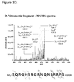
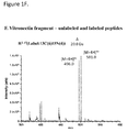
Figure 1 : (A-B) MS spectrum of C3f (aa: 1304-1320) and vitronectin (aa: 381-397) synthetic peptides and (C-D) MS/MS spectrum of their respective precursor ions ([M+4H]4+ at m/z 506.3 and at m/z 496.0. (E-F) MS spectrum of the unlabeled and labeled peptides of C3f and vitronectin synthetic peptides. aa = amino acids -
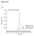
Figure 2 : A. Optimal percentage of acetonitrile in the dissolution solution of lyophilized samples for both C3f and vitronectine peptides. B and C. Optimal percentage of acetonitrile for the sample loading by the capillary pump for the C3f and vitronectine peptides. -
Figure 3 : Assessment of the biological matrix effect on our quantitative method according to the following parameters: process efficiency (PE), matrix effect (ME), extraction recovery (ER) and extraction yield (EY): A. represents the peak area of the standard in neat solution, B. is the peak area of the standard spiked into serum after WCX extraction, C. is the peak area of standards spiked in serum before WCX extraction and D. is the peak area of standards spiked in neat solution before WCX extraction. -
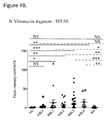
Figure 4 : Quantitation of C3f and vitronectin fragments in serum samples (n=147) by SELDI-TOF-MS (relative quantitation) and LC-MS/MS (absolute quantitation). Samples were classified into 7 groups: HV (n=12), K&0 (n=18), K&L1 (n=17), K&L2 (n=25), K&L3 (n=48), K&L4 (n=8) and RA (n=19). Peak intensities of C3f and vitronectin fragments obtained by SELDI-TOF-MS inFigure 4A and4B . Concentrations of C3f and vitronectin fragments obtained by LC-MS/MS were illustrated inFigure 4C and4D . - Both methods, SELDI-TOF-MS and LC-MS/MS (also called LC-chip-MS/MS), were then compared in
Figure 4E and4F . C3f and vitronectin peak intensities obtained by SELDI-TOF-MS were correlated to C3f and vitronectin concentrations obtained by LC-MS/MS (r=Spearman correlation). Deming regression analysis was performed. -
- OA: osteoarthritis
- K&L: Kellgren and Lawrence
- HV: healthy volunteers
- RA: rheumatoid arthritis
- SELDI-TOF-MS: surface enhanced laser desorption / ionization-time of flight mass spectrometry
- JSN: joint space narrowing
- LC: liquid chromatography
- MeOH: methanol
- ACN: acetonitrile
- FA: formic acid
- TFA: trifluoroacetic acid
- NH4OAc: ammonium acetate
- NH4OH: ammonium hydroxide
- ESI: electrospray ionization source
- m/z: mass-to-charge ratio
- EIC: extracted ion current
- NSAID: non steroidal anti-inflammatory drug
- LLOQ: lower limit of quantitation
- HLB: hydrophilic-lipophilic balance
- MCX: mixed-mode cationic exchange
- PE: process efficiency
- ME: matrix effect
- ER: extraction recovery
- EY: extraction yield
- Osteoarthritis (OA) is one of the most common chronic joint diseases causing substantial disability to work and public health costs. Further, osteoarthritis is becoming increasingly more prevalent as the population ages. Osteoarthritis is a degenerative disease characterized by a dysregulation of normal joint homeostasis that leads to intra-articular cartilage degradation, attempted repair and inflammation mediated by cytokines and growth factors. Articular cartilage is a specific connective tissue covering joint surfaces. It has a glistening, white appearance. Microscopically, it is composed of water, collagen, proteoglycan and a wide range of matrix proteins including chondronectin, fibronectin, vitronectin and thrombospondin. Chondrocytes are the unique cell type embedded in the articular cartilage tissue. They are essential for the production of a large amount of extracellular matrix. In the early stage of OA, cartilage starts to become soft with swelling. Proteoglycan overexpression enhances harmful cartilage hyper-hydration leading to cartilage breakdown and collagen fibers dissociation. Catabolic hyperactivity of chondrocytes is observed. The synovial membrane starts to secrete inflammatory mediators (cytokines and metallo-proteinases) enhancing extracellular matrix degradation in cartilage and releasing cleaved fragments of proteins inside the joint cavity. At an advanced stage, the destruction wins the deeper layers exposing the subchondral bone. Neovascularization occurs inside the cartilage improving OA biomarkers delivery in the systemic circulation.
In Annals of the rheumatic disease 2011; 70:1144-52, protein expression levels were analyzed in serum samples provided from 284 patients with knee OA and classified according to the grade of severity, the Kellgren and Lawrence (K&L) score (0-4) as reported in Annals of the rheumatic disease 1957; 16: 494-502 . Osteoarthritis serum samples were also compared to serum samples provided from healthy individuals (HV; n=36) and rheumatoid arthritis (RA) patients (n=25). RA is another arthritis pathology affecting joints but presenting autoimmune and systemic inflammatory properties while OA is primarily a degenerative joint disease with local inflammation in joints. The surface enhanced laser desorption/ionization-time of flight-mass spectrometry (SELDI-TOF-MS), a semi-quantitative approach, was used to investigate differential expression level of small proteins and cleaved fragments. Two protein fragments, identified as vitronectin fragment and C3f peptide, were found to be expressed at higher levels in sera of OA patients at all four K&L scores compared to HV and RA.
Vitronectin fragment is a 17 amino acids fragment (SQRGHSRGRNQNSRRPS - amino acids 381-397) located at the C-terminal end product of the V65 vitronectin subunit in the heparin-binding domain. Its concentration in blood and synovial fluid was 14-fold increased in OA compared to HV and was correlated with OA severity. Vitronectin is a cell adhesion and spreading factor found in serum and in many tissues including cartilage and synovium. It is mostly well known for its interaction with the αvβ3 integrin receptor via its arginine-glycine-aspartic acid motif (RGD motif, amino acids 64-66).
The second fragment was identified as complement C3f peptide. Its expression level increased 5.5-fold with OA severity. C3f peptide is a fragment released during the catabolic degradation of C3b by Factor H after C3 activation. It is a 17 amino acids fragment (SSKITHRIHWESASLLR- amino acids 1304-1320). Osteoarthritis diagnosis currently depends on patient-reported pain and disability, on blood biochemistry to rule out any other rheumatic diseases and on joint imaging (plain X-rays) that identify bone features (osteophytes) or cartilage destruction (joint space narrowing, JSN), typical of late stage disease. Fifteen-years ago, the National Institute of Health defined a biomarker as "a characteristic that is objectively measured and evaluated as an indicator of normal biologic process, pathogenic process, or pharmacologic responses to a therapeutic intervention". A review confirmed that there exists no current effective biomarker satisfying the definition. Further, they urged the scientific community to quickly find investigative biomarkers mainly through advances in "omic" research including proteomic analysis. As above mentioned, we published in 2011 the discovery of four new potential biomarkers that correlated with parameters of inflammation, bone remodeling, and cartilage turn-over using the semi-quantitative SELDI-TOF-MS technology. In this work, we have developed a new absolute quantitative approach based on the simultaneous quantitation of vitronectin fragment and C3f peptide by LC-MS/MS analysis. - The method was validated with serum samples provided from HV, OA and RA patients.
- Methods summary: Microfluidic liquid chromatography coupled to a nanoelectrospray source ion trap mass spectrometry was used for the absolute and simultaneous quantitation of C3f and vitronectin fragments in serum. The method was first carefully optimized and then validated in serum biological matrix according to FDA guidelines. Stable isotopes for the two biomarkers of interest were used as internal standards. Microelution solide phase extraction in 96-well plate format was used to purify and concentrate C3f and vitronectin fragments. Parameters for liquid chromatophraphy and for mass spectrometry were simultaneously optimized for C3f and vitronectin fragments quantitation. Serum samples (n=147) classified in 7 groups [(healthy volunteers, OA with 5 grades of severity and rheumatoid arthritis (RA) patients] were analyzed with our new quantitative method.
- Water, methanol (MeOH), acetonitrile (ACN) and formic acid (FA) 99% were all at LC/MS grade and were purchased from Biosolve. Trifluoroacetic acid (TFA) was obtained from Fluka. Ammonium hydroxide (NH4OH) was purchased from Merck and ammonium acetate (NH4OAc) 98.9% from VWR. Helium and nitrogen (Alphagaz 2) were obtained from Air Liquide. Human synthetic complement C3f fragment (1304SSKITHRIHWESASLLR1320), vitronectin fragment (381SQRGHSRGRNQNSRRPS397) and the internal standard [13C6, 15N2]Lys3, [13C6, 15N4]Arg17-Complement C3f, [13C6, 15N4]Arg3,15-vitronectin fragment were purchased from Eurogentec.
- Solid-phase extraction procedure was carried out by Oasis µElution weak cation exchange (WCX) 96 well plates with a vacuum manifold (Waters Inc.) for peptides extraction and enrichment. Sample evaporation was performed on a vacuum concentrator (Labconco). A 1200 series LC- system including nanoflow pump, a capillary pump, a well plate sampler and a LC /MS interface was used for chromatographic separation. ChemStation (Agilent Technologies) is a software package to control Agilent liquid chromatography system. Protonated peptides detection was performed by Ion Trap mass spectrometry combined with a nanoelectrospray ionisation source operating in positive mode (Agilent Technologies). TrapControl (Bruker Daltonik GmbH) determined the mass spectrometry detection parameters. Raw data obtained by mass spectrometry were processed using DataAnalysis and QuantAnalysis softwares (Bruker Daltonik GmbH).
- All peptides (labeled and unlabeled) were dissolved in a H2O/ACN/FA (80:20:0.1; v/v/v) solution to reach a concentration of 1 mg/mL, then were aliquoted and stored at -80°C. Isotopically labeled peptides ([13C6, 15N2]Lys3, [13C6, 15N4]Arg17-complement C3f and [13C6, 15N4]Arg3,15-vitronectin fragment) were used as internal standard for the two biomarkers of interest (complement C3f and vitronectin fragments) since they share the same physicochemical properties. Internal standard at a final concentration of 25 ng/mL and 10 ng/mL for complement C3f and vitronectin fragments, respectively, were prepared in H2O/TFA (99:1; v/v) solution. Calibration standards of complement C3f and vitronectin fragments were then diluted in H2O/TFA (99:1; v/v) solution in the range of 2.5 to 200 ng/mL at seven concentration levels (2.5, 5, 10, 20, 50, 100 and 200 ng/mL) for C3f and in the range of 2.5 to 100 ng/mL at six concentration levels (2.5, 5, 10, 20, 50 and 100 ng/mL) for vitronectin fragment.
- Serum samples were thawed at room temperature, vortexed and centrifuged during 5 min at 13,400 rpm. A Waters Oasis µElution WCX 96 well plate was used to purify and concentrate peptides of interest before chromatographic separation and mass spectrometry analysis. The sorbent conditioning for the OASIS-WCX was first 300 µL of MeOH followed by 300 µL of water. 100 µl of diluted human serum samples were prepared as follows: 10 µL of human serum + 50 µL of H2O/TFA (99:1; v/v) solution + 40 µL of internal standards (of C3f and vitronectin fragments). 100 µL of calibration standards were prepared as follows: 10 µL of calibration standards at expected concentration (of C3f and vitronectin fragments) + 10 µL of serum bovine (Sigma, B9433) + 40 µL of in H2O/TFA (99:1; v/v) solution + 40 µL of internal standards (of C3f and vitronectin fragments). The 100 µL of diluted serum or calibration standards were then transferred in the well and drawn through the sorbent with a vacuum manifold. The plate was then washed with 200 µL NH4OAc 25 mM, 200 µL of MeOH/H2O/NH4OH (50:47.5:2.5, v/v/v) followed by 200 µL of water. The extracts were eluted with 2 x 50 µL of MeOH/H2O/TFA (90:9:1, v/v/v). The eluates were then evaporated in a vacuum evaporator at 30°C for 65 min, reconstituted in 100 µL ACN/H2O/TFA (1:99:0.1, v/v/v) and vortexed 15 min at room temperature.
- ProtID with a 40 nL trapping column and a 43 mm x 75 µm analytical column, both packed with a
Zorbax 300SB 5 µm C18 phase (Agilent Technologies) were used for chromatographic separation. The mobile phase A [H2O/TFA (100:0.1, v/v] and the mobile phase B [ACN/H2O/TFA (90:10:0.1, v/v/v] for the capillary pump and the mobile phase A [H2O/FA (100:0.1, v/v] and the mobile phase B [ACN/H2O/FA (90:10:0.1, v/v/v] for the nanopump, were degassed by ultrasonication for 15 min before use. During the analytical process, the sample was first loaded on the trapping column during an isocratic enrichment of the column by capillary pump with 1% of the mobile phase B at a flow rate of 4 µL/min. A flush volume of 6 µL of 1% mobile phase B was used to remove unretained components. Then, a gradient of elution starting at 1% of the mobile phase B and linearly ramping up to 90% of mobile phase B in 5 min was performed in backflush mode using the nanopump. Ninety % of mobile phase B was maintained for 2 min before coming back to 1%. Ten column volumes were finally used to re-equilibrate the column before the next injection. The time for one run was 14 min. Five µL of sample was injected. The injection needle was also thoroughly rinsed twice from inside and outside with a mix solution of ACN/H2O/TFA (60:40:0.1, v/v/v). - Optimization of MS detection parameters was performed by infusion experiments at a flow rate of 36 µL/h with 1 µg/mL of C3f and vitronectin fragment and their internal standards solutions. NanoESI source was operating in a positive mode. Ions optics were optimized using the Smart Ramp tool included in Trap-Control software in order to maximize precursor ion intensity for C3f, vitronectin fragment and their internal standards. Capillary voltage was set at 1800V and the endplate offset at 500V. Nitrogen was used as drying gas at a flow rate of 4 L/min and the source temperature was set at 325°C. MS2 spectra were acquired in the mass-to-charge (m/z) ratio from 375 to 700 for C3f and in the m/z range of 350 to 600 for the vitronectin fragment.
Total ion current chromatogram was handled to yield an extracted ion current (EIC) chromatogram that summed the intensities of the selected ions for C3f and vitronectin fragments and normalized with their corresponding internal standard. EIC were smoothed using a 1 x 2 Gauss algorithm and subsequently integrated. - The 116 OA patients from University of Bristol cohort were included to validate the quantitation of C3f and vitronectin fragments by LC-MS/MS analysis. The demographic, clinical and radiographic data of these patients were previously published in Annals of the rheumatic diseases 2011. None of the OA patients was on intra-articular steroids or any potential disease-modifying drugs, the majority was on analgesics or non-steroidal anti-inflammatory drugs (NSAIDs) that are unlikely to affect serum or synovial fluid levels of investigated markers. Nineteen RA patients fulfilling the 1987 American College of Rheumatology criteria (as described in Journal of Chromatography A 2013; 1314:199-207) and 12 healthy individuals referred to as healthy volunteers (HV) were included in the study as control subjects.
- The e.noval software (Arlenda, Liege, Belgium) was used to compute trueness, precision, accuracy for all concentrations of calibration standards, as well as lower limit of quantitation (LLOQ). It was also used to choose the most appropriate regression model.
Peak intensities (by SELDI-TOF-MS) and concentrations (by LC-MS/MS) of C3f and vitronectin fragments were compared in all groups of OA to HV and RA groups by the non-parametric Kruskal-Wallis test with a post-hoc test of Dunn's. K&L1-2 values were compared to K&L3-4 values by the non-parametric Mann-Whitney U test. P-values were statistically significant at P< 0.05.
Correlation coefficients were calculated by the non-parametric Spearman analysis to correlate SELDI-TOF-MS data to LC-MS/MS data. Deming regression was used for regression analysis. - Results summary: A weighted 1/x 2 quadratic regression for C3f and a weighted 1/x quadratic regression for vitronectin peptide were selected for calibration curves. Trueness (with a relative bias <10%), precision (repeatability and intermediate precision <15%) and accuracy (risk <15%) of the method were successfully demonstrated. The linearity of results was validated in the dosing range of 2.5-200 ng/mL for C3f and 2.5-100 ng/mL for vitronectin fragment.
- Synthetic peptides for the C3f (1304SSKITHRIHWESASLLR1320) and vitronectin (381SQRGHSRGRNQNSRRPS397) fragments were first infused at a concentration of 1 µg/mL in 0.1% of TFA and analyzed in full scan mode into the ion trap in the range of m/z 200-1500. Voltages were optimized to detect the highest relative intensity using the Smart Ram tool. [M+4H]4+ at m/z 506.3 and at m/z 496.0 were the most abundant ions observed for C3f and vitronectin synthetic peptides (
Figures 1A and1B ) and were selected for MS/MS analysis. Fragmentation amplitudes at 0.95V and 1.1V were applied to the precursor ion of the C3f and vitronectin peptides, respectively, to generate multiply charged ions as observed in MS/MS spectra inFigures 1C and1D . Labeled peptides were synthesized accordingly: [13C6,15N2]Lys3, [13C6,15N4]Arg17-Complement C3f and [13C6,15N4]Arg3,15-vitronectin fragment. Both unlabeled and labeled peptides were simultaneously injected for the C3f and for the vitronectin fragment. Two precursor ions, [M+4H]4+ at m/z 506.3 and [M+4H]4+ at m/z 510.8, were distinctly observed for the unlabeled and labeled C3f peptides (Figure IE). Similarly, unlabeled ([M+4H]4+ at m/z 496.0) and labeled ([M+4H]4+ at m/z 501.0) peptides of the vitronectin fragment are represented inFigure 1F . - In this work, the method was developed for the simultaneous detection of C3f and vitronectin fragments in one single analysis of serum by LC-MS/MS method. All experimental conditions (mobile phases and dissolution medium composition) were optimized to reach the highest peak intensity in MS for both peptides. Lyophilized synthetic peptides were solubilized in a solution containing 0.1% of TFA (any retention being observed with 0.1% of formic acid for the vitronectin fragment). The dissolution solution also contained 1% ACN as the peak intensity rapidly decreased at higher percentage of ACN for the vitronectin fragment (
Figure 2A ). The percentage of ACN was also optimized during the sample loading by the capillary pump and fixed at 1% in the mobile phase B as observed inFigure 2B and2C . The chromatographic gradient was optimized according to previous studies reported in Journal of chromatography A 2011;1218:9046-54 or Journal of chromatography A 2013;1314:199-207.
Retention times of vitronectin and C3f fragments were observed at 2.7 min. and 6.4 min., respectively, which is sufficiently spaced to handle a time window for each peptide during MS detection and MS/MS analysis. - Several Oasis µElution well plates such as WCX, HLB and MCX were tested for the purification and enrichment of C3f and vitronectin peptides before chromatographic separation and mass spectrometry analysis. Oasis WCX was the most efficient in term of recovery for both peptides. Extraction conditions were optimized and summarized in the Materials and Methods section.
- Validation of this new method was then implemented on serum, the selected biological fluid.
- Bovine serum was first tested to confirm that any detection of C3f and vitronectin fragment was observed by our new method. Calibration curves were then set up by preparing calibration standards of C3f and vitronectin fragments in bovine serum as described in the solid phase extraction procedure of the Materials and Methods section. Each solution was submitted to the entire process including the extraction phase in Oasis µElution WCX well plates, nano-LC retention process and injection in the nano-LC-MS/MS for analysis. For each calibration curve, all calibration standards were processed in duplicates during three days. A weighted 1/x 2 quadratic regression for the C3f and a weighted 1/x quadratic regression for the vitronectin peptide were selected showing the highest accuracy and trueness index and the widest dosing range (Table 1). As summarized in Table 2, trueness (with a relative bias < 10%), precision (repeatability and intermediate precision < 15%) and accuracy [(risk < 15%, except for the 2.5 ng/mL calibration standard of vitronectin (17,11%)] of the method were demonstrated for all concentrations of calibration standards, as well as at the lower limit of quantitation (LLOQ=2.5 ng/mL). The linearity of the results for C3f and vitronectin fragment quantitation was validated in the dosing range of 2.5-200 ng/mL for C3f and 2.5-100 ng/mL for vitronectin. The assessment of the biological matrix effect in our quantitative bio-analytical assay was also performed according to the following references J.of chromatography A 2010; 1217:3929-37 or Analytical chemistry 2003; 75: 3019-30 and Clinical biochemistry 2005; 38:328-34. Process efficiency (PE), matrix effect (ME), extraction recovery (ER) and extraction yield (EY) were summarized in
Figure 3 and demonstrated that the signal obtained for C3f (mainly) and for vitronectin (in less extent) is enhanced in the presence of serum compared to aqueous conditions. - C3f and vitronectin quantitation was performed by LC-MS/MS on serum samples (n=147) provided from the previous cohort used for the SELDI-TOF-MS validation study. These samples were classified into 7 groups: HV (n=12), K&0 (n=18), K&L1 (n=17), K&L2 (n=25), K&L3 (n=48), K&L4 (n=8) and RA (n=19). Peak intensities of C3f and vitronectin fragments obtained by SELDI-TOF-MS, were illustrated for all serum samples (n=147) in
Figure 4A and4B , respectively. Concentrations of C3f and vitronectin fragments obtained by LC-MS/MS, were illustrated for all serum samples (n=147) inFigure 4C and4D , respectively. By SELDI-TOF-MS, C3f and vitronectin fragments levels were statistically increased in all groups of OA (except K&L0 for vitronectin fragment) compared to HV (Figure 4A and4B , plain line). By LC-MS/MS, similar results were obtained for the vitronectin fragments whereas C3f levels were only significantly increased in the late stages of OA, K&L3-4 (Figure 4C and4D , plain line) compared to HV. Compared to RA, all groups of OA showed by SELDI-TOF-MS a statistically increased level of C3f and vitronectin fragments, except K&L0 for the vitronectin fragment (Figure 4A and4B , dotted line). By LC-MS/MS, C3f level was statistically increased in K&L2-3-4 compared to RA (Figure 4C , dotted line) whereas vitronectin fragment was statistically significant only for K&L3 compared to RA (Figure 4D , dotted line). In all cases, C3f and vitronectin fragments levels were never statistically significant in HV compared to RA. Lastly, within OA patients, C3f fragment showed statistically increased expression in the K&L3-4 scores compared to those belonging to the K&L1-2 scores (P-value = 0.032), whereas no difference were observed for the vitronection fragment (P-value = 0.054). - Both methods, SELDI-TOF-MS and LC-MS/MS, were then compared in
Figure 4E and4F . C3f and vitronectin peak intensities obtained by SELDI-TOF-MS were correlated to C3f and vitronectin concentrations obtained by LC-MS/MS. Correlation coefficients of 0.53 (P<0.0001) and 0.64 (P<0.0001) were obtained for C3f and vitronectin fragments, respectively. - We have developed a new method by LC-MS/MS that combines a simultaneous quantitation of C3f (1304SSKITHRIHWESASLLR1320) and vitronectin (381SQRGHSRGRNQNSRRPS397) fragments in serum sample. Combination of both quantifications is of high interest as it considerably decreases analysis time. Further, it covers two different processes in OA: innate immunity/inflammation for C3f and extracellular matrix degradation for vitronectin fragment. WCX SPE 96 well plates approach was selected for the purification and enrichment process of both fragments. Several parameters for LC separation and for MS / MS-MS detection were optimized to get two independent windows for C3f and vitronectin fragments quantitation. The developed method for C3f and vitronectin fragment quantitation in serum was fully validated. After having selected the most appropriate regression model on the basis of the accuracy profiles, method selectivity, trueness, precision, accuracy and linearity were demonstrated according to FDA guidelines.
- This new absolute quantitative method was applied to the analysis of serum samples (n=147) previously analyzed by SELDI-TOF-MS. OA serum samples were classified according to OA severity, which is characterized by the K&L classification from 1 to 4 after X-ray examination. K&L0, 1 and 2 scores represent the early stages of OA development, for which X-ray cannot detect joint narrowing space and bone sclerosis. K&L3 and 4 are further associated to the late stages of OA with marked joint space narrowing, bone sclerosis and presence of multiple osteophytes. OA serum samples were also compared to RA serum samples to assess the specificity of C3f and vitronectin fragments. HV serum samples were also included to determine the starting concentration of C3f and vitronectin fragments in normal serum.
- Similarly to the SELDI-TOF-MS analysis, we observed by LC-MS/MS that C3f and vitronectin fragments levels increased with OA severity. Compared to HV, C3f fragment showed statistically increased expression in stages of OA where JSN is definite (K&L3-4 scores) compared to HV. Compared to RA, it was already statistically increased in the earlier stage with definite osteophyte and possible JSN (K&L2 score) of OA. Further, within OA patients, C3f fragment showed statistically increased expression in the K&L3-4 scores compared to K&L1-2 scores. C3f biomarker could therefore approach the definition of a "burden of disease" biomarker assessing disease severity in individuals with OA. Compared to HV, vitronectin fragment showed statistically increased expression in all K&L scores, except for K&L0, but was only statistically increased in K&L3 score compared to RA. Interestingly, no difference existed between K&L3-4 scores and K&L1-2 scores. Vitronectin fragment biomarker could therefore approach the definition of a "diagnostic" biomarker distinguishing between individuals with and without OA.
- With this new absolute quantitative method, we confirm that both C3f and vitronectin fragments increase with OA severity. We also confirm that C3f fragment is more related to the severity of OA, whereas vitronectin fragment is more related to early OA detection.
We also assess the sensitivity, specificity and area under the curve in the receiving operator curve of these 2 biomarkers by analyzing new cohorts of patients with rheumatic diseases (OA, RA, systemic sclerosis, lupus spondylarthropathies) and other chronic diseases such as inflammatory bowel diseases. - This research was funded by the "Fond National de la Recherche Scientifique" (Projet de Recherche # 23592988) for the Laboratory of Rheumatology and the Laboratory for the Analysis of Medicines, ULg, Liege.
Table 1: Regression models: related accuracy and trueness indexes and dosing ranges Model for C3f Accuracy Index Trueness Index Dosing range (ng/mL) Weighted 1/x2 quadratic regression 0.8392 0.9782 2.50 - 200.0 Weighted 1/x quadratic regression 0.7923 0.8976 27.53 - 200.0 Weighted 1/x2 linear regression 0.6966 0.7621 21.43 - 140.1 Weighted 1/x linear regression 0.6285 0.4528 30.06 - 200.0 Model for vitronectin fragment Weighted 1/x quadratic regression 0.8649 0.9880 2.50 - 100.0 Weighted 1/x2 quadratic regression 0.8609 0.9872 2.57 - 100.0 Weighted 1/x2 linear regression 0.8106 0.9927 2.82 - 100.0 Weighted 1/x linear regression 0.7990 0.9821 3.90 - 100.0 Table 2: A) C3f and B) vitronectin fragments quantitation - method validation: trueness, precision, accuracy and linearity properties A. C3f fragment quantitation - Method Validation Response function (k= 3) Weighted quadratic regression model calibration range (m=7) : 2.5-200 ng/mL Series 1 Series 2 Series 3 Quadratic term 0.0001165 0.00009036 0.00008730 Slope 0.03261 0.02053 0.03655 Intercept -0.3955 -0.02173 -0.04982 Weight 1/x 2 1/x 2 1/x 2 r 2 0.9909 0.9924 0.9885 Trueness (k=3, n=2) (ng/mL) Relative bias (%) 2.5 7.949 5.0 2.060 10 -5.575 20 2.715 50 4.251 100 3.443 200 -1.307 Precision (k=3, n=2) (ng/mL) Repeatability (RSD%) Intermediate precision (RSD%) 2.5 7.01 7.39 5.0 12.09 14.80 10 8.54 8.74 20 6.16 13.02 50 5.59 5.59 100 1.61 5.76 200 4.41 8.82 Accuracy (k=3, n=2) (ng/mL) ß-expectation tolerance limits (%) Risk (%) 2.5 [-4.08, 19.97] 2.56 5.0 [-23.22, 27.34] 14.50 10 [-19.68, 8.53] 3.36 20 [-22.89, 28.32] 15.61 50 [-4.72,13.22] 0.57 100 [-8.62, 15.51] 4.06 200 [-8.38,5.77] 0.18 Linearity (k=3, n=2) (ng/mL) Range 2.5-200 Slope 0.9922 Intercept 0.8931 r 2 0.9962 LOD 0.76 LLOQ 2.5 B. Vitronectin fragment quantitation - Method validation Response function (k=3) Weighted quadratic regression model calibration range (m=6) : 2.5-100 ng/mL Series 1 Series 2 Series 3 Quadratic term 0.0001232 -0.00001847 0.00002566 Slope 0.05878 0.07712 0.06873 Intercept 0.06105 0.01201 0.02518 Weight 1/x 1/x 1/x r 2 0.9957 0.9673 0.9905 Trueness (k=3, n=2) (ng/mL) Relative bias (%) 2.5 2.425 5.0 5.496 10 1.250 20 -1.875 50 3.130 100 -3.690 Precision (k=3, n=2) (ng/mL) Repeatability (RSD%) Intermediate precision (RSD%) 2.5 8.53 14.48 5.0 6.74 6.74 10 10.52 10.52 20 7.39 7.91 50 5.85 5.99 100 3.56 3.94 Accuracy (k=3, n=2) (ng/mL) ß-expectation tolerance limits (%) Risk (%) 2.5 [-24.77, 29.62] 17.11 5.0 [-5.31, 16.30] 1.32 10 [-15.63, 18.13] 4.83 20 [-14.81, 11.06] 2.15 50 [-6.54, 12.80] 0.74 100 [-10.20,2.82] 0.19 Linearity (k=3, n=2) (ng/mL) Range 2.5-100 Slope 0.9677 Intercept 0.6698 r 2 0.9954 LOD 0.76 LLOQ 2.5 k: Number of days of experiments (series); m: number of concentration levels; n: number of replicates per concentration level and per series. 
Claims (7)
- An in-vitro method for early diagnosing or prediction of immune-mediated inflammatory diseases , comprising :- obtaining a sample from a subject;- quantifying simultaneously by one LC/MS-MS analysis of said sample , a presence of V65 vitronectin fragment or fragment, variant or degradation product thereof; and/or a presence of a complement C3f or fragment or variant or degradation products thereof;
- The method according to claim 1 wherein the subject is human;
- The Method according to claim 1 or 2 wherein the sample is blood, serum, plasma, urine, or synovial fluid;
- The method according to any preceding claims, wherein the V65 vitronectin fragment comprises the following amino sequence:381SQRGHSRGRNQNSRRPS397
- The method according to any preceding claims wherein the complement C3f comprises the following amino sequence: 1304SSKITHRIHWESASLLR1320
- The method according to any preceding claims wherein the immune-mediated inflammatory diseases is osteoarthritis.
- The method according to any preceding claims wherein the LC/MS-MS analysis is a microfluidic liquid chromatography coupled to a nanoelectrospray source ion trap mass spectrometry analysis.
Priority Applications (4)
| Application Number | Priority Date | Filing Date | Title |
|---|---|---|---|
| EP16177673.7A EP3264088A1 (en) | 2016-07-02 | 2016-07-02 | Method of early diagnosis immune-mediated inflammatory disease |
| EP17735416.4A EP3479120B1 (en) | 2016-07-02 | 2017-06-23 | Method of early diagnosis of an immune-mediated inflammatory disease |
| US16/314,654 US11047856B2 (en) | 2016-07-02 | 2017-06-23 | Method of early diagnosis of immune-mediated inflammatory disease |
| PCT/EP2017/065591 WO2018007173A1 (en) | 2016-07-02 | 2017-06-23 | Method of early diagnosis of an immune-mediated inflammatory disease |
Applications Claiming Priority (1)
| Application Number | Priority Date | Filing Date | Title |
|---|---|---|---|
| EP16177673.7A EP3264088A1 (en) | 2016-07-02 | 2016-07-02 | Method of early diagnosis immune-mediated inflammatory disease |
Publications (1)
| Publication Number | Publication Date |
|---|---|
| EP3264088A1 true EP3264088A1 (en) | 2018-01-03 |
Family
ID=56368850
Family Applications (2)
| Application Number | Title | Priority Date | Filing Date |
|---|---|---|---|
| EP16177673.7A Withdrawn EP3264088A1 (en) | 2016-07-02 | 2016-07-02 | Method of early diagnosis immune-mediated inflammatory disease |
| EP17735416.4A Not-in-force EP3479120B1 (en) | 2016-07-02 | 2017-06-23 | Method of early diagnosis of an immune-mediated inflammatory disease |
Family Applications After (1)
| Application Number | Title | Priority Date | Filing Date |
|---|---|---|---|
| EP17735416.4A Not-in-force EP3479120B1 (en) | 2016-07-02 | 2017-06-23 | Method of early diagnosis of an immune-mediated inflammatory disease |
Country Status (3)
| Country | Link |
|---|---|
| US (1) | US11047856B2 (en) |
| EP (2) | EP3264088A1 (en) |
| WO (1) | WO2018007173A1 (en) |
Families Citing this family (1)
| Publication number | Priority date | Publication date | Assignee | Title |
|---|---|---|---|---|
| PL3645832T3 (en) | 2017-06-29 | 2025-06-02 | Typhon Technology Solutions, Llc | Electric power distribution for fracturing operation |
Citations (2)
| Publication number | Priority date | Publication date | Assignee | Title |
|---|---|---|---|---|
| WO2012104624A2 (en) * | 2011-01-31 | 2012-08-09 | L'université De Liège | Biomarkers for osteoarthritis |
| US8494818B2 (en) | 2008-05-28 | 2013-07-23 | Université de Liège | Analyzing spectral data for the selection of a calibration model |
-
2016
- 2016-07-02 EP EP16177673.7A patent/EP3264088A1/en not_active Withdrawn
-
2017
- 2017-06-23 WO PCT/EP2017/065591 patent/WO2018007173A1/en not_active Ceased
- 2017-06-23 EP EP17735416.4A patent/EP3479120B1/en not_active Not-in-force
- 2017-06-23 US US16/314,654 patent/US11047856B2/en not_active Expired - Fee Related
Patent Citations (2)
| Publication number | Priority date | Publication date | Assignee | Title |
|---|---|---|---|---|
| US8494818B2 (en) | 2008-05-28 | 2013-07-23 | Université de Liège | Analyzing spectral data for the selection of a calibration model |
| WO2012104624A2 (en) * | 2011-01-31 | 2012-08-09 | L'université De Liège | Biomarkers for osteoarthritis |
Non-Patent Citations (9)
| Title |
|---|
| "Annals of the rheumatic disease", vol. 16, 1957, pages: 494 - 502 |
| "Annals of the rheumatic disease", vol. 70, 2011, pages: 1144 - 52 |
| ANALYTICAL CHEMISTRY, vol. 75, 2003, pages 3019 - 30 |
| CLINICAL BIOCHEMISTRY, vol. 38, 2005, pages 328 - 34 |
| DE SENY DOMINIQUE ET AL: "Discovery and biochemical characterisation of four novel biomarkers for osteoarthritis", ANN. RHEUM. DIS., vol. 70, 1 June 2011 (2011-06-01), pages 1144 - 1152, XP002676458 * |
| GONZALEZ-IGLESIAS HECTOR ET AL: "Validation of Candidate Glaucoma Biomarkers by Quantitative Proteomics", INVESTIGATIVE OPHTHALMOLOGY & VISUAL SCIENCE - IOVS, ASSOCIATION FOR RESEARCH IN VISION AND OPHTHALMOLOGY, US, vol. 56, no. 7, 1 June 2015 (2015-06-01), pages 3676, XP009191698, ISSN: 0146-0404 * |
| J.OF CHROMATOGRAPHY A, vol. 1217, 2010, pages 3929 - 37 |
| JOURNAL OF CHROMATOGRAPHY A, vol. 1218, 2011, pages 9046 - 54 |
| JOURNAL OF CHROMATOGRAPHY A, vol. 1314, 2013, pages 199 - 207 |
Also Published As
| Publication number | Publication date |
|---|---|
| EP3479120A1 (en) | 2019-05-08 |
| WO2018007173A1 (en) | 2018-01-11 |
| EP3479120B1 (en) | 2021-03-03 |
| US20190317091A1 (en) | 2019-10-17 |
| US11047856B2 (en) | 2021-06-29 |
Similar Documents
| Publication | Publication Date | Title |
|---|---|---|
| US12163964B2 (en) | Amyloid beta detection by mass spectrometry | |
| US20130217630A1 (en) | Parathyroid hormone variants and assays related to disease | |
| US10712348B2 (en) | Kisspeptin-54 detection by tandem mass spectrometry | |
| EP3535587B1 (en) | Mass spectrometry-based methods for the detection of circulating histones h3 and h2b in plasma from sepsis or septic shock (ss) patients | |
| Cobraiville et al. | Validation of a new method by nano-liquid chromatography on chip tandem mass spectrometry for combined quantitation of C3f and the V65 vitronectin fragment as biomarkers of diagnosis and severity of osteoarthritis | |
| TW202016547A (en) | Nephropathy protein biomarker and its application | |
| CN111521828A (en) | Application of RSPH9 as diagnosis marker or therapeutic target of oligoasthenospermia | |
| CN107102152A (en) | The protein marker of urine myocardial infarction and its purposes in diagnosis and prognosis | |
| US20090280512A1 (en) | Tumor marker for renal cancer and method for determination of occurrence of renal cancer | |
| US11047856B2 (en) | Method of early diagnosis of immune-mediated inflammatory disease | |
| WO2015026909A2 (en) | Msia-srm assay for biomarker analysis | |
| WO2018007555A1 (en) | Method for diagnosing cancer | |
| CN111518868A (en) | Application of PGAM5 as diagnosis marker and treatment target of oligoasthenospermia | |
| WO2018220106A1 (en) | Biomarker molecules for sarcopenia and uses thereof | |
| JP6830899B2 (en) | Diagnostic marker for coronary artery disease | |
| Feickert et al. | Mass spectrometric studies on the peptide integrity of substance P and related human tachykinins in human biofluids | |
| Mesmin et al. | Mass spectrometric quantification of AcSDKP‐NH2 in human plasma and urine and comparison with an immunoassay | |
| EP2491402B1 (en) | Blood coagulation status measurement in saliva | |
| CN111537749A (en) | Application of MYH9 in preparation of severe oligozoospermia diagnostic reagent | |
| KR101078318B1 (en) | A method for measurement of the N-terminally variant isoforms of platelet factor-4 in acute myeloid leukemia patients in complete remission in order to provide useful information in determining whether patients have been cured or not | |
| Lopez et al. | Selected-Reaction Monitoring–Mass Spectrometric Immunoassay Analysis of Parathyroid Hormone and Related Variants | |
| Thong | The plasma proteome and outcome in patients with heart failure | |
| WO2013003897A1 (en) | Endorepellin peptides and fragments thereof as biomarkers for physical activity |
Legal Events
| Date | Code | Title | Description |
|---|---|---|---|
| PUAI | Public reference made under article 153(3) epc to a published international application that has entered the european phase |
Free format text: ORIGINAL CODE: 0009012 |
|
| AK | Designated contracting states |
Kind code of ref document: A1 Designated state(s): AL AT BE BG CH CY CZ DE DK EE ES FI FR GB GR HR HU IE IS IT LI LT LU LV MC MK MT NL NO PL PT RO RS SE SI SK SM TR |
|
| AX | Request for extension of the european patent |
Extension state: BA ME |
|
| STAA | Information on the status of an ep patent application or granted ep patent |
Free format text: STATUS: THE APPLICATION IS DEEMED TO BE WITHDRAWN |
|
| 18D | Application deemed to be withdrawn |
Effective date: 20180704 |