EP3261627B1 - Interaction of methylated lsd1 and chd1, a compound inhibiting this interaction for use in therapy, and a screening method for such a compound - Google Patents
Interaction of methylated lsd1 and chd1, a compound inhibiting this interaction for use in therapy, and a screening method for such a compound Download PDFInfo
- Publication number
- EP3261627B1 EP3261627B1 EP16705961.7A EP16705961A EP3261627B1 EP 3261627 B1 EP3261627 B1 EP 3261627B1 EP 16705961 A EP16705961 A EP 16705961A EP 3261627 B1 EP3261627 B1 EP 3261627B1
- Authority
- EP
- European Patent Office
- Prior art keywords
- chd1
- lsd1
- interaction
- compound
- lsd1me2
- Prior art date
- Legal status (The legal status is an assumption and is not a legal conclusion. Google has not performed a legal analysis and makes no representation as to the accuracy of the status listed.)
- Active
Links
Images









Classifications
-
- A—HUMAN NECESSITIES
- A61—MEDICAL OR VETERINARY SCIENCE; HYGIENE
- A61K—PREPARATIONS FOR MEDICAL, DENTAL OR TOILETRY PURPOSES
- A61K31/00—Medicinal preparations containing organic active ingredients
-
- A—HUMAN NECESSITIES
- A61—MEDICAL OR VETERINARY SCIENCE; HYGIENE
- A61P—SPECIFIC THERAPEUTIC ACTIVITY OF CHEMICAL COMPOUNDS OR MEDICINAL PREPARATIONS
- A61P35/00—Antineoplastic agents
-
- G—PHYSICS
- G01—MEASURING; TESTING
- G01N—INVESTIGATING OR ANALYSING MATERIALS BY DETERMINING THEIR CHEMICAL OR PHYSICAL PROPERTIES
- G01N33/00—Investigating or analysing materials by specific methods not covered by groups G01N1/00 - G01N31/00
- G01N33/48—Biological material, e.g. blood, urine; Haemocytometers
- G01N33/50—Chemical analysis of biological material, e.g. blood, urine; Testing involving biospecific ligand binding methods; Immunological testing
- G01N33/68—Chemical analysis of biological material, e.g. blood, urine; Testing involving biospecific ligand binding methods; Immunological testing involving proteins, peptides or amino acids
- G01N33/6803—General methods of protein analysis not limited to specific proteins or families of proteins
- G01N33/6845—Methods of identifying protein-protein interactions in protein mixtures
-
- G—PHYSICS
- G01—MEASURING; TESTING
- G01N—INVESTIGATING OR ANALYSING MATERIALS BY DETERMINING THEIR CHEMICAL OR PHYSICAL PROPERTIES
- G01N2333/00—Assays involving biological materials from specific organisms or of a specific nature
- G01N2333/90—Enzymes; Proenzymes
- G01N2333/902—Oxidoreductases (1.)
-
- G—PHYSICS
- G01—MEASURING; TESTING
- G01N—INVESTIGATING OR ANALYSING MATERIALS BY DETERMINING THEIR CHEMICAL OR PHYSICAL PROPERTIES
- G01N2333/00—Assays involving biological materials from specific organisms or of a specific nature
- G01N2333/90—Enzymes; Proenzymes
- G01N2333/902—Oxidoreductases (1.)
- G01N2333/90245—Oxidoreductases (1.) acting on paired donors with incorporation of molecular oxygen (1.14)
-
- G—PHYSICS
- G01—MEASURING; TESTING
- G01N—INVESTIGATING OR ANALYSING MATERIALS BY DETERMINING THEIR CHEMICAL OR PHYSICAL PROPERTIES
- G01N2333/00—Assays involving biological materials from specific organisms or of a specific nature
- G01N2333/90—Enzymes; Proenzymes
- G01N2333/914—Hydrolases (3)
-
- G—PHYSICS
- G01—MEASURING; TESTING
- G01N—INVESTIGATING OR ANALYSING MATERIALS BY DETERMINING THEIR CHEMICAL OR PHYSICAL PROPERTIES
- G01N2440/00—Post-translational modifications [PTMs] in chemical analysis of biological material
- G01N2440/12—Post-translational modifications [PTMs] in chemical analysis of biological material alkylation, e.g. methylation, (iso-)prenylation, farnesylation
-
- G—PHYSICS
- G01—MEASURING; TESTING
- G01N—INVESTIGATING OR ANALYSING MATERIALS BY DETERMINING THEIR CHEMICAL OR PHYSICAL PROPERTIES
- G01N2500/00—Screening for compounds of potential therapeutic value
- G01N2500/02—Screening involving studying the effect of compounds C on the interaction between interacting molecules A and B (e.g. A = enzyme and B = substrate for A, or A = receptor and B = ligand for the receptor)
Definitions
- the present invention is concerned with a method of screening for a compound inhibiting the interaction between methylated lysine-specific demethylase 1 (LSD1) and chromodomain-helicase-DNA-binding protein 1 (CHD1).
- LSD1 methylated lysine-specific demethylase 1
- CHD1 chromodomain-helicase-DNA-binding protein 1
- Prostate cancer is the most common type of cancer found in men and the second leading cause of cancer deaths in Western countries 5 .
- Chromosomal rearrangements and extensive copy number alterations are prevalent in prostate cancer, whereas point mutations are relatively rare 6,7,8 .
- Chromosomal rearrangements occur in a coordinated fashion by a phenomenon termed chromoplexy 1 .
- Chromoplectic tumors are characterized by presence of the TMPRSS2-ERG gene fusion and expression of a functional chromatin-remodelling enzyme CHD1 1,2 .
- Recent data suggest that CHD1 is directly involved in TMPRSS-ERG fusion and that the chromosomal rearrangements resulting in the generation of TMPRSS-ERG fusion are coupled to androgen receptor (AR)-dependent transcription 9 .
- AR androgen receptor
- LSD1 was identified as a potential target for prostate cancer therapy 10,11,12 .
- LSD1 is a histone demethylase that removes mono- and dimethyl marks from either lysine 4 or lysine 9 of histone 3 (H3) 10,13 .
- H3 histone 3
- the enzyme removes repressive methyl marks from H3K9, thereby enhancing AR-dependent gene transcription and prostate tumor cell proliferation 10 .
- LSD1 is not only implicated in prostate cancer but also in carcinogenesis and in maintenance of the malignant state in various other cancers.
- a role for LSD1 has also been described breast cancer (see e.g. Lim et al., Carcinogenesis. 2010 Mar;31(3): 512-20 ), colon cancer (see e.g. Huang et al., Clin Cancer Res. 2009; 15:7217-7228 ) and neuroblastoma (see e.g. Schulte et al., Cancer Res. 2009 Mar 1;69(5): 2065-71 ).
- overexpression of LSD1 has been implicated in carcinogenesis of bladder, lung and colorectal cancer (see e.g.
- LSD1 is methylated and that this methylation serves as regulatory switch to allow for interaction with CHD1.
- This interaction triggers the binding of the AR to chromatin and thus drives and increases the expression of androgen-dependent genes.
- androgen-dependent chromosomal rearrangement such as the TMPRSS2-ERG oncogenic fusion during prostate tumor evolution are induced by this interaction.
- Prostate cancer treatment inter alia aims at decreasing or blocking the expression of androgen-dependent genes as well as androgen-dependent chromosomal rearrangement, and a compound inhibiting the interaction between methylated LSD1 and CHD1 may thus be used in cancer therapy to downregulate or block hormone-dependent gene expression, in particular in prostate cancer therapy.
- the present disclosure thus relates to a compound inhibiting the interaction between LSD1me2 and CHD1.
- said compound specifically inhibits the interaction between LSD1me2 and CHD1. In another embodiment thereof, said compound inhibits only the interaction between LSD1me2 and CHD1.
- said compound inhibits the interaction between (i) LSD1me2 and (ii) an acidic surface bridging chromodomain 1 and chromodomain 2 of CHD1 and amino acids of chromodomain 1 and chromodomain 2 of CHD1.
- said compound inhibits at least one interaction between amino acids T110, S111, R112, R113, K144me2, R115 and A116 of LSD1me2 and amino acids Y295, W322, W325, D408 and D425 of CHD1. It is noted that Y295, W322 and W325 are comprised in the chromodomain 1 of CHD1, whereas D408 and D425 are comprised in the chromodomain 2 of CHD1.
- said compound inhibits at least one interaction selected from the group consisting of the interaction between amino acid S111 of LSD1me2 and D408 of CHD1, the interaction between amino acid R112 of LSD1me2 and W325 of CHD1, the interaction between amino acid R113 of LSD1me2 and D425 of CHD1, the interaction between the backbone between amino acids R113 and K114me2 of LSD1me2 and Y295 of CHD1, and the interaction between amino acid K114me2 of LSD1me2 and W322 of CHD1.
- said compound inhibits the interaction between R113 of LSD1me2 and D425 of CHD1.
- R113 of LSD1me2 and D425 of CHD1 is preferably understood herein as referring to an inhibition of the interaction between R113 of LSD1me2 and D425 of CHD1 only.
- said interaction between R113 of LSD1me2 and D425 of CHD1 is bridged by a water molecule.
- D425 of CHD1 binds to R113 of LSD1me2 (bridged via a water molecule) but apparently not to other CHD1-interacting proteins, such as e.g. H3K4me3. Accordingly, an inhibition of this interaction is most preferred since such a compound will specifically inhibit CHD1 binding to LSD1me2 but not the interaction of CHD1 e.g. with H3K4me3.
- said compound fails to affect the interaction between CHD1 and H3K4me3. It is noted that the interaction between CHD1 and H3K4me3 is mediated by at least amino acids A1, R2, K4me3, and the backbone between amino acid T3 and K4me3 of H3K4me3, and amino acids Y295, W322, G324 and D408 of CHD1.
- the present invention relates to a method of screening for a compound as defined above.
- Said method of screening for a compound inhibiting the interaction between LSD1me2 and CHD1 comprises the steps of:
- CHD1 is recombinant CHD1. Accordingly, a fragment thereof is then a recombinant fragment thereof.
- said binding site for LSD1me2 in CHD1 is an acidic surface bridging chromodomain 1 and chromodomain 2 of CHD1 and further comprised of amino acids of chromodomain 1 and chromodomain 2 of CHD1.
- said binding site for LSD1me2 in CHD1 is defined by amino acids Y295, W322, W325 from chromodomain 1 and D408 and D425 from chromodomain 2 of CHD1. This does not exclude that at least one further amino acid and/or the protein backbone of CHD1 also interacts with LSD1me2.
- said binding site for LSD1me2 in CHD1 is defined by amino acid D425 of CHD1. Since this amino acid in CHD1 binds to LSD1me2 (more specifically to R113 of LSD1me2 bridged via a water molecule) but apparently not to other CHD1-interacting proteins, such as e.g. H3K4me3, binding of the compound to this binding site is most preferred since such a compound will specifically only inhibit CHD1 binding to LSD1me2.
- said determining step 2) is carried out by a method selected from the group consisting of nuclear magnetic resonance spectroscopy, mass spectrometry, infrared spectroscopy, Raman spectroscopy, electron microscopy and X-ray crystallography. It can be preferred to use nuclear magnetic resonance or X-ray crystallography, wherein X-ray crystallography is most preferred.
- said method comprises the following steps:
- step 2) may facilitate the method of screening since a compound may initially be excluded if it fails to bind to CHD1.
- step 2) is reiterated with the compound initially identified in step 2) as binding to CHD1 or a fragment thereof.
- said step of determining in additional step 2) is carried out by a method selected from the group consisting of circular dichroism spectroscopy, differential scanning fluorimetry, nuclear magnetic resonance spectroscopy, isothermal titration calorimetry, surface plasmon resonance, and fluorescence polarization.
- the inventors of the present invention inter alia succeeded in unraveling a novel mechanism that is not only crucial for regulating androgen-dependent gene expression but also controls androgen-dependent chromosomal rearrangement such as the TMPRSS2-ERG oncogenic fusion.
- This mechanism may be referred to as the androgen-regulated G9A/LSD1 K114me2/CHD1 circuit and compounds inhibiting the interaction between LSD1 K114me2 and CHD1 may be used to downregulate or block downstream events of the LSD1 K114me-CHD1-interaction, such as e.g. the downregulation or blocking of androgen-dependent gene expression in prostate cancer.
- LSD1 refers to the enzyme "lysine-specific demethylase 1" with the UniProt accession number 060341 comprising 852 amino acids.
- LSD1me2 is interchangeable used herein with the term “LSD1 K114me2” and refers to LSD1 that is modified at lysine residue 114 by two methyl-groups. Reference to a “methylation of LSD1” also refers to this post-translational modification of LSD1.
- the indication of "me” after the amino acid "K” indicates that the corresponding lysine residue is methylated.
- the number, e.g. "2” indicates that there are two methyl-groups present at the corresponding lysine, e.g. "K114me2”.
- Another example is "K4me3", where three methyl-groups are present at lysine 4.
- CHD1 refers to the enzyme "chromodomain-helicase-DNA-binding protein 1" with the UniProt accession number 014646 comprising 1710 amino acids.
- CHD1 comprises two chromodomains, namely chromodomain 1 comprising amino acids 268 to 356 and chromodomain 2 comprising amino acids 389 to 443 of CHD1.
- chromodomain refers to a protein structural domain of about 40 to 50 amino acids commonly found in proteins associated with the remodeling and manipulation of chromatin. Chromodomain-containing proteins are responsible for aspects of gene regulation related to chromatin remodeling and formation of heterochromatin regions. Chromodomain-containing proteins bind methylated histones.
- H3K4me3 refers to histone 3 (H3) that is modified at lysine residue 4 by three methyl-groups.
- compound refers to an organic compounds, preferably a small organic compound having a low molecular weight (also termed "small molecule” in the respective field).
- the compound may be a synthetic compound not known to occur in nature or a naturally-occurring compound isolated from or known to occur in natural sources, such as e.g. plants, fungi or the like.
- natural sources such as e.g. plants, fungi or the like.
- the main focus is on a compound that is capable of inhibiting the interaction between LSD1me2 and CHD1, and that such compounds may e.g. be identified in the method of screening disclosed herein.
- the term compound may also refer to a "small chemical fragment”.
- fragment-based lead discovery relates to the starting pool of molecules during the process of drug discovery (fragment-based lead discovery"), wherein a small chemical fragment that has been identified as promising binding partner may undergo structural modifications to increase the binding affinity and/or specificity.
- inhibiting the interaction means that preferably no interaction at all (at least not to a detectable level) between two proteins /relevant fragments thereof takes place any more. However, when a given interaction between two proteins (set to 100%) is greatly reduced, e.g. to a level of about 70%, about 60%, about 50%, about 40%, about 30%, preferably about 20%, more preferably about 10% or most preferably about 5% or less, such a reduced interaction is still encompassed by the term “inhibiting the interaction". In terms of the medical use of compound inhibiting an interaction, a complete inhibition of an interaction may not be required to achieve a sufficient therapeutic effect. Thus, it needs to be understood that the term “inhibiting” as used herein also refers to a reduction of an interaction, which is sufficient to achieve the desired effect.
- the term “specifically inhibiting” as used herein means that the inhibition abolishes or reduces only the interaction between protein A and protein B, in the present case between LSD1me2 and CHD1, without e.g. affecting further interactions of LSD1me2 with other proteins and/or of CHD1 with other proteins. This may also be referred to as “inhibiting only” a given interaction.
- the effect of such a “specific inhibition” is that only the desired downstream effect is affected (in the present case abolished or reduced), whereas other downstream effects mediated via different interactions still take place.
- the inhibition according to the present invention is preferably specific for the interaction between LSD1me2 and CHD1 and preferably fails to have an effect on the interaction between CHD1 and H3K4me3.
- the present inventors could show that methylation of LSD1 is executed by G9A in an androgen-dependent manner and serves as a regulatory switch to allow for an interaction with CHD1.
- the androgen-regulated G9A/LSD1 K114me2/CHD1 circuit controls chromatin binding of AR. Accordingly, the mechanism is not only crucial for regulating androgen-dependent gene expression but also controls androgen-dependent chromosomal rearrangement such as the TMPRSS2-ERG oncogenic fusion during prostate tumor evolution.
- the method of screening for a compound inhibiting the interaction between LSD1me2 and CHD1 comprises at least the steps of:
- Said method may be carried out in the form of a virtual screening, which is known to the skilled person and which is carried out in accordance with standard and routine practice by the skilled person.
- a compound inhibiting the interaction between LSD1me2 and CHD1 may be screened for by i) designing a compound with a similar shape to amino acids T110 to A116 of LSD1me2; ii) contacting said compound with CHD1 by virtually superimposing the structure of said compound and the structure of CHD1 (or a fragment thereof, preferably amino acids 270 to 443 of CHD1); and iii) virtually determining whether said compound binds to CHD1 (or the above fragment thereof) at the binding site for LSD1me2.
- This approach may be referred to as the "ligand-based" virtual screening approach.
- a compound inhibiting the interaction between LSD1me2 and CHD1 may be screened for by i) contacting a compound with CHD1 by docking said compound into the LSD1me2 binding site; and ii) virtually determining whether said compound binds to CHD1 at the binding site for LSD2me2 by applying a scoring function to estimate the likelihood that the ligand will bind to the protein with high affinity, wherein a compound inhibiting the interaction between LSD1me2 and CHD1 binds to CHD1 at the binding site for LSD1me2 and has a relatively high scoring function.
- This approach may be referred to as the "structure-based" virtual screening approach.
- a compound that has been identified is then usually tested in in vitro test methods, such as e.g. established cell culture models, for efficacy as regards the desired effect.
- in vitro test methods such as e.g. established cell culture models, for efficacy as regards the desired effect.
- Cell culture models of particular relevance for the present invention are e.g. disclosed in example 3 below.
- the above method may also be carried out by a fragment-based drug discovery method.
- This method is also standard knowledge for the skilled person in drug discovery, see e.g. http://en.wikipedia.org/wiki/Fragment-based lead discovery . Accordingly, the "compound” referred to in the following is interchangeable with the term "small chemical fragment” or "chemical fragment”.
- modifying in step 4) as used herein means that specific substituents of the compound may be deleted, added, or substituted by different substituents.
- recombinant CHD1 comprising amino acids 260 to 443, i.e. the two chromodomains.
- said compound is contacted in step 1) with recombinant CHD1 comprising amino acids 260 to 443.
- step 2) it is determined whether said compounds binds to this fragment of CHD1, preferably by a method selected from the group consisting of circular dichroism spectroscopy, differential scanning fluorimetry, nuclear magnetic resonance spectroscopy, isothermal titration calorimetry (see also examples of the present invention, in particular example 6), surface plasmon resonance, and fluorescence polarization. It can be particularly preferred to use differential scanning fluorimetry in step 2).
- a binding affinity in the range of mM, preferably in the range of about 10 mM and more preferably in the range of 1 mM can be sufficient in this step 2) for the definition of a "binding" to CHD1.
- This step is introduced in the preferred embodiment to exclude compounds in an initial step that do not bind to CHD1 at all.
- Step 2) may be reiterated with the compounds identified as binding to CHD1 in the initial step 2). This time, more stringent parameters may be used and/or a binding affinity lower than in the mM-range (e.g. in the ⁇ M- or even in the nM-range) may be regarded as defining a "binding" of these compounds to CHD1.
- step 3 it is then determined whether said compound binds to the recombinant fragment of CHD1 discussed above at the binding site for LSD1me2.
- This can be done in principle by any method selected from the group consisting of nuclear magnetic resonance spectroscopy, mass spectrometry, infrared spectroscopy, Raman spectroscopy, electron microscopy, X-ray crystallography, and combinations thereof. It is particularly preferred to use X-ray crystallography, wherein the structure of the CHD1-fragment is solved in combination with the compound. This may be done by methods routinely known to the skilled person, e.g. by co-crystallization or by soaking of said compound. Reference to examples 2 and 6 of the present invention is made as regards suitable parameters and the like when using X-ray crystallography.
- a compound that has been identified is then usually tested in in vitro test methods, such as e.g. established cell culture models, for efficacy as regards the desired effect.
- in vitro test methods such as e.g. established cell culture models, for efficacy as regards the desired effect.
- Cell culture models of particular relevance for the present invention are e.g. disclosed in example 3 below.
- the "target protein” is CHD1 or a fragment thereof
- the "target site” is the binding site for LSD1me2
- the compound may be referred to as the "ligand”.
- DSF Differential Scanning Fluorimetry
- Sypro Orange binds nonspecifically to hydrophobic surfaces, and water strongly quenches the fluorescence of Sypro Orange.
- a fluorescence-based thermal shift assay can be performed on instruments that combine sample temperature control and dye fluorescence detection, such as RT-PCR machines. The assay allows high-throughput screening of ligands to the target protein.
- a typical assay is carried out as follows:
- Example 1 LSD1 is methylated by G9A at K114 in vitro and in vivo
- LSD1 K114A Flag-tagged LSD1 or a LSD1 protein containing a K114 to alanine (A) mutation (LSD1 K114A) in the presence or absence of G9A was expressed in 293T cells.
- Cell extracts were immunoprecipitated with an anti-Flag antibody and subjected to Western blot analysis using an antibody specific for LSD1 K114me2 ( Fig. 1b ).
- LSD1 but not LSD1 K114A is methylated in presence of G9A ( Fig. 1b ).
- LNCaP cells it was observed that approximately 6% of total LSD1 is dimethylated at K114 (data not shown).
- Example 2 CHD1 is a LSD1 K114me2 reader
- LSD1 K114me2 methyl-lysine reader proteins protein profiling was performed on protein-domain microarrays using un- and dimethylated LSD1 peptides LSD1aa104-127 and LSD1aa104-127 K114me2, respectively (see also ref. 14 ).
- LSD1aa104-127 and LSD1aa104-127 K114me2 were used in this profiling assay.
- Other GST-fusions that were used were e.g. PhD-domain proteins (BPTF, ING2 or PHF2), BROMO-domain proteins (GCN5, TAF1-D1 or TAF1-D2), or BRCT-proteins (BRCA1, 53BP1 or Crb2).
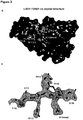
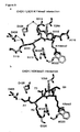
- Amino acid residues 110-116 of the LSD1 peptide were modelled according to the electron density ( Figure 2c ). Comparable to the binding of H3K4me3 peptide 15 , the LSD1 K114me2 peptide is located at an acidic surface bridging the two chromodomains ( Figure 2b ). This interaction involves residues Y295, W322, W325 from chromodomain 1 and D408 and D425 from chromodomain 2 ( Figure 2b and Figure 5a ). The methylammonium group of LSD1 K114 and side chains of W322 and W325 of CHD1 form cation- ⁇ interactions.
- the R113-D425 interaction may be a discriminating feature of CHD1 binding either LSD1 K114me2 or H3K4me3 peptide and that the corresponding surface could be exploited for the design of inhibitors specifically interfering with LSD1 K114me2 but not H3K4me3 interaction (see Figure 5c ).
- mutagenesis of either LSD1 R113 or CHD1 D425 to an alanine (A) abolished the interaction of CHD1 with LSD1 K114me2 peptide, as determined by isothermal titration calorimetry (data not shown).
- the D425A mutation in CHD1 only mildly affected H3K4me3 binding (data not shown).
- Chromatin occupancy of G9A was not altered by the presence or absence of DHT and Bix-01294. Whereas LSD1 enrichment increased upon DHT treatment at the TMPRSS2 enhancer (-13782 /-13866), this was not the case at the KLK3 enhancer (-3980 /-4048, data not shown).
- the results are cell type-independent since similar data were obtained with LAPC4 androgen-dependent prostate tumor cells (data not shown). Specificity was controlled for by also using an unrelated region that is not occupied by AR, wherein the effects was not observed. Additionally, chromatin was not enriched when using an IgG control ( Figure 3g ).
- Example 4 The presence of LSD1 K114me2 and CHD1 promotes chromating binding of AR during AR-dependent gene expression
- Example 5 DHT-induced chromosomal conformational changes at the TMPRSS2 enhancer-breakpoint regions need the integrity of the G9A/LSD1 K114me2/CHD1 circuit
- LNCaP cells were cultured in the absence and presence of DHT and subjected to chromosome conformation capture (3C) analysis.
- 3C chromosome conformation capture
- enhancer-breakpoint interactions upon treatment with DHT was observed.
- treatment with Bix-01294 blocked this loop formation in LNCaP and LAPC4 cells demonstrating that methylation of LSD1 is necessary for TMPRSS2 enhancer-breakpoint loop formation ( Figure 4a,b ).
- knockdown of either G9A, LSD1, or CHD1 also robustly impaired androgen-induced looping ( Figure 4c ).
- LSD 1 K114me2 in the control of TMPRSS2 enhancer-breakpoint loop formation, it was tested whether androgen-dependent loop formation is compromised in presence of the LSD1 K114A mutant. Therefore, LNCaP cells expressing RNAi resistant (rr) Flag-NLS-LSDl-rr and Flag-NLS-LSDl-rr K114A constructs were generated. Upon knockdown of endogenous LSD1, DHT-induced looping was observed in LNCaP cells expressing Flag-NLS-LSDl-rr but not in the cells expressing the Flag-NLS LSD1-rrK114A mutant ( Figure 4d ).
- Flag-LSDl and GST-LSD1 were described previously 19 .
- Flag-G9A was provided by Y. Shinkai.
- Flag-LSDl K114A was obtained by site directed mutagenesis.
- Chromatin immunoprecipitation ChIP experiments were performed essentially as previously described 10 .
- LNCaP and LAPC4 cells were cultured for 210 min in the absence or presence of 1 x 10⁻⁷ M DHT as indicated.
- Three days before harvesting, cells were transfected with siRNA against LSD110, G9A20, or CHD121 that were previously described and validated using Dharmafect2 (Thermo Scientific) following the manufacturer's instructions.
- Immunoprecipitation was performed with specific antibodies ( ⁇ -AR (N-20), #sc-816, lot A0207, Santa Cruz), ⁇ -LSD1 (#20752, Schuele Laboratory), ⁇ -LSD1 K114me2 (#4871, Schuele Laboratory), ⁇ -CHD1 (#A301-218A, lot A301-218A-1, Bethyl Laboratories), ⁇ -G9A (#ab40542, lot GR170862-1, Abcam) on protein A-Sepharose 4B (GE-Healthcare).
- ⁇ -AR N-20
- #sc-816 lot A0207, Santa Cruz
- ⁇ -LSD1 #20752, Schuele Laboratory
- ⁇ -LSD1 K114me2 #4871, Schuele Laboratory
- ⁇ -CHD1 #A301-218A, lot A301-218A-1, Bethyl Laboratories
- ⁇ -G9A #ab40542, lot GR170862-1, Abcam
- Primer sequences were as follows: TMPRSS2 enhancer (-13782/-13866) 5'-GCCACCTGGTGAAGTGCAGA-3' (SEQ ID NO:1) and 5'-TGGAGCTAGTGCTGCATGTC-3' (SEQ ID NO:2); KLK3 enhancer (-3980/-4048) 5'-TTATCTAGGACAGTAAGCAAGCC-3' (SEQ ID NO:3) and 5'-GGATGTTTGTAAAGCAGGCAT-3' (SEQ ID NO:4); control (chr6:151953789-151953927) 5'-TCCTAATGACATTTGATGTTCAGC-3' (SEQ ID NO:5) and 5'-CCCTTAAAAGTCAACAACAAGA-3' (SEQ ID NO:6).
- ChIP sequencing (ChIP-seq): Libraries were prepared from immunoprecipitated DNA according to standard methods. ChIP-seq libraries were sequenced using a HiSeq 2000 (Illumina) and mapped to the hg19 reference genome using bowtie 2 22 . Data were further analyzed using the peak finding algorithm MACS 1.41 23 using input as control. All peaks with FDR greater than 1 % were excluded from further analysis. The uniquely mapped reads were used to generate the genome-wide intensity profiles, which were visualized using the IGV genome browser 24 . HOMER 25 was used to annotate peaks, to calculate overlaps between different peak files, and for motif searches. The genomic features (promoter, exon, intron, 3'UTR, and intergenic regions) were defined and calculated using Refseq and HOMER. Previously reported ChIP-seq results for AR (GSE28264) were used for analysis.
- Methylation assay One ⁇ g of purified recombinant GST-LSD1 or GST-LSD1 K114A were incubated with 1 ⁇ g of purified recombinant GST-G9Aaa786-1210) (Reaction Biology Corp.) in the presence of 160 ⁇ M S-adenosyl-methionine (New England BioLabs) as indicated in methylation buffer (50 mM Tris HCl pH 8.5, 50 mM NaCl, 5 mM MgC12, 1 mM DTT, and protease inhibitor cocktail (Roche)) for 120 min at 37°C. The reaction mixture was analyzed by Western blotting using antibodies as indicated.
- ITC experiments were performed at 25 °C with a MicroCal VP-ITC microcalorimeter (GE Healthcare). Experiments were performed by injecting 12 ⁇ l of peptide (1.5 mM) into the sample cell containing 100 ⁇ M CHD1 in 20 mM BTP pH 8, 75 mM NaCl. A total of 24 injections were performed with a spacing of 240 s and a reference power of ⁇ cal s -1 . Automated baseline assignment and peak integration were performed with NITPIC version 1.0.1. 26 . Isotherms were plotted, with a single-site binding model, using SEDPHAT version 10.58d 26 . Isotherms were subsequently validated by onedimensional error surface projections in SEDFIT 27 .
- the CHD1-LSD1 complex was formed by mixing CHD1 (14 mg ml -1 ) with an eightfold molar excess of LSD1aa108-119 K114me2 peptide.
- Crystals (space group P21212) were grown by sitting drop vapour diffusion at 4°C in buffer containing 100 mM Hepes pH 7.5, 0.2 M L-proline, 10% (w/v) PEG3350. Prior to flash cooling in liquid nitrogen, crystals were cryoprotected in reservoir solution with 20% (v/v) ethylene glycol.
- the data set for the final refined model was collected at a 100K at beamline I04 at Diamond Light Source. Data were processed and analyzed with XDS28 and Aimless 29 . The structure was solved by molecular replacement with Phaser 30 using 2B2W as a search model. Manual building and refinement were performed using Coot31 and Refmac532 in the CCP4 program suite 33 . Supplementary Table 1 summarises the statistics for data collection and refinement. Structural figures were generated in PyMOL.
- RNA preparation and analysis RNA was isolated with TRIzol Reagent (Invitrogen). Two micrograms of RNA were converted to cDNA with SuperScript II reverse transcriptase (Invitrogen) and polyT oligonucleotides according to the supplier's protocol. Quantitative RT-PCR was performed using the Abgene SYBR Green PCR kit (Invitrogen) according to the supplier's protocol. HPRT was used for normalization. Primer sequences for HPRT, TMPRSS2, KLK3, PRDM4, ELK4, GREB1, IGF1R, FKBP5, and EGFR were described previously 19 .
- TMPRSS2-ERG expression analysis Expression analysis of TMPRSS2-ERG ( Fig. 4f ) was done essentially as previously described 4 .
- RNA was isolated from LNCAP cells and first strand synthesis was carried out using Superscript II (Life Technologies) with primers specific to ERG (5'-AACTGCCAAAGCTGGATCTG-3') (SEQ ID NO:7) and HPRT (5'-CCAGGTAGCTGGGTTACAGG-3') (SEQ ID NO:8) according to the supplier's protocol. Probes were analyzed on a Roche LightCycler®480 by TaqMan-based qPCR using primers and probes specific for TMPRSS-ERG (#4331182, Life Technologies) and HPRT (#4331182, Life Technologies). HPRT was used for normalization.
- RNA sequencing (RNA-seq): RNA samples were sequenced by the standard Illumina protocol to create raw sequence files (.fastq files) at the sequencing core facility of the DKFZ. These reads were aligned to the hg19 build of the human genome using TopHat version 2 34 . The aligned reads were counted with the homer software (analyze RNA) and DEG's were identified using EdgeR 35 and DESeq version 1.8.3 36 .
- SILAC Stable isotope labelling by amino acids in cell culture
- PCP-SILAC and mass spectrometry Protein correlation profiling (PCP) was essentially done as described 37 . Light (Lys0, Arg0) and heavy (Lys8, Lys10) labelled LNCaP cells were harvested and resuspended in nuclear isolation buffer (10mM HEPES-KOH pH 7.9, 1.5 mM MgC12 , 10 mM KCl, 0.5 mM DTT, Complete protease inhibitor cocktail, Roche), allowed to swell on ice for 10 min, vortexted for 10 seconds at low speed, and pelleted.
- Nuclear isolation buffer (10mM HEPES-KOH pH 7.9, 1.5 mM MgC12 , 10 mM KCl, 0.5 mM DTT, Complete protease inhibitor cocktail, Roche), allowed to swell on ice for 10 min, vortexted for 10 seconds at low speed, and pelleted.
- Isolated nuclei were resuspended in nuclear disruption buffer (20 mM HEPES-KOH pH 7.9, 25% glycerol, 420 mM NaCl, 1.5 mM MgC12, 0.2 mM EDTA, 0.5 mM DTT, Complete protease inhibitor cocktail), remained on ice for 20 min and centrifuged at 14 000 rpm for 10 min.
- the supernatants comprising the nuclear extracts of heavy and light cells were collected, and applied to a sucrose gradient made up of 5 times 1.5 ml fractions of sucrose ranging from 25 to 40% (50 mM Tris-HCl pH 8.0, 100 mM NaCl, Complete protease inhibitor cocktail, sucrose in varying concentrations). Protein complexes were separated on the gradient at 36 000 rpm for 14 h and fractions of 1 ml were collected.
- the Lys0/Arg0 PCP-SILAC fractions were combined and spiked in a 1:1 ratio to the respective Lys8/Arg10 PCP-SILAC fractions.
- the resulting fractions were resuspended in SDS loading buffer, reduced and alkylated with DTT (Sigma-Aldrich) and iodoacetamide (Sigma-Aldrich), respectively.
- the fractions were then concentrated with a 10 kDa cut-off filter. Protein mixtures were separated by SDSPAGE (4-12% Bis-Tris gradient gel, NuPAGE; Life Technologies) and in-gel digested with trypsin (Promega) at 37°C ON.
- Peptides were loaded onto the LC-system by an autosampler and separated by a gradient of buffer A (0.5% acetic acid in water) and buffer B (0.5% acetic acid in 80% ACN/water) with increasing organic proportion (loading of sample with 2% of solvent B; first separation ramp from 2 to 30% B within 105 min, and second ramp from 30 to 80% B within 10 min).
- Mass spectrometric data analysis Raw files were analyzed with the MaxQuant software (version 1.4.1.2.) 38,39 . Cysteine carbamidomethylation was set as fixed modification, methionine oxidation, protein amino-terminal acetylation and pyroglutamate and pyro-glutamic acid formation from glutamate and glutamic acid, respectively, were set as variable modifications. Double SILAC was chosen as quantification mode. Two miss cleavages were allowed, enzyme specificity was trypsin/P, precursor ion mass tolerance was 20 ppm for first search, and fragment ion mass tolerance was 0.5 Da for MS/MS spectra.
- FDR false discovery rate
- Cluster Analysis The proteins were only included in cluster analysis if respective peptides were sequenced in each of the eight fractions yielding eight data point profiles. As an additional quality criterion profiles were checked for apparent outliers. Protein profiles were defined as outliers when their summed ratios were not within the range of three times the standard deviation of the average summed ratio, and removed from datasets before cluster analysis. The protein ratios were normalized using the base 2 logarithm. Clustering was done with the k-means algorithm. To define the appropriate number of centroids for cluster analysis the algorithm was run multiple times with initial randomly selected cluster centers and checked for consistent results. Optimal configuration was obtained with a k value of 20 centroids and applied in the present analysis.
- LNCaP cells were cultured and transfected as described 10 .
- 293T cells were cultured in DMEM and LAPC4 in IMDM supplemented with 10% FCS glutamine and penicillin/streptomycin.
- Ten ⁇ g of expression plasmid coding for Flag-LSD 1, Flag-LSD1 K114A, or Flag-G9A were transfected per 293T dish and cultivated for 24 hours before harvesting ( Fig. 1b ).
- Three days before harvesting for ChIP Fig. 3g-i
- ChIP-seq Fig. 3a
- Western blot analysis expression analysis
- Fig. 3k or 3C ( Fig.
- LNCaP cells were transfected with siRNA.
- TMPRSS2-ERG expression Fig. 4f,g
- DHT 10 -6 M was added to the cells as indicated 3 days before harvesting.
- Cells were cultivated for 72 hours in medium supplemented with 10% FCS with 1 x 10 -5 M Bix-01294 as indicated.
- ChIP and ChIP-seq cells were cultivated for 210 min with or without DHT 10 -7 M and 6 hours for expression analysis. Cells were cultivated for 3 days ( Fig. 4f, g ) with DHT 10 -6 M as indicated.
- Chromosome conformation capture (3C) 3C was essentially performed as previously described 40 .
- LNCaP cells were grown for 72 hours in medium supplemented with 10% double-stripped FCS, treated for 210 min with or without DHT 10 -7 M, washed in PBS, and trypsinized 2 min at 37 °C. After washing, cells were resuspended in PBS, filtered through a 40 ⁇ m cell strainer and collected in an ice cold 2% formaldehyde solution. After 15 min incubation at 4 °C, fixation was stopped by adding glycine and cells were pelleted.
- cells were resuspended in nuclear isolation buffer (10 mM HEPES-KOH pH 7,9, 1.5 mM MgC12, 10 mM KCl, 0.5 mM DTT, Complete protease inhibitor cocktail, Roche), allowed to swell on ice for 10 min, vortexed for 10 seconds at low speed, and pelleted.
- Isolated nuclei were resuspended in NEB restriction buffer, SDS was added to a final concentration of 0.5%, and nuclei were incubated for 1h at 37 °C while shaking. Upon addition of Triton X-100, samples were incubated for 1h at 37 °C to sequester SDS.
- RNAse A DNA was isolated by phenol-chloroform extraction followed by a purification step with QIAgen PCR purification kit. Purified DNA samples were analyzed on a Roche LightCycler®480 by TaqMan-based qPCR.
- the TaqMan primers and probe for Enhancer-Breakpoint were: FW: 5'-GAGCCATGAAAGTCTTTGCTATGA-3' (SEQ ID NO: 9), probe 5'-AAGTTCACACCTTCCTGGGT-3' (SEQ ID NO:10), RV: 5'- GTGATCCTTCCCACCTGTACATT-3' (SEQ ID NO:11).
- the PCR master mix was prepared as follows: 10 ⁇ l Fast universal Master mix (Life Technologies), 7 ⁇ l H2O, 1 ⁇ l TaqMan primers and probe and 300ng DNA template.
- 10 ⁇ l Fast universal Master mix (Life Technologies)
- 7 ⁇ l H2O 7 ⁇ l H2O
- 1 ⁇ l TaqMan primers and probe 300ng DNA template.
- samples were analyzed on a 2% agarose gel and sequenced. Nomalization was done against the product obtained for Ctrl corresponding to a genomic DNA region amplified by following primers: 5'-AGGACACAGGAAAGCGATGG-3' (SEQ ID NO:12) and 5'-AGAGGGCTGTCTTGATTCGC-3' (SEQ ID NO:13).
- Fluorescence in-situ hybridization To determine TMPRSS2-ERG fusion status in LNCaP cells, a FISH break-apart assay was performed as described previously 41 .
- the FISH probe consists of two BAC clones spanning the centromeric and telomeric region of the ERG locus on chromosome 21q22.2: RP11-24A11 labelled with biotin-14-dCTP (red) and RP11-137J13 labelled with digoxigenin-dUTP (green).
- LNCaP cells were incubated with 0.075 M KCl at 37 °C for 15 min and fixed on slides (acetic acid/methanol 1:3).
- Data analysis Data are represented as mean + standard error of the mean (SEM). Significance was calculated by a two-tailed Student's t test.
Landscapes
- Health & Medical Sciences (AREA)
- Life Sciences & Earth Sciences (AREA)
- Molecular Biology (AREA)
- Engineering & Computer Science (AREA)
- Chemical & Material Sciences (AREA)
- Medicinal Chemistry (AREA)
- General Health & Medical Sciences (AREA)
- Urology & Nephrology (AREA)
- Immunology (AREA)
- Hematology (AREA)
- Biomedical Technology (AREA)
- Physics & Mathematics (AREA)
- Animal Behavior & Ethology (AREA)
- Pharmacology & Pharmacy (AREA)
- Veterinary Medicine (AREA)
- Public Health (AREA)
- Cell Biology (AREA)
- Analytical Chemistry (AREA)
- Bioinformatics & Computational Biology (AREA)
- Biophysics (AREA)
- Proteomics, Peptides & Aminoacids (AREA)
- Bioinformatics & Cheminformatics (AREA)
- Biotechnology (AREA)
- Epidemiology (AREA)
- Pathology (AREA)
- General Physics & Mathematics (AREA)
- Microbiology (AREA)
- Biochemistry (AREA)
- Food Science & Technology (AREA)
- Organic Chemistry (AREA)
- Chemical Kinetics & Catalysis (AREA)
- General Chemical & Material Sciences (AREA)
- Nuclear Medicine, Radiotherapy & Molecular Imaging (AREA)
- Medicines That Contain Protein Lipid Enzymes And Other Medicines (AREA)
- Peptides Or Proteins (AREA)
- Investigating Or Analysing Biological Materials (AREA)
Description
- The present invention is concerned with a method of screening for a compound inhibiting the interaction between methylated lysine-specific demethylase 1 (LSD1) and chromodomain-helicase-DNA-binding protein 1 (CHD1).
- Prostate cancer is the most common type of cancer found in men and the second leading cause of cancer deaths in Western countries5. Chromosomal rearrangements and extensive copy number alterations are prevalent in prostate cancer, whereas point mutations are relatively rare6,7,8. Chromosomal rearrangements occur in a coordinated fashion by a phenomenon termed chromoplexy1. Chromoplectic tumors are characterized by presence of the TMPRSS2-ERG gene fusion and expression of a functional chromatin-remodelling enzyme CHD11,2. Recent data suggest that CHD1 is directly involved in TMPRSS-ERG fusion and that the chromosomal rearrangements resulting in the generation of TMPRSS-ERG fusion are coupled to androgen receptor (AR)-dependent transcription9.
- In recent years, LSD1 was identified as a potential target for prostate cancer therapy10,11,12. LSD1 is a histone demethylase that removes mono- and dimethyl marks from either
lysine 4 orlysine 9 of histone 3 (H3)10,13. When associated with AR, the enzyme removes repressive methyl marks from H3K9, thereby enhancing AR-dependent gene transcription and prostate tumor cell proliferation10. - There is the need for compounds which block the AR-dependent gene transcription and can thus be used in prostate tumor therapy. It is of course highly desirable that these compounds are as selective as possible in order not to interfere with further signal transduction pathways in the cells. Screening methods for such compounds are of course also of high interest.
- LSD1 is not only implicated in prostate cancer but also in carcinogenesis and in maintenance of the malignant state in various other cancers. Thus, besides prostate cancer, a role for LSD1 has also been described breast cancer (see e.g. Lim et al., Carcinogenesis. 2010 Mar;31(3): 512-20), colon cancer (see e.g. Huang et al., Clin Cancer Res. 2009; 15:7217-7228) and neuroblastoma (see e.g. Schulte et al., Cancer Res. 2009 Mar 1;69(5): 2065-71). Further, overexpression of LSD1 has been implicated in carcinogenesis of bladder, lung and colorectal cancer (see e.g. Hayami et al., Int J Cancer. 2011 Feb 1;128(3): 574-86) and a role for LSD1 in the self-renewal of leukemic stem cells in acute myeloid leukemia (AML) has been described (see e.g. Mould et al., Med Res Rev 2014 Nov 24 doi: 10.1002/med.21334, wherein inhibitors of LSD1 for the treatment of AML are also disclosed).
- There is of course also an ongoing need for novel compounds for use in therapy of these cancer types. Again, it is highly desirable that these compounds are as selective as possible in order not to interfere with further signal transduction pathways in the cells. Screening methods for such compounds to be used in the therapy of other cancer types are of course also of high interest.
- The inventors of the present invention inter alia surprisingly found that LSD1 is methylated and that this methylation serves as regulatory switch to allow for interaction with CHD1. This interaction triggers the binding of the AR to chromatin and thus drives and increases the expression of androgen-dependent genes. Further, androgen-dependent chromosomal rearrangement such as the TMPRSS2-ERG oncogenic fusion during prostate tumor evolution are induced by this interaction. Prostate cancer treatment inter alia aims at decreasing or blocking the expression of androgen-dependent genes as well as androgen-dependent chromosomal rearrangement, and a compound inhibiting the interaction between methylated LSD1 and CHD1 may thus be used in cancer therapy to downregulate or block hormone-dependent gene expression, in particular in prostate cancer therapy.
- The present disclosure thus relates to a compound inhibiting the interaction between LSD1me2 and CHD1.
- In an embodiment, said compound specifically inhibits the interaction between LSD1me2 and CHD1. In another embodiment thereof, said compound inhibits only the interaction between LSD1me2 and CHD1.
- In a preferred embodiment, said compound inhibits the interaction between (i) LSD1me2 and (ii) an acidic
surface bridging chromodomain 1 andchromodomain 2 of CHD1 and amino acids ofchromodomain 1 andchromodomain 2 of CHD1. - In another preferred embodiment, said compound inhibits at least one interaction between amino acids T110, S111, R112, R113, K144me2, R115 and A116 of LSD1me2 and amino acids Y295, W322, W325, D408 and D425 of CHD1. It is noted that Y295, W322 and W325 are comprised in the
chromodomain 1 of CHD1, whereas D408 and D425 are comprised in thechromodomain 2 of CHD1. - In an even preferred embodiment, said compound inhibits at least one interaction selected from the group consisting of the interaction between amino acid S111 of LSD1me2 and D408 of CHD1, the interaction between amino acid R112 of LSD1me2 and W325 of CHD1, the interaction between amino acid R113 of LSD1me2 and D425 of CHD1, the interaction between the backbone between amino acids R113 and K114me2 of LSD1me2 and Y295 of CHD1, and the interaction between amino acid K114me2 of LSD1me2 and W322 of CHD1.
- In a most preferred embodiment, said compound inhibits the interaction between R113 of LSD1me2 and D425 of CHD1. This is preferably understood herein as referring to an inhibition of the interaction between R113 of LSD1me2 and D425 of CHD1 only. It is noted that said interaction between R113 of LSD1me2 and D425 of CHD1 is bridged by a water molecule. It is further noted that D425 of CHD1 binds to R113 of LSD1me2 (bridged via a water molecule) but apparently not to other CHD1-interacting proteins, such as e.g. H3K4me3. Accordingly, an inhibition of this interaction is most preferred since such a compound will specifically inhibit CHD1 binding to LSD1me2 but not the interaction of CHD1 e.g. with H3K4me3.
- In another preferred embodiment, said compound fails to affect the interaction between CHD1 and H3K4me3. It is noted that the interaction between CHD1 and H3K4me3 is mediated by at least amino acids A1, R2, K4me3, and the backbone between amino acid T3 and K4me3 of H3K4me3, and amino acids Y295, W322, G324 and D408 of CHD1.
- The present invention relates to a method of screening for a compound as defined above.
- Said method of screening for a compound inhibiting the interaction between LSD1me2 and CHD1 comprises the steps of:
- 1) Contacting a compound with CHD1 or a fragment thereof;
- 2) Determining whether said compound binds to CHD1 or a fragment thereof at the binding site for LSD1me2;
- In another preferred embodiment, CHD1 is recombinant CHD1. Accordingly, a fragment thereof is then a recombinant fragment thereof.
- In another preferred embodiment, said binding site for LSD1me2 in CHD1 is an acidic
surface bridging chromodomain 1 andchromodomain 2 of CHD1 and further comprised of amino acids ofchromodomain 1 andchromodomain 2 of CHD1. - In a preferred embodiment, said binding site for LSD1me2 in CHD1 is defined by amino acids Y295, W322, W325 from
chromodomain 1 and D408 and D425 fromchromodomain 2 of CHD1. This does not exclude that at least one further amino acid and/or the protein backbone of CHD1 also interacts with LSD1me2. - In the most preferred embodiment, said binding site for LSD1me2 in CHD1 is defined by amino acid D425 of CHD1. Since this amino acid in CHD1 binds to LSD1me2 (more specifically to R113 of LSD1me2 bridged via a water molecule) but apparently not to other CHD1-interacting proteins, such as e.g. H3K4me3, binding of the compound to this binding site is most preferred since such a compound will specifically only inhibit CHD1 binding to LSD1me2.
- In another embodiment, said determining step 2) is carried out by a method selected from the group consisting of nuclear magnetic resonance spectroscopy, mass spectrometry, infrared spectroscopy, Raman spectroscopy, electron microscopy and X-ray crystallography. It can be preferred to use nuclear magnetic resonance or X-ray crystallography, wherein X-ray crystallography is most preferred.
- In a preferred embodiment, said method comprises the following steps:
- 1) Contacting a compound with CHD1 or a fragment thereof;
- 2) Determining whether said compound binds to CHD1 or a fragment thereof;
- 3) Determining whether said compound binds to CHD1 or a fragment thereof at the binding site for LSD1me2;
- 4) Optionally modifying the structure of said compound to increase binding affinity and/or specificity to the binding site for LSD1me2;
- The embodiments defined above for specific steps also apply to the method comprising additional steps, wherein the new numbering of the steps is then of course to be taken into account for these embodiments. The additional step 2) may facilitate the method of screening since a compound may initially be excluded if it fails to bind to CHD1. In a preferred embodiment, step 2) is reiterated with the compound initially identified in step 2) as binding to CHD1 or a fragment thereof.
- Preferably, said step of determining in additional step 2) is carried out by a method selected from the group consisting of circular dichroism spectroscopy, differential scanning fluorimetry, nuclear magnetic resonance spectroscopy, isothermal titration calorimetry, surface plasmon resonance, and fluorescence polarization.
-
-
Figure 1 : LSD1 is methylated at K114. (a) Protein enrichment profiles of a cluster of 166 proteins obtained from PCP-SILAC in LNCaP cells. Enrichment profiles of LSD1 and G9A are represented in color lines. (b) Extracts from 293T cells transfected with Flag-LSD1 or Flag-LSD1 K114A in the presence or absence of Flag-G9A were immunoprecipitated with anti-Flag antibody and analyzed by Western blot. (c) Recombinantly expressed, purified GST-LSD1 or GST-LSD1 K114A was incubated in the presence or absence of purified GST-G9A aa786-1210 with or without S-adenosyl-methionine (SAM). (d,e) Levels of LSD1 K114me2 were analyzed in different cell types. (b-e) Western blots were decorated with the indicated antibodies. -
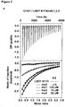
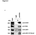
Figure 2 : CHD1 interacts with LSD1 K114me2. (a) Representative isothermal titration calorimetry (ITC) experiments displaying titration of LSD1aa104-127 K114me0, 1, 2, and 3 peptides to CHD1aa270-443. (b) Three-dimensional crystal structure of the CHD1aa270-443/LSD1aa110-116 K114me2 complex. CHD1 is shown as a gray surface representation and the LSD1 peptide as a stick model with C, O and N atoms colored yellow, red and blue, respectively. The location of the CHD1 residues forming the binding grooves for LSD1 K114me2 residues T110, S111, R113, K114me2, R115, and A116 is indicated with black labels, (c) View of a (|Fo|-|Fc|) omit map at 1.6Å resolution contoured at 1σ as a blue mesh around LSD1 K114me2 peptide bound to CHD1. (d) Extracts from 293T cells transfected with Flag-LSD1 or Flag-LSD1 K114A in the absence or presence of G9A were immunoprecipitated with anti-Flag antibody, (e) LNCaP cell lysates were subjected to sucrose gradient centrifugation. Aliquots of the gradient fractions and total cell lysate (Input) were subjected to Western blot analysis, (f) Extracts from LNCaP cells were immunoprecipitated with anti-G9A antibody and subjected to Western blot analysis. Rabbit IgG (rIgG) was used as control, (d-f) Western blots were decorated with the indicated antibodies. -
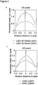
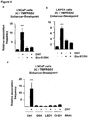
Figure 3 : LSD1 K114me2 controls chromatin binding of AR. (a) Pie charts displaying genomic distribution of LSD1 K114me2 and CHD1 in LNCaP cells cultured in the presence of the AR agonist DHT as determined by ChIP-seq analysis. (b) Venn diagram showing number and intersection of LSD1 K114me2 and CHD1 locations in LNCaP cells cultured in the presence of DHT. (c) Venn diagram showing number and intersection of LSD1 K114me2, CHD1, and AR locations in LNCaP cells cultured in the presence of DHT. (d) Venn diagram showing the intersection and number of genes where LSD1 K114me2 and CHD1 co-localize with genes that are differentially regulated in LNCaP cells upon treatment with DHT. (e,f) Meta-analysis of sequencing read density based on LSD1 K114me2 (e) and CHD1 (f) ChIP-seqs around AR peaks in LNCaP cells cultured in the absence (-DHT) or presence of DHT (+DHT). (g-i) For ChIP, LNCaP cells were cultivated in the presence or absence of DHT, treated with or without Bix-01294 (g) or transfected with siRNA (h, i), as indicated. ChIP analyses were performed with the indicated antibodies. The precipitated chromatin was quantified by qPCR analysis using primers flanking AREs in the enhancer regions of the TMPRSS2 (g,h) or KLK3 (i) genes. (j-l) Treatment with Bix-01294 (j,k) or RNAi-mediated knockdowns of G9A, LSD1, or CHD1 (l) reduces expression of the androgen-regulated TMPRSS2 gene in LNCaP (j, l) and LAPC4 (k) cells. Bars represent mean +SD (n>3). *** p<0.0001; ** p<0.001; * p<0.005 in comparison to cells cultivated in the absence of (-) DHT. -
Figure 4 : LSD1 K114me2 controls TMPRSS2-ERG fusion, (a-e) For 3C experiments, LNCaP (a,c) and LAPC4 (b) cells were cultivated in the presence or absence of DHT, treated with or without Bix-01294 (a,b) or transfected with siRNA (c), as indicated. (d,e) LNCaP cells expressing RNAi resistant (rr) Flag NLS LSD1-rr, Flag NLS LSD1-rr K114A (d), CHD1-rr, or CHD1-rr D425A (e), treated with siRNA as indicated in the presence or absence of DHT were subjected to 3C. (f) TMPRSS2-ERG expression was analyzed in LNCaP cells cultivated in the presence or absence of DHT and transfected with siRNA as indicated, (g) TMPRSS2-ERG fusion was analyzed by FISH in LNCaP cells cultivated in the presence or absence of DHT and transfected with siRNA as indicated. ERG break-apart is indicated (white arrow). Bars represent mean +SD (n>3). *** p<0.0001; ** p<0.001; * p<0.005 in comparison to cells cultured in the absence of (-) DHT (a-f). -
Figure 5 : CHD1 interacts with LSD1 K114me2. (a,b) View of the intermolecular interactions between the bound LSD1 peptide in yellow (a) and H3 peptide in cyan (b) with CHD1 residues (shown in the same orientation in gray), with hydrogen bonds shown as dashed lines. (c) Superimposition of LSD1aa110-116 K114me2 and H3aa1-5 K4me3 in complex with CHD1aa270-443. CHD1aa-270-443 is shown as a gray surface representation. LSD1 and H3 peptides are shown as stick models in yellow and cyan, respectively. The binding surface in CHD1 for inhibitors specifically interfering with LSD1 K114me2 but not H3K4me3 binding is shown in red. - The inventors of the present invention inter alia succeeded in unraveling a novel mechanism that is not only crucial for regulating androgen-dependent gene expression but also controls androgen-dependent chromosomal rearrangement such as the TMPRSS2-ERG oncogenic fusion.
- This mechanism may be referred to as the androgen-regulated G9A/LSD1 K114me2/CHD1 circuit and compounds inhibiting the interaction between LSD1 K114me2 and CHD1 may be used to downregulate or block downstream events of the LSD1 K114me-CHD1-interaction, such as e.g. the downregulation or blocking of androgen-dependent gene expression in prostate cancer.
- As used in the specification and the claims, the singular forms of "a" and "an" also include the corresponding plurals unless the context clearly dictates otherwise.
- The terms "about" and "approximately" in the context of the present invention denotes an interval of accuracy that a person skilled in the art will understand to still ensure the technical effect of the feature in question. The term typically indicates a deviation from the indicated numerical value of ±10% and preferably ±5%.
- It needs to be understood that the term "comprising" is not limiting. For the purposes of the present invention, the term "consisting of' is considered to be a preferred embodiment of the term "comprising of". If hereinafter a group is defined to comprise at least a certain number of embodiments, this is also meant to encompass a group which preferably consists of these embodiments only.
- The term "LSD1" as used herein refers to the enzyme "lysine-
specific demethylase 1" with the UniProt accession number 060341 comprising 852 amino acids. - The term "LSD1me2" is interchangeable used herein with the term "LSD1 K114me2" and refers to LSD1 that is modified at lysine residue 114 by two methyl-groups. Reference to a "methylation of LSD1" also refers to this post-translational modification of LSD1.
- In general, the indication of "me" after the amino acid "K" (i.e. lysine) indicates that the corresponding lysine residue is methylated. The number, e.g. "2" indicates that there are two methyl-groups present at the corresponding lysine, e.g. "K114me2". Another example is "K4me3", where three methyl-groups are present at
lysine 4. - The term "CHD1" as used herein refers to the enzyme "chromodomain-helicase-DNA-binding
protein 1" with the UniProt accession number 014646 comprising 1710 amino acids. CHD1 comprises two chromodomains, namely chromodomain 1 comprising amino acids 268 to 356 andchromodomain 2 comprising amino acids 389 to 443 of CHD1. The term "chromodomain" as used herein refers to a protein structural domain of about 40 to 50 amino acids commonly found in proteins associated with the remodeling and manipulation of chromatin. Chromodomain-containing proteins are responsible for aspects of gene regulation related to chromatin remodeling and formation of heterochromatin regions. Chromodomain-containing proteins bind methylated histones. - The term" H3K4me3" as used herein refers to histone 3 (H3) that is modified at
lysine residue 4 by three methyl-groups. - The term "compound" as used herein refers to an organic compounds, preferably a small organic compound having a low molecular weight (also termed "small molecule" in the respective field). The compound may be a synthetic compound not known to occur in nature or a naturally-occurring compound isolated from or known to occur in natural sources, such as e.g. plants, fungi or the like. Before the background of the present invention, it is important to note that the main focus is on a compound that is capable of inhibiting the interaction between LSD1me2 and CHD1, and that such compounds may e.g. be identified in the method of screening disclosed herein. When reference is made herein to a "compound" within the context of such a method, the term compound may also refer to a "small chemical fragment". This term is typically used in the field of drug discovery and relates to the starting pool of molecules during the process of drug discovery ("fragment-based lead discovery"), wherein a small chemical fragment that has been identified as promising binding partner may undergo structural modifications to increase the binding affinity and/or specificity.
- The term "inhibiting the interaction" as used herein means that preferably no interaction at all (at least not to a detectable level) between two proteins /relevant fragments thereof takes place any more. However, when a given interaction between two proteins (set to 100%) is greatly reduced, e.g. to a level of about 70%, about 60%, about 50%, about 40%, about 30%, preferably about 20%, more preferably about 10% or most preferably about 5% or less, such a reduced interaction is still encompassed by the term "inhibiting the interaction". In terms of the medical use of compound inhibiting an interaction, a complete inhibition of an interaction may not be required to achieve a sufficient therapeutic effect. Thus, it needs to be understood that the term "inhibiting" as used herein also refers to a reduction of an interaction, which is sufficient to achieve the desired effect.
- The term "specifically inhibiting" as used herein means that the inhibition abolishes or reduces only the interaction between protein A and protein B, in the present case between LSD1me2 and CHD1, without e.g. affecting further interactions of LSD1me2 with other proteins and/or of CHD1 with other proteins. This may also be referred to as "inhibiting only" a given interaction. The effect of such a "specific inhibition" is that only the desired downstream effect is affected (in the present case abolished or reduced), whereas other downstream effects mediated via different interactions still take place. Importantly, as outlined below in the example section in more detail, the inhibition according to the present invention is preferably specific for the interaction between LSD1me2 and CHD1 and preferably fails to have an effect on the interaction between CHD1 and H3K4me3.
- The present inventors could show that methylation of LSD1 is executed by G9A in an androgen-dependent manner and serves as a regulatory switch to allow for an interaction with CHD1. The androgen-regulated G9A/LSD1 K114me2/CHD1 circuit controls chromatin binding of AR. Accordingly, the mechanism is not only crucial for regulating androgen-dependent gene expression but also controls androgen-dependent chromosomal rearrangement such as the TMPRSS2-ERG oncogenic fusion during prostate tumor evolution.
- It can be derived inter alia from a superimposition of the crystal structures of CHD1/LSD1 K114me2 and CHD1/H3K4me3 that there is a significant difference in the binding modalities (see
Figure 5 ). This strongly suggests the potential to selectively target the CHD1/LSD1 K114me2 interface without affecting binding of CHD1 to H3K4me3. Accordingly, the binding of CHD1 to H3K4me3 would not be altered, with the result that CHD1 would still be capable of fulfilling its functions mediated via H3K4me3, which are not involved in prostate cancer tumor progression. - Thus, the examples of the present application, in particular examples 2 and 4 revealing the structures and employing specific mutants of either LSD1 and CH1 to study interactions and complex formations, clearly suggest that compounds targeting the CHD1/LSD1 K114me2 interaction represent inhibitors of in particular the AR functions for the treatment of cancer, particularly prostate cancer.
- As indicated above, the method of screening for a compound inhibiting the interaction between LSD1me2 and CHD1 comprises at least the steps of:
- 1) Contacting a compound with CHD1 or a fragment thereof;
- 2) Determining whether said compound binds to CHD1 or a fragment thereof at the binding site for LSD1me2;
- Said method may be carried out in the form of a virtual screening, which is known to the skilled person and which is carried out in accordance with standard and routine practice by the skilled person.
- In a first virtual screening approach, a compound inhibiting the interaction between LSD1me2 and CHD1 may be screened for by i) designing a compound with a similar shape to amino acids T110 to A116 of LSD1me2; ii) contacting said compound with CHD1 by virtually superimposing the structure of said compound and the structure of CHD1 (or a fragment thereof, preferably amino acids 270 to 443 of CHD1); and iii) virtually determining whether said compound binds to CHD1 (or the above fragment thereof) at the binding site for LSD1me2. This approach may be referred to as the "ligand-based" virtual screening approach.
- In a second virtual screening approach, a compound inhibiting the interaction between LSD1me2 and CHD1 may be screened for by i) contacting a compound with CHD1 by docking said compound into the LSD1me2 binding site; and ii) virtually determining whether said compound binds to CHD1 at the binding site for LSD2me2 by applying a scoring function to estimate the likelihood that the ligand will bind to the protein with high affinity, wherein a compound inhibiting the interaction between LSD1me2 and CHD1 binds to CHD1 at the binding site for LSD1me2 and has a relatively high scoring function. This approach may be referred to as the "structure-based" virtual screening approach.
- Following the virtual screening, a compound that has been identified is then usually tested in in vitro test methods, such as e.g. established cell culture models, for efficacy as regards the desired effect. Cell culture models of particular relevance for the present invention are e.g. disclosed in example 3 below.
- The above method may also be carried out by a fragment-based drug discovery method. This method is also standard knowledge for the skilled person in drug discovery, see e.g. http://en.wikipedia.org/wiki/Fragment-based lead discovery. Accordingly, the "compound" referred to in the following is interchangeable with the term "small chemical fragment" or "chemical fragment".
- The following method is preferred when carrying out the method by a fragment-based drug discovery method, wherein said method comprises at least the following steps:
- 1) Contacting a compound with CHD1 or a fragment thereof;
- 2) Determining whether said compound binds to CHD1 or a fragment thereof;
- 3) Determining whether said compound binds to CHD1 or a fragment thereof at the binding site for LSD1me2;
- 4) Optionally modifying the structure of said compound to increase binding affinity and/or specificity to the binding site for LSD1me2;
- The term "modifying" in step 4) as used herein means that specific substituents of the compound may be deleted, added, or substituted by different substituents.
- In this method comprising four steps, it is preferred to use recombinant CHD1 comprising amino acids 260 to 443, i.e. the two chromodomains. Thus, in this preferred embodiment, said compound is contacted in step 1) with recombinant CHD1 comprising amino acids 260 to 443.
- In step 2), it is determined whether said compounds binds to this fragment of CHD1, preferably by a method selected from the group consisting of circular dichroism spectroscopy, differential scanning fluorimetry, nuclear magnetic resonance spectroscopy, isothermal titration calorimetry (see also examples of the present invention, in particular example 6), surface plasmon resonance, and fluorescence polarization. It can be particularly preferred to use differential scanning fluorimetry in step 2). A binding affinity in the range of mM, preferably in the range of about 10 mM and more preferably in the range of 1 mM can be sufficient in this step 2) for the definition of a "binding" to CHD1. This step is introduced in the preferred embodiment to exclude compounds in an initial step that do not bind to CHD1 at all.
- Step 2) may be reiterated with the compounds identified as binding to CHD1 in the initial step 2). This time, more stringent parameters may be used and/or a binding affinity lower than in the mM-range (e.g. in the µM- or even in the nM-range) may be regarded as defining a "binding" of these compounds to CHD1.
- In step 3), it is then determined whether said compound binds to the recombinant fragment of CHD1 discussed above at the binding site for LSD1me2. This can be done in principle by any method selected from the group consisting of nuclear magnetic resonance spectroscopy, mass spectrometry, infrared spectroscopy, Raman spectroscopy, electron microscopy, X-ray crystallography, and combinations thereof. It is particularly preferred to use X-ray crystallography, wherein the structure of the CHD1-fragment is solved in combination with the compound. This may be done by methods routinely known to the skilled person, e.g. by co-crystallization or by soaking of said compound. Reference to examples 2 and 6 of the present invention is made as regards suitable parameters and the like when using X-ray crystallography.
- Following the fragment-based screening method, a compound that has been identified is then usually tested in in vitro test methods, such as e.g. established cell culture models, for efficacy as regards the desired effect. Cell culture models of particular relevance for the present invention are e.g. disclosed in example 3 below.
- All of the above-mentioned methods are carried out according to standard methods known to the skilled person. As explained in the method of screening above, the "target protein" is CHD1 or a fragment thereof, the "target site" is the binding site for LSD1me2, and the compound may be referred to as the "ligand".
- Just by way of example, the method of differential scanning fluorimetry is discussed in more detail in the following. The underlying principle is as follows: Differential Scanning Fluorimetry (DSF) is a thermal denaturation assay that measures the thermal stability of a target protein and a subsequent increase in protein melting temperature upon binding of a ligand to the protein. The binding of low molecular weight ligands can increase the thermal stability of a protein, as described by Koshland in 1958 and Linderstrom-Lang and Schellman in 1959. The thermal stability change is measured by performing a thermal denaturation curve in the presence of a fluorescent dye, such as Sypro Orange. Sypro orange binds nonspecifically to hydrophobic surfaces, and water strongly quenches the fluorescence of Sypro Orange. When the protein unfolds, the exposed hydrophobic surfaces bind the dye, resulting in an increase in fluorescence. The stability curve and its midpoint value (melting temperature, Tm) are obtained by gradually increasing the temperature to unfold the protein and measuring the fluorescence at each point. Curves are measured for protein only and protein + ligand, and ΔTm is calculated. A fluorescence-based thermal shift assay can be performed on instruments that combine sample temperature control and dye fluorescence detection, such as RT-PCR machines. The assay allows high-throughput screening of ligands to the target protein. A typical assay is carried out as follows:
- Materials: A fluorometer equipped with temperature control or similar instrumentation (RT-PCR machines); suitable fluorescent dye; a suitable assay plate, such as 96 well RT-PCR plate.
- Compound solutions: Test ligands are prepared at a 50- to 100-fold concentrated solution, generally in the 10-100 mM range. For titration, a typical experimental protocol employs a set of 12 well, comprising 11 different concentrations of a test compound with a single negative control well.
- Protein solution: Typically, target protein is diluted from a concentrated stock to a working concentration of ∼0.5-5 µM protein with dye into a suitable assay buffer. The exact concentrations of protein and dye are defined by experimental assay development studies.
- Centrifugation and oil dispense: A brief centrifugation (∼1000 × g-force, 1 min) of the assay plate to mix compounds into the protein solution, 1-2 µl silicone oil to prevent the evaporation during heating is overlaid onto the solution, followed by an additional centrifugation step (∼1000 × g-force, 1 min).
- Instrumental set up: A typical temperature ramp rates range from 0.1-10 °C/min but generally in the range of 1 °C/min. The fluorescence in each well is measured at regular intervals, 0.2-1 °C/image, over a temperature range spanning the typical protein unfolding temperatures of 25-95 °C.
- In the following, examples of embodiments of the present invention are outlined.
- To unravel LSD 1-associated mechanisms that might control androgen-dependent TMPRSS-ERG fusion, a global proteomics approach was initiated by performing protein correlation profiling (PCP) of human prostate tumor LNCaP cells that were stable isotope labelled by amino acid in cell culture (SILAC). Cluster analysis of the PCP-SILAC data revealed that LSD1 shares a similar protein enrichment profile with 165 proteins. Among these proteins, the histone methyltransferase G9A was identified (
Fig 1a ). Inspection of the LSD1 protein sequence suggested a potential methylation site at lysine (K) 114. Subsequently, a rabbit polyclonal antibody specific for LSD1 K114me2 was developed according to standard methods. To test whether G9A methylates LSD1 at K114 either Flag-tagged LSD1 or a LSD1 protein containing a K114 to alanine (A) mutation (LSD1 K114A) in the presence or absence of G9A was expressed in 293T cells. Cell extracts were immunoprecipitated with an anti-Flag antibody and subjected to Western blot analysis using an antibody specific for LSD1 K114me2 (Fig. 1b ). LSD1 but not LSD1 K114A is methylated in presence of G9A (Fig. 1b ). In LNCaP cells, it was observed that approximately 6% of total LSD1 is dimethylated at K114 (data not shown). To provide evidence that methylation of LSD1 at K114 by G9A is direct, recombinantly expressed and purified GST-LSD1 or GST-LSD1 K114A was incubated with G9A in the presence or absence of the methyl donor S-adenosyl methionine (SAM). As expected, GST-LSD1 but not GST-LSD1 K114A is methylated at K114 by G9A in the presence of SAM (Fig. 1c ). Finally, Western blot analyses indicated that LSD1 K114me2 is present in various cell lines including normal prostate and prostate tumor cells (Figure 1d ,e ). Taken together, the data unravel that LSD1 is methylated by G9A at K114 both in vitro and in vivo. - To identify potential LSD1 K114me2 methyl-lysine reader proteins, protein profiling was performed on protein-domain microarrays using un- and dimethylated LSD1 peptides LSD1aa104-127 and LSD1aa104-127 K114me2, respectively (see also ref.14). In this profiling assay, several GST-fusion proteins including CHD1 fused to GST were used. Other GST-fusions that were used were e.g. PhD-domain proteins (BPTF, ING2 or PHF2), BROMO-domain proteins (GCN5, TAF1-D1 or TAF1-D2), or BRCT-proteins (BRCA1, 53BP1 or Crb2). Among the proteins present on the microarray, LSD1 K114me2 but not unmethylated LSD1 peptide, specifically interacted with CHD1 double chromodomain (GST-
CHD1 Chromo 1/2) indicating that CHD1 is a reader of LSD1 K114me2. Using isothermal titration calorimetry (ITC), the dissociation constants (Kd) of 124 µM, 74 µM, and 82 µM for LSD1 K114me1, me2, and me3 peptides, respectively, were measured (Figure 2a ). These binding affinities are similar to those observed for binding of H3K4me3 peptide to CHD1 (namely a Kd=47 µM, data not shown). Unmethylated LSD1 or H3 peptide did not bind to GST-CHD1 Chromo 1/2 (Figure 2a ). - To investigate the molecular basis for LSD1 K114me2 recognition, the co-crystal structure of CHD1aa270-443 bound to LSD1aa108-119 K114me2 peptide at a resolution of 1.6 Å was solved (
Figure 2b and Supplementary Table 1 shown in the following).Supplementary Table 1 Data collection and Refinement Statistics. Data Set CHD1-LSD1me2 X-ray source Diamond Light Source, 104 X-ray detector Dectris Pilatus-6M Wavelength 0.9795 space group P2 1212 cell constants a, b, c [Å] 110.30, 44.44, 46.19 α, β, γ [°] 90.0, 90.0, 90.0 Molecules in asymmetric unit cell 1 resolution limits [Å] 55.15 - 1.60 (1.62 - 1.60) completeness (%) 98.8 (83.9) unique reflections 30,650 (1282) multiplicity (%) 4.4 (4.0) Overall B-factor, Wilson plot (Å2) 41 R merge a) 0.029 (1.41) Rmeas 0.037 (1.79) R p.i.m. 41 0.023 (0.84) mean I/σ(I) 16.4 (0.8) CC1/2 42 0.999 (0.554) refinement statistics R cryst b) 0.228 R free (%) 0.266 non-hydrogen atoms 1623 solvent molecules 89 Cruickshank's DPI43 0.100 r.m.s. deviations from ideal values bond lengths (Å) 0.019 bond angles (°) 1.91 average B values (Å2) protein main chain atoms 54.1 protein all atoms 54.2 solvent 47.3 a R merge = Σ hld [(∑ I |II - <I> |) / ∑ I II ]
b R cryst = ∑ hkl ||F obs | - |F calc || / ∑ hkl |F obs |
R free is the cross-validation R value for a test set of 5 % of unique reflections44. - Amino acid residues 110-116 of the LSD1 peptide were modelled according to the electron density (
Figure 2c ). Comparable to the binding of H3K4me3 peptide15, the LSD1 K114me2 peptide is located at an acidic surface bridging the two chromodomains (Figure 2b ). This interaction involves residues Y295, W322, W325 fromchromodomain 1 and D408 and D425 from chromodomain 2 (Figure 2b andFigure 5a ). The methylammonium group of LSD1 K114 and side chains of W322 and W325 of CHD1 form cation-π interactions. Notably, the side chains of R113 of LSD1 and D425 of CHD1 interact through an ordered water molecule (seeFigure 5a ) and superimposition of the crystal structures of CHD1/LSD1 K114me2 and CHD1/H3K4me3 showed that such an interaction cannot occur between CHD1 and H3K4me3 since the residue corresponding to LSD1 R113 is a threonine at position 3 (T3) in H3K4me3 (seeFigures 5a to c ). It was therefore hypothesized that the R113-D425 interaction may be a discriminating feature of CHD1 binding either LSD1 K114me2 or H3K4me3 peptide and that the corresponding surface could be exploited for the design of inhibitors specifically interfering with LSD1 K114me2 but not H3K4me3 interaction (seeFigure 5c ). Indeed, mutagenesis of either LSD1 R113 or CHD1 D425 to an alanine (A) abolished the interaction of CHD1 with LSD1 K114me2 peptide, as determined by isothermal titration calorimetry (data not shown). In contrast, the D425A mutation in CHD1 only mildly affected H3K4me3 binding (data not shown). Taken together, these observations suggest the possibility to specifically target the association of CHD1 and LSD1 K114me2 without affecting CHD1/H3K4me3 binding. Next, it was tested whether in vivo the interaction of CHD1 with LSD1 depends on LSD1 methylation. In the presence of G9A, endogenous CHD1 only coimmunoprecipitated with methylated Flag-LSD1, but not with Flag-LSD1 K114A (Figure 2d ). To investigate whether CHD1 and LSD1 K114me2 are present in the same protein complex, LNCaP cell extracts were subjected to sucrose gradient centrifugation. Western blot analyses showed that CHD1, LSD1 K114me2, and G9A co-fractionate, suggesting the existence of a G9A/LSD1 K114me2/CHD1 complex (Figure 2e ). Furthermore, endogenously expressed CHD1 and LSD1 K114me2 coimmunoprecipitated with G9A from LNCaP cell extracts (Figure 2f ). Together, these observations identify CHD1 as a LSD1 K114me2 reader. - To investigate whether recognition of LSD1 K114me2 by CHD1 on chromatin is involved in androgen-dependent gene regulation, we treated LNCaP cells with the AR agonist dihydrotestosterone (DHT) and performed ChIP-seq with either anti LSD1 K114me2 or anti-CHDl antibodies. The analyses shown in
Figure 3a identified 95101 high confidence LSD1 K114me2 peaks and 55987 high confidence CHD1 peaks, respectively. The overlap of LSD1 K114me2 with CHD1 locations revealed that 9660 locations are co-occupied (Figure 3b ). Intersection ofLSD 1 K114me2 and CHD1 cistromes with previously published genome-wide AR binding data16 revealed that 2941 locations representing 2397 genes are co-occupied (Figure 3c ). To correlate the presence of LSD1 K114me2 and CHD1 peaks with androgen -regulated gene expression, a global transcriptome analysis (RNA-seq) of LNCaP cells cultured in absence or presence of DHT was performed. Among the 2397 genes co-occupied by LSD1 K114me2, CHD1, and AR (Figure 3c ), 861 were differentially regulated upon treatment of LNCaP cells with DHT (Figure 3d ), supporting the idea that LSD1 K114me2/CHD1 interaction plays a role in AR signalling. The bioinformatic analyses indicated an increase of LSD1 K114me2 and CHD1 reads over the AR peaks in DHT compared to vehicle-treated cells (Figure 3e,f ). Based on these observations, it was hypothesized that subsequent to DHT treatment the elevated levels of LSD1 K114me2 and CHD1 at the AR occupied regions might promote binding of AR. To test this idea, LNCaP cells cultured in the presence or absence of DHT and the G9A inhibitor Bix-012941717 were subjected to ChIP. DHT treatment resulted in elevated LSD1 K114me2 levels accompanied by increased CHD1 and AR occupancy of the TMPRSS2 and KLK3 genes (Figure 3 ). Chromatin occupancy of G9A was not altered by the presence or absence of DHT and Bix-01294. Whereas LSD1 enrichment increased upon DHT treatment at the TMPRSS2 enhancer (-13782 /-13866), this was not the case at the KLK3 enhancer (-3980 /-4048, data not shown). The results are cell type-independent since similar data were obtained with LAPC4 androgen-dependent prostate tumor cells (data not shown). Specificity was controlled for by also using an unrelated region that is not occupied by AR, wherein the effects was not observed. Additionally, chromatin was not enriched when using an IgG control (Figure 3g ). Importantly, when cells were cultured in the presence of Bix-01294, androgen-induced methylation of LSD1 as well as androgen-induced recruitment of CHD1 and AR was blocked (Figure 3g ). Next, it was asked whether knockdown of G9A mimics the observed Bix-01294 effect. Accordingly, LNCaP cells cultured in the presence or absence of DHT were transfected with either an unrelated control siRNA or a siRNA directed against G9A and subjected to ChIP. Similar to the treatment with Bix-01294, ligand-induced methylation of LSD1 at K114, as well as recruitment of CHD1 and AR, was robustly impaired upon knockdown of G9A (Figure 3h ). Together, these data indicate that CHD1 and AR chromatin occupancy is critically dependent on LSD1 K114me2. - To further decipher the mechanism underlying DHT-induced methylation of LSD1, binding of CHD1 to LSD1 K114me2, and recruitment of AR to androgen response elements (AREs) we performed CHD1 RNAi. Loss of CHD1 did not affect chromatin occupancy of LSD1 but severely reduced methylation of LSD1, which correlates with loss of G9A and block of ligand-induced AR recruitment (
Figure 3i ). These data clearly demonstrate that AR binding to AREs not only requires methylation of LSD1, but also the presence of CHD1. Next, the effect of Bix-01294 treatment on the expression of representatives of the 2397 AR target genes that show co-localization of AR, LSD1 K114me2, and CHD1 was analyzed. DHT-induced expression of genes such as TMPRSS2, KLK3, PRDM4, ELK4, GREB1, IGF1R, FKBP5, or EGFR was greatly reduced upon treatment of LNCaP or LAPC4 cells with Bix-01294 (Figure 3j,k ). Similarly, robust effects were observed upon RNAi mediated knockdown of G9A, LSD1, or CHD1 (Figure 3l ). In summary, the results reveal that during AR-dependent gene expression, methylation of LSD1 at K114 by G9A and recruitment of CHD1 to AR binding regions is a key event controlling chromatin binding of AR. - Since it was shown that androgen-dependent methylation of LSD1 at K114 by G9A and recruitment of CHD1 control binding of AR at the enhancer of the TMPRSS2 gene, it was tested whether AR transcription-associated genomic translocations such as TMPRSS2-ERG fusion are controlled by LSD1 K114 methylation. It has been shown that androgen-induced binding of AR to AREs causes chromosomal conformational changes resulting in looped chromatin necessary for coordinated transcription18. At the TMPRSS2 locus, such chromosomal interactions involve the looping of the enhancer region with a region in
intron 1 where genomic breaks occur during the generation of TMPRSS2-ERG gene fusion4. To elucidate whether LSD1 K114me2 might control androgen-dependent TMPRSS2 enhancer-breakpoint loop formation LNCaP cells were cultured in the absence and presence of DHT and subjected to chromosome conformation capture (3C) analysis. In agreement with a previous report4, enhancer-breakpoint interactions upon treatment with DHT was observed. Importantly, however, treatment with Bix-01294 blocked this loop formation in LNCaP and LAPC4 cells demonstrating that methylation of LSD1 is necessary for TMPRSS2 enhancer-breakpoint loop formation (Figure 4a,b ). In corroboration, knockdown of either G9A, LSD1, or CHD1 also robustly impaired androgen-induced looping (Figure 4c ). To further highlight the importance ofLSD 1 K114me2 in the control of TMPRSS2 enhancer-breakpoint loop formation, it was tested whether androgen-dependent loop formation is compromised in presence of the LSD1 K114A mutant. Therefore, LNCaP cells expressing RNAi resistant (rr) Flag-NLS-LSDl-rr and Flag-NLS-LSDl-rr K114A constructs were generated. Upon knockdown of endogenous LSD1, DHT-induced looping was observed in LNCaP cells expressing Flag-NLS-LSDl-rr but not in the cells expressing the Flag-NLS LSD1-rrK114A mutant (Figure 4d ). These data further highlight the importance of LSD1 methylation at K114 for the formation of androgen-dependent chromatin loops. In a similar approach, it was tested whether the mutant protein CHD1 D425A, which fails to interact with LSD1 K114me2 peptide, impaires androgen-dependent chromatin looping. Consequently, LNCaP cells expressing RNAi resistant CHD1-rr and CHD1-rr D425A were generated. Knockdown of endogenous CHD1 allows DHT-induced looping in LNCaP cells expressing CHD1-rr but not in the cells expressing the CHD1-rr D425 mutant protein demonstrating the importance of D425 of CHD1 for the interaction with methylated LSD1 and subsequent loop formation (Figure 4e ). Together, these data demonstrate that DHT-induced chromosomal conformational changes observed at the TMPRSS2 enhancer-breakpoint regions need the integrity of the G9A/LSD1 K114me2/CHD1 circuit. Recently, Haffner et al. reported that DHT-treatment alone is sufficient to induce TMPRSS2-ERG fusion in a small proportion of prostate cells4. Thus, it was wondered whether DHT-induced TMPRSS2-ERG fusion depends on LSD1 K114me2 and, if so, might be blocked upon knockdown of LSD1, G9A, or CHD1. Consistant with Haffner et al., DHT-treatment of LNCaP cells led to the production of TMPRSS2-ERG transcript and induced ERG rearrangements, as observed by FISH (Figure 4f,g ). Knockdown of either G9A, LSD1, or CHD1 severely reduced DHTinduced expression of TMPRSS2-ERG transcripts and, importantly, TMPRRS2-ERG gene rearrangements were blocked (Figure 4f,g ). - Plasmids: Flag-LSDl and GST-LSD1 were described previously19. Flag-G9A was provided by Y. Shinkai. Flag-LSDl K114A was obtained by site directed mutagenesis. To construct expressions plasmids for GST-LSD1, GST-LSD1 K114A, or CHD1aa270-443, the corresponding cDNA fragments were cloned into pFast-Bac-HT-GST A, pDEST20, or pET15b.
- Chromatin immunoprecipitation (ChIP): ChIP experiments were performed essentially as previously described10. LNCaP and LAPC4 cells were cultured for 210 min in the absence or presence of 1 x 10⁻⁷ M DHT as indicated. Three days before harvesting, cells were transfected with siRNA against LSD110, G9A20, or CHD121 that were previously described and validated using Dharmafect2 (Thermo Scientific) following the manufacturer's instructions. Immunoprecipitation was performed with specific antibodies (α-AR (N-20), #sc-816, lot A0207, Santa Cruz), α-LSD1 (#20752, Schuele Laboratory), α-LSD1 K114me2 (#4871, Schuele Laboratory), α-CHD1 (#A301-218A, lot A301-218A-1, Bethyl Laboratories), α-G9A (#ab40542, lot GR170862-1, Abcam) on protein A-Sepharose 4B (GE-Healthcare). For PCR, 2 µl out of 70 µl DNA extract were used. Primer sequences were as follows: TMPRSS2 enhancer (-13782/-13866) 5'-GCCACCTGGTGAAGTGCAGA-3' (SEQ ID NO:1) and 5'-TGGAGCTAGTGCTGCATGTC-3' (SEQ ID NO:2); KLK3 enhancer (-3980/-4048) 5'-TTATCTAGGACAGTAAGCAAGCC-3' (SEQ ID NO:3) and 5'-GGATGTTTGTAAAGCAGGCAT-3' (SEQ ID NO:4); control (chr6:151953789-151953927) 5'-TCCTAATGACATTTGATGTTCAGC-3' (SEQ ID NO:5) and 5'-CCCTTAAAAGTCAACAACAAGA-3' (SEQ ID NO:6).
- ChIP sequencing (ChIP-seq): Libraries were prepared from immunoprecipitated DNA according to standard methods. ChIP-seq libraries were sequenced using a HiSeq 2000 (Illumina) and mapped to the hg19 reference
genome using bowtie 222. Data were further analyzed using the peak finding algorithm MACS 1.4123 using input as control. All peaks with FDR greater than 1 % were excluded from further analysis. The uniquely mapped reads were used to generate the genome-wide intensity profiles, which were visualized using the IGV genome browser24. HOMER25 was used to annotate peaks, to calculate overlaps between different peak files, and for motif searches. The genomic features (promoter, exon, intron, 3'UTR, and intergenic regions) were defined and calculated using Refseq and HOMER. Previously reported ChIP-seq results for AR (GSE28264) were used for analysis. - Western blot analysis: Experiments were performed as previously described10.
- Methylation assay: One µg of purified recombinant GST-LSD1 or GST-LSD1 K114A were incubated with 1µg of purified recombinant GST-G9Aaa786-1210) (Reaction Biology Corp.) in the presence of 160 µM S-adenosyl-methionine (New England BioLabs) as indicated in methylation buffer (50 mM Tris HCl pH 8.5, 50 mM NaCl, 5 mM MgC12, 1 mM DTT, and protease inhibitor cocktail (Roche)) for 120 min at 37°C. The reaction mixture was analyzed by Western blotting using antibodies as indicated.
- Isothermal titration calorimetry (ITC): ITC experiments were performed at 25 °C with a MicroCal VP-ITC microcalorimeter (GE Healthcare). Experiments were performed by injecting 12 µl of peptide (1.5 mM) into the sample cell containing 100 µM CHD1 in 20
mM BTP pH - X-ray crystallography: The CHD1-LSD1 complex was formed by mixing CHD1 (14 mg ml-1) with an eightfold molar excess of LSD1aa108-119 K114me2 peptide. Crystals (space group P21212) were grown by sitting drop vapour diffusion at 4°C in buffer containing 100 mM Hepes pH 7.5, 0.2 M L-proline, 10% (w/v) PEG3350. Prior to flash cooling in liquid nitrogen, crystals were cryoprotected in reservoir solution with 20% (v/v) ethylene glycol. Initial X-ray diffraction data were collected at the PXI beamline at the Swiss Light Source at 1 = 1.000 Å with a Pilatus detector. The data set for the final refined model was collected at a 100K at beamline I04 at Diamond Light Source. Data were processed and analyzed with XDS28 and Aimless29. The structure was solved by molecular replacement with Phaser30 using 2B2W as a search model. Manual building and refinement were performed using Coot31 and Refmac532 in the CCP4 program suite33. Supplementary Table 1 summarises the statistics for data collection and refinement. Structural figures were generated in PyMOL.
- RNA preparation and analysis: RNA was isolated with TRIzol Reagent (Invitrogen). Two micrograms of RNA were converted to cDNA with SuperScript II reverse transcriptase (Invitrogen) and polyT oligonucleotides according to the supplier's protocol. Quantitative RT-PCR was performed using the Abgene SYBR Green PCR kit (Invitrogen) according to the supplier's protocol. HPRT was used for normalization. Primer sequences for HPRT, TMPRSS2, KLK3, PRDM4, ELK4, GREB1, IGF1R, FKBP5, and EGFR were described previously19.
- TMPRSS2-ERG expression analysis: Expression analysis of TMPRSS2-ERG (
Fig. 4f ) was done essentially as previously described4. RNA was isolated from LNCAP cells and first strand synthesis was carried out using Superscript II (Life Technologies) with primers specific to ERG (5'-AACTGCCAAAGCTGGATCTG-3') (SEQ ID NO:7) and HPRT (5'-CCAGGTAGCTGGGTTACAGG-3') (SEQ ID NO:8) according to the supplier's protocol. Probes were analyzed on a Roche LightCycler®480 by TaqMan-based qPCR using primers and probes specific for TMPRSS-ERG (#4331182, Life Technologies) and HPRT (#4331182, Life Technologies). HPRT was used for normalization. - RNA sequencing (RNA-seq): RNA samples were sequenced by the standard Illumina protocol to create raw sequence files (.fastq files) at the sequencing core facility of the DKFZ. These reads were aligned to the hg19 build of the human genome using
TopHat version 234. The aligned reads were counted with the homer software (analyze RNA) and DEG's were identified using EdgeR35 and DESeq version 1.8.336. - Stable isotope labelling by amino acids in cell culture (SILAC): For SILAC, LNCaP cells were cultured for six passages in SILAC Dulbecco's modified Eagle's medium (Silantes) containing 10% dialyzed fetal bovine serum (Silantes), penicillin/streptomycin (Life Technologies) and glutamine (Life Technologies) and were labeled with either L-lysine and L-arginine (Lys0, Arg0, Silantes) or L-lysine13C6-15N2 and L-arginine13C6-15N4 (Lys8, Arg10, Silantes).
- PCP-SILAC and mass spectrometry: Protein correlation profiling (PCP) was essentially done as described37. Light (Lys0, Arg0) and heavy (Lys8, Lys10) labelled LNCaP cells were harvested and resuspended in nuclear isolation buffer (10mM HEPES-KOH pH 7.9, 1.5 mM MgC12 , 10 mM KCl, 0.5 mM DTT, Complete protease inhibitor cocktail, Roche), allowed to swell on ice for 10 min, vortexted for 10 seconds at low speed, and pelleted. Isolated nuclei were resuspended in nuclear disruption buffer (20 mM HEPES-KOH pH 7.9, 25% glycerol, 420 mM NaCl, 1.5 mM MgC12, 0.2 mM EDTA, 0.5 mM DTT, Complete protease inhibitor cocktail), remained on ice for 20 min and centrifuged at 14 000 rpm for 10 min. The supernatants comprising the nuclear extracts of heavy and light cells were collected, and applied to a sucrose gradient made up of 5 times 1.5 ml fractions of sucrose ranging from 25 to 40% (50 mM Tris-HCl pH 8.0, 100 mM NaCl, Complete protease inhibitor cocktail, sucrose in varying concentrations). Protein complexes were separated on the gradient at 36 000 rpm for 14 h and fractions of 1 ml were collected.
- The Lys0/Arg0 PCP-SILAC fractions were combined and spiked in a 1:1 ratio to the respective Lys8/Arg10 PCP-SILAC fractions. The resulting fractions were resuspended in SDS loading buffer, reduced and alkylated with DTT (Sigma-Aldrich) and iodoacetamide (Sigma-Aldrich), respectively. The fractions were then concentrated with a 10 kDa cut-off filter. Protein mixtures were separated by SDSPAGE (4-12% Bis-Tris gradient gel, NuPAGE; Life Technologies) and in-gel digested with trypsin (Promega) at 37°C ON. All peptide fractions were analyzed using an LTQ Orbitrap XL mass spectrometer (Thermo Fisher Scientific) coupled to an Agilent 1200 (Agilent Technologies) or an Eksigent 2D nanoflow-HPLC (AB Sciex). The reverse phase HPLC column was self-packed in a fused silica capillary (ReproSil-Pur® 3µm) with 75 µm inner diameter to a length of 25 cm. Peptides were loaded onto the LC-system by an autosampler and separated by a gradient of buffer A (0.5% acetic acid in water) and buffer B (0.5% acetic acid in 80% ACN/water) with increasing organic proportion (loading of sample with 2% of solvent B; first separation ramp from 2 to 30% B within 105 min, and second ramp from 30 to 80% B within 10 min).
- The mass spectrometer was operated in the data-dependent mode. Following the survey scan of 1 × 10⁶ ions in the Orbitrap, the five most abundant multiply-charged ions were fragmented in the linear ion trap using 35% collision energy and a target value of 5000. Parent ions with a charge state of z = 1 and unassigned charge states were excluded for fragmentation. Further MS/MS settings were: repeat duration (30 s), repeat count (1), exclusion duration (90 s), and isolation width (2). For MS/MS wideband activation was enabled. Remaining settings were set to default. The mass range for MS was m/z = 350 to 2000, and signal threshold was 1000. The resolution was set to 60000. Mass-spectrometric parameters were as follows: spray voltage 2.3 kV; no sheath and auxiliary gas flow; ion-transfer tube temperature 200 °C.
- Mass spectrometric data analysis: Raw files were analyzed with the MaxQuant software (version 1.4.1.2.)38,39. Cysteine carbamidomethylation was set as fixed modification, methionine oxidation, protein amino-terminal acetylation and pyroglutamate and pyro-glutamic acid formation from glutamate and glutamic acid, respectively, were set as variable modifications. Double SILAC was chosen as quantification mode. Two miss cleavages were allowed, enzyme specificity was trypsin/P, precursor ion mass tolerance was 20 ppm for first search, and fragment ion mass tolerance was 0.5 Da for MS/MS spectra. A false discovery rate (FDR) of smaller 1% on peptide and protein level was determined by using a forward-reverse decoy database (Uniprot Human, version July 2014). A minimum length of seven amino acids per peptide was used. Both unmodified and modified and both razor and unique peptides with a minimum ratio count of 2 were used for protein quantification.
- Cluster Analysis: The proteins were only included in cluster analysis if respective peptides were sequenced in each of the eight fractions yielding eight data point profiles. As an additional quality criterion profiles were checked for apparent outliers. Protein profiles were defined as outliers when their summed ratios were not within the range of three times the standard deviation of the average summed ratio, and removed from datasets before cluster analysis. The protein ratios were normalized using the
base 2 logarithm. Clustering was done with the k-means algorithm. To define the appropriate number of centroids for cluster analysis the algorithm was run multiple times with initial randomly selected cluster centers and checked for consistent results. Optimal configuration was obtained with a k value of 20 centroids and applied in the present analysis. - Cell culture: LNCaP cells were cultured and transfected as described10. 293T cells were cultured in DMEM and LAPC4 in IMDM supplemented with 10% FCS glutamine and penicillin/streptomycin. Ten µg of expression plasmid coding for Flag-
LSD 1, Flag-LSD1 K114A, or Flag-G9A were transfected per 293T dish and cultivated for 24 hours before harvesting (Fig. 1b ). Three days before harvesting for ChIP (Fig. 3g-i ), ChIP-seq (Fig. 3a ), Western blot analysis, expression analysis (Fig. 3k ), or3C (Fig. 4c ,d,e ), LNCaP cells were transfected with siRNA. For FISH and analysis of TMPRSS2-ERG expression (Fig. 4f,g ) cells were transfected with siRNAs height and five days before harvesting; DHT 10-6 M was added to the cells as indicated 3 days before harvesting. Cells were cultivated for 72 hours in medium supplemented with 10% FCS with 1 x 10⁻⁵ M Bix-01294 as indicated. For ChIP and ChIP-seq, cells were cultivated for 210 min with or withoutDHT 10-7 M and 6 hours for expression analysis. Cells were cultivated for 3 days (Fig. 4f, g ) with DHT 10-6 M as indicated. - Chromosome conformation capture (3C): 3C was essentially performed as previously described40. LNCaP cells were grown for 72 hours in medium supplemented with 10% double-stripped FCS, treated for 210 min with or without DHT 10-7 M, washed in PBS, and trypsinized 2 min at 37 °C. After washing, cells were resuspended in PBS, filtered through a 40 µm cell strainer and collected in an ice cold 2% formaldehyde solution. After 15 min incubation at 4 °C, fixation was stopped by adding glycine and cells were pelleted. After washing, cells were resuspended in nuclear isolation buffer (10 mM HEPES-
KOH pH µl 20% SDS solution was added and incubated for 20 min at 65 °C to inactivate the restriction enzyme. Then, 6.2 ml ligation buffer without ATP and 375 µl Triton X-100 were added and incubated for 1h at 37°C. 60 µl of 100 µM ATP solution and 2000U T4 DNA Ligase were added. Following incubation for 4h at 16 °C and 30 min at RT, Proteinase K was added and reversal of cross-Links was performed by overnight incubation at 65 °C. Upon digestion with RNAse A, DNA was isolated by phenol-chloroform extraction followed by a purification step with QIAgen PCR purification kit. Purified DNA samples were analyzed on a Roche LightCycler®480 by TaqMan-based qPCR. The TaqMan primers and probe for Enhancer-Breakpoint were: FW: 5'-GAGCCATGAAAGTCTTTGCTATGA-3' (SEQ ID NO: 9), probe 5'-AAGTTCACACCTTCCTGGGT-3' (SEQ ID NO:10), RV: 5'- GTGATCCTTCCCACCTGTACATT-3' (SEQ ID NO:11). The PCR master mix was prepared as follows: 10 µl Fast universal Master mix (Life Technologies), 7 µl H2O, 1 µl TaqMan primers and probe and 300ng DNA template. To verify Enhancer-Breakpoint PCR product, samples were analyzed on a 2% agarose gel and sequenced. Nomalization was done against the product obtained for Ctrl corresponding to a genomic DNA region amplified by following primers: 5'-AGGACACAGGAAAGCGATGG-3' (SEQ ID NO:12) and 5'-AGAGGGCTGTCTTGATTCGC-3' (SEQ ID NO:13). - Fluorescence in-situ hybridization (FISH): To determine TMPRSS2-ERG fusion status in LNCaP cells, a FISH break-apart assay was performed as described previously41. The FISH probe consists of two BAC clones spanning the centromeric and telomeric region of the ERG locus on chromosome 21q22.2: RP11-24A11 labelled with biotin-14-dCTP (red) and RP11-137J13 labelled with digoxigenin-dUTP (green). After harvesting, LNCaP cells were incubated with 0.075 M KCl at 37 °C for 15 min and fixed on slides (acetic acid/methanol 1:3). Slides were pretreated with 2x SSC at 37 °C for 60 min, digested with 1:25 Digest-All III at 37 °C for 6 min and dehydrated in an ascending ethanol series. The probes were denatured at 73 °C for 5 min and subsequently co-denaturized with the cells (85 °C, 4 min). Hybridization of the two probes was performed at 37 °C overnight. Post hybridisation washing was done with 0.5x SSC at 75°C for 5 min and the fluorescence detection was carried out using streptavidin-Alexa-594 and FITC-anti-DIG conjugates (dilution 1:250). Finally slides were counterstained with DAPI and mounted and independently evaluated by two persons.
- Data analysis: Data are represented as mean + standard error of the mean (SEM). Significance was calculated by a two-tailed Student's t test.
-
- 1: Baca, S. C. et al. Punctuated evolution of prostate cancer genomes. Cell 153, 666-677 (2013).
- 2: Tomlins, S. A. et al. Recurrent fusion of TMPRSS2 and ETS transcription factor genes in prostate cancer. Science 310, 644-648 (2005).
- 3: Lin, C. et al. Nuclear receptor-induced chromosomal proximity and DNA breaks underlie specific translocations in cancer. Cell 139, 1069-1083 (2009).
- 4: Haffner, M. C. et al. Androgen-induced TOP2B-mediated double-strand breaks and prostate cancer gene rearrangements. Nat. Genet. 42, 668-675 (2010).
- 5: Siegel, R., Naishadham, D. & Jemal, A. Cancer statistics, 2013. C.A. Cancer J. Clin. 63, 11-30 (2013).
- 6: Taylor, B. S. et al. Integrative genomic profiling of human prostate cancer. ).
- 7: Rubin, M. A., Maher, C. A. & Chinnaiyan, A. M. Common gene rearrangements in prostate cancer. J. Clin. Oncol. 29, 3659-3668 (2011).
- 8: Barbieri, C. E. et al. Exome sequencing identifies recurrent SPOP, FOXA1 and MED12 mutations in prostate cancer. Nat. Genet. 44, 685-689 (2012).
- 9: Burkhardt, L. et al. CHD1 is a 5q21 tumor suppressor required for ERG rearrangement in prostate cancer. Cancer Res. 73, 2795-2805 (2013).
- 10: Metzger, E. et al. LSD1 demethylates repressive histone marks to promote androgen-receptor-dependent transcription. Nature 437, 436-439 (2005).
- 11: Kahl, P. et al. Androgen receptor coactivators lysine-).
- 12: Cai, C. et al. Androgen receptor gene expression in prostate cancer is directly suppressed by the androgen receptor through recruitment of lysine-).
- 13: Shi, Y. et al. Histone demethylation mediated by the nuclear amine oxidase homolog LSD1. Cell 119, 941-953. (2004).
- 14: Kim, J. et al. Tudor, MBT and chromo domains gauge the degree of lysine methylation. EMBO Rep. 7, 397-403 (2006).
- 15: Flanagan, J. F. et al. Double chromodomains cooperate to recognize the methylated histone H3 tail. Nature 438, 1181-1185 (2005).
- 16: Tan, P. Y. et al. Integration of regulatory networks by NKX3-1 promotes androgen-dependent prostate cancer survival. Mol. Cell. Biol. 32, 399-414 (2012).
- 17: Kubicek, S. et al. Reversal of H3K9me2 by a small-molecule inhibitor for the G9a histone methyltransferase. Mol. ).
- 18: Wang, Q., Carroll, J. S. & Brown, M. Spatial and temporal recruitment of androgen receptor and its coactivators involves chromosomal looping and polymerase tracking. Mol. Cell 19, 631-642 (2005).
- 19: Metzger, E. et al. Phosphorylation of histone H3T6 by PKCbeta(I) controls demethylation at histone H3K4. Nature 464, 792-796 (2010).
- 20: Lee, D. Y., Northrop, J. P., Kuo, M. H. & Stallcup, M. R. ).
- 21: Huang, S. et al. Recurrent deletion of CHD1 in prostate cancer with relevance to cell invasiveness. Oncogene 31, 4164-4170 (2012).
- 22: Langmead, B., Trapnell, C., Pop, M. & Salzberg, S. L. Ultrafast and memory-efficient alignment of short DNA sequences to the human genome. Genome Biol. 10, R25 (2009).
- 23: Zhang, Y. et al. Model-based analysis of ChIP-Seq (MACS). Genome Biol. 9, R137 (2008).
- 24: Thorvaldsdottir, H., Robinson, J. T. & Mesirov, J. P. Integrative Genomics Viewer (IGV): high-performance genomics data visualization and exploration. Brief Bioinform. 14, 178-192 (2013).
- 25: Heinz, S. et al. Simple combinations of lineage-determining transcription factors prime cis-regulatory elements required for macrophage and B cell identities. Mol. Cell 38, 576-589 (2010).
- 26: Keller, S. et al. High-precision isothermal titration calorimetry with automated peak-shape analysis. Anal Chem. 84, 5066-5073 (2012).
- 27: Schuck, P. Size-distribution analysis of macromolecules by sedimentation velocity ultracentrifugation and lamm equation modeling. Biophys. J. 78, 1606-1619 (2000).
- 28: Kabsch, W. XDS. Acta Crystallogr. D. Biol. Crystallogr. 66, 125-132 (2010). 29: Evans, P. R. & Murshudov, G. N. How good are my data and what is the resolution? Acta Crystallogr. D. Biol. Crystallogr. 69, 1204-1214 (2013).
- 30: McCoy, A. J. et al. Phaser crystallographic software. J. Appl. Crystallogr. 40, 658-674 (2007).
- 31: Emsley, P., Lohkamp, B., Scott, W. G. & Cowtan, K. Features and development of Coot. Acta Crystallogr. D. Biol. Crystallogr. 66, 486-501 (2010).
- 32: Murshudov, G. N., Vagin, A. A. & Dodson, E. J. Refinement of macromolecular structures by the maximum-likelihood method. Acta. Crystallogr. D. Biol. Crystallogr. 53, 240-255 (1997).
- 33: The CCP4 suite: programs for protein crystallography. Acta Crystallogr. D. Biol. Crystallogr. 50, 760-763 (1994).
- 34: Trapnell, C. et al. Differential gene and transcript expression analysis of RNA-seq experiments with TopHat and Cufflinks. Nat. Protoc. 7, 562-578 (2012).
- 35: Robinson, M. D. & Smyth, G. K. Small-sample estimation of negative binomial dispersion, with applications to SAGE data. ).
- 36: Anders, S. & Huber, W. Differential expression analysis for sequence count data. Genome Biol. 11, R106 (2010).
- 37: Dengjel, J. et al. Identification of autophagosome-associated proteins and regulators by quantitative proteomic analysis and genetic screens. Mol. Cell. Proteomics 11, M111 014035 (2012).
- 38: Cox, J. & Mann, M. MaxQuant enables high peptide identification rates, individualized p.p.b.-range mass accuracies and proteome-wide protein quantification. Nat. Biotechnol. 26, 1367-1372 (2008).
- 39: Cox, J. et al. Andromeda: a peptide search engine integrated into the MaxQuant environment. J. Proteome Res. 10, 1794-1805 (2011).
- 40: Hagege, H. et al. Quantitative analysis of chromosome conformation capture assays (3C-qPCR). Nat. Protoc. 2, 1722-1733 (2007).
- 41: Perner, S. et al. TMPRSS2:ERG fusion-associated deletions provide insight into the heterogeneity of prostate cancer. Cancer Res. 66, 8337-8341 (2006).
-
- 41: Weiss, M. & Hilgenfeld, R. On the use of the merging R factor as a quality indicator for X-ray data. J. Appl. Crystallogr. 30, 203-205 (1997).
- 42: Karplus, P. A. & Diederichs, K. Linking Crystallographic Model and Data Quality. Science 336, 1030-1033 (2012).
- 43: Cruickshank, D. W. J. Remarks about protein structure precision. Acta Crystallographica D55, 583-601 (1999).
- 44: Brünger, A. T. Free R value: a novel statistical quantity for assessing the accuracy of crystal structures. Nature 355, 472-475 (1992).
-
- <110> Albert-Ludwigs-Universität Freiburg
- <120> Interaction of methylated LSD1 and CHD1, a compound inhibiting this interaction for use in therapy, and a screening method for such a compound
- <130> U 7587-WO / LE
- <150>
GB 1503339.2
<151> 2015-02-27 - <160> 13
- <170> PatentIn version 3.5
- <210> 1
<211> 20
<212> DNA
<213> Artificial Sequence - <220>
<223> Primer sequence - <400> 1
gccacctggt gaagtgcaga 20 - <210> 2
<211> 20
<212> DNA
<213> Artificial Sequence - <220>
<223> Primer Sequence - <400> 2
tggagctagt gctgcatgtc 20 - <210> 3
<211> 23
<212> DNA
<213> Artificial Sequence - <220>
<223> Primer Sequence - <400> 3
ttatctagga cagtaagcaa gcc 23 - <210> 4
<211> 21
<212> DNA
<213> Artificial Sequence - <220>
<223> Primer Sequence - <400> 4
ggatgtttgt aaagcaggca t 21 - <210> 5
<211> 24
<212> DNA
<213> Artificial Sequence - <220>
<223> Primer Sequence - <400> 5
tcctaatgac atttgatgtt cagc 24 - <210> 6
<211> 22
<212> DNA
<213> Artificial Sequence - <220>
<223> Primer Sequence - <400> 6
cccttaaaag tcaacaacaa ga 22 - <210> 7
<211> 20
<212> DNA
<213> Artificial Sequence - <220>
<223> Primer Sequence - <400> 7
aactgccaaa gctggatctg 20 - <210> 8
<211> 20
<212> DNA
<213> Artificial Sequence - <220>
<223> Primer Sequence - <400> 8
ccaggtagct gggttacagg 20 - <210> 9
<211> 24
<212> DNA
<213> Artificial Sequence - <220>
<223> Primer Sequence - <400> 9
gagccatgaa agtctttgct atga 24 - <210> 10
<211> 20
<212> DNA
<213> Artificial Sequence - <220>
<223> Primer Sequence - <400> 10
aagttcacac cttcctgggt 20 - <210> 11
<211> 23
<212> DNA
<213> Artificial Sequence - <220>
<223> Primer Sequence - <400> 11
gtgatccttc ccacctgtac att 23 - <210> 12
<211> 20
<212> DNA
<213> Artificial Sequence - <220>
<223> Primer Sequence - <400> 12
aggacacagg aaagcgatgg 20 - <210> 13
<211> 20
<212> DNA
<213> Artificial Sequence - <220>
<223> Primer Sequence - <400> 13
agagggctgt cttgattcgc 20
Claims (5)
- A method of screening for a compound inhibiting the interaction between LSD1me2 and CHD1 comprising the steps of:1) Contacting a compound with CHD1 or a fragment thereof;2) Determining whether said compound binds to CHD1 or a fragment thereof at the binding site for LSD1me2;wherein a compound inhibiting the interaction between LSD1me2 and CHD1 binds to CHD1 at the binding site for LSD1me2, and wherein said fragment of CHD1 comprises chromodomain 1 and chromodomain 2 of CHD1.
- The method according to claim 1, wherein a fragment of CHD1 is used.
- The method according to claim 1 or 2, wherein said binding site for LSD1me2 in CHD1 is defined by amino acids Y295, W322, W325 from chromodomain 1 and D408 and D425 from chromodomain 2 of CHD1.
- The method according to any one of claims 1 to 3, wherein said binding site for LSD1me2 in CHD1 is defined by amino acid D425 of CHD1.
- The method according to any one of claims 1 to 4, wherein said determining step 2) is carried out by a method selected from the group consisting of nuclear magnetic resonance spectroscopy, mass spectrometry, infrared spectroscopy, Raman spectroscopy, electron microscopy, X-ray crystallography and combinations thereof.
Applications Claiming Priority (2)
| Application Number | Priority Date | Filing Date | Title |
|---|---|---|---|
| GB201503339A GB201503339D0 (en) | 2015-02-27 | 2015-02-27 | Interaction of methylated LSD1 and CHD1, a compound inhibiting this interaction for use in therapy, and a screening method for such a compound |
| PCT/EP2016/053808 WO2016135165A1 (en) | 2015-02-27 | 2016-02-24 | Interaction of methylated lsd1 and chd1, a compound inhibiting this interaction for use in therapy, and a screening method for such a compound |
Publications (2)
| Publication Number | Publication Date |
|---|---|
| EP3261627A1 EP3261627A1 (en) | 2018-01-03 |
| EP3261627B1 true EP3261627B1 (en) | 2019-08-14 |
Family
ID=52876233
Family Applications (1)
| Application Number | Title | Priority Date | Filing Date |
|---|---|---|---|
| EP16705961.7A Active EP3261627B1 (en) | 2015-02-27 | 2016-02-24 | Interaction of methylated lsd1 and chd1, a compound inhibiting this interaction for use in therapy, and a screening method for such a compound |
Country Status (4)
| Country | Link |
|---|---|
| US (1) | US10463630B2 (en) |
| EP (1) | EP3261627B1 (en) |
| GB (1) | GB201503339D0 (en) |
| WO (1) | WO2016135165A1 (en) |
-
2015
- 2015-02-27 GB GB201503339A patent/GB201503339D0/en not_active Ceased
-
2016
- 2016-02-24 EP EP16705961.7A patent/EP3261627B1/en active Active
- 2016-02-24 WO PCT/EP2016/053808 patent/WO2016135165A1/en not_active Ceased
- 2016-02-24 US US15/554,169 patent/US10463630B2/en active Active
Non-Patent Citations (1)
| Title |
|---|
| None * |
Also Published As
| Publication number | Publication date |
|---|---|
| EP3261627A1 (en) | 2018-01-03 |
| GB201503339D0 (en) | 2015-04-15 |
| US20180036256A1 (en) | 2018-02-08 |
| US10463630B2 (en) | 2019-11-05 |
| WO2016135165A1 (en) | 2016-09-01 |
Similar Documents
| Publication | Publication Date | Title |
|---|---|---|
| Mulvaney et al. | Molecular basis for substrate recruitment to the PRMT5 methylosome | |
| Kosti et al. | The RNA-binding protein SERBP1 functions as a novel oncogenic factor in glioblastoma by bridging cancer metabolism and epigenetic regulation | |
| Yamanaka et al. | A proximity biotinylation-based approach to identify protein-E3 ligase interactions induced by PROTACs and molecular glues | |
| Hamouda et al. | ATP13A3 is a major component of the enigmatic mammalian polyamine transport system | |
| Oppikofer et al. | Expansion of the ISWI chromatin remodeler family with new active complexes | |
| Grebien et al. | Pharmacological targeting of the Wdr5-MLL interaction in C/EBPα N-terminal leukemia | |
| Lemeer et al. | Phosphotyrosine mediated protein interactions of the discoidin domain receptor 1 | |
| Bertran et al. | ASPP proteins discriminate between PP1 catalytic subunits through their SH3 domain and the PP1 C-tail | |
| Croucher et al. | Involvement of Lyn and the atypical kinase SgK269/PEAK1 in a basal breast cancer signaling pathway | |
| Azman et al. | An ERK1/2‐driven RNA‐binding switch in nucleolin drives ribosome biogenesis and pancreatic tumorigenesis downstream of RAS oncogene | |
| Mitra et al. | Stapled peptide inhibitors of RAB25 target context-specific phenotypes in cancer | |
| Costessi et al. | The tumour antigen PRAME is a subunit of a Cul2 ubiquitin ligase and associates with active NFY promoters | |
| Kolovskaya et al. | DNA-aptamer/protein interaction as a cause of apoptosis and arrest of proliferation in Ehrlich ascites adenocarcinoma cells | |
| Deshpande et al. | The Sir4 H‐BRCT domain interacts with phospho‐proteins to sequester and repress yeast heterochromatin | |
| Jin et al. | The dimeric deubiquitinase USP28 integrates 53BP1 and MYC functions to limit DNA damage | |
| Chen et al. | Human DCP1 is crucial for mRNA decapping and possesses paralog-specific gene regulating functions | |
| Yamada et al. | In-cell proximity target validation methods for heterobifunctional molecules with CRBN-or VHL-binder using AirID | |
| Lu et al. | Tudor-based proteomic strategy pan-specifically enriches and identifies protein arginine methylation | |
| US10463630B2 (en) | Interaction of methylated LSD1 and CHD1, a compound inhibiting this interaction for use in therapy, and a screening method for such a compound | |
| Idlin et al. | Effects of genetic ablation and pharmacological inhibition of HuR on gene expression, iron metabolism, and hormone levels | |
| Tong et al. | Proteomic analysis of the EGFR interactome and post-translational modifications associated with receptor endocytosis in response to EGF and stress | |
| Starble et al. | Epigenetic priming promotes acquisition of tyrosine kinase inhibitor resistance and oncogene amplification in human lung cancer | |
| Carr et al. | Separate transcription and splicing gene networks are linked and coordinated by the pRb–E2F pathway | |
| Gao | Chemical Biology of DNA Guanine Quadruplex and its Binding Proteins | |
| Baldwin | Mass Spectrometry Based Elucidation of Transcriptional and Protein Folding Stress |
Legal Events
| Date | Code | Title | Description |
|---|---|---|---|
| STAA | Information on the status of an ep patent application or granted ep patent |
Free format text: STATUS: THE INTERNATIONAL PUBLICATION HAS BEEN MADE |
|
| PUAI | Public reference made under article 153(3) epc to a published international application that has entered the european phase |
Free format text: ORIGINAL CODE: 0009012 |
|
| STAA | Information on the status of an ep patent application or granted ep patent |
Free format text: STATUS: REQUEST FOR EXAMINATION WAS MADE |
|
| 17P | Request for examination filed |
Effective date: 20170925 |
|
| AK | Designated contracting states |
Kind code of ref document: A1 Designated state(s): AL AT BE BG CH CY CZ DE DK EE ES FI FR GB GR HR HU IE IS IT LI LT LU LV MC MK MT NL NO PL PT RO RS SE SI SK SM TR |
|
| AX | Request for extension of the european patent |
Extension state: BA ME |
|
| RIN1 | Information on inventor provided before grant (corrected) |
Inventor name: SCHUELE, ROLAND Inventor name: METZGER, ERIC |
|
| DAV | Request for validation of the european patent (deleted) | ||
| DAX | Request for extension of the european patent (deleted) | ||
| GRAP | Despatch of communication of intention to grant a patent |
Free format text: ORIGINAL CODE: EPIDOSNIGR1 |
|
| STAA | Information on the status of an ep patent application or granted ep patent |
Free format text: STATUS: GRANT OF PATENT IS INTENDED |
|
| INTG | Intention to grant announced |
Effective date: 20190227 |
|
| GRAS | Grant fee paid |
Free format text: ORIGINAL CODE: EPIDOSNIGR3 |
|
| GRAA | (expected) grant |
Free format text: ORIGINAL CODE: 0009210 |
|
| STAA | Information on the status of an ep patent application or granted ep patent |
Free format text: STATUS: THE PATENT HAS BEEN GRANTED |
|
| AK | Designated contracting states |
Kind code of ref document: B1 Designated state(s): AL AT BE BG CH CY CZ DE DK EE ES FI FR GB GR HR HU IE IS IT LI LT LU LV MC MK MT NL NO PL PT RO RS SE SI SK SM TR |
|
| REG | Reference to a national code |
Ref country code: GB Ref legal event code: FG4D |
|
| REG | Reference to a national code |
Ref country code: CH Ref legal event code: EP Ref country code: AT Ref legal event code: REF Ref document number: 1166182 Country of ref document: AT Kind code of ref document: T Effective date: 20190815 |
|
| REG | Reference to a national code |
Ref country code: IE Ref legal event code: FG4D |
|
| REG | Reference to a national code |
Ref country code: DE Ref legal event code: R096 Ref document number: 602016018553 Country of ref document: DE |
|
| REG | Reference to a national code |
Ref country code: NL Ref legal event code: FP |
|
| REG | Reference to a national code |
Ref country code: LT Ref legal event code: MG4D |
|
| PG25 | Lapsed in a contracting state [announced via postgrant information from national office to epo] |
Ref country code: NO Free format text: LAPSE BECAUSE OF FAILURE TO SUBMIT A TRANSLATION OF THE DESCRIPTION OR TO PAY THE FEE WITHIN THE PRESCRIBED TIME-LIMIT Effective date: 20191114 Ref country code: BG Free format text: LAPSE BECAUSE OF FAILURE TO SUBMIT A TRANSLATION OF THE DESCRIPTION OR TO PAY THE FEE WITHIN THE PRESCRIBED TIME-LIMIT Effective date: 20191114 Ref country code: SE Free format text: LAPSE BECAUSE OF FAILURE TO SUBMIT A TRANSLATION OF THE DESCRIPTION OR TO PAY THE FEE WITHIN THE PRESCRIBED TIME-LIMIT Effective date: 20190814 Ref country code: LT Free format text: LAPSE BECAUSE OF FAILURE TO SUBMIT A TRANSLATION OF THE DESCRIPTION OR TO PAY THE FEE WITHIN THE PRESCRIBED TIME-LIMIT Effective date: 20190814 Ref country code: HR Free format text: LAPSE BECAUSE OF FAILURE TO SUBMIT A TRANSLATION OF THE DESCRIPTION OR TO PAY THE FEE WITHIN THE PRESCRIBED TIME-LIMIT Effective date: 20190814 Ref country code: PT Free format text: LAPSE BECAUSE OF FAILURE TO SUBMIT A TRANSLATION OF THE DESCRIPTION OR TO PAY THE FEE WITHIN THE PRESCRIBED TIME-LIMIT Effective date: 20191216 Ref country code: FI Free format text: LAPSE BECAUSE OF FAILURE TO SUBMIT A TRANSLATION OF THE DESCRIPTION OR TO PAY THE FEE WITHIN THE PRESCRIBED TIME-LIMIT Effective date: 20190814 |
|
| REG | Reference to a national code |
Ref country code: AT Ref legal event code: MK05 Ref document number: 1166182 Country of ref document: AT Kind code of ref document: T Effective date: 20190814 |
|
| PG25 | Lapsed in a contracting state [announced via postgrant information from national office to epo] |
Ref country code: GR Free format text: LAPSE BECAUSE OF FAILURE TO SUBMIT A TRANSLATION OF THE DESCRIPTION OR TO PAY THE FEE WITHIN THE PRESCRIBED TIME-LIMIT Effective date: 20191115 Ref country code: ES Free format text: LAPSE BECAUSE OF FAILURE TO SUBMIT A TRANSLATION OF THE DESCRIPTION OR TO PAY THE FEE WITHIN THE PRESCRIBED TIME-LIMIT Effective date: 20190814 Ref country code: IS Free format text: LAPSE BECAUSE OF FAILURE TO SUBMIT A TRANSLATION OF THE DESCRIPTION OR TO PAY THE FEE WITHIN THE PRESCRIBED TIME-LIMIT Effective date: 20191214 Ref country code: RS Free format text: LAPSE BECAUSE OF FAILURE TO SUBMIT A TRANSLATION OF THE DESCRIPTION OR TO PAY THE FEE WITHIN THE PRESCRIBED TIME-LIMIT Effective date: 20190814 Ref country code: LV Free format text: LAPSE BECAUSE OF FAILURE TO SUBMIT A TRANSLATION OF THE DESCRIPTION OR TO PAY THE FEE WITHIN THE PRESCRIBED TIME-LIMIT Effective date: 20190814 Ref country code: AL Free format text: LAPSE BECAUSE OF FAILURE TO SUBMIT A TRANSLATION OF THE DESCRIPTION OR TO PAY THE FEE WITHIN THE PRESCRIBED TIME-LIMIT Effective date: 20190814 |
|
| PG25 | Lapsed in a contracting state [announced via postgrant information from national office to epo] |
Ref country code: TR Free format text: LAPSE BECAUSE OF FAILURE TO SUBMIT A TRANSLATION OF THE DESCRIPTION OR TO PAY THE FEE WITHIN THE PRESCRIBED TIME-LIMIT Effective date: 20190814 |
|
| PG25 | Lapsed in a contracting state [announced via postgrant information from national office to epo] |
Ref country code: DK Free format text: LAPSE BECAUSE OF FAILURE TO SUBMIT A TRANSLATION OF THE DESCRIPTION OR TO PAY THE FEE WITHIN THE PRESCRIBED TIME-LIMIT Effective date: 20190814 Ref country code: AT Free format text: LAPSE BECAUSE OF FAILURE TO SUBMIT A TRANSLATION OF THE DESCRIPTION OR TO PAY THE FEE WITHIN THE PRESCRIBED TIME-LIMIT Effective date: 20190814 Ref country code: RO Free format text: LAPSE BECAUSE OF FAILURE TO SUBMIT A TRANSLATION OF THE DESCRIPTION OR TO PAY THE FEE WITHIN THE PRESCRIBED TIME-LIMIT Effective date: 20190814 Ref country code: IT Free format text: LAPSE BECAUSE OF FAILURE TO SUBMIT A TRANSLATION OF THE DESCRIPTION OR TO PAY THE FEE WITHIN THE PRESCRIBED TIME-LIMIT Effective date: 20190814 Ref country code: EE Free format text: LAPSE BECAUSE OF FAILURE TO SUBMIT A TRANSLATION OF THE DESCRIPTION OR TO PAY THE FEE WITHIN THE PRESCRIBED TIME-LIMIT Effective date: 20190814 Ref country code: PL Free format text: LAPSE BECAUSE OF FAILURE TO SUBMIT A TRANSLATION OF THE DESCRIPTION OR TO PAY THE FEE WITHIN THE PRESCRIBED TIME-LIMIT Effective date: 20190814 |
|
| PG25 | Lapsed in a contracting state [announced via postgrant information from national office to epo] |
Ref country code: IS Free format text: LAPSE BECAUSE OF FAILURE TO SUBMIT A TRANSLATION OF THE DESCRIPTION OR TO PAY THE FEE WITHIN THE PRESCRIBED TIME-LIMIT Effective date: 20200224 Ref country code: SK Free format text: LAPSE BECAUSE OF FAILURE TO SUBMIT A TRANSLATION OF THE DESCRIPTION OR TO PAY THE FEE WITHIN THE PRESCRIBED TIME-LIMIT Effective date: 20190814 Ref country code: SM Free format text: LAPSE BECAUSE OF FAILURE TO SUBMIT A TRANSLATION OF THE DESCRIPTION OR TO PAY THE FEE WITHIN THE PRESCRIBED TIME-LIMIT Effective date: 20190814 Ref country code: CZ Free format text: LAPSE BECAUSE OF FAILURE TO SUBMIT A TRANSLATION OF THE DESCRIPTION OR TO PAY THE FEE WITHIN THE PRESCRIBED TIME-LIMIT Effective date: 20190814 |
|
| REG | Reference to a national code |
Ref country code: DE Ref legal event code: R097 Ref document number: 602016018553 Country of ref document: DE |
|
| PLBE | No opposition filed within time limit |
Free format text: ORIGINAL CODE: 0009261 |
|
| STAA | Information on the status of an ep patent application or granted ep patent |
Free format text: STATUS: NO OPPOSITION FILED WITHIN TIME LIMIT |
|
| PG2D | Information on lapse in contracting state deleted |
Ref country code: IS |
|
| 26N | No opposition filed |
Effective date: 20200603 |
|
| PG25 | Lapsed in a contracting state [announced via postgrant information from national office to epo] |
Ref country code: SI Free format text: LAPSE BECAUSE OF FAILURE TO SUBMIT A TRANSLATION OF THE DESCRIPTION OR TO PAY THE FEE WITHIN THE PRESCRIBED TIME-LIMIT Effective date: 20190814 |
|
| PG25 | Lapsed in a contracting state [announced via postgrant information from national office to epo] |
Ref country code: MC Free format text: LAPSE BECAUSE OF FAILURE TO SUBMIT A TRANSLATION OF THE DESCRIPTION OR TO PAY THE FEE WITHIN THE PRESCRIBED TIME-LIMIT Effective date: 20190814 Ref country code: LU Free format text: LAPSE BECAUSE OF NON-PAYMENT OF DUE FEES Effective date: 20200224 |
|
| PG25 | Lapsed in a contracting state [announced via postgrant information from national office to epo] |
Ref country code: MT Free format text: LAPSE BECAUSE OF FAILURE TO SUBMIT A TRANSLATION OF THE DESCRIPTION OR TO PAY THE FEE WITHIN THE PRESCRIBED TIME-LIMIT Effective date: 20190814 Ref country code: CY Free format text: LAPSE BECAUSE OF FAILURE TO SUBMIT A TRANSLATION OF THE DESCRIPTION OR TO PAY THE FEE WITHIN THE PRESCRIBED TIME-LIMIT Effective date: 20190814 |
|
| PG25 | Lapsed in a contracting state [announced via postgrant information from national office to epo] |
Ref country code: MK Free format text: LAPSE BECAUSE OF FAILURE TO SUBMIT A TRANSLATION OF THE DESCRIPTION OR TO PAY THE FEE WITHIN THE PRESCRIBED TIME-LIMIT Effective date: 20190814 |
|
| PGFP | Annual fee paid to national office [announced via postgrant information from national office to epo] |
Ref country code: NL Payment date: 20240220 Year of fee payment: 9 Ref country code: IE Payment date: 20240216 Year of fee payment: 9 |
|
| PGFP | Annual fee paid to national office [announced via postgrant information from national office to epo] |
Ref country code: DE Payment date: 20240216 Year of fee payment: 9 Ref country code: GB Payment date: 20240222 Year of fee payment: 9 Ref country code: CH Payment date: 20240301 Year of fee payment: 9 |
|
| PGFP | Annual fee paid to national office [announced via postgrant information from national office to epo] |
Ref country code: FR Payment date: 20240222 Year of fee payment: 9 Ref country code: BE Payment date: 20240219 Year of fee payment: 9 |
|
| REG | Reference to a national code |
Ref country code: DE Ref legal event code: R119 Ref document number: 602016018553 Country of ref document: DE |
|
| REG | Reference to a national code |
Ref country code: CH Ref legal event code: PL |
|
| REG | Reference to a national code |
Ref country code: NL Ref legal event code: MM Effective date: 20250301 |
|
| PG25 | Lapsed in a contracting state [announced via postgrant information from national office to epo] |
Ref country code: CH Free format text: LAPSE BECAUSE OF NON-PAYMENT OF DUE FEES Effective date: 20250228 |
|
| GBPC | Gb: european patent ceased through non-payment of renewal fee |
Effective date: 20250224 |
|
| PG25 | Lapsed in a contracting state [announced via postgrant information from national office to epo] |
Ref country code: NL Free format text: LAPSE BECAUSE OF NON-PAYMENT OF DUE FEES Effective date: 20250301 |
|
| REG | Reference to a national code |
Ref country code: BE Ref legal event code: MM Effective date: 20250228 |
|
| PG25 | Lapsed in a contracting state [announced via postgrant information from national office to epo] |
Ref country code: DE Free format text: LAPSE BECAUSE OF NON-PAYMENT OF DUE FEES Effective date: 20250902 |
|
| PG25 | Lapsed in a contracting state [announced via postgrant information from national office to epo] |
Ref country code: GB Free format text: LAPSE BECAUSE OF NON-PAYMENT OF DUE FEES Effective date: 20250224 |
|
| PG25 | Lapsed in a contracting state [announced via postgrant information from national office to epo] |
Ref country code: FR Free format text: LAPSE BECAUSE OF NON-PAYMENT OF DUE FEES Effective date: 20250228 |
|
| PG25 | Lapsed in a contracting state [announced via postgrant information from national office to epo] |
Ref country code: BE Free format text: LAPSE BECAUSE OF NON-PAYMENT OF DUE FEES Effective date: 20250228 |
|
| PG25 | Lapsed in a contracting state [announced via postgrant information from national office to epo] |
Ref country code: IE Free format text: LAPSE BECAUSE OF NON-PAYMENT OF DUE FEES Effective date: 20250224 |