EP3254685B1 - Composition for treating inflammatory disease induced by hyper-immune response - Google Patents
Composition for treating inflammatory disease induced by hyper-immune response Download PDFInfo
- Publication number
- EP3254685B1 EP3254685B1 EP16746880.0A EP16746880A EP3254685B1 EP 3254685 B1 EP3254685 B1 EP 3254685B1 EP 16746880 A EP16746880 A EP 16746880A EP 3254685 B1 EP3254685 B1 EP 3254685B1
- Authority
- EP
- European Patent Office
- Prior art keywords
- cell
- mixed cell
- chondrocyte
- rheumatoid arthritis
- arthritis
- Prior art date
- Legal status (The legal status is an assumption and is not a legal conclusion. Google has not performed a legal analysis and makes no representation as to the accuracy of the status listed.)
- Active
Links
Images






Classifications
-
- A—HUMAN NECESSITIES
- A61—MEDICAL OR VETERINARY SCIENCE; HYGIENE
- A61K—PREPARATIONS FOR MEDICAL, DENTAL OR TOILETRY PURPOSES
- A61K35/00—Medicinal preparations containing materials or reaction products thereof with undetermined constitution
- A61K35/12—Materials from mammals; Compositions comprising non-specified tissues or cells; Compositions comprising non-embryonic stem cells; Genetically modified cells
- A61K35/32—Bones; Osteocytes; Osteoblasts; Tendons; Tenocytes; Teeth; Odontoblasts; Cartilage; Chondrocytes; Synovial membrane
-
- A—HUMAN NECESSITIES
- A61—MEDICAL OR VETERINARY SCIENCE; HYGIENE
- A61K—PREPARATIONS FOR MEDICAL, DENTAL OR TOILETRY PURPOSES
- A61K35/00—Medicinal preparations containing materials or reaction products thereof with undetermined constitution
- A61K35/12—Materials from mammals; Compositions comprising non-specified tissues or cells; Compositions comprising non-embryonic stem cells; Genetically modified cells
-
- A—HUMAN NECESSITIES
- A61—MEDICAL OR VETERINARY SCIENCE; HYGIENE
- A61K—PREPARATIONS FOR MEDICAL, DENTAL OR TOILETRY PURPOSES
- A61K38/00—Medicinal preparations containing peptides
- A61K38/16—Peptides having more than 20 amino acids; Gastrins; Somatostatins; Melanotropins; Derivatives thereof
- A61K38/17—Peptides having more than 20 amino acids; Gastrins; Somatostatins; Melanotropins; Derivatives thereof from animals; from humans
- A61K38/18—Growth factors; Growth regulators
- A61K38/1841—Transforming growth factor [TGF]
-
- A—HUMAN NECESSITIES
- A61—MEDICAL OR VETERINARY SCIENCE; HYGIENE
- A61P—SPECIFIC THERAPEUTIC ACTIVITY OF CHEMICAL COMPOUNDS OR MEDICINAL PREPARATIONS
- A61P19/00—Drugs for skeletal disorders
- A61P19/02—Drugs for skeletal disorders for joint disorders, e.g. arthritis, arthrosis
-
- A—HUMAN NECESSITIES
- A61—MEDICAL OR VETERINARY SCIENCE; HYGIENE
- A61P—SPECIFIC THERAPEUTIC ACTIVITY OF CHEMICAL COMPOUNDS OR MEDICINAL PREPARATIONS
- A61P29/00—Non-central analgesic, antipyretic or antiinflammatory agents, e.g. antirheumatic agents; Non-steroidal antiinflammatory drugs [NSAID]
-
- A—HUMAN NECESSITIES
- A61—MEDICAL OR VETERINARY SCIENCE; HYGIENE
- A61P—SPECIFIC THERAPEUTIC ACTIVITY OF CHEMICAL COMPOUNDS OR MEDICINAL PREPARATIONS
- A61P3/00—Drugs for disorders of the metabolism
- A61P3/08—Drugs for disorders of the metabolism for glucose homeostasis
- A61P3/10—Drugs for disorders of the metabolism for glucose homeostasis for hyperglycaemia, e.g. antidiabetics
-
- A—HUMAN NECESSITIES
- A61—MEDICAL OR VETERINARY SCIENCE; HYGIENE
- A61K—PREPARATIONS FOR MEDICAL, DENTAL OR TOILETRY PURPOSES
- A61K48/00—Medicinal preparations containing genetic material which is inserted into cells of the living body to treat genetic diseases; Gene therapy
Definitions
- the present invention relates to a cell therapeutic agent, more specifically, it relates to a composition for use in a method of treating a rheumatoid arthritis, which comprises (i) a chondrocyte or a cartilage precursor cell; and (ii) a cell into which a gene encoding TGF- ⁇ (transforming growth factor-beta) is introduced, wherein the cell in (ii) is hChonJb#7.
- Degenerative arthritis is a disease caused by gradual loss of cartilage which protects the joints due to aging, and it is apt to occur in the knee joint which supports a significant portion of the body weight.
- rheumatoid arthritis of an autoimmune disease is a disease that our body's immune cells attack the joints to cause inflammation and inflammation of the joints continuously infiltrates into the articular cartilage and bone tissue, resulting in tissue erosion.
- Rheumatoid arthritis often occurs in fingers and wrists, and it is especially characterized by protruded middle nodes of the fingers.
- degenerative arthritis which occurs mainly at the age of fifties or elder, rheumatoid arthritis often occurs at young ages.
- cytokines produced in Th1 and Th17 cells exacerbate the arthritic symptoms
- Th2 cells such as IL-4 and IL-10 are known to prevent or treat arthritis.
- a viral vector including IL-4 or IL-10 gene of a Th2 type cytokine is injected into a joint of an arthritis-induced mouse, the therapeutic effect is exerted not only at the injected leg but also at the other legs ( SH Kim et al., J. Immunol., 166: 3499-3505 (2001 )).
- Small molecules that have been developed so far as therapeutic agents or reducing agents for rheumatoid arthritis include methotrexate, azathioprine, cyclophosphamide, and corticosteroids, and also Enbrel (ingredient name: etanercept), and Remicade (ingredient name: infliximab), Humira (ingredient name: adalimimab) of TNF- ⁇ antagonists as biological agents.
- Most of the therapeutic agents which have been so far developed for rheumatoid arthritis have adverse effects such as gastrointestinal disorders, and cannot significantly suppress the progress of joint damage.
- TGF- ⁇ 1 one of the cell therapeutic agent components of the present invention, is a multifunctional modulator for cell growth and differentiation. It has been reported that the neuropathic pain due to nerve injury is significantly reduced by intrathecal injection of recombinant TGF- ⁇ 1 by targeting various types of cells with multi-differentiating potencies.
- the present inventors have made efforts to develop a cell therapeutic agent having anti-inflammatory action and therapeutic effect for rheumatoid arthritis, and as a result, it has been confirmed that the symptoms of rheumatism are improved when a mixed cell composition of a chondrocyte and a chondrocyte expressing TGF- ⁇ 1 is treated to a rheumatoid arthritis-induced animal model, whereby the present invention has been completed.
- KR 2009 0069013 discloses chondrocytes wherein chondrogenesis includes addition of TGF beta for use in the treatment of rheumatoid arthritis.
- US 6 413 511 discloses heterologous polynucleotide TGF beta introduced into chondrocytes by a viral vector for use in the treatment of human rheumatoid arthritis.
- MAGALI CUCCHIARINI ET AL. discloses a TGF beta gene introduced into chondrocytes for use in the treatment of osteoarthritis.
- WO2011/049291 discloses the hyperproliferation of chronic rheumatoid arthritis being suppressed whereby inflammatory cytokines and anti-inflammatory cytokines achieve a balance which each other such that a mesenchymal stem cell into which TGF beta has been inserted can suppress progression of rheumatoid arthritis and treat rheumatoid arthritis.
- WO2011/047345 discloses mesenchymal stem cells having the effect of treating rheumatoid arthritis.
- the present description discloses a therapeutic use of a chondrocyte or a cell capable of differentiating into a chondrocyte and a chondrocyte expressing TGF- ⁇ 1, which is not a chemically synthesized drug, or a specific cell capable of differentiating into a chondrocyte expressing TGF- ⁇ 1 for treating an inflammatory disease induced by a hyperimmune response.
- the present invention provides a composition for use in a method of treating a rheumatoid arthritis, which comprises (i) a chondrocyte or a cartilage precursor cell; and (ii) a cell into which a gene encoding TGF- ⁇ is introduced, wherein the cell in (ii) is hChonJb#7.
- a composition for use in a method of treating a rheumatoid arthritis wherein a ratio of (i) a chondrocyte or a cartilage precursor cell; and (ii) a cell into which a gene encoding TGF- ⁇ is introduced, wherein the cell in (ii) is hChonJb#7, ranges from 1:1 to 100:1.
- composition for the above use which inhibits the expression or activity of a cytokine selected from the group consisting of IL-6, IL-17A, IL-l ⁇ , and TNF- ⁇ .
- a mixed cell comprising an allogeneic (non-autologous) chondrocyte; and a chondrocyte expressing TGF- ⁇ is known as a cell-mediated gene therapeutic agent for degenerative arthritis.
- a phase 2 clinical study for patients suffering with osteoarthritis it has been confirmed that the mixed cell improves pain, activity and cartilage structures of patients suffering with osteoarthritis.
- a mechanism by which the mixed cell acts is not yet found.
- the present inventors observed effects on pain and structural transformation after injection of the mixed cell to an osteoarthritis-induced animal model and confirmed whether the mixed cell induces an anti-inflammatory environment, and as a result, it has been confirmed that anti-inflammatory cytokines and M2 macrophages are increased in the arthritic knee joint treated with mixed cell to induce an anti-inflammatory environment that alleviates pain and induces joint regeneration.
- the mixed cell induces an M2 macrophage-rich environment with immunosuppressive properties, and under the assumption that the mixed cell has an effect even on an inflammatory disease induced by a hyperimmune response
- a chondrocyte and a chondrocyte expressing TGF- ⁇ are mixed at a predetermined ratio and treated to knee joints.
- the metatarsal edema was reduced and further, it was confirmed that expression of IL-6, IL-17A, IL-l ⁇ , and TNF- ⁇ as cytokines that induced and promoted an inflammatory response in the synovial lavage fluid was reduced.
- a therapeutic effect by a single dose of the mixed cell lasted for 26 days.
- MTX methotrexate
- materials such as methotrexate (MTX) used as a therapeutic agent for rheumatoid arthritis may cause digestive system and mouth ulcers and are at risk for causing hepatotoxicity, renal failure, and the like.
- therapeutic agents such as anti-TNF-a antibodies comprising Remicade and the like are known to disturb a patient's innate immune system, resulting in the expression of tuberculosis (TB) from a tuberculosis carrier.
- TB tuberculosis
- a cell therapeutic agent according to the present invention has an advantage in that the above adverse effects are not shown in a clinical test result for degenerative arthritis. Accordingly, the present inventors have confirmed the possibility that the mixed cell may be a novel therapeutic agent which overcomes disadvantages of conventional therapeutic agents for rheumatoid arthritis.
- an aspect of the present invention relates to a composition for use in a method of treating a rheumatoid arthritis, which comprises (i) a chondrocyte or a cartilage precursor cell; and (ii) a cell into which a gene encoding TGF- ⁇ is introduced, wherein the cell in (ii) is hChonJb#7.
- the present description discloses also a method of treating an inflammatory disease induced by a hyperimmune response, which comprises administering to a subject with a composition comprising (i) a chondrocyte or a cell capable of differentiating into a chondrocyte; and (ii) a chondrocyte into which a gene encoding TGF- ⁇ is introduced or a cell capable of differentiating into a chondrocyte into which a gene encoding TGF- ⁇ is introduced.
- Still another example of the present description discloses a use of a composition
- a composition comprising (i) a chondrocyte or a cell capable of differentiating into a chondrocyte; and (ii) a chondrocyte into which a gene encoding TGF- ⁇ is introduced or a cell capable of differentiating into a chondrocyte into which a gene encoding TGF- ⁇ is introduced as a therapeutic agent for treating an inflammatory disease induced by a hyperimmune response.
- the inflammatory disease induced by the hyperimmune response comprises rheumatoid arthritis, multiple sclerosis, systemic lupus erythematosus, myasthenia gravis, colitis, type 1 diabetes, encephalitis, and the like, and among the diseases, it is particularly effective in the treatment for rheumatoid arthritis.
- the cell capable of differentiating into the chondrocyte may be a cartilage precursor cell or a stem cell.
- the chondrocyte or a cartilage precursor cell and (ii) the cell into which a gene encoding TGF- ⁇ is introduced, wherein the cell in (ii) is hChonJb#7, may be mixed at various mixing ratios, and the ratio of (i) and (ii) may be preferably 1 : 1 to 100 : 1 and more preferably 3 : 1.
- the composition for use in a method of treating rheumatoid arthritis comprises a non-transduced human chondrocyte (hChonJ cell) and a chondrocyte (hChonJb#7) expressing TGF- ⁇ 1, more specifically, a human allogeneic chondrocyte (hChonJ) and a chondrocyte expressing human TGF- ⁇ 1.
- the two types of cells may be mixed at various mixing ratios. For example, these two cells may be mixed at a ratio of 3 : 1, that is, a ratio at which the hChonJ cell is 3 and the hChonJb#7 is 1.
- the mixed cell at the ratio of 3 : 1 and a production method thereof are disclosed in a thesis [ Cytotherapy, 2012 Feb; 14(2): 247-256 ] and US Patent Registration Nos. 7,005,127 and 7,282,200 .
- the pain started to be reduced from 15 days after treatment and a pain relief effect was continued up to the time (an experiment end date) after 56 days after treatment. It has been confirmed that the pain relief was associated with a structural improvement of arthritic knee joints, and the treated mixed cell was found to be effective for both pain relief and cartilage regeneration. That is, the mixed cell used in the present invention is a cell-mediated gene therapy for pain relief and regeneration of cartilage tissue.
- the macrophages which are innate immune cells are classified into activated "M1 macrophages" having a pro-inflammatory property and activated “M2 macrophages” having an immunosuppressive property according to an activation phenotype.
- IFN- ⁇ is a key cytokine involved in the activation of the M1 macrophages and a TLR signal induced by pathogen recognition contributes to differentiation of the M1 macrophages.
- TNF ⁇ , IL-12 and IL-23 induced by pathogen recognition contributes to differentiation of the M1 macrophages.
- the M1 macrophages increase expression of TNF ⁇ , IL-12 and IL-23 and decrease expression of IL-10 to induce Th1 and Th17 immune responses which cause tissue damage.
- the M2 macrophages increase the expression level of IL-10 and decrease expression of IL-12, thereby reducing inflammation and promoting tissue regeneration.
- TGF- ⁇ expressed in the hChonJb#7 which is a human cell expressing TGF- ⁇ 1 and IL-10 induced through the hChonJ which is a human chondrocyte induces generation of the M2 macrophages and it is determined that the generated M2 macrophages secrete IL-10 to create an anti-inflammatory environment in the joints treated with the mixed cell.
- the synovial membrane in the animal model treated with the mixed cell are histologically analyzed, and as a result, it can be confirmed that the M2 macrophages are increased ( FIG. 8 ) .
- an immunosuppressive effect of the composition for the use of the present invention is induced by an increase in expression of TGF- ⁇ 1 and arginase 1, and in an aspect of the present invention, in the keen joint synovial membrane in an animal model of a group treated with the mixed cell, the expression of arginase 1 is increased.
- IL-10 expressed in the M2 macrophage induced by treatment of the mixed cell activates STAT3 and PI3K and an anti-inflammatory effect is shown by Hmox-1 regulated thereby.
- cytokines and growth factors such as interleukin-1 (IL-1), interleukin-8 (IL-8), tumor necrosis factor-a (TNF- ⁇ ), transforming growth factor ⁇ (TGF- ⁇ ), fibroblast growth factor (FGF), and platelet-derived growth factor (PDGF) have been detected ( Nouri et al., Clin. Exp. Immunol. 55: 275-372, 1984 ; Thornton et al., Clin. Exp. Immunol. 86: 79-86, 1991 ; Saxne, et al., Arthritis Rheum.
- IL-1 interleukin-1
- IL-8 interleukin-8
- TGF- ⁇ tumor necrosis factor-a
- TGF- ⁇ transforming growth factor ⁇
- FGF fibroblast growth factor
- PDGF platelet-derived growth factor
- IL-6 is a cytokine known as B-cell stimulating factor 2 or interferon ⁇ 2.
- IL-6 is a differentiation factor involved in activation of B lymphocytic cells ( Hirano T. et al., Nature 324, 73-76, 1986 ) and is known as a multifunctional cytokine that affects an action of various types of cells ( Akira, S. et al., Adv. In Immunology 54, 1-78, 1993 ).
- the IL-6 is known as a factor associated with the chronic rheumatoid arthritis.
- secukinumab as a biological agent is a high-affinity human monoclonal antibody that inhibits IL-17A activity.
- RA proof-of-concept (PoC) study secukinumab was co-treated to an active RA patient treated with a stable dose of MTX.
- MTX stable dose of MTX
- the mixed cell was treated to the paw of a rat in which rheumatoid arthritis was induced by treatment of a complete Freund's adjuvant (CFA) and the metatarsal edema was confirmed for 28 days.
- CFA complete Freund's adjuvant
- the composition for the ues of the present invention has an effect of inhibiting expression of IL-6, IL-17A, IL-l ⁇ , and TNF- ⁇ which are inflammatory cytokines that are excessively secreted from the joint synovial lavage fluid of the patient suffering with rheumatoid arthritis to reduce the edema.
- Example 1 Induction of pain relief and regeneration of Col II positive cartilage by treatment of mixed cell in arthritis animal model
- MIA monosodium iodoacetate, Sigma, USA
- the hChonJ used in the present invention is a human-derived chondrocyte and the hChonJb#7 is a chondrocyte transformed to secrete TGF- ⁇ .
- Preparation methods and mixing ratios of the chondrocytes are disclosed in a thesis [ Cytotherapy, 2012 Feb; 14(2): 247-256 ] and US Patent Registration Nos. 7,005,127 and 7,282,200 .
- the hChonJb#7 may be prepared by injecting a cDNA of TGF- ⁇ into cells according to a known method.
- a promoter such as methallothionein and a cDNA of TGF- ⁇ are included in a known vector [for example, pUC19 (including an ampicillin resistance gene) by Gibco-BRL Corporation] having a resistance gene such as ampicillin and neomycin to prepare a vector including the cDNA of TGF- ⁇ .
- the vector including the cDNA of TGF- ⁇ is injected into a chondrocyte according to a known method such as a calcium phosphate method or a lipofectin method to prepare the hChonJb#7.
- mixed cells ⁇ (1.2 ⁇ 10 6 cells: hChonJ (9 ⁇ 10 5 cells) + hChonJb# 7 (3.0 ⁇ 10 5 cells) ⁇ , hChonJ (9 ⁇ 10 5 cells), hChonJb#7 (3.0 ⁇ 10 5 cells) or a control ⁇ CryoStor-10 (CS-10) ⁇ were treated to the articular cavity of the left knee. General symptoms were observed, and body weights were measured once a week.
- the pain relief was observed by performing a von Frey filament test, and as a result, as illustrated in FIG. 2 , pain levels decreased from 15 days after cell treatment, and such effects lasted until the end of observation (56th day after treatment) .
- the pain relief was observed from 15 days, but after 6 weeks, it was confirmed that the effect disappeared.
- the pain relief effect was not observed.
- the paraffin-embedded synovial membrane was microsected with a thickness of 4 ⁇ m to prepare a tissue slide, and then immunohistochemical staining was performed by using CD68 antibodies (Novus, Co, USA).
- CD68-positive cells were significantly increased in the mixed cell-treated group.
- IL-10 was known to exhibit the pain relief effect.
- An arthritis model was created based the above report and the mixed cell was treated, and then expression of IL-10 was confirmed.
- the expression of IL-10 was measured in the synovial lavage fluid of the arthritis-induced rat by using a cytokine magnetic bead panel 96-well plate assay (#RECYTMAG-65K, Milipore, USA) .
- the M1 type macrophages are responsible for acute inflammation to secrete high levels of inflammatory cytokines such as IL-12 and TNF- ⁇ , and the M2 macrophages play an important role in wound healing and anti-inflammatory regulatory functions.
- Example 2 the high level of IL-10 expression was confirmed in the mixed cell-treated group, and as a result, in order to confirm whether the mixed cell induces the macrophages in the knee of the arthritis-induced animal, in the synovial membrane of the animal model, the expression of CD86 as a marker of the M1 macrophage and the expression of arginase 1 as a marker of the M2 macrophage were confirmed.
- the paraffin-embedded synovial membrane was immuno-stained by using a CD86 antibody (AbD serotec, NC, USA) and a mouse arginase 1 antibody (BD Bioscience, USA).
- the expression of CD86 had no difference in the control and the mixed cell-treated group, but arginase 1 was significantly high in the mixed cell-treated group. It is shown that the mixed cells induce the M2 macrophages.
- the total RNA was isolated from the synovial membrane of the animals and the expression of genes related with the M1 and M2 macrophages was confirmed.
- RNA in the synovial membrane was isolated by using a RNeasy Lipid Tissue Mini kit (QIAGEN, USA) and cDNA was synthesized from the isolated RNA by using a SuperScript TM III First-Strand Synthesis System (Invitrogen, USA).
- a macrophage related gene expression profile is illustrated in FIG. 9 .
- the expression of CD68 was high in the mixed cell-treated group.
- TNF- ⁇ had no difference between the control and the mixed cell-treated group.
- IL-10 as an anti-inflammatory cytokine was very highly expressed in the mixed cell-treated group.
- IL-10 interleukin 10 receptor alpha subunit
- Hmox1 heme oxygenase 1
- Example 5 Confirmation of efficacy of mixed cell in rheumatoid arthritis animal model
- rheumatoid arthritis In order to induce rheumatoid arthritis, 50 ⁇ l of a CFA (Complete Freud's Adjuvant, manufacturer: Chondrex, Inc.) solution having a concentration of 5 mg/mL was treated to the subcutaneous sole of a rat by using a 31G syringe. After 3 weeks, induction of arthritis was confirmed by measuring the size of the metatarsal edema using calipers, and thereafter, when the edema was most severely induced, a test material was treated to the Lewis-rat left knee articular cavity.
- CFA Complete Freud's Adjuvant
- Rheumatoid arthritis-induced rats were set to 6 rats per group, and after 3 weeks of CFA treatment, mixed cells ⁇ (1.2 ⁇ 10 6 cells: hChonJ (9 ⁇ 10 5 cells) + hChonJb#7 (3.0 ⁇ 10 5 cells) ⁇ , hChonJ (9 ⁇ 10 5 cells), hChonJb#7 (3.0 ⁇ 10 5 cells), and a negative control (CS-10) were treated to the left knee articular cavity.
- CS-10 5 cells a negative control
- CS-10 5 cells a negative control
- the test material was treated to the arthritis model and the size of the metatarsal edema was measured by a caliper, and the result was illustrated in FIG. 10 .
- the decrease in the size of the edema was measured on the 19th day and the 26th day, and as a result, in the mixed cell-treated group and the MTX-treated group, the decrease was statistically and significantly shown as compared with the control.
- the synovial lavage fluid was collected from the rat articular cavity by administering 50 ⁇ L of a saline solution for injection by a 31G insulin syringe and then immediately collecting the solution while the syringe pierced.
- As the cytokines, IFN- ⁇ , IL-6, IL-17, IL-1 ⁇ and TNF- ⁇ were analyzed and the analysis was performed by using Rat Cytokine/Chemokine multiplex (RECYTMAG-65K, Millipore).
- IFN- ⁇ , IL-6, IL-17, IL-1 ⁇ and TNF- ⁇ which were Pro-inflammatory cytokine items inducing and promoting an inflammatory response, were analyzed and as a result, in the mixed cell-treated group and the MTX (Methotrexate)-treated group, the cytokines were statistically and significantly decreased as compared with the control.
- the mixed cell composition of the present invention can improve symptoms of inflammatory diseases including rheumatoid arthritis and suppress a hyperimmune response by cell therapy without adverse effects from the other types of conventional drug therapy.
Landscapes
- Health & Medical Sciences (AREA)
- Life Sciences & Earth Sciences (AREA)
- Chemical & Material Sciences (AREA)
- Veterinary Medicine (AREA)
- Public Health (AREA)
- General Health & Medical Sciences (AREA)
- Animal Behavior & Ethology (AREA)
- Pharmacology & Pharmacy (AREA)
- Medicinal Chemistry (AREA)
- Engineering & Computer Science (AREA)
- Immunology (AREA)
- Zoology (AREA)
- Epidemiology (AREA)
- Rheumatology (AREA)
- Virology (AREA)
- Developmental Biology & Embryology (AREA)
- Cell Biology (AREA)
- Biotechnology (AREA)
- Biomedical Technology (AREA)
- Orthopedic Medicine & Surgery (AREA)
- Bioinformatics & Cheminformatics (AREA)
- Chemical Kinetics & Catalysis (AREA)
- Organic Chemistry (AREA)
- Nuclear Medicine, Radiotherapy & Molecular Imaging (AREA)
- General Chemical & Material Sciences (AREA)
- Diabetes (AREA)
- Pain & Pain Management (AREA)
- Gastroenterology & Hepatology (AREA)
- Proteomics, Peptides & Aminoacids (AREA)
- Obesity (AREA)
- Hematology (AREA)
- Endocrinology (AREA)
- Physical Education & Sports Medicine (AREA)
- Emergency Medicine (AREA)
- Medicines That Contain Protein Lipid Enzymes And Other Medicines (AREA)
- Medicines Containing Material From Animals Or Micro-Organisms (AREA)
- Micro-Organisms Or Cultivation Processes Thereof (AREA)
- Pharmaceuticals Containing Other Organic And Inorganic Compounds (AREA)
Description
- The present invention relates to a cell therapeutic agent, more specifically, it relates to a composition for use in a method of treating a rheumatoid arthritis, which comprises (i) a chondrocyte or a cartilage precursor cell; and (ii) a cell into which a gene encoding TGF-β (transforming growth factor-beta) is introduced, wherein the cell in (ii) is
hChonJb# 7. - Degenerative arthritis is a disease caused by gradual loss of cartilage which protects the joints due to aging, and it is apt to occur in the knee joint which supports a significant portion of the body weight.
- In contrast, rheumatoid arthritis of an autoimmune disease is a disease that our body's immune cells attack the joints to cause inflammation and inflammation of the joints continuously infiltrates into the articular cartilage and bone tissue, resulting in tissue erosion. Rheumatoid arthritis often occurs in fingers and wrists, and it is especially characterized by protruded middle nodes of the fingers. Unlike degenerative arthritis, which occurs mainly at the age of fifties or elder, rheumatoid arthritis often occurs at young ages.
- The causes, symptoms, sites of onset, and ages of onset of degenerative arthritis and rheumatoid arthritis are compared to each other as follows (Table 1).
[Table 1] Degenerative arthritis Rheumatoid Arthritis Cause Aging, overweight, repetition of severe motion Immune dysfunction Symptom Unnatural walking, Pain after movement Pain and flash at the joint site, Pain after break Region Large joints such as knees, hips, hip joints Small joints such as fingers, wrists, and toe fingers Age Elderly people after 50s All ages - It has been reported that when rheumatoid arthritis occurs, the production of cytokines increases in joint macrophages or fibroblasts and the production of IFN-γ by Th1 cells and IL-17 cytokine by Th17 cells increases. Although cytokines produced in Th1 and Th17 cells exacerbate the arthritic symptoms, cytokines produced by Th2 cells such as IL-4 and IL-10 are known to prevent or treat arthritis. It has been reported that when a viral vector including IL-4 or IL-10 gene of a Th2 type cytokine is injected into a joint of an arthritis-induced mouse, the therapeutic effect is exerted not only at the injected leg but also at the other legs (SH Kim et al., J. Immunol., 166: 3499-3505 (2001)).
- Small molecules that have been developed so far as therapeutic agents or reducing agents for rheumatoid arthritis include methotrexate, azathioprine, cyclophosphamide, and corticosteroids, and also Enbrel (ingredient name: etanercept), and Remicade (ingredient name: infliximab), Humira (ingredient name: adalimimab) of TNF-α antagonists as biological agents. Most of the therapeutic agents which have been so far developed for rheumatoid arthritis have adverse effects such as gastrointestinal disorders, and cannot significantly suppress the progress of joint damage.
- TGF-β1, one of the cell therapeutic agent components of the present invention, is a multifunctional modulator for cell growth and differentiation. It has been reported that the neuropathic pain due to nerve injury is significantly reduced by intrathecal injection of recombinant TGF-β1 by targeting various types of cells with multi-differentiating potencies.
- It has been recently reported that chondrocytes expressing TGF-β1 or fibroblasts are effective in treating degenerative arthritis (Lee KH et al., Hum Gene Ther 2001; 12: 1805-1813, SUN U. SONG et al. Tissue Engineering 2005; 11: 1516-1526), but the possibility of treating rheumatoid arthritis which has different causes and pathologies from those of degenerative arthritis has not been reported.
- Accordingly, the present inventors have made efforts to develop a cell therapeutic agent having anti-inflammatory action and therapeutic effect for rheumatoid arthritis, and as a result, it has been confirmed that the symptoms of rheumatism are improved when a mixed cell composition of a chondrocyte and a chondrocyte expressing TGF-β1 is treated to a rheumatoid arthritis-induced animal model, whereby the present invention has been completed.
-
KR 2009 0069013 -
US 6 413 511 discloses heterologous polynucleotide TGF beta introduced into chondrocytes by a viral vector for use in the treatment of human rheumatoid arthritis. - MAGALI CUCCHIARINI ET AL. discloses a TGF beta gene introduced into chondrocytes for use in the treatment of osteoarthritis.
-
WO2011/049291 discloses the hyperproliferation of chronic rheumatoid arthritis being suppressed whereby inflammatory cytokines and anti-inflammatory cytokines achieve a balance which each other such that a mesenchymal stem cell into which TGF beta has been inserted can suppress progression of rheumatoid arthritis and treat rheumatoid arthritis.WO2011/047345 discloses mesenchymal stem cells having the effect of treating rheumatoid arthritis. - The present description discloses a therapeutic use of a chondrocyte or a cell capable of differentiating into a chondrocyte and a chondrocyte expressing TGF-β1, which is not a chemically synthesized drug, or a specific cell capable of differentiating into a chondrocyte expressing TGF-β1 for treating an inflammatory disease induced by a hyperimmune response.
- The present invention provides a composition for use in a method of treating a rheumatoid arthritis, which comprises (i) a chondrocyte or a cartilage precursor cell; and (ii) a cell into which a gene encoding TGF-β is introduced, wherein the cell in (ii) is
hChonJb# 7. - In one embodiment of the present invention, a composition for use in a method of treating a rheumatoid arthritis is provided, wherein a ratio of (i) a chondrocyte or a cartilage precursor cell; and (ii) a cell into which a gene encoding TGF-β is introduced, wherein the cell in (ii) is
hChonJb# 7, ranges from 1:1 to 100:1. - In another embodiment of the present invention, the ratio of (i) a chondrocyte or a cartilage precursor cell; and (ii) a cell into which a gene encoding TGF-β is introduced, wherein the cell in (ii) is
hChonJb# 7, ranges from 1:1 to 3:1. - Yet in another embodiment of the present invention, the composition for the above use is provided which inhibits the expression or activity of a cytokine selected from the group consisting of IL-6, IL-17A, IL-lβ, and TNF-α.
- The above and other aspects, features and other advantages of the present disclosure will be more clearly understood from the following detailed description taken in conjunction with the accompanying drawings, in which:
-
Figure 1 is a diagram illustrating the mechanism of a therapeutic action of a mixed cell composition of the present invention for rheumatoid arthritis (invention) and degenerative arthritis (referential); -
Figure 2 illustrates the results for the pain relief effect after treatment of a mixed cell to an animal model suffering with degenerative arthritis (referential) confirmed through the von Frey filament test; -
Figure 3 illustrates the results for H&E staining performed to confirm the cartilage status of the knee joint after treatment of a mixed cell to an animal model suffering with degenerative arthritis (referential); -
Figure 4 illustrates the results for Masson's trichrome staining performed to confirm the cartilage status of the knee joint after treatment of a mixed cell to an animal model suffering with degenerative arthritis (referential); -
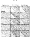
Figure 5 illustrates the results for immunostaining performed to confirm the content oftype 1 collagen andtype 2 collagen in the cartilage after treatment of a mixed cell to an animal model suffering with degenerative arthritis (referential); -
Figure 6 illustrates the results for analysis on TGF-β1 and IL-10 expression levels in synovial lavage fluid in a degenerative arthritis-induced animal model (referential); -
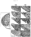
Figure 7 illustrates the results for immunochemical staining of the synovial membrane with an antibody to CD68 which is a surface antigen of a macrophage and IL-10 antibody to confirm the macrophage status of the cell-treated articular cavity of a degenerative arthritis-induced animal (referential); -
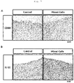
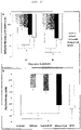
Figure 8A illustrates the results for immunochemical staining of synovial membrane with antibodies to CD86 andarginase 1, 8B illustrates the image of 8A converted to an invert version by using Image-Pro Plus, and 8C is a graph illustrating CD86 and arginase 1 positive cells measurement; -
Figure 9 illustrates the results for qRT-PCR of the gene expression profile in the synovial membrane; -
Figure 10 is a graph illustrating the measurement results for edema reduction in the metatarsal region on the 19th day and 26th day after treatment of a mixed cell to a rheumatoid arthritis-induced animal model; and -
Figure 11 illustrates the results for a change in cytokine measured by harvesting the synovial lavage fluid from the articular cavity on the 28th day after treatment of a mixed cell to a rheumatoid arthritis-induced animal model. - A mixed cell comprising an allogeneic (non-autologous) chondrocyte; and a chondrocyte expressing TGF-β is known as a cell-mediated gene therapeutic agent for degenerative arthritis. In a
phase 2 clinical study for patients suffering with osteoarthritis, it has been confirmed that the mixed cell improves pain, activity and cartilage structures of patients suffering with osteoarthritis. However, a mechanism by which the mixed cell acts is not yet found. - The present inventors observed effects on pain and structural transformation after injection of the mixed cell to an osteoarthritis-induced animal model and confirmed whether the mixed cell induces an anti-inflammatory environment, and as a result, it has been confirmed that anti-inflammatory cytokines and M2 macrophages are increased in the arthritic knee joint treated with mixed cell to induce an anti-inflammatory environment that alleviates pain and induces joint regeneration.
- Accordingly, it has been confirmed that the mixed cell induces an M2 macrophage-rich environment with immunosuppressive properties, and under the assumption that the mixed cell has an effect even on an inflammatory disease induced by a hyperimmune response, in a rheumatoid arthritis animal model, a chondrocyte and a chondrocyte expressing TGF-β are mixed at a predetermined ratio and treated to knee joints. As a result, in a group treated with the mixed cell, it was confirmed that the metatarsal edema was reduced and further, it was confirmed that expression of IL-6, IL-17A, IL-lβ, and TNF-α as cytokines that induced and promoted an inflammatory response in the synovial lavage fluid was reduced. A therapeutic effect by a single dose of the mixed cell lasted for 26 days.
- Conventionally, materials such as methotrexate (MTX) used as a therapeutic agent for rheumatoid arthritis may cause digestive system and mouth ulcers and are at risk for causing hepatotoxicity, renal failure, and the like. Further, therapeutic agents such as anti-TNF-a antibodies comprising Remicade and the like are known to disturb a patient's innate immune system, resulting in the expression of tuberculosis (TB) from a tuberculosis carrier. In contrast, a cell therapeutic agent according to the present invention has an advantage in that the above adverse effects are not shown in a clinical test result for degenerative arthritis. Accordingly, the present inventors have confirmed the possibility that the mixed cell may be a novel therapeutic agent which overcomes disadvantages of conventional therapeutic agents for rheumatoid arthritis.
- Therefore, an aspect of the present invention relates to a composition for use in a method of treating a rheumatoid arthritis, which comprises (i) a chondrocyte or a cartilage precursor cell; and (ii) a cell into which a gene encoding TGF-β is introduced, wherein the cell in (ii) is
hChonJb# 7. - The present description discloses also a method of treating an inflammatory disease induced by a hyperimmune response, which comprises administering to a subject with a composition comprising (i) a chondrocyte or a cell capable of differentiating into a chondrocyte; and (ii) a chondrocyte into which a gene encoding TGF-β is introduced or a cell capable of differentiating into a chondrocyte into which a gene encoding TGF-β is introduced.
- Still another example of the present description discloses a use of a composition comprising (i) a chondrocyte or a cell capable of differentiating into a chondrocyte; and (ii) a chondrocyte into which a gene encoding TGF-β is introduced or a cell capable of differentiating into a chondrocyte into which a gene encoding TGF-β is introduced as a therapeutic agent for treating an inflammatory disease induced by a hyperimmune response.
- The inflammatory disease induced by the hyperimmune response comprises rheumatoid arthritis, multiple sclerosis, systemic lupus erythematosus, myasthenia gravis, colitis,
type 1 diabetes, encephalitis, and the like, and among the diseases, it is particularly effective in the treatment for rheumatoid arthritis. - The cell capable of differentiating into the chondrocyte may be a cartilage precursor cell or a stem cell.
- In the present invention, (i) the chondrocyte or a cartilage precursor cell and (ii) the cell into which a gene encoding TGF-β is introduced, wherein the cell in (ii) is
hChonJb# 7, may be mixed at various mixing ratios, and the ratio of (i) and (ii) may be preferably 1 : 1 to 100 : 1 and more preferably 3 : 1. - In an aspect of the present invention, the composition for use in a method of treating rheumatoid arthritis comprises a non-transduced human chondrocyte (hChonJ cell) and a chondrocyte (hChonJb#7) expressing TGF-β1, more specifically, a human allogeneic chondrocyte (hChonJ) and a chondrocyte expressing human TGF-β1. The two types of cells may be mixed at various mixing ratios. For example, these two cells may be mixed at a ratio of 3 : 1, that is, a ratio at which the hChonJ cell is 3 and the
hChonJb# 7 is 1. The mixed cell at the ratio of 3 : 1 and a production method thereof are disclosed in a thesis [Cytotherapy, 2012 Feb; 14(2): 247-256] andUS Patent Registration Nos. 7,005,127 and7,282,200 . - In an aspect of the present invention, in an arthritis rat model treated with the mixed cell, the pain started to be reduced from 15 days after treatment and a pain relief effect was continued up to the time (an experiment end date) after 56 days after treatment. It has been confirmed that the pain relief was associated with a structural improvement of arthritic knee joints, and the treated mixed cell was found to be effective for both pain relief and cartilage regeneration. That is, the mixed cell used in the present invention is a cell-mediated gene therapy for pain relief and regeneration of cartilage tissue.
- In another aspect of the present invention, with respect to the cartilage tissue in the arthritis rat model treated with the mixed cell, staining for
type 1 collagen andtype 2 collagen was performed and histologically analyzed, and as a result, it was confirmed that the mixed cell regenerated a hyaline cartilage tissue which wastype 2 collagen-positive. - The macrophages which are innate immune cells are classified into activated "M1 macrophages" having a pro-inflammatory property and activated "M2 macrophages" having an immunosuppressive property according to an activation phenotype. IFN-γ is a key cytokine involved in the activation of the M1 macrophages and a TLR signal induced by pathogen recognition contributes to differentiation of the M1 macrophages. It has been known that the M1 macrophages increase expression of TNFα, IL-12 and IL-23 and decrease expression of IL-10 to induce Th1 and Th17 immune responses which cause tissue damage. In contrast, the M2 macrophages increase the expression level of IL-10 and decrease expression of IL-12, thereby reducing inflammation and promoting tissue regeneration.
- In an aspect of the present invention, TGF-β expressed in the
hChonJb# 7 which is a human cell expressing TGF-β1 and IL-10 induced through the hChonJ which is a human chondrocyte induces generation of the M2 macrophages and it is determined that the generated M2 macrophages secrete IL-10 to create an anti-inflammatory environment in the joints treated with the mixed cell. The synovial membrane in the animal model treated with the mixed cell are histologically analyzed, and as a result, it can be confirmed that the M2 macrophages are increased (FIG. 8 ) . - Further, it is determined that an immunosuppressive effect of the composition for the use of the present invention is induced by an increase in expression of TGF-β1 and
arginase 1, and in an aspect of the present invention, in the keen joint synovial membrane in an animal model of a group treated with the mixed cell, the expression ofarginase 1 is increased. - It is known that the increase in the expression of
arginase 1 and a decrease in L-arginine thereby cause a strong immunosuppressive effect in an immune system and further, it has been reported that the TGF-β induces transcription ofarginase 1 and activity of arginase (Durante et al., Circulation, 103(8), 1121-7, 2001) . Accordingly, it is determined that the M2 macrophages may more efficiently regulate the immune in the present of TGF-β1. - Further, in the anti-inflammatory effect of the composition for the use of the present invention, it is shown that IL-10 expressed in the M2 macrophage induced by treatment of the mixed cell activates STAT3 and PI3K and an anti-inflammatory effect is shown by Hmox-1 regulated thereby.
- Meanwhile, recently, in the synovial membrane and the synovial fluid of a patient suffering with chronic rheumatoid arthritis, cytokines and growth factors, such as interleukin-1 (IL-1), interleukin-8 (IL-8), tumor necrosis factor-a (TNF-α), transforming growth factor β (TGF-β), fibroblast growth factor (FGF), and platelet-derived growth factor (PDGF) have been detected (Nouri et al., Clin. Exp. Immunol. 55: 275-372, 1984; Thornton et al., Clin. Exp. Immunol. 86: 79-86, 1991; Saxne, et al., Arthritis Rheum. 31:1041-1045, 1988; Seitz et al., J. Clin. Invest. 87: 463-469, 1991; Lafyatis et al., J. Immunol.143: 1142-1148, 1989; Melnyk et al., Arthrtis Rheum. 33: 493-500, 1990).
- IL-6 is a cytokine known as B-
cell stimulating factor 2 or interferon β2. IL-6 is a differentiation factor involved in activation of B lymphocytic cells (Hirano T. et al., Nature 324, 73-76, 1986) and is known as a multifunctional cytokine that affects an action of various types of cells (Akira, S. et al., Adv. In Immunology 54, 1-78, 1993). - It has been reported that in the serum or the synovial fluid of the patient suffering with the chronic rheumatoid arthritis, a large amount of interleukin-6 and soluble IL-6 receptors are present (Houssiau et al., Arthritis Rheum. 31 : 784-788, 1988; Hirano et al., Eur. J. Immunol. 18: 1797-1801, 1988; Yoshioka et al., Japn. J. Rheumatol. In press) . Even in the animal model of rheumatoid arthritis, a similar result is obtained (Takai et al., Arthritis Rheum. 32: 594-600, 1989; Leisten et al., Clin. Immunol. Immunopathol. 56: 108-115, 1990), and as a result, the IL-6 is known as a factor associated with the chronic rheumatoid arthritis.
- Meanwhile, in a clinical development for rheumatoid arthritis, secukinumab as a biological agent is a high-affinity human monoclonal antibody that inhibits IL-17A activity. In the RA proof-of-concept (PoC) study, secukinumab was co-treated to an active RA patient treated with a stable dose of MTX. As a result, it was confirmed that a treatment group using secukinumab rapidly improved clinical symptoms of rheumatoid arthritis compared to placebo (Hueber et al. (2010) Sci. Transl. Med. 2(52):52-72). This is data showing that neutralization of IL-17A may be efficacious in the patient suffering with rheumatoid arthritis.
- In another aspect of the present invention, it was confirmed that the mixed cell was treated to the paw of a rat in which rheumatoid arthritis was induced by treatment of a complete Freund's adjuvant (CFA) and the metatarsal edema was confirmed for 28 days. As a result, in the group treated with the mixed cell, it was confirmed that the metatarsal edema was decreased (
FIG. 10 ), and as the result of analyzing IFN-γ, IL-6, IL-17A, IL-lβ, and TNF-α which were Pro-inflammatory cytokine items that induced and promoted the inflammatory response, it was confirmed that in the mixed cell-treated group and the MTX-treated group, the metatarsal edema was statistically and significantly decreased as compared with a control. - Therefore, the composition for the ues of the present invention has an effect of inhibiting expression of IL-6, IL-17A, IL-lβ, and TNF-α which are inflammatory cytokines that are excessively secreted from the joint synovial lavage fluid of the patient suffering with rheumatoid arthritis to reduce the edema.
- Hereinafter, the present invention will be described in more detail through Examples.
- In the case of treating a mixed cell in a
clinical phase 2 for patients suffering with arthritis, it was observed that pain was reduced and an environment of joints was structurally improved. In order to confirm a mechanism therefor, in a degenerative arthritis animal model induced by MIA, a mixed cell was treated and a joint histological examination and a von Frey filament test were performed. - In preparation of an animal model, a 6-week-old male rat (Spargue-Dawley, 200 to 225 g, Nara Biotech, Korea) were used. An animal experiment was conducted under a veterinary control after approval of the Kolon Life Science (Korea) Institutional Animal Care and Use Committee (IACUC No. KLS IACUC 2013-04) .
- In order to induce arthritis, 50 µl of an MIA (monosodium iodoacetate, Sigma, USA) solution having a concentration of 60 mg/mL was treated to the articular cavity of the left knee of a rat by using a 31G syringe.
- The hChonJ used in the present invention is a human-derived chondrocyte and the
hChonJb# 7 is a chondrocyte transformed to secrete TGF-β. Preparation methods and mixing ratios of the chondrocytes are disclosed in a thesis [Cytotherapy, 2012 Feb; 14(2): 247-256] andUS Patent Registration Nos. 7,005,127 and7,282,200 . - The
hChonJb# 7 may be prepared by injecting a cDNA of TGF-β into cells according to a known method. For example, a promoter such as methallothionein and a cDNA of TGF-β are included in a known vector [for example, pUC19 (including an ampicillin resistance gene) by Gibco-BRL Corporation] having a resistance gene such as ampicillin and neomycin to prepare a vector including the cDNA of TGF-β. Thereafter, the vector including the cDNA of TGF-β is injected into a chondrocyte according to a known method such as a calcium phosphate method or a lipofectin method to prepare thehChonJb# 7. - After 2 weeks of MIA injection, mixed cells { (1.2 × 10⁶ cells: hChonJ (9 × 10⁵ cells) + hChonJb# 7 (3.0 × 10⁵ cells) }, hChonJ (9 × 10⁵ cells), hChonJb#7 (3.0 × 10⁵ cells) or a control {CryoStor-10 (CS-10) } were treated to the articular cavity of the left knee. General symptoms were observed, and body weights were measured once a week.
- The pain relief was observed by performing a von Frey filament test, and as a result, as illustrated in
FIG. 2 , pain levels decreased from 15 days after cell treatment, and such effects lasted until the end of observation (56th day after treatment) . In the case of a group treated with hChonJb#7 (3.0 × 10⁵ cells) expressing TGF-β, the pain relief was observed from 15 days, but after 6 weeks, it was confirmed that the effect disappeared. In contrast, in a group treated with hChonJ (9 × 10⁵ cells) which was a normal human chondrocyte, the pain relief effect was not observed. - In order to confirm whether the pain relief effect of the mixed cell is associated with cartilage regeneration, left knee joints in 5 experimental groups were harvested and a histological analysis was performed through H&E staining and Masson's trichrome staining (see
FIGS. 3 and4 ). - In a control (CS-10) and an hChonJ-treated group, cartilage regeneration was not confirmed. In contrast, in an hChonJb#7-treated group and a mixed cell-treated group, more cartilage was observed than other controls.
- Further, as the immunohistochemical staining result, as illustrated in
FIG. 5 , it was confirmed that the content of type II collagen in the cartilage in the mixed cell-treated group was higher than that in the hChonJb#7-treated group, and from the result, it can be seen that the cartilage in the mixed cell-treated group is close to the hyaline cartilage. - In order to evaluate the improvement degree of the joint status, as a method disclosed in the thesis [Kobayasi, K., J. et al., J. Vet. Med. Sci., 65:1195-1199, 2003], the histopathological results and the pathology scores of the H&E-stained joint tissue were evaluated and the results were summarized in the following Table 2. In the experimental results below, the cartilage in the mixed cell-treated group was most improved.
[Table 2] Treatment No. of Animals Untreat ed 5 (x3) Control 6 (x3) hChonJ 6 (x3) hChonJb# 7 6 (x3) Invoss a 6 (x3) Structural change in the joint Surface irregularities 1 2/15 3/18 0/18 5/18 7/18 2 0/15 4/18 1/18 2/18 4/18 3 0/15 10/18 14/18 6/18 4/18 Average pathology score 0.13 2.28 2.44 1.50 1.50 Standard Error 0.09 0.23 0.22 0.29 0.25 Ulceration 1 1/15 3/18 3/18 8/18 7/18 2 0/15 8/18 7/18 6/18 6/18 3 0/15 7/18 7/18 3/18 0/18 Average pathology score 0.07 2.22 2.11 1.61 1.06 Standard Error 0.07 0.18 0.18 0.21 0.19 Fibrillation of cartilage surface 1 0/15 3/18 3/18 1/18 3/18 2 0/15 2/18 6/18 6/18 5/18 3 0/15 7/18 5/18 6/18 1/18 Average pathology score 0 1.56 1.67 1.72 0.89 Standard Error 0 0.32 0.26 0.29 0.25 Disorganization of chondrocytes 1 1/15 1/18 7/18 9/18 6/18 2 0/15 10/18 5/18 8/18 7/18 3 0/15 5/18 4/18 0/18 0/18 Average pathology score 0.07 2.00 1.61 1.39 1.11 Standard Error 0.07 0.23 0.24 0.15 0.20 Exposure of subchondral bone 1 0/15 7/18 6/18 4/18 6/18 2 0/15 1/18 0/18 4/18 1/18 3 0/15 8/18 8/18 0/18 0/18 Average pathology score 0 1.83 1.67 0.67 0.44 Standard Error 0 0.29 0.30 0.21 0.15 Cellular changes of chondrocyte H&E staining 1 2/15 7/18 6/18 9/18 9/18 2 0/15 2/18 3/18 4/18 1/18 3 0/15 4/18 3/18 1/18 2/18 Average pathology score 0.13 1.28 1.17 1.11 0.94 Standard Error 0.09 0.27 0.26 0.17 0.20 Degeneration/Necrosis 1 0/15 3/18 2/18 5/18 6/18 2 0/15 4/18 8/18 5/18 4/18 3 0/15 11/18 8/18 5/18 0/18 Average pathology score 0 2.44 2.33 1.67 0.78 Standard Error 0 0.19 0.17 0.26 0.20 H&E staining Reduction of staining in cartilage 1 0/15 5/18 4/18 10/18 10/18 2 0/15 5/18 5/18 5/18 2/18 3 0/15 6/18 7/18 1/18 0/18 Average pathology score 0 1.83 1. 94 1.28 0.78 Standard Error 0 0.23 0.23 0.17 0.14 Total pathology score 0.4±0.3 2 15.44±1. 94 14.94±1. 88 10.94±1. 75 7.5±1. 57 0: Normal 1: Slight, 2: Moderate, 3: Severe - An effect on a cytokine expression profile of the synovial fluid in the articular cavity by treatment of mixed cells in an animal model was confirmed.
- In order to confirm whether macrophages are induced at a cell site, it was confirmed that CD68 positive cells were present in the synovial membrane.
- The paraffin-embedded synovial membrane was microsected with a thickness of 4 µm to prepare a tissue slide, and then immunohistochemical staining was performed by using CD68 antibodies (Novus, Co, USA).
- As a result, as illustrated in
FIG. 7A , CD68-positive cells were significantly increased in the mixed cell-treated group. - IL-10 was known to exhibit the pain relief effect. An arthritis model was created based the above report and the mixed cell was treated, and then expression of IL-10 was confirmed. The expression of IL-10 was measured in the synovial lavage fluid of the arthritis-induced rat by using a cytokine magnetic bead panel 96-well plate assay (#RECYTMAG-65K, Milipore, USA) .
- As a result, as illustrated in
FIG. 6B , in the mixed cell-treated group and the hChonJ-treated group, it was confirmed that the expression of IL-10 was increased. From the histological analysis result, it was confirmed that IL-10-positive cells were increased in the mixed cell-treated group. These results indicate that the treatment of the mixed cell in the arthritic knee joints induces IL-10 which is an anti-inflammatory cytokine. - There are two different types of macrophages. The M1 type macrophages are responsible for acute inflammation to secrete high levels of inflammatory cytokines such as IL-12 and TNF-α, and the M2 macrophages play an important role in wound healing and anti-inflammatory regulatory functions.
- In Example 2, the high level of IL-10 expression was confirmed in the mixed cell-treated group, and as a result, in order to confirm whether the mixed cell induces the macrophages in the knee of the arthritis-induced animal, in the synovial membrane of the animal model, the expression of CD86 as a marker of the M1 macrophage and the expression of
arginase 1 as a marker of the M2 macrophage were confirmed. - To this end, the paraffin-embedded synovial membrane was immuno-stained by using a CD86 antibody (AbD serotec, NC, USA) and a
mouse arginase 1 antibody (BD Bioscience, USA). - As a result, as illustrated in
FIG. 8 , the expression of CD86 had no difference in the control and the mixed cell-treated group, butarginase 1 was significantly high in the mixed cell-treated group. It is shown that the mixed cells induce the M2 macrophages. - In order to confirm the induction of the M2 macrophages confirmed in Example 3 in the gene expression level, the total RNA was isolated from the synovial membrane of the animals and the expression of genes related with the M1 and M2 macrophages was confirmed.
- The RNA in the synovial membrane was isolated by using a RNeasy Lipid Tissue Mini kit (QIAGEN, USA) and cDNA was synthesized from the isolated RNA by using a SuperScript™ III First-Strand Synthesis System (Invitrogen, USA).
- Thereafter, real-time PCR was performed by using ABI7900 equipment. A PCR reaction mixture used 1 µL cDNA, 0.2 µM of each primer, and 10 µL SYBR Premix Ex Taq (TAKARA, Japan) at a total of 50 µL, and a reaction condition was 10 seconds at 95°C and 30 seconds at 60°C and a total of 40 cycles were performed. A gene expression level was confirmed by comparing an expression level of a β-actin gene by using a 2-△△Ct method and the used qRT-PCR primers were listed in Table 3.
[Table 3] Primer sequences used for qRT-PCR Gene Forward primer Reverse primer CD80 TGCTGGTTGGTCTTTTCCA TGACTGCTCTTCAGAACAAAA CD86 TCCTCCAGCAGTGGGAAACA TTTGTAGGTTTCGGGTATCCTTGC CD163 CTCAGCGTCTCTGCTGTCAC GGCCAGTCTCAGTTCCTTCTT Arg1 TTGATGTTGATGGACTGGAC TCTCTGGCTTATGATTACCTTC IL-1β TCCAGGATGAGGACCCAAGC TCGTCATCATCCCACGAGTCA TNFα ACTGAACTTCGGGGTGATTG GCTTGGTGGTTTGCTACGAC IL-10 CAAGGCAGTGGAGCAGGTGA CCGGGTGGTTCAATTTTTCATT Hmox-1 AGAGTTTCCGCCTCCAACCA CGGGACTGGGCTAGTTCAGG IL10Ra CTGGTCACCCTGCCATTGAT AGGCATGGCCAAAATACAAAGAAAC CD68 ACTGGGGCTCTTGGAAACTACAC CCTTGGTTTTGTTCGGGTTCA β-actin AGTTCGCCATGGATGACGAT AAGCCGGCCTTGCACAT Primer sequences for target genes for qRT-PCR of rat genes. CD = Cluster of Differentiation, Arg = Arginase, IL = Interleukin, TNFα = Tumor necrosis factors alpha, Hmox = Heme oxygenase - A macrophage related gene expression profile is illustrated in
FIG. 9 . The expression of CD68 was high in the mixed cell-treated group. TNF-α had no difference between the control and the mixed cell-treated group. IL-10 as an anti-inflammatory cytokine was very highly expressed in the mixed cell-treated group. - In order to confirm whether an increase in mRNA expression of IL-10 causes protein expression, a multiple cytokine assay in the synovial membrane was performed. As a result, as illustrated in
FIG. 10 , it was confirmed that in the mixed cell-treated group, a level of the IL-10 protein was rapidly increased. It was confirmed that the mRNA levels of IL-10RA (interleukin 10 receptor alpha subunit) and Hmox1 (heme oxygenase 1) which were additional macrophage markers were significantly increased. - When summarizing the results, in the case of administering the mixed cell in the arthritis model, it can be seen that induction of the M2 macrophages is increased.
- In a preparation of a rheumatoid arthritis-induced animal model, a 7-week-old female rat (160 to 180 g Lewis, Central laboratory animals, Japan) was used. The animal experiment was conducted after approval of the Kolon Life Science (Korea) Institutional Animal Care and Use Committee (IACUC No. KLS IACUC 2015-07).
- In order to induce rheumatoid arthritis, 50 µl of a CFA (Complete Freud's Adjuvant, manufacturer: Chondrex, Inc.) solution having a concentration of 5 mg/mL was treated to the subcutaneous sole of a rat by using a 31G syringe. After 3 weeks, induction of arthritis was confirmed by measuring the size of the metatarsal edema using calipers, and thereafter, when the edema was most severely induced, a test material was treated to the Lewis-rat left knee articular cavity.
- Rheumatoid arthritis-induced rats were set to 6 rats per group, and after 3 weeks of CFA treatment, mixed cells {(1.2 × 10⁶ cells: hChonJ (9 × 10⁵ cells) + hChonJb#7 (3.0 ×10⁵ cells) }, hChonJ (9 × 10⁵ cells), hChonJb#7 (3.0 ×10⁵ cells), and a negative control (CS-10) were treated to the left knee articular cavity. In the case of a positive control, 0.4 mg/kg of MTX (Methotrexate) was orally treated once a week. The metatarsal diameter and the body weight were measured once per three days.
- In Table 4, a treatment condition for each experimental group was illustrated.
[Table 4] treatment condition for each experimental group experimental group Cell/50µl* Number of Animal Dosing Schedule Method of Treatment AIA Negative. Control Group CS-10 6 Single after 3 weeks inducing AIA administration to the articular cavity hChonJ 9×10⁵ 6 hChonJb# 73×105 6 Mixed cell group 1.2×10⁶ 6 MTX 0.4mg/ kg 6 Once a week (21d) Oral treatment - The test material was treated to the arthritis model and the size of the metatarsal edema was measured by a caliper, and the result was illustrated in
FIG. 10 . After treatment of the control (CS-10), hChonJ,hChonJb# 7, mixed cell, and MTX (Methotrexate), the decrease in the size of the edema was measured on the 19th day and the 26th day, and as a result, in the mixed cell-treated group and the MTX-treated group, the decrease was statistically and significantly shown as compared with the control. - A sample was treated to the arthritis model, the synovial lavage fluid was harvested from the left knee articular cavity of the experimental animal rat on the 28th day, and the cytokines were analyzed, and the result was illustrated in
FIG. 11 . The synovial lavage fluid was collected from the rat articular cavity by administering 50 µL of a saline solution for injection by a 31G insulin syringe and then immediately collecting the solution while the syringe pierced. As the cytokines, IFN-γ, IL-6, IL-17, IL-1β and TNF-α were analyzed and the analysis was performed by using Rat Cytokine/Chemokine multiplex (RECYTMAG-65K, Millipore). IFN-γ, IL-6, IL-17, IL-1β and TNF-α, which were Pro-inflammatory cytokine items inducing and promoting an inflammatory response, were analyzed and as a result, in the mixed cell-treated group and the MTX (Methotrexate)-treated group, the cytokines were statistically and significantly decreased as compared with the control. - The mixed cell composition of the present invention can improve symptoms of inflammatory diseases including rheumatoid arthritis and suppress a hyperimmune response by cell therapy without adverse effects from the other types of conventional drug therapy.
- <110> KOLON LIFE SCIENCE, INC.
- <120> Composition for Treating Inflammatory Disease Caused by Hyperimmune Response
- <130> PP-B1667
- <150>
US 62/112718
<151> 2015-02-06 - <160> 22
- <170> KopatentIn 2.0
- <210> 1
<211> 19
<212> DNA
<213> Artificial Sequence - <220>
<223> primer - <400> 1
tgctggttgg tcttttcca 19 - <210> 2
<211> 20
<212> DNA
<213> Artificial Sequence - <220>
<223> primer - <400> 2
tgactgctct tcagaacaaa 20 - <210> 3
<211> 20
<212> DNA
<213> Artificial Sequence - <220>
<223> primer - <400> 3
tcctccagca gtgggaaaca 20 - <210> 4
<211> 24
<212> DNA
<213> Artificial Sequence - <220>
<223> primer - <400> 4
tttgtaggtt tcgggtatcc ttgc 24 - <210> 5
<211> 20
<212> DNA
<213> Artificial Sequence - <220>
<223> primer - <400> 5
ctcagcgtct ctgctgtcac 20 - <210> 6
<211> 21
<212> DNA
<213> Artificial Sequence - <220>
<223> primer - <400> 6
ggccagtctc agttccttctt 21 - <210> 7
<211> 20
<212> DNA
<213> Artificial Sequence - <220>
<223> primer - <400> 7
ttgatgttga tggactggac 20 - <210> 8
<211> 22
<212> DNA
<213> Artificial Sequence - <220>
<223> primer - <400> 8
tctctggctt atgattacct tc 22 - <210> 9
<211> 20
<212> DNA
<213> Artificial Sequence - <220>
<223> primer - <400> 9
tccaggatga ggacccaagc 20 - <210> 10
<211> 21
<212> DNA
<213> Artificial Sequence - <220>
<223> primer - <400> 10
tcgtcatcat cccacgagtc a 21 - <210> 11
<211> 20
<212> DNA
<213> Artificial Sequence - <220>
<223> primer - <400> 11
actgaacttc ggggtgattg 20 - <210> 12
<211> 20
<212> DNA
<213> Artificial Sequence - <220>
<223> primer - <400> 12
gcttggtggt ttgctacgac 20 - <210> 13
<211> 20
<212> DNA
<213> Artificial Sequence - <220>
<223> primer - <400> 13
caaggcagtg gagcaggtga 20 - <210> 14
<211> 22
<212> DNA
<213> Artificial Sequence - <220>
<223> primer - <400> 14
ccgggtggtt caatttttca tt 22 - <210> 15
<211> 20
<212> DNA
<213> Artificial Sequence - <220>
<223> primer - <400> 15
agagtttccg cctccaacca 20 - <210> 16
<211> 20
<212> DNA
<213> Artificial Sequence - <220>
<223> primer - <400> 16
cgggactggg ctagttcagg 20 - <210> 17
<211> 20
<212> DNA
<213> Artificial Sequence - <220>
<223> primer - <400> 17
ctggtcaccc tgccattgat 20 - <210> 18
<211> 25
<212> DNA
<213> Artificial Sequence - <220>
<223> primer - <400> 18
aggcatggcc aaaatacaaa gaaac 25 - <210> 19
<211> 23
<212> DNA
<213> Artificial Sequence - <220>
<223> primer - <400> 19
actggggctc ttggaaacta cac 23 - <210> 20
<211> 21
<212> DNA
<213> Artificial Sequence - <220>
<223> primer - <400> 20
ccttggtttt gttcgggttc a 21 - <210> 21
<211> 20
<212> DNA
<213> Artificial Sequence - <220>
<223> primer - <400> 21
agttcgccat ggatgacgat 20 - <210> 22
<211> 17
<212> DNA
<213> Artificial Sequence - <220>
<223> primer - <400> 22
aagccggcct tgcacat 17
Claims (4)
- A composition for use in a method of treating a rheumatoid arthritis, which comprises (i) a chondrocyte or a cartilage precursor cell; and (ii) a cell into which a gene encoding TGF-β (transforming growth factor-beta) is introduced, wherein the cell in (ii) is hChonJb#7.
- The composition for the use according to claim 1, wherein a ratio of (i) the chondrocyte or the cartilage precursor cell; and (ii) the cell ranges from 1:1 to 100:1.
- The composition for the use according to claim 2, wherein the ratio of (i) the chondrocyte or the cartilage precursor cell; and (ii) the cell ranges from 1:1 to 3:1.
- The composition for the use according to any of claims 1 to 3, wherein the composition inhibits the expression or activity of a cytokine selected from the group consisting of IL-6, IL-17A, IL-1β and TNF-α.
Applications Claiming Priority (2)
| Application Number | Priority Date | Filing Date | Title |
|---|---|---|---|
| US201562112718P | 2015-02-06 | 2015-02-06 | |
| PCT/KR2016/001310 WO2016126139A1 (en) | 2015-02-06 | 2016-02-05 | Composition for treating inflammatory diseases caused by hyper-immune responses |
Publications (3)
| Publication Number | Publication Date |
|---|---|
| EP3254685A1 EP3254685A1 (en) | 2017-12-13 |
| EP3254685A4 EP3254685A4 (en) | 2018-09-19 |
| EP3254685B1 true EP3254685B1 (en) | 2022-04-20 |
Family
ID=56564381
Family Applications (1)
| Application Number | Title | Priority Date | Filing Date |
|---|---|---|---|
| EP16746880.0A Active EP3254685B1 (en) | 2015-02-06 | 2016-02-05 | Composition for treating inflammatory disease induced by hyper-immune response |
Country Status (9)
| Country | Link |
|---|---|
| US (1) | US11191787B2 (en) |
| EP (1) | EP3254685B1 (en) |
| JP (1) | JP6475847B2 (en) |
| KR (1) | KR101986774B1 (en) |
| CN (1) | CN107405363A (en) |
| AU (1) | AU2016216223B2 (en) |
| CA (1) | CA2974941C (en) |
| SG (2) | SG11201706165RA (en) |
| WO (1) | WO2016126139A1 (en) |
Families Citing this family (4)
| Publication number | Priority date | Publication date | Assignee | Title |
|---|---|---|---|---|
| AU2018290629B2 (en) * | 2017-06-30 | 2022-07-07 | Kolon Life Science, Inc. | Method for assessing validity of cell therapy product |
| KR20190003413A (en) * | 2017-06-30 | 2019-01-09 | 코오롱생명과학 주식회사 | A Pharmaceutical Composition For Preventing or Treating Osteoarthritis |
| US20220160781A1 (en) * | 2019-03-29 | 2022-05-26 | Kolon Tissuegene, Inc. | Mixed-Cell Gene Therapy |
| FR3159096A1 (en) * | 2024-02-08 | 2025-08-15 | Compagnie Laitiere Europeenne | Use of TGF-β for the prevention and treatment of neuralgic manifestations |
Family Cites Families (7)
| Publication number | Priority date | Publication date | Assignee | Title |
|---|---|---|---|---|
| US6413511B1 (en) * | 1990-12-20 | 2002-07-02 | University Of Pittsburgh Of The Commonwealth System Of Higher Education | Cartilage alterations by administering to joints chondrocytes comprising a heterologous polynucleotide |
| KR100688871B1 (en) * | 2002-03-12 | 2007-03-02 | 티슈진, 인코포레이티드 | Cartilage Regeneration Using Chondrocytes and TVF-β |
| US7005127B2 (en) * | 2002-03-29 | 2006-02-28 | Tissuegene, Inc. | Mixed-cell gene therapy |
| US8299046B2 (en) * | 2006-11-17 | 2012-10-30 | Trustees Of Dartmouth College | Synthetic triterpenoids and tricyclic-bis-enones for use in stimulating bone and cartilage growth |
| KR20090069013A (en) | 2007-12-24 | 2009-06-29 | 동국대학교 산학협력단 | How to differentiate myelogenous mesenchymal stem cells into chondrocytes |
| WO2011047345A2 (en) * | 2009-10-15 | 2011-04-21 | Tai June Yoo | Methods of treating diseases of conditions using mesenchymal stem cells |
| KR101301262B1 (en) * | 2009-10-23 | 2013-08-27 | 가톨릭대학교 산학협력단 | Mesenchymal stem cell introduced with a nucleotide sequence encoding TGFβ and use thereof |
-
2016
- 2016-02-05 US US15/547,566 patent/US11191787B2/en active Active
- 2016-02-05 AU AU2016216223A patent/AU2016216223B2/en active Active
- 2016-02-05 EP EP16746880.0A patent/EP3254685B1/en active Active
- 2016-02-05 SG SG11201706165RA patent/SG11201706165RA/en unknown
- 2016-02-05 CN CN201680008586.3A patent/CN107405363A/en active Pending
- 2016-02-05 SG SG10202106872WA patent/SG10202106872WA/en unknown
- 2016-02-05 JP JP2017538300A patent/JP6475847B2/en active Active
- 2016-02-05 KR KR1020177020973A patent/KR101986774B1/en active Active
- 2016-02-05 WO PCT/KR2016/001310 patent/WO2016126139A1/en not_active Ceased
- 2016-02-05 CA CA2974941A patent/CA2974941C/en active Active
Also Published As
| Publication number | Publication date |
|---|---|
| WO2016126139A1 (en) | 2016-08-11 |
| JP2018505873A (en) | 2018-03-01 |
| CA2974941A1 (en) | 2016-08-11 |
| EP3254685A4 (en) | 2018-09-19 |
| AU2016216223A1 (en) | 2017-08-03 |
| CN107405363A (en) | 2017-11-28 |
| SG10202106872WA (en) | 2021-08-30 |
| JP6475847B2 (en) | 2019-02-27 |
| KR20170104498A (en) | 2017-09-15 |
| KR101986774B1 (en) | 2019-06-07 |
| EP3254685A1 (en) | 2017-12-13 |
| CA2974941C (en) | 2021-11-23 |
| SG11201706165RA (en) | 2017-09-28 |
| US20180264045A1 (en) | 2018-09-20 |
| AU2016216223B2 (en) | 2018-08-30 |
| US11191787B2 (en) | 2021-12-07 |
Similar Documents
| Publication | Publication Date | Title |
|---|---|---|
| EP3119806B1 (en) | Il-21 antibodies | |
| Shao et al. | Increased lipocalin-2 contributes to the pathogenesis of psoriasis by modulating neutrophil chemotaxis and cytokine secretion | |
| Amarasekara et al. | Bone loss triggered by the cytokine network in inflammatory autoimmune diseases | |
| Kugyelka et al. | Enigma of IL‐17 and Th17 cells in rheumatoid arthritis and in autoimmune animal models of arthritis | |
| Gaffen | Biology of recently discovered cytokines: interleukin-17–a unique inflammatory cytokine with roles in bone biology and arthritis | |
| Souza et al. | The role of cytokines in inflammatory bone loss | |
| JP6889201B2 (en) | Methods for the prevention or treatment of certain disorders by inhibiting the binding of IL-4 and / or IL-13 to their respective receptors | |
| Moltó et al. | Anti-IL-1 molecules: new comers and new indications | |
| CA2781576C (en) | Combination preparation including a corticosteroid and exosomes | |
| CN108289881A (en) | Sai Nikeweiluo conjoint therapies for treating fibrosis | |
| US11191787B2 (en) | Composition for treating inflammatory disease induced by hyperimmune response | |
| Viallard et al. | Analysis of leukemia inhibitory factor, type 1 and type 2 cytokine production in patients with eosinophilic fasciitis. | |
| MnIAs | Inter! eukin-1 in crescentic glomerulonephritis | |
| Tian et al. | Exogenous interleukin-17A inhibits eosinophil differentiation and alleviates allergic airway inflammation | |
| US20220096563A1 (en) | Composition for treating inflammatory disease induced by hyperimmune response | |
| MX2007004374A (en) | Chimeric protein. | |
| AU2003299919B2 (en) | The use of cytokine able to bind IL-18BP and of inhibiting the activity of a second cytokine | |
| Han et al. | Increased expression of CXCR5 and CXCL13 in mice with experimental autoimmune myocarditis. | |
| AU756167B2 (en) | Application of TNF antagonists as medicaments for treating septic diseases | |
| Sato et al. | Plasma leptin, insulin and free tryptophan contribute to cytokine-induced anorexia | |
| JPH07188056A (en) | Suppressor for bone resorption containing antibody against interleukin 6 receptor as active ingredient | |
| Milhomem et al. | Adipokines and their relationship with rheumatoid arthritis | |
| WO2024251200A1 (en) | Engineered il-7 variants and methods of use thereof | |
| Dinarello et al. | The interleukin-1 family: Role in inflammation and disease | |
| Sims et al. | IL-36: An Epithelial Cytokine Important in Psoriasis |
Legal Events
| Date | Code | Title | Description |
|---|---|---|---|
| STAA | Information on the status of an ep patent application or granted ep patent |
Free format text: STATUS: THE INTERNATIONAL PUBLICATION HAS BEEN MADE |
|
| PUAI | Public reference made under article 153(3) epc to a published international application that has entered the european phase |
Free format text: ORIGINAL CODE: 0009012 |
|
| STAA | Information on the status of an ep patent application or granted ep patent |
Free format text: STATUS: REQUEST FOR EXAMINATION WAS MADE |
|
| 17P | Request for examination filed |
Effective date: 20170717 |
|
| AK | Designated contracting states |
Kind code of ref document: A1 Designated state(s): AL AT BE BG CH CY CZ DE DK EE ES FI FR GB GR HR HU IE IS IT LI LT LU LV MC MK MT NL NO PL PT RO RS SE SI SK SM TR |
|
| AX | Request for extension of the european patent |
Extension state: BA ME |
|
| RAP1 | Party data changed (applicant data changed or rights of an application transferred) |
Owner name: KOLON LIFE SCIENCE, INC. Owner name: TISSUEGENE, INC. |
|
| DAV | Request for validation of the european patent (deleted) | ||
| DAX | Request for extension of the european patent (deleted) | ||
| A4 | Supplementary search report drawn up and despatched |
Effective date: 20180820 |
|
| RIC1 | Information provided on ipc code assigned before grant |
Ipc: A61K 35/12 20150101ALI20180813BHEP Ipc: A61P 29/00 20060101ALI20180813BHEP Ipc: A61P 19/02 20060101ALI20180813BHEP Ipc: A61P 3/10 20060101ALI20180813BHEP Ipc: A61K 35/32 20150101AFI20180813BHEP |
|
| STAA | Information on the status of an ep patent application or granted ep patent |
Free format text: STATUS: EXAMINATION IS IN PROGRESS |
|
| 17Q | First examination report despatched |
Effective date: 20191025 |
|
| RAP3 | Party data changed (applicant data changed or rights of an application transferred) |
Owner name: KOLON TISSUEGENE, INC. Owner name: KOLON LIFE SCIENCE, INC. |
|
| RIC1 | Information provided on ipc code assigned before grant |
Ipc: A61P 3/10 20060101ALI20211007BHEP Ipc: A61P 19/02 20060101ALI20211007BHEP Ipc: A61P 29/00 20060101ALI20211007BHEP Ipc: A61K 38/18 20060101ALI20211007BHEP Ipc: A61K 35/12 20150101ALI20211007BHEP Ipc: A61K 35/32 20150101AFI20211007BHEP |
|
| GRAP | Despatch of communication of intention to grant a patent |
Free format text: ORIGINAL CODE: EPIDOSNIGR1 |
|
| STAA | Information on the status of an ep patent application or granted ep patent |
Free format text: STATUS: GRANT OF PATENT IS INTENDED |
|
| INTG | Intention to grant announced |
Effective date: 20211119 |
|
| GRAS | Grant fee paid |
Free format text: ORIGINAL CODE: EPIDOSNIGR3 |
|
| GRAA | (expected) grant |
Free format text: ORIGINAL CODE: 0009210 |
|
| STAA | Information on the status of an ep patent application or granted ep patent |
Free format text: STATUS: THE PATENT HAS BEEN GRANTED |
|
| RAP3 | Party data changed (applicant data changed or rights of an application transferred) |
Owner name: KOLON TISSUEGENE, INC. Owner name: KOLON LIFE SCIENCE, INC. |
|
| AK | Designated contracting states |
Kind code of ref document: B1 Designated state(s): AL AT BE BG CH CY CZ DE DK EE ES FI FR GB GR HR HU IE IS IT LI LT LU LV MC MK MT NL NO PL PT RO RS SE SI SK SM TR |
|
| REG | Reference to a national code |
Ref country code: GB Ref legal event code: FG4D |
|
| REG | Reference to a national code |
Ref country code: CH Ref legal event code: EP |
|
| REG | Reference to a national code |
Ref country code: DE Ref legal event code: R096 Ref document number: 602016071291 Country of ref document: DE |
|
| REG | Reference to a national code |
Ref country code: IE Ref legal event code: FG4D |
|
| REG | Reference to a national code |
Ref country code: AT Ref legal event code: REF Ref document number: 1484648 Country of ref document: AT Kind code of ref document: T Effective date: 20220515 |
|
| REG | Reference to a national code |
Ref country code: LT Ref legal event code: MG9D |
|
| REG | Reference to a national code |
Ref country code: NL Ref legal event code: MP Effective date: 20220420 |
|
| REG | Reference to a national code |
Ref country code: AT Ref legal event code: MK05 Ref document number: 1484648 Country of ref document: AT Kind code of ref document: T Effective date: 20220420 |
|
| PG25 | Lapsed in a contracting state [announced via postgrant information from national office to epo] |
Ref country code: NL Free format text: LAPSE BECAUSE OF FAILURE TO SUBMIT A TRANSLATION OF THE DESCRIPTION OR TO PAY THE FEE WITHIN THE PRESCRIBED TIME-LIMIT Effective date: 20220420 |
|
| PG25 | Lapsed in a contracting state [announced via postgrant information from national office to epo] |
Ref country code: SE Free format text: LAPSE BECAUSE OF FAILURE TO SUBMIT A TRANSLATION OF THE DESCRIPTION OR TO PAY THE FEE WITHIN THE PRESCRIBED TIME-LIMIT Effective date: 20220420 Ref country code: PT Free format text: LAPSE BECAUSE OF FAILURE TO SUBMIT A TRANSLATION OF THE DESCRIPTION OR TO PAY THE FEE WITHIN THE PRESCRIBED TIME-LIMIT Effective date: 20220822 Ref country code: NO Free format text: LAPSE BECAUSE OF FAILURE TO SUBMIT A TRANSLATION OF THE DESCRIPTION OR TO PAY THE FEE WITHIN THE PRESCRIBED TIME-LIMIT Effective date: 20220720 Ref country code: LT Free format text: LAPSE BECAUSE OF FAILURE TO SUBMIT A TRANSLATION OF THE DESCRIPTION OR TO PAY THE FEE WITHIN THE PRESCRIBED TIME-LIMIT Effective date: 20220420 Ref country code: HR Free format text: LAPSE BECAUSE OF FAILURE TO SUBMIT A TRANSLATION OF THE DESCRIPTION OR TO PAY THE FEE WITHIN THE PRESCRIBED TIME-LIMIT Effective date: 20220420 Ref country code: GR Free format text: LAPSE BECAUSE OF FAILURE TO SUBMIT A TRANSLATION OF THE DESCRIPTION OR TO PAY THE FEE WITHIN THE PRESCRIBED TIME-LIMIT Effective date: 20220721 Ref country code: FI Free format text: LAPSE BECAUSE OF FAILURE TO SUBMIT A TRANSLATION OF THE DESCRIPTION OR TO PAY THE FEE WITHIN THE PRESCRIBED TIME-LIMIT Effective date: 20220420 Ref country code: ES Free format text: LAPSE BECAUSE OF FAILURE TO SUBMIT A TRANSLATION OF THE DESCRIPTION OR TO PAY THE FEE WITHIN THE PRESCRIBED TIME-LIMIT Effective date: 20220420 Ref country code: BG Free format text: LAPSE BECAUSE OF FAILURE TO SUBMIT A TRANSLATION OF THE DESCRIPTION OR TO PAY THE FEE WITHIN THE PRESCRIBED TIME-LIMIT Effective date: 20220720 Ref country code: AT Free format text: LAPSE BECAUSE OF FAILURE TO SUBMIT A TRANSLATION OF THE DESCRIPTION OR TO PAY THE FEE WITHIN THE PRESCRIBED TIME-LIMIT Effective date: 20220420 |
|
| PG25 | Lapsed in a contracting state [announced via postgrant information from national office to epo] |
Ref country code: RS Free format text: LAPSE BECAUSE OF FAILURE TO SUBMIT A TRANSLATION OF THE DESCRIPTION OR TO PAY THE FEE WITHIN THE PRESCRIBED TIME-LIMIT Effective date: 20220420 Ref country code: PL Free format text: LAPSE BECAUSE OF FAILURE TO SUBMIT A TRANSLATION OF THE DESCRIPTION OR TO PAY THE FEE WITHIN THE PRESCRIBED TIME-LIMIT Effective date: 20220420 Ref country code: LV Free format text: LAPSE BECAUSE OF FAILURE TO SUBMIT A TRANSLATION OF THE DESCRIPTION OR TO PAY THE FEE WITHIN THE PRESCRIBED TIME-LIMIT Effective date: 20220420 Ref country code: IS Free format text: LAPSE BECAUSE OF FAILURE TO SUBMIT A TRANSLATION OF THE DESCRIPTION OR TO PAY THE FEE WITHIN THE PRESCRIBED TIME-LIMIT Effective date: 20220820 |
|
| REG | Reference to a national code |
Ref country code: DE Ref legal event code: R097 Ref document number: 602016071291 Country of ref document: DE |
|
| PG25 | Lapsed in a contracting state [announced via postgrant information from national office to epo] |
Ref country code: SM Free format text: LAPSE BECAUSE OF FAILURE TO SUBMIT A TRANSLATION OF THE DESCRIPTION OR TO PAY THE FEE WITHIN THE PRESCRIBED TIME-LIMIT Effective date: 20220420 Ref country code: SK Free format text: LAPSE BECAUSE OF FAILURE TO SUBMIT A TRANSLATION OF THE DESCRIPTION OR TO PAY THE FEE WITHIN THE PRESCRIBED TIME-LIMIT Effective date: 20220420 Ref country code: RO Free format text: LAPSE BECAUSE OF FAILURE TO SUBMIT A TRANSLATION OF THE DESCRIPTION OR TO PAY THE FEE WITHIN THE PRESCRIBED TIME-LIMIT Effective date: 20220420 Ref country code: EE Free format text: LAPSE BECAUSE OF FAILURE TO SUBMIT A TRANSLATION OF THE DESCRIPTION OR TO PAY THE FEE WITHIN THE PRESCRIBED TIME-LIMIT Effective date: 20220420 Ref country code: DK Free format text: LAPSE BECAUSE OF FAILURE TO SUBMIT A TRANSLATION OF THE DESCRIPTION OR TO PAY THE FEE WITHIN THE PRESCRIBED TIME-LIMIT Effective date: 20220420 Ref country code: CZ Free format text: LAPSE BECAUSE OF FAILURE TO SUBMIT A TRANSLATION OF THE DESCRIPTION OR TO PAY THE FEE WITHIN THE PRESCRIBED TIME-LIMIT Effective date: 20220420 |
|
| PLBE | No opposition filed within time limit |
Free format text: ORIGINAL CODE: 0009261 |
|
| STAA | Information on the status of an ep patent application or granted ep patent |
Free format text: STATUS: NO OPPOSITION FILED WITHIN TIME LIMIT |
|
| 26N | No opposition filed |
Effective date: 20230123 |
|
| PG25 | Lapsed in a contracting state [announced via postgrant information from national office to epo] |
Ref country code: AL Free format text: LAPSE BECAUSE OF FAILURE TO SUBMIT A TRANSLATION OF THE DESCRIPTION OR TO PAY THE FEE WITHIN THE PRESCRIBED TIME-LIMIT Effective date: 20220420 |
|
| PG25 | Lapsed in a contracting state [announced via postgrant information from national office to epo] |
Ref country code: SI Free format text: LAPSE BECAUSE OF FAILURE TO SUBMIT A TRANSLATION OF THE DESCRIPTION OR TO PAY THE FEE WITHIN THE PRESCRIBED TIME-LIMIT Effective date: 20220420 |
|
| PG25 | Lapsed in a contracting state [announced via postgrant information from national office to epo] |
Ref country code: MC Free format text: LAPSE BECAUSE OF FAILURE TO SUBMIT A TRANSLATION OF THE DESCRIPTION OR TO PAY THE FEE WITHIN THE PRESCRIBED TIME-LIMIT Effective date: 20220420 |
|
| REG | Reference to a national code |
Ref country code: BE Ref legal event code: MM Effective date: 20230228 |
|
| PG25 | Lapsed in a contracting state [announced via postgrant information from national office to epo] |
Ref country code: LU Free format text: LAPSE BECAUSE OF NON-PAYMENT OF DUE FEES Effective date: 20230205 |
|
| REG | Reference to a national code |
Ref country code: IE Ref legal event code: MM4A |
|
| PG25 | Lapsed in a contracting state [announced via postgrant information from national office to epo] |
Ref country code: IT Free format text: LAPSE BECAUSE OF FAILURE TO SUBMIT A TRANSLATION OF THE DESCRIPTION OR TO PAY THE FEE WITHIN THE PRESCRIBED TIME-LIMIT Effective date: 20220420 Ref country code: IE Free format text: LAPSE BECAUSE OF NON-PAYMENT OF DUE FEES Effective date: 20230205 |
|
| PG25 | Lapsed in a contracting state [announced via postgrant information from national office to epo] |
Ref country code: BE Free format text: LAPSE BECAUSE OF NON-PAYMENT OF DUE FEES Effective date: 20230228 |
|
| PG25 | Lapsed in a contracting state [announced via postgrant information from national office to epo] |
Ref country code: BG Free format text: LAPSE BECAUSE OF FAILURE TO SUBMIT A TRANSLATION OF THE DESCRIPTION OR TO PAY THE FEE WITHIN THE PRESCRIBED TIME-LIMIT Effective date: 20220420 |
|
| PG25 | Lapsed in a contracting state [announced via postgrant information from national office to epo] |
Ref country code: BG Free format text: LAPSE BECAUSE OF FAILURE TO SUBMIT A TRANSLATION OF THE DESCRIPTION OR TO PAY THE FEE WITHIN THE PRESCRIBED TIME-LIMIT Effective date: 20220420 |
|
| PG25 | Lapsed in a contracting state [announced via postgrant information from national office to epo] |
Ref country code: CY Free format text: LAPSE BECAUSE OF FAILURE TO SUBMIT A TRANSLATION OF THE DESCRIPTION OR TO PAY THE FEE WITHIN THE PRESCRIBED TIME-LIMIT; INVALID AB INITIO Effective date: 20160205 |
|
| PG25 | Lapsed in a contracting state [announced via postgrant information from national office to epo] |
Ref country code: HU Free format text: LAPSE BECAUSE OF FAILURE TO SUBMIT A TRANSLATION OF THE DESCRIPTION OR TO PAY THE FEE WITHIN THE PRESCRIBED TIME-LIMIT; INVALID AB INITIO Effective date: 20160205 |
|
| PG25 | Lapsed in a contracting state [announced via postgrant information from national office to epo] |
Ref country code: TR Free format text: LAPSE BECAUSE OF FAILURE TO SUBMIT A TRANSLATION OF THE DESCRIPTION OR TO PAY THE FEE WITHIN THE PRESCRIBED TIME-LIMIT Effective date: 20220420 |
|
| PGFP | Annual fee paid to national office [announced via postgrant information from national office to epo] |
Ref country code: GB Payment date: 20251222 Year of fee payment: 11 |
|
| PGFP | Annual fee paid to national office [announced via postgrant information from national office to epo] |
Ref country code: FR Payment date: 20251223 Year of fee payment: 11 |
|
| REG | Reference to a national code |
Ref country code: CH Ref legal event code: U11 Free format text: ST27 STATUS EVENT CODE: U-0-0-U10-U11 (AS PROVIDED BY THE NATIONAL OFFICE) Effective date: 20260301 |
|
| PGFP | Annual fee paid to national office [announced via postgrant information from national office to epo] |
Ref country code: DE Payment date: 20251222 Year of fee payment: 11 |
|
| PGFP | Annual fee paid to national office [announced via postgrant information from national office to epo] |
Ref country code: CH Payment date: 20260301 Year of fee payment: 11 |