EP3250221B1 - Use of prg4 as an anti-inflammatory agent - Google Patents
Use of prg4 as an anti-inflammatory agent Download PDFInfo
- Publication number
- EP3250221B1 EP3250221B1 EP16704975.8A EP16704975A EP3250221B1 EP 3250221 B1 EP3250221 B1 EP 3250221B1 EP 16704975 A EP16704975 A EP 16704975A EP 3250221 B1 EP3250221 B1 EP 3250221B1
- Authority
- EP
- European Patent Office
- Prior art keywords
- prg4
- rhprg4
- lubricin
- inflammatory
- tnf
- Prior art date
- Legal status (The legal status is an assumption and is not a legal conclusion. Google has not performed a legal analysis and makes no representation as to the accuracy of the status listed.)
- Active
Links
Images












Classifications
-
- A—HUMAN NECESSITIES
- A61—MEDICAL OR VETERINARY SCIENCE; HYGIENE
- A61K—PREPARATIONS FOR MEDICAL, DENTAL OR TOILETRY PURPOSES
- A61K38/00—Medicinal preparations containing peptides
- A61K38/16—Peptides having more than 20 amino acids; Gastrins; Somatostatins; Melanotropins; Derivatives thereof
- A61K38/17—Peptides having more than 20 amino acids; Gastrins; Somatostatins; Melanotropins; Derivatives thereof from animals; from humans
-
- A—HUMAN NECESSITIES
- A61—MEDICAL OR VETERINARY SCIENCE; HYGIENE
- A61K—PREPARATIONS FOR MEDICAL, DENTAL OR TOILETRY PURPOSES
- A61K38/00—Medicinal preparations containing peptides
- A61K38/16—Peptides having more than 20 amino acids; Gastrins; Somatostatins; Melanotropins; Derivatives thereof
- A61K38/17—Peptides having more than 20 amino acids; Gastrins; Somatostatins; Melanotropins; Derivatives thereof from animals; from humans
- A61K38/1703—Peptides having more than 20 amino acids; Gastrins; Somatostatins; Melanotropins; Derivatives thereof from animals; from humans from vertebrates
- A61K38/1709—Peptides having more than 20 amino acids; Gastrins; Somatostatins; Melanotropins; Derivatives thereof from animals; from humans from vertebrates from mammals
-
- A—HUMAN NECESSITIES
- A61—MEDICAL OR VETERINARY SCIENCE; HYGIENE
- A61P—SPECIFIC THERAPEUTIC ACTIVITY OF CHEMICAL COMPOUNDS OR MEDICINAL PREPARATIONS
- A61P1/00—Drugs for disorders of the alimentary tract or the digestive system
-
- A—HUMAN NECESSITIES
- A61—MEDICAL OR VETERINARY SCIENCE; HYGIENE
- A61P—SPECIFIC THERAPEUTIC ACTIVITY OF CHEMICAL COMPOUNDS OR MEDICINAL PREPARATIONS
- A61P1/00—Drugs for disorders of the alimentary tract or the digestive system
- A61P1/16—Drugs for disorders of the alimentary tract or the digestive system for liver or gallbladder disorders, e.g. hepatoprotective agents, cholagogues, litholytics
-
- A—HUMAN NECESSITIES
- A61—MEDICAL OR VETERINARY SCIENCE; HYGIENE
- A61P—SPECIFIC THERAPEUTIC ACTIVITY OF CHEMICAL COMPOUNDS OR MEDICINAL PREPARATIONS
- A61P1/00—Drugs for disorders of the alimentary tract or the digestive system
- A61P1/18—Drugs for disorders of the alimentary tract or the digestive system for pancreatic disorders, e.g. pancreatic enzymes
-
- A—HUMAN NECESSITIES
- A61—MEDICAL OR VETERINARY SCIENCE; HYGIENE
- A61P—SPECIFIC THERAPEUTIC ACTIVITY OF CHEMICAL COMPOUNDS OR MEDICINAL PREPARATIONS
- A61P11/00—Drugs for disorders of the respiratory system
-
- A—HUMAN NECESSITIES
- A61—MEDICAL OR VETERINARY SCIENCE; HYGIENE
- A61P—SPECIFIC THERAPEUTIC ACTIVITY OF CHEMICAL COMPOUNDS OR MEDICINAL PREPARATIONS
- A61P11/00—Drugs for disorders of the respiratory system
- A61P11/02—Nasal agents, e.g. decongestants
-
- A—HUMAN NECESSITIES
- A61—MEDICAL OR VETERINARY SCIENCE; HYGIENE
- A61P—SPECIFIC THERAPEUTIC ACTIVITY OF CHEMICAL COMPOUNDS OR MEDICINAL PREPARATIONS
- A61P11/00—Drugs for disorders of the respiratory system
- A61P11/06—Antiasthmatics
-
- A—HUMAN NECESSITIES
- A61—MEDICAL OR VETERINARY SCIENCE; HYGIENE
- A61P—SPECIFIC THERAPEUTIC ACTIVITY OF CHEMICAL COMPOUNDS OR MEDICINAL PREPARATIONS
- A61P13/00—Drugs for disorders of the urinary system
- A61P13/08—Drugs for disorders of the urinary system of the prostate
-
- A—HUMAN NECESSITIES
- A61—MEDICAL OR VETERINARY SCIENCE; HYGIENE
- A61P—SPECIFIC THERAPEUTIC ACTIVITY OF CHEMICAL COMPOUNDS OR MEDICINAL PREPARATIONS
- A61P13/00—Drugs for disorders of the urinary system
- A61P13/12—Drugs for disorders of the urinary system of the kidneys
-
- A—HUMAN NECESSITIES
- A61—MEDICAL OR VETERINARY SCIENCE; HYGIENE
- A61P—SPECIFIC THERAPEUTIC ACTIVITY OF CHEMICAL COMPOUNDS OR MEDICINAL PREPARATIONS
- A61P17/00—Drugs for dermatological disorders
-
- A—HUMAN NECESSITIES
- A61—MEDICAL OR VETERINARY SCIENCE; HYGIENE
- A61P—SPECIFIC THERAPEUTIC ACTIVITY OF CHEMICAL COMPOUNDS OR MEDICINAL PREPARATIONS
- A61P17/00—Drugs for dermatological disorders
- A61P17/02—Drugs for dermatological disorders for treating wounds, ulcers, burns, scars, keloids, or the like
-
- A—HUMAN NECESSITIES
- A61—MEDICAL OR VETERINARY SCIENCE; HYGIENE
- A61P—SPECIFIC THERAPEUTIC ACTIVITY OF CHEMICAL COMPOUNDS OR MEDICINAL PREPARATIONS
- A61P17/00—Drugs for dermatological disorders
- A61P17/04—Antipruritics
-
- A—HUMAN NECESSITIES
- A61—MEDICAL OR VETERINARY SCIENCE; HYGIENE
- A61P—SPECIFIC THERAPEUTIC ACTIVITY OF CHEMICAL COMPOUNDS OR MEDICINAL PREPARATIONS
- A61P17/00—Drugs for dermatological disorders
- A61P17/06—Antipsoriatics
-
- A—HUMAN NECESSITIES
- A61—MEDICAL OR VETERINARY SCIENCE; HYGIENE
- A61P—SPECIFIC THERAPEUTIC ACTIVITY OF CHEMICAL COMPOUNDS OR MEDICINAL PREPARATIONS
- A61P17/00—Drugs for dermatological disorders
- A61P17/14—Drugs for dermatological disorders for baldness or alopecia
-
- A—HUMAN NECESSITIES
- A61—MEDICAL OR VETERINARY SCIENCE; HYGIENE
- A61P—SPECIFIC THERAPEUTIC ACTIVITY OF CHEMICAL COMPOUNDS OR MEDICINAL PREPARATIONS
- A61P19/00—Drugs for skeletal disorders
- A61P19/06—Antigout agents, e.g. antihyperuricemic or uricosuric agents
-
- A—HUMAN NECESSITIES
- A61—MEDICAL OR VETERINARY SCIENCE; HYGIENE
- A61P—SPECIFIC THERAPEUTIC ACTIVITY OF CHEMICAL COMPOUNDS OR MEDICINAL PREPARATIONS
- A61P21/00—Drugs for disorders of the muscular or neuromuscular system
-
- A—HUMAN NECESSITIES
- A61—MEDICAL OR VETERINARY SCIENCE; HYGIENE
- A61P—SPECIFIC THERAPEUTIC ACTIVITY OF CHEMICAL COMPOUNDS OR MEDICINAL PREPARATIONS
- A61P25/00—Drugs for disorders of the nervous system
-
- A—HUMAN NECESSITIES
- A61—MEDICAL OR VETERINARY SCIENCE; HYGIENE
- A61P—SPECIFIC THERAPEUTIC ACTIVITY OF CHEMICAL COMPOUNDS OR MEDICINAL PREPARATIONS
- A61P25/00—Drugs for disorders of the nervous system
- A61P25/04—Centrally acting analgesics, e.g. opioids
-
- A—HUMAN NECESSITIES
- A61—MEDICAL OR VETERINARY SCIENCE; HYGIENE
- A61P—SPECIFIC THERAPEUTIC ACTIVITY OF CHEMICAL COMPOUNDS OR MEDICINAL PREPARATIONS
- A61P25/00—Drugs for disorders of the nervous system
- A61P25/28—Drugs for disorders of the nervous system for treating neurodegenerative disorders of the central nervous system, e.g. nootropic agents, cognition enhancers, drugs for treating Alzheimer's disease or other forms of dementia
-
- A—HUMAN NECESSITIES
- A61—MEDICAL OR VETERINARY SCIENCE; HYGIENE
- A61P—SPECIFIC THERAPEUTIC ACTIVITY OF CHEMICAL COMPOUNDS OR MEDICINAL PREPARATIONS
- A61P27/00—Drugs for disorders of the senses
- A61P27/02—Ophthalmic agents
-
- A—HUMAN NECESSITIES
- A61—MEDICAL OR VETERINARY SCIENCE; HYGIENE
- A61P—SPECIFIC THERAPEUTIC ACTIVITY OF CHEMICAL COMPOUNDS OR MEDICINAL PREPARATIONS
- A61P29/00—Non-central analgesic, antipyretic or antiinflammatory agents, e.g. antirheumatic agents; Non-steroidal antiinflammatory drugs [NSAID]
-
- A—HUMAN NECESSITIES
- A61—MEDICAL OR VETERINARY SCIENCE; HYGIENE
- A61P—SPECIFIC THERAPEUTIC ACTIVITY OF CHEMICAL COMPOUNDS OR MEDICINAL PREPARATIONS
- A61P3/00—Drugs for disorders of the metabolism
- A61P3/08—Drugs for disorders of the metabolism for glucose homeostasis
- A61P3/10—Drugs for disorders of the metabolism for glucose homeostasis for hyperglycaemia, e.g. antidiabetics
-
- A—HUMAN NECESSITIES
- A61—MEDICAL OR VETERINARY SCIENCE; HYGIENE
- A61P—SPECIFIC THERAPEUTIC ACTIVITY OF CHEMICAL COMPOUNDS OR MEDICINAL PREPARATIONS
- A61P31/00—Antiinfectives, i.e. antibiotics, antiseptics, chemotherapeutics
- A61P31/04—Antibacterial agents
-
- A—HUMAN NECESSITIES
- A61—MEDICAL OR VETERINARY SCIENCE; HYGIENE
- A61P—SPECIFIC THERAPEUTIC ACTIVITY OF CHEMICAL COMPOUNDS OR MEDICINAL PREPARATIONS
- A61P31/00—Antiinfectives, i.e. antibiotics, antiseptics, chemotherapeutics
- A61P31/10—Antimycotics
-
- A—HUMAN NECESSITIES
- A61—MEDICAL OR VETERINARY SCIENCE; HYGIENE
- A61P—SPECIFIC THERAPEUTIC ACTIVITY OF CHEMICAL COMPOUNDS OR MEDICINAL PREPARATIONS
- A61P31/00—Antiinfectives, i.e. antibiotics, antiseptics, chemotherapeutics
- A61P31/12—Antivirals
-
- A—HUMAN NECESSITIES
- A61—MEDICAL OR VETERINARY SCIENCE; HYGIENE
- A61P—SPECIFIC THERAPEUTIC ACTIVITY OF CHEMICAL COMPOUNDS OR MEDICINAL PREPARATIONS
- A61P33/00—Antiparasitic agents
-
- A—HUMAN NECESSITIES
- A61—MEDICAL OR VETERINARY SCIENCE; HYGIENE
- A61P—SPECIFIC THERAPEUTIC ACTIVITY OF CHEMICAL COMPOUNDS OR MEDICINAL PREPARATIONS
- A61P35/00—Antineoplastic agents
-
- A—HUMAN NECESSITIES
- A61—MEDICAL OR VETERINARY SCIENCE; HYGIENE
- A61P—SPECIFIC THERAPEUTIC ACTIVITY OF CHEMICAL COMPOUNDS OR MEDICINAL PREPARATIONS
- A61P37/00—Drugs for immunological or allergic disorders
- A61P37/02—Immunomodulators
-
- A—HUMAN NECESSITIES
- A61—MEDICAL OR VETERINARY SCIENCE; HYGIENE
- A61P—SPECIFIC THERAPEUTIC ACTIVITY OF CHEMICAL COMPOUNDS OR MEDICINAL PREPARATIONS
- A61P37/00—Drugs for immunological or allergic disorders
- A61P37/02—Immunomodulators
- A61P37/06—Immunosuppressants, e.g. drugs for graft rejection
-
- A—HUMAN NECESSITIES
- A61—MEDICAL OR VETERINARY SCIENCE; HYGIENE
- A61P—SPECIFIC THERAPEUTIC ACTIVITY OF CHEMICAL COMPOUNDS OR MEDICINAL PREPARATIONS
- A61P37/00—Drugs for immunological or allergic disorders
- A61P37/08—Antiallergic agents
-
- A—HUMAN NECESSITIES
- A61—MEDICAL OR VETERINARY SCIENCE; HYGIENE
- A61P—SPECIFIC THERAPEUTIC ACTIVITY OF CHEMICAL COMPOUNDS OR MEDICINAL PREPARATIONS
- A61P39/00—General protective or antinoxious agents
-
- A—HUMAN NECESSITIES
- A61—MEDICAL OR VETERINARY SCIENCE; HYGIENE
- A61P—SPECIFIC THERAPEUTIC ACTIVITY OF CHEMICAL COMPOUNDS OR MEDICINAL PREPARATIONS
- A61P43/00—Drugs for specific purposes, not provided for in groups A61P1/00-A61P41/00
-
- A—HUMAN NECESSITIES
- A61—MEDICAL OR VETERINARY SCIENCE; HYGIENE
- A61P—SPECIFIC THERAPEUTIC ACTIVITY OF CHEMICAL COMPOUNDS OR MEDICINAL PREPARATIONS
- A61P7/00—Drugs for disorders of the blood or the extracellular fluid
- A61P7/06—Antianaemics
-
- A—HUMAN NECESSITIES
- A61—MEDICAL OR VETERINARY SCIENCE; HYGIENE
- A61P—SPECIFIC THERAPEUTIC ACTIVITY OF CHEMICAL COMPOUNDS OR MEDICINAL PREPARATIONS
- A61P9/00—Drugs for disorders of the cardiovascular system
-
- A—HUMAN NECESSITIES
- A61—MEDICAL OR VETERINARY SCIENCE; HYGIENE
- A61P—SPECIFIC THERAPEUTIC ACTIVITY OF CHEMICAL COMPOUNDS OR MEDICINAL PREPARATIONS
- A61P9/00—Drugs for disorders of the cardiovascular system
- A61P9/10—Drugs for disorders of the cardiovascular system for treating ischaemic or atherosclerotic diseases, e.g. antianginal drugs, coronary vasodilators, drugs for myocardial infarction, retinopathy, cerebrovascula insufficiency, renal arteriosclerosis
Definitions
- This invention relates to new uses of the human glycoprotein PRG4 or lubricin.
- the proteoglycan 4 gene encodes megakaryocyte stimulating factor (MSF) as well as highly glycosylated differently splice variant and glycoforms of "superficial zone protein” also known as lubricin.
- MSF megakaryocyte stimulating factor
- superficial zone protein was first localized at the surface of explant cartilage from the superficial zone and identified in conditioned medium.
- Lubricin was first isolated from synovial fluid and demonstrated lubricating ability in vitro similar to synovial fluid at a cartilage-glass interface and in a latex-glass interface.
- PRG4 has been shown to be present inside the body at the surface of synovium, tendon, articular cartilage such as meniscus, and in the protective film of the eye, among other sites, and plays an important role in joint lubrication and synovial homeostasis.
- PRG4 Prior to Applicants' inventions, PRG4 had been appreciated as a protein with only mechanical properties, providing mechanical functionalities such as lubricating joints, tendons, cartilage, and acting as a mechanical barrier to inhibit intercellular interactions. However, as shown herein, Applicants have discovered that lubricin has properties that extend beyond its ability to provide boundary lubrication and anti-adhesion. In particular, Applicants have determined that PRG4 has anti-inflammatory properties due to its ability to act as a ligand or signaling molecule, participating in ligand receptor interactions to modulate, for example, CD44 activation, NF- ⁇ B translocation, and cytokine-mediated inflammation.
- the current invention exploits the heretofore unknown anti-inflammatory properties of PRG4, also known as lubricin. Accordingly, the invention provides PRG4 for use in a method of reducing or inhibiting an inflammatory response in a patient having brain injury as defined in the claims appended hereto. Underlying the discovery of PRG4's anti-inflammatory properties is the understanding of various putative mechanisms by which PRG4 achieves its anti-inflammatory effect. These mechanisms were discovered by Applicants who have determined that PRG4 binds CD44 receptors, enabling it to act as a CD44 receptor antagonist. As a result, PRG4 is able to down-regulate pro-inflammatory responses mediated by CD44 receptor signaling.
- PRG4 The ability of PRG4 to affect CD44 signaling also has the effect of down-regulating translocation of NF- ⁇ B. Further, administration of PRG4 has been shown to inhibit production of a number of pro-inflammatory cytokines, as well as to modulate cellular responses and proliferation due to pro-inflammatory cytokine induction (e.g., TNF-a); a feature unique to PRG4 and not to other lubricants such as high molecular weight hyaluronic acid. PRG4 can therefore be used in a number of novel ways in therapeutic and prophylactic contexts to effect anti-inflammatory action via its effect on signaling pathways involved in producing an inflammatory response.
- pro-inflammatory cytokine induction e.g., TNF-a
- PRG4 can therefore be used in a number of novel ways in therapeutic and prophylactic contexts to effect anti-inflammatory action via its effect on signaling pathways involved in producing an inflammatory response.
- PRG4 may be administered systemically to the patient.
- the administration may be intravenous, by inhalation, intramuscular, subcutaneous, oral, rectal, buccal, or sublingual.
- PRG4 may also be administered locally to the patient, for example administered to the brain topically during surgery or by injection.
- the PRG4 administered is recombinant human PRG4.
- the PRG4 has the sequence of SEQ ID NO:1 minus the signal sequence.
- PRG4 is administered in an amount insufficient to provide boundary lubrication in the patient.
- PRG4 is administered in an amount ranging from 0.1 ⁇ g/kg - 4,000 ⁇ g/kg or as a coating on a tissue surface applied from a PRG4 solution comprising, for example, a concentration of PRG4 between 10 ⁇ g/mL to about 2 mg/mL.
- the reduction or inhibition of the inflammatory response may be measured by the level of production of a pro-inflammatory cytokine in the patient.
- the pro-inflammatory cytokine is selected from the group consisting of IL-1 ⁇ , IL-1 ⁇ , IL-2, IL-3, IL-4, IL-5, IL-6, IL-7, IL-8, IL-9, IL-10, IL-11, IL-12, IL-12p70, IL-13, IL-14, IL-15, IL-16, IL-17, IL-17 ⁇ , IL-18, IL-19, IL-20, IL-21, IL-22, IL-23, IL-24, IL-25, IL-26, IL-27, IL-28, IL-29, IL-30, IL-31, IL-32, IL-33, IL-34, IL-35, IL-36, TNF- ⁇ , TNF- ⁇ (lymphotoxin- ⁇ ), lymphotoxin- ⁇
- PRG4 binds a CD44 receptor on a cell in the patient, reduces or inhibits the production of a pro-inflammatory cytokine in the patient, and/or reduces or inhibits the translocation of NF- ⁇ B in a cell in the patient and thereby reduces or inhibits the inflammatory response in the patient.
- Administration of PRG4 may reduce or inhibit a pro-inflammatory cytokine selected from the group consisting of IL-2, IL-4, IL-6, IL-8, IL-10, VEGF, IFN- ⁇ , TNF- ⁇ , IL-1 ⁇ , IL-1 ⁇ , MCP-1, EGF, FGF-2, Fractalkine, IFN- ⁇ 2, GRO, MCP-3, MDC, EPO, IL-13, IL-18, MCSF, MIP-3 ⁇ , MG-CSF, IL-7, IL-5, G-CSF, Rantes, IL-17 ⁇ , or IL-12p70.
- a pro-inflammatory cytokine selected from the group consisting of IL-2, IL-4, IL-6, IL-8, IL-10, VEGF, IFN- ⁇ , TNF- ⁇ , IL-1 ⁇ , IL-1 ⁇ , MCP-1, EGF, FGF-2, Fractalkine, IFN- ⁇ 2, GRO, MCP-3, MDC, EPO, IL-13,
- the PRG4, for example rhPRG4, may be administered to a human subject in amount ranging from 0.1 ⁇ g/kg - 4,000 ⁇ g/kg.
- the PRG4, for example, rhPRG4, may be administered in an amount of 0.1 ⁇ g/mL to 30 mg/mL in small volumes of 1-100 ⁇ L per dose.
- Tthe PRG4 may be administered in an amount of 10 ⁇ g/mL to 4 mg/mL in volumes of 100 ⁇ L - 4 L per dose, for instance as an enema.
- the ligand is hyaluronan (HA), a hyaluronan serum-derived hyaluronan-associated protein complex (HA-SHAP), or a matrix metalloproteinase (e.g., MMP-9).
- HA hyaluronan
- H-SHAP hyaluronan serum-derived hyaluronan-associated protein complex
- MMP-9 matrix metalloproteinase
- the ligand is hemopexin, EMMPRIN, somatomedin-B, osteopontin, OKT3, or a complement related protein such as C3a, CD3, CD46.
- PRG4 may reduce or inhibit pro-inflammatory cytokine levels in blood, for example, in a human subject.
- PRG4 may be administered systemically in an amount sufficient to reduce or inhibit pro-inflammatory cytokine levels.
- proinflammatory cytokines include IL-2, IL-4, IL-6, IL-8, IL-10, VEGF, IFN- ⁇ , TNF- ⁇ , IL1- ⁇ , IL-1- ⁇ , MCP-1, EGF, FGF-2, fractalkine, IFN- ⁇ 2, GRO, MCP-3, MDC, EPO, IL-13, IL-18, MCSF, MIP-3 ⁇ , MG-CSF, IL-7, IL-5, G-CSF, Rantes, IL-17 ⁇ , or IL-12p70.
- PRG4 may inhibit NF- ⁇ B translocation in a cell. Contacting a cell containing NF- ⁇ B with PRG4, wherein PRG4 binds to a cell surface receptor, may inhibit activation of the NF- ⁇ B signaling pathway. PRG4 may inhibit a TNF- ⁇ receptor or an IL-1 receptor on the cell surface.
- the invention disclosed herein is based on the discovery of the previously unknown and unappreciated anti-inflammatory properties of PRG4, also known as lubricin.
- the invention exploits the anti-inflammatory properties of PRG4 to provide a number of new therapeutic and prophylactic uses for PRG4.
- the invention finds application in reducing or inhibiting an inflammatory response in a patient having brain injury.
- PRG4 can be used as an anti-inflammatory agent, for example, to mitigate inflammation when it is excessive, rogue or unwarranted provides promise for treating chronic or acute inflammatory conditions and regulating, reducing or inhibiting the levels of associated inflammation.
- PRG4 also referred to as lubricin
- MSF megakaryocyte stimulating factor
- PRG4 is a ubiquitous, endogenous glycoprotein that coats the articulating surfaces of the body.
- Lubricin is highly surface active molecule (e.g., holds onto water), that acts primarily as a potent cytoprotective, anti-adhesive and boundary lubricant.
- the molecule has a long, central mucin-like domain located between terminal protein domains that allow the molecule to adhere and protect tissue surfaces.
- Natural lubricin typically comprises multiple redundant forms of this repeat, which typically includes proline and threonine residues, with at least one threonine being glycosylated in most repeats.
- the threonine anchored O-linked sugar side chains are critical for lubricin's boundary lubricating function.
- the side chain moiety typically is a ⁇ (1-3)Gal-GalNAc moiety, with the ⁇ (1-3)Gal-GalNAc typically capped with sialic acid or N-acetylneuraminic acid.
- the polypeptide also contains N-linked oligosaccharides.
- the gene encoding naturally-occurring full length lubricin contains 12 exons, and the naturally-occurring MSF gene product contains 1,404 amino acids (including the secretion sequence) with multiple polypeptide sequence homologies to vitronectin including hemopexin-like and somatomedin-like regions.
- Centrally-located exon 6 contains 940 residues.
- Exon 6 encodes the repeat rich, O-glycosylated mucin-like domain.
- the amino acid sequence of the protein backbone of lubricin may differ depending on alternative splicing of exons of the human MSF gene. This robustness against heterogeneity was exemplified when researchers created a recombinant form of lubricin missing 474 amino acids from the central mucin domain, yet still achieved reasonable, although muted, lubrication ( Flannery et al., Arthritis Rheum 2009; 60(3):840-7 ). PRG4 has been shown to exist not only as a monomer but also as a dimer and multimer disulfide-bonded through the conserved cysteine-rich domains at both N- and C-termini.
- Lubris, LLC has developed a full-length recombinant form of human lubricin.
- the molecule is expressed using the Selexis Chinese hamster ovary cell line (CHO-M), with a final apparent molecular weight of 450-600 kDa, with polydisperse multimers frequently measuring at 1,000 kDa or more, all as estimated by comparison to molecular weight standards on SDS tris-acetate 3-8% polyacrylamide gels.
- CHO-M Selexis Chinese hamster ovary cell line
- polydisperse multimers frequently measuring at 1,000 kDa or more, all as estimated by comparison to molecular weight standards on SDS tris-acetate 3-8% polyacrylamide gels.
- Of the total glycosylations about half comprise two sugar units (GalNAc-Gal), and half three sugar units (GalNAc-Gal-Sialic acid).
- This method of recombinant human PRG4 production is disclosed in International Patent Application No. PCT/US014/
- any one or more of various native and recombinant PRG4 proteins and isoforms may be utilized in the various embodiments described herein.
- U.S. Patent Nos. 6,433,142 ; 6,743,774 ; 6,960,562 ; 7,030,223 , and 7,361,738 disclose how to make various forms of human PRG4 expression product.
- Preferred for use in the practice of the invention is full length, glycosylated, recombinant PRG4, or lubricin, expressed from CHO cells.
- This protein comprises 1,404 amino acids (see FIG.
- SEQ ID NO:1 including a central exon comprising repeats of the sequence KEPAPTT (SEQ ID NO: 3) variously glycosylated with O-linked ⁇ (1-3) Gal-GalNAc oligosaccharides, and including N and C-terminal sequences with homology to vitronectin.
- the molecule is polydisperse with the glycosylation pattern of individual molecules varying, and can comprise monomeric, dimeric, and multimeric species.
- PRG4 is used interchangeably with the term "lubricin.” Broadly, these terms refer to any functional isolated or purified native or recombinant PRG4 proteins, homologs, functional fragments, isoforms, and/or mutants thereof. All useful molecules comprise the sequence encoded by exon 6, or homologs or truncated versions thereof, for example, versions with fewer repeats within this central mucin-like KEPAPTT-repeat domain, preferably together with O-linked glycosylation. All useful molecules also comprise at least the biological active portions of the sequences encoded by exons 1-5 and 7-12, i.e., sequences responsible for imparting to the molecule its affinity for ECM and endothelial surfaces.
- a preferred PRG4 protein has an average molar mass of between 50 kDa and 500 kDa, preferably between 224 to 467 kDa, comprising one or more biological active portions of the PRG4 protein, or functional fragments, such as a lubricating fragment, or a homolog thereof.
- a PRG4 protein comprises monomers of average molar mass of between 220 kDa to about 280 kDa.
- Methods for isolation, purification, and recombinant expression of a proteins such as PRG4 protein are well known in the art.
- the method starts with cloning and isolating mRNA and cDNA encoding PRG4 proteins or isoforms using standard molecular biology techniques, such as PCR or RT-PCR.
- the isolated cDNA encoding the PRG4 protein or isoform is then cloned into an expression vector, and expressed in a host cell for producing recombinant PRG4 protein, and isolated from the cell culture supernatant.
- a method for production of recombinant human PRG4 is provided in International Patent Application No. PCT/US014/061827 .
- PRG4 The function of PRG4 heretofore has been almost entirely associated with reduction of friction and prevention of wear between articulating joints and lubrication of interfacing tissues such as between the surface of the eye and eyelid.
- CACP is manifest by camptodactyly, noninflammatory arthropathy, and hypertrophic synovitis, with coxa vara deformity, pericarditis, and pleural effusion.
- PRG4-null mice cartilage deterioration and subsequent joint failure were observed. Therefore, PRG4 expression is a necessary component of healthy synovial joints.
- a systemic boundary lubricant such as PRG4 protein as an anti-inflammatory agent as described in the present invention to Applicants' knowledge has not been previously suggested.
- PRG4 anti-inflammatory properties
- One mechanism by which PRG4 produces an anti-inflammatory effect involves CD44.
- PRG4 binds CD44 receptors, enabling it to act as a CD44 receptor antagonist.
- PRG4 is able to down-regulate pro-inflammatory responses mediated by CD44 receptor signaling.
- CD44 is a glycoprotein and a major cell surface receptor with various isoforms generated by alternative splicing and glycosylation that plays a major role in inflammation. ( Cutly et al., J Cell Biol 1992; 116(4):1055-62 ) and is involved in a variety of cell-cell interactions, tumor metastasis, and lymphocyte activation. CD44 is expressed in a large number of mammalian cell types and its levels of expression vary between cell types and their activation state. Cancerous or neoplastic cells may also express CD44 and the presence of CD44 on such cells is indicative of its involvement in the regulation and metastasis of cancer. In humans, CD44 is encoded by the CD44 gene on chromosome 1. Signaling through CD44 induces T cell proliferation and IL-2 production, dose-response-dependent enhancement of NK cytotoxic activity, and macrophage production of cytokines and chemokines, as well as other functions.
- HMW HA high molecular weight hyaluronan
- chondrocyte CD44 expression is increased with disease progression and the expression of CD44 in articular cartilage may correlate with disease severity in humans.
- HA/CD44 interactions are prevalent in a variety of disease states. Carcinomas arising from colon epithelia tend to develop in an HA-rich microenvironment, wherein CD44 receptors on epithelial tumor cells activate a tyrosine kinase mediated cell survival pathway, leading to unchecked cell division and proliferation ( Misra S et al. Connect Tissue Res. 2008;49(3):219-24 ).
- CD44 on endothelial cells acts to present HA to CD44 on antigen-activated T lymphocytes, thereby mediating a rolling interacting that directs leukocyte recruitment of inflammatory sites ( Johnson P et al. Inflamm Allergy Drug Targets. 2009 Jul;8(3):208-20 ), e.g., in a renal ischemia reperfusion model, rapid CD44 upregulation on renal capillary endothelial cells mediated neutrophil recruitment and degraded renal function and morphology, whereas CD44 deficiency reduced the influx of neutrophils independent of the level of expressed chemotactic factors ( Rouschop KMA et al. JAm Soc Nephrol 2005; 16:2034-43 ).
- CD44 can vary by cell type, inflammatory state and be site-specific. For instance, CD44 deficiency was found to enhance inflammation in E . coli -induced but not S. pneumonia -induced pneumonia, suggesting that CD44-HA dependent interactions may limit rather than increase the inflammatory response to E. coli ( Wang Q et al. Am J Pathol. 2002 Dec;161(6):2219-28 ).
- CD44 ligands include extracellular matrix components e.g. collagens, fibronectin and laminin ( Naor et al., Adv Cancer Res 1997; 71:241-319 ; Knudson et al., Cell Mol. Life Sci. 2002; 59:36-44 ), matrix metalloproteinase-9, the HA-serum-derived hyaluronan associated protein complex (HA-SHAP), hemopexin, EMMPRIN, somatomedin-B, osteopontin, OKT3, or complement related proteins (such as C3a, CD3, CD46).
- extracellular matrix components e.g. collagens, fibronectin and laminin ( Naor et al., Adv Cancer Res 1997; 71:241-319 ; Knudson et al., Cell Mol. Life Sci. 2002; 59:36-44 ), matrix metalloproteinase-9, the HA-serum-derived hyaluronan associated protein
- Example 1 the lubricin-CD44 interaction shows that this glycoprotein has functions beyond its boundary lubricating and mechanical properties.
- Examples 1A-D show that lubricin acts as a ligand, binding CD44. Accordingly, because lubricin binds CD44, lubricin may be used as a CD44 antagonist to prevent binding to CD44 of ligands, such as hyaluronic acid, that result in pro-inflammatory signaling by CD44. Further, because of these demonstrated anti-inflammatory properties, lubricin is indicated as an agent for treating inflammatory conditions, reducing or inhibiting inflammation and reducing or inhibiting an inflammatory response.
- NF- ⁇ B is a protein complex that controls transcription of DNA, is critical to cell survival, and plays a key role in regulating the immune response to infection, and in turn regulating cytokine production.
- NF- ⁇ B is generally located in the cytoplasm of almost all animal cell types, and when induced by a stimulus, such as stress, cytokines, free radicals, ultraviolet irradiation, oxidized LDL, and bacterial or viral antigens, migrates to the nucleus. Incorrect regulation of NF- ⁇ B has been linked to cancer, inflammatory and autoimmune diseases, septic shock, viral infection, and improper immune development.
- the NF-kB signaling pathway has long been considered a prototypical pro-inflammatory pathway. Activation of NF-kB signaling is triggered in a series of steps through one of three recognizable pathways: the canonical, the non-canonical, and the atypical I ⁇ K independent pathways. In its unactivated state, NF- ⁇ B is bound to I ⁇ B.
- the activating signal e.g., binding of TNF- ⁇ , IL-1 ⁇ , LPS, CD40, Lymphotoxin, UV, HER2/Neu, H 2 0 2 , or other ligand
- the free unbound NF- ⁇ B can then translocate to the nucleus and activate transcription, e.g., of pro-inflammatory cytokines, chemokines, and adhesion molecules.
- Example IF administration of lubricin can reduce NF- ⁇ B translocation in a rheumatoid arthritis fibroblast-like synoviocyte model. It appears that PRG4's effect on NF- ⁇ B translocation is mediated by PRG4's effect on at least CD44, as shown by the data in Example IF. Based on these data, PRG4 is indicated as an anti-inflammatory agent and is also indicated as an agent for treating inflammatory conditions, reducing or inhibiting inflammation, and reducing or inhibiting an inflammatory response. PRG4 is also indicated as treatment for any condition that could be improved by reducing translocation of NF- ⁇ B. This includes inflammatory conditions as NF- ⁇ B is a transcription factor involved in regulating expression of many pro-inflammatory cytokines produced by both the innate and adaptive immune system.
- lubricin achieves its anti-inflammatory effect by inhibiting or down-regulating production of a number of pro-inflammatory cytokines by mechanisms unknown. As shown by the data presented in Examples 2 and 3 below, lubricin down-regulates the production of pro-inflammatory cytokines and therefore, has an anti-inflammatory effect. This effect was demonstrated with lipopolysaccharide (LPS)-mediated inflammatory cytokine generation, TNF- ⁇ mediated-inflammatory cytokine generation, and tissue factor (TF)-mediated inflammatory cytokine generation. Accordingly, lubricin's effect on pro-inflammatory cytokine production have been verified in a number of different pathways.
- LPS lipopolysaccharide
- TF tissue factor
- Lubricin also can achieves its anti-inflammatory effect via its function as an anti-adhesive/lubricant. Applicants have observed that the mitochondria in cells subjected to mechanical stress can be deformed, perturbing their function, and leading to production of reactive oxidative species, cell death, and production of localized cell debris, and consequent local inflammation. The lubricating action of lubricin present at or about such mechanically stressed cells, to the extent it mitigates the mechanical stress on the cells and their mitochondria, also inhibits the development of localized inflammation.
- lubricin accordingly will be useful as a pan-immune modulator and as an anti-inflammatory agent to reduce or inhibit the inflammatory response through, for example, reducing or inhibiting pro-inflammatory cytokine generation. Consequently, lubricin is indicated as an agent for treating inflammatory conditions, reducing or inhibiting inflammation, and reducing or inhibiting an inflammatory response.
- PRG4 has the ability to down-regulate various signaling pathways involved in the inflammatory response and is therefore useful as an anti-inflammatory agent. As described herein, administration of PRG4 is indicated for treating a wide variety of inflammatory conditions and diseases and the inflammation associated with those conditions.
- PRG4 Because of the anti-inflammatory properties of PRG4 disclosed herein, PRG4 is indicated for new uses previously unappreciated. PRG4 is indicated for use in a method of reducing or inhibiting an inflammatory response in a patient having brain injury.
- the patient is preferably a human
- the patient may be any mammal, for example a horse, a cow, a pig, a rat, a mouse, a dog, or a cat.
- the PRG4 administered to the patient is exogenous human lubricin
- the PRG4 administered to the patient is recombinant human lubricin (rhPRG4).
- rhPRG4 has the sequence of SEQ ID NO: 1.
- the amount of lubricin administered will depend on variables such as the level or locus of the inflammation, the extent of the condition to be treated, the overall health of the patient, the pharmaceutical formulation, and the route of administration.
- the initial dosage can be increased beyond the upper level in order to rapidly achieve the desired blood-level or tissue level.
- the initial dosage can be smaller than the optimum, and the dosage may be progressively increased during the course of treatment.
- the optimal dose can be determined by routine experimentation.
- lubricin is preferably combined with a pharmaceutically acceptable carrier.
- pharmaceutically acceptable carrier means buffers, carriers, and excipients suitable for use in contact with the tissues of human beings and animals without excessive toxicity, irritation, allergic response, or other problem or complication, commensurate with a reasonable benefit/risk ratio.
- the carrier(s) should be “acceptable” in the sense of being compatible with the other ingredients of the formulations and not deleterious to the recipient.
- Pharmaceutically acceptable carriers include buffers, solvents, dispersion media, coatings, isotonic and absorption delaying agents, and the like, that are compatible with pharmaceutical administration.
- Carriers may also include biomaterials such as a matrices, hydrogels, polymers, tissue scaffolds, and resorbable carrier materials including collagen sponges.
- biomaterials such as a matrices, hydrogels, polymers, tissue scaffolds, and resorbable carrier materials including collagen sponges.
- Use of such media and agents for pharmaceutically active substances is known in the art.
- Useful formulations can be prepared by methods well known in the pharmaceutical art. For example, see Remington's Pharmaceutical Sciences, 18th ed. (Mack Publishing Company, 1990 ).
- Formulation components suitable for parenteral administration include a sterile diluent such as water for injection, saline solution, fixed oils, polyethylene glycols, glycerine, propylene glycol or other synthetic solvents; antibacterial agents such as benzyl alcohol or methyl paraben; antioxidants such as ascorbic acid or sodium bisulfite; chelating agents such as EDTA; buffers such as acetates, citrates or phosphates; and agents for the adjustment of tonicity such as sodium chloride or dextrose.
- Lubricin for administration can be presented in a dosage unit form and can be prepared by any suitable method and should be formulated to be compatible with its intended route of administration.
- PRG4 may be administered to the patient systemically or locally.
- Local administration may be warranted in cases where the inflammatory response is localized to a specific tissue or organ and accessing the tissue or organ is possible by, for example, injection or local administration.
- systemic administration is also contemplated by some embodiments of the invention. Systemic administration may be warranted when the inflammatory response is localized but local administration is not feasible or otherwise indicated. Systemic administration also may be warranted when the inflammatory response is not localized in one area of the patient but is found throughout the patient or in more than one location in the patient.
- systemic administration may be the optimal mode of administration to ensure PRG4 has the greatest opportunity to interact with and counteract the activities of pro-inflammatory cells, e.g., in treatment of sepsis.
- PRG4 may be formulated in a carrier, e.g., suspended in phosphate buffered saline, at concentrations ranging from 1 ⁇ g/mL to 1,000 ⁇ g/mL, and more preferably, 100-500 ⁇ g/mL.
- Suitable carriers include physiological saline, bacteriostatic water, Cremophor ELTM (BASF, Parsippany, NJ) or phosphate buffered saline (PBS), optionally in admixture with surfactants such as polysorbates.
- the carrier should be stable under the conditions of manufacture and storage, and should be preserved against microorganisms.
- the carrier can be a solvent or dispersion medium containing, for example, water, ethanol, polyol (for example, glycerol, propylene glycol, and liquid polyethylene glycol), and suitable mixtures thereof.
- administration of lubricin to treat inflammation that is incident to a chronic condition may result in administration daily, weekly, bi-weekly, twice daily or monthly.
- treating inflammation that is incident to an acute condition may require administration of lubricin continuously, for example, via intravenous drip, for a fixed period of time.
- the inflammatory response is an acute inflammatory response.
- the inflammatory condition that is treated according to the disclosure, or the inflammatory condition suffered from by the patient is related to or caused by an infection, for example, a viral, bacterial, fungal, parasitic infection or by exposure to microbial toxins incident to the infection; necrosis, for example, caused by ischemia, trauma, physical or chemical injury, thermal injury or irradiation; foreign bodies such as splinters, dirt, foreign tissue, or sutures or other medical implant; or an immune reaction such due to hypersensitivity such as allergies leading to, for example, anaphylactic inflammation.
- an infection for example, a viral, bacterial, fungal, parasitic infection or by exposure to microbial toxins incident to the infection
- necrosis for example, caused by ischemia, trauma, physical or chemical injury, thermal injury or irradiation
- foreign bodies such as splinters, dirt, foreign tissue, or sutures or other medical implant
- an immune reaction such due to hypersensitivity such as allergies leading to, for example, an
- the inflammatory condition that is treated according to the disclosure, or the inflammatory condition suffered from by the patient is related to or caused by a chronic inflammatory response such as a persistent injury or infection such as tuberculosis or an ulcer; prolonged exposure to a toxin; allergy, or an autoimmune condition.
- a chronic inflammatory response such as a persistent injury or infection such as tuberculosis or an ulcer; prolonged exposure to a toxin; allergy, or an autoimmune condition.
- Some chronic inflammatory conditions include diabetes, cancer, cardiovascular diseases, Alzheimer's disease, pulmonary diseases (e.g., tuberculosis), arthritis ( e.g., gout, osteoarthritis), autoimmune diseases (e.g., multiple sclerosis, rheumatoid arthritis, lupus, celiac disease etc.), and neurological diseases.
- the cell may be a white blood cell (i.e., leukocyte) such as a lymphocyte (e.g., T-cell, B-cell, NK-cell), neutrophil, eosinophil, basophil, and monocyte, macrophage, or dendritic cell; adrenal cell;; brain cell; cancer cell; cardiac cell; chondrocyte; colon cell; conjunctival cell; corneal cell; dendritic cell; endothelial cell; epithelial cell; fibroblast; gall bladder cell; gastric cell; hepatic cell; immune cell such as a mast cell, dendritic cell, lymphocyte, leukocyte or macrophage; intestinal cell; leukocytes; limbal cell; lung cell; lymphocyte; macrophage; mast cell; muscle cell; nerve cell; ocular or ophthalmic cell; osteoblast; osteoclast; pancreatic cell; rectal cell; renal cell; retinal cell; spleukocyte
- a lymphocyte e.g., T-cell, B
- the cell may be a white blood cell (i.e., leukocyte) such as a lymphocyte (e.g., T-cell, B-cell, NK-cell), neutrophil, eosinophil, basophil, and monocyte, macrophage, or dendritic cell; adrenal cell;; brain cell; cancer cell; cardiac cell; chondrocyte; colon cell; conjunctival cell; corneal cell; dendritic cell; endothelial cell; epithelial cell; fibroblast; gall bladder cell; gastric cell; hepatic cell; immune cell such as a mast cell, dendritic cell, lymphocyte, leukocyte or macrophage; intestinal cell; leukocytes; limbal cell; lung cell; lymphocyte; macrophage; mast cell; muscle cell; nerve cell; ocular or ophthalmic cell; osteoblast; osteoclast; pancreatic cell; rectal cell; renal cell; retina
- leukocyte such as a lymphocyte (e.g., T-cell, B-cell,
- PRG4 binds a CD44 receptor on a cell or where PRG4 administration reduces or inhibits translocation of NF- ⁇ B in a cell
- the cell is a synoviocyte, a chondrocyte, an osteocyte, an osteoblast, a retinal cell, a limbal cell, a trabecular meshwork cell, a corneal cell, a conjunctival cell, an ocular cell, or an ophthalmic cell
- PRG4 is administered to the patient systemically.
- the pro-inflammatory cytokine the production of which is inhibited or reduced by the administration of lubricin is IL-1 ⁇ , IL-1 ⁇ , IL-2, IL-3, IL-4, IL-5, IL-6, IL-7, IL-8, IL-9, IL-10, IL-11, IL-12, IL-12p70, IL-13, IL-14, IL-15, IL-16, IL-17, IL-17 ⁇ , IL-18, IL-19, IL-20, IL-21, IL-22, IL-23, IL-24, IL-25, IL-26, IL-27, IL-28, IL-29, IL-30, IL-31, IL-32, IL-33, IL-34, IL-35, IL-36, TNF- ⁇ , TNF- ⁇ (lymphotoxin-a), lymphotoxin- ⁇ , CXC31L (Fractalkine), CXCL-8, CCL2, CCL3, C
- the reduction or inhibition of the production of a pro-inflammatory cytokine may be confirmed by the lower level of the cytokine in a body fluid sample taken from the patient after administration of lubricin compared to the level before.
- the body fluid sample may be blood or plasma, for example.
- lubricin's effect on a given cytokine level may not be measurable immediately upon administration of lubricin, reduction or inhibition may be observed over the course of hours, days or weeks after the initial administration or lubricin.
- the disclosure provides a method of inhibiting binding of a ligand to CD44 present on a surface. According to this method, the surface is exposed to lubricin and lubricin binds to CD44 and inhibits binding of the ligand.
- the surface may be the surface of a cell expressing CD44, such as a mammalian cell.
- the surface is in a surface plasmon resonance detector.
- the surface of the cell is in a human.
- a cell expressing CD44 may include white blood cells (i.e., leukocytes) such as lymphocytes (e.g., T-cells, B-cells, NK-cells), neutrophils, eosinophils, basophils, and monocytes, macrophages, and dendritic cells; epithelial cells, endothelial cells, fibroblasts, synoviocytes, chondrocytes, osteoclasts, osteoblasts, cardiac cells, mast cells, muscle cells, lung cells, renal cells, hepatic cells, brain cells, spleen cells, urethral cells, vascular cells, nerve cells, pancreatic cells, gastric cells, intestinal cells, colon cells, rectal cells, ocular or ophthalmic cells, gall bladder cells, and stem cells.
- leukocytes such as lymphocytes (e.g., T-cells, B-cells, NK-cells), neutrophils, eosinophils, basophils, and monocytes
- a cell expressing CD44 is a neoplastic or cancer cell.
- a cancer cell includes breast cancer cells, colon cancer cells, endometrial cancer cells, ovarian cancer cells, skin cancer cells, bladder cancer cells, liver cancer cells, renal cancer cells, cervical cancer cells, lung cancer cells, tongue cancer cells, pancreatic cancer cells, non-small cell lung carcinoma cells, head and neck cancer cells, prostate cancer cells, uterine cancer cells, hepatocellular carcinoma cells, gastric cancer cells, nasopharyngeal carcinoma cells, gall bladder cancer cells, anal cancer cells, osteosarcoma cells, liposarcoma cells, leiomyosarcoma cells, rhabdomyosarcoma cells, neurofibrosarcoma cells, gastrointestinal stromal tumor cells, blood vessel tumor cells, fibrosarcoma cells, lymphoma cells, and brain cancer cells.
- the ligand inhibited by the PRG4 binding to CD44 present on a surface may be hyaluronan (HA), a hyaluronan serum-derived hyaluronan associated protein complex (HA-SHAP), matrix-metalloproteinase-9, a cytokine, a chemokine, a interferon, an interleukin, a lymphokine, a tumor necrosis factor, a growth factor, or a hormone.
- HA hyaluronan
- HA-SHAP hyaluronan serum-derived hyaluronan associated protein complex
- Lubricin may be provided in an amount that is insufficient to provide boundary lubrication. Applicants have determined that the effects of lubricin on CD44 binding can be achieved at concentrations much lower than what is necessary to achieve boundary lubrication. Accordingly, in one embodiment, lubricin is administered in an amount ranging from 0.1 ⁇ g/kg to 4,000 ⁇ g/kg. Lubricin, in one embodiment, is administered systemically, i.e., intravenous, subcutaneous, or intramuscular injection although it may be administered locally.
- the cell expressing CD44 may be a mammalian cell, for example a human cell.
- PRG4 may be used to treat brain injury caused by stroke, embolism, or trauma.
- a concentrated solution of PRG4 will be injected IV following stroke, embolism or trauma within a limited time period following brain injury.
- the PRG4 may be placed during brain surgery at a location in the brain during said surgery. This administration of such a composition to the brain is designed to stabilize blood vessels, limit vascular permeability and also confer an anti-inflammatory effect.
- PRG4 may reduce the level of pro-inflammatory cytokines in the body.
- Cytokines whose production may be inhibited or reduced by administration of lubricin include IL-1 ⁇ , IL-1 ⁇ , IL-2, IL-3, IL-4, IL-5, IL-6, IL-7, IL-8, IL-9, IL-10, IL-11, IL-12, IL-12p70, IL-13, IL-14, IL-15, IL-16, IL-17, IL-17 ⁇ , IL-18, IL-19, IL-20, IL-21, IL-22, IL-23, IL-24, IL-25, IL-26, IL-27, IL-28, IL-29, IL-30, IL-31, IL-32, IL-33, IL-34, IL-35, IL-36, TNF- ⁇ , TNF- ⁇ (lymphotoxin-a), lymphotoxin- ⁇ , CXC31L (Fractalkine),
- PRG4 may inhibit NF- ⁇ B translocation in a cell by inhibiting activation of the NF- ⁇ B signaling pathway.
- PRG4 may inhibit NF- ⁇ B translocation indirectly by binding to CD44 or reducing or inhibiting CD44 signaling or interacting with other cell-surface bound receptors.
- RG4 mediated reduction or inhibition of NF- ⁇ B translocation may result in reduction of proinflammatory cytokine levels in the blood and thereby may be used to treat an inflammatory condition.
- rhPRG4 binding to CD44 and competition with high molecular weight hyaluronic acid (HMW HA) was evaluated using a direct enzyme linked immunosorbent assay (ELISA) and surface plasmon resonance.
- ELISA direct enzyme linked immunosorbent assay
- Sialidase-A and O-glycosidase digestion of rhPRG4 was performed and CD44 binding was evaluated using ELISA.
- Rheumatoid arthritis fibroblast-like synoviocytes (RA-FLS) were stimulated with interleukin-1 beta (IL-1 ⁇ ) or tumor necrosis factor alpha (TNF- ⁇ ) for 48 hours in the presence or absence of rhPRG4 or HMW HA at 20, 40 and 80 ⁇ g/mL and cell proliferation was measured.
- IL-1 ⁇ interleukin-1 beta
- TNF- ⁇ tumor necrosis factor alpha
- CD44 contribution was assessed by co-incubation with an anti-CD44 antibody (IM7).
- IM7 anti-CD44 antibody
- rhPRG4 M r ⁇ 240 KDa
- HMW HA high molecular weight HA
- M r ⁇ 1,500 KDa medium molecular weight HA
- MMW HA medium molecular weight HA
- M r ⁇ 300KDa medium molecular weight HA
- vitronectin M r ⁇ 75 KDa
- the assay was developed using 1-step Turbo TMB ELISA reagent (ThermoScientific, USA) and absorbance was measured at 450 nm.
- the data represents an average of 4 independent assays, each with triplicate wells per group.
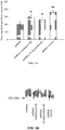
- FIG. 1A Binding of rhPRG4, HMW HA, MMW HA and vitronectin to CD44-IgG 1 Fc fusion protein and IgG 1 Fc is presented in FIG. 1A .
- the 450 nm absorbance in the CD44-IgG 1 Fc group was significantly higher (p ⁇ 0.001) than the absorbance in the IgG 1 Fc group for rhPRG4, HMW HA and MMW HA-coated wells.
- rhPRG4 binds CD44 and interferes with HMW HA CD44 binding.
- rhPRG4, HMW HA and MMW HA specifically bind to chimeric CD44 with extremely low non-specific binding.
- vitronectin that shares significant sequence homology with lubricin does not show any specificity towards CD44 binding. Because rhPRG4 binds CD44, it may function as an antagonist of CD44, thereby interfering with CD44 pro-inflammatory signaling.
- the concentration-dependent binding of rhPRG4, HMW HA and MMW HA to CD44 was performed by coating microtiter plates with 400, 200, 100, 20, 4, 2 and 0.1 ⁇ g/mL of the macromolecules. The assay was performed as described above. The absorbance values in the IgG 1 Fc wells were subtracted from the absorbance values in the CD44 IgG 1 Fc wells and the corrected CD44 IgG 1 Fc absorbance values were normalized to those of the 400 ⁇ g/mL rhPRG4 group and data was expressed as percentage binding to CD44.
- the concentration-dependent binding of rhPRG4, HMW HA and MMW HA to recombinant CD44 is depicted in FIG. 1B .
- the percentage recombinant CD44 binding was significantly higher (p ⁇ 0.001) in the rhPRG4-coated wells compared to the HMW HA or MMW HA-coated wells for the 400, 100, 20, 4 and 2 ⁇ g/mL concentrations. Additionally, the percentage recombinant CD44 binding was significantly higher (p ⁇ 0.001) in the rhPRG4-coated wells compared to the MMW HA coated wells for the 200 ⁇ g/mL concentration. There were no significant differences in percentage CD44 binding between the rhPRG4, HMW HA and MMW HA-coated wells at the 0.1 ⁇ g/mL concentration. The data represents an average of 4 independent assays, each with triplicate wells per group.
- microtiter plates were coated with either CD44 IgG 1 Fc or IgG 1 Fc at 1 ⁇ g/mL (100 ⁇ L per well) overnight at 4 °C. Subsequently, wells were washed with PBS+0.1% tween 20 and wells were blocked using 2% BSA (300 ⁇ L per well) for at least 2 hours at room temperature.
- rhPRG4 at 5 ⁇ g/mL or a combination of rhPRG4 (5 ⁇ g/mL) and HMW HA or MMW HA at 0.01, 0.05, 0.25, 1, 5 or 50 ⁇ g/mL were added to the wells (100 ⁇ L per well) and incubated at room temperature for 60 min.
- lubricin-specific monoclonal antibody (Mab 9G3) was added at 1:1,000 (100 ⁇ L per well) and incubated for 60 min at room temp.
- rhPRG4 binds to CD44 in a concentration-dependent manner with comparable affinity to HMW HA. Furthermore, rhPRG4 competes with HMW HA in binding to CD44. The presence of an excess of HMW or MMW HA reduced rhPRG4 binding to CD44 only by approximately 50%. These data suggest that rhPRG4 is an antagonist of CD44; accordingly, it has the potential to interfere with CD44 pro-inflammatory signaling.
- Binding of rhPRG4 to CD44-IgG1Fc was investigated using surface plasmon resonance (Biacore T100, GE Healthcare Lifesciences, NJ, USA). See Figure 1C .
- Series S chips were functionalized using the human antibody capture kit (GE Life Sciences) and either CD44-IgG 1 Fc or IgG 1 FC was allowed to bind to the surface of the functionalized chips in flow cell 1 (Fc 1 ) and flow cell 2 (Fc 2 ), respectively.
- rhPRG4 was injected at 30 ⁇ L/min for 8 min at concentrations of 300, 250, 200, 150, 100 and 50 ⁇ g/mL followed by a 10 min dissociation using 0.1M HEPES, 1.5M NaCl, 30mM EDTA, and 0.5% P20 (GE Life Sciences). The surface of the chip was regenerated at the end of each cycle with 1 min pulse of 3M MgCl 2 . Each analyte concentration was injected in duplicate. The resulting curves were double referenced (i.e. Fc 2 -Fc 1 , followed by subtraction of the 0 ⁇ g/mL curve).
- the binding kinetics and binding affinity were determined by BiaEvaluation software, using 1:1 binding/conformational change model or by steady-state equilibrium, respectively.
- rhPRG4 was injected at concentrations ranging between 0 and 300 ⁇ g/mL as described above.
- HMW HA was injected at 50 ⁇ g/mL (30 ⁇ L per min) for 1 min.
- the double-referenced binding signals of rhPRG4 (at various concentrations) to CD44 were then plotted against the binding signals generated by HMW HA binding to CD44 following rhPRG4 injections.
- rhPRG4 displayed a concentration-dependent association with, and dissociation from immobilized CD44-IgG 1 Fc ( FIG. 2A ), with an apparent K d ⁇ 38 nM based on a rhPRG4 molecular weight of 240 KDa.
- rhPRG4 interfered with binding of HMW HA to recombinant CD44 as shown by an inverse relationship between the HMW HA binding signal intensity ( x-axis ) and the rhPRG4 binding signal intensity ( y - axis ) ( FIG. 2B ).
- lubricin will be able to bind to CD44 on surface of synoviocytes and chondrocytes and exert a CD44-mediated biological function in the presence of HA, thereby providing a joint homeostatic role by interfering with mediators that otherwise promote inflammation.
- Lubricin's boundary lubricating ability is mediated by the O-linked ( ⁇ 1-3) Gal-GalNAc oligosaccharides ( Jay et al., Glucoconj J 2001; 18(10):807-15 ).
- a combination of neuraminidase and beta 1,3, 6 galactosidase digestions reduced lubricin's boundary lubricating ability by 50% ( Jay et al., Glucoconj J 2001; 18(10):807-15 ).
- Lubricin isolated from RA SF samples contains increased core 1 glycosylation structures and displays the sulfated epitope that is proposed to be part of the L-selectin ligand ( Estrella et al., Biochem J 2010; 429(2):359-67 ). Additionally, lubricin from RA SF binds L-selectin in a glycosylation-dependent manner and coats polymorphonuclear granulocytes recruited to inflamed synovia and SF of patients with RA ( Jin et al. J Biol Chem 2012; 287(43):35922-33 ).
- rhPRG4 was digested using sialidase A (Prozyme, USA), O-glycosidase (New England Biolabs, USA) or a combination of sialidase A and O-glycosidase for 16 hours at 37°C.
- sialidase A Prozyme, USA
- O-glycosidase New England Biolabs, USA
- a combination of sialidase A and O-glycosidase for 16 hours at 37°C.
- 12 ⁇ L of the enzyme (1U/200 ⁇ L) was added to rhPRG4 in a total reaction volume of 180 ⁇ L and a rhPRG4 final concentration of 300 ⁇ g/mL.
- Sialidase-A digestion resulted in a significant increase (p ⁇ 0.001) in the percentage binding of rhPRG4 to CD44 compared to untreated control as shown in FIG. 3A .
- There was no significant difference in percentage CD44 binding between the sialidase-A digested and the O-glycosidase codigested rhPRG4 (p 0.105).
- Sialidase-A and O-glycosidase treatments individually resulted in enhancing rhPRG4's binding to CD44 receptor. Cumulative sialidase-A and O-glycosidase digestions resulted in even more significant binding to CD44 by rhPRG4 compared to individual enzyme digestions.
- Sialidase-A cleaves branched and unbranched terminal sialic acid residues from glycoproteins, while O-glycosidase catalyzes the removal of cores 1 and 2 from glycoproteins.
- the enhancement in CD44 binding indicates that neither the core 1 glycosylation nor the sialic acid terminal residues are required in rhPRG4 binding to CD44. Accordingly, the level of sialylation and core 1 glycosylations on rhPRG4 protein core are not essential to the PRG4's ability to bind CD44. In contrast, removal of these residues may lead to a conformational change in the rhPRG4 semi-rigid rod shaped structure that results in enhanced interaction with CD44.
- RA-FLS Rheumatoid arthritis fibroblast-like synoviocytes
- CD44v7/8 a unique CD44 variant contributes to the proliferation of RA-FLS in vitro ( Wibulswas et al., Am J Pathol 2000;157(6):2037-2044 ) and pharmacological agents that bind cell surface CD44 with subsequent receptor shedding have shown efficacy in experimental arthritis models ( Runnels et al. Adv Ther 2010; 27(3):168-80 ).
- RA-FLS Rheumatoid arthritis fibroblast-like synoviocytes
- DMEM fetal bovine serum
- FBS fetal bovine serum
- TNF- ⁇ tumor necrosis factor alpha
- the total volume in each well was 200 ⁇ L.
- Cell proliferation an indication of inflammation, was determined using the CellTiter 96 AQueous one solution cell proliferation assay (MTS; Promega, USA) and the 490 nm absorbance was determined. Data is presented as the number of fold change in 490 nm absorbance compared to untreated control RA-FLS. The data represents the average of 3 independent experiments with at least triplicate wells per treatment.
- RA-FLS were stimulated with IL-1 ⁇ or TNF- ⁇ as described above.
- Treatment with rhPRG4 or HMW HA was conducted to a final concentration of 80 ⁇ g/mL in the absence or presence of IM7 (Abcam, USA), a CD44 neutralizing antibody that recognizes a conserved epitope across all CD44 isoforms ( Samson et al., Exp Eye Res, 2014; 127C:14-19 ), at a final dilution of 1:200.
- the total volume in each well was 200 ⁇ l.
- Cell proliferation across the experimental groups was determined as described above and data is presented as the number of fold change in 490 nm absorbance compared to untreated control RA-FLS. The data represents the average of 3 independent experiments with at least triplicate wells per treatment.
- IL-1 ⁇ and TNF- ⁇ induced RA-FLS proliferation over a 48 hour period is shown in FIG. 4A .
- Treatment with 40 and 80 ⁇ g/mL rhPRG4 or HMW HA significantly suppressed RA-FLS proliferation with IL-1 ⁇ stimulation (p ⁇ 0.05).
- Treatment with 20, 40 and 80 ⁇ g/mL rhPRG4 significantly suppressed RA-FLS proliferation with TNF- ⁇ stimulation (p ⁇ 0.05).
- Treatment with HMW HA did not result in suppressing RA-FLS proliferation with TNF- ⁇ .
- Co-treatment with the IM7 anti-CD44 antibody reversed the effect of rhPRG4 and HMW HA on IL-1 ⁇ stimulated RA-FLS as shown by the lack of significant difference in the change of absorbance between the IL-1 ⁇ stimulated RA-FLS treated with rhPRG4+IM7 or HMW HA+IM7 and IL-1 ⁇ stimulated RA-FLS as shown in FIG. 4B .
- co-treatment with IM7 antibody reversed the effect of rhPRG4 on TNF- ⁇ induced RA-FLS proliferation.
- rhPRG4 and HMW HA at 40 and 80 ⁇ g/mL significantly suppressed IL-1 ⁇ induced RA-FLS proliferation (p ⁇ 0.05).
- rhPRG4 at 20, 40 and 80 ⁇ g/mL significantly suppressed TNF- ⁇ induced RA-FLS proliferation (p ⁇ 0.05).
- CD44 neutralization reversed the effect of rhPRG4 on IL-1 ⁇ and TNF- ⁇ stimulated RA-FLS and the effect of HMW HA on IL-1 ⁇ stimulated RA-FLS.
- rhPRG4 inhibited the IL-1 ⁇ and TNF- ⁇ induced RA-FLS proliferation in a mechanism that involves CD44 binding.
- the downstream effect of rhPRG4 and CD44 interaction is the inhibition of nuclear translocation of NF- ⁇ B.
- HMW HA inhibited IL-1 ⁇ induced proliferation of RA-FLS but did not inhibit TNF- ⁇ induced proliferation.
- the effect of HMW HA was reversed with a CD44 antibody, indicating the role of CD44 in mediating this effect.
- rhPRG4 exerts an anti-proliferative, anti-inflammatory effect on RA-FLS subsequent to IL-1 ⁇ or TNF- ⁇ stimulation.
- rhPRG4 concentrations that demonstrate this anti-proliferative effect are generally lower than the optimal rhPRG4 concentrations required to provide boundary lubrication.
- This anti-proliferative effect of rhPRG4 is mediated by CD44 interaction with a downstream inhibition of NF- ⁇ B nuclear translocation, suggesting that therapeutically applied lubricin may mitigate the effects of pro-inflammatory cytokines on the proliferation of deleterious cell types via a CD44 dependent mechanism.
- RA-FLS (400,000 cells/well) were cultured and stimulated with TNF- ⁇ (5 ng/mL) and treated with rhPRG4 (200 ⁇ g/mL) or a commercially available NF ⁇ B translocation inhibitor MG 132 (3 ⁇ M; Tocris Bioscience) for 24 hours in serum free media.
- Cells were harvested and nuclear extraction was performed using a commercially available kit (Thermo scientific).
- Total protein was measured using a micro bicinchonic acid (BCA) kit (Thermo scientific) and 3 ⁇ g of nuclear extract of each experimental group were used.
- BCA micro bicinchonic acid
- Thermo scientific Thermo scientific
- the p50 subunit of NF ⁇ B was detected in the nuclear extract using a commercially available NF ⁇ B DNA binding assay kit (Abcam).
- TNF- ⁇ treatment resulted in significant NF ⁇ B nuclear translocation compared to untreated controls (p ⁇ 0.001) ( FIG. 4C ).
- Treatment with rhPRG4 or NF ⁇ B translocation inhibitor MG132 significantly reduced NF ⁇ B nuclear translocation compared to TNF- ⁇ -treated RA-FLS (p ⁇ 0.001).
- NF ⁇ B nuclear translocation in the TNF- ⁇ +rhPRG4+IM7 group was significantly higher than NF ⁇ B translocation in the TNF- ⁇ +rhPRG4 group (p ⁇ 0.001) and was not significantly different from the TNF- ⁇ group.
- rhPRG4 has the ability to directly reduce the pro-inflammatory effects of NF- ⁇ B nuclear translocation via a CD44 dependent mechanism.
- Synovial tissue was harvested from Prg4-/- and Prg4+/+ male mice (8-10 weeks old; 5-8 animals per genotype) and digested with pronase enzyme (2 mg/mL; Sigma Aldrich) in sterile HBSS buffer for 30 min at 37°C with shaking. This was followed by digestion with type I collagenase (1 mg/mL; Sigma Aldrich) for 4 hours at 37°C with shaking. The enzymatic reaction was stopped using DMEM+10% FBS. Cells were grown in DMEM+10%FBS and Prg4-/- synoviocytes were used between 2 nd and 4 th passages while Prg4+/+ synoviocytes were used in their second passage.
- Prg4-/- and Prg4+/+ synoviocytes were grown in chamber slides (Thermo Scientific). Cells were fixed in 4% formaldehyde for 15 min and washed twice with PBS buffer. Cells were permeabilized with 0.2% Triton X-100 for 10 min and washed 3 times with PBS buffer. Cells were blocked with 2% BSA for 30 min. Synoviocytes were incubated with IM7 anti-CD44 antibody (1:200) at 4°C overnight. Following washing three times with PBS, synoviocytes were incubated with Alexa Fluor 488 goat anti-rat IgG (Life Technologies) at 1:400 dilution for 1 hour in the dark. All incubations were performed at room temperature unless otherwise specified.
- Vectashield mounting medium containing DAPI (Vector Labs, Burlingame, CA, USA) was added. Cells were imaged with the Nikon Eclipse 90i Fluorescence Microscope using NIS Elements imaging software.
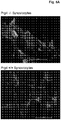
- FIG. 5A CD44 immunocytochemistry of Prg4-/- and Prg4+/+ synoviocytes is shown in FIG. 5A .
- Intense green fluorescence indicative of CD44 localization and unoccupied CD44 epitopes, was observed for Prg4-/- synoviocytes.
- no or faint green fluorescence was observed for Prg4+/+ synoviocytes, indicating that most CD44 receptors (epitopes) were occupied or antagonized by native Prg4 expression.
- IL-1 ⁇ and TNF- ⁇ treatment resulted in a significant increase in Prg4-/- synoviocytes proliferation compared to untreated Prg4-/- synoviocytes (p ⁇ 0.001) ( FIG. 5B ).
- Prg4 -/- mice exhibit early signs of cartilage degeneration, demonstrated by surface fibrillations and increased joint coefficient of friction compared to Prg4+/- and Prg4+/+ mice ( Jay et al., Arthritis Rheum, 2007; 56(11):3662-9 ). Additionally, Prg4-/- mice exhibit increased activated caspase-3 chondrocyte staining in articular cartilage compared to age-matched Prg4+/+ cartilage ( Waller et al., Proc. Natl. Acad. Sci.
- Prg4-/- mice display enhanced CD44 staining compared to Prg4+/+ synoviocytes. Additionally, pro-inflammatory cytokines induced significant proliferation of Prg4-/- synoviocytes with no appreciable effect on Prg4+/+ synoviocytes.
- rhPRG4-CD44 interaction in the setting of TNF- ⁇ and IL-1 ⁇ stimulation of Prg4-/- synoviocytes may potentially be due to rhPRG4's ability to modulate other signaling pathways independent of its ability to interact with CD44. Accordingly, rhPRG4 inhibits IL-1 ⁇ and TNF- ⁇ induced proliferation of Prg4-/- synoviocytes in a mechanism that involves CD44, and therefore is an antagonist of CD44 capable of down-regulating the pro-inflammatory activity of CD44 signaling.
- Lubricin modulates production of inflammatory cytokines in human whole blood systems.
- LPS Lipopolysaccharides
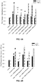
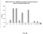
- FIGS. 8A-B supplementation of lipopolysaccharide in a 1:10 dilution in whole blood results in an increase in production of the inflammatory cytokines IL-4, IL-6, IL-8, IL-10, VEGF, TNF- ⁇ , IL1- ⁇ , and MCP-1 under the specified conditions. No changes in IL2, IFNg, and ILlb were noted.
- Figure 8B shows the percent changes in the various parameters clearly demonstrating a pronounced increase in IL6, IL8, VEGF and TNFa.
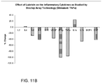
- TNF- ⁇ mediated inflammatory cytokine generation was studied using Biochip Array technology. Recombinant TNF- ⁇ was used as a trigger to generate inflammatory cytokines in whole blood. The effect of lubricin was studied on the TNF- ⁇ mediated generation of various cytokines. TNF- ⁇ alone at a concentration of 100 mg/mL was supplemented to the whole blood which was incubated for 60 minutes at 37°C. TNF- ⁇ was supplemented to whole blood in a 1:10 dilution. The modulatory effects of lubricin on TNF- ⁇ -mediated generation of various cytokines were studied by supplementing lubricin at 0.57 mg/mL in the whole blood immediately prior to addition of TNF- ⁇ .
- FIGS. 11A-B After 60 minutes plasma was obtained from centrifugation and analyzed for inflammatory cytokine using Randox Biochip arrays. As shown in FIGS. 11A-B , the presence of lubricin resulted in a decrease in IL-6, IL-8, IL-10, VEGF, TNF- ⁇ , IL-1 ⁇ , and EGF, even though challenge with TNF- ⁇ alone resulted in the production of each these cytokines.
- Figure 11B shows that lubricin decreased the levels of cytokines in a range of 10-100%.
- TNF- ⁇ stimulated cytokine expression was found to be the reduction of stimulated IFN- ⁇ and TNF- ⁇ (as lubricin apparently interrupted a positive feedback loop of TNF- ⁇ production) as compared to stimulation in the absence of lubricin.
- tissue factor recombinant tissue factor
- IL Laboratories tissue factor was used to trigger the generation of inflammatory cytokines in human whole blood.
- the effect of lubricin at 0.57 mg/mL was studied by supplementing this agent prior to the addition of tissue factor to the whole blood.
- Tissue factor alone served as a positive control.
- the blood samples were centrifuged and plasma retrieved. This plasma was then profiled for the inflammatory cytokine profile on the Randox Biochip arrays.
- TF was supplemented to whole blood in a 1:10 dilution. As shown in FIGS.
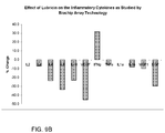
- lubricin is capable of inhibiting the generation of various inflammatory cytokines in whole blood which was supplemented bacterial lipopolysaccharide (LPS), TNF- ⁇ and tissue factor. All these agents are mediators of inflammation. Thus lubricin is capable of down-regulating the generation of inflammatory cytokines across a wide variety of mediators.
- LPS bacterial lipopolysaccharide
- TNF- ⁇ TNF- ⁇
- tissue factor tissue factor
- a rat model was used.
- Nine rats were subject to surgery to destabilize the medial meniscus (DMM surgery).
- Seven days after surgery each rat received a single intraarticular dose of lubricin of 200 ⁇ g/kg.
- Control rats received an injection of an equal volume of saline.
- Cytokine levels in serum samples taken from the test mice were compared with control mice who received surgery but no dose of lubricin post-surgery. Samples were drawn 3 weeks after the dose of lubricin was administered and analyzed using the Luminex Multiplex platform. The results are shown in FIG.
- this shows the broad anti-inflammatory effects of lubricin, as this pattern of inflammatory cytokines was distinct from the TNF- ⁇ /IL-6/IL-8 dominated profile of the LPS and TF mediators (which were generally not upregulated in this model). Nevertheless, lubricin was able to significantly reduce pro-inflammatory cytokine production in vivo.
- Synovial fluid cells were obtained from normal and osteoarthritic (OA) patients and purified with CD90+ following an immune cell depletion.
- the cells were plated at 10,000 per well and suspended in media containing DMEM, heat-inactivated Hyclone FBS (10%), and Anti-Anti (1%).
- the assays contained the cell suspension (cells + media) at 180 ⁇ L and the ligands at 20 ⁇ L for a total volume of 200 ⁇ L.
- Recombinant lubricin (rhPRG4) was then introduced to the cells at a concentration of 90 ⁇ g/mL and the negative control was PBS.
- the plate was then incubated at 37°C at 5% CO 2 for 24 hours, after which the supernatants were collected for cytokine analysis via the Luminex multiplex platform.
- normal cells did not show any changes in cytokine response when comparing the lubricin vs. PBS conditions.
- the OA cells showed a down-regulation in FGF-2 and IL-IRa cytokines when exposed to lubricin. Accordingly, in cells already exhibiting an inflammatory response, such as the osteoarthritic cells tested herein, administration of lubricin has the ability to reduce the levels of pro-inflammatory cytokines expressed from these cells.
- one or more intra-vascular clots form, blocking oxygen and nutrient delivery to brain cells, including neurons, in a volume of the brain downstream of the blockage. This also leads to destruction of neurons, thereby compromising brain function.
- the brain immune response to injury involves an increased production of pro-inflammatory mediators and the recruitment of leukocytes to the area of injury. This contributes to neuronal damage in brain regions peripheral to the site of injury, termed the "penumbra,” and exacerbates brain damage.
- Neuro-inflammation is one of the key mechanisms of secondary injury, and it is well established that post-traumatic neuro-inflammation significantly contributes to neuronal damage occurring after traumatic brain injury.
- One way to limit such brain damage is by administration of agents during a short interval (hours) after injury thereby reducing neuroinflammation and resulting neuronal damage.
- rhPRG4 also known as lubricin
- rhPRG4 can be administered systemically through the vasculature or during surgery to relieve pressure in the brain, as a means for limiting brain damage. Support for such treatment is supported by the results presented below.
- rhPRG4 was administered intravenously to test rats at an approximate dose of 2.5 mg/Kg. Normal saline (0.9% NaCl) was administered intravenously to control rats. One day later, brains were harvested and the samples of the cerebral cortex surrounding the post-traumatic lesion were analyzed using Western blotting. The analysis demonstrated that rhPRG4 reduced the post-traumatic production of proinflammatory mediators when compared with controls.
- Galectin 3 whose brain expression is rapidly increased and maintained at a high level for an extended period of time after traumatic brain injury, was down regulated by 74%.
- a 60% reduction in the magnitude of post-traumatic influx of monocytes into the injured brain parenchyma was also observed in rhPRG4-treated rats, when compared with normal saline-treated rats, and rhPRG4 also substantially attenuated (by 94%) the post-traumatic synthesis of matrix metalloproteinase 9 and inhibited (by 64%) the conversion of pro-matrix metalloproteinase 2 to its enzymatically active form.
- rhPRG4 reduced by 80% the permeability of the blood-brain barrier, which was evaluated by assessing the level of albumin in traumatized brain tissue.
- rhPRG4 limits the magnitude of post-traumatic neuro-inflammation by both directly inhibiting the chemotactic activity of invading inflammatory cells, and by curtailing the production and signaling of pro-inflammatory mediators.
- rhPRG4 selectively targets the injured areas of the brain, reducing the likelihood of off-target pharmacological effects.
- Recombinant hPRG4 limits, with high efficacy, the magnitude of neuro-inflammation caused by traumatic brain injury, by reducing the post-traumatic production of proinflammatory mediators and the influx of inflammatory cells.
- rhPRG4 exhibits a unique ability to stabilize the blood-brain barrier. The discovery of this novel rhPRG4 property is of great significance for treatment of brain injury, as blood-brain barrier dysfunction observed in brain injury not only contributes to neuronal death in the acute stage of injury, but also leads to progressive neurodegenerative changes in the injured brain and, consequently, poor neurological outcomes.
- HA or MMW HA at 0.01, 0.05, 0.25, 1, 5 or 50 ⁇ g/mL were added to the wells (100 ⁇ L per well) and incubated at room temperature for 60 min.
- lubricin-specific monoclonal antibody (Mab 9G3) was added at 1:1,000 (100 ⁇ L per well) and incubated for 60 min at room temp.
- goat anti-mouse IgG-HRP (Thermo Scientific) at 1:1,000 dilution was added (100 ⁇ L per well) and incubated for 60 min at room temp.
- the assay was developed as described above.
- the absorbance values in the IgG 1 Fc wells were subtracted from the absorbance values in the CD44-IgG 1 Fc wells and the corrected absorbance values in the rhPRG4+HA groups were normalized to the absorbance values of the rhPRG4 group and data was expressed as percentage binding to CD44.
- the data represents an average of 4 independent assays, each with triplicate wells per group.
- rhPRG4 binds to CD44 in a concentration-dependent manner with comparable affinity to HMW HA. Furthermore, rhPRG4 competes with HMW HA in binding to CD44. The presence of an excess of HMW or MMW HA reduced rhPRG4 binding to CD44 only by approximately 50%. These data suggest that rhPRG4 is an antagonist of CD44; accordingly, it has the potential to interfere with CD44 pro-inflammatory signaling.
- Binding of rhPRG4 to CD44-IgG1Fc was investigated using surface plasmon resonance (Biacore T100, GE Healthcare Lifesciences, NJ, USA). See Figure 1C .
- Series S chips were functionalized using the human antibody capture kit (GE Life Sciences) and either CD44-IgG 1 Fc or IgG 1 FC was allowed to bind to the surface of the functionalized chips in flow cell 1 (Fc 1 ) and flow cell 2 (Fc 2 ), respectively.
- rhPRG4 was injected at 30 ⁇ L/min for 8 min at concentrations of 300, 250, 200, 150, 100 and 50 ⁇ g/mL followed by a 10 min dissociation using 0.1M HEPES, 1.5M NaCl, 30mM EDTA, and 0.5% P20 (GE Life Sciences). The surface of the chip was regenerated at the end of each cycle with 1 min pulse of 3M MgCl 2 . Each analyte concentration was injected in duplicate. The resulting curves were double referenced (i.e. Fc 2 -Fc 1 , followed by subtraction of the 0 ⁇ g/mL curve).
- the binding kinetics and binding affinity were determined by BiaEvaluation software, using 1:1 binding/conformational change model or by steady-state equilibrium, respectively.
- rhPRG4 was injected at concentrations ranging between 0 and 300 ⁇ g/mL as described above.
- HMW HA was injected at 50 ⁇ g/mL (30 ⁇ L per min) for 1 min.
- the double-referenced binding signals of rhPRG4 (at various concentrations) to CD44 were then plotted against the binding signals generated by HMW HA binding to CD44 following rhPRG4 injections.
- rhPRG4 displayed a concentration-dependent association with, and dissociation from immobilized CD44-IgG 1 Fc ( FIG. 2A ), with an apparent K d ⁇ 38 nM based on a rhPRG4 molecular weight of 240 KDa.
- rhPRG4 interfered with binding of HMW HA to recombinant CD44 as shown by an inverse relationship between the HMW HA binding signal intensity ( x-axis ) and the rhPRG4 binding signal intensity ( y-axis ) ( FIG. 2B ).
- lubricin will be able to bind to CD44 on surface of synoviocytes and chondrocytes and exert a CD44-mediated biological function in the presence of HA, thereby providing a joint homeostatic role by interfering with mediators that otherwise promote inflammation.
- Lubricin's boundary lubricating ability is mediated by the O-linked ( ⁇ 1-3) Gal-GalNAc oligosaccharides ( Jay et al., Glucoconj J 2001; 18(10):807-15 ).
- a combination of neuraminidase and beta 1,3, 6 galactosidase digestions reduced lubricin's boundary lubricating ability by 50% ( Jay et al., Glucoconj J 2001; 18(10):807-15 ).
- Lubricin isolated from RA SF samples contains increased core 1 glycosylation structures and displays the sulfated epitope that is proposed to be part of the L-selectin ligand ( Estrella et al., Biochem J 2010; 429(2):359-67 ). Additionally, lubricin from RA SF binds L-selectin in a glycosylation-dependent manner and coats polymorphonuclear granulocytes recruited to inflamed synovia and SF of patients with RA ( Jin et al. J Biol Chem 2012; 287(43):35922-33 ).
- rhPRG4 was digested using sialidase A (Prozyme, USA), O-glycosidase (New England Biolabs, USA) or a combination of sialidase A and O-glycosidase for 16 hours at 37°C.
- sialidase A Prozyme, USA
- O-glycosidase New England Biolabs, USA
- a combination of sialidase A and O-glycosidase for 16 hours at 37°C.
- 12 ⁇ L of the enzyme (1U/200 ⁇ L) was added to rhPRG4 in a total reaction volume of 180 ⁇ L and a rhPRG4 final concentration of 300 ⁇ g/mL.
- Sialidase-A digestion resulted in a significant increase (p ⁇ 0.001) in the percentage binding of rhPRG4 to CD44 compared to untreated control as shown in FIG. 3A .
- There was no significant difference in percentage CD44 binding between the sialidase-A digested and the O-glycosidase codigested rhPRG4 (p 0.105).
- Sialidase-A and O-glycosidase treatments individually resulted in enhancing rhPRG4's binding to CD44 receptor. Cumulative sialidase-A and O-glycosidase digestions resulted in even more significant binding to CD44 by rhPRG4 compared to individual enzyme digestions.
- Sialidase-A cleaves branched and unbranched terminal sialic acid residues from glycoproteins, while O-glycosidase catalyzes the removal of cores 1 and 2 from glycoproteins.
- the enhancement in CD44 binding indicates that neither the core 1 glycosylation nor the sialic acid terminal residues are required in rhPRG4 binding to CD44. Accordingly, the level of sialylation and core 1 glycosylations on rhPRG4 protein core are not essential to the PRG4's ability to bind CD44. In contrast, removal of these residues may lead to a conformational change in the rhPRG4 semi-rigid rod shaped structure that results in enhanced interaction with CD44.
- RA-FLS Rheumatoid arthritis fibroblast-like synoviocytes
- CD44v7/8 a unique CD44 variant contributes to the proliferation of RA-FLS in vitro ( Wibulswas et al., Am J Pathol 2000;157(6):2037-2044 ) and pharmacological agents that bind cell surface CD44 with subsequent receptor shedding have shown efficacy in experimental arthritis models ( Runnels et al. Adv Ther 2010; 27(3):168-80 ).
- RA-FLS Rheumatoid arthritis fibroblast-like synoviocytes
- DMEM fetal bovine serum
- FBS fetal bovine serum
- TNF- ⁇ tumor necrosis factor alpha
- the total volume in each well was 200 ⁇ L.
- Cell proliferation an indication of inflammation, was determined using the CellTiter 96 AQueous one solution cell proliferation assay (MTS; Promega, USA) and the 490 nm absorbance was determined. Data is presented as the number of fold change in 490 nm absorbance compared to untreated control RA-FLS. The data represents the average of 3 independent experiments with at least triplicate wells per treatment.
- RA-FLS were stimulated with IL-1 ⁇ or TNF- ⁇ as described above.
- Treatment with rhPRG4 or HMW HA was conducted to a final concentration of 80 ⁇ g/mL in the absence or presence of IM7 (Abcam, USA), a CD44 neutralizing antibody that recognizes a conserved epitope across all CD44 isoforms ( Samson et al., Exp Eye Res, 2014; 127C:14-19 ), at a final dilution of 1:200.
- the total volume in each well was 200 ⁇ l.
- Cell proliferation across the experimental groups was determined as described above and data is presented as the number of fold change in 490 nm absorbance compared to untreated control RA-FLS. The data represents the average of 3 independent experiments with at least triplicate wells per treatment.
- IL-1 ⁇ and TNF- ⁇ induced RA-FLS proliferation over a 48 hour period is shown in FIG. 4A .
- Treatment with 40 and 80 ⁇ g/mL rhPRG4 or HMW HA significantly suppressed RA-FLS proliferation with IL-1 ⁇ stimulation (p ⁇ 0.05).
- Treatment with 20, 40 and 80 ⁇ g/mL rhPRG4 significantly suppressed RA-FLS proliferation with TNF- ⁇ stimulation (p ⁇ 0.05).
- Treatment with HMW HA did not result in suppressing RA-FLS proliferation with TNF- ⁇ .
- Co-treatment with the IM7 anti-CD44 antibody reversed the effect of rhPRG4 and HMW HA on IL-1 ⁇ stimulated RA-FLS as shown by the lack of significant difference in the change of absorbance between the IL-1 ⁇ stimulated RA-FLS treated with rhPRG4+IM7 or HMW HA+IM7 and IL-1 ⁇ stimulated RA-FLS as shown in FIG. 4B .
- co-treatment with IM7 antibody reversed the effect of rhPRG4 on TNF- ⁇ induced RA-FLS proliferation.
- rhPRG4 and HMW HA at 40 and 80 ⁇ g/mL significantly suppressed IL-1 ⁇ induced RA-FLS proliferation (p ⁇ 0.05).
- rhPRG4 at 20, 40 and 80 ⁇ g/mL significantly suppressed TNF- ⁇ induced RA-FLS proliferation (p ⁇ 0.05).
- CD44 neutralization reversed the effect of rhPRG4 on IL-1 ⁇ and TNF- ⁇ stimulated RA-FLS and the effect of HMW HA on IL-1 ⁇ stimulated RA-FLS.
- rhPRG4 inhibited the IL-1 ⁇ and TNF- ⁇ induced RA-FLS proliferation in a mechanism that involves CD44 binding.
- the downstream effect of rhPRG4 and CD44 interaction is the inhibition of nuclear translocation of NF- ⁇ B.
- HMW HA inhibited IL-1 ⁇ induced proliferation of RA-FLS but did not inhibit TNF- ⁇ induced proliferation.
- the effect of HMW HA was reversed with a CD44 antibody, indicating the role of CD44 in mediating this effect.
- rhPRG4 exerts an anti-proliferative, anti-inflammatory effect on RA-FLS subsequent to IL-1 ⁇ or TNF- ⁇ stimulation.
- rhPRG4 concentrations that demonstrate this anti-proliferative effect are generally lower than the optimal rhPRG4 concentrations required to provide boundary lubrication.
- This anti-proliferative effect of rhPRG4 is mediated by CD44 interaction with a downstream inhibition of NF- ⁇ B nuclear translocation, suggesting that therapeutically applied lubricin may mitigate the effects of pro-inflammatory cytokines on the proliferation of deleterious cell types via a CD44 dependent mechanism.
- RA-FLS (400,000 cells/well) were cultured and stimulated with TNF- ⁇ (5 ng/mL) and treated with rhPRG4 (200 ⁇ g/mL) or a commercially available NF ⁇ B translocation inhibitor MG 132 (3 ⁇ M; Tocris Bioscience) for 24 hours in serum free media.
- Cells were harvested and nuclear extraction was performed using a commercially available kit (Thermo scientific).
- Total protein was measured using a micro bicinchonic acid (BCA) kit (Thermo scientific) and 3 ⁇ g of nuclear extract of each experimental group were used.
- BCA micro bicinchonic acid
- Thermo scientific Thermo scientific
- the p50 subunit of NF ⁇ B was detected in the nuclear extract using a commercially available NF ⁇ B DNA binding assay kit (Abcam).
- TNF- ⁇ treatment resulted in significant NF ⁇ B nuclear translocation compared to untreated controls (p ⁇ 0.001) ( FIG. 4C ).
- Treatment with rhPRG4 or NF ⁇ B translocation inhibitor MG132 significantly reduced NF ⁇ B nuclear translocation compared to TNF- ⁇ -treated RA-FLS (p ⁇ 0.001).
- NF ⁇ B nuclear translocation in the TNF- ⁇ +rhPRG4+IM7 group was significantly higher than NF ⁇ B translocation in the TNF- ⁇ +rhPRG4 group (p ⁇ 0.001) and was not significantly different from the TNF- ⁇ group.
- rhPRG4 has the ability to directly reduce the pro-inflammatory effects of NF- ⁇ B nuclear translocation via a CD44 dependent mechanism.
- Synovial tissue was harvested from Prg4-/- and Prg4+/+ male mice (8-10 weeks old; 5-8 animals per genotype) and digested with pronase enzyme (2 mg/mL; Sigma Aldrich) in sterile HBSS buffer for 30 min at 37°C with shaking. This was followed by digestion with type I collagenase (1 mg/mL; Sigma Aldrich) for 4 hours at 37°C with shaking. The enzymatic reaction was stopped using DMEM+10% FBS. Cells were grown in DMEM+10%FBS and Prg4-/- synoviocytes were used between 2 nd and 4 th passages while Prg4+/+ synoviocytes were used in their second passage.
- Prg4-/- and Prg4+/+ synoviocytes were grown in chamber slides (Thermo Scientific). Cells were fixed in 4% formaldehyde for 15 min and washed twice with PBS buffer. Cells were permeabilized with 0.2% Triton X-100 for 10 min and washed 3 times with PBS buffer. Cells were blocked with 2% BSA for 30 min. Synoviocytes were incubated with IM7 anti-CD44 antibody (1:200) at 4°C overnight. Following washing three times with PBS, synoviocytes were incubated with Alexa Fluor 488 goat anti-rat IgG (Life Technologies) at 1:400 dilution for 1 hour in the dark. All incubations were performed at room temperature unless otherwise specified.
- Vectashield mounting medium containing DAPI (Vector Labs, Burlingame, CA, USA) was added. Cells were imaged with the Nikon Eclipse 90i Fluorescence Microscope using NIS Elements imaging software.
- FIG. 5A CD44 immunocytochemistry of Prg4-/- and Prg4+/+ synoviocytes is shown in FIG. 5A .
- Intense green fluorescence indicative of CD44 localization and unoccupied CD44 epitopes, was observed for Prg4-/- synoviocytes.
- no or faint green fluorescence was observed for Prg4+/+ synoviocytes, indicating that most CD44 receptors (epitopes) were occupied or antagonized by native Prg4 expression.
- IL-1 ⁇ and TNF- ⁇ treatment resulted in a significant increase in Prg4-/- synoviocytes proliferation compared to untreated Prg4-/- synoviocytes (p ⁇ 0.001) ( FIG. 5B ).
- Prg4 -/- mice exhibit early signs of cartilage degeneration, demonstrated by surface fibrillations and increased joint coefficient of friction compared to Prg4+/- and Prg4+/+ mice ( Jay et al., Arthritis Rheum, 2007; 56(11):3662-9 ). Additionally, Prg4-/- mice exhibit increased activated caspase-3 chondrocyte staining in articular cartilage compared to age-matched Prg4+/+ cartilage ( Waller et al., Proc. Natl. Acad. Sci.
- Prg4-/- mice display enhanced CD44 staining compared to Prg4+/+ synoviocytes. Additionally, pro-inflammatory cytokines induced significant proliferation of Prg4-/- synoviocytes with no appreciable effect on Prg4+/+ synoviocytes.
- rhPRG4-CD44 interaction in the setting of TNF- ⁇ and IL-1 ⁇ stimulation of Prg4-/- synoviocytes may potentially be due to rhPRG4's ability to modulate other signaling pathways independent of its ability to interact with CD44. Accordingly, rhPRG4 inhibits IL-1 ⁇ and TNF- ⁇ induced proliferation of Prg4-/- synoviocytes in a mechanism that involves CD44, and therefore is an antagonist of CD44 capable of down-regulating the pro-inflammatory activity of CD44 signaling.
- Example 2 Lubricin modulates production of inflammatory cytokines in human whole blood systems.
- LPS Lipopolysaccharides
- FIGS. 8A-B supplementation of lipopolysaccharide in a 1:10 dilution in whole blood results in an increase in production of the inflammatory cytokines IL-4, IL-6, IL-8, IL-10, VEGF, TNF- ⁇ , IL1- ⁇ , and MCP-1 under the specified conditions. No changes in IL2, IFNg, and ILlb were noted.
- Figure 8B shows the percent changes in the various parameters clearly demonstrating a pronounced increase in IL6, IL8, VEGF and TNFa.
- TNF- ⁇ mediated inflammatory cytokine generation was studied using Biochip Array technology. Recombinant TNF- ⁇ was used as a trigger to generate inflammatory cytokines in whole blood. The effect of lubricin was studied on the TNF- ⁇ mediated generation of various cytokines. TNF- ⁇ alone at a concentration of 100 mg/mL was supplemented to the whole blood which was incubated for 60 minutes at 37°C. TNF- ⁇ was supplemented to whole blood in a 1:10 dilution. The modulatory effects of lubricin on TNF- ⁇ -mediated generation of various cytokines were studied by supplementing lubricin at 0.57 mg/mL in the whole blood immediately prior to addition of TNF- ⁇ .
- FIGS. 11A-B After 60 minutes plasma was obtained from centrifugation and analyzed for inflammatory cytokine using Randox Biochip arrays. As shown in FIGS. 11A-B , the presence of lubricin resulted in a decrease in IL-6, IL-8, IL-10, VEGF, TNF- ⁇ , IL-1 ⁇ , and EGF, even though challenge with TNF- ⁇ alone resulted in the production of each these cytokines.
- Figure 11B shows that lubricin decreased the levels of cytokines in a range of 10-100%.
- TNF- ⁇ stimulated cytokine expression was found to be the reduction of stimulated IFN- ⁇ and TNF- ⁇ (as lubricin apparently interrupted a positive feedback loop of TNF- ⁇ production) as compared to stimulation in the absence of lubricin.
- tissue factor recombinant tissue factor
- IL Laboratories tissue factor was used to trigger the generation of inflammatory cytokines in human whole blood.
- the effect of lubricin at 0.57 mg/mL was studied by supplementing this agent prior to the addition of tissue factor to the whole blood.
- Tissue factor alone served as a positive control.
- the blood samples were centrifuged and plasma retrieved. This plasma was then profiled for the inflammatory cytokine profile on the Randox Biochip arrays.
- TF was supplemented to whole blood in a 1:10 dilution. As shown in FIGS.
- lubricin is capable of inhibiting the generation of various inflammatory cytokines in whole blood which was supplemented bacterial lipopolysaccharide (LPS), TNF- ⁇ and tissue factor. All these agents are mediators of inflammation. Thus lubricin is capable of down-regulating the generation of inflammatory cytokines across a wide variety of mediators.
- LPS bacterial lipopolysaccharide
- TNF- ⁇ TNF- ⁇
- tissue factor tissue factor
- Example 3 Lubricin reduces levels of pro-inflammatory cytokines in vivo
- a rat model was used.
- Nine rats were subject to surgery to destabilize the medial meniscus (DMM surgery).
- Seven days after surgery each rat received a single intraarticular dose of lubricin of 200 ⁇ g/kg.
- Control rats received an injection of an equal volume of saline.
- Cytokine levels in serum samples taken from the test mice were compared with control mice who received surgery but no dose of lubricin post-surgery. Samples were drawn 3 weeks after the dose of lubricin was administered and analyzed using the Luminex Multiplex platform. The results are shown in FIG.
- this shows the broad anti-inflammatory effects of lubricin, as this pattern of inflammatory cytokines was distinct from the TNF- ⁇ /IL-6/IL-8 dominated profile of the LPS and TF mediators (which were generally not upregulated in this model). Nevertheless, lubricin was able to significantly reduce pro-inflammatory cytokine production in vivo.
- Synovial fluid cells were obtained from normal and osteoarthritic (OA) patients and purified with CD90+ following an immune cell depletion.
- the cells were plated at 10,000 per well and suspended in media containing DMEM, heat-inactivated Hyclone FBS (10%), and Anti-Anti (1%).
- the assays contained the cell suspension (cells + media) at 180 ⁇ L and the ligands at 20 ⁇ L for a total volume of 200 ⁇ L.
- Recombinant lubricin (rhPRG4) was then introduced to the cells at a concentration of 90 ⁇ g/mL and the negative control was PBS.
- the plate was then incubated at 37°C at 5% CO 2 for 24 hours, after which the supernatants were collected for cytokine analysis via the Luminex multiplex platform.
- normal cells did not show any changes in cytokine response when comparing the lubricin vs. PBS conditions.
- the OA cells showed a down-regulation in FGF-2 and IL-IRa cytokines when exposed to lubricin. Accordingly, in cells already exhibiting an inflammatory response, such as the osteoarthritic cells tested herein, administration of lubricin has the ability to reduce the levels of pro-inflammatory cytokines expressed from these cells.
- one or more intra-vascular clots form, blocking oxygen and nutrient delivery to brain cells, including neurons, in a volume of the brain downstream of the blockage. This also leads to destruction of neurons, thereby compromising brain function.
- the brain immune response to injury involves an increased production of pro-inflammatory mediators and the recruitment of leukocytes to the area of injury. This contributes to neuronal damage in brain regions peripheral to the site of injury, termed the "penumbra,” and exacerbates brain damage.
- Neuro-inflammation is one of the key mechanisms of secondary injury, and it is well established that post-traumatic neuro-inflammation significantly contributes to neuronal damage occurring after traumatic brain injury.
- One way to limit such brain damage is by administration of agents during a short interval (hours) after injury thereby reducing neuroinflammation and resulting neuronal damage.
- rhPRG4 also known as lubricin
- rhPRG4 can be administered systemically through the vasculature or during surgery to relieve pressure in the brain, as a means for limiting brain damage. Support for such treatment is supported by the results presented below.
- rhPRG4 was administered intravenously to test rats at an approximate dose of 2.5 mg/Kg. Normal saline (0.9% NaCl) was administered intravenously to control rats. One day later, brains were harvested and the samples of the cerebral cortex surrounding the post-traumatic lesion were analyzed using Western blotting. The analysis demonstrated that rhPRG4 reduced the post-traumatic production of proinflammatory mediators when compared with controls.
- Galectin 3 whose brain expression is rapidly increased and maintained at a high level for an extended period of time after traumatic brain injury, was down regulated by 74%.
- a 60% reduction in the magnitude of post-traumatic influx of monocytes into the injured brain parenchyma was also observed in rhPRG4-treated rats, when compared with normal saline-treated rats, and rhPRG4 also substantially attenuated (by 94%) the post-traumatic synthesis of matrix metalloproteinase 9 and inhibited (by 64%) the conversion of pro-matrix metalloproteinase 2 to its enzymatically active form.
- rhPRG4 reduced by 80% the permeability of the blood-brain barrier, which was evaluated by assessing the level of albumin in traumatized brain tissue.
- rhPRG4 limits the magnitude of post-traumatic neuro-inflammation by both directly inhibiting the chemotactic activity of invading inflammatory cells, and by curtailing the production and signaling of pro-inflammatory mediators.
- rhPRG4 selectively targets the injured areas of the brain, reducing the likelihood of off-target pharmacological effects.
- Recombinant hPRG4 limits, with high efficacy, the magnitude of neuro-inflammation caused by traumatic brain injury, by reducing the post-traumatic production of proinflammatory mediators and the influx of inflammatory cells.
- rhPRG4 exhibits a unique ability to stabilize the blood-brain barrier. The discovery of this novel rhPRG4 property is of great significance for treatment of brain injury, as blood-brain barrier dysfunction observed in brain injury not only contributes to neuronal death in the acute stage of injury, but also leads to progressive neurodegenerative changes in the injured brain and, consequently, poor neurological outcomes.
- Inflammatory bowel disease is characterized by a cytokine mediated recruitment of activated T cells that results in oxidative damage and a wearing of the intestinal epithelium. It is estimated that 25%-40% of people with ulcerative colitis (UC) or Crohn's disease may progress to surgery, such as an ileal pouch anal anastomosis, or proctocolectomy that removes portions of the colon and rectum.
- Common therapies include broad spectrum anti-inflammatory medications such as corticosteroids, anti-TNF antibodies, or more targeted anti-integrin antibodies that aim to prevent gut-homing T-cell mobility. None of these approaches are suitable for long term therapy because of serious side effects such as infections and cancer that accompany these approaches.
- recombinant hPRG4 can be selectively applied to the gut to both replenish the missing epithelial glycocalyx and reduce cytokine expression locally.
- a recent characterization of the cytokine profile of predominantly colonic inflammatory bowel disease revealed increased TNF- ⁇ , GRO, CCL11 (eotaxin) in UC, IL-6 in Crohn's, and IL-8 in both UC and Crohn's versus controls ( Korolkova et al., Clin Med Insights Gastroenerol 2015 May 6;8:29-44 ). Lubricin has been shown to significantly reduce expression of these cytokines.
- rhPRG4 is administered via enema or orally, whether through gavage, whole bowel irrigation, drinking a solution, or through encapsulated pills (e.g., microparticle encapsulation, nanoparticle encapsulation, polymer encapsulation, etc.).
- encapsulated pills e.g., microparticle encapsulation, nanoparticle encapsulation, polymer encapsulation, etc.
- an enema administration of 100 mL to 4 L rhPRG4 in concentrations ranging from 10 ⁇ g/mL to 200 ⁇ g/mL, more preferably volumes of 200 mL to 500 mL in concentrations from 50 ⁇ g/mL to 150 ⁇ g/mL, suspended in an intestinally acceptable buffered salt solution replenishes the glycocalyx, prevents T cell homing and downregulates cytokine expression in the locality of the administered lubricin.
- Administration of rhPRG4 results in improved epithelial barrier function, less vascular permeability, less susceptibility to protease activity, and improved nutrient absorption.
- a magnesium citrate or other laxative is administered up to 24 hours prior to lubricin administration, followed by appropriate fasting.
- Rats undergoing injection of sodium urate crystals were studied as a model of gout. 24 hours following the sodium urate crystal administration, rats developed joint pain. Two rats received saline while another two rats received rhPRG4 injection 24 hours after the urate crystals were introduced. These rats were studied every 12 hours utilizing the Von Frey method. This method determines afferent pain sensitization as a result of joint inflammation by probing the paws of the affected limb with a thin filament wire. The data in FIG.15 shows that the rats that received rhPRG4 were in less pain than rats which received placebo.
Landscapes
- Health & Medical Sciences (AREA)
- Life Sciences & Earth Sciences (AREA)
- Veterinary Medicine (AREA)
- Pharmacology & Pharmacy (AREA)
- Chemical & Material Sciences (AREA)
- Public Health (AREA)
- General Health & Medical Sciences (AREA)
- Medicinal Chemistry (AREA)
- Animal Behavior & Ethology (AREA)
- Engineering & Computer Science (AREA)
- Bioinformatics & Cheminformatics (AREA)
- Chemical Kinetics & Catalysis (AREA)
- Organic Chemistry (AREA)
- Nuclear Medicine, Radiotherapy & Molecular Imaging (AREA)
- General Chemical & Material Sciences (AREA)
- Immunology (AREA)
- Gastroenterology & Hepatology (AREA)
- Epidemiology (AREA)
- Proteomics, Peptides & Aminoacids (AREA)
- Zoology (AREA)
- Marine Sciences & Fisheries (AREA)
- Pulmonology (AREA)
- Dermatology (AREA)
- Neurology (AREA)
- Neurosurgery (AREA)
- Diabetes (AREA)
- Pain & Pain Management (AREA)
- Biomedical Technology (AREA)
- Urology & Nephrology (AREA)
- Communicable Diseases (AREA)
- Oncology (AREA)
- Rheumatology (AREA)
- Hematology (AREA)
- Cardiology (AREA)
- Heart & Thoracic Surgery (AREA)
- Physical Education & Sports Medicine (AREA)
- Psychiatry (AREA)
- Virology (AREA)
- Hospice & Palliative Care (AREA)
- Vascular Medicine (AREA)
Description
- This invention relates to new uses of the human glycoprotein PRG4 or lubricin.
- The
proteoglycan 4 gene (PRG4) encodes megakaryocyte stimulating factor (MSF) as well as highly glycosylated differently splice variant and glycoforms of "superficial zone protein" also known as lubricin. Superficial zone protein was first localized at the surface of explant cartilage from the superficial zone and identified in conditioned medium. Lubricin was first isolated from synovial fluid and demonstrated lubricating ability in vitro similar to synovial fluid at a cartilage-glass interface and in a latex-glass interface. It was later identified as a product of synovial fibroblasts, and its lubricating ability was discovered to be dependent on O-linked β (1-3) Gal-GalNAc oligosaccharides within a large mucin like domain of 940 amino acids encoded byexon 6. Lubricin molecules are differentially glycosylated and several naturally occurring splice variants have been reported. They are collectively referred to herein as PRG4. PRG4 has been shown to be present inside the body at the surface of synovium, tendon, articular cartilage such as meniscus, and in the protective film of the eye, among other sites, and plays an important role in joint lubrication and synovial homeostasis. - Prior to Applicants' inventions, PRG4 had been appreciated as a protein with only mechanical properties, providing mechanical functionalities such as lubricating joints, tendons, cartilage, and acting as a mechanical barrier to inhibit intercellular interactions. However, as shown herein, Applicants have discovered that lubricin has properties that extend beyond its ability to provide boundary lubrication and anti-adhesion. In particular, Applicants have determined that PRG4 has anti-inflammatory properties due to its ability to act as a ligand or signaling molecule, participating in ligand receptor interactions to modulate, for example, CD44 activation, NF-κB translocation, and cytokine-mediated inflammation.
- The current invention exploits the heretofore unknown anti-inflammatory properties of PRG4, also known as lubricin. Accordingly, the invention provides PRG4 for use in a method of reducing or inhibiting an inflammatory response in a patient having brain injury as defined in the claims appended hereto. Underlying the discovery of PRG4's anti-inflammatory properties is the understanding of various putative mechanisms by which PRG4 achieves its anti-inflammatory effect. These mechanisms were discovered by Applicants who have determined that PRG4 binds CD44 receptors, enabling it to act as a CD44 receptor antagonist. As a result, PRG4 is able to down-regulate pro-inflammatory responses mediated by CD44 receptor signaling. The ability of PRG4 to affect CD44 signaling also has the effect of down-regulating translocation of NF-κB. Further, administration of PRG4 has been shown to inhibit production of a number of pro-inflammatory cytokines, as well as to modulate cellular responses and proliferation due to pro-inflammatory cytokine induction (e.g., TNF-a); a feature unique to PRG4 and not to other lubricants such as high molecular weight hyaluronic acid. PRG4 can therefore be used in a number of novel ways in therapeutic and prophylactic contexts to effect anti-inflammatory action via its effect on signaling pathways involved in producing an inflammatory response.
- PRG4 may be administered systemically to the patient. For example, the administration may be intravenous, by inhalation, intramuscular, subcutaneous, oral, rectal, buccal, or sublingual. PRG4 may also be administered locally to the patient, for example administered to the brain topically during surgery or by injection.
- In a further embodiment, the PRG4 administered is recombinant human PRG4. In another embodiment, the PRG4 has the sequence of SEQ ID NO:1 minus the signal sequence. In one embodiment, PRG4 is administered in an amount insufficient to provide boundary lubrication in the patient. In another embodiment, PRG4 is administered in an amount ranging from 0.1 µg/kg - 4,000 µg/kg or as a coating on a tissue surface applied from a PRG4 solution comprising, for example, a concentration of PRG4 between 10 µg/mL to about 2 mg/mL.
- In one embodiment, the reduction or inhibition of the inflammatory response may be measured by the level of production of a pro-inflammatory cytokine in the patient. For example, in one embodiment, the pro-inflammatory cytokine is selected from the group consisting of IL-1α, IL-1β, IL-2, IL-3, IL-4, IL-5, IL-6, IL-7, IL-8, IL-9, IL-10, IL-11, IL-12, IL-12p70, IL-13, IL-14, IL-15, IL-16, IL-17, IL-17α, IL-18, IL-19, IL-20, IL-21, IL-22, IL-23, IL-24, IL-25, IL-26, IL-27, IL-28, IL-29, IL-30, IL-31, IL-32, IL-33, IL-34, IL-35, IL-36, TNF-α, TNF-β (lymphotoxin-α), lymphotoxin-β, CXC31L (Fractalkine), CXCL-8, CCL2, CCL3, CCL4, CCL5, CCL11, CXCL10, IFN-α, IFN-β, IFN-ε, IFN-κ, IFN-ω, IFN-γ, VEGF, MCP-1, MCP-3, EGF, GMCSF, CD40L, CD27L, CD30L, FASL, 4-1BBL, OX40L, TRAIL, FGF-2, GRO, MDC, Rantes, G-CSF, M-CSF, FGF-2, EPO, MCSF, MIP3α, MG-CSF, and GCSF.
- Administration of PRG4 binds a CD44 receptor on a cell in the patient, reduces or inhibits the production of a pro-inflammatory cytokine in the patient, and/or reduces or inhibits the translocation of NF-κB in a cell in the patient and thereby reduces or inhibits the inflammatory response in the patient.
- Administration of PRG4 may reduce or inhibit a pro-inflammatory cytokine selected from the group consisting of IL-2, IL-4, IL-6, IL-8, IL-10, VEGF, IFN-γ, TNF-α, IL-1α, IL-1β, MCP-1, EGF, FGF-2, Fractalkine, IFN-α2, GRO, MCP-3, MDC, EPO, IL-13, IL-18, MCSF, MIP-3α, MG-CSF, IL-7, IL-5, G-CSF, Rantes, IL-17α, or IL-12p70.
- The PRG4, for example rhPRG4, may be administered to a human subject in amount ranging from 0.1 µg/kg - 4,000 µg/kg. The PRG4, for example, rhPRG4, may be administered in an amount of 0.1 µg/mL to 30 mg/mL in small volumes of 1-100 µL per dose. Tthe PRG4 may be administered in an amount of 10 µg/mL to 4 mg/mL in volumes of 100 µL - 4 L per dose, for instance as an enema.
- As described herein, exposing a surface to PRG4, for example, rhPRG4, antagonizes a pro-inflammatory ligand of CD44. In one embodiment, the ligand is hyaluronan (HA), a hyaluronan serum-derived hyaluronan-associated protein complex (HA-SHAP), or a matrix metalloproteinase (e.g., MMP-9). In another embodiment, the ligand is hemopexin, EMMPRIN, somatomedin-B, osteopontin, OKT3, or a complement related protein such as C3a, CD3, CD46.
- As described herein, PRG4 may reduce or inhibit pro-inflammatory cytokine levels in blood, for example, in a human subject. PRG4 may be administered systemically in an amount sufficient to reduce or inhibit pro-inflammatory cytokine levels. Exemplary proinflammatory cytokines include IL-2, IL-4, IL-6, IL-8, IL-10, VEGF, IFN-γ, TNF-α, IL1-α, IL-1-β, MCP-1, EGF, FGF-2, fractalkine, IFN-α2, GRO, MCP-3, MDC, EPO, IL-13, IL-18, MCSF, MIP-3α, MG-CSF, IL-7, IL-5, G-CSF, Rantes, IL-17α, or IL-12p70.
- PRG4 may inhibit NF-κB translocation in a cell. Contacting a cell containing NF-κB with PRG4, wherein PRG4 binds to a cell surface receptor, may inhibit activation of the NF-κB signaling pathway. PRG4 may inhibit a TNF-α receptor or an IL-1 receptor on the cell surface.
-
-
FIGS. 1A-C are bar graphs presenting data demonstrating the binding of recombinant human proteoglycan 4 (rhPRG4), high-molecular weight hyaluronic acid (HMW HA), and medium molecular weight hyaluronic acid (MMW HA) to recombinant human CD44 receptor as detected by TMB-ELISA at 450 nm. Data represents the average of 4 independent experiments with triplicate wells per group.FIG. 1A depicts binding of rhPRG4, HMW HA, MMW HA and vitronectin to CD44-IgG1Fc and using IgG1 Fc. The star (*) indicates that the 450 nm absorbance in the CD44-IgG1 Fc wells were statistically significantly higher (p<0.001) than the IgG1 Fc wells for rhPRG4, HMW HA and MMW HA.FIG. 1B shows the concentration-dependent CD44 binding of rhPRG4, HMW HA and MMW HA. CD44 binding to rhPRG4 was significantly higher than to HMW HA or MMW HA (p<0.001). The double stars (**) indicate that CD44 binding to rhPRG4 was significantly higher than to MMW HA (p<0.001).FIG. 1C depicts the competition between rhPRG4 (5 µg/mL) and either HMW HA or MMW HA (0.01 µg/mL to 50 µg/mL) on binding to CD44 coated on 96-well ELISA plates. The star (*) indicates the percentage CD44 binding in HMW HA+rhPRG4 wells was significantly lower than rhPRG4 wells (p<0.05); (**) indicates the percentage CD44 binding in MMW HA+rhPRG4 wells was significantly lower than rhPRG4 wells (p<0.05). -
FIGS. 2A-B depict binding of recombinant human proteoglycan 4 (rhPRG4) to recombinant CD44 and competition between rhPRG4 and high molecular weight hyaluronic acid (HMW HA) on CD44 binding using surface plasmon resonance.FIG. 2A is a sensogram depicting the concentration-dependent association and dissociation of rhPRG4 (300 µg/mL to 50 µg/mL) to immobilized CD44-IgG1Fc. Dashed line curves represent the binding curves of rhPRG4 to CD44 chimeric protein and the black lines represent the fitted 1:1 binding model.FIG. 2B is a plot showing the relative response-HMW HA binding vs. relative response-rhPRG4 binding. Competition between rhPRG4 and HMW HA on binding to immobilized CD44-IgG1Fc. rhPRG4 was injected at 300 (1), 250 (2), 200 (3), 150 (4), 100 (5), 50 (6) and 0 (7) µg/mL. Following dissociation of rhPRG4, HMW HA was injected at 50 µg/mL. As the concentration of rhPRG4 increased, subsequent HMW HA binding to CD44 decreased. -
FIGS. 3A-B show the impact of sialidase-A and O-glycosidase digestion of recombinant human proteoglycan 4 (rhPRG4) on binding of rhPRG4 to CD44. Data represents the average of 4 independent experiments with triplicate wells per group.FIG. 3A is a bar graph depicting binding of rhPRG4, sialidase-A digested rhPRG4, O-glycosidase digested rhPRG4 and sialidase-A + O-glycosidase digested rhPRG4 to CD44. The 450 nm absorbance values across different groups were normalized to the absorbance values in the undigested rhPRG4 group. The (*) indicates that CD44 binding in the sialidase A-digested and O-glycosidase-digested rhPRG4 was significantly higher compared to undigested rhPRG4 (p<0.01). (**) indicates that CD44 binding in the Sialidase-A +O-Glycosidase digested rhPRG4 was significantly higher compared to sialidase-A digested, O-glycosidase-digested and undigested rhPRG4 (p<0.01).FIG. 3B is a photograph of an SDS-PAGE of rhPRG4, sialidase-A digested rhPRG4, O-glycosidase digested rhPRG4 and a combination of sialidase-A and O-glycosidase digested rhPRG4. The gel was stained overnight with Commassie Blue. Digestion with sialidase-A and O-glycosidase resulted in reducing the apparent molecular weight of rhPRG4. -
FIGS. 4A-B show the impact of recombinant human proteoglycan 4 (rhPRG4) and high molecular weight hyaluronic acid (HMW HA) treatment on cytokine induced proliferation of rheumatoid arthritis fibroblast-like synoviocytes (RA-FLS). Data represents the average of 3 independent experiments with triplicate wells per treatment.FIG. 4A is a bar graph depicting the inhibition of cytokine induced RA-FLS proliferation by rhPRG4 and HMW HA. (#) indicates that cytokine stimulated RA-FLS had a significantly (p<0.001) higher absorbance than untreated cells. (*) indicates that rhPRG4 (40 and 80 µg/mL) or HMW HA (40 and 80 µg/mL) treatment of IL-1β stimulated RA-FLS significantly (p<0.05) reduced cellular proliferation compared to untreated IL-1β stimulated cells. (**) indicates that rhPRG4 (20, 40 and 80 µg/mL) treatment significantly (p<0.05) reduced cell proliferation compared to untreated TNF-α stimulated cells.FIG. 4B is a bar graph depicting the inhibition of cytokine induced RA-FLS proliferation by rhPRG4 and HMW HA in the presence and absence of IM7, a CD44-specific antibody. (#) indicates that cytokine stimulated RA-FLS had a significantly (p<0.001) higher absorbance than untreated cells. (*) indicates that rhPRG4 or HMW HA treatment had a significantly lower cell proliferation compared to untreated IL-1β or (rhPRG4 or HMW HA) +IM7 treatment (p<0.05). (**) indicates that rhPRG4 treatment had a significantly (p<0.05) lower cell proliferation compared to untreated TNF-α or rhPRG4+IM7 treatment (p<0.05), a result that was not replicated by the HMW HA.FIG. 4C is a bar graph depicting rhPRG4 inhibition of TNF-α induced NF-κB nuclear translocation in RA-FLS. Nuclear translocation of NF-κB in the TNF-α+rhPRG4 group was significantly lower than TNF-α alone or TNF-α+rhPRG4+IM7 groups (p<0.001). Treatment with rhPRG4 or NFκB translocation inhibitor MG132 significantly reduced NFκB nuclear translocation compared to TNF-α-treated RA-FLS (p<0.001). -
FIGS. 5A-C depict the impact of pro-inflammatory cytokines on Prg4-/- and Prg4+/+ synoviocyte proliferation and effect of rhPRG4.FIG. 5A shows fluorescence micrographs of Prg4-/- and Prg4+/+ synoviocytes immunocytostained using anti-CD44 antibody (IM7) (Green) and DAPI (blue). Enhanced green fluorescence in Prg4-/- synoviocytes indicates increased CD44 localization compared to Prg4+/+ synoviocytes.FIG. 5B shows a bar graph depicting cytokine induced proliferation of Prg4-/- and Prg4+/+ synoviocytes. IL-1β induced proliferation of Prg4-/- synoviocytes was significantly higher than IL-1β induced proliferation of Prg4+/+ synoviocytes (p<0.001) and TNF-α induced proliferation of Prg4-/- synoviocytes (p=0.002). TNF-α induced proliferation of Prg4-/- synoviocytes was significantly higher than TNF-α induced proliferation of Prg4+/+ synoviocytes (p<0.001).FIG. 5C shows a bar graph of the impact of rhPRG4 treatment on cytokine induced Prg4-/- synoviocyte proliferation in the presence and absence of IM7. (#) indicates cytokine stimulated Prg4-/- synoviocytes had a significantly higher absorbance compared to untreated cells (p<0.001). (*) indicates rhPRG4 treatment of IL-1β stimulated Prg4-/- synoviocytes significantly reduced cell proliferation compared to untreated IL-1β or rhPRG4+IM7 treatment (p<0.001). (**) indicates rhPRG4 treatment of TNF-α stimulated Prg4-/- synoviocytes significantly reduced cell proliferation compared to untreated TNF-α or rhPRG4+IM7 treatment (p<0.001). -
FIG. 6 is the amino acid sequence of full length (non-truncated) human PRG4 (SEQ ID NO:1: 1404 residues). Residues 1-24 (shown in bold) represent the signal sequence and residues 25-1404 represent the mature sequence of human PRG4. The glycoprotein does not require the lead sequence in its active form. -
FIG. 7 is the nucleic acid sequence for the PRG4 gene (SEQ ID NO:2) encoding thefull length 1404 AA human PRG4 protein. -
FIGS. 8A-B show how administration of lipopolysaccharide up-regulates production of inflammatory cytokines.FIG. 8A is a table showing the measured level of each cytokine present in whole blood samples after challenge with saline (control) and after challenge with lipopolysaccharide. The concentrations of the various cytokines represent a mean of duplicate determination and are expressed in pg/mL (ng/L). The percent change is shown in the bar graph inFIG. 8B and is based on the comparison of the LPS stimulated results with that of the saline control. -
FIGS. 9A-B show how administration of lubricin down-regulates production of inflammatory cytokines.FIG. 9A is a table showing the measured level of each cytokine present in whole blood samples after challenge with saline (control) and after challenge with lubricin. The concentrations of the various cytokines represent a mean of duplicate determination and are expressed in pg/mL (ng/L). The percent change for each individual cytokine is based on the comparison of the lubricin supplemented sample with that of the saline control and is shown in the bar graph inFIG. 9B . -
FIGS. 10A-B show how administration of lubricin inhibits LPS mediated inflammatory cytokine generation.FIG. 10A is a table showing the measured level of each cytokine present in whole blood samples after challenge with LPS and LPS with lubricin. The concentrations of the various cytokines represent a mean of duplicate determination and are expressed in pg/mL (ng/L). The percent change for each of the individual cytokines is based on the comparison of the LPS alone and LPS supplemented with lubricin and is shown in the bar graph inFIG. 10B . -
FIGS. 11A-B show how administration of lubricin inhibits TNF-α mediated inflammatory cytokine generation.FIG. 11A is a table showing the measured level of each cytokine present in whole blood samples after challenge with TNF-α and with lubricin and TNF-α. The concentrations of the various cytokines represent a mean of duplicate determination and are expressed in pg/mL (ng/L). The percent change for each of the individual cytokines is based on the comparison of the TNF-α alone and TNF-α supplemented with lubricin and is shown in the bar graph inFIG. 11B . -
FIGS. 12A-B show how administration of lubricin inhibits tissue factor (TF) mediated inflammatory cytokine generation.FIG. 12A is a table showing the measured level of each cytokine present in whole blood samples after challenge with TF and with lubricin and TF. The concentrations of the various cytokines represent a mean of duplicate determination and are expressed in pg/mL (ng/L). The percent change for each of the individual cytokines is based on the comparison of the TF alone and TF supplemented with lubricin and is shown in the bar graph inFIG. 12B . -
FIG. 13 is a graph showing EPO, IL-13, IL-10, IL-18, IL-1α, IL-2, MCSF, IL-1β, IL-4, IFN-γ, MIP-3α, GMCSF, IL-7, TNF-α, VEGF, MCP-1, IL-5, G-CSF, RANTES, IL-6, GRO, IL-17α, and IL-12p70 levels in serum samples taken from test mice receiving an intraarticular administration of recombinant human lubricin post-surgery (destabilization of the medial meniscus; grey bars) compared with control mice who received surgery but were administered saline rather than lubricin post-surgery (white bars). -
FIG. 14A-B are bar graphs showing the levels of cytokines expressed by osteoarthritic and normal human synoviocytes when exposed to recombinant human lubricin.FIG. 14A shows FGF-2 concentrations whileFIG. 14B shows IL-IRa concentrations. - The invention disclosed herein is based on the discovery of the previously unknown and unappreciated anti-inflammatory properties of PRG4, also known as lubricin. The invention exploits the anti-inflammatory properties of PRG4 to provide a number of new therapeutic and prophylactic uses for PRG4. For example, the invention finds application in reducing or inhibiting an inflammatory response in a patient having brain injury.
- While an inflammatory response is necessary for fighting infection and disease, many inflammatory conditions are the result of excessive, rogue or unwarranted inflammatory responses. Such is the case, for example, with autoimmune conditions, allergic reactions, chronic inflammatory conditions, and sepsis. While in some situations, inflammation may create chronic pain or discomfort and lead to low quality of life, in other situations, inflammation may be life threating as is the case with sepsis. Accordingly, the discovery that PRG4 can be used as an anti-inflammatory agent, for example, to mitigate inflammation when it is excessive, rogue or unwarranted provides promise for treating chronic or acute inflammatory conditions and regulating, reducing or inhibiting the levels of associated inflammation.
- PRG4, also referred to as lubricin, is a lubricating polypeptide, which in humans is expressed from the megakaryocyte stimulating factor (MSF) gene, also known as PRG4 (see NCBI Accession Number AK131434 - U70136). Lubricin is a ubiquitous, endogenous glycoprotein that coats the articulating surfaces of the body. Lubricin is highly surface active molecule (e.g., holds onto water), that acts primarily as a potent cytoprotective, anti-adhesive and boundary lubricant. The molecule has a long, central mucin-like domain located between terminal protein domains that allow the molecule to adhere and protect tissue surfaces. Its natural form, in all mammals investigated, contains multiple repeats of an amino acid sequence which is at least 50% identical to KEPAPTT (SEQ ID NO:3). Natural lubricin typically comprises multiple redundant forms of this repeat, which typically includes proline and threonine residues, with at least one threonine being glycosylated in most repeats. The threonine anchored O-linked sugar side chains are critical for lubricin's boundary lubricating function. The side chain moiety typically is a β(1-3)Gal-GalNAc moiety, with the β(1-3)Gal-GalNAc typically capped with sialic acid or N-acetylneuraminic acid. The polypeptide also contains N-linked oligosaccharides. The gene encoding naturally-occurring full length lubricin contains 12 exons, and the naturally-occurring MSF gene product contains 1,404 amino acids (including the secretion sequence) with multiple polypeptide sequence homologies to vitronectin including hemopexin-like and somatomedin-like regions. Centrally-located
exon 6 contains 940 residues.Exon 6 encodes the repeat rich, O-glycosylated mucin-like domain. - The amino acid sequence of the protein backbone of lubricin may differ depending on alternative splicing of exons of the human MSF gene. This robustness against heterogeneity was exemplified when researchers created a recombinant form of lubricin missing 474 amino acids from the central mucin domain, yet still achieved reasonable, although muted, lubrication (Flannery et al., Arthritis Rheum 2009; 60(3):840-7). PRG4 has been shown to exist not only as a monomer but also as a dimer and multimer disulfide-bonded through the conserved cysteine-rich domains at both N- and C-termini. Lubris, LLC has developed a full-length recombinant form of human lubricin. The molecule is expressed using the Selexis Chinese hamster ovary cell line (CHO-M), with a final apparent molecular weight of 450-600 kDa, with polydisperse multimers frequently measuring at 1,000 kDa or more, all as estimated by comparison to molecular weight standards on SDS tris-acetate 3-8% polyacrylamide gels. Of the total glycosylations, about half comprise two sugar units (GalNAc-Gal), and half three sugar units (GalNAc-Gal-Sialic acid). This method of recombinant human PRG4 production is disclosed in International Patent Application No.
PCT/US014/061827 - Any one or more of various native and recombinant PRG4 proteins and isoforms may be utilized in the various embodiments described herein. For instance,
U.S. Patent Nos. 6,433,142 ;6,743,774 ;6,960,562 ;7,030,223 , and7,361,738 disclose how to make various forms of human PRG4 expression product. Preferred for use in the practice of the invention is full length, glycosylated, recombinant PRG4, or lubricin, expressed from CHO cells. This protein comprises 1,404 amino acids (seeFIG. 6 ; SEQ ID NO:1) including a central exon comprising repeats of the sequence KEPAPTT (SEQ ID NO: 3) variously glycosylated with O-linked β (1-3) Gal-GalNAc oligosaccharides, and including N and C-terminal sequences with homology to vitronectin. The molecule is polydisperse with the glycosylation pattern of individual molecules varying, and can comprise monomeric, dimeric, and multimeric species. - As used herein, the term "PRG4" is used interchangeably with the term "lubricin." Broadly, these terms refer to any functional isolated or purified native or recombinant PRG4 proteins, homologs, functional fragments, isoforms, and/or mutants thereof. All useful molecules comprise the sequence encoded by
exon 6, or homologs or truncated versions thereof, for example, versions with fewer repeats within this central mucin-like KEPAPTT-repeat domain, preferably together with O-linked glycosylation. All useful molecules also comprise at least the biological active portions of the sequences encoded by exons 1-5 and 7-12, i.e., sequences responsible for imparting to the molecule its affinity for ECM and endothelial surfaces. In certain embodiments, a preferred PRG4 protein has an average molar mass of between 50 kDa and 500 kDa, preferably between 224 to 467 kDa, comprising one or more biological active portions of the PRG4 protein, or functional fragments, such as a lubricating fragment, or a homolog thereof. In a more preferred embodiment, a PRG4 protein comprises monomers of average molar mass of between 220 kDa to about 280 kDa. - Methods for isolation, purification, and recombinant expression of a proteins such as PRG4 protein are well known in the art. In certain cases, the method starts with cloning and isolating mRNA and cDNA encoding PRG4 proteins or isoforms using standard molecular biology techniques, such as PCR or RT-PCR. The isolated cDNA encoding the PRG4 protein or isoform is then cloned into an expression vector, and expressed in a host cell for producing recombinant PRG4 protein, and isolated from the cell culture supernatant. A method for production of recombinant human PRG4 is provided in International Patent Application No.
PCT/US014/061827 - The function of PRG4 heretofore has been almost entirely associated with reduction of friction and prevention of wear between articulating joints and lubrication of interfacing tissues such as between the surface of the eye and eyelid. The functional importance of PRG4 in joint maintenance has been shown by mutations that cause the camptodactyly-arthropathy-coxa vara-pericarditis (CACP) disease syndrome in humans. CACP is manifest by camptodactyly, noninflammatory arthropathy, and hypertrophic synovitis, with coxa vara deformity, pericarditis, and pleural effusion. Also, in PRG4-null mice, cartilage deterioration and subsequent joint failure were observed. Therefore, PRG4 expression is a necessary component of healthy synovial joints. However, use of a systemic boundary lubricant such as PRG4 protein as an anti-inflammatory agent as described in the present invention to Applicants' knowledge has not been previously suggested.
- The discovery of PRG4's anti-inflammatory properties is based on the observation of various putative mechanisms by which PRG4 achieves its anti-inflammatory effect. One mechanism by which PRG4 produces an anti-inflammatory effect involves CD44. Applicants discovered that PRG4 binds CD44 receptors, enabling it to act as a CD44 receptor antagonist. As a result, PRG4 is able to down-regulate pro-inflammatory responses mediated by CD44 receptor signaling.
- CD44 is a glycoprotein and a major cell surface receptor with various isoforms generated by alternative splicing and glycosylation that plays a major role in inflammation. (Cutly et al., J Cell Biol 1992; 116(4):1055-62) and is involved in a variety of cell-cell interactions, tumor metastasis, and lymphocyte activation. CD44 is expressed in a large number of mammalian cell types and its levels of expression vary between cell types and their activation state. Cancerous or neoplastic cells may also express CD44 and the presence of CD44 on such cells is indicative of its involvement in the regulation and metastasis of cancer. In humans, CD44 is encoded by the CD44 gene on
chromosome 1. Signaling through CD44 induces T cell proliferation and IL-2 production, dose-response-dependent enhancement of NK cytotoxic activity, and macrophage production of cytokines and chemokines, as well as other functions. - A well-established ligand for CD44 is high molecular weight hyaluronan (HMW HA), where HMW HA binds to an extracellular motif in CD44 with homology to other HA-binding proteins resulting in subsequent intracellular uptake of HMW HA. (Knudson et al., Matrix Biol 2002; 21(1):15-23; Harada et al., J Biol Chem 2007; 282(8):5597-607; Tibesku et al., Ann Rheum Dis 2006; 65(1):105-8). In experimental models of osteoarthritis, chondrocyte CD44 expression is increased with disease progression and the expression of CD44 in articular cartilage may correlate with disease severity in humans. (Fuchs et al., J Orthop Res 2004;22(4):774-80; Zhang et al., Mod Rheumatol 2013; 23(6):1186-91). HA/CD44 interactions are prevalent in a variety of disease states. Carcinomas arising from colon epithelia tend to develop in an HA-rich microenvironment, wherein CD44 receptors on epithelial tumor cells activate a tyrosine kinase mediated cell survival pathway, leading to unchecked cell division and proliferation (Misra S et al. Connect Tissue Res. 2008;49(3):219-24). CD44 on endothelial cells acts to present HA to CD44 on antigen-activated T lymphocytes, thereby mediating a rolling interacting that directs leukocyte recruitment of inflammatory sites (Johnson P et al. Inflamm Allergy Drug Targets. 2009 Jul;8(3):208-20), e.g., in a renal ischemia reperfusion model, rapid CD44 upregulation on renal capillary endothelial cells mediated neutrophil recruitment and degraded renal function and morphology, whereas CD44 deficiency reduced the influx of neutrophils independent of the level of expressed chemotactic factors (Rouschop KMA et al. JAm Soc Nephrol 2005; 16:2034-43).
- The role of CD44 can vary by cell type, inflammatory state and be site-specific. For instance, CD44 deficiency was found to enhance inflammation in E. coli-induced but not S. pneumonia-induced pneumonia, suggesting that CD44-HA dependent interactions may limit rather than increase the inflammatory response to E. coli (Wang Q et al. Am J Pathol. 2002 Dec;161(6):2219-28).
- Other CD44 ligands include extracellular matrix components e.g. collagens, fibronectin and laminin (Naor et al., Adv Cancer Res 1997; 71:241-319; Knudson et al., Cell Mol. Life Sci. 2002; 59:36-44), matrix metalloproteinase-9, the HA-serum-derived hyaluronan associated protein complex (HA-SHAP), hemopexin, EMMPRIN, somatomedin-B, osteopontin, OKT3, or complement related proteins (such as C3a, CD3, CD46).
- As shown by the data presented in Example 1 below, the lubricin-CD44 interaction shows that this glycoprotein has functions beyond its boundary lubricating and mechanical properties. In fact, Examples 1A-D show that lubricin acts as a ligand, binding CD44. Accordingly, because lubricin binds CD44, lubricin may be used as a CD44 antagonist to prevent binding to CD44 of ligands, such as hyaluronic acid, that result in pro-inflammatory signaling by CD44. Further, because of these demonstrated anti-inflammatory properties, lubricin is indicated as an agent for treating inflammatory conditions, reducing or inhibiting inflammation and reducing or inhibiting an inflammatory response.
- Another mechanism discovered by Applicants through which PRG4 provides an anti-inflammatory effect is through down-regulating translocation of NF-κB. NF-κB is a protein complex that controls transcription of DNA, is critical to cell survival, and plays a key role in regulating the immune response to infection, and in turn regulating cytokine production. NF-κB is generally located in the cytoplasm of almost all animal cell types, and when induced by a stimulus, such as stress, cytokines, free radicals, ultraviolet irradiation, oxidized LDL, and bacterial or viral antigens, migrates to the nucleus. Incorrect regulation of NF-κB has been linked to cancer, inflammatory and autoimmune diseases, septic shock, viral infection, and improper immune development. The NF-kB signaling pathway has long been considered a prototypical pro-inflammatory pathway. Activation of NF-kB signaling is triggered in a series of steps through one of three recognizable pathways: the canonical, the non-canonical, and the atypical IκK independent pathways. In its unactivated state, NF-κB is bound to IκB. The activating signal (e.g., binding of TNF-α, IL-1α, LPS, CD40, Lymphotoxin, UV, HER2/Neu,
H 202, or other ligand) causes phosphorylation of IκB and triggers its degradation. The free unbound NF-κB can then translocate to the nucleus and activate transcription, e.g., of pro-inflammatory cytokines, chemokines, and adhesion molecules. - As shown in Example IF below, administration of lubricin can reduce NF-κB translocation in a rheumatoid arthritis fibroblast-like synoviocyte model. It appears that PRG4's effect on NF-κB translocation is mediated by PRG4's effect on at least CD44, as shown by the data in Example IF. Based on these data, PRG4 is indicated as an anti-inflammatory agent and is also indicated as an agent for treating inflammatory conditions, reducing or inhibiting inflammation, and reducing or inhibiting an inflammatory response. PRG4 is also indicated as treatment for any condition that could be improved by reducing translocation of NF-κB. This includes inflammatory conditions as NF-κB is a transcription factor involved in regulating expression of many pro-inflammatory cytokines produced by both the innate and adaptive immune system.
- Applicants have also observed that lubricin achieves its anti-inflammatory effect by inhibiting or down-regulating production of a number of pro-inflammatory cytokines by mechanisms unknown. As shown by the data presented in Examples 2 and 3 below, lubricin down-regulates the production of pro-inflammatory cytokines and therefore, has an anti-inflammatory effect. This effect was demonstrated with lipopolysaccharide (LPS)-mediated inflammatory cytokine generation, TNF-α mediated-inflammatory cytokine generation, and tissue factor (TF)-mediated inflammatory cytokine generation. Accordingly, lubricin's effect on pro-inflammatory cytokine production have been verified in a number of different pathways.
- Lubricin also can achieves its anti-inflammatory effect via its function as an anti-adhesive/lubricant. Applicants have observed that the mitochondria in cells subjected to mechanical stress can be deformed, perturbing their function, and leading to production of reactive oxidative species, cell death, and production of localized cell debris, and consequent local inflammation. The lubricating action of lubricin present at or about such mechanically stressed cells, to the extent it mitigates the mechanical stress on the cells and their mitochondria, also inhibits the development of localized inflammation.
- By virtue of its anti-inflammatory properties, lubricin accordingly will be useful as a pan-immune modulator and as an anti-inflammatory agent to reduce or inhibit the inflammatory response through, for example, reducing or inhibiting pro-inflammatory cytokine generation. Consequently, lubricin is indicated as an agent for treating inflammatory conditions, reducing or inhibiting inflammation, and reducing or inhibiting an inflammatory response.
- Accordingly, through these modes of action, PRG4 has the ability to down-regulate various signaling pathways involved in the inflammatory response and is therefore useful as an anti-inflammatory agent. As described herein, administration of PRG4 is indicated for treating a wide variety of inflammatory conditions and diseases and the inflammation associated with those conditions.
- Because of the anti-inflammatory properties of PRG4 disclosed herein, PRG4 is indicated for new uses previously unappreciated. PRG4 is indicated for use in a method of reducing or inhibiting an inflammatory response in a patient having brain injury.
- While the patient is preferably a human, the patient may be any mammal, for example a horse, a cow, a pig, a rat, a mouse, a dog, or a cat.
- While lubricin is produced naturally within the body, the effects of the invention are observed when exogenous lubricin is administered to the patient. Accordingly, in one embodiment, the PRG4 administered to the patient is exogenous human lubricin, while in another embodiment, the PRG4 administered to the patient is recombinant human lubricin (rhPRG4). In another embodiment, rhPRG4 has the sequence of SEQ ID NO: 1.
- The amount of lubricin administered will depend on variables such as the level or locus of the inflammation, the extent of the condition to be treated, the overall health of the patient, the pharmaceutical formulation, and the route of administration. The initial dosage can be increased beyond the upper level in order to rapidly achieve the desired blood-level or tissue level. Alternatively, the initial dosage can be smaller than the optimum, and the dosage may be progressively increased during the course of treatment. The optimal dose can be determined by routine experimentation.
- For administration, lubricin is preferably combined with a pharmaceutically acceptable carrier. As used herein, "pharmaceutically acceptable carrier" means buffers, carriers, and excipients suitable for use in contact with the tissues of human beings and animals without excessive toxicity, irritation, allergic response, or other problem or complication, commensurate with a reasonable benefit/risk ratio. The carrier(s) should be "acceptable" in the sense of being compatible with the other ingredients of the formulations and not deleterious to the recipient. Pharmaceutically acceptable carriers include buffers, solvents, dispersion media, coatings, isotonic and absorption delaying agents, and the like, that are compatible with pharmaceutical administration. Carriers may also include biomaterials such as a matrices, hydrogels, polymers, tissue scaffolds, and resorbable carrier materials including collagen sponges. The use of such media and agents for pharmaceutically active substances is known in the art. Useful formulations can be prepared by methods well known in the pharmaceutical art. For example, see Remington's Pharmaceutical Sciences, 18th ed. (Mack Publishing Company, 1990). Formulation components suitable for parenteral administration include a sterile diluent such as water for injection, saline solution, fixed oils, polyethylene glycols, glycerine, propylene glycol or other synthetic solvents; antibacterial agents such as benzyl alcohol or methyl paraben; antioxidants such as ascorbic acid or sodium bisulfite; chelating agents such as EDTA; buffers such as acetates, citrates or phosphates; and agents for the adjustment of tonicity such as sodium chloride or dextrose. Lubricin for administration can be presented in a dosage unit form and can be prepared by any suitable method and should be formulated to be compatible with its intended route of administration.
- The invention contemplates that PRG4 may be administered to the patient systemically or locally. Local administration may be warranted in cases where the inflammatory response is localized to a specific tissue or organ and accessing the tissue or organ is possible by, for example, injection or local administration. However, systemic administration is also contemplated by some embodiments of the invention. Systemic administration may be warranted when the inflammatory response is localized but local administration is not feasible or otherwise indicated. Systemic administration also may be warranted when the inflammatory response is not localized in one area of the patient but is found throughout the patient or in more than one location in the patient. Further, because pro-inflammatory cells travel through the circulatory system of the patient, systemic administration may be the optimal mode of administration to ensure PRG4 has the greatest opportunity to interact with and counteract the activities of pro-inflammatory cells, e.g., in treatment of sepsis.
- For use in the practice of the invention PRG4 may be formulated in a carrier, e.g., suspended in phosphate buffered saline, at concentrations ranging from 1 µg/mL to 1,000 µg/mL, and more preferably, 100-500 µg/mL. Suitable carriers include physiological saline, bacteriostatic water, Cremophor ELTM (BASF, Parsippany, NJ) or phosphate buffered saline (PBS), optionally in admixture with surfactants such as polysorbates. The carrier should be stable under the conditions of manufacture and storage, and should be preserved against microorganisms. The carrier can be a solvent or dispersion medium containing, for example, water, ethanol, polyol (for example, glycerol, propylene glycol, and liquid polyethylene glycol), and suitable mixtures thereof.
- The timing of administration will depend on a variety of factors and the condition to be treated. For example, in one embodiment, administration of lubricin to treat inflammation that is incident to a chronic condition may result in administration daily, weekly, bi-weekly, twice daily or monthly. In another embodiment, treating inflammation that is incident to an acute condition, may require administration of lubricin continuously, for example, via intravenous drip, for a fixed period of time.
- In one aspect, the inflammatory response is an acute inflammatory response. Accordingly, in one aspect, the inflammatory condition that is treated according to the disclosure, or the inflammatory condition suffered from by the patient is related to or caused by an infection, for example, a viral, bacterial, fungal, parasitic infection or by exposure to microbial toxins incident to the infection; necrosis, for example, caused by ischemia, trauma, physical or chemical injury, thermal injury or irradiation; foreign bodies such as splinters, dirt, foreign tissue, or sutures or other medical implant; or an immune reaction such due to hypersensitivity such as allergies leading to, for example, anaphylactic inflammation. In another aspect, the inflammatory condition that is treated according to the disclosure, or the inflammatory condition suffered from by the patient is related to or caused by a chronic inflammatory response such as a persistent injury or infection such as tuberculosis or an ulcer; prolonged exposure to a toxin; allergy, or an autoimmune condition. Some chronic inflammatory conditions include diabetes, cancer, cardiovascular diseases, Alzheimer's disease, pulmonary diseases (e.g., tuberculosis), arthritis (e.g., gout, osteoarthritis), autoimmune diseases (e.g., multiple sclerosis, rheumatoid arthritis, lupus, celiac disease etc.), and neurological diseases.
- In some cases where PRG4 binds a CD44 receptor on a cell, the cell may be a white blood cell (i.e., leukocyte) such as a lymphocyte (e.g., T-cell, B-cell, NK-cell), neutrophil, eosinophil, basophil, and monocyte, macrophage, or dendritic cell; adrenal cell;; brain cell; cancer cell; cardiac cell; chondrocyte; colon cell; conjunctival cell; corneal cell; dendritic cell; endothelial cell; epithelial cell; fibroblast; gall bladder cell; gastric cell; hepatic cell; immune cell such as a mast cell, dendritic cell, lymphocyte, leukocyte or macrophage; intestinal cell; leukocytes; limbal cell; lung cell; lymphocyte; macrophage; mast cell; muscle cell; nerve cell; ocular or ophthalmic cell; osteoblast; osteoclast; pancreatic cell; rectal cell; renal cell; retinal cell; spleen cell; stem cell; synoviocyte; thymus cell; thyroid cell; trabecular meshwork cell; urethral cell; or vascular cell. However, this list is not limiting.
- In some cases where PRG4 administration reduces or inhibits translocation of NF-κB in a cell, the cell may be a white blood cell (i.e., leukocyte) such as a lymphocyte (e.g., T-cell, B-cell, NK-cell), neutrophil, eosinophil, basophil, and monocyte, macrophage, or dendritic cell; adrenal cell;; brain cell; cancer cell; cardiac cell; chondrocyte; colon cell; conjunctival cell; corneal cell; dendritic cell; endothelial cell; epithelial cell; fibroblast; gall bladder cell; gastric cell; hepatic cell; immune cell such as a mast cell, dendritic cell, lymphocyte, leukocyte or macrophage; intestinal cell; leukocytes; limbal cell; lung cell; lymphocyte; macrophage; mast cell; muscle cell; nerve cell; ocular or ophthalmic cell; osteoblast; osteoclast; pancreatic cell; rectal cell; renal cell; retinal cell; spleen cell; stem cell; synoviocyte; thymus cell; thyroid cell; trabecular meshwork cell; urethral cell; or vascular cell. However, this list is not limiting.
- In some cases where PRG4 binds a CD44 receptor on a cell or where PRG4 administration reduces or inhibits translocation of NF-κB in a cell, when the cell is a synoviocyte, a chondrocyte, an osteocyte, an osteoblast, a retinal cell, a limbal cell, a trabecular meshwork cell, a corneal cell, a conjunctival cell, an ocular cell, or an ophthalmic cell, PRG4 is administered to the patient systemically.
- In a further case, the pro-inflammatory cytokine, the production of which is inhibited or reduced by the administration of lubricin is IL-1α, IL-1β, IL-2, IL-3, IL-4, IL-5, IL-6, IL-7, IL-8, IL-9, IL-10, IL-11, IL-12, IL-12p70, IL-13, IL-14, IL-15, IL-16, IL-17, IL-17α, IL-18, IL-19, IL-20, IL-21, IL-22, IL-23, IL-24, IL-25, IL-26, IL-27, IL-28, IL-29, IL-30, IL-31, IL-32, IL-33, IL-34, IL-35, IL-36, TNF-α, TNF-β (lymphotoxin-a), lymphotoxin-β, CXC31L (Fractalkine), CXCL-8, CCL2, CCL3, CCL4, CCL5, CCL11, CXCL10, IFN-α, IFN-β, IFN-ε, IFN-κ, IFN-ω, IFN-γ, VEGF, MCP-1, MCP-3, EGF, GMCSF, CD40L, CD27L, CD30L, FASL, 4-1BBL, OX40L, TRAIL, FGF-2, GRO, MDC, Rantes, G-CSF, M-CSF, FGF-2, EPO, MCSF, MIP3α, MG-CSF, or GCSF. In a further case, administration of lubricin reduces or inhibits the level of one or more, two or more, or three or more, or four or more of the aforementioned pro-inflammatory cytokines.
- The reduction or inhibition of the production of a pro-inflammatory cytokine may be confirmed by the lower level of the cytokine in a body fluid sample taken from the patient after administration of lubricin compared to the level before. The body fluid sample may be blood or plasma, for example. Given that lubricin's effect on a given cytokine level may not be measurable immediately upon administration of lubricin, reduction or inhibition may be observed over the course of hours, days or weeks after the initial administration or lubricin.
- In another aspect, the disclosure provides a method of inhibiting binding of a ligand to CD44 present on a surface. According to this method, the surface is exposed to lubricin and lubricin binds to CD44 and inhibits binding of the ligand.
- In one case, the surface may be the surface of a cell expressing CD44, such as a mammalian cell. In another case, the surface is in a surface plasmon resonance detector. In yet another case, the surface of the cell is in a human.
- A cell expressing CD44 may include white blood cells (i.e., leukocytes) such as lymphocytes (e.g., T-cells, B-cells, NK-cells), neutrophils, eosinophils, basophils, and monocytes, macrophages, and dendritic cells; epithelial cells, endothelial cells, fibroblasts, synoviocytes, chondrocytes, osteoclasts, osteoblasts, cardiac cells, mast cells, muscle cells, lung cells, renal cells, hepatic cells, brain cells, spleen cells, urethral cells, vascular cells, nerve cells, pancreatic cells, gastric cells, intestinal cells, colon cells, rectal cells, ocular or ophthalmic cells, gall bladder cells, and stem cells.
- In another case, a cell expressing CD44 is a neoplastic or cancer cell. In one case, a cancer cell includes breast cancer cells, colon cancer cells, endometrial cancer cells, ovarian cancer cells, skin cancer cells, bladder cancer cells, liver cancer cells, renal cancer cells, cervical cancer cells, lung cancer cells, tongue cancer cells, pancreatic cancer cells, non-small cell lung carcinoma cells, head and neck cancer cells, prostate cancer cells, uterine cancer cells, hepatocellular carcinoma cells, gastric cancer cells, nasopharyngeal carcinoma cells, gall bladder cancer cells, anal cancer cells, osteosarcoma cells, liposarcoma cells, leiomyosarcoma cells, rhabdomyosarcoma cells, neurofibrosarcoma cells, gastrointestinal stromal tumor cells, blood vessel tumor cells, fibrosarcoma cells, lymphoma cells, and brain cancer cells.
- The ligand inhibited by the PRG4 binding to CD44 present on a surface may be hyaluronan (HA), a hyaluronan serum-derived hyaluronan associated protein complex (HA-SHAP), matrix-metalloproteinase-9, a cytokine, a chemokine, a interferon, an interleukin, a lymphokine, a tumor necrosis factor, a growth factor, or a hormone.
- Lubricin may be provided in an amount that is insufficient to provide boundary lubrication. Applicants have determined that the effects of lubricin on CD44 binding can be achieved at concentrations much lower than what is necessary to achieve boundary lubrication. Accordingly, in one embodiment, lubricin is administered in an amount ranging from 0.1 µg/kg to 4,000 µg/kg. Lubricin, in one embodiment, is administered systemically, i.e., intravenous, subcutaneous, or intramuscular injection although it may be administered locally.
- The cell expressing CD44 may be a mammalian cell, for example a human cell.
- As described herein, PRG4 may be used to treat brain injury caused by stroke, embolism, or trauma. For treatment of brain injury, a concentrated solution of PRG4 will be injected IV following stroke, embolism or trauma within a limited time period following brain injury. Alternatively, the PRG4 may be placed during brain surgery at a location in the brain during said surgery. This administration of such a composition to the brain is designed to stabilize blood vessels, limit vascular permeability and also confer an anti-inflammatory effect.
- As described herein, PRG4 may reduce the level of pro-inflammatory cytokines in the body. Cytokines whose production may be inhibited or reduced by administration of lubricin include IL-1α, IL-1β, IL-2, IL-3, IL-4, IL-5, IL-6, IL-7, IL-8, IL-9, IL-10, IL-11, IL-12, IL-12p70, IL-13, IL-14, IL-15, IL-16, IL-17, IL-17α, IL-18, IL-19, IL-20, IL-21, IL-22, IL-23, IL-24, IL-25, IL-26, IL-27, IL-28, IL-29, IL-30, IL-31, IL-32, IL-33, IL-34, IL-35, IL-36, TNF-α, TNF-β (lymphotoxin-a), lymphotoxin-β, CXC31L (Fractalkine), CXCL-8, CCL2, CCL3, CCL4, CCL5, CCL11, CXCL10, IFN-α, IFN-β, IFN-ε, IFN-κ, IFN-ω, IFN-γ, VEGF, MCP-1, MCP-3, EGF, GMCSF, CD40L, CD27L, CD30L, FASL, 4-1BBL, OX40L, TRAIL, FGF-2, GRO, MDC, Rantes, G-CSF, M-CSF, FGF-2, EPO, MCSF, MIP3α, MG-CSF, or GCSF.
- As described herein, PRG4 may inhibit NF-κB translocation in a cell by inhibiting activation of the NF-κB signaling pathway. PRG4 may inhibit NF-κB translocation indirectly by binding to CD44 or reducing or inhibiting CD44 signaling or interacting with other cell-surface bound receptors.
- RG4 mediated reduction or inhibition of NF-κB translocation may result in reduction of proinflammatory cytokine levels in the blood and thereby may be used to treat an inflammatory condition.
- To evaluate the interaction between
human proteoglycan 4 and the CD44 receptor and the consequence of this interaction on pro-inflammatory cytokine induced synoviocyte proliferation, the following experiments were performed. - rhPRG4 binding to CD44 and competition with high molecular weight hyaluronic acid (HMW HA) was evaluated using a direct enzyme linked immunosorbent assay (ELISA) and surface plasmon resonance. Sialidase-A and O-glycosidase digestion of rhPRG4 was performed and CD44 binding was evaluated using ELISA. Rheumatoid arthritis fibroblast-like synoviocytes (RA-FLS) were stimulated with interleukin-1 beta (IL-1β) or tumor necrosis factor alpha (TNF-α) for 48 hours in the presence or absence of rhPRG4 or HMW HA at 20, 40 and 80 µg/mL and cell proliferation was measured. CD44 contribution was assessed by co-incubation with an anti-CD44 antibody (IM7). The anti-proliferative effect of rhPRG4 was investigated following treatment of Prg4-/- synoviocytes with IL-1β or TNF-α in the presence or absence of IM7.
- Variables were initially tested for normality and equal variances. Variables that satisfied both assumptions were tested for statistical significance using Student's t-test or analysis of variance (ANOVA) with Tukey's post-hoc test for two group and more than two group comparisons, respectively. Variables that did not satisfy the normality assumption were tested using Mann-Whitney U test or ANOVA on the ranks. The level of statistical significance was set at α=0.05. Data is graphically represented as the average ± standard deviation.
- High-binding microtiter plates (Corning, Sigma Aldrich, USA) were coated with rhPRG4 (Mr≅240 KDa), high molecular weight HA (HMW HA; Mr≅1,500 KDa) (R & D System, USA), medium molecular weight HA (MMW HA; Mr≅300KDa) (R & D System) and vitronectin (Mr≅75 KDa) (Sigma Aldrich) at 400 µg/mL in PBS buffer (100 µL per well) overnight at 4°C. rhPRG4 is a full-length product produced by CHO-M cells (Lubris, Framingham, MA, USA). Following washing with PBS+0.1
% Tween 20, wells were blocked with 2% bovine serum albumin (BSA; 300 µL per well) for at least 2 hours at room temperature. CD44-IgG1Fc (R & D systems) or IgG1Fc (R & D systems), each at 1 µg/mL (100 µl per well), were added to the plate and incubated for 60 min at room temperature. Following washing with PBS+0.1% tween 20, anti-IgG1Fc-HRP (Sigma Aldrich) was added at 1:10,000 dilution (100 µL per well) and incubated for 60 min at room temp. Following washing with PBS+0.1% tween 20, the assay was developed using 1-step Turbo TMB ELISA reagent (ThermoScientific, USA) and absorbance was measured at 450 nm. The data represents an average of 4 independent assays, each with triplicate wells per group. - Binding of rhPRG4, HMW HA, MMW HA and vitronectin to CD44-IgG1Fc fusion protein and IgG1Fc is presented in
FIG. 1A . The 450 nm absorbance in the CD44-IgG1Fc group was significantly higher (p<0.001) than the absorbance in the IgG1Fc group for rhPRG4, HMW HA and MMW HA-coated wells. In contrast, there was no significant difference between CD44-IgG1Fc and IgG1Fc in the vitronectin-coated wells. - These data show that rhPRG4 binds CD44 and interferes with HMW HA CD44 binding. rhPRG4, HMW HA and MMW HA specifically bind to chimeric CD44 with extremely low non-specific binding. In contrast, vitronectin that shares significant sequence homology with lubricin does not show any specificity towards CD44 binding. Because rhPRG4 binds CD44, it may function as an antagonist of CD44, thereby interfering with CD44 pro-inflammatory signaling.
- The concentration-dependent binding of rhPRG4, HMW HA and MMW HA to CD44 was performed by coating microtiter plates with 400, 200, 100, 20, 4, 2 and 0.1 µg/mL of the macromolecules. The assay was performed as described above. The absorbance values in the IgG1Fc wells were subtracted from the absorbance values in the CD44 IgG1Fc wells and the corrected CD44 IgG1Fc absorbance values were normalized to those of the 400 µg/mL rhPRG4 group and data was expressed as percentage binding to CD44. The concentration-dependent binding of rhPRG4, HMW HA and MMW HA to recombinant CD44 is depicted in
FIG. 1B . The percentage recombinant CD44 binding was significantly higher (p<0.001) in the rhPRG4-coated wells compared to the HMW HA or MMW HA-coated wells for the 400, 100, 20, 4 and 2 µg/mL concentrations. Additionally, the percentage recombinant CD44 binding was significantly higher (p<0.001) in the rhPRG4-coated wells compared to the MMW HA coated wells for the 200 µg/mL concentration. There were no significant differences in percentage CD44 binding between the rhPRG4, HMW HA and MMW HA-coated wells at the 0.1 µg/mL concentration. The data represents an average of 4 independent assays, each with triplicate wells per group. - To evaluate the competition between rhPRG4 and either HMW HA or MMW HA on binding to CD44, microtiter plates were coated with either CD44 IgG1Fc or IgG1Fc at 1 µg/mL (100 µL per well) overnight at 4 °C. Subsequently, wells were washed with PBS+0.1
% tween 20 and wells were blocked using 2% BSA (300 µL per well) for at least 2 hours at room temperature. Either rhPRG4 at 5 µg/mL or a combination of rhPRG4 (5 µg/mL) and HMW HA or MMW HA at 0.01, 0.05, 0.25, 1, 5 or 50 µg/mL were added to the wells (100 µL per well) and incubated at room temperature for 60 min. Following washing with PBS+0.1% tween 20, lubricin-specific monoclonal antibody (Mab 9G3) was added at 1:1,000 (100 µL per well) and incubated for 60 min at room temp. Following washing with PBS+0.1% tween 20, goat anti-mouse IgG-HRP (Thermo Scientific) at 1:1,000 dilution was added (100 µL per well) and incubated for 60 min at room temp. The assay was developed as described above. The absorbance values in the IgG1Fc wells were subtracted from the absorbance values in the CD44-IgG1Fc wells and the corrected absorbance values in the rhPRG4+HA groups were normalized to the absorbance values of the rhPRG4 group and data was expressed as percentage binding to CD44. The data represents an average of 4 independent assays, each with triplicate wells per group. - The competition between rhPRG4 and HMW HA or MMW HA in binding to recombinant CD44 is presented in
FIG. 1C . HMW HA or MMW HA at 0.05, 0.25, 1, 5 and 25 µg/mL significantly reduced rhPRG4's binding to CD44 (p<0.05). - These data demonstrate that rhPRG4 binds to CD44 in a concentration-dependent manner with comparable affinity to HMW HA. Furthermore, rhPRG4 competes with HMW HA in binding to CD44. The presence of an excess of HMW or MMW HA reduced rhPRG4 binding to CD44 only by approximately 50%. These data suggest that rhPRG4 is an antagonist of CD44; accordingly, it has the potential to interfere with CD44 pro-inflammatory signaling.
- Binding of rhPRG4 to CD44-IgG1Fc was investigated using surface plasmon resonance (Biacore T100, GE Healthcare Lifesciences, NJ, USA). See
Figure 1C . Series S chips were functionalized using the human antibody capture kit (GE Life Sciences) and either CD44-IgG1Fc or IgG1FC was allowed to bind to the surface of the functionalized chips in flow cell 1 (Fc1) and flow cell 2 (Fc2), respectively. rhPRG4 was injected at 30 µL/min for 8 min at concentrations of 300, 250, 200, 150, 100 and 50 µg/mL followed by a 10 min dissociation using 0.1M HEPES, 1.5M NaCl, 30mM EDTA, and 0.5% P20 (GE Life Sciences). The surface of the chip was regenerated at the end of each cycle with 1 min pulse of 3M MgCl2. Each analyte concentration was injected in duplicate. The resulting curves were double referenced (i.e. Fc2-Fc1, followed by subtraction of the 0 µg/mL curve). The binding kinetics and binding affinity were determined by BiaEvaluation software, using 1:1 binding/conformational change model or by steady-state equilibrium, respectively. To study the competition between rhPRG4 and HMW HA in binding to CD44, rhPRG4 was injected at concentrations ranging between 0 and 300 µg/mL as described above. Following the end of dissociation phase, HMW HA was injected at 50 µg/mL (30 µL per min) for 1 min. The double-referenced binding signals of rhPRG4 (at various concentrations) to CD44 were then plotted against the binding signals generated by HMW HA binding to CD44 following rhPRG4 injections. - The binding of rhPRG4 to recombinant CD44 was confirmed using surface plasmon resonance. rhPRG4 displayed a concentration-dependent association with, and dissociation from immobilized CD44-IgG1Fc (
FIG. 2A ), with an apparent Kd ≅ 38 nM based on a rhPRG4 molecular weight of 240 KDa. rhPRG4 interfered with binding of HMW HA to recombinant CD44 as shown by an inverse relationship between the HMW HA binding signal intensity (x-axis) and the rhPRG4 binding signal intensity (y-axis) (FIG. 2B ). - These data demonstrate that rhPRG4 binds to CD44 in a concentration-dependent manner with comparable affinity to HMW HA. Further, as demonstrated in Example 1B and 1C, the presence of rhPRG4 bound to CD44 prevented HMW HA from binding to CD44 in a concentration-dependent manner and may indicate that rhPRG4 and HMW HA share a common binding site on the receptor. In the joint environment where HA SF concentration is roughly 10 times higher than that of lubricin, and based on the competitive binding data shown herein, it is expected that lubricin will be able to bind to CD44 on surface of synoviocytes and chondrocytes and exert a CD44-mediated biological function in the presence of HA, thereby providing a joint homeostatic role by interfering with mediators that otherwise promote inflammation.
- Lubricin's boundary lubricating ability is mediated by the O-linked (β1-3) Gal-GalNAc oligosaccharides (Jay et al., Glucoconj J 2001; 18(10):807-15). A combination of neuraminidase and
beta core 1 glycosylation structures and displays the sulfated epitope that is proposed to be part of the L-selectin ligand (Estrella et al., Biochem J 2010; 429(2):359-67). Additionally, lubricin from RA SF binds L-selectin in a glycosylation-dependent manner and coats polymorphonuclear granulocytes recruited to inflamed synovia and SF of patients with RA (Jin et al. J Biol Chem 2012; 287(43):35922-33). - rhPRG4 was digested using sialidase A (Prozyme, USA), O-glycosidase (New England Biolabs, USA) or a combination of sialidase A and O-glycosidase for 16 hours at 37°C. In the sialidase A digestion, 12 µL of the enzyme (1U/200 µL) was added to rhPRG4 in a total reaction volume of 180 µL and a rhPRG4 final concentration of 300 µg/mL. In the O-glycosidase digestion, 4.8 µL of the enzyme (40million units/mL) was added to rhPRG4 in a total reaction volume of 180 µL and a rhPRG4 final concentration of 300 µg/mL under non-denaturing conditions. In the sialidase-A and O-glycosidase digestion, the enzymes were used in volumes identical to the ones stated above and incubated with rhPRG4 in a total reaction volume and final rhPRG4 concentration as stated above. The effect of sialidase-A and O-glycosidase digestions on rhPRG4 apparent molecular weight was determined by SDS-PAGE using 4-12% Bis-Tris gel (NuPage, life technologies, USA). A total of 20 µL of rhPRG4 or enzyme-digested rhPRG4 was run under reducing conditions (200 mV for 60 min) followed by staining using Gelcode Blue Stain (Thermo Scientific, USA). Binding of enzymatically digested rhPRG4 to CD44 was compared to undigested rhPRG4 using the direct ELISA approach described above and using an rhPRG4 coating concentration of 30 µg/mL. Data represents the average of 4 independent experiments, each with triplicate wells per group.
- Sialidase-A digestion resulted in a significant increase (p<0.001) in the percentage binding of rhPRG4 to CD44 compared to untreated control as shown in
FIG. 3A . Similarly, O-glycosidase digestion resulted in a significant increase (p=0.008) in the percentage binding of rhPRG4 to CD44 compared to untreated control. There was no significant difference in percentage CD44 binding between the sialidase-A digested and the O-glycosidase codigested rhPRG4 (p=0.105). The percentage binding to CD44 in the sialidase-A and O-glycosidase-digested rhPRG4 was significantly higher than sialidase-A digested rhPRG4 (p=0.007), O-glycosidase digested rhPRG4 (p<0.001) and untreated control (p<0.001). Digestion of rhPRG4 with sialidase-A and O-glycosidase resulted in reducing the apparent molecular weight of rhPRG4 to approximately 200 KDa (FIG. 3B ). - Removal of sialic acid and O-glycosylations significantly increased CD44 binding by rhPRG4 (p<0.001). Sialidase-A and O-glycosidase treatments individually resulted in enhancing rhPRG4's binding to CD44 receptor. Cumulative sialidase-A and O-glycosidase digestions resulted in even more significant binding to CD44 by rhPRG4 compared to individual enzyme digestions. Sialidase-A cleaves branched and unbranched terminal sialic acid residues from glycoproteins, while O-glycosidase catalyzes the removal of
cores core 1 glycosylation nor the sialic acid terminal residues are required in rhPRG4 binding to CD44. Accordingly, the level of sialylation andcore 1 glycosylations on rhPRG4 protein core are not essential to the PRG4's ability to bind CD44. In contrast, removal of these residues may lead to a conformational change in the rhPRG4 semi-rigid rod shaped structure that results in enhanced interaction with CD44. - The synovia of patients with RA contain considerable amounts of various CD44 isoforms and are generally present to a higher degree compared to OA or normal synovia (Naor et al., Arthritis Res Ther 2003;5(3):105-15; . Grisar et al., Clin Exp Rheumatol 2012; 30(1):64-72). Rheumatoid arthritis fibroblast-like synoviocytes (RA-FLS) play an important role in the invasiveness of the synovia of patients with RA. The expression of a unique CD44 variant (CD44v7/8) contributes to the proliferation of RA-FLS in vitro (Wibulswas et al., ) and pharmacological agents that bind cell surface CD44 with subsequent receptor shedding have shown efficacy in experimental arthritis models (Runnels et al. Adv Ther 2010; 27(3):168-80).
- Rheumatoid arthritis fibroblast-like synoviocytes (RA-FLS; Cell Applications, USA) between 3rd and 6th passages were used to conduct these experiments. In sterile 96 well plates, RA-FLS (5,000 cells per well in 80 µL) were cultured in DMEM supplemented with 1% FBS and 1 mM pyruvate and stimulated with recombinant human interleukin-1 beta (IL-1β; R & D systems) at 20 ng/mL or tumor necrosis factor alpha (TNF-α; R & D systems) at 5 ng/mL for 48 hours at 37°C in the absence or presence of rhPRG4 or HMW HA at a final concentration of 20, 40 or 80 µg/mL. The total volume in each well was 200 µL. Cell proliferation, an indication of inflammation, was determined using the CellTiter 96 AQueous one solution cell proliferation assay (MTS; Promega, USA) and the 490 nm absorbance was determined. Data is presented as the number of fold change in 490 nm absorbance compared to untreated control RA-FLS. The data represents the average of 3 independent experiments with at least triplicate wells per treatment. To evaluate the contribution of CD44 to the effect of rhPRG4 or HMW HA, RA-FLS were stimulated with IL-1β or TNF-α as described above. Treatment with rhPRG4 or HMW HA was conducted to a final concentration of 80 µg/mL in the absence or presence of IM7 (Abcam, USA), a CD44 neutralizing antibody that recognizes a conserved epitope across all CD44 isoforms (Samson et al., Exp Eye Res, 2014; 127C:14-19), at a final dilution of 1:200. The total volume in each well was 200 µl. Cell proliferation across the experimental groups was determined as described above and data is presented as the number of fold change in 490 nm absorbance compared to untreated control RA-FLS. The data represents the average of 3 independent experiments with at least triplicate wells per treatment.
- IL-1β and TNF-α induced RA-FLS proliferation over a 48 hour period is shown in
FIG. 4A . Treatment with 40 and 80 µg/mL rhPRG4 or HMW HA significantly suppressed RA-FLS proliferation with IL-1β stimulation (p<0.05). Treatment with 20, 40 and 80 µg/mL rhPRG4 significantly suppressed RA-FLS proliferation with TNF-α stimulation (p<0.05). Treatment with HMW HA did not result in suppressing RA-FLS proliferation with TNF-α. Co-treatment with the IM7 anti-CD44 antibody reversed the effect of rhPRG4 and HMW HA on IL-1β stimulated RA-FLS as shown by the lack of significant difference in the change of absorbance between the IL-1β stimulated RA-FLS treated with rhPRG4+IM7 or HMW HA+IM7 and IL-1β stimulated RA-FLS as shown inFIG. 4B . Similarly, co-treatment with IM7 antibody reversed the effect of rhPRG4 on TNF-α induced RA-FLS proliferation. - rhPRG4 and HMW HA at 40 and 80 µg/mL significantly suppressed IL-1β induced RA-FLS proliferation (p<0.05). rhPRG4 at 20, 40 and 80 µg/mL significantly suppressed TNF-α induced RA-FLS proliferation (p<0.05). CD44 neutralization reversed the effect of rhPRG4 on IL-1β and TNF-α stimulated RA-FLS and the effect of HMW HA on IL-1β stimulated RA-FLS.
- IL-1β and TNF-α induced RA-FLS proliferation with higher cell proliferation observed with TNF-α stimulation, which is in agreement with other published reports (e.g., Lacey et al. Arthritis Rheum 2003;48(1): 103-109). rhPRG4 inhibited the IL-1β and TNF-α induced RA-FLS proliferation in a mechanism that involves CD44 binding. The downstream effect of rhPRG4 and CD44 interaction is the inhibition of nuclear translocation of NF-κB. In this cell proliferation assay, HMW HA inhibited IL-1β induced proliferation of RA-FLS but did not inhibit TNF-α induced proliferation. As with rhPRG4 treatment, the effect of HMW HA was reversed with a CD44 antibody, indicating the role of CD44 in mediating this effect.
- These data show that rhPRG4 exerts an anti-proliferative, anti-inflammatory effect on RA-FLS subsequent to IL-1β or TNF-α stimulation. Interestingly, rhPRG4 concentrations that demonstrate this anti-proliferative effect are generally lower than the optimal rhPRG4 concentrations required to provide boundary lubrication. This anti-proliferative effect of rhPRG4 is mediated by CD44 interaction with a downstream inhibition of NF-κB nuclear translocation, suggesting that therapeutically applied lubricin may mitigate the effects of pro-inflammatory cytokines on the proliferation of deleterious cell types via a CD44 dependent mechanism.
- RA-FLS (400,000 cells/well) were cultured and stimulated with TNF-α (5 ng/mL) and treated with rhPRG4 (200 µg/mL) or a commercially available NFκB translocation inhibitor MG 132 (3 µM; Tocris Bioscience) for 24 hours in serum free media. Cells were harvested and nuclear extraction was performed using a commercially available kit (Thermo scientific). Total protein was measured using a micro bicinchonic acid (BCA) kit (Thermo scientific) and 3 µg of nuclear extract of each experimental group were used. The p50 subunit of NFκB was detected in the nuclear extract using a commercially available NFκB DNA binding assay kit (Abcam). Data is presented as the number of fold change in NFκB nuclear levels compared to untreated control. To evaluate whether inhibition of NFκB translocation by rhPRG4 is CD44 dependent, the above experiment was repeated in the presence or absence of IM7 CD44 antibody (1:1,000 dilution). Data represents the average of 3 independent experiments with at least triplicate wells per treatment.
- TNF-α treatment resulted in significant NFκB nuclear translocation compared to untreated controls (p<0.001) (
FIG. 4C ). Treatment with rhPRG4 or NFκB translocation inhibitor MG132 significantly reduced NFκB nuclear translocation compared to TNF-α-treated RA-FLS (p<0.001). NFκB nuclear translocation in the TNF-α+rhPRG4+IM7 group was significantly higher than NFκB translocation in the TNF-α+rhPRG4 group (p<0.001) and was not significantly different from the TNF-α group. Accordingly, the anti-proliferative, anti-inflammatory effect of rhPRG4 is mediated by CD44 interaction, with a downstream inhibition of NFκB nuclear translocation, suggesting that rhPRG4 has the ability to directly reduce the pro-inflammatory effects of NF-κB nuclear translocation via a CD44 dependent mechanism. - Synovial tissue was harvested from Prg4-/- and Prg4+/+ male mice (8-10 weeks old; 5-8 animals per genotype) and digested with pronase enzyme (2 mg/mL; Sigma Aldrich) in sterile HBSS buffer for 30 min at 37°C with shaking. This was followed by digestion with type I collagenase (1 mg/mL; Sigma Aldrich) for 4 hours at 37°C with shaking. The enzymatic reaction was stopped using DMEM+10% FBS. Cells were grown in DMEM+10%FBS and Prg4-/- synoviocytes were used between 2nd and 4th passages while Prg4+/+ synoviocytes were used in their second passage.
- Prg4-/- and Prg4+/+ synoviocytes were grown in chamber slides (Thermo Scientific). Cells were fixed in 4% formaldehyde for 15 min and washed twice with PBS buffer. Cells were permeabilized with 0.2% Triton X-100 for 10 min and washed 3 times with PBS buffer. Cells were blocked with 2% BSA for 30 min. Synoviocytes were incubated with IM7 anti-CD44 antibody (1:200) at 4°C overnight. Following washing three times with PBS, synoviocytes were incubated with Alexa Fluor 488 goat anti-rat IgG (Life Technologies) at 1:400 dilution for 1 hour in the dark. All incubations were performed at room temperature unless otherwise specified. Following washing with PBS for 5 min, Vectashield mounting medium containing DAPI (Vector Labs, Burlingame, CA, USA) was added. Cells were imaged with the Nikon Eclipse 90i Fluorescence Microscope using NIS Elements imaging software.
- CD44 immunocytochemistry of Prg4-/- and Prg4+/+ synoviocytes is shown in
FIG. 5A . Intense green fluorescence, indicative of CD44 localization and unoccupied CD44 epitopes, was observed for Prg4-/- synoviocytes. Alternatively, no or faint green fluorescence was observed for Prg4+/+ synoviocytes, indicating that most CD44 receptors (epitopes) were occupied or antagonized by native Prg4 expression. IL-1β and TNF-α treatment resulted in a significant increase in Prg4-/- synoviocytes proliferation compared to untreated Prg4-/- synoviocytes (p<0.001) (FIG. 5B ). In contrast, only IL-1β stimulation resulted in a significant increase in Prg4+/+ synoviocyte proliferation compared to untreated Prg4+/+ synoviocytes (p<0.001). Additionally, the fold increase in IL-1β and TNF-α induced proliferation of Prg4-/- synoviocytes was significantly higher than the fold increase in cytokine induced proliferation of Prg4+/+ synoviocytes (p<0.001). - Treatment with rhPRG4 significantly inhibited IL-1β and TNF-α induced proliferation of Prg4-/- synoviocytes (p<0.001) (
FIG. 5C ). Co-treatment with IM7 reversed the effect of rhPRG4. This is illustrated by the significant increase (p<0.001) in Prg4-/- synoviocyte proliferation in the IL-1β+rhPRG4+IM7 and TNF-α+rhPRG4+IM7 groups compared to IL-1β+rhPRG4 and TNF-α+rhPRG4 groups, respectively. There was no significant difference in Prg4-/- synoviocyte proliferation between TNF-α+rhPRG4+IM7 and TNF-α groups. In contrast, Prg4-/- synoviocyte proliferation was significantly higher in the IL-1β group than the IL-1β+rhPRG4+IM7 group (p<0.001). - Prg4 -/- mice exhibit early signs of cartilage degeneration, demonstrated by surface fibrillations and increased joint coefficient of friction compared to Prg4+/- and Prg4+/+ mice (Jay et al., Arthritis Rheum, 2007; 56(11):3662-9). Additionally, Prg4-/- mice exhibit increased activated caspase-3 chondrocyte staining in articular cartilage compared to age-matched Prg4+/+ cartilage (Waller et al., Proc. Natl. Acad. Sci. USA, 2013; 110(15): 5852-7) and synovial hyperplasia and overgrowth is evident in Prg4 -/- mice, with no obvious synovial hyperplasia in Prg4 +/- or Prg4 +/+ mice (Rhee et al., J. Clin. Invest, 2005; 115(3):622-31). Prg4-/- synoviocytes display enhanced CD44 staining compared to Prg4+/+ synoviocytes. Additionally, pro-inflammatory cytokines induced significant proliferation of Prg4-/- synoviocytes with no appreciable effect on Prg4+/+ synoviocytes. Combined, these observations indicate an ongoing inflammation in Prg4-/- joints with a proliferating synoviocyte phenotype resembling that of RA-FLS. rhPRG4 inhibited cytokine-induced Prg4-/- synoviocyte proliferation and this effect was mediated by rhPRG4-CD44 interaction. Neutralizing CD44 completely reversed the anti-proliferative effect of rhPRG4 following TNF-α stimulation and partially reversed the anti-proliferative effect of rhPRG4 following IL-1β stimulation. This difference related to rhPRG4-CD44 interaction in the setting of TNF-α and IL-1β stimulation of Prg4-/- synoviocytes may potentially be due to rhPRG4's ability to modulate other signaling pathways independent of its ability to interact with CD44. Accordingly, rhPRG4 inhibits IL-1β and TNF-α induced proliferation of Prg4-/- synoviocytes in a mechanism that involves CD44, and therefore is an antagonist of CD44 capable of down-regulating the pro-inflammatory activity of CD44 signaling.
- Lipopolysaccharides (LPS) are found in the outer membrane of Gram-negative bacteria and elicit strong immune inflammatory responses in animals. The effect of LPS challenge on the production of inflammatory cytokines was studied in citrated whole human blood using Biochip Array technology to profile the generation of various cytokines and the modulation of their production. The generation of IL2, IL4, IL6, IL8, IL10, VEGF, IFNg, TNFa, IL1a, IL1b, MCP1 and EGF was studied in citrated whole blood samples supplemented with saline (1-10 ratio) and LPS at 10 µg/mL in samples incubated for 60 minutes at 37 °C. These mixtures were centrifuged and the supernatant plasma was analyzed for the 14 inflammatory biomarkers using a high sensitivity cytokine array on a Randox Invesitigator Biochip reader. These studies were run in duplicate and tabulated in the form of a table and a figure. As shown in
FIGS. 8A-B , supplementation of lipopolysaccharide in a 1:10 dilution in whole blood results in an increase in production of the inflammatory cytokines IL-4, IL-6, IL-8, IL-10, VEGF, TNF-α, IL1-β, and MCP-1 under the specified conditions. No changes in IL2, IFNg, and ILlb were noted.Figure 8B shows the percent changes in the various parameters clearly demonstrating a pronounced increase in IL6, IL8, VEGF and TNFa. - Utilizing the same approach the effect of lubricin on the generation of inflammatory cytokines in whole blood was also studied using similar experimental settings. In these studies the effect of lubricin (.57 mg/mL) was studied by supplementing it to the whole citrated blood drawn from the normal healthy volunteers. The samples were profiled for inflammatory cytokine levels along with a saline control in samples incubated for 60 minutes. Whole blood was centrifuged to obtain plasma which was profiled for various inflammatory cytokines using high sensitivity cytokine assay. As shown in
FIGS. 9A-B , supplementation with lubricin at 0.57 mg/mL in whole blood resulted in a decrease in production of IL-4, IL-6, IL-8, IL-10, VEGF, TNF-α, IL-1β, MCP-1, and EGF. The most prominent decrease was seen in VEGF, IL8, IL6 and IL10. - The effect of lubricin on LPS-mediated inflammatory cytokine generation was studied using Biochip Array technology. In these studies LPS alone at 10 ng/mL and LPS pre-supplementation at 10 ng/mL followed by lubricin supplementation at 0.57 mg/mL were compared in the whole citrated blood for the generation of various inflammatory cytokines. All samples were incubated for 60 minutes and centrifuged to obtain plasma. This plasma was subsequently analyzed using the biochip array for inflammatory cytokines. LPS was supplemented to whole blood in a 1:10 dilution. As shown in
FIGS. 10A-B , the presence of lubricin resulted in a decrease in IL-4, IL-6, IL-8, IL-10, VEGF, TNF-α, IL-1β, MCP-1 and EGF, even though, as discussed above, presence of LPS alone resulted in an increase in production for each of these cytokines. As shown inFigure 10B lubricin resulted in a marked decrease in the EGF, IL10, VEGF, MCP1, ILlb and TNF levels. These data suggest that supplementation of lubricin interferes with LPS-mediated inflammatory cytokine generation in a significant way, indicating that lubricin has anti-inflammatory properties. - The effect of lubricin on TNF-α mediated inflammatory cytokine generation was studied using Biochip Array technology. Recombinant TNF-α was used as a trigger to generate inflammatory cytokines in whole blood. The effect of lubricin was studied on the TNF-α mediated generation of various cytokines. TNF-α alone at a concentration of 100 mg/mL was supplemented to the whole blood which was incubated for 60 minutes at 37°C. TNF-α was supplemented to whole blood in a 1:10 dilution. The modulatory effects of lubricin on TNF-α-mediated generation of various cytokines were studied by supplementing lubricin at 0.57 mg/mL in the whole blood immediately prior to addition of TNF-α. After 60 minutes plasma was obtained from centrifugation and analyzed for inflammatory cytokine using Randox Biochip arrays. As shown in
FIGS. 11A-B , the presence of lubricin resulted in a decrease in IL-6, IL-8, IL-10, VEGF, TNF-α, IL-1β, and EGF, even though challenge with TNF-α alone resulted in the production of each these cytokines.Figure 11B shows that lubricin decreased the levels of cytokines in a range of 10-100%. The most dramatic decrease in TNF-α stimulated cytokine expression was found to be the reduction of stimulated IFN-γ and TNF-α (as lubricin apparently interrupted a positive feedback loop of TNF-α production) as compared to stimulation in the absence of lubricin. These data suggest that administration of lubricin interferes with TNF-α-mediated inflammatory cytokine generation in a significant way, indicating that lubricin has anti-inflammatory properties. - The effect of lubricin on recombinant tissue factor (TF) mediated inflammatory cytokine generation was studied using Biochip Array technology. In these studies recombiplastin brand (IL Laboratories) tissue factor was used to trigger the generation of inflammatory cytokines in human whole blood. The effect of lubricin at 0.57 mg/mL was studied by supplementing this agent prior to the addition of tissue factor to the whole blood. Tissue factor alone served as a positive control. The blood samples were centrifuged and plasma retrieved. This plasma was then profiled for the inflammatory cytokine profile on the Randox Biochip arrays. TF was supplemented to whole blood in a 1:10 dilution. As shown in
FIGS. 12A-B , the presence of lubricin resulted in a decrease in IL-6, IL-8, VEGF, TNF-α, IL-1α, IL-1β, MCP-1 and EGF, even though challenge with TF alone resulted in the production of each these cytokines. As shown onFigure 12B , lubricin produced a pronounced decrease in TNF-α and VEGF levels in tissue factor supplemented samples. These data suggest that administration of lubricin interferes with TF-mediated inflammatory cytokine generation in a significant way, indicating that lubricin has anti-inflammatory properties. - These studies show that lubricin is capable of inhibiting the generation of various inflammatory cytokines in whole blood which was supplemented bacterial lipopolysaccharide (LPS), TNF-α and tissue factor. All these agents are mediators of inflammation. Thus lubricin is capable of down-regulating the generation of inflammatory cytokines across a wide variety of mediators.
- In order to determine if lubricin could modulate pro-inflammatory cytokine levels in vivo, a rat model was used. Nine rats were subject to surgery to destabilize the medial meniscus (DMM surgery). Seven days after surgery, each rat received a single intraarticular dose of lubricin of 200 µg/kg. Control rats received an injection of an equal volume of saline. Cytokine levels in serum samples taken from the test mice were compared with control mice who received surgery but no dose of lubricin post-surgery. Samples were drawn 3 weeks after the dose of lubricin was administered and analyzed using the Luminex Multiplex platform. The results are shown in
FIG. 13 where the measured levels of EPO, IL-13, IL-10, IL-18, IL-1α, IL-2, MCSF, IL-1β, IL-4, IFN-γ, MIP-3α, GMCSF, IL-7, TNF-α, VEGF, MCP-1, IL-5, G-CSF, RANTES, IL-6, GRO, IL-17α, and IL-12p70 are shown. The levels of IL-18, MCSF, MCP-1, RANTES, and GRO were all reduced in rats receiving lubricin as compared to rats that were injected with saline alone. More specifically, this shows the broad anti-inflammatory effects of lubricin, as this pattern of inflammatory cytokines was distinct from the TNF-α/IL-6/IL-8 dominated profile of the LPS and TF mediators (which were generally not upregulated in this model). Nevertheless, lubricin was able to significantly reduce pro-inflammatory cytokine production in vivo. - Synovial fluid cells were obtained from normal and osteoarthritic (OA) patients and purified with CD90+ following an immune cell depletion. The cells were plated at 10,000 per well and suspended in media containing DMEM, heat-inactivated Hyclone FBS (10%), and Anti-Anti (1%). The assays contained the cell suspension (cells + media) at 180 µL and the ligands at 20 µL for a total volume of 200 µL. Recombinant lubricin (rhPRG4) was then introduced to the cells at a concentration of 90 µg/mL and the negative control was PBS. The plate was then incubated at 37°C at 5% CO2 for 24 hours, after which the supernatants were collected for cytokine analysis via the Luminex multiplex platform. As shown in
FIGS. 14A- B, normal cells did not show any changes in cytokine response when comparing the lubricin vs. PBS conditions. The OA cells, however, showed a down-regulation in FGF-2 and IL-IRa cytokines when exposed to lubricin. Accordingly, in cells already exhibiting an inflammatory response, such as the osteoarthritic cells tested herein, administration of lubricin has the ability to reduce the levels of pro-inflammatory cytokines expressed from these cells. - During head trauma there are frequently contusions of brain tissue and disruption of vascular integrity resulting in subarachnoid hemorrhage and/or subdural hematomas. Consequently, neurons are lost leading to compromised brain function. In addition, in CVA's and TIA's, one or more intra-vascular clots form, blocking oxygen and nutrient delivery to brain cells, including neurons, in a volume of the brain downstream of the blockage. This also leads to destruction of neurons, thereby compromising brain function. The brain immune response to injury involves an increased production of pro-inflammatory mediators and the recruitment of leukocytes to the area of injury. This contributes to neuronal damage in brain regions peripheral to the site of injury, termed the "penumbra," and exacerbates brain damage. Neuro-inflammation is one of the key mechanisms of secondary injury, and it is well established that post-traumatic neuro-inflammation significantly contributes to neuronal damage occurring after traumatic brain injury. One way to limit such brain damage is by administration of agents during a short interval (hours) after injury thereby reducing neuroinflammation and resulting neuronal damage. Accordingly, in an embodiment of the present invention, rhPRG4, also known as lubricin, can be administered systemically through the vasculature or during surgery to relieve pressure in the brain, as a means for limiting brain damage, Support for such treatment is supported by the results presented below.
- Approximately one hour after traumatic brain injury to rats, using an established brain injury model involving a controlled cortical impact, rhPRG4 was administered intravenously to test rats at an approximate dose of 2.5 mg/Kg. Normal saline (0.9% NaCl) was administered intravenously to control rats. One day later, brains were harvested and the samples of the cerebral cortex surrounding the post-traumatic lesion were analyzed using Western blotting. The analysis demonstrated that rhPRG4 reduced the post-traumatic production of proinflammatory mediators when compared with controls.
-
Galectin 3, whose brain expression is rapidly increased and maintained at a high level for an extended period of time after traumatic brain injury, was down regulated by 74%. A 60% reduction in the magnitude of post-traumatic influx of monocytes into the injured brain parenchyma was also observed in rhPRG4-treated rats, when compared with normal saline-treated rats, and rhPRG4 also substantially attenuated (by 94%) the post-traumatic synthesis of matrix metalloproteinase 9 and inhibited (by 64%) the conversion ofpro-matrix metalloproteinase 2 to its enzymatically active form. In addition, rhPRG4 reduced by 80% the permeability of the blood-brain barrier, which was evaluated by assessing the level of albumin in traumatized brain tissue. - Injection of fluorescently labeled rhPRG4 showed that it enters the brain parenchyma in the injured areas of the brain, while being totally absent from the undamaged areas of the brain. Together with the results of in vitro studies involving the monocytic cell line THP-1, these observations indicate that rhPRG4 limits the magnitude of post-traumatic neuro-inflammation by both directly inhibiting the chemotactic activity of invading inflammatory cells, and by curtailing the production and signaling of pro-inflammatory mediators.
- Thus, rhPRG4 selectively targets the injured areas of the brain, reducing the likelihood of off-target pharmacological effects. Recombinant hPRG4 limits, with high efficacy, the magnitude of neuro-inflammation caused by traumatic brain injury, by reducing the post-traumatic production of proinflammatory mediators and the influx of inflammatory cells. Also, rhPRG4 exhibits a unique ability to stabilize the blood-brain barrier. The discovery of this novel rhPRG4 property is of great significance for treatment of brain injury, as blood-brain barrier dysfunction observed in brain injury not only contributes to neuronal death in the acute stage of injury, but also leads to progressive neurodegenerative changes in the injured brain and, consequently, poor neurological outcomes.
- While preferred embodiments of the present invention have been shown and described herein, it will be apparent to those skilled in the art that such embodiments are provided by way of example only. Numerous variations, changes, and substitutions will now occur to those skilled in the art without departing from the invention. It should be understood that various alternatives to the embodiments of the invention described herein may be employed in practicing the invention and many other embodiments of the invention will be apparent to one of skill in the art upon a review of the description and examples.
HA or MMW HA at 0.01, 0.05, 0.25, 1, 5 or 50 µg/mL were added to the wells (100 µL per well) and incubated at room temperature for 60 min. Following washing with PBS+0.1% tween 20, lubricin-specific monoclonal antibody (Mab 9G3) was added at 1:1,000 (100 µL per well) and incubated for 60 min at room temp. Following washing with PBS+0.1% tween 20, goat anti-mouse IgG-HRP (Thermo Scientific) at 1:1,000 dilution was added (100 µL per well) and incubated for 60 min at room temp. The assay was developed as described above. The absorbance values in the IgG1Fc wells were subtracted from the absorbance values in the CD44-IgG1Fc wells and the corrected absorbance values in the rhPRG4+HA groups were normalized to the absorbance values of the rhPRG4 group and data was expressed as percentage binding to CD44. The data represents an average of 4 independent assays, each with triplicate wells per group. - The competition between rhPRG4 and HMW HA or MMW HA in binding to recombinant CD44 is presented in
FIG. 1C . HMW HA or MMW HA at 0.05, 0.25, 1, 5 and 25 µg/mL significantly reduced rhPRG4's binding to CD44 (p<0.05). - These data demonstrate that rhPRG4 binds to CD44 in a concentration-dependent manner with comparable affinity to HMW HA. Furthermore, rhPRG4 competes with HMW HA in binding to CD44. The presence of an excess of HMW or MMW HA reduced rhPRG4 binding to CD44 only by approximately 50%. These data suggest that rhPRG4 is an antagonist of CD44; accordingly, it has the potential to interfere with CD44 pro-inflammatory signaling.
- Binding of rhPRG4 to CD44-IgG1Fc was investigated using surface plasmon resonance (Biacore T100, GE Healthcare Lifesciences, NJ, USA). See
Figure 1C . Series S chips were functionalized using the human antibody capture kit (GE Life Sciences) and either CD44-IgG1Fc or IgG1FC was allowed to bind to the surface of the functionalized chips in flow cell 1 (Fc1) and flow cell 2 (Fc2), respectively. rhPRG4 was injected at 30 µL/min for 8 min at concentrations of 300, 250, 200, 150, 100 and 50 µg/mL followed by a 10 min dissociation using 0.1M HEPES, 1.5M NaCl, 30mM EDTA, and 0.5% P20 (GE Life Sciences). The surface of the chip was regenerated at the end of each cycle with 1 min pulse of 3M MgCl2. Each analyte concentration was injected in duplicate. The resulting curves were double referenced (i.e. Fc2-Fc1, followed by subtraction of the 0 µg/mL curve). The binding kinetics and binding affinity were determined by BiaEvaluation software, using 1:1 binding/conformational change model or by steady-state equilibrium, respectively. To study the competition between rhPRG4 and HMW HA in binding to CD44, rhPRG4 was injected at concentrations ranging between 0 and 300 µg/mL as described above. Following the end of dissociation phase, HMW HA was injected at 50 µg/mL (30 µL per min) for 1 min. The double-referenced binding signals of rhPRG4 (at various concentrations) to CD44 were then plotted against the binding signals generated by HMW HA binding to CD44 following rhPRG4 injections. - The binding of rhPRG4 to recombinant CD44 was confirmed using surface plasmon resonance. rhPRG4 displayed a concentration-dependent association with, and dissociation from immobilized CD44-IgG1Fc (
FIG. 2A ), with an apparent Kd ≅ 38 nM based on a rhPRG4 molecular weight of 240 KDa. rhPRG4 interfered with binding of HMW HA to recombinant CD44 as shown by an inverse relationship between the HMW HA binding signal intensity (x-axis) and the rhPRG4 binding signal intensity (y-axis) (FIG. 2B ). - These data demonstrate that rhPRG4 binds to CD44 in a concentration-dependent manner with comparable affinity to HMW HA. Further, as demonstrated in Example 1B and 1C, the presence of rhPRG4 bound to CD44 prevented HMW HA from binding to CD44 in a concentration-dependent manner and may indicate that rhPRG4 and HMW HA share a common binding site on the receptor. In the joint environment where HA SF concentration is roughly 10 times higher than that of lubricin, and based on the competitive binding data shown herein, it is expected that lubricin will be able to bind to CD44 on surface of synoviocytes and chondrocytes and exert a CD44-mediated biological function in the presence of HA, thereby providing a joint homeostatic role by interfering with mediators that otherwise promote inflammation.
- Lubricin's boundary lubricating ability is mediated by the O-linked (β1-3) Gal-GalNAc oligosaccharides (Jay et al., Glucoconj J 2001; 18(10):807-15). A combination of neuraminidase and
beta core 1 glycosylation structures and displays the sulfated epitope that is proposed to be part of the L-selectin ligand (Estrella et al., Biochem J 2010; 429(2):359-67). Additionally, lubricin from RA SF binds L-selectin in a glycosylation-dependent manner and coats polymorphonuclear granulocytes recruited to inflamed synovia and SF of patients with RA (Jin et al. J Biol Chem 2012; 287(43):35922-33). - rhPRG4 was digested using sialidase A (Prozyme, USA), O-glycosidase (New England Biolabs, USA) or a combination of sialidase A and O-glycosidase for 16 hours at 37°C. In the sialidase A digestion, 12 µL of the enzyme (1U/200 µL) was added to rhPRG4 in a total reaction volume of 180 µL and a rhPRG4 final concentration of 300 µg/mL. In the O-glycosidase digestion, 4.8 µL of the enzyme (40million units/mL) was added to rhPRG4 in a total reaction volume of 180 µL and a rhPRG4 final concentration of 300 µg/mL under non-denaturing conditions. In the sialidase-A and O-glycosidase digestion, the enzymes were used in volumes identical to the ones stated above and incubated with rhPRG4 in a total reaction volume and final rhPRG4 concentration as stated above. The effect of sialidase-A and O-glycosidase digestions on rhPRG4 apparent molecular weight was determined by SDS-PAGE using 4-12% Bis-Tris gel (NuPage, life technologies, USA). A total of 20 µL of rhPRG4 or enzyme-digested rhPRG4 was run under reducing conditions (200 mV for 60 min) followed by staining using Gelcode Blue Stain (Thermo Scientific, USA). Binding of enzymatically digested rhPRG4 to CD44 was compared to undigested rhPRG4 using the direct ELISA approach described above and using an rhPRG4 coating concentration of 30 µg/mL. Data represents the average of 4 independent experiments, each with triplicate wells per group.
- Sialidase-A digestion resulted in a significant increase (p<0.001) in the percentage binding of rhPRG4 to CD44 compared to untreated control as shown in
FIG. 3A . Similarly, O-glycosidase digestion resulted in a significant increase (p=0.008) in the percentage binding of rhPRG4 to CD44 compared to untreated control. There was no significant difference in percentage CD44 binding between the sialidase-A digested and the O-glycosidase codigested rhPRG4 (p=0.105). The percentage binding to CD44 in the sialidase-A and O-glycosidase-digested rhPRG4 was significantly higher than sialidase-A digested rhPRG4 (p=0.007), O-glycosidase digested rhPRG4 (p<0.001) and untreated control (p<0.001). Digestion of rhPRG4 with sialidase-A and O-glycosidase resulted in reducing the apparent molecular weight of rhPRG4 to approximately 200 KDa (FIG. 3B ). - Removal of sialic acid and O-glycosylations significantly increased CD44 binding by rhPRG4 (p<0.001). Sialidase-A and O-glycosidase treatments individually resulted in enhancing rhPRG4's binding to CD44 receptor. Cumulative sialidase-A and O-glycosidase digestions resulted in even more significant binding to CD44 by rhPRG4 compared to individual enzyme digestions. Sialidase-A cleaves branched and unbranched terminal sialic acid residues from glycoproteins, while O-glycosidase catalyzes the removal of
cores core 1 glycosylation nor the sialic acid terminal residues are required in rhPRG4 binding to CD44. Accordingly, the level of sialylation andcore 1 glycosylations on rhPRG4 protein core are not essential to the PRG4's ability to bind CD44. In contrast, removal of these residues may lead to a conformational change in the rhPRG4 semi-rigid rod shaped structure that results in enhanced interaction with CD44. - The synovia of patients with RA contain considerable amounts of various CD44 isoforms and are generally present to a higher degree compared to OA or normal synovia (Naor et al., Arthritis Res Ther 2003;5(3):105-15; . Grisar et al., Clin Exp Rheumatol 2012; 30(1):64-72). Rheumatoid arthritis fibroblast-like synoviocytes (RA-FLS) play an important role in the invasiveness of the synovia of patients with RA. The expression of a unique CD44 variant (CD44v7/8) contributes to the proliferation of RA-FLS in vitro (Wibulswas et al., ) and pharmacological agents that bind cell surface CD44 with subsequent receptor shedding have shown efficacy in experimental arthritis models (Runnels et al. Adv Ther 2010; 27(3):168-80).
- Rheumatoid arthritis fibroblast-like synoviocytes (RA-FLS; Cell Applications, USA) between 3rd and 6th passages were used to conduct these experiments. In sterile 96 well plates, RA-FLS (5,000 cells per well in 80 µL) were cultured in DMEM supplemented with 1% FBS and 1 mM pyruvate and stimulated with recombinant human interleukin-1 beta (IL-1β; R & D systems) at 20 ng/mL or tumor necrosis factor alpha (TNF-α; R & D systems) at 5 ng/mL for 48 hours at 37°C in the absence or presence of rhPRG4 or HMW HA at a final concentration of 20, 40 or 80 µg/mL. The total volume in each well was 200 µL. Cell proliferation, an indication of inflammation, was determined using the CellTiter 96 AQueous one solution cell proliferation assay (MTS; Promega, USA) and the 490 nm absorbance was determined. Data is presented as the number of fold change in 490 nm absorbance compared to untreated control RA-FLS. The data represents the average of 3 independent experiments with at least triplicate wells per treatment. To evaluate the contribution of CD44 to the effect of rhPRG4 or HMW HA, RA-FLS were stimulated with IL-1β or TNF-α as described above. Treatment with rhPRG4 or HMW HA was conducted to a final concentration of 80 µg/mL in the absence or presence of IM7 (Abcam, USA), a CD44 neutralizing antibody that recognizes a conserved epitope across all CD44 isoforms (Samson et al., Exp Eye Res, 2014; 127C:14-19), at a final dilution of 1:200. The total volume in each well was 200 µl. Cell proliferation across the experimental groups was determined as described above and data is presented as the number of fold change in 490 nm absorbance compared to untreated control RA-FLS. The data represents the average of 3 independent experiments with at least triplicate wells per treatment.
- IL-1β and TNF-α induced RA-FLS proliferation over a 48 hour period is shown in
FIG. 4A . Treatment with 40 and 80 µg/mL rhPRG4 or HMW HA significantly suppressed RA-FLS proliferation with IL-1β stimulation (p<0.05). Treatment with 20, 40 and 80 µg/mL rhPRG4 significantly suppressed RA-FLS proliferation with TNF-α stimulation (p<0.05). Treatment with HMW HA did not result in suppressing RA-FLS proliferation with TNF-α. Co-treatment with the IM7 anti-CD44 antibody reversed the effect of rhPRG4 and HMW HA on IL-1β stimulated RA-FLS as shown by the lack of significant difference in the change of absorbance between the IL-1β stimulated RA-FLS treated with rhPRG4+IM7 or HMW HA+IM7 and IL-1β stimulated RA-FLS as shown inFIG. 4B . Similarly, co-treatment with IM7 antibody reversed the effect of rhPRG4 on TNF-α induced RA-FLS proliferation. - rhPRG4 and HMW HA at 40 and 80 µg/mL significantly suppressed IL-1β induced RA-FLS proliferation (p<0.05). rhPRG4 at 20, 40 and 80 µg/mL significantly suppressed TNF-α induced RA-FLS proliferation (p<0.05). CD44 neutralization reversed the effect of rhPRG4 on IL-1β and TNF-α stimulated RA-FLS and the effect of HMW HA on IL-1β stimulated RA-FLS.
- IL-1β and TNF-α induced RA-FLS proliferation with higher cell proliferation observed with TNF-α stimulation, which is in agreement with other published reports (e.g., Lacey et al. Arthritis Rheum 2003;48(1): 103-109). rhPRG4 inhibited the IL-1β and TNF-α induced RA-FLS proliferation in a mechanism that involves CD44 binding. The downstream effect of rhPRG4 and CD44 interaction is the inhibition of nuclear translocation of NF-κB. In this cell proliferation assay, HMW HA inhibited IL-1β induced proliferation of RA-FLS but did not inhibit TNF-α induced proliferation. As with rhPRG4 treatment, the effect of HMW HA was reversed with a CD44 antibody, indicating the role of CD44 in mediating this effect.
- These data show that rhPRG4 exerts an anti-proliferative, anti-inflammatory effect on RA-FLS subsequent to IL-1β or TNF-α stimulation. Interestingly, rhPRG4 concentrations that demonstrate this anti-proliferative effect are generally lower than the optimal rhPRG4 concentrations required to provide boundary lubrication. This anti-proliferative effect of rhPRG4 is mediated by CD44 interaction with a downstream inhibition of NF-κB nuclear translocation, suggesting that therapeutically applied lubricin may mitigate the effects of pro-inflammatory cytokines on the proliferation of deleterious cell types via a CD44 dependent mechanism.
- RA-FLS (400,000 cells/well) were cultured and stimulated with TNF-α (5 ng/mL) and treated with rhPRG4 (200 µg/mL) or a commercially available NFκB translocation inhibitor MG 132 (3 µM; Tocris Bioscience) for 24 hours in serum free media. Cells were harvested and nuclear extraction was performed using a commercially available kit (Thermo scientific). Total protein was measured using a micro bicinchonic acid (BCA) kit (Thermo scientific) and 3 µg of nuclear extract of each experimental group were used. The p50 subunit of NFκB was detected in the nuclear extract using a commercially available NFκB DNA binding assay kit (Abcam). Data is presented as the number of fold change in NFκB nuclear levels compared to untreated control. To evaluate whether inhibition of NFκB translocation by rhPRG4 is CD44 dependent, the above experiment was repeated in the presence or absence of IM7 CD44 antibody (1:1,000 dilution). Data represents the average of 3 independent experiments with at least triplicate wells per treatment.
- TNF-α treatment resulted in significant NFκB nuclear translocation compared to untreated controls (p<0.001) (
FIG. 4C ). Treatment with rhPRG4 or NFκB translocation inhibitor MG132 significantly reduced NFκB nuclear translocation compared to TNF-α-treated RA-FLS (p<0.001). NFκB nuclear translocation in the TNF-α+rhPRG4+IM7 group was significantly higher than NFκB translocation in the TNF-α+rhPRG4 group (p<0.001) and was not significantly different from the TNF-α group. Accordingly, the anti-proliferative, anti-inflammatory effect of rhPRG4 is mediated by CD44 interaction, with a downstream inhibition of NFκB nuclear translocation, suggesting that rhPRG4 has the ability to directly reduce the pro-inflammatory effects of NF-κB nuclear translocation via a CD44 dependent mechanism. - Synovial tissue was harvested from Prg4-/- and Prg4+/+ male mice (8-10 weeks old; 5-8 animals per genotype) and digested with pronase enzyme (2 mg/mL; Sigma Aldrich) in sterile HBSS buffer for 30 min at 37°C with shaking. This was followed by digestion with type I collagenase (1 mg/mL; Sigma Aldrich) for 4 hours at 37°C with shaking. The enzymatic reaction was stopped using DMEM+10% FBS. Cells were grown in DMEM+10%FBS and Prg4-/- synoviocytes were used between 2nd and 4th passages while Prg4+/+ synoviocytes were used in their second passage.
- Prg4-/- and Prg4+/+ synoviocytes were grown in chamber slides (Thermo Scientific). Cells were fixed in 4% formaldehyde for 15 min and washed twice with PBS buffer. Cells were permeabilized with 0.2% Triton X-100 for 10 min and washed 3 times with PBS buffer. Cells were blocked with 2% BSA for 30 min. Synoviocytes were incubated with IM7 anti-CD44 antibody (1:200) at 4°C overnight. Following washing three times with PBS, synoviocytes were incubated with Alexa Fluor 488 goat anti-rat IgG (Life Technologies) at 1:400 dilution for 1 hour in the dark. All incubations were performed at room temperature unless otherwise specified. Following washing with PBS for 5 min, Vectashield mounting medium containing DAPI (Vector Labs, Burlingame, CA, USA) was added. Cells were imaged with the Nikon Eclipse 90i Fluorescence Microscope using NIS Elements imaging software.
- CD44 immunocytochemistry of Prg4-/- and Prg4+/+ synoviocytes is shown in
FIG. 5A . Intense green fluorescence, indicative of CD44 localization and unoccupied CD44 epitopes, was observed for Prg4-/- synoviocytes. Alternatively, no or faint green fluorescence was observed for Prg4+/+ synoviocytes, indicating that most CD44 receptors (epitopes) were occupied or antagonized by native Prg4 expression. IL-1β and TNF-α treatment resulted in a significant increase in Prg4-/- synoviocytes proliferation compared to untreated Prg4-/- synoviocytes (p<0.001) (FIG. 5B ). In contrast, only IL-1β stimulation resulted in a significant increase in Prg4+/+ synoviocyte proliferation compared to untreated Prg4+/+ synoviocytes (p<0.001). Additionally, the fold increase in IL-1β and TNF-α induced proliferation of Prg4-/- synoviocytes was significantly higher than the fold increase in cytokine induced proliferation of Prg4+/+ synoviocytes (p<0.001). - Treatment with rhPRG4 significantly inhibited IL-1β and TNF-α induced proliferation of Prg4-/- synoviocytes (p<0.001) (
FIG. 5C ). Co-treatment with IM7 reversed the effect of rhPRG4. This is illustrated by the significant increase (p<0.001) in Prg4-/- synoviocyte proliferation in the IL-1β+rhPRG4+IM7 and TNF-α+rhPRG4+IM7 groups compared to IL-1β+rhPRG4 and TNF-α+rhPRG4 groups, respectively. There was no significant difference in Prg4-/- synoviocyte proliferation between TNF-α+rhPRG4+IM7 and TNF-α groups. In contrast, Prg4-/- synoviocyte proliferation was significantly higher in the IL-1β group than the IL-1β+rhPRG4+IM7 group (p<0.001). - Prg4 -/- mice exhibit early signs of cartilage degeneration, demonstrated by surface fibrillations and increased joint coefficient of friction compared to Prg4+/- and Prg4+/+ mice (Jay et al., Arthritis Rheum, 2007; 56(11):3662-9). Additionally, Prg4-/- mice exhibit increased activated caspase-3 chondrocyte staining in articular cartilage compared to age-matched Prg4+/+ cartilage (Waller et al., Proc. Natl. Acad. Sci. USA, 2013; 110(15): 5852-7) and synovial hyperplasia and overgrowth is evident in Prg4 -/- mice, with no obvious synovial hyperplasia in Prg4 +/- or Prg4 +/+ mice (Rhee et al., J. Clin. Invest, 2005; 115(3):622-31). Prg4-/- synoviocytes display enhanced CD44 staining compared to Prg4+/+ synoviocytes. Additionally, pro-inflammatory cytokines induced significant proliferation of Prg4-/- synoviocytes with no appreciable effect on Prg4+/+ synoviocytes. Combined, these observations indicate an ongoing inflammation in Prg4-/- joints with a proliferating synoviocyte phenotype resembling that of RA-FLS. rhPRG4 inhibited cytokine-induced Prg4-/- synoviocyte proliferation and this effect was mediated by rhPRG4-CD44 interaction. Neutralizing CD44 completely reversed the anti-proliferative effect of rhPRG4 following TNF-α stimulation and partially reversed the anti-proliferative effect of rhPRG4 following IL-1β stimulation. This difference related to rhPRG4-CD44 interaction in the setting of TNF-α and IL-1β stimulation of Prg4-/- synoviocytes may potentially be due to rhPRG4's ability to modulate other signaling pathways independent of its ability to interact with CD44. Accordingly, rhPRG4 inhibits IL-1β and TNF-α induced proliferation of Prg4-/- synoviocytes in a mechanism that involves CD44, and therefore is an antagonist of CD44 capable of down-regulating the pro-inflammatory activity of CD44 signaling.
- Lipopolysaccharides (LPS) are found in the outer membrane of Gram-negative bacteria and elicit strong immune inflammatory responses in animals. The effect of LPS challenge on the production of inflammatory cytokines was studied in citrated whole human blood using Biochip Array technology to profile the generation of various cytokines and the modulation of their production. The generation of IL2, IL4, IL6, IL8, IL10, VEGF, IFNg, TNFa, ILla, ILlb, MCP1 and EGF was studied in citrated whole blood samples supplemented with saline (1-10 ratio) and LPS at 10 µg/mL in samples incubated for 60 minutes at 37 °C. These mixtures were centrifuged and the supernatant plasma was analyzed for the 14 inflammatory biomarkers using a high sensitivity cytokine array on a Randox Invesitigator Biochip reader. These studies were run in duplicate and tabulated in the form of a table and a figure. As shown in
FIGS. 8A-B , supplementation of lipopolysaccharide in a 1:10 dilution in whole blood results in an increase in production of the inflammatory cytokines IL-4, IL-6, IL-8, IL-10, VEGF, TNF-α, IL1-β, and MCP-1 under the specified conditions. No changes in IL2, IFNg, and ILlb were noted.Figure 8B shows the percent changes in the various parameters clearly demonstrating a pronounced increase in IL6, IL8, VEGF and TNFa. - Utilizing the same approach the effect of lubricin on the generation of inflammatory cytokines in whole blood was also studied using similar experimental settings. In these studies the effect of lubricin (.57 mg/mL) was studied by supplementing it to the whole citrated blood drawn from the normal healthy volunteers. The samples were profiled for inflammatory cytokine levels along with a saline control in samples incubated for 60 minutes. Whole blood was centrifuged to obtain plasma which was profiled for various inflammatory cytokines using high sensitivity cytokine assay. As shown in
FIGS. 9A-B , supplementation with lubricin at 0.57 mg/mL in whole blood resulted in a decrease in production of IL-4, IL-6, IL-8, IL-10, VEGF, TNF-α, IL-1β, MCP-1, and EGF. The most prominent decrease was seen in VEGF, IL8, IL6 and IL10. - The effect of lubricin on LPS-mediated inflammatory cytokine generation was studied using Biochip Array technology. In these studies LPS alone at 10 ng/mL and LPS pre-supplementation at 10 ng/mL followed by lubricin supplementation at 0.57 mg/mL were compared in the whole citrated blood for the generation of various inflammatory cytokines. All samples were incubated for 60 minutes and centrifuged to obtain plasma. This plasma was subsequently analyzed using the biochip array for inflammatory cytokines. LPS was supplemented to whole blood in a 1:10 dilution. As shown in
FIGS. 10A-B , the presence of lubricin resulted in a decrease in IL-4, IL-6, IL-8, IL-10, VEGF, TNF-α, IL-1β, MCP-1 and EGF, even though, as discussed above, presence of LPS alone resulted in an increase in production for each of these cytokines. As shown inFigure 10B lubricin resulted in a marked decrease in the EGF, IL10, VEGF, MCP1, ILlb and TNF levels. These data suggest that supplementation of lubricin interferes with LPS-mediated inflammatory cytokine generation in a significant way, indicating that lubricin has anti-inflammatory properties. - The effect of lubricin on TNF-α mediated inflammatory cytokine generation was studied using Biochip Array technology. Recombinant TNF-α was used as a trigger to generate inflammatory cytokines in whole blood. The effect of lubricin was studied on the TNF-α mediated generation of various cytokines. TNF-α alone at a concentration of 100 mg/mL was supplemented to the whole blood which was incubated for 60 minutes at 37°C. TNF-α was supplemented to whole blood in a 1:10 dilution. The modulatory effects of lubricin on TNF-α-mediated generation of various cytokines were studied by supplementing lubricin at 0.57 mg/mL in the whole blood immediately prior to addition of TNF-α. After 60 minutes plasma was obtained from centrifugation and analyzed for inflammatory cytokine using Randox Biochip arrays. As shown in
FIGS. 11A-B , the presence of lubricin resulted in a decrease in IL-6, IL-8, IL-10, VEGF, TNF-α, IL-1β, and EGF, even though challenge with TNF-α alone resulted in the production of each these cytokines.Figure 11B shows that lubricin decreased the levels of cytokines in a range of 10-100%. The most dramatic decrease in TNF-α stimulated cytokine expression was found to be the reduction of stimulated IFN-γ and TNF-α (as lubricin apparently interrupted a positive feedback loop of TNF-α production) as compared to stimulation in the absence of lubricin. These data suggest that administration of lubricin interferes with TNF-α-mediated inflammatory cytokine generation in a significant way, indicating that lubricin has anti-inflammatory properties. - The effect of lubricin on recombinant tissue factor (TF) mediated inflammatory cytokine generation was studied using Biochip Array technology. In these studies recombiplastin brand (IL Laboratories) tissue factor was used to trigger the generation of inflammatory cytokines in human whole blood. The effect of lubricin at 0.57 mg/mL was studied by supplementing this agent prior to the addition of tissue factor to the whole blood. Tissue factor alone served as a positive control. The blood samples were centrifuged and plasma retrieved. This plasma was then profiled for the inflammatory cytokine profile on the Randox Biochip arrays. TF was supplemented to whole blood in a 1:10 dilution. As shown in
FIGS. 12A-B , the presence of lubricin resulted in a decrease in IL-6, IL-8, VEGF, TNF-α, IL-1α, IL-1β, MCP-1 and EGF, even though challenge with TF alone resulted in the production of each these cytokines. As shown onFigure 12B , lubricin produced a pronounced decrease in TNF-α and VEGF levels in tissue factor supplemented samples. These data suggest that administration of lubricin interferes with TF-mediated inflammatory cytokine generation in a significant way, indicating that lubricin has anti-inflammatory properties. - These studies show that lubricin is capable of inhibiting the generation of various inflammatory cytokines in whole blood which was supplemented bacterial lipopolysaccharide (LPS), TNF-α and tissue factor. All these agents are mediators of inflammation. Thus lubricin is capable of down-regulating the generation of inflammatory cytokines across a wide variety of mediators.
- In order to determine if lubricin could modulate pro-inflammatory cytokine levels in vivo, a rat model was used. Nine rats were subject to surgery to destabilize the medial meniscus (DMM surgery). Seven days after surgery, each rat received a single intraarticular dose of lubricin of 200 µg/kg. Control rats received an injection of an equal volume of saline. Cytokine levels in serum samples taken from the test mice were compared with control mice who received surgery but no dose of lubricin post-surgery. Samples were drawn 3 weeks after the dose of lubricin was administered and analyzed using the Luminex Multiplex platform. The results are shown in
FIG. 13 where the measured levels of EPO, IL-13, IL-10, IL-18, IL-1α, IL-2, MCSF, IL-1β, IL-4, IFN-γ, MIP-3α, GMCSF, IL-7, TNF-α, VEGF, MCP-1, IL-5, G-CSF, RANTES, IL-6, GRO, IL-17α, and IL-12p70 are shown. The levels of IL-18, MCSF, MCP-1, RANTES, and GRO were all reduced in rats receiving lubricin as compared to rats that were injected with saline alone. More specifically, this shows the broad anti-inflammatory effects of lubricin, as this pattern of inflammatory cytokines was distinct from the TNF-α/IL-6/IL-8 dominated profile of the LPS and TF mediators (which were generally not upregulated in this model). Nevertheless, lubricin was able to significantly reduce pro-inflammatory cytokine production in vivo. - Synovial fluid cells were obtained from normal and osteoarthritic (OA) patients and purified with CD90+ following an immune cell depletion. The cells were plated at 10,000 per well and suspended in media containing DMEM, heat-inactivated Hyclone FBS (10%), and Anti-Anti (1%). The assays contained the cell suspension (cells + media) at 180 µL and the ligands at 20 µL for a total volume of 200 µL. Recombinant lubricin (rhPRG4) was then introduced to the cells at a concentration of 90 µg/mL and the negative control was PBS. The plate was then incubated at 37°C at 5% CO2 for 24 hours, after which the supernatants were collected for cytokine analysis via the Luminex multiplex platform. As shown in
FIGS. 14A- B, normal cells did not show any changes in cytokine response when comparing the lubricin vs. PBS conditions. The OA cells, however, showed a down-regulation in FGF-2 and IL-IRa cytokines when exposed to lubricin. Accordingly, in cells already exhibiting an inflammatory response, such as the osteoarthritic cells tested herein, administration of lubricin has the ability to reduce the levels of pro-inflammatory cytokines expressed from these cells. - During head trauma there are frequently contusions of brain tissue and disruption of vascular integrity resulting in subarachnoid hemorrhage and/or subdural hematomas. Consequently, neurons are lost leading to compromised brain function. In addition, in CVA's and TIA's, one or more intra-vascular clots form, blocking oxygen and nutrient delivery to brain cells, including neurons, in a volume of the brain downstream of the blockage. This also leads to destruction of neurons, thereby compromising brain function. The brain immune response to injury involves an increased production of pro-inflammatory mediators and the recruitment of leukocytes to the area of injury. This contributes to neuronal damage in brain regions peripheral to the site of injury, termed the "penumbra," and exacerbates brain damage. Neuro-inflammation is one of the key mechanisms of secondary injury, and it is well established that post-traumatic neuro-inflammation significantly contributes to neuronal damage occurring after traumatic brain injury. One way to limit such brain damage is by administration of agents during a short interval (hours) after injury thereby reducing neuroinflammation and resulting neuronal damage. Accordingly, in an embodiment of the present invention, rhPRG4, also known as lubricin, can be administered systemically through the vasculature or during surgery to relieve pressure in the brain, as a means for limiting brain damage, Support for such treatment is supported by the results presented below.
- Approximately one hour after traumatic brain injury to rats, using an established brain injury model involving a controlled cortical impact, rhPRG4 was administered intravenously to test rats at an approximate dose of 2.5 mg/Kg. Normal saline (0.9% NaCl) was administered intravenously to control rats. One day later, brains were harvested and the samples of the cerebral cortex surrounding the post-traumatic lesion were analyzed using Western blotting. The analysis demonstrated that rhPRG4 reduced the post-traumatic production of proinflammatory mediators when compared with controls.
-
Galectin 3, whose brain expression is rapidly increased and maintained at a high level for an extended period of time after traumatic brain injury, was down regulated by 74%. A 60% reduction in the magnitude of post-traumatic influx of monocytes into the injured brain parenchyma was also observed in rhPRG4-treated rats, when compared with normal saline-treated rats, and rhPRG4 also substantially attenuated (by 94%) the post-traumatic synthesis of matrix metalloproteinase 9 and inhibited (by 64%) the conversion ofpro-matrix metalloproteinase 2 to its enzymatically active form. In addition, rhPRG4 reduced by 80% the permeability of the blood-brain barrier, which was evaluated by assessing the level of albumin in traumatized brain tissue. - Injection of fluorescently labeled rhPRG4 showed that it enters the brain parenchyma in the injured areas of the brain, while being totally absent from the undamaged areas of the brain. Together with the results of in vitro studies involving the monocytic cell line THP-1, these observations indicate that rhPRG4 limits the magnitude of post-traumatic neuro-inflammation by both directly inhibiting the chemotactic activity of invading inflammatory cells, and by curtailing the production and signaling of pro-inflammatory mediators.
- Thus, rhPRG4 selectively targets the injured areas of the brain, reducing the likelihood of off-target pharmacological effects. Recombinant hPRG4 limits, with high efficacy, the magnitude of neuro-inflammation caused by traumatic brain injury, by reducing the post-traumatic production of proinflammatory mediators and the influx of inflammatory cells. Also, rhPRG4 exhibits a unique ability to stabilize the blood-brain barrier. The discovery of this novel rhPRG4 property is of great significance for treatment of brain injury, as blood-brain barrier dysfunction observed in brain injury not only contributes to neuronal death in the acute stage of injury, but also leads to progressive neurodegenerative changes in the injured brain and, consequently, poor neurological outcomes.
- Inflammatory bowel disease is characterized by a cytokine mediated recruitment of activated T cells that results in oxidative damage and a wearing of the intestinal epithelium. It is estimated that 25%-40% of people with ulcerative colitis (UC) or Crohn's disease may progress to surgery, such as an ileal pouch anal anastomosis, or proctocolectomy that removes portions of the colon and rectum. Common therapies include broad spectrum anti-inflammatory medications such as corticosteroids, anti-TNF antibodies, or more targeted anti-integrin antibodies that aim to prevent gut-homing T-cell mobility. None of these approaches are suitable for long term therapy because of serious side effects such as infections and cancer that accompany these approaches. By contrast, recombinant hPRG4 can be selectively applied to the gut to both replenish the missing epithelial glycocalyx and reduce cytokine expression locally. A recent characterization of the cytokine profile of predominantly colonic inflammatory bowel disease revealed increased TNF-α, GRO, CCL11 (eotaxin) in UC, IL-6 in Crohn's, and IL-8 in both UC and Crohn's versus controls (Korolkova et al., Clin Med Insights Gastroenerol 2015 May 6;8:29-44). Lubricin has been shown to significantly reduce expression of these cytokines. In one embodiment, rhPRG4 is administered via enema or orally, whether through gavage, whole bowel irrigation, drinking a solution, or through encapsulated pills (e.g., microparticle encapsulation, nanoparticle encapsulation, polymer encapsulation, etc.). By way of example, an enema administration of 100 mL to 4 L rhPRG4 in concentrations ranging from 10 µg/mL to 200 µg/mL, more preferably volumes of 200 mL to 500 mL in concentrations from 50 µg/mL to 150 µg/mL, suspended in an intestinally acceptable buffered salt solution, replenishes the glycocalyx, prevents T cell homing and downregulates cytokine expression in the locality of the administered lubricin. Administration of rhPRG4 results in improved epithelial barrier function, less vascular permeability, less susceptibility to protease activity, and improved nutrient absorption. In certain embodiments, a magnesium citrate or other laxative is administered up to 24 hours prior to lubricin administration, followed by appropriate fasting.
- Rats undergoing injection of sodium urate crystals were studied as a model of gout. 24 hours following the sodium urate crystal administration, rats developed joint pain. Two rats received saline while another two rats received
rhPRG4 injection 24 hours after the urate crystals were introduced. These rats were studied every 12 hours utilizing the Von Frey method. This method determines afferent pain sensitization as a result of joint inflammation by probing the paws of the affected limb with a thin filament wire. The data inFIG.15 shows that the rats that received rhPRG4 were in less pain than rats which received placebo. - While preferred embodiments of the present invention have been shown and described herein, it will be apparent to those skilled in the art that such embodiments are provided by way of example only. Numerous variations, changes, and substitutions will now occur to those skilled in the art without departing from the invention. It should be understood that various alternatives to the embodiments of the invention described herein may be employed in practicing the invention. It is intended that the following claims define the scope of the invention and that methods and structures within the scope of these claims and their equivalents be covered thereby.
- Other features and advantages of the invention will be apparent from the following description of the preferred embodiments thereof and from the claims. These and many other variations and embodiments of the invention will be apparent to one of skill in the art upon a review of the description and examples.
-
- <110> LUBRIS LLC
- <120> USE OF PRG4 AS AN ANTI-INFLAMMATORY AGENT
- <130> LUB-024PC
- <140>
PCT/US2016/014952
<141> 2016-01-26 - <150>
62/273,059
<151> 2015-12-30 - <150>
62/107,799
<151> 2015-01-26 - <160> 3
- <170> PatentIn version 3.5
- <210> 1
<211> 1404
<212> PRT
<213> Homo sapiens - <400> 1


- <210> 2
<211> 5041
<212> DNA
<213> Homo sapiens - <400> 2


- <210> 3
<211> 7
<212> PRT
<213> Homo sapiens - <400> 3

Claims (15)
- PRG4 for use in a method of reducing or inhibiting an inflammatory response in a patient having brain injury.
- PRG4 for use according to claim 1, wherein the PRG4 is administered systemically to the patient.
- PRG4 for use according to claim 2, wherein the administration is intravenous, by inhalation, intramuscular, subcutaneous, oral, rectal, buccal, or sublingual.
- PRG4 for use according to claim 1, wherein the PRG4 is administered locally to the patient.
- PRG4 for use according to claim 4, wherein the PRG4 is administered to the brain topically during surgery or by injection.
- PRG4 for use according to any one of the preceding claims, wherein the PRG4 is exogenous human PRG4.
- PRG4 for use according to any one of the preceding claims, wherein the PRG4 is recombinant human PRG4.
- PRG4 for use according to any one of the preceding claims, wherein the PRG4 has the sequence of SEQ ID NO:1 less the signal sequence.
- PRG4 for use according to any one of the preceding claims, wherein PRG4 is administered in an amount insufficient to provide boundary lubrication in the patient.
- PRG4 for use according to any one of the preceding claims, wherein PRG4 is administered in an amount ranging from 0.1 µg/kg - 4000 µg/kg.
- PRG4 for use according to any one of the preceding claims, wherein PRG4 is administered locally to provide a coating of PRG4 from a PRG4 solution.
- PRG4 for use according to any one of the preceding claims, wherein the reduction or inhibition of the inflammatory response is measurable by level of production of a pro-inflammatory cytokine in the patient, optionally wherein the pro-inflammatory cytokine is selected from the group consisting of IL-1α, IL-1β, IL-2, IL-3, IL-4, IL-5, IL-6, IL-7, IL-8, IL-9, IL-10, IL-11, IL-12, IL-12p70, IL-13, IL-14, IL-15, IL-16, IL-17, IL-17α, IL-18, IL-19, IL-20, IL-21, IL-22, IL-23, IL-24, IL-25, IL-26, IL-27, IL-28, IL-29, IL-30, IL-31, IL-32, IL-33, IL-34, IL-35, IL-36, TNF-α, TNF-β (lymphotoxin-a), lymphotoxin-β, CXC31L (Fractalkine), CXCL-8, CCL2, CCL3, CCL4, CCL5, CCL11, CXCL10, IFN-α, IFN-β, IFN-ε, IFN-κ, IFN-ω, IFN-γ, VEGF, MCP-1, MCP-3, EGF, GMCSF, CD40L, CD27L, CD30L, FASL, 4-1BBL, OX40L, TRAIL, FGF-2, GRO, MDC, Rantes, G-CSF, M-CSF, FGF-2, EPO, MCSF, MIP3α, MG-CSF, and GCSF.
- PRG4 for use according to any one of the preceding claims, wherein the brain injury is traumatic brain injury.
- PRG4 for use according to any one of the preceding claims for reducing or inhibiting neuronal damage in brain regions peripheral to the site of injury
- PRG4 for use according to any one of the preceding claims, wherein the patient is a human.
Priority Applications (1)
| Application Number | Priority Date | Filing Date | Title |
|---|---|---|---|
| EP20170338.6A EP3718559B1 (en) | 2015-01-26 | 2016-01-26 | Use of prg4 as an anti-inflammatory agent |
Applications Claiming Priority (3)
| Application Number | Priority Date | Filing Date | Title |
|---|---|---|---|
| US201562107799P | 2015-01-26 | 2015-01-26 | |
| US201562273059P | 2015-12-30 | 2015-12-30 | |
| PCT/US2016/014952 WO2016123123A1 (en) | 2015-01-26 | 2016-01-26 | Use of prg4 as an anti-inflammatory agent |
Related Child Applications (2)
| Application Number | Title | Priority Date | Filing Date |
|---|---|---|---|
| EP20170338.6A Division EP3718559B1 (en) | 2015-01-26 | 2016-01-26 | Use of prg4 as an anti-inflammatory agent |
| EP20170338.6A Division-Into EP3718559B1 (en) | 2015-01-26 | 2016-01-26 | Use of prg4 as an anti-inflammatory agent |
Publications (2)
| Publication Number | Publication Date |
|---|---|
| EP3250221A1 EP3250221A1 (en) | 2017-12-06 |
| EP3250221B1 true EP3250221B1 (en) | 2020-05-27 |
Family
ID=55361962
Family Applications (2)
| Application Number | Title | Priority Date | Filing Date |
|---|---|---|---|
| EP20170338.6A Active EP3718559B1 (en) | 2015-01-26 | 2016-01-26 | Use of prg4 as an anti-inflammatory agent |
| EP16704975.8A Active EP3250221B1 (en) | 2015-01-26 | 2016-01-26 | Use of prg4 as an anti-inflammatory agent |
Family Applications Before (1)
| Application Number | Title | Priority Date | Filing Date |
|---|---|---|---|
| EP20170338.6A Active EP3718559B1 (en) | 2015-01-26 | 2016-01-26 | Use of prg4 as an anti-inflammatory agent |
Country Status (19)
| Country | Link |
|---|---|
| US (2) | US10967048B2 (en) |
| EP (2) | EP3718559B1 (en) |
| JP (2) | JP6758299B2 (en) |
| KR (1) | KR20170123619A (en) |
| CN (2) | CN107847556B (en) |
| AU (2) | AU2016211694B2 (en) |
| BR (1) | BR112017015310A8 (en) |
| CL (2) | CL2017001893A1 (en) |
| EA (1) | EA036291B1 (en) |
| ES (2) | ES2808199T3 (en) |
| IL (2) | IL253596B (en) |
| MA (1) | MA53108A (en) |
| MX (1) | MX2017009723A (en) |
| PH (1) | PH12017501323A1 (en) |
| RU (1) | RU2017129880A (en) |
| SG (2) | SG11201705636SA (en) |
| TN (1) | TN2017000270A1 (en) |
| WO (1) | WO2016123123A1 (en) |
| ZA (1) | ZA201705378B (en) |
Families Citing this family (21)
| Publication number | Priority date | Publication date | Assignee | Title |
|---|---|---|---|---|
| WO2015060935A1 (en) | 2013-10-22 | 2015-04-30 | Lubris, Llc | Control of rheological properties of mixed hyaluronate/lubricin solutions |
| EP4062924A1 (en) | 2013-11-26 | 2022-09-28 | Lubris LLC | Compositions and methods for inhibiting intercellular interactions |
| RU2017129880A (en) | 2015-01-26 | 2019-03-04 | ЛУБРИС ЭлЭлСи | APPLICATION OF PRG4 AS AN ANTI-INFLAMMATORY AGENT |
| HK1251130A1 (en) | 2015-05-01 | 2019-01-25 | Gentex Corporation | Helmet impact attenuation article |
| JP7291459B2 (en) | 2015-05-19 | 2023-06-15 | ルブリス,エルエルシー. | Use of PRG4 to improve dynamic visual acuity and higher order aberrations |
| CN106215170A (en) * | 2016-08-23 | 2016-12-14 | 重庆医科大学 | Use of interleukin 36α in the preparation of medicines for treating, diagnosing or preventing sepsis |
| US10813761B2 (en) | 2017-05-23 | 2020-10-27 | Harmony Development Group, Inc. | Tethered implantable device having a vortical intracardiac velocity adjusting balloon |
| US10940002B2 (en) | 2017-06-28 | 2021-03-09 | Harmony Development Group, Inc. | Force transducting inflatable implant system including a dual force annular transduction implant |
| US11167122B2 (en) | 2018-03-05 | 2021-11-09 | Harmony Development Group, Inc. | Force transducting implant system for the mitigation of atrioventricular pressure gradient loss and the restoration of healthy ventricular geometry |
| CN108795867A (en) * | 2018-06-05 | 2018-11-13 | 华东理工大学 | The method for shifting external threedimensional model for building colon cancer cell peritonaeum |
| MX2020013869A (en) * | 2018-06-21 | 2021-03-02 | Lubris Llc | Lubricin for use in wound healing. |
| CN109125709B (en) * | 2018-08-23 | 2021-10-22 | 成都华创生物技术有限公司 | Application of TRAIL mutant in preparation of medicine for treating acne and preparation |
| US12285182B2 (en) | 2018-10-10 | 2025-04-29 | Innova Vascular, Inc. | Devices and methods for removing an embolus |
| CN109620952A (en) * | 2019-01-04 | 2019-04-16 | 北京中台恒基生物技术有限公司 | A kind of tumor vaccine and preparation method thereof |
| CN113573724B (en) * | 2019-01-15 | 2025-12-19 | 康奈尔大学 | Recombinant lubricin and compositions and methods of use thereof |
| EP3976085A4 (en) * | 2019-06-03 | 2023-04-05 | Lubris LLC | Use of prg4 to treat cancer |
| WO2022010996A1 (en) * | 2020-07-07 | 2022-01-13 | University Of Connecticut | Method and composition for reducing or preventing bone resorption |
| IT202000022636A1 (en) * | 2020-09-25 | 2022-03-25 | Ente Ospedaliero Specializzato In Gastroenterologia Istituto Nazionale Di Ricovero E Cura A Caratter | “COMBINATION THERAPY OF SORAFENIB AND/OR REGORAFENIB WITH THE RECOMBINANT HUMAN PROTEOGLYCAN-4 PROTEIN FOR THE TREATMENT OF HEPATOCARCINOMA” |
| JP2022070061A (en) * | 2020-10-26 | 2022-05-12 | サンスター株式会社 | External composition |
| CN114184792B (en) * | 2021-11-17 | 2024-05-28 | 中国人民解放军总医院第二医学中心 | Coronary heart disease marker IL-29 and application thereof |
| KR102882547B1 (en) * | 2022-05-16 | 2025-11-05 | 중앙대학교 산학협력단 | Novel peptide for specific detection of Interleukin 1 beta |
Family Cites Families (32)
| Publication number | Priority date | Publication date | Assignee | Title |
|---|---|---|---|---|
| US6433142B1 (en) | 1989-08-08 | 2002-08-13 | Genetics Institute, Llc | Megakaryocyte stimulating factors |
| US6960562B2 (en) | 1999-04-23 | 2005-11-01 | Rhode Island Hospital, A Lifespan Partner | Tribonectin polypeptides and uses thereof |
| US6743774B1 (en) | 1999-04-23 | 2004-06-01 | Rhode Island Hospital | Tribonectins |
| US7001881B1 (en) | 1999-04-23 | 2006-02-21 | Rhode Island Hospital | Tribonectins |
| US20020006665A1 (en) | 2000-04-05 | 2002-01-17 | D'elia John | Ketogulonigenium endogenous plasmids |
| US7415381B2 (en) | 2003-01-09 | 2008-08-19 | Rhode Island Hospital, A Lifespan Partner | Joint friction sensing |
| US20060240037A1 (en) | 2003-06-04 | 2006-10-26 | Edward Fey | Methods and compositions for the treatment and prevention of degenerative joint disorders |
| UA91818C2 (en) * | 2003-08-14 | 2010-09-10 | Уайт | Recombinant lubricin molecules and uses thereof |
| WO2005102363A2 (en) | 2004-04-20 | 2005-11-03 | Mucosal Therapeutics Llc | Methods of promoting cartilage healing or cartilage integration |
| WO2006012492A2 (en) | 2004-07-23 | 2006-02-02 | Mucosal Therapeutics Llc | Compositions and methods for viscosupplementation |
| WO2006060473A2 (en) * | 2004-12-03 | 2006-06-08 | Mucosal Therapeutics Llc | Methods of treatment of injured or diseased joints with lubricin compositions |
| US20070111327A1 (en) | 2005-05-05 | 2007-05-17 | Jay Gregory D | Methods of detecting lubricin |
| CN101077887A (en) * | 2006-05-23 | 2007-11-28 | 中国医学科学院血液学研究所 | Human accelerated haematogenous cell proliferated cytokine, preparation method and use thereof |
| UY30776A1 (en) | 2006-12-21 | 2008-07-03 | Medarex Inc | CD44 ANTIBODIES |
| WO2008143816A1 (en) | 2007-05-15 | 2008-11-27 | Mucosal Therapeutics | Compositions and methods for reducing friction between the surface of tendons or other soft tissues |
| US20090068247A1 (en) | 2007-09-12 | 2009-03-12 | Mucosal Therapeutics | Biocompatible devices coated with a tribonectin and methods for their production |
| US20090104148A1 (en) | 2007-09-18 | 2009-04-23 | Jay Gregory D | Treatment and prevention of joint disease |
| US20090191287A1 (en) | 2008-01-29 | 2009-07-30 | Johnson W Dudley | Mitigation of Inflammation-Related Injuries |
| CN105664136A (en) * | 2008-05-07 | 2016-06-15 | 加利福尼亚大学董事会 | Replenishment and Enrichment of Ocular Surface Lubrication |
| US8506944B2 (en) | 2008-05-07 | 2013-08-13 | The Regents Of The University Of California | Replenishment and enrichment of ocular surface lubrication |
| EP2381957B1 (en) * | 2009-01-13 | 2015-01-07 | Lubris LLC | Therapeutic modulation of vaginal epithelium boundary lubrication |
| CA3110817C (en) * | 2009-05-22 | 2023-08-15 | Lubris, Llc | Application and uses of prg4 and therapeutic modulation thereof |
| US9107885B2 (en) | 2009-08-13 | 2015-08-18 | Lubris Llc | PRG4 treatment for interstitial cystitis |
| ES2627103T3 (en) | 2010-01-19 | 2017-07-26 | Lubris Llc | Oral care compositions and methods |
| US20130116186A1 (en) | 2011-10-04 | 2013-05-09 | Rhode Island Hospital, A Lifespan Partner | Lubricin injections to maintain cartilage health |
| PL2948553T3 (en) | 2013-01-25 | 2020-11-16 | Baylor College Of Medicine | A helper-dependent adenoviral gene therapy delivery and expression system |
| WO2015060935A1 (en) | 2013-10-22 | 2015-04-30 | Lubris, Llc | Control of rheological properties of mixed hyaluronate/lubricin solutions |
| EP4062924A1 (en) | 2013-11-26 | 2022-09-28 | Lubris LLC | Compositions and methods for inhibiting intercellular interactions |
| RU2017129880A (en) | 2015-01-26 | 2019-03-04 | ЛУБРИС ЭлЭлСи | APPLICATION OF PRG4 AS AN ANTI-INFLAMMATORY AGENT |
| US20180161393A1 (en) | 2015-01-26 | 2018-06-14 | Lubris Llc | Prg4 for treating gout and its symptoms |
| JP7291459B2 (en) | 2015-05-19 | 2023-06-15 | ルブリス,エルエルシー. | Use of PRG4 to improve dynamic visual acuity and higher order aberrations |
| US20170312335A1 (en) | 2015-12-30 | 2017-11-02 | Lubris Llc | Topical use of prg4 for treatment of allergy and symptoms of inflammation |
-
2016
- 2016-01-26 RU RU2017129880A patent/RU2017129880A/en not_active Application Discontinuation
- 2016-01-26 CN CN201680007369.2A patent/CN107847556B/en not_active Expired - Fee Related
- 2016-01-26 ES ES16704975T patent/ES2808199T3/en active Active
- 2016-01-26 ES ES20170338T patent/ES3014621T3/en active Active
- 2016-01-26 BR BR112017015310A patent/BR112017015310A8/en not_active IP Right Cessation
- 2016-01-26 CN CN202111044599.5A patent/CN113908257A/en active Pending
- 2016-01-26 EP EP20170338.6A patent/EP3718559B1/en active Active
- 2016-01-26 EA EA201791688A patent/EA036291B1/en unknown
- 2016-01-26 JP JP2017535349A patent/JP6758299B2/en active Active
- 2016-01-26 TN TN2017000270A patent/TN2017000270A1/en unknown
- 2016-01-26 MX MX2017009723A patent/MX2017009723A/en unknown
- 2016-01-26 US US15/546,192 patent/US10967048B2/en active Active
- 2016-01-26 AU AU2016211694A patent/AU2016211694B2/en not_active Ceased
- 2016-01-26 SG SG11201705636SA patent/SG11201705636SA/en unknown
- 2016-01-26 WO PCT/US2016/014952 patent/WO2016123123A1/en not_active Ceased
- 2016-01-26 MA MA053108A patent/MA53108A/en unknown
- 2016-01-26 EP EP16704975.8A patent/EP3250221B1/en active Active
- 2016-01-26 SG SG10201809947UA patent/SG10201809947UA/en unknown
- 2016-01-26 KR KR1020177023786A patent/KR20170123619A/en not_active Ceased
-
2017
- 2017-07-20 IL IL253596A patent/IL253596B/en unknown
- 2017-07-20 PH PH12017501323A patent/PH12017501323A1/en unknown
- 2017-07-24 CL CL2017001893A patent/CL2017001893A1/en unknown
- 2017-08-08 ZA ZA201705378A patent/ZA201705378B/en unknown
-
2018
- 2018-11-21 CL CL2018003302A patent/CL2018003302A1/en unknown
-
2020
- 2020-08-19 JP JP2020138846A patent/JP7212653B2/en active Active
-
2021
- 2021-03-08 US US17/194,747 patent/US11717557B2/en active Active
- 2021-08-11 IL IL285553A patent/IL285553A/en unknown
-
2022
- 2022-02-08 AU AU2022200812A patent/AU2022200812A1/en not_active Abandoned
Non-Patent Citations (1)
| Title |
|---|
| None * |
Also Published As
Similar Documents
| Publication | Publication Date | Title |
|---|---|---|
| US11717557B2 (en) | Use of PRG4 as an anti-inflammatory agent | |
| Dinarello | The IL-1 family of cytokines and receptors in rheumatic diseases | |
| JP7021320B2 (en) | How to choose a therapeutic molecule | |
| Hawkes et al. | Psoriasis pathogenesis and the development of novel targeted immune therapies | |
| Amarasekara et al. | Bone loss triggered by the cytokine network in inflammatory autoimmune diseases | |
| Dinarello et al. | Treating inflammation by blocking interleukin-1 in a broad spectrum of diseases | |
| Aulin et al. | Early stage blockade of the alarmin HMGB1 reduces cartilage destruction in experimental OA | |
| US6203792B1 (en) | Composition and methods using myelin-associated glycoprotein (MAG) and inhibitors thereof | |
| Zhang et al. | Effects and mechanisms of potent caspase-1 inhibitor VX765 treatment on collagen-induced arthritis in mice | |
| US20180161393A1 (en) | Prg4 for treating gout and its symptoms | |
| Xiao et al. | Advancements in targeting macrophage senescence for age-associated conditions | |
| Regatieri et al. | Dual role of intravitreous infliximab in experimental choroidal neovascularization: effect on the expression of sulfated glycosaminoglycans | |
| JP5847590B2 (en) | Biomaterials and their use | |
| Mehta et al. | Amelioration of collagen antibody induced arthritis in mice by an antibody directed against the fibronectin type III repeats of tenascin-C: Targeting fibronectin type III repeats of tenascin-C in rheumatoid arthritis | |
| JP2022501388A (en) | PTPRS and proteoglycans in rheumatoid arthritis | |
| HK1247834B (en) | Use of prg4 as an anti-inflammatory agent | |
| R. Flannery | Novel therapies in OA | |
| CA2972817C (en) | Use of prg4 as an anti-inflammatory agent | |
| A. Wisler et al. | New organ-specific pharmacological strategies interfering with signaling pathways in inflammatory disorders/autoimmune disorders | |
| Shimogaki et al. | Inhibition of the NF-κB pathway as a candidate therapeutic strategy for cryopyrin-associated periodic syndrome | |
| HK40036871A (en) | Use of prg4 as an anti-inflammatory agent | |
| AU2017205185B2 (en) | Therapeutics for ocular immunoinflammatory diseases | |
| EP1891972A1 (en) | Agent for promoting hepatic cell replication and agent for improving insulin resistance | |
| Cappello | MOLECULAR MECHANISMS OF ACTION OF LOW-FREQUENCY PULSED ELECTROMAGNETIC FIELDS EFFECTS ON OSTEOARTICULAR CELLS | |
| Towns et al. | Inhibition of Interleukin-1 as a Strategy for the Treatment of Rheumatoid Arthritis |
Legal Events
| Date | Code | Title | Description |
|---|---|---|---|
| STAA | Information on the status of an ep patent application or granted ep patent |
Free format text: STATUS: THE INTERNATIONAL PUBLICATION HAS BEEN MADE |
|
| PUAI | Public reference made under article 153(3) epc to a published international application that has entered the european phase |
Free format text: ORIGINAL CODE: 0009012 |
|
| STAA | Information on the status of an ep patent application or granted ep patent |
Free format text: STATUS: REQUEST FOR EXAMINATION WAS MADE |
|
| 17P | Request for examination filed |
Effective date: 20170828 |
|
| AK | Designated contracting states |
Kind code of ref document: A1 Designated state(s): AL AT BE BG CH CY CZ DE DK EE ES FI FR GB GR HR HU IE IS IT LI LT LU LV MC MK MT NL NO PL PT RO RS SE SI SK SM TR |
|
| AX | Request for extension of the european patent |
Extension state: BA ME |
|
| RIN1 | Information on inventor provided before grant (corrected) |
Inventor name: KRAWETZ, ROMAN Inventor name: SULLIVAN, BENJAMIN, D. Inventor name: TRUITT, EDWARD, R. Inventor name: JAY, GREGORY, D. Inventor name: CHODOBSKA, ADAM Inventor name: SCHMIDT, TANNIN, AVERY Inventor name: SZMYDYNGER-CHODOBSKA, JOANNA Inventor name: ELSAID, KHALED Inventor name: FAREED, JAWED |
|
| RAV | Requested validation state of the european patent: fee paid |
Extension state: MA Effective date: 20170828 |
|
| RAP1 | Party data changed (applicant data changed or rights of an application transferred) |
Owner name: LUBRIS, LLC Owner name: RHODE ISLAND HOSPITAL |
|
| REG | Reference to a national code |
Ref country code: HK Ref legal event code: DE Ref document number: 1247834 Country of ref document: HK |
|
| STAA | Information on the status of an ep patent application or granted ep patent |
Free format text: STATUS: EXAMINATION IS IN PROGRESS |
|
| 17Q | First examination report despatched |
Effective date: 20190308 |
|
| GRAP | Despatch of communication of intention to grant a patent |
Free format text: ORIGINAL CODE: EPIDOSNIGR1 |
|
| STAA | Information on the status of an ep patent application or granted ep patent |
Free format text: STATUS: GRANT OF PATENT IS INTENDED |
|
| INTG | Intention to grant announced |
Effective date: 20191210 |
|
| RIN1 | Information on inventor provided before grant (corrected) |
Inventor name: FAREED, JAWED Inventor name: KRAWETZ, ROMAN Inventor name: ELSAID, KHALED Inventor name: JAY, GREGORY, D. Inventor name: SCHMIDT, TANNIN, AVERY Inventor name: CHODOBSKI, ADAM Inventor name: TRUITT, EDWARD, R. Inventor name: SZMYDYNGER-CHODOBSKA, JOANNA Inventor name: SULLIVAN, BENJAMIN, D. |
|
| GRAS | Grant fee paid |
Free format text: ORIGINAL CODE: EPIDOSNIGR3 |
|
| GRAA | (expected) grant |
Free format text: ORIGINAL CODE: 0009210 |
|
| STAA | Information on the status of an ep patent application or granted ep patent |
Free format text: STATUS: THE PATENT HAS BEEN GRANTED |
|
| AK | Designated contracting states |
Kind code of ref document: B1 Designated state(s): AL AT BE BG CH CY CZ DE DK EE ES FI FR GB GR HR HU IE IS IT LI LT LU LV MC MK MT NL NO PL PT RO RS SE SI SK SM TR |
|
| AX | Request for extension of the european patent |
Extension state: BA ME |
|
| REG | Reference to a national code |
Ref country code: GB Ref legal event code: FG4D |
|
| REG | Reference to a national code |
Ref country code: CH Ref legal event code: EP |
|
| REG | Reference to a national code |
Ref country code: DE Ref legal event code: R096 Ref document number: 602016036987 Country of ref document: DE |
|
| REG | Reference to a national code |
Ref country code: AT Ref legal event code: REF Ref document number: 1273836 Country of ref document: AT Kind code of ref document: T Effective date: 20200615 |
|
| REG | Reference to a national code |
Ref country code: LT Ref legal event code: MG4D |
|
| PG25 | Lapsed in a contracting state [announced via postgrant information from national office to epo] |
Ref country code: GR Free format text: LAPSE BECAUSE OF FAILURE TO SUBMIT A TRANSLATION OF THE DESCRIPTION OR TO PAY THE FEE WITHIN THE PRESCRIBED TIME-LIMIT Effective date: 20200828 Ref country code: IS Free format text: LAPSE BECAUSE OF FAILURE TO SUBMIT A TRANSLATION OF THE DESCRIPTION OR TO PAY THE FEE WITHIN THE PRESCRIBED TIME-LIMIT Effective date: 20200927 Ref country code: PT Free format text: LAPSE BECAUSE OF FAILURE TO SUBMIT A TRANSLATION OF THE DESCRIPTION OR TO PAY THE FEE WITHIN THE PRESCRIBED TIME-LIMIT Effective date: 20200928 Ref country code: FI Free format text: LAPSE BECAUSE OF FAILURE TO SUBMIT A TRANSLATION OF THE DESCRIPTION OR TO PAY THE FEE WITHIN THE PRESCRIBED TIME-LIMIT Effective date: 20200527 Ref country code: NO Free format text: LAPSE BECAUSE OF FAILURE TO SUBMIT A TRANSLATION OF THE DESCRIPTION OR TO PAY THE FEE WITHIN THE PRESCRIBED TIME-LIMIT Effective date: 20200827 Ref country code: SE Free format text: LAPSE BECAUSE OF FAILURE TO SUBMIT A TRANSLATION OF THE DESCRIPTION OR TO PAY THE FEE WITHIN THE PRESCRIBED TIME-LIMIT Effective date: 20200527 Ref country code: LT Free format text: LAPSE BECAUSE OF FAILURE TO SUBMIT A TRANSLATION OF THE DESCRIPTION OR TO PAY THE FEE WITHIN THE PRESCRIBED TIME-LIMIT Effective date: 20200527 |
|
| REG | Reference to a national code |
Ref country code: NL Ref legal event code: MP Effective date: 20200527 |
|
| PG25 | Lapsed in a contracting state [announced via postgrant information from national office to epo] |
Ref country code: RS Free format text: LAPSE BECAUSE OF FAILURE TO SUBMIT A TRANSLATION OF THE DESCRIPTION OR TO PAY THE FEE WITHIN THE PRESCRIBED TIME-LIMIT Effective date: 20200527 Ref country code: BG Free format text: LAPSE BECAUSE OF FAILURE TO SUBMIT A TRANSLATION OF THE DESCRIPTION OR TO PAY THE FEE WITHIN THE PRESCRIBED TIME-LIMIT Effective date: 20200827 Ref country code: LV Free format text: LAPSE BECAUSE OF FAILURE TO SUBMIT A TRANSLATION OF THE DESCRIPTION OR TO PAY THE FEE WITHIN THE PRESCRIBED TIME-LIMIT Effective date: 20200527 Ref country code: HR Free format text: LAPSE BECAUSE OF FAILURE TO SUBMIT A TRANSLATION OF THE DESCRIPTION OR TO PAY THE FEE WITHIN THE PRESCRIBED TIME-LIMIT Effective date: 20200527 |
|
| REG | Reference to a national code |
Ref country code: AT Ref legal event code: MK05 Ref document number: 1273836 Country of ref document: AT Kind code of ref document: T Effective date: 20200527 |
|
| PG25 | Lapsed in a contracting state [announced via postgrant information from national office to epo] |
Ref country code: AL Free format text: LAPSE BECAUSE OF FAILURE TO SUBMIT A TRANSLATION OF THE DESCRIPTION OR TO PAY THE FEE WITHIN THE PRESCRIBED TIME-LIMIT Effective date: 20200527 Ref country code: NL Free format text: LAPSE BECAUSE OF FAILURE TO SUBMIT A TRANSLATION OF THE DESCRIPTION OR TO PAY THE FEE WITHIN THE PRESCRIBED TIME-LIMIT Effective date: 20200527 |
|
| PG25 | Lapsed in a contracting state [announced via postgrant information from national office to epo] |
Ref country code: DK Free format text: LAPSE BECAUSE OF FAILURE TO SUBMIT A TRANSLATION OF THE DESCRIPTION OR TO PAY THE FEE WITHIN THE PRESCRIBED TIME-LIMIT Effective date: 20200527 Ref country code: EE Free format text: LAPSE BECAUSE OF FAILURE TO SUBMIT A TRANSLATION OF THE DESCRIPTION OR TO PAY THE FEE WITHIN THE PRESCRIBED TIME-LIMIT Effective date: 20200527 Ref country code: AT Free format text: LAPSE BECAUSE OF FAILURE TO SUBMIT A TRANSLATION OF THE DESCRIPTION OR TO PAY THE FEE WITHIN THE PRESCRIBED TIME-LIMIT Effective date: 20200527 Ref country code: SM Free format text: LAPSE BECAUSE OF FAILURE TO SUBMIT A TRANSLATION OF THE DESCRIPTION OR TO PAY THE FEE WITHIN THE PRESCRIBED TIME-LIMIT Effective date: 20200527 Ref country code: CZ Free format text: LAPSE BECAUSE OF FAILURE TO SUBMIT A TRANSLATION OF THE DESCRIPTION OR TO PAY THE FEE WITHIN THE PRESCRIBED TIME-LIMIT Effective date: 20200527 Ref country code: RO Free format text: LAPSE BECAUSE OF FAILURE TO SUBMIT A TRANSLATION OF THE DESCRIPTION OR TO PAY THE FEE WITHIN THE PRESCRIBED TIME-LIMIT Effective date: 20200527 |
|
| REG | Reference to a national code |
Ref country code: ES Ref legal event code: FG2A Ref document number: 2808199 Country of ref document: ES Kind code of ref document: T3 Effective date: 20210225 |
|
| PG25 | Lapsed in a contracting state [announced via postgrant information from national office to epo] |
Ref country code: SK Free format text: LAPSE BECAUSE OF FAILURE TO SUBMIT A TRANSLATION OF THE DESCRIPTION OR TO PAY THE FEE WITHIN THE PRESCRIBED TIME-LIMIT Effective date: 20200527 Ref country code: PL Free format text: LAPSE BECAUSE OF FAILURE TO SUBMIT A TRANSLATION OF THE DESCRIPTION OR TO PAY THE FEE WITHIN THE PRESCRIBED TIME-LIMIT Effective date: 20200527 |
|
| REG | Reference to a national code |
Ref country code: DE Ref legal event code: R097 Ref document number: 602016036987 Country of ref document: DE |
|
| PLBE | No opposition filed within time limit |
Free format text: ORIGINAL CODE: 0009261 |
|
| STAA | Information on the status of an ep patent application or granted ep patent |
Free format text: STATUS: NO OPPOSITION FILED WITHIN TIME LIMIT |
|
| 26N | No opposition filed |
Effective date: 20210302 |
|
| PG25 | Lapsed in a contracting state [announced via postgrant information from national office to epo] |
Ref country code: SI Free format text: LAPSE BECAUSE OF FAILURE TO SUBMIT A TRANSLATION OF THE DESCRIPTION OR TO PAY THE FEE WITHIN THE PRESCRIBED TIME-LIMIT Effective date: 20200527 |
|
| PG25 | Lapsed in a contracting state [announced via postgrant information from national office to epo] |
Ref country code: MC Free format text: LAPSE BECAUSE OF FAILURE TO SUBMIT A TRANSLATION OF THE DESCRIPTION OR TO PAY THE FEE WITHIN THE PRESCRIBED TIME-LIMIT Effective date: 20200527 |
|
| REG | Reference to a national code |
Ref country code: CH Ref legal event code: PL |
|
| PG25 | Lapsed in a contracting state [announced via postgrant information from national office to epo] |
Ref country code: LU Free format text: LAPSE BECAUSE OF NON-PAYMENT OF DUE FEES Effective date: 20210126 |
|
| REG | Reference to a national code |
Ref country code: BE Ref legal event code: MM Effective date: 20210131 |
|
| PG25 | Lapsed in a contracting state [announced via postgrant information from national office to epo] |
Ref country code: LI Free format text: LAPSE BECAUSE OF NON-PAYMENT OF DUE FEES Effective date: 20210131 Ref country code: CH Free format text: LAPSE BECAUSE OF NON-PAYMENT OF DUE FEES Effective date: 20210131 |
|
| PG25 | Lapsed in a contracting state [announced via postgrant information from national office to epo] |
Ref country code: IE Free format text: LAPSE BECAUSE OF NON-PAYMENT OF DUE FEES Effective date: 20210126 |
|
| PG25 | Lapsed in a contracting state [announced via postgrant information from national office to epo] |
Ref country code: BE Free format text: LAPSE BECAUSE OF NON-PAYMENT OF DUE FEES Effective date: 20210131 |
|
| P01 | Opt-out of the competence of the unified patent court (upc) registered |
Effective date: 20230514 |
|
| PG25 | Lapsed in a contracting state [announced via postgrant information from national office to epo] |
Ref country code: CY Free format text: LAPSE BECAUSE OF FAILURE TO SUBMIT A TRANSLATION OF THE DESCRIPTION OR TO PAY THE FEE WITHIN THE PRESCRIBED TIME-LIMIT Effective date: 20200527 |
|
| PG25 | Lapsed in a contracting state [announced via postgrant information from national office to epo] |
Ref country code: HU Free format text: LAPSE BECAUSE OF FAILURE TO SUBMIT A TRANSLATION OF THE DESCRIPTION OR TO PAY THE FEE WITHIN THE PRESCRIBED TIME-LIMIT; INVALID AB INITIO Effective date: 20160126 |
|
| PG25 | Lapsed in a contracting state [announced via postgrant information from national office to epo] |
Ref country code: MK Free format text: LAPSE BECAUSE OF FAILURE TO SUBMIT A TRANSLATION OF THE DESCRIPTION OR TO PAY THE FEE WITHIN THE PRESCRIBED TIME-LIMIT Effective date: 20200527 |
|
| VS25 | Lapsed in a validation state [announced via postgrant information from nat. office to epo] |
Ref country code: MA Free format text: LAPSE BECAUSE OF FAILURE TO SUBMIT A TRANSLATION OF THE DESCRIPTION OR TO PAY THE FEE WITHIN THE PRESCRIBED TIME-LIMIT Effective date: 20200527 |
|
| PG25 | Lapsed in a contracting state [announced via postgrant information from national office to epo] |
Ref country code: TR Free format text: LAPSE BECAUSE OF FAILURE TO SUBMIT A TRANSLATION OF THE DESCRIPTION OR TO PAY THE FEE WITHIN THE PRESCRIBED TIME-LIMIT Effective date: 20200527 |
|
| PG25 | Lapsed in a contracting state [announced via postgrant information from national office to epo] |
Ref country code: MT Free format text: LAPSE BECAUSE OF FAILURE TO SUBMIT A TRANSLATION OF THE DESCRIPTION OR TO PAY THE FEE WITHIN THE PRESCRIBED TIME-LIMIT Effective date: 20200527 |
|
| PGFP | Annual fee paid to national office [announced via postgrant information from national office to epo] |
Ref country code: GB Payment date: 20260127 Year of fee payment: 11 |
|
| PGFP | Annual fee paid to national office [announced via postgrant information from national office to epo] |
Ref country code: ES Payment date: 20260202 Year of fee payment: 11 |
|
| PGFP | Annual fee paid to national office [announced via postgrant information from national office to epo] |
Ref country code: DE Payment date: 20260128 Year of fee payment: 11 |
|
| PGFP | Annual fee paid to national office [announced via postgrant information from national office to epo] |
Ref country code: IT Payment date: 20260121 Year of fee payment: 11 |
|
| PGFP | Annual fee paid to national office [announced via postgrant information from national office to epo] |
Ref country code: FR Payment date: 20260126 Year of fee payment: 11 |