EP3243079B1 - Biomarkers related to kidney function and methods involving their use - Google Patents
Biomarkers related to kidney function and methods involving their use Download PDFInfo
- Publication number
- EP3243079B1 EP3243079B1 EP16702760.6A EP16702760A EP3243079B1 EP 3243079 B1 EP3243079 B1 EP 3243079B1 EP 16702760 A EP16702760 A EP 16702760A EP 3243079 B1 EP3243079 B1 EP 3243079B1
- Authority
- EP
- European Patent Office
- Prior art keywords
- biomarker
- aki
- sample
- subject
- level
- Prior art date
- Legal status (The legal status is an assumption and is not a legal conclusion. Google has not performed a legal analysis and makes no representation as to the accuracy of the status listed.)
- Active
Links
Images




Classifications
-
- G—PHYSICS
- G01—MEASURING; TESTING
- G01N—INVESTIGATING OR ANALYSING MATERIALS BY DETERMINING THEIR CHEMICAL OR PHYSICAL PROPERTIES
- G01N33/00—Investigating or analysing materials by specific methods not covered by groups G01N1/00 - G01N31/00
- G01N33/48—Biological material, e.g. blood, urine; Haemocytometers
- G01N33/50—Chemical analysis of biological material, e.g. blood, urine; Testing involving biospecific ligand binding methods; Immunological testing
- G01N33/84—Chemical analysis of biological material, e.g. blood, urine; Testing involving biospecific ligand binding methods; Immunological testing involving inorganic compounds or pH
-
- G—PHYSICS
- G01—MEASURING; TESTING
- G01N—INVESTIGATING OR ANALYSING MATERIALS BY DETERMINING THEIR CHEMICAL OR PHYSICAL PROPERTIES
- G01N33/00—Investigating or analysing materials by specific methods not covered by groups G01N1/00 - G01N31/00
- G01N33/48—Biological material, e.g. blood, urine; Haemocytometers
- G01N33/50—Chemical analysis of biological material, e.g. blood, urine; Testing involving biospecific ligand binding methods; Immunological testing
- G01N33/68—Chemical analysis of biological material, e.g. blood, urine; Testing involving biospecific ligand binding methods; Immunological testing involving proteins, peptides or amino acids
- G01N33/6893—Chemical analysis of biological material, e.g. blood, urine; Testing involving biospecific ligand binding methods; Immunological testing involving proteins, peptides or amino acids related to diseases not provided for elsewhere
-
- G—PHYSICS
- G01—MEASURING; TESTING
- G01N—INVESTIGATING OR ANALYSING MATERIALS BY DETERMINING THEIR CHEMICAL OR PHYSICAL PROPERTIES
- G01N33/00—Investigating or analysing materials by specific methods not covered by groups G01N1/00 - G01N31/00
- G01N33/48—Biological material, e.g. blood, urine; Haemocytometers
- G01N33/50—Chemical analysis of biological material, e.g. blood, urine; Testing involving biospecific ligand binding methods; Immunological testing
- G01N33/68—Chemical analysis of biological material, e.g. blood, urine; Testing involving biospecific ligand binding methods; Immunological testing involving proteins, peptides or amino acids
- G01N33/6893—Chemical analysis of biological material, e.g. blood, urine; Testing involving biospecific ligand binding methods; Immunological testing involving proteins, peptides or amino acids related to diseases not provided for elsewhere
- G01N33/6896—Neurological disorders, e.g. Alzheimer's disease
-
- G—PHYSICS
- G01—MEASURING; TESTING
- G01N—INVESTIGATING OR ANALYSING MATERIALS BY DETERMINING THEIR CHEMICAL OR PHYSICAL PROPERTIES
- G01N21/00—Investigating or analysing materials by the use of optical means, i.e. using sub-millimetre waves, infrared, visible or ultraviolet light
- G01N21/62—Systems in which the material investigated is excited whereby it emits light or causes a change in wavelength of the incident light
- G01N21/63—Systems in which the material investigated is excited whereby it emits light or causes a change in wavelength of the incident light optically excited
- G01N21/64—Fluorescence; Phosphorescence
- G01N21/645—Specially adapted constructive features of fluorimeters
- G01N21/648—Specially adapted constructive features of fluorimeters using evanescent coupling or surface plasmon coupling for the excitation of fluorescence
-
- G—PHYSICS
- G01—MEASURING; TESTING
- G01N—INVESTIGATING OR ANALYSING MATERIALS BY DETERMINING THEIR CHEMICAL OR PHYSICAL PROPERTIES
- G01N2800/00—Detection or diagnosis of diseases
- G01N2800/08—Hepato-biliairy disorders other than hepatitis
- G01N2800/085—Liver diseases, e.g. portal hypertension, fibrosis, cirrhosis, bilirubin
-
- G—PHYSICS
- G01—MEASURING; TESTING
- G01N—INVESTIGATING OR ANALYSING MATERIALS BY DETERMINING THEIR CHEMICAL OR PHYSICAL PROPERTIES
- G01N2800/00—Detection or diagnosis of diseases
- G01N2800/34—Genitourinary disorders
- G01N2800/347—Renal failures; Glomerular diseases; Tubulointerstitial diseases, e.g. nephritic syndrome, glomerulonephritis; Renovascular diseases, e.g. renal artery occlusion, nephropathy
Definitions
- This invention relates to biomarkers related to kidney function, and in particular to biomarkers useful in the diagnosis of Acute Kidney Injury or diagnosing in a subject delayed graft function of a transplanted kidney.
- the invention provides methods involving the detection of such biomarkers, in particular in relation to the diagnosis of acute kidney injury or the diagnosis in a subject delayed graft function of a transplanted kidney.
- the methods of the invention may also be useful in relation to certain other disease states.
- the kidney is responsible for water and solute excretion from the body. Its functions include maintenance of acid-base balance, regulation of electolyte concentrations, control of blood volume, and regulation of blood pressure. As such, loss of kidney function through injury and/or disease results in substantial morbidity and mortality.
- Acute Kidney Injury is a relatively sudden deterioration in renal function, over hours or days (in contrast to a slow decline in renal function over months and years, which is known as chronic kidney disease, CKD).
- AKI has various causes, the common ones in the hospitalised population being hypovolaemia (dehydration from diarrhoea or vomiting, blood loss from trauma or surgery), ischaemia (reduced blood supply to the kidneys, eg caused by surgery, sudden drop in blood pressure, etc), sepsis (eg as a consequence of a severe infection such as pneumonia) and nephrotoxins (drugs or other substances that are toxic to the kidneys, eg certain types of chemotherapy, antibiotics, and contrast agents used in coronary angiograms and some radiological procedures).
- hypovolaemia dehydration from diarrhoea or vomiting, blood loss from trauma or surgery
- ischaemia reduced blood supply to the kidneys, eg caused by surgery, sudden drop in blood pressure, etc
- sepsis eg as
- AKI is usually identified by a proportional rise in serum creatinine (SCr) over a defined time. It can also be defined by a fall in hourly urine output, but this is not measured routinely outside intensive care or renal units, so in practice SCr criteria are used. Internationally accepted classification systems have been published to define and stage AKI. There are 3 stages, Stages 1-3, of increasing severity. In a proportion of patients with Stage 3 AKI, "renal replacement therapy" (RRT - either dialysis on the renal unit or haemofiltration on the intensive care unit) might be required, if kidney function is inadequate for safety or survival.
- RRT renal replacement therapy
- AKI has a number of important clinical consequences.
- AKI is associated with:
- AKI has substantial economic consequences for health service providers. It has been estimated that the marginal costs of AKI to the United Kingdom National Health Service are approximately £450-650 million (around 1 billion $US) per year. Increased length of stay is a major problem for most acute hospitals, with significant cost implications, and the cost of treating increased incidence of CKD is enormous.
- AKI is managed poorly in the United Kingdom (National Confidential Enquiry into Patient Outcome and Death, NCEPOD, 'Adding Insult to Injury' 2009). In particular, detection of AKI is poor. A high proportion of cases are either not detected, or are detected late. The report considered that approximately 30% of cases studied were potentially avoidable.
- SCr serum creatinine
- AKI is defined by specified proportional rise in SCr.
- SCr is acknowledged as a poor marker of kidney injury. SCr levels are indicative of the filtration function of the kidney, rather than of damage to the kidney (which might then lead to reduced filtration function).
- SCr probably would not rise for 24 hours or more, even in patients who go on to develop severe AKI (eg those requiring RRT). SCr therefore does not identify those at risk of AKI at a very early stage.
- SCr is also influenced by many factors such as state of hydration, diet and muscle mass.
- KRITTAPOOM AKRAWINTHAWONG ET AL "Urine Catalytic Iron and Neutrophil Gelatinase-Associated Lipocalin as Companion Early Markers of Acute Kidney Injury after Cardiac Surgery: A Prospective Pilot Study", CARDIORENAL MEDICINE, vol. 3, no. 1, 1 January 2013 (2013-01-01), pages 7-16 discloses the use of urine catalytic iron as a biomarker for Acute Kidney Injury.
- WO 2008/066862 A2 discloses methods of predicting and diagnosing gadolinium toxicity by measuring the iron status of a patient, and methods of alleviating gadolinium toxicity by administering to a patient a metal chelator before, concurrently with, or after exposure to gadolinium.
- WO 03/077901 A1 discloses agents effective in lowering the copper values content of the patient's body sufficient to improve tissue repair.
- WO 2009/039298 A2 discloses sensors comprising AlGaN/GaN high electron mobility transistors (HEMTs) with functionalization at a gate surface with: a thin gold layer and bound antibodies; a thin gold layer and chelating agents; a non- native gate dielectric, or nanorods of a non-native dielectric with an immobilized enzyme.
- HEMTs high electron mobility transistors
- Embodiments are methods of using the sensors for detecting kidney injury.
- WO 2010/057184 A2 discloses methods and kits comprising the use of agents directed against acute kidney injury and biomarkers for facilitating and enhancing the diagnosis of AKI.
- FERRARO PIETRO MANUEL ET AL "Low level exposure to cadmium increases the risk of chronic kidney disease: analysis of the NHANES 1999-2006",BMC PUBLIC HEAL TH, BIOMED CENTRAL, LONDON, GB, vol. 10, no. 1, 3 June 2010 (2010-06-03), page 304 is a research article that discusses the association of low-level cadmium exposure with the risk of CKD.
- Podracka L ET AL "[Relation between metabolism of trace elements and the antioxidant defense system in chronic nephropathies].
- -PubMed - NCBI 24 May 1999 (1999-05-24 ) describes a study concerned with the association of trace elements with antioxidant enzymes.
- the inventors have developed a pig model of AKI, and have surprisingly discovered that levels of certain atomic species, present at very low levels in bodily fluids such as urine, rise in urine soon (typically within about one hour) after kidney injury, and then fall to baseline concentrations over the course of several hours.
- the present invention is based on the use of these atomic species as biomarkers for AKI.
- the atomic species useful as biomarkers in accordance with the present invention are cadmium or copper or zinc.
- transition metals any element in the d-block of the Periodic Table, which includes Groups 3 to 12 of the Periodic Table, as well as the f-block Lanthanide and Actinide series, though the latter may be of lesser utility in practice.
- transition metals also includes the elements zinc, cadmium and mercury that, because they - unlike other transition metals - have no incomplete d-shell in their electronic configuration, are sometimes referred to as "post-transition metals”.
- transition metals for use in the present invention cadmium, copper and zinc, and other elements of potential utility, which are not embodied by the present invention are those of Periods 4, 5 and 6 and Groups 3 to 12 of the Periodic Table, ie the elements set out in Table 2: Table 2 Period Group 3 4 5 6 7 8 9 10 11 12 4 Sc Ti V Cr Mn Fe Co Ni Cu Zn 5 Y Zr Nb Mo Tc Ru Rh Pd Ag Cd 6 Hf Ta w Re Os Ir Pt Au Hg
- Biomarkers in accordance with the present invention namely cadmium, copper and zinc, and other elements of potential utility, which are not embodied by the present invention, are those commonly referred to as "heavy metals", which term encompasses any metal or metalloid of environmental concern. Whilst originally used in relation to cadmium, mercury and lead, all of which have harmful effects and are denser than iron, the term is now applied to any other similarly toxic metal or metalloid, regardless of density.
- cadmium, copper and zinc may accumulate in the epithelial cells that line renal tubules through which urine flows when it is being produced and adjusted by the kidney.
- those tubular epithelial cells may be damaged, and the accumulated elemental species thereby released into the urine. Elevated levels of the elements in question may therefore be indicative of AKI.
- a method for diagnosing in a subject Acute Kidney Injury, or diagnosing in a subject delayed graft function of a transplanted kidney which method comprises the steps of
- the method of the invention may be applied to a subject that displays symptoms of AKI or other disease state, or to a subject to which applies one or more risk factors associated with AKI or other disease state.
- the method may be applied routinely for screening of all patients.
- the invention provides use of cadmium or copper or zinc as a biomarker for Acute Kidney Injury or a biomarker for delayed graft function of a transplanted kidney.
- the invention may be performed using apparatus that comprises an analytical element sensitive to the presence and/or concentration, in a sample taken from the subject, of a biomarker selected from cadmium, copper and zinc.
- the apparatus may also comprise means for receiving the sample or for otherwise bringing the sample into contact with the analytical element that is sensitive to the presence and/or concentration of the biomarker in the sample.
- the analytical element may comprise, without limitation, an optical waveguide or an electrochemical cell, or may be a test strip or dipstick or the like that, in use, is immersed in the sample.
- the apparatus may also comprise means for comparing the level of the biomarker in the sample to a reference level of the biomarker that is characteristic of a healthy subject, eg a subject having normal renal function.
- the apparatus may be configured as a Point-of-Care Test (POCT).
- POCT Point-of-Care Test
- the invention may be performed using a test kit comprising an analytical element sensitive to the presence and/or concentration, in a sample taken from a subject, of a biomarker selected from cadmium, copper and zinc, together with instructions for use of the analytical element in a method for diagnosing in a subject Acute Kidney Injury or diagnosing in a subject delayed graft function of a transplanted kidney.
- cadmium, copper and zinc as biomarkers is advantageous in that the concentration of the biomarker is stable in the sample taken from the subject, as the biomarker is not subject to degradation as might be the case for a more complex organic biomarker.
- the analysis of the sample can be carried out accurately using appropriate analytical equipment, and for many such atomic species concentrations can be determined using equipment that is portable and/or simple enough for use by relatively untrained personnel at the point of care.
- the inventors' investigations indicate that levels of these species become elevated very soon after injury to the kidney, thereby providing a very early indication of AKI, leading to early intervention and optimisation of care, with corresponding clinical and economic benefits.
- the methods of the invention may be particularly beneficial in relation to older subjects, who are more likely to suffer AKI, as concentrations of the biomarkers in the kidneys of such subjects is likely to be greater than for younger subjects, due to a longer period of inadvertent environmental exposure to these atomic species, and hence greater accumulation.
- the sample may be used in the form in which it is obtained from the subject, or may be subjected to pre- treatment prior to analysis, eg dilution, concentration, fractionation, reaction with one or more chemical reagents, or other physical or chemical pre-treatment.
- the sample is a sample of urine, and most preferably the urine sample is analysed without any physical or chemical pre-treatment.
- the sample may be collected by conventional means.
- the urine sample may be collected in a standard urine cup or other vessel, with or without the use of urinary catheterisation.
- the reference level with which the level of the biomarker in the sample is compared in the methods of the invention may be a standard reference level that is considered to be normal in the population in general, or in a subset of the population to which the subject belongs. Such a subset may be defined in terms of, for instance, gender, age, weight, body mass index, particular health parameters, or any combination of such characteristics. Standard reference levels may be established by routine measurement of the biomarkers in large groups of subjects, eg in all or some patients admitted to hospitals.
- the reference level may be a baseline level measured in the subject prior to an event that may cause AKI.
- the reference level may be established by measuring the level of the biomarker before the subject undergoes a surgical procedure with the potential to cause AKI.
- the level of the biomarker may be determined in a single measurement. For instance, a subject that is to undergo a surgical procedure may have the level of the biomarker measured prior to surgery and again a predetermined time after the surgical procedure. Alternatively, and more commonly, a series of measurements of the level of the biomarker are made, as a function of time. The timescale over which measurements are made, or the specific time at which a single measurement is made, will be determined by the typical behaviour of the level of the biomarker following an event that causes AKI. The inventors' investigations indicate that levels of the biomarker rise to a peak within 1 to 4 hours, and return to baseline values over a timescale of 12 to 48 hours.
- the methods of the invention thus typically comprise the analysis of a sample taken from the subject at a time within four hours of an event that may induce AKI, and more typically the analysis of samples taken from the subject at a plurality of times within four hours of such an event, and optionally one or more times greater than four hours.
- the methods may also comprise the analysis of one or more samples taken from the subject prior to the event.
- the methods of the invention may involve the measurement of the level of a single biomarker selected from cadmium, copper and zinc, or the methods may involve the measurement of the levels of a panel of two or more such biomarkers.
- the methods of the invention may also involve the measurement of the level of at least one such biomarker and also the level of at least one other biomarker, for instance one or more known biomarkers for AKI such as NGAL, KIM-1, IL-18 or L-FABP.
- ICP-MS inductively coupled plasma mass spectrometry
- ICP-MS inductively coupled plasma mass spectrometry
- This is achieved by ionising the sample with inductively coupled plasma and then using a mass spectrometer to separate and quantify the ionised species.
- the ions from the plasma are introduced into the mass spectrometer and separated on the basis of the mass-to-charge ratio.
- the detector of the mass spectrometer receives a signal that is proportional to the concentration of the atomic species in the sample.
- ICP-MS may be used in combination with other techniques, such as high performance liquid chromatography (HPLC) or field flow fractionation (FFF).
- HPLC high performance liquid chromatography
- FFF field flow fractionation
- ICP-MS has the capability to quantify more than one element simultaneously, and this technique may therefore offer particular benefits where more than one biomarker is used in a method of the invention, eg where a panel of biomarkers selected from cadmium, copper and zinc is used.
- biomarker Where only a single biomarker is used, however, other techniques may be feasible, notably atomic absorption techniques such as atomic absorption spectroscopy, in which the sample is atomised and the absorption of radiation, usually visible light, by the free atoms is used to quantify the concentration of the atomic species of interest.
- atomic absorption techniques such as atomic absorption spectroscopy, in which the sample is atomised and the absorption of radiation, usually visible light, by the free atoms is used to quantify the concentration of the atomic species of interest.
- POCT Point-of-Care Test
- Concentrations of cadmium can be measured by methods involving contacting of a sample with dithizone, which produces a pink to red colour that can be extracted with chloroform. Photometric measurement and comparison with a calibration curve prepared from standard cadmium solutions treated in the same manner as the sample may be used to derive the concentration of cadmium in the sample. In simple test kits, the analysis may involve merely visual comparison of a coloured solution, obtained by reaction of the sample with a reagent such as dithizone, with a colour chart.
- anodic or cathodic stripping voltammetry which are voltammetric methods for quantitative determination of specific ionic species.
- anodic stripping voltammetry the analyte of interest is electroplated on a working electrode during a deposition step, and oxidized from the electrode during the stripping step. The current is measured during the stripping step. The oxidation of species is registered as a peak in the current signal at the potential at which the species begins to be oxidized.
- Anodic stripping voltammetry equipment usually incorporates three electrodes: a working electrode, auxiliary electrode, and reference electrode.
- the solution being analyzed usually has an electrolyte added to it.
- the working electrode is a bismuth or mercury film electrode.
- the mercury film forms an amalgam with the analyte of interest, which upon oxidation results in a sharp peak, improving resolution between analytes.
- the mercury film is formed over a glassy carbon electrode.
- a solid, inert metal such as silver, gold, or platinum may also be used.
- Cathodic stripping voltammetry is similar to anodic stripping voltammetry, save that, for the plating step, the potential is held at an oxidizing potential, and the oxidized species are stripped from the electrode by sweeping the potential positively.
- the analysis may be carried out using a biosensor.
- a biosensor typically includes a sample cell having a surface that is sensitive to the presence of the biomarker that is to be detected.
- the surface may be coated with biomolecules bound to ligands that bind competitively with the biomarker, such that the ligands dissociate from the surface if, and to the extent that, the ligand is present in the sample. If the ligands are labelled, eg with a fluorescent label, then the dissociation of the ligand from the surface can be detected and quantified.
- DNA-based sensors may be used to detect metal ions by virtue of the ability of certain metal ions to bind selectively to some nucleotide bases to form stable metal-mediated DNA duplexes.
- Structure- switching DNA biosensors for the detection of various metal ions have been proposed, using various detection methods including fluorescence, surface-enhanced Raman spectroscopy, resonance scattering, colorimetry, electrochemical methods, and evanescent wave optical techniques.
- structure-switching is induced by the formation of weak non-covalent bonds, which is generally specific to the interaction of interest and is unaffected by other species present in the sample. The switching occurs rapidly so that such devices are suitable for immediate in situ (POCT) monitoring of specific targets.
- POCT immediate in situ
- an evanescent wave optical biosensor is used.
- a rapidly decaying electromagnetic field or evanescent wave is generated.
- the evanescent wave decays exponentially from the interface, with a typical penetration depth of the order of 100nm.
- the evanescent wave can excite fluorescence in fluorophores located within it, but the limited depth of the evanescent wave allows for differentiation between fluorophores that are bound to the interface, within the evanescent wave, and those that are unbound, in the bulk sample.
- the biomarkers of the present invention are believed to be reliable, sensitive and specific biomarkers for AKI. As such, there is the potential for the frequent and widespread use of the methods of the present invention in acute hospitals internationally. Any patient at risk of AKI could be screened with one or more such tests. Specific risk factors for AKI include: older age (eg >65 years), CKD, previous AKI, liver disease, heart failure, sepsis, haematological malignancy, neurological impairment. Consequently, patients admitted unwell, with any of these risk factors, might be screened with the biomarker assay. Similarly, patients undergoing any of the following procedures might be screened to identify those at risk of subsequently developing AKI:
- the methods of the invention may also be utility in the routine screening of any patients admitted to intensive care or coronary care units. Similarly, as AKI can occur after pregnancy, the biomarkers may be measured routinely ante- and post-natally in maternity units.
- the methods of the invention are used in connection with renal transplantation, which involves a significant ischaemic insult to the transplanted kidney, especially when there is a deceased donor kidney, but also for live donor kidneys.
- the kidney is often slow to start working after implantation into the recipient ("delayed graft function"), or works initially but then develops the equivalent of AKI after a few days.
- the biomarkers of the invention may be elevated in a urine sample taken from a prospective donor prior to harvesting of a kidney for transplantation, and this may be indicative of a risk of delayed graft function.
- the biomarkers of the invention may be elevated in the urine of the recipient (if they pass urine), which might allow estimation of the risk of the transplanted kidney developing the equivalent of AKI. This information may be useful in various ways, including deciding on when to discharge the patient, whether to undertake renal biopsy, and/or whether to change immunosuppression drugs.
- the methods of the present invention may be useful in relation to acute liver damage. It is known that the liver also accumulates cadmium, and it is therefore possible that, in acute liver damage (drug toxicity, hepatitis), cadmium levels in blood might rise.
- the subject is most likely to be a human subject, it is to be expected that the atomic species of cadmium or copper or zinc will be biomarkers of AKI in other mammals.
- the methods of the invention may have utility in the veterinary field.
- a pig model of AKI was developed.
- the pig is an excellent model for human kidney disease because the anatomy and physiology of pig and human kidneys are similar.
- the pigs are anaesthetised for general surgery, intubated and subjected to a midline laparotomy to reveal the right and left kidneys, which are each fully clamped for variable times (20-180 minutes), after which the clamps are removed and the incision site closed.
- a bladder catheter (12 Fr) is inserted for urine collection, and the pigs are recovered to a pen.
- the model thus involves clamping the renal artery for a defined time (up to 180 minutes), and then reperfusing to cause an Ischaemia-Reperfusion (IR)-induced injury, which is an extreme type of insult to the kidney.
- IR Ischaemia-Reperfusion
- the concentration of various biomarkers in the urine samples is measured by ICP-MS.
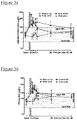
- Figure 1a shows (on a logarithmic scale) the concentration of cadmium in the urine of the pigs prior to, and at several time points after, various degrees of IR-induced injury to the kidney, the degree of injury being dependent on the duration of clamping of the renal artery (20, 40 or 60 minutes).
- measurements were also made for pigs that underwent surgery but without clamping of the renal artery to induce injury to the kidney ("Sham").
- urinary cadmium is an excellent, early (within the first 24 hours post-injury) biomarker of AKI.
- Figures 2a and 2b , and 3a and 3b show data similar to Figure 1 , but for copper and iron (respectively), rather than cadmium.
- a Receiver-Operating-Characteristic (ROC) curve is a graphical plot that illustrates the performance of a binary classifier system as its discrimination threshold is varied. The curve is created by plotting the true positive rate (sensitivity) against the false positive rate at various threshold settings.
- Figures 4, 5 and 6 show ROC curves for all data generated in relation to controls and IR-induced injury subjects for cadmium ( Figure 4 ), copper ( Figure 5 ) and iron ( Figure 6 ).
- the area under the curve (AUC) is a measure of the reliability of the test in identifying individuals with or without the disease state under consideration, ie AKI.
- An AUC of 0.75 or greater is an indication that the test is very good.
- the AUC was 0.81, with a 95% confidence interval of 0.76-0.86.
- the AUC was 0.77, with a 95% confidence interval of 0.71-0.83.
- the AUC was 0.75, with a 95% confidence interval of 0.69-0.81.
- Figure 7 is a plot of the ROC-derived estimates for sensitivity (left y-axis) and specificity (right y-axis) against urinary cadmium (x-axis on a log 10 scale) and it indicates that the cut-off for positive prediction of AKI with >80% confidence based upon spot-measurement of urinary cadmium is >0.26 ⁇ g/L.
- FIGs 8 and 9 are the same plots for copper and iron respectively.
- the cut-offs indicated for positive prediction of acute disease are >57 ⁇ g/L for copper and >65 ⁇ g/L for iron.
- Major and trace elements were measured in a known mass of freeze-dried tissue after nitric acid hydrolysis using inductively-coupled plasma mass spectrometry (ICP-MS). Mean concentrations ⁇ 1 S.D. are shown in Table 5 below, expressed in ⁇ g/g dry matter (DM).
- the young pigs were 3-4 months old, the aged pigs were 5-6 years of age and the young rats (Wistar) were 10-12 weeks of age. All pigs were mixed-breed (landrace/large white/duroc) commercial stock.
- the elemental composition is shown to be broadly similar for young and aged pigs and young laboratory rats. However, of note, and in agreement with published literature from cadaveric material, the concentration of cadmium, copper and iron increases with age, reinforcing the notion of bioaccumulation of these elements in the main route of elimination for water-soluble toxins and metabolic wastes.
- kidney tissue concentration of cadmium and iron in particular had depleted, consistent with loss of proximal tubular epithelial cells that specifically accumulate these elements in highest concentration. It is hypothesised that the results for copper may be distorted by the copper content in drinking water and/or food, and some degree of variability in iron concentrations in kidney tissue may be expected for the same reason.
- the concentrations of urinary cadmium, copper and iron are similar between young healthy male and female volunteers and below all cut-offs established from the preclinical animal (porcine) model (for reference see Figures 7-9 ).
- the urinary trace element profile obtained from the cohort of pigs was also broadly similar, with any differences likely reflecting differences in dietary sources (eg low aluminium, high selenium).
Landscapes
- Life Sciences & Earth Sciences (AREA)
- Health & Medical Sciences (AREA)
- Engineering & Computer Science (AREA)
- Chemical & Material Sciences (AREA)
- Biomedical Technology (AREA)
- Molecular Biology (AREA)
- Urology & Nephrology (AREA)
- Hematology (AREA)
- Immunology (AREA)
- Cell Biology (AREA)
- Analytical Chemistry (AREA)
- Biotechnology (AREA)
- Pathology (AREA)
- Food Science & Technology (AREA)
- Medicinal Chemistry (AREA)
- Physics & Mathematics (AREA)
- Microbiology (AREA)
- Biochemistry (AREA)
- General Health & Medical Sciences (AREA)
- General Physics & Mathematics (AREA)
- Proteomics, Peptides & Aminoacids (AREA)
- Inorganic Chemistry (AREA)
- Neurology (AREA)
- Neurosurgery (AREA)
- Investigating Or Analysing Biological Materials (AREA)
Description
- This invention relates to biomarkers related to kidney function, and in particular to biomarkers useful in the diagnosis of Acute Kidney Injury or diagnosing in a subject delayed graft function of a transplanted kidney. The invention provides methods involving the detection of such biomarkers, in particular in relation to the diagnosis of acute kidney injury or the diagnosis in a subject delayed graft function of a transplanted kidney. The methods of the invention may also be useful in relation to certain other disease states.
- The kidney is responsible for water and solute excretion from the body. Its functions include maintenance of acid-base balance, regulation of electolyte concentrations, control of blood volume, and regulation of blood pressure. As such, loss of kidney function through injury and/or disease results in substantial morbidity and mortality.
- Acute Kidney Injury (AKI) is a relatively sudden deterioration in renal function, over hours or days (in contrast to a slow decline in renal function over months and years, which is known as chronic kidney disease, CKD). AKI has various causes, the common ones in the hospitalised population being hypovolaemia (dehydration from diarrhoea or vomiting, blood loss from trauma or surgery), ischaemia (reduced blood supply to the kidneys, eg caused by surgery, sudden drop in blood pressure, etc), sepsis (eg as a consequence of a severe infection such as pneumonia) and nephrotoxins (drugs or other substances that are toxic to the kidneys, eg certain types of chemotherapy, antibiotics, and contrast agents used in coronary angiograms and some radiological procedures).
- AKI is usually identified by a proportional rise in serum creatinine (SCr) over a defined time. It can also be defined by a fall in hourly urine output, but this is not measured routinely outside intensive care or renal units, so in practice SCr criteria are used. Internationally accepted classification systems have been published to define and stage AKI. There are 3 stages, Stages 1-3, of increasing severity. In a proportion of patients with Stage 3 AKI, "renal replacement therapy" (RRT - either dialysis on the renal unit or haemofiltration on the intensive care unit) might be required, if kidney function is inadequate for safety or survival.
- With relatively recent agreement on the definition of AKI, it has become clear that incidence of AKI in the hospitalised population is high, probably 10-20% for any stage of AKI. The inventors have studied incidence and outcomes of AKI locally and have established a local incidence of about 16% of all hospital admissions, which is similar to published studies from other centres internationally.
- AKI has a number of important clinical consequences. In particular, AKI is associated with:
- a) Significantly increased mortality - both early and late.
- b) Increased length of hospital stay: from local data, median length of stay for patients without AKI is 3 days; for those with
Stage 1, 2 and 3 AKI it is about 9 days, and for those with Stage '3R' (ie those requiring RRT) it is 22 days. - c) Increased risk of development of chronic kidney disease (CKD), or worsening of pre-existing CKD. CKD itself leads to increased risk of cardiovascular morbidity and mortality.
- Moreover, AKI has substantial economic consequences for health service providers. It has been estimated that the marginal costs of AKI to the United Kingdom National Health Service are approximately £450-650 million (around 1 billion $US) per year. Increased length of stay is a major problem for most acute hospitals, with significant cost implications, and the cost of treating increased incidence of CKD is enormous.
- AKI is managed poorly in the United Kingdom (National Confidential Enquiry into Patient Outcome and Death, NCEPOD, 'Adding Insult to Injury' 2009). In particular, detection of AKI is poor. A high proportion of cases are either not detected, or are detected late. The report considered that approximately 30% of cases studied were potentially avoidable.
- Disease states analogous to AKI may also occur in renal transplantation, which involves significant ischaemic insult to the transplanted kidney. Other organs too, such as the liver, may be subject to analogous forms of injury.
- The current standard method of assessing kidney function involves measurement of serum creatinine (SCr), and indeed - as noted above - AKI is defined by specified proportional rise in SCr. However, SCr is acknowledged as a poor marker of kidney injury. SCr levels are indicative of the filtration function of the kidney, rather than of damage to the kidney (which might then lead to reduced filtration function). Moreover, after an insult to the kidney (eg temporary reduction in blood supply during major surgery), SCr probably would not rise for 24 hours or more, even in patients who go on to develop severe AKI (eg those requiring RRT). SCr therefore does not identify those at risk of AKI at a very early stage. SCr is also influenced by many factors such as state of hydration, diet and muscle mass.
- In recent years, there has been great interest in the identification of a sensitive and specific biomarker for AKI. An ideal biomarker would be:
- a) sensitive and specific;
- b) detectable soon after the renal insult;
- c) predictive of AKI (and ideally its severity);
- d) easy to measure (from blood or urine), involving easy sample preparation and stability in transit to a measuring laboratory, or ideally being measurable in the clinic itself (ie as a 'Point-of-Care Test'); and
- e) inexpensive.
- Several new putative AKI biomarkers have been reported and developed. Most of the better ones are proteins, measured by ELISA-type tests. To date, however, none has appeared to be clearly superior to others. Examples of biomarkers that have become relatively well established in recent years are:
- a. NGAL (Neutrophil Gelatinase Associated Lipocalin): measured in blood or urine; sensitive but not very specific; levels start to rise after 2 hours, and peak at about 4 hours.
- b. KIM-1 (Kidney Injury Molecule 1): measured in urine; levels rise later, and peak at about 10 hours after injury; more specific than NGAL.
- c. IL-18 (Interleukin 18): measured in urine; rises and peaks later.
- d. L-FABP (Liver type fatty acid binding protein).
- Since none of these biomarkers is ideal, it has been suggested that specificity and sensitivity might be improved by combining them as a panel of biomarkers, eg NGAL for early sensitive detection, then KIM-1 to increase specificity. Clearly, however, such an approach leads to increased complexity and cost, and does not lead to the desired outcomes of both specificity and rapidity.
- There is thus a significant unmet clinical need for a sensitive, accurate, specific and rapid test that provides a reliable indication of the occurrence of AKI.
- KRITTAPOOM AKRAWINTHAWONG ET AL: "Urine Catalytic Iron and Neutrophil Gelatinase-Associated Lipocalin as Companion Early Markers of Acute Kidney Injury after Cardiac Surgery: A Prospective Pilot Study", CARDIORENAL MEDICINE, vol. 3, no. 1, 1 January 2013 (2013-01-01), pages 7-16 discloses the use of urine catalytic iron as a biomarker for Acute Kidney Injury.
-
WO 2008/066862 A2 discloses methods of predicting and diagnosing gadolinium toxicity by measuring the iron status of a patient, and methods of alleviating gadolinium toxicity by administering to a patient a metal chelator before, concurrently with, or after exposure to gadolinium. -
WO 03/077901 A1 -
WO 2009/039298 A2 discloses sensors comprising AlGaN/GaN high electron mobility transistors (HEMTs) with functionalization at a gate surface with: a thin gold layer and bound antibodies; a thin gold layer and chelating agents; a non- native gate dielectric, or nanorods of a non-native dielectric with an immobilized enzyme. Embodiments are methods of using the sensors for detecting kidney injury. -
WO 2010/057184 A2 discloses methods and kits comprising the use of agents directed against acute kidney injury and biomarkers for facilitating and enhancing the diagnosis of AKI. - FERRARO PIETRO MANUEL ET AL: "Low level exposure to cadmium increases the risk of chronic kidney disease: analysis of the NHANES 1999-2006",BMC PUBLIC HEAL TH, BIOMED CENTRAL, LONDON, GB, vol. 10, no. 1, 3 June 2010 (2010-06-03), is a research article that discusses the association of low-level cadmium exposure with the risk of CKD.
- Podracka L ET AL: "[Relation between metabolism of trace elements and the antioxidant defense system in chronic nephropathies]. -PubMed - NCBI", 24 May 1999 (1999-05-24) describes a study concerned with the association of trace elements with antioxidant enzymes.
- The inventors have developed a pig model of AKI, and have surprisingly discovered that levels of certain atomic species, present at very low levels in bodily fluids such as urine, rise in urine soon (typically within about one hour) after kidney injury, and then fall to baseline concentrations over the course of several hours. The present invention is based on the use of these atomic species as biomarkers for AKI.
- The atomic species useful as biomarkers in accordance with the present invention are cadmium or copper or zinc.
- A redacted form of the Periodic Table, showing the elements of utility in accordance with the present invention, cadmium, copper and zinc, and also showing examples of other elements of potential utility, which are not embodied by the present invention, is shown in Table 1 below.

- Elements that are of utility in the present invention, cadmium, copper and zinc, and other elements of potential utility, which are not embodied by the present invention, are transition metals. By "transition metals" is meant in this context any element in the d-block of the Periodic Table, which includes Groups 3 to 12 of the Periodic Table, as well as the f-block Lanthanide and Actinide series, though the latter may be of lesser utility in practice. In this context, "transition metals" also includes the elements zinc, cadmium and mercury that, because they - unlike other transition metals - have no incomplete d-shell in their electronic configuration, are sometimes referred to as "post-transition metals".
- The transition metals for use in the present invention cadmium, copper and zinc, and other elements of potential utility, which are not embodied by the present invention are those of
Periods Table 2 Period Group 3 4 5 6 7 8 9 10 11 12 4 Sc Ti V Cr Mn Fe Co Ni Cu Zn 5 Y Zr Nb Mo Tc Ru Rh Pd Ag Cd 6 Hf Ta w Re Os Ir Pt Au Hg - Another subset of elements for use in the present invention, namely cadmium, copper and zinc, and of other elements of potential utility, which are not embodied by the present invention, are those of
Periods Table 3 Period Group 3 4 5 6 7 8 9 10 11 12 13 14 15 16 17 4 Sc Ti V Cr Mn Fe Co Ni Cu Zn Ga Ge As Se Br 5 Y Zr Nb Mo Tc Ru Rh Pd Ag Cd In Sn Sb Te I - Biomarkers in accordance with the present invention, namely cadmium, copper and zinc, and other elements of potential utility, which are not embodied by the present invention, are those commonly referred to as "heavy metals", which term encompasses any metal or metalloid of environmental concern. Whilst originally used in relation to cadmium, mercury and lead, all of which have harmful effects and are denser than iron, the term is now applied to any other similarly toxic metal or metalloid, regardless of density. Commonly encountered heavy metals that are for use in the present invention, namely cadmium, copper and zinc, and other elements of potential utility, which are not embodied by the present invention, are those set out in Table 4:
Table 4 Cr chromium Fe iron Co cobalt Ni nickel Cu copper Zn zinc As arsenic Se selenium Ag silver Cd cadmium Sb antimony Hg mercury Tl thallium Pb lead - Without wishing to be bound by any theory, it is believed that cadmium, copper and zinc may accumulate in the epithelial cells that line renal tubules through which urine flows when it is being produced and adjusted by the kidney. In the event of AKI, those tubular epithelial cells may be damaged, and the accumulated elemental species thereby released into the urine. Elevated levels of the elements in question may therefore be indicative of AKI.
- Thus, according to a first aspect of the invention, there is provided a method for diagnosing in a subject Acute Kidney Injury, or diagnosing in a subject delayed graft function of a transplanted kidney, which method comprises the steps of
- a) analysing a urine sample obtained from a subject to determine the level of a biomarker selected from cadmium or copper or zinc; and
- b) comparing the level of the biomarker in the sample to a reference level of the biomarker that is characteristic of a healthy subject, eg a subject having normal renal function;
- The method of the invention may be applied to a subject that displays symptoms of AKI or other disease state, or to a subject to which applies one or more risk factors associated with AKI or other disease state. Alternatively, in view of the prevalence of AKI in the hospitalised population, the method may be applied routinely for screening of all patients.
- In a related aspect, the invention provides use of cadmium or copper or zinc as a biomarker for Acute Kidney Injury or a biomarker for delayed graft function of a transplanted kidney.
- The invention may be performed using apparatus that comprises an analytical element sensitive to the presence and/or concentration, in a sample taken from the subject, of a biomarker selected from cadmium, copper and zinc.
- The apparatus may also comprise means for receiving the sample or for otherwise bringing the sample into contact with the analytical element that is sensitive to the presence and/or concentration of the biomarker in the sample. The analytical element may comprise, without limitation, an optical waveguide or an electrochemical cell, or may be a test strip or dipstick or the like that, in use, is immersed in the sample.
- The apparatus may also comprise means for comparing the level of the biomarker in the sample to a reference level of the biomarker that is characteristic of a healthy subject, eg a subject having normal renal function.
- The apparatus may be configured as a Point-of-Care Test (POCT).
- The invention may be performed using a test kit comprising an analytical element sensitive to the presence and/or concentration, in a sample taken from a subject, of a biomarker selected from cadmium, copper and zinc, together with instructions for use of the analytical element in a method for diagnosing in a subject Acute Kidney Injury or diagnosing in a subject delayed graft function of a transplanted kidney.
- The use of cadmium, copper and zinc as biomarkers is advantageous in that the concentration of the biomarker is stable in the sample taken from the subject, as the biomarker is not subject to degradation as might be the case for a more complex organic biomarker. The analysis of the sample can be carried out accurately using appropriate analytical equipment, and for many such atomic species concentrations can be determined using equipment that is portable and/or simple enough for use by relatively untrained personnel at the point of care. Furthermore, the inventors' investigations indicate that levels of these species become elevated very soon after injury to the kidney, thereby providing a very early indication of AKI, leading to early intervention and optimisation of care, with corresponding clinical and economic benefits.
- The methods of the invention may be particularly beneficial in relation to older subjects, who are more likely to suffer AKI, as concentrations of the biomarkers in the kidneys of such subjects is likely to be greater than for younger subjects, due to a longer period of inadvertent environmental exposure to these atomic species, and hence greater accumulation.
- The sample may be used in the form in which it is obtained from the subject, or may be subjected to pre- treatment prior to analysis, eg dilution, concentration, fractionation, reaction with one or more chemical reagents, or other physical or chemical pre-treatment.
- The sample is a sample of urine, and most preferably the urine sample is analysed without any physical or chemical pre-treatment.
- The sample may be collected by conventional means. The urine sample may be collected in a standard urine cup or other vessel, with or without the use of urinary catheterisation.
- The reference level with which the level of the biomarker in the sample is compared in the methods of the invention may be a standard reference level that is considered to be normal in the population in general, or in a subset of the population to which the subject belongs. Such a subset may be defined in terms of, for instance, gender, age, weight, body mass index, particular health parameters, or any combination of such characteristics. Standard reference levels may be established by routine measurement of the biomarkers in large groups of subjects, eg in all or some patients admitted to hospitals.
- Alternatively, the reference level may be a baseline level measured in the subject prior to an event that may cause AKI. For instance, the reference level may be established by measuring the level of the biomarker before the subject undergoes a surgical procedure with the potential to cause AKI.
- The level of the biomarker may be determined in a single measurement. For instance, a subject that is to undergo a surgical procedure may have the level of the biomarker measured prior to surgery and again a predetermined time after the surgical procedure. Alternatively, and more commonly, a series of measurements of the level of the biomarker are made, as a function of time. The timescale over which measurements are made, or the specific time at which a single measurement is made, will be determined by the typical behaviour of the level of the biomarker following an event that causes AKI. The inventors' investigations indicate that levels of the biomarker rise to a peak within 1 to 4 hours, and return to baseline values over a timescale of 12 to 48 hours. The methods of the invention thus typically comprise the analysis of a sample taken from the subject at a time within four hours of an event that may induce AKI, and more typically the analysis of samples taken from the subject at a plurality of times within four hours of such an event, and optionally one or more times greater than four hours. As noted above, the methods may also comprise the analysis of one or more samples taken from the subject prior to the event.
- The methods of the invention may involve the measurement of the level of a single biomarker selected from cadmium, copper and zinc, or the methods may involve the measurement of the levels of a panel of two or more such biomarkers. The methods of the invention may also involve the measurement of the level of at least one such biomarker and also the level of at least one other biomarker, for instance one or more known biomarkers for AKI such as NGAL, KIM-1, IL-18 or L-FABP.
- Analysis of the sample may be carried out by any suitable analytical technique. One example is inductively coupled plasma mass spectrometry (ICP-MS), which is capable of detecting metals and certain non-metallic species at very low concentrations. This is achieved by ionising the sample with inductively coupled plasma and then using a mass spectrometer to separate and quantify the ionised species. The ions from the plasma are introduced into the mass spectrometer and separated on the basis of the mass-to-charge ratio. The detector of the mass spectrometer receives a signal that is proportional to the concentration of the atomic species in the sample.
- ICP-MS may be used in combination with other techniques, such as high performance liquid chromatography (HPLC) or field flow fractionation (FFF).
- ICP-MS has the capability to quantify more than one element simultaneously, and this technique may therefore offer particular benefits where more than one biomarker is used in a method of the invention, eg where a panel of biomarkers selected from cadmium, copper and zinc is used.
- Where only a single biomarker is used, however, other techniques may be feasible, notably atomic absorption techniques such as atomic absorption spectroscopy, in which the sample is atomised and the absorption of radiation, usually visible light, by the free atoms is used to quantify the concentration of the atomic species of interest.
- Whilst analysis of the sample may be carried out using laboratory equipment and methodology such as that described above, such methods may not be available at all hospitals and/or may require specialised equipment and highly trained personnel. In general, it may therefore be preferable for the analysis to be carried out as a "Point-of-Care Test" (POCT), which can be performed by clinical personnel immediately after collection of a sample.
- Methods for the routine determination of certain of the atomic species of utility in the present invention are available, having previously been used in relation to, for instance, the determination of such species in domestic water supplies. Such methods may be adapted for use in the methods of the present invention.
- Concentrations of cadmium, for instance, can be measured by methods involving contacting of a sample with dithizone, which produces a pink to red colour that can be extracted with chloroform. Photometric measurement and comparison with a calibration curve prepared from standard cadmium solutions treated in the same manner as the sample may be used to derive the concentration of cadmium in the sample. In simple test kits, the analysis may involve merely visual comparison of a coloured solution, obtained by reaction of the sample with a reagent such as dithizone, with a colour chart.
- Another technology that may be used in methods according to the invention involves anodic or cathodic stripping voltammetry, which are voltammetric methods for quantitative determination of specific ionic species. In anodic stripping voltammetry, the analyte of interest is electroplated on a working electrode during a deposition step, and oxidized from the electrode during the stripping step. The current is measured during the stripping step. The oxidation of species is registered as a peak in the current signal at the potential at which the species begins to be oxidized.
- Anodic stripping voltammetry equipment usually incorporates three electrodes: a working electrode, auxiliary electrode, and reference electrode. The solution being analyzed usually has an electrolyte added to it. For most standard tests, the working electrode is a bismuth or mercury film electrode. The mercury film forms an amalgam with the analyte of interest, which upon oxidation results in a sharp peak, improving resolution between analytes. The mercury film is formed over a glassy carbon electrode. In cases where the analyte of interest has an oxidizing potential above that of mercury, or where a mercury electrode would be otherwise unsuitable, a solid, inert metal such as silver, gold, or platinum may also be used.
- Cathodic stripping voltammetry is similar to anodic stripping voltammetry, save that, for the plating step, the potential is held at an oxidizing potential, and the oxidized species are stripped from the electrode by sweeping the potential positively.
- In other embodiments, the analysis may be carried out using a biosensor. Such a system typically includes a sample cell having a surface that is sensitive to the presence of the biomarker that is to be detected. For instance, the surface may be coated with biomolecules bound to ligands that bind competitively with the biomarker, such that the ligands dissociate from the surface if, and to the extent that, the ligand is present in the sample. If the ligands are labelled, eg with a fluorescent label, then the dissociation of the ligand from the surface can be detected and quantified.
- By way of example, DNA-based sensors may be used to detect metal ions by virtue of the ability of certain metal ions to bind selectively to some nucleotide bases to form stable metal-mediated DNA duplexes. Structure- switching DNA biosensors for the detection of various metal ions have been proposed, using various detection methods including fluorescence, surface-enhanced Raman spectroscopy, resonance scattering, colorimetry, electrochemical methods, and evanescent wave optical techniques. In such biosensors, structure-switching is induced by the formation of weak non-covalent bonds, which is generally specific to the interaction of interest and is unaffected by other species present in the sample. The switching occurs rapidly so that such devices are suitable for immediate in situ (POCT) monitoring of specific targets.
- In one form of apparatus that may be suitable for carrying out methods of the invention, an evanescent wave optical biosensor is used. When light propagates through an optical fibre or waveguide by total internal reflection, a rapidly decaying electromagnetic field or evanescent wave is generated. The evanescent wave decays exponentially from the interface, with a typical penetration depth of the order of 100nm. The evanescent wave can excite fluorescence in fluorophores located within it, but the limited depth of the evanescent wave allows for differentiation between fluorophores that are bound to the interface, within the evanescent wave, and those that are unbound, in the bulk sample.
- Analogous methodologies may be used for the determination of other biomarkers according to the invention.
- The biomarkers of the present invention are believed to be reliable, sensitive and specific biomarkers for AKI. As such, there is the potential for the frequent and widespread use of the methods of the present invention in acute hospitals internationally. Any patient at risk of AKI could be screened with one or more such tests. Specific risk factors for AKI include: older age (eg >65 years), CKD, previous AKI, liver disease, heart failure, sepsis, haematological malignancy, neurological impairment. Consequently, patients admitted unwell, with any of these risk factors, might be screened with the biomarker assay. Similarly, patients undergoing any of the following procedures might be screened to identify those at risk of subsequently developing AKI:
- Major surgery, including without limitation cardiac surgery;
- Coronary angiogram or angioplasty;
- Radiological investigations requiring injection of contrast agents (risk of "contrast agent-induced AKI");
- Trauma;
- Burns;
- Chemotherapy.
- The methods of the invention may also be utility in the routine screening of any patients admitted to intensive care or coronary care units. Similarly, as AKI can occur after pregnancy, the biomarkers may be measured routinely ante- and post-natally in maternity units.
- Apart from screening for AKI, the methods of the invention are used in connection with renal transplantation, which involves a significant ischaemic insult to the transplanted kidney, especially when there is a deceased donor kidney, but also for live donor kidneys. The kidney is often slow to start working after implantation into the recipient ("delayed graft function"), or works initially but then develops the equivalent of AKI after a few days. The biomarkers of the invention may be elevated in a urine sample taken from a prospective donor prior to harvesting of a kidney for transplantation, and this may be indicative of a risk of delayed graft function. Similarly, the biomarkers of the invention may be elevated in the urine of the recipient (if they pass urine), which might allow estimation of the risk of the transplanted kidney developing the equivalent of AKI. This information may be useful in various ways, including deciding on when to discharge the patient, whether to undertake renal biopsy, and/or whether to change immunosuppression drugs.
- In addition to the above, the methods of the present invention may be useful in relation to acute liver damage. It is known that the liver also accumulates cadmium, and it is therefore possible that, in acute liver damage (drug toxicity, hepatitis), cadmium levels in blood might rise.
- Although the subject is most likely to be a human subject, it is to be expected that the atomic species of cadmium or copper or zinc will be biomarkers of AKI in other mammals. Thus, the methods of the invention may have utility in the veterinary field.
- The invention will now be illustrated, by way of example only, with reference to the accompanying Figures, wherein the elements for use in the present invention are cadmium, copper and zinc, with the illustrated examples of other elements of potential utility not being embodied by the present invention.
-
Figure 1a shows the change in urinary cadmium (normalised to creatinine levels) with time in a pig of similar weight to an adult human (50-70kg), after inducing a known degree of kidney injury (Ischaemia-Reperfusion (IR)-induced injury at time zero). -
Figure 1b is similar to Figure la, but with the data shown without normalisation to creatinine levels. -
Figure 2a shows the change in urinary copper (normalised to creatinine levels) with time in a pig of similar weight to an adult human (50-70kg), after inducing a known degree of kidney injury (Ischaemia-Reperfusion (IR)-induced injury at time zero). -
Figure 2b is similar toFigure 2a , but with the data shown without normalisation to creatinine levels. -
Figure 3a shows the change in urinary iron with time in a pig of similar weight to an adult human (50-70kg), after inducing a known degree of kidney injury (Ischaemia-Reperfusion (IR)-induced injury at time zero). -
Figure 3b is similar toFigure 3a , but with the data shown without normalisation to creatinine levels. -
Figure 4 is a Receiver-Operating-Characteristic (ROC) curve for urinary cadmium as a marker of AKI. -
Figure 5 is a Receiver-Operating-Characteristic (ROC) curve for urinary copper as a marker of AKI. -
Figure 6 is a Receiver-Operating-Characteristic (ROC) curve for urinary iron as a marker of AKI. -
Figure 7 is a plot of the ROC-derived estimates for sensitivity (left y-axis) and specificity (right y-axis) against urinary cadmium (x-axis on a log10 scale), indicating the cut-off for positive prediction of disease. -
Figure 8 is a plot of the ROC-derived estimates for sensitivity (left y-axis) and specificity (right y-axis) against urinary copper (x-axis on a log10 scale), indicating the cut-off for positive prediction of disease. -
Figure 9 is a plot of the ROC-derived estimates for sensitivity (left y-axis) and specificity (right y-axis) against urinary iron (x-axis on a log10 scale), indicating the cut-off for positive prediction of disease. -
Figure 10 shows the concentration of cadmium (top panel), copper (middle panel) and iron (lower panel) in porcine kidneys measured at 5 weeks post IR-injury. -
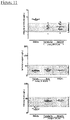
Figure 11 shows baseline levels of urinary cadmium (top panel), copper (middle panel) and iron (lower panel) in elderly, healthy volunteers. - A pig model of AKI was developed. The pig is an excellent model for human kidney disease because the anatomy and physiology of pig and human kidneys are similar.
- The pigs are anaesthetised for general surgery, intubated and subjected to a midline laparotomy to reveal the right and left kidneys, which are each fully clamped for variable times (20-180 minutes), after which the clamps are removed and the incision site closed. A bladder catheter (12 Fr) is inserted for urine collection, and the pigs are recovered to a pen.
- The model thus involves clamping the renal artery for a defined time (up to 180 minutes), and then reperfusing to cause an Ischaemia-Reperfusion (IR)-induced injury, which is an extreme type of insult to the kidney.
- Urine samples are collected before clamping of the renal artery (baseline), immediately after reperfusion (t=0), and at various time intervals thereafter (up to 48hrs).
- The concentration of various biomarkers in the urine samples is measured by ICP-MS.
-
Figure 1a shows (on a logarithmic scale) the concentration of cadmium in the urine of the pigs prior to, and at several time points after, various degrees of IR-induced injury to the kidney, the degree of injury being dependent on the duration of clamping of the renal artery (20, 40 or 60 minutes). As a control, measurements were also made for pigs that underwent surgery but without clamping of the renal artery to induce injury to the kidney ("Sham"). - It can be seen that the baseline (pre-injury) concentrations of cadmium for each group of subjects was similar, and for the Sham group the concentration of cadmium remained within the basal range.
- For the pigs that suffered injury to the kidney, an increase in cadmium concentration was observed, the magnitude of the increase being proportional to the duration of clamping of the renal artery, and hence the severity of the injury. The time taken for the concentration of cadmium to return to the basal range is also shown to be proportional to the duration of clamping of the renal artery, and hence the severity of the injury (return to baseline was approximately 6 hours, 24 hours and 48 hours for IR-20, IR-40 and IR-60 respectively).
- The results indicate that urinary cadmium is an excellent, early (within the first 24 hours post-injury) biomarker of AKI.
- In Figure la, the concentration of cadmium is adjusted to urine creatinine (14,256 µmol/L), to compensate for possible variations due to large differences in volume of urine passed by different subjects. However, data not adjusted for creatinine in that way is shown in
Figure 1b ; it can be seen that the observed behaviour is qualitatively very similar to that depicted in Figure la. -
Figures 2a and 2b , and3a and 3b show data similar toFigure 1 , but for copper and iron (respectively), rather than cadmium. - The observed results are qualitatively very similar to those for cadmium (
Figures 1a and 1b ), with a marked increase in urinary copper and iron post-injury. - The results indicate that urinary copper and urinary iron are excellent, early (within the first 24 hours post-injury) biomarkers of AKI.
- A Receiver-Operating-Characteristic (ROC) curve is a graphical plot that illustrates the performance of a binary classifier system as its discrimination threshold is varied. The curve is created by plotting the true positive rate (sensitivity) against the false positive rate at various threshold settings.
-
Figures 4, 5 and6 show ROC curves for all data generated in relation to controls and IR-induced injury subjects for cadmium (Figure 4 ), copper (Figure 5 ) and iron (Figure 6 ). The area under the curve (AUC) is a measure of the reliability of the test in identifying individuals with or without the disease state under consideration, ie AKI. An AUC of 0.75 or greater is an indication that the test is very good. - For cadmium, the AUC was 0.81, with a 95% confidence interval of 0.76-0.86.
- For copper, the AUC was 0.77, with a 95% confidence interval of 0.71-0.83.
- For iron, the AUC was 0.75, with a 95% confidence interval of 0.69-0.81.
- Thus, cadmium, copper and iron are indicated to be very good biomarkers for AKI.
- Plotting the ROC-derived estimates for sensitivity (left y-axis) and specificity (right y-axis) against urinary biomarker concentration (x-axis on a log10 scale) indicates the cut-off for positive prediction of AKI with >80% confidence based upon spot-measurement of the urinary biomarker.
- Thus,
Figure 7 is a plot of the ROC-derived estimates for sensitivity (left y-axis) and specificity (right y-axis) against urinary cadmium (x-axis on a log10 scale) and it indicates that the cut-off for positive prediction of AKI with >80% confidence based upon spot-measurement of urinary cadmium is >0.26 µg/L. -
Figures 8 and 9 are the same plots for copper and iron respectively. - The cut-offs indicated for positive prediction of acute disease are >57 µg/L for copper and >65 µg/L for iron.
- The elemental composition of kidney tissue was measured for young healthy pigs (n=20), aged healthy pigs (n=8) and, for comparison purposes, young healthy laboratory rats (n=12). Major and trace elements were measured in a known mass of freeze-dried tissue after nitric acid hydrolysis using inductively-coupled plasma mass spectrometry (ICP-MS). Mean concentrations ± 1 S.D. are shown in Table 5 below, expressed in µg/g dry matter (DM). The young pigs were 3-4 months old, the aged pigs were 5-6 years of age and the young rats (Wistar) were 10-12 weeks of age. All pigs were mixed-breed (landrace/large white/duroc) commercial stock.
Table 5 Major element (µg/g DM) Young Pigs Aged Pigs Young rats P-value for age in pigs Sodium 7373 ± 1971 5634 ± 1165 6710 ± 703 0.03 Potassium 11814 ± 2547 10143 ± 2509 12012 ± 1143 0.14 Calcium 384 ± 256 597 ± 95 258 ± 42 0.04 Magnesium 790 ± 179 883 ± 267 859 ± 79 0.30 Phosphorus 15163 ± 3892 12304 ± 1186 12438 ± 993 0.08 Sulphur 10119 ± 1981 15364 ± 6366 6229 ± 828 0.04 Total ME Trace element (µg/g DM) 45301 ± 10269 44928 ± 9410 38511 ± 3458 - Iron 170 ± 103 354 ± 48 329 ± 44 <.001 Zinc 155 ± 56 147 ± 22 97.2 ± 7.14 0.72 Copper 33.5 ± 14.3 49.7 ± 8.4 27.4 ± 3.0 0.009 Rubidium 16.2 ± 4.9 14.7 ± 3.7 20.9 ± 1.5 0.53 Aluminium nd nd 10.95 ± 5.53 - Selenium 7.51 ± 1.54 13.8 ± 3.9 4.99 ± 0.38 <.001 Manganese 6.72 ± 2.06 8.29 ± 1.40 3.41 ± 0.48 0.07 Molybdenum 2.90 ± 0.95 2.99 ± 0.23 1.15 + 0.14 0.27 Chromium 3.55 ± 1.79 3.32 ± 0.43 0.17 ± 0.23 0.27 Cadmium 1.47 ± 0.53 3.17 ± 0.79 0.02 ± 0.00 <.001 Barium nd nd 0.09 ± 0.03 - Strontium nd nd 0.05 ± 0.05 - Cobalt 0.05 ± 0.02 0.09 ± 0.04 1.19 ± 0.09 0.004 Caesium 0.05 ± 0.03 0.06 ± 0.02 0.12 ± 0.012 0.83 Vanadium 0.07 ± 0.06 0.04 ± 0.03 0.03 ± 0.009 0.13 Thallium nd nd 0.04 ± 0.006 - Total TE 363 ± 19 597 ± 62 499 ± 50 - The elemental composition is shown to be broadly similar for young and aged pigs and young laboratory rats. However, of note, and in agreement with published literature from cadaveric material, the concentration of cadmium, copper and iron increases with age, reinforcing the notion of bioaccumulation of these elements in the main route of elimination for water-soluble toxins and metabolic wastes.
-
Figure 10 shows the results of an additional study in which the concentration of cadmium, copper and iron in porcine kidneys was measured at 5 weeks post IR-injury (Control, n=3; IR-AKI, n=3). Data is presented as a dot-plot (line at geometric mean) of the concentration in µg/g dry matter (DM). All graphs and ROC curve statistics were generated in Graphpad Prism 6 (Graphpad Software Inc, Ca, USA). Quantitative time-series data were analysed by REML with treatment (Control vs. IR) as a fixed effect and time as a within-animal repeated measure (Genstat v16, Rothampsted, UK). Elemental data were log10 transformed prior to analysis. Statistical significance was accepted at P<0.05. - The kidney tissue concentration of cadmium and iron in particular had depleted, consistent with loss of proximal tubular epithelial cells that specifically accumulate these elements in highest concentration. It is hypothesised that the results for copper may be distorted by the copper content in drinking water and/or food, and some degree of variability in iron concentrations in kidney tissue may be expected for the same reason.
- Major and trace elements were measured in spot-samples of urine from young, healthy male and female volunteers (n=12 males, n=12 females), and for comparison catheterised but healthy pigs (n=24-53, baseline urine sample), by inductively-coupled plasma mass spectrometry (ICP-MS). The results shown in Table 6 below are median concentration values (1st and 3rd interquartile range (IQR)). The young pigs were 3-4 months old (50-60 kg). The data were compared by one-way ANOVA, with urinary creatinine as a co-variate.
Table 6 Major element (mg/L) Human (female) Human (male) Pigs (female) P-value (humans) Potassium 1751 (1101,3850) 3145 (1549,3850) 1681 (794,2654) 0.51 Sodium 765 (260,2330) 1841 (871,2330) 625 (324,1002) 0.58 Phosphorus 340 (191,873) 691 (288,1122) 16.4 (8.9,95) 0.94 Sulphur 304 (191,873) 715 (344,873) 422 (208,570) 0.32 Calcium 49.5 (35.9,146) 100 (58,146) 68.8 (22.4,182) 0.43 Magnesium 42.5 (20.2,109) 61.5 (34.4,109) 76.6 (51.3,135) 0.25 Boron 0.99 (0.60,1.74) 1.04 (0.61,1.74) 0.89 (0.48,1.12) 0.67 Total major elements 3073 (2324,9168) 6203 (2863,9168) 2627 (1541,4145) 0.10 Trace element (µg/L) Rubidium 1188 (827,2295) 1912 (1282,2295) 661 (459,1238) 0.27 Zinc 134 (83,505) 341 (180,505) 348 (204,688) 0.23 Strontium 72.5 (46.3,195) 118 (44,195) 157 (80,270) 0.26 Molybdenum 24.0 (13.0,73.0) 47.7 (22.9,73.0) 56.7 (39.1,109) 0.38 Aluminium 42.4 (37.3,46.1) 44.5 (42.6,46.1) 12.3 (6.1,33.8) 0.76 Iron 32.2 (31.6,37.8) 35.4 (34.2,37.8) 39.3 (20.8,95.8) 0.95 Selenium 20.3 (7.7,37.7) 26.7 (16.4,37.7) 68.0 (36.8,99.1) 0.41 Lithium 16.8 (10.7,26.9) 18.6 (13.3,26.9) - 0.67 Copper 15.0 (13.9,19.1) 18.5 (14.6,19.1) 35.5 (18.1,69.1) 0.67 Arsenic 9.00 (4.41,35.8) 25.4 (11.6,35.8) 1.55 (0.94,2.46) 0.30 Caesium 5.30 (3.06,10.00) 7.17 (6.07,10.00) 1.75 (1.05,2.54) 0.52 Nickel 5.65 (5.20,6.31) 5.96 (5.27,6.31) 9.76 (3.23,16.9) 0.92 Chromium 5.29 (5.19,5.63) 5.42 (5.25,5.63) 1.32 (0.62,2.10) 0.31 Barium 2.30 (1.60,3.80) 2.09 (1.78,3.80) 13.9 (7.8,17.3) 0.39 Manganese 1.14 (1.06,1.25) 1.17 (1.14,1.25) 3.50 (1.32,6.72) 0.44 Lead 0.92 (0.84,1.15) 0.97 (0.90,1.15) 1.16 (0.72,2.41) 0.56 Vanadium 0.37 (0.30,0.91) 0.57 (0.31,0.91) 0.77 (0.50,1.34) 0.26 Cadmium 0.20 (0.16,0.33) 0.21 (0.14,0.33) 0.14 (0.08,0.28) 0.41 Cobalt 0.30 (0.19,0.32) 0.23 (0.18,0.32) 1.17 (0.76,2.83) 0.63 Thallium 0.18 (0.12,0.39) 0.28 (0.12,0.39) - 0.30 Beryllium 0.01 (0.01,0.02) 0.01 (0.01,0.02) nd 0.50 Osmolality (Osm/L) 251 (179,734) 462 (248,734) 417 (362,490) 0.28 Creatinine (µmol/L) 3301 (2575,14424) 9821 (4199,14424) 8617 (5425,11684) 0.01 Total trace elements 1622 (1202,3182) 2699 (1831,3182) 1800 (1287,3212) 0.15 - The concentrations of urinary cadmium, copper and iron are similar between young healthy male and female volunteers and below all cut-offs established from the preclinical animal (porcine) model (for reference see
Figures 7-9 ). The urinary trace element profile obtained from the cohort of pigs was also broadly similar, with any differences likely reflecting differences in dietary sources (eg low aluminium, high selenium). - The urinary trace element profiles of elderly (≥ 65 years of age), healthy male and female volunteers were also measured and were also below all cut-offs established from the preclinical animal (porcine) model, with the exception of an increase in baseline cadmium. The results for cadmium, copper and iron are shown in
Figure 11 . Elements were determined in spot-samples by inductively-coupled plasma mass spectrometry (ICP-MS). The basal range (horizontal shaded area) was determined from summary statistics (1st and 3rd quartile) of all baseline determinations for each element. Graphs were generated in Graphpad Prism 6 (Graphpad Software Inc, Ca, USA).
Claims (11)
- A method for diagnosing in a subject Acute Kidney Injury, or diagnosing in a subject delayed graft function of a transplanted kidney, which method comprises the steps ofa) analysing a urine sample obtained from a subject to determine the level of a biomarker selected from cadmium or copper or zinc; andb) comparing the level of the biomarker in the sample to a reference level of the biomarker that is characteristic of a healthy subject, eg a subject having normal renal function;wherein an elevated level of the biomarker in the sample relative to the reference level is indicative of Acute Kidney Injury or delayed graft function of a transplanted kidney.
- The use of cadmium or copper or zinc as a biomarker for Acute Kidney Injury or a biomarker for delayed graft function of a transplanted kidney.
- A method according to any preceding claim, in which the reference level with which the level of the biomarker in the sample is compared is a standard reference level that is considered to be normal in the population in general, or in a subset of the population to which the subject belongs, or a baseline level measured in the subject prior to an event that may cause AKI or delayed graft function of a transplanted kidney.
- A method according to any preceding claim, in which the level of the biomarker is determined in a single measurement, or a series of measurements of the level of the biomarker are made, as a function of time.
- A method according to Claim 4, wherein measurements are made on a sample taken from the subject at a time within four hours of an event that may induce AKI, and more typically the analysis of samples taken from the subject at a plurality of times within four hours of such an event, and optionally one or more times greater than four hours.
- A method according to Claim 5, further comprising the analysis of one or more samples taken from the subject prior to the event.
- A method according to any one of Claims 1 to 6, which involves the measurement of the levels of a panel of two or more biomarkers, the panel comprising the said biomarker and one or more other biomarker selected from cadmium, copper, zinc and iron.
- A method according to Claim 6 or Claim 7, which also involves the measurement of the level of at least one other biomarker, for instance one or more known biomarkers for AKI such as NGAL, KIM-1, IL-18 or L-FABP.
- A method according to any preceding claim, wherein analysis of the sample is carried out by ICP-MS, an atomic absorption technique such as atomic absorption spectroscopy, anodic or cathodic stripping voltammetry, or using a biosensor.
- A method according to any preceding claim, wherein the method comprises providing a plurality of samples, at least one sample being taken within 4 hours of an event that may induce AKI, and at least one sample being taken after 4 hours from an event that may induce AKI.
- The method of any of Claims 1 to 9, wherein the sample is taken within 24 hours of an event which may induce AKI.
Applications Claiming Priority (2)
| Application Number | Priority Date | Filing Date | Title |
|---|---|---|---|
| GB201500200 | 2015-01-07 | ||
| PCT/GB2016/050028 WO2016110701A1 (en) | 2015-01-07 | 2016-01-07 | Biomarkers related to kidney function and methods involving their use |
Publications (2)
| Publication Number | Publication Date |
|---|---|
| EP3243079A1 EP3243079A1 (en) | 2017-11-15 |
| EP3243079B1 true EP3243079B1 (en) | 2022-11-02 |
Family
ID=55299663
Family Applications (1)
| Application Number | Title | Priority Date | Filing Date |
|---|---|---|---|
| EP16702760.6A Active EP3243079B1 (en) | 2015-01-07 | 2016-01-07 | Biomarkers related to kidney function and methods involving their use |
Country Status (8)
| Country | Link |
|---|---|
| US (1) | US10761103B2 (en) |
| EP (1) | EP3243079B1 (en) |
| JP (1) | JP2018505407A (en) |
| AU (1) | AU2016205886A1 (en) |
| BR (1) | BR112017014773A2 (en) |
| ES (1) | ES2937273T3 (en) |
| MX (1) | MX2017008959A (en) |
| WO (1) | WO2016110701A1 (en) |
Families Citing this family (1)
| Publication number | Priority date | Publication date | Assignee | Title |
|---|---|---|---|---|
| WO2024085722A1 (en) * | 2022-10-20 | 2024-04-25 | 건국대학교 산학협력단 | Biomarker composition for early diagnosis of kidney diseases, and method for providing information required for early diagnosis of kidney diseases by using same |
Family Cites Families (9)
| Publication number | Priority date | Publication date | Assignee | Title |
|---|---|---|---|---|
| JP4578590B2 (en) * | 1998-11-26 | 2010-11-10 | 森永乳業株式会社 | Nutritional composition |
| DK1173757T3 (en) * | 1999-04-23 | 2008-12-08 | Shiva Biomedical Llc | Diagnosis of human kidney disease |
| EP2500018B1 (en) | 2002-03-08 | 2017-07-19 | PhilERA New Zealand Limited | Preventing and/or Treating Cardiovascular Disease and/or Associated Heart Failure |
| EP1776118A2 (en) | 2004-08-06 | 2007-04-25 | Shiva Biomedical, LLC | Iron chelators for treating radiocontrast-associated renal dysfunction |
| US20080138440A1 (en) * | 2006-11-29 | 2008-06-12 | Cormedix Inc. | Methods of diagnosing and alleviating gadolinium toxicity |
| WO2009039298A2 (en) * | 2007-09-18 | 2009-03-26 | University Of Florida Research Foundation, Inc. | Sensors using aigan/gan high electron mobility transistors |
| US20110287964A1 (en) * | 2008-11-17 | 2011-11-24 | The Brigham And Women's Hospital, Inc. | Urinary biomarkers for sensitive and specific detection of acute kidney injury in humans |
| JP2013529907A (en) * | 2010-05-24 | 2013-07-25 | ザ トラスティーズ オブ コロンビア ユニヴァーシティ イン ザ シティ オブ ニューヨーク | NGAL protein variants and uses thereof |
| EP2568291A1 (en) * | 2011-09-07 | 2013-03-13 | Roche Diagnostics GmbH | L-FABP based diagnosis of kidney injury after an acute event or after a surgical intervention |
-
2016
- 2016-01-07 JP JP2017536870A patent/JP2018505407A/en active Pending
- 2016-01-07 MX MX2017008959A patent/MX2017008959A/en unknown
- 2016-01-07 ES ES16702760T patent/ES2937273T3/en active Active
- 2016-01-07 AU AU2016205886A patent/AU2016205886A1/en not_active Abandoned
- 2016-01-07 BR BR112017014773A patent/BR112017014773A2/en not_active Application Discontinuation
- 2016-01-07 US US15/541,980 patent/US10761103B2/en active Active
- 2016-01-07 EP EP16702760.6A patent/EP3243079B1/en active Active
- 2016-01-07 WO PCT/GB2016/050028 patent/WO2016110701A1/en not_active Ceased
Non-Patent Citations (2)
| Title |
|---|
| FERRARO PIETRO MANUEL ET AL: "Low level exposure to cadmium increases the risk of chronic kidney disease: analysis of the NHANES 1999-2006", BMC PUBLIC HEALTH, BIOMED CENTRAL, LONDON, GB, vol. 10, no. 1, 3 June 2010 (2010-06-03), pages 304, XP021075764, ISSN: 1471-2458, DOI: 10.1186/1471-2458-10-304 * |
| PODRACKÁ L ET AL: "[Relation between metabolism of trace elements and the antioxidant defense system in chronic nephropathies]. - PubMed - NCBI", 24 May 1999 (1999-05-24), XP055523860, Retrieved from the Internet <URL:https://www.ncbi.nlm.nih.gov/pubmed/10422345> [retrieved on 20181114] * |
Also Published As
| Publication number | Publication date |
|---|---|
| CA2973172A1 (en) | 2016-07-14 |
| BR112017014773A2 (en) | 2018-01-16 |
| ES2937273T3 (en) | 2023-03-27 |
| JP2018505407A (en) | 2018-02-22 |
| AU2016205886A1 (en) | 2017-08-03 |
| MX2017008959A (en) | 2018-05-17 |
| US10761103B2 (en) | 2020-09-01 |
| EP3243079A1 (en) | 2017-11-15 |
| US20180017581A1 (en) | 2018-01-18 |
| WO2016110701A1 (en) | 2016-07-14 |
Similar Documents
| Publication | Publication Date | Title |
|---|---|---|
| Yang et al. | Association of plasma and urine metals levels with kidney function: A population-based cross-sectional study in China | |
| Solak et al. | Red cell distribution width is independently related to endothelial dysfunction in patients with chronic kidney disease | |
| CN102105794B (en) | A marker for graft failure and mortality | |
| US20140303021A1 (en) | Methods and devices for detecting obstructive uropathy and associated disorders | |
| EP2442106B1 (en) | Method for test on diabetic nephropathy | |
| Fonseca et al. | Neutrophil gelatinase‐associated lipocalin in kidney transplantation is an early marker of graft dysfunction and is associated with one‐year renal function | |
| Barneo-Caragol et al. | Strontium and its role in preeclampsia | |
| WO2007137584A1 (en) | Methods and devices for rapid assessment of severity of injury | |
| Wijerathna et al. | Albuminuria and other renal damage biomarkers detect acute kidney injury soon after acute ingestion of oxalic acid and potassium permanganate | |
| Yay et al. | Can immature granulocytes and neutrophil-lymphocyte ratio be biomarkers to evaluate diabetic nephropathy?: A cross-sectional study | |
| WO2020052294A1 (en) | Use of mercury and cadmium mixed exposure detection in non-obstructive azoospermia auxiliary diagnosis | |
| US10761103B2 (en) | Biomarkers related to kidney function and methods involving their use | |
| EP3358347B1 (en) | Marker for determining diabetic nephropathy | |
| Khalid et al. | Assessment of serum podocalyxin as a biomarker for diabetic nephropathy in type 2 diabetes patients in Duhok city | |
| Abdulameer et al. | NGAL as a biomarker in chronic kidney disease | |
| EP3736570A1 (en) | Method for the diagnosis, sub-classification and prognosis of acute kidney injury by detecting cct7 | |
| Maryam et al. | Assessment of Some Biochemical Parameters, Neutrophil Gelatinase Lipocalin, And Kidney Injury Molecule-1 in Type 2 Diabetic Patients with Chronic Kidney Disease | |
| US20250314663A1 (en) | Methods for treating acute kidney injury | |
| Seyoum et al. | Serum bilirubin association with estimated glomerular filtration rate and other risk factors in the progression of chronic kidney disease among CKD patients: A Hospital-based cross sectional study design | |
| Idris et al. | Reference interval for serum neutrophil gelatinase-associated lipocalin in apparently healthy caribbean population | |
| Rana et al. | Hematological Markers: An Early Road to Recovery in Sepsis | |
| Mehvish et al. | Study Of Serum Hs-C Reactive Protein And Serum Uric Acid In Type-II Diabetic Patients | |
| WO2026087320A1 (en) | Method for predicting mortality risk | |
| EP4515245A1 (en) | Biomarkers of diabetic nephropathy | |
| Fonseca et al. | 4.2. STUDY II |
Legal Events
| Date | Code | Title | Description |
|---|---|---|---|
| STAA | Information on the status of an ep patent application or granted ep patent |
Free format text: STATUS: THE INTERNATIONAL PUBLICATION HAS BEEN MADE |
|
| PUAI | Public reference made under article 153(3) epc to a published international application that has entered the european phase |
Free format text: ORIGINAL CODE: 0009012 |
|
| STAA | Information on the status of an ep patent application or granted ep patent |
Free format text: STATUS: REQUEST FOR EXAMINATION WAS MADE |
|
| 17P | Request for examination filed |
Effective date: 20170706 |
|
| AK | Designated contracting states |
Kind code of ref document: A1 Designated state(s): AL AT BE BG CH CY CZ DE DK EE ES FI FR GB GR HR HU IE IS IT LI LT LU LV MC MK MT NL NO PL PT RO RS SE SI SK SM TR |
|
| AX | Request for extension of the european patent |
Extension state: BA ME |
|
| DAV | Request for validation of the european patent (deleted) | ||
| DAX | Request for extension of the european patent (deleted) | ||
| STAA | Information on the status of an ep patent application or granted ep patent |
Free format text: STATUS: EXAMINATION IS IN PROGRESS |
|
| 17Q | First examination report despatched |
Effective date: 20181121 |
|
| GRAP | Despatch of communication of intention to grant a patent |
Free format text: ORIGINAL CODE: EPIDOSNIGR1 |
|
| STAA | Information on the status of an ep patent application or granted ep patent |
Free format text: STATUS: GRANT OF PATENT IS INTENDED |
|
| INTG | Intention to grant announced |
Effective date: 20220610 |
|
| GRAS | Grant fee paid |
Free format text: ORIGINAL CODE: EPIDOSNIGR3 |
|
| GRAA | (expected) grant |
Free format text: ORIGINAL CODE: 0009210 |
|
| STAA | Information on the status of an ep patent application or granted ep patent |
Free format text: STATUS: THE PATENT HAS BEEN GRANTED |
|
| AK | Designated contracting states |
Kind code of ref document: B1 Designated state(s): AL AT BE BG CH CY CZ DE DK EE ES FI FR GB GR HR HU IE IS IT LI LT LU LV MC MK MT NL NO PL PT RO RS SE SI SK SM TR |
|
| REG | Reference to a national code |
Ref country code: GB Ref legal event code: FG4D |
|
| REG | Reference to a national code |
Ref country code: CH Ref legal event code: EP Ref country code: AT Ref legal event code: REF Ref document number: 1529108 Country of ref document: AT Kind code of ref document: T Effective date: 20221115 |
|
| REG | Reference to a national code |
Ref country code: DE Ref legal event code: R096 Ref document number: 602016076021 Country of ref document: DE |
|
| REG | Reference to a national code |
Ref country code: IE Ref legal event code: FG4D |
|
| REG | Reference to a national code |
Ref country code: LT Ref legal event code: MG9D |
|
| REG | Reference to a national code |
Ref country code: NL Ref legal event code: MP Effective date: 20221102 |
|
| REG | Reference to a national code |
Ref country code: ES Ref legal event code: FG2A Ref document number: 2937273 Country of ref document: ES Kind code of ref document: T3 Effective date: 20230327 |
|
| REG | Reference to a national code |
Ref country code: AT Ref legal event code: MK05 Ref document number: 1529108 Country of ref document: AT Kind code of ref document: T Effective date: 20221102 |
|
| PG25 | Lapsed in a contracting state [announced via postgrant information from national office to epo] |
Ref country code: SE Free format text: LAPSE BECAUSE OF FAILURE TO SUBMIT A TRANSLATION OF THE DESCRIPTION OR TO PAY THE FEE WITHIN THE PRESCRIBED TIME-LIMIT Effective date: 20221102 Ref country code: PT Free format text: LAPSE BECAUSE OF FAILURE TO SUBMIT A TRANSLATION OF THE DESCRIPTION OR TO PAY THE FEE WITHIN THE PRESCRIBED TIME-LIMIT Effective date: 20230302 Ref country code: NO Free format text: LAPSE BECAUSE OF FAILURE TO SUBMIT A TRANSLATION OF THE DESCRIPTION OR TO PAY THE FEE WITHIN THE PRESCRIBED TIME-LIMIT Effective date: 20230202 Ref country code: LT Free format text: LAPSE BECAUSE OF FAILURE TO SUBMIT A TRANSLATION OF THE DESCRIPTION OR TO PAY THE FEE WITHIN THE PRESCRIBED TIME-LIMIT Effective date: 20221102 Ref country code: FI Free format text: LAPSE BECAUSE OF FAILURE TO SUBMIT A TRANSLATION OF THE DESCRIPTION OR TO PAY THE FEE WITHIN THE PRESCRIBED TIME-LIMIT Effective date: 20221102 Ref country code: AT Free format text: LAPSE BECAUSE OF FAILURE TO SUBMIT A TRANSLATION OF THE DESCRIPTION OR TO PAY THE FEE WITHIN THE PRESCRIBED TIME-LIMIT Effective date: 20221102 |
|
| PG25 | Lapsed in a contracting state [announced via postgrant information from national office to epo] |
Ref country code: RS Free format text: LAPSE BECAUSE OF FAILURE TO SUBMIT A TRANSLATION OF THE DESCRIPTION OR TO PAY THE FEE WITHIN THE PRESCRIBED TIME-LIMIT Effective date: 20221102 Ref country code: PL Free format text: LAPSE BECAUSE OF FAILURE TO SUBMIT A TRANSLATION OF THE DESCRIPTION OR TO PAY THE FEE WITHIN THE PRESCRIBED TIME-LIMIT Effective date: 20221102 Ref country code: LV Free format text: LAPSE BECAUSE OF FAILURE TO SUBMIT A TRANSLATION OF THE DESCRIPTION OR TO PAY THE FEE WITHIN THE PRESCRIBED TIME-LIMIT Effective date: 20221102 Ref country code: IS Free format text: LAPSE BECAUSE OF FAILURE TO SUBMIT A TRANSLATION OF THE DESCRIPTION OR TO PAY THE FEE WITHIN THE PRESCRIBED TIME-LIMIT Effective date: 20230302 Ref country code: HR Free format text: LAPSE BECAUSE OF FAILURE TO SUBMIT A TRANSLATION OF THE DESCRIPTION OR TO PAY THE FEE WITHIN THE PRESCRIBED TIME-LIMIT Effective date: 20221102 Ref country code: GR Free format text: LAPSE BECAUSE OF FAILURE TO SUBMIT A TRANSLATION OF THE DESCRIPTION OR TO PAY THE FEE WITHIN THE PRESCRIBED TIME-LIMIT Effective date: 20230203 |
|
| P01 | Opt-out of the competence of the unified patent court (upc) registered |
Effective date: 20230524 |
|
| PG25 | Lapsed in a contracting state [announced via postgrant information from national office to epo] |
Ref country code: NL Free format text: LAPSE BECAUSE OF FAILURE TO SUBMIT A TRANSLATION OF THE DESCRIPTION OR TO PAY THE FEE WITHIN THE PRESCRIBED TIME-LIMIT Effective date: 20221102 |
|
| PG25 | Lapsed in a contracting state [announced via postgrant information from national office to epo] |
Ref country code: SM Free format text: LAPSE BECAUSE OF FAILURE TO SUBMIT A TRANSLATION OF THE DESCRIPTION OR TO PAY THE FEE WITHIN THE PRESCRIBED TIME-LIMIT Effective date: 20221102 Ref country code: RO Free format text: LAPSE BECAUSE OF FAILURE TO SUBMIT A TRANSLATION OF THE DESCRIPTION OR TO PAY THE FEE WITHIN THE PRESCRIBED TIME-LIMIT Effective date: 20221102 Ref country code: EE Free format text: LAPSE BECAUSE OF FAILURE TO SUBMIT A TRANSLATION OF THE DESCRIPTION OR TO PAY THE FEE WITHIN THE PRESCRIBED TIME-LIMIT Effective date: 20221102 Ref country code: DK Free format text: LAPSE BECAUSE OF FAILURE TO SUBMIT A TRANSLATION OF THE DESCRIPTION OR TO PAY THE FEE WITHIN THE PRESCRIBED TIME-LIMIT Effective date: 20221102 Ref country code: CZ Free format text: LAPSE BECAUSE OF FAILURE TO SUBMIT A TRANSLATION OF THE DESCRIPTION OR TO PAY THE FEE WITHIN THE PRESCRIBED TIME-LIMIT Effective date: 20221102 |
|
| REG | Reference to a national code |
Ref country code: DE Ref legal event code: R097 Ref document number: 602016076021 Country of ref document: DE |
|
| PG25 | Lapsed in a contracting state [announced via postgrant information from national office to epo] |
Ref country code: SK Free format text: LAPSE BECAUSE OF FAILURE TO SUBMIT A TRANSLATION OF THE DESCRIPTION OR TO PAY THE FEE WITHIN THE PRESCRIBED TIME-LIMIT Effective date: 20221102 Ref country code: AL Free format text: LAPSE BECAUSE OF FAILURE TO SUBMIT A TRANSLATION OF THE DESCRIPTION OR TO PAY THE FEE WITHIN THE PRESCRIBED TIME-LIMIT Effective date: 20221102 |
|
| REG | Reference to a national code |
Ref country code: CH Ref legal event code: PL |
|
| PLBE | No opposition filed within time limit |
Free format text: ORIGINAL CODE: 0009261 |
|
| STAA | Information on the status of an ep patent application or granted ep patent |
Free format text: STATUS: NO OPPOSITION FILED WITHIN TIME LIMIT |
|
| PG25 | Lapsed in a contracting state [announced via postgrant information from national office to epo] |
Ref country code: LU Free format text: LAPSE BECAUSE OF NON-PAYMENT OF DUE FEES Effective date: 20230107 |
|
| REG | Reference to a national code |
Ref country code: BE Ref legal event code: MM Effective date: 20230131 |
|
| 26N | No opposition filed |
Effective date: 20230803 |
|
| PG25 | Lapsed in a contracting state [announced via postgrant information from national office to epo] |
Ref country code: LI Free format text: LAPSE BECAUSE OF NON-PAYMENT OF DUE FEES Effective date: 20230131 Ref country code: CH Free format text: LAPSE BECAUSE OF NON-PAYMENT OF DUE FEES Effective date: 20230131 |
|
| PG25 | Lapsed in a contracting state [announced via postgrant information from national office to epo] |
Ref country code: SI Free format text: LAPSE BECAUSE OF FAILURE TO SUBMIT A TRANSLATION OF THE DESCRIPTION OR TO PAY THE FEE WITHIN THE PRESCRIBED TIME-LIMIT Effective date: 20221102 Ref country code: BE Free format text: LAPSE BECAUSE OF NON-PAYMENT OF DUE FEES Effective date: 20230131 |
|
| PG25 | Lapsed in a contracting state [announced via postgrant information from national office to epo] |
Ref country code: IE Free format text: LAPSE BECAUSE OF NON-PAYMENT OF DUE FEES Effective date: 20230107 |
|
| PG25 | Lapsed in a contracting state [announced via postgrant information from national office to epo] |
Ref country code: MC Free format text: LAPSE BECAUSE OF FAILURE TO SUBMIT A TRANSLATION OF THE DESCRIPTION OR TO PAY THE FEE WITHIN THE PRESCRIBED TIME-LIMIT Effective date: 20221102 |
|
| PG25 | Lapsed in a contracting state [announced via postgrant information from national office to epo] |
Ref country code: MC Free format text: LAPSE BECAUSE OF FAILURE TO SUBMIT A TRANSLATION OF THE DESCRIPTION OR TO PAY THE FEE WITHIN THE PRESCRIBED TIME-LIMIT Effective date: 20221102 |
|
| PG25 | Lapsed in a contracting state [announced via postgrant information from national office to epo] |
Ref country code: BG Free format text: LAPSE BECAUSE OF FAILURE TO SUBMIT A TRANSLATION OF THE DESCRIPTION OR TO PAY THE FEE WITHIN THE PRESCRIBED TIME-LIMIT Effective date: 20221102 |
|
| PG25 | Lapsed in a contracting state [announced via postgrant information from national office to epo] |
Ref country code: BG Free format text: LAPSE BECAUSE OF FAILURE TO SUBMIT A TRANSLATION OF THE DESCRIPTION OR TO PAY THE FEE WITHIN THE PRESCRIBED TIME-LIMIT Effective date: 20221102 |
|
| PG25 | Lapsed in a contracting state [announced via postgrant information from national office to epo] |
Ref country code: CY Free format text: LAPSE BECAUSE OF FAILURE TO SUBMIT A TRANSLATION OF THE DESCRIPTION OR TO PAY THE FEE WITHIN THE PRESCRIBED TIME-LIMIT; INVALID AB INITIO Effective date: 20160107 |
|
| PG25 | Lapsed in a contracting state [announced via postgrant information from national office to epo] |
Ref country code: HU Free format text: LAPSE BECAUSE OF FAILURE TO SUBMIT A TRANSLATION OF THE DESCRIPTION OR TO PAY THE FEE WITHIN THE PRESCRIBED TIME-LIMIT; INVALID AB INITIO Effective date: 20160107 |
|
| PG25 | Lapsed in a contracting state [announced via postgrant information from national office to epo] |
Ref country code: TR Free format text: LAPSE BECAUSE OF FAILURE TO SUBMIT A TRANSLATION OF THE DESCRIPTION OR TO PAY THE FEE WITHIN THE PRESCRIBED TIME-LIMIT Effective date: 20221102 |
|
| PGFP | Annual fee paid to national office [announced via postgrant information from national office to epo] |
Ref country code: GB Payment date: 20251203 Year of fee payment: 11 |
|
| PGFP | Annual fee paid to national office [announced via postgrant information from national office to epo] |
Ref country code: FR Payment date: 20251204 Year of fee payment: 11 |
|
| PGFP | Annual fee paid to national office [announced via postgrant information from national office to epo] |
Ref country code: ES Payment date: 20260220 Year of fee payment: 11 |
|
| PGFP | Annual fee paid to national office [announced via postgrant information from national office to epo] |
Ref country code: DE Payment date: 20251209 Year of fee payment: 11 |
|
| PGFP | Annual fee paid to national office [announced via postgrant information from national office to epo] |
Ref country code: IT Payment date: 20251219 Year of fee payment: 11 |