EP3231349A1 - Encapsulated endoscope and encapsulated endoscope system - Google Patents
Encapsulated endoscope and encapsulated endoscope system Download PDFInfo
- Publication number
- EP3231349A1 EP3231349A1 EP15863528.4A EP15863528A EP3231349A1 EP 3231349 A1 EP3231349 A1 EP 3231349A1 EP 15863528 A EP15863528 A EP 15863528A EP 3231349 A1 EP3231349 A1 EP 3231349A1
- Authority
- EP
- European Patent Office
- Prior art keywords
- image pickup
- image
- information
- brightness information
- capsule endoscope
- Prior art date
- Legal status (The legal status is an assumption and is not a legal conclusion. Google has not performed a legal analysis and makes no representation as to the accuracy of the status listed.)
- Withdrawn
Links
- 239000002775 capsule Substances 0.000 claims abstract description 97
- 230000005540 biological transmission Effects 0.000 claims description 4
- 238000010586 diagram Methods 0.000 description 11
- 238000005375 photometry Methods 0.000 description 10
- 238000000034 method Methods 0.000 description 7
- 238000001727 in vivo Methods 0.000 description 3
- 230000000717 retained effect Effects 0.000 description 3
- 238000005286 illumination Methods 0.000 description 2
- 230000002572 peristaltic effect Effects 0.000 description 2
- 230000004075 alteration Effects 0.000 description 1
- 238000003745 diagnosis Methods 0.000 description 1
- 230000000694 effects Effects 0.000 description 1
- 230000014759 maintenance of location Effects 0.000 description 1
- 238000012986 modification Methods 0.000 description 1
- 230000004048 modification Effects 0.000 description 1
- 230000003287 optical effect Effects 0.000 description 1
- 210000004798 organs belonging to the digestive system Anatomy 0.000 description 1
Images


Classifications
-
- A—HUMAN NECESSITIES
- A61—MEDICAL OR VETERINARY SCIENCE; HYGIENE
- A61B—DIAGNOSIS; SURGERY; IDENTIFICATION
- A61B1/00—Instruments for performing medical examinations of the interior of cavities or tubes of the body by visual or photographical inspection, e.g. endoscopes; Illuminating arrangements therefor
- A61B1/04—Instruments for performing medical examinations of the interior of cavities or tubes of the body by visual or photographical inspection, e.g. endoscopes; Illuminating arrangements therefor combined with photographic or television appliances
- A61B1/041—Capsule endoscopes for imaging
-
- A—HUMAN NECESSITIES
- A61—MEDICAL OR VETERINARY SCIENCE; HYGIENE
- A61B—DIAGNOSIS; SURGERY; IDENTIFICATION
- A61B1/00—Instruments for performing medical examinations of the interior of cavities or tubes of the body by visual or photographical inspection, e.g. endoscopes; Illuminating arrangements therefor
- A61B1/00002—Operational features of endoscopes
- A61B1/00004—Operational features of endoscopes characterised by electronic signal processing
- A61B1/00009—Operational features of endoscopes characterised by electronic signal processing of image signals during a use of endoscope
-
- A—HUMAN NECESSITIES
- A61—MEDICAL OR VETERINARY SCIENCE; HYGIENE
- A61B—DIAGNOSIS; SURGERY; IDENTIFICATION
- A61B1/00—Instruments for performing medical examinations of the interior of cavities or tubes of the body by visual or photographical inspection, e.g. endoscopes; Illuminating arrangements therefor
- A61B1/00002—Operational features of endoscopes
- A61B1/00011—Operational features of endoscopes characterised by signal transmission
- A61B1/00016—Operational features of endoscopes characterised by signal transmission using wireless means
-
- A—HUMAN NECESSITIES
- A61—MEDICAL OR VETERINARY SCIENCE; HYGIENE
- A61B—DIAGNOSIS; SURGERY; IDENTIFICATION
- A61B1/00—Instruments for performing medical examinations of the interior of cavities or tubes of the body by visual or photographical inspection, e.g. endoscopes; Illuminating arrangements therefor
- A61B1/00002—Operational features of endoscopes
- A61B1/0002—Operational features of endoscopes provided with data storages
-
- A—HUMAN NECESSITIES
- A61—MEDICAL OR VETERINARY SCIENCE; HYGIENE
- A61B—DIAGNOSIS; SURGERY; IDENTIFICATION
- A61B1/00—Instruments for performing medical examinations of the interior of cavities or tubes of the body by visual or photographical inspection, e.g. endoscopes; Illuminating arrangements therefor
- A61B1/00002—Operational features of endoscopes
- A61B1/00025—Operational features of endoscopes characterised by power management
- A61B1/00036—Means for power saving, e.g. sleeping mode
-
- A—HUMAN NECESSITIES
- A61—MEDICAL OR VETERINARY SCIENCE; HYGIENE
- A61B—DIAGNOSIS; SURGERY; IDENTIFICATION
- A61B1/00—Instruments for performing medical examinations of the interior of cavities or tubes of the body by visual or photographical inspection, e.g. endoscopes; Illuminating arrangements therefor
- A61B1/04—Instruments for performing medical examinations of the interior of cavities or tubes of the body by visual or photographical inspection, e.g. endoscopes; Illuminating arrangements therefor combined with photographic or television appliances
- A61B1/045—Control thereof
-
- A—HUMAN NECESSITIES
- A61—MEDICAL OR VETERINARY SCIENCE; HYGIENE
- A61B—DIAGNOSIS; SURGERY; IDENTIFICATION
- A61B1/00—Instruments for performing medical examinations of the interior of cavities or tubes of the body by visual or photographical inspection, e.g. endoscopes; Illuminating arrangements therefor
- A61B1/06—Instruments for performing medical examinations of the interior of cavities or tubes of the body by visual or photographical inspection, e.g. endoscopes; Illuminating arrangements therefor with illuminating arrangements
- A61B1/0661—Endoscope light sources
-
- H—ELECTRICITY
- H04—ELECTRIC COMMUNICATION TECHNIQUE
- H04N—PICTORIAL COMMUNICATION, e.g. TELEVISION
- H04N23/00—Cameras or camera modules comprising electronic image sensors; Control thereof
- H04N23/60—Control of cameras or camera modules
- H04N23/66—Remote control of cameras or camera parts, e.g. by remote control devices
- H04N23/661—Transmitting camera control signals through networks, e.g. control via the Internet
-
- H—ELECTRICITY
- H04—ELECTRIC COMMUNICATION TECHNIQUE
- H04N—PICTORIAL COMMUNICATION, e.g. TELEVISION
- H04N23/00—Cameras or camera modules comprising electronic image sensors; Control thereof
- H04N23/70—Circuitry for compensating brightness variation in the scene
- H04N23/71—Circuitry for evaluating the brightness variation
-
- H—ELECTRICITY
- H04—ELECTRIC COMMUNICATION TECHNIQUE
- H04N—PICTORIAL COMMUNICATION, e.g. TELEVISION
- H04N23/00—Cameras or camera modules comprising electronic image sensors; Control thereof
- H04N23/70—Circuitry for compensating brightness variation in the scene
- H04N23/76—Circuitry for compensating brightness variation in the scene by influencing the image signals
-
- A—HUMAN NECESSITIES
- A61—MEDICAL OR VETERINARY SCIENCE; HYGIENE
- A61B—DIAGNOSIS; SURGERY; IDENTIFICATION
- A61B1/00—Instruments for performing medical examinations of the interior of cavities or tubes of the body by visual or photographical inspection, e.g. endoscopes; Illuminating arrangements therefor
- A61B1/00002—Operational features of endoscopes
- A61B1/00004—Operational features of endoscopes characterised by electronic signal processing
- A61B1/00006—Operational features of endoscopes characterised by electronic signal processing of control signals
-
- H—ELECTRICITY
- H04—ELECTRIC COMMUNICATION TECHNIQUE
- H04N—PICTORIAL COMMUNICATION, e.g. TELEVISION
- H04N23/00—Cameras or camera modules comprising electronic image sensors; Control thereof
- H04N23/50—Constructional details
- H04N23/555—Constructional details for picking-up images in sites, inaccessible due to their dimensions or hazardous conditions, e.g. endoscopes or borescopes
Definitions
- the present invention relates to a capsule endoscope and a capsule endoscope system configured to be introduced into a subject and be able to acquire in-vivo information.
- Endoscopes in a medical field are conventionally used for in-vivo observation or the like.
- a capsule endoscope has been proposed in recent years which is disposed in a body cavity when swallowed by a subject, picks up images of the subject while moving through the body cavity along with a peristaltic movement, and can wirelessly send picked-up images of the subject to outside as image pickup signals.
- Such a capsule endoscope in general, often photographs two frames per second. Photographing of two frames per second is set because the capsule endoscope is retained in the body cavity for a considerably long period of time, while the capacity of a battery incorporated in the capsule endoscope is limited.
- Movement of the capsule endoscope in the body cavity depends on peristaltic movement or the like, and so it is highly probable that the capsule endoscope may be retained in a specific place in the body cavity for a long period of time.
- images are periodically picked up even during the retention period, completely identical images may be repeatedly acquired. However, only one frame of these images is sufficient to contribute to diagnosis and other images are wasted.
- Japanese Patent Application Laid-Open Publication No. 2005-20755 proposes a capsule endoscope configured to compare photographed images with last transmitted images, transmit only photographed images which are substantially different from the last photographed images to an external receiving apparatus to thereby save energy consumed.
- a capsule endoscope includes an image pickup section, a storage section configured to store brightness information acquired by the image pickup section, and a control section configured to perform control so as to compare the brightness information acquired by the image pickup section with brightness information acquired by the image pickup section immediately before acquiring the brightness information and stored in the storage section, further acquire, when the comparison result falls within a predetermined threshold, brightness information through the image pickup section, further compare the acquired brightness information with the brightness information acquired immediately before by the image pickup section and stored in the storage section, and on the other hand, when the comparison result is greater than the predetermined threshold, pick up an image through the image pickup section and store the picked-up image in the storage section or transmit the picked-up image to outside the capsule endoscope through an image transmitting section provided in the capsule endoscope.
- a capsule endoscope system includes a capsule endoscope including an image pickup section, a storage section configured to store brightness information acquired by the image pickup section, and a control section configured to perform control so as to compare the brightness information acquired by the image pickup section with brightness information acquired by the image pickup section immediately before acquiring the brightness information and stored in the storage section, further acquire, when the comparison result falls within a predetermined threshold, brightness information through the image pickup section, further compare the acquired brightness information with the brightness information acquired immediately before by the image pickup section and stored in the storage section, and on the other hand, pick up an image when the comparison result is greater than the predetermined threshold, through the image pickup section and store the picked-up image in the storage section or transmit the picked-up image to outside the capsule endoscope through an image transmitting section provided in the capsule endoscope, and an external apparatus including an image receiving section configured to receive an image transmitted to outside by the image transmitting section and an external storage section configured to store the image received by the image receiving section.
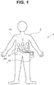
- Fig. 1 is a diagram for describing a usage form of the endoscope system according to the first embodiment
- Fig. 2 is a diagram for describing a detailed configuration of the capsule endoscope and a receiving apparatus according to the first embodiment
- Fig. 3 is a diagram for describing a pre-exposure area for performing pre-exposure
- Fig. 4 is a diagram for describing exposure timing
- Figs. 5A and 5B are diagrams for describing an interval of pre-exposure.
- an endoscope system 1 which is a capsule endoscope system is constructed of a capsule endoscope 10 and a receiving apparatus 20 as an external apparatus.
- the capsule endoscope 10 is introduced into an in-vivo digestive organ lumen by being swallowed by an examinee 2.
- the capsule endoscope 10 includes, as shown in Fig. 2 , a battery 11, an illumination section 12 configured to illuminate an object, an image pickup device 13 as an image pickup section configured to pick up an image of the object, an image transmitting section 14 configured to wirelessly transmit the image pickup signal (endoscope image), a memory 15 as a storage section configured to temporarily store a photometric result, which will be described later, and a control section 16 configured to control the entire capsule endoscope 10, all of which are accommodated in a case, to constitute main parts.
- the receiving apparatus 20 disposed outside the body of the examinee 2 includes an antenna unit 21 configured to receive an image pickup signal from the capsule endoscope 10 and a main unit 25, for example, to be worn on the waist of the examinee 2.
- the main unit 25 is constructed, as shown in Fig. 2 , of an image receiving section 22 configured to receive an image pickup signal (endoscope image) wirelessly transmitted from the capsule endoscope 10 via the antenna unit 21, an external memory 23 as an external storage section configured to store the endoscope image received by the image receiving section 22 via the antenna unit 21, and a display section 24 configured to display the endoscope image received by the image receiving section 22 or the endoscope image stored in the external memory 23.
- the capsule endoscope 10 of the present embodiment performs pre-exposure and then photometry before performing image pickup. That is, the control section 16 causes the illumination section 12 provided in the capsule endoscope 10 to emit light and causes the image pickup device 13 to pick up an image.
- the control section 16 acquires brightness information of the image (that is, performs photometry) from only part of pixel information of a pre-exposure area, which will be described later, without using all pixel information of the image pickup device 13 in this image pickup. Note that photometry performed using pixel information of the pre-exposure area is also called pre-photometry in the following description.
- a pre-exposure area 13a is provided so as to include a central pixel of the image pickup device 13 and the control section 16 acquires brightness information based on the pixel information of this part, that is, information obtained from the pre-exposure area 13a.
- the pre-exposure area provided in the image pickup device 13 is not limited to the pre-exposure area 13a provided so as to include the central pixel.
- pre-exposure areas 13b may be provided near four corners on a plane on which the image pickup device 13 is disposed and the control section 16 may acquire brightness information based on information obtained from the four pre-exposure areas 13b.
- pre-exposure areas 13c may be provided at positions which are in adjacent areas of the pixel region 13d used to display pixels of the image pickup device 13 and not reproduced or displayed on a monitor screen, and the control section 16 may acquire brightness information based on information obtained from the four pre-exposure areas 13c. That is, the pre-exposure areas 13c are provided in regions called "optical black" which are provided outside the pixel region 13d used for display to detect black.
- control section 16 is not limited to acquisition of brightness information based on information obtained from the pre-exposure areas 13a, 13b or 13c, but the control section 16 may acquire the brightness information by combining the pre-exposure area 13a and the four pre-exposure areas 13b and based on information obtained from the pre-exposure areas 13a and 13b.
- the control section 16 temporarily stores the brightness information (pre-photometric result) acquired in this way in the memory 15. As shown in Fig. 4 , when the pre-photometric operation ends, the control section 16 performs the next pre-exposure and pre-photometric operation.
- the pre-exposure and pre-photometric operation have the aforementioned contents and the same pre-exposure and pre-photometric operation are performed.
- a first pattern is one in which the interval of pre-exposure is minimized, that is, the interval of pre-exposure is made as short as possible. Minimizing the interval of pre-exposure makes it possible to prevent movement of the capsule endoscope 10 from being overlooked.
- a second pattern is one shown in Fig. 5B in which the interval of pre-exposure is set to a predetermined interval.
- the interval of pre-exposure is set to 2 fps, but the interval of pre-exposure is not limited to this and may be set to other intervals.
- pickup images of 2 frames per second are acquired as in the case of the prior art, but when no movement is found in the capsule endoscope 10, pickup images of 1 frame per second are acquired or no pickup image is acquired at all, and it is thereby possible to reduce power consumption of the capsule endoscope 10 compared to the prior art.
- the control section 16 compares brightness information obtained by second pre-photometry with the last brightness information stored in the memory 15. More specifically, the control section 16 calculates a difference value between the brightness information obtained by the second pre-exposure and the last brightness information stored in the memory 15. When the comparison result falls within a predetermined threshold, the control section 16 determines that the capsule endoscope 10 has not moved (is retained) for a period between two photometric operations. Upon determining that the capsule endoscope 10 has not moved, the control section 16 further proceeds to the next pre-exposure and pre-photometric operation. While repeating such pre-exposure and pre-photometric operation, if the comparison result between the latest brightness information and the last brightness information exceeds a predetermined threshold, the control section 16 determines that the capsule endoscope 10 has moved.
- the control section 16 determines the movement of the capsule endoscope 10 from a change in the brightness information of each pre-exposure area.
- the control section 16 determines that the capsule endoscope 10 has moved. Note that the control section 16 may also determine that the capsule endoscope 10 has moved when all the brightness information in each pre-exposure area has changed.
- the control section 16 determines that the capsule endoscope 10 has moved, and performs image pickup, that is, post-exposure following the pre-photometric operation.
- image pickup all the pixel information of the image pickup device 13, here substantially all the pixel information including all pixels which become images displayed on the monitor is acquired.
- the control section 16 applies predetermined signal processing to pixel information acquired by the image pickup device 13 and outputs an image signal acquired to the image transmitting section 14.
- the image transmitting section 14 wirelessly transmits the image signal to which predetermined signal processing is applied by the control section 16 to the external receiving apparatus 20. Note that although the image transmitting section 14 transmits the image signal to the external receiving apparatus 20, the image signal may be stored in the memory 15 in the capsule endoscope 10 via the control section 16.
- the wirelessly transmitted image signal is received by the image receiving section 22 via the external antenna unit 21.
- the image receiving section 22 stores the received image signal in the external memory 23 or outputs the received image signal to the display section 24 to display an endoscope image.
- the receiving apparatus 20 may add a time stamp to the endoscope image and store the endoscope image with the time stamp in the external memory 23.
- the control section 16 resumes the pre-exposure and pre-photometric operation and repeats the aforementioned processes. That is, the control section 16 compares the brightness information obtained as a result of pre-photometry with the brightness information stored in the memory 15, that is, latest brightness information, an image of which is determined to be picked up since an immediate preceding image is picked up, and determines whether or not the capsule endoscope 10 has moved. Upon determining that the capsule endoscope 10 has not moved, the control section 16 continues pre-exposure and pre-photometric operation and picks up an image with post-exposure upon determining that the capsule endoscope 10 has moved, and transmits data of the image signal obtained.
- Fig. 6 is a flowchart for describing operation of the capsule endoscope 10 according to the first embodiment.
- a target value of light adjustment and an area are set (step S1) and a pre-exposure area is set (step S2).
- a target value of light adjustment is calculated from the target value of light adjustment and the area and the pre-exposure area (step S3).
- pre-exposure is executed (step S4) and pre-photometry is executed (step S5).
- step S6 It is determined whether or not a difference value between the pre-photometric value and an immediate preceding pre-photometric value falls within a predetermined threshold (step S6).
- the result is YES, the flow returns to step S4 and similar processes are repeated.
- the difference value between the pre-photometric value and the immediate preceding pre-photometric value is greater than the predetermined threshold, the result is NO, a difference value between the pre-photometric value and the target value of light adjustment is calculated and the amount of exposure is calculated and set (step S7).
- step S8 a post-exposure is executed with a set amount of exposure
- step S9 the data of the endoscope image acquired in step S8 is transmitted
- step S9 the flow is returned to step S4 and similar processes are repeated.
- the capsule endoscope 10 measures brightness by performing pre-exposure and pre-photometry, compares this brightness information with last measured brightness information, detects movement of the capsule endoscope 10 and then picks up an image. For this reason, the capsule endoscope 10 can reduce power consumption without uselessly picking up images.
- the capsule endoscope 10 executes a pre-exposure in the pre-exposure area 13a which is part of the region of the image pickup device 13, can thereby shorten the exposure time period and shorten the pre-photometric time period by reducing the exposure area. Moreover, since a pre-exposure and a pre-photometry can be executed in a shorter time period than a post-exposure, it is possible to detect movement of the capsule endoscope 10 faster and pick up images at optimum timing.
- the capsule endoscope and the capsule endoscope system of the present embodiment it is possible to prevent useless image pickup and reduce power consumption.
- the capsule endoscope 10 for example, when an image pickup target region has a large width from dark to bright regions, that is, when the amount of brightness is very small or very large, the photometric value becomes very small or very large, and accurate brightness information may not be acquired.
- the capsule endoscope 10 will be described in the second embodiment which can accurately acquire brightness information even when an image pickup target region has a large width from dark to bright regions.
- an overall configuration of the capsule endoscope 10 of the second embodiment is similar to that of the first embodiment, and so only components different from those of the first embodiment will be described.
- a plurality of pixel areas are provided in a pre-exposure area.
- the pre-exposure area 13a in Fig. 3 will be described but the pre-exposure areas 13b and 13c may also have similar configurations.
- Fig. 7 is an enlarged view of the pre-exposure area 13a.
- the pre-exposure area 13a is constructed of four pixel areas 13a1, 13a2, 13a3 and 13a4, and the pixel areas 13a1, 13a2, 13a3 and 13a4 are arranged in a mosaic pattern.
- the pre-exposure area 13a is constructed of the four pixel areas 13a1, 13a2, 13a3 and 13a4, the pre-exposure area 13a is not limited to this, but the pre-exposure area 13a needs only to include at least two or more pixel areas.
- the pixel areas 13a1, 13a2, 13a3 and 13a4 are not limited to the mosaic arrangement, but may also be arranged in parallel, for example.
- the pixel area 13a1 is an area with a gain of 0 db whose gain is never increased
- the pixel area 13a2 is an area whose gain is increased by 6 db
- the pixel area 13a3 is an area whose gain is increased by 12 db
- the pixel area 13a4 is an area whose gain is increased by 18 db. Note that the amount of gain increase is not limited to the aforementioned 6 db, 12 db and 18 db, but may be other amounts of gain increase.
- the control section 16 compares photometric values acquired in the respective pixel areas 13a1 to 13a4 with photometric values acquired in the respective pixel areas 13a1 to 13a4 through last pre-exposures, determines whether or not the comparison results in all the pixel areas 13a1 to 13a4 fall within a predetermined threshold and detects movement of the capsule endoscope 10.
- the control section 16 determines that the capsule endoscope 10 has not moved, and when the comparison result in at least one of the pixel areas 13a1 to 13a4 is greater than the predetermined threshold, the control section 16 determines that the capsule endoscope 10 has moved.
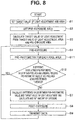
- Fig. 8 is a flowchart for describing operation of the capsule endoscope 10 according to the second embodiment. Note that in Fig. 8 , processes identical to those in Fig. 6 are assigned identical reference numerals and description thereof will be omitted.
- step S4 when a pre-exposure is executed, pre-photometry is executed for each pixel area 13a1 to 13a4 (step S11). It is determined whether or not a difference value between a pre-photometric value of each pixel area 13a1 to 13a4 and an immediate preceding pre-photometric value of each pixel area 13a1 to 13a4 falls within a predetermined threshold (step S12).
- step S4 When it is determined that the difference value between the pre-photometric value of each pixel area 13a1 to 13a4 and the immediate preceding pre-photometric value of each pixel area 13a1 to 13a4 falls within the predetermined threshold, the result is YES, the flow returns to step S4 and similar processes are repeated.
- the result is NO, and the flow proceeds to step S7, the difference value between the pre-photometric value and the target value of light adjustment is calculated and the amount of exposure is calculated and set.
- step S8 a post-exposure is executed with the set amount of exposure, the data of the endoscope image acquired in step S8 is transmitted, the flow then returns to step S4 and similar processes are repeated.
- the capsule endoscope 10 provides the pixel areas 13a1 to 13a4 having different gains in the pre-exposure area 13a.
- the pixel area 13a1 with a gain of 0 db has a very small amount of brightness, and so the photometric value becomes very small and accurate brightness information cannot be acquired, whereas providing the pixel areas 13a2, 13a3 and 13a4 whose gains are increased by 6 db, 12 db and 18 db respectively in a mosaic pattern makes it possible to obtain sufficient photometric information (brightness information) at pixels whose gains are increased to 12 db, for example.
- the capsule endoscope 10 of the present embodiment has an effect of being able to widen a dynamic range compared to the first embodiment without changing or increasing the pre-exposure time period.
- steps in the respective flowcharts in the Specification may be changed, steps may be executed simultaneously or steps may be executed in a different order at each execution.
Landscapes
- Health & Medical Sciences (AREA)
- Life Sciences & Earth Sciences (AREA)
- Surgery (AREA)
- Engineering & Computer Science (AREA)
- Radiology & Medical Imaging (AREA)
- Heart & Thoracic Surgery (AREA)
- Biophysics (AREA)
- Nuclear Medicine, Radiotherapy & Molecular Imaging (AREA)
- Optics & Photonics (AREA)
- Pathology (AREA)
- Veterinary Medicine (AREA)
- Public Health (AREA)
- Biomedical Technology (AREA)
- Physics & Mathematics (AREA)
- Medical Informatics (AREA)
- Molecular Biology (AREA)
- Animal Behavior & Ethology (AREA)
- General Health & Medical Sciences (AREA)
- Signal Processing (AREA)
- Multimedia (AREA)
- Computer Networks & Wireless Communication (AREA)
- Endoscopes (AREA)
- Studio Devices (AREA)
Abstract
Description
- The present invention relates to a capsule endoscope and a capsule endoscope system configured to be introduced into a subject and be able to acquire in-vivo information.
- Endoscopes in a medical field are conventionally used for in-vivo observation or the like. As one such endoscope, a capsule endoscope has been proposed in recent years which is disposed in a body cavity when swallowed by a subject, picks up images of the subject while moving through the body cavity along with a peristaltic movement, and can wirelessly send picked-up images of the subject to outside as image pickup signals.
- Such a capsule endoscope, in general, often photographs two frames per second. Photographing of two frames per second is set because the capsule endoscope is retained in the body cavity for a considerably long period of time, while the capacity of a battery incorporated in the capsule endoscope is limited.
- Movement of the capsule endoscope in the body cavity depends on peristaltic movement or the like, and so it is highly probable that the capsule endoscope may be retained in a specific place in the body cavity for a long period of time. On the other hand, since images are periodically picked up even during the retention period, completely identical images may be repeatedly acquired. However, only one frame of these images is sufficient to contribute to diagnosis and other images are wasted.
- Therefore, Japanese Patent Application Laid-Open Publication No.
2005-20755 - However, with the capsule endoscope described in Japanese Patent Application Laid-Open Publication No.
2005-20755 - It is therefore an object of the present invention to provide a capsule endoscope and a capsule endoscope system capable of preventing useless image pickup and reducing power consumption.
- A capsule endoscope according to an aspect of the present invention includes an image pickup section, a storage section configured to store brightness information acquired by the image pickup section, and a control section configured to perform control so as to compare the brightness information acquired by the image pickup section with brightness information acquired by the image pickup section immediately before acquiring the brightness information and stored in the storage section, further acquire, when the comparison result falls within a predetermined threshold, brightness information through the image pickup section, further compare the acquired brightness information with the brightness information acquired immediately before by the image pickup section and stored in the storage section, and on the other hand, when the comparison result is greater than the predetermined threshold, pick up an image through the image pickup section and store the picked-up image in the storage section or transmit the picked-up image to outside the capsule endoscope through an image transmitting section provided in the capsule endoscope.
- A capsule endoscope system according to an aspect of the present invention includes a capsule endoscope including an image pickup section, a storage section configured to store brightness information acquired by the image pickup section, and a control section configured to perform control so as to compare the brightness information acquired by the image pickup section with brightness information acquired by the image pickup section immediately before acquiring the brightness information and stored in the storage section, further acquire, when the comparison result falls within a predetermined threshold, brightness information through the image pickup section, further compare the acquired brightness information with the brightness information acquired immediately before by the image pickup section and stored in the storage section, and on the other hand, pick up an image when the comparison result is greater than the predetermined threshold, through the image pickup section and store the picked-up image in the storage section or transmit the picked-up image to outside the capsule endoscope through an image transmitting section provided in the capsule endoscope, and an external apparatus including an image receiving section configured to receive an image transmitted to outside by the image transmitting section and an external storage section configured to store the image received by the image receiving section.
-
-
Fig. 1 is a diagram for describing a usage form of an endoscope system according to a first embodiment; -
Fig. 2 is a diagram for describing a detailed configuration of a capsule endoscope and a receiving apparatus according to the first embodiment; -
Fig. 3 is a diagram for describing a pre-exposure area for performing pre-exposure; -
Fig. 4 is a diagram for describing exposure timing; -
Fig. 5A is a diagram for describing an interval of pre-exposure; -
Fig. 5B is a diagram for describing an interval of pre-exposure; -
Fig. 6 is a flowchart for describing operation of thecapsule endoscope 10 according to the first embodiment; -
Fig. 7 is an enlarged view of apre-exposure area 13a; and -
Fig. 8 is a flowchart for describing operation of acapsule endoscope 10 according to a second embodiment. - Hereinafter, embodiments of the present invention will be described with reference to the accompanying drawings.
- First, a configuration of an endoscope system according to a first embodiment of the present invention will be described using
Fig. 1 to Fig. 5B .Fig. 1 is a diagram for describing a usage form of the endoscope system according to the first embodiment,Fig. 2 is a diagram for describing a detailed configuration of the capsule endoscope and a receiving apparatus according to the first embodiment,Fig. 3 is a diagram for describing a pre-exposure area for performing pre-exposure,Fig. 4 is a diagram for describing exposure timing andFigs. 5A and 5B are diagrams for describing an interval of pre-exposure. - As shown in
Fig. 1 , anendoscope system 1 which is a capsule endoscope system is constructed of acapsule endoscope 10 and a receivingapparatus 20 as an external apparatus. - The
capsule endoscope 10 is introduced into an in-vivo digestive organ lumen by being swallowed by anexaminee 2. Thecapsule endoscope 10 includes, as shown inFig. 2 , abattery 11, anillumination section 12 configured to illuminate an object, animage pickup device 13 as an image pickup section configured to pick up an image of the object, animage transmitting section 14 configured to wirelessly transmit the image pickup signal (endoscope image), amemory 15 as a storage section configured to temporarily store a photometric result, which will be described later, and acontrol section 16 configured to control theentire capsule endoscope 10, all of which are accommodated in a case, to constitute main parts. - On the other hand, the
receiving apparatus 20 disposed outside the body of theexaminee 2 includes anantenna unit 21 configured to receive an image pickup signal from thecapsule endoscope 10 and amain unit 25, for example, to be worn on the waist of theexaminee 2. Themain unit 25 is constructed, as shown inFig. 2 , of animage receiving section 22 configured to receive an image pickup signal (endoscope image) wirelessly transmitted from thecapsule endoscope 10 via theantenna unit 21, anexternal memory 23 as an external storage section configured to store the endoscope image received by theimage receiving section 22 via theantenna unit 21, and adisplay section 24 configured to display the endoscope image received by theimage receiving section 22 or the endoscope image stored in theexternal memory 23. - The
capsule endoscope 10 of the present embodiment performs pre-exposure and then photometry before performing image pickup. That is, thecontrol section 16 causes theillumination section 12 provided in thecapsule endoscope 10 to emit light and causes theimage pickup device 13 to pick up an image. Thecontrol section 16 acquires brightness information of the image (that is, performs photometry) from only part of pixel information of a pre-exposure area, which will be described later, without using all pixel information of theimage pickup device 13 in this image pickup. Note that photometry performed using pixel information of the pre-exposure area is also called pre-photometry in the following description. - As shown in
Fig. 3 , apre-exposure area 13a is provided so as to include a central pixel of theimage pickup device 13 and thecontrol section 16 acquires brightness information based on the pixel information of this part, that is, information obtained from thepre-exposure area 13a. Note that the pre-exposure area provided in theimage pickup device 13 is not limited to thepre-exposure area 13a provided so as to include the central pixel. - For example, four
pre-exposure areas 13b may be provided near four corners on a plane on which theimage pickup device 13 is disposed and thecontrol section 16 may acquire brightness information based on information obtained from the fourpre-exposure areas 13b. - Furthermore, four
pre-exposure areas 13c may be provided at positions which are in adjacent areas of thepixel region 13d used to display pixels of theimage pickup device 13 and not reproduced or displayed on a monitor screen, and thecontrol section 16 may acquire brightness information based on information obtained from the four pre-exposureareas 13c. That is, thepre-exposure areas 13c are provided in regions called "optical black" which are provided outside thepixel region 13d used for display to detect black. - Furthermore, the
control section 16 is not limited to acquisition of brightness information based on information obtained from thepre-exposure areas control section 16 may acquire the brightness information by combining thepre-exposure area 13a and the fourpre-exposure areas 13b and based on information obtained from thepre-exposure areas - The
control section 16 temporarily stores the brightness information (pre-photometric result) acquired in this way in thememory 15. As shown inFig. 4 , when the pre-photometric operation ends, thecontrol section 16 performs the next pre-exposure and pre-photometric operation. The pre-exposure and pre-photometric operation have the aforementioned contents and the same pre-exposure and pre-photometric operation are performed. - Here, an interval of pre-exposure will be described using
Fig. 5A and Fig. 5B . In the present embodiment, the following two patterns are assumed about the interval of pre-exposure, but other patterns may be adopted without being limited to this. - As shown in
Fig. 5A , a first pattern is one in which the interval of pre-exposure is minimized, that is, the interval of pre-exposure is made as short as possible. Minimizing the interval of pre-exposure makes it possible to prevent movement of thecapsule endoscope 10 from being overlooked. - That is, conventional capsule endoscopes acquire pickup images of 2 frames per second, but in
Fig. 5A , the interval of pre-exposure is minimized and when thecapsule endoscope 10 moves, it is possible to acquire pickup images of 3 or more frames per second and prevent lesioned regions or the like from being overlooked. - A second pattern is one shown in
Fig. 5B in which the interval of pre-exposure is set to a predetermined interval. Note that in the example inFig. 5B , the interval of pre-exposure is set to 2 fps, but the interval of pre-exposure is not limited to this and may be set to other intervals. In the case of the example inFig. 5B , when movement of thecapsule endoscope 10 is found, pickup images of 2 frames per second are acquired as in the case of the prior art, but when no movement is found in thecapsule endoscope 10, pickup images of 1 frame per second are acquired or no pickup image is acquired at all, and it is thereby possible to reduce power consumption of thecapsule endoscope 10 compared to the prior art. - The
control section 16 compares brightness information obtained by second pre-photometry with the last brightness information stored in thememory 15. More specifically, thecontrol section 16 calculates a difference value between the brightness information obtained by the second pre-exposure and the last brightness information stored in thememory 15. When the comparison result falls within a predetermined threshold, thecontrol section 16 determines that thecapsule endoscope 10 has not moved (is retained) for a period between two photometric operations. Upon determining that thecapsule endoscope 10 has not moved, thecontrol section 16 further proceeds to the next pre-exposure and pre-photometric operation. While repeating such pre-exposure and pre-photometric operation, if the comparison result between the latest brightness information and the last brightness information exceeds a predetermined threshold, thecontrol section 16 determines that thecapsule endoscope 10 has moved. - Note that when, for example, there are a plurality of pre-exposure areas such as the
pre-exposure areas control section 16 determines the movement of thecapsule endoscope 10 from a change in the brightness information of each pre-exposure area. When the brightness information in each pre-exposure area changes even at least one location, thecontrol section 16 determines that thecapsule endoscope 10 has moved. Note that thecontrol section 16 may also determine that thecapsule endoscope 10 has moved when all the brightness information in each pre-exposure area has changed. - For example, as shown in
Fig. 4 , when a comparison result between the brightness information obtained by a pre-exposure after time t1 and the last brightness information exceeds a predetermined threshold, thecontrol section 16 determines that thecapsule endoscope 10 has moved, and performs image pickup, that is, post-exposure following the pre-photometric operation. During the image pickup, all the pixel information of theimage pickup device 13, here substantially all the pixel information including all pixels which become images displayed on the monitor is acquired. - The
control section 16 applies predetermined signal processing to pixel information acquired by theimage pickup device 13 and outputs an image signal acquired to theimage transmitting section 14. Theimage transmitting section 14 wirelessly transmits the image signal to which predetermined signal processing is applied by thecontrol section 16 to theexternal receiving apparatus 20. Note that although theimage transmitting section 14 transmits the image signal to theexternal receiving apparatus 20, the image signal may be stored in thememory 15 in thecapsule endoscope 10 via thecontrol section 16. - The wirelessly transmitted image signal is received by the
image receiving section 22 via theexternal antenna unit 21. Theimage receiving section 22 stores the received image signal in theexternal memory 23 or outputs the received image signal to thedisplay section 24 to display an endoscope image. Note that upon receiving the image signal (endoscope image) from thecapsule endoscope 10, the receivingapparatus 20 may add a time stamp to the endoscope image and store the endoscope image with the time stamp in theexternal memory 23. - When a series of processes from image pickup to image transmission end, the
control section 16 resumes the pre-exposure and pre-photometric operation and repeats the aforementioned processes. That is, thecontrol section 16 compares the brightness information obtained as a result of pre-photometry with the brightness information stored in thememory 15, that is, latest brightness information, an image of which is determined to be picked up since an immediate preceding image is picked up, and determines whether or not thecapsule endoscope 10 has moved. Upon determining that thecapsule endoscope 10 has not moved, thecontrol section 16 continues pre-exposure and pre-photometric operation and picks up an image with post-exposure upon determining that thecapsule endoscope 10 has moved, and transmits data of the image signal obtained. - Next, operation of the
capsule endoscope 10 configured as shown above will be described. -
Fig. 6 is a flowchart for describing operation of thecapsule endoscope 10 according to the first embodiment. - First, a target value of light adjustment and an area are set (step S1) and a pre-exposure area is set (step S2). Next, a target value of light adjustment is calculated from the target value of light adjustment and the area and the pre-exposure area (step S3). Next, pre-exposure is executed (step S4) and pre-photometry is executed (step S5).
- It is determined whether or not a difference value between the pre-photometric value and an immediate preceding pre-photometric value falls within a predetermined threshold (step S6). When the difference value between the pre-photometric value and the immediate preceding pre-photometric value falls within the predetermined threshold, the result is YES, the flow returns to step S4 and similar processes are repeated. On the other hand, when the difference value between the pre-photometric value and the immediate preceding pre-photometric value is greater than the predetermined threshold, the result is NO, a difference value between the pre-photometric value and the target value of light adjustment is calculated and the amount of exposure is calculated and set (step S7).
- Next, a post-exposure is executed with a set amount of exposure (step S8), the data of the endoscope image acquired in step S8 is transmitted (step S9), the flow is returned to step S4 and similar processes are repeated.
- As described above, when it is determined that the
capsule endoscope 10 has moved, images are consecutively picked up at a predetermined interval and when it is determined that thecapsule endoscope 10 has not moved, a state continues in which images are not picked up. As a result, a group of images picked up by thecapsule endoscope 10 are picked up at random time intervals without being bound by a concept of, for example, 2 frames per second. - The
capsule endoscope 10 according to the present embodiment measures brightness by performing pre-exposure and pre-photometry, compares this brightness information with last measured brightness information, detects movement of thecapsule endoscope 10 and then picks up an image. For this reason, thecapsule endoscope 10 can reduce power consumption without uselessly picking up images. - Furthermore, the
capsule endoscope 10 executes a pre-exposure in thepre-exposure area 13a which is part of the region of theimage pickup device 13, can thereby shorten the exposure time period and shorten the pre-photometric time period by reducing the exposure area. Moreover, since a pre-exposure and a pre-photometry can be executed in a shorter time period than a post-exposure, it is possible to detect movement of thecapsule endoscope 10 faster and pick up images at optimum timing. - Thus, according to the capsule endoscope and the capsule endoscope system of the present embodiment, it is possible to prevent useless image pickup and reduce power consumption.
- Next, a second embodiment will be described. With the
capsule endoscope 10 according to the first embodiment, for example, when an image pickup target region has a large width from dark to bright regions, that is, when the amount of brightness is very small or very large, the photometric value becomes very small or very large, and accurate brightness information may not be acquired. Thus, thecapsule endoscope 10 will be described in the second embodiment which can accurately acquire brightness information even when an image pickup target region has a large width from dark to bright regions. - Note that an overall configuration of the
capsule endoscope 10 of the second embodiment is similar to that of the first embodiment, and so only components different from those of the first embodiment will be described. In the second embodiment, a plurality of pixel areas are provided in a pre-exposure area. In the following description, thepre-exposure area 13a inFig. 3 will be described but thepre-exposure areas -
Fig. 7 is an enlarged view of thepre-exposure area 13a. As shown inFig. 7 , thepre-exposure area 13a is constructed of four pixel areas 13a1, 13a2, 13a3 and 13a4, and the pixel areas 13a1, 13a2, 13a3 and 13a4 are arranged in a mosaic pattern. Note that although thepre-exposure area 13a is constructed of the four pixel areas 13a1, 13a2, 13a3 and 13a4, thepre-exposure area 13a is not limited to this, but thepre-exposure area 13a needs only to include at least two or more pixel areas. The pixel areas 13a1, 13a2, 13a3 and 13a4 are not limited to the mosaic arrangement, but may also be arranged in parallel, for example. - Different gains are set for the pixel areas 13a1, 13a2, 13a3 and 13a4 respectively. The pixel area 13a1 is an area with a gain of 0 db whose gain is never increased, the pixel area 13a2 is an area whose gain is increased by 6 db. The pixel area 13a3 is an area whose gain is increased by 12 db and the pixel area 13a4 is an area whose gain is increased by 18 db. Note that the amount of gain increase is not limited to the aforementioned 6 db, 12 db and 18 db, but may be other amounts of gain increase.
- The
control section 16 compares photometric values acquired in the respective pixel areas 13a1 to 13a4 with photometric values acquired in the respective pixel areas 13a1 to 13a4 through last pre-exposures, determines whether or not the comparison results in all the pixel areas 13a1 to 13a4 fall within a predetermined threshold and detects movement of thecapsule endoscope 10. - When the comparison results in all the pixel areas 13a1 to 13a4 fall within the predetermined threshold, the
control section 16 determines that thecapsule endoscope 10 has not moved, and when the comparison result in at least one of the pixel areas 13a1 to 13a4 is greater than the predetermined threshold, thecontrol section 16 determines that thecapsule endoscope 10 has moved. - Next, operation of the
capsule endoscope 10 configured in this way will be described. -
Fig. 8 is a flowchart for describing operation of thecapsule endoscope 10 according to the second embodiment. Note that inFig. 8 , processes identical to those inFig. 6 are assigned identical reference numerals and description thereof will be omitted. - In step S4, when a pre-exposure is executed, pre-photometry is executed for each pixel area 13a1 to 13a4 (step S11). It is determined whether or not a difference value between a pre-photometric value of each pixel area 13a1 to 13a4 and an immediate preceding pre-photometric value of each pixel area 13a1 to 13a4 falls within a predetermined threshold (step S12).
- When it is determined that the difference value between the pre-photometric value of each pixel area 13a1 to 13a4 and the immediate preceding pre-photometric value of each pixel area 13a1 to 13a4 falls within the predetermined threshold, the result is YES, the flow returns to step S4 and similar processes are repeated. On the other hand, when any one difference value between the pre-photometric value of each pixel area 13a1 to 13a4 and the immediate preceding pre-photometric value of each pixel area 13a1 to 13a4 is greater than the predetermined threshold, the result is NO, and the flow proceeds to step S7, the difference value between the pre-photometric value and the target value of light adjustment is calculated and the amount of exposure is calculated and set. Then, in step S8, a post-exposure is executed with the set amount of exposure, the data of the endoscope image acquired in step S8 is transmitted, the flow then returns to step S4 and similar processes are repeated.
- As described above, the
capsule endoscope 10 according to the present embodiment provides the pixel areas 13a1 to 13a4 having different gains in thepre-exposure area 13a. When, for example, an image pickup target region is dark, the pixel area 13a1 with a gain of 0 db has a very small amount of brightness, and so the photometric value becomes very small and accurate brightness information cannot be acquired, whereas providing the pixel areas 13a2, 13a3 and 13a4 whose gains are increased by 6 db, 12 db and 18 db respectively in a mosaic pattern makes it possible to obtain sufficient photometric information (brightness information) at pixels whose gains are increased to 12 db, for example. - With such a configuration, when, for example, the image pickup target region is bright, the brightness information becomes very large in the pixel areas 13a2, 13a3 and 13a4 whose gains are increased to 6 db, 12 db and 18 db, the photometric values thereby become very large, making it impossible to acquire accurate brightness information, but providing the pixel area 13a1 with a gain of 0 db allows sufficient photometric information to be obtained in pixels where gains are not increased.
- As a result, the
capsule endoscope 10 of the present embodiment has an effect of being able to widen a dynamic range compared to the first embodiment without changing or increasing the pre-exposure time period. - Note that regarding steps in the respective flowcharts in the Specification, as long as it does not go against the nature of the steps, the order in which steps are executed may be changed, steps may be executed simultaneously or steps may be executed in a different order at each execution.
- The present invention is not limited to the aforementioned embodiments, but various modifications or alterations or the like may be made without departing from the spirit and scope of the present invention.
- The present application claims priority based on Japanese Patent Application No.
2014-240323
Claims (16)
- A capsule endoscope comprising:an image pickup section;a storage section configured to store brightness information acquired by the image pickup section; anda control section configured to perform control so as to compare the brightness information acquired by the image pickup section with brightness information acquired by the image pickup section immediately before acquiring the brightness information and stored in the storage section, further acquire, when the comparison result falls within a predetermined threshold, brightness information through the image pickup section, further compare the acquired brightness information with brightness information acquired by the image pickup section immediately before and stored in the storage section, and on the other hand, when the comparison result is greater than the predetermined threshold, pick up an image through the image pickup section and store the picked-up image in the storage section or transmit the picked-up image to outside the capsule endoscope through an image transmitting section provided in the capsule endoscope.
- The capsule endoscope according to claim 1, wherein the control section acquires brightness information through the image pickup section following storage of the image or an end of the transmission and compares the acquired brightness information with the brightness information acquired by the image pickup section immediately before and stored in the storage section.
- The capsule endoscope according to claim 1, wherein the control section compares the brightness information acquired by the image pickup section with the brightness information acquired by the image pickup section immediately before acquiring the brightness information and stored in the storage section at a predetermined interval and performs control, when the comparison result is equal to or less than the predetermined threshold, so as to prevent the image pickup section from picking up images and prevent the image transmitting section from performing transmission to outside the capsule endoscope.
- The capsule endoscope according to claim 1, wherein the brightness information is acquired based on information obtained from part of pixels of the image pickup device including a central pixel of the image pickup device.
- The capsule endoscope according to claim 1, wherein the brightness information is acquired based on information obtained from part of pixels of the image pickup device which are pixels of four parts in adjacent areas of four corners of a plane on which image pickup pixels of the image pickup device are disposed.
- The capsule endoscope according to claim 1, wherein the brightness information is acquired based on information obtained from pixels located at positions in adjacent areas of pixels used for display out of pixels of the image pickup device and corresponding to positions which are not reproduced or displayed on a monitor screen.
- The capsule endoscope according to claim 1, wherein the image pickup device comprises at least a first pixel group and a second pixel group, and
first information for which no gain increase is performed is generated for information obtained from the first pixel group, second information for which a gain increase is performed by a first amount is generated for information obtained from the second pixel group, and the brightness information is acquired based on the first information and the second information. - The capsule endoscope according to claim 7, wherein the image pickup device further comprises a third pixel group, and
third information for which a gain increase is performed by an amount greater than the first amount is generated for information obtained from the third pixel group and the brightness information is acquired based on the first information, the second information and the third information. - A capsule endoscope system comprising:a capsule endoscope comprising:an image pickup section;a storage section configured to store brightness information acquired by the image pickup section; anda control section configured to perform control so as to compare the brightness information acquired by the image pickup section with brightness information acquired by the image pickup section immediately before acquiring the brightness information and stored in the storage section, further acquire, when the comparison result falls within a predetermined threshold, brightness information through the image pickup section, further compare the acquired brightness information with the brightness information acquired by the image pickup section immediately before and stored in the storage section, and on the other hand, when the comparison result is greater than the predetermined threshold, pick up an image through the image pickup section and store the picked-up image in the storage section or transmit the picked-up image to outside the capsule endoscope through an image transmitting section provided in the capsule endoscope; andan external apparatus comprising:an image receiving section configured to receive the image transmitted to outside by the image transmitting section; andan external storage section configured to store the image received by the image receiving section.
- The capsule endoscope system according to claim 9, wherein the external apparatus adds, when the image receiving section receives the image, a time stamp to the image and stores the image with the time stamp in the external storage section.
- The capsule endoscope system according to claim 9, wherein the control section acquires brightness information through the image pickup section following storage of the image or an end of the transmission and compares the acquired brightness information with the brightness information acquired by the image pickup section immediately before and stored in the storage section.
- The capsule endoscope system according to claim 9, wherein the brightness information is acquired based on information obtained from part of pixels of the image pickup device including a central pixel of the image pickup device.
- The capsule endoscope system according to claim 9, wherein the brightness information is acquired based on information obtained from part of pixels of the image pickup device which are pixels of four parts in adjacent areas of four corners of a plane on which image pickup pixels of the image pickup device are disposed.
- The capsule endoscope system according to claim 9, wherein the brightness information is acquired based on information obtained from pixels located at positions in adjacent areas of pixels used for display out of pixels of the image pickup device and corresponding to positions which are not reproduced or displayed on a monitor screen.
- The capsule endoscope system according to claim 9, wherein the image pickup device comprises at least a first pixel group and a second pixel group, and
first information for which no gain increase is performed is generated for information obtained from the first pixel group, second information for which a gain increase by a first amount is performed is generated for information obtained from the second pixel group, and the brightness information is acquired based on the first information and the second information. - The capsule endoscope system according to claim 15, wherein the image pickup device further comprises a third pixel group, and
third information for which a gain increase is performed by a greater amount than the first amount is generated for information obtained from the third pixel group and the brightness information is acquired based on the first information, the second information and the third information.
Applications Claiming Priority (2)
| Application Number | Priority Date | Filing Date | Title |
|---|---|---|---|
| JP2014240323 | 2014-11-27 | ||
| PCT/JP2015/077074 WO2016084467A1 (en) | 2014-11-27 | 2015-09-25 | Encapsulated endoscope and encapsulated endoscope system |
Publications (2)
| Publication Number | Publication Date |
|---|---|
| EP3231349A1 true EP3231349A1 (en) | 2017-10-18 |
| EP3231349A4 EP3231349A4 (en) | 2018-08-22 |
Family
ID=56074046
Family Applications (1)
| Application Number | Title | Priority Date | Filing Date |
|---|---|---|---|
| EP15863528.4A Withdrawn EP3231349A4 (en) | 2014-11-27 | 2015-09-25 | Encapsulated endoscope and encapsulated endoscope system |
Country Status (5)
| Country | Link |
|---|---|
| US (1) | US9993143B2 (en) |
| EP (1) | EP3231349A4 (en) |
| JP (1) | JP5977907B1 (en) |
| CN (1) | CN106255444B (en) |
| WO (1) | WO2016084467A1 (en) |
Families Citing this family (5)
| Publication number | Priority date | Publication date | Assignee | Title |
|---|---|---|---|---|
| CA2835870A1 (en) | 2011-05-12 | 2012-11-15 | Olive Medical Corporation | Pixel array area optimization using stacking scheme for hybrid image sensor with minimal vertical interconnects |
| CN104486987A (en) | 2012-07-26 | 2015-04-01 | 橄榄医疗公司 | Camera system with smallest area monolithic CMOS image sensor |
| AU2014233192B2 (en) | 2013-03-15 | 2018-11-22 | DePuy Synthes Products, Inc. | Minimize image sensor I/O and conductor counts in endoscope applications |
| WO2014145246A1 (en) | 2013-03-15 | 2014-09-18 | Olive Medical Corporation | Image sensor synchronization without input clock and data transmission clock |
| WO2020250776A1 (en) * | 2019-06-11 | 2020-12-17 | ソニー株式会社 | Medical system, communication method, image capturing device, information processing device, and endoscope system |
Family Cites Families (12)
| Publication number | Priority date | Publication date | Assignee | Title |
|---|---|---|---|---|
| IL122602A0 (en) * | 1997-12-15 | 1998-08-16 | Tally Eitan Zeev Pearl And Co | Energy management of a video capsule |
| IL162740A (en) * | 2003-06-26 | 2010-06-16 | Given Imaging Ltd | Device, method and system for reduced transmission imaging |
| US8449458B2 (en) * | 2005-02-25 | 2013-05-28 | Olympus Corporation | Body-insertable apparatus and radio in-vivo information acquiring system |
| US7983458B2 (en) * | 2005-09-20 | 2011-07-19 | Capso Vision, Inc. | In vivo autonomous camera with on-board data storage or digital wireless transmission in regulatory approved band |
| JP4830652B2 (en) * | 2006-06-12 | 2011-12-07 | 日産自動車株式会社 | Image processing apparatus and image processing method |
| US8213698B2 (en) * | 2006-09-19 | 2012-07-03 | Capso Vision Inc. | Systems and methods for capsule camera control |
| DE102008018931A1 (en) * | 2007-04-17 | 2008-11-13 | Gyrus ACMI, Inc., Southborough | Light source power based on a predetermined detected condition |
| CN101677751B (en) * | 2007-05-22 | 2012-06-27 | 奥林巴斯株式会社 | Encapsulated medical device and encapsulated medical system |
| CN101179725A (en) * | 2007-12-12 | 2008-05-14 | 北京中星微电子有限公司 | Motion detecting method and apparatus |
| JP4875691B2 (en) * | 2007-12-17 | 2012-02-15 | オリンパスメディカルシステムズ株式会社 | Imaging device, image display device, and image display system |
| JP5649657B2 (en) * | 2009-11-20 | 2015-01-07 | ギブン イメージング リミテッドGiven Imaging Ltd. | System and method for controlling power consumption of in-vivo devices |
| WO2012098791A1 (en) * | 2011-01-20 | 2012-07-26 | オリンパスメディカルシステムズ株式会社 | Capsule endoscope |
-
2015
- 2015-09-25 WO PCT/JP2015/077074 patent/WO2016084467A1/en not_active Ceased
- 2015-09-25 EP EP15863528.4A patent/EP3231349A4/en not_active Withdrawn
- 2015-09-25 CN CN201580020012.3A patent/CN106255444B/en active Active
- 2015-09-25 JP JP2016520117A patent/JP5977907B1/en active Active
-
2016
- 2016-10-12 US US15/291,190 patent/US9993143B2/en active Active
Also Published As
| Publication number | Publication date |
|---|---|
| US9993143B2 (en) | 2018-06-12 |
| EP3231349A4 (en) | 2018-08-22 |
| CN106255444A (en) | 2016-12-21 |
| JPWO2016084467A1 (en) | 2017-04-27 |
| JP5977907B1 (en) | 2016-08-24 |
| WO2016084467A1 (en) | 2016-06-02 |
| CN106255444B (en) | 2018-04-13 |
| US20170027425A1 (en) | 2017-02-02 |
Similar Documents
| Publication | Publication Date | Title |
|---|---|---|
| US8562515B2 (en) | Capsule endoscope, capsule endoscopic system, and endoscope control method | |
| US9993143B2 (en) | Capsule endoscope and capsule endoscope system | |
| JP5649657B2 (en) | System and method for controlling power consumption of in-vivo devices | |
| US9560956B2 (en) | Device, system and method of displaying in-vivo images at variable rate | |
| JP5388657B2 (en) | Image processing apparatus, method of operating image processing apparatus, and system | |
| US20090192348A1 (en) | Capsule endoscope, method of controlling the same, and information manager | |
| EP2353491A1 (en) | Subject intra-corporeal introduction device and in-vivo information acquiring system | |
| US11234578B2 (en) | Receiving apparatus and radio wave interference determination method | |
| WO2015182185A1 (en) | Capsule endoscope apparatus | |
| US8830310B2 (en) | Capsule endoscope | |
| US10462440B2 (en) | Image processing apparatus | |
| EP2633798B1 (en) | Image processing device, image processing method, image processing program, and endoscope system | |
| US10979922B2 (en) | Estimation device, medical system, and estimation method | |
| US10939037B2 (en) | Capsule endoscope, receiving device, operation method of capsule endoscope, and computer readable recording medium | |
| US10702133B2 (en) | Image processing device, endoscope system, image processing method, and computer-readable recording medium | |
| US10201266B2 (en) | Single image sensor control for capturing mixed mode images | |
| JP2016077683A (en) | Receiving device and capsule-type endoscope system | |
| US20200196845A1 (en) | Capsule endoscope system, capsule endoscope, and receiving device | |
| JP2007090060A (en) | Apparatus, system, and method for determining spatial dimensions of anatomical objects for in vivo pathology detection | |
| JPWO2019171616A1 (en) | Receiving device and receiving method | |
| JP5896877B2 (en) | Light control device | |
| JP5815166B1 (en) | Capsule endoscope device | |
| JPWO2017217029A1 (en) | Motion determination device, in-subject introduction device, motion determination method, and program |
Legal Events
| Date | Code | Title | Description |
|---|---|---|---|
| STAA | Information on the status of an ep patent application or granted ep patent |
Free format text: STATUS: THE INTERNATIONAL PUBLICATION HAS BEEN MADE |
|
| PUAI | Public reference made under article 153(3) epc to a published international application that has entered the european phase |
Free format text: ORIGINAL CODE: 0009012 |
|
| STAA | Information on the status of an ep patent application or granted ep patent |
Free format text: STATUS: REQUEST FOR EXAMINATION WAS MADE |
|
| 17P | Request for examination filed |
Effective date: 20161010 |
|
| AK | Designated contracting states |
Kind code of ref document: A1 Designated state(s): AL AT BE BG CH CY CZ DE DK EE ES FI FR GB GR HR HU IE IS IT LI LT LU LV MC MK MT NL NO PL PT RO RS SE SI SK SM TR |
|
| AX | Request for extension of the european patent |
Extension state: BA ME |
|
| DAV | Request for validation of the european patent (deleted) | ||
| DAX | Request for extension of the european patent (deleted) | ||
| A4 | Supplementary search report drawn up and despatched |
Effective date: 20180724 |
|
| RIC1 | Information provided on ipc code assigned before grant |
Ipc: A61B 1/045 20060101ALI20180718BHEP Ipc: A61B 1/04 20060101AFI20180718BHEP Ipc: H04N 5/235 20060101ALI20180718BHEP Ipc: H04N 5/225 20060101ALI20180718BHEP Ipc: H04N 5/243 20060101ALI20180718BHEP Ipc: A61B 1/00 20060101ALI20180718BHEP Ipc: H04N 5/232 20060101ALI20180718BHEP |
|
| STAA | Information on the status of an ep patent application or granted ep patent |
Free format text: STATUS: THE APPLICATION IS DEEMED TO BE WITHDRAWN |
|
| 18D | Application deemed to be withdrawn |
Effective date: 20190221 |