EP3227458B1 - Epigenetic method for the identification of follicular t-helper-(tfh-) cells - Google Patents
Epigenetic method for the identification of follicular t-helper-(tfh-) cells Download PDFInfo
- Publication number
- EP3227458B1 EP3227458B1 EP15805467.6A EP15805467A EP3227458B1 EP 3227458 B1 EP3227458 B1 EP 3227458B1 EP 15805467 A EP15805467 A EP 15805467A EP 3227458 B1 EP3227458 B1 EP 3227458B1
- Authority
- EP
- European Patent Office
- Prior art keywords
- cells
- helper
- follicular
- cell
- follicular helper
- Prior art date
- Legal status (The legal status is an assumption and is not a legal conclusion. Google has not performed a legal analysis and makes no representation as to the accuracy of the status listed.)
- Active
Links
Images



Classifications
-
- C—CHEMISTRY; METALLURGY
- C12—BIOCHEMISTRY; BEER; SPIRITS; WINE; VINEGAR; MICROBIOLOGY; ENZYMOLOGY; MUTATION OR GENETIC ENGINEERING
- C12Q—MEASURING OR TESTING PROCESSES INVOLVING ENZYMES, NUCLEIC ACIDS OR MICROORGANISMS; COMPOSITIONS OR TEST PAPERS THEREFOR; PROCESSES OF PREPARING SUCH COMPOSITIONS; CONDITION-RESPONSIVE CONTROL IN MICROBIOLOGICAL OR ENZYMOLOGICAL PROCESSES
- C12Q1/00—Measuring or testing processes involving enzymes, nucleic acids or microorganisms; Compositions therefor; Processes of preparing such compositions
- C12Q1/68—Measuring or testing processes involving enzymes, nucleic acids or microorganisms; Compositions therefor; Processes of preparing such compositions involving nucleic acids
- C12Q1/6876—Nucleic acid products used in the analysis of nucleic acids, e.g. primers or probes
- C12Q1/6881—Nucleic acid products used in the analysis of nucleic acids, e.g. primers or probes for tissue or cell typing, e.g. human leukocyte antigen [HLA] probes
-
- C—CHEMISTRY; METALLURGY
- C12—BIOCHEMISTRY; BEER; SPIRITS; WINE; VINEGAR; MICROBIOLOGY; ENZYMOLOGY; MUTATION OR GENETIC ENGINEERING
- C12Q—MEASURING OR TESTING PROCESSES INVOLVING ENZYMES, NUCLEIC ACIDS OR MICROORGANISMS; COMPOSITIONS OR TEST PAPERS THEREFOR; PROCESSES OF PREPARING SUCH COMPOSITIONS; CONDITION-RESPONSIVE CONTROL IN MICROBIOLOGICAL OR ENZYMOLOGICAL PROCESSES
- C12Q1/00—Measuring or testing processes involving enzymes, nucleic acids or microorganisms; Compositions therefor; Processes of preparing such compositions
- C12Q1/68—Measuring or testing processes involving enzymes, nucleic acids or microorganisms; Compositions therefor; Processes of preparing such compositions involving nucleic acids
- C12Q1/6876—Nucleic acid products used in the analysis of nucleic acids, e.g. primers or probes
- C12Q1/6883—Nucleic acid products used in the analysis of nucleic acids, e.g. primers or probes for diseases caused by alterations of genetic material
-
- C—CHEMISTRY; METALLURGY
- C12—BIOCHEMISTRY; BEER; SPIRITS; WINE; VINEGAR; MICROBIOLOGY; ENZYMOLOGY; MUTATION OR GENETIC ENGINEERING
- C12Q—MEASURING OR TESTING PROCESSES INVOLVING ENZYMES, NUCLEIC ACIDS OR MICROORGANISMS; COMPOSITIONS OR TEST PAPERS THEREFOR; PROCESSES OF PREPARING SUCH COMPOSITIONS; CONDITION-RESPONSIVE CONTROL IN MICROBIOLOGICAL OR ENZYMOLOGICAL PROCESSES
- C12Q1/00—Measuring or testing processes involving enzymes, nucleic acids or microorganisms; Compositions therefor; Processes of preparing such compositions
- C12Q1/68—Measuring or testing processes involving enzymes, nucleic acids or microorganisms; Compositions therefor; Processes of preparing such compositions involving nucleic acids
- C12Q1/6876—Nucleic acid products used in the analysis of nucleic acids, e.g. primers or probes
- C12Q1/6883—Nucleic acid products used in the analysis of nucleic acids, e.g. primers or probes for diseases caused by alterations of genetic material
- C12Q1/6886—Nucleic acid products used in the analysis of nucleic acids, e.g. primers or probes for diseases caused by alterations of genetic material for cancer
-
- C—CHEMISTRY; METALLURGY
- C12—BIOCHEMISTRY; BEER; SPIRITS; WINE; VINEGAR; MICROBIOLOGY; ENZYMOLOGY; MUTATION OR GENETIC ENGINEERING
- C12Q—MEASURING OR TESTING PROCESSES INVOLVING ENZYMES, NUCLEIC ACIDS OR MICROORGANISMS; COMPOSITIONS OR TEST PAPERS THEREFOR; PROCESSES OF PREPARING SUCH COMPOSITIONS; CONDITION-RESPONSIVE CONTROL IN MICROBIOLOGICAL OR ENZYMOLOGICAL PROCESSES
- C12Q2600/00—Oligonucleotides characterized by their use
- C12Q2600/106—Pharmacogenomics, i.e. genetic variability in individual responses to drugs and drug metabolism
-
- C—CHEMISTRY; METALLURGY
- C12—BIOCHEMISTRY; BEER; SPIRITS; WINE; VINEGAR; MICROBIOLOGY; ENZYMOLOGY; MUTATION OR GENETIC ENGINEERING
- C12Q—MEASURING OR TESTING PROCESSES INVOLVING ENZYMES, NUCLEIC ACIDS OR MICROORGANISMS; COMPOSITIONS OR TEST PAPERS THEREFOR; PROCESSES OF PREPARING SUCH COMPOSITIONS; CONDITION-RESPONSIVE CONTROL IN MICROBIOLOGICAL OR ENZYMOLOGICAL PROCESSES
- C12Q2600/00—Oligonucleotides characterized by their use
- C12Q2600/118—Prognosis of disease development
-
- C—CHEMISTRY; METALLURGY
- C12—BIOCHEMISTRY; BEER; SPIRITS; WINE; VINEGAR; MICROBIOLOGY; ENZYMOLOGY; MUTATION OR GENETIC ENGINEERING
- C12Q—MEASURING OR TESTING PROCESSES INVOLVING ENZYMES, NUCLEIC ACIDS OR MICROORGANISMS; COMPOSITIONS OR TEST PAPERS THEREFOR; PROCESSES OF PREPARING SUCH COMPOSITIONS; CONDITION-RESPONSIVE CONTROL IN MICROBIOLOGICAL OR ENZYMOLOGICAL PROCESSES
- C12Q2600/00—Oligonucleotides characterized by their use
- C12Q2600/154—Methylation markers
-
- C—CHEMISTRY; METALLURGY
- C12—BIOCHEMISTRY; BEER; SPIRITS; WINE; VINEGAR; MICROBIOLOGY; ENZYMOLOGY; MUTATION OR GENETIC ENGINEERING
- C12Q—MEASURING OR TESTING PROCESSES INVOLVING ENZYMES, NUCLEIC ACIDS OR MICROORGANISMS; COMPOSITIONS OR TEST PAPERS THEREFOR; PROCESSES OF PREPARING SUCH COMPOSITIONS; CONDITION-RESPONSIVE CONTROL IN MICROBIOLOGICAL OR ENZYMOLOGICAL PROCESSES
- C12Q2600/00—Oligonucleotides characterized by their use
- C12Q2600/156—Polymorphic or mutational markers
-
- C—CHEMISTRY; METALLURGY
- C12—BIOCHEMISTRY; BEER; SPIRITS; WINE; VINEGAR; MICROBIOLOGY; ENZYMOLOGY; MUTATION OR GENETIC ENGINEERING
- C12Q—MEASURING OR TESTING PROCESSES INVOLVING ENZYMES, NUCLEIC ACIDS OR MICROORGANISMS; COMPOSITIONS OR TEST PAPERS THEREFOR; PROCESSES OF PREPARING SUCH COMPOSITIONS; CONDITION-RESPONSIVE CONTROL IN MICROBIOLOGICAL OR ENZYMOLOGICAL PROCESSES
- C12Q2600/00—Oligonucleotides characterized by their use
- C12Q2600/158—Expression markers
Definitions
- the present invention relates to a method, in particular an in vitro method, for identifying follicular helper T cells, comprising analyzing the methylation status of at least one CpG position in the mammalian gene region for leukemia inhibitory factor (LIF), wherein a demethylation of said gene region is indicative for a follicular helper T cell, when compared to a non-follicular helper T cell.
- LIF leukemia inhibitory factor
- the analyses according to the invention can identify follicular helper T cells on an epigenetic level and distinguish them from all other cells in complex samples, such as, for example, other blood cells.
- the present invention furthermore provides an improved method for quantifying follicular helper T cells in complex samples.
- the method can be performed without a step of purifying and/or enriching cells, preferably in whole blood and/or non-trypsinized tissue.
- the present invention relates to the use of a kit for performing the above methods. It is one aim of this invention to provide a novel, more robust means to quantitatively detect and measure follicular helper T cells of the blood within any solid organs or tissue or any body fluid of a mammal.
- Follicular B helper T cells also known as just Follicular helper T cells or TFH
- Follicular B helper T cells are antigen-experienced CD4 + T cells found in the periphery within B cell follicles of secondary lymphoid organs such as lymph nodes, spleens and Peyer's patches, and are commonly identified by their constitutive expression of the B cell follicle homing receptor CXCR5 (Fazilleau et al.; Mark, L; McHeyzer-Williams, LJ; McHeyzer-Williams, MG (March 2009). "Follicular helper T cells: lineage and location”. Immunity 30 (3): 324-35 ).
- T(FH) cells depend on expression of the master regulator transcription factor Bcl6. Other distinguishing features of T(FH) cells are the expression of PD-1, SAP (SH2D1A), IL-21, and ICOS, among other molecules, and the absence of Blimp-1 (prdm1). T(FH) cells are important for the formation of germinal centers. Once germinal centers are formed, T(FH) cells are needed to maintain them and to regulate germinal center B cell differentiation into plasma cells and memory B cells.
- the primary target of mentioned DNA modifications is the two-nucleotide sequence Cytosine-Guanine (a 'CpG site'); within this context cytosine (C) can undergo a simple chemical modification to become formylated, methylated, hydroxymethylated, or carboxylated.
- C Cytosine-Guanine
- the CG sequence is much rarer than expected, except in certain relatively dense clusters called 'CpG islands'.
- CpG islands are frequently associated with gene promoters, and it has been estimated that more than half of the human genes have CpG islands ( Antequera and Bird, Proc Natl Acad Sci USA 90: 11995-9, 1993 ).
- Methylation profiles have been recognized to be tumor specific (i.e., changes in the methylation pattern of particular genes or even individual CpGs are diagnostic of particular tumor types), and there is now an extensive collection of diagnostic markers for bladder, breast, colon, esophagus, stomach, liver, lung, and prostate cancers (summarized, for example, by Laird, Nature Reviews/Cancer 3:253-266, 2003 ).
- Hale JS et al. (in: Distinct memory CD4+ T cells with commitment to T follicular helper- and T helper 1-cell lineages are generated after acute viral infection. Immunity. 2013 Apr 18;38(4):805-17 ) based on gene expression profiles, epigenetic studies, and phenotypic and functional analyses found that there are distinct memory CD4(+) T cell populations with commitment to either Tfh- or Th1-cell lineages. Epigenetic modifications of the granzyme B locus distinguished Tfh memory from Th1 memory CD4+ T cells.
- follicular Th (Tfh) cells which are non-Thl, non-Th2 effector T cells that express CXCR5 and provide help for B cells to produce Ig, also express lymphocyte attenuator (BTLA).
- WO 02/083162 describes a method to treat, inhibit or prevent immune-driven rejection of grafted tissue or cells in a recipient host by administering a pharmaceutically effective amount of CD8+ T cell inhibitory agent.
- US 2012-0177597 discloses a method for promoting development of IL-21 producing T follicular helper cells (Tfh) in a subject from naive CD4.sup.+ T cells comprising the steps of: providing the one or more naive CD4.sup.+ T cells; contacting the one or more naive CD4 + T cells with a cytokine or a cytokine cocktail selected from: IL-6/TGF-b/IL-12; or IL-12; or TGF-b and IL-12; and differentiating the one or more naive CD4 + T cells into the one or more IL-21 producing Tfh cells.
- a cytokine or a cytokine cocktail selected from: IL-6/TGF-b/IL-12; or IL-12; or TGF-b and IL-12
- the present invention solves the above object by providing method for identifying follicular helper T cells in a sample, comprising analyzing the methylation status (bisulfite convertibility) of at least one CpG position in the mammalian gene region for leukemia inhibitory factor (LIF), wherein said at least one CpG position is selected from CpG positions 1, 2, 3, 4, 5, 6, 7, 8, 9, 10, 11, 12, 13, 14, 15, 16, 17, 18, 19, 20, 21, 22, and 23 in the amplicon No. 2305 according to SEQ ID No. 1, wherein a demethylation of said gene region is indicative for a follicular helper T cell, when compared to a non-follicular helper T cell.
- LIF leukemia inhibitory factor
- the protein leukemia inhibitory factor (LIF, or CDF; DIA; HILDA; MLPLI) has the capacity to induce terminal differentiation in leukemic cells. Its activities include the induction of hematopoietic differentiation in normal and myeloid leukemia cells, the induction of neuronal cell differentiation, and the stimulation of acute-phase protein synthesis in hepatocytes.
- LIF protein leukemia inhibitory factor
- the gene for human LIF is found on chromosome 22, NC_000022.11 (30240447..30257381, complement).
- the present invention is further based on the surprising identification of a region of the LIF gene by the inventors, as specific epigenetic marker, allowing the identification of follicular helper T cells as well as the clinical routine application of said analysis.
- the genomic region according to SEQ ID No. 1 allows the identification of follicular helper T cells.
- the discriminatory pattern of bisulfite convertible and non-convertible cytosine is particularly and even exclusively limited to the genomic region according to SEQ ID No. 1 for follicular helper T cells as shown using the amplicon according to SEQ ID No. 1.
- the genomic region as analyzed in the present invention comprises the amplicon No. 2305 according to SEQ ID No. 1, and optionally includes further about 100, 200, 300, 400, 500, 600, 700 800, 900, or 1000 bases, or even 1500 or 2000 bases to a maximum of about 2200 additional bases upstream of amplicon AMP2305, and/or optionally includes further about 100, 200, 300, 400, 500, 600, 700 800, 900, or 1000 bases, or even 1500, 2000 or 2500 bases to a maximum of about 2600 additional bases downstream.
- the length of the genomic region as analyzed comprising the amplicon AMP2305 (about 500 bases) can be about 600, 700, 800, 900, 1000, or 1500 bases up to about 5300 bases.
- genomic region comprising the sequence of AMP2305, and furthermore the sequence including the three CpGs upstream thereof and 10 of the CpGs downstream thereof (as indicated in Figure 3 ). More preferably, the genomic region downstream includes all CpGs as shown in Figure 3 .
- the inventors could demonstrate that in the follicular helper T cells the CpG motifs are almost completely demethylated (i.e. to more than 70%, preferably 80%, preferably, more than 90% and most preferred more than 95%), whereas the same motifs are completely methylated in all other immune cells.
- the differential methylation of the CpG motifs within the aforementioned regions is a valuable tool to identify follicular helper T cells, such as will be required/or at least of some value for identifying and quantifying said cells in autoimmune diseases, transplant rejections, cancer, allergy, primary and secondary immunodeficiencies, such as, for example, HIV infections and AIDS, Graft versus Host (GvH), hematologic malignancies, rheumatoid arthritis, multiple sclerosis, or a cytotoxic T cell related immune status in any envisionable diagnostic context.
- the assay allows measurement of follicular helper T cells without purification or any staining procedures.
- Another preferred aspect of the method according to the present invention then further comprises a quantification of the relative amount of follicular helper T cells based on comparing relative amounts of said methylation frequency in the region as analyzed with relative amounts of the methylation frequency in a control gene, such as, for example, GAPDH. Said quantification is thus achieved based on the ratio of the bisulfite convertible DNA to non-convertible DNA in the genetic region of LIF (SEQ ID No. 1) as described and analyzed herein.
- a quantification of the relative amount of follicular helper T cells is based on an (preferably parallel or simultaneous) analysis of the relative amount of bisulfite convertible DNA of cell-specific region for LIF, and of the relative amount of bisulfite convertible DNA of cell-unspecific genes (preferably designated "control genes” or “control regions”, such as, for example, the gene for GAPDH).
- Said analysis of bisulfite convertibility as disclosed preferably comprises amplification with at least one primer of suitable primer pairs that can be suitably designed based on SEQ ID No. 1, preferably oligomers according to any of SEQ ID No. 2 to 4.
- the measurement(s) and analyses can be done independent of purification, storage - and to quite some extent - also to tissue quality.
- the amplification involves a polymerase enzyme, a PCR or chemical amplification reaction, or other amplification methods as known to the person of skill as described below, e.g. in the context of MSP, HeavyMethyl, Scorpion, MS-SNUPE, MethylLight, bisulfite sequencing, methyl specific restriction assays and/or digital PCR (see, for example Kristensen and Hansen PCR-Based Methods for Detecting Single-Locus DNA Methylation Biomarkers in Cancer Diagnostics, Prognostics, and Response to Treatment Clinical Chemistry 55:8 1471-1483 (2009 )).
- an amplicon of LIF gene region is produced that is a particularly preferred "tool" for performing the method(s) according to the present invention. Consequently, oligomers according to any of SEQ ID No. 2 to 4 or an amplicon as amplified by a primer pair based on SEQ ID No. 2 to 4 as mentioned above are disclosed.
- the sequences of SEQ ID No. 2 to 4 can be used to design primers for amplifications, i.e. serve as "beacons” in the sequence as relevant.
- additional primers can be designed based on the amplicon according to SEQ ID No. 1. Amplification can take place either in the genomic and/or bisulfite (i.e. "converted") DNA sequence.
- the person of skill will furthermore be able to select specific subsets of CpG positions in order to minimize the amount of sites to be analyzed, for example at least one of CpG position selected from a CpG position in an amplicon according to SEQ ID No. 1 selected from CpG positions 1, 2, 3, 4, 5, 6, 7, 8, 9, 10, 11, 12, 13, 14, 15, 16, 17, 18, 19, 20, 21, 22, and 23 in the amplicon No. 2305 according to SEQ ID No. 1.
- the positions are numerically counted from the 5'-end of an amplicon as generated and analyzed.
- Preferred are combinations of 3, 4, 5, 6, 7, 8, 9, or 10 positions, the analysis of which produces sufficient data and/or information in order to be informative in the context of the present invention.
- the person of skill will furthermore be able to select specific subsets of CpG positions in order to minimize the amount of sites to be analyzed, for example at least one of CpG position 9, 10, 11, 12, 13, 14, 15, 16, 17, 18, 19, and/or 20 in the amplicon No. 2305 of the LIF specific bisulfite convertible region (SEQ ID No. 1), or all sites as present on the bisulfite convertible region according to SEQ ID No 1.
- the analysis of the methylation status comprises a method selected from methylation specific enzymatic digests, bisulphite sequencing, analysis selected from promoter methylation, CpG island methylation, MSP, HeavyMethyl, MethyLight, Ms-SNuPE or other methods relying on a detection of amplified DNA.
- said method is suitable for routine application, for example on a DNA-chip.
- said method is performed without a step of purifying and/or enriching said cells to be identified, preferably using whole blood and/or non-trypsinized tissue.
- the identification comprises a distinction of said follicular helper T cells from all major peripheral blood cell types and/or non-blood cells, preferably, but not limited to, from B-cells, cytotoxic T-cells, granulocytes, monocytes, NK-cells, and other T-helper cells, and other cell types derived from other organs than blood.
- the sample is selected from a mammalian body fluid, including human blood samples, or a tissue, organ or a sample of leukocytes or a purified or separated fraction of such tissue, organ or leukocytes or a cell type sample.
- a mammal is a mouse, rat, monkey or human.
- the samples can be suitably pooled, if required.
- Another preferred aspect of the method according to the present invention then further comprises the step of concluding on the immune status of said mammal based on said follicular helper T cells.
- the follicular helper T cells can be quantified and be used as a benchmark to relatively quantify further detailed subpopulations (as but not limited to CD4, Th1, Th2, Th9, Th17, Th22, Treg), or it can be used as a predictive and/or screening and/or diagnostic and/or prognostic and/or adverse events detecting factor, or it can be used to finally detect this population to determine the overall immune activity status.
- the mammal suffers from or is likely to suffer from autoimmune diseases, transplant rejections, infection diseases, cancer, and/or allergy as but not limited to Trypanosoma cruzi-infection, Malaria and HIV infection; Hematologic Malignancies as but not limited to chronic Myelogenous Leukemia, Multiple Myeloma, Non Hodgkin's Lymphoma, Hodgkin's Disease, chronic Lymphocytic Leukemia, Graft versus Host and Host versus Graft Disease, Mycosis fungoides, Extranodal T cell lymphoma, Cutaneous T cell lymphomas, Anaplastic large cell lymphoma, Angioimmunoblastic T cell lymphoma and other T-cell, B-cell and NK cell neoplasms, T cell deficiencies such as but not limited to lymphocytopenia, severe combined immunodeficiency (SCID), Omenn syndrome, Cartilage-hair hypoplasia
- SCID severe combined immunodeficiency
- Another preferred aspect of the method according to the present invention relates to a method as above, further comprising measuring and/or monitoring the amount of follicular helper T cells in response to chemical and/or biological substances that are provided to said mammal, i.e. in response to a treatment of said patient.
- Said method comprises the steps as above, and comparing said relative amount of said cells as identified to a sample taken earlier or in parallel from the same mammal, and/or to a control sample. Based on the results as provided by the method(s) of the invention, the attending physician will be able to conclude on the immune status of the patient, and adjust a treatment of the underlying disease accordingly.
- said method is performed without a step of purifying and/or enriching cells, preferably in whole blood and/or non-trypsinized tissue, or any other biological sample potentially containing said follicular helper T cells as e.g. a sample for cell transfer into a patient.
- Another preferred aspect of the method according to the present invention then relates to a method as above, further comprising formulating said follicular helper T cells as identified for transplantation into a patient.
- Pharmaceutical preparations for these purposes and methods for their production are performed according to methods known in the art of transplantation medicine.
- kits comprising a) a bisulfite reagent, and b) materials for the analysis of the methylation status of CpG positions selected from CpG positions 1, 2, 3, 4, 5, 6, 7, 8, 9, 10, 11, 12, 13, 14, 15, 16, 17, 18, 19, 20, 21, 22, and 23 in the amplicon No. 2305 according to SEQ ID No. 1 for identifying, quantifying, and/or monitoring follicular T helper cells in a mammal.
- the present disclosure also encompasses the use of oligomers or amplicon according to the present invention for identifying and/or for monitoring follicular T helper cells in a mammal as described herein.
- non-bisulfite convertible cytosine encompasses 5-methylcytosine (mC) and 5-hydroxymethylcytosine (hmC)
- bisulfite convertible i.e. the "bisulfite convertibility"
- cytosine encompasses 5-formylcytosine (fC), 5-carboxycytosine (cC), as well as non-modified cytosine.
- past inventions are based on (i) the ratio of bisulfite convertible cytosine to whole amount of chromatin (cell-type independent, 100% bisulfite convertible DNA locus) or (ii) on the ratio of bisulfite convertible cytosine (fC, cC, non-modified cytosine) to non-bisulfite convertible cytosine (hmC and mC).
- fC, cC, non-modified cytosine to non-bisulfite convertible cytosine
- hmC and mC non-bisulfite convertible cytosine
- methylated DNA encompasses mC as well as hmC.
- non-methylated DNA encompasses fC, cC, and non-modified DNA. It is expected that novel variants of DNA modifications will be discovered in future. Each type of modification will be either bisulfite convertible or not. However, since the present method reliably distinguishes between the two groups, these novel modifications will also be usable as markers.
- histones undergo posttranslational modifications that alter their interaction with DNA and nuclear proteins. Modifications include methylation, acetylation, phosphorylation, ubiquitination, sumoylation, citrullination, and ADP-ribosylation.
- the core of the histones H2A, H2B, and H3 can also be modified. Histone modifications act in diverse biological processes such as gene regulation, DNA repair, chromosome condensation (mitosis) and spermatogenesis (meiosis).
- a specific pattern of modification is specific for different cell types, cell stages, differentiation status and such a pattern can be analyzed for bisulfite convertibility or similar methods in order to identify certain cells and cell stages.
- the present invention also encompasses a use of these modifications.
- the inventors very specifically identified, quantified and particularly differentiated follicular T helper cells, and in their relation to other cell types in a sample, for example to overall T-lymphocytes.
- SEQ ID No. 1 shows the genomic sequence of amplicon AMP2305.
- SEQ ID No. 2 to SEQ ID No. 4 show the sequences of specific oligomers.
- SEQ ID No. 5 shows the genomic sequence of the region of interest, with the about 2200 bases upstream of amplicon AMP2305, and the about 2600 bases downstream.
- TFH- cells follicular helper T cells
- qPCR was performed on bisulphite converted samples stemming from the human genomic region according to the following sequence (AMP2305, SEQ ID No. 1):
- the cell type specificity was found as follows: Immune blood cell type FACS characterization Tissue Percentage of TFH cells (%) TFH06 (control) CD3+, CD4+ CD45RA-, PD1++, CXCR5+ tonsil 95.03 activated T helper cells CD4+ CD69+, PD1-(TFH-) tonsil 85.39 activated cytotoxic T cells CD8+ CD69+, PD1-(TFH-) tonsil 15.84 pTFH01 (peri.TFH) Peripheral blood 94.79 pTFH02 (peri.TFH) Peripheral blood 74.91 pTFH03 (peri.TFH) Peripheral blood 73.82 pTFH04 (peri.TFH) Peripheral blood 73.95 pTFH05 (peri.TFH) Peripheral blood 91.81 pTFH06 (peri.TFH) Peripheral blood 90.23 Granulocytes CD15+ Peripheral blood 94.79
Landscapes
- Chemical & Material Sciences (AREA)
- Life Sciences & Earth Sciences (AREA)
- Health & Medical Sciences (AREA)
- Proteomics, Peptides & Aminoacids (AREA)
- Organic Chemistry (AREA)
- Zoology (AREA)
- Analytical Chemistry (AREA)
- Wood Science & Technology (AREA)
- Engineering & Computer Science (AREA)
- Immunology (AREA)
- Genetics & Genomics (AREA)
- Microbiology (AREA)
- Bioinformatics & Cheminformatics (AREA)
- Molecular Biology (AREA)
- Biophysics (AREA)
- Physics & Mathematics (AREA)
- Pathology (AREA)
- Biochemistry (AREA)
- Biotechnology (AREA)
- General Engineering & Computer Science (AREA)
- General Health & Medical Sciences (AREA)
- Cell Biology (AREA)
- Hospice & Palliative Care (AREA)
- Oncology (AREA)
- Measuring Or Testing Involving Enzymes Or Micro-Organisms (AREA)
- Micro-Organisms Or Cultivation Processes Thereof (AREA)
Description
- The present invention relates to a method, in particular an in vitro method, for identifying follicular helper T cells, comprising analyzing the methylation status of at least one CpG position in the mammalian gene region for leukemia inhibitory factor (LIF), wherein a demethylation of said gene region is indicative for a follicular helper T cell, when compared to a non-follicular helper T cell. The analyses according to the invention can identify follicular helper T cells on an epigenetic level and distinguish them from all other cells in complex samples, such as, for example, other blood cells. The present invention furthermore provides an improved method for quantifying follicular helper T cells in complex samples. The method can be performed without a step of purifying and/or enriching cells, preferably in whole blood and/or non-trypsinized tissue.
- Furthermore, the present invention relates to the use of a kit for performing the above methods. It is one aim of this invention to provide a novel, more robust means to quantitatively detect and measure follicular helper T cells of the blood within any solid organs or tissue or any body fluid of a mammal.
- T cell help to B cells is a fundamental aspect of adaptive immunity and the generation of immunological memory. Follicular B helper T cells (also known as just Follicular helper T cells or TFH), are antigen-experienced CD4+ T cells found in the periphery within B cell follicles of secondary lymphoid organs such as lymph nodes, spleens and Peyer's patches, and are commonly identified by their constitutive expression of the B cell follicle homing receptor CXCR5 (Fazilleau et al.; Mark, L; McHeyzer-Williams, LJ; McHeyzer-Williams, MG (March 2009). "Follicular helper T cells: lineage and location". Immunity 30 (3): 324-35).
- T(FH) cells depend on expression of the master regulator transcription factor Bcl6. Other distinguishing features of T(FH) cells are the expression of PD-1, SAP (SH2D1A), IL-21, and ICOS, among other molecules, and the absence of Blimp-1 (prdm1). T(FH) cells are important for the formation of germinal centers. Once germinal centers are formed, T(FH) cells are needed to maintain them and to regulate germinal center B cell differentiation into plasma cells and memory B cells.
- Even though almost all cells in an individual contain the exact same complement of DNA code, higher organisms must impose and maintain different patterns of gene expression in the various types of tissue. Most gene regulation is transitory, depending on the current state of the cell and changes in external stimuli. Persistent regulation, on the other hand, is a primary role of epigenetics - heritable regulatory patterns that do not alter the basic genetic coding of the DNA. DNA methylation is the archetypical form of epigenetic regulation; it serves as the stable memory for cells and performs a crucial role in maintaining the long-term identity of various cell types. Recently, other forms of epigenetic regulation were discovered. In addition to the "fifth base" 5-methylcytosine (mC), a sixth (5-hydroxymethylcytosine, hmC), seventh (5-formylcytosine, fC) and eighth (5-carboxycytosine, cC) can be found (Michael J. Booth et al. Quantitative Sequencing of 5-Methylcytosine and 5-Hydroxymethylcytosine at Single-Base Resolution Science 18 May 2012, Vol. 336 no. 6083 pp. 934-937).
- The primary target of mentioned DNA modifications is the two-nucleotide sequence Cytosine-Guanine (a 'CpG site'); within this context cytosine (C) can undergo a simple chemical modification to become formylated, methylated, hydroxymethylated, or carboxylated. In the human genome, the CG sequence is much rarer than expected, except in certain relatively dense clusters called 'CpG islands'. CpG islands are frequently associated with gene promoters, and it has been estimated that more than half of the human genes have CpG islands (Antequera and Bird, Proc Natl Acad Sci USA 90: 11995-9, 1993).
- Aberrant methylation of DNA is frequently associated with the transformation from healthy to cancerous cells. Among the observed effects are genome-wide hypomethylation, increased methylation of tumor suppressor genes, and hypomethylation of many oncogenes (reviewed, for example, by Jones and Laird, Nature Genetics 21:163-167, 1999; Esteller, Oncogene 21:5427-5440, 2002; and Laird, Nature Reviews/Cancer 3:253-266, 2003). Methylation profiles have been recognized to be tumor specific (i.e., changes in the methylation pattern of particular genes or even individual CpGs are diagnostic of particular tumor types), and there is now an extensive collection of diagnostic markers for bladder, breast, colon, esophagus, stomach, liver, lung, and prostate cancers (summarized, for example, by Laird, Nature Reviews/Cancer 3:253-266, 2003).
- For one of the recently described modification of cytosine, 5-hydroxymethylation, the utility of oxidative bisulfite sequencing to map and quantify 5hmC at CpG islands was shown (Michael J. Booth et al. Quantitative Sequencing of 5-Methylcytosine and 5-Hydroxymethylcytosine at Single-Base Resolution Science 18 May 2012, Vol. 336 no. 6083 pp. 934-937). High levels of 5hmC were found in CpG islands associated with transcriptional regulators and in long interspersed nuclear elements. It is suggested that these regions might undergo epigenetic reprogramming in embryonic stem cells.
- Hale JS et al. (in: Distinct memory CD4+ T cells with commitment to T follicular helper- and T helper 1-cell lineages are generated after acute viral infection. Immunity. 2013 Apr 18;38(4):805-17) based on gene expression profiles, epigenetic studies, and phenotypic and functional analyses found that there are distinct memory CD4(+) T cell populations with commitment to either Tfh- or Th1-cell lineages. Epigenetic modifications of the granzyme B locus distinguished Tfh memory from Th1 memory CD4+ T cells.
- Kashiwakuma et al. (in: B and T lymphocyte attenuator suppresses IL-21 production from follicular Th cells and subsequent humoral immune responses. J Immunol. 2010 Sep 1;185(5):2730-6) disclose that follicular Th (Tfh) cells, which are non-Thl, non-Th2 effector T cells that express CXCR5 and provide help for B cells to produce Ig, also express lymphocyte attenuator (BTLA).
-
WO 02/083162 -
US 2012-0177597 discloses a method for promoting development of IL-21 producing T follicular helper cells (Tfh) in a subject from naive CD4.sup.+ T cells comprising the steps of: providing the one or more naive CD4.sup.+ T cells; contacting the one or more naive CD4+ T cells with a cytokine or a cytokine cocktail selected from: IL-6/TGF-b/IL-12; or IL-12; or TGF-b and IL-12; and differentiating the one or more naive CD4+ T cells into the one or more IL-21 producing Tfh cells. - In view of the above, it is an object of the present invention to provide an improved and in particular robust method based on DNA-methylation analysis as a superior tool in order to more conveniently and reliably detect, identify, discriminate, and quantify follicular helper T cells.
- The present invention solves the above object by providing method for identifying follicular helper T cells in a sample, comprising analyzing the methylation status (bisulfite convertibility) of at least one CpG position in the mammalian gene region for leukemia inhibitory factor (LIF), wherein said at least one CpG position is selected from
CpG positions - The protein leukemia inhibitory factor (LIF, or CDF; DIA; HILDA; MLPLI) has the capacity to induce terminal differentiation in leukemic cells. Its activities include the induction of hematopoietic differentiation in normal and myeloid leukemia cells, the induction of neuronal cell differentiation, and the stimulation of acute-phase protein synthesis in hepatocytes. The gene for human LIF is found on chromosome 22, NC_000022.11 (30240447..30257381, complement).
- The present invention is further based on the surprising identification of a region of the LIF gene by the inventors, as specific epigenetic marker, allowing the identification of follicular helper T cells as well as the clinical routine application of said analysis.
- In the context of the present invention, the genomic region according to SEQ ID No. 1 allows the identification of follicular helper T cells. Surprisingly, the discriminatory pattern of bisulfite convertible and non-convertible cytosine is particularly and even exclusively limited to the genomic region according to SEQ ID No. 1 for follicular helper T cells as shown using the amplicon according to SEQ ID No. 1.
- The genomic region as analyzed in the present invention comprises the amplicon No. 2305 according to SEQ ID No. 1, and optionally includes further about 100, 200, 300, 400, 500, 600, 700 800, 900, or 1000 bases, or even 1500 or 2000 bases to a maximum of about 2200 additional bases upstream of amplicon AMP2305, and/or optionally includes further about 100, 200, 300, 400, 500, 600, 700 800, 900, or 1000 bases, or even 1500, 2000 or 2500 bases to a maximum of about 2600 additional bases downstream. In other words, the length of the genomic region as analyzed comprising the amplicon AMP2305 (about 500 bases) can be about 600, 700, 800, 900, 1000, or 1500 bases up to about 5300 bases.
- More preferred is the genomic region comprising the sequence of AMP2305, and furthermore the sequence including the three CpGs upstream thereof and 10 of the CpGs downstream thereof (as indicated in
Figure 3 ). More preferably, the genomic region downstream includes all CpGs as shown inFigure 3 . - "About" in the context of the present invention shall mean the indicated value +/- 10%.
- The inventors could demonstrate that in the follicular helper T cells the CpG motifs are almost completely demethylated (i.e. to more than 70%, preferably 80%, preferably, more than 90% and most preferred more than 95%), whereas the same motifs are completely methylated in all other immune cells.
- The differential methylation of the CpG motifs within the aforementioned regions is a valuable tool to identify follicular helper T cells, such as will be required/or at least of some value for identifying and quantifying said cells in autoimmune diseases, transplant rejections, cancer, allergy, primary and secondary immunodeficiencies, such as, for example, HIV infections and AIDS, Graft versus Host (GvH), hematologic malignancies, rheumatoid arthritis, multiple sclerosis, or a cytotoxic T cell related immune status in any envisionable diagnostic context. The assay allows measurement of follicular helper T cells without purification or any staining procedures.
- Another preferred aspect of the method according to the present invention then further comprises a quantification of the relative amount of follicular helper T cells based on comparing relative amounts of said methylation frequency in the region as analyzed with relative amounts of the methylation frequency in a control gene, such as, for example, GAPDH. Said quantification is thus achieved based on the ratio of the bisulfite convertible DNA to non-convertible DNA in the genetic region of LIF (SEQ ID No. 1) as described and analyzed herein. Most preferred is a quantification of the relative amount of follicular helper T cells is based on an (preferably parallel or simultaneous) analysis of the relative amount of bisulfite convertible DNA of cell-specific region for LIF, and of the relative amount of bisulfite convertible DNA of cell-unspecific genes (preferably designated "control genes" or "control regions", such as, for example, the gene for GAPDH).
- Said analysis of bisulfite convertibility as disclosed preferably comprises amplification with at least one primer of suitable primer pairs that can be suitably designed based on SEQ ID No. 1, preferably oligomers according to any of SEQ ID No. 2 to 4.
- In contrast to FACS and mRNA measurements, using the methods according to the present invention, the measurement(s) and analyses can be done independent of purification, storage - and to quite some extent - also to tissue quality.
- Preferably, the amplification involves a polymerase enzyme, a PCR or chemical amplification reaction, or other amplification methods as known to the person of skill as described below, e.g. in the context of MSP, HeavyMethyl, Scorpion, MS-SNUPE, MethylLight, bisulfite sequencing, methyl specific restriction assays and/or digital PCR (see, for example Kristensen and Hansen PCR-Based Methods for Detecting Single-Locus DNA Methylation Biomarkers in Cancer Diagnostics, Prognostics, and Response to Treatment Clinical Chemistry 55:8 1471-1483 (2009)).
- With the amplification, an amplicon of LIF gene region is produced that is a particularly preferred "tool" for performing the method(s) according to the present invention. Consequently, oligomers according to any of SEQ ID No. 2 to 4 or an amplicon as amplified by a primer pair based on SEQ ID No. 2 to 4 as mentioned above are disclosed. Thus, the sequences of SEQ ID No. 2 to 4 (and, if needed, the complementary sequences thereto) can be used to design primers for amplifications, i.e. serve as "beacons" in the sequence as relevant. Similarly, additional primers can be designed based on the amplicon according to SEQ ID No. 1. Amplification can take place either in the genomic and/or bisulfite (i.e. "converted") DNA sequence.
- The person of skill will furthermore be able to select specific subsets of CpG positions in order to minimize the amount of sites to be analyzed, for example at least one of CpG position selected from a CpG position in an amplicon according to SEQ ID No. 1 selected from
CpG positions - The person of skill will furthermore be able to select specific subsets of CpG positions in order to minimize the amount of sites to be analyzed, for example at least one of CpG position 9, 10, 11, 12, 13, 14, 15, 16, 17, 18, 19, and/or 20 in the amplicon No. 2305 of the LIF specific bisulfite convertible region (SEQ ID No. 1), or all sites as present on the bisulfite convertible region according to
SEQ ID No 1. - In order to analyze the bisulfite convertibility of CpG positions, any known method to analyze DNA methylation can be used. In a preferred embodiment of the method according to the present invention, the analysis of the methylation status comprises a method selected from methylation specific enzymatic digests, bisulphite sequencing, analysis selected from promoter methylation, CpG island methylation, MSP, HeavyMethyl, MethyLight, Ms-SNuPE or other methods relying on a detection of amplified DNA. These methods are well known to the person of skill, and can be found in the respective literature.
- In a preferred embodiment of the method according to the present invention, said method is suitable for routine application, for example on a DNA-chip. Based on the above information and the respective literature, the person of skill will be able to adjust the method as above to such settings.
- In yet another preferred embodiment of the methods according to the present invention, said method is performed without a step of purifying and/or enriching said cells to be identified, preferably using whole blood and/or non-trypsinized tissue.
- In another preferred embodiment of the method according to the present invention, the identification comprises a distinction of said follicular helper T cells from all major peripheral blood cell types and/or non-blood cells, preferably, but not limited to, from B-cells, cytotoxic T-cells, granulocytes, monocytes, NK-cells, and other T-helper cells, and other cell types derived from other organs than blood.
- In yet another preferred embodiment of the method according to the present invention, the sample is selected from a mammalian body fluid, including human blood samples, or a tissue, organ or a sample of leukocytes or a purified or separated fraction of such tissue, organ or leukocytes or a cell type sample. Preferably, said mammal is a mouse, rat, monkey or human. The samples can be suitably pooled, if required.
- Another preferred aspect of the method according to the present invention then further comprises the step of concluding on the immune status of said mammal based on said follicular helper T cells. The follicular helper T cells can be quantified and be used as a benchmark to relatively quantify further detailed subpopulations (as but not limited to CD4, Th1, Th2, Th9, Th17, Th22, Treg), or it can be used as a predictive and/or screening and/or diagnostic and/or prognostic and/or adverse events detecting factor, or it can be used to finally detect this population to determine the overall immune activity status.
- In yet another preferred embodiment of the methods according to the present invention, the mammal suffers from or is likely to suffer from autoimmune diseases, transplant rejections, infection diseases, cancer, and/or allergy as but not limited to Trypanosoma cruzi-infection, Malaria and HIV infection; Hematologic Malignancies as but not limited to chronic Myelogenous Leukemia, Multiple Myeloma, Non Hodgkin's Lymphoma, Hodgkin's Disease, chronic Lymphocytic Leukemia, Graft versus Host and Host versus Graft Disease, Mycosis fungoides, Extranodal T cell lymphoma, Cutaneous T cell lymphomas, Anaplastic large cell lymphoma, Angioimmunoblastic T cell lymphoma and other T-cell, B-cell and NK cell neoplasms, T cell deficiencies such as but not limited to lymphocytopenia, severe combined immunodeficiency (SCID), Omenn syndrome, Cartilage-hair hypoplasia, acquired immune deficiency syndrome (AIDS), and hereditary conditions such as DiGeorge syndrome (DGS), chromosomal breakage syndromes (CBSs), multiple sclerosis, rheumatoid arthritis, systemic lupus erythematosus, Sjögren's syndrome, systemic sclerosis, dermatomyositis, primary biliary cirrhosis, primary sclerosing cholangitis, ulcerative colitis, Crohn's disease, psoriasis, vitiligo, bullous pemphigoid, alopecia areata, idiopathic dilated cardiomyopathy, type 1 diabetes mellitus, Graves' disease, Hashimoto's thyroiditis, myasthenia gravis, IgA nephropathy, membranous nephropathy, and pernicious anemia; and B-cell and T-cell combined disorders such as but not limited to ataxia telangiectasia (AT) and Wiskott-Aldrich syndrome (WAS); and carcinomas such as but not limited to breast cancer, colorectal cancer, gastric cancer, pancreatic cancer, hepatocellular carcinoma, cholangiocarcinoma, melanoma, and head and neck cancer.
- Another preferred aspect of the method according to the present invention then relates to a method as above, further comprising measuring and/or monitoring the amount of follicular helper T cells in response to chemical and/or biological substances that are provided to said mammal, i.e. in response to a treatment of said patient. Said method comprises the steps as above, and comparing said relative amount of said cells as identified to a sample taken earlier or in parallel from the same mammal, and/or to a control sample. Based on the results as provided by the method(s) of the invention, the attending physician will be able to conclude on the immune status of the patient, and adjust a treatment of the underlying disease accordingly.
- Preferably, said method is performed without a step of purifying and/or enriching cells, preferably in whole blood and/or non-trypsinized tissue, or any other biological sample potentially containing said follicular helper T cells as e.g. a sample for cell transfer into a patient.
- Another preferred aspect of the method according to the present invention then relates to a method as above, further comprising formulating said follicular helper T cells as identified for transplantation into a patient. Pharmaceutical preparations for these purposes and methods for their production are performed according to methods known in the art of transplantation medicine.
- Yet another preferred aspect of the present invention then relates to the use of a kit comprising a) a bisulfite reagent, and b) materials for the analysis of the methylation status of CpG positions selected from
CpG positions - The present disclosure also encompasses the use of oligomers or amplicon according to the present invention for identifying and/or for monitoring follicular T helper cells in a mammal as described herein.
- As mentioned above, recently three new cytosine modifications were discovered. Therefore, it is expected that future scientific findings will correct epigenetic patterns of modification described in the past. These past patterns of cytosine modification encompass bisulfite convertible (non-methylated, non-modified) and non-convertible (methylated, modified) cytosine. Both termini need to be corrected, as described. According to the novel scientific findings (i) non-bisulfite convertible cytosine encompasses 5-methylcytosine (mC) and 5-hydroxymethylcytosine (hmC), and (ii) bisulfite convertible (i.e. the "bisulfite convertibility") cytosine encompasses 5-formylcytosine (fC), 5-carboxycytosine (cC), as well as non-modified cytosine.
- Additionally, past inventions are based on (i) the ratio of bisulfite convertible cytosine to whole amount of chromatin (cell-type independent, 100% bisulfite convertible DNA locus) or (ii) on the ratio of bisulfite convertible cytosine (fC, cC, non-modified cytosine) to non-bisulfite convertible cytosine (hmC and mC). These ratios characterize cell type, cell differentiation, cell stage as well as pathological cell stages. Therefore, new techniques will result in novel, more specific ratios and might supplement current cell specific, cell state specific as well as pathological patterns of epigenetic modifications and therefore, define potential novel biomarkers. Novel ratios to be discovered as biomarkers can be defined as:


- For the purpose of definition for the present application, "epigenetic modifications" in the DNA sequence is referred to by the terminology of (i) bisulfite convertible cytosine (5-formylcytosine, (fC) and/or 5-carboxycytosine (cC)) and (ii) non-bisulfite convertible cytosine ((including 5-methylcytosine (mC), 5-hydroxymethylcytosine, (hmC)). As both kinds of methylation, mC and hmC, are not bisulfite convertible, it is not possible to distinguish between these two. Likewise, fC, cC as well as non-modified cytosine are bisulfite convertible and can also not be distinguished from each other as well. The term "methylated" DNA encompasses mC as well as hmC. The term "non-methylated" DNA encompasses fC, cC, and non-modified DNA. It is expected that novel variants of DNA modifications will be discovered in future. Each type of modification will be either bisulfite convertible or not. However, since the present method reliably distinguishes between the two groups, these novel modifications will also be usable as markers.
- Furthermore, apart from the modifications of DNA, also histones undergo posttranslational modifications that alter their interaction with DNA and nuclear proteins. Modifications include methylation, acetylation, phosphorylation, ubiquitination, sumoylation, citrullination, and ADP-ribosylation. The core of the histones H2A, H2B, and H3 can also be modified. Histone modifications act in diverse biological processes such as gene regulation, DNA repair, chromosome condensation (mitosis) and spermatogenesis (meiosis). Also for these modifications a specific pattern of modification is specific for different cell types, cell stages, differentiation status and such a pattern can be analyzed for bisulfite convertibility or similar methods in order to identify certain cells and cell stages. The present invention also encompasses a use of these modifications.
- In summary, using the LIF genetic region and in particular the amplicon as described herein as a marker, the inventors very specifically identified, quantified and particularly differentiated follicular T helper cells, and in their relation to other cell types in a sample, for example to overall T-lymphocytes.
- The invention will now be further described based on the following examples and with reference to the accompanying figures and the sequence listing, without being limited thereto.
-
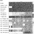
Figure 1 shows the chromosomal location of the gene LIF (leukemia inhibitory factor) - ENSG00000128342 on chromosome 22: 30,240,447-30,246,851 reverse strand and the location of the amplicon AMP2305 according to the invention in the genetic region of LIF. -
Figure 2 shows the analysis of CpG sites on amplicon No. 2305. The horizontal boxes in the table correspond to the CpG positions in the amplicon as analyzed (e.g.CpG -
Figure 3 shows SEQ ID NO: 5, with the amplicon AMP2305 according to the present invention indicated in bold, and the relevant CpG motifs underlined. - SEQ ID No. 1 shows the genomic sequence of amplicon AMP2305.
- SEQ ID No. 2 to SEQ ID No. 4 show the sequences of specific oligomers.
- SEQ ID No. 5 shows the genomic sequence of the region of interest, with the about 2200 bases upstream of amplicon AMP2305, and the about 2600 bases downstream.
- In order to identify TFH- cells (follicular helper T cells), qPCR was performed on bisulphite converted samples stemming from the human genomic region according to the following sequence (AMP2305, SEQ ID No. 1):

- The following primers and probe were used for the qPCR:
Forward Primer q2305_TFH_fw GTTTAATGTTATTGTTGATATTTTGT (SEQ ID No. 2) Reverse Primer q2305_TFH_rev ACATCCACATAACCCACA (SEQ ID No. 3) Probe q2305_TFH_P_nm TGTGTTGTGTTGTTTGTGTAGTAAGTATTAT (SEQ ID No. 4) - The cell type specificity (as measured by qPCR) was found as follows:
Immune blood cell type FACS characterization Tissue Percentage of TFH cells (%) TFH06 (control) CD3+, CD4+ CD45RA-, PD1++, CXCR5+ tonsil 95.03 activated T helper cells CD4+ CD69+, PD1-(TFH-) tonsil 85.39 activated cytotoxic T cells CD8+ CD69+, PD1-(TFH-) tonsil 15.84 pTFH01 (peri.TFH) Peripheral blood 94.79 pTFH02 (peri.TFH) Peripheral blood 74.91 pTFH03 (peri.TFH) Peripheral blood 73.82 pTFH04 (peri.TFH) Peripheral blood 73.95 pTFH05 (peri.TFH) Peripheral blood 91.81 pTFH06 (peri.TFH) Peripheral blood 90.23 Granulocytes CD15+ Peripheral blood 0.01 Total cytotoxic T-cells CD3+, CD8+ Peripheral blood 0.83 Total T helper cells CD3+, CD4+ Peripheral blood 23.41 NK cells CD3-, CD56+ Peripheral blood 0.53 monocytes CD14+ Peripheral blood 0.00 Naive CD4+ cells CD4+, CD45RA+, CCR7+ Periphery 0.12 Terminal differential effector memory CD4+ cells CD4+, CD45RA+, CCR7- Periphery 62.94 central memory CD4+ cells CD4+, CD45RA-, CCR7+ Periphery 26.65 effector memory CD4+ cells CD4+, CD45RA-, CCR7- Periphery 57.18 - In whole blood qPCR measurements, the following amount of TFH cells was determined/quantified. The samples are peripheral whole blood of an overall of 11 different (healthy) donors:
Sample Copy number TFH demethylated (LIF, SEQ ID No. 1) Copy number TFH methylated (LIF, SEQ ID No. 1) TFH cells (%) 1 351.0 15166.7 2.26 2 260.7 16300.0 1.57 3 209.0 6680.0 3.03 4 234.7 11400.0 2.02 5 645.3 16500.0 3.76 6 462.7 14133.3 3.17 7 520.0 13633.3 3.67 8 595.7 12800.0 4.45 9 401.0 16600.0 2.36 10 339.7 14066.7 2.36 11 249.3 14500.0 1.69 Mean value 2.78 - As similar analysis is made using the CpG positions as found within about 2200 bases upstream and/or within about 2600 bases downstream of amplicon AMP2305 (see SEQ ID No.5), whereby additional informative CpG positons are identified.
-
- <110> Epiontis GmbH
- <120> Epigenetic method for the identification of follicular T-helper-(TFH-) cells
- <130> E31776WO
- <150>
EP14196613.5
<151> 2014-12-05 - <160> 5
- <170> PatentIn version 3.5
- <210> 1
<211> 496
<212> DNA
<213> Homo sapiens - <400> 1

- <210> 2
<211> 26
<212> DNA
<213> Homo sapiens - <400> 2
gtttaatgtt attgttgata ttttgt 26 - <210> 3
<211> 18
<212> DNA
<213> Homo sapiens - <400> 3
acatccacat aacccaca 18 - <210> 4
<211> 31
<212> DNA
<213> homo sapiens - <400> 4
tgtgttgtgt tgtttgtgta gtaagtatta t 31 - <210> 5
<211> 5531
<212> DNA
<213> homo sapiens - <400> 5


Claims (11)
- A method for identifying follicular helper T cells in a sample, comprising analyzing the methylation status of at least one CpG position in the mammalian gene region for leukemia inhibitory factor (LIF), wherein said at least one CpG position is selected from CpG positions 1, 2, 3, 4, 5, 6, 7, 8, 9, 10, 11, 12, 13, 14, 15, 16, 17, 18, 19, 20, 21, 22, and 23 in the amplicon No. 2305 according to SEQ ID No. 1, wherein a demethylation of said gene region is indicative for a follicular helper T cell, when compared to a non-follicular helper T cell.
- The method according to claim 1, further comprising a method selected from a methylation specific enzymatic digest, bisulfite sequencing, CpG island methylation, MSP, HeavyMethyl, MethyLight, Ms-SNuPE, and other methods relying on a detection of amplified DNA.
- The method according to any of claims 1 or 2, further comprising a quantification of the relative amount of follicular helper T cells based on comparing relative amounts of said methylation frequency in the region as analyzed with relative amounts of the methylation frequency in a control gene, such as, for example, GAPDH.
- The method according to any of claims 1 to 3, wherein said sample is selected from a mammalian body fluid, including human blood samples, or a tissue, organ or cell type blood sample, a sample of blood lymphocytes or a fraction thereof
- The method according to any of claims 1 to 4, further comprising a distinguishing of said follicular helper T cells from all or at least one of the cell types selected from B-cells, cytotoxic T-cells, granulocytes, monocytes, NK-cells, and other T-helper cells.
- The method according to any of claims 1 to 5, wherein said method is performed without a step of purifying and/or enriching said cells to be identified, preferably using whole blood and/or non-trypsinized tissue.
- The method according to any of claims 1 to 6, further comprising the step of concluding on the immune status of said mammal based on said follicular helper T cells as identified.
- A method for monitoring the level of follicular helper T cells in a mammal, comprising performing the method according to any of claims 3 to 7, and comparing said relative amount of said cells as identified to a sample taken earlier or in parallel from the same mammal, and/or to a control sample.
- The method according to any of claims 1 to 8, further comprising measuring and/or monitoring the amount of said follicular helper T cells in response to chemical and/or biological substances that are provided to said mammal.
- The method according to any of claims 1 to 9, wherein said mammal suffers from or is likely to suffer from autoimmune diseases, transplant rejections, infection diseases, cancer, and/or allergy.
- Use of a kit comprising a) a bisulfite reagent, and b) materials for the analysis of the methylation status of CpG positions selected from CpG positions 1, 2, 3, 4, 5, 6, 7, 8, 9, 10, 11, 12, 13, 14, 15, 16, 17, 18, 19, 20, 21, 22, and 23 in the amplicon No. 2305 according to SEQ ID No. 1 for identifying, quantifying, and/or monitoring follicular T helper cells in a mammal.
Priority Applications (1)
| Application Number | Priority Date | Filing Date | Title |
|---|---|---|---|
| PL15805467T PL3227458T3 (en) | 2014-12-05 | 2015-12-04 | Epigenetic method for the identification of follicular t-helper-(tfh-) cells |
Applications Claiming Priority (2)
| Application Number | Priority Date | Filing Date | Title |
|---|---|---|---|
| EP14196613.5A EP3029150B1 (en) | 2014-12-05 | 2014-12-05 | Epigenetic method for the identification of follicular T-helper-(TFH-) cells |
| PCT/EP2015/078725 WO2016087663A1 (en) | 2014-12-05 | 2015-12-04 | Epigenetic method for the identification of follicular t-helper-(tfh-) cells |
Publications (2)
| Publication Number | Publication Date |
|---|---|
| EP3227458A1 EP3227458A1 (en) | 2017-10-11 |
| EP3227458B1 true EP3227458B1 (en) | 2019-11-20 |
Family
ID=52003680
Family Applications (2)
| Application Number | Title | Priority Date | Filing Date |
|---|---|---|---|
| EP14196613.5A Active EP3029150B1 (en) | 2014-12-05 | 2014-12-05 | Epigenetic method for the identification of follicular T-helper-(TFH-) cells |
| EP15805467.6A Active EP3227458B1 (en) | 2014-12-05 | 2015-12-04 | Epigenetic method for the identification of follicular t-helper-(tfh-) cells |
Family Applications Before (1)
| Application Number | Title | Priority Date | Filing Date |
|---|---|---|---|
| EP14196613.5A Active EP3029150B1 (en) | 2014-12-05 | 2014-12-05 | Epigenetic method for the identification of follicular T-helper-(TFH-) cells |
Country Status (9)
| Country | Link |
|---|---|
| US (1) | US11725243B2 (en) |
| EP (2) | EP3029150B1 (en) |
| JP (1) | JP6778190B2 (en) |
| CN (1) | CN107002143B (en) |
| CA (1) | CA2969370A1 (en) |
| DK (2) | DK3029150T3 (en) |
| ES (2) | ES2695179T3 (en) |
| PL (2) | PL3029150T3 (en) |
| WO (1) | WO2016087663A1 (en) |
Families Citing this family (2)
| Publication number | Priority date | Publication date | Assignee | Title |
|---|---|---|---|---|
| DE102017125019B4 (en) | 2017-10-25 | 2019-10-17 | Epiontis Gmbh | PDCD1 as an epigenetic marker for the identification of immune cells, in particular PD1 + cells |
| DE102018112644B4 (en) | 2018-05-25 | 2020-06-10 | Epiontis Gmbh | CXCR3 as an epigenetic marker for the identification of inflammatory immune cells, in particular CD8 + memory T cells |
Family Cites Families (12)
| Publication number | Priority date | Publication date | Assignee | Title |
|---|---|---|---|---|
| WO2002083162A1 (en) | 2001-04-13 | 2002-10-24 | University Of Chicago | Use of a cd8+ t cell inhibitory agent in the presence of a cd4+ t cell inhibitory agent for inhibition of transplant rejection |
| EP1961827A3 (en) * | 2004-07-18 | 2008-09-17 | Epigenomics AG | Epigenetic methods and nucleic acids for the detection of breast cell proliferative disorders |
| US20070243161A1 (en) * | 2006-02-28 | 2007-10-18 | Sven Olek | Epigenetic modification of the loci for CAMTA1 and/or FOXP3 as a marker for cancer treatment |
| PL2274430T3 (en) * | 2008-04-30 | 2017-09-29 | Sanbio Inc | Neural regenerating cells with alterations in dna methylation |
| EP2141245A1 (en) * | 2008-07-03 | 2010-01-06 | Ivana Türbachova | DNA methylation analysis of regulatory T cells through DNA-methylation analysis of the TSDR region of the gene foxP3 |
| US20110028333A1 (en) * | 2009-05-01 | 2011-02-03 | Brown University | Diagnosing, prognosing, and early detection of cancers by dna methylation profiling |
| JPWO2012063875A1 (en) * | 2010-11-11 | 2014-05-12 | シスメックス株式会社 | Marker for detecting human follicular helper T cell and method for detecting human follicular helper T cell |
| WO2012096851A1 (en) | 2011-01-12 | 2012-07-19 | Baylor Research Institute | Compositions and methods of alteration of autoimmune diseases |
| EP2788510B1 (en) * | 2012-03-12 | 2016-12-14 | Epiontis GmbH | Epigenetic marker for the identification of cd3cd4 positive t lymphocytes |
| PL3327141T3 (en) * | 2013-04-19 | 2020-07-27 | Epiontis Gmbh | Method for identifying the quantitative cellular composition in a biological sample |
| CN103421800B (en) * | 2013-09-05 | 2015-07-22 | 新乡医学院 | Recombinant gene, recombinant carrier and recombinant cell for treating spinal cord injuries |
| CN103487572B (en) * | 2013-10-08 | 2017-03-29 | 北京大学人民医院 | Application of the folliculus helper T cell (Tfh) in autoimmune disease state of an illness early warning |
-
2014
- 2014-12-05 DK DK14196613.5T patent/DK3029150T3/en active
- 2014-12-05 ES ES14196613T patent/ES2695179T3/en active Active
- 2014-12-05 EP EP14196613.5A patent/EP3029150B1/en active Active
- 2014-12-05 PL PL14196613T patent/PL3029150T3/en unknown
-
2015
- 2015-12-04 JP JP2017529294A patent/JP6778190B2/en not_active Expired - Fee Related
- 2015-12-04 ES ES15805467T patent/ES2770077T3/en active Active
- 2015-12-04 WO PCT/EP2015/078725 patent/WO2016087663A1/en not_active Ceased
- 2015-12-04 PL PL15805467T patent/PL3227458T3/en unknown
- 2015-12-04 EP EP15805467.6A patent/EP3227458B1/en active Active
- 2015-12-04 US US15/526,632 patent/US11725243B2/en active Active
- 2015-12-04 CN CN201580064819.7A patent/CN107002143B/en active Active
- 2015-12-04 DK DK15805467.6T patent/DK3227458T3/en active
- 2015-12-04 CA CA2969370A patent/CA2969370A1/en active Pending
Non-Patent Citations (1)
| Title |
|---|
| None * |
Also Published As
| Publication number | Publication date |
|---|---|
| PL3029150T3 (en) | 2019-03-29 |
| JP6778190B2 (en) | 2020-10-28 |
| CN107002143A (en) | 2017-08-01 |
| EP3029150B1 (en) | 2018-08-15 |
| CA2969370A1 (en) | 2016-06-09 |
| US11725243B2 (en) | 2023-08-15 |
| ES2695179T3 (en) | 2019-01-02 |
| ES2770077T3 (en) | 2020-06-30 |
| DK3227458T3 (en) | 2020-03-02 |
| EP3029150A1 (en) | 2016-06-08 |
| US20190100800A1 (en) | 2019-04-04 |
| EP3227458A1 (en) | 2017-10-11 |
| JP2017537623A (en) | 2017-12-21 |
| WO2016087663A1 (en) | 2016-06-09 |
| DK3029150T3 (en) | 2018-12-10 |
| CN107002143B (en) | 2021-03-30 |
| PL3227458T3 (en) | 2020-06-01 |
| HK1245343A1 (en) | 2018-08-24 |
Similar Documents
| Publication | Publication Date | Title |
|---|---|---|
| EP3353318B1 (en) | Lrp5 as epigenetic marker for the identification of immune cells, in particular b-cells | |
| EP3353319B1 (en) | Park2 as epigenetic marker for the identification of immune cells, in particular monocytes | |
| EP3353321B1 (en) | Mvd as epigenetic marker for the identification of cd56+ nk cells | |
| EP3669000B1 (en) | Endosialin (cd248) as epigenetic marker for the identification of immune cells, in particular naïve cd8+ t-cells | |
| EP3029150B1 (en) | Epigenetic method for the identification of follicular T-helper-(TFH-) cells | |
| EP3669001B1 (en) | Amplicon region as epigenetic marker for the identification of non-classical monocytes | |
| EP3669003B1 (en) | Pdcd1 as epigenetic marker for the identification of immune cells, in particular pd1+ cells | |
| EP3669005B1 (en) | Ergic1 as epigenetic marker for the identification of immune cells, in particular monocytic myeloid-derived suppressor cells (mmdscs) | |
| HK1245343B (en) | Epigenetic method for the identification of follicular t-helper-(tfh-) cells | |
| CA2999611C (en) | Lrp5 as epigenetic marker for the identification of immune cells, in particular b-cells | |
| CA3101383A1 (en) | Cxcr3 as epigenetic marker for the identification of inflammatory immune cells, in particular cd8+ memory t cells | |
| HK1257582B (en) | Park2 as epigenetic marker for the identification of immune cells, in particular monocytes |
Legal Events
| Date | Code | Title | Description |
|---|---|---|---|
| STAA | Information on the status of an ep patent application or granted ep patent |
Free format text: STATUS: THE INTERNATIONAL PUBLICATION HAS BEEN MADE |
|
| PUAI | Public reference made under article 153(3) epc to a published international application that has entered the european phase |
Free format text: ORIGINAL CODE: 0009012 |
|
| STAA | Information on the status of an ep patent application or granted ep patent |
Free format text: STATUS: REQUEST FOR EXAMINATION WAS MADE |
|
| 17P | Request for examination filed |
Effective date: 20170619 |
|
| AK | Designated contracting states |
Kind code of ref document: A1 Designated state(s): AL AT BE BG CH CY CZ DE DK EE ES FI FR GB GR HR HU IE IS IT LI LT LU LV MC MK MT NL NO PL PT RO RS SE SI SK SM TR |
|
| AX | Request for extension of the european patent |
Extension state: BA ME |
|
| DAV | Request for validation of the european patent (deleted) | ||
| DAX | Request for extension of the european patent (deleted) | ||
| STAA | Information on the status of an ep patent application or granted ep patent |
Free format text: STATUS: EXAMINATION IS IN PROGRESS |
|
| 17Q | First examination report despatched |
Effective date: 20180608 |
|
| REG | Reference to a national code |
Ref country code: HK Ref legal event code: DE Ref document number: 1245343 Country of ref document: HK |
|
| REG | Reference to a national code |
Ref country code: DE Ref legal event code: R079 Ref document number: 602015042189 Country of ref document: DE Free format text: PREVIOUS MAIN CLASS: C12Q0001680000 Ipc: C12Q0001688100 |
|
| GRAP | Despatch of communication of intention to grant a patent |
Free format text: ORIGINAL CODE: EPIDOSNIGR1 |
|
| STAA | Information on the status of an ep patent application or granted ep patent |
Free format text: STATUS: GRANT OF PATENT IS INTENDED |
|
| RIC1 | Information provided on ipc code assigned before grant |
Ipc: C12Q 1/6886 20180101ALI20190430BHEP Ipc: C12Q 1/6883 20180101ALI20190430BHEP Ipc: C12Q 1/6881 20180101AFI20190430BHEP |
|
| INTG | Intention to grant announced |
Effective date: 20190523 |
|
| RIN1 | Information on inventor provided before grant (corrected) |
Inventor name: HOFFMUELLER, ULRICH Inventor name: OLEK, SVEN |
|
| GRAS | Grant fee paid |
Free format text: ORIGINAL CODE: EPIDOSNIGR3 |
|
| GRAA | (expected) grant |
Free format text: ORIGINAL CODE: 0009210 |
|
| STAA | Information on the status of an ep patent application or granted ep patent |
Free format text: STATUS: THE PATENT HAS BEEN GRANTED |
|
| AK | Designated contracting states |
Kind code of ref document: B1 Designated state(s): AL AT BE BG CH CY CZ DE DK EE ES FI FR GB GR HR HU IE IS IT LI LT LU LV MC MK MT NL NO PL PT RO RS SE SI SK SM TR |
|
| RAP1 | Party data changed (applicant data changed or rights of an application transferred) |
Owner name: EPIONTIS GMBH |
|
| REG | Reference to a national code |
Ref country code: GB Ref legal event code: FG4D |
|
| REG | Reference to a national code |
Ref country code: CH Ref legal event code: EP |
|
| REG | Reference to a national code |
Ref country code: IE Ref legal event code: FG4D |
|
| REG | Reference to a national code |
Ref country code: DE Ref legal event code: R096 Ref document number: 602015042189 Country of ref document: DE |
|
| REG | Reference to a national code |
Ref country code: AT Ref legal event code: REF Ref document number: 1204250 Country of ref document: AT Kind code of ref document: T Effective date: 20191215 |
|
| REG | Reference to a national code |
Ref country code: CH Ref legal event code: NV Representative=s name: KATZAROV SA, CH |
|
| PGFP | Annual fee paid to national office [announced via postgrant information from national office to epo] |
Ref country code: LU Payment date: 20191125 Year of fee payment: 5 |
|
| PGFP | Annual fee paid to national office [announced via postgrant information from national office to epo] |
Ref country code: IE Payment date: 20191209 Year of fee payment: 5 Ref country code: FI Payment date: 20191209 Year of fee payment: 5 Ref country code: NL Payment date: 20191125 Year of fee payment: 5 |
|
| REG | Reference to a national code |
Ref country code: SE Ref legal event code: TRGR |
|
| REG | Reference to a national code |
Ref country code: FI Ref legal event code: FGE |
|
| PGFP | Annual fee paid to national office [announced via postgrant information from national office to epo] |
Ref country code: BE Payment date: 20191125 Year of fee payment: 5 Ref country code: DK Payment date: 20191210 Year of fee payment: 5 |
|
| REG | Reference to a national code |
Ref country code: DK Ref legal event code: T3 Effective date: 20200225 |
|
| REG | Reference to a national code |
Ref country code: NL Ref legal event code: FP |
|
| REG | Reference to a national code |
Ref country code: NO Ref legal event code: T2 Effective date: 20191120 |
|
| REG | Reference to a national code |
Ref country code: LT Ref legal event code: MG4D |
|
| PG25 | Lapsed in a contracting state [announced via postgrant information from national office to epo] |
Ref country code: BG Free format text: LAPSE BECAUSE OF FAILURE TO SUBMIT A TRANSLATION OF THE DESCRIPTION OR TO PAY THE FEE WITHIN THE PRESCRIBED TIME-LIMIT Effective date: 20200220 Ref country code: LV Free format text: LAPSE BECAUSE OF FAILURE TO SUBMIT A TRANSLATION OF THE DESCRIPTION OR TO PAY THE FEE WITHIN THE PRESCRIBED TIME-LIMIT Effective date: 20191120 Ref country code: GR Free format text: LAPSE BECAUSE OF FAILURE TO SUBMIT A TRANSLATION OF THE DESCRIPTION OR TO PAY THE FEE WITHIN THE PRESCRIBED TIME-LIMIT Effective date: 20200221 Ref country code: LT Free format text: LAPSE BECAUSE OF FAILURE TO SUBMIT A TRANSLATION OF THE DESCRIPTION OR TO PAY THE FEE WITHIN THE PRESCRIBED TIME-LIMIT Effective date: 20191120 |
|
| PGFP | Annual fee paid to national office [announced via postgrant information from national office to epo] |
Ref country code: ES Payment date: 20200102 Year of fee payment: 5 Ref country code: NO Payment date: 20191210 Year of fee payment: 5 Ref country code: IT Payment date: 20200221 Year of fee payment: 5 |
|
| PG25 | Lapsed in a contracting state [announced via postgrant information from national office to epo] |
Ref country code: RS Free format text: LAPSE BECAUSE OF FAILURE TO SUBMIT A TRANSLATION OF THE DESCRIPTION OR TO PAY THE FEE WITHIN THE PRESCRIBED TIME-LIMIT Effective date: 20191120 Ref country code: HR Free format text: LAPSE BECAUSE OF FAILURE TO SUBMIT A TRANSLATION OF THE DESCRIPTION OR TO PAY THE FEE WITHIN THE PRESCRIBED TIME-LIMIT Effective date: 20191120 |
|
| PGFP | Annual fee paid to national office [announced via postgrant information from national office to epo] |
Ref country code: IS Payment date: 20200115 Year of fee payment: 5 |
|
| PG25 | Lapsed in a contracting state [announced via postgrant information from national office to epo] |
Ref country code: AL Free format text: LAPSE BECAUSE OF FAILURE TO SUBMIT A TRANSLATION OF THE DESCRIPTION OR TO PAY THE FEE WITHIN THE PRESCRIBED TIME-LIMIT Effective date: 20191120 |
|
| REG | Reference to a national code |
Ref country code: ES Ref legal event code: FG2A Ref document number: 2770077 Country of ref document: ES Kind code of ref document: T3 Effective date: 20200630 |
|
| PG25 | Lapsed in a contracting state [announced via postgrant information from national office to epo] |
Ref country code: EE Free format text: LAPSE BECAUSE OF FAILURE TO SUBMIT A TRANSLATION OF THE DESCRIPTION OR TO PAY THE FEE WITHIN THE PRESCRIBED TIME-LIMIT Effective date: 20191120 Ref country code: PT Free format text: LAPSE BECAUSE OF FAILURE TO SUBMIT A TRANSLATION OF THE DESCRIPTION OR TO PAY THE FEE WITHIN THE PRESCRIBED TIME-LIMIT Effective date: 20200412 Ref country code: RO Free format text: LAPSE BECAUSE OF FAILURE TO SUBMIT A TRANSLATION OF THE DESCRIPTION OR TO PAY THE FEE WITHIN THE PRESCRIBED TIME-LIMIT Effective date: 20191120 |
|
| PGFP | Annual fee paid to national office [announced via postgrant information from national office to epo] |
Ref country code: CZ Payment date: 20191127 Year of fee payment: 5 |
|
| REG | Reference to a national code |
Ref country code: DE Ref legal event code: R097 Ref document number: 602015042189 Country of ref document: DE |
|
| PG25 | Lapsed in a contracting state [announced via postgrant information from national office to epo] |
Ref country code: MC Free format text: LAPSE BECAUSE OF FAILURE TO SUBMIT A TRANSLATION OF THE DESCRIPTION OR TO PAY THE FEE WITHIN THE PRESCRIBED TIME-LIMIT Effective date: 20191120 Ref country code: SK Free format text: LAPSE BECAUSE OF FAILURE TO SUBMIT A TRANSLATION OF THE DESCRIPTION OR TO PAY THE FEE WITHIN THE PRESCRIBED TIME-LIMIT Effective date: 20191120 Ref country code: SM Free format text: LAPSE BECAUSE OF FAILURE TO SUBMIT A TRANSLATION OF THE DESCRIPTION OR TO PAY THE FEE WITHIN THE PRESCRIBED TIME-LIMIT Effective date: 20191120 |
|
| PGFP | Annual fee paid to national office [announced via postgrant information from national office to epo] |
Ref country code: PL Payment date: 20200117 Year of fee payment: 5 Ref country code: SE Payment date: 20191210 Year of fee payment: 5 |
|
| PLBE | No opposition filed within time limit |
Free format text: ORIGINAL CODE: 0009261 |
|
| STAA | Information on the status of an ep patent application or granted ep patent |
Free format text: STATUS: NO OPPOSITION FILED WITHIN TIME LIMIT |
|
| 26N | No opposition filed |
Effective date: 20200821 |
|
| PG25 | Lapsed in a contracting state [announced via postgrant information from national office to epo] |
Ref country code: SI Free format text: LAPSE BECAUSE OF FAILURE TO SUBMIT A TRANSLATION OF THE DESCRIPTION OR TO PAY THE FEE WITHIN THE PRESCRIBED TIME-LIMIT Effective date: 20191120 |
|
| PG25 | Lapsed in a contracting state [announced via postgrant information from national office to epo] |
Ref country code: CY Free format text: LAPSE BECAUSE OF FAILURE TO SUBMIT A TRANSLATION OF THE DESCRIPTION OR TO PAY THE FEE WITHIN THE PRESCRIBED TIME-LIMIT Effective date: 20191120 |
|
| REG | Reference to a national code |
Ref country code: FI Ref legal event code: MAE |
|
| REG | Reference to a national code |
Ref country code: DK Ref legal event code: EBP Effective date: 20201231 |
|
| REG | Reference to a national code |
Ref country code: NO Ref legal event code: MMEP |
|
| PG25 | Lapsed in a contracting state [announced via postgrant information from national office to epo] |
Ref country code: FI Free format text: LAPSE BECAUSE OF NON-PAYMENT OF DUE FEES Effective date: 20201204 Ref country code: CZ Free format text: LAPSE BECAUSE OF NON-PAYMENT OF DUE FEES Effective date: 20201204 Ref country code: MT Free format text: LAPSE BECAUSE OF FAILURE TO SUBMIT A TRANSLATION OF THE DESCRIPTION OR TO PAY THE FEE WITHIN THE PRESCRIBED TIME-LIMIT Effective date: 20191120 Ref country code: HU Free format text: LAPSE BECAUSE OF FAILURE TO SUBMIT A TRANSLATION OF THE DESCRIPTION OR TO PAY THE FEE WITHIN THE PRESCRIBED TIME-LIMIT; INVALID AB INITIO Effective date: 20151204 |
|
| REG | Reference to a national code |
Ref country code: SE Ref legal event code: EUG |
|
| REG | Reference to a national code |
Ref country code: NL Ref legal event code: MM Effective date: 20210101 |
|
| REG | Reference to a national code |
Ref country code: BE Ref legal event code: MM Effective date: 20201231 |
|
| PG25 | Lapsed in a contracting state [announced via postgrant information from national office to epo] |
Ref country code: NL Free format text: LAPSE BECAUSE OF NON-PAYMENT OF DUE FEES Effective date: 20210101 |
|
| PG25 | Lapsed in a contracting state [announced via postgrant information from national office to epo] |
Ref country code: IE Free format text: LAPSE BECAUSE OF NON-PAYMENT OF DUE FEES Effective date: 20201204 Ref country code: IT Free format text: LAPSE BECAUSE OF NON-PAYMENT OF DUE FEES Effective date: 20201204 Ref country code: LU Free format text: LAPSE BECAUSE OF NON-PAYMENT OF DUE FEES Effective date: 20201204 |
|
| PG25 | Lapsed in a contracting state [announced via postgrant information from national office to epo] |
Ref country code: SE Free format text: LAPSE BECAUSE OF NON-PAYMENT OF DUE FEES Effective date: 20201205 Ref country code: NO Free format text: LAPSE BECAUSE OF NON-PAYMENT OF DUE FEES Effective date: 20201231 |
|
| PG25 | Lapsed in a contracting state [announced via postgrant information from national office to epo] |
Ref country code: DK Free format text: LAPSE BECAUSE OF NON-PAYMENT OF DUE FEES Effective date: 20201231 |
|
| REG | Reference to a national code |
Ref country code: ES Ref legal event code: FD2A Effective date: 20220207 |
|
| REG | Reference to a national code |
Ref country code: AT Ref legal event code: MM01 Ref document number: 1204250 Country of ref document: AT Kind code of ref document: T Effective date: 20201204 |
|
| REG | Reference to a national code |
Ref country code: AT Ref legal event code: UEP Ref document number: 1204250 Country of ref document: AT Kind code of ref document: T Effective date: 20191120 |
|
| PG25 | Lapsed in a contracting state [announced via postgrant information from national office to epo] |
Ref country code: AT Free format text: LAPSE BECAUSE OF NON-PAYMENT OF DUE FEES Effective date: 20201204 |
|
| PG25 | Lapsed in a contracting state [announced via postgrant information from national office to epo] |
Ref country code: TR Free format text: LAPSE BECAUSE OF FAILURE TO SUBMIT A TRANSLATION OF THE DESCRIPTION OR TO PAY THE FEE WITHIN THE PRESCRIBED TIME-LIMIT Effective date: 20191120 Ref country code: ES Free format text: LAPSE BECAUSE OF NON-PAYMENT OF DUE FEES Effective date: 20201205 |
|
| PG25 | Lapsed in a contracting state [announced via postgrant information from national office to epo] |
Ref country code: MK Free format text: LAPSE BECAUSE OF FAILURE TO SUBMIT A TRANSLATION OF THE DESCRIPTION OR TO PAY THE FEE WITHIN THE PRESCRIBED TIME-LIMIT Effective date: 20191120 |
|
| PG25 | Lapsed in a contracting state [announced via postgrant information from national office to epo] |
Ref country code: BE Free format text: LAPSE BECAUSE OF NON-PAYMENT OF DUE FEES Effective date: 20201231 |
|
| REG | Reference to a national code |
Ref country code: DE Ref legal event code: R082 Ref document number: 602015042189 Country of ref document: DE Representative=s name: KRAUSS, JAN, DR., DE |
|
| PG25 | Lapsed in a contracting state [announced via postgrant information from national office to epo] |
Ref country code: PL Free format text: LAPSE BECAUSE OF NON-PAYMENT OF DUE FEES Effective date: 20201204 |
|
| P01 | Opt-out of the competence of the unified patent court (upc) registered |
Effective date: 20230525 |
|
| PG25 | Lapsed in a contracting state [announced via postgrant information from national office to epo] |
Ref country code: IS Free format text: LAPSE BECAUSE OF NON-PAYMENT OF DUE FEES Effective date: 20210628 |
|
| REG | Reference to a national code |
Ref country code: CH Ref legal event code: U11 Free format text: ST27 STATUS EVENT CODE: U-0-0-U10-U11 (AS PROVIDED BY THE NATIONAL OFFICE) Effective date: 20260101 |
|
| PGFP | Annual fee paid to national office [announced via postgrant information from national office to epo] |
Ref country code: GB Payment date: 20251218 Year of fee payment: 11 |
|
| PGFP | Annual fee paid to national office [announced via postgrant information from national office to epo] |
Ref country code: FR Payment date: 20251217 Year of fee payment: 11 |
|
| PGFP | Annual fee paid to national office [announced via postgrant information from national office to epo] |
Ref country code: DE Payment date: 20251222 Year of fee payment: 11 |
|
| PGFP | Annual fee paid to national office [announced via postgrant information from national office to epo] |
Ref country code: CH Payment date: 20260101 Year of fee payment: 11 |