EP3226923B1 - Gel de cartilage pour la réparation cartilagineuse, comprenant du chitosane et des chondrocytes - Google Patents
Gel de cartilage pour la réparation cartilagineuse, comprenant du chitosane et des chondrocytes Download PDFInfo
- Publication number
- EP3226923B1 EP3226923B1 EP15817457.3A EP15817457A EP3226923B1 EP 3226923 B1 EP3226923 B1 EP 3226923B1 EP 15817457 A EP15817457 A EP 15817457A EP 3226923 B1 EP3226923 B1 EP 3226923B1
- Authority
- EP
- European Patent Office
- Prior art keywords
- cells
- chitosan
- chondrocytes
- hydrogel
- particles
- Prior art date
- Legal status (The legal status is an assumption and is not a legal conclusion. Google has not performed a legal analysis and makes no representation as to the accuracy of the status listed.)
- Active
Links
Images


Classifications
-
- C—CHEMISTRY; METALLURGY
- C12—BIOCHEMISTRY; BEER; SPIRITS; WINE; VINEGAR; MICROBIOLOGY; ENZYMOLOGY; MUTATION OR GENETIC ENGINEERING
- C12N—MICROORGANISMS OR ENZYMES; COMPOSITIONS THEREOF; PROPAGATING, PRESERVING, OR MAINTAINING MICROORGANISMS; MUTATION OR GENETIC ENGINEERING; CULTURE MEDIA
- C12N5/00—Undifferentiated human, animal or plant cells, e.g. cell lines; Tissues; Cultivation or maintenance thereof; Culture media therefor
- C12N5/06—Animal cells or tissues; Human cells or tissues
- C12N5/0602—Vertebrate cells
- C12N5/0652—Cells of skeletal and connective tissues; Mesenchyme
- C12N5/0655—Chondrocytes; Cartilage
-
- A—HUMAN NECESSITIES
- A61—MEDICAL OR VETERINARY SCIENCE; HYGIENE
- A61L—METHODS OR APPARATUS FOR STERILISING MATERIALS OR OBJECTS IN GENERAL; DISINFECTION, STERILISATION OR DEODORISATION OF AIR; CHEMICAL ASPECTS OF BANDAGES, DRESSINGS, ABSORBENT PADS OR SURGICAL ARTICLES; MATERIALS FOR BANDAGES, DRESSINGS, ABSORBENT PADS OR SURGICAL ARTICLES
- A61L27/00—Materials for grafts or prostheses or for coating grafts or prostheses
- A61L27/14—Macromolecular materials
- A61L27/20—Polysaccharides
-
- A—HUMAN NECESSITIES
- A61—MEDICAL OR VETERINARY SCIENCE; HYGIENE
- A61K—PREPARATIONS FOR MEDICAL, DENTAL OR TOILETRY PURPOSES
- A61K35/00—Medicinal preparations containing materials or reaction products thereof with undetermined constitution
- A61K35/12—Materials from mammals; Compositions comprising non-specified tissues or cells; Compositions comprising non-embryonic stem cells; Genetically modified cells
- A61K35/32—Bones; Osteocytes; Osteoblasts; Tendons; Tenocytes; Teeth; Odontoblasts; Cartilage; Chondrocytes; Synovial membrane
-
- A—HUMAN NECESSITIES
- A61—MEDICAL OR VETERINARY SCIENCE; HYGIENE
- A61L—METHODS OR APPARATUS FOR STERILISING MATERIALS OR OBJECTS IN GENERAL; DISINFECTION, STERILISATION OR DEODORISATION OF AIR; CHEMICAL ASPECTS OF BANDAGES, DRESSINGS, ABSORBENT PADS OR SURGICAL ARTICLES; MATERIALS FOR BANDAGES, DRESSINGS, ABSORBENT PADS OR SURGICAL ARTICLES
- A61L27/00—Materials for grafts or prostheses or for coating grafts or prostheses
- A61L27/14—Macromolecular materials
- A61L27/22—Polypeptides or derivatives thereof, e.g. degradation products
- A61L27/225—Fibrin; Fibrinogen
-
- A—HUMAN NECESSITIES
- A61—MEDICAL OR VETERINARY SCIENCE; HYGIENE
- A61L—METHODS OR APPARATUS FOR STERILISING MATERIALS OR OBJECTS IN GENERAL; DISINFECTION, STERILISATION OR DEODORISATION OF AIR; CHEMICAL ASPECTS OF BANDAGES, DRESSINGS, ABSORBENT PADS OR SURGICAL ARTICLES; MATERIALS FOR BANDAGES, DRESSINGS, ABSORBENT PADS OR SURGICAL ARTICLES
- A61L27/00—Materials for grafts or prostheses or for coating grafts or prostheses
- A61L27/36—Materials for grafts or prostheses or for coating grafts or prostheses containing ingredients of undetermined constitution or reaction products thereof, e.g. transplant tissue, natural bone, extracellular matrix
- A61L27/3604—Materials for grafts or prostheses or for coating grafts or prostheses containing ingredients of undetermined constitution or reaction products thereof, e.g. transplant tissue, natural bone, extracellular matrix characterised by the human or animal origin of the biological material, e.g. hair, fascia, fish scales, silk, shellac, pericardium, pleura, renal tissue, amniotic membrane, parenchymal tissue, fetal tissue, muscle tissue, fat tissue, enamel
- A61L27/3612—Cartilage, synovial fluid
-
- A—HUMAN NECESSITIES
- A61—MEDICAL OR VETERINARY SCIENCE; HYGIENE
- A61L—METHODS OR APPARATUS FOR STERILISING MATERIALS OR OBJECTS IN GENERAL; DISINFECTION, STERILISATION OR DEODORISATION OF AIR; CHEMICAL ASPECTS OF BANDAGES, DRESSINGS, ABSORBENT PADS OR SURGICAL ARTICLES; MATERIALS FOR BANDAGES, DRESSINGS, ABSORBENT PADS OR SURGICAL ARTICLES
- A61L27/00—Materials for grafts or prostheses or for coating grafts or prostheses
- A61L27/36—Materials for grafts or prostheses or for coating grafts or prostheses containing ingredients of undetermined constitution or reaction products thereof, e.g. transplant tissue, natural bone, extracellular matrix
- A61L27/3641—Materials for grafts or prostheses or for coating grafts or prostheses containing ingredients of undetermined constitution or reaction products thereof, e.g. transplant tissue, natural bone, extracellular matrix characterised by the site of application in the body
- A61L27/3645—Connective tissue
- A61L27/3654—Cartilage, e.g. meniscus
-
- A—HUMAN NECESSITIES
- A61—MEDICAL OR VETERINARY SCIENCE; HYGIENE
- A61L—METHODS OR APPARATUS FOR STERILISING MATERIALS OR OBJECTS IN GENERAL; DISINFECTION, STERILISATION OR DEODORISATION OF AIR; CHEMICAL ASPECTS OF BANDAGES, DRESSINGS, ABSORBENT PADS OR SURGICAL ARTICLES; MATERIALS FOR BANDAGES, DRESSINGS, ABSORBENT PADS OR SURGICAL ARTICLES
- A61L27/00—Materials for grafts or prostheses or for coating grafts or prostheses
- A61L27/36—Materials for grafts or prostheses or for coating grafts or prostheses containing ingredients of undetermined constitution or reaction products thereof, e.g. transplant tissue, natural bone, extracellular matrix
- A61L27/38—Materials for grafts or prostheses or for coating grafts or prostheses containing ingredients of undetermined constitution or reaction products thereof, e.g. transplant tissue, natural bone, extracellular matrix containing added animal cells
- A61L27/3804—Materials for grafts or prostheses or for coating grafts or prostheses containing ingredients of undetermined constitution or reaction products thereof, e.g. transplant tissue, natural bone, extracellular matrix containing added animal cells characterised by specific cells or progenitors thereof, e.g. fibroblasts, connective tissue cells, kidney cells
- A61L27/3817—Cartilage-forming cells, e.g. pre-chondrocytes
-
- A—HUMAN NECESSITIES
- A61—MEDICAL OR VETERINARY SCIENCE; HYGIENE
- A61L—METHODS OR APPARATUS FOR STERILISING MATERIALS OR OBJECTS IN GENERAL; DISINFECTION, STERILISATION OR DEODORISATION OF AIR; CHEMICAL ASPECTS OF BANDAGES, DRESSINGS, ABSORBENT PADS OR SURGICAL ARTICLES; MATERIALS FOR BANDAGES, DRESSINGS, ABSORBENT PADS OR SURGICAL ARTICLES
- A61L27/00—Materials for grafts or prostheses or for coating grafts or prostheses
- A61L27/36—Materials for grafts or prostheses or for coating grafts or prostheses containing ingredients of undetermined constitution or reaction products thereof, e.g. transplant tissue, natural bone, extracellular matrix
- A61L27/38—Materials for grafts or prostheses or for coating grafts or prostheses containing ingredients of undetermined constitution or reaction products thereof, e.g. transplant tissue, natural bone, extracellular matrix containing added animal cells
- A61L27/3895—Materials for grafts or prostheses or for coating grafts or prostheses containing ingredients of undetermined constitution or reaction products thereof, e.g. transplant tissue, natural bone, extracellular matrix containing added animal cells using specific culture conditions, e.g. stimulating differentiation of stem cells, pulsatile flow conditions
-
- A—HUMAN NECESSITIES
- A61—MEDICAL OR VETERINARY SCIENCE; HYGIENE
- A61P—SPECIFIC THERAPEUTIC ACTIVITY OF CHEMICAL COMPOUNDS OR MEDICINAL PREPARATIONS
- A61P19/00—Drugs for skeletal disorders
-
- A—HUMAN NECESSITIES
- A61—MEDICAL OR VETERINARY SCIENCE; HYGIENE
- A61L—METHODS OR APPARATUS FOR STERILISING MATERIALS OR OBJECTS IN GENERAL; DISINFECTION, STERILISATION OR DEODORISATION OF AIR; CHEMICAL ASPECTS OF BANDAGES, DRESSINGS, ABSORBENT PADS OR SURGICAL ARTICLES; MATERIALS FOR BANDAGES, DRESSINGS, ABSORBENT PADS OR SURGICAL ARTICLES
- A61L24/00—Surgical adhesives or cements; Adhesives for colostomy devices
-
- A—HUMAN NECESSITIES
- A61—MEDICAL OR VETERINARY SCIENCE; HYGIENE
- A61L—METHODS OR APPARATUS FOR STERILISING MATERIALS OR OBJECTS IN GENERAL; DISINFECTION, STERILISATION OR DEODORISATION OF AIR; CHEMICAL ASPECTS OF BANDAGES, DRESSINGS, ABSORBENT PADS OR SURGICAL ARTICLES; MATERIALS FOR BANDAGES, DRESSINGS, ABSORBENT PADS OR SURGICAL ARTICLES
- A61L2430/00—Materials or treatment for tissue regeneration
- A61L2430/06—Materials or treatment for tissue regeneration for cartilage reconstruction, e.g. meniscus
-
- C—CHEMISTRY; METALLURGY
- C12—BIOCHEMISTRY; BEER; SPIRITS; WINE; VINEGAR; MICROBIOLOGY; ENZYMOLOGY; MUTATION OR GENETIC ENGINEERING
- C12N—MICROORGANISMS OR ENZYMES; COMPOSITIONS THEREOF; PROPAGATING, PRESERVING, OR MAINTAINING MICROORGANISMS; MUTATION OR GENETIC ENGINEERING; CULTURE MEDIA
- C12N2533/00—Supports or coatings for cell culture, characterised by material
- C12N2533/50—Proteins
- C12N2533/56—Fibrin; Thrombin
-
- C—CHEMISTRY; METALLURGY
- C12—BIOCHEMISTRY; BEER; SPIRITS; WINE; VINEGAR; MICROBIOLOGY; ENZYMOLOGY; MUTATION OR GENETIC ENGINEERING
- C12N—MICROORGANISMS OR ENZYMES; COMPOSITIONS THEREOF; PROPAGATING, PRESERVING, OR MAINTAINING MICROORGANISMS; MUTATION OR GENETIC ENGINEERING; CULTURE MEDIA
- C12N2533/00—Supports or coatings for cell culture, characterised by material
- C12N2533/90—Substrates of biological origin, e.g. extracellular matrix, decellularised tissue
Definitions
- the present invention relates to compositions making it possible in particular to reconstruct cartilage, and to a process making it possible to obtain such compositions. More particularly, the present application relates to an environment or structure extremely favorable not only to the proliferation of cells capable of forming hyaline cartilage, but also to the synthesis of extracellular cartilage matrix by these cells; the cells in this environment or structure constitute an implantable composition which can be grafted at the level of cartilage lesions in humans or in animals. The structure also constitutes a favorable environment during installation.
- Cartilage, or cartilage tissue is made up of specific cells, namely chondrocytes, distributed within an extracellular matrix, comprising at least 80% water. Chondrocytes are able to synthesize or degrade the components of the extracellular matrix of cartilage, composed of glycosaminoglycans and collagen fibers, mainly type II for hyaline cartilage. The chondrocytes are therefore responsible not only for the synthesis but also for the maintenance of the cartilage tissue.
- Cartilage, and particularly articular cartilage in adulthood has a very low self-repair capacity, due in particular to its avascular nature and the fact that mature chondrocytes do not proliferate.
- Cartilage damage is therefore essentially irreversible and therefore constitutes a major cause of pain and disability, in particular following trauma, mechanical wear or a degenerative joint disease such as osteoarthritis.
- chondrocytes a major difficulty in the transplant of chondrocytes, and more particularly of autologous chondrocytes, lies in the number of cells to be implanted.
- a reduced number of chondrocytes can generally be taken from the native cartilage of the patient who is to be treated; it is therefore necessary to carry out a first stage of expansion of the chondrocytes taken in order to drastically increase their number.
- the in vitro expansion of chondrocytes until a sufficient number of cells is obtained with a view to implantation proves to be very delicate.
- chondrocytes dedifferentiate themselves and lose their chondrocytic phenotype, then expressing type I collagen rather than type II collagen, so that once reimplanted, they do not lead to the formation from a tissue with satisfactory properties, but rather to a scar-style tissue or, in the context of cartilage repair, to the formation of a non-functional fibrocartilage, essentially composed of type I collagen.
- Some authors have proposed culture media making it possible to redifferentiate the chondrocytes which are dedifferentiated during the stage of proliferation in monolayers.
- Seeding in a 3D structure can also offer the advantage of facilitating the in vivo implantation of chondrocytes, of limiting cell leaks and of treating large lesions.
- Many materials have been tested for the seeding of chondrocytes in 3D structure; there may be mentioned alginate, collagen, polyethylene glycol and polylactic acid. The most suitable are those which meet the following biological properties: cytocompatibility, low immunogenicity, and biodegradability with non-toxic degradation products.
- natural polymers which exhibit these biological properties, certainly by virtue of their chemical and biological similarities with living tissues, present themselves as prime candidates. Natural polymers are highly biocompatible, easily bioresorbable and bioassimilable by the body.
- chitosan A natural polymer often cited for its attractive properties as a basic biomaterial of three-dimensional structures is chitosan. Indeed, according to the literature, this biopolymer is known to be biodegradable, biocompatible, non-toxic, hemocompatible, cytocompatible, and in addition, bioactive, hemostatic, healing, bacteriostatic and fungistatic. In addition, chitosan is known to promote cell adhesion. It is also appreciated for its ability to maintain the chondrocyte phenotype and stimulate the process of extracellular matrix synthesis during the culture of chondrocytes. In addition, no infection or allergy linked to chitosan has been reported, so it is not immunogenic.
- Chitosan As for its resorption time, it is possible to vary it by playing on its physicochemical properties. Chitosan is relatively easy to use and can be produced in various physical forms including solutions, films (Lahiji, et al, 2000), fibers, sponges, beads, hydrogels or microparticles. . It has therefore been used in extremely varied structures. In the context of the inoculation of chondrocytes in 3D structure, particular attention is paid to structures allowing both good distribution and good maintenance of chondrocytes as well as an environment favorable to the stimulation of their chondrogenic potential. To this end, different approaches have been considered.
- compositions from a polymer crosslinked in situ and allowing the production of a tissue which can be grafted in the form of a plug (Hao et al, 2010). These methods can nevertheless present difficulties during implantation.
- some authors have tested the hydrogel form and more particularly the physical hydrogel, synthesized without adding a crosslinking agent and thus promoting the biocompatibility and the bioresorability of the structure. It was therefore considered the development of a neo-tissue by bringing it into contact with a physical hydrogel of chitosan (Montembault et al).
- the step of prior multiplication in monolayers is deliberately rapid in order to avoid or limit the dedifferentiation of the chondrocytes before inoculation, consequently involving a relatively low multiplication of the cells.
- the three-dimensional structure is largely reabsorbed during the in vitro step of synthesizing the extracellular matrix, said structure no longer forming part of the graft (see in particular WO02078760 , Laboratoires Genevrier et al).
- Such a process is particularly long to implement, from 4 to 6 weeks, before obtaining a neo-tissue capable of being grafted, and said neo-tissue is no longer supported by the 3D structure at the time of implantation. .
- hyaluronic acid Another polymer which has been frequently tested in the field of chondrocyte implantation is hyaluronic acid. It is a natural polysaccharide, also biocompatible and biodegradable. In addition, it is a major component of the liquid synovial membrane and glycosaminoglycans (GAGs) present in articular cartilage.
- GAGs glycosaminoglycans
- Hyaluronic acid helps protect joints by increasing the viscosity of synovial fluid and making cartilage more elastic. It has also been shown that hyaluronic acid promotes the expression of the chondrocytic phenotype. As a result, hyaluronic acid has sometimes been combined with chitosan compositions in the culture of chondrocytes.
- chitosan sponges alone are to be preferred, compared to chitosan sponges complexed with GAGs such as hyaluronic acid, for the proliferation of chondrocytes and for other associated biological properties.
- GAGs such as hyaluronic acid
- a hydrogel of chitosan and photo-crosslinked hyaluronic acid has also been proposed in which chondrocytes are encapsulated (Park et al, 2013). However, such an encapsulation does not sufficiently promote proliferation despite relatively long culture times (proliferation factor 4 in 3 weeks).
- chitosan seems to have, according to the literature, interesting properties for the reconstruction of cartilage, it should be noted that the rate of proliferation and synthesis of extracellular matrix of chondrocytes on sponges of chitosan alone strongly depends on the size of the pores of the structure (Griffon et al, 2006). Some authors have even considered that chitosan was not suitable for the proliferation of chondrocytes (Suh et al, 2000).
- the present inventors have developed a method for obtaining chondrocytes by sufficient number, within a structure ensuring at the same time their perfect multiplication, their re-differentiation and the production of cartilaginous matrix.
- This structure is also suitable for implantation at the lesion level.
- the present inventors have found, quite unexpectedly, that chitosan, in the form of physical hydrogel particles, is an excellent environment for chondrocytes, not only for the synthesis of extracellular matrix of hyaline cartilage, but also with a view to their multiplication.
- the inventors were able to carry out, in the same chitosan structure, a proliferation step making it possible to obtain a proliferation rate similar or even greater than that observed by the ordinary technique of monolayers (on plastic or in sponges. ) and then easily allowing re-differentiation of the cells.
- the inventors have consequently developed a process which makes it possible successively to multiply chondrocytes, directly after extraction in the primary state, then to induce re-differentiation and the synthesis of specific extracellular matrix, within the same structure. , thus avoiding the steps of trypsination and structural change.
- this structure is compatible with in vivo implantation without requiring additional modification.
- chondrocytes distributed homogeneously within the structure and with a density allowing their reimplantation, in a reduced time compared to what is described up to 'to date, under conditions very favorable to the repair of cartilage tissue, in a structure directly compatible with implantation.
- chitosan is meant a polysaccharide composed of D-glucosamine units (deacetylated unit) and N-acetyl-D-glucosamine (acetylated unit) linked in ⁇ - (1-4). It can be produced by chemical or enzymatic deacetylation of chitin; it also exists naturally. In naturally occurring chitosan, the polysaccharide is usually associated with negligible amounts of beta-glucan. In the context of the present invention, the natural presence of beta-glucan, in amounts consistent with those found naturally in chitosan, is without influence.
- chitosan will be said to be “pure” even in the event of the presence of beta-glucan, as long as the presence of the latter remains less than 5% by mass, relative to the polysaccharide.
- chitosan derivative denotes any chitosan polymer having undergone a reaction aimed at modifying the chemical groups of the chitosan in order to change its functionalities, for example methylation, halogenation, etc.
- a chitosan derivative does not have more than three types of lead, preferably only two types of lead, and more preferably only one type of lead.
- Chitosan derivatives particularly considered in the context of the present invention are glycol-chitosan, N-succinyl chitosan, N- or O-Carboxymethyl Chitosane, without this list being limiting.
- the degree of acetylation (DA) is the percentage of acetylated units relative to the number of total units (acetylated and deacetylated units), it can be determined by Fourier transform infrared spectrometry (IRTF) or by proton NMR.
- IRTF Fourier transform infrared spectrometry
- chitosan hydrogel is understood to mean that the very majority constituent, therefore more than 80% or even more than 90%, or even 95%, of the hydrogel (by mass) is chitosan, apart from water.
- a chitosan derivative hydrogel is understood to mean a hydrogel in which the very majority constituent, at more than 80%, or even more than 90% or 95% of the hydrogel, is the chitosan derivative, apart from the water.
- a hydrogel of chitosan or of a derivative of chitosan means a hydrogel preferably comprising at least 70% water, or even at least 80% water.
- physical hydrogel is meant a hydrogel obtained by a gelling process in an aqueous medium or in a hydroalcoholic medium, not requiring the addition of a crosslinking agent.
- chondrocytic phenotype denotes cells of the chondrocyte type, preferentially expressing type II collagen over type I collagen.
- hyaluronic acid is meant a polysaccharide composed of units of D-glucuronic acid and of DN-acetylglucosamine, linked together by glycosidic bonds.
- hyaluronic acid derivative denotes any hyaluronic acid polymer which has undergone a reaction aimed at modifying the chemical groups of hyaluronic acid in order to change its functionalities, for example by esterification.
- the present invention therefore relates, according to a first aspect, to an in vitro process for obtaining a composition which can be implanted by arthroscopy for cartilage repair, comprising particles or fragments of physical hydrogel of chitosan or of a chitosan derivative. , and cells forming cartilage, preferably hyaline cartilage.
- the composition obtained at the end of the implementation of the method can be qualified as a cartilage gel.
- a cartilage gel in fact comprises cells, synthesizing the cartilage matrix, the whole distributed homogeneously without cell gradient, in a three-dimensional structure based on chitosan particles.
- Such a method according to the invention comprises in particular a step of amplifying primary cells, in a three-dimensional structure (3D structure) then a step of re-differentiation and induction of the synthesis of extracellular matrix, within this same structure.
- three-dimensional that is to say without changing the environment cells.
- chitosan is in fact a biocompatible, bioresorbable biomaterial with non-toxic, non-immunogenic, cytocompatible and bioactive degradation products. It is furthermore fully compatible with pharmaceutical requirements as an implantable device.
- the hydrogel of chitosan or of chitosan derivative is a physical hydrogel, obtained without adding a crosslinking agent.
- the cells that are amplified, in the first step, are seeded into this 3D structure.
- They can be either chondrocytes or precursor cells of chondrocytes obtained from stem cells, for example mesenchymal stem cells, or induced pluripotent cells (IPS).
- IPS induced pluripotent cells
- They can be any cells differentiated into chondrocytes.
- they are chondrocytes, and more preferably articular chondrocytes.
- the chondrocytes or the stem cells can be obtained by any method known to those skilled in the art making it possible to recover these cells from a biological sample capable of containing them. Such methods are described in particular in the document FR2965278 (Univ. De Caen Basse-Normandie, et al). They are preferably human or animal cells, in particular equine or canine cells. They can be articular cells, auricular cells or even stemming from the nasal septum. Particularly preferred cells are human cells, for example human chondrocytes, and more particularly human articular chondrocytes for a human patient. It is the same for an animal, in particular for a horse or a dog.
- the primary cells seeded in the structure can be allogeneic, xenogeneic, heterologous, or autologous cells, with respect to the organism which is to be treated.
- these are autologous cells, that is to say cells which originate from the patient, human or animal, which is to be treated. Most preferably, they are therefore autologous human chondrocytes, which can be reimplanted in the donor at the end of the method of the invention.
- animal chondrocytes for example for racehorses or dogs, which may also benefit from chondrocyte transplantation.
- They may also be xenogeneic or allogeneic cells, because certain measures well known to those skilled in the art can be implemented to avoid rejection during implantation. According to one embodiment, they are not human embryonic stem cells.
- the cells are seeded within a three-dimensional or scaffold structure, or even a biomaterial, comprising fragments or particles of physical hydrogel of pure chitosan, or of a chitosan derivative. Said particles thus form a compatible scaffolding or structure favorable to the formation of three-dimensional tissue, until the cells produce sufficient extracellular matrix to maintain the structure mechanically.
- the cells are added after the gelation phase and the formation of the chitosan hydrogel particles.
- the cells are therefore located outside the fragments or particles of hydrogel; they remain on the surface of the fragments or particles, they are neither trapped nor encapsulated in the hydrogel, nor do they infiltrate the pores of the hydrogel.
- the chitosan used for the design of the device or three-dimensional structure is obtained for example by deacetylation of chitin which can come from the exoskeleton of arthropods (shrimps, insects, crab, etc.), from the endoskeleton of cephalopods (squid) or the cell wall of fungi.
- chitosan can be of ⁇ (cell wall of mushrooms, shrimps, crabs) or ⁇ (squid) or ⁇ (insects) conformation, which strongly influences its biological properties.
- the chitosan used can come from these different sources, but it will preferably be used a chitosan of non-animal origin, for reasons of biocompatibility, low endotoxin content, reproducibility of batches and compliance. pharmaceutical standards.
- the chitosan used in the context of the present invention is chitosan extracted from the cell wall of fungi, more particularly of button fungi, Agaricus bisporus.
- the chitosan used in the present invention extracted from button mushrooms, complies with pharmaceutical requirements in terms of endotoxin levels, microbiological residues and heavy metals.
- the method of the invention is characterized in particular by the succession of two steps, a first step of amplification of primary cells in a three-dimensional structure and a second step of inducing differentiation and synthesis of extracellular matrix (ECM), at within this same three-dimensional structure.
- ECM extracellular matrix
- the primary cells are seeded in the structure of the invention, without a prior proliferation step, and therefore without requiring them to be peeled off, in particular by trypsinization, or by any other means liable to damage the wall. cellular. They then proliferate within the structure, then are induced to redifferentiate and to produce the cartilage matrix, again without it being necessary to take them off, to trypsinize them, or to subject them to any other treatment of a nature. to damage the cell wall.
- the method of the invention therefore makes it possible to obtain cells which, after extraction, have not been damaged and have not undergone stress conditions, which makes it possible not only to guarantee an optimization of the number of cells.
- the viability rate of the cells is greater than 90%, or even greater than 93% or even greater than 97%.
- the two major steps of the process of the invention that is to say on the one hand the amplification and on the other hand the re-differentiation and the synthesis of ECM, are understood to be distinct steps. .
- the inventors have in fact observed that the capacity to proliferate and the capacity to produce ECM were preferentially successive steps for cells such as chondrocytes, so that the proliferation and synthesis yields of the cartilage matrix were much better when the cells.
- two stages were distinct.
- the two steps as described are therefore not only successive but also distinct, the second step not starting until the end of the first step.
- the amplification step is distinct from that of the synthesis of cartilage matrix when the number of chondrocytes increases during the first step and remains relatively stable during the second step, with little or no amplification.
- the amplification step is distinct from that of the synthesis of cartilage matrix according to the viscosity of the medium: with the observation of a low viscosity during the amplification step and an increased viscosity during the second step, confirming the production of extracellular matrix.
- the synthesis of extracellular matrix is low. It is also possible to observe whether such a matrix is synthesized by using, for example, immunohistochemistry methods, such as those described in the examples below.
- Type II collagen is a characteristic marker of hyaline cartilage; it is a homo-trimer of three ⁇ 1 (II) chains, encoded by the Col2a1 gene. The analysis of this type of collagen is conventionally carried out to identify differentiated chondrocytes.
- Type I collagen (COLI) ⁇ 1 2 ⁇ 2 1 heterotrimer produced from the Col1a1 and Col1a2 genes, for its part, is classically considered to be a marker for the de-differentiation of chondrocytes.
- the method according to the invention is characterized by maintaining the capacity of the cells which have been cultured within the structure to redifferentiate into chondrocytes.
- This capacity it is meant that the majority of cells, at the end of the second step, exhibit a chondrocytic phenotype, preferably at least 60%, preferably at least 70%.
- the inventors have in fact observed that the majority of the cells seeded in the structure as described, based on particles of chitosan hydrogel or a derivative, exhibit a stable chondrocytic phenotype during the second step of the process for the synthesis of the extracellular matrix. .
- the cells within the composition exhibit little or no expression of type I collagen and / or do not produce it at the protein level but express a COLII / COLI differentiation index greater than 1.
- the cells present in this composition synthesize type II collagen proteins with a COLII / COLI protein ratio greater than 1, preferably greater than 1.5; and / or the level of collagen type II messenger RNA is significantly higher than the level of type I messenger RNA.
- the method according to the invention as described above preferably makes it possible to obtain the composition or gel of cartilage ready for implantation in less than 40 days, preferably in less than 36 days, or even in less than thirty. days, for example in less than 28 days, or even less than 21 days. It therefore makes it possible, from a biopsy of chondrocytes or of primary stem cells differentiated into chondrocytes, to obtain a composition ready for implantation, having a sufficient number of chondrocytes to be able to repair the joint lesion, and this in one month. and a half, or less than a month, or less than three weeks.
- the amplification step is carried out in one to three weeks, preferably in about two weeks, from 12 to 16 days, or even in less than two weeks.
- the multiplication of the number of living cells, relative to the number of cells initially seeded in the structure is at least 4, or even at least 6, or even at least 7 or greater than 7.
- the density of the cells during seeding in the 3D structure must of course be adapted accordingly.
- the multiplication step lasts between 1 and 3 weeks and must make it possible to multiply the cells within the 3D structure by a factor of at least 4, or even at least 6, or even at least 7. Consequently, a biopsy of 300 to 500 mg of cartilage, comprising approximately 10 6 to 1.5 x 10 6 cells makes it possible to obtain, at the end of the amplification step from 4 x 10 6 cells to 10, 5 ⁇ 10 6 cells due to the amplification factor observed by the inventors, and this in 1 to 3 weeks. Such a number of cells is considered suitable for the implantation of a chondrocyte transplant.
- the second step linked to re-differentiation accompanied by synthesis of extracellular matrix, can have a variable duration.
- a step lasts between 2 and 4 weeks, preferably about three weeks, or much less.
- the duration of this second step can optionally be adjusted as a function of the reimplantation mode chosen.
- a composition or cartilage gel is therefore obtained, comprising chondrocytic cells, distributed in a newly synthesized cartilage matrix, and which are capable of continuing the synthesis of tissue.
- cartilage within a 3D structure composed of particles of physical hydrogel of chitosan or of a derivative of chitosan, such that said composition or cartilage gel is directly implantable in a human being or an animal, in particular to fill a lesion articular cartilage, for example following traumatic and limited lesions of the articular cartilage, or lesions of the superficial beginner osteoarthritis type, or deeper lesions of the osteochondral type.
- a cartilage kit or gel is obtained ready for injection or implantation in vivo in a human being or an animal.
- the 3D structure comprising the particles of chitosan hydrogel or the chitosan derivative can be partly biodegraded during the process, it is preferably very little, preferably less than 50% of the 3 D structure. initial based on chitosan hydrogel particles before seeding.
- the composition of the invention or cartilage gel can be injectable into a human being or an animal.
- the duration of the second stage of synthesis of ECM will be reduced by a few days, in order to ensure that the composition remains injectable.
- the ECM synthesis step will be adjusted in order to obtain the desired consistency in order then to be able to adjust the shape of the composition to that desired.
- the composition as described above comprises hyaline cartilage, synthesized by the cells during the second step of the process, hence its name cartilage gel.
- a major advantage of the present invention indeed resides in the directly implantable nature of the composition obtained at the end of the process. It is in particular not necessary to change the cells of 3D structure, and therefore not necessary to subject them to any treatment. It is not necessary either to ensure the disappearance of the structure, nor to wait for such degradation.
- the chitosan-based structure is still present at the end of the ECM synthesis step and forms part of the composition, or neo-tissue, intended to be implanted.
- such a structure serves as a scaffolding ensuring the maintenance of the cells in a suitable environment such that it allows, after implantation, the continuation of the synthesis of ECM, in order to fit perfectly into the lesion, for example. articular.
- the chitosan structure is therefore not only intended to promote the synthesis of ECM in vitro, but also, after reimplantation, to support the implanted cells; it therefore participates in the structure of the reimplanted neo-tissue.
- the structure also ensures an optimal spatial distribution of the seeded cells, it therefore allows a harmonious distribution of the synthesized ECM, during the process of the invention, and also after reimplantation.
- the number of cells implanted in the structure of the invention is variable as a function of the size of the lesion which it is desired to fill, and also as a function of the number of cells which it is possible to envisage removing for the purposes of seeding. .
- at least about 10 5 primary cells are seeded, especially at least about 6 ⁇ 10 5 primary cells, or at least 10 6 primary cells, preferably human, canine or equine primary chondrocytes; at the end of the process of the invention, the final composition preferably comprises at least 3 ⁇ 10 5 chondrocytes, or at least 6 ⁇ 10 6 chondrocytes, or even more.
- the chondrocytes within the composition are at a concentration of approximately at least 10 6 cells / g, preferably approximately at least 6 ⁇ 10 6 cells / g of 3D structure at the time of cultivation.
- two different culture media are used.
- a medium which promotes the proliferation of the cells without inducing the synthesis of ECM.
- the proliferation of cells is generally accompanied by a phenomenon of de-differentiation; however, a medium will be used which preserves their ability to redifferentiate into chondrocytes at the end of the proliferation step.
- the inventors have in fact demonstrated the fact that, within a three-dimensional structure, it was preferable to carry out an intense amplification step, without however inducing the synthesis of MEC.
- a medium particularly suitable for the proliferation step is a medium inducing amplification, in particular a medium comprising fibroblast growth factor (FGF-2, "fibroblast growth factor”), and incidentally also insulin, corresponding to a so-called “FI” medium illustrated in the experimental part (Claus et al, 2012).
- FGF-2 is preferably present between 2 and 10 ng / mL and insulin between 2 and 10 ⁇ g / mL.
- other culture media well known to those skilled in the art, can also be used, in particular all the culture media generally used for this type of cell, but in monolayers.
- the cells seeded in the structure of the invention mainly retain their round morphology and their ability to subsequently redifferentiate into chondrocytes.
- media conventionally used will preferably be used in order to promote the differentiation or re-differentiation of dedifferentiated chondrocytes and allowing the synthesis of specific ECM.
- Such backgrounds are well known of the skilled person.
- a very particularly preferred medium comprises BMP-2 (Bone morphogenetic protein 2); preferably, the medium is that used in the experimental part, composed in particular of BMP-2 and insulin, and preferably also of triiodothyronine T3 (Liu et al, 2007, Claus et al, 2012), corresponding to a medium says "BIT".
- the BMP-2 is preferably at a concentration between 100 and 500 ng / ml, the insulin between 2 and 10 ⁇ g / ml and the triiodothyronine T3 between 50 and 250 mM.
- BMP-2 will preferably be chosen from the same species as the cells used, ie a human BMP-2 for human cells. The same is true for animal cells.
- the pure chitosan hydrogel, or chitosan derivative is made from chitosan, preferably extracted from fungi and whose average molar mass by weight (Mw), will preferably be greater than 150 kDa (ie 150,000 g / mol ) with a view to promoting the physical gelation process by the presence of long macromolecular chains. It will preferably be between 150 and 220 kDa. If the chitosan has a widely different molar mass, in particular if it is extracted from another source, the process for producing the physical hydrogel of chitosan as described in the experimental part can be easily adapted by those skilled in the art. , using well known techniques.
- chitosan for the hydrogel, which has a varying degree of acetylation; preferably however, the degree of acetylation of the chitosan is between 5% and 60%, preferably greater than 25%, for example between 25 and 60%, or between 28 and 40%.
- This degree of acetylation in fact induces a favorable environment for the cells, leading to good adhesion between the hydrogel formed and the cells, and good results in chondrogenesis.
- a chitosan solution is preferably used, the concentration of which is high enough to allow the entanglement of the macromolecular chains, and thus to promote physical gelation.
- the hydrogel is in fact obtained within the framework of the invention by an entirely physical process, without any chemical crosslinking agent.
- concentration of chitosan in solution can be between 0.5-4% (w / w), from preferably greater than 1.5%, or even 2%.
- the chitosan, or the chitosan derivative has a mass concentration in the hydrogel of between 3.4 and 4.2% before neutralization.
- the hydrogel used in the context of the present invention is obtained by an evaporation process in an alcoholic medium, as illustrated in Example 1 in the experimental part; such a process is also called hydroacloolic gelation.
- the hydrogel obtained is preferably between 3 and 5 mm thick.
- the pore size of the resulting hydrogel must be both smaller than the size of the cells and at the same time sufficient to allow the free diffusion of nutrients and the elimination of waste.
- the chitosan or chitosan derivative hydrogel is such that the size of its pores does not allow the penetration of the cells inside the hydrogel.
- the cells seeded within the three-dimensional structure according to the invention therefore proliferate without entering the hydrogel. Because the cells do not multiply in the pores of the hydrogel but can freely circulate around the hydrogel particles, allows to obtain a distribution, or distribution, homogeneous of the cells in the structure, without the important cellular gradients reported in Correia et al, 2011.
- the hydrogel particles constituting the basis of the three-dimensional structure of the invention
- the hydrogel thus obtained is handled by any suitable means, well known to those skilled in the art; in this way, the hydrogel is fragmented into particles.
- the hydrogel particles thus obtained are of irregular shape, but preferably have a relatively homogeneous size distribution, i.e. 50% of the particles have a size between -20% and + 20% of the size. average.
- the particles have an average size of between 10 ⁇ m and 1500 ⁇ m; preferably between 200 ⁇ m and 1200 ⁇ m (1.2 mm), and more preferably between 400 ⁇ m and 700 ⁇ m.
- particle size is meant the length of the edge, if the particles are assimilated to rectangles, the length of the largest diameter if the particles are assimilated to ellipses.
- the particles are preferably similar to ellipses.
- the inventors have demonstrated better results for particles of a size average greater than a hundred microns, in particular beyond 60 ⁇ m, or even beyond 400 ⁇ m and less than 1.2 mm.
- a certain variability in the sizes of chitosan hydrogel particles appears to be favorable for the culture of chondrocytes.
- the hydrogel is first obtained by gelation by the hydroalcoholic route, then fragmented by any suitable means.
- the three-dimensional structure according to the invention therefore comprises particles of physical hydrogel of chitosan or one of its derivatives, as detailed above, constituting a three-dimensional structure within which the cells are seeded, or migrate naturally.
- the cells are mixed with the hydrogel particles.
- the physical hydrogel of pure chitosan or chitosan derivative preferably consists solely of chitosan or a derivative, and water with a content of preferably at least 70%, preferably at least 80%.
- the large percentage of water ensures that it is indeed a chitosan hydrogel and not a structure of sponge or other type, obtained by lyophilization of a solution of chitosan. It should be noted that the large percentage of water in the hydrogel makes it possible to best mimic the natural environment of the chondrocytes, the cartilaginous tissue comprising approximately 80% of water.
- an anionic molecule is added to the particles of chitosan hydrogel or chitosan derivative making it possible to reinforce the mechanical and biological properties of the three-dimensional structure.
- This molecule does not enter into the composition of the hydrogel, it is a component added after gelling of the hydrogel of chitosan or of chitosan derivative, and preferably after making the hydrogel particles.
- the anionic molecule therefore interacts only at the surface of the hydrogel particles according to the invention, thus promoting the formation of hairy chitosan particles by the linear chains of this anionic molecule.
- the anionic molecule associated with the surface of the chitosan particles is preferably in the form of a polymer, for example it is hyaluronic acid or chondroitin sulfate.
- the chitosan chains exhibiting positive charges, due to the amino groups in the protonated NH 3+ form , and the chains of this anionic molecule interact by electrostatic bonds, thus forming a stable complex in a physiological medium (pH between 5 and 8, and more generally between 6 and 7).
- the anionic molecule thus makes it possible to electrostatically crosslink the fragments or hydrogel particles by interacting with the cationic chains of the chitosan at the periphery of the particles, thus reinforcing the mechanical properties of the three-dimensional structure or 'scaffold' in which cells are seeded. It should be noted that the anionic molecule can be added to the three-dimensional structure before seeding with the primary cells, and therefore be present during the step of proliferation and synthesis of ECM, and therefore also during the subsequent implantation of the composition.
- the anionic molecule will preferably be hyaluronic acid, one of the constituents of the synovial fluid, known for its chondroprotection properties and favorable to chondrogenesis. The quantity of hyaluronic acid necessary to modify the visco-elastic characteristics of the three-dimensional structure is thus added.
- Electrostatic interactions take place between the amino groups in the protonated NH3 + form of chitosan and the carboxylic groups of hyaluronic acid. It can also be a derivative of hyaluronic acid or a complex of hyaluronic acid.
- the relative proportion of the anionic molecule vis-à-vis the chitosan hydrogel is preferably between 1 and 10%, preferably between 1 and 3%.
- the hyaluronic acid can be of animal origin, for example by extraction of rooster's crest or of non-animal origin, obtained by bacterial fermentation. In the context of the present invention, the hyaluronic acid will most preferably be chosen of bacterial origin.
- the hyaluronic acid obtained by bacterial fermentation is known for its better biocompatibility properties, thus avoiding allergies and rejection, batch reproducibility and compliance with pharmaceutical standards.
- the hyaluronic acid used in the context of the present invention complies with pharmaceutical standards. Its molecular mass by weight is preferably from 50 kDa to 4 MDa, it will be chosen preferably greater than 500 kDa, preferably between 500 kDa and 2 MDa, for example between 1 MDa and 2MDa.
- the constituents of the three-dimensional structure which are particles of chitosan hydrogel or of chitosan derivatives, with or without the addition of anionic compound chains, must be absorbable in vivo. .
- the association of the particles of chitosan hydrogel or of chitosan derivatives, with or without the anionic compound chains will be reabsorbed after several weeks once implanted, for example after at least two weeks, preferably after at least 4 weeks. It is generally preferred that the resorption time does not exceed 6 months, even preference does not exceed 4 months.
- the absorption time can be adjusted by a person skilled in the art.
- compositions according to the present invention can be adapted in terms of shape, diameter, concentration, content depending on the different applications envisaged.
- the three-dimensional structure formed by particles of chitosan hydrogel or chitosan derivative, with or without the anionic compound chains can be designed so that its shape corresponds to that of the lesion observed in which it will be reimplanted to the resulting from the method according to the invention.
- no dehydration step is necessary or desired prior to reimplantation.
- the method makes it possible to obtain a composition or gel of cartilage ready for implantation or injection in vivo, in particular in a human being, or an animal such as a dog or a horse.
- it can be considered to change the medium, or else to add additional compounds, in particular soluble within the three-dimensional structure.
- additional compounds such as anti-inflammatory agents, anesthetics, analgesics, corticosteroids, vitamins, minerals, compounds aimed at reducing the immune response, and / or promoting transplantation, all or part of these compounds or a combination of these compounds, without this list being limiting.
- the present invention relates to a three-dimensional structure formed of particles of physical hydrogel of chitosan or of a derivative of chitosan, and of an anionic molecule associated with these particles.
- a matrix can advantageously be used for inoculating chondrocytes by causing them to proliferate and then synthesize the extracellular matrix, most particularly the matrix characteristic of hyaline cartilage, in particular for use according to the present invention, before being implanted or injected, under gel form of cartilage, especially within a joint injury.
- such a structure in fact makes it possible not only to ensure the proliferation of cells such as chondrocytes, and also provides a particularly favorable environment for the synthesis of hyaline cartilage matrix.
- the invention also relates to an implantable or injectable composition comprising this three-dimensional structure and differentiated chondrocytes capable of synthesizing cartilaginous tissue.
- the composition, or cartilage gel also comprises the cartilaginous matrix, synthesized by the chondrocytes that it contains.
- the various elements mentioned are as described for the first aspect of the invention, in particular the three-dimensional structure, the chitosan or its derivative, the hydrogel, the particles, the anionic molecule, with regard to the process of the invention.
- the preferred implementations within the framework of this first aspect are also preferred within the framework of this second aspect.
- the chitosan is preferably a chitosan obtained from fungi, and more particularly extracted from the cell wall of button mushrooms, Agaricus bisporus.
- the particles have the sizes specified above, ie between 10 ⁇ m and 1200 ⁇ m, preferably between 400 and 700 ⁇ m on average.
- the structure is therefore preferably a 3D structure formed of particles of physical hydrogel of chitosan, with an average size of between 400 ⁇ m and 700 ⁇ m, where said chitosan is extracted from button mushrooms.
- anionic molecule as specified for the process of the invention, it is preferably a polymeric molecule and most preferably of hyaluronic acid or of a derivative of hyaluronic acid or a complex of hyaluronic acid, and more specifically hyaluronic acid originating from bacterial fermentation.
- the differentiated chondrocytes, present within the composition of the invention are, for example, articular chondrocytes.
- chondrocytes are human or animal chondrocytes, in particular canine or equine.
- Chondrocytes are differentiated chondrocytes, exhibiting a chondrocyte phenotype, in particular having a COLII / COLI differentiation index greater than 1.
- the chondrocytes present in this composition mainly synthesize type II collagen proteins with a COLII / COLI protein ratio. greater than 1, preferably greater than 1.5; and / or the level of collagen type II messenger RNA is significantly higher than the level of type I messenger RNA, with a ratio of the COLII / COLI transcriptional level of the cells greater than 1, for example greater than 100, or greater to 1000.
- the relative proportion of chondrocytes within the composition corresponds to a concentration of between 10 6 and 10 7 cells / g of 3D hydrogel structure, at the time of cultivation.
- the composition of the invention can also comprise other compounds or molecules, and in particular of the extracellular matrix (ECM).
- ECM extracellular matrix
- a composition as described, or cartilage gel is capable of being obtained by implementing the method of the invention, in particular by culturing the cells within the three-dimensional structure, then their proliferation, followed by their re-differentiation accompanied by synthesis of MEC.
- the three-dimensional structure as described is advantageously resorbable, in particular bioresorbable in vivo.
- the properties of the chitosan hydrogel or of the chitosan derivative will be chosen as a function of the desired time scale until the complete resorption of the three-dimensional structure, once the composition has been implanted.
- the resorption time will be adapted so that such resorption is carried out concomitantly with the synthesis of the cartilage matrix by the chondrocytes present in the composition; preferably, the resorption time will be adjusted such that the cartilage matrix formed by the chondrocytes completely replaces the three-dimensional structure of chitosan hydrogel or chitosan derivative.
- the composition as obtained at the end of the process of the invention, or as described according to the second aspect of the invention is for therapeutic and / or surgical use, in particular for use as an implant or graft in the repair or reconstruction of cartilage tissue, or in the treatment of osteoarthritis, and more generally in the treatment of any pathology characterized by degradation or disappearance of cartilage tissue, in particular cartilage lesion, for example following traumatic and limited lesions of the articular cartilage, or deeper lesions of the osteochondral type.
- Such a composition for use in vivo is in particular envisaged in surgery, in rheumatology, or as a vector of active principle.
- the composition, or cartilage gel can be implanted arthroscopically.
- chondrocyte transplantation is chondrocyte transplantation.
- the chondrocytes present within the composition to be grafted are autologous or allogeneic cells, preferably they are human, canine or equine chondrocytes.
- the present invention also relates to a three-dimensional structure formed of particles or fragments of physical hydrogel of pure chitosan or of a derivative of chitosan, and its use for the seeding of cells in vitro, in particular for the purposes of proliferation and synthesis of extracellular matrix, in particular for the implementation of the method according to the first aspect of the present invention.
- the three-dimensional structure is as described above; the same is true for the hydrogel of chitosan or chitosan derivative.
- the latter is preferably extracted from button mushrooms as explained above, having an average molar mass by weight of preferably between 150 and 220 kDa.
- the hydrogel particles preferably have an average size of between 200 ⁇ m and 1.2 mm, and most preferably between 400 and 700 ⁇ m.
- An anionic molecule is preferably added to the hydrogel particles of the three-dimensional structure; it is preferably an anionic polymer, very particularly of hyaluronic acid or a derivative of hyaluronic acid or a complex of hyaluronic acid, in particular obtained by bacterial fermentation. All the preferred implementations detailed above concerning the elements of the three-dimensional structure are also applicable to this aspect of the invention, and most particularly the following characteristics: the hydrogel particles preferably have a size of between 200 ⁇ m and 1.
- chitosan has an average molar mass by weight of preferably greater than 50 kDa, preferably between 150 and 220 kDa, and / or the chitosan has a degree of acetylation of between 5 and 60%, preferably between 28 and 40%, and / or the hyaluronic acid has an average molar mass by weight of between 50 kDa and 4MDa, preferably between 1 and 2 MDa.
- the proportion of hyaluronic acid vis-à-vis the chitosan hydrogel is preferably between 1 and 10%, preferably between 1 and 3%.
- this three-dimensional structure is advantageously used for seeding or culturing primary cells, in particular primary chondrocytes, or primary stem cells differentiated into chondrocytes, in particular mesenchymal stem cells.
- primary cells in particular primary chondrocytes, or primary stem cells differentiated into chondrocytes, in particular mesenchymal stem cells.
- other types of cells can be cultured in this three-dimensional structure, in particular bone cells, fibroblasts, keratinocytes, or combinations of certain of these cells, without this list being limiting.
- this three-dimensional structure provides a three-dimensional architecture which is particularly favorable to cells, whether in phases of proliferation or of multiplication, than in phases of synthesis of an extracellular matrix.
- this three-dimensional structure is biodegradable and bioresorbable, and can therefore be implanted in vivo, in humans or animals, once seeded with cells.
- a three-dimensional structure of the invention that is to say a three-dimensional structure comprising fragments or particles of physical hydrogel of chitosan or of a derivative. of chitosan, crosslinked electrostatically with an anionic molecule, preferably a polymer, in particular hyaluronic acid or a derivative of hyaluronic acid or a complex of hyaluronic acid, said structure being devoid of cells, such that the structure 3D is precisely colonized, in vivo, by cells.
- the three-dimensional structure as described in the context of the various aspects of this invention is preferably sterilized before any seeding.
- FIGURES are a diagrammatic representation of FIGURES.
- the chitosan used is of non-animal origin, it is extracted from the cell wall of button mushrooms, Agaricus bisporus. Its average molar mass by weight (Mw) is 170 g / mol; and its degree of acetylation (DA) of 32%. It is used in powder form.
- the pure chitosan is dissolved in an acid solution of acetic acid (1% in water) in a stoichiometric amount with the amine groups of the chitosan. The solution is stirred at room temperature until complete dissolution of the chitosan, ie for at least 3 h, preferably at least 6 h.
- 1,2 propanediol is added in an amount identical to that of acetic acid and the mixture is left to stir for at least 30 min, preferably 1 hour at room temperature.
- the mixture can then be degassed at room temperature, or under vacuum if necessary, if the solution has a lot of air bubbles.
- the solution is then poured into receptacles, of the multi-well plate or petri dish type 3 cm in diameter, and is then left to stand, preferably overnight.
- the solution is then placed in a vacuum oven, preferably at 50 ° C., for the time required for the formation of the gel, preferably at least 20 hours.
- the gelation step can also be carried out at room temperature, but then requires longer times (5-8 days depending on the intrinsic characteristics of the chitosan).
- the thickness of the solution before gelation can be between 2 and 7 mm, preferably between 3-6 mm, to promote evaporation and hydrophobic-type interactions for good gel setting.
- the physical hydrogel obtained is then neutralized in a basic medium, with 0.1N sodium hydroxide solution NaOH, preferably for 1 hour.
- Several washes are then carried out with water, preferably sterile.
- the washes preferably last about 1 hour each to remove excess alcohol and bring the hydrogel to neutral pH. In general, at least 6 washes were carried out.
- the gel thus obtained has a water content of about 80% by mass.
- the final mass concentration of chitosan in the hydrogel is between 1% and 4.5% before neutralization, preferably between 3.4 and 4.2% before neutralization. It is important to control the temperature and humidity conditions during the synthesis of hydrogels based on chitosan, most particularly when the latter is extracted from fungi, preferably under ambient temperature conditions below 25 ° C.
- the hydrogel obtained at the end of these different steps has a thickness of 3 to 6 mm, preferably between 4 and 5 mm thick, it is translucent white in color, and its surfaces are smooth and regular. However, its appearance may vary depending on the intrinsic properties of the basic chitosan, in particular the degree of acetylation, the molar mass and the concentration. It is in the form of a viscoelastic block, the mechanical properties of which depend on the intrinsic characteristics of the starting chitosan, in particular once again the degree of acetylation, the molar mass and the concentration.
- the hydrogel obtained is easy to handle, it comes off without difficulty and without tearing from the flat surface on which it was produced.
- Example 2 Synthesis of chitosan hydrogel particles / fragments in order to develop the 3D structure (M1 structure and M2 structure).
- the chitosan hydrogel obtained at the end of Example 1 is cut into small squares of 1 mm side and then placed in 10 ml of water, preferably sterile.
- the hydrogel is then ground using an ultra-turrax, at a rate of 6,000 to 17,000 rpm, for 10 seconds 2-4 times.
- grinding is preferably carried out at 6000 rpm for 10 seconds on 3 occasions, making it possible to achieve particle sizes of: 400-700 ⁇ m (50% ), or even 250-900 ⁇ m (> 80%), with an average of around 650 microns.
- the solution obtained is centrifuged, preferably at 1375 g for 7 minutes, in order to recover the pellet consisting of particles of chitosan hydrogel.
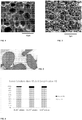
- the figure 3 illustrates an example of chitosan particles obtained.
- a mini-spoon is used to measure the amount of chitosan particles that will come into contact with the chondrogenic cells. Preliminary tests made it possible to validate the reproducibility of the measurement.
- a second structure (M2), by adding an anionic constituent, interacting with the cationic functions of the chitosan.
- the polymer chosen is hyaluronic acid, preferably of bacterial origin because such a constituent is known for its better biocompatibility properties, in order to avoid allergies or possible rejections.
- the molecular mass by weight of the hyaluronic acid used in the realization of the structure M2 is approximately 2 MDa.
- Hyaluronic acid is added after the preparation of the chitosan hydrogel particles.
- Example 3 Culture of cells within the 3D structure.
- the cells used in the context of this example are human chondrocytes obtained from human samples and treated according to the protocol described in the document FR2965278 (Univ. De Caen Basse-Normandie, et al).
- the hydrogel particles obtained at the end of Example 2, with hyaluronic acid (3D structure M2) or else without (3D structure M1), are sterilized, for example at 121 ° C for 15 min, before the contact with the cells.
- a few mini-spoons of hydrogel particles are taken, preferably 2 mini-spoons corresponding to 80 to 84 particles, which are introduced into the well of a 24-well culture plate previously covered with an insert (pore size 8 ⁇ m).
- Cells between 10 5 and 10 7 , preferably of the order of 6 ⁇ 10 5 cells / well, are added thereto for 80-84 hydrogel particles, which are gently mixed with the chitosan hydrogel particles.
- This proportion of cells relative to the 3D structure corresponds to approximately 6.7 x 10 6 cells per gram of 3D structure at the time of seeding.
- the culture is carried out in a controlled atmosphere in an oven at 37 ° C. with a CO2 level of 5%, under conditions of normoxia.
- the cells spontaneously adhere to the chitosan hydrogel particles.
- the amount of cells falling to the bottom of the well is considered negligible.
- 6.10 5 cells obtained as described above, are cultured in monolayers on plastic in 24-well plates with culture conditions identical to those described above for the 3D structures (controlled atmosphere in an oven at 37 ° C, with a CO 2 level of 5%, normoxia). The cells also adhere to it spontaneously.
- Example 4 Cell proliferation step.
- the culture medium chosen is favorable to the multiplication of cells.
- the medium chosen is a solution of 50/50 DMEM-HAM F12 + 1% AB (streptomycin / penicillin) + 10 FCS supplemented with a so-called “FI” solution comprising FGF-2 at 5 ng / ml + insulin at 5 ⁇ g / ml. (Claus et al; 2012).
- the mixture described in the previous step is covered, after a short time, with this culture medium known to be favorable to cell proliferation.
- the culture medium is renewed 3 times per week, for cultures in 3D structures as for cultures in monolayers. The proliferation period lasts between one and two weeks to obtain a sufficient number of cells.
- the inventors observed that the amplification phase lasted about two weeks, when the cells were seeded in a structure consisting of pure hydrogel particles (3D M1 structure) and could be shortened to 1 week in the presence of supplemented hydrogel particles. by linear chains of an anionic molecule such as hyaluronic acid (3D structure M2).
- the initial quantity of cells of 6.10 5 cells / insert can quite be increased, if the condition of the number of cells / mass of hydrogel or number of cells / volume of hydrogel, or number of cells / number of hydrogel particles is respected.
- Reverse phase optical microscopy monitoring was performed. It validated that the cells adhere well to the chitosan hydrogel particles and that this environment is favorable for their culture.
- the culture conditions (three-dimensional structure M1 or M2, and culture medium F1) promote the proliferation and division of chondrocytes.
- the cells observed can proliferate either in isolation or in clusters / clusters.
- Cultures in 3D structures of hydrogel particles show predominantly round cells.
- the elongated shape, characteristic of fibroblasts, is not observed in 3D structures M1 and M2, except sometimes at the periphery, that is to say at the interface between the structure and the external environment.
- the inventors carried out cultures in monolayers in parallel. After 24 hours of culture, in the same FI medium as the cells seeded in structures M1 and M2, the chondrocytes adopt an elongated morphology characteristic of fibroblast cells.
- the viability of the chondrocytes seeded in the three-dimensional structures was measured with the Live and Dead kit on fractions of cultures at 7 days (during amplification, in IF medium).
- the enumeration of dead cells (red) and living cells (green) is carried out with the ImageJ software from images taken by fluorescence microscopy at x20.
- the percentage of dead cells is calculated for each condition by making the ratio of dead cells / total cells.
- the viability is greater than 93%, or even greater than 97%, which shows good biocompatibility of the 3D structure.
- the figure 4 illustrates the results obtained.
- Proliferation tests were carried out by counting the total cells with a Cellometer T4 after detachment of the cells with trypsin and staining of the dead cells with trypan blue. The measurements were carried out after 1 day (D1), 14 days (D14) and 21 days (D21) of culture after seeding of primary chondrocytes on D0.
- the figure Fig. 5 illustrates the evolution of the cell population. After 7 days, the increase in the number of cells is clearly observed. Cells survive and proliferate very well in 3D hydrogel structures as well as monolayers (MC). In three-dimensional structure, M1 or M2, the cells remain round during the multiplication step while they adopt an elongated shape, fibroblast type, in monolayers.
- the figure 6 illustrates that in monolayers, the cell population is identical from 7 days, regardless of the initial density of seeded cells.
- Example 5 Differentiation and production of extracellular matrix
- the stages of multiplication and differentiation are preferably distinct: firstly, the cells multiply and secondly, they differentiate and produce the extracellular matrix.
- the culture medium used for the previous multiplication step is modified after 15 days.
- the culture medium is then preferably composed of: 50/50 DMEM-HAM F12 + 1% AB (streptomycin / penicillin) + 10 SVF, to which is added a BIT solution composed of: BMP-2 at 200ng / ml + Insulin at 5 ⁇ g / ml + triiodothyronine T3 at 100mM (Claus et al, 2012).
- This new culture medium is renewed every two or three days.
- the period of re-differentiation and chondrogenesis preferably lasts 3 weeks.
- the same change of medium is carried out for the cells cultured in monolayers.
- cells can agglutinate the hydrogel particles and form a sort of more or less compact “ball”. This observation is made under most conditions containing the hydrogels, not in the monolayers, however, which serve as a control. This observation constitutes proof of the strong production of extracellular matrix which has accumulated around the cells. Cells make more extracellular matrix in a three-dimensional environment than in monolayers. It should be noted that the composition consisting of said beads remains however, under certain conditions, injectable, despite the synthesis of a large amount of extracellular matrix. Either way, the composition is implantable.
- the COLII / GAPDH results obtained at the end of the MEC synthesis step show that there are more COLII transcripts for the hydrogel cultures than for the monolayer cultures.
- the COLI / GAPDH results show that there are, on the contrary, more COLI transcripts in monolayer cultures than in cultures within three-dimensional structures.
- the result of calculating the COLII / COLI ratio is shown in figure Fig. 9 . It shows that there is a much better ratio, and therefore a much better re-differentiation after de-differentiation, within 3D structures made up of chitosan hydrogel particles compared to monolayers.
- the three-dimensional environment tested therefore clearly promotes the re-differentiation of de-differentiated chondrocytes, following an intense prior multiplication, by a factor of at least 6.
- This 3D structure promotes the expression of the chondrocyte phenotype.
- the culture conditions (three-dimensional structure based on chitosan hydrogel, and BIT culture medium) therefore promote re-differentiation of the chondrocytes and the production of cartilage matrix, compared to a culture in monolayers.
- compositions obtained were then observed by immunohistochemistry, in order to compare the implementation of the new process, in three-dimensional structure, and the traditional culture in monolayers, at the level of the synthesis of ECM, proteoglycans, type I and II collagen. .
- the results are illustrated by the photos in figure Fig.11 .
- SO Safranin O
- the succession of steps, in the same three-dimensional medium, comprising particles of chitosan hydrogel with / without structuring molecule, of amplification then of differentiation / chondrogenesis is very favorable to obtaining an injectable or implantable cartilage neo-tissue. exhibiting excellent mechanical and biological properties.
- the addition of hyaluronic acid improves the system by accelerating the process of cell amplification and allowing to increase the number of cells to be implanted or to gain a week on the overall protocol.
- the configuration of the structure makes it possible to optimize the contact surface with the cells.
- Example 7 Comparison of the different 3D structures.
- the inventors have reproduced the 3D structures described in the preceding examples, in particular in Example 2, by varying the type of chitosan constituting the hydrogel particles, the size of the hydrogel particles, and the presence and concentration of acid. hyaluronic.
- the proliferation rates were compared and the results obtained are illustrated in Table 1.
- the value 1 was assigned to the M1 structure corresponding to particles of several hundred microns, obtained from fungal chitosan.
- the proliferation rate obtained with the M2 structure is 2 times higher compared to the M1 structure (mushroom extract not supplemented with hyaluronic acid), which itself makes it possible to obtain a proliferation rate 1.5 times greater than with chitosan extracted from squid, or else with particles of size of the order of tens of ⁇ m.
- Table 1 ⁇ /u> Proliferation ratios.
Landscapes
- Health & Medical Sciences (AREA)
- Life Sciences & Earth Sciences (AREA)
- Engineering & Computer Science (AREA)
- Chemical & Material Sciences (AREA)
- Biomedical Technology (AREA)
- General Health & Medical Sciences (AREA)
- Medicinal Chemistry (AREA)
- Veterinary Medicine (AREA)
- Animal Behavior & Ethology (AREA)
- Public Health (AREA)
- Epidemiology (AREA)
- Transplantation (AREA)
- Oral & Maxillofacial Surgery (AREA)
- Dermatology (AREA)
- Zoology (AREA)
- Cell Biology (AREA)
- Chemical Kinetics & Catalysis (AREA)
- Botany (AREA)
- Rheumatology (AREA)
- Biotechnology (AREA)
- Organic Chemistry (AREA)
- Bioinformatics & Cheminformatics (AREA)
- Urology & Nephrology (AREA)
- Developmental Biology & Embryology (AREA)
- Genetics & Genomics (AREA)
- Wood Science & Technology (AREA)
- Pharmacology & Pharmacy (AREA)
- Vascular Medicine (AREA)
- Immunology (AREA)
- Orthopedic Medicine & Surgery (AREA)
- Virology (AREA)
- Microbiology (AREA)
- Molecular Biology (AREA)
- Biochemistry (AREA)
- General Engineering & Computer Science (AREA)
- General Chemical & Material Sciences (AREA)
- Physical Education & Sports Medicine (AREA)
- Nuclear Medicine, Radiotherapy & Molecular Imaging (AREA)
- Materials For Medical Uses (AREA)
- Micro-Organisms Or Cultivation Processes Thereof (AREA)
Applications Claiming Priority (2)
| Application Number | Priority Date | Filing Date | Title |
|---|---|---|---|
| FR1461746A FR3029116B1 (fr) | 2014-12-01 | 2014-12-01 | Procede d'obtention d'un gel de cartilage pour la reparation cartilagineuse, comprenant du chitosane et des chondrocytes |
| PCT/FR2015/053271 WO2016087762A1 (fr) | 2014-12-01 | 2015-12-01 | Gel de cartilage pour la réparation cartilagineuse, comprenant du chitosane et des chondrocytes |
Publications (2)
| Publication Number | Publication Date |
|---|---|
| EP3226923A1 EP3226923A1 (fr) | 2017-10-11 |
| EP3226923B1 true EP3226923B1 (fr) | 2021-02-17 |
Family
ID=53177537
Family Applications (1)
| Application Number | Title | Priority Date | Filing Date |
|---|---|---|---|
| EP15817457.3A Active EP3226923B1 (fr) | 2014-12-01 | 2015-12-01 | Gel de cartilage pour la réparation cartilagineuse, comprenant du chitosane et des chondrocytes |
Country Status (9)
| Country | Link |
|---|---|
| US (2) | US10612001B2 (https=) |
| EP (1) | EP3226923B1 (https=) |
| JP (1) | JP6821583B2 (https=) |
| CA (1) | CA2969285C (https=) |
| DK (1) | DK3226923T3 (https=) |
| ES (1) | ES2870505T3 (https=) |
| FR (2) | FR3029116B1 (https=) |
| PT (1) | PT3226923T (https=) |
| WO (1) | WO2016087762A1 (https=) |
Families Citing this family (20)
| Publication number | Priority date | Publication date | Assignee | Title |
|---|---|---|---|---|
| FR3038318B1 (fr) * | 2015-07-02 | 2017-08-04 | Univ De Lille 1 Sciences Et Technologies | Procede de fabrication d'hydrogel a base de chitosan et de polyelectrolytes charges negativement et materiau poreux alveolaire issu dudit hydrogel |
| US10898608B2 (en) | 2017-02-02 | 2021-01-26 | Nanofiber Solutions, Llc | Methods of improving bone-soft tissue healing using electrospun fibers |
| EP3595515A4 (en) | 2017-03-14 | 2020-12-30 | University of Connecticut | BIODEGRADABLE PRESSURE SENSOR |
| FR3074043B1 (fr) | 2017-11-28 | 2020-11-13 | Kiomed Pharma | Chitosane a charge anionique |
| FR3074044B1 (fr) | 2017-11-28 | 2019-11-01 | Kiomed Pharma | Carboxyalkyl chitosane |
| WO2019113511A1 (en) * | 2017-12-08 | 2019-06-13 | Cohen Brian S | Electrospun fibers for the repair and regrowth of hyaline cartilage |
| CN108774330B (zh) * | 2018-08-30 | 2020-11-10 | 华熙生物科技股份有限公司 | 一种交联透明质酸凝胶的制备方法及所得产品和应用 |
| JP2022513442A (ja) | 2018-12-11 | 2022-02-08 | ナノファイバー ソリューションズ、エルエルシー | 電界紡糸繊維を使用した慢性創傷の治療方法 |
| US11826495B2 (en) | 2019-03-01 | 2023-11-28 | University Of Connecticut | Biodegradable piezoelectric ultrasonic transducer system |
| KR102197871B1 (ko) * | 2019-03-28 | 2021-01-04 | 이화여자대학교 산학협력단 | 엽산, 엽산 유도체, 또는 엽산 저해제를 이용한 줄기세포의 연골세포로의 분화 유도 방법 |
| KR102337350B1 (ko) * | 2019-11-22 | 2021-12-08 | 아주대학교산학협력단 | 내재성 전구세포 또는 줄기세포 포집능 및 포집된 세포의 혈관 분화 유도능을 갖는 주입형 하이드로겔 조성물 |
| US11745001B2 (en) | 2020-03-10 | 2023-09-05 | University Of Connecticut | Therapeutic bandage |
| US12491290B2 (en) | 2020-06-08 | 2025-12-09 | University Of Connecticut | Biodegradable piezoelectric composite materials |
| FR3116534B1 (fr) | 2020-11-23 | 2023-12-22 | Kiomed Pharma | Billes à base de chitosane, préparation, compositions et applications |
| US20230404765A1 (en) * | 2020-12-07 | 2023-12-21 | University Of Connecticut | A system and method for osteoarthritis treatment |
| CN114652895A (zh) * | 2020-12-23 | 2022-06-24 | 上海其胜生物制剂有限公司 | 一种可注射组织再生型壳聚糖复合凝胶支架的制备方法 |
| CN113663085B (zh) * | 2021-08-25 | 2023-12-15 | 福州市大福瑞生物科技有限公司 | 基于卤代环氧烷烃接枝制备皂苷-壳聚糖衍生物的方法及其应用 |
| CN114908038B (zh) * | 2022-06-06 | 2024-03-15 | 苏州大学 | 一种体外3d关节损伤模型及其构建与应用 |
| CN115154673B (zh) * | 2022-06-28 | 2023-07-14 | 北京航空航天大学宁波创新研究院 | 人工软骨植入物预成型体及制备方法和应用、类生人工软骨植入物胶原纤维原位构建方法 |
| PL443403A1 (pl) | 2023-01-03 | 2024-07-08 | Politechnika Gdańska | Hydrożelowy kompozyt na bazie chitozanu, sposób wytwarzania hydrożelowego kompozytu na bazie chitozanu oraz zastosowanie kompozytu w postaci biotuszu do tworzenia przestrzennych obiektów, zwłaszcza z wykorzystaniem technologii wytwarzania addytywnego, zwłaszcza celem prowadzenia hodowli komórkowych |
Family Cites Families (13)
| Publication number | Priority date | Publication date | Assignee | Title |
|---|---|---|---|---|
| AU3097999A (en) | 1998-03-18 | 1999-10-11 | University Of Pittsburgh | Chitosan-based composite materials containing glycosaminoglycan for cartilage repair |
| KR100375422B1 (ko) * | 1999-12-21 | 2003-03-10 | 한국과학기술연구원 | 다공성 키토산 구슬 및 그의 제조 방법 |
| JP2002209573A (ja) * | 2001-01-15 | 2002-07-30 | Toru Yamazaki | 細胞及び又は生体組織の担持体と移植体、及び担持方法 |
| JP3616344B2 (ja) * | 2001-03-29 | 2005-02-02 | 紳一郎 西村 | 軟骨細胞培養方法および軟骨組織再生基材 |
| WO2002078761A1 (fr) | 2001-03-30 | 2002-10-10 | Laboratoires Genevrier | Neo-tissu cartilagineux greffable |
| FR2822842B1 (fr) * | 2001-03-30 | 2004-11-19 | Genevrier Lab | Procede de preparation d'un neo-tissu cartilagineux |
| US8173171B2 (en) * | 2003-11-17 | 2012-05-08 | Taiwan Textile Research Institute | Hydrogel microparticle composition, application thereof and method for preparing the same |
| US7524514B2 (en) * | 2003-12-01 | 2009-04-28 | Tissue Engineering Consultants, Inc. | Biomimetic composition reinforced by a polyelectrolytic complex of hyaluronic acid and chitosan |
| CN101098718A (zh) * | 2004-12-20 | 2008-01-02 | 卡博格拉夫特有限公司 | 壳聚糖组合物 |
| SG10201500237TA (en) | 2008-01-30 | 2015-04-29 | Histogen Inc | Extracellular matrix compositions |
| EP2361640A1 (en) * | 2010-02-25 | 2011-08-31 | Université de Liège | Cell cultivation in chitosan alginate hydrogel beads |
| FR2963351B1 (fr) | 2010-07-30 | 2013-03-08 | Univ Claude Bernard Lyon | Particules formees d'un complexe polyelectrolyte de chitosane et d'un polysaccharide anionique, presentant une stabilite amelioree |
| FR2965278B1 (fr) | 2010-09-23 | 2014-10-10 | Univ Caen Basse Normandie | Procede d'obtention in vitro ou ex vivo de chondrocytes et leurs utilisations |
-
2014
- 2014-12-01 FR FR1461746A patent/FR3029116B1/fr active Active
-
2015
- 2015-12-01 DK DK15817457.3T patent/DK3226923T3/da active
- 2015-12-01 JP JP2017547081A patent/JP6821583B2/ja active Active
- 2015-12-01 US US15/532,020 patent/US10612001B2/en active Active
- 2015-12-01 CA CA2969285A patent/CA2969285C/fr active Active
- 2015-12-01 ES ES15817457T patent/ES2870505T3/es active Active
- 2015-12-01 WO PCT/FR2015/053271 patent/WO2016087762A1/fr not_active Ceased
- 2015-12-01 PT PT158174573T patent/PT3226923T/pt unknown
- 2015-12-01 EP EP15817457.3A patent/EP3226923B1/fr active Active
-
2018
- 2018-01-23 FR FR1850498A patent/FR3062064B1/fr active Active
-
2020
- 2020-03-25 US US16/829,634 patent/US11718828B2/en active Active
Non-Patent Citations (1)
| Title |
|---|
| None * |
Also Published As
| Publication number | Publication date |
|---|---|
| DK3226923T3 (da) | 2021-05-17 |
| FR3062064B1 (fr) | 2020-11-27 |
| ES2870505T3 (es) | 2021-10-27 |
| CA2969285C (fr) | 2023-09-05 |
| US10612001B2 (en) | 2020-04-07 |
| US11718828B2 (en) | 2023-08-08 |
| US20200248144A1 (en) | 2020-08-06 |
| JP2017535405A (ja) | 2017-11-30 |
| WO2016087762A1 (fr) | 2016-06-09 |
| US20170306295A1 (en) | 2017-10-26 |
| FR3062064A1 (fr) | 2018-07-27 |
| EP3226923A1 (fr) | 2017-10-11 |
| PT3226923T (pt) | 2021-05-07 |
| JP6821583B2 (ja) | 2021-01-27 |
| FR3029116A1 (fr) | 2016-06-03 |
| CA2969285A1 (fr) | 2016-06-09 |
| FR3029116B1 (fr) | 2018-03-30 |
Similar Documents
| Publication | Publication Date | Title |
|---|---|---|
| EP3226923B1 (fr) | Gel de cartilage pour la réparation cartilagineuse, comprenant du chitosane et des chondrocytes | |
| JP7312761B2 (ja) | インビトロ培養及び移植のための組織構築物の生理学的3dバイオプリンティングのためのバイオガム及び植物性ガムハイドロゲルバイオインク | |
| Jang et al. | ASC/chondrocyte-laden alginate hydrogel/PCL hybrid scaffold fabricated using 3D printing for auricle regeneration | |
| Xu et al. | Photocrosslinked natural hydrogel composed of hyaluronic acid and gelatin enhances cartilage regeneration of decellularized trachea matrix | |
| Guo et al. | Hydrogels of collagen/chondroitin sulfate/hyaluronan interpenetrating polymer network for cartilage tissue engineering | |
| US10434216B2 (en) | Ultra-thin film silk fibroin/collagen composite implant and manufacturing method therefor | |
| US20110097406A1 (en) | Methods and compositions for retaining ecm materials in hydrogels | |
| CN106999635A (zh) | 软骨修复用移植物支架及其制造方法 | |
| Wan et al. | BMSCs laden injectable amino-diethoxypropane modified alginate-chitosan hydrogel for hyaline cartilage reconstruction | |
| Nair | Injectable hydrogels for regenerative engineering | |
| EP3328923B1 (fr) | Procédé de préparation de matrices poreuse polymères biocompatibles et biodegradables trimensionnelles | |
| EP2874672B1 (fr) | Hydrogel de chitosane pour la reparation du tissu nerveux | |
| CA2625604C (fr) | Matrice poreuse biocompatible et biodegradable notamment utile pour la reconstruction tissulaire | |
| Fan et al. | Potential use of alginate beads as a chondrocyte delivery vehicle and stepwise dissolving porogen in a hydrogel scaffold for cartilage tissue engineering | |
| FR2927632A1 (fr) | Cornee et muqueuse reconstruites. | |
| De Angelis et al. | The in vitro biocompatibility of D-(+) raffinose modified chitosan: Two-dimensional and three-dimensional systems for culturing of horse articular chondrocytes | |
| EP1960011B1 (fr) | Utilisation d' un polysaccharide excrete par l'espece vibrio diabolicus a des fins d'ingenierie des tissus conjonctifs non mineralises | |
| EP4359022A1 (fr) | Procede de consolidation d'un hydrogel alginate / gelatine | |
| CA2940352A1 (fr) | Materiau pour la culture des cellules, procedes de preparation et ses utilisations | |
| Pizzolitto | SVILUPPO E CARATTERIZZAZIONE DI STRUTTURE 3D E DI IDROGELI A BASE DI POLISACCARIDI NATURALI E MODIFICATI E LORO APPLICAZIONE IN CAMPO BIOMEDICO | |
| EP4359025A1 (fr) | Implants corporels tridimensionnels | |
| WO2021234088A1 (fr) | Biomateriau comprenant au moins une matrice elastomere et un polysaccharide non sulfate et ses utilisations | |
| de Carvalho | Development of Hyaluronic Acid, Dextrin and Extracellular Matrix Hydrogels for Cell Expansion | |
| FR3081712A1 (fr) | Matrice pour la preparation d'une composition de regeneration cellulaire, tissulaire et/ou osseuse |
Legal Events
| Date | Code | Title | Description |
|---|---|---|---|
| STAA | Information on the status of an ep patent application or granted ep patent |
Free format text: STATUS: THE INTERNATIONAL PUBLICATION HAS BEEN MADE |
|
| PUAI | Public reference made under article 153(3) epc to a published international application that has entered the european phase |
Free format text: ORIGINAL CODE: 0009012 |
|
| STAA | Information on the status of an ep patent application or granted ep patent |
Free format text: STATUS: REQUEST FOR EXAMINATION WAS MADE |
|
| 17P | Request for examination filed |
Effective date: 20170630 |
|
| AK | Designated contracting states |
Kind code of ref document: A1 Designated state(s): AL AT BE BG CH CY CZ DE DK EE ES FI FR GB GR HR HU IE IS IT LI LT LU LV MC MK MT NL NO PL PT RO RS SE SI SK SM TR |
|
| AX | Request for extension of the european patent |
Extension state: BA ME |
|
| DAV | Request for validation of the european patent (deleted) | ||
| DAX | Request for extension of the european patent (deleted) | ||
| STAA | Information on the status of an ep patent application or granted ep patent |
Free format text: STATUS: EXAMINATION IS IN PROGRESS |
|
| 17Q | First examination report despatched |
Effective date: 20190325 |
|
| GRAP | Despatch of communication of intention to grant a patent |
Free format text: ORIGINAL CODE: EPIDOSNIGR1 |
|
| STAA | Information on the status of an ep patent application or granted ep patent |
Free format text: STATUS: GRANT OF PATENT IS INTENDED |
|
| INTG | Intention to grant announced |
Effective date: 20200930 |
|
| GRAS | Grant fee paid |
Free format text: ORIGINAL CODE: EPIDOSNIGR3 |
|
| GRAA | (expected) grant |
Free format text: ORIGINAL CODE: 0009210 |
|
| STAA | Information on the status of an ep patent application or granted ep patent |
Free format text: STATUS: THE PATENT HAS BEEN GRANTED |
|
| AK | Designated contracting states |
Kind code of ref document: B1 Designated state(s): AL AT BE BG CH CY CZ DE DK EE ES FI FR GB GR HR HU IE IS IT LI LT LU LV MC MK MT NL NO PL PT RO RS SE SI SK SM TR |
|
| REG | Reference to a national code |
Ref country code: GB Ref legal event code: FG4D Free format text: NOT ENGLISH |
|
| REG | Reference to a national code |
Ref country code: CH Ref legal event code: EP |
|
| REG | Reference to a national code |
Ref country code: DE Ref legal event code: R096 Ref document number: 602015065719 Country of ref document: DE |
|
| REG | Reference to a national code |
Ref country code: AT Ref legal event code: REF Ref document number: 1360612 Country of ref document: AT Kind code of ref document: T Effective date: 20210315 |
|
| REG | Reference to a national code |
Ref country code: IE Ref legal event code: FG4D Free format text: LANGUAGE OF EP DOCUMENT: FRENCH |
|
| REG | Reference to a national code |
Ref country code: PT Ref legal event code: SC4A Ref document number: 3226923 Country of ref document: PT Date of ref document: 20210507 Kind code of ref document: T Free format text: AVAILABILITY OF NATIONAL TRANSLATION Effective date: 20210503 |
|
| REG | Reference to a national code |
Ref country code: DK Ref legal event code: T3 Effective date: 20210512 |
|
| REG | Reference to a national code |
Ref country code: SE Ref legal event code: TRGR |
|
| REG | Reference to a national code |
Ref country code: NL Ref legal event code: FP |
|
| REG | Reference to a national code |
Ref country code: NO Ref legal event code: T2 Effective date: 20210217 Ref country code: LT Ref legal event code: MG9D |
|
| PG25 | Lapsed in a contracting state [announced via postgrant information from national office to epo] |
Ref country code: BG Free format text: LAPSE BECAUSE OF FAILURE TO SUBMIT A TRANSLATION OF THE DESCRIPTION OR TO PAY THE FEE WITHIN THE PRESCRIBED TIME-LIMIT Effective date: 20210517 Ref country code: GR Free format text: LAPSE BECAUSE OF FAILURE TO SUBMIT A TRANSLATION OF THE DESCRIPTION OR TO PAY THE FEE WITHIN THE PRESCRIBED TIME-LIMIT Effective date: 20210518 Ref country code: HR Free format text: LAPSE BECAUSE OF FAILURE TO SUBMIT A TRANSLATION OF THE DESCRIPTION OR TO PAY THE FEE WITHIN THE PRESCRIBED TIME-LIMIT Effective date: 20210217 Ref country code: FI Free format text: LAPSE BECAUSE OF FAILURE TO SUBMIT A TRANSLATION OF THE DESCRIPTION OR TO PAY THE FEE WITHIN THE PRESCRIBED TIME-LIMIT Effective date: 20210217 Ref country code: LT Free format text: LAPSE BECAUSE OF FAILURE TO SUBMIT A TRANSLATION OF THE DESCRIPTION OR TO PAY THE FEE WITHIN THE PRESCRIBED TIME-LIMIT Effective date: 20210217 |
|
| REG | Reference to a national code |
Ref country code: AT Ref legal event code: MK05 Ref document number: 1360612 Country of ref document: AT Kind code of ref document: T Effective date: 20210217 |
|
| PG25 | Lapsed in a contracting state [announced via postgrant information from national office to epo] |
Ref country code: RS Free format text: LAPSE BECAUSE OF FAILURE TO SUBMIT A TRANSLATION OF THE DESCRIPTION OR TO PAY THE FEE WITHIN THE PRESCRIBED TIME-LIMIT Effective date: 20210217 Ref country code: LV Free format text: LAPSE BECAUSE OF FAILURE TO SUBMIT A TRANSLATION OF THE DESCRIPTION OR TO PAY THE FEE WITHIN THE PRESCRIBED TIME-LIMIT Effective date: 20210217 Ref country code: PL Free format text: LAPSE BECAUSE OF FAILURE TO SUBMIT A TRANSLATION OF THE DESCRIPTION OR TO PAY THE FEE WITHIN THE PRESCRIBED TIME-LIMIT Effective date: 20210217 |
|
| RAP2 | Party data changed (patent owner data changed or rights of a patent transferred) |
Owner name: ADVANCED CHITOSAN SOLUTIONS BIOTECH |
|
| REG | Reference to a national code |
Ref country code: ES Ref legal event code: FG2A Ref document number: 2870505 Country of ref document: ES Kind code of ref document: T3 Effective date: 20211027 |
|
| PG25 | Lapsed in a contracting state [announced via postgrant information from national office to epo] |
Ref country code: SM Free format text: LAPSE BECAUSE OF FAILURE TO SUBMIT A TRANSLATION OF THE DESCRIPTION OR TO PAY THE FEE WITHIN THE PRESCRIBED TIME-LIMIT Effective date: 20210217 Ref country code: AT Free format text: LAPSE BECAUSE OF FAILURE TO SUBMIT A TRANSLATION OF THE DESCRIPTION OR TO PAY THE FEE WITHIN THE PRESCRIBED TIME-LIMIT Effective date: 20210217 Ref country code: CZ Free format text: LAPSE BECAUSE OF FAILURE TO SUBMIT A TRANSLATION OF THE DESCRIPTION OR TO PAY THE FEE WITHIN THE PRESCRIBED TIME-LIMIT Effective date: 20210217 Ref country code: EE Free format text: LAPSE BECAUSE OF FAILURE TO SUBMIT A TRANSLATION OF THE DESCRIPTION OR TO PAY THE FEE WITHIN THE PRESCRIBED TIME-LIMIT Effective date: 20210217 |
|
| REG | Reference to a national code |
Ref country code: DE Ref legal event code: R097 Ref document number: 602015065719 Country of ref document: DE |
|
| PG25 | Lapsed in a contracting state [announced via postgrant information from national office to epo] |
Ref country code: SK Free format text: LAPSE BECAUSE OF FAILURE TO SUBMIT A TRANSLATION OF THE DESCRIPTION OR TO PAY THE FEE WITHIN THE PRESCRIBED TIME-LIMIT Effective date: 20210217 Ref country code: RO Free format text: LAPSE BECAUSE OF FAILURE TO SUBMIT A TRANSLATION OF THE DESCRIPTION OR TO PAY THE FEE WITHIN THE PRESCRIBED TIME-LIMIT Effective date: 20210217 |
|
| PLBE | No opposition filed within time limit |
Free format text: ORIGINAL CODE: 0009261 |
|
| STAA | Information on the status of an ep patent application or granted ep patent |
Free format text: STATUS: NO OPPOSITION FILED WITHIN TIME LIMIT |
|
| 26N | No opposition filed |
Effective date: 20211118 |
|
| PG25 | Lapsed in a contracting state [announced via postgrant information from national office to epo] |
Ref country code: AL Free format text: LAPSE BECAUSE OF FAILURE TO SUBMIT A TRANSLATION OF THE DESCRIPTION OR TO PAY THE FEE WITHIN THE PRESCRIBED TIME-LIMIT Effective date: 20210217 |
|
| PG25 | Lapsed in a contracting state [announced via postgrant information from national office to epo] |
Ref country code: SI Free format text: LAPSE BECAUSE OF FAILURE TO SUBMIT A TRANSLATION OF THE DESCRIPTION OR TO PAY THE FEE WITHIN THE PRESCRIBED TIME-LIMIT Effective date: 20210217 |
|
| PG25 | Lapsed in a contracting state [announced via postgrant information from national office to epo] |
Ref country code: MC Free format text: LAPSE BECAUSE OF FAILURE TO SUBMIT A TRANSLATION OF THE DESCRIPTION OR TO PAY THE FEE WITHIN THE PRESCRIBED TIME-LIMIT Effective date: 20210217 |
|
| PG25 | Lapsed in a contracting state [announced via postgrant information from national office to epo] |
Ref country code: LU Free format text: LAPSE BECAUSE OF NON-PAYMENT OF DUE FEES Effective date: 20211201 |
|
| PG25 | Lapsed in a contracting state [announced via postgrant information from national office to epo] |
Ref country code: IT Free format text: LAPSE BECAUSE OF NON-PAYMENT OF DUE FEES Effective date: 20211201 |
|
| PGFP | Annual fee paid to national office [announced via postgrant information from national office to epo] |
Ref country code: IE Payment date: 20221121 Year of fee payment: 8 Ref country code: DK Payment date: 20221125 Year of fee payment: 8 |
|
| PGFP | Annual fee paid to national office [announced via postgrant information from national office to epo] |
Ref country code: IS Payment date: 20221116 Year of fee payment: 8 |
|
| PG25 | Lapsed in a contracting state [announced via postgrant information from national office to epo] |
Ref country code: HU Free format text: LAPSE BECAUSE OF FAILURE TO SUBMIT A TRANSLATION OF THE DESCRIPTION OR TO PAY THE FEE WITHIN THE PRESCRIBED TIME-LIMIT; INVALID AB INITIO Effective date: 20151201 |
|
| P01 | Opt-out of the competence of the unified patent court (upc) registered |
Effective date: 20230519 |
|
| PG25 | Lapsed in a contracting state [announced via postgrant information from national office to epo] |
Ref country code: CY Free format text: LAPSE BECAUSE OF FAILURE TO SUBMIT A TRANSLATION OF THE DESCRIPTION OR TO PAY THE FEE WITHIN THE PRESCRIBED TIME-LIMIT Effective date: 20210217 |
|
| PG25 | Lapsed in a contracting state [announced via postgrant information from national office to epo] |
Ref country code: MK Free format text: LAPSE BECAUSE OF FAILURE TO SUBMIT A TRANSLATION OF THE DESCRIPTION OR TO PAY THE FEE WITHIN THE PRESCRIBED TIME-LIMIT Effective date: 20210217 |
|
| PG25 | Lapsed in a contracting state [announced via postgrant information from national office to epo] |
Ref country code: TR Free format text: LAPSE BECAUSE OF FAILURE TO SUBMIT A TRANSLATION OF THE DESCRIPTION OR TO PAY THE FEE WITHIN THE PRESCRIBED TIME-LIMIT Effective date: 20210217 |
|
| REG | Reference to a national code |
Ref country code: DK Ref legal event code: EBP Effective date: 20231231 |
|
| PG25 | Lapsed in a contracting state [announced via postgrant information from national office to epo] |
Ref country code: MT Free format text: LAPSE BECAUSE OF FAILURE TO SUBMIT A TRANSLATION OF THE DESCRIPTION OR TO PAY THE FEE WITHIN THE PRESCRIBED TIME-LIMIT Effective date: 20210217 |
|
| REG | Reference to a national code |
Ref country code: IE Ref legal event code: MM4A |
|
| PG25 | Lapsed in a contracting state [announced via postgrant information from national office to epo] |
Ref country code: IE Free format text: LAPSE BECAUSE OF NON-PAYMENT OF DUE FEES Effective date: 20231201 |
|
| PG25 | Lapsed in a contracting state [announced via postgrant information from national office to epo] |
Ref country code: IE Free format text: LAPSE BECAUSE OF NON-PAYMENT OF DUE FEES Effective date: 20231201 |
|
| PG25 | Lapsed in a contracting state [announced via postgrant information from national office to epo] |
Ref country code: DK Free format text: LAPSE BECAUSE OF NON-PAYMENT OF DUE FEES Effective date: 20231231 |
|
| PG25 | Lapsed in a contracting state [announced via postgrant information from national office to epo] |
Ref country code: DK Free format text: LAPSE BECAUSE OF NON-PAYMENT OF DUE FEES Effective date: 20231231 |
|
| PG25 | Lapsed in a contracting state [announced via postgrant information from national office to epo] |
Ref country code: IS Free format text: LAPSE BECAUSE OF NON-PAYMENT OF DUE FEES Effective date: 20240607 |
|
| PGFP | Annual fee paid to national office [announced via postgrant information from national office to epo] |
Ref country code: PT Payment date: 20251120 Year of fee payment: 11 |
|
| REG | Reference to a national code |
Ref country code: CH Ref legal event code: U11 Free format text: ST27 STATUS EVENT CODE: U-0-0-U10-U11 (AS PROVIDED BY THE NATIONAL OFFICE) Effective date: 20260101 |
|
| PGFP | Annual fee paid to national office [announced via postgrant information from national office to epo] |
Ref country code: GB Payment date: 20251226 Year of fee payment: 11 |
|
| PGFP | Annual fee paid to national office [announced via postgrant information from national office to epo] |
Ref country code: NO Payment date: 20251230 Year of fee payment: 11 |
|
| PGFP | Annual fee paid to national office [announced via postgrant information from national office to epo] |
Ref country code: NL Payment date: 20251230 Year of fee payment: 11 Ref country code: FR Payment date: 20251117 Year of fee payment: 11 |
|
| PGFP | Annual fee paid to national office [announced via postgrant information from national office to epo] |
Ref country code: BE Payment date: 20251226 Year of fee payment: 11 |
|
| PGFP | Annual fee paid to national office [announced via postgrant information from national office to epo] |
Ref country code: SE Payment date: 20251218 Year of fee payment: 11 |
|
| PGFP | Annual fee paid to national office [announced via postgrant information from national office to epo] |
Ref country code: ES Payment date: 20260112 Year of fee payment: 11 |
|
| PGFP | Annual fee paid to national office [announced via postgrant information from national office to epo] |
Ref country code: DE Payment date: 20251231 Year of fee payment: 11 |
|
| PGFP | Annual fee paid to national office [announced via postgrant information from national office to epo] |
Ref country code: CH Payment date: 20260101 Year of fee payment: 11 |