EP3222630A1 - Peptides for targeting colorectal cancer, and medical use thereof - Google Patents
Peptides for targeting colorectal cancer, and medical use thereof Download PDFInfo
- Publication number
- EP3222630A1 EP3222630A1 EP15861389.3A EP15861389A EP3222630A1 EP 3222630 A1 EP3222630 A1 EP 3222630A1 EP 15861389 A EP15861389 A EP 15861389A EP 3222630 A1 EP3222630 A1 EP 3222630A1
- Authority
- EP
- European Patent Office
- Prior art keywords
- peptide
- colorectal cancer
- cancer
- seq
- radio
- Prior art date
- Legal status (The legal status is an assumption and is not a legal conclusion. Google has not performed a legal analysis and makes no representation as to the accuracy of the status listed.)
- Granted
Links
Images




Classifications
-
- G—PHYSICS
- G01—MEASURING; TESTING
- G01N—INVESTIGATING OR ANALYSING MATERIALS BY DETERMINING THEIR CHEMICAL OR PHYSICAL PROPERTIES
- G01N33/00—Investigating or analysing materials by specific methods not covered by groups G01N1/00 - G01N31/00
- G01N33/48—Biological material, e.g. blood, urine; Haemocytometers
- G01N33/50—Chemical analysis of biological material, e.g. blood, urine; Testing involving biospecific ligand binding methods; Immunological testing
- G01N33/53—Immunoassay; Biospecific binding assay; Materials therefor
- G01N33/575—Immunoassay; Biospecific binding assay; Materials therefor for cancer
-
- A—HUMAN NECESSITIES
- A61—MEDICAL OR VETERINARY SCIENCE; HYGIENE
- A61K—PREPARATIONS FOR MEDICAL, DENTAL OR TOILETRY PURPOSES
- A61K47/00—Medicinal preparations characterised by the non-active ingredients used, e.g. carriers or inert additives; Targeting or modifying agents chemically bound to the active ingredient
- A61K47/50—Medicinal preparations characterised by the non-active ingredients used, e.g. carriers or inert additives; Targeting or modifying agents chemically bound to the active ingredient the non-active ingredient being chemically bound to the active ingredient, e.g. polymer-drug conjugates
-
- A—HUMAN NECESSITIES
- A61—MEDICAL OR VETERINARY SCIENCE; HYGIENE
- A61K—PREPARATIONS FOR MEDICAL, DENTAL OR TOILETRY PURPOSES
- A61K49/00—Preparations for testing in vivo
- A61K49/0004—Screening or testing of compounds for diagnosis of disorders, assessment of conditions, e.g. renal clearance, gastric emptying, testing for diabetes, allergy, rheuma, pancreas functions
-
- A—HUMAN NECESSITIES
- A61—MEDICAL OR VETERINARY SCIENCE; HYGIENE
- A61K—PREPARATIONS FOR MEDICAL, DENTAL OR TOILETRY PURPOSES
- A61K49/00—Preparations for testing in vivo
- A61K49/001—Preparation for luminescence or biological staining
- A61K49/0013—Luminescence
- A61K49/0017—Fluorescence in vivo
- A61K49/0019—Fluorescence in vivo characterised by the fluorescent group, e.g. oligomeric, polymeric or dendritic molecules
- A61K49/0021—Fluorescence in vivo characterised by the fluorescent group, e.g. oligomeric, polymeric or dendritic molecules the fluorescent group being a small organic molecule
- A61K49/0032—Methine dyes, e.g. cyanine dyes
-
- A—HUMAN NECESSITIES
- A61—MEDICAL OR VETERINARY SCIENCE; HYGIENE
- A61K—PREPARATIONS FOR MEDICAL, DENTAL OR TOILETRY PURPOSES
- A61K49/00—Preparations for testing in vivo
- A61K49/001—Preparation for luminescence or biological staining
- A61K49/0013—Luminescence
- A61K49/0017—Fluorescence in vivo
- A61K49/005—Fluorescence in vivo characterised by the carrier molecule carrying the fluorescent agent
- A61K49/0056—Peptides, proteins, polyamino acids
-
- A—HUMAN NECESSITIES
- A61—MEDICAL OR VETERINARY SCIENCE; HYGIENE
- A61K—PREPARATIONS FOR MEDICAL, DENTAL OR TOILETRY PURPOSES
- A61K51/00—Preparations containing radioactive substances for use in therapy or testing in vivo
- A61K51/02—Preparations containing radioactive substances for use in therapy or testing in vivo characterised by the carrier, i.e. characterised by the agent or material covalently linked or complexing the radioactive nucleus
- A61K51/04—Organic compounds
- A61K51/08—Peptides, e.g. proteins, carriers being peptides, polyamino acids, proteins
-
- A—HUMAN NECESSITIES
- A61—MEDICAL OR VETERINARY SCIENCE; HYGIENE
- A61P—SPECIFIC THERAPEUTIC ACTIVITY OF CHEMICAL COMPOUNDS OR MEDICINAL PREPARATIONS
- A61P35/00—Antineoplastic agents
-
- C—CHEMISTRY; METALLURGY
- C07—ORGANIC CHEMISTRY
- C07K—PEPTIDES
- C07K7/00—Peptides having 5 to 20 amino acids in a fully defined sequence; Derivatives thereof
- C07K7/04—Linear peptides containing only normal peptide links
- C07K7/06—Linear peptides containing only normal peptide links having 5 to 11 amino acids
-
- G—PHYSICS
- G01—MEASURING; TESTING
- G01N—INVESTIGATING OR ANALYSING MATERIALS BY DETERMINING THEIR CHEMICAL OR PHYSICAL PROPERTIES
- G01N33/00—Investigating or analysing materials by specific methods not covered by groups G01N1/00 - G01N31/00
- G01N33/48—Biological material, e.g. blood, urine; Haemocytometers
- G01N33/50—Chemical analysis of biological material, e.g. blood, urine; Testing involving biospecific ligand binding methods; Immunological testing
- G01N33/53—Immunoassay; Biospecific binding assay; Materials therefor
- G01N33/575—Immunoassay; Biospecific binding assay; Materials therefor for cancer
- G01N33/57535—Immunoassay; Biospecific binding assay; Materials therefor for cancer of the large intestine, e.g. colon, rectum or anus
-
- G—PHYSICS
- G01—MEASURING; TESTING
- G01N—INVESTIGATING OR ANALYSING MATERIALS BY DETERMINING THEIR CHEMICAL OR PHYSICAL PROPERTIES
- G01N33/00—Investigating or analysing materials by specific methods not covered by groups G01N1/00 - G01N31/00
- G01N33/48—Biological material, e.g. blood, urine; Haemocytometers
- G01N33/50—Chemical analysis of biological material, e.g. blood, urine; Testing involving biospecific ligand binding methods; Immunological testing
- G01N33/58—Chemical analysis of biological material, e.g. blood, urine; Testing involving biospecific ligand binding methods; Immunological testing involving labelled substances
-
- A—HUMAN NECESSITIES
- A61—MEDICAL OR VETERINARY SCIENCE; HYGIENE
- A61K—PREPARATIONS FOR MEDICAL, DENTAL OR TOILETRY PURPOSES
- A61K47/00—Medicinal preparations characterised by the non-active ingredients used, e.g. carriers or inert additives; Targeting or modifying agents chemically bound to the active ingredient
-
- A—HUMAN NECESSITIES
- A61—MEDICAL OR VETERINARY SCIENCE; HYGIENE
- A61K—PREPARATIONS FOR MEDICAL, DENTAL OR TOILETRY PURPOSES
- A61K49/00—Preparations for testing in vivo
-
- C—CHEMISTRY; METALLURGY
- C07—ORGANIC CHEMISTRY
- C07K—PEPTIDES
- C07K14/00—Peptides having more than 20 amino acids; Gastrins; Somatostatins; Melanotropins; Derivatives thereof
-
- G—PHYSICS
- G01—MEASURING; TESTING
- G01N—INVESTIGATING OR ANALYSING MATERIALS BY DETERMINING THEIR CHEMICAL OR PHYSICAL PROPERTIES
- G01N33/00—Investigating or analysing materials by specific methods not covered by groups G01N1/00 - G01N31/00
Definitions
- the present invention relates to a peptide for targeting colorectal cancer, a composition for diagnosing prognosis of colorectal cancer based on radioresponsiveness using the peptide, and drug delivery use of the peptide.
- cancer refers to cell proliferation that is not inhibited, and cancer destroys the structure and function of normal cells and organs. In this regard, it is significantly important to diagnose and treat cancer.
- drug delivery systems or targeted therapies that selectively deliver drugs to cancer cells and cancer tissues are technologies that have received much attention, because even if the same amount of an anticancer agent is used, drug efficacy may be increased while side effects of drugs on normal tissues may be significantly reduced at the same time.
- selective delivery of virus to cancer cells can increase treatment efficacy and reduce severe side effects.
- antigens that are mainly specific to tumor cells and antibodies that target such antigens have been developed up to date. However, in the case of antibodies, there are problems including concerns of immune response and low efficiency of penetration into tissues.
- peptides In the case of peptides, a molecular weight thereof is so small that there is less concern of an immune responses and the penetration of peptides into tissues is easy. Therefore, if cancer-targeting peptides are coupled with existing anticancer drugs, such resulting products can be utilized as intelligent drug vehicles that selectively deliver drugs to tumors.
- the present invention unlike screening methods that have been studied at the existing cell culture levels, establishes mouse models transplanted with a cancer tissue of an actual human, to thereby divide them into an irradiated population and a non-irradiated population as a control group.
- each population is divided into a radio-sensitive population and a radio-resistant population, and a method of screening a peptide that specifically binds to each population above is disclosed to provide a novel peptide for targeting colorectal cancer and a medical use of such a novel peptide.
- the present invention provides a peptide for targeting colorectal cancer and a polynucleotide encoding the peptide, the peptide including an amino acid sequence selected from the group consisting of SEQ ID NOs: 1 to 40.
- the present invention provides a peptide for targeting colorectal cancer, the peptide including an amino acid selected from the group consisting of SEQ ID NOs: 12 to 15, 17, and 33 to 40 and being specific to a colorectal cancer tissue that is irradiated.
- the present invention provides a composition including the peptide for diagnosing colorectal cancer and a composition including the peptide for diagnosing radio-reactive colorectal cancer.
- the present invention provides a composition including the peptide for delivering a drug.
- the present invention relates to a peptide for targeting colorectal cancer, a composition for diagnosing prognosis of colorectal cancer using the peptide, and a drug delivery use of the peptide.
- a functional peptide capable of targeting cancer has been discovered so as to establish personalized diagnosis and treatment for individual patients.
- Animal models similar to cancer microenvironments of actual patients having cancer are prepared and divided into irradiated populations and non-irradiated populations as a control group, to thereby test target efficiency for respective peptides that are selected by screening peptides specifically binding to the respective populations.
- the present invention can be finally utilized in the technical development of image diagnosis for predicting responsiveness to radiotherapy, and accordingly, the development of customized targeted therapeutic agents.
- the present invention provides a peptide for targeting colorectal cancer, the peptide including an amino acid sequence selected from the group consisting of SEQ ID NOs: 1 to 40.
- the above-mentioned amino acid sequences are shown in Tables 1 and 2.
- the peptide of the present invention is a low-molecular weight peptide consisting of 7 amino acids. Such a low-molecular weight peptide is small in size so that it can be stabilized three-dimensionally.
- a low-molecular weight peptide has the advantage of being able to easily pass through a membrane and to recognize a target molecule deep in tissues. Since the stability of the low-molecular weight peptide of the present invention is secured through local injection and the immunoreactivity can be minimized, there is an advantage that cancer can be diagnosed early.
- the mass production of the low-molecular weight peptide of the present invention is relatively easy compared that of an antibody, and the toxicity of the low-molecular weight peptide of the present invention is weak.
- the low-molecular weight peptide of the present invention is has an advantage of a strong binding force to a target material compared to an antibody, and do not undergo denaturation during thermal/chemical treatment.
- the low-molecular weight peptide can be used as a fused protein as being attached to other proteins.
- the low-molecular weight peptide can be also used as being attached to a high-molecular weight protein chain, and accordingly, can be used as a diagnosis kit and a drug delivery carrier.
- the low-molecular weight peptide of the present invention can be easily prepared according to the chemical process known in the art ( Creighton, Proteins; Structures and Molecular Principles, W. H. Freeman and Co., NY, 1983 ). As representative methods, liquid or solid phase synthesis, fractional condensation, F-MOC or T-BOC chemical method, or the like may be used ( Chemical Approaches to the Synthesis of Peptides and Proteins, Williams et al., Eds., CRC Press, Boca Raton Florida, 1997 ; A Practical Approach, Athert on & Sheppard, Eds., IRL Press, Oxford, England, 1989 ), but the method is not limited thereto.
- the low-molecular weight peptide of the present invention can be prepared according to a genetic engineering method.
- a DNA sequence encoding the sequence low-molecular weight peptide is prepared.
- a DNA sequence can be prepared by PCR amplification using an appropriate primer.
- a DNA sequence can be synthesized using, for example, an automatic DNA synthesizer (manufactured by Biosearch or AppliedBiosystems).
- Such a synthesized DNA sequence is inserted to a vector including one or more expression control sequences (for example: a promoter, an enhancer, or the like) that are operatively linked to the DNA sequence to control expression of the DNA sequence, and then, a host cell is transformed with a recombinant expression vector prepared therefrom.
- a resulting transformant is cultured in an appropriate medium under suitable conditions to allow the expression of the DNA sequence, so that substantially pure peptides that are encoded by the DAN sequence are recovered from the culture. Such recovery may be performed according to a method known in the art (for example, chromatography).
- the term 'substantially pure peptides' used herein refers to peptides that do not substantially include any protein derived from the host.
- the present invention provides a peptide for targeting colorectal cancer, the peptide being specific to an irradiated colorectal cancer tissue and including an amino acid sequence selected from the group consisting of SEQ ID NOs: 12 to 15, 17, and 33 to 40.
- a peptide including an amino acid sequence selected from the group consisting of SEQ ID NOs: 12 to 15, 17, and 33 to 40 specifically binds to a colorectal cancer tissue, in particular, an irradiated colorectal cancer tissue.
- target refers to the ability to specifically bind only to a colorectal cancer tissue, especially an irradiated colorectal cancer tissue, without binding to other normal tissues.
- a colorectal cancer-specific peptide can specifically bind to the inside or outside of a colorectal cancer tissue.
- the present invention provides a peptide for targeting colorectal cancer, the peptide being specific to a radio-resistant colorectal cancer tissue and including an amino acid sequence selected from the group consisting of SEQ ID NOs: 12 to 15.
- the present invention provides a peptide for targeting colorectal cancer, the peptide being specific to a radio-sensitive colorectal cancer tissue and including an amino acid sequence selected from the group consisting of SEQ ID NOs: 33 to 40.
- radio-resistance refers to a condition that is not easily influenced by irradiation and has little change in cell repair or proliferation upon exposure to radiation.
- radio-sensitive refers to a condition that is easily influenced by irradiation and shows changes in cell repair or proliferation upon exposure to radiation.
- the present invention by determining radio-resistance or radio-sensitivity of target colorectal cancer to be treated with radiation therapy, such determination can be used to decide a radiation dose for the radiation therapy, and accordingly, the efficiency of the radiation therapy and therapeutic results may be improved.
- the present invention provides a polynucleotide encoding an amino acid sequence selected from the group consisting of SEQ ID NOs: 1 to 40.
- polynucleotide refers to a single-stranded or doublestranded polymer of deoxyribonucleotides or ribonucleotides. Such a polynucleotide includes a RNA genome sequence, a DNA sequence (for example, gDNA and cDNA), and a RNA sequence transcribed from the DNA sequence. Unless otherwise mentioned, a polynucleotide includes an analog of a natural polynucleotide.
- the polynucleotide includes not only a nucleotide sequence that encodes the peptide for targeting colorectal cancer, but also a sequence complementary to the nucleotide sequence, wherein such a complementary sequence includes not only a perfectly complementary sequence, but also a substantially complementary sequence.
- polynucleotide may be subjected to modifications. Such modifications include addition, deletion, non-conservative substitution, or conservative substitution of a nucleotide.
- the polynucleotide encoding the amino acid sequence is also interpreted to include a nucleotide sequence that exhibits substantial identity to the nucleotide sequence.
- the substantial identity is obtained by aligning the nucleotide sequence with any other sequences to the greatest extent and by analyzing the aligned sequence using algorithms commonly used in the art, and in this regard, the substantial identity may indicate a sequence having at least 80% homology, at least 90% homology, or at least 95% homology with the aligned sequence.
- the present invention provides a composition for diagnosing colorectal cancer, the composition including a peptide including an amino acid sequence selected from the group consisting of SEQ ID NOs: 1 to 40.
- the present invention provides a composition for diagnosing radio-sensitive colorectal cancer, the composition including a peptide being specific to an irradiated colorectal cancer tissue and including an amino acid sequence selected from the group consisting of SEQ ID NOs: 12 to 15, 17, and 33 to 40.
- the present invention provides a composition for diagnosing prognosis of colorectal cancer based on radiation, the composition including a peptide for targeting colorectal cancer, wherein the peptide includes an amino acid sequence selected from the group consisting of SEQ ID NOs: 12 to 15 or 33 to 40.
- the diagnosis of prognosis of colorectal cancer based on radiation refers to determination of radio-resistance or radio-sensitivity of colorectal cancer.
- diagnosis refers to identification of the presence or characteristic of a pathological condition.
- diagnosis is to identify the presence or characteristic of colorectal cancer.
- the diagnosis of colorectal cancer using the peptide of the present invention may be performed by detecting binding of the peptide of the present invention to a corresponding tissue or cell directly obtained from blood, urine, or biopsy.
- the peptide of the present invention can be provided in a labeled state. That is, the peptide provided herein may be linked to a detectable label (for example, via covalent binding or cross-linking).
- the detectable label may be a chromogenic enzyme (for example, peroxidase and alkaline phosphatase), a radioactive isotope (for example, 124 I, 125 I, 111 In, 99 mTc, 32 P, and 35 S), a chromophore, or a luminescent material or a fluorescent material (for example, FITC, RITC, rhodamine, cyanine, Texas Red, fluorescein, phycoerythrin, or quantum dots).
- a chromogenic enzyme for example, peroxidase and alkaline phosphatase
- a radioactive isotope for example, 124 I, 125 I, 111 In, 99 mTc, 32 P, and 35 S
- a chromophore or a luminescent material or a fluorescent material (for example, FITC, RITC, rhodamine, cyanine, Texas Red, fluorescein, phycoeryth
- the detectable label may be an antibody-epitope, a substrate, a cofactor, an inhibitor, or a affinity ligand. Such labeling may be performed during the synthesis of the peptide of the present invention, or may be additionally performed on a peptide that is already synthesized.
- a fluorescent material is used as a detectable label
- cancer may be diagnosed according to fluorescence mediated tomography (FMT).
- FMT fluorescence mediated tomography
- the peptide of the present invention labeled with a fluorescent material may be circulated into the blood, and the fluorescence by the peptide may be observed by FMT. If fluorescent is observed, it is diagnosed as cancer.
- the present invention provides a composition for delivering a drug, the composition including the peptide for targeting colorectal cancer.
- the peptide of the present invention may be used as an intelligent drug delivery vehicle that selectively delivers a drug to a cancer tissue. If the peptide of the present invention is used in combination with drugs of the related art in terms of treatment of cancer, the peptide of the present invention selectively delivers a drug only to a cancer tissue and a cancer cell, so that drug efficacy may be increased while drug side effects on a normal tissue may be significantly reduced at the same time.
- any anticancer drug that is conventionally used in the treatment of cancer can be used so long as the anticancer drug is able to be linked to the peptide of the present invention.
- the drug include cisplatin, 5-fluorouracil, adriamycin, methotrexate, vinblastine, busulfan, chlorambucil, cyclophosphamide, melphalan, nitrogen mustard, nitreosourea, taxol, paclitaxel, docetaxel, 6-mercapropurine, 6-thioguanine, bleomycin, daunorubicin, doxorubicin, epirubicin, idarubicin, mitomycin-C, and hydroxyurea.
- linking of the anticancer drug to the peptide of the present invention may be performed by a method known in the art, for example, covalent bonding, cross linking, or the like.
- the peptide of the present invention may be, if necessary, subjected to chemical modifications to the extent that the activity thereof is not lost.
- Example 1 Establishment of a mouse model transplanted with a patient's colorectal cancer tissue
- the cancer tissue extracted from a patient was cultured in an NOD/SCID mouse. Once the cancer tissue was found in the NOD/SCID mouse, subculturing was carried out by using a Balb/c nude mouse, beginning from the next subculturing. All experiments related to the establishment of the corresponding mouse model were carried out after approval under the strict regulations by of the Clinical Research Ethics Committee of the Asan Medical Center and the Ethics Committee for Animal Experiments of the Asan Institute for Life Science. In addition, the patient's cancer tissue was distributed from the Bio Research Center (BRC) of the Asan Medical Center via a regulated route.
- BRC Bio Research Center
- the distributed cancer tissue was kept in a specific preservative solution and transferred directly to an animal chamber, so as to maintain a state in which the cancer tissue was not separated from a patient yet.
- an NOD/SCID mouse was anesthetized via intraperitoneal injection of anesthetics, the cancer tissue was cut into pieces each having a size of 3x3 mm.
- each of both flanks of the NOD/SCID mouse was transplanted with a piece of the cut cancer tissues, and sutured.
- the NOD/SCID mouse was then recovered on a heating pad for about 2 hours (conditions of the preservative solution: HBSS containing 100 IU/ml penicillin, 100 ⁇ g/ml streptomycin, 100 ⁇ g/ml gentamicin, 2.5 ⁇ g/ml amphotericin B, and 5% fetal bovine serum).
- the NOD/SCID mouse was raised until tumors were formed. Meanwhile, the formation and growth of tumors were checked at least once a week.
- an extraction process was performed thereon. Then, the extracted cancer tissues were each cut into pieces each having a size of 3x3 mm for subculturing.
- a nude mouse was anesthetized via intraperitoneal injection of anesthetics, and a piece of the cut cancer tissues was transplanted subcutaneously on the right thigh of the nude mouse.
- a transplantation site was changed from the flank to the thigh so that irradiation can be locally done without affecting other organs during irradiation.
- a finally used mouse model transplanted with a patient's colorectal cancer tissue in the present invention was a mouse model established in a way that a cancer tissue that underwent subculturing four times was re-transplanted to form a cancer tissue.
- a method for identifying a peptide having high specificity during in vivo peptide screening using a random loop peptide library was designed.
- a loop peptide library manufactured to have about 2.7 billion different amino acids sequences via random array of 7 amino acids [(i.e., a library fused with an M13 phage gp3 minor coat protein)-Ph.DTM phage display peptide library kit, New England Biolabs(NEB)] was purchased.
- a M13 phage peptide library was injected into the tail vein of the mouse so that the M13 phage peptide library was circulated in vivo through the tail vein of the mouse (also known as a method of binding an in vivo cancer tissue with an M13 phage peptide library). Then, a peptide-expressing phage specifically binding to the cancer tissue only was selected, and such a screening scheme is shown in FIG. 3 . In detail, FIG.
- FIG. 3 shows an M13 phage screening scheme for screening a cancer tissue-targeting peptide, wherein (1) a mouse model in which a cancer tissue was formed on the right thigh was established, (2) a phage library in which a loop peptide library consisting of 7 amino acids was expressed on a surface of a M13 phage was injected into the tail vein of the mouse to allow in vivo circulation of the phage library for 15 minutes, (3) undesired phages were washed (also known as perfusing), and (4) the cancer tissue was finally extracted and homogenized to elute phages bound with the cancer tissue.
- phage plaques were selected randomly, and the M13 phage genomic DNA (single-stranded circular DNA) was separated and purified to identify a gene sequence, thereby identifying an amino acid sequence of a peptide expressed in a phage-surface protein (e.g., a gp3 minor coat protein) and targeting the cancer tissue.
- a phage-surface protein e.g., a gp3 minor coat protein
- the same procedures were carried out on each of the irradiated group (2 groups) and the control group (2 groups) of Example 2 in terms of the in vivo peptide screening.
- the Clustal W program for sequence analysis was used to summarize peptide sequences, and the results are shown in Tables 1 and 2. [Table 1] Patient case Irradiation No.
- the peptide-expressing phage was amplified, and a process of linking a fluorescent probe (Cy5.5) was performed.
- a fluorescent probe Cy5.5
- 1 ⁇ g/ ⁇ l of N-hydroxysuccinimide esters of Cy5.5 (Amersham) was added to 1 mL of bicarbonate buffer (pH 8.3) having the phage concentration of 10 12 plaque forming units (pfu), and then, in a condition where a dark environment was maintained, a phage-surface protein was linked to the Cy5.5 fluorescent probe at room temperature for 3 hours. That is, amine groups of the phage-surface protein were chemically linked to N-hydroxysuccinimide esters of the Cy5.5 fluorescent probe via a covalent bond.
- the peptide-expressing phages to which the Cy5.5 fluorescent probe was linked were each obtained by precipitation with 170 ⁇ l of 20%(w/v) PEG 8000/2.5 M NaCl solution and purification.
- an IVIS spectrum imaging system Xenogen
- ROI region of interest
- the pieces of the extracted cancer tissue were collected independently, and phages bound to the cancer tissues were eluted.
- the concentration of the eluted phages was measured as well as the weight of each cancer tissue in terms of establishing numerical standardization. In this regard, the concentration of the phages relative to the weight of the cancer tissue was calculated and quantified.
- ROI values were calculated to obtain total flux values.
- the in vivo imaging was attempted again for a B5 peptide, which is a peptide sequence screened only from the irradiated population in the radio-resistant case (case C65).
- a crossover experiment was carried out.
- the in vivo imaging was carried out to determine the targeting of the B5 peptide sequence in 4 populations in total.
- the irradiated population of case C65 was set as an experimental group while the non-irradiated population of case C65 and all the populations of case C87 were set as control groups.
- peptide-expressing phages were amplified and a process of linking a Cy5.5 fluorescent probe was carried out. Then, through the tail vein injection in each population, images thereof were observed until the end of the last 7 th day. Consequently, when compared with a population in which injection was made by processing a Cy5.5 probe with a phage-free Cy5.5 dye and an empty phage where a peptide was not expressed, it was confirmed that only the irradiated population of the radio-resistant case (case C65) has exhibited excellent targeting ability.
- the B5 peptide sequence may be a sequence that targets a factor (e.g., a biomarker) inducing resistance against radiation during radiotherapy, and in other words, the B5 peptide sequence may be a sequence that was responsive to cancer microenvironments influenced by irradiation.
- a factor e.g., a biomarker
- the imaging of the B5 peptide sequence was obtained. Then, cancer tissues of each population were extracted to prepare paraffin blocks and slices. In detail, (1) the extracted cancer tissues were immersed in a formaldehyde solution at room temperature for 24 hours in terms of for immobilization. Then, following a dehydration process, paraffin was added to the solution through penetration to form paraffin blocks. Afterwards, a microtome was used to manufacture slices having a thickness of 3 ⁇ m. To perform real immunohistological staining, (2) following a deparaffinization process performed on the slices, (3) an unmasking process was performed so that structures of various proteins immobilized to the tissue slices were recovered to restore sites where antibodies normally bind.
- the B5 peptide was applied to a mouse transplanted with other colorectal cancer tissues of a patient.
- radio-sensitive case 1 and radio-resistant case 1 regarding other colorectal cancer tissues of a patient characterized as adenocarcinoma were introduced in addition to the radio-sensitive case and the radio-resistant case of Example 1.
- the B5 peptide-expressing phages were amplified and a process of linking a Cy5.5 fluorescent probe was carried out.
- the in vivo imaging was used for observation in the same manner as in Example 5. Consequently, it was confirmed that the radio-sensitive colorectal cancer tissue did not show any targeting ability regardless of irradiation, and that the radio-resistant colorectal cancer tissue exhibited targeting ability only in the case of irradiation.
- FIG. 9 shows the results of the present embodiment.
- the targeting ability of the B5 peptide sequence targeting a factor inducing resistance against radiation during radiation therapy in Example 6 was also verified in other colorectal cancer tissues of a radio-resistant patient, thereby confirming the possibility of the B5 peptide sequence as a sequence that is responsive to cancer microenvironments causing induction of resistivity during radiotherapy on colorectal cancer.
- the B5 peptide sequence, as a peptide sequence showing no targeting ability regardless of irradiation was a sequence selectively tracing a radio-resistant factor.
- the targeting ability of the B5 peptide sequence was not limited to the radio-resistant case including specific patients having colorectal cancer, but was rather applicable to other colorectal cancer tissues of a radio-resistant patient. In this regard, the limited range of application in clinical applications was overcome.
Landscapes
- Health & Medical Sciences (AREA)
- Life Sciences & Earth Sciences (AREA)
- Chemical & Material Sciences (AREA)
- Engineering & Computer Science (AREA)
- General Health & Medical Sciences (AREA)
- Biomedical Technology (AREA)
- Immunology (AREA)
- Molecular Biology (AREA)
- Urology & Nephrology (AREA)
- Medicinal Chemistry (AREA)
- Hematology (AREA)
- Animal Behavior & Ethology (AREA)
- Public Health (AREA)
- Veterinary Medicine (AREA)
- Biochemistry (AREA)
- Proteomics, Peptides & Aminoacids (AREA)
- Epidemiology (AREA)
- Pathology (AREA)
- Physics & Mathematics (AREA)
- Organic Chemistry (AREA)
- Analytical Chemistry (AREA)
- Biotechnology (AREA)
- Food Science & Technology (AREA)
- Microbiology (AREA)
- Cell Biology (AREA)
- General Physics & Mathematics (AREA)
- Pharmacology & Pharmacy (AREA)
- Genetics & Genomics (AREA)
- Biophysics (AREA)
- Gastroenterology & Hepatology (AREA)
- Rheumatology (AREA)
- Bioinformatics & Cheminformatics (AREA)
- Optics & Photonics (AREA)
- Diabetes (AREA)
- Endocrinology (AREA)
- Toxicology (AREA)
- Chemical Kinetics & Catalysis (AREA)
- Nuclear Medicine, Radiotherapy & Molecular Imaging (AREA)
- General Chemical & Material Sciences (AREA)
- Medicines Containing Antibodies Or Antigens For Use As Internal Diagnostic Agents (AREA)
Abstract
Description
- The present invention relates to a peptide for targeting colorectal cancer, a composition for diagnosing prognosis of colorectal cancer based on radioresponsiveness using the peptide, and drug delivery use of the peptide.
- Cells which are the smallest unit of the human body maintain the balance of cell number by cell division upon intracellular regulatory functions, cell growth, and cell death and disappear, when cells are normal. If the cells are damaged by any cause, cells may be recovered by treatment to thereby serve as normal cells. However, if cells are not recovered, cells die by themselves. A condition in which abnormal cells that do not control proliferation and inhibition thereof for a variety of reasons are excessively proliferated and also cause tumefaction and destruction of normal tissues by invading surrounding tissues and organs is defined as cancer. As such, cancer refers to cell proliferation that is not inhibited, and cancer destroys the structure and function of normal cells and organs. In this regard, it is significantly important to diagnose and treat cancer.
- However, there are problems during treatment in which treatment cases of respective patients differ due to different therapeutic responses resulting from genetic differences in the individual patients having cancer. Thus, in order to effectively treat cancer patients, it is required to develop a functional targeting agent capable of targeting tumor microenvironment, which depends on radioresponsiveness, and a biomarker. Accordingly, it is possible to establish personalized diagnosis and treatment for individual patients.
- In addition, drug delivery systems or targeted therapies that selectively deliver drugs to cancer cells and cancer tissues are technologies that have received much attention, because even if the same amount of an anticancer agent is used, drug efficacy may be increased while side effects of drugs on normal tissues may be significantly reduced at the same time. In addition, when such technologies are applied to gene therapy, selective delivery of virus to cancer cells can increase treatment efficacy and reduce severe side effects. For this purpose, antigens that are mainly specific to tumor cells and antibodies that target such antigens have been developed up to date. However, in the case of antibodies, there are problems including concerns of immune response and low efficiency of penetration into tissues. In the case of peptides, a molecular weight thereof is so small that there is less concern of an immune responses and the penetration of peptides into tissues is easy. Therefore, if cancer-targeting peptides are coupled with existing anticancer drugs, such resulting products can be utilized as intelligent drug vehicles that selectively deliver drugs to tumors.
- The present invention, unlike screening methods that have been studied at the existing cell culture levels, establishes mouse models transplanted with a cancer tissue of an actual human, to thereby divide them into an irradiated population and a non-irradiated population as a control group. In particular, each population is divided into a radio-sensitive population and a radio-resistant population, and a method of screening a peptide that specifically binds to each population above is disclosed to provide a novel peptide for targeting colorectal cancer and a medical use of such a novel peptide.
- To solve the technical problem above, the present invention provides a peptide for targeting colorectal cancer and a polynucleotide encoding the peptide, the peptide including an amino acid sequence selected from the group consisting of SEQ ID NOs: 1 to 40.
- In an embodiment, the present invention provides a peptide for targeting colorectal cancer, the peptide including an amino acid selected from the group consisting of SEQ ID NOs: 12 to 15, 17, and 33 to 40 and being specific to a colorectal cancer tissue that is irradiated.
- In an embodiment, the present invention provides a composition including the peptide for diagnosing colorectal cancer and a composition including the peptide for diagnosing radio-reactive colorectal cancer.
- In an embodiment, the present invention provides a composition including the peptide for delivering a drug.
- The present invention relates to a peptide for targeting colorectal cancer, a composition for diagnosing prognosis of colorectal cancer using the peptide, and a drug delivery use of the peptide. Considering problems during treatment in which treatment cases of respective patients differ due to different therapeutic responses resulting from genetic differences in the individual patients having cancer, a functional peptide capable of targeting cancer has been discovered so as to establish personalized diagnosis and treatment for individual patients. Animal models similar to cancer microenvironments of actual patients having cancer are prepared and divided into irradiated populations and non-irradiated populations as a control group, to thereby test target efficiency for respective peptides that are selected by screening peptides specifically binding to the respective populations. As such, the present invention can be finally utilized in the technical development of image diagnosis for predicting responsiveness to radiotherapy, and accordingly, the development of customized targeted therapeutic agents.
-
-
FIG. 1 is an image showing a method of establishing an irradiated animal model after transplanting an actual patient's colorectal cancer tissue into a mouse according to Example 1 of the present invention, and also showing confirmation results of the established animal model.FIG. 1A shows a patient's colorectal cancer tissue distributed from the Bio Research Center (BRC, Korea),FIG. 1B shows a NOD/SCID mouse that undergoes heterotrophic transplantation into the flanks of the mouse,FIG. 1C shows a cancer tissue cut into pieces each having a size of 3x3 mm to be used for subculturing, when the size of the cancer tissue ofFIG. 1B is increased up to 500 mm3,FIG. 1D shows a mouse model prepared in a way that a nude mouse is anesthetized via intraperitoneal injection and undergoes heterotrophic transplantation of one piece of the cut tissues subcutaneously on the right thigh of the mouse, andFIG. 1E shows local irradiation of 2 grays (Gy) of radiation over the thigh portion where the cancer cell is formed, when the size of the cancer tissue is increased up to 250 mm3. Here, a control group is not subjected to irradiation. -
FIG. 2 shows graphs of cancer proliferation curves obtained depending on whether a mouse transplanted with a patient's cancer tissue is irradiated, so that the mouse model can be classified based on reactivity by irradiation according to Example 2. As a result of completing and confirming the corresponding proliferation curves, case C65 refers to a radio-resistant mouse while case C87 refers to a radio-sensitive mouse. -
FIG. 3 shows a biopanning scheme for identifying a sequence of a peptide, which targets a colorectal cancer tissue of an in vivo patient, in each population using an M13 phage display method according to embodiments of the present invention -
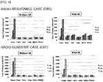
FIG. 4 shows the results of comparing phage concentrations obtained by eluting phages after extracting heart, lung, liver, spleen, kidney, and tumor during biopanning process performed three times. -
FIG. 5 shows the results confirming that, after injecting a peptide phage labeled with a Cy5.5 fluorescent probe into the mouse model of each population, the radio-resistant mouse (case C65;FIG. 5A ) on the 7th day of the injection and the radio-sensitive mouse (case C87;FIG. 5B ) on the 11th day of the injection were to be specifically bind to the cancer tissue in vivo based on images. -
FIG. 6 shows cross-validation results regarding specific binding of a B5 peptide sequence only to an irradiated population in radio-resistant case, by comparison with other control groups. -
FIG. 7 shows results obtained by which ROI values are calculated and standardized based on the cross-validation results regarding the B5 peptide. -
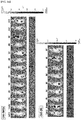
FIG. 8 shows results that verify excellent targeting ability of the B5 peptide through tissue immunohistochemistry. -
FIG. 9 shows colorectal cancer tissues of a patient other than the existing radio-sensitive/radio-resistant colorectal cancer tissues of patients according to Example 7 of the present invention, and provides results for confirming selective targeting ability of the B5 peptide in each population of the radio-sensitive case the ratio-resistant patient case depending on the presence of irradiation. - The present invention provides a peptide for targeting colorectal cancer, the peptide including an amino acid sequence selected from the group consisting of SEQ ID NOs: 1 to 40. The above-mentioned amino acid sequences are shown in Tables 1 and 2.
- The peptide of the present invention is a low-molecular weight peptide consisting of 7 amino acids. Such a low-molecular weight peptide is small in size so that it can be stabilized three-dimensionally. In addition, a low-molecular weight peptide has the advantage of being able to easily pass through a membrane and to recognize a target molecule deep in tissues. Since the stability of the low-molecular weight peptide of the present invention is secured through local injection and the immunoreactivity can be minimized, there is an advantage that cancer can be diagnosed early. In addition, the mass production of the low-molecular weight peptide of the present invention is relatively easy compared that of an antibody, and the toxicity of the low-molecular weight peptide of the present invention is weak.
- In addition, the low-molecular weight peptide of the present invention is has an advantage of a strong binding force to a target material compared to an antibody, and do not undergo denaturation during thermal/chemical treatment. In addition, due to a small molecular size, the low-molecular weight peptide can be used as a fused protein as being attached to other proteins. In detail, the low-molecular weight peptide can be also used as being attached to a high-molecular weight protein chain, and accordingly, can be used as a diagnosis kit and a drug delivery carrier.
- The low-molecular weight peptide of the present invention can be easily prepared according to the chemical process known in the art (Creighton, Proteins; Structures and Molecular Principles, W. H. Freeman and Co., NY, 1983). As representative methods, liquid or solid phase synthesis, fractional condensation, F-MOC or T-BOC chemical method, or the like may be used (Chemical Approaches to the Synthesis of Peptides and Proteins, Williams et al., Eds., CRC Press, Boca Raton Florida, 1997; A Practical Approach, Athert on & Sheppard, Eds., IRL Press, Oxford, England, 1989), but the method is not limited thereto.
- In addition, the low-molecular weight peptide of the present invention can be prepared according to a genetic engineering method. First, according to a conventional method, a DNA sequence encoding the sequence low-molecular weight peptide is prepared. Here, a DNA sequence can be prepared by PCR amplification using an appropriate primer. Alternatively, according to a standard method known in the art, a DNA sequence can be synthesized using, for example, an automatic DNA synthesizer (manufactured by Biosearch or AppliedBiosystems). Such a synthesized DNA sequence is inserted to a vector including one or more expression control sequences (for example: a promoter, an enhancer, or the like) that are operatively linked to the DNA sequence to control expression of the DNA sequence, and then, a host cell is transformed with a recombinant expression vector prepared therefrom. A resulting transformant is cultured in an appropriate medium under suitable conditions to allow the expression of the DNA sequence, so that substantially pure peptides that are encoded by the DAN sequence are recovered from the culture. Such recovery may be performed according to a method known in the art (for example, chromatography). The term 'substantially pure peptides' used herein refers to peptides that do not substantially include any protein derived from the host.
- In addition, the present invention provides a peptide for targeting colorectal cancer, the peptide being specific to an irradiated colorectal cancer tissue and including an amino acid sequence selected from the group consisting of SEQ ID NOs: 12 to 15, 17, and 33 to 40.
- In detail, a peptide including an amino acid sequence selected from the group consisting of SEQ ID NOs: 12 to 15, 17, and 33 to 40 specifically binds to a colorectal cancer tissue, in particular, an irradiated colorectal cancer tissue.
- The term "target" or "specific" used herein refers to the ability to specifically bind only to a colorectal cancer tissue, especially an irradiated colorectal cancer tissue, without binding to other normal tissues. A colorectal cancer-specific peptide can specifically bind to the inside or outside of a colorectal cancer tissue.
- In addition, the present invention provides a peptide for targeting colorectal cancer, the peptide being specific to a radio-resistant colorectal cancer tissue and including an amino acid sequence selected from the group consisting of SEQ ID NOs: 12 to 15.
- In addition, the present invention provides a peptide for targeting colorectal cancer, the peptide being specific to a radio-sensitive colorectal cancer tissue and including an amino acid sequence selected from the group consisting of SEQ ID NOs: 33 to 40.
- The term "radio-resistance" used herein refers to a condition that is not easily influenced by irradiation and has little change in cell repair or proliferation upon exposure to radiation.
- The term "radio-sensitive" used herein refers to a condition that is easily influenced by irradiation and shows changes in cell repair or proliferation upon exposure to radiation.
- In the present invention, by determining radio-resistance or radio-sensitivity of target colorectal cancer to be treated with radiation therapy, such determination can be used to decide a radiation dose for the radiation therapy, and accordingly, the efficiency of the radiation therapy and therapeutic results may be improved.
- In addition, the present invention provides a polynucleotide encoding an amino acid sequence selected from the group consisting of SEQ ID NOs: 1 to 40.
- The term "polynucleotide" used herein refers to a single-stranded or doublestranded polymer of deoxyribonucleotides or ribonucleotides. Such a polynucleotide includes a RNA genome sequence, a DNA sequence (for example, gDNA and cDNA), and a RNA sequence transcribed from the DNA sequence. Unless otherwise mentioned, a polynucleotide includes an analog of a natural polynucleotide.
- The polynucleotide includes not only a nucleotide sequence that encodes the peptide for targeting colorectal cancer, but also a sequence complementary to the nucleotide sequence, wherein such a complementary sequence includes not only a perfectly complementary sequence, but also a substantially complementary sequence.
- In addition, the polynucleotide may be subjected to modifications. Such modifications include addition, deletion, non-conservative substitution, or conservative substitution of a nucleotide. The polynucleotide encoding the amino acid sequence is also interpreted to include a nucleotide sequence that exhibits substantial identity to the nucleotide sequence. The substantial identity is obtained by aligning the nucleotide sequence with any other sequences to the greatest extent and by analyzing the aligned sequence using algorithms commonly used in the art, and in this regard, the substantial identity may indicate a sequence having at least 80% homology, at least 90% homology, or at least 95% homology with the aligned sequence.
- In addition, the present invention provides a composition for diagnosing colorectal cancer, the composition including a peptide including an amino acid sequence selected from the group consisting of SEQ ID NOs: 1 to 40.
- In addition, the present invention provides a composition for diagnosing radio-sensitive colorectal cancer, the composition including a peptide being specific to an irradiated colorectal cancer tissue and including an amino acid sequence selected from the group consisting of SEQ ID NOs: 12 to 15, 17, and 33 to 40.
- In addition, the present invention provides a composition for diagnosing prognosis of colorectal cancer based on radiation, the composition including a peptide for targeting colorectal cancer, wherein the peptide includes an amino acid sequence selected from the group consisting of SEQ ID NOs: 12 to 15 or 33 to 40.
- In detail, the diagnosis of prognosis of colorectal cancer based on radiation refers to determination of radio-resistance or radio-sensitivity of colorectal cancer.
- The term "diagnosis" used herein refers to identification of the presence or characteristic of a pathological condition. For the purpose of the present invention, the diagnosis is to identify the presence or characteristic of colorectal cancer.
- The diagnosis of colorectal cancer using the peptide of the present invention may be performed by detecting binding of the peptide of the present invention to a corresponding tissue or cell directly obtained from blood, urine, or biopsy.
- In addition, to easily confirm, detect, and quantify binding of the peptide of the present invention to the colorectal cancer tissue, the peptide of the present invention can be provided in a labeled state. That is, the peptide provided herein may be linked to a detectable label (for example, via covalent binding or cross-linking). The detectable label may be a chromogenic enzyme (for example, peroxidase and alkaline phosphatase), a radioactive isotope (for example, 124I, 125I, 111In, 99mTc, 32P, and 35S), a chromophore, or a luminescent material or a fluorescent material (for example, FITC, RITC, rhodamine, cyanine, Texas Red, fluorescein, phycoerythrin, or quantum dots).
- Similarly, the detectable label may be an antibody-epitope, a substrate, a cofactor, an inhibitor, or a affinity ligand. Such labeling may be performed during the synthesis of the peptide of the present invention, or may be additionally performed on a peptide that is already synthesized. When using a fluorescent material is used as a detectable label, cancer may be diagnosed according to fluorescence mediated tomography (FMT). For example, the peptide of the present invention labeled with a fluorescent material may be circulated into the blood, and the fluorescence by the peptide may be observed by FMT. If fluorescent is observed, it is diagnosed as cancer.
- In addition, the present invention provides a composition for delivering a drug, the composition including the peptide for targeting colorectal cancer.
- The peptide of the present invention may be used as an intelligent drug delivery vehicle that selectively delivers a drug to a cancer tissue. If the peptide of the present invention is used in combination with drugs of the related art in terms of treatment of cancer, the peptide of the present invention selectively delivers a drug only to a cancer tissue and a cancer cell, so that drug efficacy may be increased while drug side effects on a normal tissue may be significantly reduced at the same time.
- For use as the drug, any anticancer drug that is conventionally used in the treatment of cancer can be used so long as the anticancer drug is able to be linked to the peptide of the present invention. Examples of the drug include cisplatin, 5-fluorouracil, adriamycin, methotrexate, vinblastine, busulfan, chlorambucil, cyclophosphamide, melphalan, nitrogen mustard, nitreosourea, taxol, paclitaxel, docetaxel, 6-mercapropurine, 6-thioguanine, bleomycin, daunorubicin, doxorubicin, epirubicin, idarubicin, mitomycin-C, and hydroxyurea. In addition, the linking of the anticancer drug to the peptide of the present invention may be performed by a method known in the art, for example, covalent bonding, cross linking, or the like. For this purpose, the peptide of the present invention may be, if necessary, subjected to chemical modifications to the extent that the activity thereof is not lost.
- Hereinafter, to promote understanding of one or more exemplary embodiments, reference has been made in detail to embodiments. The present invention, however, may be embodied in many different forms and should not be construed as being limited to the exemplary embodiments set forth herein. Rather, these embodiments are provided so that this disclosure will be thorough and complete, and will fully convey the scope of the present invention to one of ordinary skill in the art.
- In order to reproduce patient's cancer microenvironments as similar as possible, a cancer tissue extracted from an actual patient having colorectal cancer was transplanted into a mouse, thereby establishing an animal model most similar to a patient having cancer. Accordingly, an ideal animal model of cancer that has overcome limitations that existing animal models transplanted with cultured cancer cells had was obtained.
- Regarding the establishment of such an animal model transplanted with a patient's colorectal cancer tissue, the cancer tissue extracted from a patient was cultured in an NOD/SCID mouse. Once the cancer tissue was found in the NOD/SCID mouse, subculturing was carried out by using a Balb/c nude mouse, beginning from the next subculturing. All experiments related to the establishment of the corresponding mouse model were carried out after approval under the strict regulations by of the Clinical Research Ethics Committee of the Asan Medical Center and the Ethics Committee for Animal Experiments of the Asan Institute for Life Science. In addition, the patient's cancer tissue was distributed from the Bio Research Center (BRC) of the Asan Medical Center via a regulated route.
- Then, the distributed cancer tissue was kept in a specific preservative solution and transferred directly to an animal chamber, so as to maintain a state in which the cancer tissue was not separated from a patient yet. After an NOD/SCID mouse was anesthetized via intraperitoneal injection of anesthetics, the cancer tissue was cut into pieces each having a size of 3x3 mm. Next, each of both flanks of the NOD/SCID mouse was transplanted with a piece of the cut cancer tissues, and sutured. The NOD/SCID mouse was then recovered on a heating pad for about 2 hours (conditions of the preservative solution: HBSS containing 100 IU/ml penicillin, 100 µg/ml streptomycin, 100 µg/ml gentamicin, 2.5 µg/ml amphotericin B, and 5% fetal bovine serum). After completion of the transplantation, the NOD/SCID mouse was raised until tumors were formed. Meanwhile, the formation and growth of tumors were checked at least once a week. When the transplanted cancer tissues grew to a size of at least 500 mm3, an extraction process was performed thereon. Then, the extracted cancer tissues were each cut into pieces each having a size of 3x3 mm for subculturing. A nude mouse was anesthetized via intraperitoneal injection of anesthetics, and a piece of the cut cancer tissues was transplanted subcutaneously on the right thigh of the nude mouse. A transplantation site was changed from the flank to the thigh so that irradiation can be locally done without affecting other organs during irradiation. That is, a finally used mouse model transplanted with a patient's colorectal cancer tissue in the present invention was a mouse model established in a way that a cancer tissue that underwent subculturing four times was re-transplanted to form a cancer tissue. When the size of the cancer tissue was increased to about 250 mm3, only the cancer tissue was irradiated with 2 grays (Gy) of radiation. After having recovery time for no longer than 24 hours, in vivo peptide screening was performed. Here, a non-irradiated population, which is a control group, was also subjected to screening in a separate manner. The establishment of such a mouse model was performed in the same manner for two colorectal cancer tissues, and the results thereof are shown in
FIG. 1 . - In order to complete drawing of growth curves of distinct cancer after the formation of tumor was confirmed in the mouse transplanted with the patient's cancer tissue, two different animal models transplanted with the patient's colorectal cancer tissue were prepared so that growth curves of cancer were obtained from each of the two different animal models. The growth curves obtained therefrom were then utilized as the standard for control groups of an invention in the future. Here, the tumor size was measured by using a caliper every 2-3 days, and the resulting measurement value was substituted in a formula for calculating the tumor volume, thereby drawing the growth curves (Formula for calculating tumor size: Tumor volume = a2b/2 (a: a short diameter of tumor, b: a long diameter of tumor)).
- For each patient case, 6 subcultured (P3) nude mice were prepared to be divided into two groups of three mice, and an experiment was carried out with an irradiated population and a non-irradiated population as a control group. When the tumor size was increased up to 100 mm3, the right hind leg of the mouse where tumor was formed was irradiated with 2 Gy of radiation. Beginning from the first day after the irradiation, the tumor size was measured until it was increased up to about 1,000 mm3, thereby plotting the tumor volume and completing drawing the growth curves. Based on these results and depending on the sensitivity to the radiotherapy, patient cases were classified into radio-sensitive patient groups (also known as case C87) and radio-resistant cases (also known as case C65). The irradiation was performed by using a 6 MV photon beam linear accelerator (CL/1800, Varian, CA, USA) and a 1 cm bolus. The corresponding results of the above contents are shown in
FIG. 2 . - Regarding the mouse model transplanted with the patient's colorectal cancer tissue and classified depending on the radio-sensitivity in Example 2, a method for identifying a peptide having high specificity during in vivo peptide screening using a random loop peptide library was designed. For use as the library, a loop peptide library manufactured to have about 2.7 billion different amino acids sequences via random array of 7 amino acids [(i.e., a library fused with an M13 phage gp3 minor coat protein)-Ph.D™ phage display peptide library kit, New England Biolabs(NEB)] was purchased. To screen a peptide showing specific binding in each of the irradiated population (2 groups) and the control group (2 groups) of the established mouse model, a M13 phage peptide library was injected into the tail vein of the mouse so that the M13 phage peptide library was circulated in vivo through the tail vein of the mouse (also known as a method of binding an in vivo cancer tissue with an M13 phage peptide library). Then, a peptide-expressing phage specifically binding to the cancer tissue only was selected, and such a screening scheme is shown in
FIG. 3 . In detail,FIG. 3 shows an M13 phage screening scheme for screening a cancer tissue-targeting peptide, wherein (1) a mouse model in which a cancer tissue was formed on the right thigh was established, (2) a phage library in which a loop peptide library consisting of 7 amino acids was expressed on a surface of a M13 phage was injected into the tail vein of the mouse to allow in vivo circulation of the phage library for 15 minutes, (3) undesired phages were washed (also known as perfusing), and (4) the cancer tissue was finally extracted and homogenized to elute phages bound with the cancer tissue. The eluted phages infected Escherichia coli, and through amplification of the phages and counting of the number of the phages (also known as titering), the resulting phages were injected again into the tail vein of the mouse to allow in vivo circulation. By repeating such cycles, the number of repetition also increased, and accordingly, a process of screening phages that were gradually specific to the cancer tissue and showed a strong binding strength was performed (also known as biopanning). Biopanning was finally performed three times in total so that a phage expressing a sequence of a peptide specifically binding to the patient's in vivo colorectal cancer tissue was obtained. In addition, to confirm whether the peptide-expressing phage selected above targeted the cancer tissue only, other in vivo organs were also subjected to comparison. That is, for every biopanning, phages were eluted from each of extracted heart, lung, liver, spleen, kidney, and cancer tissue, and the phage concentration was measured for comparison. The results of the comparison are shown inFIG. 4 . Finally, the obtained phages infected E. coli ER2738 cells that are host cells, and were subjected to amplification in an LB medium. Then, 100 phage plaques were selected randomly, and the M13 phage genomic DNA (single-stranded circular DNA) was separated and purified to identify a gene sequence, thereby identifying an amino acid sequence of a peptide expressed in a phage-surface protein (e.g., a gp3 minor coat protein) and targeting the cancer tissue. The same procedures were carried out on each of the irradiated group (2 groups) and the control group (2 groups) of Example 2 in terms of the in vivo peptide screening. The Clustal W program for sequence analysis was used to summarize peptide sequences, and the results are shown in Tables 1 and 2.[Table 1] Patient case Irradiation No. Peptide sequence Radio-resistant colorectal cancer tissue of patient Non-irradiated population A1 SHMEYPR (SEQ ID NO: 1) (case C65) A2 NTGSPYE (SEQ ID NO: 2) A3 SNVYDHN (SEQ ID NO: 3) A4 STEHTRS (SEQ ID NO: 4) A5 SWGDRIL (SEQ ID NO: 5) A6 LQPKASQ (SEQ ID NO: 6) A7 ASKSTHD (SEQ ID NO: 7) A8 DVSGNW (SEQ ID NO: 8) A9 DLLHRGA (SEQ ID NO: 9) A10 MARYMSA (SEQ ID NO: 10) A11 NTWMKSK (SEQ ID NO: 11) Irradiated population B1 STMNGKV (SEQ ID NO: 12) B2 SHMEYPR (SEQ ID NO: 1) B3 TVRSSAE (SEQ ID NO: 13) B4 VPSQKSK (SEQ ID NO: 14) B5 TPSFSKI (SEQ ID NO: 15) B6 GYSSFNR (SEQ ID NO: 16) B7 LKLGEKW (SEQ ID NO: 17) [Table 2] Radio-sensitive colorectal cancer tissue of patient (case C87) Non-irradiated population E1 SHMEYPR (SEQ ID NO: 1) E2 GYSSFNR (SEQ ID NO: 16) E3 NTGSPYE (SEQ ID NO: 2) E4 VQMPAHS (SEQ ID NO: 18) E5 GGNSNQR (SEQ ID NO: 19) E6 TAFSLGK (SEQ ID NO: 20) E7 QQTKNYY (SEQ ID NO: 21) E8 AIKNTKS (SEQ ID NO: 22) E9 TVRTSAV (SEQ ID NO: 23) E10 LTGSVNK (SEQ ID NO: 24) E11 MNFIQKN (SEQ ID NO: 25) E12 DETSGQI (SEQ ID NO: 26) E13 KISLPHN (SEQ ID NO: 27) E14 AFKSPSG (SEQ ID NO: 28) E15 LKTSGTY (SEQ ID NO: 29) E16 RSTGSSC (SEQ ID NO: 30) E17 YVSKNNS (SEQ ID NO: 31) E18 HKTEHRS (SEQ ID NO: 32) Irradiated population F1 KLTQMM (SEQ ID NO: 33) F2 LKLGEKW (SEQ ID NO: 17) F3 SFFPFST (SEQ ID NO: 34) F4 DNSKLVE (SEQ ID NO: 35) F5 QLWQKEQ (SEQ ID NO: 36) F6 NTGSPYE (SEQ ID NO: 2) F7 LGSVGRD (SEQ ID NO: 37) F8 GQSGARF (SEQ ID NO: 38) F9 NGNSNTL (SEQ ID NO: 39) F10 TNPYPLD (SEQ ID NO: 40) - To verify whether the obtained phage expressing a loop peptide has targeted the in vivo transplanted cancer tissue and to confirm targeting efficiency of the obtained phage, in vivo imaging was performed.
- In detail, regarding in vivo imaging verification, the peptide-expressing phage was amplified, and a process of linking a fluorescent probe (Cy5.5) was performed. In particular, 1 µg/µl of N-hydroxysuccinimide esters of Cy5.5 (Amersham) was added to 1 mL of bicarbonate buffer (pH 8.3) having the phage concentration of 1012 plaque forming units (pfu), and then, in a condition where a dark environment was maintained, a phage-surface protein was linked to the Cy5.5 fluorescent probe at room temperature for 3 hours. That is, amine groups of the phage-surface protein were chemically linked to N-hydroxysuccinimide esters of the Cy5.5 fluorescent probe via a covalent bond. The peptide-expressing phages to which the Cy5.5 fluorescent probe was linked were each obtained by precipitation with 170 µl of 20%(w/v) PEG 8000/2.5 M NaCl solution and purification. To determine a proportion of the Cy5.5 fluorescent probe linked to each of the finally obtained phage samples, an IVIS spectrum imaging system (Xenogen) was used for measurement, and region of interest (ROI) values were determined using the software program of a corresponding device. Accordingly, it was confirmed that the Cy5.5 fluorescent probe was linked to each of the phage samples at almost the same linking proportion.
- After each of the prepared phage samples was injected into the mouse model of Example 1 through the vein tail of the mouse, images thereof were measured once a day immediately after the injection, thereby confirming in vivo circulation and discharge process of the peptide-expressing phage. Although there were difference in populations, the images were measured from the last 7th day to the last 11th day, thereby screening the peptide-expressing phage verified to target the cancer tissue only. In addition, on the last day of the in vivo imaging, the cancer tissues were each extracted, to thereby obtain ex vivo images thereof. The results are shown in
FIG. 5 . - To compare the targeting efficiency of the peptide-expressing phage selected from each population based on the results of
FIG. 5 , the pieces of the extracted cancer tissue were collected independently, and phages bound to the cancer tissues were eluted. The concentration of the eluted phages was measured as well as the weight of each cancer tissue in terms of establishing numerical standardization. In this regard, the concentration of the phages relative to the weight of the cancer tissue was calculated and quantified. In addition, to quantify the results obtained from the in vivo imaging and the ex vivo imaging according to the fluorescence intensity, ROI values were calculated to obtain total flux values. Each of the values being calculated and quantified was standardized by comparing the control group with the mouse injected by processing a Cy5.5 probe with a phage-free Cy5.5 dye and an empty phage where a peptide was not expressed. The results are shown in Tables 3 and 4. Based on these results of Tables 3 and 4, peptide sequences each having 4 different sequences were selected in each population. When compared with sequences of other peptide-expressing phages, it was confirmed that the selected peptides had excellent targeting ability.[Table 3] Patient case Irradiation Sample pfu/mg in vivo ROI ex vivo ROI Radio-resistant colorectal cancer tissue of patient (case C65) Non-irradiated population Cy5.5 - 1.00 1.00 Empty 0.23 1.43 1.16 A1 136.36 1.77 1.27 A2 181.48 2.04 1.61 A3 34.8 1.62 0.67 A4 264.86 3.20 1.83 A5 51.38 2.16 1.52 A6 15.4 2.75 1.47 A7 27.14 1.80 1.08 A8 8.56 2.99 1.57 A9 54.17 2.35 0.56 A10 183.16 2.26 1.58 A11 6.71 1.93 0.95 Irradiated population Cy5.5 - 1.00 1.00 Empty - 1.40 0.72 B1 16.67 1.52 1.48 B2 230 2.27 1.89 B3 195.40 2.38 1.63 B4 20.62 2.12 1.36 B5 209.47 4.07 2.43 B6 211.76 3.03 2.06 B7 3.45 1.72 0.62 [Table 4] Patient case Irradiation Sample pfu/mg in vivo ROI ex vivo ROI Radio-sensitive colorectal cancer tissue of patient (case C87) Non-irradiated population Cy5.5 - 1.00 1.00 Empty 1.40 1.21 0.82 E1 74.12 1.68 1.72 E2 3.96 1.12 0.62 E3 1.55 1.13 0.77 E4 5.00 0.84 0.94 E5 15.96 0.92 0.76 E6 4.24 0.78 0.77 E7 58.32 2.02 1.38 E8 15.00 0.90 0.49 E9 86.40 1.27 1.20 E10 2.25 1.19 1.00 E11 4.43 1.05 0.34 E12 5.56 1.24 1.07 E13 1.22 1.50 0.93 E14 2.89 0.70 0.55 E15 6.13 0.77 0.47 E16 2.14 0.50 0.24 E17 204.00 1.66 1.29 E18 4.15 0.69 0.21 Irradiated population Cy5.5 - 1.00 1.00 Empty 8.18 0.96 0.83 F1 6.73 1.63 2.39 F2 1.90 1.00 2.92 F3 22.31 1.07 2.64 F4 69.18 1.59 3.11 F5 144.55 1.63 3.72 F6 4.75 1.15 2.64 F7 141.04 1.61 3.97 F8 6.10 1.18 1.92 F9 95.24 1.77 4.45 F10 7.86 1.91 1.74 - Among the various peptide-expressing phages selected from the above, the in vivo imaging was attempted again for a B5 peptide, which is a peptide sequence screened only from the irradiated population in the radio-resistant case (case C65). At the same time, to confirm the responsiveness in control groups other than the population of interest, a crossover experiment was carried out. In detail, in the presence of differences in irradiation between the radio-sensitive case and the radio-resistant case of Example 1, the in vivo imaging was carried out to determine the targeting of the B5 peptide sequence in 4 populations in total. Here, the irradiated population of case C65 was set as an experimental group while the non-irradiated population of case C65 and all the populations of case C87 were set as control groups. In the same manner as in Example 4, peptide-expressing phages were amplified and a process of linking a Cy5.5 fluorescent probe was carried out. Then, through the tail vein injection in each population, images thereof were observed until the end of the last 7th day. Consequently, when compared with a population in which injection was made by processing a Cy5.5 probe with a phage-free Cy5.5 dye and an empty phage where a peptide was not expressed, it was confirmed that only the irradiated population of the radio-resistant case (case C65) has exhibited excellent targeting ability.
FIG. 6 shows the results of the present embodiment, andFIG. 7 shows the results of standardization of ROI values. Consequently, regarding the B5 peptide sequence, it was confirmed that the B5 peptide sequence may be a sequence that targets a factor (e.g., a biomarker) inducing resistance against radiation during radiotherapy, and in other words, the B5 peptide sequence may be a sequence that was responsive to cancer microenvironments influenced by irradiation. - To verify histological targeting ability of the B5 peptide sequence verified in Example 5, the imaging of the B5 peptide sequence was obtained. Then, cancer tissues of each population were extracted to prepare paraffin blocks and slices. In detail, (1) the extracted cancer tissues were immersed in a formaldehyde solution at room temperature for 24 hours in terms of for immobilization. Then, following a dehydration process, paraffin was added to the solution through penetration to form paraffin blocks. Afterwards, a microtome was used to manufacture slices having a thickness of 3 µm. To perform real immunohistological staining, (2) following a deparaffinization process performed on the slices, (3) an unmasking process was performed so that structures of various proteins immobilized to the tissue slices were recovered to restore sites where antibodies normally bind. Sequentially, (4) a blocking process was performed using a 5% BSA solution, (5) primary antibodies were bound (wherein the antibodies used herein were anti-mouse M13 IgG recognizing M13 phage capsid proteins), (6) and secondary antibodies were bound while HRP was bound. Afterwards, (7) sites where phages were present were stained using DAB development, (7) the nuclei of the phages were stained with hematoxylin, and (8) a dehydration process was performed thereon. Once completed, mounting was performed so that the tissue slices that were immunohistochemically stained were permanently preserved. The results of immunohistochemical staining performed as described above are shown in
FIG. 8 . The results ofFIG. 7 clearly confirm that the B5 peptide sequence of Example 5 specifically bound only to the irradiated population in the radio-resistant colorectal cancer patient case (C65). - To confirm whether the selected B5 peptide having the ability to trace the radio-resistant factor exhibited the same targeting ability in a patient's radio-resistant colorectal tissue other than the patient's colorectal tissue used in the screening above, the B5 peptide was applied to a mouse transplanted with other colorectal cancer tissues of a patient. In detail, by extending the crossover experiment verified in Example 4, radio-
sensitive case 1 and radio-resistant case 1 regarding other colorectal cancer tissues of a patient characterized as adenocarcinoma were introduced in addition to the radio-sensitive case and the radio-resistant case of Example 1. In the same manner as in Example 4, the B5 peptide-expressing phages were amplified and a process of linking a Cy5.5 fluorescent probe was carried out. Regarding the confirmation of the targeting ability of the B5 peptide, the in vivo imaging was used for observation in the same manner as in Example 5. Consequently, it was confirmed that the radio-sensitive colorectal cancer tissue did not show any targeting ability regardless of irradiation, and that the radio-resistant colorectal cancer tissue exhibited targeting ability only in the case of irradiation.FIG. 9 shows the results of the present embodiment. In detail, the targeting ability of the B5 peptide sequence targeting a factor inducing resistance against radiation during radiation therapy in Example 6 was also verified in other colorectal cancer tissues of a radio-resistant patient, thereby confirming the possibility of the B5 peptide sequence as a sequence that is responsive to cancer microenvironments causing induction of resistivity during radiotherapy on colorectal cancer. However, in the patients having radio-sensitive colorectal cancer, it was clearly confirmed that the B5 peptide sequence, as a peptide sequence showing no targeting ability regardless of irradiation, was a sequence selectively tracing a radio-resistant factor. Ultimately, it was verified that the targeting ability of the B5 peptide sequence was not limited to the radio-resistant case including specific patients having colorectal cancer, but was rather applicable to other colorectal cancer tissues of a radio-resistant patient. In this regard, the limited range of application in clinical applications was overcome. - It should be understood that embodiments described herein should be considered in a descriptive sense only and not for purposes of limitation. Descriptions of features or aspects within each embodiment should typically be considered as available for other similar features or aspects in other embodiments. While one or more embodiments have been described with reference to the figures, it will be understood by those of ordinary skill in the art that various changes in form and details may be made therein without departing from the spirit and scope as defined by the following claims.


Claims (12)
- A peptide for targeting colorectal cancer, the peptide comprising an amino acid sequence selected from the group consisting of SEQ ID NOs: 1 to 40.
- The peptide of claim 1, wherein the peptide comprising an amino acid selected from the group consisting of SEQ ID NOs: 12 to 15, 17, and 33 to 40 is specific to an irradiated colorectal cancer tissue.
- The peptide of claim 2, wherein the peptide comprising an amino acid selected from the group consisting of SEQ ID NOs: 12 to 15 is specific to a colorectal cancer tissue that is radio-resistant.
- The peptide of claim 2, wherein the peptide comprising an amino acid selected from the group consisting of SEQ ID NOs: 33 to 40 is specific to a colorectal cancer tissue that is radio-sensitive.
- A polynucleotide encoding the peptide of claim 1.
- A composition for diagnosing colorectal cancer, the composition comprising the peptide of claim 1.
- A composition for diagnosing radio-sensitive colorectal cancer, the composition comprising the peptide of claim 2.
- The composition of claim 6 or 7, wherein the peptide is labeled with one selected from the group consisting of a chromogenic enzyme, a radioactive isotope, a chromopore, and a luminescent or fluorescent material.
- A composition for diagnosing prognosis of colorectal cancer based on radiation, the composition comprising the peptide of claim 3 or 4.
- The composition of claim 9, wherein the diagnosis of prognosis of colorectal cancer based on radiation is determined by radio-resistance or radio-sensitivity.
- A composition for delivering a drug, the composition comprising the peptide of claim 1.
- The composition of claim 11, wherein the drug comprises an anticancer drug.
Applications Claiming Priority (3)
| Application Number | Priority Date | Filing Date | Title |
|---|---|---|---|
| KR20140160817 | 2014-11-18 | ||
| KR1020150106580A KR101795140B1 (en) | 2014-11-18 | 2015-07-28 | Targeting Peptides for Colorectal Cancer and Medical Use Thereof |
| PCT/KR2015/007942 WO2016080632A1 (en) | 2014-11-18 | 2015-07-29 | Peptides for targeting colorectal cancer, and medical use thereof |
Publications (3)
| Publication Number | Publication Date |
|---|---|
| EP3222630A1 true EP3222630A1 (en) | 2017-09-27 |
| EP3222630A4 EP3222630A4 (en) | 2018-08-08 |
| EP3222630B1 EP3222630B1 (en) | 2019-12-04 |
Family
ID=56106109
Family Applications (1)
| Application Number | Title | Priority Date | Filing Date |
|---|---|---|---|
| EP15861389.3A Active EP3222630B1 (en) | 2014-11-18 | 2015-07-29 | Peptides for targeting colorectal cancer, and medical use thereof |
Country Status (3)
| Country | Link |
|---|---|
| US (1) | US10393744B2 (en) |
| EP (1) | EP3222630B1 (en) |
| KR (1) | KR101795140B1 (en) |
Families Citing this family (5)
| Publication number | Priority date | Publication date | Assignee | Title |
|---|---|---|---|---|
| KR102259726B1 (en) * | 2016-11-28 | 2021-06-02 | 재단법인 아산사회복지재단 | CCSP-2 Specific Peptide and Use thereof |
| CN110041400B (en) * | 2018-01-15 | 2022-07-29 | 中国医学科学院药物研究所 | Polypeptide and application thereof in early diagnosis of colorectal cancer |
| KR102387320B1 (en) * | 2020-08-14 | 2022-04-18 | 고려대학교 산학협력단 | composition for diagnosing or detecting colorectal cancer for predicting drug responsiveness, composition for preventing or treating colorectal cancer |
| CN116284255B (en) * | 2021-12-21 | 2024-08-02 | 中国医学科学院药物研究所 | Targeting polypeptide compound for early colorectal tumor and application |
| CN116063384A (en) * | 2023-01-31 | 2023-05-05 | 贵州医科大学 | A kind of fluorescent probe and its application |
Family Cites Families (8)
| Publication number | Priority date | Publication date | Assignee | Title |
|---|---|---|---|---|
| AU2002329759A1 (en) * | 2001-08-17 | 2003-03-03 | Monsanto Technology Llc | Methyltransferase genes and uses thereof |
| EP2544006A1 (en) | 2007-06-04 | 2013-01-09 | Diagnoplex | Biomarker combinations for colorectal cancer |
| KR101029881B1 (en) | 2007-12-14 | 2011-04-18 | 한국생명공학연구원 | Colorectal Cancer Diagnostic Markers Using Colorectal Cancer Overexpression Genes |
| CN103328496A (en) | 2010-10-25 | 2013-09-25 | 中央研究院 | Cancer targeting peptides and their applications in cancer therapy and diagnosis |
| US8691189B2 (en) | 2011-02-14 | 2014-04-08 | Institute Of Nuclear Energy Research, Atomic Energy Council | Method of colorectal cancer detection by using radiolabeled anti-GRP78 peptide |
| KR20130040493A (en) * | 2011-10-14 | 2013-04-24 | 전흥철 | Injection device of microorganism fermentation liquid for food garbage |
| KR101478826B1 (en) | 2011-10-28 | 2015-01-08 | 전남대학교산학협력단 | A novel diagnostic marker for colon cancer and a diagnostic kit for colon cancer using the same |
| US20140256586A1 (en) * | 2013-03-09 | 2014-09-11 | The Wistar Institute Of Anatomy And Biology | Methods and compositions for diagnosis of colorectal cancer |
-
2015
- 2015-07-28 KR KR1020150106580A patent/KR101795140B1/en not_active Expired - Fee Related
- 2015-07-29 US US15/527,716 patent/US10393744B2/en active Active
- 2015-07-29 EP EP15861389.3A patent/EP3222630B1/en active Active
Also Published As
| Publication number | Publication date |
|---|---|
| US20170328906A1 (en) | 2017-11-16 |
| US10393744B2 (en) | 2019-08-27 |
| EP3222630B1 (en) | 2019-12-04 |
| KR101795140B1 (en) | 2017-11-09 |
| KR20160059944A (en) | 2016-05-27 |
| EP3222630A4 (en) | 2018-08-08 |
Similar Documents
| Publication | Publication Date | Title |
|---|---|---|
| US10627402B2 (en) | Peptides for targeting gastric cancer, and medical use tehreof | |
| US10393744B2 (en) | Peptides for targeting colorectal cancer, and medical use thereof | |
| JP7386711B2 (en) | Methods for treating tumors | |
| US12458618B2 (en) | Compounds and methods for the detection of TRPV-6 cancers and drug delivery | |
| KR20210044247A (en) | MICRORNA-based treatment for LCP-1 positive cancer | |
| WO2009070767A2 (en) | Systemic instigation systems to study tumor growth or metastasis | |
| JP2014504149A (en) | Cancer imaging combined with treatment: Theranostics | |
| Ding et al. | Nanodrug modified with engineered cell membrane targets CDKs to activate aPD-L1 immunotherapy against liver metastasis of immune-desert colon cancer | |
| KR101467676B1 (en) | Targeting Peptide for Cancer and Medical Use Thereof | |
| EP4431524A2 (en) | Enhancing blood-brain barrier drug transport by targeting endogenous regulators | |
| KR20210007358A (en) | Method for enhancing migration of stem cells into cancer cells | |
| Conti et al. | Optical imaging detection of microscopic mammary cancer in ErbB‐2 transgenic mice through the DA364 probe binding αvβ3 integrins | |
| WO2016080632A1 (en) | Peptides for targeting colorectal cancer, and medical use thereof | |
| US9581598B2 (en) | Diagnosis and treatment of brain tumor | |
| EP3800251A1 (en) | Peptide having accumulation specific to pancreatic cancer, and use of said peptide | |
| 최진영 | Spatial transcriptomics-based identification of molecular markers for fluorescence labeled drug distribution | |
| Zhang et al. | Advanced extracellular vesicle immunotherapy | |
| CN119857133A (en) | MBNL3 protein and application of encoding gene thereof in preparation of tumor medicaments and tumor therapeutic medicaments | |
| CN118384291A (en) | A preparation for reversing drug resistance of diffuse large B-cell lymphoma and its preparation method and application | |
| Deb et al. | Cardiovascular Nanomedicine | |
| Sakai et al. | Increased fibrosis and impaired intratumoral accumulation of macromolecules in | |
| HK1169661B (en) | Soricidin derived peptides and methods for the detection of trpv-6 cancers and drug delivery | |
| 하승균 | Development of novel cyclic form of fluorephore-or radiotracer-labeled c-Met peptide to detect high-c-Met expressing lung cancer |
Legal Events
| Date | Code | Title | Description |
|---|---|---|---|
| STAA | Information on the status of an ep patent application or granted ep patent |
Free format text: STATUS: THE INTERNATIONAL PUBLICATION HAS BEEN MADE |
|
| PUAI | Public reference made under article 153(3) epc to a published international application that has entered the european phase |
Free format text: ORIGINAL CODE: 0009012 |
|
| STAA | Information on the status of an ep patent application or granted ep patent |
Free format text: STATUS: REQUEST FOR EXAMINATION WAS MADE |
|
| 17P | Request for examination filed |
Effective date: 20170619 |
|
| AK | Designated contracting states |
Kind code of ref document: A1 Designated state(s): AL AT BE BG CH CY CZ DE DK EE ES FI FR GB GR HR HU IE IS IT LI LT LU LV MC MK MT NL NO PL PT RO RS SE SI SK SM TR |
|
| AX | Request for extension of the european patent |
Extension state: BA ME |
|
| DAV | Request for validation of the european patent (deleted) | ||
| DAX | Request for extension of the european patent (deleted) | ||
| RIC1 | Information provided on ipc code assigned before grant |
Ipc: C07K 7/06 20060101ALI20180403BHEP Ipc: C07K 14/00 20060101AFI20180403BHEP Ipc: A61P 35/00 20060101ALI20180403BHEP Ipc: A61K 47/50 20170101ALI20180403BHEP Ipc: G01N 33/58 20060101ALI20180403BHEP Ipc: G01N 33/574 20060101ALI20180403BHEP |
|
| RIC1 | Information provided on ipc code assigned before grant |
Ipc: G01N 33/574 20060101ALI20180620BHEP Ipc: C07K 7/06 20060101ALI20180620BHEP Ipc: A61P 35/00 20060101ALI20180620BHEP Ipc: C07K 14/00 20060101AFI20180620BHEP Ipc: G01N 33/58 20060101ALI20180620BHEP Ipc: A61K 47/50 20170101ALI20180620BHEP |
|
| RIC1 | Information provided on ipc code assigned before grant |
Ipc: C07K 14/00 20060101AFI20180628BHEP Ipc: G01N 33/574 20060101ALI20180628BHEP Ipc: C07K 7/06 20060101ALI20180628BHEP Ipc: A61K 47/50 20170101ALI20180628BHEP Ipc: A61P 35/00 20060101ALI20180628BHEP Ipc: G01N 33/58 20060101ALI20180628BHEP |
|
| A4 | Supplementary search report drawn up and despatched |
Effective date: 20180706 |
|
| STAA | Information on the status of an ep patent application or granted ep patent |
Free format text: STATUS: EXAMINATION IS IN PROGRESS |
|
| 17Q | First examination report despatched |
Effective date: 20181023 |
|
| GRAP | Despatch of communication of intention to grant a patent |
Free format text: ORIGINAL CODE: EPIDOSNIGR1 |
|
| STAA | Information on the status of an ep patent application or granted ep patent |
Free format text: STATUS: GRANT OF PATENT IS INTENDED |
|
| INTG | Intention to grant announced |
Effective date: 20190627 |
|
| GRAS | Grant fee paid |
Free format text: ORIGINAL CODE: EPIDOSNIGR3 |
|
| GRAA | (expected) grant |
Free format text: ORIGINAL CODE: 0009210 |
|
| STAA | Information on the status of an ep patent application or granted ep patent |
Free format text: STATUS: THE PATENT HAS BEEN GRANTED |
|
| AK | Designated contracting states |
Kind code of ref document: B1 Designated state(s): AL AT BE BG CH CY CZ DE DK EE ES FI FR GB GR HR HU IE IS IT LI LT LU LV MC MK MT NL NO PL PT RO RS SE SI SK SM TR |
|
| REG | Reference to a national code |
Ref country code: GB Ref legal event code: FG4D |
|
| REG | Reference to a national code |
Ref country code: CH Ref legal event code: EP |
|
| REG | Reference to a national code |
Ref country code: AT Ref legal event code: REF Ref document number: 1209283 Country of ref document: AT Kind code of ref document: T Effective date: 20191215 |
|
| REG | Reference to a national code |
Ref country code: DE Ref legal event code: R096 Ref document number: 602015043223 Country of ref document: DE |
|
| REG | Reference to a national code |
Ref country code: IE Ref legal event code: FG4D |
|
| REG | Reference to a national code |
Ref country code: NL Ref legal event code: MP Effective date: 20191204 |
|
| REG | Reference to a national code |
Ref country code: LT Ref legal event code: MG4D |
|
| PG25 | Lapsed in a contracting state [announced via postgrant information from national office to epo] |
Ref country code: LT Free format text: LAPSE BECAUSE OF FAILURE TO SUBMIT A TRANSLATION OF THE DESCRIPTION OR TO PAY THE FEE WITHIN THE PRESCRIBED TIME-LIMIT Effective date: 20191204 Ref country code: NO Free format text: LAPSE BECAUSE OF FAILURE TO SUBMIT A TRANSLATION OF THE DESCRIPTION OR TO PAY THE FEE WITHIN THE PRESCRIBED TIME-LIMIT Effective date: 20200304 Ref country code: GR Free format text: LAPSE BECAUSE OF FAILURE TO SUBMIT A TRANSLATION OF THE DESCRIPTION OR TO PAY THE FEE WITHIN THE PRESCRIBED TIME-LIMIT Effective date: 20200305 Ref country code: LV Free format text: LAPSE BECAUSE OF FAILURE TO SUBMIT A TRANSLATION OF THE DESCRIPTION OR TO PAY THE FEE WITHIN THE PRESCRIBED TIME-LIMIT Effective date: 20191204 Ref country code: SE Free format text: LAPSE BECAUSE OF FAILURE TO SUBMIT A TRANSLATION OF THE DESCRIPTION OR TO PAY THE FEE WITHIN THE PRESCRIBED TIME-LIMIT Effective date: 20191204 Ref country code: BG Free format text: LAPSE BECAUSE OF FAILURE TO SUBMIT A TRANSLATION OF THE DESCRIPTION OR TO PAY THE FEE WITHIN THE PRESCRIBED TIME-LIMIT Effective date: 20200304 Ref country code: FI Free format text: LAPSE BECAUSE OF FAILURE TO SUBMIT A TRANSLATION OF THE DESCRIPTION OR TO PAY THE FEE WITHIN THE PRESCRIBED TIME-LIMIT Effective date: 20191204 |
|
| PG25 | Lapsed in a contracting state [announced via postgrant information from national office to epo] |
Ref country code: RS Free format text: LAPSE BECAUSE OF FAILURE TO SUBMIT A TRANSLATION OF THE DESCRIPTION OR TO PAY THE FEE WITHIN THE PRESCRIBED TIME-LIMIT Effective date: 20191204 Ref country code: HR Free format text: LAPSE BECAUSE OF FAILURE TO SUBMIT A TRANSLATION OF THE DESCRIPTION OR TO PAY THE FEE WITHIN THE PRESCRIBED TIME-LIMIT Effective date: 20191204 |
|
| PG25 | Lapsed in a contracting state [announced via postgrant information from national office to epo] |
Ref country code: AL Free format text: LAPSE BECAUSE OF FAILURE TO SUBMIT A TRANSLATION OF THE DESCRIPTION OR TO PAY THE FEE WITHIN THE PRESCRIBED TIME-LIMIT Effective date: 20191204 |
|
| PG25 | Lapsed in a contracting state [announced via postgrant information from national office to epo] |
Ref country code: EE Free format text: LAPSE BECAUSE OF FAILURE TO SUBMIT A TRANSLATION OF THE DESCRIPTION OR TO PAY THE FEE WITHIN THE PRESCRIBED TIME-LIMIT Effective date: 20191204 Ref country code: PT Free format text: LAPSE BECAUSE OF FAILURE TO SUBMIT A TRANSLATION OF THE DESCRIPTION OR TO PAY THE FEE WITHIN THE PRESCRIBED TIME-LIMIT Effective date: 20200429 Ref country code: CZ Free format text: LAPSE BECAUSE OF FAILURE TO SUBMIT A TRANSLATION OF THE DESCRIPTION OR TO PAY THE FEE WITHIN THE PRESCRIBED TIME-LIMIT Effective date: 20191204 Ref country code: RO Free format text: LAPSE BECAUSE OF FAILURE TO SUBMIT A TRANSLATION OF THE DESCRIPTION OR TO PAY THE FEE WITHIN THE PRESCRIBED TIME-LIMIT Effective date: 20191204 Ref country code: NL Free format text: LAPSE BECAUSE OF FAILURE TO SUBMIT A TRANSLATION OF THE DESCRIPTION OR TO PAY THE FEE WITHIN THE PRESCRIBED TIME-LIMIT Effective date: 20191204 |
|
| PG25 | Lapsed in a contracting state [announced via postgrant information from national office to epo] |
Ref country code: SM Free format text: LAPSE BECAUSE OF FAILURE TO SUBMIT A TRANSLATION OF THE DESCRIPTION OR TO PAY THE FEE WITHIN THE PRESCRIBED TIME-LIMIT Effective date: 20191204 Ref country code: SK Free format text: LAPSE BECAUSE OF FAILURE TO SUBMIT A TRANSLATION OF THE DESCRIPTION OR TO PAY THE FEE WITHIN THE PRESCRIBED TIME-LIMIT Effective date: 20191204 Ref country code: IS Free format text: LAPSE BECAUSE OF FAILURE TO SUBMIT A TRANSLATION OF THE DESCRIPTION OR TO PAY THE FEE WITHIN THE PRESCRIBED TIME-LIMIT Effective date: 20200404 |
|
| REG | Reference to a national code |
Ref country code: DE Ref legal event code: R097 Ref document number: 602015043223 Country of ref document: DE |
|
| REG | Reference to a national code |
Ref country code: AT Ref legal event code: MK05 Ref document number: 1209283 Country of ref document: AT Kind code of ref document: T Effective date: 20191204 |
|
| PLBE | No opposition filed within time limit |
Free format text: ORIGINAL CODE: 0009261 |
|
| STAA | Information on the status of an ep patent application or granted ep patent |
Free format text: STATUS: NO OPPOSITION FILED WITHIN TIME LIMIT |
|
| PG25 | Lapsed in a contracting state [announced via postgrant information from national office to epo] |
Ref country code: ES Free format text: LAPSE BECAUSE OF FAILURE TO SUBMIT A TRANSLATION OF THE DESCRIPTION OR TO PAY THE FEE WITHIN THE PRESCRIBED TIME-LIMIT Effective date: 20191204 Ref country code: DK Free format text: LAPSE BECAUSE OF FAILURE TO SUBMIT A TRANSLATION OF THE DESCRIPTION OR TO PAY THE FEE WITHIN THE PRESCRIBED TIME-LIMIT Effective date: 20191204 |
|
| 26N | No opposition filed |
Effective date: 20200907 |
|
| PG25 | Lapsed in a contracting state [announced via postgrant information from national office to epo] |
Ref country code: SI Free format text: LAPSE BECAUSE OF FAILURE TO SUBMIT A TRANSLATION OF THE DESCRIPTION OR TO PAY THE FEE WITHIN THE PRESCRIBED TIME-LIMIT Effective date: 20191204 Ref country code: PL Free format text: LAPSE BECAUSE OF FAILURE TO SUBMIT A TRANSLATION OF THE DESCRIPTION OR TO PAY THE FEE WITHIN THE PRESCRIBED TIME-LIMIT Effective date: 20191204 Ref country code: AT Free format text: LAPSE BECAUSE OF FAILURE TO SUBMIT A TRANSLATION OF THE DESCRIPTION OR TO PAY THE FEE WITHIN THE PRESCRIBED TIME-LIMIT Effective date: 20191204 |
|
| PG25 | Lapsed in a contracting state [announced via postgrant information from national office to epo] |
Ref country code: IT Free format text: LAPSE BECAUSE OF FAILURE TO SUBMIT A TRANSLATION OF THE DESCRIPTION OR TO PAY THE FEE WITHIN THE PRESCRIBED TIME-LIMIT Effective date: 20191204 |
|
| PG25 | Lapsed in a contracting state [announced via postgrant information from national office to epo] |
Ref country code: MC Free format text: LAPSE BECAUSE OF FAILURE TO SUBMIT A TRANSLATION OF THE DESCRIPTION OR TO PAY THE FEE WITHIN THE PRESCRIBED TIME-LIMIT Effective date: 20191204 |
|
| REG | Reference to a national code |
Ref country code: CH Ref legal event code: PL |
|
| GBPC | Gb: european patent ceased through non-payment of renewal fee |
Effective date: 20200729 |
|
| REG | Reference to a national code |
Ref country code: BE Ref legal event code: MM Effective date: 20200731 |
|
| PG25 | Lapsed in a contracting state [announced via postgrant information from national office to epo] |
Ref country code: CH Free format text: LAPSE BECAUSE OF NON-PAYMENT OF DUE FEES Effective date: 20200731 Ref country code: GB Free format text: LAPSE BECAUSE OF NON-PAYMENT OF DUE FEES Effective date: 20200729 Ref country code: FR Free format text: LAPSE BECAUSE OF NON-PAYMENT OF DUE FEES Effective date: 20200731 Ref country code: LI Free format text: LAPSE BECAUSE OF NON-PAYMENT OF DUE FEES Effective date: 20200731 Ref country code: LU Free format text: LAPSE BECAUSE OF NON-PAYMENT OF DUE FEES Effective date: 20200729 |
|
| PG25 | Lapsed in a contracting state [announced via postgrant information from national office to epo] |
Ref country code: BE Free format text: LAPSE BECAUSE OF NON-PAYMENT OF DUE FEES Effective date: 20200731 |
|
| PG25 | Lapsed in a contracting state [announced via postgrant information from national office to epo] |
Ref country code: IE Free format text: LAPSE BECAUSE OF NON-PAYMENT OF DUE FEES Effective date: 20200729 |
|
| PG25 | Lapsed in a contracting state [announced via postgrant information from national office to epo] |
Ref country code: TR Free format text: LAPSE BECAUSE OF FAILURE TO SUBMIT A TRANSLATION OF THE DESCRIPTION OR TO PAY THE FEE WITHIN THE PRESCRIBED TIME-LIMIT Effective date: 20191204 Ref country code: MT Free format text: LAPSE BECAUSE OF FAILURE TO SUBMIT A TRANSLATION OF THE DESCRIPTION OR TO PAY THE FEE WITHIN THE PRESCRIBED TIME-LIMIT Effective date: 20191204 Ref country code: CY Free format text: LAPSE BECAUSE OF FAILURE TO SUBMIT A TRANSLATION OF THE DESCRIPTION OR TO PAY THE FEE WITHIN THE PRESCRIBED TIME-LIMIT Effective date: 20191204 |
|
| PG25 | Lapsed in a contracting state [announced via postgrant information from national office to epo] |
Ref country code: MK Free format text: LAPSE BECAUSE OF FAILURE TO SUBMIT A TRANSLATION OF THE DESCRIPTION OR TO PAY THE FEE WITHIN THE PRESCRIBED TIME-LIMIT Effective date: 20191204 |
|
| PGFP | Annual fee paid to national office [announced via postgrant information from national office to epo] |
Ref country code: DE Payment date: 20230630 Year of fee payment: 9 |
|
| REG | Reference to a national code |
Ref country code: DE Ref legal event code: R119 Ref document number: 602015043223 Country of ref document: DE |
|
| PG25 | Lapsed in a contracting state [announced via postgrant information from national office to epo] |
Ref country code: DE Free format text: LAPSE BECAUSE OF NON-PAYMENT OF DUE FEES Effective date: 20250201 |