EP3213680A1 - Device for identifying the site of cardiac arrhythmias - Google Patents
Device for identifying the site of cardiac arrhythmias Download PDFInfo
- Publication number
- EP3213680A1 EP3213680A1 EP15855226.5A EP15855226A EP3213680A1 EP 3213680 A1 EP3213680 A1 EP 3213680A1 EP 15855226 A EP15855226 A EP 15855226A EP 3213680 A1 EP3213680 A1 EP 3213680A1
- Authority
- EP
- European Patent Office
- Prior art keywords
- locating
- intracavity
- electrodes
- cardiac arrhythmias
- dimensional reconstruction
- Prior art date
- Legal status (The legal status is an assumption and is not a legal conclusion. Google has not performed a legal analysis and makes no representation as to the accuracy of the status listed.)
- Granted
Links
Images
Classifications
-
- A—HUMAN NECESSITIES
- A61—MEDICAL OR VETERINARY SCIENCE; HYGIENE
- A61B—DIAGNOSIS; SURGERY; IDENTIFICATION
- A61B5/00—Measuring for diagnostic purposes; Identification of persons
- A61B5/24—Detecting, measuring or recording bioelectric or biomagnetic signals of the body or parts thereof
- A61B5/316—Modalities, i.e. specific diagnostic methods
- A61B5/318—Heart-related electrical modalities, e.g. electrocardiography [ECG]
- A61B5/346—Analysis of electrocardiograms
- A61B5/349—Detecting specific parameters of the electrocardiograph cycle
- A61B5/361—Detecting fibrillation
-
- A—HUMAN NECESSITIES
- A61—MEDICAL OR VETERINARY SCIENCE; HYGIENE
- A61B—DIAGNOSIS; SURGERY; IDENTIFICATION
- A61B5/00—Measuring for diagnostic purposes; Identification of persons
- A61B5/0033—Features or image-related aspects of imaging apparatus, e.g. for MRI, optical tomography or impedance tomography apparatus; Arrangements of imaging apparatus in a room
- A61B5/004—Features or image-related aspects of imaging apparatus, e.g. for MRI, optical tomography or impedance tomography apparatus; Arrangements of imaging apparatus in a room adapted for image acquisition of a particular organ or body part
- A61B5/0044—Features or image-related aspects of imaging apparatus, e.g. for MRI, optical tomography or impedance tomography apparatus; Arrangements of imaging apparatus in a room adapted for image acquisition of a particular organ or body part for the heart
-
- A—HUMAN NECESSITIES
- A61—MEDICAL OR VETERINARY SCIENCE; HYGIENE
- A61B—DIAGNOSIS; SURGERY; IDENTIFICATION
- A61B5/00—Measuring for diagnostic purposes; Identification of persons
- A61B5/0033—Features or image-related aspects of imaging apparatus, e.g. for MRI, optical tomography or impedance tomography apparatus; Arrangements of imaging apparatus in a room
- A61B5/0035—Features or image-related aspects of imaging apparatus, e.g. for MRI, optical tomography or impedance tomography apparatus; Arrangements of imaging apparatus in a room adapted for acquisition of images from more than one imaging mode, e.g. combining MRI and optical tomography
-
- A—HUMAN NECESSITIES
- A61—MEDICAL OR VETERINARY SCIENCE; HYGIENE
- A61B—DIAGNOSIS; SURGERY; IDENTIFICATION
- A61B5/00—Measuring for diagnostic purposes; Identification of persons
- A61B5/24—Detecting, measuring or recording bioelectric or biomagnetic signals of the body or parts thereof
- A61B5/25—Bioelectric electrodes therefor
- A61B5/279—Bioelectric electrodes therefor specially adapted for particular uses
- A61B5/28—Bioelectric electrodes therefor specially adapted for particular uses for electrocardiography [ECG]
-
- A—HUMAN NECESSITIES
- A61—MEDICAL OR VETERINARY SCIENCE; HYGIENE
- A61B—DIAGNOSIS; SURGERY; IDENTIFICATION
- A61B5/00—Measuring for diagnostic purposes; Identification of persons
- A61B5/24—Detecting, measuring or recording bioelectric or biomagnetic signals of the body or parts thereof
- A61B5/25—Bioelectric electrodes therefor
- A61B5/279—Bioelectric electrodes therefor specially adapted for particular uses
- A61B5/28—Bioelectric electrodes therefor specially adapted for particular uses for electrocardiography [ECG]
- A61B5/282—Holders for multiple electrodes
-
- A—HUMAN NECESSITIES
- A61—MEDICAL OR VETERINARY SCIENCE; HYGIENE
- A61B—DIAGNOSIS; SURGERY; IDENTIFICATION
- A61B5/00—Measuring for diagnostic purposes; Identification of persons
- A61B5/24—Detecting, measuring or recording bioelectric or biomagnetic signals of the body or parts thereof
- A61B5/316—Modalities, i.e. specific diagnostic methods
- A61B5/318—Heart-related electrical modalities, e.g. electrocardiography [ECG]
- A61B5/33—Heart-related electrical modalities, e.g. electrocardiography [ECG] specially adapted for cooperation with other devices
-
- A—HUMAN NECESSITIES
- A61—MEDICAL OR VETERINARY SCIENCE; HYGIENE
- A61B—DIAGNOSIS; SURGERY; IDENTIFICATION
- A61B5/00—Measuring for diagnostic purposes; Identification of persons
- A61B5/24—Detecting, measuring or recording bioelectric or biomagnetic signals of the body or parts thereof
- A61B5/316—Modalities, i.e. specific diagnostic methods
- A61B5/318—Heart-related electrical modalities, e.g. electrocardiography [ECG]
- A61B5/367—Electrophysiological study [EPS], e.g. electrical activation mapping or electro-anatomical mapping
-
- A—HUMAN NECESSITIES
- A61—MEDICAL OR VETERINARY SCIENCE; HYGIENE
- A61B—DIAGNOSIS; SURGERY; IDENTIFICATION
- A61B5/00—Measuring for diagnostic purposes; Identification of persons
- A61B5/72—Signal processing specially adapted for physiological signals or for diagnostic purposes
- A61B5/7271—Specific aspects of physiological measurement analysis
- A61B5/7278—Artificial waveform generation or derivation, e.g. synthesizing signals from measured signals
-
- G—PHYSICS
- G06—COMPUTING OR CALCULATING; COUNTING
- G06T—IMAGE DATA PROCESSING OR GENERATION, IN GENERAL
- G06T17/00—Three-dimensional [3D] modelling for computer graphics
-
- G—PHYSICS
- G16—INFORMATION AND COMMUNICATION TECHNOLOGY [ICT] SPECIALLY ADAPTED FOR SPECIFIC APPLICATION FIELDS
- G16H—HEALTHCARE INFORMATICS, i.e. INFORMATION AND COMMUNICATION TECHNOLOGY [ICT] SPECIALLY ADAPTED FOR THE HANDLING OR PROCESSING OF MEDICAL OR HEALTHCARE DATA
- G16H50/00—ICT specially adapted for medical diagnosis, medical simulation or medical data mining; ICT specially adapted for detecting, monitoring or modelling epidemics or pandemics
- G16H50/50—ICT specially adapted for medical diagnosis, medical simulation or medical data mining; ICT specially adapted for detecting, monitoring or modelling epidemics or pandemics for simulation or modelling of medical disorders
-
- A—HUMAN NECESSITIES
- A61—MEDICAL OR VETERINARY SCIENCE; HYGIENE
- A61B—DIAGNOSIS; SURGERY; IDENTIFICATION
- A61B18/00—Surgical instruments, devices or methods for transferring non-mechanical forms of energy to or from the body
- A61B18/04—Surgical instruments, devices or methods for transferring non-mechanical forms of energy to or from the body by heating
- A61B18/12—Surgical instruments, devices or methods for transferring non-mechanical forms of energy to or from the body by heating by passing a current through the tissue to be heated, e.g. high-frequency current
- A61B18/14—Probes or electrodes therefor
- A61B18/1492—Probes or electrodes therefor having a flexible, catheter-like structure, e.g. for heart ablation
-
- A—HUMAN NECESSITIES
- A61—MEDICAL OR VETERINARY SCIENCE; HYGIENE
- A61B—DIAGNOSIS; SURGERY; IDENTIFICATION
- A61B2576/00—Medical imaging apparatus involving image processing or analysis
- A61B2576/02—Medical imaging apparatus involving image processing or analysis specially adapted for a particular organ or body part
- A61B2576/023—Medical imaging apparatus involving image processing or analysis specially adapted for a particular organ or body part for the heart
-
- A—HUMAN NECESSITIES
- A61—MEDICAL OR VETERINARY SCIENCE; HYGIENE
- A61B—DIAGNOSIS; SURGERY; IDENTIFICATION
- A61B5/00—Measuring for diagnostic purposes; Identification of persons
- A61B5/24—Detecting, measuring or recording bioelectric or biomagnetic signals of the body or parts thereof
- A61B5/25—Bioelectric electrodes therefor
- A61B5/279—Bioelectric electrodes therefor specially adapted for particular uses
- A61B5/28—Bioelectric electrodes therefor specially adapted for particular uses for electrocardiography [ECG]
- A61B5/283—Invasive
- A61B5/287—Holders for multiple electrodes, e.g. electrode catheters for electrophysiological study [EPS]
Definitions
- the present invention discloses a device for locating cardiac arrhythmias and, in particular, for locating and detecting the cardiac regions responsible for arrhythmias with patterns that are not necessarily stochastic, such as, for example, atrial fibrillation.
- Cardiac arrhythmias are the main cause of death in Europe.
- treatment of said arrhythmias by means of ablation is performed by the electrical isolation of the cardiac region that causes the onset and/or maintenance of each arrhythmia.
- the key to adequately treating said arrhythmias lie in the detection of the target cardiac region for the ablation process.
- said detection is performed by means of catheterisation processes, which invasively and sequentially map cardiac activity and, if the arrhythmia has a stable pattern, locate the target ablation region.
- the equipment used in said processes is significantly limited in its capacity to precisely reconstruct cardiac activity on the basis of the signals recorded on the torso during irregular arrhythmias with multiple wavefronts such as, for example, atrial fibrillation.
- the epicardial potential maps reconstructed by means of quadratic stabilisation functions show a paradoxically simple electrical activity that is in contrast with the complexity observed in both invasively recorded electrograms [ Konings KTS, et al. High-density mapping of electrically-induced atrial fibrillation in humans.
- Circulation 1994; 89:1665-1680 and optical mapping records during atrial fibrillation in animal models [ Mansour M, et al. Left-to-right gradient of atrial frequencies during acute atrial fibrillation in the isolated sheep heart. Circulation 2001; 103:2631-2636 , and Jalife J, et al. Mother rotors and fibrillatory conduction: a mechanism of atrial fibrillation. Cardiovasc Res 2002; 54:204-216 .].
- the present invention discloses a device that solves the problems in the prior art, insofar as it incorporates more real-time information, which makes it possible to more accurately locate the cardiac region responsible for the onset and/or maintenance of irregular cardiac arrhythmias.
- the present invention makes it possible to characterise cardiac electrophysiological behaviour by means of the combined analysis of the overall information obtained by means of mapping and, optionally, intracavity information obtained by means of catheters. Consequently, the method applied may be implemented non-invasively or, alternatively, with less invasion of the patient than with already-known methods and devices, which would contribute to increasing the accuracy of the data obtained.
- the present invention discloses a device for locating cardiac arrhythmias that comprises a three-dimensional reconstruction of a patient's torso and a number of surface electrodes, which comprises:
- the means for locating the surface electrodes detect the position of the electrodes by processing the images obtained using at least one camera.
- the present invention considers, as an example, that said cameras may visible imaging cameras and/or angiographic cameras.
- At least two images are required, which may be obtained by one camera taking images from different positions or by a number of cameras located at different sites.
- the surface electrocardiographic map comprises means for detecting the area of the torso that presents a cardiac arrhythmia. This is performed by processing the signals originating from the surface electrodes and correlating these signals with the areas of the body where each of the electrodes is placed. In this way, it is possible to determine to which electrode a signal of arrhythmia corresponds and, consequently, the area where it is located.
- the present invention uses at least one intracavity catheter.
- This intracavity catheter provides a greater amount of data to the device, which allows for higher precision in regards to the area that presents the arrhythmia.
- an intracavity anatomical reconstruction i.e. a three-dimensional representation with the dimensions of the heart and each of its cavities, as well as the electrical signals corresponding to each of said cavities (signals originating from the external electrodes as well as from the intracavity electrodes). This is obtained by solving what, hereinafter, will be called the inverse problem, which is solved by combining quadratic and non-quadratic stabilisation functions under discontinuity conditions.
- the processing means may generate an electroanatomical map wherein the electrical activity of each area is identified and, furthermore, that, on the basis of said electroanatomical map, means for detecting cardiac arrhythmias may be obtained.
- Figure 1 shows a preferred embodiment of the present invention. In this embodiment, it may be observed that the device essentially involves taking three measurements.
- the first measurement involves performing a three-dimensional reconstruction (4) of the patient's torso by means of a set of images (1), for example, two-dimensional images obtained by means of a camera.
- This reconstruction is performed by means of at least two photographs taken using image processing techniques that are widely known in the prior art.
- the second measurement is a surface electrocardiographic map.
- This map is performed by taking data from a number of surface electrodes (2) and associating the data taken from said electrodes with a particular area of the patient's body.
- the data obtained by means of said electrodes are a number of electrical signals (5) obtained non-invasively (without any surgical procedure whatsoever).
- the present invention considers means for detecting the position of the electrodes.
- This detection of the position of the electrodes is most preferably performed by means of image analysis, in particular, analysis of the images (1) with which the three-dimensional reconstruction (4) of the patient's torso is performed, or, alternatively, of other images obtained using the same means for obtaining images.
- image analysis in particular, analysis of the images (1) with which the three-dimensional reconstruction (4) of the patient's torso is performed, or, alternatively, of other images obtained using the same means for obtaining images.
- a correlation between the electrical data and the position of the electrodes with respect to the patient's body may be performed, i.e.
- intracavity records may be taken (3) by using at least one catheter. Although this process is intrusive, it requires less intrusion than the mapping processes in the prior art.
- an intracavity anatomical reconstruction (6) which may be used for the reconstruction of the epicardium (i.e. an intracavity anatomical reconstruction) on the basis of the non-invasive records, using a regularisation of the solution of the inverse problem (8) based on quadratic and non-quadratic stabilisation functions under spatio-temporal discontinuity conditions.
- the signals calculated on the basis of the non-invasive records are used for the representation of the epicardial electroanatomical maps (11).
- the intracavity mapping (3) is not essential for the system to calculate the epicardial potentials, which are calculated on the basis of the non-invasive surface records by solving the inverse problem, but using a few intracavity points is very helpful for a reliable reconstruction of the mathematical problem, which guarantees reliability even during irregular arrhythmias, such as atrial fibrillation.
- an intracavity anatomical reconstruction (6) which may be used for the reconstruction of the epicardium on the basis of the non-invasive records, by using a regularisation of the solution of the inverse problem based on quadratic and non-quadratic stabilisation functions under spatio-temporal discontinuity conditions, i.e. an electroanatomical correlation (8) is made taking into consideration the data obtained by means of the surface electrocardiographic map (7) and the intracavity anatomical reconstruction (6).
- the intracavity electrical signals (10) obtained by means of the intracavity catheter may be taken as an anchor and validation point for the reconstruction of the entire epicardial map on the basis of the non-invasive signals, in order to perform a stabilisation (9) of the inverse problem through the spatio-temporal correlation of the intracavity recording points according to the time, phase, modulus, spectrum and causality information.
- This makes it possible to reconstruct the activity of the entire atrium in a quick, reliable manner, even during irregular arrhythmias, such as atrial fibrillation.
Landscapes
- Health & Medical Sciences (AREA)
- Life Sciences & Earth Sciences (AREA)
- Engineering & Computer Science (AREA)
- Physics & Mathematics (AREA)
- Medical Informatics (AREA)
- Public Health (AREA)
- Pathology (AREA)
- Biomedical Technology (AREA)
- General Health & Medical Sciences (AREA)
- Heart & Thoracic Surgery (AREA)
- Molecular Biology (AREA)
- Surgery (AREA)
- Animal Behavior & Ethology (AREA)
- Biophysics (AREA)
- Veterinary Medicine (AREA)
- Cardiology (AREA)
- Nuclear Medicine, Radiotherapy & Molecular Imaging (AREA)
- Physiology (AREA)
- Radiology & Medical Imaging (AREA)
- Computer Graphics (AREA)
- Geometry (AREA)
- Software Systems (AREA)
- General Physics & Mathematics (AREA)
- Theoretical Computer Science (AREA)
- Artificial Intelligence (AREA)
- Computer Vision & Pattern Recognition (AREA)
- Psychiatry (AREA)
- Signal Processing (AREA)
- Data Mining & Analysis (AREA)
- Databases & Information Systems (AREA)
- Epidemiology (AREA)
- Primary Health Care (AREA)
- Measurement And Recording Of Electrical Phenomena And Electrical Characteristics Of The Living Body (AREA)
- Nuclear Medicine (AREA)
Abstract
Description
- The present invention discloses a device for locating cardiac arrhythmias and, in particular, for locating and detecting the cardiac regions responsible for arrhythmias with patterns that are not necessarily stochastic, such as, for example, atrial fibrillation.
- Cardiac arrhythmias are the main cause of death in Europe. Currently, treatment of said arrhythmias by means of ablation is performed by the electrical isolation of the cardiac region that causes the onset and/or maintenance of each arrhythmia. For this reason, the key to adequately treating said arrhythmias lie in the detection of the target cardiac region for the ablation process. Currently, said detection is performed by means of catheterisation processes, which invasively and sequentially map cardiac activity and, if the arrhythmia has a stable pattern, locate the target ablation region.
- Although this process is effective in the case of arrhythmias with stable patterns, said process has significant limitations for arrhythmias with non-stationary activation, such as, for example, atrial fibrillation.
- On the other hand, there exist non-invasive electrocardiographic mapping systems for the simultaneous reconstruction of cardiac activity in the cardiac chambers. One example of said systems is disclosed in document
WO2013006724A2 (Cardioinsight). Despite the advantages offered by these systems, the solutions presented thus far have significant limitations. - In the first place, correct operation of the equipment requires constructing a model of the patient's torso on the basis of computerised axial tomography or magnetic resonance images prior to the process, which increases the cost thereof and hinders its clinical implantation. Moreover, the equipment used in said processes is significantly limited in its capacity to precisely reconstruct cardiac activity on the basis of the signals recorded on the torso during irregular arrhythmias with multiple wavefronts such as, for example, atrial fibrillation. Specifically, the epicardial potential maps reconstructed by means of quadratic stabilisation functions show a paradoxically simple electrical activity that is in contrast with the complexity observed in both invasively recorded electrograms [Konings KTS, et al. High-density mapping of electrically-induced atrial fibrillation in humans. Circulation 1994; 89:1665-1680] and optical mapping records during atrial fibrillation in animal models [Mansour M, et al. Left-to-right gradient of atrial frequencies during acute atrial fibrillation in the isolated sheep heart. Circulation 2001; 103:2631-2636, and Jalife J, et al. Mother rotors and fibrillatory conduction: a mechanism of atrial fibrillation. Cardiovasc Res 2002; 54:204-216.]. The different behaviour of traditional techniques for solving the inverse problem when addressing organised and disorganised electrical activities may be explained on the basis of the mathematical formulation of the inverse problem of electrocardiography, which is usually posed as an optimisation problem, and wherein a compromise is sought between the similarity between the potentials recorded on the torso and the potentials that the estimated epicardial potentials would generate on the torso, and the homogeneity in the estimated epicardial potentials. Although minimising the homogeneity in epicardial potentials makes it possible to obtain robust solutions against noise, it involves excessive simplification in the estimated potentials during irregular arrhythmias, such as, for example, atrial fibrillation.
- The present invention discloses a device that solves the problems in the prior art, insofar as it incorporates more real-time information, which makes it possible to more accurately locate the cardiac region responsible for the onset and/or maintenance of irregular cardiac arrhythmias. In fact, the present invention makes it possible to characterise cardiac electrophysiological behaviour by means of the combined analysis of the overall information obtained by means of mapping and, optionally, intracavity information obtained by means of catheters. Consequently, the method applied may be implemented non-invasively or, alternatively, with less invasion of the patient than with already-known methods and devices, which would contribute to increasing the accuracy of the data obtained.
- In particular, the present invention discloses a device for locating cardiac arrhythmias that comprises a three-dimensional reconstruction of a patient's torso and a number of surface electrodes, which comprises:
- three-dimensional reconstruction means that generate the three-dimensional reconstruction of the patient's torso through a number of images obtained by means of at least one camera;
- means for locating the surface electrodes, which detect the position of the electrodes with respect to the patient's torso;
- data processing means that generate, through the three-dimensional reconstruction and the position of the electrodes, a surface electrocardiographic map.
- Preferably, the means for locating the surface electrodes detect the position of the electrodes by processing the images obtained using at least one camera.
- In regards to the cameras, the present invention considers, as an example, that said cameras may visible imaging cameras and/or angiographic cameras.
- Moreover, it considers that, for the three-dimensional reconstruction of the patient's torso, at least two images are required, which may be obtained by one camera taking images from different positions or by a number of cameras located at different sites.
- In particular, the surface electrocardiographic map comprises means for detecting the area of the torso that presents a cardiac arrhythmia. This is performed by processing the signals originating from the surface electrodes and correlating these signals with the areas of the body where each of the electrodes is placed. In this way, it is possible to determine to which electrode a signal of arrhythmia corresponds and, consequently, the area where it is located.
- In an especially preferred embodiment, the present invention uses at least one intracavity catheter. The use of this intracavity catheter provides a greater amount of data to the device, which allows for higher precision in regards to the area that presents the arrhythmia.
- If, in addition to having at least one intracavity catheter, there are means for locating intracavity catheters, it is possible to generate, using means for generating anatomical reconstructions, an intracavity anatomical reconstruction, i.e. a three-dimensional representation with the dimensions of the heart and each of its cavities, as well as the electrical signals corresponding to each of said cavities (signals originating from the external electrodes as well as from the intracavity electrodes). This is obtained by solving what, hereinafter, will be called the inverse problem, which is solved by combining quadratic and non-quadratic stabilisation functions under discontinuity conditions.
- Moreover, the present invention considers that the processing means, using the intracavity anatomical reconstruction and the surface electrocardiographic map, may generate an electroanatomical map wherein the electrical activity of each area is identified and, furthermore, that, on the basis of said electroanatomical map, means for detecting cardiac arrhythmias may be obtained.
- In order to supplement the description being made, and to contribute to a better understanding of the characteristics of the invention, according to a preferred embodiment thereof, a set of drawings is attached to said description as an integral part thereof, where the following is represented for illustrative, non-limiting purposes:
-
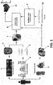
Figure 1 .- Shows a schematic view of the device for detecting cardiac arrhythmias according to the present invention, as well as the detection method. -
Figure 1 shows a preferred embodiment of the present invention. In this embodiment, it may be observed that the device essentially involves taking three measurements. - The first measurement involves performing a three-dimensional reconstruction (4) of the patient's torso by means of a set of images (1), for example, two-dimensional images obtained by means of a camera. This reconstruction is performed by means of at least two photographs taken using image processing techniques that are widely known in the prior art.
- The second measurement is a surface electrocardiographic map. This map is performed by taking data from a number of surface electrodes (2) and associating the data taken from said electrodes with a particular area of the patient's body. The data obtained by means of said electrodes are a number of electrical signals (5) obtained non-invasively (without any surgical procedure whatsoever). Moreover, as mentioned above, it is important to find a correlation between the electrical signals (5) and the position of the electrode that has taken each of the signals, in order to determine to which part of the heart each signal corresponds.
- Consequently, the present invention considers means for detecting the position of the electrodes. This detection of the position of the electrodes is most preferably performed by means of image analysis, in particular, analysis of the images (1) with which the three-dimensional reconstruction (4) of the patient's torso is performed, or, alternatively, of other images obtained using the same means for obtaining images. On the basis of this detection, a correlation between the electrical data and the position of the electrodes with respect to the patient's body may be performed, i.e. we may obtain a three-dimensional reconstruction (4) of the patient's torso, the position of the electrodes on the torso and the electrical signals measured for each of the points of the torso, and, with these data, obtain a surface electrical map that is nothing less than the combination of all these data into a graphic representation.
- Although on the basis of these data we could already have a three-dimensional representation of the functioning of each of the areas of the heart and locate cardiac arrhythmias, the present invention considers that, in order to increase the accuracy in locating the area wherein said arrhythmias appear, intracavity records may be taken (3) by using at least one catheter. Although this process is intrusive, it requires less intrusion than the mapping processes in the prior art.
- Basically, for this measurement, there is a catheter inside the heart that sequentially measures the activity at various points in the atrium. The position of this catheter at each moment may be determined using means for detecting the position of the catheter (for example, using two-dimensional photographs of the type used to perform the three-dimensional reconstruction (4)). Once the activity has been measured at several points and recorded in several intracavity records (3), we may obtain an intracavity anatomical reconstruction (6), which may be used for the reconstruction of the epicardium (i.e. an intracavity anatomical reconstruction) on the basis of the non-invasive records, using a regularisation of the solution of the inverse problem (8) based on quadratic and non-quadratic stabilisation functions under spatio-temporal discontinuity conditions. In this way, the signals calculated on the basis of the non-invasive records are used for the representation of the epicardial electroanatomical maps (11).
- It is worth mentioning that the intracavity mapping (3) is not essential for the system to calculate the epicardial potentials, which are calculated on the basis of the non-invasive surface records by solving the inverse problem, but using a few intracavity points is very helpful for a reliable reconstruction of the mathematical problem, which guarantees reliability even during irregular arrhythmias, such as atrial fibrillation.
- Once the activity has been measured at several intracavity points (3), we may obtain an intracavity anatomical reconstruction (6), which may be used for the reconstruction of the epicardium on the basis of the non-invasive records, by using a regularisation of the solution of the inverse problem based on quadratic and non-quadratic stabilisation functions under spatio-temporal discontinuity conditions, i.e. an electroanatomical correlation (8) is made taking into consideration the data obtained by means of the surface electrocardiographic map (7) and the intracavity anatomical reconstruction (6).
- Specifically, the solution of the inverse problem is performed by means of the iterative estimation of the transfer matrix between the potentials in the atrial epicardium (UA) and the potentials on the torso (UT):

- This is an ill-conditioned problem, since the number of estimated points on the surface of the epicardium (e.g. 2000 epicardial points) is much larger than the number of potentials on the torso (e.g. 120 electrodes on the torso). For this reason, calculation of the inverse transfer matrix is performed by minimising the error according to the following equation:

- In addition to what has been discussed above, the intracavity electrical signals (10) obtained by means of the intracavity catheter may be taken as an anchor and validation point for the reconstruction of the entire epicardial map on the basis of the non-invasive signals, in order to perform a stabilisation (9) of the inverse problem through the spatio-temporal correlation of the intracavity recording points according to the time, phase, modulus, spectrum and causality information. This makes it possible to reconstruct the activity of the entire atrium in a quick, reliable manner, even during irregular arrhythmias, such as atrial fibrillation.
Claims (10)
- Device for locating cardiac arrhythmias which comprises a three-dimensional reconstruction (4) of the patient's torso and a number of surface electrodes (2), characterised in that it comprises:• three-dimensional reconstruction means that generate the three-dimensional reconstruction (4) of the patient's torso through a number of images (1) obtained by means of at least one camera;• means for locating the surface electrodes (2), which detect the position of the electrodes with respect to the patient's torso;• data processing means that generate, using the three-dimensional reconstruction (4) and the position of the electrodes, a surface electrocardiographic map (7),said surface electrocardiographic map (7) comprising a number of data corresponding to readings of the surface electrodes (2) related to areas of the three-dimensional reconstruction (4).
- Device for locating cardiac arrhythmias, according to claim 1, wherein the means for locating the surface electrodes detect the position of the electrodes by processing the images obtained by means of at least one camera.
- Device for locating cardiac arrhythmias, according to claim 1, wherein at least one camera is a visible imaging camera.
- Device for locating cardiac arrhythmias, according to claim 1, wherein at least one camera is an angiographic camera.
- Device for locating cardiac arrhythmias, according to claim 1, wherein the surface electrocardiographic map (7) comprises means for detecting the area of the torso that presents a cardiac arrhythmia.
- Device for locating cardiac arrhythmias, according to claim 1, comprising at least one intracavity catheter that takes intracavity records (3).
- Device for locating cardiac arrhythmias, according to claim 6, comprising means for locating intracavity catheters.
- Device for locating cardiac arrhythmias, according to claim 7, comprising means for generating an intracavity anatomical reconstruction (6) on the basis of the intracavity records (3) and the means for locating the intracavity catheters.
- Device for locating cardiac arrhythmias, according to claim 8, wherein, by correlating the data from the intracavity anatomical reconstruction (6) and the surface electrocardiographic map (7), the processing means generate an electroanatomical map (11) wherein the electrical activity of each area is identified.
- Device, according to claim 9, comprising means for detecting cardiac arrhythmias in the electroanatomical map (11).
Applications Claiming Priority (2)
| Application Number | Priority Date | Filing Date | Title |
|---|---|---|---|
| ES201431597A ES2572142B1 (en) | 2014-10-30 | 2014-10-30 | CARDIAC ARRITMIAS LOCATION DEVICE |
| PCT/ES2015/070779 WO2016066879A1 (en) | 2014-10-30 | 2015-10-29 | Device for identifying the site of cardiac arrhythmias |
Publications (4)
| Publication Number | Publication Date |
|---|---|
| EP3213680A1 true EP3213680A1 (en) | 2017-09-06 |
| EP3213680A4 EP3213680A4 (en) | 2018-06-27 |
| EP3213680C0 EP3213680C0 (en) | 2023-09-06 |
| EP3213680B1 EP3213680B1 (en) | 2023-09-06 |
Family
ID=55856656
Family Applications (1)
| Application Number | Title | Priority Date | Filing Date |
|---|---|---|---|
| EP15855226.5A Active EP3213680B1 (en) | 2014-10-30 | 2015-10-29 | Device for identifying the site of cardiac arrhythmias |
Country Status (4)
| Country | Link |
|---|---|
| US (1) | US11672463B2 (en) |
| EP (1) | EP3213680B1 (en) |
| ES (2) | ES2572142B1 (en) |
| WO (1) | WO2016066879A1 (en) |
Families Citing this family (5)
| Publication number | Priority date | Publication date | Assignee | Title |
|---|---|---|---|---|
| ES2572142B1 (en) * | 2014-10-30 | 2017-06-21 | Fundación Para La Investigación Biomédica Del Hospital Gregorio Marañón | CARDIAC ARRITMIAS LOCATION DEVICE |
| US11006886B2 (en) * | 2018-12-20 | 2021-05-18 | Biosense Webster (Israel) Ltd. | Visualization of different cardiac rhythms using different timing-pattern displays |
| US11134895B2 (en) | 2019-01-17 | 2021-10-05 | Welch Allyn, Inc. | Method and apparatus for accurate placement of electrocardiogram electrodes |
| US10939863B2 (en) * | 2019-05-28 | 2021-03-09 | Biosense Webster (Israel) Ltd. | Determining occurrence of focal and/or rotor arrhythmogenic activity in cardiac tissue regions |
| US11540879B2 (en) | 2021-04-16 | 2023-01-03 | Physcade, Inc. | Personalized heart rhythm therapy |
Family Cites Families (58)
| Publication number | Priority date | Publication date | Assignee | Title |
|---|---|---|---|---|
| US7930012B2 (en) * | 1992-09-23 | 2011-04-19 | St. Jude Medical, Atrial Fibrillation Division, Inc. | Chamber location method |
| US5687737A (en) * | 1992-10-09 | 1997-11-18 | Washington University | Computerized three-dimensional cardiac mapping with interactive visual displays |
| WO1999006112A1 (en) * | 1997-07-31 | 1999-02-11 | Case Western Reserve University | Electrolphysiological cardiac mapping system based on a non-contact non-expandable miniature multi-electrode catheter and method therefor |
| US6975900B2 (en) * | 1997-07-31 | 2005-12-13 | Case Western Reserve University | Systems and methods for determining a surface geometry |
| US6545678B1 (en) * | 1998-11-05 | 2003-04-08 | Duke University | Methods, systems, and computer program products for generating tissue surfaces from volumetric data thereof using boundary traces |
| US6584343B1 (en) * | 2000-03-15 | 2003-06-24 | Resolution Medical, Inc. | Multi-electrode panel system for sensing electrical activity of the heart |
| US6856830B2 (en) * | 2001-07-19 | 2005-02-15 | Bin He | Method and apparatus of three dimension electrocardiographic imaging |
| US8175680B2 (en) * | 2001-11-09 | 2012-05-08 | Boston Scientific Scimed, Inc. | Systems and methods for guiding catheters using registered images |
| US20040006265A1 (en) * | 2002-04-30 | 2004-01-08 | Karim Alhussiny | Wireless transmission-ST-segment preserved of the standard 12 leads EKG apparatus for the remote administration of thrrombolytic therapy under severe cellular channel impairment |
| US7433730B1 (en) * | 2003-03-11 | 2008-10-07 | Berrier Keith L | Systems and methods for reconstructing information using a Duncan and Horn formulation of the Kalman filter for regularization |
| US20050228626A1 (en) * | 2004-04-02 | 2005-10-13 | Kim Simelius | System and method for health analysis |
| US9943274B2 (en) * | 2004-11-09 | 2018-04-17 | Spectrum Dynamics Medical Limited | Radioimaging using low dose isotope |
| US20140193336A1 (en) * | 2005-07-19 | 2014-07-10 | Biosensors International Group, Ltd. | Imaging protocols |
| US7918793B2 (en) * | 2005-10-28 | 2011-04-05 | Biosense Webster, Inc. | Synchronization of ultrasound imaging data with electrical mapping |
| WO2007134190A2 (en) * | 2006-05-10 | 2007-11-22 | Regents Of The University Of Minnesota | Methods and apparatus of three dimensional cardiac electrophysiological imaging |
| US7729752B2 (en) * | 2006-06-13 | 2010-06-01 | Rhythmia Medical, Inc. | Non-contact cardiac mapping, including resolution map |
| CA2932956C (en) * | 2006-08-03 | 2020-04-14 | Christoph Scharf | Method and device for determining and presenting surface charge and dipole densities on cardiac walls |
| US9370312B2 (en) * | 2006-09-06 | 2016-06-21 | Biosense Webster, Inc. | Correlation of cardiac electrical maps with body surface measurements |
| WO2008086430A1 (en) * | 2007-01-09 | 2008-07-17 | Cyberheart, Inc. | Method for depositing radiation in heart muscle |
| US9370310B2 (en) * | 2007-01-18 | 2016-06-21 | General Electric Company | Determination of cellular electrical potentials |
| US7805179B2 (en) * | 2007-06-08 | 2010-09-28 | Herng-Er Horng | Method of examining dynamic cardiac electromagnetic activity and detection of cardiac functions using results thereof |
| US8386014B2 (en) * | 2007-06-21 | 2013-02-26 | The Trustees Of Columbia University In The City Of New York | Systems and methods for implementing heart geometrical measurements |
| US20100198101A1 (en) * | 2007-09-24 | 2010-08-05 | Xubo Song | Non-invasive location and tracking of tumors and other tissues for radiation therapy |
| US9259166B2 (en) * | 2008-08-11 | 2016-02-16 | Washington University In St. Louis | Systems and methods for on-site and real-time electrocardiographic imaging (ECGI) |
| US8097926B2 (en) * | 2008-10-07 | 2012-01-17 | Mc10, Inc. | Systems, methods, and devices having stretchable integrated circuitry for sensing and delivering therapy |
| US9119533B2 (en) * | 2008-10-07 | 2015-09-01 | Mc10, Inc. | Systems, methods, and devices having stretchable integrated circuitry for sensing and delivering therapy |
| US8137343B2 (en) * | 2008-10-27 | 2012-03-20 | Rhythmia Medical, Inc. | Tracking system using field mapping |
| EP2348979B1 (en) * | 2008-11-07 | 2019-10-30 | Cardioinsight Technologies, Inc. | Visualization of physiological data for virtual electrodes |
| JP5911726B2 (en) * | 2008-11-10 | 2016-04-27 | カーディオインサイト テクノロジーズ インコーポレイテッド | Visualization of electrophysiological data |
| RU2417051C2 (en) * | 2008-11-27 | 2011-04-27 | Амиран Шотаевич РЕВИШВИЛИ | Method of noninvasive electrophysiological heart study |
| RU2435518C2 (en) * | 2008-11-27 | 2011-12-10 | Амиран Шотаевич РЕВИШВИЛИ | Method of non-invasive electrophysiological examination of heart |
| EP2233069B1 (en) * | 2009-03-25 | 2013-04-24 | Sorin CRM SAS | Active medical device including non-linear filtering means for reconstructing a surface electrocardiogram from an intracardiac electrogram |
| DE102009025077A1 (en) * | 2009-06-10 | 2010-12-16 | Karl Storz Gmbh & Co. Kg | System for orientation support and representation of an instrument in the interior of an examination object, in particular in the human body |
| EP2266459A1 (en) * | 2009-06-24 | 2010-12-29 | Cortius B.V. i.o. | Inverse imaging of electrical activity of a heart muscle |
| US20110080471A1 (en) * | 2009-10-06 | 2011-04-07 | Iowa State University Research Foundation, Inc. | Hybrid method for 3D shape measurement |
| US20140031668A1 (en) * | 2010-09-08 | 2014-01-30 | Disruptive Navigational Technologies, Llc | Surgical and Medical Instrument Tracking Using a Depth-Sensing Device |
| EP2635181B1 (en) * | 2010-11-03 | 2020-01-01 | CardioInsight Technologies, Inc. | Method for assessing heart function |
| WO2012106729A1 (en) * | 2011-02-04 | 2012-08-09 | Phase Space Systems Corporation | System and method for evaluating an electrophysiological signal |
| RU2622371C2 (en) * | 2011-02-17 | 2017-06-14 | Конинклейке Филипс Электроникс Н.В. | Electrical activity map provision system |
| US10391277B2 (en) * | 2011-02-18 | 2019-08-27 | Voxel Rad, Ltd. | Systems and methods for 3D stereoscopic angiovision, angionavigation and angiotherapeutics |
| US8553956B2 (en) * | 2011-02-28 | 2013-10-08 | Seiko Epson Corporation | 3D current reconstruction from 2D dense MCG images |
| GB2503055B (en) * | 2012-04-04 | 2018-08-29 | Cardiocity Ltd | Heart monitoring apparatus |
| US9271678B2 (en) * | 2011-04-08 | 2016-03-01 | Siemens Aktiengesellschaft | Constrained registration for motion compensation in atrial fibrillation ablation procedures |
| US9510763B2 (en) * | 2011-05-03 | 2016-12-06 | Medtronic, Inc. | Assessing intra-cardiac activation patterns and electrical dyssynchrony |
| EP2729214B1 (en) | 2011-07-05 | 2019-09-04 | CardioInsight Technologies, Inc. | System to facilitate providing therapy to a patient |
| CN103748591B (en) * | 2011-08-22 | 2017-09-01 | 皇家飞利浦有限公司 | data management system and method |
| US9180288B2 (en) * | 2011-09-01 | 2015-11-10 | Zoll Medical Corporation | Medical equipment electrodes |
| JP6259762B2 (en) * | 2011-10-12 | 2018-01-10 | カーディオインサイト テクノロジーズ インコーポレイテッド | Detection zone for spatially related electrical information |
| US8983589B2 (en) * | 2011-11-23 | 2015-03-17 | Ecg-Tech Corporation | Automatic measurement of ischemic levels in coronary segments to determine artery responsible therefor |
| CN103202727B (en) * | 2012-01-12 | 2015-11-25 | 通用电气公司 | Non-invasive arrhythmia treatment system |
| WO2013169933A1 (en) * | 2012-05-09 | 2013-11-14 | Cardioinsight Technologies, Inc. | Channel integrity detection |
| US9125581B2 (en) * | 2012-05-23 | 2015-09-08 | Seiko Epson Corporation | Continuous modeling for dipole localization from 2D MCG images with unknown depth |
| US8792969B2 (en) * | 2012-11-19 | 2014-07-29 | Xerox Corporation | Respiratory function estimation from a 2D monocular video |
| US9974458B2 (en) * | 2013-01-17 | 2018-05-22 | Cardioinsight Technologies, Inc. | Non-local mean filtering for electrophysiological signals |
| EP2945533A4 (en) * | 2013-01-17 | 2016-10-05 | Cardioinsight Technologies Inc | MULTIPARAMETER PHYSIOLOGICAL MAPPING |
| GB2510452A (en) | 2013-01-31 | 2014-08-06 | Naviconix Ltd | Method of mapping the heart with a trackable electrode catheter |
| US10049771B2 (en) * | 2013-03-15 | 2018-08-14 | St. Jude Medical, Atrial Fibrillation Division, Inc. | Laplacian and Tikhonov regularization for voltage mapping with a medical device |
| ES2572142B1 (en) * | 2014-10-30 | 2017-06-21 | Fundación Para La Investigación Biomédica Del Hospital Gregorio Marañón | CARDIAC ARRITMIAS LOCATION DEVICE |
-
2014
- 2014-10-30 ES ES201431597A patent/ES2572142B1/en active Active
-
2015
- 2015-10-29 ES ES15855226T patent/ES2969995T3/en active Active
- 2015-10-29 US US15/522,590 patent/US11672463B2/en active Active
- 2015-10-29 WO PCT/ES2015/070779 patent/WO2016066879A1/en not_active Ceased
- 2015-10-29 EP EP15855226.5A patent/EP3213680B1/en active Active
Also Published As
| Publication number | Publication date |
|---|---|
| ES2572142R2 (en) | 2016-09-13 |
| ES2969995T3 (en) | 2024-05-23 |
| US11672463B2 (en) | 2023-06-13 |
| WO2016066879A1 (en) | 2016-05-06 |
| EP3213680A4 (en) | 2018-06-27 |
| EP3213680C0 (en) | 2023-09-06 |
| ES2572142A2 (en) | 2016-05-30 |
| EP3213680B1 (en) | 2023-09-06 |
| ES2572142B1 (en) | 2017-06-21 |
| US20180132741A1 (en) | 2018-05-17 |
Similar Documents
| Publication | Publication Date | Title |
|---|---|---|
| US9427166B2 (en) | Physiological mapping for arrhythmia | |
| US7841986B2 (en) | Methods and apparatus of three dimensional cardiac electrophysiological imaging | |
| JP5281570B2 (en) | Non-contact cardiac mapping including catheter movement and multi-beat integration | |
| EP3071101B1 (en) | System and method for temporal sparse promoting imaging of cardiac activation | |
| EP3102102B1 (en) | Integrated analysis of electrophysiological data | |
| JP6534516B2 (en) | Inverted ECG mapping | |
| EP2945530B1 (en) | Composite singularity mapping | |
| US20170000364A1 (en) | Signal analysis related to treatment sites | |
| EP2945532B1 (en) | Wave front detection for electrophysiological signals | |
| EP2765902A1 (en) | Sensing zone for spatially relevant electrical information | |
| EP3213680B1 (en) | Device for identifying the site of cardiac arrhythmias | |
| US20140235996A1 (en) | Method for non-invasive mapping of myocardial electric activity | |
| EP3773169B1 (en) | Multi-dimensional method of fundamental solutions for reconstruction of electrophysiological activity | |
| Yu et al. | Three-dimensional noninvasive imaging of ventricular arrhythmias in patients with premature ventricular contractions | |
| Punshchykova et al. | Influence of torso model complexity on the noninvasive localization of ectopic ventricular activity | |
| EP4304478B1 (en) | Late activation of cardiac signals | |
| Kadambadi et al. | Electrocardiographic imaging: technical developments and future applications for non-invasive electroanatomical study of the human heart | |
| Seger et al. | Non-invasive imaging of atrial flutter | |
| EP4410209A1 (en) | Systems for high resolution electrographic flow (egf) mapping and analysis | |
| KR20260021070A (en) | Electrocardiographic examination-based determination of cardiac activation zones | |
| Seger et al. | Online noninvasive localization of accessory pathways in the EP lab | |
| Wang | Noninvasive Electrocardiographic Imaging of Cardiac Arrhythmias: Enhance the Diagnosis | |
| Ramanathan | Electrocardiographic imaging (ECGI): application of an iterative method and validation in humans |
Legal Events
| Date | Code | Title | Description |
|---|---|---|---|
| STAA | Information on the status of an ep patent application or granted ep patent |
Free format text: STATUS: THE INTERNATIONAL PUBLICATION HAS BEEN MADE |
|
| PUAI | Public reference made under article 153(3) epc to a published international application that has entered the european phase |
Free format text: ORIGINAL CODE: 0009012 |
|
| STAA | Information on the status of an ep patent application or granted ep patent |
Free format text: STATUS: REQUEST FOR EXAMINATION WAS MADE |
|
| 17P | Request for examination filed |
Effective date: 20170529 |
|
| AK | Designated contracting states |
Kind code of ref document: A1 Designated state(s): AL AT BE BG CH CY CZ DE DK EE ES FI FR GB GR HR HU IE IS IT LI LT LU LV MC MK MT NL NO PL PT RO RS SE SI SK SM TR |
|
| AX | Request for extension of the european patent |
Extension state: BA ME |
|
| DAV | Request for validation of the european patent (deleted) | ||
| DAX | Request for extension of the european patent (deleted) | ||
| A4 | Supplementary search report drawn up and despatched |
Effective date: 20180525 |
|
| RIC1 | Information provided on ipc code assigned before grant |
Ipc: G06F 19/00 20110101ALI20180518BHEP Ipc: A61B 5/0402 20060101ALI20180518BHEP Ipc: A61B 5/0408 20060101ALI20180518BHEP Ipc: G06T 17/00 20060101ALI20180518BHEP Ipc: A61B 5/00 20060101ALI20180518BHEP Ipc: A61B 5/04 20060101AFI20180518BHEP |
|
| STAA | Information on the status of an ep patent application or granted ep patent |
Free format text: STATUS: EXAMINATION IS IN PROGRESS |
|
| 17Q | First examination report despatched |
Effective date: 20200709 |
|
| REG | Reference to a national code |
Ref country code: DE Ref legal event code: R079 Free format text: PREVIOUS MAIN CLASS: A61B0005040000 Ipc: A61B0005240000 Ref document number: 602015085610 Country of ref document: DE |
|
| RIC1 | Information provided on ipc code assigned before grant |
Ipc: A61B 5/25 20210101ALI20220804BHEP Ipc: A61B 5/24 20210101AFI20220804BHEP |
|
| GRAP | Despatch of communication of intention to grant a patent |
Free format text: ORIGINAL CODE: EPIDOSNIGR1 |
|
| STAA | Information on the status of an ep patent application or granted ep patent |
Free format text: STATUS: GRANT OF PATENT IS INTENDED |
|
| INTG | Intention to grant announced |
Effective date: 20230328 |
|
| GRAS | Grant fee paid |
Free format text: ORIGINAL CODE: EPIDOSNIGR3 |
|
| GRAA | (expected) grant |
Free format text: ORIGINAL CODE: 0009210 |
|
| STAA | Information on the status of an ep patent application or granted ep patent |
Free format text: STATUS: THE PATENT HAS BEEN GRANTED |
|
| AK | Designated contracting states |
Kind code of ref document: B1 Designated state(s): AL AT BE BG CH CY CZ DE DK EE ES FI FR GB GR HR HU IE IS IT LI LT LU LV MC MK MT NL NO PL PT RO RS SE SI SK SM TR |
|
| REG | Reference to a national code |
Ref country code: GB Ref legal event code: FG4D |
|
| REG | Reference to a national code |
Ref country code: CH Ref legal event code: EP |
|
| REG | Reference to a national code |
Ref country code: IE Ref legal event code: FG4D |
|
| REG | Reference to a national code |
Ref country code: DE Ref legal event code: R096 Ref document number: 602015085610 Country of ref document: DE |
|
| RAP4 | Party data changed (patent owner data changed or rights of a patent transferred) |
Owner name: UNIVERSITAT POLITECNICA DE VALENCIA Owner name: FUNDACION PARA LA INVESTIGACION BIOMEDICA DEL HOSPITAL GREGORIO MARANON |
|
| U01 | Request for unitary effect filed |
Effective date: 20231006 |
|
| U07 | Unitary effect registered |
Designated state(s): AT BE BG DE DK EE FI FR IT LT LU LV MT NL PT SE SI Effective date: 20231018 |
|
| REG | Reference to a national code |
Ref country code: NO Ref legal event code: T2 Effective date: 20230906 |
|
| PG25 | Lapsed in a contracting state [announced via postgrant information from national office to epo] |
Ref country code: GR Free format text: LAPSE BECAUSE OF FAILURE TO SUBMIT A TRANSLATION OF THE DESCRIPTION OR TO PAY THE FEE WITHIN THE PRESCRIBED TIME-LIMIT Effective date: 20231207 |
|
| PG25 | Lapsed in a contracting state [announced via postgrant information from national office to epo] |
Ref country code: RS Free format text: LAPSE BECAUSE OF FAILURE TO SUBMIT A TRANSLATION OF THE DESCRIPTION OR TO PAY THE FEE WITHIN THE PRESCRIBED TIME-LIMIT Effective date: 20230906 Ref country code: HR Free format text: LAPSE BECAUSE OF FAILURE TO SUBMIT A TRANSLATION OF THE DESCRIPTION OR TO PAY THE FEE WITHIN THE PRESCRIBED TIME-LIMIT Effective date: 20230906 Ref country code: GR Free format text: LAPSE BECAUSE OF FAILURE TO SUBMIT A TRANSLATION OF THE DESCRIPTION OR TO PAY THE FEE WITHIN THE PRESCRIBED TIME-LIMIT Effective date: 20231207 |
|
| U20 | Renewal fee for the european patent with unitary effect paid |
Year of fee payment: 9 Effective date: 20240110 |
|
| PG25 | Lapsed in a contracting state [announced via postgrant information from national office to epo] |
Ref country code: IS Free format text: LAPSE BECAUSE OF FAILURE TO SUBMIT A TRANSLATION OF THE DESCRIPTION OR TO PAY THE FEE WITHIN THE PRESCRIBED TIME-LIMIT Effective date: 20240106 |
|
| PG25 | Lapsed in a contracting state [announced via postgrant information from national office to epo] |
Ref country code: SM Free format text: LAPSE BECAUSE OF FAILURE TO SUBMIT A TRANSLATION OF THE DESCRIPTION OR TO PAY THE FEE WITHIN THE PRESCRIBED TIME-LIMIT Effective date: 20230906 Ref country code: RO Free format text: LAPSE BECAUSE OF FAILURE TO SUBMIT A TRANSLATION OF THE DESCRIPTION OR TO PAY THE FEE WITHIN THE PRESCRIBED TIME-LIMIT Effective date: 20230906 Ref country code: IS Free format text: LAPSE BECAUSE OF FAILURE TO SUBMIT A TRANSLATION OF THE DESCRIPTION OR TO PAY THE FEE WITHIN THE PRESCRIBED TIME-LIMIT Effective date: 20240106 Ref country code: CZ Free format text: LAPSE BECAUSE OF FAILURE TO SUBMIT A TRANSLATION OF THE DESCRIPTION OR TO PAY THE FEE WITHIN THE PRESCRIBED TIME-LIMIT Effective date: 20230906 Ref country code: SK Free format text: LAPSE BECAUSE OF FAILURE TO SUBMIT A TRANSLATION OF THE DESCRIPTION OR TO PAY THE FEE WITHIN THE PRESCRIBED TIME-LIMIT Effective date: 20230906 |
|
| REG | Reference to a national code |
Ref country code: ES Ref legal event code: FG2A Ref document number: 2969995 Country of ref document: ES Kind code of ref document: T3 Effective date: 20240523 |
|
| PG25 | Lapsed in a contracting state [announced via postgrant information from national office to epo] |
Ref country code: PL Free format text: LAPSE BECAUSE OF FAILURE TO SUBMIT A TRANSLATION OF THE DESCRIPTION OR TO PAY THE FEE WITHIN THE PRESCRIBED TIME-LIMIT Effective date: 20230906 |
|
| REG | Reference to a national code |
Ref country code: DE Ref legal event code: R097 Ref document number: 602015085610 Country of ref document: DE |
|
| PG25 | Lapsed in a contracting state [announced via postgrant information from national office to epo] |
Ref country code: MC Free format text: LAPSE BECAUSE OF FAILURE TO SUBMIT A TRANSLATION OF THE DESCRIPTION OR TO PAY THE FEE WITHIN THE PRESCRIBED TIME-LIMIT Effective date: 20230906 |
|
| PLBE | No opposition filed within time limit |
Free format text: ORIGINAL CODE: 0009261 |
|
| STAA | Information on the status of an ep patent application or granted ep patent |
Free format text: STATUS: NO OPPOSITION FILED WITHIN TIME LIMIT |
|
| PG25 | Lapsed in a contracting state [announced via postgrant information from national office to epo] |
Ref country code: MC Free format text: LAPSE BECAUSE OF FAILURE TO SUBMIT A TRANSLATION OF THE DESCRIPTION OR TO PAY THE FEE WITHIN THE PRESCRIBED TIME-LIMIT Effective date: 20230906 |
|
| 26N | No opposition filed |
Effective date: 20240607 |
|
| U20 | Renewal fee for the european patent with unitary effect paid |
Year of fee payment: 10 Effective date: 20241004 |
|
| PG25 | Lapsed in a contracting state [announced via postgrant information from national office to epo] |
Ref country code: CY Free format text: LAPSE BECAUSE OF FAILURE TO SUBMIT A TRANSLATION OF THE DESCRIPTION OR TO PAY THE FEE WITHIN THE PRESCRIBED TIME-LIMIT; INVALID AB INITIO Effective date: 20151029 |
|
| PG25 | Lapsed in a contracting state [announced via postgrant information from national office to epo] |
Ref country code: HU Free format text: LAPSE BECAUSE OF FAILURE TO SUBMIT A TRANSLATION OF THE DESCRIPTION OR TO PAY THE FEE WITHIN THE PRESCRIBED TIME-LIMIT; INVALID AB INITIO Effective date: 20151029 |
|
| U20 | Renewal fee for the european patent with unitary effect paid |
Year of fee payment: 11 Effective date: 20250924 |
|
| REG | Reference to a national code |
Ref country code: CH Ref legal event code: U11 Free format text: ST27 STATUS EVENT CODE: U-0-0-U10-U11 (AS PROVIDED BY THE NATIONAL OFFICE) Effective date: 20251101 |
|
| PG25 | Lapsed in a contracting state [announced via postgrant information from national office to epo] |
Ref country code: TR Free format text: LAPSE BECAUSE OF FAILURE TO SUBMIT A TRANSLATION OF THE DESCRIPTION OR TO PAY THE FEE WITHIN THE PRESCRIBED TIME-LIMIT Effective date: 20230906 |
|
| PGFP | Annual fee paid to national office [announced via postgrant information from national office to epo] |
Ref country code: GB Payment date: 20251027 Year of fee payment: 11 |
|
| PGFP | Annual fee paid to national office [announced via postgrant information from national office to epo] |
Ref country code: NO Payment date: 20251029 Year of fee payment: 11 |
|
| PGFP | Annual fee paid to national office [announced via postgrant information from national office to epo] |
Ref country code: CH Payment date: 20251101 Year of fee payment: 11 |
|
| PGFP | Annual fee paid to national office [announced via postgrant information from national office to epo] |
Ref country code: IE Payment date: 20251027 Year of fee payment: 11 |
|
| PGFP | Annual fee paid to national office [announced via postgrant information from national office to epo] |
Ref country code: ES Payment date: 20251103 Year of fee payment: 11 |