EP3189812A1 - Elution of platelet rich plasma components from coated devices - Google Patents
Elution of platelet rich plasma components from coated devices Download PDFInfo
- Publication number
- EP3189812A1 EP3189812A1 EP17150587.8A EP17150587A EP3189812A1 EP 3189812 A1 EP3189812 A1 EP 3189812A1 EP 17150587 A EP17150587 A EP 17150587A EP 3189812 A1 EP3189812 A1 EP 3189812A1
- Authority
- EP
- European Patent Office
- Prior art keywords
- coated device
- biological molecule
- buffer
- mineral
- bodily fluid
- Prior art date
- Legal status (The legal status is an assumption and is not a legal conclusion. Google has not performed a legal analysis and makes no representation as to the accuracy of the status listed.)
- Withdrawn
Links
- 210000004623 platelet-rich plasma Anatomy 0.000 title claims description 56
- 238000010828 elution Methods 0.000 title description 31
- 229910052500 inorganic mineral Inorganic materials 0.000 claims abstract description 84
- 239000011707 mineral Substances 0.000 claims abstract description 84
- 238000000034 method Methods 0.000 claims abstract description 68
- 235000010755 mineral Nutrition 0.000 claims description 82
- 229910019142 PO4 Inorganic materials 0.000 claims description 79
- 239000010452 phosphate Substances 0.000 claims description 79
- NBIIXXVUZAFLBC-UHFFFAOYSA-K phosphate Chemical compound [O-]P([O-])([O-])=O NBIIXXVUZAFLBC-UHFFFAOYSA-K 0.000 claims description 79
- 210000001124 body fluid Anatomy 0.000 claims description 65
- 239000000872 buffer Substances 0.000 claims description 65
- 238000000576 coating method Methods 0.000 claims description 55
- 239000011248 coating agent Substances 0.000 claims description 49
- HEMHJVSKTPXQMS-UHFFFAOYSA-M Sodium hydroxide Chemical compound [OH-].[Na+] HEMHJVSKTPXQMS-UHFFFAOYSA-M 0.000 claims description 33
- VEXZGXHMUGYJMC-UHFFFAOYSA-N Hydrochloric acid Chemical compound Cl VEXZGXHMUGYJMC-UHFFFAOYSA-N 0.000 claims description 30
- 239000000758 substrate Substances 0.000 claims description 27
- 210000004369 blood Anatomy 0.000 claims description 25
- 239000008280 blood Substances 0.000 claims description 25
- -1 dicalcium phosophate Chemical compound 0.000 claims description 20
- TWRXJAOTZQYOKJ-UHFFFAOYSA-L Magnesium chloride Chemical compound [Mg+2].[Cl-].[Cl-] TWRXJAOTZQYOKJ-UHFFFAOYSA-L 0.000 claims description 18
- FAPWRFPIFSIZLT-UHFFFAOYSA-M Sodium chloride Chemical compound [Na+].[Cl-] FAPWRFPIFSIZLT-UHFFFAOYSA-M 0.000 claims description 17
- PUZPDOWCWNUUKD-UHFFFAOYSA-M sodium fluoride Chemical compound [F-].[Na+] PUZPDOWCWNUUKD-UHFFFAOYSA-M 0.000 claims description 16
- 238000004090 dissolution Methods 0.000 claims description 14
- UXVMQQNJUSDDNG-UHFFFAOYSA-L Calcium chloride Chemical compound [Cl-].[Cl-].[Ca+2] UXVMQQNJUSDDNG-UHFFFAOYSA-L 0.000 claims description 9
- 239000001110 calcium chloride Substances 0.000 claims description 9
- 229910001628 calcium chloride Inorganic materials 0.000 claims description 9
- 229910001629 magnesium chloride Inorganic materials 0.000 claims description 9
- 239000011780 sodium chloride Substances 0.000 claims description 9
- 239000011775 sodium fluoride Substances 0.000 claims description 8
- 235000013024 sodium fluoride Nutrition 0.000 claims description 8
- NBIIXXVUZAFLBC-UHFFFAOYSA-N Phosphoric acid Chemical group OP(O)(O)=O NBIIXXVUZAFLBC-UHFFFAOYSA-N 0.000 claims description 6
- 239000008363 phosphate buffer Substances 0.000 claims description 6
- QORWJWZARLRLPR-UHFFFAOYSA-H tricalcium bis(phosphate) Chemical compound [Ca+2].[Ca+2].[Ca+2].[O-]P([O-])([O-])=O.[O-]P([O-])([O-])=O QORWJWZARLRLPR-UHFFFAOYSA-H 0.000 claims description 6
- OYPRJOBELJOOCE-UHFFFAOYSA-N Calcium Chemical compound [Ca] OYPRJOBELJOOCE-UHFFFAOYSA-N 0.000 claims description 5
- BVKZGUZCCUSVTD-UHFFFAOYSA-L Carbonate Chemical compound [O-]C([O-])=O BVKZGUZCCUSVTD-UHFFFAOYSA-L 0.000 claims description 5
- KCXVZYZYPLLWCC-UHFFFAOYSA-N EDTA Chemical compound OC(=O)CN(CC(O)=O)CCN(CC(O)=O)CC(O)=O KCXVZYZYPLLWCC-UHFFFAOYSA-N 0.000 claims description 5
- 239000011575 calcium Substances 0.000 claims description 5
- 229910052791 calcium Inorganic materials 0.000 claims description 5
- 239000001506 calcium phosphate Substances 0.000 claims description 5
- VTYYLEPIZMXCLO-UHFFFAOYSA-L Calcium carbonate Chemical compound [Ca+2].[O-]C([O-])=O VTYYLEPIZMXCLO-UHFFFAOYSA-L 0.000 claims description 4
- 210000001185 bone marrow Anatomy 0.000 claims description 4
- 210000001175 cerebrospinal fluid Anatomy 0.000 claims description 4
- 229910052588 hydroxylapatite Inorganic materials 0.000 claims description 4
- XYJRXVWERLGGKC-UHFFFAOYSA-D pentacalcium;hydroxide;triphosphate Chemical compound [OH-].[Ca+2].[Ca+2].[Ca+2].[Ca+2].[Ca+2].[O-]P([O-])([O-])=O.[O-]P([O-])([O-])=O.[O-]P([O-])([O-])=O XYJRXVWERLGGKC-UHFFFAOYSA-D 0.000 claims description 4
- 229920001432 poly(L-lactide) Polymers 0.000 claims description 4
- 229920001606 poly(lactic acid-co-glycolic acid) Polymers 0.000 claims description 4
- 210000002966 serum Anatomy 0.000 claims description 4
- 210000001179 synovial fluid Anatomy 0.000 claims description 4
- 210000002700 urine Anatomy 0.000 claims description 4
- 229910000147 aluminium phosphate Inorganic materials 0.000 claims description 3
- 229920001610 polycaprolactone Polymers 0.000 claims description 3
- UEZVMMHDMIWARA-UHFFFAOYSA-N Metaphosphoric acid Chemical compound OP(=O)=O UEZVMMHDMIWARA-UHFFFAOYSA-N 0.000 claims description 2
- 229910000019 calcium carbonate Inorganic materials 0.000 claims description 2
- 229910000389 calcium phosphate Inorganic materials 0.000 claims description 2
- 235000011010 calcium phosphates Nutrition 0.000 claims description 2
- 229910000392 octacalcium phosphate Inorganic materials 0.000 claims description 2
- YIGWVOWKHUSYER-UHFFFAOYSA-F tetracalcium;hydrogen phosphate;diphosphate Chemical compound [Ca+2].[Ca+2].[Ca+2].[Ca+2].OP([O-])([O-])=O.[O-]P([O-])([O-])=O.[O-]P([O-])([O-])=O YIGWVOWKHUSYER-UHFFFAOYSA-F 0.000 claims description 2
- 229910000391 tricalcium phosphate Inorganic materials 0.000 claims description 2
- 229940078499 tricalcium phosphate Drugs 0.000 claims description 2
- 239000012475 sodium chloride buffer Substances 0.000 claims 1
- 241001465754 Metazoa Species 0.000 abstract description 5
- 238000002955 isolation Methods 0.000 abstract description 5
- 238000010188 recombinant method Methods 0.000 abstract description 4
- 238000001815 biotherapy Methods 0.000 abstract description 2
- 238000000746 purification Methods 0.000 abstract 1
- 102000004169 proteins and genes Human genes 0.000 description 98
- 108090000623 proteins and genes Proteins 0.000 description 98
- 108091003079 Bovine Serum Albumin Proteins 0.000 description 15
- 238000003556 assay Methods 0.000 description 13
- 229940098773 bovine serum albumin Drugs 0.000 description 13
- XLYOFNOQVPJJNP-UHFFFAOYSA-N water Substances O XLYOFNOQVPJJNP-UHFFFAOYSA-N 0.000 description 13
- 210000001772 blood platelet Anatomy 0.000 description 12
- 229960000074 biopharmaceutical Drugs 0.000 description 11
- 239000003102 growth factor Substances 0.000 description 11
- 238000011534 incubation Methods 0.000 description 9
- 239000000463 material Substances 0.000 description 8
- 239000000243 solution Substances 0.000 description 8
- AFYNADDZULBEJA-UHFFFAOYSA-N bicinchoninic acid Chemical compound C1=CC=CC2=NC(C=3C=C(C4=CC=CC=C4N=3)C(=O)O)=CC(C(O)=O)=C21 AFYNADDZULBEJA-UHFFFAOYSA-N 0.000 description 7
- 230000009881 electrostatic interaction Effects 0.000 description 6
- JKMHFZQWWAIEOD-UHFFFAOYSA-N 2-[4-(2-hydroxyethyl)piperazin-1-yl]ethanesulfonic acid Chemical compound OCC[NH+]1CCN(CCS([O-])(=O)=O)CC1 JKMHFZQWWAIEOD-UHFFFAOYSA-N 0.000 description 5
- 239000000306 component Substances 0.000 description 5
- 238000010790 dilution Methods 0.000 description 5
- 239000012895 dilution Substances 0.000 description 5
- 230000000399 orthopedic effect Effects 0.000 description 5
- 239000007995 HEPES buffer Substances 0.000 description 4
- 229940079593 drug Drugs 0.000 description 4
- 239000003814 drug Substances 0.000 description 4
- 230000035876 healing Effects 0.000 description 4
- 230000001105 regulatory effect Effects 0.000 description 4
- 238000005406 washing Methods 0.000 description 4
- RKDVKSZUMVYZHH-UHFFFAOYSA-N 1,4-dioxane-2,5-dione Chemical compound O=C1COC(=O)CO1 RKDVKSZUMVYZHH-UHFFFAOYSA-N 0.000 description 3
- 102100024506 Bone morphogenetic protein 2 Human genes 0.000 description 3
- 101000762366 Homo sapiens Bone morphogenetic protein 2 Proteins 0.000 description 3
- 241001494479 Pecora Species 0.000 description 3
- 101710093543 Probable non-specific lipid-transfer protein Proteins 0.000 description 3
- 108060008682 Tumor Necrosis Factor Proteins 0.000 description 3
- 239000011324 bead Substances 0.000 description 3
- 239000012149 elution buffer Substances 0.000 description 3
- 230000012010 growth Effects 0.000 description 3
- 230000003301 hydrolyzing effect Effects 0.000 description 3
- 210000002381 plasma Anatomy 0.000 description 3
- 229920000747 poly(lactic acid) Polymers 0.000 description 3
- 238000012360 testing method Methods 0.000 description 3
- 210000001519 tissue Anatomy 0.000 description 3
- 238000002965 ELISA Methods 0.000 description 2
- 102000018233 Fibroblast Growth Factor Human genes 0.000 description 2
- 108050007372 Fibroblast Growth Factor Proteins 0.000 description 2
- 102100035363 Growth/differentiation factor 7 Human genes 0.000 description 2
- 102100039939 Growth/differentiation factor 8 Human genes 0.000 description 2
- 108090000100 Hepatocyte Growth Factor Proteins 0.000 description 2
- 102100021866 Hepatocyte growth factor Human genes 0.000 description 2
- 101001023968 Homo sapiens Growth/differentiation factor 7 Proteins 0.000 description 2
- 108010050904 Interferons Proteins 0.000 description 2
- 102000014150 Interferons Human genes 0.000 description 2
- 108010025020 Nerve Growth Factor Proteins 0.000 description 2
- 102000015336 Nerve Growth Factor Human genes 0.000 description 2
- 102100035194 Placenta growth factor Human genes 0.000 description 2
- 108010009583 Transforming Growth Factors Proteins 0.000 description 2
- 102000009618 Transforming Growth Factors Human genes 0.000 description 2
- 108010019530 Vascular Endothelial Growth Factors Proteins 0.000 description 2
- 102000005789 Vascular Endothelial Growth Factors Human genes 0.000 description 2
- 238000004458 analytical method Methods 0.000 description 2
- 230000015572 biosynthetic process Effects 0.000 description 2
- 210000000988 bone and bone Anatomy 0.000 description 2
- 230000015556 catabolic process Effects 0.000 description 2
- 210000004027 cell Anatomy 0.000 description 2
- 239000000515 collagen sponge Substances 0.000 description 2
- 238000006731 degradation reaction Methods 0.000 description 2
- 238000011161 development Methods 0.000 description 2
- 231100000673 dose–response relationship Toxicity 0.000 description 2
- 210000003743 erythrocyte Anatomy 0.000 description 2
- 238000001502 gel electrophoresis Methods 0.000 description 2
- 229940047124 interferons Drugs 0.000 description 2
- 150000002500 ions Chemical class 0.000 description 2
- 238000002483 medication Methods 0.000 description 2
- 238000012986 modification Methods 0.000 description 2
- 230000004048 modification Effects 0.000 description 2
- 229940053128 nerve growth factor Drugs 0.000 description 2
- 239000002245 particle Substances 0.000 description 2
- NGVDGCNFYWLIFO-UHFFFAOYSA-N pyridoxal 5'-phosphate Chemical compound CC1=NC=C(COP(O)(O)=O)C(C=O)=C1O NGVDGCNFYWLIFO-UHFFFAOYSA-N 0.000 description 2
- 230000008439 repair process Effects 0.000 description 2
- 238000001878 scanning electron micrograph Methods 0.000 description 2
- 239000012890 simulated body fluid Substances 0.000 description 2
- 238000002415 sodium dodecyl sulfate polyacrylamide gel electrophoresis Methods 0.000 description 2
- 230000004936 stimulating effect Effects 0.000 description 2
- 238000001356 surgical procedure Methods 0.000 description 2
- 102000003390 tumor necrosis factor Human genes 0.000 description 2
- 238000001262 western blot Methods 0.000 description 2
- MZOFCQQQCNRIBI-VMXHOPILSA-N (3s)-4-[[(2s)-1-[[(2s)-1-[[(1s)-1-carboxy-2-hydroxyethyl]amino]-4-methyl-1-oxopentan-2-yl]amino]-5-(diaminomethylideneamino)-1-oxopentan-2-yl]amino]-3-[[2-[[(2s)-2,6-diaminohexanoyl]amino]acetyl]amino]-4-oxobutanoic acid Chemical compound OC[C@@H](C(O)=O)NC(=O)[C@H](CC(C)C)NC(=O)[C@H](CCCN=C(N)N)NC(=O)[C@H](CC(O)=O)NC(=O)CNC(=O)[C@@H](N)CCCCN MZOFCQQQCNRIBI-VMXHOPILSA-N 0.000 description 1
- 101800000263 Acidic protein Proteins 0.000 description 1
- 229920000936 Agarose Polymers 0.000 description 1
- 108010088751 Albumins Proteins 0.000 description 1
- 102000009027 Albumins Human genes 0.000 description 1
- 208000025674 Anterior Cruciate Ligament injury Diseases 0.000 description 1
- 102100026376 Artemin Human genes 0.000 description 1
- 102000004152 Bone morphogenetic protein 1 Human genes 0.000 description 1
- 108090000654 Bone morphogenetic protein 1 Proteins 0.000 description 1
- 102100024504 Bone morphogenetic protein 3 Human genes 0.000 description 1
- 102100024505 Bone morphogenetic protein 4 Human genes 0.000 description 1
- 102100022526 Bone morphogenetic protein 5 Human genes 0.000 description 1
- 102100022525 Bone morphogenetic protein 6 Human genes 0.000 description 1
- 102100022544 Bone morphogenetic protein 7 Human genes 0.000 description 1
- 235000019739 Dicalciumphosphate Nutrition 0.000 description 1
- 108010066486 EGF Family of Proteins Proteins 0.000 description 1
- 102000018386 EGF Family of Proteins Human genes 0.000 description 1
- 102100040897 Embryonic growth/differentiation factor 1 Human genes 0.000 description 1
- 102000009024 Epidermal Growth Factor Human genes 0.000 description 1
- 102000003971 Fibroblast Growth Factor 1 Human genes 0.000 description 1
- 108090000386 Fibroblast Growth Factor 1 Proteins 0.000 description 1
- 102000003974 Fibroblast growth factor 2 Human genes 0.000 description 1
- 108090000379 Fibroblast growth factor 2 Proteins 0.000 description 1
- 102100028043 Fibroblast growth factor 3 Human genes 0.000 description 1
- 102100028072 Fibroblast growth factor 4 Human genes 0.000 description 1
- 102100040895 Growth/differentiation factor 10 Human genes 0.000 description 1
- 102100040898 Growth/differentiation factor 11 Human genes 0.000 description 1
- 102100040896 Growth/differentiation factor 15 Human genes 0.000 description 1
- 102100040892 Growth/differentiation factor 2 Human genes 0.000 description 1
- 102100035364 Growth/differentiation factor 3 Human genes 0.000 description 1
- 102100035379 Growth/differentiation factor 5 Human genes 0.000 description 1
- 102100035368 Growth/differentiation factor 6 Human genes 0.000 description 1
- 108050006583 Growth/differentiation factor 8 Proteins 0.000 description 1
- 102100035970 Growth/differentiation factor 9 Human genes 0.000 description 1
- 101000785776 Homo sapiens Artemin Proteins 0.000 description 1
- 101000762375 Homo sapiens Bone morphogenetic protein 3 Proteins 0.000 description 1
- 101000762379 Homo sapiens Bone morphogenetic protein 4 Proteins 0.000 description 1
- 101000899388 Homo sapiens Bone morphogenetic protein 5 Proteins 0.000 description 1
- 101000899390 Homo sapiens Bone morphogenetic protein 6 Proteins 0.000 description 1
- 101000899361 Homo sapiens Bone morphogenetic protein 7 Proteins 0.000 description 1
- 101000893552 Homo sapiens Embryonic growth/differentiation factor 1 Proteins 0.000 description 1
- 101001060280 Homo sapiens Fibroblast growth factor 3 Proteins 0.000 description 1
- 101001060274 Homo sapiens Fibroblast growth factor 4 Proteins 0.000 description 1
- 101000893563 Homo sapiens Growth/differentiation factor 10 Proteins 0.000 description 1
- 101000893545 Homo sapiens Growth/differentiation factor 11 Proteins 0.000 description 1
- 101000893549 Homo sapiens Growth/differentiation factor 15 Proteins 0.000 description 1
- 101000893585 Homo sapiens Growth/differentiation factor 2 Proteins 0.000 description 1
- 101001023986 Homo sapiens Growth/differentiation factor 3 Proteins 0.000 description 1
- 101001023988 Homo sapiens Growth/differentiation factor 5 Proteins 0.000 description 1
- 101001023964 Homo sapiens Growth/differentiation factor 6 Proteins 0.000 description 1
- 101000886562 Homo sapiens Growth/differentiation factor 8 Proteins 0.000 description 1
- 101001075110 Homo sapiens Growth/differentiation factor 9 Proteins 0.000 description 1
- 101001076604 Homo sapiens Inhibin alpha chain Proteins 0.000 description 1
- 101001054725 Homo sapiens Inhibin beta B chain Proteins 0.000 description 1
- 101001054832 Homo sapiens Inhibin beta C chain Proteins 0.000 description 1
- 101001054830 Homo sapiens Inhibin beta E chain Proteins 0.000 description 1
- 101000967920 Homo sapiens Left-right determination factor 1 Proteins 0.000 description 1
- 101000967918 Homo sapiens Left-right determination factor 2 Proteins 0.000 description 1
- 101100241084 Homo sapiens NRTN gene Proteins 0.000 description 1
- 101001136670 Homo sapiens Persephin Proteins 0.000 description 1
- 101001001487 Homo sapiens Phosphatidylinositol-glycan biosynthesis class F protein Proteins 0.000 description 1
- 101000595923 Homo sapiens Placenta growth factor Proteins 0.000 description 1
- 101000611888 Homo sapiens Platelet-derived growth factor C Proteins 0.000 description 1
- 101000611892 Homo sapiens Platelet-derived growth factor D Proteins 0.000 description 1
- 102100025885 Inhibin alpha chain Human genes 0.000 description 1
- 102100027004 Inhibin beta A chain Human genes 0.000 description 1
- 102100027003 Inhibin beta B chain Human genes 0.000 description 1
- 102100026812 Inhibin beta C chain Human genes 0.000 description 1
- 102100026818 Inhibin beta E chain Human genes 0.000 description 1
- 108090000723 Insulin-Like Growth Factor I Proteins 0.000 description 1
- 102100037850 Interferon gamma Human genes 0.000 description 1
- 108010047761 Interferon-alpha Proteins 0.000 description 1
- 102000006992 Interferon-alpha Human genes 0.000 description 1
- 108010074328 Interferon-gamma Proteins 0.000 description 1
- 108010002352 Interleukin-1 Proteins 0.000 description 1
- 108010065805 Interleukin-12 Proteins 0.000 description 1
- 108090000176 Interleukin-13 Proteins 0.000 description 1
- 108050003558 Interleukin-17 Proteins 0.000 description 1
- 108010002350 Interleukin-2 Proteins 0.000 description 1
- 108010065637 Interleukin-23 Proteins 0.000 description 1
- 108090000978 Interleukin-4 Proteins 0.000 description 1
- 108010002616 Interleukin-5 Proteins 0.000 description 1
- 108090001005 Interleukin-6 Proteins 0.000 description 1
- 108010063738 Interleukins Proteins 0.000 description 1
- 102000015696 Interleukins Human genes 0.000 description 1
- 102100040508 Left-right determination factor 1 Human genes 0.000 description 1
- 102100040511 Left-right determination factor 2 Human genes 0.000 description 1
- 102100030173 Muellerian-inhibiting factor Human genes 0.000 description 1
- 101710122877 Muellerian-inhibiting factor Proteins 0.000 description 1
- 102100021584 Neurturin Human genes 0.000 description 1
- 206010030113 Oedema Diseases 0.000 description 1
- 101100137581 Ovis aries PRNP gene Proteins 0.000 description 1
- 102100036660 Persephin Human genes 0.000 description 1
- 108010082093 Placenta Growth Factor Proteins 0.000 description 1
- 108010038512 Platelet-Derived Growth Factor Proteins 0.000 description 1
- 102000010780 Platelet-Derived Growth Factor Human genes 0.000 description 1
- 102100040681 Platelet-derived growth factor C Human genes 0.000 description 1
- 102100040682 Platelet-derived growth factor D Human genes 0.000 description 1
- 102100037596 Platelet-derived growth factor subunit A Human genes 0.000 description 1
- 102100040990 Platelet-derived growth factor subunit B Human genes 0.000 description 1
- 101710098940 Pro-epidermal growth factor Proteins 0.000 description 1
- 108010019674 Proto-Oncogene Proteins c-sis Proteins 0.000 description 1
- 102000007056 Recombinant Fusion Proteins Human genes 0.000 description 1
- 108010008281 Recombinant Fusion Proteins Proteins 0.000 description 1
- 108010071390 Serum Albumin Proteins 0.000 description 1
- 102000007562 Serum Albumin Human genes 0.000 description 1
- 102000013275 Somatomedins Human genes 0.000 description 1
- 208000021945 Tendon injury Diseases 0.000 description 1
- 208000003441 Transfusion reaction Diseases 0.000 description 1
- 102000000852 Tumor Necrosis Factor-alpha Human genes 0.000 description 1
- 108010073929 Vascular Endothelial Growth Factor A Proteins 0.000 description 1
- 108010073925 Vascular Endothelial Growth Factor B Proteins 0.000 description 1
- 108010073923 Vascular Endothelial Growth Factor C Proteins 0.000 description 1
- 108010073919 Vascular Endothelial Growth Factor D Proteins 0.000 description 1
- 102100039037 Vascular endothelial growth factor A Human genes 0.000 description 1
- 102100038217 Vascular endothelial growth factor B Human genes 0.000 description 1
- 102100038232 Vascular endothelial growth factor C Human genes 0.000 description 1
- 102100038234 Vascular endothelial growth factor D Human genes 0.000 description 1
- 230000000735 allogeneic effect Effects 0.000 description 1
- 238000013459 approach Methods 0.000 description 1
- 230000005540 biological transmission Effects 0.000 description 1
- 239000012503 blood component Substances 0.000 description 1
- 230000036770 blood supply Effects 0.000 description 1
- 244000078885 bloodborne pathogen Species 0.000 description 1
- 239000007853 buffer solution Substances 0.000 description 1
- 239000000969 carrier Substances 0.000 description 1
- 239000012876 carrier material Substances 0.000 description 1
- 238000005119 centrifugation Methods 0.000 description 1
- 238000006243 chemical reaction Methods 0.000 description 1
- 230000008473 connective tissue growth Effects 0.000 description 1
- 230000003247 decreasing effect Effects 0.000 description 1
- 230000007547 defect Effects 0.000 description 1
- NEFBYIFKOOEVPA-UHFFFAOYSA-K dicalcium phosphate Chemical compound [Ca+2].[Ca+2].[O-]P([O-])([O-])=O NEFBYIFKOOEVPA-UHFFFAOYSA-K 0.000 description 1
- 229910000390 dicalcium phosphate Inorganic materials 0.000 description 1
- 229940038472 dicalcium phosphate Drugs 0.000 description 1
- 239000012154 double-distilled water Substances 0.000 description 1
- 238000011049 filling Methods 0.000 description 1
- 239000007888 film coating Substances 0.000 description 1
- 238000009501 film coating Methods 0.000 description 1
- 230000036541 health Effects 0.000 description 1
- 239000007943 implant Substances 0.000 description 1
- 238000001727 in vivo Methods 0.000 description 1
- 108010019691 inhibin beta A subunit Proteins 0.000 description 1
- 238000003780 insertion Methods 0.000 description 1
- 230000037431 insertion Effects 0.000 description 1
- 108010074108 interleukin-21 Proteins 0.000 description 1
- 210000002510 keratinocyte Anatomy 0.000 description 1
- 239000004816 latex Substances 0.000 description 1
- 229920000126 latex Polymers 0.000 description 1
- 210000000265 leukocyte Anatomy 0.000 description 1
- 238000004519 manufacturing process Methods 0.000 description 1
- 230000000921 morphogenic effect Effects 0.000 description 1
- OHDXDNUPVVYWOV-UHFFFAOYSA-N n-methyl-1-(2-naphthalen-1-ylsulfanylphenyl)methanamine Chemical compound CNCC1=CC=CC=C1SC1=CC=CC2=CC=CC=C12 OHDXDNUPVVYWOV-UHFFFAOYSA-N 0.000 description 1
- 230000006911 nucleation Effects 0.000 description 1
- 238000010899 nucleation Methods 0.000 description 1
- 230000011164 ossification Effects 0.000 description 1
- 210000004180 plasmocyte Anatomy 0.000 description 1
- 108010017843 platelet-derived growth factor A Proteins 0.000 description 1
- 238000002360 preparation method Methods 0.000 description 1
- 238000002731 protein assay Methods 0.000 description 1
- 239000002904 solvent Substances 0.000 description 1
- 241000894007 species Species 0.000 description 1
- 210000002435 tendon Anatomy 0.000 description 1
- 230000001225 therapeutic effect Effects 0.000 description 1
- 238000002560 therapeutic procedure Methods 0.000 description 1
- 238000012549 training Methods 0.000 description 1
- 208000037918 transfusion-transmitted disease Diseases 0.000 description 1
Images









Classifications
-
- A—HUMAN NECESSITIES
- A61—MEDICAL OR VETERINARY SCIENCE; HYGIENE
- A61K—PREPARATIONS FOR MEDICAL, DENTAL OR TOILETRY PURPOSES
- A61K47/00—Medicinal preparations characterised by the non-active ingredients used, e.g. carriers or inert additives; Targeting or modifying agents chemically bound to the active ingredient
- A61K47/50—Medicinal preparations characterised by the non-active ingredients used, e.g. carriers or inert additives; Targeting or modifying agents chemically bound to the active ingredient the non-active ingredient being chemically bound to the active ingredient, e.g. polymer-drug conjugates
- A61K47/69—Medicinal preparations characterised by the non-active ingredients used, e.g. carriers or inert additives; Targeting or modifying agents chemically bound to the active ingredient the non-active ingredient being chemically bound to the active ingredient, e.g. polymer-drug conjugates the conjugate being characterised by physical or galenical forms, e.g. emulsion, particle, inclusion complex, stent or kit
- A61K47/6957—Medicinal preparations characterised by the non-active ingredients used, e.g. carriers or inert additives; Targeting or modifying agents chemically bound to the active ingredient the non-active ingredient being chemically bound to the active ingredient, e.g. polymer-drug conjugates the conjugate being characterised by physical or galenical forms, e.g. emulsion, particle, inclusion complex, stent or kit the form being a device or a kit, e.g. stents or microdevices
-
- A—HUMAN NECESSITIES
- A61—MEDICAL OR VETERINARY SCIENCE; HYGIENE
- A61L—METHODS OR APPARATUS FOR STERILISING MATERIALS OR OBJECTS IN GENERAL; DISINFECTION, STERILISATION OR DEODORISATION OF AIR; CHEMICAL ASPECTS OF BANDAGES, DRESSINGS, ABSORBENT PADS OR SURGICAL ARTICLES; MATERIALS FOR BANDAGES, DRESSINGS, ABSORBENT PADS OR SURGICAL ARTICLES
- A61L17/00—Materials for surgical sutures or for ligaturing blood vessels ; Materials for prostheses or catheters
- A61L17/005—Materials for surgical sutures or for ligaturing blood vessels ; Materials for prostheses or catheters containing a biologically active substance, e.g. a medicament or a biocide
-
- A—HUMAN NECESSITIES
- A61—MEDICAL OR VETERINARY SCIENCE; HYGIENE
- A61L—METHODS OR APPARATUS FOR STERILISING MATERIALS OR OBJECTS IN GENERAL; DISINFECTION, STERILISATION OR DEODORISATION OF AIR; CHEMICAL ASPECTS OF BANDAGES, DRESSINGS, ABSORBENT PADS OR SURGICAL ARTICLES; MATERIALS FOR BANDAGES, DRESSINGS, ABSORBENT PADS OR SURGICAL ARTICLES
- A61L17/00—Materials for surgical sutures or for ligaturing blood vessels ; Materials for prostheses or catheters
- A61L17/06—At least partially resorbable materials
- A61L17/10—At least partially resorbable materials containing macromolecular materials
- A61L17/12—Homopolymers or copolymers of glycolic acid or lactic acid
-
- A—HUMAN NECESSITIES
- A61—MEDICAL OR VETERINARY SCIENCE; HYGIENE
- A61L—METHODS OR APPARATUS FOR STERILISING MATERIALS OR OBJECTS IN GENERAL; DISINFECTION, STERILISATION OR DEODORISATION OF AIR; CHEMICAL ASPECTS OF BANDAGES, DRESSINGS, ABSORBENT PADS OR SURGICAL ARTICLES; MATERIALS FOR BANDAGES, DRESSINGS, ABSORBENT PADS OR SURGICAL ARTICLES
- A61L17/00—Materials for surgical sutures or for ligaturing blood vessels ; Materials for prostheses or catheters
- A61L17/14—Post-treatment to improve physical properties
- A61L17/145—Coating
-
- A—HUMAN NECESSITIES
- A61—MEDICAL OR VETERINARY SCIENCE; HYGIENE
- A61L—METHODS OR APPARATUS FOR STERILISING MATERIALS OR OBJECTS IN GENERAL; DISINFECTION, STERILISATION OR DEODORISATION OF AIR; CHEMICAL ASPECTS OF BANDAGES, DRESSINGS, ABSORBENT PADS OR SURGICAL ARTICLES; MATERIALS FOR BANDAGES, DRESSINGS, ABSORBENT PADS OR SURGICAL ARTICLES
- A61L27/00—Materials for grafts or prostheses or for coating grafts or prostheses
- A61L27/14—Macromolecular materials
- A61L27/18—Macromolecular materials obtained otherwise than by reactions only involving carbon-to-carbon unsaturated bonds
-
- A—HUMAN NECESSITIES
- A61—MEDICAL OR VETERINARY SCIENCE; HYGIENE
- A61L—METHODS OR APPARATUS FOR STERILISING MATERIALS OR OBJECTS IN GENERAL; DISINFECTION, STERILISATION OR DEODORISATION OF AIR; CHEMICAL ASPECTS OF BANDAGES, DRESSINGS, ABSORBENT PADS OR SURGICAL ARTICLES; MATERIALS FOR BANDAGES, DRESSINGS, ABSORBENT PADS OR SURGICAL ARTICLES
- A61L27/00—Materials for grafts or prostheses or for coating grafts or prostheses
- A61L27/28—Materials for coating prostheses
- A61L27/30—Inorganic materials
- A61L27/306—Other specific inorganic materials not covered by A61L27/303 - A61L27/32
-
- A—HUMAN NECESSITIES
- A61—MEDICAL OR VETERINARY SCIENCE; HYGIENE
- A61L—METHODS OR APPARATUS FOR STERILISING MATERIALS OR OBJECTS IN GENERAL; DISINFECTION, STERILISATION OR DEODORISATION OF AIR; CHEMICAL ASPECTS OF BANDAGES, DRESSINGS, ABSORBENT PADS OR SURGICAL ARTICLES; MATERIALS FOR BANDAGES, DRESSINGS, ABSORBENT PADS OR SURGICAL ARTICLES
- A61L27/00—Materials for grafts or prostheses or for coating grafts or prostheses
- A61L27/28—Materials for coating prostheses
- A61L27/30—Inorganic materials
- A61L27/32—Phosphorus-containing materials, e.g. apatite
-
- A—HUMAN NECESSITIES
- A61—MEDICAL OR VETERINARY SCIENCE; HYGIENE
- A61L—METHODS OR APPARATUS FOR STERILISING MATERIALS OR OBJECTS IN GENERAL; DISINFECTION, STERILISATION OR DEODORISATION OF AIR; CHEMICAL ASPECTS OF BANDAGES, DRESSINGS, ABSORBENT PADS OR SURGICAL ARTICLES; MATERIALS FOR BANDAGES, DRESSINGS, ABSORBENT PADS OR SURGICAL ARTICLES
- A61L27/00—Materials for grafts or prostheses or for coating grafts or prostheses
- A61L27/36—Materials for grafts or prostheses or for coating grafts or prostheses containing ingredients of undetermined constitution or reaction products thereof, e.g. transplant tissue, natural bone, extracellular matrix
- A61L27/3604—Materials for grafts or prostheses or for coating grafts or prostheses containing ingredients of undetermined constitution or reaction products thereof, e.g. transplant tissue, natural bone, extracellular matrix characterised by the human or animal origin of the biological material, e.g. hair, fascia, fish scales, silk, shellac, pericardium, pleura, renal tissue, amniotic membrane, parenchymal tissue, fetal tissue, muscle tissue, fat tissue, enamel
- A61L27/3616—Blood, e.g. platelet-rich plasma
-
- A—HUMAN NECESSITIES
- A61—MEDICAL OR VETERINARY SCIENCE; HYGIENE
- A61L—METHODS OR APPARATUS FOR STERILISING MATERIALS OR OBJECTS IN GENERAL; DISINFECTION, STERILISATION OR DEODORISATION OF AIR; CHEMICAL ASPECTS OF BANDAGES, DRESSINGS, ABSORBENT PADS OR SURGICAL ARTICLES; MATERIALS FOR BANDAGES, DRESSINGS, ABSORBENT PADS OR SURGICAL ARTICLES
- A61L27/00—Materials for grafts or prostheses or for coating grafts or prostheses
- A61L27/50—Materials characterised by their function or physical properties, e.g. injectable or lubricating compositions, shape-memory materials, surface modified materials
- A61L27/54—Biologically active materials, e.g. therapeutic substances
-
- A—HUMAN NECESSITIES
- A61—MEDICAL OR VETERINARY SCIENCE; HYGIENE
- A61L—METHODS OR APPARATUS FOR STERILISING MATERIALS OR OBJECTS IN GENERAL; DISINFECTION, STERILISATION OR DEODORISATION OF AIR; CHEMICAL ASPECTS OF BANDAGES, DRESSINGS, ABSORBENT PADS OR SURGICAL ARTICLES; MATERIALS FOR BANDAGES, DRESSINGS, ABSORBENT PADS OR SURGICAL ARTICLES
- A61L31/00—Materials for other surgical articles, e.g. stents, stent-grafts, shunts, surgical drapes, guide wires, materials for adhesion prevention, occluding devices, surgical gloves, tissue fixation devices
- A61L31/005—Ingredients of undetermined constitution or reaction products thereof
-
- A—HUMAN NECESSITIES
- A61—MEDICAL OR VETERINARY SCIENCE; HYGIENE
- A61L—METHODS OR APPARATUS FOR STERILISING MATERIALS OR OBJECTS IN GENERAL; DISINFECTION, STERILISATION OR DEODORISATION OF AIR; CHEMICAL ASPECTS OF BANDAGES, DRESSINGS, ABSORBENT PADS OR SURGICAL ARTICLES; MATERIALS FOR BANDAGES, DRESSINGS, ABSORBENT PADS OR SURGICAL ARTICLES
- A61L31/00—Materials for other surgical articles, e.g. stents, stent-grafts, shunts, surgical drapes, guide wires, materials for adhesion prevention, occluding devices, surgical gloves, tissue fixation devices
- A61L31/04—Macromolecular materials
- A61L31/06—Macromolecular materials obtained otherwise than by reactions only involving carbon-to-carbon unsaturated bonds
-
- A—HUMAN NECESSITIES
- A61—MEDICAL OR VETERINARY SCIENCE; HYGIENE
- A61L—METHODS OR APPARATUS FOR STERILISING MATERIALS OR OBJECTS IN GENERAL; DISINFECTION, STERILISATION OR DEODORISATION OF AIR; CHEMICAL ASPECTS OF BANDAGES, DRESSINGS, ABSORBENT PADS OR SURGICAL ARTICLES; MATERIALS FOR BANDAGES, DRESSINGS, ABSORBENT PADS OR SURGICAL ARTICLES
- A61L31/00—Materials for other surgical articles, e.g. stents, stent-grafts, shunts, surgical drapes, guide wires, materials for adhesion prevention, occluding devices, surgical gloves, tissue fixation devices
- A61L31/08—Materials for coatings
- A61L31/082—Inorganic materials
- A61L31/086—Phosphorus-containing materials, e.g. apatite
-
- A—HUMAN NECESSITIES
- A61—MEDICAL OR VETERINARY SCIENCE; HYGIENE
- A61L—METHODS OR APPARATUS FOR STERILISING MATERIALS OR OBJECTS IN GENERAL; DISINFECTION, STERILISATION OR DEODORISATION OF AIR; CHEMICAL ASPECTS OF BANDAGES, DRESSINGS, ABSORBENT PADS OR SURGICAL ARTICLES; MATERIALS FOR BANDAGES, DRESSINGS, ABSORBENT PADS OR SURGICAL ARTICLES
- A61L31/00—Materials for other surgical articles, e.g. stents, stent-grafts, shunts, surgical drapes, guide wires, materials for adhesion prevention, occluding devices, surgical gloves, tissue fixation devices
- A61L31/08—Materials for coatings
- A61L31/082—Inorganic materials
- A61L31/088—Other specific inorganic materials not covered by A61L31/084 or A61L31/086
-
- A—HUMAN NECESSITIES
- A61—MEDICAL OR VETERINARY SCIENCE; HYGIENE
- A61L—METHODS OR APPARATUS FOR STERILISING MATERIALS OR OBJECTS IN GENERAL; DISINFECTION, STERILISATION OR DEODORISATION OF AIR; CHEMICAL ASPECTS OF BANDAGES, DRESSINGS, ABSORBENT PADS OR SURGICAL ARTICLES; MATERIALS FOR BANDAGES, DRESSINGS, ABSORBENT PADS OR SURGICAL ARTICLES
- A61L31/00—Materials for other surgical articles, e.g. stents, stent-grafts, shunts, surgical drapes, guide wires, materials for adhesion prevention, occluding devices, surgical gloves, tissue fixation devices
- A61L31/14—Materials characterised by their function or physical properties, e.g. injectable or lubricating compositions, shape-memory materials, surface modified materials
- A61L31/16—Biologically active materials, e.g. therapeutic substances
-
- A—HUMAN NECESSITIES
- A61—MEDICAL OR VETERINARY SCIENCE; HYGIENE
- A61L—METHODS OR APPARATUS FOR STERILISING MATERIALS OR OBJECTS IN GENERAL; DISINFECTION, STERILISATION OR DEODORISATION OF AIR; CHEMICAL ASPECTS OF BANDAGES, DRESSINGS, ABSORBENT PADS OR SURGICAL ARTICLES; MATERIALS FOR BANDAGES, DRESSINGS, ABSORBENT PADS OR SURGICAL ARTICLES
- A61L2300/00—Biologically active materials used in bandages, wound dressings, absorbent pads or medical devices
- A61L2300/20—Biologically active materials used in bandages, wound dressings, absorbent pads or medical devices containing or releasing organic materials
- A61L2300/252—Polypeptides, proteins, e.g. glycoproteins, lipoproteins, cytokines
-
- A—HUMAN NECESSITIES
- A61—MEDICAL OR VETERINARY SCIENCE; HYGIENE
- A61L—METHODS OR APPARATUS FOR STERILISING MATERIALS OR OBJECTS IN GENERAL; DISINFECTION, STERILISATION OR DEODORISATION OF AIR; CHEMICAL ASPECTS OF BANDAGES, DRESSINGS, ABSORBENT PADS OR SURGICAL ARTICLES; MATERIALS FOR BANDAGES, DRESSINGS, ABSORBENT PADS OR SURGICAL ARTICLES
- A61L2300/00—Biologically active materials used in bandages, wound dressings, absorbent pads or medical devices
- A61L2300/40—Biologically active materials used in bandages, wound dressings, absorbent pads or medical devices characterised by a specific therapeutic activity or mode of action
- A61L2300/412—Tissue-regenerating or healing or proliferative agents
- A61L2300/414—Growth factors
Definitions
- the present disclosure relates generally to medical devices. More particularly, the present disclosure relates to medical devices having a mineral coated substrate and an autologous biological molecule. The present disclosure further relates to methods for selectively isolating a biological molecule from a bodily fluid using the coated devices and methods for selectively eluting a biological molecule from the coated devices.
- biologicals such as growth factors to promote musculoskeletal healing
- biological molecules such as growth factors
- One delivery strategy involves embedding biologics within collagen sponges for insertion into a tissue defect. This strategy has been used clinically for delivery of the biologic BMP2 and is under development for delivery of other emerging biologics, such as BMP12.
- biologics there are significant limitations related to their delivery.
- the materials that serve as carriers for biologics such as collagen sponges, are often inappropriate for orthopedic applications because they do not seamlessly incorporate into standard clinical procedures, and thus, require adoption and training of the medical practitioner.
- biologic molecules may quickly diffuse away from carrier materials and may rapidly degrade in vivo (e.g., t1/2 of BMP2 ⁇ minutes), which results in limited bioavailability and a need to deliver large doses of the biological molecules.
- These large doses may be costly and may present a significant safety concern in the clinical orthopedics community, as they have led to edema and ectopic bone formation in multiple recent clinical studies.
- there are significant challenges associated with developing biologics for clinical applications are significant challenges associated with developing biologics for clinical applications.
- Blood is a biological therapy that may be used for whole blood transfusions or making medications.
- Medications produced from specific portions of the whole blood may be, for example, plasma, platelets, red blood cells, and white blood cells.
- Allogeneic (or homologous) blood transfusion uses blood that is collected from a blood donor and is used for the transfusion of another subject. A specific blood type must be matched for safe transfusion.
- Another common blood donation practice is for a subject to have blood withdrawn in anticipation of needing blood (i.e., self-donation). If, for example, a subject is planning to undergo surgery where a blood transfusion may be necessary, the subject may have blood withdrawn and stored prior to the procedure. Autologous donation may eliminate reactions due to donor-recipient incompatibility and may preclude exposure to transfusion transmitted infection.
- Platelet rich plasma therapy is an emerging biologic treatment that may influence the healing of tissues.
- Platelet rich plasma (“PRP”) is blood plasma that has been enriched with platelets. Platelet enrichment involves the collection of whole blood that is anticoagulated before undergoing centrifugation to separate PRP from platelet-poor plasma and red blood cells. In humans, the baseline platelet concentration of whole blood is about 160-370 k/ ⁇ l. PRP contains about 4-10X concentration of baseline platelet concentration. The healing influence of PRP may be attributed to the supraphysiological concentrations of growth factors that are released by activated platelets. Many of the growth factors contained in PRP have been identified as possible biologics and studies are under way to isolate and develop these growth factors for use as biologics.
- PRP may accelerate healing, especially in tissues having a poor blood supply.
- PRP administration improved filling and biomechanical testing of partial anterior cruciate ligament tears.
- PRP administration has also been shown to increase failure force for Achilles tendon injury and stimulate the development of new bone and tendon in infraspinatus model.
- Clinical use of PRP has produced promising, but inconsistent results due to the broad variability in the production of PRP by various concentrating equipment and techniques, as well as individual variability in the platelet concentration of plasma.
- biologics especially those such as blood and blood-derived products
- Many growth factors used as biologics are prepared using recombinant protein expression methods, which must undergo regulatory approval.
- approval of biologics by the U.S. Food and Drug Administration and the European Medicines Agency involves showing the safety, purity, potency and efficacy of a biologic.
- Blood used for transfusions also undergoes a significant battery of tests to avoid the transmission of blood-borne pathogens.
- the present disclosure relates generally to materials and methods for isolating and delivering biological molecules from bodily fluids. More particularly, the present disclosure relates to coated devices that can deliver biological molecules isolated from bodily fluids such as, for example, PRP and PRP components. The present disclosure also relates to methods for isolating and eluting biological molecules from bodily fluids. The coated devices and methods may be used in the operating room, for example, prior to or during a surgical procedure, to isolate biological molecules from a bodily fluid from a patient.
- the coated devices and methods offer the possibility of selecting specific biological molecules from bodily fluids such as, for example, blood and blood-derived solutions that may be obtained intraoperatively. Moreover, because the biological molecules may be autologous biological molecules, the dosing and regulatory issues facing current biological molecules may be mitigated.
- the present disclosure is directed to a coated device for delivering an autologous biological molecule having a mineral coating on a substrate and an autologous biological molecule.
- the present disclosure is directed to a method for selectively isolating a biological molecule from a bodily fluid.
- the method includes preparing a coated device having a mineral coating on a substrate.
- the coated device is then incubated with a bodily fluid comprising a biological molecule, wherein the bodily fluid further comprises an ionic buffer.
- the present disclosure is directed to a method for selectively eluting a biological molecule from a coated device.
- the method includes preparing a coated device having a mineral coating on a substrate; incubating the coated device with a bodily fluid; and eluting the biological molecule from the coated device.
- coated devices and methods for selectively binding and eluting biological molecules from bodily fluids using the coated devices have been discovered.
- Methods using coated devices as disclosed herein provide a high throughput platform for selectively binding a desired biological molecule as well as a high throughput platform for determining a desired release profile of a biological molecule from the coated device.
- the coated device having an autologous biological molecule of the present disclosure permits the delivery of a biological molecule obtained from the same subject. This feature avoids significant safety concerns with biological molecules prepared using traditional methods such as, for example, recombinant methods and isolation methods from animal sources.
- the methods further allow for the selective isolation and selective elution of specific biological molecules in a bodily fluid such that specific biological molecules may be delivered to a subject from the coated devices of the present disclosure.
- the present disclosure is directed to a coated device for delivering an autologous biological molecule.
- the coated device has a mineral coating on a substrate and an autologous biological molecule attached thereto.
- Suitable coated devices may be, for example, an orthopedic device, a particle, a film, a dish, a plate, and a suture.
- Particularly suitable orthopedic devices may be, for example, an arrow, a barb, a tack, an anchor, a nail, a pin, a screw, a staple, a plate, and combinations thereof.
- Particularly suitable particles may be, for example, agarose beads, latex beads, magnetic beads, and combinations thereof.
- Particularly suitable plates may be, for example, microtiter plates having, for example, 6, 14, 96, or more sample wells.
- the coated device includes a mineral coating on the surface of a substrate.
- Suitable mineral coatings are made from mineral forming materials such as, for example, calcium, phosphate, carbonate, and combinations thereof. More particularly, as described more fully herein, the mineral coatings may be formed on the substrate by surface hydrolyzing the substrate under alkaline conditions. After surface hydrolyzing, the substrate is incubated in a modified simulated body fluid (mSBF) containing a suitable mineral-forming material.
- Suitable substrates for use with the coatings may be, for example, a poly( ⁇ -hydroxy ester). Particularly suitable poly( ⁇ -hydroxy esters) may be, for example, poly(L-lactide), poly(lactide-co-glycolide), poly( ⁇ -caprolactone), and combinations thereof.
- autologous biological molecule is obtained from an autologous bodily fluid.
- autologous bodily fluid is used herein to refer to a bodily fluid that is obtained from a subject and used as the source of the autologous biological molecule, which is attached to the coated device that is returned to the same subject.
- the subject is both the donor and recipient of the autologous biological molecule.
- the autologous bodily fluid is platelet rich plasma (PRP)
- PRP platelet rich plasma
- the PRP is obtained from a subject and is then incubated with the coated device according to the method described herein to bind an autologous biological molecule contained within the subject's own PRP.
- Suitable autologous bodily fluids may be, for example, whole blood, serum, plasma, platelet rich plasma, bone marrow, cerebrospinal fluid, urine, synovial fluid, and combinations thereof.
- Suitable autologous biological molecules obtained from the autologous bodily fluids may be proteins, for example.
- Particularly suitable proteins may be, for example, basic proteins.
- the term "basic protein” is used herein according to its ordinary meaning as understood by those skilled in the art to refer to the category of proteins that have a high isoelectric point (pI of from about 7.1 to about 14), and therefore, tend to be positively charged at physiological pH ( ⁇ 7.4).
- the term “acidic protein” is used herein according to its ordinary meaning as understood by those skilled in the art to refer to the category of proteins that have a low isoelectric point (pI of from about 0 to about 6.9), and therefore tend to be negatively charged at physiological pH ( ⁇ 7.4).
- Particularly suitable basic proteins may be, for example, growth factors.
- Suitable growth factors may be, for example, bone morphogenic proteins (BMPs), connective tissue growth factors, epidermal growth factors, fibroblast growth factors (FGFs), insulin-like growth factors, interleukin, keratinocyte growth factors, platelet-derived growth factor (PDGFs), transforming growth factors (TGFs), vascular endothelial growth factors (VEGF), nerve growth factor (NGF), hepatocyte growth factor (HGF), tumor necrosis factors (TNF), interferons (IFN), and combinations thereof.
- BMPs bone morphogenic proteins
- connective tissue growth factors epidermal growth factors
- FGFs fibroblast growth factors
- FGFs fibroblast growth factors
- insulin-like growth factors interleukin
- interleukin interleukin
- keratinocyte growth factors platelet-derived growth factor (PDGFs)
- TGFs transforming growth factors
- VEGF vascular endothelial growth factors
- NGF nerve growth factor
- HGF hepatocyte growth
- Specific suitable growth factors may be, for example, BMP1, BMP2, BMP3, BMP4, BMP5, BMP6, BMP7, BMP8a, EGF, PDGFA, PDGFB, PDGFC, PDGFD, PDGFAB, VEGF-A, placenta growth factor (PIGF), VEGF-B, VEGF-C, VEGF-D, TGF-f31, TGF-f32, TGF-f33, AMH, ARTN, GDF1, GDF2, GDF3, GDF3A, GDF5, GDF6, GDF7, GDF8, GDF9, GDF10, GDF11, GDF15, GDFN, INHA, INHBA, INHBB, INHBC, INHBE, LEFTY1, LEFTY2, MSTN, NODAL, NRTN, PSPN, IL-1, IL-2, IL-4, IL-5, IL-6, IL-12, IL-13, IL-21, IL-23, IL-17, IFN- ⁇ ,
- the resulting coated device having an autologous biological molecule bound thereto may then be administered, for example, back into the same subject that served as the donor of the autologous bodily fluid.
- the coated device may be implanted in a subject to deliver the autologous biological molecule to the subject.
- the coated device of the present disclosure has an autologous biological molecule
- the coated device allows for the delivery of the autologous biological molecule without the concerns associated with using biological molecules obtained by traditional methods such as, for example, recombinant methods and from animal sources.
- delivery of the autologous biological molecules may be controlled such as through controlled elution of the biological molecule from the mineralized coating and/or controlled degradation of the mineral coating. As the mineral coating degrades, the attached autologous biological molecule is released from the coated device.
- the autologous biological molecule attached to the coated device may stimulate repair and/or growth by stimulating cells surrounding or recruited to the area containing the coated device. Additionally, therapeutically effective amounts of the autologous biological molecule may be administered as the concentration of the autologous biological molecules on the coated device may be controlled.
- the present disclosure is directed to a method for selectively isolating a biological molecule from a bodily fluid.
- the method includes preparing a coated device comprising a mineral coating on a substrate and incubating the coated device with a bodily fluid comprising a biological molecule, wherein the bodily fluid further comprises an ionic buffer.
- a mineral coating may first be formed on the substrate using methods described in U.S. Patent Application Pub. No. 20080095817 , U.S. Patent No. 8,075,562 , and U.S. Patent Application Pub. No. 20110305760 , which are hereby incorporated by reference to the extent they are consistent herewith.
- the mineral coating may be formed by surface hydrolyzing a substrate under alkaline conditions such as, for example, NaOH, followed by incubation in a modified simulated body fluid (mSBF) at a physiologic temperature and pH 6.8 for mineral nucleation and growth.
- the mSBF solution contains mineral-forming ions, including calcium, phosphate, carbonate, and combinations thereof.
- the resulting coating may be any suitable coating material containing calcium, phosphate and carbonate, such as, for example, hydroxyapatite (HAP), ⁇ -tricalcium phosphate ( ⁇ -TCP), f33-tricalcium phosphate (f33-TCP), amorphous calcium phosphate, dicalcium phosphate, octacalcium phosphate, and calcium carbonate.
- HAP hydroxyapatite
- ⁇ -TCP ⁇ -tricalcium phosphate
- f33-tricalcium phosphate f33-TCP
- amorphous calcium phosphate dicalcium phosphate
- octacalcium phosphate octacalcium phosphate
- calcium carbonate calcium carbonate
- suitable substrates may be, for example, a poly( ⁇ -hydroxy ester).
- Particularly suitable poly( ⁇ -hydroxy esters) may be, for example, poly(L-lactide), poly(lactide-co-glycolide), poly(E-caprolactone), and combinations thereof.
- the coated device having the mineral coated substrate is then incubated with a bodily fluid including one or more desired biological molecules.
- the bodily fluid may include autologous bodily fluid as described above.
- the bodily fluid is a heterologous bodily fluid.
- heterologous bodily fluid i.e., non-autologous solution
- a bodily fluid that is obtained from one subject and used in the method to prepare a coated device having a biological molecule attached thereto.
- the resulting coated device is then used for treating a different subject (i.e., not the subject that donated the bodily fluid).
- a subject who is the donor of the heterologous bodily fluid used in the method is different from a subject who is the recipient of the coated device.
- heterologous bodily fluid also refers to a bodily fluid that is obtained from a different species of animal, which is then used in the method for isolating a biological molecule of the present disclosure.
- a bodily fluid such as PRP from a sheep may be used to isolate a biological molecule from the sheep PRP, which is then used for a non-sheep animal as the recipient.
- Suitable autologous and heterologous bodily fluids may be, for example, whole blood, serum, plasma, platelet rich plasma, bone marrow, cerebrospinal fluid, urine, synovial fluid, and combinations thereof.
- the bodily fluid further includes an ionic buffer.
- an ionic buffer in the bodily fluid allows for the selective binding of biological molecules to the coated device.
- the ionic buffer may be added to the bodily fluid while the coated device is incubated.
- adding an ionic buffer to the bodily fluid during incubation with the coated device may interfere with the formation of electrostatic interactions between the mineral coating and the biological molecule contained in the bodily fluid.
- the ionic buffer added to the bodily fluid is high enough to prevent the formation of electrostatic interactions between a biological molecule and the mineral coating, the biological molecule may not bind to the mineral coating.
- the ionic strength of the buffer that is added to the bodily fluid influences binding of a biological molecule to the mineral coating.
- the ionic buffer may influence binding of a biological molecule such that the biological molecule does not interact at all to the coated device, weakly interacts with the coated device, and/or strongly interacts with the coated device.
- Suitable ionic buffers that may be used in the method for selectively isolating a biological molecule from a bodily fluid may be any ionic buffer that disrupts, interferes with, and/or competes with the electrostatic interaction between the biological molecule and the mineral of the mineral coating on the coated device.
- Particularly suitable ionic buffers may be, for example, phosphate buffers, sodium chloride buffers, magnesium chloride buffers, calcium chloride buffers, sodium fluoride buffers, and combinations thereof.
- Particularly suitable phosphate buffers may have a phosphate concentration up to, and including, 0.5 M phosphate, and including from about 0.001 M to 0.5 M phosphate.
- Particularly suitable sodium chloride buffers may have a sodium chloride concentration up to, and including, 0.2 M sodium chloride.
- Particularly suitable magnesium chloride buffers may have a magnesium chloride concentration up to, and including, 5.0 M magnesium chloride.
- Particularly suitable calcium chloride buffers may have a calcium chloride concentration up to, and including, 5.0 M calcium chloride.
- suitable sodium fluoride buffers may have a sodium fluoride concentration up to, and including, 0.4 M sodium fluoride.
- Suitable ionic buffers may be, for example, buffers having varying pH ranges. Suitable pH ionic buffers may have a pH range of from about 6.4 to about 7.8.
- the coated device having the mineral coated substrate is incubated with the bodily fluid for a sufficient period of time to allow attachment of a biological molecule to the mineral coating of the substrate.
- Suitable time to allow the biological molecule to attach to the mineral coating of the substrate may be, for example, from less than a minute to about 120 minutes.
- the biological molecule attaches to the mineral coating by electrostatic interactions.
- the method may further include washing the coated device to remove components contained within the bodily fluid that do not bind to the mineral coating. Washing may remove serum albumin, for example.
- the coated device may be washed using any suitable washing solution.
- suitable washing solutions may be, for example, HEPES (4-(2-hydroxyethyl)-1-piperazineethanesulfonic acid), saline, 0.001 M phosphate buffer, double distilled water, and combinations thereof.
- Attachment of a biological molecule may be monitored using methods known to those skilled in the art.
- the total protein concentration of the bodily fluid before and after incubation with the coated device may be monitored using a BCA (bicinchoninic acid) assay.
- BCA bicinchoninic acid
- suitable assays that may be used to monitor attachment and/or elution may be, for example, ELISA, Western blot, 1D and 2D SDS-PAGE, non-equilibrium pH gel electrophoresis (NEPHGE), AGILENTTM protein analysis, and combinations thereof.
- the coated devices resulting from the methods may be coated devices having a mineral coating on a substrate and a heterologous or autologous biological molecule attached thereto. If the resultant coated device is one having a heterologous biological molecule, the coated device is used for a subject that is different from the subject that donated the bodily fluid (the heterologous bodily fluid) used in the method to selectively isolate the biological molecule. Alternatively, if the resultant coated device is one having an autologous biological molecule, the coated device is used for a subject that also was the subject that donated the bodily fluid (the autologous bodily fluid) used in the method to selectively isolate the biological molecule.
- the resultant coated device may be administered to a subject.
- the resultant coated device may be administered, for example, as an implant.
- the resultant coated device may be implanted in a subject to deliver the biological molecule to a subject.
- the resultant coated device having an autologous biological molecule allows for the delivery of the autologous biological molecule without the concerns associated with using biological molecules obtained by traditional methods such as, for example, recombinant methods and isolation methods from animal sources.
- the resultant coated device having an autologous biological molecule can avoid regulatory hurdles and safety issues associated with biological molecules obtained from sources other than from the subject's own bodily fluids.
- the resultant coated device having a heterologous biological molecule allows for the delivery of the heterologous biological molecule under conditions where the recipient subject may not have a sufficient amount of a biological molecule such that the recipient can also be the donor of the bodily fluid used in the method.
- a resultant coated device having a heterologous biological molecule may be suitable for use in a veterinary setting, where one donor subject may be used to provide the bodily fluid used in the method for preparing multiple coated devices or where having an autologous biological molecule is not desired.
- the coated devices have a mineral coating that is degradable
- delivery of the autologous biological molecule and the heterologous biological molecule may be controlled such through controlled elution of the biological molecule from the mineral coating as described herein and/or controlled degradation of the mineral coating.
- the attached autologous biological molecule and/or the heterologous biological molecule may be released from the coated device.
- the ionic concentration of the environment in which the coated device is implanted changes, it may influence the electrostatic interaction between the biological molecule and the mineral coating such that the biological molecule detaches from the mineral coating.
- the autologous biological molecules and the heterologous biological molecules may stimulate repair and/or growth by stimulating cells surrounding or recruited to the area containing the coated device.
- Therapeutically effective amounts of the autologous biological molecule and/or the heterologous biological molecule may be administered as the concentration of the autologous biological molecules and the heterologous biological molecule on the coated device may be controlled.
- the present disclosure is directed to a method for selectively eluting a biological molecule from a coated device.
- the method includes preparing a coated device having a mineral coating on a substrate; incubating the coated device with a bodily fluid having a biological molecule; and eluting the biological molecule from the coated device. Eluting may remove biological molecules that are attached to the mineral coating on the substrate, and in some embodiments, allows for selectively removing a biological molecule from the coated device.
- eluting at least one biological molecule from the coated device includes contacting the coated device with an elution buffer.
- Suitable elution buffers may be any ion-containing buffer that disrupts the electrostatic interaction between the biological molecule and the mineral of the mineral coating on the coated device.
- Particularly suitable elution buffers that may be used to elute at least one biological molecule from the coated device may be, for example, phosphate buffers (up to 0.5 M phosphate), sodium chloride buffers (up to0.2 M sodium chloride), magnesium chloride buffers (up to 5.0 M magnesium chloride), calcium chloride buffers (up to 5.0 M calcium chloride), sodium fluoride buffers (up to 0.4 M sodium fluoride), and combinations thereof.
- Selective elution of the biological molecule may be performed as a "batch-type" elution in which the coated device is contacted with a particular ionic strength buffer such that elution of the biological molecule occurs at once.
- Selective elution of the biological molecule may also be performed using a concentration gradient in which the beginning elution is performed using a low ionic strength buffer and continues with increasing ionic strength buffer. The gradient may be performed as a step-wise gradient or as a continuous gradient.
- eluting at least one biological molecule from the coated device includes contacting the coated device with a mineral dissolution buffer.
- the mineral dissolution buffer causes the mineral of the mineral coating to dissolve.
- a biological molecule that is electrostatically attached to the mineral coating loses its attachment and elutes from the coated device.
- Suitable mineral dissolution buffers may be any buffer that causes the mineral coating to dissolve.
- Particularly suitable mineral dissolution buffers may include phosphoric acid, ethylenediaminetetraacetic acid (EDTA), 0.25 M hydrochloric acid (HCl), 0.25 M sodium hydroxide (NaOH), for example.
- the mineral dissolution buffer may include phosphoric acid up to, and including, 0.5 M phosphoric acid.
- the mineral dissolution buffer may include EDTA up to, and including, 20% (w/v) EDTA.
- the amount of HCl in the mineral dissolution buffer may be up to, and including, 0.25 M HCl.
- Elution of a biological molecule may be monitored using methods known to those skilled in the art.
- the total protein concentration of the bodily fluid before and after incubation with the coated device as well as after the coated device is contacted with an ionic buffer or a mineral dissolution buffer may be monitored using a BCA assay.
- Other suitable assays that may be used to monitor elution may be, for example, ELISA, Western blot, 1D and 2D SDS-PAGE, non-equilibrium pH gel electrophoresis (NEPHGE), AGILENTTM protein analysis, and combinations thereof.
- the protein concentration of PRP was determined after incubation with a mineralized poly lactide glycolide (PLG) film.
- poly lactide glycolide (85:15) was solvent casted into a film.
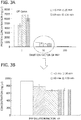
- the film was mineralized for 10 days using mSBF to form a mineralized coating on the PLG film ( FIG. 1A and FIG. 1B ).
- Blood was collected from sheep and centrifuged at 312 X g and 1248 X g to obtain PRP having a platelet count of 886 k/ ⁇ l.
- PRP was subjected to cycles of freeze-thaw to lyse platelets.
- the resulting PRP was diluted by factors of 1, 10, and 100 in 0.001 M phosphate (PO 4 ) buffer.
- the total protein concentration of each PRP dilution was determined by BCA assay.
- mineralized PLG films were incubated with each PRP dilution at 0 minute, 30 minute, 60 minute and 120 minute time points to allow proteins to bind to the mineralized PLG films. After incubation, the films were transferred to a new plate and rinsed with 0.001 M PO 4 buffer to remove unbound protein. The PRP solution from which the films were transferred was collected and used to measure unbound protein. The bound proteins were then eluted from the mineralized PLG films using 0.2 M NaOH and neutralized using 0.2 M HCl in 0.01 M HEPES. Protein concentration of (1) PRP before addition to the mineralized PLG films, (2) PRP after incubation with the mineralized PLG films, and (3) 0.2 M NaOH eluate were determined using a BCA assay.
- the total protein concentration of PRP decreased as the dilution increased.
- incubation of PRP with mineralized PLG films resulted in binding of proteins to the mineralized PLG films. It was also possible to elute the protein that was bound to the mineralized PLG film. Of the three dilutions, the condition that resulted in the most total bound protein was by the mineralized PLG film incubated for 120 minutes with PRP that was diluted by a factor of 10.
- Proteins that remained bound after the 1 st Protein Elution were eluted from the mineralized PLG wells by incubating the mineralized PLG wells using 0.2 M NaOH.
- the 0.2 M NaOH eluate was neutralized using 0.2 M HCl in 0.01 M HEPES (2 nd Protein Elution).
- the protein concentration of the 2 nd Elution was measured using a BCA assay.
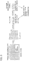
- PLG wells were mineralized as described above. PRP was obtained and subjected to cycles of freeze-thaw to lyse platelets as described above. As shown in FIG. 9 , mineralized PLG wells were incubated for 1 hour to allow proteins to bind. Unbound proteins were rinsed off using water. Bound proteins were eluted from the mineralized PLG wells for less than 5 minutes (buffers were placed into the wells and immediately collected), 30 minutes and 90 minutes using water, 0.001 M, 0.05 M, 0.11 M, 0.17 M, and 0.25 M PO 4 (1 st Protein Elution; PO 4 eluted) and measured using an AGILENTTM protein assay.
- Proteins that remained bound after the 1 st Protein Elution were eluted a second time from the mineralized PLG wells by incubating the mineralized PLG wells using 0.2 M HCl/0.01 M HEPES.
- the 0.2 M HCl/0.01 M HEPES eluate was neutralized using 0.2 M NaOH (2 nd Protein Elution; HCl eluted).
- the protein concentration of the 2 nd Elution was measured using an AGILENTTM assay.
- both water and 0.011 M PO 4 eluted the least amount of proteins, whereas 0.05 M, 0.11 M, 0.17 M, and 0.25 M PO 4 eluted more proteins.
- both water and 0.001 M PO 4 bound the most proteins, whereas 0.05 M, 0.11 M, 0.17 M, and 0.25 M PO 4 bound less proteins. This data also indicated that proteins exposed to PO 4 for 30 minutes have sufficient time to fully elute from the mineral coating. As shown in FIG.
- PLG films were mineralized as described above.
- PRP was obtained and subjected to cycles of freeze-thaw to lyse platelets as described above.
- Varying concentrations of phosphate (0.001 M, 0.05 M, 0.11 M, 0.17 M, and 0.25 M PO 4 ) were added to the PRP to form the PO 4 -modified PRP.
- No phosphate was added to control PRP ("Cx").
- Cx control PRP
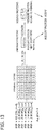
- mineralized PLP films were incubated with Cx and modified-PRP for 15 minute, 60 minute and 90 minute time points. After the 15 minute, 30 minute and 90 minute incubation periods, the buffer (1 st Elution) was collected and analyzed using an AGILENTTM assay to measure total unbound protein.
- Proteins that bound to the mineralized PLG films were eluted with 0.2 N HCl (2 nd Elution) to release bound proteins.
- the solution was neutralized with 0.2 N NaOH and proteins that selectively bound to the mineralized PLG film were thereby measured using an AGILENTTM assay.
- both water, 0.001 M and 0.05 M PO 4 bound the most proteins, whereas 0.11 M, 0.17 M and 0.25 M PO 4 bound less proteins.
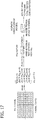
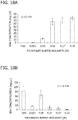
- BSA was added to mineral coated films and allowed to bind for 1 hour. Unbound BSA was rinsed away. PO 4 buffer at one of the following concentrations, 0.001 M, 0.05 M, 0.11 M, 0.17 M, and 0.25 M, was added to the mineralized films and allowed to incubate for less than 5 minutes (buffer was placed into wells and immediately collected) and at a 15 minute time point. The buffer solution was then collected to measure proteins that eluted from the mineralized film. The mineralized film was then dissolved using 0.2 N HCl to release the bound proteins. The solution was neutralized with 0.2 N NaOH and proteins that selectively bound to the mineralized film were thereby measured using an AGILENTTM assay.
- concentrations of 0.001 M and 0.05 M PO 4 eluted the least amount of proteins whereas 0.11 M, 0.17 M, and 0.25 M PO 4 eluted more proteins.
- water and concentrations of 0.001 M and 0.05 M PO 4 bound the most amount of BSA proteins and 0.11 M, 0.17 M, and 0.25 M PO 4 bound less proteins.
- concentrations of 0.001 M and 0.05 M PO 4 eluted the least amount of proteins whereas 0.11 M, 0.17 M, and 0.25 M PO 4 eluted more proteins.
- water and concentrations of 0.001 M and 0.05 M PO 4 bound the most amount of BSA proteins and 0.11 M, 0.17 M, and 0.25 M PO 4 bound less proteins.
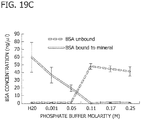
- FIG. 19C illustrates the relationship of bound versus unbound BSA after a 15 minute exposure to varying PO 4 concentrations.
- BSA bound to the mineral coating in a PO 4 -dose dependent manner Combining two different assays, it has been shown that: 1) there is reduced binding to mineral coating with increasing PO 4 molarity (solid line); and 2) there is increased free protein with increasing PO 4 molarity (dotted line). Taken together, these results indicate that albumin binding to mineral is controlled via PO 4 concentration.
- Embodiments of the Invention include embodiments 1-6 below:
Landscapes
- Health & Medical Sciences (AREA)
- Chemical & Material Sciences (AREA)
- Life Sciences & Earth Sciences (AREA)
- Animal Behavior & Ethology (AREA)
- Veterinary Medicine (AREA)
- Public Health (AREA)
- General Health & Medical Sciences (AREA)
- Epidemiology (AREA)
- Engineering & Computer Science (AREA)
- Surgery (AREA)
- Medicinal Chemistry (AREA)
- Vascular Medicine (AREA)
- Heart & Thoracic Surgery (AREA)
- Dermatology (AREA)
- Transplantation (AREA)
- Inorganic Chemistry (AREA)
- Biomedical Technology (AREA)
- Oral & Maxillofacial Surgery (AREA)
- Molecular Biology (AREA)
- Chemical Kinetics & Catalysis (AREA)
- Materials Engineering (AREA)
- Urology & Nephrology (AREA)
- Zoology (AREA)
- Botany (AREA)
- Hematology (AREA)
- Pharmacology & Pharmacy (AREA)
- Bioinformatics & Cheminformatics (AREA)
- Medicines Containing Material From Animals Or Micro-Organisms (AREA)
- Medicines That Contain Protein Lipid Enzymes And Other Medicines (AREA)
Abstract
Description
- This invention was made with government support under AR059916 and HL093282 awarded by the National Institutes of Health. The government has certain rights in the invention.
- The present disclosure relates generally to medical devices. More particularly, the present disclosure relates to medical devices having a mineral coated substrate and an autologous biological molecule. The present disclosure further relates to methods for selectively isolating a biological molecule from a bodily fluid using the coated devices and methods for selectively eluting a biological molecule from the coated devices.
- The delivery of biological molecules ("biologics") such as growth factors to promote musculoskeletal healing has become a popular approach in industry, with the market for orthopedic growth factors, for example, nearly quadrupling between 2003 and 2008. One delivery strategy involves embedding biologics within collagen sponges for insertion into a tissue defect. This strategy has been used clinically for delivery of the biologic BMP2 and is under development for delivery of other emerging biologics, such as BMP12.
- Despite the clinical success of biologics, there are significant limitations related to their delivery. For example, the materials that serve as carriers for biologics, such as collagen sponges, are often inappropriate for orthopedic applications because they do not seamlessly incorporate into standard clinical procedures, and thus, require adoption and training of the medical practitioner. In addition, biologic molecules may quickly diffuse away from carrier materials and may rapidly degrade in vivo (e.g., t1/2 of BMP2 ∼ minutes), which results in limited bioavailability and a need to deliver large doses of the biological molecules. These large doses may be costly and may present a significant safety concern in the clinical orthopedics community, as they have led to edema and ectopic bone formation in multiple recent clinical studies. Thus, there are significant challenges associated with developing biologics for clinical applications.
- Blood is a biological therapy that may be used for whole blood transfusions or making medications. Medications produced from specific portions of the whole blood may be, for example, plasma, platelets, red blood cells, and white blood cells.
- Allogeneic (or homologous) blood transfusion uses blood that is collected from a blood donor and is used for the transfusion of another subject. A specific blood type must be matched for safe transfusion. Another common blood donation practice is for a subject to have blood withdrawn in anticipation of needing blood (i.e., self-donation). If, for example, a subject is planning to undergo surgery where a blood transfusion may be necessary, the subject may have blood withdrawn and stored prior to the procedure. Autologous donation may eliminate reactions due to donor-recipient incompatibility and may preclude exposure to transfusion transmitted infection.
- Use of platelet rich plasma therapy is an emerging biologic treatment that may influence the healing of tissues. Platelet rich plasma ("PRP") is blood plasma that has been enriched with platelets. Platelet enrichment involves the collection of whole blood that is anticoagulated before undergoing centrifugation to separate PRP from platelet-poor plasma and red blood cells. In humans, the baseline platelet concentration of whole blood is about 160-370 k/µl. PRP contains about 4-10X concentration of baseline platelet concentration. The healing influence of PRP may be attributed to the supraphysiological concentrations of growth factors that are released by activated platelets. Many of the growth factors contained in PRP have been identified as possible biologics and studies are under way to isolate and develop these growth factors for use as biologics.
- Recent studies indicate that PRP may accelerate healing, especially in tissues having a poor blood supply. For example, PRP administration improved filling and biomechanical testing of partial anterior cruciate ligament tears. PRP administration has also been shown to increase failure force for Achilles tendon injury and stimulate the development of new bone and tendon in infraspinatus model. Clinical use of PRP has produced promising, but inconsistent results due to the broad variability in the production of PRP by various concentrating equipment and techniques, as well as individual variability in the platelet concentration of plasma.
- The preparation and use of biologics, especially those such as blood and blood-derived products, may involve a cumbersome regulatory path. Many growth factors used as biologics are prepared using recombinant protein expression methods, which must undergo regulatory approval. For example, approval of biologics by the U.S. Food and Drug Administration and the European Medicines Agency involves showing the safety, purity, potency and efficacy of a biologic. Blood used for transfusions also undergoes a significant battery of tests to avoid the transmission of blood-borne pathogens.
- Accordingly, there exists a need to develop materials and methods for isolating biological molecules such as blood and blood components for therapeutic applications.
- The present disclosure relates generally to materials and methods for isolating and delivering biological molecules from bodily fluids. More particularly, the present disclosure relates to coated devices that can deliver biological molecules isolated from bodily fluids such as, for example, PRP and PRP components. The present disclosure also relates to methods for isolating and eluting biological molecules from bodily fluids. The coated devices and methods may be used in the operating room, for example, prior to or during a surgical procedure, to isolate biological molecules from a bodily fluid from a patient.
- The coated devices and methods offer the possibility of selecting specific biological molecules from bodily fluids such as, for example, blood and blood-derived solutions that may be obtained intraoperatively. Moreover, because the biological molecules may be autologous biological molecules, the dosing and regulatory issues facing current biological molecules may be mitigated.
- In one aspect, the present disclosure is directed to a coated device for delivering an autologous biological molecule having a mineral coating on a substrate and an autologous biological molecule.
- In another aspect, the present disclosure is directed to a method for selectively isolating a biological molecule from a bodily fluid. The method includes preparing a coated device having a mineral coating on a substrate. The coated device is then incubated with a bodily fluid comprising a biological molecule, wherein the bodily fluid further comprises an ionic buffer.
- In another aspect, the present disclosure is directed to a method for selectively eluting a biological molecule from a coated device. The method includes preparing a coated device having a mineral coating on a substrate; incubating the coated device with a bodily fluid; and eluting the biological molecule from the coated device.
- The disclosure will be better understood, and features, aspects and advantages other than those set forth above will become apparent when consideration is given to the following detailed description thereof. Such detailed description makes reference to the following drawings, wherein:
-
FIG. 1A is a scanning electron micrograph of a mineralized poly lactide glycolide (PLG) film as discussed in Example 1. -
FIG. 1B is a scanning electron micrograph of a mineralized PLG film at a higher magnification as discussed in Example 1. -
FIG. 2 is a schematic depicting binding of platelet rich plasma (PRP) components to mineralized PLG films as analyzed in Example 1. -
FIG. 3A is a graph illustrating the total protein concentration of diluted PRP incubated with the mineralized PLG film as analyzed in Example 1. -
FIG. 3B is a scaled up version of the graph shown inFIG. 3A illustrating the total protein concentration of the 10 dilution incubated with the mineralized PLG film as analyzed in Example 1. -
FIG. 4 is a graph illustrating the time- and dose-dependent changes in PRP protein binding to the mineralized PLG films as analyzed in Example 1. -
FIG. 5 is a schematic depicting the selective elution of PRP as analyzed in Example 2. -
FIG. 6 is a graph illustrating the amount of protein eluted from mineral coated wells after exposure to varying PO4 concentrations for 15 minutes as analyzed in Example 2. -
FIG. 7 is a graph illustrating the amount of protein eluted from mineral coated wells after exposure to varying PO4 concentrations for 60 minutes as analyzed in Example 2. -
FIG. 8 is a graph illustrating the amount of protein eluted from mineral coated wells after exposure to varying PO4 concentrations for 90 minutes as analyzed in Example 2. -
FIG. 9 is a schematic depicting the selective elution of PRP as analyzed in Example 3. -
FIG. 10A is a graph illustrating the amount of protein eluted from mineral coated wells after a time point of less than 5 minutes using varying PO4 concentrations as analyzed in Example 3. -
FIG. 10B is a graph illustrating the amount of protein bound to mineral coated wells after exposure to varying PO4 concentrations as analyzed in Example 3. -
FIG. 11A is a graph illustrating the amount of protein eluted from mineral coated wells after a time point of 30 minutes using varying PO4 concentrations as analyzed in Example 3. -
FIG. 11B is a graph illustrating the amount of protein bound to mineral coated wells after exposure to varying PO4 concentrations as analyzed in Example 3. -
FIG. 12A is a graph illustrating the amount of protein eluted from mineral coated wells after a time point of 90 minutes using varying PO4 concentrations as analyzed in Example 3. -
FIG. 12B is a graph illustrating the amount of protein bound to mineral coated wells after exposure to varying PO4 concentrations as analyzed in Example 3. -
FIG. 14 is a graph illustrating the amount of protein bound to a mineralized PLG film coating after a 15 minute concomitant exposure to PRP and PO4 buffer as analyzed in Example 4. -
FIG. 15 is a graph illustrating the amount of protein bound to a mineralized PLG film after a 30 minute concomitant exposure to PRP and PO4 buffer as analyzed in Example 4. -
FIG. 16 is a graph illustrating the amount of protein bound to a mineralized PLG film after a 90 minute concomitant exposure to PRP and PO4 buffer as analyzed in Example 4. -
FIG. 17 is a schematic depicting the selective elution of bovine serum albumin (BSA) as analyzed in Example 5. -
FIG. 18A is a graph illustrating the amount of BSA eluted from a mineralized PLG film after exposure to varying PO4 concentrations for less than 5 minutes as analyzed in Example 5. -
FIG. 18B is a graph illustrating the amount of BSA bound to a mineralized PLG film after exposure to varying PO4 concentrations for less than 5 minutes as analyzed in Example 5. -
FIG. 19A is a graph illustrating the amount of BSA eluted from a mineralized PLG film after exposure to varying PO4 concentrations for 15 minutes as analyzed in Example 5. -
FIG. 19B is a graph illustrating the amount of BSA bound to a mineralized PLG film after exposure to varying PO4 concentrations for 15 minutes as analyzed in Example 5. -
FIG. 19C is a graph illustrating the relationship between bound and unbound BSA after a 15 minute exposure to varying PO4 concentrations as analyzed in Example 5. - While the disclosure is susceptible to various modifications and alternative forms, specific embodiments thereof have been shown by way of example in the drawings and are herein described below in detail. It should be understood, however, that the description of specific embodiments is not intended to limit the disclosure to cover all modifications, equivalents and alternatives falling within the spirit and scope of the disclosure as defined by the appended claims.
- Unless defined otherwise, all technical and scientific terms used herein have the same meaning as commonly understood by one of ordinary skill in the art to which the disclosure belongs. Although any methods and materials similar to or equivalent to those described herein may be used in the practice or testing of the present disclosure, the preferred materials and methods are described below.
- In accordance with the present disclosure, coated devices and methods for selectively binding and eluting biological molecules from bodily fluids using the coated devices have been discovered. Methods using coated devices as disclosed herein provide a high throughput platform for selectively binding a desired biological molecule as well as a high throughput platform for determining a desired release profile of a biological molecule from the coated device. Significantly, the coated device having an autologous biological molecule of the present disclosure permits the delivery of a biological molecule obtained from the same subject. This feature avoids significant safety concerns with biological molecules prepared using traditional methods such as, for example, recombinant methods and isolation methods from animal sources. The methods further allow for the selective isolation and selective elution of specific biological molecules in a bodily fluid such that specific biological molecules may be delivered to a subject from the coated devices of the present disclosure.
- In one aspect, the present disclosure is directed to a coated device for delivering an autologous biological molecule. The coated device has a mineral coating on a substrate and an autologous biological molecule attached thereto. Suitable coated devices may be, for example, an orthopedic device, a particle, a film, a dish, a plate, and a suture. Particularly suitable orthopedic devices may be, for example, an arrow, a barb, a tack, an anchor, a nail, a pin, a screw, a staple, a plate, and combinations thereof. Particularly suitable particles may be, for example, agarose beads, latex beads, magnetic beads, and combinations thereof. Particularly suitable plates may be, for example, microtiter plates having, for example, 6, 14, 96, or more sample wells.
- The coated device includes a mineral coating on the surface of a substrate. Suitable mineral coatings are made from mineral forming materials such as, for example, calcium, phosphate, carbonate, and combinations thereof. More particularly, as described more fully herein, the mineral coatings may be formed on the substrate by surface hydrolyzing the substrate under alkaline conditions. After surface hydrolyzing, the substrate is incubated in a modified simulated body fluid (mSBF) containing a suitable mineral-forming material. Suitable substrates for use with the coatings may be, for example, a poly(α-hydroxy ester). Particularly suitable poly(α-hydroxy esters) may be, for example, poly(L-lactide), poly(lactide-co-glycolide), poly(ε-caprolactone), and combinations thereof.
- Attached to the coating, is an autologous biological molecule. The autologous biological molecule is obtained from an autologous bodily fluid. The term "autologous bodily fluid" is used herein to refer to a bodily fluid that is obtained from a subject and used as the source of the autologous biological molecule, which is attached to the coated device that is returned to the same subject. Thus, the subject is both the donor and recipient of the autologous biological molecule. For example, if the autologous bodily fluid is platelet rich plasma (PRP), the PRP is obtained from a subject and is then incubated with the coated device according to the method described herein to bind an autologous biological molecule contained within the subject's own PRP. Suitable autologous bodily fluids may be, for example, whole blood, serum, plasma, platelet rich plasma, bone marrow, cerebrospinal fluid, urine, synovial fluid, and combinations thereof.
- Suitable autologous biological molecules obtained from the autologous bodily fluids may be proteins, for example. Particularly suitable proteins may be, for example, basic proteins. The term "basic protein" is used herein according to its ordinary meaning as understood by those skilled in the art to refer to the category of proteins that have a high isoelectric point (pI of from about 7.1 to about 14), and therefore, tend to be positively charged at physiological pH (∼7.4). By contrast, the term "acidic protein" is used herein according to its ordinary meaning as understood by those skilled in the art to refer to the category of proteins that have a low isoelectric point (pI of from about 0 to about 6.9), and therefore tend to be negatively charged at physiological pH (∼7.4). Particularly suitable basic proteins may be, for example, growth factors.
- Suitable growth factors may be, for example, bone morphogenic proteins (BMPs), connective tissue growth factors, epidermal growth factors, fibroblast growth factors (FGFs), insulin-like growth factors, interleukin, keratinocyte growth factors, platelet-derived growth factor (PDGFs), transforming growth factors (TGFs), vascular endothelial growth factors (VEGF), nerve growth factor (NGF), hepatocyte growth factor (HGF), tumor necrosis factors (TNF), interferons (IFN), and combinations thereof. Specific suitable growth factors may be, for example, BMP1, BMP2, BMP3, BMP4, BMP5, BMP6, BMP7, BMP8a, EGF, PDGFA, PDGFB, PDGFC, PDGFD, PDGFAB, VEGF-A, placenta growth factor (PIGF), VEGF-B, VEGF-C, VEGF-D, TGF-f31, TGF-f32, TGF-f33, AMH, ARTN, GDF1, GDF2, GDF3, GDF3A, GDF5, GDF6, GDF7, GDF8, GDF9, GDF10, GDF11, GDF15, GDFN, INHA, INHBA, INHBB, INHBC, INHBE, LEFTY1, LEFTY2, MSTN, NODAL, NRTN, PSPN, IL-1, IL-2, IL-4, IL-5, IL-6, IL-12, IL-13, IL-21, IL-23, IL-17, IFN-α, IFN-γ, TNF-α, TNF-f3, FGF1, FGF2, FGF3, and FGF4.
- The resulting coated device having an autologous biological molecule bound thereto may then be administered, for example, back into the same subject that served as the donor of the autologous bodily fluid. For example, the coated device may be implanted in a subject to deliver the autologous biological molecule to the subject.
- Because the coated device of the present disclosure has an autologous biological molecule, the coated device allows for the delivery of the autologous biological molecule without the concerns associated with using biological molecules obtained by traditional methods such as, for example, recombinant methods and from animal sources. Also, because the coated devices have a mineral coating that is degradable, delivery of the autologous biological molecules may be controlled such as through controlled elution of the biological molecule from the mineralized coating and/or controlled degradation of the mineral coating. As the mineral coating degrades, the attached autologous biological molecule is released from the coated device. Moreover, the autologous biological molecule attached to the coated device may stimulate repair and/or growth by stimulating cells surrounding or recruited to the area containing the coated device. Additionally, therapeutically effective amounts of the autologous biological molecule may be administered as the concentration of the autologous biological molecules on the coated device may be controlled.
- In one aspect, the present disclosure is directed to a method for selectively isolating a biological molecule from a bodily fluid. The method includes preparing a coated device comprising a mineral coating on a substrate and incubating the coated device with a bodily fluid comprising a biological molecule, wherein the bodily fluid further comprises an ionic buffer.
- To prepare the coated device, a mineral coating may first be formed on the substrate using methods described in
U.S. Patent Application Pub. No. 20080095817 ,U.S. Patent No. 8,075,562 , andU.S. Patent Application Pub. No. 20110305760 , which are hereby incorporated by reference to the extent they are consistent herewith. For example, the mineral coating may be formed by surface hydrolyzing a substrate under alkaline conditions such as, for example, NaOH, followed by incubation in a modified simulated body fluid (mSBF) at a physiologic temperature and pH 6.8 for mineral nucleation and growth. The mSBF solution contains mineral-forming ions, including calcium, phosphate, carbonate, and combinations thereof. The resulting coating may be any suitable coating material containing calcium, phosphate and carbonate, such as, for example, hydroxyapatite (HAP), α-tricalcium phosphate (α-TCP), f33-tricalcium phosphate (f33-TCP), amorphous calcium phosphate, dicalcium phosphate, octacalcium phosphate, and calcium carbonate. Further, the coating formed on the substrate develops as a porous mineral coating (seeFIGS. 1A and 1B ). Although porous mineral coatings are particularly suitable, the mineral coatings may also be nonporous. - As described above, suitable substrates may be, for example, a poly(α-hydroxy ester). Particularly suitable poly(α-hydroxy esters) may be, for example, poly(L-lactide), poly(lactide-co-glycolide), poly(E-caprolactone), and combinations thereof.
- The coated device having the mineral coated substrate is then incubated with a bodily fluid including one or more desired biological molecules. In one embodiment, the bodily fluid may include autologous bodily fluid as described above.
- Alternatively, the bodily fluid is a heterologous bodily fluid. As used herein, the term "heterologous bodily fluid" (i.e., non-autologous solution) refers to a bodily fluid that is obtained from one subject and used in the method to prepare a coated device having a biological molecule attached thereto. The resulting coated device is then used for treating a different subject (i.e., not the subject that donated the bodily fluid). Thus, a subject who is the donor of the heterologous bodily fluid used in the method is different from a subject who is the recipient of the coated device. The term "heterologous bodily fluid" also refers to a bodily fluid that is obtained from a different species of animal, which is then used in the method for isolating a biological molecule of the present disclosure. For example, a bodily fluid such as PRP from a sheep may be used to isolate a biological molecule from the sheep PRP, which is then used for a non-sheep animal as the recipient.
- Suitable autologous and heterologous bodily fluids may be, for example, whole blood, serum, plasma, platelet rich plasma, bone marrow, cerebrospinal fluid, urine, synovial fluid, and combinations thereof.
- The bodily fluid further includes an ionic buffer. Advantageously, including an ionic buffer in the bodily fluid allows for the selective binding of biological molecules to the coated device. The ionic buffer may be added to the bodily fluid while the coated device is incubated. Without being bound by theory, adding an ionic buffer to the bodily fluid during incubation with the coated device may interfere with the formation of electrostatic interactions between the mineral coating and the biological molecule contained in the bodily fluid. For example, if the ionic buffer added to the bodily fluid is high enough to prevent the formation of electrostatic interactions between a biological molecule and the mineral coating, the biological molecule may not bind to the mineral coating. Thus, the ionic strength of the buffer that is added to the bodily fluid influences binding of a biological molecule to the mineral coating. For example, the ionic buffer may influence binding of a biological molecule such that the biological molecule does not interact at all to the coated device, weakly interacts with the coated device, and/or strongly interacts with the coated device.
- Suitable ionic buffers that may be used in the method for selectively isolating a biological molecule from a bodily fluid may be any ionic buffer that disrupts, interferes with, and/or competes with the electrostatic interaction between the biological molecule and the mineral of the mineral coating on the coated device. Particularly suitable ionic buffers may be, for example, phosphate buffers, sodium chloride buffers, magnesium chloride buffers, calcium chloride buffers, sodium fluoride buffers, and combinations thereof. Particularly suitable phosphate buffers may have a phosphate concentration up to, and including, 0.5 M phosphate, and including from about 0.001 M to 0.5 M phosphate. Particularly suitable sodium chloride buffers may have a sodium chloride concentration up to, and including, 0.2 M sodium chloride. Particularly suitable magnesium chloride buffers may have a magnesium chloride concentration up to, and including, 5.0 M magnesium chloride. Particularly suitable calcium chloride buffers may have a calcium chloride concentration up to, and including, 5.0 M calcium chloride. Particularly, suitable sodium fluoride buffers may have a sodium fluoride concentration up to, and including, 0.4 M sodium fluoride.
- Suitable ionic buffers may be, for example, buffers having varying pH ranges. Suitable pH ionic buffers may have a pH range of from about 6.4 to about 7.8.
- The coated device having the mineral coated substrate is incubated with the bodily fluid for a sufficient period of time to allow attachment of a biological molecule to the mineral coating of the substrate. Suitable time to allow the biological molecule to attach to the mineral coating of the substrate may be, for example, from less than a minute to about 120 minutes. The biological molecule attaches to the mineral coating by electrostatic interactions.
- The method may further include washing the coated device to remove components contained within the bodily fluid that do not bind to the mineral coating. Washing may remove serum albumin, for example.
- The coated device may be washed using any suitable washing solution. Suitable washing solutions may be, for example, HEPES (4-(2-hydroxyethyl)-1-piperazineethanesulfonic acid), saline, 0.001 M phosphate buffer, double distilled water, and combinations thereof.
- Attachment of a biological molecule may be monitored using methods known to those skilled in the art. For example, the total protein concentration of the bodily fluid before and after incubation with the coated device may be monitored using a BCA (bicinchoninic acid) assay. Other suitable assays that may be used to monitor attachment and/or elution may be, for example, ELISA, Western blot, 1D and 2D SDS-PAGE, non-equilibrium pH gel electrophoresis (NEPHGE), AGILENTTM protein analysis, and combinations thereof.
- The coated devices resulting from the methods may be coated devices having a mineral coating on a substrate and a heterologous or autologous biological molecule attached thereto. If the resultant coated device is one having a heterologous biological molecule, the coated device is used for a subject that is different from the subject that donated the bodily fluid (the heterologous bodily fluid) used in the method to selectively isolate the biological molecule. Alternatively, if the resultant coated device is one having an autologous biological molecule, the coated device is used for a subject that also was the subject that donated the bodily fluid (the autologous bodily fluid) used in the method to selectively isolate the biological molecule.
- The resultant coated device may be administered to a subject. The resultant coated device may be administered, for example, as an implant. For example, the resultant coated device may be implanted in a subject to deliver the biological molecule to a subject.
- The resultant coated device having an autologous biological molecule allows for the delivery of the autologous biological molecule without the concerns associated with using biological molecules obtained by traditional methods such as, for example, recombinant methods and isolation methods from animal sources. Thus, the resultant coated device having an autologous biological molecule can avoid regulatory hurdles and safety issues associated with biological molecules obtained from sources other than from the subject's own bodily fluids. The resultant coated device having a heterologous biological molecule allows for the delivery of the heterologous biological molecule under conditions where the recipient subject may not have a sufficient amount of a biological molecule such that the recipient can also be the donor of the bodily fluid used in the method. Additionally, a resultant coated device having a heterologous biological molecule may be suitable for use in a veterinary setting, where one donor subject may be used to provide the bodily fluid used in the method for preparing multiple coated devices or where having an autologous biological molecule is not desired.
- Because the coated devices have a mineral coating that is degradable, delivery of the autologous biological molecule and the heterologous biological molecule may be controlled such through controlled elution of the biological molecule from the mineral coating as described herein and/or controlled degradation of the mineral coating. As the mineral coating degrades, the attached autologous biological molecule and/or the heterologous biological molecule may be released from the coated device. Additionally, or alternatively, as the ionic concentration of the environment in which the coated device is implanted changes, it may influence the electrostatic interaction between the biological molecule and the mineral coating such that the biological molecule detaches from the mineral coating.
- Moreover, the autologous biological molecules and the heterologous biological molecules may stimulate repair and/or growth by stimulating cells surrounding or recruited to the area containing the coated device. Therapeutically effective amounts of the autologous biological molecule and/or the heterologous biological molecule may be administered as the concentration of the autologous biological molecules and the heterologous biological molecule on the coated device may be controlled.
- In another aspect, the present disclosure is directed to a method for selectively eluting a biological molecule from a coated device. The method includes preparing a coated device having a mineral coating on a substrate; incubating the coated device with a bodily fluid having a biological molecule; and eluting the biological molecule from the coated device. Eluting may remove biological molecules that are attached to the mineral coating on the substrate, and in some embodiments, allows for selectively removing a biological molecule from the coated device.
- In one aspect, eluting at least one biological molecule from the coated device includes contacting the coated device with an elution buffer. Suitable elution buffers may be any ion-containing buffer that disrupts the electrostatic interaction between the biological molecule and the mineral of the mineral coating on the coated device. Particularly suitable elution buffers that may be used to elute at least one biological molecule from the coated device may be, for example, phosphate buffers (up to 0.5 M phosphate), sodium chloride buffers (up to0.2 M sodium chloride), magnesium chloride buffers (up to 5.0 M magnesium chloride), calcium chloride buffers (up to 5.0 M calcium chloride), sodium fluoride buffers (up to 0.4 M sodium fluoride), and combinations thereof.
- Selective elution of the biological molecule may be performed as a "batch-type" elution in which the coated device is contacted with a particular ionic strength buffer such that elution of the biological molecule occurs at once. Selective elution of the biological molecule may also be performed using a concentration gradient in which the beginning elution is performed using a low ionic strength buffer and continues with increasing ionic strength buffer. The gradient may be performed as a step-wise gradient or as a continuous gradient.
- In another aspect, eluting at least one biological molecule from the coated device includes contacting the coated device with a mineral dissolution buffer. The mineral dissolution buffer causes the mineral of the mineral coating to dissolve. As the mineral coating dissolves, a biological molecule that is electrostatically attached to the mineral coating loses its attachment and elutes from the coated device. Suitable mineral dissolution buffers may be any buffer that causes the mineral coating to dissolve.
- Particularly suitable mineral dissolution buffers may include phosphoric acid, ethylenediaminetetraacetic acid (EDTA), 0.25 M hydrochloric acid (HCl), 0.25 M sodium hydroxide (NaOH), for example. The mineral dissolution buffer may include phosphoric acid up to, and including, 0.5 M phosphoric acid. The mineral dissolution buffer may include EDTA up to, and including, 20% (w/v) EDTA. The amount of HCl in the mineral dissolution buffer may be up to, and including, 0.25 M HCl.
- Elution of a biological molecule may be monitored using methods known to those skilled in the art. For example, the total protein concentration of the bodily fluid before and after incubation with the coated device as well as after the coated device is contacted with an ionic buffer or a mineral dissolution buffer may be monitored using a BCA assay. Other suitable assays that may be used to monitor elution may be, for example, ELISA, Western blot, 1D and 2D SDS-PAGE, non-equilibrium pH gel electrophoresis (NEPHGE), AGILENTTM protein analysis, and combinations thereof.
- The disclosure will be more fully understood upon consideration of the following non-limiting Examples.
- In this Example, the protein concentration of PRP was determined after incubation with a mineralized poly lactide glycolide (PLG) film.
- Specifically, poly lactide glycolide (85:15) was solvent casted into a film. The film was mineralized for 10 days using mSBF to form a mineralized coating on the PLG film (
FIG. 1A and FIG. 1B ). Blood was collected from sheep and centrifuged at 312 X g and 1248 X g to obtain PRP having a platelet count of 886 k/µl. PRP was subjected to cycles of freeze-thaw to lyse platelets. The resulting PRP was diluted by factors of 1, 10, and 100 in 0.001 M phosphate (PO4) buffer. The total protein concentration of each PRP dilution was determined by BCA assay. - As shown in
FIG. 2 , mineralized PLG films were incubated with each PRP dilution at 0 minute, 30 minute, 60 minute and 120 minute time points to allow proteins to bind to the mineralized PLG films. After incubation, the films were transferred to a new plate and rinsed with 0.001 M PO4 buffer to remove unbound protein. The PRP solution from which the films were transferred was collected and used to measure unbound protein. The bound proteins were then eluted from the mineralized PLG films using 0.2 M NaOH and neutralized using 0.2 M HCl in 0.01 M HEPES. Protein concentration of (1) PRP before addition to the mineralized PLG films, (2) PRP after incubation with the mineralized PLG films, and (3) 0.2 M NaOH eluate were determined using a BCA assay. - As shown in
FIG. 3A and FIG. 3B , the total protein concentration of PRP decreased as the dilution increased. As shown inFIG. 4 , incubation of PRP with mineralized PLG films resulted in binding of proteins to the mineralized PLG films. It was also possible to elute the protein that was bound to the mineralized PLG film. Of the three dilutions, the condition that resulted in the most total bound protein was by the mineralized PLG film incubated for 120 minutes with PRP that was diluted by a factor of 10. - In this Example, elution of proteins from mineralized PLG wells with varying phosphate molarities was determined.
- Specifically, wells of a 96-well plate were coated with a mineralized film made as described above. PRP was obtained and subjected to cycles of freeze-thaw to lyse platelets as described above. As shown in
FIG. 5 , mineralized PLG wells were incubated for 1 hour to allow proteins to bind. Unbound proteins were rinsed off using water. Bound proteins were eluted from the mineralized PLG wells for 15 minute, 60 minute, and 90 minute time points using 0.001 M, 0.05 M, 0.11 M, 0.17 M, and 0.25 M PO4 (1st Protein Elution) and measured by BCA assay. Proteins that remained bound after the 1st Protein Elution were eluted from the mineralized PLG wells by incubating the mineralized PLG wells using 0.2 M NaOH. The 0.2 M NaOH eluate was neutralized using 0.2 M HCl in 0.01 M HEPES (2nd Protein Elution). The protein concentration of the 2nd Elution was measured using a BCA assay. - As shown in
FIG. 6 for the 15 minute time point, a concentration of 0.001 M PO4 eluted the least amount of proteins, whereas 0.05 M, 0.11 M, 0.17 M, and 0.25 M PO4 eluted more proteins. - As shown in
FIG. 7 for the 60 minute time point, a concentration of 0.001 M PO4 eluted the least amount of proteins, whereas 0.05 M, 0.11 M, 0.17 M, and 0.25 M PO4 eluted more proteins. - As shown in
FIG. 8 for the 90 minute time point, a concentration of 0.001 M PO4 eluted the least amount of proteins, whereas 0.05 M, 0.11 M, 0.17 M, and 0.25 M PO4 eluted more proteins. - In this Example, elution of proteins from mineralized PLG wells with varying phosphate molarities was determined.
- Specifically, PLG wells were mineralized as described above. PRP was obtained and subjected to cycles of freeze-thaw to lyse platelets as described above. As shown in
FIG. 9 , mineralized PLG wells were incubated for 1 hour to allow proteins to bind. Unbound proteins were rinsed off using water. Bound proteins were eluted from the mineralized PLG wells for less than 5 minutes (buffers were placed into the wells and immediately collected), 30 minutes and 90 minutes using water, 0.001 M, 0.05 M, 0.11 M, 0.17 M, and 0.25 M PO4 (1st Protein Elution; PO4 eluted) and measured using an AGILENTTM protein assay. Proteins that remained bound after the 1st Protein Elution (using phosphate buffer) were eluted a second time from the mineralized PLG wells by incubating the mineralized PLG wells using 0.2 M HCl/0.01 M HEPES. The 0.2 M HCl/0.01 M HEPES eluate was neutralized using 0.2 M NaOH (2nd Protein Elution; HCl eluted). The protein concentration of the 2nd Elution was measured using an AGILENTTM assay. - As shown in
FIGS. 10A and 10B for the <5 minute time point, most of the protein eluted in the 1st Protein Elution as compared with the percent protein eluted in the 2nd Elution. Additionally, as shown inFIG. 10A , both water and 0.011 M PO4 eluted the least amount of proteins, whereas 0.05 M, 0.11 M, 0.17 M, and 0.25 M PO4 eluted more proteins. As shown inFIG. 10B , however, after mineral dissolution via HCl, both water and 0.001 M PO4 bound the most proteins, whereas 0.05 M, 0.11 M, 0.17 M, and 0.25 M PO4 bound less proteins. - As shown in
FIG. 11A for the 30 minute time point, both water and 0.011 M PO4 eluted the least amount of proteins, whereas 0.05 M, 0.11 M, 0.17 M, and 0.25 M PO4 eluted more proteins. As shown inFIG. 11B , however, after mineral dissolution via HCl, both water and 0.001 M PO4 bound the most proteins, whereas 0.05 M, 0.11 M, 0.17 M, and 0.25 M PO4 bound less proteins. This data also indicated that proteins exposed to PO4 for 30 minutes have sufficient time to fully elute from the mineral coating. As shown inFIG. 12A for the 90 minute time point, 0.011 M PO4 eluted the most amount of proteins, whereas 0.05 M, 0.11 M, 0.17 M, and 0.25 M PO4 eluted less proteins. As shown inFIG. 12B , however, after mineral dissolution via HCl, both water, 0.001 M and 0.05 M PO4 bound the most proteins, whereas 0.11 M, 0.17 M, and 0.25 M PO4 bound less proteins. This data also indicated the ability of proteins to bind and release at lesser concentrations from lower density mineral coated wells. - In this Example, binding of proteins in PO4-modified PRP to mineralized PLP films was determined.
- Specifically, PLG films were mineralized as described above. PRP was obtained and subjected to cycles of freeze-thaw to lyse platelets as described above. Varying concentrations of phosphate (0.001 M, 0.05 M, 0.11 M, 0.17 M, and 0.25 M PO4) were added to the PRP to form the PO4-modified PRP. No phosphate was added to control PRP ("Cx"). As shown in
FIG. 13 , mineralized PLP films were incubated with Cx and modified-PRP for 15 minute, 60 minute and 90 minute time points. After the 15 minute, 30 minute and 90 minute incubation periods, the buffer (1st Elution) was collected and analyzed using an AGILENTTM assay to measure total unbound protein. Proteins that bound to the mineralized PLG films were eluted with 0.2 N HCl (2nd Elution) to release bound proteins. The solution was neutralized with 0.2 N NaOH and proteins that selectively bound to the mineralized PLG film were thereby measured using an AGILENTTM assay. - As shown in
FIG. 14 for the 15 minute time point, both water, 0.001 M and 0.05 M PO4 bound the most proteins, whereas 0.11 M, 0.17 M and 0.25 M PO4 bound less proteins. - As shown in
FIG. 15 for the 30 minute time point, both water and 0.001 M PO4 bound the most proteins, whereas 0.05 M, 0.11 M, 0.17 M and 0.25 M PO4 bound less proteins. - As shown in
FIG. 16 for the 90 minute time point, both water and 0.001 M PO4 bound the most proteins, whereas 0.05 M, 0.11 M, 0.17 M and 0.25 M PO4 bound less proteins. - In this Example, binding of bovine serum albumin (BSA) to mineralized PLG films was determined.
- As shown in
FIG. 17 , BSA was added to mineral coated films and allowed to bind for 1 hour. Unbound BSA was rinsed away. PO4 buffer at one of the following concentrations, 0.001 M, 0.05 M, 0.11 M, 0.17 M, and 0.25 M, was added to the mineralized films and allowed to incubate for less than 5 minutes (buffer was placed into wells and immediately collected) and at a 15 minute time point. The buffer solution was then collected to measure proteins that eluted from the mineralized film. The mineralized film was then dissolved using 0.2 N HCl to release the bound proteins. The solution was neutralized with 0.2 N NaOH and proteins that selectively bound to the mineralized film were thereby measured using an AGILENTTM assay. - As shown in
FIG. 18A , at the less than 5 minute time point, concentrations of 0.001 M and 0.05 M PO4 eluted the least amount of proteins whereas 0.11 M, 0.17 M, and 0.25 M PO4 eluted more proteins. As shown inFIG. 18B , water and concentrations of 0.001 M and 0.05 M PO4 bound the most amount of BSA proteins and 0.11 M, 0.17 M, and 0.25 M PO4 bound less proteins. - As shown in
FIG. 19A , at the 15 minute time point, concentrations of 0.001 M and 0.05 M PO4 eluted the least amount of proteins whereas 0.11 M, 0.17 M, and 0.25 M PO4 eluted more proteins. As shown inFIG. 19B , water and concentrations of 0.001 M and 0.05 M PO4 bound the most amount of BSA proteins and 0.11 M, 0.17 M, and 0.25 M PO4 bound less proteins. -
FIG. 19C illustrates the relationship of bound versus unbound BSA after a 15 minute exposure to varying PO4 concentrations. BSA bound to the mineral coating in a PO4-dose dependent manner. Combining two different assays, it has been shown that: 1) there is reduced binding to mineral coating with increasing PO4 molarity (solid line); and 2) there is increased free protein with increasing PO4 molarity (dotted line). Taken together, these results indicate that albumin binding to mineral is controlled via PO4 concentration. - The examples described above demonstrate that the mineral coatings and methods offer the ability to select specific biological molecules from bodily fluids that may be obtained intraoperatively. This will allow for the isolation of a specific biological molecule or set of biological molecules from a subject, then delivery of the specific biological molecule back into the same subject or into a different subject.
- In view of the above, it will be seen that the several advantages of the disclosure are achieved and other advantageous results attained. As various changes could be made in the above devices and methods without departing from the scope of the disclosure, it is intended that all matter contained in the above description and shown in the accompanying drawings shall be interpreted as illustrative and not in a limiting sense.
- When introducing elements of the present disclosure or the various versions, embodiment(s) or aspects thereof, the articles "a", "an", "the" and "said" are intended to mean that there are one or more of the elements. The terms "comprising", "including" and "having" are intended to be inclusive and mean that there may be additional elements other than the listed elements.
- Embodiments of the Invention include embodiments 1-6 below:
- 1. A method for selectively isolating a biological molecule from a bodily fluid, the method comprising:
- preparing a coated device comprising a mineral coating on a substrate;
- incubating the coated device with a bodily fluid comprising a biological molecule,
- wherein the bodily fluid further comprises an ionic buffer.
- 2. The method of
embodiment 1, wherein the bodily fluid is selected from the group consisting of whole blood, serum, plasma, platelet rich plasma, bone marrow, cerebrospinal fluid, urine, synovial fluid, and combinations thereof. - 3. The method of
embodiment 1, wherein the ionic buffer comprises at least one of phosphate, sodium chloride, magnesium chloride, and calcium chloride. - 4. The method of
embodiment 3, wherein the ionic buffer comprises up to 0.5 M phosphate. - 5. The method of
embodiment 1, wherein the substrate comprises a poly(α-hydroxy ester) selected from the group consisting of poly(L-lactide), poly(lactide-co-glycolide), poly(ε-caprolactone), and combinations thereof. - 6. The method of
embodiment 1, wherein the mineral coating is selected from the group consisting of calcium, phosphate, carbonate, and combinations thereof.
Claims (14)
- A method for selectively eluting a biological molecule from a coated device, the method comprising:preparing a coated device comprising a mineral coating on a substrate;incubating the coated device with a bodily fluid comprising a biological molecule; andeluting the biological molecule from the coated device.
- The method of claim 1, wherein the bodily fluid is selected from the group consisting of blood, serum, plasma, platelet rich plasma, bone marrow, cerebrospinal fluid, urine, synovial fluid, and combinations thereof.
- The method of claim 1, wherein incubating the coated device with the bodily fluid comprises incubating the coated device with the bodily fluid for a time period of from less than one minute to about 120 minutes.
- The method of claim 1, wherein eluting the biological molecule from the coated device comprises contacting the coated device with an ionic buffer selected from the group consisting of a phosphate buffer, a sodium chloride buffer, a magnesium chloride buffer, a calcium chloride buffer, a sodium fluoride buffer, and combinations thereof.
- The method of claim 4, wherein the ionic buffer comprises up to 0.5 M phosphate.
- The method of claim 4, wherein the ionic buffer comprises up to 0.2 M sodium chloride.
- The method of claim 4, wherein the ionic buffer comprises up to 5.0 M magnesium chloride.
- The method of claim 4, wherein the ionic buffer comprises up to 5.0 M calcium chloride.
- The method of claim 4, wherein the ionic buffer comprises up to 0.4 M sodium fluoride.
- The method of claim 1, wherein eluting the biological molecule from the coated device comprises contacting the coated device with a mineral dissolution buffer, wherein the mineral dissolution buffer is selected from the group consisting of phosphoric acid, ethylenediaminetetraacetic acid, hydrochloric acid, sodium hydroxide, and combinations thereof.
- The method of claim 10, wherein the mineral dissolution buffer comprises up to 0.5 M phosphoric acid.
- The method of claim 1, wherein the mineral coating is selected from the group consisting of calcium, phosphate, carbonate, and combinations thereof.
- The method of claim 12, wherein the mineral coating is elected from the group consisting of hydroxyapatite (HAP), α-tricalcium phosphate (α-TCP), β3-tricalcium phosphate (β3-TCP), amorphous calcium phosphate, dicalcium phosophate, octacalcium phosphate, and calcium carbonate.
- The method of claim 1, wherein the substrate comprises a poly(α-hydroxy ester) selected from the group consisting of poly(L-lactide), poly(lactide-co-glycolide), poly(ε-caprolactone), and combinations thereof.
Applications Claiming Priority (2)
| Application Number | Priority Date | Filing Date | Title |
|---|---|---|---|
| US13/680,251 US20140141047A1 (en) | 2012-11-19 | 2012-11-19 | Selection of platelet rich plasma components via mineral binding |
| EP13815600.5A EP2919713A2 (en) | 2012-11-19 | 2013-11-08 | Selection of platelet rich plasma components via mineral binding |
Related Parent Applications (1)
| Application Number | Title | Priority Date | Filing Date |
|---|---|---|---|
| EP13815600.5A Division EP2919713A2 (en) | 2012-11-19 | 2013-11-08 | Selection of platelet rich plasma components via mineral binding |
Publications (1)
| Publication Number | Publication Date |
|---|---|
| EP3189812A1 true EP3189812A1 (en) | 2017-07-12 |
Family
ID=49911778
Family Applications (2)
| Application Number | Title | Priority Date | Filing Date |
|---|---|---|---|
| EP13815600.5A Withdrawn EP2919713A2 (en) | 2012-11-19 | 2013-11-08 | Selection of platelet rich plasma components via mineral binding |
| EP17150587.8A Withdrawn EP3189812A1 (en) | 2012-11-19 | 2013-11-08 | Elution of platelet rich plasma components from coated devices |
Family Applications Before (1)
| Application Number | Title | Priority Date | Filing Date |
|---|---|---|---|
| EP13815600.5A Withdrawn EP2919713A2 (en) | 2012-11-19 | 2013-11-08 | Selection of platelet rich plasma components via mineral binding |
Country Status (5)
| Country | Link |
|---|---|
| US (1) | US20140141047A1 (en) |
| EP (2) | EP2919713A2 (en) |
| AU (1) | AU2013345076A1 (en) |
| CA (1) | CA2890011A1 (en) |
| WO (1) | WO2014078180A2 (en) |
Citations (1)
| Publication number | Priority date | Publication date | Assignee | Title |
|---|---|---|---|---|
| WO2012118843A1 (en) * | 2011-02-28 | 2012-09-07 | Tissue Regeneration Systems, Inc. | Modular tissue scaffolds |
Family Cites Families (6)
| Publication number | Priority date | Publication date | Assignee | Title |
|---|---|---|---|---|
| MXPA04002716A (en) * | 2001-09-24 | 2005-11-08 | Verigen Ag | Autologous growth factor cocktail composition, method of production and use. |
| US7087086B2 (en) * | 2003-01-31 | 2006-08-08 | Depuy Products, Inc. | Biological agent-containing ceramic coating and method |
| US9295755B2 (en) | 2006-10-18 | 2016-03-29 | Wisconsin Alumni Research Foundation | Multilayer tissue regeneration system |
| WO2010036919A1 (en) * | 2008-09-25 | 2010-04-01 | Tissue Regeneration Systems, Inc. | Mineral-coated microspheres |
| US8075562B2 (en) | 2007-06-25 | 2011-12-13 | Wisconsin Alumni Research Foundation | Controlled release of biopharmaceutical growth factors from hydroxyapatite coating on bioresorbable interference screws used in cruciate ligament reconstruction surgery |
| US8778869B2 (en) | 2010-06-09 | 2014-07-15 | Wisconsin Alumni Research Foundation | Tissue regeneration system |
-
2012
- 2012-11-19 US US13/680,251 patent/US20140141047A1/en not_active Abandoned
-
2013
- 2013-11-08 WO PCT/US2013/069096 patent/WO2014078180A2/en not_active Ceased
- 2013-11-08 EP EP13815600.5A patent/EP2919713A2/en not_active Withdrawn
- 2013-11-08 EP EP17150587.8A patent/EP3189812A1/en not_active Withdrawn
- 2013-11-08 CA CA2890011A patent/CA2890011A1/en not_active Abandoned
- 2013-11-08 AU AU2013345076A patent/AU2013345076A1/en not_active Abandoned
Patent Citations (1)
| Publication number | Priority date | Publication date | Assignee | Title |
|---|---|---|---|---|
| WO2012118843A1 (en) * | 2011-02-28 | 2012-09-07 | Tissue Regeneration Systems, Inc. | Modular tissue scaffolds |
Non-Patent Citations (2)
| Title |
|---|
| D SUAREZ-GONZALES ET AL.: "Controllable mineral coatings on PCL scaffolds as carriers for growth factor release", BIOMATERIALS., vol. 33, 19 October 2011 (2011-10-19), ELSEVIER SCIENCE PUBLISHERS BV., BARKING., pages 713 - 721, XP002722086, ISSN: 0142-9612 * |
| K OHTA ET AL.: "Interaction between hydroxyapatite and proteins by liquid chromatograhy using simulated body fluids as eluents", JOURNAL OF MATERIALS SCIENCE: MATERIALS IN MEDICINE, vol. 13, no. 7, 2002, Kluwer Academic Publishers, pages 633 - 637, XP002770509, ISSN: 1573-4838 * |
Also Published As
| Publication number | Publication date |
|---|---|
| US20140141047A1 (en) | 2014-05-22 |
| CA2890011A1 (en) | 2014-05-22 |
| WO2014078180A2 (en) | 2014-05-22 |
| WO2014078180A3 (en) | 2014-08-21 |
| EP2919713A2 (en) | 2015-09-23 |
| AU2013345076A1 (en) | 2015-05-21 |
Similar Documents
| Publication | Publication Date | Title |
|---|---|---|
| Anitua et al. | The effects of PRGF on bone regeneration and on titanium implant osseointegration in goats: a histologic and histomorphometric study | |
| Thorn et al. | Autologous fibrin glue with growth factors in reconstructive maxillofacial surgery | |
| Anitua et al. | Release kinetics of platelet-derived and plasma-derived growth factors from autologous plasma rich in growth factors | |
| Arpornmaeklong et al. | Influence of platelet-rich plasma (PRP) on osteogenic differentiation of rat bone marrow stromal cells. An in vitro study | |
| Xu et al. | Human fetal mesenchymal stem cell secretome enhances bone consolidation in distraction osteogenesis | |
| Civinini et al. | The use of autologous blood-derived growth factors in bone regeneration | |
| Guzel et al. | The biomechanical and histological effects of platelet-rich plasma on fracture healing | |
| Lollobrigida et al. | Biomimetic implant surface functionalization with liquid L‐PRF products: In vitro study | |
| CN102316890A (en) | Bone grafts with reduced protease activity and methods of selection and use | |
| EP3258945A1 (en) | Modified blood clots | |
| Crowe et al. | Tendon regeneration with a novel tendon hydrogel: in vitro effects of platelet-rich plasma on rat adipose-derived stem cells | |
| JP2025096273A (en) | Compositions and methods for treating bone injuries | |
| Qin et al. | SDF-1α/BMP-12 loaded biphasic sustained-release SIS hydrogel/SA microspheres composite for tendon regeneration | |
| Wang et al. | Non-bone-derived exosomes: a new perspective on regulators of bone homeostasis | |
| Yang et al. | The phase‐transited lysozyme coating modified small intestinal submucosa membrane loaded with calcium and zinc ions for enhanced bone regeneration | |
| Dreyer et al. | Optimizing combination of vascular endothelial growth factor and mesenchymal stem cells on ectopic bone formation in SCID mice | |
| Alsousou et al. | Platelet-rich plasma in regenerative medicine | |
| EP4631535A1 (en) | Compounds for use as bone replacement and/or bone rebuilding material | |
| US20140141047A1 (en) | Selection of platelet rich plasma components via mineral binding | |
| Shah et al. | Bioactive sutures: advances in surgical suture functionalization | |
| Özdemir et al. | Osteocalcin and osteonectin expression after double-application of platelet-rich plasma in rabbits | |
| CA3228661A1 (en) | Compositions and methods for treating bone injuries | |
| Machmud et al. | Platelet-rich plasma improves initial bone remodeling | |
| Ardakani et al. | Experimental study on healing of long bone defects treated with fibrin membrane enriched with platelet growth factors and periosteal mesenchymal stem cells in rabbit: Radiographical and histopathological evaluations | |
| Grgurević et al. | Tapping the potential of bone regeneration by delivery of concentrated extracellular vesicles to the fracture site: A new horizon in bone regeneration therapy research? |
Legal Events
| Date | Code | Title | Description |
|---|---|---|---|
| PUAI | Public reference made under article 153(3) epc to a published international application that has entered the european phase |
Free format text: ORIGINAL CODE: 0009012 |
|
| STAA | Information on the status of an ep patent application or granted ep patent |
Free format text: STATUS: THE APPLICATION HAS BEEN PUBLISHED |
|
| AC | Divisional application: reference to earlier application |
Ref document number: 2919713 Country of ref document: EP Kind code of ref document: P |
|
| AK | Designated contracting states |
Kind code of ref document: A1 Designated state(s): AL AT BE BG CH CY CZ DE DK EE ES FI FR GB GR HR HU IE IS IT LI LT LU LV MC MK MT NL NO PL PT RO RS SE SI SK SM TR |
|
| STAA | Information on the status of an ep patent application or granted ep patent |
Free format text: STATUS: THE APPLICATION IS DEEMED TO BE WITHDRAWN |
|
| 18D | Application deemed to be withdrawn |
Effective date: 20180113 |