EP3184648A1 - Analytical process for genotoxicity assessment - Google Patents
Analytical process for genotoxicity assessment Download PDFInfo
- Publication number
- EP3184648A1 EP3184648A1 EP16185408.8A EP16185408A EP3184648A1 EP 3184648 A1 EP3184648 A1 EP 3184648A1 EP 16185408 A EP16185408 A EP 16185408A EP 3184648 A1 EP3184648 A1 EP 3184648A1
- Authority
- EP
- European Patent Office
- Prior art keywords
- cells
- agent
- process according
- nes
- mrnas
- Prior art date
- Legal status (The legal status is an assumption and is not a legal conclusion. Google has not performed a legal analysis and makes no representation as to the accuracy of the status listed.)
- Withdrawn
Links
- 238000000034 method Methods 0.000 title claims abstract description 48
- 230000008569 process Effects 0.000 title claims abstract description 46
- 230000001738 genotoxic effect Effects 0.000 title claims description 17
- 231100000025 genetic toxicology Toxicity 0.000 title description 2
- 239000003795 chemical substances by application Substances 0.000 claims abstract description 69
- 230000003612 virological effect Effects 0.000 claims abstract description 34
- 150000007523 nucleic acids Chemical class 0.000 claims abstract description 28
- 102000039446 nucleic acids Human genes 0.000 claims abstract description 22
- 108020004707 nucleic acids Proteins 0.000 claims abstract description 22
- 231100000734 genotoxic potential Toxicity 0.000 claims abstract description 18
- 239000002245 particle Substances 0.000 claims abstract description 17
- 210000003958 hematopoietic stem cell Anatomy 0.000 claims abstract description 15
- 210000004027 cell Anatomy 0.000 claims description 82
- 108091032973 (ribonucleotides)n+m Proteins 0.000 claims description 45
- 108020004999 messenger RNA Proteins 0.000 claims description 41
- 102000040650 (ribonucleotides)n+m Human genes 0.000 claims description 24
- 238000004113 cell culture Methods 0.000 claims description 21
- 239000000523 sample Substances 0.000 claims description 20
- 239000013641 positive control Substances 0.000 claims description 16
- -1 GM805 Proteins 0.000 claims description 10
- 239000002609 medium Substances 0.000 claims description 9
- 206010028980 Neoplasm Diseases 0.000 claims description 7
- 230000002074 deregulated effect Effects 0.000 claims description 7
- 108091028043 Nucleic acid sequence Proteins 0.000 claims description 6
- 102000004127 Cytokines Human genes 0.000 claims description 5
- 108090000695 Cytokines Proteins 0.000 claims description 5
- 102000003815 Interleukin-11 Human genes 0.000 claims description 5
- 108090000177 Interleukin-11 Proteins 0.000 claims description 5
- 108010002386 Interleukin-3 Proteins 0.000 claims description 5
- 108010002586 Interleukin-7 Proteins 0.000 claims description 5
- 238000004458 analytical method Methods 0.000 claims description 5
- 102100024049 A-kinase anchor protein 13 Human genes 0.000 claims description 4
- 102100026607 ALS2 C-terminal-like protein Human genes 0.000 claims description 4
- 102100040635 Actin filament-associated protein 1-like 1 Human genes 0.000 claims description 4
- 102100021626 Ankyrin repeat and SOCS box protein 2 Human genes 0.000 claims description 4
- 102100036301 C-C chemokine receptor type 7 Human genes 0.000 claims description 4
- 102100021615 Class A basic helix-loop-helix protein 15 Human genes 0.000 claims description 4
- 102100038945 Coiled-coil domain-containing protein 102A Human genes 0.000 claims description 4
- 102100027823 Complexin-2 Human genes 0.000 claims description 4
- 102100037101 Deoxycytidylate deaminase Human genes 0.000 claims description 4
- 102100028987 Dual specificity protein phosphatase 2 Human genes 0.000 claims description 4
- 102100032038 EH domain-containing protein 3 Human genes 0.000 claims description 4
- 102100039249 Elongation of very long chain fatty acids protein 6 Human genes 0.000 claims description 4
- 108050007786 Elongation of very long chain fatty acids protein 6 Proteins 0.000 claims description 4
- 102100028043 Fibroblast growth factor 3 Human genes 0.000 claims description 4
- 102100023001 Growth hormone-regulated TBC protein 1 Human genes 0.000 claims description 4
- 102100040754 Guanylate cyclase soluble subunit alpha-1 Human genes 0.000 claims description 4
- 102100024594 Histone-lysine N-methyltransferase PRDM16 Human genes 0.000 claims description 4
- 101000833679 Homo sapiens A-kinase anchor protein 13 Proteins 0.000 claims description 4
- 101000717960 Homo sapiens ALS2 C-terminal-like protein Proteins 0.000 claims description 4
- 101000892363 Homo sapiens Actin filament-associated protein 1-like 1 Proteins 0.000 claims description 4
- 101000754299 Homo sapiens Ankyrin repeat and SOCS box protein 2 Proteins 0.000 claims description 4
- 101000716065 Homo sapiens C-C chemokine receptor type 7 Proteins 0.000 claims description 4
- 101000740818 Homo sapiens Coiled-coil domain-containing protein 102A Proteins 0.000 claims description 4
- 101000859628 Homo sapiens Complexin-2 Proteins 0.000 claims description 4
- 101000955042 Homo sapiens Deoxycytidylate deaminase Proteins 0.000 claims description 4
- 101000838335 Homo sapiens Dual specificity protein phosphatase 2 Proteins 0.000 claims description 4
- 101000921212 Homo sapiens EH domain-containing protein 3 Proteins 0.000 claims description 4
- 101001060280 Homo sapiens Fibroblast growth factor 3 Proteins 0.000 claims description 4
- 101000903509 Homo sapiens Growth hormone-regulated TBC protein 1 Proteins 0.000 claims description 4
- 101001038755 Homo sapiens Guanylate cyclase soluble subunit alpha-1 Proteins 0.000 claims description 4
- 101000686942 Homo sapiens Histone-lysine N-methyltransferase PRDM16 Proteins 0.000 claims description 4
- 101001034652 Homo sapiens Insulin-like growth factor 1 receptor Proteins 0.000 claims description 4
- 101001056627 Homo sapiens Janus kinase and microtubule-interacting protein 1 Proteins 0.000 claims description 4
- 101000717987 Homo sapiens LIM domain-containing protein ajuba Proteins 0.000 claims description 4
- 101000592685 Homo sapiens Meiotic nuclear division protein 1 homolog Proteins 0.000 claims description 4
- 101000584314 Homo sapiens Myc target protein 1 Proteins 0.000 claims description 4
- 101001109620 Homo sapiens Nucleolar and coiled-body phosphoprotein 1 Proteins 0.000 claims description 4
- 101000595489 Homo sapiens Phosphatidylinositol N-acetylglucosaminyltransferase subunit A Proteins 0.000 claims description 4
- 101000583702 Homo sapiens Pleckstrin homology-like domain family A member 2 Proteins 0.000 claims description 4
- 101000729626 Homo sapiens Probable phospholipid-transporting ATPase IIA Proteins 0.000 claims description 4
- 101000983170 Homo sapiens Proliferation-associated protein 2G4 Proteins 0.000 claims description 4
- 101000693024 Homo sapiens Protein arginine N-methyltransferase 7 Proteins 0.000 claims description 4
- 101000702138 Homo sapiens Protein spinster homolog 2 Proteins 0.000 claims description 4
- 101000666171 Homo sapiens Protein-glutamine gamma-glutamyltransferase 2 Proteins 0.000 claims description 4
- 101000629807 Homo sapiens RNA-binding protein MEX3A Proteins 0.000 claims description 4
- 101000584583 Homo sapiens Receptor activity-modifying protein 1 Proteins 0.000 claims description 4
- 101001111742 Homo sapiens Rhombotin-2 Proteins 0.000 claims description 4
- 101001001648 Homo sapiens Serine/threonine-protein kinase pim-2 Proteins 0.000 claims description 4
- 101000689224 Homo sapiens Src-like-adapter 2 Proteins 0.000 claims description 4
- 101000715050 Homo sapiens Thromboxane A2 receptor Proteins 0.000 claims description 4
- 101000825060 Homo sapiens Transcription factor SOX-14 Proteins 0.000 claims description 4
- 101000642514 Homo sapiens Transcription factor SOX-4 Proteins 0.000 claims description 4
- 101000753253 Homo sapiens Tyrosine-protein kinase receptor Tie-1 Proteins 0.000 claims description 4
- 101000931374 Homo sapiens Zinc finger protein ZFPM1 Proteins 0.000 claims description 4
- 102100039688 Insulin-like growth factor 1 receptor Human genes 0.000 claims description 4
- 102100025834 Janus kinase and microtubule-interacting protein 1 Human genes 0.000 claims description 4
- 101150032743 Klrb1f gene Proteins 0.000 claims description 4
- 102100026447 LIM domain-containing protein ajuba Human genes 0.000 claims description 4
- 108010018650 MEF2 Transcription Factors Proteins 0.000 claims description 4
- 102100033679 Meiotic nuclear division protein 1 homolog Human genes 0.000 claims description 4
- 102000047724 Member 2 Solute Carrier Family 12 Human genes 0.000 claims description 4
- 102100026261 Metalloproteinase inhibitor 3 Human genes 0.000 claims description 4
- 101100437443 Mus musculus Bex6 gene Proteins 0.000 claims description 4
- 101100111630 Mus musculus Naip1 gene Proteins 0.000 claims description 4
- 101100214348 Mus musculus Zfp184 gene Proteins 0.000 claims description 4
- 102100030625 Myc target protein 1 Human genes 0.000 claims description 4
- 102100039229 Myocyte-specific enhancer factor 2C Human genes 0.000 claims description 4
- 102100022726 Nucleolar and coiled-body phosphoprotein 1 Human genes 0.000 claims description 4
- 102100036050 Phosphatidylinositol N-acetylglucosaminyltransferase subunit A Human genes 0.000 claims description 4
- 108010051742 Platelet-Derived Growth Factor beta Receptor Proteins 0.000 claims description 4
- 102100026547 Platelet-derived growth factor receptor beta Human genes 0.000 claims description 4
- 102100030926 Pleckstrin homology-like domain family A member 2 Human genes 0.000 claims description 4
- 102100031600 Probable phospholipid-transporting ATPase IIA Human genes 0.000 claims description 4
- 102100026899 Proliferation-associated protein 2G4 Human genes 0.000 claims description 4
- 102100026297 Protein arginine N-methyltransferase 7 Human genes 0.000 claims description 4
- 102100030292 Protein spinster homolog 2 Human genes 0.000 claims description 4
- 102100038095 Protein-glutamine gamma-glutamyltransferase 2 Human genes 0.000 claims description 4
- 102100026875 RNA-binding protein MEX3A Human genes 0.000 claims description 4
- 102100030697 Receptor activity-modifying protein 1 Human genes 0.000 claims description 4
- 102100023876 Rhombotin-2 Human genes 0.000 claims description 4
- 108091006620 SLC12A2 Proteins 0.000 claims description 4
- 108091006969 SLC35F2 Proteins 0.000 claims description 4
- 102100036120 Serine/threonine-protein kinase pim-2 Human genes 0.000 claims description 4
- 102100030097 Solute carrier family 35 member F2 Human genes 0.000 claims description 4
- 102100024510 Src-like-adapter 2 Human genes 0.000 claims description 4
- 102100036704 Thromboxane A2 receptor Human genes 0.000 claims description 4
- 108010031429 Tissue Inhibitor of Metalloproteinase-3 Proteins 0.000 claims description 4
- 102100022431 Transcription factor SOX-14 Human genes 0.000 claims description 4
- 102100036693 Transcription factor SOX-4 Human genes 0.000 claims description 4
- 102100023345 Tyrosine-protein kinase ITK/TSK Human genes 0.000 claims description 4
- 102100024036 Tyrosine-protein kinase Lck Human genes 0.000 claims description 4
- 102100022007 Tyrosine-protein kinase receptor Tie-1 Human genes 0.000 claims description 4
- 102100020993 Zinc finger protein ZFPM1 Human genes 0.000 claims description 4
- 201000011510 cancer Diseases 0.000 claims description 4
- 238000009396 hybridization Methods 0.000 claims description 4
- 101000714762 Homo sapiens Transmembrane protein 176A Proteins 0.000 claims description 3
- 101000714756 Homo sapiens Transmembrane protein 176B Proteins 0.000 claims description 3
- 102000003812 Interleukin-15 Human genes 0.000 claims description 3
- 108090000172 Interleukin-15 Proteins 0.000 claims description 3
- 108090001005 Interleukin-6 Proteins 0.000 claims description 3
- 108700019961 Neoplasm Genes Proteins 0.000 claims description 3
- 102000048850 Neoplasm Genes Human genes 0.000 claims description 3
- 102100036380 Transmembrane protein 176A Human genes 0.000 claims description 3
- 102100036387 Transmembrane protein 176B Human genes 0.000 claims description 3
- 239000003623 enhancer Substances 0.000 claims description 3
- 238000010606 normalization Methods 0.000 claims description 3
- 101000923091 Danio rerio Aristaless-related homeobox protein Proteins 0.000 claims description 2
- 102100022404 E3 ubiquitin-protein ligase Midline-1 Human genes 0.000 claims description 2
- 102100031470 Homeobox protein ARX Human genes 0.000 claims description 2
- 101000680670 Homo sapiens E3 ubiquitin-protein ligase Midline-1 Proteins 0.000 claims description 2
- 101000923090 Homo sapiens Homeobox protein ARX Proteins 0.000 claims description 2
- 101001030211 Homo sapiens Myc proto-oncogene protein Proteins 0.000 claims description 2
- 101000648265 Homo sapiens Thymocyte selection-associated high mobility group box protein TOX Proteins 0.000 claims description 2
- 101000636213 Homo sapiens Transcriptional activator Myb Proteins 0.000 claims description 2
- 101001050476 Homo sapiens Tyrosine-protein kinase ITK/TSK Proteins 0.000 claims description 2
- 102100038895 Myc proto-oncogene protein Human genes 0.000 claims description 2
- 241000713880 Spleen focus-forming virus Species 0.000 claims description 2
- 102100028788 Thymocyte selection-associated high mobility group box protein TOX Human genes 0.000 claims description 2
- 102100030780 Transcriptional activator Myb Human genes 0.000 claims description 2
- 239000013256 coordination polymer Substances 0.000 claims description 2
- 239000001963 growth medium Substances 0.000 claims description 2
- 239000003446 ligand Substances 0.000 claims description 2
- 230000001177 retroviral effect Effects 0.000 claims description 2
- 238000000729 Fisher's exact test Methods 0.000 claims 1
- 101001047681 Homo sapiens Tyrosine-protein kinase Lck Proteins 0.000 claims 1
- 101710113649 Thyroid peroxidase Proteins 0.000 claims 1
- 241000700605 Viruses Species 0.000 claims 1
- 230000001131 transforming effect Effects 0.000 abstract description 4
- 150000001875 compounds Chemical class 0.000 abstract description 3
- 231100000219 mutagenic Toxicity 0.000 abstract description 3
- 230000003505 mutagenic effect Effects 0.000 abstract description 3
- 239000013642 negative control Substances 0.000 description 11
- 230000036961 partial effect Effects 0.000 description 8
- 210000000130 stem cell Anatomy 0.000 description 8
- 241001529936 Murinae Species 0.000 description 7
- 108090000623 proteins and genes Proteins 0.000 description 7
- 108020004414 DNA Proteins 0.000 description 6
- 230000008901 benefit Effects 0.000 description 6
- 238000010790 dilution Methods 0.000 description 5
- 239000012895 dilution Substances 0.000 description 5
- 102000000646 Interleukin-3 Human genes 0.000 description 4
- 102100021592 Interleukin-7 Human genes 0.000 description 4
- 231100000024 genotoxic Toxicity 0.000 description 4
- 238000011534 incubation Methods 0.000 description 4
- 229940074383 interleukin-11 Drugs 0.000 description 4
- 229940076264 interleukin-3 Drugs 0.000 description 4
- 229940100994 interleukin-7 Drugs 0.000 description 4
- 238000011536 re-plating Methods 0.000 description 4
- 241000894007 species Species 0.000 description 4
- 101001135406 Homo sapiens Polypyrimidine tract-binding protein 2 Proteins 0.000 description 3
- 102100033280 Polypyrimidine tract-binding protein 2 Human genes 0.000 description 3
- 108091093126 WHP Posttrascriptional Response Element Proteins 0.000 description 3
- 230000010261 cell growth Effects 0.000 description 3
- 238000011161 development Methods 0.000 description 3
- 230000004069 differentiation Effects 0.000 description 3
- 201000010099 disease Diseases 0.000 description 3
- 208000037265 diseases, disorders, signs and symptoms Diseases 0.000 description 3
- 238000001415 gene therapy Methods 0.000 description 3
- 230000000670 limiting effect Effects 0.000 description 3
- 210000004962 mammalian cell Anatomy 0.000 description 3
- 238000005259 measurement Methods 0.000 description 3
- 239000000203 mixture Substances 0.000 description 3
- 239000002243 precursor Substances 0.000 description 3
- 238000004445 quantitative analysis Methods 0.000 description 3
- 230000002441 reversible effect Effects 0.000 description 3
- 239000000758 substrate Substances 0.000 description 3
- CDKIEBFIMCSCBB-UHFFFAOYSA-N 1-(6,7-dimethoxy-3,4-dihydro-1h-isoquinolin-2-yl)-3-(1-methyl-2-phenylpyrrolo[2,3-b]pyridin-3-yl)prop-2-en-1-one;hydrochloride Chemical compound Cl.C1C=2C=C(OC)C(OC)=CC=2CCN1C(=O)C=CC(C1=CC=CN=C1N1C)=C1C1=CC=CC=C1 CDKIEBFIMCSCBB-UHFFFAOYSA-N 0.000 description 2
- UZOVYGYOLBIAJR-UHFFFAOYSA-N 4-isocyanato-4'-methyldiphenylmethane Chemical compound C1=CC(C)=CC=C1CC1=CC=C(N=C=O)C=C1 UZOVYGYOLBIAJR-UHFFFAOYSA-N 0.000 description 2
- 108010028006 B-Cell Activating Factor Proteins 0.000 description 2
- 101000905936 Homo sapiens RAS guanyl-releasing protein 2 Proteins 0.000 description 2
- 101000688582 Homo sapiens SH3 domain-containing kinase-binding protein 1 Proteins 0.000 description 2
- 101000739767 Homo sapiens Semaphorin-7A Proteins 0.000 description 2
- 101000836755 Homo sapiens Type 2 lactosamine alpha-2,3-sialyltransferase Proteins 0.000 description 2
- 102000004889 Interleukin-6 Human genes 0.000 description 2
- 102100038269 Large neutral amino acids transporter small subunit 3 Human genes 0.000 description 2
- 101710143111 Mothers against decapentaplegic homolog 3 Proteins 0.000 description 2
- 102100025748 Mothers against decapentaplegic homolog 3 Human genes 0.000 description 2
- 108020004711 Nucleic Acid Probes Proteins 0.000 description 2
- 102100023488 RAS guanyl-releasing protein 2 Human genes 0.000 description 2
- 102100024244 SH3 domain-containing kinase-binding protein 1 Human genes 0.000 description 2
- 108091006993 SLC43A1 Proteins 0.000 description 2
- 102100037545 Semaphorin-7A Human genes 0.000 description 2
- 102000036693 Thrombopoietin Human genes 0.000 description 2
- 108010041111 Thrombopoietin Proteins 0.000 description 2
- 102100036922 Tumor necrosis factor ligand superfamily member 13B Human genes 0.000 description 2
- 102100027107 Type 2 lactosamine alpha-2,3-sialyltransferase Human genes 0.000 description 2
- 238000001514 detection method Methods 0.000 description 2
- 238000007865 diluting Methods 0.000 description 2
- 238000000605 extraction Methods 0.000 description 2
- 239000012737 fresh medium Substances 0.000 description 2
- 210000000777 hematopoietic system Anatomy 0.000 description 2
- 229940100601 interleukin-6 Drugs 0.000 description 2
- 238000002493 microarray Methods 0.000 description 2
- 238000007431 microscopic evaluation Methods 0.000 description 2
- 239000002853 nucleic acid probe Substances 0.000 description 2
- 238000004806 packaging method and process Methods 0.000 description 2
- 238000003752 polymerase chain reaction Methods 0.000 description 2
- 238000003753 real-time PCR Methods 0.000 description 2
- 231100000419 toxicity Toxicity 0.000 description 2
- 230000001988 toxicity Effects 0.000 description 2
- 208000031404 Chromosome Aberrations Diseases 0.000 description 1
- 102100020715 Fms-related tyrosine kinase 3 ligand protein Human genes 0.000 description 1
- 101710162577 Fms-related tyrosine kinase 3 ligand protein Proteins 0.000 description 1
- 101000787286 Homo sapiens Valine-tRNA ligase Proteins 0.000 description 1
- 102000018682 Interleukin Receptor Common gamma Subunit Human genes 0.000 description 1
- 108010066719 Interleukin Receptor Common gamma Subunit Proteins 0.000 description 1
- 206010025323 Lymphomas Diseases 0.000 description 1
- 101100076419 Mus musculus Mecom gene Proteins 0.000 description 1
- 108091034117 Oligonucleotide Proteins 0.000 description 1
- 102000010292 Peptide Elongation Factor 1 Human genes 0.000 description 1
- 108010077524 Peptide Elongation Factor 1 Proteins 0.000 description 1
- 108700020978 Proto-Oncogene Proteins 0.000 description 1
- 102000052575 Proto-Oncogene Human genes 0.000 description 1
- 108700019146 Transgenes Proteins 0.000 description 1
- 102100025607 Valine-tRNA ligase Human genes 0.000 description 1
- 101100082060 Xenopus laevis pou5f1.1 gene Proteins 0.000 description 1
- JLCPHMBAVCMARE-UHFFFAOYSA-N [3-[[3-[[3-[[3-[[3-[[3-[[3-[[3-[[3-[[3-[[3-[[5-(2-amino-6-oxo-1H-purin-9-yl)-3-[[3-[[3-[[3-[[3-[[3-[[5-(2-amino-6-oxo-1H-purin-9-yl)-3-[[5-(2-amino-6-oxo-1H-purin-9-yl)-3-hydroxyoxolan-2-yl]methoxy-hydroxyphosphoryl]oxyoxolan-2-yl]methoxy-hydroxyphosphoryl]oxy-5-(5-methyl-2,4-dioxopyrimidin-1-yl)oxolan-2-yl]methoxy-hydroxyphosphoryl]oxy-5-(6-aminopurin-9-yl)oxolan-2-yl]methoxy-hydroxyphosphoryl]oxy-5-(6-aminopurin-9-yl)oxolan-2-yl]methoxy-hydroxyphosphoryl]oxy-5-(6-aminopurin-9-yl)oxolan-2-yl]methoxy-hydroxyphosphoryl]oxy-5-(6-aminopurin-9-yl)oxolan-2-yl]methoxy-hydroxyphosphoryl]oxyoxolan-2-yl]methoxy-hydroxyphosphoryl]oxy-5-(5-methyl-2,4-dioxopyrimidin-1-yl)oxolan-2-yl]methoxy-hydroxyphosphoryl]oxy-5-(4-amino-2-oxopyrimidin-1-yl)oxolan-2-yl]methoxy-hydroxyphosphoryl]oxy-5-(5-methyl-2,4-dioxopyrimidin-1-yl)oxolan-2-yl]methoxy-hydroxyphosphoryl]oxy-5-(5-methyl-2,4-dioxopyrimidin-1-yl)oxolan-2-yl]methoxy-hydroxyphosphoryl]oxy-5-(6-aminopurin-9-yl)oxolan-2-yl]methoxy-hydroxyphosphoryl]oxy-5-(6-aminopurin-9-yl)oxolan-2-yl]methoxy-hydroxyphosphoryl]oxy-5-(4-amino-2-oxopyrimidin-1-yl)oxolan-2-yl]methoxy-hydroxyphosphoryl]oxy-5-(4-amino-2-oxopyrimidin-1-yl)oxolan-2-yl]methoxy-hydroxyphosphoryl]oxy-5-(4-amino-2-oxopyrimidin-1-yl)oxolan-2-yl]methoxy-hydroxyphosphoryl]oxy-5-(6-aminopurin-9-yl)oxolan-2-yl]methoxy-hydroxyphosphoryl]oxy-5-(4-amino-2-oxopyrimidin-1-yl)oxolan-2-yl]methyl [5-(6-aminopurin-9-yl)-2-(hydroxymethyl)oxolan-3-yl] hydrogen phosphate Polymers Cc1cn(C2CC(OP(O)(=O)OCC3OC(CC3OP(O)(=O)OCC3OC(CC3O)n3cnc4c3nc(N)[nH]c4=O)n3cnc4c3nc(N)[nH]c4=O)C(COP(O)(=O)OC3CC(OC3COP(O)(=O)OC3CC(OC3COP(O)(=O)OC3CC(OC3COP(O)(=O)OC3CC(OC3COP(O)(=O)OC3CC(OC3COP(O)(=O)OC3CC(OC3COP(O)(=O)OC3CC(OC3COP(O)(=O)OC3CC(OC3COP(O)(=O)OC3CC(OC3COP(O)(=O)OC3CC(OC3COP(O)(=O)OC3CC(OC3COP(O)(=O)OC3CC(OC3COP(O)(=O)OC3CC(OC3COP(O)(=O)OC3CC(OC3COP(O)(=O)OC3CC(OC3COP(O)(=O)OC3CC(OC3COP(O)(=O)OC3CC(OC3CO)n3cnc4c(N)ncnc34)n3ccc(N)nc3=O)n3cnc4c(N)ncnc34)n3ccc(N)nc3=O)n3ccc(N)nc3=O)n3ccc(N)nc3=O)n3cnc4c(N)ncnc34)n3cnc4c(N)ncnc34)n3cc(C)c(=O)[nH]c3=O)n3cc(C)c(=O)[nH]c3=O)n3ccc(N)nc3=O)n3cc(C)c(=O)[nH]c3=O)n3cnc4c3nc(N)[nH]c4=O)n3cnc4c(N)ncnc34)n3cnc4c(N)ncnc34)n3cnc4c(N)ncnc34)n3cnc4c(N)ncnc34)O2)c(=O)[nH]c1=O JLCPHMBAVCMARE-UHFFFAOYSA-N 0.000 description 1
- 230000004913 activation Effects 0.000 description 1
- 230000002411 adverse Effects 0.000 description 1
- 238000003491 array Methods 0.000 description 1
- 238000003556 assay Methods 0.000 description 1
- 239000008280 blood Substances 0.000 description 1
- 210000004369 blood Anatomy 0.000 description 1
- 210000002798 bone marrow cell Anatomy 0.000 description 1
- 108091092328 cellular RNA Proteins 0.000 description 1
- 231100000005 chromosome aberration Toxicity 0.000 description 1
- 230000001419 dependent effect Effects 0.000 description 1
- 230000000694 effects Effects 0.000 description 1
- 238000011156 evaluation Methods 0.000 description 1
- 239000007850 fluorescent dye Substances 0.000 description 1
- 230000003394 haemopoietic effect Effects 0.000 description 1
- 238000000338 in vitro Methods 0.000 description 1
- 238000003780 insertion Methods 0.000 description 1
- 230000037431 insertion Effects 0.000 description 1
- 230000010354 integration Effects 0.000 description 1
- 208000032839 leukemia Diseases 0.000 description 1
- 239000011159 matrix material Substances 0.000 description 1
- 238000010172 mouse model Methods 0.000 description 1
- 238000007481 next generation sequencing Methods 0.000 description 1
- 230000003287 optical effect Effects 0.000 description 1
- 230000035755 proliferation Effects 0.000 description 1
- 102000004169 proteins and genes Human genes 0.000 description 1
- 238000011002 quantification Methods 0.000 description 1
- 238000011160 research Methods 0.000 description 1
- 238000010839 reverse transcription Methods 0.000 description 1
- 230000035945 sensitivity Effects 0.000 description 1
- 239000012679 serum free medium Substances 0.000 description 1
- 230000003068 static effect Effects 0.000 description 1
- 238000003756 stirring Methods 0.000 description 1
- 238000012360 testing method Methods 0.000 description 1
- ANRHNWWPFJCPAZ-UHFFFAOYSA-M thionine Chemical compound [Cl-].C1=CC(N)=CC2=[S+]C3=CC(N)=CC=C3N=C21 ANRHNWWPFJCPAZ-UHFFFAOYSA-M 0.000 description 1
- 230000026683 transduction Effects 0.000 description 1
- 238000010361 transduction Methods 0.000 description 1
- 238000012546 transfer Methods 0.000 description 1
- 230000009466 transformation Effects 0.000 description 1
- 238000013519 translation Methods 0.000 description 1
- 230000001960 triggered effect Effects 0.000 description 1
- 210000004881 tumor cell Anatomy 0.000 description 1
- 241001430294 unidentified retrovirus Species 0.000 description 1
- 230000003827 upregulation Effects 0.000 description 1
- 108700026220 vif Genes Proteins 0.000 description 1
Images
Classifications
-
- C—CHEMISTRY; METALLURGY
- C12—BIOCHEMISTRY; BEER; SPIRITS; WINE; VINEGAR; MICROBIOLOGY; ENZYMOLOGY; MUTATION OR GENETIC ENGINEERING
- C12Q—MEASURING OR TESTING PROCESSES INVOLVING ENZYMES, NUCLEIC ACIDS OR MICROORGANISMS; COMPOSITIONS OR TEST PAPERS THEREFOR; PROCESSES OF PREPARING SUCH COMPOSITIONS; CONDITION-RESPONSIVE CONTROL IN MICROBIOLOGICAL OR ENZYMOLOGICAL PROCESSES
- C12Q1/00—Measuring or testing processes involving enzymes, nucleic acids or microorganisms; Compositions therefor; Processes of preparing such compositions
- C12Q1/68—Measuring or testing processes involving enzymes, nucleic acids or microorganisms; Compositions therefor; Processes of preparing such compositions involving nucleic acids
- C12Q1/6876—Nucleic acid products used in the analysis of nucleic acids, e.g. primers or probes
-
- C—CHEMISTRY; METALLURGY
- C12—BIOCHEMISTRY; BEER; SPIRITS; WINE; VINEGAR; MICROBIOLOGY; ENZYMOLOGY; MUTATION OR GENETIC ENGINEERING
- C12Q—MEASURING OR TESTING PROCESSES INVOLVING ENZYMES, NUCLEIC ACIDS OR MICROORGANISMS; COMPOSITIONS OR TEST PAPERS THEREFOR; PROCESSES OF PREPARING SUCH COMPOSITIONS; CONDITION-RESPONSIVE CONTROL IN MICROBIOLOGICAL OR ENZYMOLOGICAL PROCESSES
- C12Q1/00—Measuring or testing processes involving enzymes, nucleic acids or microorganisms; Compositions therefor; Processes of preparing such compositions
- C12Q1/68—Measuring or testing processes involving enzymes, nucleic acids or microorganisms; Compositions therefor; Processes of preparing such compositions involving nucleic acids
- C12Q1/6876—Nucleic acid products used in the analysis of nucleic acids, e.g. primers or probes
- C12Q1/6883—Nucleic acid products used in the analysis of nucleic acids, e.g. primers or probes for diseases caused by alterations of genetic material
-
- G—PHYSICS
- G01—MEASURING; TESTING
- G01N—INVESTIGATING OR ANALYSING MATERIALS BY DETERMINING THEIR CHEMICAL OR PHYSICAL PROPERTIES
- G01N33/00—Investigating or analysing materials by specific methods not covered by groups G01N1/00 - G01N31/00
- G01N33/48—Biological material, e.g. blood, urine; Haemocytometers
- G01N33/50—Chemical analysis of biological material, e.g. blood, urine; Testing involving biospecific ligand binding methods; Immunological testing
- G01N33/5005—Chemical analysis of biological material, e.g. blood, urine; Testing involving biospecific ligand binding methods; Immunological testing involving human or animal cells
- G01N33/5008—Chemical analysis of biological material, e.g. blood, urine; Testing involving biospecific ligand binding methods; Immunological testing involving human or animal cells for testing or evaluating the effect of chemical or biological compounds, e.g. drugs, cosmetics
- G01N33/5014—Chemical analysis of biological material, e.g. blood, urine; Testing involving biospecific ligand binding methods; Immunological testing involving human or animal cells for testing or evaluating the effect of chemical or biological compounds, e.g. drugs, cosmetics for testing toxicity
-
- C—CHEMISTRY; METALLURGY
- C12—BIOCHEMISTRY; BEER; SPIRITS; WINE; VINEGAR; MICROBIOLOGY; ENZYMOLOGY; MUTATION OR GENETIC ENGINEERING
- C12Q—MEASURING OR TESTING PROCESSES INVOLVING ENZYMES, NUCLEIC ACIDS OR MICROORGANISMS; COMPOSITIONS OR TEST PAPERS THEREFOR; PROCESSES OF PREPARING SUCH COMPOSITIONS; CONDITION-RESPONSIVE CONTROL IN MICROBIOLOGICAL OR ENZYMOLOGICAL PROCESSES
- C12Q2600/00—Oligonucleotides characterized by their use
- C12Q2600/142—Toxicological screening, e.g. expression profiles which identify toxicity
-
- C—CHEMISTRY; METALLURGY
- C12—BIOCHEMISTRY; BEER; SPIRITS; WINE; VINEGAR; MICROBIOLOGY; ENZYMOLOGY; MUTATION OR GENETIC ENGINEERING
- C12Q—MEASURING OR TESTING PROCESSES INVOLVING ENZYMES, NUCLEIC ACIDS OR MICROORGANISMS; COMPOSITIONS OR TEST PAPERS THEREFOR; PROCESSES OF PREPARING SUCH COMPOSITIONS; CONDITION-RESPONSIVE CONTROL IN MICROBIOLOGICAL OR ENZYMOLOGICAL PROCESSES
- C12Q2600/00—Oligonucleotides characterized by their use
- C12Q2600/158—Expression markers
Definitions
- the present invention relates to an analytical process for the assessment of the genotoxic potential, also termed mutagenic or transforming activity, of an agent in respect to hematopoietic cells.
- the agent can be selected from natural and synthetic chemical compounds, and preferably from heterologous expression cassettes, e.g. contained in non-natural viral particles or non-natural viral nucleic acid constructs, e.g. nucleic acid constructs containing a viral element.
- the heterologous, e.g. viral element can e.g. be a promoter or enhancer region for transgene expression, a viral primer binding site, a viral 5' region containing a U3 region, and/or a viral packaging signal.
- the analytical process is suitable for assessing the genotoxic potential of viral particles for mammalian cells, e.g. for viral particles containing a nucleic acid construct intended for gene therapy.
- the hematopoietic cells are primary stem cells and/or stem precursor cells of the hematopoietic system, e.g. of human, murine or rat origin.
- the process is an in vitro process, using the primary stem cells and/or stem precursor cells of the hematopoietic system.
- the replating phenotype is triggered or accompanied by activation of a very limited number of proto-oncogenes (mainly Mecom). Insertional upregulation of genes of the lymphoid differentiation lineage cannot be detected.
- a further disadvantage is that the cultivation of cells takes a long time and that the microscopic analysis of cell growth yields no information about the underlying transforming event.
- the alternative process can be performed in a shorter time and/or yields a more detailed picture of the genotoxic potential.
- the invention achieves the object by the features of the claims, especially by providing a process for analysis of the genotoxic activity of an agent for hematopoietic cells, the process comprising the steps of adding the agent to hematopoietic stem and progenitor cells in a cell culture under cell culture conditions, cultivating these cells for a pre-determined duration to produce cultivated cells, isolating total RNA, preferably total mRNA, from the cultivated cells and determining the concentration of the RNAs encoding at least one, preferably all of the RNA sequences of Table 1, preferably using the indicated probe sequences.
- the RNAs are mRNAs.
- RNA and preferred specific probe sequence RNA name probe sequence
- Probe sequence SEQ ID NO: AFAP1L1 1 ALS2CL 2 ARX 3 BEX6 4 CCDC102A 5 DCTD 6 EHD3 7 GM805 8 GUCY1A3 9
- MEX3A 10 MYCT1 11 NAIP1 12 NOLC1 13 PA2G4 14 PDGFRB 15 PHLDA2 16 SLA2 17 SLC12A2 18 SOX14 19 SOX4 20 SPNS2 21 TBXA2R 22 TC1649157 23 TIE1 24 TMEM176A 25 TMEM176B 26 TOX 27 ZFPM1 28 and preferably relating the concentrations of the RNAs from cultivated cells to which the agent was added to the concentrations of RNAs determined for cells cultivated under the same cell culture conditions for the same duration in the absence of the agent.
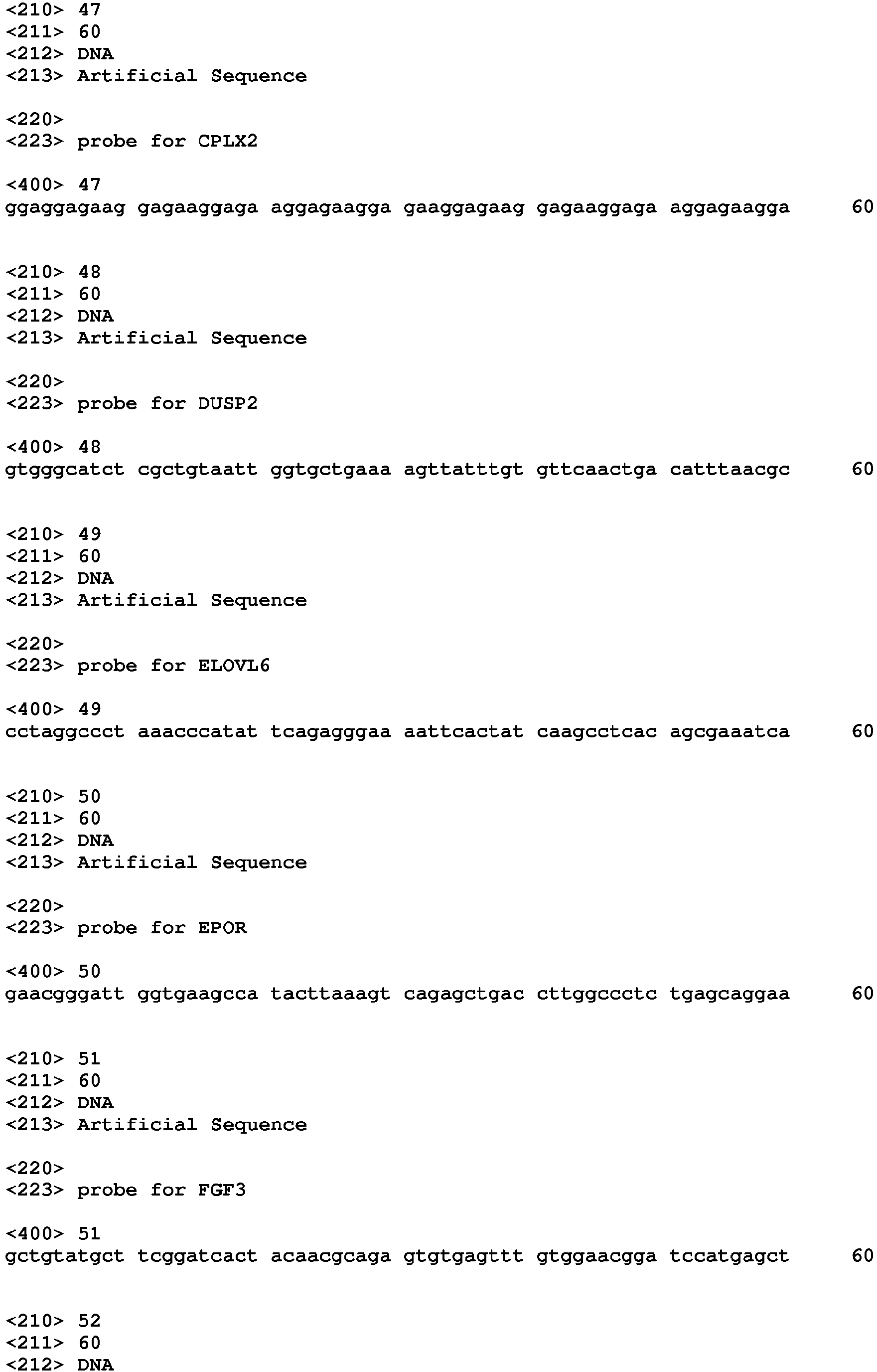
- the concentration of at least one or all of the RNAs of Table 2 is additionally determined: Table 2: additional RNA and preferred specific probe sequence RNA name probe sequence Probe sequence SEQ ID NO: AJUBA 29 BHLHA15 30 CP 31 ENSMUST00 000103558 32 GLRP1 33 GRTP1 34 LCK 35 NAP002592-003 36 PIGA 37 PRMT7 38 SLC35F2 39 TGM2 40 TIMP3 41 ZFP184 42 AKAP13 43 ASB2 44 ATP9A 45 CCR7 46 CPLX2 47 DUSP2 48 ELOVL6 49 EPOR 50 FGF3 51 IGF1R 52 ITK 53 JAKMIP1 54 KLRB1F 55 LMO2 56 MEF2C 57 MID1 58 MND1 59 MYB 60 MYC 61 PIM2 62 PRDM16 63 RAMP1 64 RASGRP2 65 SEMA7A 66 SH3KBP1 67 SLC43A1 68 SMAD3 69 ST3
- the step of relating the concentrations of the mRNAs determined for the cells to which the agent was added with subsequent cultivation to the concentrations of the mRNAs determined for the same type of cells which were cultivated without addition of the agent under the same cell culture conditions results in the determination of differences in the expression levels of these RNAs, which differences in expression levels are characteristic for the genotoxic effect of the agent.
- the expression levels of mRNAs have been found to depend on the culture conditions, the cells are cultivated under the same culture conditions and the same duration for each comparison, preferably performed in parallel each time.
- the cells cultivated under the same cell culture conditions for the same duration in the absence of the agent are also referred to as a negative control or mock.
- an agent that is known to have a significant genotoxic potential is used under the same culture conditions to provide a positive control, preferably performed each time in parallel to the cells to which the agent was added, and the process comprises the step of comparing the concentrations of the mRNAs determined for the positive control to the concentrations of mRNAs determined for the cells to which the agent to be analysed was added.
- the process preferably comprises the comparison of expression levels of the RNAs determined for the cells to which the agent was added to the expression levels of the cellular RNAs determined for the negative control and/or of the positive control, wherein all cells were cultivated under the same cell culture conditions for the same pre-determined duration.
- immortalized cells are considered to represent precancerous cells and/or tumour cells, indicating a high genotoxic activity of the agent analysed in the process.
- expression levels of the RNAs that are determined for the cells to which the agent was added which expression levels have differences in comparison to the expression levels of the mRNAs determined for the negative control and/or no or less important differences in comparison to a positive control, are characteristic for the genotoxic activity of the agent.
- RNAs Using a viral particle known to have genotoxic activity as an exemplary agent and murine hematopoietic stem cells and their progenitor cells, significant differences in expression levels of the RNAs were found in comparison to the expression levels determined for the RNAs in a parallel negative control.
- the step of relating the concentrations of the mRNAs determined for the hematopoietic cells to which the agent was added to the concentrations of mRNAs determined for the hematopoietic cells cultivated under the same cell culture conditions for the same duration in the absence of the agent preferably is by calculating the ratio of the specific mRNA concentrations.
- the concentration ratio can be replaced by the ratio of measurement values measured for the mRNA concentrations, e.g. by the ratio of fluorescence measurement values.
- only the mRNAs are considered as having an expression level which significantly differs from the expression determined for the negative control. For example, a significant difference can be defined for a difference of at least a log 2 of the concentration ratio bigger than +1 or smaller than -1.
- This exemplary cut-off for expression levels which compared to the negative control differ at least by such a factor can be used in order to limit the RNAs to those which have an important difference in expression level.
- the copy number of this nucleic acid in relation to the genome of the cells is determined.
- the influence of the copy number can be taken into account in the determination of the genotoxic risk of an agent.
- the concentration of the agent should be measured in order to take into account the concentration effect, for example by dividing the concentration of RNAs determined in the cells with agent by the concentration of the added agent.
- the culture conditions comprise the composition of the cultivation medium, cultivation temperature, the composition of the gas atmosphere, and the movement of the cultivation medium, e.g. by shaking, stirring, or static incubation.
- a preferred medium for the hematopoietic stem cells and their progenitor cells used in the process contains growth-promoting cytokines, especially the following cytokines: stem cell factor (SCF), preferably in addition interleukin-3 (IL-3), Flt3-ligand, interleukin-11 (IL-11), and more preferably additionally interleukin-7 (IL-7), and further optionally additionally interleukin-6 (IL-6), interleukin-15 (IL-15), thrombopoietin (TPO) and further optionally additionally at least one notch ligand (DL1 or DL4).
- SCF stem cell factor
- IL-3 interleukin-3
- Flt3-ligand interleukin-11
- IL-7 interleukin-7
- TPO thrombopoietin
- at least one notch ligand DL1 or DL4
- An advantage of the process of the invention is that it is not restricted to measure presence of cells that proliferate during a long period of cultivation as e.g. in the process of Modlich (2006), as it has been found that the RNA isolated in the process according to the invention does not necessarily contain, and preferably not contain relevant levels of the mRNA Lmo2, whereas in cells treated in the process of Modlich (2006) all cells that finally proliferated, when analysed by the process of the invention were found to express Lmo2. Therefore, the process of the invention has the advantage of identifying Lmo2, which is an indicator for precedence of adverse events in gene therapy, e.g. an indicator associated with the development of leukemia by gene therapy. As a further advantage, the process is not dependent on the expression of Lmo2 by the cells to be analysed.
- the process can comprise the step of incubating the cells in the presence of the agent and removing the agent, e.g. by exchanging the culture medium of the cells in the cell culture for fresh medium without the agent, e.g. following a prior incubation period of 1 to 3 d.
- the agent is a non-natural nucleic acid construct, optionally packaged as a viral particle.
- the non-natural nucleic acid construct can e.g. contain a non-natural or a natural nucleic acid sequence having the ability to integrate into genomic DNA, e.g. at least a sequence having homology to the cell, a viral primer binding site, a viral 5' region containing a U3 region, and/or a viral packaging signal promoter sequence.
- the process preferably comprises the step of performing a quantitative analysis of the nucleic acid construct in total DNA obtained during the pre-determined duration from an aliquot of the cultivated cells for determination of the average copy number of the agent in the mammalian cell.
- the quantitative analysis of the nucleic acid construct of the agent in total DNA obtained from the cells is made by quantitative PCR (polymerase chain reaction) using a primer pair specific for the nucleic acid construct, and preferably also a primer pair specific for a genomic DNA section of the cells, in a separate or in the same PCR.
- the step of collecting DNA from the cells for the quantitative analysis of the nucleic acid construct can e.g.
- the agent be made at 2 to 8 d, preferably 4 to 8 d, more preferably 4 d following the addition of the agent to mammalian cells in a cell culture, in order to avoid detection of viral nucleic acids from episomes or from transiently infected cells.
- the pre-determined duration of cultivating the cells following the step of adding the agent to the cells in culture can e.g. amount to 10 to 15 days, and this duration can optionally be divided into a first and a second partial duration, wherein the first partial duration of cultivating the cells is followed by a step of diluting the cells and then by cultivating the cells for the second partial duration, e.g. by re-plating the cells at lower cell number and cultivating for a second partial duration.
- the step of diluting the cells for a second partial duration of incubation can increase the selection pressure on the cells and therefore result in more pronounced differences in the translation of mRNA species.
- the first partial duration can be 2 to 10 d, preferably 6 to 8 d
- the second partial duration that follows the dilution of the cells for further cultivation can be 5 to 12 d, e.g. 5 to 7 d.
- the pre-determined duration of cultivating the cells can be 2 to 6 d, preferably 3 to 4 d.
- Such duration can have the advantage of determining concentrations of mRNAs, which preferably are mRNAs, characteristic for general toxicity of agents, whereas longer durations, especially durations of 8 to 15 d, have been found to result in specific differences in expression levels of the mRNAs regarding genotoxicity.
- a further advantage of the process of the invention is that it does not require the replating step needed for microscopic analysis of transformed or immortalized cells and hence can use shorter pre-determined durations of cultivating cells as e.g. used in the process of Modlich et al. (2006), which uses an expansion plus limiting dilution of the murine hematopoietic stem and progenitor cells in culture for 4 weeks following transduction of cells.
- the primary hematopoietic stem cells and/or stem progenitor cells obtained freshly or used after a freeze-thaw-cycle, which are used in the process of the invention, e.g. of murine origin, can be cultivated under cell culture conditions prior to adding the agent, adding nothing or an inactive compound for the negative control, and adding a known genotoxic agent as a positive control, respectively, to separate cells in culture at the same prior incubation period.
- This cultivation period prior to adding the agent can e.g. last for 1 to 3 d, e.g. 2 d for freshly obtained primary cells.
- the step of determining the concentration of the mRNAs can be by reverse transcription followed by quantitative PCR, preferably by hybridisation of the mRNA, preferably the total RNA isolated from the cultivated cells, to nucleic acid probes which are immobilized on a substrate, e.g. arranged on a substrate as an array of probes.
- Arrays of nucleic acid probes immobilized on a substrate are generally known as microarrays which are suitable for quantification of hybridizing mRNAs.
- concentration of mRNAs can be quantified by next generation sequencing of the reverse transcribed RNAs.
- the process contains the evaluation of the mRNAs which are considered as deregulated.
- the mRNAs are considered deregulated for which subsequent to normalization, preferably Quantil normalization (e.g. performed using the R-package limma - linear models for microarrays), for the concentration ratio with the negative control a log 2 of bigger than +1 or smaller than -1 is found.
- a normalized enrichment score is calculated for each of the mRNAs considered deregulated.
- the NES is calculated as described by Subramanian et al., Proc Natl Acad Sci USA (2005) Oct 25; 102(43):15545-50 .
- the NES is calculated by the steps of generating a numerical matrix of the protein encoding genes, creating a running-sum statistic with a maximum deviation from zero (Enrichment Score), determination of a nominal P value (Estimation of Significance Level) and adjusting the Enrichment Score for Multiple Hypothesis Testing (Normalized Enrichment Score).
- an NES above 2.0 indicates a high probability that the agent has transformed the cells
- an NES below 1.2 indicates a low probability that the agent has genotoxic potential
- an intermediate NES, i.e. between 1.2 and 2.0 indicates that the agent has potential genotoxic activity.
- RNAs considered deregulated are searched for the mRNAs considered deregulated, the databanks containing RNAs, which preferably are mRNAs, which have been found associated with diseases, e.g. relevant in diseases, e.g. in tumour patients, e.g. in AML, ALL or MDS patients, or in mouse models of such a disease.
- a hit in each databank for an RNA considered deregulated adds weight to this RNA as the overlap percentage for a hit in the databank.
- the result of the addition of the weight, e.g. the percentage of overlap to the at least one databank, e.g. to the log 2 ratio for each mRNA yields a numerical value reflecting the genotoxic potential.
- the databanks contain genes which when deregulated can have a genotoxic effect at least of viral particles.
- the databanks e.g. at least one, are selected from the following ones:
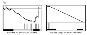
- Fig. 1 a graphic representation of exemplary NES values determined for an exemplary agent by a preferred embodiment of the process.
- Example 1 Analysis of genotoxic potential by determination of mRNA concentrations in cultivated cells
- Murine primary hematopoietic stem precursor cells that were isolated, cryopreserved, thawed, cultivated for 3 d in serum-free medium, preferably in StemSpan medium containing SCF, Flt3-L, IL-3, IL-11, IL-7 and expanded in IMDM medium (same cytokines) for 2 d at 37°C, 5% CO 2 atmosphere in 24-well cell culture dishes.
- IMDM medium ame cytokines
- the exemplary agent suspected of having a genotoxic activity was a viral particle containing a nucleic acid construct coding for the IL-2 receptor common gamma chain under an elongation factor 1 alpha short promoter, designated LVEFS.
- the positive control was a viral particle containing a nucleic acid construct with intact LTR regions (promoter/enhancer regions) from the spleen focus forming virus, designated RVSF.
- the primers of Table 3 were used to detect the viral elements indicated.
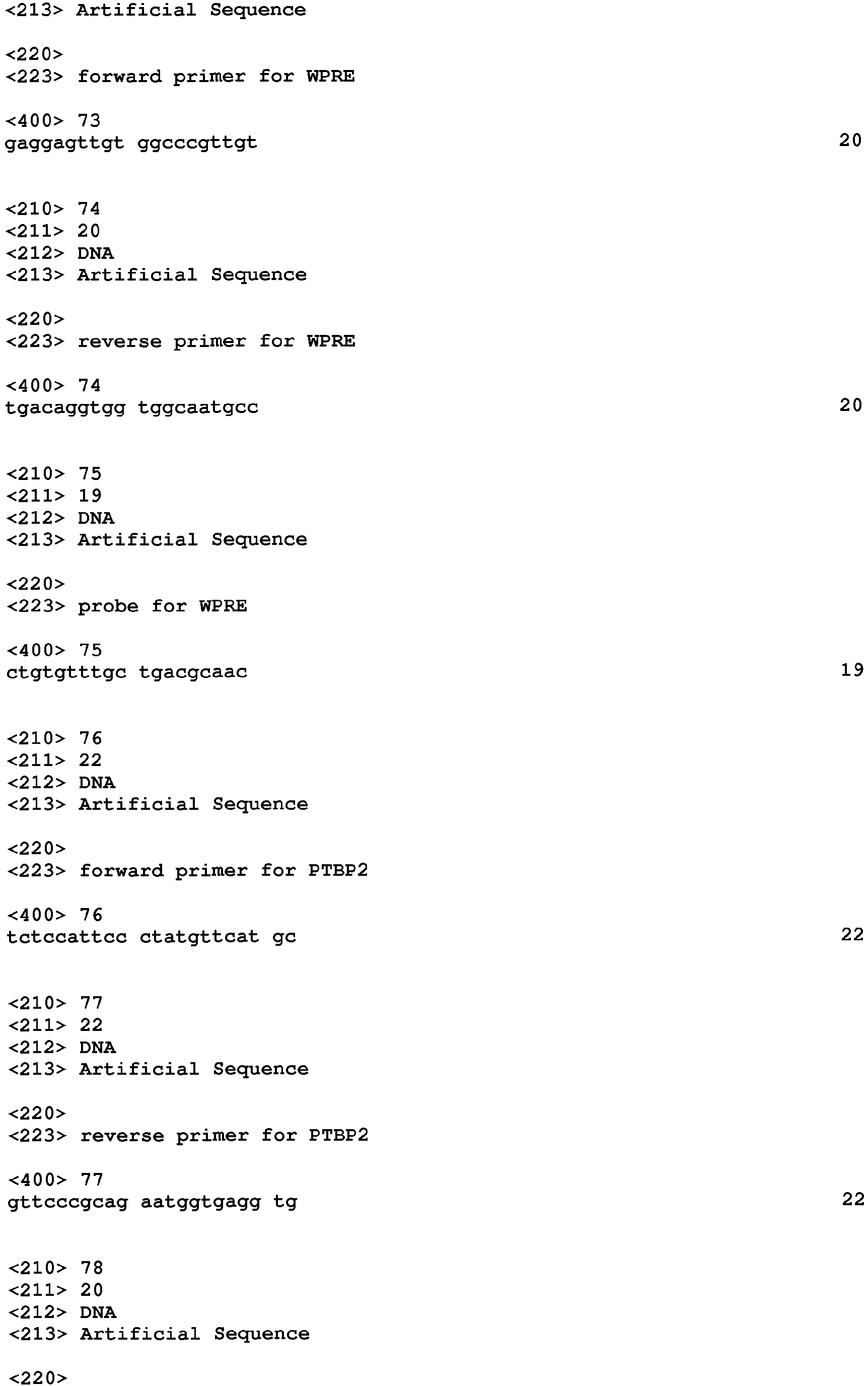
- primers for detecting viral elements of a viral particle viral element function of primer or probe primer or probe SEQ ID NO: WPRE forward GAGGAGTTGTGGCCCGTTGT 73 WPRE reverse TGACAGGTGGTGGCAATGCC 74 WPRE Taqman Probe CTGTGTTTGCTGACGCAAC 75 PTBP2 forward TCTCCATTCCCTATGTTCATGC 76 PTBP2 reverse GTTCCCGCAGAATGGTGAGGTG 77 PTBP2 Taqman Probe ATGTTCCTCGGACCAACTTG 78
- the cultivated cells were expanded in IMDM medium containing cytokines SCF (100 ng/ml), IL-3 (20 ng/ml), Flt-3L (100 ng/ml), IL-11 (100 ng/ml), and IL-7 (100 ng/ml) for further 4 days, then collected and re-plated at a 1:10 dilution in fresh medium of this composition and cultivated for another 7 d.
- the medium in addition contained OP9-DL1 or DL4 at a concentration of 5 ⁇ g/ml. Then total RNA was collected from the cells using RNAzol and the extraction kit from Zymo Research.
- RNA from each culture was analysed for the concentration of the following RNAs of Table 1: AFAP1L1, ALS2CL, ARX, BEX6, CCDC102A, DCTD, EHD3, GM805, GUCY1A3, MEX3A, MYCT1, NAIP1, NOLC1, PA2G4, PDGFRB, PHLDA2, SLA2, SLC12A2, SOX14, SOX4, SPNS2, TBXA2R, TC1649157, TIE1, TMEM176A, TMEM176B, TOX, ZFPM1.
- RNAs of Table 2 were analysed: AJUBA, BHLHA15, CP, ENSMUST00000103558, GLRP1, GRTP1, LCK, NAP002592-003, PIGA, PRMT7, SLC35F2, TGM2, TIMP3, ZFP184, AKAP13, ASB2, ATP9A, CCR7, CPLX2, DUSP2, ELOVL6, EPOR, FGF3, IGF1R, ITK, JAKMIP1, KLRB1F, LMO2, MEF2C, MIDI, MND1, MYB, MYC, PIM2, PRDM16, RAMP1, RASGRP2, SEMA7A, SH3KBP1, SLC43A1, SMAD3, ST3GAL6, TNFSF13B, VARS.
- hybridisation to immobilized specific oligonucleotides contained on a chip, followed by optical detection using laser irradiation of the fluorescent dye Cyanine 3 were used
- the ratio of the mRNA concentrations were determined as the quotient of the fluorescence signal intensity determined for the cells to which the agent was added to the signal intensity determined in the negative control for each mRNA species.
- the differentiation between immortalized cells and non-immortalized cells was verified by performing the conventional limiting dilution protocol of the Modlich (2006) assay in parallel. Differences between immortalized and non-immortalized cells can be assessed by the measurement of the mRNA species of Table 1.
- a normalized enrichment score (NES) was calculated for the significantly expressed mRNAs using the GSEA method.
- the exemplary agent LVEFS had a NES of -1.4 which was significantly different from the positive control RVSF with an NES of 2.62.
- RNA species of Table 1 and of the additional RNAs of Table 2 that were determined in the cells to which the exemplary agent LVEFS or RVSF was added were analysed for overlaps with the database.
- the sample LVEFS had a mean overlap of 0 out of 44 and the RVSF 17 out of 44. This corresponds to a p-value ⁇ 0.001 and is regarded highly significant.
- Fig. 1 shows the normalized enrichment scores of sample LVEFS (-1.4) and the positive control RVSF (2.62).
Landscapes
- Chemical & Material Sciences (AREA)
- Life Sciences & Earth Sciences (AREA)
- Health & Medical Sciences (AREA)
- Engineering & Computer Science (AREA)
- Proteomics, Peptides & Aminoacids (AREA)
- Organic Chemistry (AREA)
- Analytical Chemistry (AREA)
- Immunology (AREA)
- Zoology (AREA)
- Wood Science & Technology (AREA)
- Molecular Biology (AREA)
- Genetics & Genomics (AREA)
- Physics & Mathematics (AREA)
- Biotechnology (AREA)
- Biochemistry (AREA)
- Bioinformatics & Cheminformatics (AREA)
- General Health & Medical Sciences (AREA)
- Microbiology (AREA)
- Biomedical Technology (AREA)
- Pathology (AREA)
- Biophysics (AREA)
- General Engineering & Computer Science (AREA)
- Toxicology (AREA)
- Hematology (AREA)
- Urology & Nephrology (AREA)
- Tropical Medicine & Parasitology (AREA)
- Cell Biology (AREA)
- Food Science & Technology (AREA)
- Medicinal Chemistry (AREA)
- General Physics & Mathematics (AREA)
- Measuring Or Testing Involving Enzymes Or Micro-Organisms (AREA)
Abstract
The present invention relates to an analytical process for the assessment of the genotoxic potential, also termed mutagenic or transforming activity, of an agent in respect to hematopoietic cells. The agent can be selected from natural and synthetic chemical compounds, and preferably from heterologous expression cassettes, e.g. contained in non-natural viral particles or non-natural viral nucleic acid constructs, e.g. nucleic acid constructs containing a viral element.
Description
- The present invention relates to an analytical process for the assessment of the genotoxic potential, also termed mutagenic or transforming activity, of an agent in respect to hematopoietic cells. The agent can be selected from natural and synthetic chemical compounds, and preferably from heterologous expression cassettes, e.g. contained in non-natural viral particles or non-natural viral nucleic acid constructs, e.g. nucleic acid constructs containing a viral element. The heterologous, e.g. viral element can e.g. be a promoter or enhancer region for transgene expression, a viral primer binding site, a viral 5' region containing a U3 region, and/or a viral packaging signal.
- The analytical process is suitable for assessing the genotoxic potential of viral particles for mammalian cells, e.g. for viral particles containing a nucleic acid construct intended for gene therapy. The hematopoietic cells are primary stem cells and/or stem precursor cells of the hematopoietic system, e.g. of human, murine or rat origin. The process is an in vitro process, using the primary stem cells and/or stem precursor cells of the hematopoietic system.
- Modlich et al., Blood 108 (8), 2545-2553 (2006) describe that gene transfer to primary murine lineage negative bone marrow cells by adding viral nucleic acid constructs containing a SIN (self-inactivating 3'LTR upon integration into the host genome) nucleic acid construct resulted in lower frequencies of replated cells than an LTR-driven nucleic acid construct after 28 days under cell culture conditions. Higher replating frequencies in the cell culture were detected as cell growth in a limiting dilution series and were found to be indicative of a higher transforming potential of the viral particle. A disadvantage of this method is that the increased cell growth which is determined as an indicator for transformation strongly depends on the myeloid cultivation conditions. The replating phenotype is triggered or accompanied by activation of a very limited number of proto-oncogenes (mainly Mecom). Insertional upregulation of genes of the lymphoid differentiation lineage cannot be detected. A further disadvantage is that the cultivation of cells takes a long time and that the microscopic analysis of cell growth yields no information about the underlying transforming event.
- It is an object of the invention to provide an alternative process for analysis of the genotoxic potential of an agent in mammalian hematopoietic cells. Preferably, the alternative process can be performed in a shorter time and/or yields a more detailed picture of the genotoxic potential.
- The invention achieves the object by the features of the claims, especially by providing a process for analysis of the genotoxic activity of an agent for hematopoietic cells, the process comprising the steps of adding the agent to hematopoietic stem and progenitor cells in a cell culture under cell culture conditions, cultivating these cells for a pre-determined duration to produce cultivated cells, isolating total RNA, preferably total mRNA, from the cultivated cells and determining the concentration of the RNAs encoding at least one, preferably all of the RNA sequences of Table 1, preferably using the indicated probe sequences. Generally preferred, the RNAs are mRNAs.
Table 1: RNA and preferred specific probe sequence RNA name probe sequence Probe sequence SEQ ID NO: AFAP1L1 
1 ALS2CL 
2 ARX 
3 BEX6 
4 CCDC102A 
5 DCTD 
6 EHD3 
7 GM805 
8 GUCY1A3 
9 MEX3A 
10 MYCT1 
11 NAIP1 
12 NOLC1 
13 PA2G4 
14 PDGFRB 
15 PHLDA2 
16 SLA2 
17 SLC12A2 
18 SOX14 
19 SOX4 
20 SPNS2 
21 TBXA2R 
22 TC1649157 
23 TIE1 
24 TMEM176A 
25 TMEM176B 
26 TOX 
27 ZFPM1 
28 - Preferably, the concentration of at least one or all of the RNAs of Table 2 is additionally determined:
Table 2: additional RNA and preferred specific probe sequence RNA name probe sequence Probe sequence SEQ ID NO: AJUBA 
29 BHLHA15 
30 CP 
31 ENSMUST00 000103558 
32 GLRP1 
33 GRTP1 
34 LCK 
35 NAP002592-003 
36 PIGA 
37 PRMT7 
38 SLC35F2 
39 TGM2 
40 TIMP3 
41 ZFP184 
42 AKAP13 
43 ASB2 
44 ATP9A 
45 CCR7 
46 CPLX2 
47 DUSP2 
48 ELOVL6 
49 EPOR 
50 FGF3 
51 IGF1R 
52 ITK 
53 JAKMIP1 
54 KLRB1F 
55 LMO2 
56 MEF2C 
57 MID1 
58 MND1 
59 MYB 
60 MYC 
61 PIM2 
62 PRDM16 
63 RAMP1 
64 RASGRP2 
65 SEMA7A 
66 SH3KBP1 
67 SLC43A1 
68 SMAD3 
69 ST3GAL6 
70 TNFSF13B 
71 VARS 
72 - The step of relating the concentrations of the mRNAs determined for the cells to which the agent was added with subsequent cultivation to the concentrations of the mRNAs determined for the same type of cells which were cultivated without addition of the agent under the same cell culture conditions results in the determination of differences in the expression levels of these RNAs, which differences in expression levels are characteristic for the genotoxic effect of the agent. As the expression levels of mRNAs have been found to depend on the culture conditions, the cells are cultivated under the same culture conditions and the same duration for each comparison, preferably performed in parallel each time. The cells cultivated under the same cell culture conditions for the same duration in the absence of the agent are also referred to as a negative control or mock.
- Preferably, an agent that is known to have a significant genotoxic potential is used under the same culture conditions to provide a positive control, preferably performed each time in parallel to the cells to which the agent was added, and the process comprises the step of comparing the concentrations of the mRNAs determined for the positive control to the concentrations of mRNAs determined for the cells to which the agent to be analysed was added. Generally, the process preferably comprises the comparison of expression levels of the RNAs determined for the cells to which the agent was added to the expression levels of the cellular RNAs determined for the negative control and/or of the positive control, wherein all cells were cultivated under the same cell culture conditions for the same pre-determined duration.
- It has been found that the determination of the concentration of these mRNAs allows the differentiation between immortalized cells and non-immortalized cells. Therein, immortalized cells are considered to represent precancerous cells and/or tumour cells, indicating a high genotoxic activity of the agent analysed in the process. Specifically, it has been found that expression levels of the RNAs that are determined for the cells to which the agent was added, which expression levels have differences in comparison to the expression levels of the mRNAs determined for the negative control and/or no or less important differences in comparison to a positive control, are characteristic for the genotoxic activity of the agent. Using a viral particle known to have genotoxic activity as an exemplary agent and murine hematopoietic stem cells and their progenitor cells, significant differences in expression levels of the RNAs were found in comparison to the expression levels determined for the RNAs in a parallel negative control.
- The step of relating the concentrations of the mRNAs determined for the hematopoietic cells to which the agent was added to the concentrations of mRNAs determined for the hematopoietic cells cultivated under the same cell culture conditions for the same duration in the absence of the agent preferably is by calculating the ratio of the specific mRNA concentrations. Generally, the concentration ratio can be replaced by the ratio of measurement values measured for the mRNA concentrations, e.g. by the ratio of fluorescence measurement values. Preferably, only the mRNAs are considered as having an expression level which significantly differs from the expression determined for the negative control. For example, a significant difference can be defined for a difference of at least a log2 of the concentration ratio bigger than +1 or smaller than -1. This exemplary cut-off for expression levels which compared to the negative control differ at least by such a factor can be used in order to limit the RNAs to those which have an important difference in expression level.
- Preferably, for agents containing a nucleic acid, the copy number of this nucleic acid in relation to the genome of the cells is determined. In this manner, the influence of the copy number can be taken into account in the determination of the genotoxic risk of an agent. For agents not containing a nucleic acid, the concentration of the agent should be measured in order to take into account the concentration effect, for example by dividing the concentration of RNAs determined in the cells with agent by the concentration of the added agent.
- The culture conditions comprise the composition of the cultivation medium, cultivation temperature, the composition of the gas atmosphere, and the movement of the cultivation medium, e.g. by shaking, stirring, or static incubation.
- A preferred medium for the hematopoietic stem cells and their progenitor cells used in the process contains growth-promoting cytokines, especially the following cytokines: stem cell factor (SCF), preferably in addition interleukin-3 (IL-3), Flt3-ligand, interleukin-11 (IL-11), and more preferably additionally interleukin-7 (IL-7), and further optionally additionally interleukin-6 (IL-6), interleukin-15 (IL-15), thrombopoietin (TPO) and further optionally additionally at least one notch ligand (DL1 or DL4).
- An advantage of the process of the invention is that it is not restricted to measure presence of cells that proliferate during a long period of cultivation as e.g. in the process of Modlich (2006), as it has been found that the RNA isolated in the process according to the invention does not necessarily contain, and preferably not contain relevant levels of the mRNA Lmo2, whereas in cells treated in the process of Modlich (2006) all cells that finally proliferated, when analysed by the process of the invention were found to express Lmo2. Therefore, the process of the invention has the advantage of identifying Lmo2, which is an indicator for precedence of adverse events in gene therapy, e.g. an indicator associated with the development of leukemia by gene therapy. As a further advantage, the process is not dependent on the expression of Lmo2 by the cells to be analysed.
- Following addition of the agent to the cells in cell culture and prior to cultivating the cells, the process can comprise the step of incubating the cells in the presence of the agent and removing the agent, e.g. by exchanging the culture medium of the cells in the cell culture for fresh medium without the agent, e.g. following a prior incubation period of 1 to 3 d.
- Preferably, the agent is a non-natural nucleic acid construct, optionally packaged as a viral particle. The non-natural nucleic acid construct can e.g. contain a non-natural or a natural nucleic acid sequence having the ability to integrate into genomic DNA, e.g. at least a sequence having homology to the cell, a viral primer binding site, a viral 5' region containing a U3 region, and/or a viral packaging signal promoter sequence.
- When the agent comprises a nucleic acid construct, e.g. the agent is a viral particle, the process preferably comprises the step of performing a quantitative analysis of the nucleic acid construct in total DNA obtained during the pre-determined duration from an aliquot of the cultivated cells for determination of the average copy number of the agent in the mammalian cell. Preferably, the quantitative analysis of the nucleic acid construct of the agent in total DNA obtained from the cells is made by quantitative PCR (polymerase chain reaction) using a primer pair specific for the nucleic acid construct, and preferably also a primer pair specific for a genomic DNA section of the cells, in a separate or in the same PCR. The step of collecting DNA from the cells for the quantitative analysis of the nucleic acid construct can e.g. be made at 2 to 8 d, preferably 4 to 8 d, more preferably 4 d following the addition of the agent to mammalian cells in a cell culture, in order to avoid detection of viral nucleic acids from episomes or from transiently infected cells.
- The pre-determined duration of cultivating the cells following the step of adding the agent to the cells in culture can e.g. amount to 10 to 15 days, and this duration can optionally be divided into a first and a second partial duration, wherein the first partial duration of cultivating the cells is followed by a step of diluting the cells and then by cultivating the cells for the second partial duration, e.g. by re-plating the cells at lower cell number and cultivating for a second partial duration. In this embodiment, the step of diluting the cells for a second partial duration of incubation can increase the selection pressure on the cells and therefore result in more pronounced differences in the translation of mRNA species. Preferably, the first partial duration can be 2 to 10 d, preferably 6 to 8 d, and the second partial duration that follows the dilution of the cells for further cultivation can be 5 to 12 d, e.g. 5 to 7 d.
- Optionally, the pre-determined duration of cultivating the cells, optionally the first partial duration can be 2 to 6 d, preferably 3 to 4 d. Such duration can have the advantage of determining concentrations of mRNAs, which preferably are mRNAs, characteristic for general toxicity of agents, whereas longer durations, especially durations of 8 to 15 d, have been found to result in specific differences in expression levels of the mRNAs regarding genotoxicity. This shows a specific advantage of the process of the invention in that it generally results in the identification of genotoxic activity of an agent at higher sensitivity and identification but also the identification of a general toxicity of an agent that prevents or reduces proliferation of the cells.
- A further advantage of the process of the invention is that it does not require the replating step needed for microscopic analysis of transformed or immortalized cells and hence can use shorter pre-determined durations of cultivating cells as e.g. used in the process of Modlich et al. (2006), which uses an expansion plus limiting dilution of the murine hematopoietic stem and progenitor cells in culture for 4 weeks following transduction of cells.
- The primary hematopoietic stem cells and/or stem progenitor cells obtained freshly or used after a freeze-thaw-cycle, which are used in the process of the invention, e.g. of murine origin, can be cultivated under cell culture conditions prior to adding the agent, adding nothing or an inactive compound for the negative control, and adding a known genotoxic agent as a positive control, respectively, to separate cells in culture at the same prior incubation period. This cultivation period prior to adding the agent can e.g. last for 1 to 3 d, e.g. 2 d for freshly obtained primary cells.
- The step of determining the concentration of the mRNAs can be by reverse transcription followed by quantitative PCR, preferably by hybridisation of the mRNA, preferably the total RNA isolated from the cultivated cells, to nucleic acid probes which are immobilized on a substrate, e.g. arranged on a substrate as an array of probes. Arrays of nucleic acid probes immobilized on a substrate are generally known as microarrays which are suitable for quantification of hybridizing mRNAs. Optionally, concentration of mRNAs can be quantified by next generation sequencing of the reverse transcribed RNAs.
- In an optional embodiment, the process contains the evaluation of the mRNAs which are considered as deregulated. Therein, the mRNAs are considered deregulated for which subsequent to normalization, preferably Quantil normalization (e.g. performed using the R-package limma - linear models for microarrays), for the concentration ratio with the negative control a log2 of bigger than +1 or smaller than -1 is found.
- Preferably additionally, a normalized enrichment score (NES) is calculated for each of the mRNAs considered deregulated. The NES is calculated as described by Subramanian et al., Proc Natl Acad Sci USA (2005) Oct 25; 102(43):15545-50. In short, the NES is calculated by the steps of generating a numerical matrix of the protein encoding genes, creating a running-sum statistic with a maximum deviation from zero (Enrichment Score), determination of a nominal P value (Estimation of Significance Level) and adjusting the Enrichment Score for Multiple Hypothesis Testing (Normalized Enrichment Score). Therein, an NES above 2.0 indicates a high probability that the agent has transformed the cells, an NES below 1.2 indicates a low probability that the agent has genotoxic potential, and an intermediate NES, i.e. between 1.2 and 2.0 indicates that the agent has potential genotoxic activity.
- Preferably, in a subsequent step databanks are searched for the mRNAs considered deregulated, the databanks containing RNAs, which preferably are mRNAs, which have been found associated with diseases, e.g. relevant in diseases, e.g. in tumour patients, e.g. in AML, ALL or MDS patients, or in mouse models of such a disease. A hit in each databank for an RNA considered deregulated adds weight to this RNA as the overlap percentage for a hit in the databank. The result of the addition of the weight, e.g. the percentage of overlap to the at least one databank, e.g. to the log2 ratio for each mRNA, yields a numerical value reflecting the genotoxic potential.
- It was found that this calculation of a value reflecting the genotoxic potential for viral particles which are currently considered as having a low genotoxic potential yields values close to zero, and that this calculation for known mutagenic viral particles yields values which are significantly bigger.
- The databanks contain genes which when deregulated can have a genotoxic effect at least of viral particles. The databanks, e.g. at least one, are selected from the following ones:
- 1. Retroviral Tagged Cancer Gene Database (RTCGD, available from http://variation.osu.edu/rtcgd/) containing insertions detected in the vicinity of genes caused by retrovirus (RCGD-retro) or by transposons (RTCGD-transposon) in at least one murine tumour.
- 2. Bushman Cancer Gene List (available from www.bushmanlab.org/links/genelists) containing gene names of several cancer data bases, e.g. Atlas, CANgenes, CIS (RTCGD), human lymphoma-associated genes, a list of retrovirally induced tumours, the Sanger Cancer Development List, the Vogelstein List of chromosomal aberrations having importance in cancer development.
- The invention is now described by way of examples with reference to the figure, which shows in
Fig. 1 a graphic representation of exemplary NES values determined for an exemplary agent by a preferred embodiment of the process. - Murine primary hematopoietic stem precursor cells that were isolated, cryopreserved, thawed, cultivated for 3 d in serum-free medium, preferably in StemSpan medium containing SCF, Flt3-L, IL-3, IL-11, IL-7 and expanded in IMDM medium (same cytokines) for 2 d at 37°C, 5% CO2 atmosphere in 24-well cell culture dishes. At day 0, the agent, a positive control and medium as a negative control were added to separate dishes of the cultivated cells. The exemplary agent suspected of having a genotoxic activity was a viral particle containing a nucleic acid construct coding for the IL-2 receptor common gamma chain under an elongation factor 1 alpha short promoter, designated LVEFS. The positive control was a viral particle containing a nucleic acid construct with intact LTR regions (promoter/enhancer regions) from the spleen focus forming virus, designated RVSF.
- The cells with added agent, positive control and negative control, respectively, were incubated under cell culture conditions for 4 days, then an aliquot of each cell culture was removed for extraction of genomic DNA, from which the copy number of the viral nucleic acid sequence was determined. In detail, the primers of Table 3 were used to detect the viral elements indicated.
Table 3: primers for detecting viral elements of a viral particle viral element function of primer or probe primer or probe SEQ ID NO: WPRE forward GAGGAGTTGTGGCCCGTTGT 73 WPRE reverse TGACAGGTGGTGGCAATGCC 74 WPRE Taqman Probe CTGTGTTTGCTGACGCAAC 75 PTBP2 forward TCTCCATTCCCTATGTTCATGC 76 PTBP2 reverse GTTCCCGCAGAATGGTGAGGTG 77 PTBP2 Taqman Probe ATGTTCCTCGGACCAACTTG 78 - The cultivated cells were expanded in IMDM medium containing cytokines SCF (100 ng/ml), IL-3 (20 ng/ml), Flt-3L (100 ng/ml), IL-11 (100 ng/ml), and IL-7 (100 ng/ml) for further 4 days, then collected and re-plated at a 1:10 dilution in fresh medium of this composition and cultivated for another 7 d. Alternatively, the medium in addition contained OP9-DL1 or DL4 at a concentration of 5 µg/ml. Then total RNA was collected from the cells using RNAzol and the extraction kit from Zymo Research.
- The RNA from each culture was analysed for the concentration of the following RNAs of Table 1: AFAP1L1, ALS2CL, ARX, BEX6, CCDC102A, DCTD, EHD3, GM805, GUCY1A3, MEX3A, MYCT1, NAIP1, NOLC1, PA2G4, PDGFRB, PHLDA2, SLA2, SLC12A2, SOX14, SOX4, SPNS2, TBXA2R, TC1649157, TIE1, TMEM176A, TMEM176B, TOX, ZFPM1. Additionally, the following RNAs of Table 2 were analysed: AJUBA, BHLHA15, CP, ENSMUST00000103558, GLRP1, GRTP1, LCK, NAP002592-003, PIGA, PRMT7, SLC35F2, TGM2, TIMP3, ZFP184, AKAP13, ASB2, ATP9A, CCR7, CPLX2, DUSP2, ELOVL6, EPOR, FGF3, IGF1R, ITK, JAKMIP1, KLRB1F, LMO2, MEF2C, MIDI, MND1, MYB, MYC, PIM2, PRDM16, RAMP1, RASGRP2, SEMA7A, SH3KBP1, SLC43A1, SMAD3, ST3GAL6, TNFSF13B, VARS. For analysis of the RNAs, hybridisation to immobilized specific oligonucleotides contained on a chip, followed by optical detection using laser irradiation of the fluorescent dye Cyanine 3 were used.
- The ratio of the mRNA concentrations were determined as the quotient of the fluorescence signal intensity determined for the cells to which the agent was added to the signal intensity determined in the negative control for each mRNA species.
- The differentiation between immortalized cells and non-immortalized cells was verified by performing the conventional limiting dilution protocol of the Modlich (2006) assay in parallel. Differences between immortalized and non-immortalized cells can be assessed by the measurement of the mRNA species of Table 1. For determination of the numerical value discriminating the genotoxic potential, a normalized enrichment score (NES) was calculated for the significantly expressed mRNAs using the GSEA method. The exemplary agent LVEFS had a NES of -1.4 which was significantly different from the positive control RVSF with an NES of 2.62.
- For determination of the genotoxic potential according to the database RTCGD, the significantly increased concentrations of RNA species of Table 1 and of the additional RNAs of Table 2 that were determined in the cells to which the exemplary agent LVEFS or RVSF was added, were analysed for overlaps with the database. The sample LVEFS had a mean overlap of 0 out of 44 and the RVSF 17 out of 44. This corresponds to a p-value < 0.001 and is regarded highly significant.
Fig. 1 shows the normalized enrichment scores of sample LVEFS (-1.4) and the positive control RVSF (2.62).





Claims (15)
- Process for analysis of the genotoxic activity of an agent for primary hematopoietic cells, the process comprising the steps of adding the agent to the primary hematopoietic cells in culture under cell culture conditions, cultivating the cells for a pre-determined duration to produce cultivated cells, characterized by the step of isolating total RNA from the cultivated cells, determining the concentration of at least one, preferably all, of the RNAs selected from the group comprising
AFAP1L1, PDGFRB, ALS2CL, PHLDA2, ARX, SLA2, BEX6, SLC12A2, CCDC102A, SOX14, DCTD, SOX4, EHD3, SPNS2, GM805, TBXA2R, GUCY1A3, TC1649157, MEX3A, TIE1, MYCT1, TMEM176A, NAIP1, TMEM176B, NOLC1, TOX, and PA2G4, ZFPM1, - Process according to claim 1, characterized in that the RNA is detected by hybridization to a probe having a nucleic acid sequence
Specific for RNA named probe sequence Probe sequence SEQ ID NO: AFAP1L1 
1 ALS2CL 
2 ARX 
3 BEX6 
4 CCDC102A 
5 DCTD 
6 EHD3 
7 GM805 
8 GUCY1A3 
9 MEX3A 
10 MYCT1 
11 NAIP1 
12 NOLC1 
13 PA2G4 
14 PDGFRB 
15 PHLDA2 
16 SLA2 
17 SLC12A2 
18 SOX14 
19 SOX4 
20 SPNS2 
21 TBXA2R 
22 TC1649157 
23 TIE1 
24 TMEM176 A 
25 TMEM176 B 
26 TOX 
27 ZFPM1 
28 - Process according to one of the preceding claims, characterized in that the concentrations of all the RNAs of claim 1 are analysed.
- Process according to one of the preceding claims, characterized by additionally determining the concentration of at least one of the RNAs selected from the group comprising
AJUBA, CPLX2, BHLHA15, DUSP2, CP, ELOVL6, ENSMUST00000103558, EPOR, GLRP1, FGF3, GRTP1, IGF1R, LCK, ITK, NAP002592-003, JAKMIP1, PIGA, KLRB1F, PRMT7, LMO2, SLC35F2, MEF2C, TGM2, MIDI, TIMP3, MND1, ZFP184, MYB, AKAP13, MYC, ASB2, PIM2, ATP9A, PRDM16, and CCR7, RAMP1, - Process according to claim 4, characterized in that the RNA is detected by hybridization to a probe having a nucleic acid sequence
Specific for RNA named probe sequence Probe sequence SEQ ID NO: AJUBA 
29 BHLHA15 
30 CP 
31 ENSMUS T0000010 3558 
32 GLRP1 
33 GRTP1 
34 LCK 
35 NAP0025 92-003 
36 PIGA 
37 PRMT7 
38 SLC35F2 
39 TGM2 
40 TIMP3 
41 ZFP184 
42 AKAP13 
43 ASB2 
44 ATP9A 
45 CCR7 
46 CPLX2 
47 DUSP2 
48 ELOVL6 
49 EPOR 
50 FGF3 
51 IGF1R 
52 ITK 
53 JAKMIP1 
54 KLRB1F 
55 LMO2 
56 MEF2C 
57 MID1 
58 MND1 
59 MYB 
60 MYC 
61 PIM2 
62 PRDM16 
63 RAMP1 
64 - Process according to one of the preceding claims, characterized in that the agent is a virus or a viral particle containing a non-natural nucleic acid sequence, the process comprising the step of determining the copy number of the nucleic acid sequence of the viral particle in the genome of the cells.
- Process according to one of the preceding claims, characterized in that the duration is 7 to 15 d, during which the cells are provided at least once with fresh culture medium.
- Process according to one of the preceding claims, characterized in that under the culture conditions the medium contains cytokines consisting of the group of SCF, IL-3, Flt-3L, IL-11, IL-15, IL-6, IL-7, TPO, and Notch-ligands DL-1 and DL-4.
- Process according to one of the preceding claims, characterized in that only those RNAs are considered deregulated for which following normalization the ratio of RNA concentration determined for the cells to which the agent was added to the RNA concentration of cells cultivated in the absence of the agent is significantly different.
- Process according to one of the preceding claims, characterized in that a normalized enrichment score (NES) is calculated and that an NES above 2.0 indicates a high genotoxic activity and an NES below 1.2 indicates a low genotoxic activity of the agent.
- Process according to one of the preceding claims, characterized in that for each RNA an overlap with a databank is determined and the total number of mRNAs overlapping with a database for the cells to which the agent was added and cells cultivated in the presence of the positive control is added to a numerical value reflecting the genotoxic potential, wherein the databank is at least one selected from the databanks comprised in the group of:Retroviral Tagged Cancer Gene Database, and Bushman Cancer Database.
- Process according to one of the preceding claims, characterized in that it includes separate treatment of identical primary hematopoietic cells by adding an agent of known genotoxic activity as a positive control, wherein the cells are cultivated under the same cell culture conditions and for the same duration, and determining the concentrations of the mRNAs for cells for the positive control.
- Process according to one of the preceding claims, characterized in that the numerical value is calculated by a Fisher's exact test, where a p-value < 0.05 (agent vs. positive control) is regarded statistically significant, reflecting a lower genotoxic potential of the agent compared to the positive control.
- Process according to one of the preceding claims, characterized in that the positive control is a viral particle containing a nucleic acid construct with intact LTR regions (promoter/enhancer regions) from the spleen focus forming virus.
- Process according to one of the preceding claims, characterized in that the concentration of the RNAs is determined as the normalized enrichment score (NES) and a NES value above 2.0 is taken as a high probability for genotoxic potential, a NES value below 1.2 is taken as a low probability for genotoxic potential, and a NES value of 1.2 to 2.0 is taken as genotoxic potential, and that the NES values of the RNAs analyzed are added and compared to the added NES values of mRNAs determined for cells cultivated under the same cell culture conditions for the same duration in the absence of the agent.
Priority Applications (4)
| Application Number | Priority Date | Filing Date | Title |
|---|---|---|---|
| EP16822643.9A EP3394286B1 (en) | 2015-12-23 | 2016-12-16 | Analytical process for genotoxicity assessment |
| CA3007881A CA3007881A1 (en) | 2015-12-23 | 2016-12-16 | Analytical process for genotoxicity assessment |
| PCT/EP2016/081351 WO2017108598A1 (en) | 2015-12-23 | 2016-12-16 | Analytical process for genotoxicity assessment |
| US16/063,938 US20200264164A1 (en) | 2015-12-23 | 2016-12-16 | Analytical process for genotoxicity assessment |
Applications Claiming Priority (1)
| Application Number | Priority Date | Filing Date | Title |
|---|---|---|---|
| EP15202623 | 2015-12-23 |
Publications (1)
| Publication Number | Publication Date |
|---|---|
| EP3184648A1 true EP3184648A1 (en) | 2017-06-28 |
Family
ID=55027497
Family Applications (2)
| Application Number | Title | Priority Date | Filing Date |
|---|---|---|---|
| EP16185408.8A Withdrawn EP3184648A1 (en) | 2015-12-23 | 2016-08-23 | Analytical process for genotoxicity assessment |
| EP16822643.9A Active EP3394286B1 (en) | 2015-12-23 | 2016-12-16 | Analytical process for genotoxicity assessment |
Family Applications After (1)
| Application Number | Title | Priority Date | Filing Date |
|---|---|---|---|
| EP16822643.9A Active EP3394286B1 (en) | 2015-12-23 | 2016-12-16 | Analytical process for genotoxicity assessment |
Country Status (4)
| Country | Link |
|---|---|
| US (1) | US20200264164A1 (en) |
| EP (2) | EP3184648A1 (en) |
| CA (1) | CA3007881A1 (en) |
| WO (1) | WO2017108598A1 (en) |
Families Citing this family (1)
| Publication number | Priority date | Publication date | Assignee | Title |
|---|---|---|---|---|
| CN110135720B (en) * | 2019-05-09 | 2021-02-05 | 集美大学 | A Quantitative Evaluation Method of Proliferation and Release Effect |
Citations (2)
| Publication number | Priority date | Publication date | Assignee | Title |
|---|---|---|---|---|
| WO2010070059A1 (en) * | 2008-12-17 | 2010-06-24 | Universiteit Maastricht | Method for the identification of carcinogenic compounds |
| WO2014170436A1 (en) * | 2013-04-19 | 2014-10-23 | INSERM (Institut National de la Santé et de la Recherche Médicale) | Methods and kits for testing the genotoxicity of an agent |
-
2016
- 2016-08-23 EP EP16185408.8A patent/EP3184648A1/en not_active Withdrawn
- 2016-12-16 WO PCT/EP2016/081351 patent/WO2017108598A1/en not_active Ceased
- 2016-12-16 US US16/063,938 patent/US20200264164A1/en not_active Abandoned
- 2016-12-16 EP EP16822643.9A patent/EP3394286B1/en active Active
- 2016-12-16 CA CA3007881A patent/CA3007881A1/en not_active Abandoned
Patent Citations (2)
| Publication number | Priority date | Publication date | Assignee | Title |
|---|---|---|---|---|
| WO2010070059A1 (en) * | 2008-12-17 | 2010-06-24 | Universiteit Maastricht | Method for the identification of carcinogenic compounds |
| WO2014170436A1 (en) * | 2013-04-19 | 2014-10-23 | INSERM (Institut National de la Santé et de la Recherche Médicale) | Methods and kits for testing the genotoxicity of an agent |
Non-Patent Citations (10)
| Title |
|---|
| "METHODS IN ENZYMOLOGY", vol. 507, 1 January 2012, ACADEMIC PRESS, US, ISSN: 0076-6879, article EUGENIO MONTINI ET AL: "Genotoxicity Assay for Gene Therapy Vectors in Tumor Prone Cdkn2a-/- Mice", pages: 171 - 185, XP055326107, DOI: 10.1016/B978-0-12-386509-0.00009-0 * |
| ALI NOWROUZI ET AL: "Retroviral Vectors: Post Entry Events and Genomic Alterations", VIRUSES, vol. 3, no. 12, 29 December 2011 (2011-12-29), CH, pages 429 - 455, XP055327182, ISSN: 1999-4915, DOI: 10.3390/v3050429 * |
| JULIA D SUERTH ET AL: "Alpharetroviral Self-inactivating Vectors: Long-term Transgene Expression in Murine Hematopoietic Cells and Low Genotoxicity", MOLECULAR THERAPY, vol. 20, no. 5, 14 February 2012 (2012-02-14), GB, pages 1022 - 1032, XP055327184, ISSN: 1525-0016, DOI: 10.1038/mt.2011.309 * |
| LEE MICHAEL ET AL: "cDNA microarray gene expression profiling of hydroxyurea, paclitaxel, and p-anisidine, genotoxic compounds with differing tumorigenicity results", ENVIRONMENTAL AND MOLECULAR MUTAGENESIS, WILEY-LISS, INC. CHICHESTER, GB, vol. 42, no. 2, 1 January 2003 (2003-01-01), pages 91 - 97, XP002388888, ISSN: 0893-6692, DOI: 10.1002/EM.10177 * |
| MARK H. CHIN ET AL: "Induced Pluripotent Stem Cells and Embryonic Stem Cells Are Distinguished by Gene Expression Signatures", CELL STEM CELL, vol. 5, no. 1, 1 July 2009 (2009-07-01), pages 111 - 123, XP055013700, ISSN: 1934-5909, DOI: 10.1016/j.stem.2009.06.008 * |
| MODLICH ET AL., BLOOD, vol. 108, no. 8, 2006, pages 2545 - 2553 |
| MODLICH U ET AL: "Cell-culture assays reveal the importance of retroviral vector design for insertional genotoxicity", BLOOD, AMERICAN SOCIETY OF HEMATOLOGY, US, vol. 108, no. 8, 6 July 2006 (2006-07-06), pages 2545 - 2553, XP002455802, ISSN: 0006-4971, DOI: 10.1182/BLOOD-2005-08-024976 * |
| NEWTON RONALD K ET AL: "The utility of DNA microarrays for characterizing genotoxicity", ENVIRONMENTAL HEALTH PERSPECTIVES, U.S. DEPARTMENT OF HEALTH AND HUMAN SERVICES, NATIONAL INSTITUTE OF ENVIRONMENTAL HEALTH SCIENCES, US, vol. 112, no. 4, 1 March 2004 (2004-03-01), pages 420 - 422, XP002388886, ISSN: 0091-6765 * |
| SO GUN HONG ET AL: "Assessing the Risks of Genotoxicity in the Therapeutic Development of Induced Pluripotent Stem Cells", MOLECULAR THERAPY, vol. 21, no. 2, 4 December 2012 (2012-12-04), GB, pages 272 - 281, XP055327237, ISSN: 1525-0016, DOI: 10.1038/mt.2012.255 * |
| SUBRAMANIAN ET AL., PROC NATL ACAD SCI USA, vol. 102, no. 43, 25 October 2005 (2005-10-25), pages 15545 - 50 |
Also Published As
| Publication number | Publication date |
|---|---|
| EP3394286B1 (en) | 2021-06-16 |
| WO2017108598A1 (en) | 2017-06-29 |
| US20200264164A1 (en) | 2020-08-20 |
| EP3394286A1 (en) | 2018-10-31 |
| CA3007881A1 (en) | 2017-06-29 |
Similar Documents
| Publication | Publication Date | Title |
|---|---|---|
| EP2971128B1 (en) | Biomarkers for diagnosis of lung diseases and methods of use thereof | |
| US7332280B2 (en) | Classification of patients having diffuse large B-cell lymphoma based upon gene expression | |
| Gandoura et al. | Gene-and exon-expression profiling reveals an extensive LPS-induced response in immune cells in patients with cirrhosis | |
| Beard et al. | High-throughput genomic mapping of vector integration sites in gene therapy studies | |
| KR20140105836A (en) | Identification of multigene biomarkers | |
| WO2015069790A9 (en) | Method for subtyping lymphoma types by means of expression profiling | |
| EP3358020A2 (en) | Diagnostic test system for specific, sensitive and reproducible detection of circulating nucleic acids in whole blood | |
| Warren et al. | The immune cell landscape and response of Marek’s disease resistant and susceptible chickens infected with Marek’s disease virus | |
| WO2005021745A1 (en) | Hepatocellular cancer-associated gene | |
| EP3947738A1 (en) | Classification of b-cell non-hodgkin lymphomas | |
| EP3394286B1 (en) | Analytical process for genotoxicity assessment | |
| US20230340611A1 (en) | Biomarkers For Immune Checkpoint Inhibitors Treatment | |
| KR102242143B1 (en) | Specific biomarker for identification of exposure to ketones and the method of identification using the same | |
| US20220136065A1 (en) | High-grade serous ovarian carcinoma (hgsoc) | |
| Lindberg et al. | Transcriptional profiling of multiple sclerosis: towards improved diagnosis and treatment | |
| CN113234832B (en) | Human EGFR gene missense mutation molecular marker and application thereof in prediction of drug resistance of targeted inhibitor | |
| Lareau et al. | Codon affinity in mitochondrial DNA shapes evolutionary and somatic fitness | |
| Garza et al. | Activation of transposable elements is linked to a region-and cell-type-specific interferon response in Parkinson’s disease | |
| CN102827935A (en) | Reagent kid for quantitatively testing mRNA (messenger ribonucleic acid) level of FIP1L1-PDGFRA (feline infectious peritonitis 1 like 1-platelet-derived growth factor receptor alpha) fusion genes | |
| KR101978612B1 (en) | Transcriptomic Biomarkers for prediction of fertility | |
| US9493836B2 (en) | Method for diagnosing hematological disorders | |
| Paulsen et al. | MutaSeq reveals the transcriptomic consequences of clonal evolution in acute myeloid leukemia | |
| Yang et al. | Trophectoderm cells derived from blastocyst biopsy are suitable for array CGH analysis of 24 chromosomes | |
| Romanos Sklavenitis-Pistofidis | Characterizing the dependency and drug sensitivity landscape of 1q-amplified Multiple Myeloma | |
| KR20250022532A (en) | Intermediate progenitor cell specific biomarkers and uses thereof |
Legal Events
| Date | Code | Title | Description |
|---|---|---|---|
| PUAI | Public reference made under article 153(3) epc to a published international application that has entered the european phase |
Free format text: ORIGINAL CODE: 0009012 |
|
| AK | Designated contracting states |
Kind code of ref document: A1 Designated state(s): AL AT BE BG CH CY CZ DE DK EE ES FI FR GB GR HR HU IE IS IT LI LT LU LV MC MK MT NL NO PL PT RO RS SE SI SK SM TR |
|
| AX | Request for extension of the european patent |
Extension state: BA ME |
|
| STAA | Information on the status of an ep patent application or granted ep patent |
Free format text: STATUS: THE APPLICATION IS DEEMED TO BE WITHDRAWN |
|
| 18D | Application deemed to be withdrawn |
Effective date: 20180103 |