EP3181587B1 - Anti-human il-17 monoclonal antibody and use thereof - Google Patents
Anti-human il-17 monoclonal antibody and use thereof Download PDFInfo
- Publication number
- EP3181587B1 EP3181587B1 EP16758458.0A EP16758458A EP3181587B1 EP 3181587 B1 EP3181587 B1 EP 3181587B1 EP 16758458 A EP16758458 A EP 16758458A EP 3181587 B1 EP3181587 B1 EP 3181587B1
- Authority
- EP
- European Patent Office
- Prior art keywords
- sequence
- antibody
- human
- monoclonal antibody
- variable region
- Prior art date
- Legal status (The legal status is an assumption and is not a legal conclusion. Google has not performed a legal analysis and makes no representation as to the accuracy of the status listed.)
- Active
Links
Images



Classifications
-
- C—CHEMISTRY; METALLURGY
- C07—ORGANIC CHEMISTRY
- C07K—PEPTIDES
- C07K16/00—Immunoglobulins [IG], e.g. monoclonal or polyclonal antibodies
- C07K16/18—Immunoglobulins [IG], e.g. monoclonal or polyclonal antibodies against material from animals or humans
- C07K16/24—Immunoglobulins [IG], e.g. monoclonal or polyclonal antibodies against material from animals or humans against cytokines, lymphokines or interferons
- C07K16/244—Interleukins [IL]
-
- A—HUMAN NECESSITIES
- A61—MEDICAL OR VETERINARY SCIENCE; HYGIENE
- A61K—PREPARATIONS FOR MEDICAL, DENTAL OR TOILETRY PURPOSES
- A61K39/00—Medicinal preparations containing antigens or antibodies
- A61K39/395—Antibodies; Immunoglobulins; Immune serum, e.g. antilymphocytic serum
-
- C—CHEMISTRY; METALLURGY
- C07—ORGANIC CHEMISTRY
- C07K—PEPTIDES
- C07K16/00—Immunoglobulins [IG], e.g. monoclonal or polyclonal antibodies
- C07K16/18—Immunoglobulins [IG], e.g. monoclonal or polyclonal antibodies against material from animals or humans
- C07K16/24—Immunoglobulins [IG], e.g. monoclonal or polyclonal antibodies against material from animals or humans against cytokines, lymphokines or interferons
-
- A—HUMAN NECESSITIES
- A61—MEDICAL OR VETERINARY SCIENCE; HYGIENE
- A61K—PREPARATIONS FOR MEDICAL, DENTAL OR TOILETRY PURPOSES
- A61K39/00—Medicinal preparations containing antigens or antibodies
- A61K2039/505—Medicinal preparations containing antigens or antibodies comprising antibodies
-
- C—CHEMISTRY; METALLURGY
- C07—ORGANIC CHEMISTRY
- C07K—PEPTIDES
- C07K2317/00—Immunoglobulins specific features
- C07K2317/20—Immunoglobulins specific features characterized by taxonomic origin
- C07K2317/21—Immunoglobulins specific features characterized by taxonomic origin from primates, e.g. man
-
- C—CHEMISTRY; METALLURGY
- C07—ORGANIC CHEMISTRY
- C07K—PEPTIDES
- C07K2317/00—Immunoglobulins specific features
- C07K2317/50—Immunoglobulins specific features characterized by immunoglobulin fragments
- C07K2317/52—Constant or Fc region; Isotype
-
- C—CHEMISTRY; METALLURGY
- C07—ORGANIC CHEMISTRY
- C07K—PEPTIDES
- C07K2317/00—Immunoglobulins specific features
- C07K2317/50—Immunoglobulins specific features characterized by immunoglobulin fragments
- C07K2317/54—F(ab')2
-
- C—CHEMISTRY; METALLURGY
- C07—ORGANIC CHEMISTRY
- C07K—PEPTIDES
- C07K2317/00—Immunoglobulins specific features
- C07K2317/50—Immunoglobulins specific features characterized by immunoglobulin fragments
- C07K2317/55—Fab or Fab'
-
- C—CHEMISTRY; METALLURGY
- C07—ORGANIC CHEMISTRY
- C07K—PEPTIDES
- C07K2317/00—Immunoglobulins specific features
- C07K2317/50—Immunoglobulins specific features characterized by immunoglobulin fragments
- C07K2317/56—Immunoglobulins specific features characterized by immunoglobulin fragments variable (Fv) region, i.e. VH and/or VL
-
- C—CHEMISTRY; METALLURGY
- C07—ORGANIC CHEMISTRY
- C07K—PEPTIDES
- C07K2317/00—Immunoglobulins specific features
- C07K2317/50—Immunoglobulins specific features characterized by immunoglobulin fragments
- C07K2317/56—Immunoglobulins specific features characterized by immunoglobulin fragments variable (Fv) region, i.e. VH and/or VL
- C07K2317/565—Complementarity determining region [CDR]
-
- C—CHEMISTRY; METALLURGY
- C07—ORGANIC CHEMISTRY
- C07K—PEPTIDES
- C07K2317/00—Immunoglobulins specific features
- C07K2317/60—Immunoglobulins specific features characterized by non-natural combinations of immunoglobulin fragments
- C07K2317/62—Immunoglobulins specific features characterized by non-natural combinations of immunoglobulin fragments comprising only variable region components
- C07K2317/622—Single chain antibody (scFv)
-
- C—CHEMISTRY; METALLURGY
- C07—ORGANIC CHEMISTRY
- C07K—PEPTIDES
- C07K2317/00—Immunoglobulins specific features
- C07K2317/70—Immunoglobulins specific features characterized by effect upon binding to a cell or to an antigen
- C07K2317/76—Antagonist effect on antigen, e.g. neutralization or inhibition of binding
-
- C—CHEMISTRY; METALLURGY
- C07—ORGANIC CHEMISTRY
- C07K—PEPTIDES
- C07K2317/00—Immunoglobulins specific features
- C07K2317/90—Immunoglobulins specific features characterized by (pharmaco)kinetic aspects or by stability of the immunoglobulin
- C07K2317/92—Affinity (KD), association rate (Ka), dissociation rate (Kd) or EC50 value
-
- C—CHEMISTRY; METALLURGY
- C07—ORGANIC CHEMISTRY
- C07K—PEPTIDES
- C07K2317/00—Immunoglobulins specific features
- C07K2317/90—Immunoglobulins specific features characterized by (pharmaco)kinetic aspects or by stability of the immunoglobulin
- C07K2317/94—Stability, e.g. half-life, pH, temperature or enzyme-resistance
Definitions
- the present invention generally relates to the field of genetic engineered antibody pharmaceuticals, and in particular, to an anti-human IL-17 monoclonal antibody and use thereof.
- a new human anti-human IL-17 monoclonal antibody, and the use of subject antibody in treating IL-17-meditated diseases are provided in the present invention.
- the interleukin 17 (IL-17) family includes six cytokines, namely, IL-17A, IL-17B, IL-17C, IL-17D, IL-17E and IL-17F.
- the members of the IL-17 family do not share sequence similarity with any other known cytokines, and also share relatively low sequence similarity among themselves ( Gaffen SL, Nat.Rev. Immunol. 2009 Aug; 9(8):556-67 ). Function of the IL-17 family members is mainly involved in the modulation of the immune response.
- IL-17A a homodimer glycoprotein of 20-30kD, is mainly produced by activated CD4 + T cells, and acts as a pro-inflammatory cytokine.
- IL-17 is secreted by activated T cells at an inflammatory site, not during the systemic circulation.
- IL17 has many biological properties, including up-regulating adhesion molecules, and inducing the production of numerous inflammatory cytokines and chemokines in various cell types, including synovial cells, cartilage cells, fibroblasts, endothelial cells, epithelial cells, keratinocytes and macrophages.
- IL-17 induces the aggregation of neutrophilic granulocytes at an inflammation site by inducing the release of chemokines, stimulates the production of prostaglandins and metalloproteases, and inhibits the synthesis of proteoglycans.
- IL-17 plays a critical role in the maturation of hemopoietic progenitor cells. IL-17 is involved in the signal transduction in various organs and tissues, including lung, articular cartilage, bone, brain, hemopoietic cells, kidneys, skin and intestines. Therefore, the immune reaction mediated by IL-17A/Th17 is systemic, and leads to an inflammatory reaction mainly expressed as neutrophilic granulocyte infiltration.
- IL-17 plays a role in the degradation of collagen matrix, inflammation and joint injury, independent of IL-lb. Also, IL-17 and TNF- ⁇ synergistically increase inflammation.
- IL-17 becomes a new therapeutic target for RA and other autoimmune diseases.
- a medicament targeting IL17 may potentially have higher safety than medicaments targeting other pro-inflammatory cytokines in systemic circulation (e.g. TNF).
- Cosentyxus is secukinumab, which is a monoclonal antibody against IL-17.
- WO 2015/022656 discloses methods, treatment regimens, uses, kits and therapies for treating generalized pustular psoriasis and inter alia plaque-type psoriasis by the administration of an IL-17 antagonist, such as the IL-17 antibody secukinumab with its CDR regions, and further mentions the IL-17 antibody ixekizumab.
- an IL-17 antagonist such as the IL-17 antibody secukinumab with its CDR regions
- a monoclonal antibody that specifically binds to human IL-17A comprising a heavy chain variable region comprising HCDR1, HCDR2 and HCDR3, and a light chain variable region comprising LCDR1, LCDR2 and LCDR3, wherein HCDR1 has the sequence GX 1 X 2 X 3 X 4 X 5 Y, HCDR2 has the sequence NQDGX 6 E, HCDR3 has the sequence DYYDX 7 ISDYYIHYWYFDL, LCDR1 has the sequence RASQNVHNRLT, LCDR2 has the sequence GASNLES, and LCDR3 has the sequence QQYNGSPTT; wherein the sequence X 1 X 2 X 3 X 4 X 5 is FTIDN, MSMSD or ITMDD, X 6 is N or D, and X 7 is V or L; and wherein the HCDRs and LCDRs are defined according to Chothia.
- the heavy chain variable region of the antibody has a sequence as set forth in SEQ ID NO: 24, and the light chain variable region has a sequence as set forth in SEQ ID NO:21; or the heavy chain variable region has a sequence as set forth in SEQ ID NO: 25, and the light chain variable region has a sequence as set forth in SEQ ID NO:21; or the heavy chain variable region has a sequence as set forth in SEQ ID NO: 26, and the light chain variable region has a sequence as set forth in SEQ ID NO:21.
- the antibody is an intact antibody, a substantively intact antibody, a Fab fragment, a F(ab') 2 fragment or a single-chain Fv fragment.
- the antibody is a fully human antibody.
- the antibody further comprises a heavy chain constant region selected from the group consisting of IgG1 and IgG4 subtypes, and/or a light chain constant region selected from the group consisting of kappa and lambda subtypes.
- the monoclonal antibody is capable of inhibiting the activity of 2 nM human IL-17A by 50% at a concentration less than 1 nM, wherein the activity inhibition is measured by determining human IL-17A-induced IL-6 production in human dermal fibroblasts (HDFa).
- composition comprising a monoclonal antibody in any one of the above aspects.
- a monoclonal antibody in any one of the above aspects in treating a human IL-17A-mediated disease.
- the disease is an autoimmune disease.
- the disease is psoriasis, rheumatoid arthritis or ankylosing spondylitis.
- a monoclonal antibody in any one of the above aspects in the manufacture of a medicament for treating a human IL-17A-mediated disease.
- the disease is an autoimmune disease.
- the disease is psoriasis, rheumatoid arthritis or ankylosing spondylitis.
- Various inventions in the present application are partially based on construction of human phage antibody library.
- the inventors have screened human anti-IL-17 monoclonal antibodies with desired properties from the constructed antibody library.
- a novel anti-human IL-17 monoclonal antibody or an antigen-binding fragment thereof a polynucleotide encoding the monoclonal antibody or the antigen-binding fragment, a vector comprising the polynucleotide, a host cell comprising the polynucleotide or the vector, a method for preparing and purifying the antibody, and medical use of the antibody or the antigen-binding fragment.
- an intact antibody molecule can be formulated as a medicament for use in clinically treating an IL-17-mediated autoimmune disease, including but not limited to psoriasis, rheumatoid arthritis and ankylosing spondylitis.
- polypeptide refers to a polymer constituted by amino acid residues linked via a peptide bond.
- protein usually refers to a relatively large polypeptide.
- peptide usually refers to a relatively small polypeptide (e.g., containing at most 100, 80, 60, 50, 30 or 20 amino acid residues).
- the term "antibody” refers to an immunoglobulin molecule that specifically binds to a target via at least one antigen recognition epitope in the variable region of the immunoglobulin molecule.
- the examples of the target include but not limited to a carbohydrate, a polynucleotide, a lipid, a polypeptide.
- the term "antibody” includes not only an intact (full-length) antibody, but also an antigen-binding fragment (e.g., Fab, Fab', F(ab') 2 , Fv), a variant, a fusion protein comprising an antibody portion, a humanized antibody, a chimeric antibody, a diabody, a linear antibody, a single-strand antibody, a multi-specific antibody thereof (e.g., a bi-specific antibody), and any other modified versions of an immunoglobulin molecule comprising the antigen recognition site exhibiting desired specificity , including a glycosylated variant of an antibody, a variant of an antibody with a modified amino acid sequence, and a covalently modified antibody.
- an antigen-binding fragment e.g., Fab, Fab', F(ab') 2 , Fv
- a variant e.g., Fab, Fab', F(ab') 2 , Fv
- a variant e.g.
- an intact or full-length antibody comprises two heavy chains and two light chains.
- Each heavy chain comprises a heavy chain variable region (VH) and a first, second and third constant region (CH1, CH2 and CH3).
- Each light chain comprises a light chain variable region (VL) and a constant region (CL).
- a full-length antibody may be an antibody of any classes, e.g., IgD, IgE, IgG, IgA or IgM (or a subclass of the above). However, the antibody of the present application does not necessarily belong to any specific class. Based on the amino acid sequence of the constant domain of the heavy chain of an antibody, an immunoglobulin can be divided into different classes.
- immunoglobulins there are five classes of immunoglobulins, i.e., IgA, IgD, IgE, IgG and IgM. Some of these classes can be further divided into several subclasses (isoforms), e.g., IgG1, IgG2, IgG3, IgG4, IgA1 and IgA2. Constant domains of heavy chains of different classes of immunoglobulins are termed as ⁇ , ⁇ , ⁇ , ⁇ and ⁇ . The structures of subunits and the three-dimensional structures of various classes of immunoglobulins are well known in the art.
- an antigen-binding fragment refers to a portion or region of an intact antibody molecule that is responsible for binding to an antigen.
- An antigen-binding domain may comprise a heavy chain variable region (VH), a light chain variable region (VL) or both.
- VH and VL generally comprises three complementary determining regions, i.e., CDR1, CDR2 and CDR3.
- an antigen-binding fragment examples include but not limited to, (1) a Fab fragment, which may be a monovalent fragment comprising a VL-CL chain and a VH-CH1 chain; (2) a F(ab') 2 fragment, which may be a bivalent fragment comprising two Fab fragments which are linked by a disulfide bridge in the hinge region (i.e.
- a dimer of Fab (3) a Fv fragment comprising a VL domain and a VH domain from a single arm of an antibody; (4) a single-chain Fv (scFv), which may be a single polypeptide chain comprising a VH domain and a VL domain linked by a peptide linker; and 5) (scFv) 2 , which may comprise two VH domains linked by a peptide linker and two VL domains which are conjugated to the two VH domains via a disulfide bridge.
- scFv single-chain Fv
- the term “specifically bind to” refer to a non-random binding reaction between two molecules, e.g., binding of an antibody to an antigen epitope.
- nucleic acid sequences provided herein involve use of degenerate bases in addition to the conventional A, T, C and G.
- the meaning of a degenerate base is the same as that commonly understood by a person skilled in the art.
- R refers to A or G
- Y refers to C or T
- M refers to A or C
- K refers to G or T
- S refers to C or G
- W refers to A or T
- H refers to A or C or T
- B refers to C or G or T
- V refers to A or C or G
- D refers to A or G or T
- N refers to A or C or G or T.
- a monoclonal antibody that specifically binds to human IL-17A comprising a heavy chain variable region comprising HCDR1, HCDR2 and HCDR3, and a light chain variable region comprising LCDR1, LCDR2 and LCDR3, wherein HCDR1 has the sequence GX 1 X 2 X 3 X 4 X 5 Y, HCDR2 has the sequence NQDGX 6 E, HCDR3 has the sequence DYYDX 7 ISDYYIHYWYFDL, LCDR1 has the sequence RASQNVHNRLT, LCDR2 has the sequence GASNLES, and LCDR3 has the sequence QQYNGSPTT; wherein the sequence X 1 X 2 X 3 X 4 X 5 is FTIDN, MSMSD or ITMDD, X 6 is N or D, and X 7 is V or L; and wherein the HCDRs and LCDRs are defined according to Chothia.
- the heavy chain variable region of the antibody has a sequence as set forth in SEQ ID NO: 24, and the light chain variable region has a sequence as set forth in SEQ ID NO:21; or the heavy chain variable region has a sequence as set forth in SEQ ID NO: 25, and the light chain variable region has a sequence as set forth in SEQ ID NO:21; or the heavy chain variable region has a sequence as set forth in SEQ ID NO: 26, and the light chain variable region has a sequence as set forth in SEQ ID NO:21.
- the antibody is an intact antibody, a substantively intact antibody, a Fab fragment, a F(ab') 2 fragment or a single-chain Fv fragment.
- the antibody is a fully human antibody.
- the antibody further comprises a heavy chain constant region selected from the group consisting of IgG1 and IgG4 subtypes, and/or a light chain constant region selected from the group consisting of kappa and lambda subtypes.
- the monoclonal antibody is capable of inhibiting the activity of 2 nM human IL-17A by 50% at a concentration less than 1 nM, wherein the activity inhibition is measured by determining human IL-17A- induced IL-6 production in human dermal fibroblasts (HDFa).
- the antibody is a fully human monoclonal antibody, and reduces, antagonizes or eliminates at least one in vitro or in vivo biological activity involving IL-17 or a portion thereof.
- the antibody is characterized by high binding affinity to human IL-17.
- the antibody is characterized in that it can specifically bind to human IL-17, Macaca mulatta IL-17 and Macaca fascicularis IL-17 at a level higher than background, but not Mus musculus IL-17.
- the antibody comprises a variable region and a constant region, wherein the heavy chain constant region may be of IgG1 subtype (SEQ ID NO: 29) or IgG4 subtype (SEQ ID NO: 30), and the light chain constant region may be of kappa subtype (SEQ ID NO: 31) or lambda subtype (SEQ ID NO: 32).
- a polynucleotide encoding a monoclonal antibody or an antigen-binding fragment thereof in the above aspects, a vector comprising the polynucleotide, and a host cell transfected with the vector.
- the host cell is a CHO cell or a HEK293 cell.
- composition comprising a monoclonal antibody in any one of the above aspects.
- the pharmaceutical composition further comprises a pharmaceutically acceptable carrier, excipient and/or diluent.
- the pharmaceutical composition comprises a therapeutically effective amount of the monoclonal antibody.
- a monoclonal antibody in any one of the above aspects in treating a human IL-17A-mediated disease.
- the disease is an autoimmune disease.
- the disease is psoriasis, rheumatoid arthritis or ankylosing spondylitis.
- the pharmaceutical activity of an antibody of the present application can be tested by a standard assay in the art, e.g., see, Hwang SY, Kim JY, Kim KW, Park MK, Moon Y, Kim WU, Kim HY: IL-17 induces production of IL-6 and IL-8 in rheumatoid arthritis synovial fibroblasts via NF-kappaB- and PI3-kinase/Akt-dependent pathways. Arthritis research & therapy 2004, 6(2):R120-128 . Briefly, in the presence of an antibody of the present application at various concentrations, the recombinant IL-17 is used to stimulate human dermal fibroblasts (HDFa cells).
- HDFa cells human dermal fibroblasts
- an antibody of the present application preferably has an IC 50 value of about 0.22 nM or less, more preferably about 0.12 nM or less, and even more preferably about 0.07 nM or less in respect of inhibition of production of IL-6 as defined above.
- the stability of an antibody of the present application in human serum can be tested by a standard assay in the art. Briefly, a monoclonal antibody sample sterilized by filtration is diluted into 200 ⁇ l of sterilized normal human serum mixture or PBS to a final concentration of 30 ⁇ g/ml, respectively, mixed and placed in a water bath at 37°C for 8 days (192 h). Next, monoclonal antibodies in the serum sample, monoclonal antibodies in the PBS sample and monoclonal antibodies from a cryopreserved sample are assessed for their binding to human IL17 by an ELISA assay. The change in binding capabilities of the monoclonal antibodies in different samples to IL17 is compared. When the above test is carried out, an antibody of the present application preferably retains about 87%, more preferably about 91%, and even more preferably about 97% of IL17 binding capability.
- the pharmacokinetics of an antibody of the present application in mice can be assessed by a standard assay in the art. Briefly, a monoclonal antibody sample is formulated in a PBS buffer (pH7.0) at a concentration of 0.5 mg/ml, and administered to mice at a dosage of 5 mg/kg with a single injection into tail vein. Each group contains eight BALB/c mice with four male and four female. Blood samples are then taken by intra-orbital sampling at 4, 24, 48, 96, 168, 240 and 336 hours after injection, and stored at -70°C. After all samples are obtained, the concentrations of the monoclonal antibody in the samples are quantitatively determined.
- PBS buffer pH7.0
- an antibody of the present application preferably has a half-life (tl/2) in mice of about 6.7 h, more preferably about 7.4 h, and even more preferably about 8.5 h.
- an isolated nucleic acid molecule encoding an antibody of the present application or a light chain or a heavy chain thereof, a vector comprising the nucleic acid molecule, a host cell comprising the vector, and a method for producing the antibody.
- the nucleic acid molecule is operatively linked to a regulatory sequence, which can be recognized by a host cell transformed with the vector.
- the method for producing the antibody comprises culturing the host cell such that the nucleic acid is expressed.
- the method for producing the antibody further comprises recovering the antibody from the culture medium in which the host cell is cultured.
- Mus musculus IL-17 (SEQ ID NO:9), Macaca mulatta IL-17 (SEQ ID NO:10) or Macaca fascicularis IL-17 (SEQ ID NO:11), an isolated nucleic acid molecule encoding any one of them, a vector comprising the nucleic acid molecule, a host cell comprising the vector, a method for producing the Mus musculus IL-17, Macaca mulatta IL-17 or Macaca fascicularis IL-17.
- Example 1 Construction of high-quality phage display antibody library
- Antibody library technique is an important method for preparing human monoclonal antibodies.
- antibody library technique based on phage display had been well-established, and successfully applied to preparation of human monoclonal antibodies. This example demonstrates the strategy and methodology for constructing a phage display antibody library using many genetic engineering techniques.
- genes of heavy chain variable regions (VH) and light chain variable regions (VL) of human antibodies must be acquired first.
- Genes of antibody variable regions may be derived from lymphocytes in peripheral blood from healthy subjects or fully synthesized.
- the cDNA was prepared using a reverse transcription kit from TransGen Biotech Inc. (Cat#:AT301-03).
- the primer sets shown in the Table 1 below were used in a PCR reaction to amplify heavy chain variable region genes (VH) and light chain variable region genes (VL, including Vk and Vl) of antibodies, respectively.
- the basic strategy of preparation of fully synthesized antibody genes involves using degenerate primers to introduce designed mutations into CDRs of a chosen antibody gene template.
- three human antibody heavy chain variable region templates VH1;VH3 and VH5
- two human antibody light chain variable region templates VK1 and Vl3 were chosen in this example to construct a fully synthesized human antibody library.
- VH1 (SEQ ID NO:1), VH3 (SEQ ID NO:2), VH5 (SEQ IDNO:3), VK1 (SEQ ID NO:4) and Vl3 (SEQ ID NO:5)
- Primers as shown in Table 2 were designed and synthesized to introduce designed mutations into the CDR1, CDR2 and CDR3 of the five variable region genes, respectively.
- the designed mutations were introduced into corresponding CDRs.
- VH1.VH3,VH5 heavy chain variable region genes
- VK1,VL3 light chain variable region genes
- Heavy chain variable region light chain variable region Native antibody gene VH VL VK+Vlmixed
- phage display vectors are based on lac promotors (Plac).
- lac promotors impact the capability and diversity of the antibody library due to the properties thereof such as leaked expression.
- pCANTAB5E Amersham Biosciences/GE, Inc.
- the Plac promoter and g3 signal peptide portion in the pCANTAB5E vector were replaced with a fragment comprising AraC gene, arabinose promoter (Para) and PelB leader, in which the AraC gene and Para were from the pBADhis vector from Invitrogen Inc., and the PelB leader sequence (SEQ ID NO: 6) was an artificial sequence.
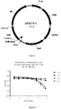
- a stuff sequence of about 750bp was cloned between NcoI and NotI, thereby constructing the final novel phage display vector pADSCFV-S ( Figure 1 ).
- the NcoI and NotI sites in this vector could facilitate cloning of a single-chain antibody (ScFv) gene.
- 2YTAG liquid medium A: ampicillin, 100 ⁇ g/ml; G: glucose, 2%
- M13 helper phages M13KO7, NEB Inc.
- phage-ScFv purified phages
- phage-ScFv purified phages
- the phage-ScFvs from the 12 sub-libraries as prepared were mixed proportionally in view of the capabilities, thereby preparing a phage display human antibody library.
- the final titer of phage library was 6*10E12 cfu/ml.
- the product was stored at -70°C. This phage display antibody library could be used for screening human antibodies specific to various antigens of interest.
- Preparation of anti-IL17 monoclonal antibodies required use of multiple different recombinant proteins, including human ( homo sapiens ) IL17 (huIL17, SEQ ID NO:8), mouse ( Mus musculus ) IL17 (moIL17, SEQ ID NO:9), Macaca mulatta IL17 (mmIL17, SEQ ID NO:10), Macaca fascicularis IL17 (mfIL17, SEQ ID NO:11), and human IL17R extracellular region (IL17R, SEQ ID NO:12). Since these proteins have glycosylation modifications, a mammalian cell expression system is advantageous for maintaining the structures and functions of the recombinant proteins. In addition, to facilitate purification, His-tag (SEQ ID NO: 13) or human antibody Fc fragment (SEQ ID NO: 14) was added to the C-terminal of these recombinant proteins.
- His-tag SEQ ID NO: 13
- human antibody Fc fragment SEQ ID NO: 14
- the genes (comprising His-tag or Fc encoding gene) of the above recombinant proteins were designed and synthesized.
- the synthesized recombinant protein genes were cloned into proper eukaryotic expression vectors (e.g., pcDNA3.1 from Invitrogen Inc.).
- liposomes e.g., 293fectin from Invitrogen Inc.
- other transfection agents e.g., PEI
- the cells were incubated in suspension under serum-free condition for 3-4 days. Then, supernatant of the culture was harvested by centrifugation.
- the recombinant proteins in the supernatant were further purified using metal chelate affinity chromatography columns (e.g., HisTrap FF from GE Inc.).
- metal chelate affinity chromatography columns e.g., HisTrap FF from GE Inc.
- Protein A/G affinity chromatography columns e.g., Mabselect SURE from GE Inc.
- desalination columns e.g., Hitrap desaulting from GE Inc.
- Example 3 Preparation of anti-human IL17 monoclonal antibodies using phage display antibody library technique
- huIL17-his (hereafter referred to as "IL17-His") (5 ⁇ g/ml) was used as an antigen to coat an ELISA plate (150 ⁇ l/well, four wells in total). The plate was incubated overnight at 4°C. PBST-1%BSA was used to block the ELISA plate at 37°C for 1h. In the meantime, the phage display antibody library (phage-scFv) prepared in Example 1 was blocked with PBST-1%BSA at room temperature for 1h with the input amount of phages being approximately 10 12 . After blocking, the phage antibody library was added into the ELISA plate for antibody-to-antigen binding at 37°C for 1h.
- IL17-His huIL17-his
- the neutralized phages were used to infect TG1 bacteria that had grown to logarithmic phase.
- the bacteria were incubated at 37°C for 30min, and oscillated at 150rpm at 37°C for 30min. 1% of the bacteria solution was plated for counting, and the rest solution was centrifuged at 4000rpm for 5min. After removal of supernatant, the bacteria were plated onto 2YTAG solid medium (A: ampicillin, 100 ⁇ g/ml; G: glucose, 2%) in a dish, and incubated overnight at 33°C for next screening procedure.
- 2YTAG solid medium A: ampicillin, 100 ⁇ g/ml
- G glucose, 26%
- Bacteria on the dish were collected. A proper amount of collected bacteria were seeded into 2YTAG liquid medium (A: ampicillin, 100 ⁇ g/ml; G: glucose, 2%) and incubated with oscillation to reach logarithmic phase. The bacteria were infected with M13KO7. After infection, the medium was changed to 2YTAKA liquid medium (A: ampicillin, 100 ⁇ g/ml; K: kanamycin, 70 ⁇ g/ml; A: arabinose, 0.001%), and the bacteria were incubated overnight at 28°C with oscillation at 220rpm for phage amplification. PEG/NaCl precipitation method was used to purify phages for next screening procedure. A total of three rounds of phage library enrichment screening were carried out.
- the bacteria were re-suspended in 2YTAKA medium (A: ampicillin, 100 ⁇ g/ml; K: kanamycin, 70 ⁇ g/ml; A: arabinose, 0.001 %), and incubated overnight at 28°C with oscillation at 220rpm.
- 2YTAKA medium A: ampicillin, 100 ⁇ g/ml; K: kanamycin, 70 ⁇ g/ml; A: arabinose, 0.001 %), and incubated overnight at 28°C with oscillation at 220rpm.
- IL17-Fc was coated with a carbonate buffer (pH9.6), and incubated overnight at 4°C. After washing three times with PBST, a PBST-4% milk solution was used for blocking at 37°C for 1h. The supernatant from overnight incubated monoclonal phage culture was proportionally diluted into PBST-4% milk, which was added at 100 ⁇ l/well to the blocked ELISA plate to bind at 37°C for 1h. The ELISA plate was washed with PBST. HRP-anti-M13 antibody was diluted at 1:5000, and added to the ELISA plate at 100 ⁇ l/well. After incubation at 37°C for 1h, an OPD developing solution was added to develop for 5-20min. Then, 1M H 2 SO 4 was added at 50 ml/well to terminate the development. Optical densities were determined using a microplate reader at the dual-wavelength of 492nm/630nm.
- the four monoclonal strains (11A, 9G, 3E, 6D) were seeded into 2YTAG liquid medium, and incubated to reach logarithmic phase.
- the helper phage M13KO7 the amount of which is approximately 20-times of bacterium number, was added for infection. After infection, the medium was changed to 2YTAKA medium, and the cells were incubated overnight at 28°C with oscillation at 220rpm for phage amplification. PEG/NaCl precipitation method was used to purify phages.
- IL17-his was coated with a carbonate solution (pH9.6), and incubated overnight at 4°C. After washing three times with PBST, a PBST-4% milk solution was used for blocking at 37°C for 1h.
- the four phages were diluted to a titer of 5*10E11cfu/ml. The dilutions of the four phages were used to dilute IL17R-his. The starting concentration of IL17R-his was 20 ⁇ g/ml.
- a three-time gradient dilution was carried out with each sample containing seven rounds of dilutions. The dilutions were added to an ELISA plate at 100 ⁇ l/well, and incubated at 37°C for 1h.
- a PBST solution was used to wish the ELISA plate.
- An HRP-anti-M13 secondary antibody was diluted at 1:5000, and added to the ELISA plate at 100 ⁇ l/well. After incubation at 37°C for 1h, an OPD developing solution was added to develop for 5-20min. Then, 1M H 2 SO 4 was added at 50 ml/well to terminate the development.
- Optical densities were determined using a microplate reader at the dual-wavelength of 492nm/630nm.
- Example 4 Affinity maturation of anti-IL17 monoclonal antibodies based on light chain substitution strategy
- the kappa light chain constant region gene of the synthesized human antibody (SEQ ID NO:17) was cloned into the vector pADSCFV-S to replace the gIII gene in the initial vector, thereby constructing the vector pADK-S (see, figure 3 ).
- This vector could be used to clone and express the kappa light chain gene of human antibody.
- the fusion gene of the heavy chain constant region CH1 of the synthesized human antibody and the C-terminal domain of the gIII protein (SEQ ID NO: 18) or the lambda light chain constant region of the human antibody (SEQ ID NO:19) was cloned into the vector pADSCFV-S to replace the gIII gene in the initial vector, thereby constructing the vectors pADG-S and pADL-S.
- CmR Chloramphenicol-resistant gene
- NEB Inc. pACYC184 plasmid
- the replication origin gene of the CDF plasmid (CDFori, SEQ ID NO: 20) was artificially synthesized. Then, by using conventional overlapping PCR method, a CmR-CDoriF fusion gene was constructed (XbaI and AflIII sites were added to two ends respectively).
- the CmR-CDFori fusion gene was cloned into the vectors pADK-S, pADG-S and pADL-S respectively to replace the flori-Ampr-pBRori segment, thereby constructing three new plasmids pAK-S (see, figure 3 ), pAG-S and pAL-S.
- the plasmids pADK-S and pADL-S were used as mother plasmids to construct a high-quality human antibody light chain library. Details were described as follows.
- VK1 genes were amplified by using the DNA from the four sub-libraries containing VK1 variable region genes prepared in Example 1 as templates and the primers shown in Table 5. Then, by dual enzyme digestion of NcoI and PmlI, the amplified VK1 genes were cloned into the vector pADK-S to replace the stuff sequence. The ligation products were electrotransfected into TG1 competent cells, thereby constructing a human VK1 light chain library. The capability of the library was calculated by dilution methods. 30 colonies were randomly selected for sequencing, so as to calculate the accuracy of each VK1 light chain library. The results were shown in Table 6.
- VL3 genes were amplified by using the DNA from the four sub-libraries containing VL3 variable region genes prepared in Example 1 as templates and the primers shown in Table 5. Then, by dual enzyme digestion of NcoI and PmlI, the amplified VL3 genes were cloned into the vector pADL-S to replace the stuff sequence. The ligation products were electrotransfected into TG1 competent cells, thereby constructing a human VL3 light chain library. The capability of the library was calculated by dilution methods. 30 colonies were randomly selected for sequencing, so as to calculate the accuracy of each VL3 light chain library. The results were shown in Table 6. The total capabilities of the two light chain libraries were 1.0*10E8 with accuracies over 90%.
- the two prepared light chain sub-libraries were respectively seeded in 2YTAG (A: ampicillin, 100ug/ml; G: glucose, 2%) liquid medium, and incubated to reach logarithmic phase.
- the cells were infected with M13 helper phages (M13KO7). After infection, the medium was changed to 2YTAKG (A: ampicillin, 100 ⁇ g/ml; K: kanamycin, 70 ⁇ g/ml; G: glucose, 2%) liquid medium, and the cells were incubated overnight at 28°C at 220rpm for phage amplification.
- the PEG/NaCl precipitation method was used to prepare purified phages, which were then subjected to titration.
- phage-VK1+VL3 a phage library for assembling human antibody light chains.
- the final titer of phage library was 5.4*10E11 cfu/ml.
- the product was stored at -70°C. This phage library could be used in light chain substitution procedures of various antibodies. Table 5.
- VK1 and VL3 light chain libraries Use of primer Primer Construction of VK1 light chain library PVK1F1:CCAGCCATGGCCGATATCCAGATGACCCA PVK1R1:CGTACGTTTGATCTCCACTTTGGTGC Construction of VL3 light chain library PVL3F1:CCAGCCATGGCCAGCTACGAACTGACCCAGCC PVL3R1: GGTCAGTTTGGTGCCACCGC Table 6. Capabilities and accuracies of VK1 and VL3 light chain libraries. Sub-library Capability Accuracy pADK-VK1 7.2*10E7 95% pADL-VL3 3.7*10E7 90%
- the light chains (11AVK, 9GVK) and heavy chains (11AVH, 9GVH) of the two single-chain antibodies 11A and 9G screened from the human antibody library were cloned into prokaryotic expression vectors pADK-s and pAG-s, respectively, thereby obtaining plasmids pADK-11AVK, pADK-9GVK, pAG-11AVH, pAG-9GVH expressing respective light chains and heavy chains.
- the heavy chain plasmids (pAG-11AVH, pAG-9GVH) were used to transform TG1 cells.
- the cells were infected with phage packing the light chain library (phage-VK1+VL3), thereby obtaining the mutated light chain libraries of the two antibodies (11AVH-VK1+VL3 and 9GVH-VK1+VL3).
- the two mutated light chain libraries (11AVH-VK1+VL3 and 9GVH-VK1+VL3) were respectively seeded in 2YTACG liquid medium (A: ampicillin, 100 ⁇ g/ml; C: chloramphenicol, 34 ⁇ g/ml; G: glucose, 2%), and incubated to reach logarithmic phase. Then, the cells were infected with M13 helper phages (M13KO7).
- the medium was changed to 2YTACKA (A: ampicillin, 100 ⁇ g/ml; C: chloramphenicol, 34 ⁇ g/ml; K: kanamycin, 70 ⁇ g/ml; A: arabinose, 0.001%) liquid medium, and the cells were incubated overnight at 28°C at 220rpm for phage amplification. Then, the PEG/NaCl precipitation method was used to prepare purified phage library (phage-11AVH-VK1+VL3 and phage-9GVH-VK1+VL3), which was then subjected to titration. Then, the two phage libraries were mixed in equal proportion. The final titer of phage library was 1.9*10E13 cfu/ml. The product was stored at -70°C.
- IL17-His was used in two rounds of enrichment of the phage-11AVH-VK1+VL3 and phage-9GVH-VK1+VL3 mixed library. Then, the phage ELISA method was used to identify the enriched single colonies (approximately 400 colonies). The colonies with high ELISA signal values were selected for sequencing. The colonies with correct sequence results were used to prepare purified phages. Meanwhile, the plasmids expressing the light chains and heavy chains of the two antibodies (11A and 9G) were co-transformed into TG1 cells. Purified phages were prepared as the positive control.
- the titers of the multiple purified monoclonal phages were adjusted to 1*10E11/cfu.
- a three-time gradient dilution was carried out using PBST-4% milk solution.
- Coated IL17-Fc (2 ⁇ g/ml) and conventional phage ELISA method were used to analyze the IL17-binding capabilities of the phage antibodies in individual dilutions.
- the phage-ELISA results showed that, the several phage antibodies screened from the light chain substitution procedure carried out on 11A and 9G were all capable of binding to IL17.
- the relative affinities of C2 (SEQ ID NO: 21) and D5 (SEQ ID NO: 22) were substantially higher than those of other colonies (see, figure 4 ). It was noted that the heavy chains of C2 and D5 were 9GVH.
- a 9G heavy chain variable region mutant library was constructed.
- the dual vector display system was used to screen the heavy chain mutant library.
- 9G heavy chain variable region gene (9GVH, SEQ ID NO: 23) was used as a template.
- conventional PCR technique and the degenerate primers shown in Table 7, various designed mutations were introduced into the CDRs of 9GVH.
- overlapping PCR method was used in assembling procedure to obtain mutated genes of intact 9G heavy chain variable regions.
- mutated 9GVH genes were cloned into the vector pADG-S.
- the ligation products were transformed into TG1 competent cells by electroporation, thereby constructing a 9GVH mutant library with library capability of approximately 4*10E6. 20 single colonies were randomly selected for sequencing. The results showed that the accuracy of the mutant library was 70% (14/20).
- the constructed 9GVH mutant library (TG1 bacteria) was seeded in 2YTAG liquid medium (A: ampicillin, 100ug/ml; G: glucose, 2%), and incubated to reach logarithmic phase. Then, the cells were infected with M13 helper phages (M13KO7). After infection, the medium was changed to 2YTAKG (A: ampicillin, 100 ⁇ g/ml; K: kanamycin, 70 ⁇ g/ml; G: glucose, 2%) liquid medium, and the cells were incubated overnight at 28°C at 220rpm for phage amplification.
- 2YTAG liquid medium A: ampicillin, 100ug/ml; G: glucose, 2%
- M13 helper phages M13 helper phages
- the two light chain genes C2 and D5 screened in Example 4 were cloned into the vector pAK-S which was then transformed into TG1 bacteria, thereby obtaining the two strains pAK-C2/TG1 and pAK-D5/TG1.
- the phage library (phage-9GVH) was used to infect pAK-C2/TG1 and pAK-D5/TG1 at logarithmic phase, thereby obtaining two 9GVH mutant libraries (light chains were C2 and D5, respectively).
- M13KO7 was used for infection.
- the two Fab libraries containing 9GVH mutants were respectively displayed on the surfaces of phages, thereby preparing two Fab phage display libraries (phage-C2-9GVH and phage-D5-9GVH).
- the two phage display libraries were mixed in equal proportion.
- IL17-his was used for two rounds of screening phage-C2-9GVH and phage-D5-9GVH mixed library. Approximately 300 colonies were selected for monoclonal phage ELISA identification.
- 9GVH mutant strains were selected for subsequent test, which were named as 9GA3 (SEQ ID NO: 24), 9GC2 (SEQ ID NO: 25) and 9GC5 (SEQ ID NO: 26). In addition, these three 9GVH mutants all had the best matching light chain C2 (SEQ ID NO: 21).
- the heavy chain and light chain genes of an antibody of interest were cloned into an appropriate eukaryotic expression vector (e.g., pcDNA3.1 from Invitrogen Inc.).
- the antibody heavy chain constant region could be of IgG1 subtype (SEQ ID NO: 29) or IgG4 subtype (SEQ ID NO: 30), and the light chain constant region could be of kappa subtype (SEQ ID NO: 31) or lambda subtype (SEQ ID NO: 32).
- liposomes e.g., 293fectin from Invitrogen Inc.
- other transfection agents e.g., PEI
- HEK293 cells e.g., HEK293F from Invitrogen Inc.
- CHO cells e.g., CHO-S from Invitrogen Inc.
- the cells were incubated in suspension under serum-free condition for 3-4 days. Then, supernatant of the culture was harvested by centrifugation.
- Protein A/G affinity chromatography column (e.g., Mabselect SURE from GE Inc.) was used to further purify the recombinant antibody in the supernatant. Then, desalination columns (e.g., Hitrap desaulting from GE Inc.) were used to change the storing buffer of the antibody to PBS (pH7.0) or other appropriate buffers (e.g., 0.1NaCl, 0.01M sodium citrate, pH 6.0). If necessary, the antibody samples could be subjected to filtration sterilization, and then split and stored at -20°C.
- PBS pH7.0
- other appropriate buffers e.g., 0.1NaCl, 0.01M sodium citrate, pH 6.0
- Example 7 Binding assay of anti-IL17 monoclonal antibody and human IL17
- IL17-His-binding capabilities of four anti-IL17 monoclonal antibodies 9GA3, 9GC2, 9GC5 and a control antibody Secukinumab (VH: SEQ ID NO:27, VK: SEQ ID NO:28) were assessed using ELISA assays.
- IL17-his was used to coat plates overnight at 4°C (0.5 ⁇ g/ml, 100 ⁇ l/well). Each monoclonal antibody had a starting concentration of 300 nM, and was subjected to a three-time gradient dilution. Eleven gradient dilutions were set for each monoclonal antibody sample. HRP-goat-anti-human IgG was used to determine the IL17-binding capabilities of the monoclonal antibodies at each dilution (see, figure 5 ).
- Example 8 Binding between anti-IL17 monoclonal antibodies and IL17 from different species
- IL17 prepared IL17 (huIL17, mIL17, mmIL17, and mfIL17) were used to coat 96-well ELISA plates overnight at 4°C (1 ⁇ g/ml, 100 ⁇ l/well).
- a blocking solution 2% milk powder-PBST
- various anti-IL17 monoclonal antibodies including 9GA3, 9GC2, 9GC5, secukinumab and Ixekizumab (VH: SEQ ID NO:33, VK: SEQ ID NO:34), were added and allowed to bind at 37°C for 1h. Then, the plates were washed four times with PBST.
- Example 9 Competitive IL17-binding of anti-human IL17 monoclonal antibodies with IL17 receptor (IL17R)
- a functional anti-IL17 monoclonal antibody should be able to block the binding of IL17R to IL17 at protein level.
- the abilities of four anti-IL17 monoclonal antibodies (9GA3, 9GC2, 9GC5, and secukinumab) to inhibit the binding of IL17R to IL17 were assessed.
- IL17-Fc was used to coat plates overnight at 4°C (0.5 ⁇ g/well, 100 ⁇ l/well). Each monoclonal antibody (starting concentration of 200 ⁇ g/ml) was subjected to a three-time gradient dilution with IL17R-his (2 ⁇ g/ml). Ten gradient dilutions were set for each monoclonal antibody. HRP-mouse-anti-his monoclonal antibody was used to determine the binding signal between IL17R-his and IL17-Fc. Then, GraphPad Prism 6 was used for data analysis and plotting (see, figure 7 ).
- HDFa cells adult dermal fibroblasts
- Sciencell Inc. Cat#:2320
- the cells were cultured and passaged according to the instructions provided by Sciencell Inc..
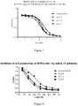
- the HDFa cells were seeded in 96-well plates with cell density of 1*10E4 cells/well. 1% FBS was added to FM medium, and the other components were the same as complete FM medium. The cells were cultured overnight at 37°C. On the second day, the medium was changed to the medium (FM+1%FBS) containing 2nM IL17-His and anti-IL17 monoclonal antibodies (9GA3, 9GC2, 9GC5, and secukinumab) at various concentrations (0.01-10nM), and the cells were cultured for 24 h. Then, the supernatant of the culture was collected.
- FM+1%FBS 2nM IL17-His and anti-IL17 monoclonal antibodies
- IL6 quantification kit (Cat#:EHoo4-96) from Excell Biology Inc. was used to determine the amount of IL6 in the supernatant from each culture. Then, GraphPad Prism 6 was used for data analysis and plotting (see, figure 8 ).
- the IC 50 of each anti-IL17 monoclonal antibody in inhibiting the release of IL6 was shown in Table 8. Table 8: IC 50 of four anti-IL17 monoclonal antibodies in inhibiting IL17-induced the release of IL6 by HDFa cells mAbs 9GA3 9GC2 9GC5 secukinumab IC 50 (nM) 0.12 0.22 0.07 0.48
- mice In order to determine the in vivo metabolic properties of anti-IL17 monoclonal antibodies in mice, various anti-IL17 monoclonal antibodies were administered in a single dose via tail veins of mice to assess pK.
- This study involved four anti-IL17 monoclonal antibodies (IgG4 subtype), which were 9GA3, 9GC2, 9GC5 and Ixekizumab.
- Monoclonal antibody samples were formulated in PBS buffering system (pH7.0) at a concentration of 0.5mg/ml. The administration dose was 5mg/kg. Single-dose was administered via tail veins. Each group included eight BALB/c mice with hale male and half female. Serum samples were collected at 4, 24, 48, 96, 168, 240, and 336 h after administration (orbital blood sampling), and stored at -70°C. After all sampling, the concentrations of the monoclonal antibodies were determined.
- Quantification of blood concentrations was carried out by quantitative ELISA assay based on coated IL17.
- Four purified anti-IL17 monoclonal antibody samples (9GA3, 9GC2, 9GC5 and Ixekizumab) were used as standards in quantification, and standard curves were respectively established for use in quantification of blood concentrations of the monoclonal antibodies.
- the change trends of in vivo concentrations of the four monoclonal antibodies in mice are shown in figure 10 .
Landscapes
- Chemical & Material Sciences (AREA)
- Health & Medical Sciences (AREA)
- Organic Chemistry (AREA)
- Life Sciences & Earth Sciences (AREA)
- Medicinal Chemistry (AREA)
- General Health & Medical Sciences (AREA)
- Immunology (AREA)
- Biochemistry (AREA)
- Proteomics, Peptides & Aminoacids (AREA)
- Molecular Biology (AREA)
- Genetics & Genomics (AREA)
- Biophysics (AREA)
- Epidemiology (AREA)
- Public Health (AREA)
- Veterinary Medicine (AREA)
- Bioinformatics & Cheminformatics (AREA)
- Animal Behavior & Ethology (AREA)
- Pharmacology & Pharmacy (AREA)
- Mycology (AREA)
- Microbiology (AREA)
- Engineering & Computer Science (AREA)
- Peptides Or Proteins (AREA)
- Medicines Containing Antibodies Or Antigens For Use As Internal Diagnostic Agents (AREA)
- Preparation Of Compounds By Using Micro-Organisms (AREA)
Description
- The present invention generally relates to the field of genetic engineered antibody pharmaceuticals, and in particular, to an anti-human IL-17 monoclonal antibody and use thereof. A new human anti-human IL-17 monoclonal antibody, and the use of subject antibody in treating IL-17-meditated diseases are provided in the present invention.
- Currently, the interleukin 17 (IL-17) family includes six cytokines, namely, IL-17A, IL-17B, IL-17C, IL-17D, IL-17E and IL-17F. The members of the IL-17 family do not share sequence similarity with any other known cytokines, and also share relatively low sequence similarity among themselves (Gaffen SL, Nat.Rev. Immunol. 2009 Aug; 9(8):556-67). Function of the IL-17 family members is mainly involved in the modulation of the immune response.
- IL-17A, a homodimer glycoprotein of 20-30kD, is mainly produced by activated CD4+T cells, and acts as a pro-inflammatory cytokine. IL-17 is secreted by activated T cells at an inflammatory site, not during the systemic circulation. IL17 has many biological properties, including up-regulating adhesion molecules, and inducing the production of numerous inflammatory cytokines and chemokines in various cell types, including synovial cells, cartilage cells, fibroblasts, endothelial cells, epithelial cells, keratinocytes and macrophages. In addition, IL-17 induces the aggregation of neutrophilic granulocytes at an inflammation site by inducing the release of chemokines, stimulates the production of prostaglandins and metalloproteases, and inhibits the synthesis of proteoglycans. Moreover, IL-17 plays a critical role in the maturation of hemopoietic progenitor cells. IL-17 is involved in the signal transduction in various organs and tissues, including lung, articular cartilage, bone, brain, hemopoietic cells, kidneys, skin and intestines. Therefore, the immune reaction mediated by IL-17A/Th17 is systemic, and leads to an inflammatory reaction mainly expressed as neutrophilic granulocyte infiltration.
- As demonstrated by extensive studies, the increase in IL-17 is involved in various diseases, including airway inflammation, rheumatoid arthritis (RA), osteoarthritis, bone erosion, inflammatory bowel disease (IBD), allograft rejection, psoriasis, some types of cancer, angiogenesis, atherosclerosis and multiple sclerosis (MS) (Witkowski et al., Cell. Mol. Life Sci.61:567-579, 2004). Additionally, IL-17 plays a role in the degradation of collagen matrix, inflammation and joint injury, independent of IL-lb. Also, IL-17 and TNF-α synergistically increase inflammation. As confirmed by further studies, by blocking the in vivo biological activity of IL-17 with an antibody specifically binding to IL-17 or a soluble IL17 receptor, inflammation and bone erosion in various animal arthritis models are effectively reduced. Therefore, IL-17 becomes a new therapeutic target for RA and other autoimmune diseases. Moreover, since IL17 is mainly present in an inflammatory site, a medicament targeting IL17 may potentially have higher safety than medicaments targeting other pro-inflammatory cytokines in systemic circulation (e.g. TNF).
- Currently, European Union and the FDA in the US have approved Cosentyx from Novartis Pharma AG for treating adult patients suffering from moderate-to-severe plaque psoriasis. The active ingredient of Cosentyxus is secukinumab, which is a monoclonal antibody against IL-17.
-
WO 2015/022656 discloses methods, treatment regimens, uses, kits and therapies for treating generalized pustular psoriasis and inter alia plaque-type psoriasis by the administration of an IL-17 antagonist, such as the IL-17 antibody secukinumab with its CDR regions, and further mentions the IL-17 antibody ixekizumab. - Nevertheless, there is still a need in the art in more improved anti-IL-17 antibodies suitable for treatment of a patient.
- In an aspect, there is provided in the present application a monoclonal antibody that specifically binds to human IL-17A, comprising a heavy chain variable region comprising HCDR1, HCDR2 and HCDR3, and a light chain variable region comprising LCDR1, LCDR2 and LCDR3, wherein HCDR1 has the sequence GX1X2X3X4X5Y, HCDR2 has the sequence NQDGX6E, HCDR3 has the sequence DYYDX7ISDYYIHYWYFDL, LCDR1 has the sequence RASQNVHNRLT, LCDR2 has the sequence GASNLES, and LCDR3 has the sequence QQYNGSPTT; wherein the sequence X1X2X3X4X5 is FTIDN, MSMSD or ITMDD, X6 is N or D, and X7 is V or L; and wherein the HCDRs and LCDRs are defined according to Chothia.
- In some embodiments, the heavy chain variable region of the antibody has a sequence as set forth in SEQ ID NO: 24, and the light chain variable region has a sequence as set forth in SEQ ID NO:21; or the heavy chain variable region has a sequence as set forth in SEQ ID NO: 25, and the light chain variable region has a sequence as set forth in SEQ ID NO:21; or the heavy chain variable region has a sequence as set forth in SEQ ID NO: 26, and the light chain variable region has a sequence as set forth in SEQ ID NO:21.
- In some embodiments in the above aspect, the antibody is an intact antibody, a substantively intact antibody, a Fab fragment, a F(ab')2 fragment or a single-chain Fv fragment.
- In some embodiments in the above aspect, the antibody is a fully human antibody.
- In some embodiments in the above aspect, the antibody further comprises a heavy chain constant region selected from the group consisting of IgG1 and IgG4 subtypes, and/or a light chain constant region selected from the group consisting of kappa and lambda subtypes.
- In some embodiments in the above aspect, the monoclonal antibody is capable of inhibiting the activity of 2 nM human IL-17A by 50% at a concentration less than 1 nM, wherein the activity inhibition is measured by determining human IL-17A-induced IL-6 production in human dermal fibroblasts (HDFa).
- In an aspect, there is provided in the present application a pharmaceutical composition comprising a monoclonal antibody in any one of the above aspects.
- In an aspect, there is provided in the present application a use of a monoclonal antibody in any one of the above aspects in treating a human IL-17A-mediated disease.
- In some embodiments, the disease is an autoimmune disease.
- In some embodiments, the disease is psoriasis, rheumatoid arthritis or ankylosing spondylitis.
- In an aspect, there is further provided in the present disclosure a use of a monoclonal antibody in any one of the above aspects in the manufacture of a medicament for treating a human IL-17A-mediated disease.
- In some embodiments, the disease is an autoimmune disease.
- In some embodiments, the disease is psoriasis, rheumatoid arthritis or ankylosing spondylitis.
-
-
Figure 1 shows a structural diagram of a novel phage display vector pADSCFV-S (Para: arabinose promoter, PelB leader: PelB signal peptide gene, NcoI: NcoI restriction site, Stuff: 750bp stuff sequence, NotI: NotI restriction site, gIII: gIII gene of M13 phage, flori: replication origin of M13 phage, Ampr: ampicillin resistance gene, pBRori: replication origin of pBR322, AraC: AraC gene). -
Figure 2 shows competitive binding of a monoclonal phage and IL17R to IL17. -
Figure 3 shows structural diagrams of plasmids pADK-S and pAK-S (Para: arabinose promoter, leader: PelB signal peptide gene, NcoI: NcoI restriction site, Stuff: 750bp stuff sequence, PmlI: PmlI restriction site, CK: kappa light chain constant region gene of human antibody; flori: replication origin of M13 phage, Ampr: ampicillin resistance gene, pBRori: replication origin of pBR322, AraC: AraC gene, CmR: chloromycetin resistance gene, CDFori: replication origin of CDF plasmid). -
Figure 4 shows measurement of relative affinity of phage antibodies by phage ELISA assays. -
Figure 5 shows binding capability of anti-IL17 antibodies to human IL17 as measured by ELISA assays. -
Figure 6 shows binding capability of anti-IL17 monoclonal antibodies to IL17 proteins from different species as measured by ELISA assays. -
Figure 7 shows competitive binding of anti-huIL17 monoclonal antibodies and IL17R to IL17. -
Figure 8 shows comparison analysis of inhibition of IL17-induced IL6 production in HDFa cells by anti-IL17 monoclonal antibodies. -
Figure 9 shows stability analysis of various anti-IL17 monoclonal antibodies in human sera. -
Figure 10 shows concentration alteration trend of four anti-IL17 monoclonal antibodies in mice. - Various inventions in the present application are partially based on construction of human phage antibody library. The inventors have screened human anti-IL-17 monoclonal antibodies with desired properties from the constructed antibody library.
- In various aspects of the present application, there is provided a novel anti-human IL-17 monoclonal antibody or an antigen-binding fragment thereof, a polynucleotide encoding the monoclonal antibody or the antigen-binding fragment, a vector comprising the polynucleotide, a host cell comprising the polynucleotide or the vector, a method for preparing and purifying the antibody, and medical use of the antibody or the antigen-binding fragment. Based on the sequences of variable regions of the antibody of the present application, an intact antibody molecule can be formulated as a medicament for use in clinically treating an IL-17-mediated autoimmune disease, including but not limited to psoriasis, rheumatoid arthritis and ankylosing spondylitis.
- Unless otherwise specified, all technical and scientific terms used herein have the same meaning as those understood by an ordinary person skilled in the relevant art.
- As used herein, the term "polypeptide" refers to a polymer constituted by amino acid residues linked via a peptide bond. The term "protein" usually refers to a relatively large polypeptide. The term "peptide" usually refers to a relatively small polypeptide (e.g., containing at most 100, 80, 60, 50, 30 or 20 amino acid residues).
- As used herein, the term "antibody" refers to an immunoglobulin molecule that specifically binds to a target via at least one antigen recognition epitope in the variable region of the immunoglobulin molecule. The examples of the target include but not limited to a carbohydrate, a polynucleotide, a lipid, a polypeptide. As used herein, the term "antibody" includes not only an intact (full-length) antibody, but also an antigen-binding fragment (e.g., Fab, Fab', F(ab')2, Fv), a variant, a fusion protein comprising an antibody portion, a humanized antibody, a chimeric antibody, a diabody, a linear antibody, a single-strand antibody, a multi-specific antibody thereof (e.g., a bi-specific antibody), and any other modified versions of an immunoglobulin molecule comprising the antigen recognition site exhibiting desired specificity , including a glycosylated variant of an antibody, a variant of an antibody with a modified amino acid sequence, and a covalently modified antibody.
- Generally, an intact or full-length antibody comprises two heavy chains and two light chains. Each heavy chain comprises a heavy chain variable region (VH) and a first, second and third constant region (CH1, CH2 and CH3). Each light chain comprises a light chain variable region (VL) and a constant region (CL). A full-length antibody may be an antibody of any classes, e.g., IgD, IgE, IgG, IgA or IgM (or a subclass of the above). However, the antibody of the present application does not necessarily belong to any specific class. Based on the amino acid sequence of the constant domain of the heavy chain of an antibody, an immunoglobulin can be divided into different classes. Generally, there are five classes of immunoglobulins, i.e., IgA, IgD, IgE, IgG and IgM. Some of these classes can be further divided into several subclasses (isoforms), e.g., IgG1, IgG2, IgG3, IgG4, IgA1 and IgA2. Constant domains of heavy chains of different classes of immunoglobulins are termed as α, δ, ε, γ and µ. The structures of subunits and the three-dimensional structures of various classes of immunoglobulins are well known in the art.
- As used herein, the term "antigen-binding fragment" refers to a portion or region of an intact antibody molecule that is responsible for binding to an antigen. An antigen-binding domain may comprise a heavy chain variable region (VH), a light chain variable region (VL) or both. Each of VH and VL generally comprises three complementary determining regions, i.e., CDR1, CDR2 and CDR3.
- Examples of an antigen-binding fragment include but not limited to, (1) a Fab fragment, which may be a monovalent fragment comprising a VL-CL chain and a VH-CH1 chain; (2) a F(ab')2 fragment, which may be a bivalent fragment comprising two Fab fragments which are linked by a disulfide bridge in the hinge region (i.e. a dimer of Fab); (3) a Fv fragment comprising a VL domain and a VH domain from a single arm of an antibody; (4) a single-chain Fv (scFv), which may be a single polypeptide chain comprising a VH domain and a VL domain linked by a peptide linker; and 5) (scFv)2, which may comprise two VH domains linked by a peptide linker and two VL domains which are conjugated to the two VH domains via a disulfide bridge.
- As used herein, the term "specifically bind to" refer to a non-random binding reaction between two molecules, e.g., binding of an antibody to an antigen epitope.
- The nucleic acid sequences provided herein involve use of degenerate bases in addition to the conventional A, T, C and G. The meaning of a degenerate base is the same as that commonly understood by a person skilled in the art. For example, R refers to A or G, Y refers to C or T, M refers to A or C, K refers to G or T, S refers to C or G, W refers to A or T, H refers to A or C or T, B refers to C or G or T, V refers to A or C or G, D refers to A or G or T, and N refers to A or C or G or T.
- In an aspect, there is provided in the present application a monoclonal antibody that specifically binds to human IL-17A, comprising a heavy chain variable region comprising HCDR1, HCDR2 and HCDR3, and a light chain variable region comprising LCDR1, LCDR2 and LCDR3, wherein HCDR1 has the sequence GX1X2X3X4X5Y, HCDR2 has the sequence NQDGX6E, HCDR3 has the sequence DYYDX7ISDYYIHYWYFDL, LCDR1 has the sequence RASQNVHNRLT, LCDR2 has the sequence GASNLES, and LCDR3 has the sequence QQYNGSPTT; wherein the sequence X1X2X3X4X5 is FTIDN, MSMSD or ITMDD, X6 is N or D, and X7 is V or L; and wherein the HCDRs and LCDRs are defined according to Chothia.
- In some embodiments, the heavy chain variable region of the antibody has a sequence as set forth in SEQ ID NO: 24, and the light chain variable region has a sequence as set forth in SEQ ID NO:21; or the heavy chain variable region has a sequence as set forth in SEQ ID NO: 25, and the light chain variable region has a sequence as set forth in SEQ ID NO:21; or the heavy chain variable region has a sequence as set forth in SEQ ID NO: 26, and the light chain variable region has a sequence as set forth in SEQ ID NO:21.
- In some embodiments in the above aspect, the antibody is an intact antibody, a substantively intact antibody, a Fab fragment, a F(ab')2 fragment or a single-chain Fv fragment.
- In some embodiments in the above aspect, the antibody is a fully human antibody.
- In some embodiments in the above aspect, the antibody further comprises a heavy chain constant region selected from the group consisting of IgG1 and IgG4 subtypes, and/or a light chain constant region selected from the group consisting of kappa and lambda subtypes.
- In some embodiments in the above aspect, the monoclonal antibody is capable of inhibiting the activity of 2 nM human IL-17A by 50% at a concentration less than 1 nM, wherein the activity inhibition is measured by determining human IL-17A- induced IL-6 production in human dermal fibroblasts (HDFa).
- In some embodiments in the above aspect, the antibody is a fully human monoclonal antibody, and reduces, antagonizes or eliminates at least one in vitro or in vivo biological activity involving IL-17 or a portion thereof.
- In some embodiments in the above aspect, the antibody is characterized by high binding affinity to human IL-17.
- In some embodiments in the above aspect, the antibody is characterized in that it can specifically bind to human IL-17, Macaca mulatta IL-17 and Macaca fascicularis IL-17 at a level higher than background, but not Mus musculus IL-17.
- In some embodiments in the above aspect, the antibody comprises a variable region and a constant region, wherein the heavy chain constant region may be of IgG1 subtype (SEQ ID NO: 29) or IgG4 subtype (SEQ ID NO: 30), and the light chain constant region may be of kappa subtype (SEQ ID NO: 31) or lambda subtype (SEQ ID NO: 32).
- In another aspect, there is further provided in the present disclosure a polynucleotide encoding a monoclonal antibody or an antigen-binding fragment thereof in the above aspects, a vector comprising the polynucleotide, and a host cell transfected with the vector. In some embodiments, the host cell is a CHO cell or a HEK293 cell.
- In an aspect, there is provided in the present application a pharmaceutical composition comprising a monoclonal antibody in any one of the above aspects.
- In some embodiments, the pharmaceutical composition further comprises a pharmaceutically acceptable carrier, excipient and/or diluent.
- In some embodiments, the pharmaceutical composition comprises a therapeutically effective amount of the monoclonal antibody.
- In an aspect, there is provided in the present application a use of a monoclonal antibody in any one of the above aspects in treating a human IL-17A-mediated disease.
- In some embodiments, the disease is an autoimmune disease.
- In some embodiments, the disease is psoriasis, rheumatoid arthritis or ankylosing spondylitis.
- In some embodiments, the pharmaceutical activity of an antibody of the present application can be tested by a standard assay in the art, e.g., see, Hwang SY, Kim JY, Kim KW, Park MK, Moon Y, Kim WU, Kim HY: IL-17 induces production of IL-6 and IL-8 in rheumatoid arthritis synovial fibroblasts via NF-kappaB- and PI3-kinase/Akt-dependent pathways. Arthritis research & therapy 2004, 6(2):R120-128. Briefly, in the presence of an antibody of the present application at various concentrations, the recombinant IL-17 is used to stimulate human dermal fibroblasts (HDFa cells). After stimulated for 24h, supernatant is obtained and detected for IL-6 by an ELISA assay. When the above test is carried out, an antibody of the present application preferably has an IC50 value of about 0.22 nM or less, more preferably about 0.12 nM or less, and even more preferably about 0.07 nM or less in respect of inhibition of production of IL-6 as defined above.
- In some embodiments, the stability of an antibody of the present application in human serum can be tested by a standard assay in the art. Briefly, a monoclonal antibody sample sterilized by filtration is diluted into 200 µl of sterilized normal human serum mixture or PBS to a final concentration of 30 µg/ml, respectively, mixed and placed in a water bath at 37°C for 8 days (192 h). Next, monoclonal antibodies in the serum sample, monoclonal antibodies in the PBS sample and monoclonal antibodies from a cryopreserved sample are assessed for their binding to human IL17 by an ELISA assay. The change in binding capabilities of the monoclonal antibodies in different samples to IL17 is compared. When the above test is carried out, an antibody of the present application preferably retains about 87%, more preferably about 91%, and even more preferably about 97% of IL17 binding capability.
- In some embodiments, the pharmacokinetics of an antibody of the present application in mice can be assessed by a standard assay in the art. Briefly, a monoclonal antibody sample is formulated in a PBS buffer (pH7.0) at a concentration of 0.5 mg/ml, and administered to mice at a dosage of 5 mg/kg with a single injection into tail vein. Each group contains eight BALB/c mice with four male and four female. Blood samples are then taken by intra-orbital sampling at 4, 24, 48, 96, 168, 240 and 336 hours after injection, and stored at -70°C. After all samples are obtained, the concentrations of the monoclonal antibody in the samples are quantitatively determined. The quantitation of plasma concentration of a drug is carried out based on a quantitative ELISA assay with coated IL17. According to the first order reaction model, the natural logarithm of the antibody concentration (lnC) is plotted with respect to time (t), yielding a slope constant k. Then, the half-life of the anti-IL17 monoclonal antibody in mice (tl/2) is calculated according to the formula tl/2=0.693/k. When the above test is carried out, an antibody of the present application preferably has a half-life (tl/2) in mice of about 6.7 h, more preferably about 7.4 h, and even more preferably about 8.5 h.
- In an aspect, there is further provided in the present disclosure an isolated nucleic acid molecule encoding an antibody of the present application or a light chain or a heavy chain thereof, a vector comprising the nucleic acid molecule, a host cell comprising the vector, and a method for producing the antibody. In some embodiments, the nucleic acid molecule is operatively linked to a regulatory sequence, which can be recognized by a host cell transformed with the vector. In some embodiments, the method for producing the antibody comprises culturing the host cell such that the nucleic acid is expressed. In some embodiments, the method for producing the antibody further comprises recovering the antibody from the culture medium in which the host cell is cultured.
- In an aspect, there is further provided in the present disclosure Mus musculus IL-17 (SEQ ID NO:9), Macaca mulatta IL-17 (SEQ ID NO:10) or Macaca fascicularis IL-17 (SEQ ID NO:11), an isolated nucleic acid molecule encoding any one of them, a vector comprising the nucleic acid molecule, a host cell comprising the vector, a method for producing the Mus musculus IL-17, Macaca mulatta IL-17 or Macaca fascicularis IL-17.
- The following examples are provided only for the purpose of further illustrating the inventions of the present application, but should not be construed as any limitation to the inventions.
- Antibody library technique is an important method for preparing human monoclonal antibodies. Currently, antibody library technique based on phage display had been well-established, and successfully applied to preparation of human monoclonal antibodies. This example demonstrates the strategy and methodology for constructing a phage display antibody library using many genetic engineering techniques.
- In order to construct a human antibody library, genes of heavy chain variable regions (VH) and light chain variable regions (VL) of human antibodies must be acquired first. Genes of antibody variable regions may be derived from lymphocytes in peripheral blood from healthy subjects or fully synthesized.
- Blood was taken from 19 healthy volunteers (50 ml for each). Then, lymphocytes were isolated using a lymphocyte isolation solution (MP Biomedicals Inc., Cat#:0850494). RNA was prepared using a Total RNA extraction kit from Omega Inc. (Cat#:R6834-01). The cDNA was prepared using a reverse transcription kit from TransGen Biotech Inc. (Cat#:AT301-03). Finally, the primer sets shown in the Table 1 below were used in a PCR reaction to amplify heavy chain variable region genes (VH) and light chain variable region genes (VL, including Vk and Vl) of antibodies, respectively. Amplified PCR products (VH,VK or Vl) were purified and recovered using standard agarose gel electrophoresis, and stored at -20°C for further use.
Table 1: Primers for amplifying natural human antibody heavy chain and light chain genes primer primer sequence VH forward primer VHF1 CAGRTGCAGCTGGTGCARTCTGG VHF2 SAGGTCCAGCTGGTRCAGTCTGG VHF3 CAGRTCACCTTGAAGGAGTCTGG VHF4 SAGGTGCAGCTGGTGGAGTCTGG VHF5 GAGGTGCAGCTGGTGGAGWCYGG VHF6 CAGGTGCAGCTACAGCAGTGGGG VHF7 CAGSTGCAGCTGCAGGAGTCSGG VHF8 GARGTGCAGCTGGTGCAGTCTGG VHF9 CAGGTACAGCTGCAGCAGTCAGG VH reverse primer VHR1 TGAGGAGACGGTGACCAGGGTGCC VHR2 TGAAGAGACGGTGACCATTGTCCC VHR3 TGAGGAGACGGTGACCAGGGTTCC VHR4 TGAGGAGACGGTGACCGTGGTCCC VK forward primer VKF1 GACATCCAGWTGACCCAGTCTCC VKF2 GATGTTGTGATGACTCAGTCTCC VKF3 GAAATTGTGWTGACRCAGTCTCC VKF4 GATATTGTGATGACCCAGACTCC VKF5 GAAACGACACTCACGCAGTCTCC VKF6 GAAATTGTGCTGACTCAGTCTCC VK reverse primer VKR1 ACGTTTGATCTCGAGCTTGGTCCCYTGGCCRAA VKR2 ACGTTTGATCTCGAGTTTGGTCCCAGGGCCGAA VKR3 ACGTTTGATCTCGAGCTTGGTCCCTCCGCCGAA VKR4 ACGTTTAATCTCGAGTCGTGTCCCTTGGCCGAA V1 forward primer V1F1 CAGTCTGTGYTGACKCAGCCRCC V1F2 CARTCTGCCCTGACTCAGCCT V1F3 TCCTATGWGCTGACTCAGCCA V1F4 TCTTCTGAGCTGACTCAGGACCC V1F5 CAGGCTGTGCTGACTCAGCCG V1F6 AATTTTATGCTGACTCAGCCCCA V1F7 CAGRCTGTGGTGACYCAGGAGCC V1F8 CWGCCTGTGCTGACTCAGCC V1 reverse primer V1R1 ACCTAGGACGGTGACCTTGGTCCC V1R2 ACCTAGGACGGTCAGCTTGGTCCC V1R3 ACCTAAAACGGTGAGCTGGGTCCC - The basic strategy of preparation of fully synthesized antibody genes involves using degenerate primers to introduce designed mutations into CDRs of a chosen antibody gene template. In order to construct a fully synthesized human antibody library, three human antibody heavy chain variable region templates (VH1;VH3 and VH5) and two human antibody light chain variable region templates (VK1 and Vl3) were chosen in this example to construct a fully synthesized human antibody library.
- Five antibody variable region genes, i.e., VH1 (SEQ ID NO:1), VH3 (SEQ ID NO:2), VH5 (SEQ IDNO:3), VK1 (SEQ ID NO:4) and Vl3 (SEQ ID NO:5), were designed and synthesized by Ming Chen Zhi Yuan Inc.. Primers as shown in Table 2 were designed and synthesized to introduce designed mutations into the CDR1, CDR2 and CDR3 of the five variable region genes, respectively. By using the conventional PCR technique and respective sets of degenerate primers containing the designed mutations, the designed mutations were introduced into corresponding CDRs. Then, by using 2-3 rounds of overlapping PCR, intact heavy chain variable region genes (VH1.VH3,VH5) or light chain variable region genes (VK1,VL3) were constructed. Finally, amplified PCR products of the variable region genes were recovered by agarose gel electrophoresis, and stored at -20°C for further use.
Table 2. Primers for amplifying fully synthesized human antibody variable region genes. primer primer sequence VH1 CDR1 PVH1-1:ACTAGCTAGCGCGCAGGTGCAGTTAGTGCAGAG 
CDR2 

CDR3 


PVH1-7b:TCATAGCGGCCGCCGCGGTGCTGGTAGATTTGTC VH3 CDR1 PVH3-1:ATAGCTAGCGCGGAAGTGCAATTGGTGGAAAGC 


CDR2 PVH3-3:CACCCATTCCAGACCTTTACCCGGTGCCTGGCGCACCCA 
CDR3 


VH5 CDR1 PVH5-1:CAGCCATGGCCGAAGTTC 


CDR2 

PVH5-8:CTAAGCGGCCGCGCGTGCACAATAGTACATAGC CDR3 


PVH5-12a: GAGACGGTGACCAGAGTACCTTGACCCCA PVH5-12b:GAGACGGTGACCAGAGTACCTTGACCCCAGDAATCGAA 

PVH5-14:CTAAGCGGCCGCGCTCGAGACGGTGACCAGAGTACC VK1 CDR1 PVK1-1:ATAGCTAGCGCGGATATCCAGATGACCCAGAGCC 

CDR2 

CDR3 

VL3 CDR1 PVL3-1: ataGCTAGCGCGAGCTACGAACTGACCCAGC 
CDR2 


CDR3 


- In order to construct single-chain antibody genes (ScFv), a commonly used flexible linker consisting of 15 amino acids was added between the heavy chain variable region (VH) and light chain variable region (VL). The sequence of the linker was GGGGSGGGGSGGGGS with encoding sequence of ggtggaggcggttctggcggaggtgggagcggaggcggaggttca. The structure of the designed single-chain antibody was VH-Linker-VL.
- Using the method described in the first section of this Example, many heavy chain and light chain variable region genes as shown in Table 3 below were obtained, including four different heavy chain variable region genes and three light chain variable region genes.
Table 3: Different heavy chain and light chain variable region genes. heavy chain variable region light chain variable region Native antibody gene VH VL (VK+Vlmixed) Synthesized antibody gene VH1 VK1 VH3 VL3 VH5 - Based on the above design of single-chain antibodies and well-developed overlapping PCR technique, different heavy chains and light chains as shown in this Table could be conveniently combined. A total of 12 different single-chain antibody genes were constructed. The 12 single-chain antibody genes amplified by PCT reactions were purified and recovered by agarose gel electrophoresis, and stored at -20°C for further use.
- Common phage display vectors are based on lac promotors (Plac). However, lac promotors impact the capability and diversity of the antibody library due to the properties thereof such as leaked expression. We engineered a common phage display vector pCANTAB5E (Amersham Biosciences/GE, Inc.) by using conventional molecular biology techniques as follows.
- By dual enzyme digestion of AflIII and NotI, the Plac promoter and g3 signal peptide portion in the pCANTAB5E vector were replaced with a fragment comprising AraC gene, arabinose promoter (Para) and PelB leader, in which the AraC gene and Para were from the pBADhis vector from Invitrogen Inc., and the PelB leader sequence (SEQ ID NO: 6) was an artificial sequence. Then, by dual enzyme digestion of NcoI and NotI, a stuff sequence of about 750bp (SEQ ID NO: 7) was cloned between NcoI and NotI, thereby constructing the final novel phage display vector pADSCFV-S (
Figure 1 ). The NcoI and NotI sites in this vector could facilitate cloning of a single-chain antibody (ScFv) gene. - By conventional molecular biology techniques and dual enzyme digestion of NcoI and NotI, 12 ScFvs prepared in Section 1.2 were respectively cloned into the vector pADSCFV-S. The ligation products were electrotransfected into TG1 competent cells, in which about 20 rounds of electrotransfection were performed for each sub-library and a total of about 240 rounds of electrotransfection were carried out. The capability of each sub-library was calculated by dilution methods. 30-40 colonies were randomly selected from each sub-library for sequencing, so as to calculate the accuracy of each sub-library. The capabilities and accuracies of the 12 sub-libraries were shown in Table 4. The total capability of the 12 sub-libraries reached 1.0*10E9 with average accuracy of more than 75%.
Table 4. Capabilities and accuracies of the 12 sub-libraries Sub-library Capability Accuracy ScFv -VH1-VK1 4.79∗10E7 81% ScFv -VH1-VL3 3.72∗10E7 76% ScFv -VH1-VL 2.2∗10E7 70% ScFv -VH3-VK1 2.2∗10E7 83% ScFv -VH3-VL3 3.14∗10E7 78% ScFv -VH3-VL 7.7∗10E7 70% ScFv -VH5-VK1 2.68∗10E7 74% ScFv -VH5-VL3 2.5∗10E7 76% ScFv -VH5-VL 9.2∗10E7 84% ScFv -VH-VK1 5.9∗10E7 75% ScFv -VH-VL3 7.7∗10E7 72% ScFv -VH-VL 27.2∗10E7 85% - The 12 sub-libraries were respectively seeded in 2YTAG liquid medium (A: ampicillin, 100 µg/ml; G: glucose, 2%), and incubated at 37°C with oscillation at 220rpm to reach logarithmic phase (OD600=0.8). Then, the cells were infected with M13 helper phages (M13KO7, NEB Inc.). After infection, the medium was changed to 2YTAKA liquid medium (A: ampicillin, 100µg/ml; K: kanamycin, 70µg/ml; A: arabinose, 0.001%), and the cells were incubated overnight at 28°Cwith oscillation at 220rpm for phage amplification. Then, the PEG/NaCl precipitation method was used to prepare purified phages (phage-ScFv), which was then subjected to titration. Then, the phage-ScFvs from the 12 sub-libraries as prepared were mixed proportionally in view of the capabilities, thereby preparing a phage display human antibody library. The final titer of phage library was 6*10E12 cfu/ml. The product was stored at -70°C. This phage display antibody library could be used for screening human antibodies specific to various antigens of interest.
- Preparation of anti-IL17 monoclonal antibodies required use of multiple different recombinant proteins, including human (homo sapiens) IL17 (huIL17, SEQ ID NO:8), mouse (Mus musculus) IL17 (moIL17, SEQ ID NO:9), Macaca mulatta IL17 (mmIL17, SEQ ID NO:10), Macaca fascicularis IL17 (mfIL17, SEQ ID NO:11), and human IL17R extracellular region (IL17R, SEQ ID NO:12). Since these proteins have glycosylation modifications, a mammalian cell expression system is advantageous for maintaining the structures and functions of the recombinant proteins. In addition, to facilitate purification, His-tag (SEQ ID NO: 13) or human antibody Fc fragment (SEQ ID NO: 14) was added to the C-terminal of these recombinant proteins.
- Based on the amino acid sequences of various recombinant proteins of interest recorded in the Uniprot database, the genes (comprising His-tag or Fc encoding gene) of the above recombinant proteins were designed and synthesized. By conventional molecular biology techniques, the synthesized recombinant protein genes were cloned into proper eukaryotic expression vectors (e.g., pcDNA3.1 from Invitrogen Inc.). Then, liposomes (e.g., 293fectin from Invitrogen Inc.) or other transfection agents (e.g., PEI) are used to transfect the recombinant protein expression plasmids as prepared into HEK293 cells (e.g., HEK293F from Invitrogen Inc.). The cells were incubated in suspension under serum-free condition for 3-4 days. Then, supernatant of the culture was harvested by centrifugation.
- For recombinant proteins fused with His-tags, the recombinant proteins in the supernatant were further purified using metal chelate affinity chromatography columns (e.g., HisTrap FF from GE Inc.). For recombinant proteins fused with Fc, Protein A/G affinity chromatography columns (e.g., Mabselect SURE from GE Inc.) was used for further purification. Then, desalination columns (e.g., Hitrap desaulting from GE Inc.) were used to change the storing buffer of the recombinant proteins to PBS (pH7.0) or other appropriate buffers. If necessary, the antibody samples could be subjected to filtration sterilization, and then split and stored at -20°C.
- huIL17-his (hereafter referred to as "IL17-His") (5 µg/ml) was used as an antigen to coat an ELISA plate (150 µl/well, four wells in total). The plate was incubated overnight at 4°C. PBST-1%BSA was used to block the ELISA plate at 37°C for 1h. In the meantime, the phage display antibody library (phage-scFv) prepared in Example 1 was blocked with PBST-1%BSA at room temperature for 1h with the input amount of phages being approximately 1012. After blocking, the phage antibody library was added into the ELISA plate for antibody-to-antigen binding at 37°C for 1h. Unbound phages were removed by washing with PBST/PBS. Finally, 0.1M Glycine-HCl solution (pH2.2) was used to elute bound phages, and the eluted phages were neutralized with 1.5M Tris-HCl solution (pH8.8).
- The neutralized phages were used to infect TG1 bacteria that had grown to logarithmic phase. The bacteria were incubated at 37°C for 30min, and oscillated at 150rpm at 37°C for 30min. 1% of the bacteria solution was plated for counting, and the rest solution was centrifuged at 4000rpm for 5min. After removal of supernatant, the bacteria were plated onto 2YTAG solid medium (A: ampicillin, 100µg/ml; G: glucose, 2%) in a dish, and incubated overnight at 33°C for next screening procedure.
- Bacteria on the dish were collected. A proper amount of collected bacteria were seeded into 2YTAG liquid medium (A: ampicillin, 100µg/ml; G: glucose, 2%) and incubated with oscillation to reach logarithmic phase. The bacteria were infected with M13KO7. After infection, the medium was changed to 2YTAKA liquid medium (A: ampicillin, 100µg/ml; K: kanamycin, 70µg/ml; A: arabinose, 0.001%), and the bacteria were incubated overnight at 28°C with oscillation at 220rpm for phage amplification. PEG/NaCl precipitation method was used to purify phages for next screening procedure. A total of three rounds of phage library enrichment screening were carried out.
- After three rounds of screening, well-separated single colonies were picked and seeded into a 96-well plate containing 2YTAG medium, and incubated at 37°C at 220rpm to reach logarithmic phase. About 1010 helper phage M13KO7 was added to each well. The plate was incubated at 37°C for 30min, oscillated at 150rpm at 37°C for 30min, and centrifuged at 2000rpm at room temperature for 10min. After removal of supernatant, the bacteria were re-suspended in 2YTAKA medium (A: ampicillin, 100µg/ml; K: kanamycin, 70µg/ml; A: arabinose, 0.001 %), and incubated overnight at 28°C with oscillation at 220rpm.
- IL17-Fc was coated with a carbonate buffer (pH9.6), and incubated overnight at 4°C. After washing three times with PBST, a PBST-4% milk solution was used for blocking at 37°C for 1h. The supernatant from overnight incubated monoclonal phage culture was proportionally diluted into PBST-4% milk, which was added at 100 µl/well to the blocked ELISA plate to bind at 37°C for 1h. The ELISA plate was washed with PBST. HRP-anti-M13 antibody was diluted at 1:5000, and added to the ELISA plate at 100 µl/well. After incubation at 37°C for 1h, an OPD developing solution was added to develop for 5-20min. Then, 1M H2SO4 was added at 50 ml/well to terminate the development. Optical densities were determined using a microplate reader at the dual-wavelength of 492nm/630nm.
- Following the above procedures, approximately 300 single colonies were obtained, among which four strains with single-chain antibodies (scFv) having different sequences and relatively high affinity to IL17 were identified, which were named as 11A, 9G, 3E and 6D.
- The four monoclonal strains (11A, 9G, 3E, 6D) were seeded into 2YTAG liquid medium, and incubated to reach logarithmic phase. The helper phage M13KO7, the amount of which is approximately 20-times of bacterium number, was added for infection. After infection, the medium was changed to 2YTAKA medium, and the cells were incubated overnight at 28°C with oscillation at 220rpm for phage amplification. PEG/NaCl precipitation method was used to purify phages.
- IL17-his was coated with a carbonate solution (pH9.6), and incubated overnight at 4°C. After washing three times with PBST, a PBST-4% milk solution was used for blocking at 37°C for 1h. The four phages were diluted to a titer of 5*10E11cfu/ml. The dilutions of the four phages were used to dilute IL17R-his. The starting concentration of IL17R-his was 20 µg/ml. A three-time gradient dilution was carried out with each sample containing seven rounds of dilutions. The dilutions were added to an ELISA plate at 100 µl/well, and incubated at 37°C for 1h. Then, a PBST solution was used to wish the ELISA plate. An HRP-anti-M13 secondary antibody was diluted at 1:5000, and added to the ELISA plate at 100µl/well. After incubation at 37°C for 1h, an OPD developing solution was added to develop for 5-20min. Then, 1M H2SO4 was added at 50 ml/well to terminate the development. Optical densities were determined using a microplate reader at the dual-wavelength of 492nm/630nm.
- The results (see,
Figure 2 ) showed that single-chain antibodies 11A and 9G could compete with IL17R-his for binding to IL17-his, indicating that 11A (SEQ ID NO:15) and 9G (SEQ ID NO:16) have the same binding site for IL17 as IL17R. In contrast, single-chain antibodies 3E and 6D did not compete with IL17R-his, and are non-functional antibodies. - In order to facilitate introduction of mutations into the light chains and heavy chains of antibodies, three sets (six in total) of prokaryotic expression vectors that can co-exist in a single E.coli cell were constructed. Details were described as follows.
- Using the plasmid pADSCFV-S in Example 1 as a mother plasmid and dual enzyme digestion of PmlI and XbaI, the kappa light chain constant region gene of the synthesized human antibody (SEQ ID NO:17) was cloned into the vector pADSCFV-S to replace the gIII gene in the initial vector, thereby constructing the vector pADK-S (see,
figure 3 ). This vector could be used to clone and express the kappa light chain gene of human antibody. - Similarly, using the plasmid pADSCFV-S in Example 1 as a mother plasmid and dual enzyme digestion of PmlI and XbaI, the fusion gene of the heavy chain constant region CH1 of the synthesized human antibody and the C-terminal domain of the gIII protein (SEQ ID NO: 18) or the lambda light chain constant region of the human antibody (SEQ ID NO:19) was cloned into the vector pADSCFV-S to replace the gIII gene in the initial vector, thereby constructing the vectors pADG-S and pADL-S.
- Chloramphenicol-resistant gene (CmR) was amplified from the pACYC184 plasmid (NEB Inc.) by PCR. The replication origin gene of the CDF plasmid (CDFori, SEQ ID NO: 20) was artificially synthesized. Then, by using conventional overlapping PCR method, a CmR-CDoriF fusion gene was constructed (XbaI and AflIII sites were added to two ends respectively). Then, by dual enzyme digestion of XbaI and AflIII, the CmR-CDFori fusion gene was cloned into the vectors pADK-S, pADG-S and pADL-S respectively to replace the flori-Ampr-pBRori segment, thereby constructing three new plasmids pAK-S (see,
figure 3 ), pAG-S and pAL-S. - In order to facilitate light chain substitution procedure of antibodies, the plasmids pADK-S and pADL-S were used as mother plasmids to construct a high-quality human antibody light chain library. Details were described as follows.
- By conventional PCR technique, VK1 genes were amplified by using the DNA from the four sub-libraries containing VK1 variable region genes prepared in Example 1 as templates and the primers shown in Table 5. Then, by dual enzyme digestion of NcoI and PmlI, the amplified VK1 genes were cloned into the vector pADK-S to replace the stuff sequence. The ligation products were electrotransfected into TG1 competent cells, thereby constructing a human VK1 light chain library. The capability of the library was calculated by dilution methods. 30 colonies were randomly selected for sequencing, so as to calculate the accuracy of each VK1 light chain library. The results were shown in Table 6.
- Similarly, VL3 genes were amplified by using the DNA from the four sub-libraries containing VL3 variable region genes prepared in Example 1 as templates and the primers shown in Table 5. Then, by dual enzyme digestion of NcoI and PmlI, the amplified VL3 genes were cloned into the vector pADL-S to replace the stuff sequence. The ligation products were electrotransfected into TG1 competent cells, thereby constructing a human VL3 light chain library. The capability of the library was calculated by dilution methods. 30 colonies were randomly selected for sequencing, so as to calculate the accuracy of each VL3 light chain library. The results were shown in Table 6. The total capabilities of the two light chain libraries were 1.0*10E8 with accuracies over 90%.
- Then, the two prepared light chain sub-libraries were respectively seeded in 2YTAG (A: ampicillin, 100ug/ml; G: glucose, 2%) liquid medium, and incubated to reach logarithmic phase. Then, the cells were infected with M13 helper phages (M13KO7). After infection, the medium was changed to 2YTAKG (A: ampicillin, 100µg/ml; K: kanamycin, 70µg/ml; G: glucose, 2%) liquid medium, and the cells were incubated overnight at 28°C at 220rpm for phage amplification. Then, the PEG/NaCl precipitation method was used to prepare purified phages, which were then subjected to titration. Then, the phages from the two prepared sub-libraries were mixed proportionally in view of the capabilities, thereby preparing a phage library (phage-VK1+VL3) for assembling human antibody light chains. The final titer of phage library was 5.4*10E11 cfu/ml. The product was stored at -70°C. This phage library could be used in light chain substitution procedures of various antibodies.
Table 5. Primers for constructing VK1 and VL3 light chain libraries Use of primer Primer Construction of VK1 light chain library PVK1F1:CCAGCCATGGCCGATATCCAGATGACCCA PVK1R1:CGTACGTTTGATCTCCACTTTGGTGC Construction of VL3 light chain library PVL3F1:CCAGCCATGGCCAGCTACGAACTGACCCAGCC PVL3R1: GGTCAGTTTGGTGCCACCGC Table 6. Capabilities and accuracies of VK1 and VL3 light chain libraries. Sub-library Capability Accuracy pADK-VK1 7.2*10E7 95% pADL-VL3 3.7*10E7 90% - The light chains (11AVK, 9GVK) and heavy chains (11AVH, 9GVH) of the two single-
chain antibodies 11A and 9G screened from the human antibody library were cloned into prokaryotic expression vectors pADK-s and pAG-s, respectively, thereby obtaining plasmids pADK-11AVK, pADK-9GVK, pAG-11AVH, pAG-9GVH expressing respective light chains and heavy chains. The heavy chain plasmids (pAG-11AVH, pAG-9GVH) were used to transform TG1 cells. Then, the cells were infected with phage packing the light chain library (phage-VK1+VL3), thereby obtaining the mutated light chain libraries of the two antibodies (11AVH-VK1+VL3 and 9GVH-VK1+VL3). - Then, the two mutated light chain libraries (11AVH-VK1+VL3 and 9GVH-VK1+VL3) were respectively seeded in 2YTACG liquid medium (A: ampicillin, 100µg/ml; C: chloramphenicol, 34µg/ml; G: glucose, 2%), and incubated to reach logarithmic phase. Then, the cells were infected with M13 helper phages (M13KO7). After infection, the medium was changed to 2YTACKA (A: ampicillin, 100µg/ml; C: chloramphenicol, 34µg/ml; K: kanamycin, 70µg/ml; A: arabinose, 0.001%) liquid medium, and the cells were incubated overnight at 28°C at 220rpm for phage amplification. Then, the PEG/NaCl precipitation method was used to prepare purified phage library (phage-11AVH-VK1+VL3 and phage-9GVH-VK1+VL3), which was then subjected to titration. Then, the two phage libraries were mixed in equal proportion. The final titer of phage library was 1.9*10E13 cfu/ml. The product was stored at -70°C.
- By conventional phage display methodology and technique, IL17-His was used in two rounds of enrichment of the phage-11AVH-VK1+VL3 and phage-9GVH-VK1+VL3 mixed library. Then, the phage ELISA method was used to identify the enriched single colonies (approximately 400 colonies). The colonies with high ELISA signal values were selected for sequencing. The colonies with correct sequence results were used to prepare purified phages. Meanwhile, the plasmids expressing the light chains and heavy chains of the two antibodies (11A and 9G) were co-transformed into TG1 cells. Purified phages were prepared as the positive control.
- The titers of the multiple purified monoclonal phages were adjusted to 1*10E11/cfu. A three-time gradient dilution was carried out using PBST-4% milk solution. Coated IL17-Fc (2 µg/ml) and conventional phage ELISA method were used to analyze the IL17-binding capabilities of the phage antibodies in individual dilutions. The phage-ELISA results showed that, the several phage antibodies screened from the light chain substitution procedure carried out on 11A and 9G were all capable of binding to IL17. In addition, the relative affinities of C2 (SEQ ID NO: 21) and D5 (SEQ ID NO: 22) were substantially higher than those of other colonies (see,
figure 4 ). It was noted that the heavy chains of C2 and D5 were 9GVH. - In order to further improve the affinity of the anti-IL17 monoclonal antibody, a 9G heavy chain variable region mutant library was constructed. The dual vector display system was used to screen the heavy chain mutant library.
- 9G heavy chain variable region gene (9GVH, SEQ ID NO: 23) was used as a template. By using conventional PCR technique and the degenerate primers shown in Table 7, various designed mutations were introduced into the CDRs of 9GVH. Then, overlapping PCR method was used in assembling procedure to obtain mutated genes of intact 9G heavy chain variable regions. By dual enzyme digestion of NcoI and PmlI, mutated 9GVH genes were cloned into the vector pADG-S. The ligation products were transformed into TG1 competent cells by electroporation, thereby constructing a 9GVH mutant library with library capability of approximately 4*10E6. 20 single colonies were randomly selected for sequencing. The results showed that the accuracy of the mutant library was 70% (14/20).
- Then, the constructed 9GVH mutant library (TG1 bacteria) was seeded in 2YTAG liquid medium (A: ampicillin, 100ug/ml; G: glucose, 2%), and incubated to reach logarithmic phase. Then, the cells were infected with M13 helper phages (M13KO7). After infection, the medium was changed to 2YTAKG (A: ampicillin, 100µg/ml; K: kanamycin, 70µg/ml; G: glucose, 2%) liquid medium, and the cells were incubated overnight at 28°C at 220rpm for phage amplification. Then, the PEG/NaCl precipitation method was used to prepare purified phages packing the 9GVH mutant library (phage-9GVH), which was then subjected to titration. The final titer of phage library was 1.4*10E13 cfu/ml. The product was stored at -70°C.
Table 7: Primers for introducing mutations into individual CDRs of 9G heavy chain (9GVH) CDRs Primer HCDR1 P9GF1: CCAGCCATGGCCGAGGTG 
HCDR2 P9GF2: TACTGGATGAATTGGGTCCG 
HCDR3 P9GF3: GAGAAATACTATGTGGGCTCTGTG 
- By conventional PCR technique, the two light chain genes C2 and D5 screened in Example 4 were cloned into the vector pAK-S which was then transformed into TG1 bacteria, thereby obtaining the two strains pAK-C2/TG1 and pAK-D5/TG1. Then, the phage library (phage-9GVH) was used to infect pAK-C2/TG1 and pAK-D5/TG1 at logarithmic phase, thereby obtaining two 9GVH mutant libraries (light chains were C2 and D5, respectively).
- Then, in accordance with conventional phage display protocols, M13KO7 was used for infection. The two Fab libraries containing 9GVH mutants were respectively displayed on the surfaces of phages, thereby preparing two Fab phage display libraries (phage-C2-9GVH and phage-D5-9GVH). The two phage display libraries were mixed in equal proportion. IL17-his was used for two rounds of screening phage-C2-9GVH and phage-D5-9GVH mixed library. Approximately 300 colonies were selected for monoclonal phage ELISA identification. Finally, three 9GVH mutant strains were selected for subsequent test, which were named as 9GA3 (SEQ ID NO: 24), 9GC2 (SEQ ID NO: 25) and 9GC5 (SEQ ID NO: 26). In addition, these three 9GVH mutants all had the best matching light chain C2 (SEQ ID NO: 21).
- Since antibodies are large proteins containing complicated post-translation modifications, the expression of a recombinant intact antibody usually takes advantage of a mammalian cell expression system. In addition, by using the protein A/G affinity chromatography method, antibodies can be easily purified to reach purity above 95%. This example briefly describes methods and technique for preparing conventional recombinant antibodies.
- By using conventional molecular biology technique, the heavy chain and light chain genes of an antibody of interest were cloned into an appropriate eukaryotic expression vector (e.g., pcDNA3.1 from Invitrogen Inc.). The antibody heavy chain constant region could be of IgG1 subtype (SEQ ID NO: 29) or IgG4 subtype (SEQ ID NO: 30), and the light chain constant region could be of kappa subtype (SEQ ID NO: 31) or lambda subtype (SEQ ID NO: 32).
- Then, liposomes (e.g., 293fectin from Invitrogen Inc.) or other transfection agents (e.g., PEI) were used to co-transfect plasmids expressing the heavy chain and the light chain as prepared into HEK293 cells (e.g., HEK293F from Invitrogen Inc.) or CHO cells (e.g., CHO-S from Invitrogen Inc.). The cells were incubated in suspension under serum-free condition for 3-4 days. Then, supernatant of the culture was harvested by centrifugation. Protein A/G affinity chromatography column (e.g., Mabselect SURE from GE Inc.) was used to further purify the recombinant antibody in the supernatant. Then, desalination columns (e.g., Hitrap desaulting from GE Inc.) were used to change the storing buffer of the antibody to PBS (pH7.0) or other appropriate buffers (e.g., 0.1NaCl, 0.01M sodium citrate, pH 6.0). If necessary, the antibody samples could be subjected to filtration sterilization, and then split and stored at -20°C.
- The IL17-His-binding capabilities of four anti-IL17 monoclonal antibodies 9GA3, 9GC2, 9GC5 and a control antibody Secukinumab (VH: SEQ ID NO:27, VK: SEQ ID NO:28) were assessed using ELISA assays.
- IL17-his was used to coat plates overnight at 4°C (0.5 µg/ml, 100 µl/well). Each monoclonal antibody had a starting concentration of 300 nM, and was subjected to a three-time gradient dilution. Eleven gradient dilutions were set for each monoclonal antibody sample. HRP-goat-anti-human IgG was used to determine the IL17-binding capabilities of the monoclonal antibodies at each dilution (see,
figure 5 ). - Four kinds of prepared IL17 (huIL17, mIL17, mmIL17, and mfIL17) were used to coat 96-well ELISA plates overnight at 4°C (1 µg/ml, 100 µl/well). A blocking solution (2% milk powder-PBST) was added, and the plates were incubated at 37°C for 1h. Then, various anti-IL17 monoclonal antibodies, including 9GA3, 9GC2, 9GC5, secukinumab and Ixekizumab (VH: SEQ ID NO:33, VK: SEQ ID NO:34), were added and allowed to bind at 37°C for 1h. Then, the plates were washed four times with PBST. Then, HRP-anti-human IgG (a secondary antibody) was added, and allowed to incubate at 37°C for 1h. Then, the plates were washed four times with PBST. An OPD developing solution was added to develop for 5-10min. Then, 1M H2SO4 was added to terminate the development.The microreader from Molecular Device (Spectra Max190) was used to determine optical densities at 492nm. As shown in the results in
figure 6 , the anti-IL17 monoclonal antibodies of the invention and the control antibodies could recognize human, Macaca mulatta and Macaca fascicularis IL17, but did not bind to mouse IL17. - A functional anti-IL17 monoclonal antibody should be able to block the binding of IL17R to IL17 at protein level. In this Example, the abilities of four anti-IL17 monoclonal antibodies (9GA3, 9GC2, 9GC5, and secukinumab) to inhibit the binding of IL17R to IL17 were assessed.
- IL17-Fc was used to coat plates overnight at 4°C (0.5 µg/well, 100µl/well). Each monoclonal antibody (starting concentration of 200 µg/ml) was subjected to a three-time gradient dilution with IL17R-his (2 µg/ml). Ten gradient dilutions were set for each monoclonal antibody. HRP-mouse-anti-his monoclonal antibody was used to determine the binding signal between IL17R-his and IL17-Fc. Then,
GraphPad Prism 6 was used for data analysis and plotting (see,figure 7 ). - HDFa cells (adult dermal fibroblasts) were purchased from Sciencell Inc. (Cat#:2320). The cells were cultured and passaged according to the instructions provided by Sciencell Inc..
- When the activities of the anti-IL17 monoclonal antibodies were assessed using HDFa cells, the HDFa cells were seeded in 96-well plates with cell density of 1*10E4 cells/well. 1% FBS was added to FM medium, and the other components were the same as complete FM medium. The cells were cultured overnight at 37°C. On the second day, the medium was changed to the medium (FM+1%FBS) containing 2nM IL17-His and anti-IL17 monoclonal antibodies (9GA3, 9GC2, 9GC5, and secukinumab) at various concentrations (0.01-10nM), and the cells were cultured for 24 h. Then, the supernatant of the culture was collected. IL6 quantification kit (Cat#:EHoo4-96) from Excell Biology Inc. was used to determine the amount of IL6 in the supernatant from each culture. Then,
GraphPad Prism 6 was used for data analysis and plotting (see,figure 8 ). The IC50 of each anti-IL17 monoclonal antibody in inhibiting the release of IL6 was shown in Table 8.Table 8: IC50 of four anti-IL17 monoclonal antibodies in inhibiting IL17-induced the release of IL6 by HDFa cells mAbs 9GA3 9GC2 9GC5 secukinumab IC50 (nM) 0.12 0.22 0.07 0.48 - In order to preliminarily assess the specificities and serum stabilities of various anti-IL17 monoclonal antibodies, assessment of stabilities of anti-IL17 monoclonal antibodies in human serum was carried out. This study involved four anti-IL17 monoclonal antibodies, i.e., 9GA3, 9GC2, 9GC5 and secukinumab. Purified monoclonal antibody samples were formulated in 0.01M sodium citrate, 0.1M NaCl, pH6.0. Monoclonal antibody samples, which had been subjected to filtration sterilization, were diluted in 200 µl sterile serum mixture from healthy subjects or PBS to a final concentration of 30 µg/ml. After sufficient mixing, the samples were placed in a water bath at 37°C for eight days (192 h). After eight days, the human IL17-binding capabilities of serum samples (A), PBS samples (B) or frozen monoclonal antibody samples (C) were assessed by ELISA assays (see,
figure 9 ). The alteration in IL17-binding capabilities of each monoclonal antibody sample was compared (A/C). As shown in Table 9 below, four anti-IL17 monoclonal antibodies had good serum stabilities .Table 9: Alteration in IL17-binding capabilities of monoclonal antibody samples under different treatment conditions monoclonal antibodies 9GA3 9GC2 9GC5 Secukinumab A/C 87% 91% 97% 91% - In order to determine the in vivo metabolic properties of anti-IL17 monoclonal antibodies in mice, various anti-IL17 monoclonal antibodies were administered in a single dose via tail veins of mice to assess pK. This study involved four anti-IL17 monoclonal antibodies (IgG4 subtype), which were 9GA3, 9GC2, 9GC5 and Ixekizumab. Monoclonal antibody samples were formulated in PBS buffering system (pH7.0) at a concentration of 0.5mg/ml. The administration dose was 5mg/kg. Single-dose was administered via tail veins. Each group included eight BALB/c mice with hale male and half female. Serum samples were collected at 4, 24, 48, 96, 168, 240, and 336 h after administration (orbital blood sampling), and stored at -70°C. After all sampling, the concentrations of the monoclonal antibodies were determined.
- Quantification of blood concentrations was carried out by quantitative ELISA assay based on coated IL17. Four purified anti-IL17 monoclonal antibody samples (9GA3, 9GC2, 9GC5 and Ixekizumab) were used as standards in quantification, and standard curves were respectively established for use in quantification of blood concentrations of the monoclonal antibodies. The change trends of in vivo concentrations of the four monoclonal antibodies in mice are shown in
figure 10 . - According to the first-order reaction model, natural logarithms (lnC) of antibody concentrations were plotted with respect to time (t), thereby calculating the slope constant k. Then, According to the formula tl/2=0.693/k, the in vivo half-lives (tl/2) of the four anti-IL17 monoclonal antibodies in mice were calculated. The results were shown in Table 10.
Table 10: In vivo half-lives (tl/2) of four anti-IL17 monoclonal antibodies in mice monoclonal antibodies 9GA3 9GC2 9GC5 Ixekizumab t1/2 (h) 8.5 6.7 7.4 4.0 -
- <110> Beijing Bettermab Biotechnology Co., Ltd Beijing Wisdomab Biotechnology Co., Ltd GENRIX (Shanghai) Biopharmaceutical Co.Ltd. Chongqing GENRIX Biopharmaceutical Co., Ltd. LIU, Zhigang SHEN, Shijie LIU, Yulan GUO, Jingjing HAO, Xiaobo
- <120> Anti-human IL-17 monoclonal antibodies and use thereof
- <130> B32807WOEP
- <141> 2016-12-02
- <150>
CN201510097117.0
<151> 2015-03-05 - <160>34
- <210>1
<211>357
<212>DNA
<213>Homo sapiens - <220>
<223>VH1 - <400>1

- <210>2
<211>354
<212>DNA
<213>Homo sapiens - <220>
<223>VH3 - <400>2


- <210>3
<211>354
<212>DNA
<213>Homo sapiens - <220>
<223>VH5 - <400>3

- <210>4
<211>321
<212>DNA
<213>Homo sapiens - <220>
<223>VK1 - <400>4

- <210>5
<211>330
<212>DNA
<213>Homo sapiens - <220>
<223>VL3 - <400>5


- <210>6
<211>66
<212>DNA
<213>Pectobacterium carotovorum - <220>
<223>pelB leader - <400>6

- <210>7
<211>696
<212>DNA
<213>Artificial Sequence - <220>
<223>stuff sequence - <400>7

- <210>8
<211>132
<212>PRT
<213>Homo sapiens - <220>
<223>human IL17 - <400>8


- <210>9
<211>133
<212>PRT
<213>Mus musculus - <220>
<223>mouse IL17 - <400>9

- <210>10
<211>132
<212>PRT
<213>Macaca mulatta - <220>
<223>Macaca mulatta IL17 - <400>10


- <210>11
<211>132
<212>PRT
<213>Macaca fascicularis - <220>
<223>Macaca fascicularis IL17 - <400>11

- <210>12
<211>288
<212>PRT
<213>Homo sapiens - <220>
<223>human IL17 receptor extracellular region - <400>12


- <210>13
<211>11
<212>PRT
<213>Artificial Sequence - <220>
<223>his tag - <400>13

- <210>14
<211>234
<212>PRT
<213>Homo sapiens - <220>
<223>Fc tag - <400>14


- <210>15
<211>747
<212>DNA
<213>Artificial Sequence - <220>
<223>single-chain antibody 11A - <400>15

- <210>16
<211>747
<212>DNA
<213>Artificial Sequence - <220>
<223>single-chain antibody 9G - <400>16


- <210>17
<211>318
<212>DNA
<213>Homo sapiens - <220>
<223>kappa light chain constant region of human antibody - <400>17

- <210>18
<211>804
<212>DNA
<213>Artificial Sequence - <220>
<223>fusion gene of heavy chain constant region CH1 of human antibody and
C-terminal domain of gIII protein - <400>18


- <210>19
<211>354
<212>DNA
<213>Homo sapiens - <220>
<223>lambda light chain constant region of human antibody - <400>19

- <210>20
<211>739
<212>DNA
<213>Artificial Sequence - <220>
<223>CDF ori - <400>20
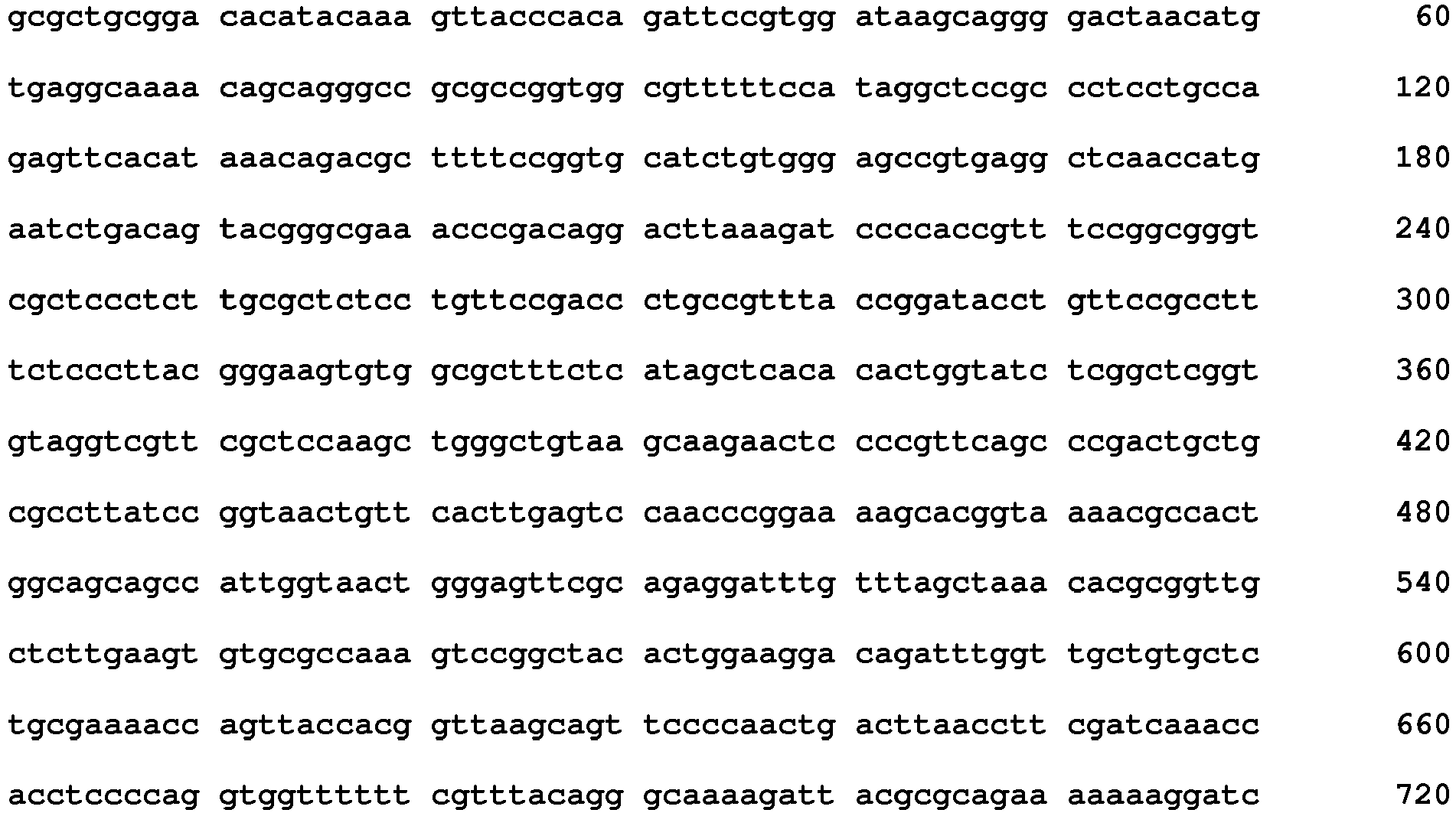

- <210>21
<211>107
<212>PRT
<213>Artificial Sequence - <220>
<223>antibody light chain - <400>21

- <210>22
<211>107
<212>PRT
<213>Artificial Sequence - <220>
<223>antibody light chain - <400>22
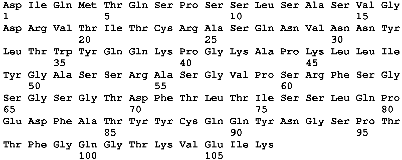
- <210>23
<211>381
<212>DNA
<213>Artificial Sequence - <220>
<223>antibodyheavy chain 9G - <400>23


- <210>24
<211>127
<212>PRT
<213>Artificial Sequence - <220>
<223>antibody heavy chain - <400>24

- <210>25
<211>127
<212>PRT
<213>Artificial Sequence - <220>
<223>antibody heavy chain - <400>25

- <210>26
<211>127
<212>PRT
<213>Artificial Sequence - <220>
<223>antibody heavy chain - <400>26

- <210>27
<211>127
<212>PRT
<213>Artificial Sequence - <220>
<223>VH - <400>27

- <210>28
<211>108
<212>PRT
<213>Artificial Sequence - <220>
<223>VK - <400>28


- <210>29
<211>330
<212>PRT
<213>Homo sapiens - <220>
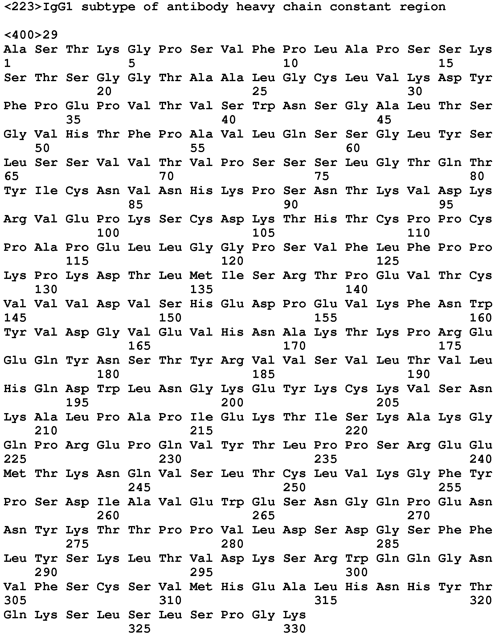
- <210>30
<211>327
<212>PRT
<213>Homo sapiens - <220>
<223>IgG4 subtype of antibody heavy chain constant region - <400>30

- <210>31
<211>107
<212>PRT
<213>Homo sapiens - <220>
<223>kappa subtype light chain constant region - <400>31

- <210>32
<211>105
<212>PRT
<213>Homo sapiens - <220>
<223>lambda subtype light chain constant region - <400>32

- <210>33
<211>119
<212>PRT
<213>Artificial Sequence - <220>
<223>VH - <400>33


- <210>34
<211>112
<212>PRT
<213>Artificial Sequence - <220>
<223>VK - <400>34

Claims (10)
- A monoclonal antibody that specifically binds to human IL-17A, comprising a heavy chain variable region comprising HCDR1, HCDR2 and HCDR3, and a light chain variable region comprising LCDR1, LCDR2 and LCDR3, wherein
HCDR1 has the sequence GFTIDNY, HCDR2 has the sequence NQDGNE, HCDR3 has the sequence DYYDVISDYYIHYWYFDL, or
HCDR1 has the sequence GMSMSDY, HCDR2 has the sequence NQDGDE, HCDR3 has the sequence DYYDLISDYYIHYWYFDL, or
HCDR1 has the sequence GITMDDY, HCDR2 has the sequence NQDGNE, HCDR3 has the sequence DYYDLISDYYIHYWYFDL; and
LCDR1 has the sequence RASQNVHNRLT, LCDR2 has the sequence GASNLES, and LCDR3 has the sequence QQYNGSPTT; and
wherein HCDR1, HCDR2, HCDR3, LCDR1, LCDR2 and LCDR3 are defined according to Chothia. - The monoclonal antibody according to claim 1, wherein
the heavy chain variable region of the antibody has a sequence as set forth in SEQ ID NO: 24, and the light chain variable region has a sequence as set forth in SEQ ID NO:21; or
the heavy chain variable region has a sequence as set forth in SEQ ID NO: 25, and the light chain variable region has a sequence as set forth in SEQ ID NO:21; or
the heavy chain variable region has a sequence as set forth in SEQ ID NO: 26, and the light chain variable region has a sequence as set forth in SEQ ID NO:21. - The monoclonal antibody according to any one of claims 1 to 2, wherein the antibody is an intact antibody, a substantively intact antibody, a Fab fragment, a F(ab')2 fragment or a single chain Fv fragment.
- The monoclonal antibody according to claim 3, wherein the antibody is a fully human antibody.
- The monoclonal antibody according to any one of claims 1 to 4, wherein the antibody further comprises a heavy chain constant region selected from the group consisting of IgG1 and IgG4 subtypes, and/or a light chain constant region selected from the group consisting of kappa and lambda subtypes.
- The monoclonal antibody according to any one of claims 1 to 5, wherein the monoclonal antibody is capable of inhibiting the activity of 2nM human IL-17A by 50% at a concentration less than 1nM, wherein the activity inhibition is measured by determining human IL-17A-induced IL-6 production in human dermal fibroblasts (HDFa).
- A pharmaceutical composition, comprising a monoclonal antibody according to any one of claims 1 to 6.
- A monoclonal antibody according to any one of claims 1 to 6 for use in treating a human IL-17A-mediated disease.
- The monoclonal antibody for use of claim 8, wherein the disease is an autoimmune disease.
- The monoclonal antibody for use of claim 8, wherein the disease is psoriasis, rheumatoid arthritis or ankylosing spondylitis.
Applications Claiming Priority (2)
| Application Number | Priority Date | Filing Date | Title |
|---|---|---|---|
| CN201510097117.0A CN105315371B (en) | 2015-03-05 | 2015-03-05 | Anti-human IL-17 monoclonal antibody |
| PCT/CN2016/074805 WO2016138842A1 (en) | 2015-03-05 | 2016-02-29 | Anti-human il-17 monoclonal antibody and use thereof |
Publications (3)
| Publication Number | Publication Date |
|---|---|
| EP3181587A1 EP3181587A1 (en) | 2017-06-21 |
| EP3181587A4 EP3181587A4 (en) | 2017-12-06 |
| EP3181587B1 true EP3181587B1 (en) | 2019-08-28 |
Family
ID=55243758
Family Applications (1)
| Application Number | Title | Priority Date | Filing Date |
|---|---|---|---|
| EP16758458.0A Active EP3181587B1 (en) | 2015-03-05 | 2016-02-29 | Anti-human il-17 monoclonal antibody and use thereof |
Country Status (6)
| Country | Link |
|---|---|
| US (1) | US10392436B2 (en) |
| EP (1) | EP3181587B1 (en) |
| CN (2) | CN105315371B (en) |
| RU (1) | RU2663721C1 (en) |
| WO (1) | WO2016138842A1 (en) |
| ZA (1) | ZA201701920B (en) |
Families Citing this family (22)
| Publication number | Priority date | Publication date | Assignee | Title |
|---|---|---|---|---|
| CN105315371B (en) * | 2015-03-05 | 2018-05-29 | 北京百特美博生物科技有限公司 | Anti-human IL-17 monoclonal antibody |
| CN107236041B (en) * | 2016-03-29 | 2020-07-24 | 舒泰神(北京)生物制药股份有限公司 | IL-17 antibody |
| WO2018050028A1 (en) * | 2016-09-14 | 2018-03-22 | 北京韩美药品有限公司 | Antibody specifically binding to il-17a and functional fragment thereof |
| CN107698680B (en) | 2017-01-22 | 2019-03-01 | 北京东方百泰生物科技有限公司 | Anti- PCSK9 monoclonal antibody |
| CN109796534B (en) * | 2017-11-16 | 2021-08-10 | 北京比洋生物技术有限公司 | anti-IL-17 antibody/TNFR ECD fusion protein and application thereof |
| CN110551215A (en) * | 2018-05-30 | 2019-12-10 | 中山康方生物医药有限公司 | Anti-interleukin-17A antibody, pharmaceutical composition and use thereof |
| CN109679920B (en) * | 2018-12-26 | 2019-11-22 | 北京贝来生物科技有限公司 | A kind of mescenchymal stem cell for expressing IL-17A signal path blocking agent |
| EP4591879A3 (en) * | 2019-01-31 | 2025-10-22 | Numab Therapeutics AG | Multispecific antibodies having specificity for tnfa and il-17a, antibodies targeting il-17a, and methods of use thereof |
| EP3689907A1 (en) * | 2019-01-31 | 2020-08-05 | Numab Therapeutics AG | Antibodies targeting il-17a and methods of use thereof |
| CN110229232B (en) | 2019-06-19 | 2020-05-19 | 北京智仁美博生物科技有限公司 | Bispecific antibodies and their uses |
| CN111171155B (en) | 2020-02-05 | 2021-02-19 | 北京智仁美博生物科技有限公司 | Anti-CD3 and CD123 bispecific antibodies and uses thereof |
| WO2021227937A1 (en) * | 2020-05-11 | 2021-11-18 | 神州细胞工程有限公司 | Method for enhancing immunogenicity of protein/peptide antigen by means of forming fusion protein with modified fc fragment |
| CN112142841B (en) * | 2020-09-27 | 2022-03-22 | 江苏荃信生物医药股份有限公司 | Novel anti-human IL-17A monoclonal antibody, kit containing same and detection method thereof |
| CN112300285B (en) * | 2020-11-06 | 2022-08-02 | 江苏荃信生物医药股份有限公司 | An enzyme-linked immunosorbent assay method for quantitatively detecting the content of anti-human interleukin-17 monoclonal antibody in serum |
| WO2022144023A1 (en) * | 2021-01-04 | 2022-07-07 | 江苏恒瑞医药股份有限公司 | Method for treating autoimmune diseases and inflammation by using anti-il-17 antibody |
| CN113896793B (en) * | 2021-09-30 | 2023-05-26 | 港科鹏禾生物(苏州)有限公司 | Monoclonal antibody for resisting human IL-17RC and application thereof |
| CN114920838B (en) * | 2022-03-25 | 2023-11-24 | 南京融捷康生物科技有限公司 | anti-IL-17A single domain antibody and application thereof |
| CN115057938B (en) * | 2022-06-24 | 2023-01-06 | 广东菲鹏制药股份有限公司 | Novel coronavirus resistant humanized multivalent binding protein and application thereof |
| CN117430696A (en) * | 2022-07-22 | 2024-01-23 | 北京智仁美博生物科技有限公司 | Recombinant systems for expressing Fab fragment libraries |
| CN119264248A (en) | 2022-07-22 | 2025-01-07 | 北京智仁美博生物科技有限公司 | Anti-respiratory syncytial virus neutralizing antibodies and uses thereof |
| CN118330232A (en) * | 2024-04-17 | 2024-07-12 | 中日友好医院(中日友好临床医学研究所) | A reagent for detecting free drugs of secukinumab |
| WO2026017005A1 (en) * | 2024-07-15 | 2026-01-22 | 重庆智翔金泰生物制药股份有限公司 | Aqueous pharmaceutical product for treating axial spondyloarthritis and use |
Family Cites Families (16)
| Publication number | Priority date | Publication date | Assignee | Title |
|---|---|---|---|---|
| ATE435239T1 (en) * | 2002-03-29 | 2009-07-15 | Schering Corp | HUMAN MONOCLONAL ANTIBODIES TO INTERLEUKIN-5 AND THESE COMPREHENSIVE METHODS AND COMPOSITIONS |
| WO2005090407A1 (en) | 2004-03-19 | 2005-09-29 | Imclone Systems Incorporated | Human anti-epidermal growth factor receptor antibody |
| GB0417487D0 (en) * | 2004-08-05 | 2004-09-08 | Novartis Ag | Organic compound |
| CN101374864A (en) * | 2006-01-31 | 2009-02-25 | 诺瓦提斯公司 | IL-17 antagonistic antibodies for targeting cancer |
| TW200815469A (en) * | 2006-06-23 | 2008-04-01 | Astrazeneca Ab | Compounds |
| GB0612928D0 (en) * | 2006-06-29 | 2006-08-09 | Ucb Sa | Biological products |
| CN101646690B (en) * | 2006-08-11 | 2013-10-23 | 默沙东公司 | anti-IL-17A antibody |
| GB0620729D0 (en) * | 2006-10-18 | 2006-11-29 | Ucb Sa | Biological products |
| KR100958308B1 (en) * | 2007-10-15 | 2010-05-19 | 주식회사 아이지세라피 | Antibody fragment expression vector and method for producing recombinant phage to display antibody using the vector |
| BRPI0906490A2 (en) * | 2008-01-15 | 2015-07-14 | Abbott Lab | Improved Mammal Expression Vectors and Usage |
| KR101318549B1 (en) * | 2008-09-29 | 2013-10-16 | 로슈 글리카트 아게 | Antibodies against human il17 and uses thereof |
| JP5922025B2 (en) | 2009-10-30 | 2016-05-25 | ヤンセン バイオテツク,インコーポレーテツド | IL-17A antagonist |
| ES2633145T3 (en) | 2010-08-05 | 2017-09-19 | Anaptysbio, Inc. | Antibodies directed against IL-17 |
| EP4223312A1 (en) * | 2013-08-15 | 2023-08-09 | Novartis AG | Methods of treating plaque psoriasis using il-17 antagonists |
| CN103936854B (en) | 2014-04-30 | 2016-08-17 | 北京精益泰翔技术发展有限公司 | Anti-IL-17A monoclonal antibody and preparation and application thereof |
| CN105315371B (en) | 2015-03-05 | 2018-05-29 | 北京百特美博生物科技有限公司 | Anti-human IL-17 monoclonal antibody |
-
2015
- 2015-03-05 CN CN201510097117.0A patent/CN105315371B/en active Active
- 2015-03-05 CN CN201810041670.6A patent/CN108251431B/en active Active
-
2016
- 2016-02-29 RU RU2017123752A patent/RU2663721C1/en active
- 2016-02-29 US US15/510,655 patent/US10392436B2/en active Active
- 2016-02-29 WO PCT/CN2016/074805 patent/WO2016138842A1/en not_active Ceased
- 2016-02-29 EP EP16758458.0A patent/EP3181587B1/en active Active
-
2017
- 2017-03-17 ZA ZA2017/01920A patent/ZA201701920B/en unknown
Non-Patent Citations (1)
| Title |
|---|
| None * |
Also Published As
| Publication number | Publication date |
|---|---|
| US10392436B2 (en) | 2019-08-27 |
| EP3181587A1 (en) | 2017-06-21 |
| RU2663721C1 (en) | 2018-08-08 |
| CN108251431B (en) | 2020-12-08 |
| EP3181587A4 (en) | 2017-12-06 |
| US20170327571A1 (en) | 2017-11-16 |
| CN108251431A (en) | 2018-07-06 |
| CN105315371B (en) | 2018-05-29 |
| ZA201701920B (en) | 2018-07-25 |
| WO2016138842A1 (en) | 2016-09-09 |
| CN105315371A (en) | 2016-02-10 |
Similar Documents
| Publication | Publication Date | Title |
|---|---|---|
| US10392436B2 (en) | Anti-human IL-17 monoclonal antibodies and use thereof | |
| US20230257458A1 (en) | Antibody of il-11 and use thereof | |
| WO2018023976A1 (en) | Human ifnar1 antibody and uses thereof | |
| US10981983B2 (en) | Antibody targeting interleukin 17A and preparation method and application thereof | |
| JP2017533705A (en) | Anti-TNF- / anti-IL-23 bispecific antibody | |
| CN116133684B (en) | Anti-human NR1 antibody derivatives | |
| JP5971238B2 (en) | Novel anti-human IL-23 receptor antibody | |
| TWI843763B (en) | Anti-human fn14 antibody | |
| JP2021533770A (en) | Anti-IL-1β antibody and pharmaceutical compositions thereof and their use | |
| WO2025016063A1 (en) | Antibody capable of binding to interleukin-31 or antigen binding fragment thereof, and preparation method therefor and use thereof | |
| JP6040943B2 (en) | Novel anti-human CTGF antibody | |
| CN111196849B (en) | Anti-sclerostin antibodies, antigen-binding fragments thereof, and medical uses thereof | |
| EP2821416B1 (en) | Novel anti-human il-23 receptor antibody | |
| CN115109154A (en) | Antibody targeting CLDN18.2 or antigen binding fragment thereof and application thereof | |
| EP4155322A1 (en) | Anti-trka antibody or antigen-binding fragment thereof, preparation method therefor, and application thereof | |
| JP2022513224A (en) | Anti-IL-17A antibody and its application | |
| HK40082063A (en) | Anti-trka antibody or antigen-binding fragment thereof, preparation method therefor, and application thereof | |
| KR20240022546A (en) | Anti-IL-36R antibody and use thereof | |
| CN121609799A (en) | Anti-PD-L1 nano antibody, bispecific recombinant protein containing same and application thereof |
Legal Events
| Date | Code | Title | Description |
|---|---|---|---|
| STAA | Information on the status of an ep patent application or granted ep patent |
Free format text: STATUS: THE INTERNATIONAL PUBLICATION HAS BEEN MADE |
|
| PUAI | Public reference made under article 153(3) epc to a published international application that has entered the european phase |
Free format text: ORIGINAL CODE: 0009012 |
|
| STAA | Information on the status of an ep patent application or granted ep patent |
Free format text: STATUS: REQUEST FOR EXAMINATION WAS MADE |
|
| 17P | Request for examination filed |
Effective date: 20170316 |
|
| AK | Designated contracting states |
Kind code of ref document: A1 Designated state(s): AL AT BE BG CH CY CZ DE DK EE ES FI FR GB GR HR HU IE IS IT LI LT LU LV MC MK MT NL NO PL PT RO RS SE SI SK SM TR |
|
| AX | Request for extension of the european patent |
Extension state: BA ME |
|
| A4 | Supplementary search report drawn up and despatched |
Effective date: 20171107 |
|
| RIC1 | Information provided on ipc code assigned before grant |
Ipc: C07K 16/24 20060101AFI20171030BHEP Ipc: A61P 37/02 20060101ALI20171030BHEP Ipc: A61K 39/395 20060101ALI20171030BHEP |
|
| DAV | Request for validation of the european patent (deleted) | ||
| DAX | Request for extension of the european patent (deleted) | ||
| STAA | Information on the status of an ep patent application or granted ep patent |
Free format text: STATUS: EXAMINATION IS IN PROGRESS |
|
| 17Q | First examination report despatched |
Effective date: 20180829 |
|
| GRAP | Despatch of communication of intention to grant a patent |
Free format text: ORIGINAL CODE: EPIDOSNIGR1 |
|
| STAA | Information on the status of an ep patent application or granted ep patent |
Free format text: STATUS: GRANT OF PATENT IS INTENDED |
|
| INTG | Intention to grant announced |
Effective date: 20190312 |
|
| GRAS | Grant fee paid |
Free format text: ORIGINAL CODE: EPIDOSNIGR3 |
|
| GRAA | (expected) grant |
Free format text: ORIGINAL CODE: 0009210 |
|
| STAA | Information on the status of an ep patent application or granted ep patent |
Free format text: STATUS: THE PATENT HAS BEEN GRANTED |
|
| RAP1 | Party data changed (applicant data changed or rights of an application transferred) |
Owner name: BEIJING BETTERMAB BIOTECHNOLOGY CO., LTD Owner name: BEIJING WISDOMAB BIOTECHNOLOGY CO., LTD Owner name: GENRIX(SHANGHAI) BIOPHARMACEUTICAL CO., LTD. Owner name: CHONGQING GENRIX BIOPHARMACEUTIAL CO., LTD. |
|
| AK | Designated contracting states |
Kind code of ref document: B1 Designated state(s): AL AT BE BG CH CY CZ DE DK EE ES FI FR GB GR HR HU IE IS IT LI LT LU LV MC MK MT NL NO PL PT RO RS SE SI SK SM TR |
|
| REG | Reference to a national code |
Ref country code: GB Ref legal event code: FG4D |
|
| REG | Reference to a national code |
Ref country code: CH Ref legal event code: EP |
|
| REG | Reference to a national code |
Ref country code: AT Ref legal event code: REF Ref document number: 1172283 Country of ref document: AT Kind code of ref document: T Effective date: 20190915 |
|
| REG | Reference to a national code |
Ref country code: IE Ref legal event code: FG4D |
|
| REG | Reference to a national code |
Ref country code: DE Ref legal event code: R096 Ref document number: 602016019496 Country of ref document: DE |
|
| REG | Reference to a national code |
Ref country code: NL Ref legal event code: MP Effective date: 20190828 |
|
| REG | Reference to a national code |
Ref country code: LT Ref legal event code: MG4D |
|
| PG25 | Lapsed in a contracting state [announced via postgrant information from national office to epo] |
Ref country code: SE Free format text: LAPSE BECAUSE OF FAILURE TO SUBMIT A TRANSLATION OF THE DESCRIPTION OR TO PAY THE FEE WITHIN THE PRESCRIBED TIME-LIMIT Effective date: 20190828 Ref country code: NO Free format text: LAPSE BECAUSE OF FAILURE TO SUBMIT A TRANSLATION OF THE DESCRIPTION OR TO PAY THE FEE WITHIN THE PRESCRIBED TIME-LIMIT Effective date: 20191128 Ref country code: FI Free format text: LAPSE BECAUSE OF FAILURE TO SUBMIT A TRANSLATION OF THE DESCRIPTION OR TO PAY THE FEE WITHIN THE PRESCRIBED TIME-LIMIT Effective date: 20190828 Ref country code: PT Free format text: LAPSE BECAUSE OF FAILURE TO SUBMIT A TRANSLATION OF THE DESCRIPTION OR TO PAY THE FEE WITHIN THE PRESCRIBED TIME-LIMIT Effective date: 20191230 Ref country code: HR Free format text: LAPSE BECAUSE OF FAILURE TO SUBMIT A TRANSLATION OF THE DESCRIPTION OR TO PAY THE FEE WITHIN THE PRESCRIBED TIME-LIMIT Effective date: 20190828 Ref country code: BG Free format text: LAPSE BECAUSE OF FAILURE TO SUBMIT A TRANSLATION OF THE DESCRIPTION OR TO PAY THE FEE WITHIN THE PRESCRIBED TIME-LIMIT Effective date: 20191128 Ref country code: NL Free format text: LAPSE BECAUSE OF FAILURE TO SUBMIT A TRANSLATION OF THE DESCRIPTION OR TO PAY THE FEE WITHIN THE PRESCRIBED TIME-LIMIT Effective date: 20190828 Ref country code: LT Free format text: LAPSE BECAUSE OF FAILURE TO SUBMIT A TRANSLATION OF THE DESCRIPTION OR TO PAY THE FEE WITHIN THE PRESCRIBED TIME-LIMIT Effective date: 20190828 |
|
| PG25 | Lapsed in a contracting state [announced via postgrant information from national office to epo] |
Ref country code: ES Free format text: LAPSE BECAUSE OF FAILURE TO SUBMIT A TRANSLATION OF THE DESCRIPTION OR TO PAY THE FEE WITHIN THE PRESCRIBED TIME-LIMIT Effective date: 20190828 Ref country code: LV Free format text: LAPSE BECAUSE OF FAILURE TO SUBMIT A TRANSLATION OF THE DESCRIPTION OR TO PAY THE FEE WITHIN THE PRESCRIBED TIME-LIMIT Effective date: 20190828 Ref country code: GR Free format text: LAPSE BECAUSE OF FAILURE TO SUBMIT A TRANSLATION OF THE DESCRIPTION OR TO PAY THE FEE WITHIN THE PRESCRIBED TIME-LIMIT Effective date: 20191129 Ref country code: AL Free format text: LAPSE BECAUSE OF FAILURE TO SUBMIT A TRANSLATION OF THE DESCRIPTION OR TO PAY THE FEE WITHIN THE PRESCRIBED TIME-LIMIT Effective date: 20190828 Ref country code: IS Free format text: LAPSE BECAUSE OF FAILURE TO SUBMIT A TRANSLATION OF THE DESCRIPTION OR TO PAY THE FEE WITHIN THE PRESCRIBED TIME-LIMIT Effective date: 20191228 Ref country code: RS Free format text: LAPSE BECAUSE OF FAILURE TO SUBMIT A TRANSLATION OF THE DESCRIPTION OR TO PAY THE FEE WITHIN THE PRESCRIBED TIME-LIMIT Effective date: 20190828 |
|
| REG | Reference to a national code |
Ref country code: AT Ref legal event code: MK05 Ref document number: 1172283 Country of ref document: AT Kind code of ref document: T Effective date: 20190828 |
|
| PG25 | Lapsed in a contracting state [announced via postgrant information from national office to epo] |
Ref country code: TR Free format text: LAPSE BECAUSE OF FAILURE TO SUBMIT A TRANSLATION OF THE DESCRIPTION OR TO PAY THE FEE WITHIN THE PRESCRIBED TIME-LIMIT Effective date: 20190828 |
|
| PG25 | Lapsed in a contracting state [announced via postgrant information from national office to epo] |
Ref country code: RO Free format text: LAPSE BECAUSE OF FAILURE TO SUBMIT A TRANSLATION OF THE DESCRIPTION OR TO PAY THE FEE WITHIN THE PRESCRIBED TIME-LIMIT Effective date: 20190828 Ref country code: PL Free format text: LAPSE BECAUSE OF FAILURE TO SUBMIT A TRANSLATION OF THE DESCRIPTION OR TO PAY THE FEE WITHIN THE PRESCRIBED TIME-LIMIT Effective date: 20190828 Ref country code: DK Free format text: LAPSE BECAUSE OF FAILURE TO SUBMIT A TRANSLATION OF THE DESCRIPTION OR TO PAY THE FEE WITHIN THE PRESCRIBED TIME-LIMIT Effective date: 20190828 Ref country code: IT Free format text: LAPSE BECAUSE OF FAILURE TO SUBMIT A TRANSLATION OF THE DESCRIPTION OR TO PAY THE FEE WITHIN THE PRESCRIBED TIME-LIMIT Effective date: 20190828 Ref country code: AT Free format text: LAPSE BECAUSE OF FAILURE TO SUBMIT A TRANSLATION OF THE DESCRIPTION OR TO PAY THE FEE WITHIN THE PRESCRIBED TIME-LIMIT Effective date: 20190828 Ref country code: EE Free format text: LAPSE BECAUSE OF FAILURE TO SUBMIT A TRANSLATION OF THE DESCRIPTION OR TO PAY THE FEE WITHIN THE PRESCRIBED TIME-LIMIT Effective date: 20190828 |
|
| PG25 | Lapsed in a contracting state [announced via postgrant information from national office to epo] |
Ref country code: CZ Free format text: LAPSE BECAUSE OF FAILURE TO SUBMIT A TRANSLATION OF THE DESCRIPTION OR TO PAY THE FEE WITHIN THE PRESCRIBED TIME-LIMIT Effective date: 20190828 Ref country code: SM Free format text: LAPSE BECAUSE OF FAILURE TO SUBMIT A TRANSLATION OF THE DESCRIPTION OR TO PAY THE FEE WITHIN THE PRESCRIBED TIME-LIMIT Effective date: 20190828 Ref country code: IS Free format text: LAPSE BECAUSE OF FAILURE TO SUBMIT A TRANSLATION OF THE DESCRIPTION OR TO PAY THE FEE WITHIN THE PRESCRIBED TIME-LIMIT Effective date: 20200224 Ref country code: SK Free format text: LAPSE BECAUSE OF FAILURE TO SUBMIT A TRANSLATION OF THE DESCRIPTION OR TO PAY THE FEE WITHIN THE PRESCRIBED TIME-LIMIT Effective date: 20190828 |
|
| REG | Reference to a national code |
Ref country code: DE Ref legal event code: R097 Ref document number: 602016019496 Country of ref document: DE |
|
| PLBE | No opposition filed within time limit |
Free format text: ORIGINAL CODE: 0009261 |
|
| STAA | Information on the status of an ep patent application or granted ep patent |
Free format text: STATUS: NO OPPOSITION FILED WITHIN TIME LIMIT |
|
| PG2D | Information on lapse in contracting state deleted |
Ref country code: IS |
|
| 26N | No opposition filed |
Effective date: 20200603 |
|
| PG25 | Lapsed in a contracting state [announced via postgrant information from national office to epo] |
Ref country code: SI Free format text: LAPSE BECAUSE OF FAILURE TO SUBMIT A TRANSLATION OF THE DESCRIPTION OR TO PAY THE FEE WITHIN THE PRESCRIBED TIME-LIMIT Effective date: 20190828 |
|
| REG | Reference to a national code |
Ref country code: CH Ref legal event code: PL |
|
| REG | Reference to a national code |
Ref country code: BE Ref legal event code: MM Effective date: 20200229 |
|
| PG25 | Lapsed in a contracting state [announced via postgrant information from national office to epo] |
Ref country code: MC Free format text: LAPSE BECAUSE OF FAILURE TO SUBMIT A TRANSLATION OF THE DESCRIPTION OR TO PAY THE FEE WITHIN THE PRESCRIBED TIME-LIMIT Effective date: 20190828 Ref country code: LU Free format text: LAPSE BECAUSE OF NON-PAYMENT OF DUE FEES Effective date: 20200229 |
|
| PG25 | Lapsed in a contracting state [announced via postgrant information from national office to epo] |
Ref country code: CH Free format text: LAPSE BECAUSE OF NON-PAYMENT OF DUE FEES Effective date: 20200229 Ref country code: LI Free format text: LAPSE BECAUSE OF NON-PAYMENT OF DUE FEES Effective date: 20200229 |
|
| PG25 | Lapsed in a contracting state [announced via postgrant information from national office to epo] |
Ref country code: IE Free format text: LAPSE BECAUSE OF NON-PAYMENT OF DUE FEES Effective date: 20200229 |
|
| REG | Reference to a national code |
Ref country code: DE Ref legal event code: R082 Ref document number: 602016019496 Country of ref document: DE Representative=s name: BOEHMERT & BOEHMERT ANWALTSPARTNERSCHAFT MBB -, DE Ref country code: DE Ref legal event code: R081 Ref document number: 602016019496 Country of ref document: DE Owner name: GENRIX(SHANGHAI) BIOPHARMACEUTICAL CO., LTD., CN Free format text: FORMER OWNERS: BEIJING BETTERMAB BIOTECHNOLOGY CO., LTD, BEIJING, CN; BEIJING WISDOMAB BIOTECHNOLOGY CO., LTD, BEIJING, CN; CHONGQING GENRIX BIOPHARMACEUTIAL CO., LTD., CHONGQING, CN; GENRIX(SHANGHAI) BIOPHARMACEUTICAL CO., LTD., SHANGHAI, CN Ref country code: DE Ref legal event code: R081 Ref document number: 602016019496 Country of ref document: DE Owner name: CHONGQING GENRIX PHARMACEUTICAL CO., LTD., CN Free format text: FORMER OWNERS: BEIJING BETTERMAB BIOTECHNOLOGY CO., LTD, BEIJING, CN; BEIJING WISDOMAB BIOTECHNOLOGY CO., LTD, BEIJING, CN; CHONGQING GENRIX BIOPHARMACEUTIAL CO., LTD., CHONGQING, CN; GENRIX(SHANGHAI) BIOPHARMACEUTICAL CO., LTD., SHANGHAI, CN Ref country code: DE Ref legal event code: R081 Ref document number: 602016019496 Country of ref document: DE Owner name: BEIJING WISDOMAB BIOTECHNOLOGY CO., LTD, CN Free format text: FORMER OWNERS: BEIJING BETTERMAB BIOTECHNOLOGY CO., LTD, BEIJING, CN; BEIJING WISDOMAB BIOTECHNOLOGY CO., LTD, BEIJING, CN; CHONGQING GENRIX BIOPHARMACEUTIAL CO., LTD., CHONGQING, CN; GENRIX(SHANGHAI) BIOPHARMACEUTICAL CO., LTD., SHANGHAI, CN |
|
| PG25 | Lapsed in a contracting state [announced via postgrant information from national office to epo] |
Ref country code: BE Free format text: LAPSE BECAUSE OF NON-PAYMENT OF DUE FEES Effective date: 20200229 |
|
| REG | Reference to a national code |
Ref country code: GB Ref legal event code: 732E Free format text: REGISTERED BETWEEN 20210415 AND 20210421 |
|
| PG25 | Lapsed in a contracting state [announced via postgrant information from national office to epo] |
Ref country code: MT Free format text: LAPSE BECAUSE OF FAILURE TO SUBMIT A TRANSLATION OF THE DESCRIPTION OR TO PAY THE FEE WITHIN THE PRESCRIBED TIME-LIMIT Effective date: 20190828 Ref country code: CY Free format text: LAPSE BECAUSE OF FAILURE TO SUBMIT A TRANSLATION OF THE DESCRIPTION OR TO PAY THE FEE WITHIN THE PRESCRIBED TIME-LIMIT Effective date: 20190828 |
|
| PG25 | Lapsed in a contracting state [announced via postgrant information from national office to epo] |
Ref country code: MK Free format text: LAPSE BECAUSE OF FAILURE TO SUBMIT A TRANSLATION OF THE DESCRIPTION OR TO PAY THE FEE WITHIN THE PRESCRIBED TIME-LIMIT Effective date: 20190828 |
|
| P01 | Opt-out of the competence of the unified patent court (upc) registered |
Effective date: 20230524 |
|
| PGFP | Annual fee paid to national office [announced via postgrant information from national office to epo] |
Ref country code: GB Payment date: 20260219 Year of fee payment: 11 |
|
| PGFP | Annual fee paid to national office [announced via postgrant information from national office to epo] |
Ref country code: DE Payment date: 20260218 Year of fee payment: 11 |
|
| PGFP | Annual fee paid to national office [announced via postgrant information from national office to epo] |
Ref country code: FR Payment date: 20260218 Year of fee payment: 11 |