EP3180019B1 - Neural stem cells and a monomer or homodimer of il12p40 to enhance nerve regeneration - Google Patents
Neural stem cells and a monomer or homodimer of il12p40 to enhance nerve regeneration Download PDFInfo
- Publication number
- EP3180019B1 EP3180019B1 EP15831363.5A EP15831363A EP3180019B1 EP 3180019 B1 EP3180019 B1 EP 3180019B1 EP 15831363 A EP15831363 A EP 15831363A EP 3180019 B1 EP3180019 B1 EP 3180019B1
- Authority
- EP
- European Patent Office
- Prior art keywords
- nerve
- stem cells
- conduit
- cells
- composition
- Prior art date
- Legal status (The legal status is an assumption and is not a legal conclusion. Google has not performed a legal analysis and makes no representation as to the accuracy of the status listed.)
- Active
Links
Images





Classifications
-
- A—HUMAN NECESSITIES
- A61—MEDICAL OR VETERINARY SCIENCE; HYGIENE
- A61K—PREPARATIONS FOR MEDICAL, DENTAL OR TOILETRY PURPOSES
- A61K35/00—Medicinal preparations containing materials or reaction products thereof with undetermined constitution
- A61K35/12—Materials from mammals; Compositions comprising non-specified tissues or cells; Compositions comprising non-embryonic stem cells; Genetically modified cells
- A61K35/30—Nerves; Brain; Eyes; Corneal cells; Cerebrospinal fluid; Neuronal stem cells; Neuronal precursor cells; Glial cells; Oligodendrocytes; Schwann cells; Astroglia; Astrocytes; Choroid plexus; Spinal cord tissue
-
- A—HUMAN NECESSITIES
- A61—MEDICAL OR VETERINARY SCIENCE; HYGIENE
- A61K—PREPARATIONS FOR MEDICAL, DENTAL OR TOILETRY PURPOSES
- A61K35/00—Medicinal preparations containing materials or reaction products thereof with undetermined constitution
- A61K35/12—Materials from mammals; Compositions comprising non-specified tissues or cells; Compositions comprising non-embryonic stem cells; Genetically modified cells
- A61K35/28—Bone marrow; Haematopoietic stem cells; Mesenchymal stem cells of any origin, e.g. adipose-derived stem cells
-
- A—HUMAN NECESSITIES
- A61—MEDICAL OR VETERINARY SCIENCE; HYGIENE
- A61K—PREPARATIONS FOR MEDICAL, DENTAL OR TOILETRY PURPOSES
- A61K35/00—Medicinal preparations containing materials or reaction products thereof with undetermined constitution
- A61K35/12—Materials from mammals; Compositions comprising non-specified tissues or cells; Compositions comprising non-embryonic stem cells; Genetically modified cells
- A61K35/48—Reproductive organs
- A61K35/50—Placenta; Placental stem cells; Amniotic fluid; Amnion; Amniotic stem cells
-
- A—HUMAN NECESSITIES
- A61—MEDICAL OR VETERINARY SCIENCE; HYGIENE
- A61K—PREPARATIONS FOR MEDICAL, DENTAL OR TOILETRY PURPOSES
- A61K38/00—Medicinal preparations containing peptides
- A61K38/16—Peptides having more than 20 amino acids; Gastrins; Somatostatins; Melanotropins; Derivatives thereof
- A61K38/17—Peptides having more than 20 amino acids; Gastrins; Somatostatins; Melanotropins; Derivatives thereof from animals; from humans
- A61K38/18—Growth factors; Growth regulators
- A61K38/1825—Fibroblast growth factor [FGF]
-
- A—HUMAN NECESSITIES
- A61—MEDICAL OR VETERINARY SCIENCE; HYGIENE
- A61K—PREPARATIONS FOR MEDICAL, DENTAL OR TOILETRY PURPOSES
- A61K38/00—Medicinal preparations containing peptides
- A61K38/16—Peptides having more than 20 amino acids; Gastrins; Somatostatins; Melanotropins; Derivatives thereof
- A61K38/17—Peptides having more than 20 amino acids; Gastrins; Somatostatins; Melanotropins; Derivatives thereof from animals; from humans
- A61K38/18—Growth factors; Growth regulators
- A61K38/185—Nerve growth factor [NGF]; Brain derived neurotrophic factor [BDNF]; Ciliary neurotrophic factor [CNTF]; Glial derived neurotrophic factor [GDNF]; Neurotrophins, e.g. NT-3
-
- A—HUMAN NECESSITIES
- A61—MEDICAL OR VETERINARY SCIENCE; HYGIENE
- A61K—PREPARATIONS FOR MEDICAL, DENTAL OR TOILETRY PURPOSES
- A61K38/00—Medicinal preparations containing peptides
- A61K38/16—Peptides having more than 20 amino acids; Gastrins; Somatostatins; Melanotropins; Derivatives thereof
- A61K38/17—Peptides having more than 20 amino acids; Gastrins; Somatostatins; Melanotropins; Derivatives thereof from animals; from humans
- A61K38/18—Growth factors; Growth regulators
- A61K38/1858—Platelet-derived growth factor [PDGF]
-
- A—HUMAN NECESSITIES
- A61—MEDICAL OR VETERINARY SCIENCE; HYGIENE
- A61K—PREPARATIONS FOR MEDICAL, DENTAL OR TOILETRY PURPOSES
- A61K38/00—Medicinal preparations containing peptides
- A61K38/16—Peptides having more than 20 amino acids; Gastrins; Somatostatins; Melanotropins; Derivatives thereof
- A61K38/17—Peptides having more than 20 amino acids; Gastrins; Somatostatins; Melanotropins; Derivatives thereof from animals; from humans
- A61K38/19—Cytokines; Lymphokines; Interferons
- A61K38/20—Interleukins [IL]
- A61K38/208—IL-12
-
- A—HUMAN NECESSITIES
- A61—MEDICAL OR VETERINARY SCIENCE; HYGIENE
- A61K—PREPARATIONS FOR MEDICAL, DENTAL OR TOILETRY PURPOSES
- A61K9/00—Medicinal preparations characterised by special physical form
- A61K9/0012—Galenical forms characterised by the site of application
- A61K9/0019—Injectable compositions; Intramuscular, intravenous, arterial, subcutaneous administration; Compositions to be administered through the skin in an invasive manner
-
- A—HUMAN NECESSITIES
- A61—MEDICAL OR VETERINARY SCIENCE; HYGIENE
- A61K—PREPARATIONS FOR MEDICAL, DENTAL OR TOILETRY PURPOSES
- A61K9/00—Medicinal preparations characterised by special physical form
- A61K9/0012—Galenical forms characterised by the site of application
- A61K9/0019—Injectable compositions; Intramuscular, intravenous, arterial, subcutaneous administration; Compositions to be administered through the skin in an invasive manner
- A61K9/0024—Solid, semi-solid or solidifying implants, which are implanted or injected in body tissue
-
- A—HUMAN NECESSITIES
- A61—MEDICAL OR VETERINARY SCIENCE; HYGIENE
- A61P—SPECIFIC THERAPEUTIC ACTIVITY OF CHEMICAL COMPOUNDS OR MEDICINAL PREPARATIONS
- A61P25/00—Drugs for disorders of the nervous system
-
- A—HUMAN NECESSITIES
- A61—MEDICAL OR VETERINARY SCIENCE; HYGIENE
- A61P—SPECIFIC THERAPEUTIC ACTIVITY OF CHEMICAL COMPOUNDS OR MEDICINAL PREPARATIONS
- A61P25/00—Drugs for disorders of the nervous system
- A61P25/02—Drugs for disorders of the nervous system for peripheral neuropathies
-
- A—HUMAN NECESSITIES
- A61—MEDICAL OR VETERINARY SCIENCE; HYGIENE
- A61P—SPECIFIC THERAPEUTIC ACTIVITY OF CHEMICAL COMPOUNDS OR MEDICINAL PREPARATIONS
- A61P25/00—Drugs for disorders of the nervous system
- A61P25/14—Drugs for disorders of the nervous system for treating abnormal movements, e.g. chorea, dyskinesia
- A61P25/16—Anti-Parkinson drugs
-
- A—HUMAN NECESSITIES
- A61—MEDICAL OR VETERINARY SCIENCE; HYGIENE
- A61P—SPECIFIC THERAPEUTIC ACTIVITY OF CHEMICAL COMPOUNDS OR MEDICINAL PREPARATIONS
- A61P25/00—Drugs for disorders of the nervous system
- A61P25/28—Drugs for disorders of the nervous system for treating neurodegenerative disorders of the central nervous system, e.g. nootropic agents, cognition enhancers, drugs for treating Alzheimer's disease or other forms of dementia
-
- A—HUMAN NECESSITIES
- A61—MEDICAL OR VETERINARY SCIENCE; HYGIENE
- A61P—SPECIFIC THERAPEUTIC ACTIVITY OF CHEMICAL COMPOUNDS OR MEDICINAL PREPARATIONS
- A61P43/00—Drugs for specific purposes, not provided for in groups A61P1/00-A61P41/00
-
- A—HUMAN NECESSITIES
- A61—MEDICAL OR VETERINARY SCIENCE; HYGIENE
- A61K—PREPARATIONS FOR MEDICAL, DENTAL OR TOILETRY PURPOSES
- A61K2300/00—Mixtures or combinations of active ingredients, wherein at least one active ingredient is fully defined in groups A61K31/00 - A61K41/00
Definitions
- the present application relates to a composition and methods to enhance nerve regeneration utilizing neural stem cells and IL12p40.
- Severed peripheral nerve injury causes a reduction in motor and sensory neuron activities, and degeneration of nerve fibers and the surrounding tissues.
- the regeneration of injured peripheral nerve is a multiplex process, with Wallerian degeneration (WD) being the most elementary reaction and Schwann cells playing an important role ( Ren Z. et al., Reviews in the Neurosciences 2012, 23:135-143 ).
- WD creates a microenvironment for regeneration of surviving neurons and benefits functional recovery ( Navarro X. et al., Progress in Neurobiology 2007, 82:163-201 .).
- the control of WD involves the existence of Schwann cells, the secretion of neurotrophic factors, and special extracellular matrix that acts as a scaffold for neural cells ( Gaudet A.D. et al., Journal of Neuroinflammation 2011, 8:110 ; Kehoe S. et al., Injury 2012, 43:553-572 .).
- Nerve conduits provide mechanical support and direct axonal sprouting between the injured nerve stumps.
- Conduits have been shown to retain neurotrophic factors secreted from or recruited by the damaged cells ( Kehoe S. et al, Injury 2012, 43:553-572 .) and prevent ingrowth of fibrous tissue at the injury site.
- Recent studies reveal that implantation of neural stem cells (NSCs) in conduits promote regeneration of injured peripheral nerves ( Zhang H. et al., Journal of Translational Medicine 2008, 6:67 ; Shi Y. et al., Acta Oto-Laryngologica 2009, 129: 906-914 .).
- NSCs neural stem cells
- the promotion of nerve regeneration may depend on the ability of implanted NSCs to differentiate into Schwann cells, to secrete neurotrophic factors per se, or create a microenvironment to enrich neurotrophic factors from milieu, and to assist in myelination ( Ren Z. et al., Reviews in the Neurosciences 2012, 23:135-143 ). However, the nature of cytokines or growth factors that are involved in this process is not clear. The molecular mechanism for the Schwann cell differentiation of the implanted NSCs into newly regenerated axons is also not well established.
- IL12p80 induced the neuroglia differentiation of mouse NSCs in vitro through phosphorylation of signal transducer and activator of transcription 3 (Stat3).
- the neuroglia comprises astroglia, oligodendrocytes, and Schwann cells as reported by Kettenmann and Verkhratsky ( Kettenmann H, Verkhratsky A, Fort suitse der Neurologie-Psychiatrie 2011, 79:588-597 .).
- EHTESHAM M et al. describe the use of interleukin 12-secreting neural stem cells for the treatment of intracranial glioma ( CANCER RESEARCH, AACR -AMERICAN ASSOCIATION FOR CANCER RESEARCH, Vol. 62, No. 20, 2002, pp. 5657-5663 ).
- IL-12p40 an inherently agonistic cytokine ( TRENDS IN IMMUNO, ELSEVIER LTD., TRENDS JOURNALS, Vol. 28, No. 1, 2006, pp. 33-38 ).
- the present application provides a composition for nerve regeneration comprising neural stem cells and a neurotrophic factor, which is IL12p80 and/or a monomer of IL12p40.
- the present application provides a composition for nerve regeneration comprising neural stem cells, a neurotrophic factor, which is IL12p80 and/or a monomer of IL12p40, and a nerve regeneration enhancing element, as defined in claim 5.
- the present application further provides a composition for nerve regeneration comprising neural stem cells, a neurotrophic factor, which IL12p80 and/or a monomer of IL12p40, and a nerve conduit for carrying at least one of the neural stem cells and the neurotrophic factors.
- the present application provides a nerve regeneration composition comprising neural stem cells and a neurotrophic factor containing IL12p80 and/or a monomer of IL12p40 for use in the treatment of nerve injuries, peripheral nerve injury, spinal cord injury, stroke, neurodegenerative diseases, Alzheimer's disease, Parkinson's disease or multiple sclerosis.
- a composition for nerve regeneration comprises NSCs and a neurotrophic factor, which is IL12p80 and/or a monomer of IL12p40.
- IL12p40 has a protein sequence shown as NCBI Accession No.: NM_008352 (Mouse) or 1F42_A (Human).
- the neurotrophic factor can be a monomer of IL12p40, or a homodimer of IL12p40.
- the homodimer is IL12p80.
- the NSCs of the composition for nerve regeneration can be characterized as brain cells that are positive for CD133 and GFAP, or characterized as F1B-GFP + cells.
- the present application provides a composition further comprising a nerve regeneration enhancing element selected from Schwann cells, mesenchymal stem cells (MSCs), adipose-derived stem cells (ADSCs), amniotic fluid stem cells (AFSCs), induced neurons (iNs), induced neural stem cells (iNSCs), or cell that are derived from induced pluripotent stem cells (iPSCs), fibroblast growth factor 1 (FGF1), fibroblast growth factor 2 (FGF2), platelet derived growth factor (PDGF), brain derived neurotrophic factor (BDNF), glial cell derived neurotrophic factor (GDNF), nerve growth factor (NGF).
- a nerve regeneration enhancing element selected from Schwann cells, mesenchymal stem cells (MSCs), adipose-derived stem cells (ADSCs), amniotic fluid stem cells (AFSCs), induced neurons (iNs), induced neural stem cells (iNSCs), or cell that are derived from induced pluripotent
- the composition can further comprise a nerve conduit for carrying at least one of the NSCs and the neurotrophic factors.
- the nerve conduit further carries the nerve regeneration enhancing elements.
- a method for regenerating nerve comprising providing a nerve regeneration composition comprising neural stem cells and a neurotrophic factor which is IL12p80 and/or a monomer of IL12p40, to a subject.
- the nerve regeneration composition can further comprise NSCs.
- the nerve conduit is implanted in a target nerve or a tissue around the target nerve.
- the neurotrophic factor can be a monomer of IL12p40, or a homodimer of IL12p40, a trimer of IL12p40, a tetramer of IL12p40 or any combination thereof. In some embodiments, the neurotrophic factor is IL12p80.
- the subject is a mammalian.
- the mammalian can be mice or humans.
- the composition can be provided by injection, implantation, transdermal route, and/or oral administration.
- IL12p40 The protein sequence of IL12p40 is shown as NCBI Accession No.: NM_008352 (Mouse) or 1F42_A (Human), which are available from the website of National Center for Biotechnology Information (http://www.ncbi.nlm.nih.gov/).
- KT98/F1B-GFP cells were cultured in Dulbecco's modified Eagle's medium (DMEM)/F12 (1:1) containing 10% fetal bovine serum (FBS), 1% penicillin/streptomycin, and 500 ⁇ g/ml G418 (Merck, USA).
- DMEM Dulbecco's modified Eagle's medium
- FBS fetal bovine serum
- penicillin/streptomycin 500 ⁇ g/ml G418 (Merck, USA).
- KT98/F1B-GFP+ The GFP-positive KT98/F1B-GFP (KT98/F1B-GFP+) cells were sorted using FACSAria cell sorter (BD Bioscience) and cultured in neurosphere formation medium (DMEM/F12 containing IX B27 (Gibco), 20 ng/ml EGF (PeproTech Inc.), 20 ng/ml FGF2 (PeproTech Inc.), 2 ⁇ g/ml heparin (Sigma), and 500 ⁇ g/ml G418) for 7 days, which induced KT98/F1B-GFP+ neurosphere formation. Then KT98/F1B-GFP+ neurosphere-derived single cells were used in subsequent animal experiments and cell differentiation assays. All cells were cultured at 37 °C with 5% CO 2 .
- PHA poly(L-lactic acid)
- Conduit only group conduits were filled with 5 ⁇ l matrigel (BD Bioscience)/phosphate buffer saline (PBS) mixture (1:1).
- conduits were filled with 5 ⁇ l matrigel/PBS mixture (1:1) with 1 ⁇ 106 NSCs and 100 ng mouse IL12p80 (BioLegend).
- SFI sciatic functional index
- the Rotarod test was executed by RT series Rotarod Treadmill (SINGA) at the fourth and eighth week after surgery.
- SINGA Rotarod Treadmill
- each mouse ran on the rotating rod for three times each at 10 rpm, 12 rpm, and 15 as a pre-test course.
- mice ran on the rotating rod six times at 20 rpm.
- the maximal recording time was 120 seconds.
- Protein samples were extracted from the implanted conduits using 1X RIPA buffer [50 mM HEPES, pH 7.3, 150 mM NaCl, 2 mM EDTA, 20 mM ⁇ -gylcerophosphate, 0.1 mM Na3VO 4 , 1 mM NaF, 0.5 mM DTT, and 0.5% NP-40] containing IX protease inhibitor cocktail (Roche). 100 ⁇ g of protein sample was used in the mouse angiogenesis protein antibody array (RayBiotech) analysis, which followed the manufacturer's protocol. Protein levels were detected with chemiluminescence methods.
- Single cells were dissociated from the neurospheres using 1X HyQTase (Hyclone). 2 ⁇ 10 3 cells were seeded onto Poly-D-Lysine (BD Bioscience)-coated chamber slides with neural differentiation medium (DMEM/F12 containing 2% FBS) supplemented with or without inducing factors.
- neural differentiation medium DMEM/F12 containing 2% FBS
- CNTF+T3 group neural differentiation medium were supplemented with 50 ng/ml CNTF and 10 ng/ml T3.
- hIL12 and mIL12 group neural differentiation medium were supplemented with 100 ng/ml human IL12p80 and mouse IL12p80, separately.
- neural differentiation medium were supplemented with 50 ng/ml CNTF, 10 ng/ml T3 and 100 ng/ml human IL12p80.
- neural differentiation medium were supplemented with 50 ng/ml CNTF, 10 ng/ml T3 and 100 ng/ml mouse IL12p80.
- CNTF, T3, and human IL12p80 were purchased from PeproTech. Culture media was replaced every three days.
- Cells were grown on chamber slides (Nunc, Naperville, IL, USA) at 37 °C with 5% CO 2 .
- immunofluorescent staining cells were washed with PBS and fixed with 4% (v/v) paraformaldehyde (PFA; Electron Microscopy Sciences) in PBS for 15 min at room temperature. Cells were then permeated with 0.1% (v/v) Triton X-100 in PBS for 15 min at room temperature. Cells were then blocked with blocking solution (1% BSA in IX PBS) at RT for 1 hour, and followed by incubation with specific primary antibody for 1 hour at room temperature.
- PFA paraformaldehyde
- the differentiation capacity is verified by using immunofluorescence staining with mouse monoclonal antibody against galactocerebroside (Galc, 1:1000, Millipore), rabbit polyclonal antibody to oligodendrocyte specific protein (OSP, 1:1000, Abcam), and chicken polyclonal antibody to myelin protein zero (PZO, 1:1000, GeneTex). Subsequently, the cells were incubated with rhodamine-conjugated secondary antibody (Millipore, Billerica, MA, USA) for 1 hour at room temperature.
- Galc galactocerebroside
- OSP rabbit polyclonal antibody to oligodendrocyte specific protein
- PZO myelin protein zero
- DAPI 2-(4-Amidinophenyl)-6-indolecarbamidine dihydrochloride
- GFAP glial fibrillary acidic protein
- OSP oligodendrocyte specific protein
- PZO myelin protein zero
- a-tubulin rabbit polyclonal antibody against ⁇ -tubulin (a-tubulin, 1:8000 diluted; GeneTex) were used to detect the corresponding protein levels by the immunoblotting method in differentiated cells, which were isolated from neurospheres and cultured with neural differentiation medium with or without inducing factors (Table 1) for 7 days.
- Protein samples from differentiated cells were harvested by 1X RIPA Lysis Buffer (Millipore, Billerica, MA, USA) supplemented with 1X protease inhibitor cocktail (Roche).
- the protein concentration of each sample was determined by protein assay dye reagent (Bio-Rad Laboratories). Equal amounts of protein samples were size fractionated by sodium dodecyl sulfate-polyacrylamide gel (SDS-PAGE) and then transferred to polyvinylidene fluoride (PVDF) membranes (Amersham). Thereafter, membranes were blocked in 5% bovine serum albumin (Bio Basic, Markham Ontario, Canada) blocking buffer for 1 hour at room temperature. The membranes were then incubated with specific primary antibodies in 5% bovine serum albumin blocking buffer followed by incubation with corresponding HRP-conjugated secondary antibodies (1:10000)(Millipore). Protein levels were revealed using ECL reagents (Millipore) and X-ray films with ⁇ -tubulin as an internal control. Immunoreactive bands of three individual experiments were quantitated by ImageJ software and normalized to the Control group.
- BIOPAC MP36 and BIOPAC BSL 4.0 software BIOPAC system Inc.
- the stainless steel electrode was 0.22 mm in diameter. Stimulating electrodes were placed in the sciatic notch and recording electrodes were placed in the gastrocnemius muscle (approximately 2 cm from the sciatic notch). Stimulation voltage was 6 volts, stimulus duration was 0.1 millisecond, and acquisition length was 200 milliseconds. Distances were measured with a vernier caliper, and skin temperature was maintained at 36°C in a room maintained at a constant temperature of 25°C.
- H&E Hematoxylin and Eosin staining and immunohistochemistry staining.
- Implanted conduits were collected on the eighth week after surgery. In briefly, the implanted conduits were fixed with 4% PFA at 4 °C overnight. Fixed samples were dehydrated using 30% sucrose in 1X PBS at 4 °C overnight. Samples were embedded in Tissue-Tek O.C.T. (Sakura, Netherlands), then frozen in liquid nitrogen and stored at -80 °C.
- cryo-embedded nerve conduits were sliced at 10 ⁇ m onto slides using a cryostat microtome (MICROM HM550). Immunohistochemistry staining was performed by specific antibody: rabbit polyclonal antibody against neurofilament 200 (NF-200, 1:500, marker of nerve fiber) and chicken polyclonal antibody against myelin protein zero (PZO, 1:500, marker of myelinated Schwann cell). After PBS washing, samples were incubated with respective secondary antibodies at RT for 1 hour. All samples were stained for nuclei with DAPI (1:5000) at RT for 3 minutes and mounted with FluorSave reagent (Merck). All image data of regenerated tissue were observed and collected using fluorescent microscopy (Olympus) and confocal microscopy (Leica).
- Group name Inducing factors CNTF+T3 50 ng/ml CNTF and 10 ng/ml T3 hIL12 100 ng/ml human IL12p80 mIL12 100 ng/ml mouse IL12p80 CNTF+T3+hIL12 50 ng/ml CNTF, 10 ng/ml T3 and 100 ng/ml human IL12p80 CNTF+T3+mIL12 50 ng/ml CNTF, 10 ng/ml T3 and 100 ng/ml mouse IL12p80
- Implantation of NSCs with nerve conduits improves functional recovery of sciatic nerve injury in mice more than conduits only.
- Sciatic functional index is a calculated score of data from Walking track analysis that combines gait analysis and the temporal and spatial relationship of one footprint to another during walking. The SFI is on a scale from 0 to -100, where 0 corresponds to the normal walking function and -100 refers to the complete loss of function.
- sham control mice were defined as mice with the same surgical procedure but no actual damage of sciatic nerve. These mice showed no apparent difference in SFI score throughout the first week to eighth week after surgery when compared with the pre-surgery SFI score ( Fig. 1A , Sham control).
- the SFI score showed no significant difference between the two groups (-73.2 ⁇ 2.5 vs. -77.0 ⁇ 3.9) at the first week after surgery.
- the Conduit+NSC group showed a higher SFI score than the Conduit only group ( p ⁇ 0.01). The functional betterment in the Conduit+NSC group than the Conduit only group continued until the eighth week ( Fig. 1A ).
- the Rotarod test is also used to assess the recovery of motor function and balance ability of injury sciatic nerve mice.
- Control group mice were defined as without nerve dissection and muscle injury.
- the results of the Control group, Conduit+NSC group and Conduit only group in the Rotarod test at fourth week were 55 ⁇ 8, 40 ⁇ 7, and 16 ⁇ 1 s, and at eighth week were 62 ⁇ 14, 53 ⁇ 9, and 25 ⁇ 4 seconds, respectively ( Fig. 1B ).
- the Conduit only group showed a significantly reduced ability in balancing and walking on the Rotarod than the Control and Conduit+NSC group at the fourth and eighth week after surgery.
- no difference is observed in the Rotarod test between the Control and Conduit+NSC group at the fourth and eighth week after surgery.
- IL12p80 is involved in NSCs-mediated sciatic nerve repair.
- NSCs hold the potential to differentiate into all three neuroectodermal lineages including Schwann cells, and may also secrete or recruit trophic factors for nerve regeneration ( Ren Z. et al., Reviews in the Neurosciences 2012, 23:135-143 ). Therefore, to identify factors that were involved in NSCs-mediated nerve regeneration, the mouse antibody array is used to investigate the protein expression level between Conduit only and Conduit+NSC groups. Protein lysates extracted from the implanted nerve conduits with or without NSCs were harvested at the fourth week after surgery for protein antibody array.
- IL12p40 was one of the subunits in IL12p70, and 20% to 40% of total IL12p40 subunit could be secreted as a homodimer form (IL12p80), which was 25-50-fold more active than IL12p40 monomer in the induction of biological function ( Heinzel F.P.
- the Conduit+NSC group increased the expression levels of IL12p40 but not IL12p35, which indicated that IL12p80, the IL12p40 bioactive form, may be a factor involved in nerve regeneration.
- IL12p40 binds to IL12 receptor ⁇ 1 and results in phosphorylation of Stat3 and activation of the Stat3 downstream signaling ( Heinzel F.P.
- Implantation of NSCs with conduit and IL12p80 promotes functional recovery and nerve regeneration.
- Conduit+NSC ( Fig. 4B ) and Conduit+NSC+mIL12 ( Fig. 4C ) groups showed significant enhancement in the integration of regenerated axons compared with the Conduit only group ( Fig. 4A ).
- the status of nerve regeneration is further verified by using immunohistochemistry staining using anti-neurofilament 200 antibody (NF200, a marker of nerve fiber) and anti-protein zero antibody (PZO, a marker of myelinated Schwann cells).
- NF200 anti-neurofilament 200 antibody
- PZO anti-protein zero antibody
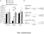
- the diameter of regenerative nerves is measured by reconstructing serial sample sections stained by H&E in Proximal, Medial, and Distal sites of implanted nerve conduits: Proximal site (1.0 mm from proximal end of conduit), Distal site (3.0 mm from the Proximal site), and Medial site (the center between the Proximal site and Distal site).
- the diameter of regenerative nerve showed no significant difference among the three groups (262 ⁇ 44 ⁇ m, 284 ⁇ 42 ⁇ m, and 302 ⁇ 97 ⁇ m at Proximal sites and 279 ⁇ 56 ⁇ m, 298 ⁇ 75 ⁇ m, and 322 ⁇ 123 ⁇ m at Distal sites, respectively).
- the diameter in the Medial site of the Conduit+NSC (189 ⁇ 18 ⁇ m) and Conduit+NSC+mIL12 (295 ⁇ 47 ⁇ m) groups were 2.9-fold and 4.5-fold thicker, respectively, than the Conduit only group (65 ⁇ 21 ⁇ m).
- IL12p80 increased the nerve diameter 1.6-fold compared with the group without IL12p80 ( Fig.4G ).
- the schematic diagram of regeneration in the three groups is shown in Fig. 4H .
- Nerve conduction is measured by electrical stimulating the sciatic nerve and recording the compound muscle action potential (CMAP), which is the summated voltage response from individual muscle fiber action potential ( Mallik A. et al., J Neurol Neurosurg Psychiatry 2005, 76 Suppl 2: ii23-31 .).
- CMAP compound muscle action potential
- the recovery percentage was calculated as the CMAP of injured leg normalized with the CMAP of contralateral normal leg ( Fig. 5 ).
- the Conduit+NSC+mIL12 group showed better recovery status than either Conduit+NSC or Conduit only (17.7 ⁇ 4.3 %, 8.5 ⁇ 2.1 %, and 6.6 ⁇ 0.2 %, respectively).
- the Conduit+NSC+mIL12 group showed lesser gastrocnemius muscle atrophy than Conduit only group (injured leg/ normal leg was 0.39 ⁇ 0.02 and 0.26 ⁇ 0.07, respectively, p ⁇ 0.01).
- IL12p80 stimulates differentiation of mouse NSCs to the oligodendrocyte and Schwann cell lineages.
- the present application studied the potential of IL12p80 in inducing oligodendrocyte and Schwann cell differentiation in mouse NSCs.
- Cells dissociated from neurospheres were cultured in DMEM/F12 supplemented with 2% FBS as differentiation control medium (Control group), and oligodendrocyte differentiation medium (CNTF+T3 group), respectively ( Hsu Y.C. et al., Developmental Dynamics 2009, 238:302-314 ; Lee D.C. et al, Molecular and Cellular Neurosciences 2009, 41:348-363 .).
- IL12p80 100 ng/ml human IL12p80 (hIL12p80) or mouse IL12p80 (mIL12p80) were added to the control group or CNTF+T3 group and named hIL12, mIL12, CNTF+T3+hIL12, and CNTF+T3+mIL12 groups.
- the differentiated cells were verified using immunofluorescence staining with primary antibody recognizing specific cell markers: galactocerebroside (Gale; oligodendrocyte), oligodendrocyte specific protein (OSP; oligodendrocyte), and myelin protein zero (PZO; myelinated Schwann cells), and fluorescent dye conjugated secondary antibodies ( Fig. 6A ).
- the percentage of marker positive cells are quantified and summarized in Fig. 6B .
- Cell morphology and immunofluorescence staining results showed that IL12p80 could replace CNTF+T3 for inducing oligodendrocyte or myelinated Schwann cells differentiation in mouse NSCs.
- GFAP glial fibrillary acidic protein
- OSP astroglia marker
- PZO glial fibrillary acidic protein
- IL12p80 induces phosphorylation of Stat3 in NSCs
- IL12p40 subunit can bind to IL12 receptor ⁇ 1 and then induce Stat3 phosphorylation and the downstream signaling pathway ( Jacobson N.G. et al., Journal of Experimental Medicine 1995, 181:1755-1762 .). Therefore, IL12p80 could trigger oligodendrocyte differentiation in NSCs through Stat3 activation.
- the phosphorylation status of Stat3 in hIL12p80 or mIL12p80 treated NSCs is analyzed by using Western blotting.
- IL12p80 in nerve conduits improves motor function recovery, promotes nerve regeneration, improves nerve conduction, and increases the diameter of newly regenerated nerve.
- the regenerated nerve from the Conduit+NSC+IL12 is up to 4.5 fold thicker than the Conduit only group at the medial section of the injured nerve.
- IL12p80 could induce NSCs to differentiate into oligodendrocytes or myelinated Schwann cells in vitro. It is further showed that the induction of differentiation may be through the phosphorylation of Stat3. The administering of IL12p80 alone could achieve a certain degree of neural repair.
- IL 12p80 is a homodimer of IL 12p40, while IL 12p70 is a heterdimer of IL12p40 and IL12p35.
- IL12p40 binds to IL12 receptor ⁇ 1 and IL12p35 binds to IL12 receptor ⁇ 2.
- IL23 is a heterodimer of IL12p40 and IL23p19, with IL23p19 binding to the IL23 receptor. It has been shown that IL12p80 along with NSCs in nerve conduits improves nerve injury repair. The administering of IL12p80 alone could achieve a certain degree of neural repair.
- IL12p70 could enhance the neurite out growth of mouse sympathetic superior cervical ganglion neurons in vitro ( Lin H. et al., Neurosci. Lett. 2000, 278:129-132 .). Therefore, proteins that contain IL12p40 subunit, including IL12p80, IL12p70, and IL23 could all contribute to neural repair.
- the scope of neural repair could include not only neurogenesis of nerve injuries, such as peripheral nerve injury, spinal cord injury and stroke, but also neurodegenerative diseases such as Alzheimer's disease, Parkinson's disease and multiple sclerosis ( Beckervordersandforth R. et al., Stem Cell Reports 2014, 2:153-162 .).
Landscapes
- Health & Medical Sciences (AREA)
- Life Sciences & Earth Sciences (AREA)
- Engineering & Computer Science (AREA)
- General Health & Medical Sciences (AREA)
- Chemical & Material Sciences (AREA)
- Veterinary Medicine (AREA)
- Medicinal Chemistry (AREA)
- Public Health (AREA)
- Pharmacology & Pharmacy (AREA)
- Animal Behavior & Ethology (AREA)
- Epidemiology (AREA)
- Immunology (AREA)
- Biomedical Technology (AREA)
- Zoology (AREA)
- Bioinformatics & Cheminformatics (AREA)
- Neurosurgery (AREA)
- Cell Biology (AREA)
- Developmental Biology & Embryology (AREA)
- Proteomics, Peptides & Aminoacids (AREA)
- Gastroenterology & Hepatology (AREA)
- Neurology (AREA)
- Biotechnology (AREA)
- Virology (AREA)
- Dermatology (AREA)
- Chemical Kinetics & Catalysis (AREA)
- Nuclear Medicine, Radiotherapy & Molecular Imaging (AREA)
- General Chemical & Material Sciences (AREA)
- Organic Chemistry (AREA)
- Psychology (AREA)
- Ophthalmology & Optometry (AREA)
- Hematology (AREA)
- Pregnancy & Childbirth (AREA)
- Reproductive Health (AREA)
- Hospice & Palliative Care (AREA)
- Psychiatry (AREA)
- Medicines That Contain Protein Lipid Enzymes And Other Medicines (AREA)
- Materials For Medical Uses (AREA)
- Peptides Or Proteins (AREA)
- Micro-Organisms Or Cultivation Processes Thereof (AREA)
- Medicines Containing Material From Animals Or Micro-Organisms (AREA)
Description
- The present application relates to a composition and methods to enhance nerve regeneration utilizing neural stem cells and IL12p40.
- Severed peripheral nerve injury causes a reduction in motor and sensory neuron activities, and degeneration of nerve fibers and the surrounding tissues. The regeneration of injured peripheral nerve is a multiplex process, with Wallerian degeneration (WD) being the most elementary reaction and Schwann cells playing an important role (Ren Z. et al., Reviews in the Neurosciences 2012, 23:135-143). WD creates a microenvironment for regeneration of surviving neurons and benefits functional recovery (Navarro X. et al., Progress in Neurobiology 2007, 82:163-201.). The control of WD involves the existence of Schwann cells, the secretion of neurotrophic factors, and special extracellular matrix that acts as a scaffold for neural cells (Gaudet A.D. et al., Journal of Neuroinflammation 2011, 8:110; Kehoe S. et al., Injury 2012, 43:553-572.).
- Nerve conduits provide mechanical support and direct axonal sprouting between the injured nerve stumps. Conduits have been shown to retain neurotrophic factors secreted from or recruited by the damaged cells (Kehoe S. et al, Injury 2012, 43:553-572.) and prevent ingrowth of fibrous tissue at the injury site. Recent studies reveal that implantation of neural stem cells (NSCs) in conduits promote regeneration of injured peripheral nerves (Zhang H. et al., Journal of Translational Medicine 2008, 6:67; Shi Y. et al., Acta Oto-Laryngologica 2009, 129: 906-914.).
- The promotion of nerve regeneration may depend on the ability of implanted NSCs to differentiate into Schwann cells, to secrete neurotrophic factors per se, or create a microenvironment to enrich neurotrophic factors from milieu, and to assist in myelination (Ren Z. et al., Reviews in the Neurosciences 2012, 23:135-143). However, the nature of cytokines or growth factors that are involved in this process is not clear. The molecular mechanism for the Schwann cell differentiation of the implanted NSCs into newly regenerated axons is also not well established.
- In this study, it is an aim to identify factors that are involved in NSCs-mediated nerve regeneration and functional recovery. Using a protein antibody array, we searched for protein level differences in a mouse sciatic nerve injury model using conduits with or without NSCs. The levels of IL12p80 (the bioactive homodimer form of IL12p40) (Heinzel F.P. et al., Journal of Immunology 1997, 158:4381-4388) in these conduits were nearly two-fold higher than those in conduits without NSCs. Implantation of NSCs with nerve conduit and IL12p80 improved motor function in a sciatic nerve injury mouse model.
- Administration of IL12p80 further enhanced nerve regeneration as evidenced by the increased diameter in the regenerated nerve, up to 4.5-fold thicker than the Conduit only group at the medial section of the regenerated nerve and improved nerve conduction. This is showed that IL12p80 induced the neuroglia differentiation of mouse NSCs in vitro through phosphorylation of signal transducer and activator of transcription 3 (Stat3). The neuroglia comprises astroglia, oligodendrocytes, and Schwann cells as reported by Kettenmann and Verkhratsky (Kettenmann H, Verkhratsky A, Fortschritte der Neurologie-Psychiatrie 2011, 79:588-597.).
- EHTESHAM M et al. describe the use of interleukin 12-secreting neural stem cells for the treatment of intracranial glioma (CANCER RESEARCH, AACR -AMERICAN ASSOCIATION FOR CANCER RESEARCH, Vol. 62, No. 20, 2002, pp. 5657-5663).
- COOPER et al. describe IL-12p40: an inherently agonistic cytokine (TRENDS IN IMMUNO, ELSEVIER LTD., TRENDS JOURNALS, Vol. 28, No. 1, 2006, pp. 33-38).
- The present application provides a composition for nerve regeneration comprising neural stem cells and a neurotrophic factor, which is IL12p80 and/or a monomer of IL12p40.
- The present application provides a composition for nerve regeneration comprising neural stem cells, a neurotrophic factor, which is IL12p80 and/or a monomer of IL12p40, and a nerve regeneration enhancing element, as defined in
claim 5. - The present application further provides a composition for nerve regeneration comprising neural stem cells, a neurotrophic factor, which IL12p80 and/or a monomer of IL12p40, and a nerve conduit for carrying at least one of the neural stem cells and the neurotrophic factors.
- The present application provides a nerve regeneration composition comprising neural stem cells and a neurotrophic factor containing IL12p80 and/or a monomer of IL12p40 for use in the treatment of nerve injuries, peripheral nerve injury, spinal cord injury, stroke, neurodegenerative diseases, Alzheimer's disease, Parkinson's disease or multiple sclerosis.
-
-
Fig. 1 is schematic diagrams illustrating the implantation of NSCs with nerve conduits improved functional recovery of sciatic nerve injury in mice more than conduits only. (A) SFI was used to assess functional recovery weekly from the first week to the eighth week. Statistical analyses indicated that the Conduit+NSC group had significantly better recovery than the Conduit only group. (B) The Rotarod test was done on the fourth and eighth week after surgery. The Conduit only group had significantly worse balance and walking on the Rotarod than the Control and Conduit+NSC groups. The Conduit+NSC group had better motor recovery and did not differ from the Control group. All data are presented as mean ± standard error of the mean (SEM) (n = 3 in Sham control group, n = 8 in Conduit only group, n = 8 in Conduit+NSC group, and n = 8 at the fourth week and n = 8 at the eighth week in the Control group). Statistical differences are indicated by *p < 0.05 and **p < 0.01. -
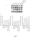
Fig. 2 is a table and photos illustrating the antibody array of cell extracts from implanted conduits with or without NSCs. The signal of IL12p40 (indicated by red dot square) existed in the cell extracts harvested from the Conduit+NSC group was 1.89-fold higher than the Conduit only group (n = 4 in both Conduit only and Conduit+NSC group). These experiments were repeated three times and IL12p40 levels in the Conduit+NSC group were more greatly expressed than the Conduit only group. -
Fig. 3 is schematic diagrams illustrating the comparative analysis of functional assessment using SFI and Rotarod tests in NSCs transplant with or without IL12p80. On the eighth week after surgery, mice of the Conduit+NSC and Conduit+NSC+mIL12 group showed significantly higher SFI scores than the Conduit only group (A). In the Rotarod test, mice of the Conduit+NSC and Conduit+NSC+mIL12 group showed greater ability of walking and balance than the Conduit only group (B). These data indicated that the Conduit+NSC+mIL12 group showed statistically more significant results (**p < 0.01) than the group without IL-12p80 (*p < 0.05) in functional assessment of both SFI and Rotarod analyses. Data are presented as mean ± SEM (In SFI, n = 3 in the Conduit only group, n = 3 in the Conduit+NSC group, and n = 4 in the Conduit+NSC+mIL12 group. In the Rotarod test, n = 7 in the Conduit only group, n = 5 in the Conduit+NSC group, and n = 8 in the Conduit+NSC+mIL12 group). -
Fig. 4 illustrates the H&E staining and immunohistochemistry of regenerated sciatic nerve sections. (A-C) The sections of newly regenerated axon were stained with hematoxylin and eosin and "P" and "D" indicate the ends of residual nerve ("P" is 1.0 mm from the proximal end of conduit and "D" is 3.0 mm distal from "P"). The Conduit only group (A) showed less integration of regenerated axon than the Conduit+NSC group (B) and Conduit+NSC+mIL12 group (C). Furthermore, we verified the characteristics of regenerating nerve using immunohistochemistry staining with anti-neurofilament 200 antibody (NF200, a marker of nerve fiber) and anti-protein zero antibody (PZO, a marker of myelinated Schwann cells). The staining results showed the failure of axon regeneration in the Conduit only group (D). We could observe that myelinated Schwann cells (PZO positive cell) coupling nerve fiber (NF200 positive cell) existed in the medial region of conduits in the Conduit+NSC (E) and more so in the Conduit+NSC+mIL12 groups (F). The relative locations of D, E, and F are shown in dotted squares in A, B, and C, respectively. The quantitative results of regenerated nerve diameter at different sites are shown in (G), where the Proximal site indicated is 1.0 mm from the proximal end of the conduit, the Distal site indicated is 3.0 mm from the Proximal site, and the Medial site indicated is central between the Proximal and Distal sites (n = 3 in the Conduit only group, n = 4 in the Conduit+NSC group, and n = 3 in the Conduit+NSC+mIL12 group). The schematic diagram of regeneration of different treatment groups is shown in (H). Scale bars in (A), (B), and (C) are 1 mm, (D), (E), and (F) are 100 µm. Statistical differences are shown as *p < 0.05 and **p < 0.01. -
Fig. 5 is a schematic diagram illustrating recovery sciatic nerve injury measured in compound muscle action potential. The CMAPs of injured leg and contralateral normal leg were measured. The recovery of CMAP was calculated as injured leg divided by normal leg. The recovery of CMAP of different groups were shown (n = 4 in the Conduit only group, n = 4 in the Conduit+NSC group, n = 6 in the Conduit+NSC+mIL12 group, and n=3 in the Sham control). -
Fig. 6 illustrates the immunocytochemistry results showing that IL12p80 could replace CNTF+T3 to induce oligodendrocyte differentiation of mouse NSCs. (A) Mouse NSCs were induced to differentiate into oligodendrocytes using CNTF+T3 or IL12p80 (listed in Y axis). Differentiated cells were stained with oligodendrocyte markers: anti-Gale antibody and anti-OSP antibody, or myelinated Schwann cell marker: anti-PZO antibody (listed in X axis). The folds of marker-positive cells were quantified and summarized in (B). Scale bar in each graph is 100 µm. Statistical differences are shown as *p < 0.05 and **p < 0.01. -
Fig. 7 illustrates the western blotting results showing that IL12p80 could differentiate mouse NSCs to oligodendrocytes and Schwann cells. Mouse NSCs were induced to differentiate into oligodendrocytes using CNTF+T3 or IL12p80. Differentiation groups were named by different cytokine treating conditions. Cell differentiation was characterized using Western blotting with lineage specific antibody against astroglial marker GFAP, oligodendrocyte marker OSP, and myelinated Schwann cell marker PZO (A). The quantification results are shown in (B). Statistical differences are shown as *p < 0.05 and **p < 0.01. -
Fig. 8 illustrates that the IL12p80 induces phosphorylation of Stat3 in NSCs. The phosphorylation levels of Stat3 in Y705 and S727 sites (pStat3) in control medium (Control) or differentiation medium containing human IL12p80 (hIL12) or mouse IL12p70 (mIL12) at different time points (Sphere, 0 min, 15 min, 30 min, 1 h, 2 h, 4 h, and 8 h) were determined by Western blotting analyses. The results show that the levels of pStat3 peaked at 15 minutes when cultured with IL12p80. The total Stat3 and α-Tubulin were used as controls. - In the present application, a composition for nerve regeneration comprises NSCs and a neurotrophic factor, which is IL12p80 and/or a monomer of IL12p40. In some embodiment, IL12p40 has a protein sequence shown as NCBI Accession No.: NM_008352 (Mouse) or 1F42_A (Human).
- The neurotrophic factor can be a monomer of IL12p40, or a homodimer of IL12p40. The homodimer is IL12p80.
- In the present application, the NSCs of the composition for nerve regeneration can be characterized as brain cells that are positive for CD133 and GFAP, or characterized as F1B-GFP+ cells.
- The present application provides a composition further comprising a nerve regeneration enhancing element selected from Schwann cells, mesenchymal stem cells (MSCs), adipose-derived stem cells (ADSCs), amniotic fluid stem cells (AFSCs), induced neurons (iNs), induced neural stem cells (iNSCs), or cell that are derived from induced pluripotent stem cells (iPSCs), fibroblast growth factor 1 (FGF1), fibroblast growth factor 2 (FGF2), platelet derived growth factor (PDGF), brain derived neurotrophic factor (BDNF), glial cell derived neurotrophic factor (GDNF), nerve growth factor (NGF).
- In an embodiment, the composition can further comprise a nerve conduit for carrying at least one of the NSCs and the neurotrophic factors. In some embodiments, the nerve conduit further carries the nerve regeneration enhancing elements.
- Also disclosed is a method for regenerating nerve, comprising providing a nerve regeneration composition comprising neural stem cells and a neurotrophic factor which is IL12p80 and/or a monomer of IL12p40, to a subject. In some embodiments, the nerve regeneration composition can further comprise NSCs. In embodiments, the nerve conduit is implanted in a target nerve or a tissue around the target nerve.
- The neurotrophic factor can be a monomer of IL12p40, or a homodimer of IL12p40, a trimer of IL12p40, a tetramer of IL12p40 or any combination thereof. In some embodiments, the neurotrophic factor is IL12p80.
- The subject is a mammalian. In an embodiment, the mammalian can be mice or humans. In the present application, the composition can be provided by injection, implantation, transdermal route, and/or oral administration.
- The protein sequence of IL12p40 is shown as NCBI Accession No.: NM_008352 (Mouse) or 1F42_A (Human), which are available from the website of National Center for Biotechnology Information (http://www.ncbi.nlm.nih.gov/).
- Mouse NSCs isolation and cell culture were performed as described in two previous publications (Hsu Y.C. et al., Developmental Dynamics 2009, 238:302-314; Lee D.C. et al., Molecular and Cellular Neurosciences 2009, 41:348-363; the two publications are incorporated by references in theirs entirety.)
- KT98/F1B-GFP cells were cultured in Dulbecco's modified Eagle's medium (DMEM)/F12 (1:1) containing 10% fetal bovine serum (FBS), 1% penicillin/streptomycin, and 500 µg/ml G418 (Merck, USA). For NSCs isolation, The GFP-positive KT98/F1B-GFP (KT98/F1B-GFP+) cells were sorted using FACSAria cell sorter (BD Bioscience) and cultured in neurosphere formation medium (DMEM/F12 containing IX B27 (Gibco), 20 ng/ml EGF (PeproTech Inc.), 20 ng/ml FGF2 (PeproTech Inc.), 2 µg/ml heparin (Sigma), and 500 µg/ml G418) for 7 days, which induced KT98/F1B-GFP+ neurosphere formation. Then KT98/F1B-GFP+ neurosphere-derived single cells were used in subsequent animal experiments and cell differentiation assays. All cells were cultured at 37 °C with 5% CO2.
- In the present application, all animal experimental procedures followed the ethical guidelines and were approved by the Institutional Animal Care and Use Committee (IACUC) of National Health Research Institutes (Protocol No. NHRI-IACUC-101067A). FVB mice (8-10 weeks old) were used for animal experiments and were maintained in National Health Research Institutes (NHRI) Animal Center. Before surgery, mouse was anesthetized by 5% isoflurane (Halocarbon) air inhalation and anesthetization was maintained by 2% isoflurane air inhalation during surgery. In sciatic nerve injury surgery, a 3 mm mouse sciatic nerve segment was excised with microscissors. For the surgical implantation of nerve conduits, the poly(L-lactic acid) (PLA) conduit was used, as previously described (Hsu S.H. et al., Artif Organs 2009, 33:26-35; the publication is incorporated by reference in its entirety.)
- The 5 mm conduit with or without NSCs and/or mouse IL12p80 were implanted into the sciatic nerve injury site. Proximal and distal end nerves of the sciatic nerve injury site were anchored into the conduit with 1 mm residual nerve using 6-0 nylon microsutures. Mice which did not undergo sciatic nerve injury were defined as the Sham control group (n = 3).
- The surgical implantation groups included a Conduit only group (n = 8), Conduit+NSC group (n = 8), and a Conduit+NSC+mIL12 group (n = 8). In the Conduit only group, conduits were filled with 5 µl matrigel (BD Bioscience)/phosphate buffer saline (PBS) mixture (1:1). For the Conduit+NSC group (n = 8), conduits were filled with 5 µl matrigel/PBS mixture (1:1) with 1 × 10⁶ NSCs. In the Conduit+NSC+mIL12 group (n = 8), conduits were filled with 5 µl matrigel/PBS mixture (1:1) with 1 × 10⁶ NSCs and 100 ng mouse IL12p80 (BioLegend).
- Walking track analysis was performed using the Treadmill/TreadScan system (CleverSys) and presented as sciatic functional index (SFI) every week after surgery. The formula for SFI is as follows:

- SFI calculation was according to normal (N) and experimental (E) feet where PL indicated the length of the footprint (the longitudinal distance between the tip of the longest toe and the heel), TS indicated the total toe spread (the cross-sectional distance between the first and fifth toes) and IT indicated the intermediate toe spread (the cross-sectional distance between the second and the fourth toes). Adult FVB mice were used to obtain the normal walking video (total 1500 frames were collected in a complete walking period of one mouse) and these image data were used to calibrate the TreadScan software (10-12 outlines of each step were sufficient to train the software for identification of the paw position). After calibration, the well-established program is used to exclude abnormal walking status and irregular toe spread during an entire walking period of sciatic nerve injury mice.
- The Rotarod test was executed by RT series Rotarod Treadmill (SINGA) at the fourth and eighth week after surgery. In the Control group, mice were defined as without nerve dissection and muscle injury (n = 8 at the fourth week and n = 8 at the eighth week). Before formal data collection, each mouse ran on the rotating rod for three times each at 10 rpm, 12 rpm, and 15 as a pre-test course. For data collection, mice ran on the rotating rod six times at 20 rpm. The maximal recording time was 120 seconds.
- Protein samples were extracted from the implanted conduits using 1X RIPA buffer [50 mM HEPES, pH 7.3, 150 mM NaCl, 2 mM EDTA, 20 mM β-gylcerophosphate, 0.1 mM Na3VO4, 1 mM NaF, 0.5 mM DTT, and 0.5% NP-40] containing IX protease inhibitor cocktail (Roche). 100 µg of protein sample was used in the mouse angiogenesis protein antibody array (RayBiotech) analysis, which followed the manufacturer's protocol. Protein levels were detected with chemiluminescence methods.
- Single cells were dissociated from the neurospheres using 1X HyQTase (Hyclone). 2 ×10³ cells were seeded onto Poly-D-Lysine (BD Bioscience)-coated chamber slides with neural differentiation medium (DMEM/F12 containing 2% FBS) supplemented with or without inducing factors. For CNTF+T3 group, neural differentiation medium were supplemented with 50 ng/ml CNTF and 10 ng/ml T3. For hIL12 and mIL12 group, neural differentiation medium were supplemented with 100 ng/ml human IL12p80 and mouse IL12p80, separately. For CNTF+T3+hIL12 group, neural differentiation medium were supplemented with 50 ng/ml CNTF, 10 ng/ml T3 and 100 ng/ml human IL12p80. For CNTF+T3+mIL12 group, neural differentiation medium were supplemented with 50 ng/ml CNTF, 10 ng/ml T3 and 100 ng/ml mouse IL12p80. CNTF, T3, and human IL12p80 were purchased from PeproTech. Culture media was replaced every three days.
- Cells were grown on chamber slides (Nunc, Naperville, IL, USA) at 37 °C with 5% CO2. For immunofluorescent staining, cells were washed with PBS and fixed with 4% (v/v) paraformaldehyde (PFA; Electron Microscopy Sciences) in PBS for 15 min at room temperature. Cells were then permeated with 0.1% (v/v) Triton X-100 in PBS for 15 min at room temperature. Cells were then blocked with blocking solution (1% BSA in IX PBS) at RT for 1 hour, and followed by incubation with specific primary antibody for 1 hour at room temperature. In the present application, the differentiation capacity is verified by using immunofluorescence staining with mouse monoclonal antibody against galactocerebroside (Galc, 1:1000, Millipore), rabbit polyclonal antibody to oligodendrocyte specific protein (OSP, 1:1000, Abcam), and chicken polyclonal antibody to myelin protein zero (PZO, 1:1000, GeneTex). Subsequently, the cells were incubated with rhodamine-conjugated secondary antibody (Millipore, Billerica, MA, USA) for 1 hour at room temperature. All sections were stained for nuclei with 2-(4-Amidinophenyl)-6-indolecarbamidine dihydrochloride (DAPI) (Molecular Probes), and observed under a fluorescent microscope (Olympus, Tokyo, Japan). Galc/DAPI, OSP/DAPI and PZO/DAPI double-positive cells were counted and normalized to the Control group.
- Rabbit polyclonal antibody against glial fibrillary acidic protein (GFAP, 1:1000; Abcam), rabbit polyclonal antibody against oligodendrocyte specific protein (OSP, 1:4000; Abeam), chicken polyclonal antibody against myelin protein zero (PZO, 1: 4000; GeneTex), and rabbit polyclonal antibody against α-tubulin (a-tubulin, 1:8000 diluted; GeneTex) were used to detect the corresponding protein levels by the immunoblotting method in differentiated cells, which were isolated from neurospheres and cultured with neural differentiation medium with or without inducing factors (Table 1) for 7 days. Protein samples from differentiated cells were harvested by 1X RIPA Lysis Buffer (Millipore, Billerica, MA, USA) supplemented with 1X protease inhibitor cocktail (Roche).
- Mouse monoclonal antibody against Stat3 (1:1000; Cell Signaling), rabbit polyclonal antibody against pStat3-Y705 site (1:1000; Cell Signaling), rabbit polyclonal antibody against pStat3-S727 site (1:1000; Cell Signaling), and rabbit polyclonal antibody against α-tubulin (α-tubulin, 1:8000 diluted; GeneTex) were used to analyze Stat3 phosphorylation status in mouse NSCs after IL12p80 treatment. Single cells were isolated from neurospheres and experimental signal cells were harvested by centrifugation for 3 minutes and cell lysates were extracted at different time points (Sphere, 0 min, 15 min, 30 min, 1 h, 2 h, 4 h, and 8 h). Thus, at time point "0 min," cells had been in the presence of with IL12p80 for 3 minutes during the centrifugation period. Cell lysates were analyzed using Western blotting.
- The protein concentration of each sample was determined by protein assay dye reagent (Bio-Rad Laboratories). Equal amounts of protein samples were size fractionated by sodium dodecyl sulfate-polyacrylamide gel (SDS-PAGE) and then transferred to polyvinylidene fluoride (PVDF) membranes (Amersham). Thereafter, membranes were blocked in 5% bovine serum albumin (Bio Basic, Markham Ontario, Canada) blocking buffer for 1 hour at room temperature. The membranes were then incubated with specific primary antibodies in 5% bovine serum albumin blocking buffer followed by incubation with corresponding HRP-conjugated secondary antibodies (1:10000)(Millipore). Protein levels were revealed using ECL reagents (Millipore) and X-ray films with α-tubulin as an internal control. Immunoreactive bands of three individual experiments were quantitated by ImageJ software and normalized to the Control group.
- Compound muscle action potential was recorded and analyzed with BIOPAC MP36 and BIOPAC BSL 4.0 software (BIOPAC system Inc.). The stainless steel electrode was 0.22 mm in diameter. Stimulating electrodes were placed in the sciatic notch and recording electrodes were placed in the gastrocnemius muscle (approximately 2 cm from the sciatic notch). Stimulation voltage was 6 volts, stimulus duration was 0.1 millisecond, and acquisition length was 200 milliseconds. Distances were measured with a vernier caliper, and skin temperature was maintained at 36°C in a room maintained at a constant temperature of 25°C.
- Implanted conduits were collected on the eighth week after surgery. In briefly, the implanted conduits were fixed with 4% PFA at 4 °C overnight. Fixed samples were dehydrated using 30% sucrose in 1X PBS at 4 °C overnight. Samples were embedded in Tissue-Tek O.C.T. (Sakura, Netherlands), then frozen in liquid nitrogen and stored at -80 °C.
- For Hematoxylin and Eosin (H&E) and immunohistochemistry staining, cryo-embedded nerve conduits were sliced at 10 µm onto slides using a cryostat microtome (MICROM HM550). Immunohistochemistry staining was performed by specific antibody: rabbit polyclonal antibody against neurofilament 200 (NF-200, 1:500, marker of nerve fiber) and chicken polyclonal antibody against myelin protein zero (PZO, 1:500, marker of myelinated Schwann cell). After PBS washing, samples were incubated with respective secondary antibodies at RT for 1 hour. All samples were stained for nuclei with DAPI (1:5000) at RT for 3 minutes and mounted with FluorSave reagent (Merck). All image data of regenerated tissue were observed and collected using fluorescent microscopy (Olympus) and confocal microscopy (Leica).
- Data were expressed as mean ± standard error of the mean (SEM). Student's t-test was used for comparing two groups and one-way ANOVA was used for comparing multiple groups. Statistical significance was accepted when p < 0.05.
Table 1. Conditions of inducing cell differentiation. Group name Inducing factors CNTF+ T3 50 ng/ml CNTF and 10 ng/ ml T3 hIL12 100 ng/ml human IL12p80 mIL12 100 ng/ml mouse IL12p80 CNTF+T3+ hIL12 50 ng/ml CNTF, 10 ng/ml T3 and 100 ng/ml human IL12p80 CNTF+T3+ mIL12 50 ng/ml CNTF, 10 ng/ml T3 and 100 ng/ml mouse IL12p80 - After excision of left sciatic nerve (3 mm), all mice lost motor function at the left hindlimb, which showed the dragging walk phenotype with cringed toes. In this study, we implanted NSCs with conduit to repair the injured sciatic nerve. Functional recovery was assessed using non-invasive methods, Walking track analysis and the Rotarod test, during the period of regeneration. Sciatic functional index (SFI) is a calculated score of data from Walking track analysis that combines gait analysis and the temporal and spatial relationship of one footprint to another during walking. The SFI is on a scale from 0 to -100, where 0 corresponds to the normal walking function and -100 refers to the complete loss of function. In track analysis, sham control mice were defined as mice with the same surgical procedure but no actual damage of sciatic nerve. These mice showed no apparent difference in SFI score throughout the first week to eighth week after surgery when compared with the pre-surgery SFI score (
Fig. 1A , Sham control). For the implantation of NSCs with conduit (Fig. 1A , Conduit+NSC group) or conduit only (Fig. 1A , Conduit only group), the SFI score showed no significant difference between the two groups (-73.2 ± 2.5 vs. -77.0 ± 3.9) at the first week after surgery. Beginning from the second week after surgery, the Conduit+NSC group showed a higher SFI score than the Conduit only group (p < 0.01). The functional betterment in the Conduit+NSC group than the Conduit only group continued until the eighth week (Fig. 1A ). - The Rotarod test is also used to assess the recovery of motor function and balance ability of injury sciatic nerve mice. In the Rotarod test, Control group mice were defined as without nerve dissection and muscle injury. The results of the Control group, Conduit+NSC group and Conduit only group in the Rotarod test at fourth week were 55 ± 8, 40 ± 7, and 16 ± 1 s, and at eighth week were 62 ± 14, 53 ± 9, and 25 ± 4 seconds, respectively (
Fig. 1B ). The Conduit only group showed a significantly reduced ability in balancing and walking on the Rotarod than the Control and Conduit+NSC group at the fourth and eighth week after surgery. Interestingly, no difference is observed in the Rotarod test between the Control and Conduit+NSC group at the fourth and eighth week after surgery. These results indicated that implantation of NSCs with conduit facilitated the functional recovery in sciatic nerve injury mice. - NSCs hold the potential to differentiate into all three neuroectodermal lineages including Schwann cells, and may also secrete or recruit trophic factors for nerve regeneration (Ren Z. et al., Reviews in the Neurosciences 2012, 23:135-143). Therefore, to identify factors that were involved in NSCs-mediated nerve regeneration, the mouse antibody array is used to investigate the protein expression level between Conduit only and Conduit+NSC groups. Protein lysates extracted from the implanted nerve conduits with or without NSCs were harvested at the fourth week after surgery for protein antibody array. According to the quantification data of the antibody array, expression of IL12p40 in the cell extracts harvested from the Conduit+NSC group was 1.89-fold higher compared with the Conduit only group, yet expression of IL12p35 in the Conduit+NSC group was similar to (1.04-fold) the Conduit only group (
Fig. 2 , IL12p40 and IL12p35). IL12p40 was one of the subunits in IL12p70, and 20% to 40% of total IL12p40 subunit could be secreted as a homodimer form (IL12p80), which was 25-50-fold more active than IL12p40 monomer in the induction of biological function (Heinzel F.P. et al., Journal of Immunology 1997, 158:4381-4388; Gillessen S. et al., European Journal of Immunology 1995, 25:200-206; Jacobson N.G. et al., Journal of Experimental Medicine 1995, 181:1755-1762.) In the antibody array data, the Conduit+NSC group increased the expression levels of IL12p40 but not IL12p35, which indicated that IL12p80, the IL12p40 bioactive form, may be a factor involved in nerve regeneration. IL12p40 binds to IL12 receptor β1 and results in phosphorylation of Stat3 and activation of the Stat3 downstream signaling (Heinzel F.P. et al., Journal of Immunology 1997, 158:4381-4388; Gillessen S. et al., European Journal of Immunology 1995, 25:200-206; Jacobson N.G. et al., Journal of Experimental Medicine 1995, 181:1755-1762.). Previous studies indicate that Stat3 activation induces differentiation of neural progenitor cells into the astroglial lineage (Wang B. et al., PLoS One 2008, 3:e1856. ). These results imply a role for IL12p80 in the activation of Stat3 downstream signaling and participation in nerve regeneration. - To test the function of IL12p80 in nerve regeneration, the sciatic nerve injury mice treated with the following conditions were compared: Conduit only group, conduit with NSCs (Conduit+NSC group), and conduit with NSCs and mouse IL12p80 (Conduit+NSC+mIL12 group). On the eighth week after implantation, functional assessment was performed by Walking track analysis and the Rotarod test. Mice of Conduit+NSC and Conduit+NSC+mIL12 group showed a significantly higher SFI score than the Conduit only group (
Fig. 3A ). In the Rotarod test, mice of the Conduit+NSC and Conduit+NSC+mIL12 group showed better walking and balancing abilities than the Conduit only group (Fig 3B ). Moreover, these data demonstrated that the Conduit+NSC+mIL12 group showed more significant results (p < 0.01) than the group without IL12p80 (p < 0.05) in functional assessment of both Walking track and Rotarod analyses (Fig. 3A and 3B ). However, the effect of additional administration with IL12p80 (Conduit+NSC+mIL12 group) was too feeble to discriminate the assisted ability of nerve repair from that without IL12p80 (Conduit+NSC group) in the functional assessments. Therefore, the effect of IL12p80 in nerve repair is demonstrated by histological section assessment (Fig. 4 ) and nerve conduction study (Fig. 5 ). - In the nerve regeneration study, 5 mm nerve conduit is implanted to connect the 3 mm sciatic nerve injury gap. The conduit sutured to the proximal and distal ends of injured sciatic nerve with 1 mm residual nerve on each end. Therefore, axons existed in the central region of the conduit were considered newly regenerated nerve. Axonal regeneration is observed by hematoxylin and eosin (H&E) staining, and immunohistochemistry using antibodies recognizing specific neural cell specific markers. In
Fig. 4A, 4B and 4C , "P" and "D" indicate the ends of residual nerve ("P" is 1.0 mm from proximal end of the implanted conduit and "D" is 3.0 mm distal from "P"). In H&E staining, Conduit+NSC (Fig. 4B ) and Conduit+NSC+mIL12 (Fig. 4C ) groups showed significant enhancement in the integration of regenerated axons compared with the Conduit only group (Fig. 4A ). The status of nerve regeneration is further verified by using immunohistochemistry staining using anti-neurofilament 200 antibody (NF200, a marker of nerve fiber) and anti-protein zero antibody (PZO, a marker of myelinated Schwann cells). The immunohistochemical staining results demonstrated that the coupling of myelinated Schwann cells (PZO positive cell) with nerve fiber (NF200 positive cell) of regenerated nerve in the conduit (dotted line region inFig. 4A, 4B, and 4C , respectively) occurred in the Conduit+NSC (Fig. 4E ) and Conduit+NSC+mIL12 group (Fig. 4F ), but not in the Conduit only group (Fig. 4D ). Furthermore, the diameter of regenerative nerves is measured by reconstructing serial sample sections stained by H&E in Proximal, Medial, and Distal sites of implanted nerve conduits: Proximal site (1.0 mm from proximal end of conduit), Distal site (3.0 mm from the Proximal site), and Medial site (the center between the Proximal site and Distal site). At the Proximal and Distal sites, the diameter of regenerative nerve showed no significant difference among the three groups (262 ± 44 µm, 284 ± 42 µm, and 302 ± 97 µm at Proximal sites and 279 ± 56 µm, 298 ± 75 µm, and 322 ± 123 µm at Distal sites, respectively). In contrast, the diameter in the Medial site of the Conduit+NSC (189 ± 18 µm) and Conduit+NSC+mIL12 (295 ± 47 µm) groups were 2.9-fold and 4.5-fold thicker, respectively, than the Conduit only group (65 ± 21 µm). Remarkably, in the Medial site of the regenerative sciatic nerve, the addition of IL12p80 increased the nerve diameter 1.6-fold compared with the group without IL12p80 (Fig.4G ). The schematic diagram of regeneration in the three groups is shown inFig. 4H . Nerve conduction is measured by electrical stimulating the sciatic nerve and recording the compound muscle action potential (CMAP), which is the summated voltage response from individual muscle fiber action potential (Mallik A. et al., J Neurol Neurosurg Psychiatry 2005, 76 Suppl 2: ii23-31.). In the present application, the CMAPs of injured leg and contralateral normal leg were measured. The recovery percentage was calculated as the CMAP of injured leg normalized with the CMAP of contralateral normal leg (Fig. 5 ). The Conduit+NSC+mIL12 group showed better recovery status than either Conduit+NSC or Conduit only (17.7 ± 4.3 %, 8.5 ± 2.1 %, and 6.6 ± 0.2 %, respectively). Furthermore, the Conduit+NSC+mIL12 group showed lesser gastrocnemius muscle atrophy than Conduit only group (injured leg/ normal leg was 0.39 ± 0.02 and 0.26 ± 0.07, respectively, p < 0.01). These data demonstrated that implantation of NSCs with conduits facilitated the functional recovery and nerve regeneration. Importantly, additional administration of IL12p80 showed statistically significant results in the functional assessment of both SFI and Rotarod analyses, improvement of nerve conduction, and increase of the diameter of newly regenerated nerves. - The present application then studied the potential of IL12p80 in inducing oligodendrocyte and Schwann cell differentiation in mouse NSCs. Cells dissociated from neurospheres were cultured in DMEM/F12 supplemented with 2% FBS as differentiation control medium (Control group), and oligodendrocyte differentiation medium (CNTF+T3 group), respectively (Hsu Y.C. et al., Developmental Dynamics 2009, 238:302-314; Lee D.C. et al, Molecular and Cellular Neurosciences 2009, 41:348-363.). To test the potential of IL12p80 in triggering oligodendrocyte and Schwann cell differentiation, 100 ng/ml human IL12p80 (hIL12p80) or mouse IL12p80 (mIL12p80) were added to the control group or CNTF+T3 group and named hIL12, mIL12, CNTF+T3+hIL12, and CNTF+T3+mIL12 groups. The differentiated cells were verified using immunofluorescence staining with primary antibody recognizing specific cell markers: galactocerebroside (Gale; oligodendrocyte), oligodendrocyte specific protein (OSP; oligodendrocyte), and myelin protein zero (PZO; myelinated Schwann cells), and fluorescent dye conjugated secondary antibodies (
Fig. 6A ). The percentage of marker positive cells are quantified and summarized inFig. 6B . Cell morphology and immunofluorescence staining results showed that IL12p80 could replace CNTF+T3 for inducing oligodendrocyte or myelinated Schwann cells differentiation in mouse NSCs. The cell differentiation and maturation status are verified by Western blotting using lineage specific antibodies against glial fibrillary acidic protein (GFAP, astroglia marker), OSP, and PZO (Fig. 7A ), respectively. The quantification result showed that protein levels of GFAP, OSP and PZO were upregulated in the differentiation conditions containing IL12p80 (Fig. 7B ). These results demonstrated that IL12p80 could induce NSCs to differentiate into oligodendrocytes and myelinated Schwann cells. - In NSCs, Stat3 phosphorylation has been shown to participate in oligodendrocyte differentiation processes (Wang B. et al., PLoS One 2008, 3:e1856.). In T cells, IL12p40 subunit can bind to IL12 receptor β1 and then induce Stat3 phosphorylation and the downstream signaling pathway (Jacobson N.G. et al., Journal of Experimental Medicine 1995, 181:1755-1762.). Therefore, IL12p80 could trigger oligodendrocyte differentiation in NSCs through Stat3 activation. The phosphorylation status of Stat3 in hIL12p80 or mIL12p80 treated NSCs is analyzed by using Western blotting. In this study, the phosphorylation status of both Y705 and S727 (Stat3 phosphorylation sites) were slightly expressed in neurospheres (
Fig. 8 , lanes of Sphere). Both human and mouse IL12p80 induced phosphorylation of Stat3 at Y705; the intensity peaked at 15 minutes, and began to decline at 30 minutes. Similarly, phosphorylation of Stat3 at S727 was increased by IL12p80 and sustained for 8 hours (Fig. 8 , hIL12 group, and mIL12 group). These results revealed that Stat3 phosphorylation was crucial for IL12p80-induced oligodendrocyte differentiation in NSCs. - In this study, it is demonstrated that implantation of IL12p80 along with NSCs in nerve conduits improves motor function recovery, promotes nerve regeneration, improves nerve conduction, and increases the diameter of newly regenerated nerve. The regenerated nerve from the Conduit+NSC+IL12 is up to 4.5 fold thicker than the Conduit only group at the medial section of the injured nerve. IL12p80 could induce NSCs to differentiate into oligodendrocytes or myelinated Schwann cells in vitro. It is further showed that the induction of differentiation may be through the phosphorylation of Stat3. The administering of IL12p80 alone could achieve a certain degree of neural repair.
- IL 12p80 is a homodimer of IL 12p40, while IL 12p70 is a heterdimer of IL12p40 and IL12p35. IL12p40 binds to IL12 receptor β1 and IL12p35 binds to IL12 receptor β2. It was also reported that IL23 is a heterodimer of IL12p40 and IL23p19, with IL23p19 binding to the IL23 receptor. It has been shown that IL12p80 along with NSCs in nerve conduits improves nerve injury repair. The administering of IL12p80 alone could achieve a certain degree of neural repair.
- It has been shown that IL12p70 could enhance the neurite out growth of mouse sympathetic superior cervical ganglion neurons in vitro (Lin H. et al., Neurosci. Lett. 2000, 278:129-132.). Therefore, proteins that contain IL12p40 subunit, including IL12p80, IL12p70, and IL23 could all contribute to neural repair. The scope of neural repair could include not only neurogenesis of nerve injuries, such as peripheral nerve injury, spinal cord injury and stroke, but also neurodegenerative diseases such as Alzheimer's disease, Parkinson's disease and multiple sclerosis (Beckervordersandforth R. et al., Stem Cell Reports 2014, 2:153-162.).
Claims (10)
- A composition for nerve regeneration comprising:neural stem cells, anda neurotrophic factor, which is IL12p80 and/or a monomer of IL12p40.
- The composition of claim 1, wherein IL12p40 has a protein sequence shown as NCBI Accession No.: NM_008352 (Mouse) or 1F42_A (Human).
- The composition of claim 1, wherein the neural stem cells are characterized as brain cells that are positive for CD133 and GFAP, as F1BGFP+ cells, or any combination thereof.
- The composition of claim 1, which further comprises a nerve conduit for carrying at least one of the neural stem cells and the neurotrophic factors.
- The composition of claim 1, which further comprises a nerve regeneration enhancing element selected from Schwann cells, mesenchymal stem cells (MSCs), adipose-derived stem cells (ADSCs), amniotic fluid stem cells (AFSCs), induced neurons (iNs), induced neural stem cells (iNSCs), cells that are derived from induced pluripotent stem cells (iPSCs), fibroblast growth factor 1 (FGF1), fibroblast growth factor 2 (FGF2), platelet derived growth factor (PDGF), brain derived neurotrophic factor (BDNF), glial cell derived neurotrophic factor (GDNF), and nerve growth factor (NGF).
- A nerve regeneration composition comprising neural stem cells and a neurotrophic factor containing IL12p80 and/or a monomer of IL12p40 for use in the treatment of nerve injuries, peripheral nerve injury, spinal cord injury, stroke, neurodegenerative diseases, Alzheimer's disease, Parkinson's disease or multiple sclerosis.
- The composition for use according to claim 6, which further comprises a nerve conduit for carrying at least one of the neural stem cells and the neurotrophic factors.
- The composition for use according to claim 6, which further comprises a nerve regeneration enhancing element selected from the group consisting of Schwann cells, mesenchymal stem cells (MSCs), adipose-derived stem cells (ADSCs), amniotic fluid stem cells (AFSCs), induced neurons (iNs), induced neural stem cells (iNSCs), cells that are derived from induced pluripotent stem cells (iPSCs), fibroblast growth factor 1 (FGF1), fibroblast growth factor 2 (FGF2), platelet derived growth factor (PDGF), brain derived neurotrophic factor (BDNF), glial cell derived neurotrophic factor (GDNF) and nerve growth factor (NGF).
- The composition for use according to claim 6, which is provided by injection, implantation, transdermal route, and/or oral administration.
- The composition for use according to claim 7, wherein the nerve conduit is implanted in a target nerve or a tissue around the target nerve.
Applications Claiming Priority (2)
| Application Number | Priority Date | Filing Date | Title |
|---|---|---|---|
| US201462037612P | 2014-08-15 | 2014-08-15 | |
| PCT/CA2015/050775 WO2016023130A1 (en) | 2014-08-15 | 2015-08-14 | Methods to enhance nerve regeneration utilizing neural stem cells and il12p40 |
Publications (3)
| Publication Number | Publication Date |
|---|---|
| EP3180019A1 EP3180019A1 (en) | 2017-06-21 |
| EP3180019A4 EP3180019A4 (en) | 2018-05-09 |
| EP3180019B1 true EP3180019B1 (en) | 2019-12-25 |
Family
ID=55303739
Family Applications (1)
| Application Number | Title | Priority Date | Filing Date |
|---|---|---|---|
| EP15831363.5A Active EP3180019B1 (en) | 2014-08-15 | 2015-08-14 | Neural stem cells and a monomer or homodimer of il12p40 to enhance nerve regeneration |
Country Status (11)
| Country | Link |
|---|---|
| US (2) | US10391149B2 (en) |
| EP (1) | EP3180019B1 (en) |
| JP (1) | JP6339737B2 (en) |
| KR (1) | KR101897422B1 (en) |
| CN (1) | CN107148278B (en) |
| AU (1) | AU2015303798B2 (en) |
| CA (1) | CA2958398C (en) |
| HK (1) | HK1243648A1 (en) |
| SG (1) | SG11201700971XA (en) |
| TW (1) | TWI694828B (en) |
| WO (1) | WO2016023130A1 (en) |
Families Citing this family (3)
| Publication number | Priority date | Publication date | Assignee | Title |
|---|---|---|---|---|
| US20190290283A1 (en) * | 2018-03-26 | 2019-09-26 | Wichita State University | Composite neural conduit |
| WO2021007192A1 (en) * | 2019-07-08 | 2021-01-14 | The Board Of Regents Of The University Of Texas System | Use of immune modulators to improve nerve regeneration |
| CN110938598B (en) * | 2019-12-31 | 2020-10-02 | 重庆市铂而斐细胞生物技术有限公司 | Culture method and application of Schwann cells |
Family Cites Families (4)
| Publication number | Priority date | Publication date | Assignee | Title |
|---|---|---|---|---|
| IL148175A0 (en) * | 1999-08-18 | 2002-09-12 | Gen Hospital Corp | Methods, compositions and kits for promoting recovery from damage to the central nervous system |
| JP2002078792A (en) * | 2000-09-07 | 2002-03-19 | Kuraray Co Ltd | Nerve regeneration material |
| WO2002072144A1 (en) * | 2001-03-12 | 2002-09-19 | Institute Of Gene And Brain Science | Remedies for nerve damages |
| CN101306194A (en) * | 2008-06-20 | 2008-11-19 | 奔驰生物科技(云南)有限公司 | Medicine for treating nerves damage and preparation method thereof |
-
2015
- 2015-08-14 AU AU2015303798A patent/AU2015303798B2/en not_active Ceased
- 2015-08-14 US US15/504,301 patent/US10391149B2/en not_active Expired - Fee Related
- 2015-08-14 JP JP2017508088A patent/JP6339737B2/en not_active Expired - Fee Related
- 2015-08-14 KR KR1020177006888A patent/KR101897422B1/en not_active Expired - Fee Related
- 2015-08-14 WO PCT/CA2015/050775 patent/WO2016023130A1/en not_active Ceased
- 2015-08-14 CN CN201580043623.XA patent/CN107148278B/en not_active Expired - Fee Related
- 2015-08-14 EP EP15831363.5A patent/EP3180019B1/en active Active
- 2015-08-14 SG SG11201700971XA patent/SG11201700971XA/en unknown
- 2015-08-14 CA CA2958398A patent/CA2958398C/en active Active
- 2015-08-14 HK HK18103277.8A patent/HK1243648A1/en unknown
- 2015-08-14 TW TW104126631A patent/TWI694828B/en not_active IP Right Cessation
-
2019
- 2019-07-17 US US16/514,701 patent/US20190351022A1/en not_active Abandoned
Non-Patent Citations (1)
| Title |
|---|
| None * |
Also Published As
| Publication number | Publication date |
|---|---|
| HK1243648A1 (en) | 2018-07-20 |
| CA2958398A1 (en) | 2016-02-18 |
| EP3180019A1 (en) | 2017-06-21 |
| US10391149B2 (en) | 2019-08-27 |
| EP3180019A4 (en) | 2018-05-09 |
| TW201613623A (en) | 2016-04-16 |
| CN107148278A (en) | 2017-09-08 |
| KR101897422B1 (en) | 2018-09-10 |
| WO2016023130A1 (en) | 2016-02-18 |
| SG11201700971XA (en) | 2017-03-30 |
| US20170224776A1 (en) | 2017-08-10 |
| CN107148278B (en) | 2021-06-29 |
| AU2015303798A1 (en) | 2017-03-02 |
| JP6339737B2 (en) | 2018-06-06 |
| KR20170065504A (en) | 2017-06-13 |
| CA2958398C (en) | 2019-05-14 |
| TWI694828B (en) | 2020-06-01 |
| AU2015303798B2 (en) | 2020-03-12 |
| JP2017529329A (en) | 2017-10-05 |
| US20190351022A1 (en) | 2019-11-21 |
Similar Documents
| Publication | Publication Date | Title |
|---|---|---|
| Lee et al. | Neural stem cells promote nerve regeneration through IL12-induced Schwann cell differentiation | |
| Gómez et al. | C ell therapy for spinal cord injury with olfactory ensheathing glia cells (OEC s) | |
| Ji et al. | Plasticity in ventral pallidal cholinergic neuron-derived circuits contributes to comorbid chronic pain-like and depression-like behaviour in male mice | |
| Li et al. | Cetuximab modified collagen scaffold directs neurogenesis of injury-activated endogenous neural stem cells for acute spinal cord injury repair | |
| Sun et al. | IGF-1R stimulation alters microglial polarization via TLR4/NF-κB pathway after cerebral hemorrhage in mice | |
| Zhou et al. | Dual electrical stimulation at spinal-muscular interface reconstructs spinal sensorimotor circuits after spinal cord injury | |
| Hu et al. | Epidermal neural crest stem cell (EPI-NCSC)—mediated recovery of sensory function in a mouse model of spinal cord injury | |
| Nijhuis et al. | Natural conduits for bridging a 15-mm nerve defect: comparison of the vein supported by muscle and bone marrow stromal cells with a nerve autograft | |
| Boldyreva et al. | Plasmid-based gene therapy with hepatocyte growth factor stimulates peripheral nerve regeneration after traumatic injury | |
| US20190351022A1 (en) | Methods to enhance nerve regeneration utilizing neural stem cells and il12p40 | |
| Xiao et al. | Human adult olfactory neural progenitors promote axotomized rubrospinal tract axonal reinnervation and locomotor recovery | |
| Lai et al. | NeuroD1 overexpression in spinal neurons accelerates axonal regeneration after sciatic nerve injury | |
| Su et al. | Application of amniotic fluid stem cells in repairing sciatic nerve injury in minipigs | |
| Hiratsuka et al. | VEGF-and PDGF-dependent proliferation of oligodendrocyte progenitor cells in the medulla oblongata after LPC-induced focal demyelination | |
| Kingham et al. | Adipose-derived stem cells for nerve repair: hype or reality? | |
| Han et al. | Lentiviral-mediated netrin-1 overexpression improves motor and sensory functions in SCT rats associated with SYP and GAP-43 expressions | |
| KR20120109671A (en) | Compositions of stem cells and stem cell factors and methods for their use and manufacture | |
| Wang et al. | Study of synergistic role of allogenic skin-derived precursor differentiated Schwann cells and heregulin-1β in nerve regeneration with an acellular nerve allograft | |
| Collins et al. | Partial recovery of proprioception in rats with dorsal root injury after human olfactory bulb cell transplantation | |
| Audisio et al. | Neuregulin1 administration increases axonal elongation in dissociated primary sensory neuron cultures | |
| Jørgensen et al. | Lentiviral delivery of meteorin protects striatal neurons against excitotoxicity and reverses motor deficits in the quinolinic acid rat model | |
| Chen et al. | Human IL12p80 and Neural Stem Cells Enhance Sciatic Nerve Regeneration | |
| Charsar | Respiratory Axon Plasticity Drives Recovery of Diaphragm Function After Spinal Cord Injury | |
| Stavrovskaya et al. | Transcranial Direct Current Stimulation for Improvement of Neurotransplantation Outcomes in Rats with 6-Hydroxydopamine-Induced Parkinsonism | |
| Jaberi et al. | Transplanted autologous neural stem cells show promise in restoring motor function in monkey spinal cord injury |
Legal Events
| Date | Code | Title | Description |
|---|---|---|---|
| STAA | Information on the status of an ep patent application or granted ep patent |
Free format text: STATUS: THE INTERNATIONAL PUBLICATION HAS BEEN MADE |
|
| PUAI | Public reference made under article 153(3) epc to a published international application that has entered the european phase |
Free format text: ORIGINAL CODE: 0009012 |
|
| STAA | Information on the status of an ep patent application or granted ep patent |
Free format text: STATUS: REQUEST FOR EXAMINATION WAS MADE |
|
| 17P | Request for examination filed |
Effective date: 20170215 |
|
| AK | Designated contracting states |
Kind code of ref document: A1 Designated state(s): AL AT BE BG CH CY CZ DE DK EE ES FI FR GB GR HR HU IE IS IT LI LT LU LV MC MK MT NL NO PL PT RO RS SE SI SK SM TR |
|
| AX | Request for extension of the european patent |
Extension state: BA ME |
|
| RAP1 | Party data changed (applicant data changed or rights of an application transferred) |
Owner name: NATIONAL HEALTH RESEARCH INSTITUTES |
|
| DAV | Request for validation of the european patent (deleted) | ||
| DAX | Request for extension of the european patent (deleted) | ||
| A4 | Supplementary search report drawn up and despatched |
Effective date: 20180411 |
|
| RIC1 | Information provided on ipc code assigned before grant |
Ipc: A61P 25/02 20060101ALI20180405BHEP Ipc: A61K 35/30 20150101ALI20180405BHEP Ipc: A61K 38/20 20060101AFI20180405BHEP |
|
| RIC1 | Information provided on ipc code assigned before grant |
Ipc: A61P 25/02 20060101ALI20190529BHEP Ipc: A61P 25/16 20060101ALI20190529BHEP Ipc: A61K 35/30 20150101ALI20190529BHEP Ipc: A61P 25/28 20060101ALI20190529BHEP Ipc: A61K 38/20 20060101AFI20190529BHEP |
|
| GRAP | Despatch of communication of intention to grant a patent |
Free format text: ORIGINAL CODE: EPIDOSNIGR1 |
|
| STAA | Information on the status of an ep patent application or granted ep patent |
Free format text: STATUS: GRANT OF PATENT IS INTENDED |
|
| INTG | Intention to grant announced |
Effective date: 20190716 |
|
| GRAS | Grant fee paid |
Free format text: ORIGINAL CODE: EPIDOSNIGR3 |
|
| GRAA | (expected) grant |
Free format text: ORIGINAL CODE: 0009210 |
|
| STAA | Information on the status of an ep patent application or granted ep patent |
Free format text: STATUS: THE PATENT HAS BEEN GRANTED |
|
| AK | Designated contracting states |
Kind code of ref document: B1 Designated state(s): AL AT BE BG CH CY CZ DE DK EE ES FI FR GB GR HR HU IE IS IT LI LT LU LV MC MK MT NL NO PL PT RO RS SE SI SK SM TR |
|
| REG | Reference to a national code |
Ref country code: GB Ref legal event code: FG4D |
|
| REG | Reference to a national code |
Ref country code: CH Ref legal event code: EP |
|
| REG | Reference to a national code |
Ref country code: AT Ref legal event code: REF Ref document number: 1216414 Country of ref document: AT Kind code of ref document: T Effective date: 20200115 |
|
| REG | Reference to a national code |
Ref country code: DE Ref legal event code: R096 Ref document number: 602015044409 Country of ref document: DE |
|
| REG | Reference to a national code |
Ref country code: IE Ref legal event code: FG4D |
|
| REG | Reference to a national code |
Ref country code: NL Ref legal event code: MP Effective date: 20191225 |
|
| PG25 | Lapsed in a contracting state [announced via postgrant information from national office to epo] |
Ref country code: LV Free format text: LAPSE BECAUSE OF FAILURE TO SUBMIT A TRANSLATION OF THE DESCRIPTION OR TO PAY THE FEE WITHIN THE PRESCRIBED TIME-LIMIT Effective date: 20191225 Ref country code: LT Free format text: LAPSE BECAUSE OF FAILURE TO SUBMIT A TRANSLATION OF THE DESCRIPTION OR TO PAY THE FEE WITHIN THE PRESCRIBED TIME-LIMIT Effective date: 20191225 Ref country code: BG Free format text: LAPSE BECAUSE OF FAILURE TO SUBMIT A TRANSLATION OF THE DESCRIPTION OR TO PAY THE FEE WITHIN THE PRESCRIBED TIME-LIMIT Effective date: 20200325 Ref country code: NO Free format text: LAPSE BECAUSE OF FAILURE TO SUBMIT A TRANSLATION OF THE DESCRIPTION OR TO PAY THE FEE WITHIN THE PRESCRIBED TIME-LIMIT Effective date: 20200325 Ref country code: GR Free format text: LAPSE BECAUSE OF FAILURE TO SUBMIT A TRANSLATION OF THE DESCRIPTION OR TO PAY THE FEE WITHIN THE PRESCRIBED TIME-LIMIT Effective date: 20200326 Ref country code: SE Free format text: LAPSE BECAUSE OF FAILURE TO SUBMIT A TRANSLATION OF THE DESCRIPTION OR TO PAY THE FEE WITHIN THE PRESCRIBED TIME-LIMIT Effective date: 20191225 Ref country code: FI Free format text: LAPSE BECAUSE OF FAILURE TO SUBMIT A TRANSLATION OF THE DESCRIPTION OR TO PAY THE FEE WITHIN THE PRESCRIBED TIME-LIMIT Effective date: 20191225 |
|
| REG | Reference to a national code |
Ref country code: LT Ref legal event code: MG4D |
|
| PG25 | Lapsed in a contracting state [announced via postgrant information from national office to epo] |
Ref country code: RS Free format text: LAPSE BECAUSE OF FAILURE TO SUBMIT A TRANSLATION OF THE DESCRIPTION OR TO PAY THE FEE WITHIN THE PRESCRIBED TIME-LIMIT Effective date: 20191225 Ref country code: HR Free format text: LAPSE BECAUSE OF FAILURE TO SUBMIT A TRANSLATION OF THE DESCRIPTION OR TO PAY THE FEE WITHIN THE PRESCRIBED TIME-LIMIT Effective date: 20191225 |
|
| PG25 | Lapsed in a contracting state [announced via postgrant information from national office to epo] |
Ref country code: AL Free format text: LAPSE BECAUSE OF FAILURE TO SUBMIT A TRANSLATION OF THE DESCRIPTION OR TO PAY THE FEE WITHIN THE PRESCRIBED TIME-LIMIT Effective date: 20191225 |
|
| PG25 | Lapsed in a contracting state [announced via postgrant information from national office to epo] |
Ref country code: EE Free format text: LAPSE BECAUSE OF FAILURE TO SUBMIT A TRANSLATION OF THE DESCRIPTION OR TO PAY THE FEE WITHIN THE PRESCRIBED TIME-LIMIT Effective date: 20191225 Ref country code: PT Free format text: LAPSE BECAUSE OF FAILURE TO SUBMIT A TRANSLATION OF THE DESCRIPTION OR TO PAY THE FEE WITHIN THE PRESCRIBED TIME-LIMIT Effective date: 20200520 Ref country code: CZ Free format text: LAPSE BECAUSE OF FAILURE TO SUBMIT A TRANSLATION OF THE DESCRIPTION OR TO PAY THE FEE WITHIN THE PRESCRIBED TIME-LIMIT Effective date: 20191225 Ref country code: RO Free format text: LAPSE BECAUSE OF FAILURE TO SUBMIT A TRANSLATION OF THE DESCRIPTION OR TO PAY THE FEE WITHIN THE PRESCRIBED TIME-LIMIT Effective date: 20191225 Ref country code: NL Free format text: LAPSE BECAUSE OF FAILURE TO SUBMIT A TRANSLATION OF THE DESCRIPTION OR TO PAY THE FEE WITHIN THE PRESCRIBED TIME-LIMIT Effective date: 20191225 |
|
| PG25 | Lapsed in a contracting state [announced via postgrant information from national office to epo] |
Ref country code: IS Free format text: LAPSE BECAUSE OF FAILURE TO SUBMIT A TRANSLATION OF THE DESCRIPTION OR TO PAY THE FEE WITHIN THE PRESCRIBED TIME-LIMIT Effective date: 20200425 Ref country code: SM Free format text: LAPSE BECAUSE OF FAILURE TO SUBMIT A TRANSLATION OF THE DESCRIPTION OR TO PAY THE FEE WITHIN THE PRESCRIBED TIME-LIMIT Effective date: 20191225 Ref country code: SK Free format text: LAPSE BECAUSE OF FAILURE TO SUBMIT A TRANSLATION OF THE DESCRIPTION OR TO PAY THE FEE WITHIN THE PRESCRIBED TIME-LIMIT Effective date: 20191225 |
|
| REG | Reference to a national code |
Ref country code: DE Ref legal event code: R097 Ref document number: 602015044409 Country of ref document: DE |
|
| PG25 | Lapsed in a contracting state [announced via postgrant information from national office to epo] |
Ref country code: ES Free format text: LAPSE BECAUSE OF FAILURE TO SUBMIT A TRANSLATION OF THE DESCRIPTION OR TO PAY THE FEE WITHIN THE PRESCRIBED TIME-LIMIT Effective date: 20191225 Ref country code: DK Free format text: LAPSE BECAUSE OF FAILURE TO SUBMIT A TRANSLATION OF THE DESCRIPTION OR TO PAY THE FEE WITHIN THE PRESCRIBED TIME-LIMIT Effective date: 20191225 |
|
| PLBE | No opposition filed within time limit |
Free format text: ORIGINAL CODE: 0009261 |
|
| STAA | Information on the status of an ep patent application or granted ep patent |
Free format text: STATUS: NO OPPOSITION FILED WITHIN TIME LIMIT |
|
| REG | Reference to a national code |
Ref country code: AT Ref legal event code: MK05 Ref document number: 1216414 Country of ref document: AT Kind code of ref document: T Effective date: 20191225 |
|
| PG25 | Lapsed in a contracting state [announced via postgrant information from national office to epo] |
Ref country code: SI Free format text: LAPSE BECAUSE OF FAILURE TO SUBMIT A TRANSLATION OF THE DESCRIPTION OR TO PAY THE FEE WITHIN THE PRESCRIBED TIME-LIMIT Effective date: 20191225 |
|
| 26N | No opposition filed |
Effective date: 20200928 |
|
| PG25 | Lapsed in a contracting state [announced via postgrant information from national office to epo] |
Ref country code: AT Free format text: LAPSE BECAUSE OF FAILURE TO SUBMIT A TRANSLATION OF THE DESCRIPTION OR TO PAY THE FEE WITHIN THE PRESCRIBED TIME-LIMIT Effective date: 20191225 Ref country code: IT Free format text: LAPSE BECAUSE OF FAILURE TO SUBMIT A TRANSLATION OF THE DESCRIPTION OR TO PAY THE FEE WITHIN THE PRESCRIBED TIME-LIMIT Effective date: 20191225 |
|
| PG25 | Lapsed in a contracting state [announced via postgrant information from national office to epo] |
Ref country code: PL Free format text: LAPSE BECAUSE OF FAILURE TO SUBMIT A TRANSLATION OF THE DESCRIPTION OR TO PAY THE FEE WITHIN THE PRESCRIBED TIME-LIMIT Effective date: 20191225 |
|
| PG25 | Lapsed in a contracting state [announced via postgrant information from national office to epo] |
Ref country code: MC Free format text: LAPSE BECAUSE OF FAILURE TO SUBMIT A TRANSLATION OF THE DESCRIPTION OR TO PAY THE FEE WITHIN THE PRESCRIBED TIME-LIMIT Effective date: 20191225 |
|
| REG | Reference to a national code |
Ref country code: CH Ref legal event code: PL |
|
| PG25 | Lapsed in a contracting state [announced via postgrant information from national office to epo] |
Ref country code: LI Free format text: LAPSE BECAUSE OF NON-PAYMENT OF DUE FEES Effective date: 20200831 Ref country code: LU Free format text: LAPSE BECAUSE OF NON-PAYMENT OF DUE FEES Effective date: 20200814 Ref country code: CH Free format text: LAPSE BECAUSE OF NON-PAYMENT OF DUE FEES Effective date: 20200831 |
|
| REG | Reference to a national code |
Ref country code: BE Ref legal event code: MM Effective date: 20200831 |
|
| PG25 | Lapsed in a contracting state [announced via postgrant information from national office to epo] |
Ref country code: IE Free format text: LAPSE BECAUSE OF NON-PAYMENT OF DUE FEES Effective date: 20200814 Ref country code: BE Free format text: LAPSE BECAUSE OF NON-PAYMENT OF DUE FEES Effective date: 20200831 |
|
| PG25 | Lapsed in a contracting state [announced via postgrant information from national office to epo] |
Ref country code: TR Free format text: LAPSE BECAUSE OF FAILURE TO SUBMIT A TRANSLATION OF THE DESCRIPTION OR TO PAY THE FEE WITHIN THE PRESCRIBED TIME-LIMIT Effective date: 20191225 Ref country code: MT Free format text: LAPSE BECAUSE OF FAILURE TO SUBMIT A TRANSLATION OF THE DESCRIPTION OR TO PAY THE FEE WITHIN THE PRESCRIBED TIME-LIMIT Effective date: 20191225 Ref country code: CY Free format text: LAPSE BECAUSE OF FAILURE TO SUBMIT A TRANSLATION OF THE DESCRIPTION OR TO PAY THE FEE WITHIN THE PRESCRIBED TIME-LIMIT Effective date: 20191225 |
|
| PG25 | Lapsed in a contracting state [announced via postgrant information from national office to epo] |
Ref country code: MK Free format text: LAPSE BECAUSE OF FAILURE TO SUBMIT A TRANSLATION OF THE DESCRIPTION OR TO PAY THE FEE WITHIN THE PRESCRIBED TIME-LIMIT Effective date: 20191225 |
|
| PGFP | Annual fee paid to national office [announced via postgrant information from national office to epo] |
Ref country code: GB Payment date: 20220829 Year of fee payment: 8 Ref country code: DE Payment date: 20220829 Year of fee payment: 8 |
|
| PGFP | Annual fee paid to national office [announced via postgrant information from national office to epo] |
Ref country code: FR Payment date: 20220825 Year of fee payment: 8 |
|
| REG | Reference to a national code |
Ref country code: DE Ref legal event code: R119 Ref document number: 602015044409 Country of ref document: DE |
|
| GBPC | Gb: european patent ceased through non-payment of renewal fee |
Effective date: 20230814 |
|
| PG25 | Lapsed in a contracting state [announced via postgrant information from national office to epo] |
Ref country code: GB Free format text: LAPSE BECAUSE OF NON-PAYMENT OF DUE FEES Effective date: 20230814 |
|
| PG25 | Lapsed in a contracting state [announced via postgrant information from national office to epo] |
Ref country code: GB Free format text: LAPSE BECAUSE OF NON-PAYMENT OF DUE FEES Effective date: 20230814 Ref country code: FR Free format text: LAPSE BECAUSE OF NON-PAYMENT OF DUE FEES Effective date: 20230831 Ref country code: DE Free format text: LAPSE BECAUSE OF NON-PAYMENT OF DUE FEES Effective date: 20240301 |