EP3177331B1 - Dental barrier membrane using cocoon and method for manufacturing same - Google Patents
Dental barrier membrane using cocoon and method for manufacturing same Download PDFInfo
- Publication number
- EP3177331B1 EP3177331B1 EP15830245.5A EP15830245A EP3177331B1 EP 3177331 B1 EP3177331 B1 EP 3177331B1 EP 15830245 A EP15830245 A EP 15830245A EP 3177331 B1 EP3177331 B1 EP 3177331B1
- Authority
- EP
- European Patent Office
- Prior art keywords
- cocoon
- barrier membrane
- thickness
- dental
- present
- Prior art date
- Legal status (The legal status is an assumption and is not a legal conclusion. Google has not performed a legal analysis and makes no representation as to the accuracy of the status listed.)
- Active
Links
Images
Classifications
-
- A—HUMAN NECESSITIES
- A61—MEDICAL OR VETERINARY SCIENCE; HYGIENE
- A61C—DENTISTRY; APPARATUS OR METHODS FOR ORAL OR DENTAL HYGIENE
- A61C8/00—Means to be fixed to the jaw-bone for consolidating natural teeth or for fixing dental prostheses thereon; Dental implants; Implanting tools
- A61C8/0003—Not used, see subgroups
- A61C8/0004—Consolidating natural teeth
- A61C8/0006—Periodontal tissue or bone regeneration
-
- A—HUMAN NECESSITIES
- A61—MEDICAL OR VETERINARY SCIENCE; HYGIENE
- A61L—METHODS OR APPARATUS FOR STERILISING MATERIALS OR OBJECTS IN GENERAL; DISINFECTION, STERILISATION OR DEODORISATION OF AIR; CHEMICAL ASPECTS OF BANDAGES, DRESSINGS, ABSORBENT PADS OR SURGICAL ARTICLES; MATERIALS FOR BANDAGES, DRESSINGS, ABSORBENT PADS OR SURGICAL ARTICLES
- A61L27/00—Materials for grafts or prostheses or for coating grafts or prostheses
- A61L27/36—Materials for grafts or prostheses or for coating grafts or prostheses containing ingredients of undetermined constitution or reaction products thereof, e.g. transplant tissue, natural bone, extracellular matrix
- A61L27/3604—Materials for grafts or prostheses or for coating grafts or prostheses containing ingredients of undetermined constitution or reaction products thereof, e.g. transplant tissue, natural bone, extracellular matrix characterised by the human or animal origin of the biological material, e.g. hair, fascia, fish scales, silk, shellac, pericardium, pleura, renal tissue, amniotic membrane, parenchymal tissue, fetal tissue, muscle tissue, fat tissue, enamel
-
- A—HUMAN NECESSITIES
- A61—MEDICAL OR VETERINARY SCIENCE; HYGIENE
- A61K—PREPARATIONS FOR MEDICAL, DENTAL OR TOILETRY PURPOSES
- A61K35/00—Medicinal preparations containing materials or reaction products thereof with undetermined constitution
- A61K35/56—Materials from animals other than mammals
- A61K35/63—Arthropods
-
- A—HUMAN NECESSITIES
- A61—MEDICAL OR VETERINARY SCIENCE; HYGIENE
- A61K—PREPARATIONS FOR MEDICAL, DENTAL OR TOILETRY PURPOSES
- A61K35/00—Medicinal preparations containing materials or reaction products thereof with undetermined constitution
- A61K35/56—Materials from animals other than mammals
- A61K35/63—Arthropods
- A61K35/64—Insects, e.g. bees, wasps or fleas
-
- A—HUMAN NECESSITIES
- A61—MEDICAL OR VETERINARY SCIENCE; HYGIENE
- A61L—METHODS OR APPARATUS FOR STERILISING MATERIALS OR OBJECTS IN GENERAL; DISINFECTION, STERILISATION OR DEODORISATION OF AIR; CHEMICAL ASPECTS OF BANDAGES, DRESSINGS, ABSORBENT PADS OR SURGICAL ARTICLES; MATERIALS FOR BANDAGES, DRESSINGS, ABSORBENT PADS OR SURGICAL ARTICLES
- A61L27/00—Materials for grafts or prostheses or for coating grafts or prostheses
- A61L27/14—Macromolecular materials
- A61L27/22—Polypeptides or derivatives thereof, e.g. degradation products
-
- A—HUMAN NECESSITIES
- A61—MEDICAL OR VETERINARY SCIENCE; HYGIENE
- A61L—METHODS OR APPARATUS FOR STERILISING MATERIALS OR OBJECTS IN GENERAL; DISINFECTION, STERILISATION OR DEODORISATION OF AIR; CHEMICAL ASPECTS OF BANDAGES, DRESSINGS, ABSORBENT PADS OR SURGICAL ARTICLES; MATERIALS FOR BANDAGES, DRESSINGS, ABSORBENT PADS OR SURGICAL ARTICLES
- A61L27/00—Materials for grafts or prostheses or for coating grafts or prostheses
- A61L27/36—Materials for grafts or prostheses or for coating grafts or prostheses containing ingredients of undetermined constitution or reaction products thereof, e.g. transplant tissue, natural bone, extracellular matrix
- A61L27/3641—Materials for grafts or prostheses or for coating grafts or prostheses containing ingredients of undetermined constitution or reaction products thereof, e.g. transplant tissue, natural bone, extracellular matrix characterised by the site of application in the body
- A61L27/3645—Connective tissue
- A61L27/365—Bones
-
- A—HUMAN NECESSITIES
- A61—MEDICAL OR VETERINARY SCIENCE; HYGIENE
- A61L—METHODS OR APPARATUS FOR STERILISING MATERIALS OR OBJECTS IN GENERAL; DISINFECTION, STERILISATION OR DEODORISATION OF AIR; CHEMICAL ASPECTS OF BANDAGES, DRESSINGS, ABSORBENT PADS OR SURGICAL ARTICLES; MATERIALS FOR BANDAGES, DRESSINGS, ABSORBENT PADS OR SURGICAL ARTICLES
- A61L27/00—Materials for grafts or prostheses or for coating grafts or prostheses
- A61L27/36—Materials for grafts or prostheses or for coating grafts or prostheses containing ingredients of undetermined constitution or reaction products thereof, e.g. transplant tissue, natural bone, extracellular matrix
- A61L27/3683—Materials for grafts or prostheses or for coating grafts or prostheses containing ingredients of undetermined constitution or reaction products thereof, e.g. transplant tissue, natural bone, extracellular matrix subjected to a specific treatment prior to implantation, e.g. decellularising, demineralising, grinding, cellular disruption/non-collagenous protein removal, anti-calcification, crosslinking, supercritical fluid extraction, enzyme treatment
-
- A—HUMAN NECESSITIES
- A61—MEDICAL OR VETERINARY SCIENCE; HYGIENE
- A61L—METHODS OR APPARATUS FOR STERILISING MATERIALS OR OBJECTS IN GENERAL; DISINFECTION, STERILISATION OR DEODORISATION OF AIR; CHEMICAL ASPECTS OF BANDAGES, DRESSINGS, ABSORBENT PADS OR SURGICAL ARTICLES; MATERIALS FOR BANDAGES, DRESSINGS, ABSORBENT PADS OR SURGICAL ARTICLES
- A61L27/00—Materials for grafts or prostheses or for coating grafts or prostheses
- A61L27/36—Materials for grafts or prostheses or for coating grafts or prostheses containing ingredients of undetermined constitution or reaction products thereof, e.g. transplant tissue, natural bone, extracellular matrix
- A61L27/3683—Materials for grafts or prostheses or for coating grafts or prostheses containing ingredients of undetermined constitution or reaction products thereof, e.g. transplant tissue, natural bone, extracellular matrix subjected to a specific treatment prior to implantation, e.g. decellularising, demineralising, grinding, cellular disruption/non-collagenous protein removal, anti-calcification, crosslinking, supercritical fluid extraction, enzyme treatment
- A61L27/3691—Materials for grafts or prostheses or for coating grafts or prostheses containing ingredients of undetermined constitution or reaction products thereof, e.g. transplant tissue, natural bone, extracellular matrix subjected to a specific treatment prior to implantation, e.g. decellularising, demineralising, grinding, cellular disruption/non-collagenous protein removal, anti-calcification, crosslinking, supercritical fluid extraction, enzyme treatment characterised by physical conditions of the treatment, e.g. applying a compressive force to the composition, pressure cycles, ultrasonic/sonication or microwave treatment, lyophilisation
-
- A—HUMAN NECESSITIES
- A61—MEDICAL OR VETERINARY SCIENCE; HYGIENE
- A61L—METHODS OR APPARATUS FOR STERILISING MATERIALS OR OBJECTS IN GENERAL; DISINFECTION, STERILISATION OR DEODORISATION OF AIR; CHEMICAL ASPECTS OF BANDAGES, DRESSINGS, ABSORBENT PADS OR SURGICAL ARTICLES; MATERIALS FOR BANDAGES, DRESSINGS, ABSORBENT PADS OR SURGICAL ARTICLES
- A61L27/00—Materials for grafts or prostheses or for coating grafts or prostheses
- A61L27/36—Materials for grafts or prostheses or for coating grafts or prostheses containing ingredients of undetermined constitution or reaction products thereof, e.g. transplant tissue, natural bone, extracellular matrix
- A61L27/38—Materials for grafts or prostheses or for coating grafts or prostheses containing ingredients of undetermined constitution or reaction products thereof, e.g. transplant tissue, natural bone, extracellular matrix containing added animal cells
-
- A—HUMAN NECESSITIES
- A61—MEDICAL OR VETERINARY SCIENCE; HYGIENE
- A61L—METHODS OR APPARATUS FOR STERILISING MATERIALS OR OBJECTS IN GENERAL; DISINFECTION, STERILISATION OR DEODORISATION OF AIR; CHEMICAL ASPECTS OF BANDAGES, DRESSINGS, ABSORBENT PADS OR SURGICAL ARTICLES; MATERIALS FOR BANDAGES, DRESSINGS, ABSORBENT PADS OR SURGICAL ARTICLES
- A61L27/00—Materials for grafts or prostheses or for coating grafts or prostheses
- A61L27/50—Materials characterised by their function or physical properties, e.g. injectable or lubricating compositions, shape-memory materials, surface modified materials
- A61L27/54—Biologically active materials, e.g. therapeutic substances
-
- A—HUMAN NECESSITIES
- A61—MEDICAL OR VETERINARY SCIENCE; HYGIENE
- A61L—METHODS OR APPARATUS FOR STERILISING MATERIALS OR OBJECTS IN GENERAL; DISINFECTION, STERILISATION OR DEODORISATION OF AIR; CHEMICAL ASPECTS OF BANDAGES, DRESSINGS, ABSORBENT PADS OR SURGICAL ARTICLES; MATERIALS FOR BANDAGES, DRESSINGS, ABSORBENT PADS OR SURGICAL ARTICLES
- A61L31/00—Materials for other surgical articles, e.g. stents, stent-grafts, shunts, surgical drapes, guide wires, materials for adhesion prevention, occluding devices, surgical gloves, tissue fixation devices
- A61L31/005—Ingredients of undetermined constitution or reaction products thereof
-
- A—HUMAN NECESSITIES
- A61—MEDICAL OR VETERINARY SCIENCE; HYGIENE
- A61L—METHODS OR APPARATUS FOR STERILISING MATERIALS OR OBJECTS IN GENERAL; DISINFECTION, STERILISATION OR DEODORISATION OF AIR; CHEMICAL ASPECTS OF BANDAGES, DRESSINGS, ABSORBENT PADS OR SURGICAL ARTICLES; MATERIALS FOR BANDAGES, DRESSINGS, ABSORBENT PADS OR SURGICAL ARTICLES
- A61L2300/00—Biologically active materials used in bandages, wound dressings, absorbent pads or medical devices
- A61L2300/40—Biologically active materials used in bandages, wound dressings, absorbent pads or medical devices characterised by a specific therapeutic activity or mode of action
- A61L2300/412—Tissue-regenerating or healing or proliferative agents
-
- A—HUMAN NECESSITIES
- A61—MEDICAL OR VETERINARY SCIENCE; HYGIENE
- A61L—METHODS OR APPARATUS FOR STERILISING MATERIALS OR OBJECTS IN GENERAL; DISINFECTION, STERILISATION OR DEODORISATION OF AIR; CHEMICAL ASPECTS OF BANDAGES, DRESSINGS, ABSORBENT PADS OR SURGICAL ARTICLES; MATERIALS FOR BANDAGES, DRESSINGS, ABSORBENT PADS OR SURGICAL ARTICLES
- A61L2430/00—Materials or treatment for tissue regeneration
- A61L2430/02—Materials or treatment for tissue regeneration for reconstruction of bones; weight-bearing implants
-
- A—HUMAN NECESSITIES
- A61—MEDICAL OR VETERINARY SCIENCE; HYGIENE
- A61L—METHODS OR APPARATUS FOR STERILISING MATERIALS OR OBJECTS IN GENERAL; DISINFECTION, STERILISATION OR DEODORISATION OF AIR; CHEMICAL ASPECTS OF BANDAGES, DRESSINGS, ABSORBENT PADS OR SURGICAL ARTICLES; MATERIALS FOR BANDAGES, DRESSINGS, ABSORBENT PADS OR SURGICAL ARTICLES
- A61L2430/00—Materials or treatment for tissue regeneration
- A61L2430/12—Materials or treatment for tissue regeneration for dental implants or prostheses
Definitions
- the present invention relates to a dental barrier membrane for guided bone regeneration using a cocoon, and a manufacturing method thereof. More particularly, the present invention relates to a cocoon-based barrier membrane that is biocompatible, has excellent tensile strength, and promotes osteogenesis in bone defects, and a method for manufacturing the same.
- a dental barrier membrane for guided bone regeneration is a device used in dental surgery, such as implants, to induce bone regeneration in a bone defect.
- a dental barrier membrane When applied to a bone defect, a dental barrier membrane functions to mechanically block the migration of epithelial or connective tissue cells into the bone defect, thus retaining the space until bone regeneration.
- dental barrier membranes were made of non-biodegradable materials such as polytetrafluoroethylene, cellulose acetate, silicon rubber, or polyurethane.
- barrier membranes made of non-biodegradable synthetic polymers are apt to cause inflammation or tissue necrosis because of their poor biocompatibility.
- biocompatible, natural materials for use in biodegradable barrier membranes have recently been studied. There are no significant differences in bone regeneration between biodegradable barrier membranes and non-biodegradable barrier membranes. When applied in clinical practice, however, the biodegradable barrier membranes developed thus far cannot retain their shapes due to their insufficient tensile strength, and thus are likely to fail in securing the space where the tissue of interest is to grow, which may lead to secondary inflammation.
- Korean Patent Unexamined Publication Application No. 10-2014-0003942 (issued on January 10, 2014 , titled “Dental Barrier Membrane Having Good Growth of Soft Tissues or Bone Tissues") and Korean Patent No. 10-1403473 (issued on June 11, 2014 , titled Dental Membrane”).
- an aspect of the present invention provides a cocoon-based dental barrier membrane, which is prepared by dividing a cocoon having a first shell thickness into two or more fragments having a predetermined form.
- each of the fragments is delaminated into a lamellar fragment having a second thickness, the second thickness being smaller than the first thickness.
- the lamellar fragment having a second thickness is an inner, middle, or outer stratum of the cocoon.
- the lamellar fragment is sterilized and can be packed.
- the present invention provides a method for manufacturing a cocoon-based dental barrier membrane, comprising a first step of dividing a cocoon into two or more fragments having a predetermined form, the cocoon having a shell having a first thickness.
- the method may further comprise a second step of delaminating each of the fragments into a lamellar fragment having a second thickness, the second thickness being smaller than the first thickness.
- the method may further comprise a third step of packing the fragments of the second thickness prepared in the second step.
- the method further comprises conducting sterilization before or after each step.
- the lamellar fragment having a second thickness is an inner, middle or outer stratum of the cocoon.
- the cocoon-based dental barrier membrane of the present invention can be prepared easily, and thus in a cost-efficient manner, compared to conventional dental barrier membranes.
- the dental barrier membrane of the present invention has high tensile strength and porosity, and can promote osteogenesis in bone defects.
- dental barrier membrane refers to a barrier membrane designed to reconstruct bone tissues that have defects due to various dental diseases.
- the dental barrier membrane should significantly improve osteogenesis in bone defects or around implants, be hard enough to be sutured, and have biocompatibility for safe use invivo.
- a dental barrier membrane for guided bone regeneration is required to avoid post-operative infection and tissue degeneration, and closely adhere to periodontal tissues without giving a sensation of irritation.
- dental barrier membranes should guarantee cell growth.
- dental barrier membranes When hydrolyzed invivo , however, most conventional dental barrier membranes cause the generation of acids such as lactic acid, glycolic acids, etc, which lower the pH in a local region, causing inflammation and histotoxicity. Further, for obtaining the desired properties, the dental barrier membranes used to date require pretreatment processes, such as physical crosslinking, etc. However, such pretreated dental barrier membranes may be toxic to the body and remain as foreign matter upon long-term use in vivo.
- Step 1 Preparation of cocoon fragment having first thickness.
- a cocoon 10 the shell of which has a first thickness, is prepared.
- a cocoon is a casting spun of silk by silkworms and is used as a material for silk fibers.
- cocoons which may be unused resources, are up-cycled into a new high value-added product, thus bringing economic benefits to silkworm farmers.
- Naturally constructed by silkworms, which eat clean mulberry leaves, cocoons are free of toxicity and are suitable for use as an environment-friendly material.
- the present invention takes a cocoon 10 as a material for a dental barrier membrane.
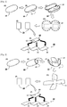
- the cocoon 10 is processed, as shown in FIGS. 1B to IE, into two or more planar fragments, each having a first thickness.
- oval cocoon 10 is dissected along a cutting line 11 into halves, as shown in FIG. 1B .
- the dissected halves have semi-oval shapes, and are opened to expose the inside surface 13 of the cocoon, as shown in FIG. 1C .
- the cocoon halves with curved inside surfaces 13 are planarized to some degree by cutting many sites along the edge as shown in FIG. ID, and planar regions are cut out to obtain cocoon fragments having a first thickness, as shown in FIG. IE.
- the dental barrier membrane prepared in the present invention need not have a planar surface. Because a cocoon originally has an elliptical ball shape, the curved shape of the dissected cocoon may be utilized to yield curved dental barrier membranes if necessary. For use as a small dental barrier membrane for guided bone regeneration, a cocoon fragment having a small area may be relatively planar. In contrast, when a relatively large area of the dissected cocoon halves is taken, the dental barrier membranes may have curved surfaces.
- FIG. 2 a modified method for preparing a cocoon fragment having a first thickness is described.
- an oval-shaped cocoon 10 is cut along a second cutting line 15 and a third cutting line 17 to expose the inside 13 of the cocoon 10, as shown in FIGS. 2B and 2C .
- the dissected cocoon having a curved surface is spread as shown in FIG. 2D , and then cocoon fragments 20 having a first thickness are obtained as shown in FIG. 2E .
- a cocoon fragment having a first thickness may be prepared by cutting a cocoon in the manners shown in FIGS. 1 and 2 , but other cutting methods may be used.
- Step 2 Preparation of cocoon fragment having second thickness (dental barrier membrane)
- the cocoon fragments 20 having a first thickness, prepared in step 1 have a multilayer structure identical to that of the cocoon shell, the multilayer structure may be split into thinner layers for use as a dental barrier membrane.
- the cocoon fragment 20 having a first thickness, prepared in step 1 is itself usable as a dental barrier membrane, it is subjected to thickness splitting to give cocoon fragments 30 having a second thickness.
- the second thickness is smaller than the first thickness.
- the cocoon fragment having the first thickness may be itself used as a dental barrier membrane without splitting to reduce the thickness thereof.
- a cocoon shell varies in thickness (first thickness) from 0.3 to 1.0 mm depending on the silkworm species.
- first thickness any kind of cocoon may be used in the present invention.
- a cocoon having a shell thickness of 0.5 ⁇ 0.8 mm is employed.
- a cocoon fragment can be easily delaminated into up to 16 lamellas, although the number of delaminations is dependent on the shell thickness.
- the thicknesses of the lamellas can be determined according to the strength and elongation necessary for the kind and use of the dental barrier membrane. From a cocoon having a shell thickness of 0.5 ⁇ 0.8 mm, a dental barrier membrane 0.01 mm - 0.7 mm thick can be prepared by delamination. According to the use of the dental barrier membrane, selection may be made of cocoon fragments 30 having various thicknesses.
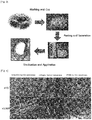
- FIG. 3 The procedure of manufacturing a dental barrier membrane in accordance with the present invention is illustrated in FIG. 3 .
- a cocoon 10 was prepared, and cut at a proper site to expose the inside thereof.
- the cut cocoon was further processed to make the curved inside planar.
- the planarized cocoon was cut into rectangular fragments 20.
- the cocoon fragments 20 were peeled into strata having a suitable thickness, followed by sterilizing the strata to afford dental barrier membranes.
- Morphologies of the dental barrier membranes prepared in Examples 1 to 3 were observed by scanning electron microscopy (SEM), with commercially available collagen and PTFE (polytetrafluoroethylene) barrier membranes used as controls for comparison.
- the dental barrier membrane of the present invention was observed to have many plies of irregularly entangled fibrous strands various in thickness, with many pores among the fibrous strands. Having a porous structure highly transmissive to nutrients and water, the dental barrier membrane of the present invention is therefore expect to find effective applications in osteogenesis at bone defects thanks to its ability to allow for the supply of nutrients and water necessary for bone regeneration.
- Example 1 The physical properties of the cocoon-based dental barrier membranes prepared in Example 1 were measured.
- a tensile test was conducted using a universal testing machine (DAEYEONG, Korea), with commercially available barrier membranes made of collagen or PTFE (polytetrafluoroethylene) serving as controls.
- DAEYEONG universal testing machine
- PTFE polytetrafluoroethylene
- Test specimens having sizes of 5 x 20 (width x length) mm were used. The specimens were extended at a rate of 10 mm/min, with an initial gauge length set to be 10 mm.
- Tensile strength was measured to be 27.6 ⁇ 1.5 MPa in the barrier membrane of the present invention, 3.5 ⁇ 1.3 MPa in the collagen barrier membrane, and 4.3 ⁇ 1.7 MPa in the PTFE membrane.
- the modulus of elasticity was measured to be 39.5 ⁇ 3.1 in the barrier membrane of the present invention, 9.6 ⁇ 2.2 MPa in the collagen barrier membrane, and 304.5 ⁇ 5.3 MPa in the PTFE membrane.
- the barrier membrane of the present invention exhibited tensile strength 9 times higher than that of the collagen barrier membrane, and 7 times higher than that of the PTFE barrier membrane. Also, the barrier membrane of the present invention is elastic, as the modulus of elasticity was 4 times higher than that of the collagen barrier membrane.
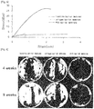
- FIG. 5 is graph in which stresses are plotted against strains in the barrier membrane of the present invention, the collagen barrier membrane, and the PTFE barrier membrane.
- the PTFE barrier membrane was observed to greatly increase only in elongation within the range where a relatively large increase in tensile strength occurred.
- the collagen barrier membrane underwent fracture before a great increase in tensile strength.
- the barrier membrane of the present invention increased in elongation with the increase of tensile strength. Accordingly, the barrier membrane of the present invention has higher tensile strength than do conventional collagen or PTFE barrier membranes.
- the barrier membrane For use in tissue regeneration and osteogenesis invivo , the barrier membrane should maintain its shape and have tensile strength, modulus of elasticity, and elongation large enough to withstand pressures in implantedregions.
- the cocoon-based membrane of the present invention is superior in tensile strength, modulus of elasticity, and elongation to commercially available controls, and thus is suitable for use as a dental barrier membrane.
- the dental barrier membrane prepared in Example 1 was implanted into a bone defect of rabbits, with a collagen barrier membrane or PTFE (polytetrafluoroethylene) barrier membrane serving as a control.
- a collagen barrier membrane or PTFE (polytetrafluoroethylene) barrier membrane serving as a control.
- ⁇ -CT micro-computerized tomography
- Masson's trichrome staining histological analysis at 4 and 8 weeks after the implantation of the barrier membranes.
- ⁇ -CT micro-computerized tomography
- the barrier membrane of the present invention Over the overall region of the bone defect implanted with the barrier membrane of the present invention was formed new bone tissue, with a significant improvement compared to that at post-operative week 4.
- the barrier membrane of the present invention notably exhibited much more efficient bone regeneration effects over the same period of time than did conventional barrier membranes.
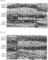
- FIG. 7A shows histological images of barrier membrane-implanted bone defects at 4 weeks after implantation with barrier membranes, along with inserts of expanded views in which ossification by Ca 2+ depositionappearsred.
- the red portion was distributed deeply across the implanted region, indicating that the barrier membrane of the present invention allowed for active osteogenesis in the bone defect.
- FIG. 7B shows histological images of barrier membrane-implanted bone defects at 8 weeks after implantation with barrier membranes, along with inserts of expanded views in which ossification appears blue.
- red portions at post-operative week 4 turned blue as new bone was formed.
- FIG. 8 is a graph in which the percentages of new bone formation on the Y axis are plotted against barrier membranes on the Y axis, on the basis of histological analysis, with a statistical significance of P ⁇ 0.05.
- the formation rate of new bone was 30% in the bone defect implanted with the barrier membrane of the present invention at post-operative week 4, and increased to 40% at post-operative week 8.
- new bone formation was 15% at post-operative week 4 and 20% at post-operative week 8.
- the formation rate of new bone was changed from 0% at post-operative week 4 to 10% at post-operative week 8. Therefore, the barrier membrane of the present invention allowed up to 4 times more new bone to be formed than did the controls.
- the barrier membrane of the present invention exhibits an excellent bone regeneration effect on bone defects and is useful as a dental barrier membrane.
Landscapes
- Health & Medical Sciences (AREA)
- Life Sciences & Earth Sciences (AREA)
- Chemical & Material Sciences (AREA)
- Veterinary Medicine (AREA)
- Epidemiology (AREA)
- Animal Behavior & Ethology (AREA)
- General Health & Medical Sciences (AREA)
- Public Health (AREA)
- Engineering & Computer Science (AREA)
- Biomedical Technology (AREA)
- Medicinal Chemistry (AREA)
- Oral & Maxillofacial Surgery (AREA)
- Transplantation (AREA)
- Dermatology (AREA)
- Chemical Kinetics & Catalysis (AREA)
- Botany (AREA)
- Molecular Biology (AREA)
- Insects & Arthropods (AREA)
- Zoology (AREA)
- Orthopedic Medicine & Surgery (AREA)
- Pharmacology & Pharmacy (AREA)
- Vascular Medicine (AREA)
- Dentistry (AREA)
- Developmental Biology & Embryology (AREA)
- Urology & Nephrology (AREA)
- Heart & Thoracic Surgery (AREA)
- Surgery (AREA)
- Animal Husbandry (AREA)
- Cell Biology (AREA)
- Materials For Medical Uses (AREA)
- Dental Preparations (AREA)
- Dental Prosthetics (AREA)
- Dental Tools And Instruments Or Auxiliary Dental Instruments (AREA)
Description
- The present invention relates to a dental barrier membrane for guided bone regeneration using a cocoon, and a manufacturing method thereof. More particularly, the present invention relates to a cocoon-based barrier membrane that is biocompatible, has excellent tensile strength, and promotes osteogenesis in bone defects, and a method for manufacturing the same.
- A document in the art is exemplified by
WO 2012/081944 A2 . - A dental barrier membrane for guided bone regeneration is a device used in dental surgery, such as implants, to induce bone regeneration in a bone defect. When applied to a bone defect, a dental barrier membrane functions to mechanically block the migration of epithelial or connective tissue cells into the bone defect, thus retaining the space until bone regeneration.
- At first, dental barrier membranes were made of non-biodegradable materials such as polytetrafluoroethylene, cellulose acetate, silicon rubber, or polyurethane. However, barrier membranes made of non-biodegradable synthetic polymers are apt to cause inflammation or tissue necrosis because of their poor biocompatibility.
- To overcome such problems, biocompatible, natural materials for use in biodegradable barrier membranes have recently been studied. There are no significant differences in bone regeneration between biodegradable barrier membranes and non-biodegradable barrier membranes. When applied in clinical practice, however, the biodegradable barrier membranes developed thus far cannot retain their shapes due to their insufficient tensile strength, and thus are likely to fail in securing the space where the tissue of interest is to grow, which may lead to secondary inflammation.
- Now, research has been directed toward the development of materials for dental barrier membranes that are biocompatible and have sufficient tensile strength to allow the barrier membranes to retain their shape.
- With regard to the related art, reference may be made to Korean Patent Unexamined Publication Application No.
10-2014-0003942 (issued on January 10, 2014 10-1403473 (issued on June 11, 2014 - It is an object of the present invention to provide a biocompatible, cocoon-based dental barrier membrane that has excellent tensile strength and porosity and which promotes osteogenesis in bone defects, and a method for manufacturing the same.
- It is another object of the present invention to provide a cocoon-based dental barrier membrane that can be relatively simply manufactured in a more cost-efficient manner than conventional artificial barrier membranes, and a method for manufacturing the same.
- Embodiments of the present invention will be described in detail with reference to the accompanying drawings. These embodiments will be described in detail in order to allow those skilled in the art to practice the present invention. The detailed description provided below should not be construed as being restrictive. In addition, the scope of the present invention is defined only by the accompanying claims and their equivalents if appropriate.
- In order to accomplish the above objects, an aspect of the present invention provides a cocoon-based dental barrier membrane, which is prepared by dividing a cocoon having a first shell thickness into two or more fragments having a predetermined form.
- In one exemplary embodiment of the present invention, each of the fragments is delaminated into a lamellar fragment having a second thickness, the second thickness being smaller than the first thickness.
- In another exemplary embodiment of the present invention, the lamellar fragment having a second thickness is an inner, middle, or outer stratum of the cocoon.
- According to the present invention, the lamellar fragment is sterilized and can be packed.
- In accordance with another aspect thereof, the present invention provides a method for manufacturing a cocoon-based dental barrier membrane, comprising a first step of dividing a cocoon into two or more fragments having a predetermined form, the cocoon having a shell having a first thickness.
- In one exemplary embodiment of the present invention, the method may further comprise a second step of delaminating each of the fragments into a lamellar fragment having a second thickness, the second thickness being smaller than the first thickness.
- In another exemplary embodiment of the present invention, the method may further comprise a third step of packing the fragments of the second thickness prepared in the second step.
- According to the present invention, the method further comprises conducting sterilization before or after each step.
- In another exemplary embodiment of the present invention, the lamellar fragment having a second thickness is an inner, middle or outer stratum of the cocoon.
- The cocoon-based dental barrier membrane of the present invention can be prepared easily, and thus in a cost-efficient manner, compared to conventional dental barrier membranes.
- In addition to be biocompatible, the dental barrier membrane of the present invention has high tensile strength and porosity, and can promote osteogenesis in bone defects.
-
-
FIG. 1 is a schematic view illustrating a procedure of manufacturing a cocoon-based, dental barrier membrane for guided bone regeneration according to one embodiment of the present invention; -
FIG. 2 is a schematic view illustrating the procedure of manufacturing a cocoon-based, dental barrier membrane for guided bone regeneration according to a modified embodiment of the present invention; -
FIG. 3 shows photographs of cocoons and cocoon fragments in a process of manufacturing dental barrier membranes in accordance with the present invention; -
FIG. 4 shows the morphologies of cocoon fragments used in the dental barrier membranes of the present invention; -
FIG. 5 is a graph showing a mechanical property (the tensile strength) of the cocoon-based dental barrier membrane of the present invention, with conventional barrier membranes used as controls; -
FIG. 6 shows µ-CT (micro-computerized tomography) images of new bone formed in bone defects implanted with the dental barrier membrane of the present invention or control barrier membranes; and -
FIG. 7 shows Masson's trichrome stained images of new bone formed in bone defects at 4 weeks (a) and 8 weeks (b) after implantation with the dental barrier membrane of the present invention or control barrier membranes; and -
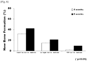
FIG. 8 is a graph showing the percentages of new bone formation in bone defects implanted with the dental barrier membrane of the present invention and with control barrier membranes. - Unless otherwise defined, all terms used herein have the same meaning as commonly understood by one of ordinary skill in the art to which example embodiments belong. It will be further understood that terms, e.g., those defined in commonly used dictionaries, should be interpreted as having a meaning that is consistent with their meaning in the context of the relevant art and will not be interpreted in an idealized or overly formal sense unless expressly so defined herein.
- The term dental barrier membrane, as used herein, refers to a barrier membrane designed to reconstruct bone tissues that have defects due to various dental diseases. The dental barrier membrane should significantly improve osteogenesis in bone defects or around implants, be hard enough to be sutured, and have biocompatibility for safe use invivo.
- In addition, a dental barrier membrane for guided bone regeneration is required to avoid post-operative infection and tissue degeneration, and closely adhere to periodontal tissues without giving a sensation of irritation. Importantly, dental barrier membranes should guarantee cell growth.
- When hydrolyzed invivo, however, most conventional dental barrier membranes cause the generation of acids such as lactic acid, glycolic acids, etc, which lower the pH in a local region, causing inflammation and histotoxicity. Further, for obtaining the desired properties, the dental barrier membranes used to date require pretreatment processes, such as physical crosslinking, etc. However, such pretreated dental barrier membranes may be toxic to the body and remain as foreign matter upon long-term use in vivo.
- Leading to the present invention, intensive and thorough research into a dental barrier membrane for guided bone regeneration, conducted by the present inventor, resulted in the finding that a fragment prepared from a cocoon is biocompatible and useful as a biomembrane not only because it has high tensile strength and porosity but also because it can promote osteogenesis in a bone defect, and that the fragment can be produced at significantly lower cost, compared to conventional dental barrier membranes.
- With reference to
FIG. 1 , the method for manufacturing a cocoon-based dental barrier membrane for guided bone regeneration in accordance with the present invention is explained below. - As shown in
FIG. 1A , acocoon 10, the shell of which has a first thickness, is prepared. - A cocoon is a casting spun of silk by silkworms and is used as a material for silk fibers. In the present invention, cocoons, which may be unused resources, are up-cycled into a new high value-added product, thus bringing economic benefits to silkworm farmers. Naturally constructed by silkworms, which eat clean mulberry leaves, cocoons are free of toxicity and are suitable for use as an environment-friendly material.
- Hence, the present invention takes a
cocoon 10 as a material for a dental barrier membrane. Thecocoon 10 is processed, as shown inFIGS. 1B to IE, into two or more planar fragments, each having a first thickness. - In greater detail, the
oval cocoon 10 is dissected along a cuttingline 11 into halves, as shown inFIG. 1B . The dissected halves have semi-oval shapes, and are opened to expose theinside surface 13 of the cocoon, as shown inFIG. 1C . - Next, the cocoon halves with curved inside surfaces 13 are planarized to some degree by cutting many sites along the edge as shown in FIG. ID, and planar regions are cut out to obtain cocoon fragments having a first thickness, as shown in FIG. IE.
- The dental barrier membrane prepared in the present invention need not have a planar surface. Because a cocoon originally has an elliptical ball shape, the curved shape of the dissected cocoon may be utilized to yield curved dental barrier membranes if necessary. For use as a small dental barrier membrane for guided bone regeneration, a cocoon fragment having a small area may be relatively planar. In contrast, when a relatively large area of the dissected cocoon halves is taken, the dental barrier membranes may have curved surfaces.
- With reference to
FIG. 2 , a modified method for preparing a cocoon fragment having a first thickness is described. In detail, an oval-shapedcocoon 10 is cut along asecond cutting line 15 and athird cutting line 17 to expose the inside 13 of thecocoon 10, as shown inFIGS. 2B and 2C . Then, the dissected cocoon having a curved surface is spread as shown inFIG. 2D , and then cocoon fragments 20 having a first thickness are obtained as shown inFIG. 2E . - The preparation methods of cocoon fragments described in
FIGS. 1 and 2 are only illustrative, but are not given as limitative embodiments. A cocoon fragment having a first thickness may be prepared by cutting a cocoon in the manners shown inFIGS. 1 and 2 , but other cutting methods may be used. - Because the cocoon fragments 20 having a first thickness, prepared in
step 1, have a multilayer structure identical to that of the cocoon shell, the multilayer structure may be split into thinner layers for use as a dental barrier membrane. - Although the
cocoon fragment 20 having a first thickness, prepared instep 1, is itself usable as a dental barrier membrane, it is subjected to thickness splitting to givecocoon fragments 30 having a second thickness. In this regard, the second thickness is smaller than the first thickness. As mentioned above, the cocoon fragment having the first thickness may be itself used as a dental barrier membrane without splitting to reduce the thickness thereof. - A cocoon shell varies in thickness (first thickness) from 0.3 to 1.0 mm depending on the silkworm species. In principal, any kind of cocoon may be used in the present invention. For the purpose of the present invention, a cocoon having a shell thickness of 0.5∼0.8 mm is employed.
- A cocoon fragment can be easily delaminated into up to 16 lamellas, although the number of delaminations is dependent on the shell thickness. The thicknesses of the lamellas can be determined according to the strength and elongation necessary for the kind and use of the dental barrier membrane. From a cocoon having a shell thickness of 0.5∼0.8 mm, a dental barrier membrane 0.01 mm - 0.7 mm thick can be prepared by delamination. According to the use of the dental barrier membrane, selection may be made of cocoon fragments 30 having various thicknesses.
- A better understanding of the present invention may be obtained through the following examples that are set forth to illustrate, but are not to be construed as limiting the present invention.
- The procedure of manufacturing a dental barrier membrane in accordance with the present invention is illustrated in
FIG. 3 . First, acocoon 10 was prepared, and cut at a proper site to expose the inside thereof. - Next, the cut cocoon was further processed to make the curved inside planar. The planarized cocoon was cut into
rectangular fragments 20. - The cocoon fragments 20 were peeled into strata having a suitable thickness, followed by sterilizing the strata to afford dental barrier membranes.
- Morphologies of the dental barrier membranes prepared in Examples 1 to 3 were observed by scanning electron microscopy (SEM), with commercially available collagen and PTFE (polytetrafluoroethylene) barrier membranes used as controls for comparison.
- As can be seen in
FIG. 4 , there were distinct differences in pore form and surface morphology between the dental barrier membrane of the present invention and the control, as observed by SEM. On the planar surface of the collagen barrier membrane, relatively large pores were arranged in a uniform pattern while the PTFE barrier membrane had regular arrangements of pores having constant sizes on planar surfaces. - In contrast, the dental barrier membrane of the present invention was observed to have many plies of irregularly entangled fibrous strands various in thickness, with many pores among the fibrous strands. Having a porous structure highly transmissive to nutrients and water, the dental barrier membrane of the present invention is therefore expect to find effective applications in osteogenesis at bone defects thanks to its ability to allow for the supply of nutrients and water necessary for bone regeneration.
- The physical properties of the cocoon-based dental barrier membranes prepared in Example 1 were measured. In this regard, a tensile test was conducted using a universal testing machine (DAEYEONG, Korea), with commercially available barrier membranes made of collagen or PTFE (polytetrafluoroethylene) serving as controls.
- Test specimens having sizes of 5 x 20 (width x length) mm were used. The specimens were extended at a rate of 10 mm/min, with an initial gauge length set to be 10 mm.
- In addition, considering the fact that dental barrier membranes, when used in the mouth, are continuously wetted with saliva, the specimens were immersed for 1 hr in physiological saline before the measurement of the mechanical properties (tensile strength).
-
[Table 1] Tensile Strength (MPa) Modulus of Elasticity Inventive Barrier Membrane 27.6±1.5 39.5±3.1 Collagen Barrier Membrane 3.5±1.3 9.6±2.2 PTFE Barrier Membrane 4.3±1.7 304.5±5.3 - The dental barrier membrane of the present invention and the controls, all being wetted, were measured for tensile strength and modulus of elasticity, and the results are given in Table 1. Tensile strength was measured to be 27.6±1.5 MPa in the barrier membrane of the present invention, 3.5±1.3 MPa in the collagen barrier membrane, and 4.3±1.7 MPa in the PTFE membrane. In addition, the modulus of elasticity was measured to be 39.5±3.1 in the barrier membrane of the present invention, 9.6±2.2 MPa in the collagen barrier membrane, and 304.5±5.3 MPa in the PTFE membrane.
- As can be seen, the barrier membrane of the present invention exhibited tensile strength 9 times higher than that of the collagen barrier membrane, and 7 times higher than that of the PTFE barrier membrane. Also, the barrier membrane of the present invention is elastic, as the modulus of elasticity was 4 times higher than that of the collagen barrier membrane.
- Next,
FIG. 5 is graph in which stresses are plotted against strains in the barrier membrane of the present invention, the collagen barrier membrane, and the PTFE barrier membrane. The PTFE barrier membrane was observed to greatly increase only in elongation within the range where a relatively large increase in tensile strength occurred. The collagen barrier membrane underwent fracture before a great increase in tensile strength. In contrast, the barrier membrane of the present invention increased in elongation with the increase of tensile strength. Accordingly, the barrier membrane of the present invention has higher tensile strength than do conventional collagen or PTFE barrier membranes. - For use in tissue regeneration and osteogenesis invivo,the barrier membrane should maintain its shape and have tensile strength, modulus of elasticity, and elongation large enough to withstand pressures in implantedregions. The cocoon-based membrane of the present invention is superior in tensile strength, modulus of elasticity, and elongation to commercially available controls, and thus is suitable for use as a dental barrier membrane.
- The dental barrier membrane prepared in Example 1 was implanted into a bone defect of rabbits, with a collagen barrier membrane or PTFE (polytetrafluoroethylene) barrier membrane serving as a control.
- The formation of new bone was examined by µ-CT (micro-computerized tomography), Masson's trichrome staining, and histological analysis at 4 and 8 weeks after the implantation of the barrier membranes.
- µ-CT (micro-computerized tomography) images of new bone formed after implantation of the dental barrier membrane of the present invention or control barrier membranes are given in
FIG. 6 . In the images, newly formed bone appears white. - At
post-operative week 4, new bone tissue was difficult to detect in the bone defect implanted with the PTFE barrier membrane, and occupied about 1/3 of the bone defect implanted with the collagen barrier membrane. The bone defect implanted with the barrier membrane of the present invention was 2/3 filled with new bone. - Even at
post-operative week 8, it was still difficult to detect new bone in the PTFE barrier membrane-implanted bone defect, with little difference frompost-operative week 4. The collagen barrier membrane allowed for the formation of new bone over about 2/3 of the overall bone defect, which was improved compared topost-operative week 4. - Over the overall region of the bone defect implanted with the barrier membrane of the present invention was formed new bone tissue, with a significant improvement compared to that at
post-operative week 4. Thus, the barrier membrane of the present invention notably exhibited much more efficient bone regeneration effects over the same period of time than did conventional barrier membranes. - Histological images after staining with Masson's trichrome are depicted in
FIG. 7. FIG. 7A shows histological images of barrier membrane-implanted bone defects at 4 weeks after implantation with barrier membranes, along with inserts of expanded views in which ossification by Ca2+depositionappearsred. - As shown in the histological image of the bone defect implanted with the barrier membrane of the present invention, the red portion was distributed deeply across the implanted region, indicating that the barrier membrane of the present invention allowed for active osteogenesis in the bone defect.
- In the expanded view for the collagen barrier membrane-implanted bone defect, part of the tissue appeared red, while almost no red portions were detected in the PTFE barrier membrane-implanted bone defect.
-
FIG. 7B shows histological images of barrier membrane-implanted bone defects at 8 weeks after implantation with barrier membranes, along with inserts of expanded views in which ossification appears blue. In the bone defect implanted with the barrier membrane of the present invention, red portions atpost-operative week 4 turned blue as new bone was formed. - For the collagen barrier membrane-implanted bone defect, ossification was observed to be in progress as red colors remained around blue colors accounting for new bone. In the histological image of the PTFT barrier membrane-implanted tissue, red colors appeared, accounting for the formation of new bone. These data were consistent with the µ-CT (micro-computerized tomography) data of
FIG. 6 . -
FIG. 8 is a graph in which the percentages of new bone formation on the Y axis are plotted against barrier membranes on the Y axis, on the basis of histological analysis, with a statistical significance of P<0.05. - As seen, the formation rate of new bone was 30% in the bone defect implanted with the barrier membrane of the present invention at
post-operative week 4, and increased to 40% atpost-operative week 8. When a bone defect was implanted with the collagen barrier membrane, new bone formation was 15% atpost-operative week post-operative week 8. For the PTFE barrier membrane, the formation rate of new bone was changed from 0% atpost-operative week 4 to 10% atpost-operative week 8. Therefore, the barrier membrane of the present invention allowed up to 4 times more new bone to be formed than did the controls. - Taken together, the data obtained above imply that the barrier membrane of the present invention exhibits an excellent bone regeneration effect on bone defects and is useful as a dental barrier membrane.
- Although the preferred embodiments of the present invention have been disclosed for illustrative purposes, those skilled in the art will appreciate that various modifications, additions and substitutions are possible, without departing from the scope of the invention as disclosed in the accompanying claims.
-
- 10: Cocoon
- 11: cutting
line 1 - 13: Inside surface
- 15: cutting
line 2 - 17: cutting
line 3 - 20: Cocoon fragment with a first thickness
- 30: Cocoon fragment with a second thickness
- 31: Inner stratum
- 33: Mid stratum
- 35: Outer stratum
Claims (12)
- A dental barrier membrane comprising a sterilized fragment (20) of a silkworm cocoon (10), prepared by dividing a silkworm cocoon (10) into two or more fragments (20) in a predetermined form, the cocoon (10) having a shell having a first thickness.
- The dental barrier membrane of claim 1, wherein each of the fragments (20) is delaminated into a lamellar fragment (30) having a second thickness, the second thickness being smaller than the first thickness.
- The dental barrier membrane of claim 2, wherein the lamellar fragment (30) having a second thickness is an inner stratum (31) of the cocoon (10).
- The dental barrier membrane of claim 2, wherein the lamellar fragment (30) having a second thickness is a middle stratum (32) of the cocoon (10).
- The dental barrier membrane of claim 2, wherein the lamellar fragment (30) having a second thickness is an outer stratum (33) of the cocoon (10).
- The dental barrier membrane of any one of claims 2 to 5, wherein the lamellar fragment (30) is packed.
- A method for manufacturing a dental barrier membrane comprising a sterilized fragment (20) of a silkworm cocoon (10), wherein the method comprises a first step of dividing a silkworm cocoon (10) into two or more fragments (20) in a predetermined form, the cocoon (10) having a shell having a first thickness, and wherein sterilization is conducted before or after each step.
- The method of claim 7, further comprising a second step of delaminating each of the fragments (20) into a lamellar fragment (30) having a second thickness, the second thickness being less than the first thickness.
- The method of claim 8, further comprising a third step of packing the fragments (30) having the second thickness prepared in the second step.
- The method of claim 8, wherein the lamellar fragment (30) having a second thickness is an inner stratum (31) of the cocoon.
- The method of claim 8, wherein the lamellar fragment (30) having a second thickness is a mid stratum (32) of the cocoon.
- The method of claim 8, wherein the lamellar fragment (30) having a second thickness is an outer stratum (33) of the cocoon.
Applications Claiming Priority (2)
| Application Number | Priority Date | Filing Date | Title |
|---|---|---|---|
| KR1020140099678A KR101570717B1 (en) | 2014-08-04 | 2014-08-04 | Dental Membrain using Cocoon and Method for manufacturing thereof |
| PCT/KR2015/007848 WO2016021859A1 (en) | 2014-08-04 | 2015-07-28 | Dental barrier membrane using cocoon and methdof for manufacturing same |
Publications (3)
| Publication Number | Publication Date |
|---|---|
| EP3177331A1 EP3177331A1 (en) | 2017-06-14 |
| EP3177331A4 EP3177331A4 (en) | 2017-08-23 |
| EP3177331B1 true EP3177331B1 (en) | 2019-06-26 |
Family
ID=54845147
Family Applications (1)
| Application Number | Title | Priority Date | Filing Date |
|---|---|---|---|
| EP15830245.5A Active EP3177331B1 (en) | 2014-08-04 | 2015-07-28 | Dental barrier membrane using cocoon and method for manufacturing same |
Country Status (8)
| Country | Link |
|---|---|
| US (1) | US10525165B2 (en) |
| EP (1) | EP3177331B1 (en) |
| JP (1) | JP6343390B2 (en) |
| KR (1) | KR101570717B1 (en) |
| CN (1) | CN106535950B (en) |
| ES (1) | ES2743305T3 (en) |
| HU (1) | HUE046332T2 (en) |
| WO (1) | WO2016021859A1 (en) |
Families Citing this family (4)
| Publication number | Priority date | Publication date | Assignee | Title |
|---|---|---|---|---|
| KR101602797B1 (en) * | 2015-10-21 | 2016-03-11 | 대한민국 | Artificial biomembrane using silk matrix and Method for manufacturing thereof |
| KR101602791B1 (en) * | 2015-10-21 | 2016-03-11 | 대한민국 | Vascular Patch using silk matrix and Method for manufaturing thereof |
| USD956979S1 (en) * | 2020-04-29 | 2022-07-05 | Megagen Implant Co., Ltd. | Dental membrane |
| USD956978S1 (en) * | 2020-04-29 | 2022-07-05 | Megagen Implant Co., Ltd. | Dental membrane |
Family Cites Families (13)
| Publication number | Priority date | Publication date | Assignee | Title |
|---|---|---|---|---|
| JPH06166850A (en) * | 1992-02-13 | 1994-06-14 | Marie Murase | Method for solubilizing and molding cocoon silk, artificial organ made of cocoon silk, and medical element made of cocoon silk |
| IT1316885B1 (en) | 2000-10-02 | 2003-05-13 | Consorzio Per Gli Studi Uni | PROCEDURE FOR THE PREPARATION OF A NON-WOVEN FABRIC IN SILK FIBER. |
| JP3772207B2 (en) | 2002-06-19 | 2006-05-10 | 独立行政法人農業生物資源研究所 | Biodegradable biopolymer material, production method thereof, and functional material comprising the polymer material |
| KR100762928B1 (en) * | 2004-10-29 | 2007-10-04 | 재단법인서울대학교산학협력재단 | Non-woven form of bone tissue-induced regeneration shielding membrane made of silk fibroin nanofibers and method for manufacturing same |
| US9381273B2 (en) * | 2008-01-31 | 2016-07-05 | Yissum Research Development Company Of The Hebrew University Of Jerusalem | Scaffolds with oxygen carriers, and their use in tissue regeneration |
| KR101060910B1 (en) | 2009-05-08 | 2011-08-30 | 대한민국(관리부서:농촌진흥청장) | Artificial tympanum using silk protein and its manufacturing method |
| WO2012081944A2 (en) * | 2010-12-16 | 2012-06-21 | Republic Of Korea(Management : Rural Development Administration) | Dental membrane and method of manufacturing the same |
| KR101403473B1 (en) * | 2011-09-27 | 2014-06-11 | 대한민국 | Dental membrane |
| CN102220686B (en) * | 2011-05-05 | 2014-02-05 | 苏州大学 | Method for improving degradation property of silk threads, and modified degummed silk threads |
| KR20140003942A (en) | 2012-07-02 | 2014-01-10 | 주식회사 네오바이오텍 | Dental barrier membrane having good growth of soft tissues or bone tissues |
| CN102847194A (en) * | 2012-09-17 | 2013-01-02 | 浙江星月生物科技股份有限公司 | Stent type silk fibroin film insoluble in water, and preparation and application of stent type silk fibroin film |
| CN103656762A (en) * | 2012-09-25 | 2014-03-26 | 耿兆波 | Application of silk fibroin film as absorbable tissue regeneration guiding film |
| CN103951831B (en) * | 2014-02-28 | 2016-08-17 | 华中科技大学同济医学院附属协和医院 | The preparation method and applications of sericin hydrogel |
-
2014
- 2014-08-04 KR KR1020140099678A patent/KR101570717B1/en active Active
-
2015
- 2015-07-28 JP JP2017505865A patent/JP6343390B2/en not_active Expired - Fee Related
- 2015-07-28 ES ES15830245T patent/ES2743305T3/en active Active
- 2015-07-28 US US15/326,747 patent/US10525165B2/en not_active Expired - Fee Related
- 2015-07-28 CN CN201580039457.6A patent/CN106535950B/en active Active
- 2015-07-28 WO PCT/KR2015/007848 patent/WO2016021859A1/en not_active Ceased
- 2015-07-28 HU HUE15830245A patent/HUE046332T2/en unknown
- 2015-07-28 EP EP15830245.5A patent/EP3177331B1/en active Active
Non-Patent Citations (1)
| Title |
|---|
| None * |
Also Published As
| Publication number | Publication date |
|---|---|
| CN106535950A (en) | 2017-03-22 |
| CN106535950B (en) | 2020-03-10 |
| EP3177331A4 (en) | 2017-08-23 |
| US20170203003A1 (en) | 2017-07-20 |
| ES2743305T3 (en) | 2020-02-18 |
| US10525165B2 (en) | 2020-01-07 |
| JP6343390B2 (en) | 2018-06-13 |
| WO2016021859A1 (en) | 2016-02-11 |
| HUE046332T2 (en) | 2020-03-30 |
| EP3177331A1 (en) | 2017-06-14 |
| JP2017529127A (en) | 2017-10-05 |
| KR101570717B1 (en) | 2015-11-24 |
Similar Documents
| Publication | Publication Date | Title |
|---|---|---|
| EP2289569B1 (en) | Collagen device and method of preparing the same | |
| US12226548B2 (en) | Method for treating a tissue defect with a collagen-containing membrane | |
| WO2017088818A1 (en) | Tissue repair fiber membrane, preparation method and application thereof, and tissue repair product | |
| US10525165B2 (en) | Dental barrier membrane using cocoon and method for manufacturing same | |
| EP3154600B1 (en) | Artificial biomembrane using cocoon and method for manufacturing same | |
| CN106730037B (en) | A kind of composite collagen biofilm and preparation method thereof | |
| Bellón et al. | Ultrastructural alterations of polytetrafluoroethylene prostheses implanted in abdominal wall provoked by infection: clinical and experimental study | |
| CN107708602B (en) | Dental barrier film using silk matrix and method of making the same | |
| EP3215134B1 (en) | Cocoon-based vascular patch and manufacturing method thereof | |
| CN108144128A (en) | A kind of repeatedly crosslinking breast sticking patch and preparation method thereof | |
| CN121846379A (en) | Bionic collagen matrix with density gradient double-layer structure and preparation method and application thereof |
Legal Events
| Date | Code | Title | Description |
|---|---|---|---|
| STAA | Information on the status of an ep patent application or granted ep patent |
Free format text: STATUS: THE INTERNATIONAL PUBLICATION HAS BEEN MADE |
|
| PUAI | Public reference made under article 153(3) epc to a published international application that has entered the european phase |
Free format text: ORIGINAL CODE: 0009012 |
|
| STAA | Information on the status of an ep patent application or granted ep patent |
Free format text: STATUS: REQUEST FOR EXAMINATION WAS MADE |
|
| 17P | Request for examination filed |
Effective date: 20170203 |
|
| AK | Designated contracting states |
Kind code of ref document: A1 Designated state(s): AL AT BE BG CH CY CZ DE DK EE ES FI FR GB GR HR HU IE IS IT LI LT LU LV MC MK MT NL NO PL PT RO RS SE SI SK SM TR |
|
| AX | Request for extension of the european patent |
Extension state: BA ME |
|
| A4 | Supplementary search report drawn up and despatched |
Effective date: 20170721 |
|
| RIC1 | Information provided on ipc code assigned before grant |
Ipc: A61L 27/38 20060101ALI20170714BHEP Ipc: A61C 5/00 20170101ALI20170714BHEP Ipc: A61C 8/02 20060101ALI20170714BHEP Ipc: A61L 27/22 20060101AFI20170714BHEP Ipc: A61C 13/107 20060101ALI20170714BHEP |
|
| DAV | Request for validation of the european patent (deleted) | ||
| DAX | Request for extension of the european patent (deleted) | ||
| STAA | Information on the status of an ep patent application or granted ep patent |
Free format text: STATUS: EXAMINATION IS IN PROGRESS |
|
| 17Q | First examination report despatched |
Effective date: 20180724 |
|
| GRAP | Despatch of communication of intention to grant a patent |
Free format text: ORIGINAL CODE: EPIDOSNIGR1 |
|
| STAA | Information on the status of an ep patent application or granted ep patent |
Free format text: STATUS: GRANT OF PATENT IS INTENDED |
|
| RIC1 | Information provided on ipc code assigned before grant |
Ipc: A61C 5/00 20170101ALI20181210BHEP Ipc: A61L 27/38 20060101ALI20181210BHEP Ipc: A61L 27/22 20060101AFI20181210BHEP Ipc: A61C 8/02 20060101ALI20181210BHEP Ipc: A61C 13/107 20060101ALI20181210BHEP |
|
| INTG | Intention to grant announced |
Effective date: 20190104 |
|
| GRAS | Grant fee paid |
Free format text: ORIGINAL CODE: EPIDOSNIGR3 |
|
| GRAA | (expected) grant |
Free format text: ORIGINAL CODE: 0009210 |
|
| STAA | Information on the status of an ep patent application or granted ep patent |
Free format text: STATUS: THE PATENT HAS BEEN GRANTED |
|
| AK | Designated contracting states |
Kind code of ref document: B1 Designated state(s): AL AT BE BG CH CY CZ DE DK EE ES FI FR GB GR HR HU IE IS IT LI LT LU LV MC MK MT NL NO PL PT RO RS SE SI SK SM TR |
|
| REG | Reference to a national code |
Ref country code: GB Ref legal event code: FG4D |
|
| REG | Reference to a national code |
Ref country code: CH Ref legal event code: EP |
|
| REG | Reference to a national code |
Ref country code: AT Ref legal event code: REF Ref document number: 1147557 Country of ref document: AT Kind code of ref document: T Effective date: 20190715 |
|
| REG | Reference to a national code |
Ref country code: DE Ref legal event code: R096 Ref document number: 602015032860 Country of ref document: DE |
|
| REG | Reference to a national code |
Ref country code: IE Ref legal event code: FG4D |
|
| REG | Reference to a national code |
Ref country code: CH Ref legal event code: NV Representative=s name: SERVOPATENT GMBH, CH |
|
| REG | Reference to a national code |
Ref country code: SE Ref legal event code: TRGR |
|
| REG | Reference to a national code |
Ref country code: NL Ref legal event code: MP Effective date: 20190626 |
|
| PG25 | Lapsed in a contracting state [announced via postgrant information from national office to epo] |
Ref country code: LT Free format text: LAPSE BECAUSE OF FAILURE TO SUBMIT A TRANSLATION OF THE DESCRIPTION OR TO PAY THE FEE WITHIN THE PRESCRIBED TIME-LIMIT Effective date: 20190626 Ref country code: HR Free format text: LAPSE BECAUSE OF FAILURE TO SUBMIT A TRANSLATION OF THE DESCRIPTION OR TO PAY THE FEE WITHIN THE PRESCRIBED TIME-LIMIT Effective date: 20190626 Ref country code: NO Free format text: LAPSE BECAUSE OF FAILURE TO SUBMIT A TRANSLATION OF THE DESCRIPTION OR TO PAY THE FEE WITHIN THE PRESCRIBED TIME-LIMIT Effective date: 20190926 Ref country code: FI Free format text: LAPSE BECAUSE OF FAILURE TO SUBMIT A TRANSLATION OF THE DESCRIPTION OR TO PAY THE FEE WITHIN THE PRESCRIBED TIME-LIMIT Effective date: 20190626 Ref country code: AL Free format text: LAPSE BECAUSE OF FAILURE TO SUBMIT A TRANSLATION OF THE DESCRIPTION OR TO PAY THE FEE WITHIN THE PRESCRIBED TIME-LIMIT Effective date: 20190626 |
|
| REG | Reference to a national code |
Ref country code: LT Ref legal event code: MG4D |
|
| PG25 | Lapsed in a contracting state [announced via postgrant information from national office to epo] |
Ref country code: BG Free format text: LAPSE BECAUSE OF FAILURE TO SUBMIT A TRANSLATION OF THE DESCRIPTION OR TO PAY THE FEE WITHIN THE PRESCRIBED TIME-LIMIT Effective date: 20190926 Ref country code: RS Free format text: LAPSE BECAUSE OF FAILURE TO SUBMIT A TRANSLATION OF THE DESCRIPTION OR TO PAY THE FEE WITHIN THE PRESCRIBED TIME-LIMIT Effective date: 20190626 Ref country code: GR Free format text: LAPSE BECAUSE OF FAILURE TO SUBMIT A TRANSLATION OF THE DESCRIPTION OR TO PAY THE FEE WITHIN THE PRESCRIBED TIME-LIMIT Effective date: 20190927 Ref country code: LV Free format text: LAPSE BECAUSE OF FAILURE TO SUBMIT A TRANSLATION OF THE DESCRIPTION OR TO PAY THE FEE WITHIN THE PRESCRIBED TIME-LIMIT Effective date: 20190626 |
|
| REG | Reference to a national code |
Ref country code: AT Ref legal event code: MK05 Ref document number: 1147557 Country of ref document: AT Kind code of ref document: T Effective date: 20190626 |
|
| PG25 | Lapsed in a contracting state [announced via postgrant information from national office to epo] |
Ref country code: NL Free format text: LAPSE BECAUSE OF FAILURE TO SUBMIT A TRANSLATION OF THE DESCRIPTION OR TO PAY THE FEE WITHIN THE PRESCRIBED TIME-LIMIT Effective date: 20190626 Ref country code: RO Free format text: LAPSE BECAUSE OF FAILURE TO SUBMIT A TRANSLATION OF THE DESCRIPTION OR TO PAY THE FEE WITHIN THE PRESCRIBED TIME-LIMIT Effective date: 20190626 Ref country code: EE Free format text: LAPSE BECAUSE OF FAILURE TO SUBMIT A TRANSLATION OF THE DESCRIPTION OR TO PAY THE FEE WITHIN THE PRESCRIBED TIME-LIMIT Effective date: 20190626 Ref country code: AT Free format text: LAPSE BECAUSE OF FAILURE TO SUBMIT A TRANSLATION OF THE DESCRIPTION OR TO PAY THE FEE WITHIN THE PRESCRIBED TIME-LIMIT Effective date: 20190626 Ref country code: CZ Free format text: LAPSE BECAUSE OF FAILURE TO SUBMIT A TRANSLATION OF THE DESCRIPTION OR TO PAY THE FEE WITHIN THE PRESCRIBED TIME-LIMIT Effective date: 20190626 Ref country code: SK Free format text: LAPSE BECAUSE OF FAILURE TO SUBMIT A TRANSLATION OF THE DESCRIPTION OR TO PAY THE FEE WITHIN THE PRESCRIBED TIME-LIMIT Effective date: 20190626 Ref country code: PT Free format text: LAPSE BECAUSE OF FAILURE TO SUBMIT A TRANSLATION OF THE DESCRIPTION OR TO PAY THE FEE WITHIN THE PRESCRIBED TIME-LIMIT Effective date: 20191028 |
|
| REG | Reference to a national code |
Ref country code: ES Ref legal event code: FG2A Ref document number: 2743305 Country of ref document: ES Kind code of ref document: T3 Effective date: 20200218 |
|
| PG25 | Lapsed in a contracting state [announced via postgrant information from national office to epo] |
Ref country code: SM Free format text: LAPSE BECAUSE OF FAILURE TO SUBMIT A TRANSLATION OF THE DESCRIPTION OR TO PAY THE FEE WITHIN THE PRESCRIBED TIME-LIMIT Effective date: 20190626 Ref country code: IS Free format text: LAPSE BECAUSE OF FAILURE TO SUBMIT A TRANSLATION OF THE DESCRIPTION OR TO PAY THE FEE WITHIN THE PRESCRIBED TIME-LIMIT Effective date: 20191026 Ref country code: IT Free format text: LAPSE BECAUSE OF FAILURE TO SUBMIT A TRANSLATION OF THE DESCRIPTION OR TO PAY THE FEE WITHIN THE PRESCRIBED TIME-LIMIT Effective date: 20190626 |
|
| REG | Reference to a national code |
Ref country code: HU Ref legal event code: AG4A Ref document number: E046332 Country of ref document: HU |
|
| PG25 | Lapsed in a contracting state [announced via postgrant information from national office to epo] |
Ref country code: TR Free format text: LAPSE BECAUSE OF FAILURE TO SUBMIT A TRANSLATION OF THE DESCRIPTION OR TO PAY THE FEE WITHIN THE PRESCRIBED TIME-LIMIT Effective date: 20190626 Ref country code: MC Free format text: LAPSE BECAUSE OF FAILURE TO SUBMIT A TRANSLATION OF THE DESCRIPTION OR TO PAY THE FEE WITHIN THE PRESCRIBED TIME-LIMIT Effective date: 20190626 |
|
| REG | Reference to a national code |
Ref country code: BE Ref legal event code: MM Effective date: 20190731 |
|
| PG25 | Lapsed in a contracting state [announced via postgrant information from national office to epo] |
Ref country code: DK Free format text: LAPSE BECAUSE OF FAILURE TO SUBMIT A TRANSLATION OF THE DESCRIPTION OR TO PAY THE FEE WITHIN THE PRESCRIBED TIME-LIMIT Effective date: 20190626 Ref country code: PL Free format text: LAPSE BECAUSE OF FAILURE TO SUBMIT A TRANSLATION OF THE DESCRIPTION OR TO PAY THE FEE WITHIN THE PRESCRIBED TIME-LIMIT Effective date: 20190626 |
|
| PG25 | Lapsed in a contracting state [announced via postgrant information from national office to epo] |
Ref country code: IS Free format text: LAPSE BECAUSE OF FAILURE TO SUBMIT A TRANSLATION OF THE DESCRIPTION OR TO PAY THE FEE WITHIN THE PRESCRIBED TIME-LIMIT Effective date: 20200224 Ref country code: BE Free format text: LAPSE BECAUSE OF NON-PAYMENT OF DUE FEES Effective date: 20190731 Ref country code: LU Free format text: LAPSE BECAUSE OF NON-PAYMENT OF DUE FEES Effective date: 20190728 |
|
| REG | Reference to a national code |
Ref country code: DE Ref legal event code: R097 Ref document number: 602015032860 Country of ref document: DE |
|
| REG | Reference to a national code |
Ref country code: CH Ref legal event code: PCAR Free format text: NEW ADDRESS: WANNERSTRASSE 9/1, 8045 ZUERICH (CH) |
|
| PLBE | No opposition filed within time limit |
Free format text: ORIGINAL CODE: 0009261 |
|
| STAA | Information on the status of an ep patent application or granted ep patent |
Free format text: STATUS: NO OPPOSITION FILED WITHIN TIME LIMIT |
|
| PG2D | Information on lapse in contracting state deleted |
Ref country code: IS |
|
| PG25 | Lapsed in a contracting state [announced via postgrant information from national office to epo] |
Ref country code: IE Free format text: LAPSE BECAUSE OF NON-PAYMENT OF DUE FEES Effective date: 20190728 |
|
| 26N | No opposition filed |
Effective date: 20200603 |
|
| PG25 | Lapsed in a contracting state [announced via postgrant information from national office to epo] |
Ref country code: SI Free format text: LAPSE BECAUSE OF FAILURE TO SUBMIT A TRANSLATION OF THE DESCRIPTION OR TO PAY THE FEE WITHIN THE PRESCRIBED TIME-LIMIT Effective date: 20190626 |
|
| PG25 | Lapsed in a contracting state [announced via postgrant information from national office to epo] |
Ref country code: CY Free format text: LAPSE BECAUSE OF FAILURE TO SUBMIT A TRANSLATION OF THE DESCRIPTION OR TO PAY THE FEE WITHIN THE PRESCRIBED TIME-LIMIT Effective date: 20190626 |
|
| PG25 | Lapsed in a contracting state [announced via postgrant information from national office to epo] |
Ref country code: MT Free format text: LAPSE BECAUSE OF FAILURE TO SUBMIT A TRANSLATION OF THE DESCRIPTION OR TO PAY THE FEE WITHIN THE PRESCRIBED TIME-LIMIT Effective date: 20190626 |
|
| PG25 | Lapsed in a contracting state [announced via postgrant information from national office to epo] |
Ref country code: MK Free format text: LAPSE BECAUSE OF FAILURE TO SUBMIT A TRANSLATION OF THE DESCRIPTION OR TO PAY THE FEE WITHIN THE PRESCRIBED TIME-LIMIT Effective date: 20190626 |
|
| PGFP | Annual fee paid to national office [announced via postgrant information from national office to epo] |
Ref country code: SE Payment date: 20220603 Year of fee payment: 8 Ref country code: GB Payment date: 20220607 Year of fee payment: 8 |
|
| PGFP | Annual fee paid to national office [announced via postgrant information from national office to epo] |
Ref country code: FR Payment date: 20220607 Year of fee payment: 8 |
|
| PGFP | Annual fee paid to national office [announced via postgrant information from national office to epo] |
Ref country code: ES Payment date: 20220811 Year of fee payment: 8 Ref country code: DE Payment date: 20220603 Year of fee payment: 8 |
|
| PGFP | Annual fee paid to national office [announced via postgrant information from national office to epo] |
Ref country code: HU Payment date: 20220708 Year of fee payment: 8 |
|
| PGFP | Annual fee paid to national office [announced via postgrant information from national office to epo] |
Ref country code: CH Payment date: 20220801 Year of fee payment: 8 |
|
| REG | Reference to a national code |
Ref country code: DE Ref legal event code: R119 Ref document number: 602015032860 Country of ref document: DE |
|
| REG | Reference to a national code |
Ref country code: CH Ref legal event code: PL |
|
| REG | Reference to a national code |
Ref country code: SE Ref legal event code: EUG |
|
| GBPC | Gb: european patent ceased through non-payment of renewal fee |
Effective date: 20230728 |
|
| PG25 | Lapsed in a contracting state [announced via postgrant information from national office to epo] |
Ref country code: HU Free format text: LAPSE BECAUSE OF NON-PAYMENT OF DUE FEES Effective date: 20230729 Ref country code: DE Free format text: LAPSE BECAUSE OF NON-PAYMENT OF DUE FEES Effective date: 20240201 Ref country code: CH Free format text: LAPSE BECAUSE OF NON-PAYMENT OF DUE FEES Effective date: 20230731 Ref country code: GB Free format text: LAPSE BECAUSE OF NON-PAYMENT OF DUE FEES Effective date: 20230728 |
|
| PG25 | Lapsed in a contracting state [announced via postgrant information from national office to epo] |
Ref country code: SE Free format text: LAPSE BECAUSE OF NON-PAYMENT OF DUE FEES Effective date: 20230729 Ref country code: FR Free format text: LAPSE BECAUSE OF NON-PAYMENT OF DUE FEES Effective date: 20230731 |
|
| REG | Reference to a national code |
Ref country code: ES Ref legal event code: FD2A Effective date: 20240902 |
|
| PG25 | Lapsed in a contracting state [announced via postgrant information from national office to epo] |
Ref country code: ES Free format text: LAPSE BECAUSE OF NON-PAYMENT OF DUE FEES Effective date: 20230729 |
|
| PG25 | Lapsed in a contracting state [announced via postgrant information from national office to epo] |
Ref country code: ES Free format text: LAPSE BECAUSE OF NON-PAYMENT OF DUE FEES Effective date: 20230729 |