EP3175779A1 - Method, system and computer program for measuring light diffusion in the eyeball or in the ocular region - Google Patents
Method, system and computer program for measuring light diffusion in the eyeball or in the ocular region Download PDFInfo
- Publication number
- EP3175779A1 EP3175779A1 EP15827160.1A EP15827160A EP3175779A1 EP 3175779 A1 EP3175779 A1 EP 3175779A1 EP 15827160 A EP15827160 A EP 15827160A EP 3175779 A1 EP3175779 A1 EP 3175779A1
- Authority
- EP
- European Patent Office
- Prior art keywords
- ocular
- image
- measurement
- eye
- light diffusion
- Prior art date
- Legal status (The legal status is an assumption and is not a legal conclusion. Google has not performed a legal analysis and makes no representation as to the accuracy of the status listed.)
- Granted
Links
Images
Classifications
-
- A—HUMAN NECESSITIES
- A61—MEDICAL OR VETERINARY SCIENCE; HYGIENE
- A61B—DIAGNOSIS; SURGERY; IDENTIFICATION
- A61B3/00—Apparatus for testing the eyes; Instruments for examining the eyes
- A61B3/10—Objective types, i.e. instruments for examining the eyes independent of the patients' perceptions or reactions
- A61B3/103—Objective types, i.e. instruments for examining the eyes independent of the patients' perceptions or reactions for determining refraction, e.g. refractometers, skiascopes
- A61B3/1035—Objective types, i.e. instruments for examining the eyes independent of the patients' perceptions or reactions for determining refraction, e.g. refractometers, skiascopes for measuring astigmatism
-
- A—HUMAN NECESSITIES
- A61—MEDICAL OR VETERINARY SCIENCE; HYGIENE
- A61B—DIAGNOSIS; SURGERY; IDENTIFICATION
- A61B3/00—Apparatus for testing the eyes; Instruments for examining the eyes
- A61B3/18—Arrangement of plural eye-testing or -examining apparatus
- A61B3/185—Arrangement of plural eye-testing or -examining apparatus characterised by modular construction
-
- A—HUMAN NECESSITIES
- A61—MEDICAL OR VETERINARY SCIENCE; HYGIENE
- A61B—DIAGNOSIS; SURGERY; IDENTIFICATION
- A61B3/00—Apparatus for testing the eyes; Instruments for examining the eyes
- A61B3/10—Objective types, i.e. instruments for examining the eyes independent of the patients' perceptions or reactions
-
- A—HUMAN NECESSITIES
- A61—MEDICAL OR VETERINARY SCIENCE; HYGIENE
- A61B—DIAGNOSIS; SURGERY; IDENTIFICATION
- A61B3/00—Apparatus for testing the eyes; Instruments for examining the eyes
- A61B3/10—Objective types, i.e. instruments for examining the eyes independent of the patients' perceptions or reactions
- A61B3/1015—Objective types, i.e. instruments for examining the eyes independent of the patients' perceptions or reactions for wavefront analysis
-
- A—HUMAN NECESSITIES
- A61—MEDICAL OR VETERINARY SCIENCE; HYGIENE
- A61B—DIAGNOSIS; SURGERY; IDENTIFICATION
- A61B3/00—Apparatus for testing the eyes; Instruments for examining the eyes
- A61B3/10—Objective types, i.e. instruments for examining the eyes independent of the patients' perceptions or reactions
- A61B3/12—Objective types, i.e. instruments for examining the eyes independent of the patients' perceptions or reactions for looking at the eye fundus, e.g. ophthalmoscopes
-
- A—HUMAN NECESSITIES
- A61—MEDICAL OR VETERINARY SCIENCE; HYGIENE
- A61B—DIAGNOSIS; SURGERY; IDENTIFICATION
- A61B3/00—Apparatus for testing the eyes; Instruments for examining the eyes
- A61B3/10—Objective types, i.e. instruments for examining the eyes independent of the patients' perceptions or reactions
- A61B3/14—Arrangements specially adapted for eye photography
-
- A—HUMAN NECESSITIES
- A61—MEDICAL OR VETERINARY SCIENCE; HYGIENE
- A61B—DIAGNOSIS; SURGERY; IDENTIFICATION
- A61B3/00—Apparatus for testing the eyes; Instruments for examining the eyes
- A61B3/10—Objective types, i.e. instruments for examining the eyes independent of the patients' perceptions or reactions
- A61B3/14—Arrangements specially adapted for eye photography
- A61B3/145—Arrangements specially adapted for eye photography by video means
-
- A—HUMAN NECESSITIES
- A61—MEDICAL OR VETERINARY SCIENCE; HYGIENE
- A61B—DIAGNOSIS; SURGERY; IDENTIFICATION
- A61B3/00—Apparatus for testing the eyes; Instruments for examining the eyes
- A61B3/18—Arrangement of plural eye-testing or -examining apparatus
Definitions
- the present invention generally relates, in a first aspect, to a method for measuring light diffusion in the eyeball or in the ocular region, and more particularly to a method combining ocular aberration measurements information with information on the image of the retinal plane of one or both eyes of a patient.
- a second aspect of the invention relates to a system suitable for implementing the method of the first aspect.
- a third aspect of the invention relates to a computer program suitable for implementing the method of the first aspect.
- Light diffusion in the eyeball or in the ocular region is one of the three causes of image quality deterioration in the retina in the human eye, the other two causes being optical aberrations and diffraction.
- the combined contribution of optical aberrations and intraocular diffusion affects retinal image quality.
- the double-pass technique J. Santamaria, P. Artal, J. Bescos, "Determination of the point-spread function of human eyes using a hybrid optical-digital method", J., Opt. Soc. Am. A, 4, 1109-1114 (1987 )
- based on projecting a collimated light beam onto the retina of a patient, and directly recording the light reflected therein after the double passage of the light through the ocular media allows obtaining an objective measurement of the contribution of aberrations and intraocular diffusion to ocular optical quality
- Ciencia., 47, 1710-1716 (2006 ), describes the separate use of both the double-pass technique and of the ocular aberrations measurement technique by means of sensors of wavefront (particularly by means of a Hartmann-Shack sensor), for the purpose of comparing the results obtained by means of both techniques for determining when one is better than the other when estimating the quality of the image of the retina, concluding that for eyes with low intraocular diffusion, both techniques offer similar results, and that, in contrast, for eyes with intermediate or high intraocular diffusion values the double-pass technique is better because it produces a more precise description of the optical quality, best correlated with vision quality, whereas the aberration measurement technique can produce results that overestimate the retinal image quality.
- said article propose combining the results obtained by means of using each of the two mentioned techniques for obtaining a combined result, or measuring light diffusion in the eyeball or in the ocular region, or performing any other kind of measurement.
- patent application EP2147633A1 proposes a method and a system for measuring light diffusion in the eyeball or in the ocular region, which comprises performing the steps described in the preamble of claim 1 of the present invention.
- the present invention relates, in a first aspect, to a method for measuring light diffusion in the eyeball or in the ocular region, which comprises performing the following steps:
- the method proposed by the first aspect of the invention characteristically comprises, at the same time as said capturing of said image of the retinal plane, taking a high- and low-order ocular aberration measurement in the plane of the pupil of said eye and taking a light diffusion measurement, combining information obtained by means of said ocular aberration measurement with information on said image of the retinal plane.
- Capturing the image of the retinal plane and taking the high- and low-order ocular aberration measurement at the same time is essential for obtaining good results as regards the light diffusion measurement, because if they did not take place at the same time, the conditions in which both measurements (the measurement associated with capturing the image of the retinal plane and the ocular aberration measurement) would be taken would not be identical or similar enough to be combined with one another.
- Said low-order aberrations preferably include astigmatism and defocusing.
- the method proposed by the first aspect of the invention is implemented using open field techniques and/or in a binocular manner in both eyes of the patient.
- the high- and low-order ocular aberration measurement is a second measurement, wherein the method comprises previously taking a first ocular aberration measurement of the eye of the patient and using the obtained results for the mentioned correcting of low-order ocular aberrations of said eye.
- Low-order ocular aberrations can be corrected using any technique known, ranging from the technique associated with a standard methodology to the technique disclosed by the authors of the present invention in patent application EP2147633A1 .
- the method of the first aspect of the invention comprises using one and the same system for measuring aberrations (generally an aberrometer) for taking the first and second ocular aberration measurements.
- an aberrometer for taking the first and second ocular aberration measurements.
- two systems for measuring aberrations one for each of the first and second measurements.
- the method comprises performing the mentioned high- and low-order ocular aberration measurement on the wavefront coming from the reflection of said punctiform light beam on the retina, as it passes through the plane of the pupil.
- the method of the present invention comprises analyzing the distribution of light of the image of the retinal plane and the of an image corresponding to said ocular aberration measurement on said wavefront, in the plane of the pupil, and taking a light diffusion measurement by comparing both distributions of light, advantageously for each of the light spots of both images.
- the method comprises calculating the objective scatter index (OSI) described in patent application EP2147633A1 with the information on the image of the retinal plane, and combining the result obtained with the result obtained by applying any known methodology to the ocular aberration measurement information, for obtaining the final light diffusion measurement.
- OSI objective scatter index
- the method comprises applying respective optical transfer functions, or OTF, to the ocular aberration measurement information and to the information on the image of the retinal plane and taking a light diffusion measurement, combining the results provided by said OTF functions.
- OTF optical transfer functions
- the method proposed by the first aspect of the invention comprises performing the light diffusion measurement, combining the results provided by said OTF functions and also comparing both of said distributions of light.
- said functions include at least absolute values in respective modulation transfer functions, or MTF, wherein the method comprises performing the light diffusion measurement dividing values associated with profiles (for example radial profiles or one-way profiles) generated with said absolute values, for example calculating the existing areas under the curves of said profiles, performing said division with the values calculated for said areas.
- profiles for example radial profiles or one-way profiles
- said OTF functions include complex argument values in respective phase transfer functions, or PTF.
- a second aspect of the present invention relates to a system for measuring light diffusion in the eyeball or in the ocular region, comprising:
- the system proposed by the second aspect of the present invention comprises
- the system proposed by the second aspect of the invention is provided for implementing the method according to the first aspect.
- the mentioned means for projecting a punctiform light beam onto the retina of an eye of a patient and the means for capturing and recording an image of the retinal plane are part of a double-pass ophthalmoscopic system.
- the system proposed by the second aspect of the invention is configured and arranged for using open field techniques.
- the system proposed by the second aspect of the invention is configured and arranged for implementing a binocular system applied to both eyes of the patient.
- system proposed by the second aspect of the invention is configured and arranged for implementing a monocular system.
- the means for correcting low-order ocular aberrations comprise or are associated with the means for taking an ocular aberration measurement, for correcting ocular aberrations depending on measurements taken with the means for taking an ocular aberration measurement.
- the means for correcting low-order ocular aberrations and the means for taking an ocular aberration measurement are independent from one another.
- a third aspect of the present invention relates to a computer program including code instructions which, when run in a computer, take a light diffusion measurement in the eyeball or in the ocular region according to the method of the first aspect, processing, in a combined manner, data corresponding to the information obtained by means of the ocular aberration measurement with data corresponding to the information on the image of the retinal plane.
- the system for measuring light diffusion in the eyeball or in the ocular region proposed by the second aspect of the present invention is an open field binocular system comprising means for projecting a punctiform light beam onto the retina of both eyes 01, 02 of a patient, which include a punctiform light source SLD generating a collimated (generally a laser light) beam which is distributed into two respective emission sub-beams by a mask or pupil P1, which are directed towards each of the two eyes O1, 02 by a series of common optical elements, particularly beam splitters BS1 and BS2 acting as mirrors and lenses L1 and L2, and after each of the emission sub-beams is redirected towards the respective eye O1, 02 by the mirror M1, each of them is individually directed by one of respective groups of optical elements, each of them formed particularly by a pair of cylindrical lenses C1-2 and C3-4, after which there are arranged another lens LL1 and LL2, another pupil P2 and P3, a respective mirror M2 and M4, another lens
- SLD punctiform light source
- Figure 1 also shows how the system comprises means consisting of two cameras CCD-DPly CCD-DP2, each of them provided for capturing and recording an image of the plane of one of the retinas, which image is formed after reflection of the two punctiform light emission sub-beams in the retina and a double passage through the ocular media of both eyes 01, 02, generating respective reflection sub-beams following a path opposite the emission sub-beam path, going through the optical elements to the beam splitter BS2, which reflects a portion of each of the reflection sub-beams, going through the lens L1 and the beam splitter BS1, after which each of the reflection sub-beams is directed to one of the cameras CCD-DP1 and CCD-DP2 by the mirror M3.
- the system comprises means consisting of two cameras CCD-DPly CCD-DP2, each of them provided for capturing and recording an image of the plane of one of the retinas, which image is formed after reflection of the two punctiform light emission sub-beams in the retina and a double passage
- the pairs of cylindrical lenses C1-2 and C3-4 are part of or are means for correcting low-order ocular aberrations of the eyes 01, 02 prior to capturing and recording the images of the retinal planes.
- Figure 1 also shows how the system also comprises means for performing a high- and low-order ocular aberration measurement in the plane of the pupil of both eyes 01, 02, which particularly consists of a Hartmann-Shack sensor HS, which is struck by portions of the two reflection sub-beams going through the beam splitter BS2 and the lens L4.
- means for performing a high- and low-order ocular aberration measurement in the plane of the pupil of both eyes 01, 02 which particularly consists of a Hartmann-Shack sensor HS, which is struck by portions of the two reflection sub-beams going through the beam splitter BS2 and the lens L4.
- Figure 1 likewise illustrates, by means of arrows, the direction of the light in the first and second passage, i.e., the emission sub-beams towards the retinas and the reflection sub-beams in the opposite direction.
- Figure 1 also illustrates an electronic system SE communicated in a bidirectional manner with the cameras CCD-DP1, CCD-DP2 and the Hartmann-Shack sensor HS, and comprising control means MC controlling at least CCD-DP1, CCD-DP2 and HS so that they operate at the same time, and processing means MP processing, in a combined manner, information obtained by means of CCD-DP1, CCD-DP2 and HS and providing, as a result of said processing, the value or values of the light diffusion measurement.
- control means MC controlling at least CCD-DP1, CCD-DP2 and HS so that they operate at the same time
- processing means MP processing in a combined manner, information obtained by means of CCD-DP1, CCD-DP2 and HS and providing, as a result of said processing, the value or values of the light diffusion measurement.
- control means MC are connected with the pairs of cylindrical lenses C1-2 and C3-4 (and/or with any other type of alternative mechanism suitable for correcting low-order ocular aberrations) to control them for the purpose of the mentioned correcting of ocular aberrations, depending on ocular aberration measurements taken with the Hartmann-Shack sensor HS.
Landscapes
- Health & Medical Sciences (AREA)
- Life Sciences & Earth Sciences (AREA)
- Engineering & Computer Science (AREA)
- Medical Informatics (AREA)
- Surgery (AREA)
- Biophysics (AREA)
- Biomedical Technology (AREA)
- Heart & Thoracic Surgery (AREA)
- Physics & Mathematics (AREA)
- Molecular Biology (AREA)
- Ophthalmology & Optometry (AREA)
- Animal Behavior & Ethology (AREA)
- General Health & Medical Sciences (AREA)
- Public Health (AREA)
- Veterinary Medicine (AREA)
- Multimedia (AREA)
- Eye Examination Apparatus (AREA)
- Testing Of Optical Devices Or Fibers (AREA)
Abstract
- projecting a punctiform light beam onto the retina;
- correcting low-order ocular aberrations of the eye;
- capturing and recording an image of the retinal plane formed after reflection of the punctiform light beam on the retina and a double passage through the ocular media of the eye; and
- at the same time as capturing the image of the retinal plane, performing a high- and low-order ocular aberration measurement in the plane of the pupil and performing a light diffusion measurement, combining ocular aberration measurement information with information on the image of the retinal plane.
Description
- The present invention generally relates, in a first aspect, to a method for measuring light diffusion in the eyeball or in the ocular region, and more particularly to a method combining ocular aberration measurements information with information on the image of the retinal plane of one or both eyes of a patient.
- A second aspect of the invention relates to a system suitable for implementing the method of the first aspect.
- A third aspect of the invention relates to a computer program suitable for implementing the method of the first aspect.
- Light diffusion in the eyeball or in the ocular region is one of the three causes of image quality deterioration in the retina in the human eye, the other two causes being optical aberrations and diffraction.
- The combined contribution of optical aberrations and intraocular diffusion affects retinal image quality. The double-pass technique (J. Santamaria, P. Artal, J. Bescos, "Determination of the point-spread function of human eyes using a hybrid optical-digital method", J., Opt. Soc. Am. A, 4, 1109-1114 (1987)), based on projecting a collimated light beam onto the retina of a patient, and directly recording the light reflected therein after the double passage of the light through the ocular media, allows obtaining an objective measurement of the contribution of aberrations and intraocular diffusion to ocular optical quality (F. Diaz-Douton, A. Benito, J. Pujol, M. Arjona, JL Güell, P. Artal, "Comparison of the retinal image quality obtained with a Hartmann-Shack sensor and a double-pass instrument ", Inv. Ophthal. Vis. Ciencia., 47, 1710-1716 (2006)).
- In the article entitled "Comparison of the retinal image quality obtained with a Hartmann-Shack sensor and a double-pass instrument ", F. Diaz-Douton, A. Benito, J. Pujol, M. Arjona, JL Güell, P. Artal, Inv. Ophthal. Vis. Ciencia., 47, 1710-1716 (2006), describes the separate use of both the double-pass technique and of the ocular aberrations measurement technique by means of sensors of wavefront (particularly by means of a Hartmann-Shack sensor), for the purpose of comparing the results obtained by means of both techniques for determining when one is better than the other when estimating the quality of the image of the retina, concluding that for eyes with low intraocular diffusion, both techniques offer similar results, and that, in contrast, for eyes with intermediate or high intraocular diffusion values the double-pass technique is better because it produces a more precise description of the optical quality, best correlated with vision quality, whereas the aberration measurement technique can produce results that overestimate the retinal image quality.
- At no point does said article propose combining the results obtained by means of using each of the two mentioned techniques for obtaining a combined result, or measuring light diffusion in the eyeball or in the ocular region, or performing any other kind of measurement.
- In addition, patent application
EP2147633A1 proposes a method and a system for measuring light diffusion in the eyeball or in the ocular region, which comprises performing the steps described in the preamble of claim 1 of the present invention. - It is necessary to offer an alternative to the prior art that covers the voids therein, by providing a light diffusion measurement in the eyeball or in the ocular region that is more precise than the one obtained by means of the proposals from the prior art.
- For that purpose, the present invention relates, in a first aspect, to a method for measuring light diffusion in the eyeball or in the ocular region, which comprises performing the following steps:
- projecting a punctiform light beam onto the retina of at least one eye of a patient;
- correcting low-order ocular aberrations of said eye; and
- capturing and recording, once said low-order aberrations have been corrected, at least one image of the retinal plane formed after reflection of said punctiform light beam on the retina and a double passage through the ocular media of said eye.
- Unlike the known methods for taking measurement, where at most one ocular aberration measurement has been used to validate the results obtained by means of processing an image of the retinal plane and the information on said image, the method proposed by the first aspect of the invention characteristically comprises, at the same time as said capturing of said image of the retinal plane, taking a high- and low-order ocular aberration measurement in the plane of the pupil of said eye and taking a light diffusion measurement, combining information obtained by means of said ocular aberration measurement with information on said image of the retinal plane.
- Capturing the image of the retinal plane and taking the high- and low-order ocular aberration measurement at the same time is essential for obtaining good results as regards the light diffusion measurement, because if they did not take place at the same time, the conditions in which both measurements (the measurement associated with capturing the image of the retinal plane and the ocular aberration measurement) would be taken would not be identical or similar enough to be combined with one another.
- Said low-order aberrations preferably include astigmatism and defocusing.
- According to one embodiment, the method proposed by the first aspect of the invention is implemented using open field techniques and/or in a binocular manner in both eyes of the patient.
- According to one embodiment, the high- and low-order ocular aberration measurement is a second measurement, wherein the method comprises previously taking a first ocular aberration measurement of the eye of the patient and using the obtained results for the mentioned correcting of low-order ocular aberrations of said eye.
- Low-order ocular aberrations can be corrected using any technique known, ranging from the technique associated with a standard methodology to the technique disclosed by the authors of the present invention in patent application
EP2147633A1 . - Advantageously, the method of the first aspect of the invention comprises using one and the same system for measuring aberrations (generally an aberrometer) for taking the first and second ocular aberration measurements. For another less preferred embodiment, are used two systems for measuring aberrations, one for each of the first and second measurements.
- The method comprises performing the mentioned high- and low-order ocular aberration measurement on the wavefront coming from the reflection of said punctiform light beam on the retina, as it passes through the plane of the pupil.
- According to one embodiment, the method of the present invention comprises analyzing the distribution of light of the image of the retinal plane and the of an image corresponding to said ocular aberration measurement on said wavefront, in the plane of the pupil, and taking a light diffusion measurement by comparing both distributions of light, advantageously for each of the light spots of both images.
- For one embodiment, the method comprises calculating the objective scatter index (OSI) described in patent application
EP2147633A1 with the information on the image of the retinal plane, and combining the result obtained with the result obtained by applying any known methodology to the ocular aberration measurement information, for obtaining the final light diffusion measurement. - For another embodiment, the method comprises applying respective optical transfer functions, or OTF, to the ocular aberration measurement information and to the information on the image of the retinal plane and taking a light diffusion measurement, combining the results provided by said OTF functions.
- According to a variant of said embodiment, the method proposed by the first aspect of the invention comprises performing the light diffusion measurement, combining the results provided by said OTF functions and also comparing both of said distributions of light.
- As regards OTF functions, said functions include at least absolute values in respective modulation transfer functions, or MTF, wherein the method comprises performing the light diffusion measurement dividing values associated with profiles (for example radial profiles or one-way profiles) generated with said absolute values, for example calculating the existing areas under the curves of said profiles, performing said division with the values calculated for said areas.
- According to one embodiment, said OTF functions include complex argument values in respective phase transfer functions, or PTF.
- A second aspect of the present invention relates to a system for measuring light diffusion in the eyeball or in the ocular region, comprising:
- means for projecting a punctiform light beam onto the retina of at least one eye of a patient;
- means for capturing and recording an image of the retinal plane formed after reflection of said punctiform light beam on the retina and a double passage through the ocular media of said eye; and
- means for correcting low-order ocular aberrations of said eye prior to said capturing and recording.
- Unlike known systems, the system proposed by the second aspect of the present invention comprises
- means for taking a high- and low-order ocular aberration measurement in the plane of the pupil of said eye;
- control means controlling at least said means for capturing and recording an image of the retinal plane and said means for taking an ocular aberration measurement, so that they operate at the same time; and
- processing means processing, in a combined manner, information obtained by means of said ocular aberration measurement with information on said image of the retinal plane and providing, as a result of said processing, the value or values of said light diffusion measurement.
- The system proposed by the second aspect of the invention is provided for implementing the method according to the first aspect.
- Preferably, the mentioned means for projecting a punctiform light beam onto the retina of an eye of a patient and the means for capturing and recording an image of the retinal plane are part of a double-pass ophthalmoscopic system.
- Advantageously, the system proposed by the second aspect of the invention is configured and arranged for using open field techniques.
- For a preferred embodiment, the system proposed by the second aspect of the invention is configured and arranged for implementing a binocular system applied to both eyes of the patient.
- For another less preferred embodiment, the system proposed by the second aspect of the invention is configured and arranged for implementing a monocular system.
- According to a preferred embodiment, the means for correcting low-order ocular aberrations comprise or are associated with the means for taking an ocular aberration measurement, for correcting ocular aberrations depending on measurements taken with the means for taking an ocular aberration measurement.
- For another less preferred embodiment, the means for correcting low-order ocular aberrations and the means for taking an ocular aberration measurement are independent from one another.
- A third aspect of the present invention relates to a computer program including code instructions which, when run in a computer, take a light diffusion measurement in the eyeball or in the ocular region according to the method of the first aspect, processing, in a combined manner, data corresponding to the information obtained by means of the ocular aberration measurement with data corresponding to the information on the image of the retinal plane.
- The foregoing and other advantages and features will be better understood based on the following detailed description of several embodiments making reference to the attached drawings, which must be interpreted in a non-limiting illustrative manner, in which:
-
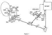
Figure 1 is a perspective view of a schematic depiction of the system proposed by the second aspect of the present invention for an embodiment in which an open field binocular system is implemented. - According to the embodiment illustrated in
Figure 1 , the system for measuring light diffusion in the eyeball or in the ocular region proposed by the second aspect of the present invention is an open field binocular system comprising means for projecting a punctiform light beam onto the retina of botheyes 01, 02 of a patient, which include a punctiform light source SLD generating a collimated (generally a laser light) beam which is distributed into two respective emission sub-beams by a mask or pupil P1, which are directed towards each of the two eyes O1, 02 by a series of common optical elements, particularly beam splitters BS1 and BS2 acting as mirrors and lenses L1 and L2, and after each of the emission sub-beams is redirected towards the respective eye O1, 02 by the mirror M1, each of them is individually directed by one of respective groups of optical elements, each of them formed particularly by a pair of cylindrical lenses C1-2 and C3-4, after which there are arranged another lens LL1 and LL2, another pupil P2 and P3, a respective mirror M2 and M4, another lens L3 and L5, a dichroic mirror DM1 and DM2 and, finally, a heat mirror HM1 and HM2, after which the emission sub-beam strikes the retina of the eye O1, 02. -
Figure 1 also shows how the system comprises means consisting of two cameras CCD-DPly CCD-DP2, each of them provided for capturing and recording an image of the plane of one of the retinas, which image is formed after reflection of the two punctiform light emission sub-beams in the retina and a double passage through the ocular media of botheyes 01, 02, generating respective reflection sub-beams following a path opposite the emission sub-beam path, going through the optical elements to the beam splitter BS2, which reflects a portion of each of the reflection sub-beams, going through the lens L1 and the beam splitter BS1, after which each of the reflection sub-beams is directed to one of the cameras CCD-DP1 and CCD-DP2 by the mirror M3. - The pairs of cylindrical lenses C1-2 and C3-4 are part of or are means for correcting low-order ocular aberrations of the
eyes 01, 02 prior to capturing and recording the images of the retinal planes. - In addition,
Figure 1 also shows how the system also comprises means for performing a high- and low-order ocular aberration measurement in the plane of the pupil of botheyes 01, 02, which particularly consists of a Hartmann-Shack sensor HS, which is struck by portions of the two reflection sub-beams going through the beam splitter BS2 and the lens L4. -
Figure 1 likewise illustrates, by means of arrows, the direction of the light in the first and second passage, i.e., the emission sub-beams towards the retinas and the reflection sub-beams in the opposite direction. -
Figure 1 also illustrates an electronic system SE communicated in a bidirectional manner with the cameras CCD-DP1, CCD-DP2 and the Hartmann-Shack sensor HS, and comprising control means MC controlling at least CCD-DP1, CCD-DP2 and HS so that they operate at the same time, and processing means MP processing, in a combined manner, information obtained by means of CCD-DP1, CCD-DP2 and HS and providing, as a result of said processing, the value or values of the light diffusion measurement. - For a non-illustrated embodiment, the control means MC are connected with the pairs of cylindrical lenses C1-2 and C3-4 (and/or with any other type of alternative mechanism suitable for correcting low-order ocular aberrations) to control them for the purpose of the mentioned correcting of ocular aberrations, depending on ocular aberration measurements taken with the Hartmann-Shack sensor HS.
- A person skilled in the art could introduce changes and modifications in the described embodiments without departing from the scope of the invention as it is defined in the attached claims.
Claims (21)
- A method for measuring light diffusion in the eyeball or in the ocular region, which comprises performing the following steps:- projecting a punctiform light beam onto the retina of at least one eye of a patient;- correcting low-order ocular aberrations of said eye; and- capturing and recording, once said low-order aberrations have been corrected, at least one image of the retinal plane formed after reflection of said punctiform light beam on the retina and a double passage through the ocular media of said eye;the method being characterized in that it comprises, at the same time as said capturing of said image of the retinal plane, taking a high- and low-order ocular aberration measurement in the plane of the pupil of said eye, and taking said light diffusion measurement, combining information obtained by means of said ocular aberration measurement with information on said image of the retinal plane.
- The method according to claim 1, characterized in that it is implemented using open field techniques.
- The method according to claim 1 or 2, characterized in that it is implemented in a binocular manner in both eyes of the patient.
- The method according to any one of the preceding claims, characterized in that it comprises taking said high- and low-order ocular aberration measurement on the wavefront coming from the reflection of said punctiform light beam on the retina, as it passes through the plane of the pupil.
- The method according to any one of the preceding claims, characterized in that said high- and low-order ocular aberration measurement is a second measurement, wherein the method comprises previously taking a first ocular aberration measurement of said eye of the patient and using the obtained results for said correcting of low-order ocular aberrations of said eye.
- The method according to claim 5, characterized in that it comprises using one and the same system for measuring aberrations for taking said first and second ocular aberration measurements.
- The method according to claim 4, characterized in that it comprises analyzing the distribution of light of the image of the retinal plane and the distribution of light of an image corresponding to said ocular aberration measurement on said wavefront, in the plane of the pupil, and taking a light diffusion measurement by comparing both distributions of light.
- The method according to claim 7, characterized in that it comprises analyzing said distribution of light for each of the light spots of both images.
- The method according to any one of the preceding claims, characterized in that it comprises applying respective optical transfer functions, or OTF, to the ocular aberration measurement information and to the information on the image of the retinal plane, and carrying out a light diffusion measurement, combining the results provided by said OTF functions.
- The method according to claim 9 when it depends on claim 7 or 8, characterized in that it comprises taking the light diffusion measurement, combining the results provided by said OTF functions and also by comparing both of said distributions of light.
- The method according to claim 10, characterized in that said OTF functions include at least absolute values in respective modulation transfer functions, or MTF, wherein the method comprises performing the light diffusion measurement, dividing values associated with profiles generated with said absolute values.
- The method according to claim 11, characterized in that it comprises obtaining said values associated with said profiles, calculating the existing areas under the curves of said profiles, performing said division with the values calculated for said areas.
- The method according to claim 10, 11 or 12, characterized in that said OTF functions include complex argument values in respective phase transfer functions, or PTF.
- The method according to any one of the preceding claims, characterized in that said low-order aberrations include astigmatism and defocusing.
- A system for measuring light diffusion in the eyeball or in the ocular region, comprising:- means for projecting a punctiform light beam onto the retina of at least one eye (01, 02) of a patient;- means (CCD-DP1, CCD-DP2) for capturing and recording an image of the retinal plane formed after reflection of said punctiform light beam on the retina and a double passage through the ocular media of said eye (01, 02); and- means (C1-2) for correcting low-order ocular aberrations of said eye (01, 02) prior to said capturing and recording;the system being characterized in that it comprises:- means (HS) for taking a high- and low-order ocular aberration measurement in the plane of the pupil of said eye (01, 02);- control means (MC) controlling at least said means (CCD-DP1, CCD-DP2) for capturing and recording an image of the retinal plane and said means (HS) for taking an ocular aberration measurement, so that they operate at the same time; and- processing means (MP) processing, in a combined manner, information obtained by means of said ocular aberration measurement with information on said image of the retinal plane and providing, as a result of said processing, the value or values of said light diffusion measurement.
- The system according to claim 15, characterized in that is provided for implementing the method according to any one of claims 1 to 14.
- The system according to claim 16, characterized in that said means for projecting a punctiform light beam onto the retina of an eye of a patient and said means (CCD-DP1, CCD-DP2) for capturing and recording an image of the retinal plane are part of a double-pass ophthalmoscopic system.
- The system according to claim 15, 16 or 17, characterized in that it is configured and arranged for using open field techniques.
- The system according to any one of claims 15 to 18, characterized in that it is configured and arranged for implementing a binocular system applied to both eyes (O1, 02) of the patient.
- The system according to any one of claims 15 to 19, characterized in that said means (C1-2) for correcting low-order ocular aberrations comprise or are associated with said means (HS) for performing an ocular aberration measurement, for said correcting of ocular aberrations depending on measurements taken with the means (HS) for performing an ocular aberration measurement.
- A computer program including code instructions which, when run in a computer, measure light diffusion in the eyeball or in the ocular region according to the method of any one of claims 1 to 14, processing, in a combined manner, data corresponding to the information obtained by means of measuring ocular aberrations with data corresponding to the information on the image of the retinal plane.
Applications Claiming Priority (2)
| Application Number | Priority Date | Filing Date | Title |
|---|---|---|---|
| ES201431163A ES2558257A1 (en) | 2014-07-31 | 2014-07-31 | COMPUTER METHOD, SYSTEM AND PROGRAM FOR THE MEASUREMENT OF THE DISSEMINATION OF LIGHT IN THE EYE BALLOON OR REGION |
| PCT/ES2015/070586 WO2016016499A1 (en) | 2014-07-31 | 2015-07-29 | Method, system and computer program for measuring light diffusion in the eyeball or in the ocular region |
Publications (3)
| Publication Number | Publication Date |
|---|---|
| EP3175779A1 true EP3175779A1 (en) | 2017-06-07 |
| EP3175779A4 EP3175779A4 (en) | 2018-04-18 |
| EP3175779B1 EP3175779B1 (en) | 2021-07-21 |
Family
ID=55176337
Family Applications (1)
| Application Number | Title | Priority Date | Filing Date |
|---|---|---|---|
| EP15827160.1A Active EP3175779B1 (en) | 2014-07-31 | 2015-07-29 | Method, system and computer program for measuring light diffusion in the eyeball or in the ocular region |
Country Status (13)
| Country | Link |
|---|---|
| US (1) | US10022048B2 (en) |
| EP (1) | EP3175779B1 (en) |
| JP (1) | JP6602368B2 (en) |
| KR (1) | KR102520351B1 (en) |
| CN (1) | CN106659382B (en) |
| AU (1) | AU2015295166B2 (en) |
| BR (1) | BR112017001906A2 (en) |
| CL (1) | CL2017000229A1 (en) |
| CO (1) | CO2017000911A2 (en) |
| ES (1) | ES2558257A1 (en) |
| IL (1) | IL250379B (en) |
| MX (1) | MX2017001410A (en) |
| WO (1) | WO2016016499A1 (en) |
Family Cites Families (13)
| Publication number | Priority date | Publication date | Assignee | Title |
|---|---|---|---|---|
| WO2002083078A2 (en) * | 2001-04-16 | 2002-10-24 | Tracey Technologies, Llc | Determining clinical refraction of eye |
| EP1429652A4 (en) * | 2001-07-27 | 2007-05-02 | Tracey Technologies Llc | MEASURING REFRACTIVE CHARACTERISTICS OF THE HUMAN EYE |
| US6827442B2 (en) * | 2001-09-12 | 2004-12-07 | Denwood F. Ross | Ophthalmic wavefront measuring devices |
| JP3805227B2 (en) * | 2001-10-04 | 2006-08-02 | 株式会社トプコン | Ophthalmic optical characteristic measuring device |
| JP4318463B2 (en) * | 2003-02-03 | 2009-08-26 | 株式会社トプコン | Correction data measurement device, correction data measurement program, recording medium recording correction data measurement program, and eye characteristic measurement device |
| ES2265225B1 (en) * | 2004-06-15 | 2008-01-01 | Universidad De Murcia | DEVICE AND METHOD FOR DIFFUSION MEASUREMENT (SCATTERING) IN OPTICAL SYSTEMS. |
| WO2007035334A2 (en) * | 2005-09-19 | 2007-03-29 | Advanced Vision Engineering, Inc. | Methods and apparatus for comprehensive vision diagnosis |
| ES2315171B1 (en) * | 2007-05-04 | 2010-01-08 | Universitat Politecnica De Catalunya | DEVICE AND METHOD FOR THE MEASUREMENT OF OCULAR DISSEMINATION (SCATTERING) BASED ON THE REGISTRATION AND PROCESSING OF RETINIAN IMAGES. |
| JP2011115301A (en) * | 2009-12-02 | 2011-06-16 | Nidek Co Ltd | Fundus imaging apparatus |
| ES2375130B1 (en) * | 2010-05-04 | 2013-01-30 | Universitat Politècnica De Catalunya | SYSTEM AND METHOD OF CHARACTERIZATION OF OPTICAL QUALITY AND PSEUDOACOMODATIVE RANGE OF MULTIFOCAL MEDIA USED FOR CORRECTION OF VISUAL DEFECTS. |
| CN102429634B (en) * | 2011-08-23 | 2013-12-25 | 中国科学院光电技术研究所 | Human eye hartmann contrast sensitivity measuring instrument |
| JP6422629B2 (en) * | 2012-11-06 | 2018-11-14 | 株式会社ニデック | Fundus photographing device |
| JP2014097191A (en) * | 2012-11-14 | 2014-05-29 | Canon Inc | Imaging apparatus, imaging method and program |
-
2014
- 2014-07-31 ES ES201431163A patent/ES2558257A1/en active Pending
-
2015
- 2015-07-29 CN CN201580048011.XA patent/CN106659382B/en not_active Expired - Fee Related
- 2015-07-29 JP JP2017505549A patent/JP6602368B2/en active Active
- 2015-07-29 US US15/500,208 patent/US10022048B2/en active Active - Reinstated
- 2015-07-29 MX MX2017001410A patent/MX2017001410A/en unknown
- 2015-07-29 EP EP15827160.1A patent/EP3175779B1/en active Active
- 2015-07-29 KR KR1020177005703A patent/KR102520351B1/en active Active
- 2015-07-29 AU AU2015295166A patent/AU2015295166B2/en active Active
- 2015-07-29 BR BR112017001906A patent/BR112017001906A2/en not_active IP Right Cessation
- 2015-07-29 WO PCT/ES2015/070586 patent/WO2016016499A1/en not_active Ceased
-
2017
- 2017-01-27 CL CL2017000229A patent/CL2017000229A1/en unknown
- 2017-01-31 CO CONC2017/0000911A patent/CO2017000911A2/en unknown
- 2017-01-31 IL IL250379A patent/IL250379B/en active IP Right Grant
Also Published As
| Publication number | Publication date |
|---|---|
| MX2017001410A (en) | 2017-09-07 |
| ES2558257A1 (en) | 2016-02-02 |
| EP3175779B1 (en) | 2021-07-21 |
| CO2017000911A2 (en) | 2017-04-20 |
| JP6602368B2 (en) | 2019-11-06 |
| IL250379B (en) | 2020-10-29 |
| WO2016016499A1 (en) | 2016-02-04 |
| CN106659382A (en) | 2017-05-10 |
| US10022048B2 (en) | 2018-07-17 |
| AU2015295166B2 (en) | 2019-05-02 |
| CL2017000229A1 (en) | 2017-08-18 |
| EP3175779A4 (en) | 2018-04-18 |
| KR20170038882A (en) | 2017-04-07 |
| BR112017001906A2 (en) | 2017-11-28 |
| AU2015295166A1 (en) | 2017-03-02 |
| CN106659382B (en) | 2018-12-18 |
| IL250379A0 (en) | 2017-03-30 |
| US20170258324A1 (en) | 2017-09-14 |
| JP2017522143A (en) | 2017-08-10 |
| KR102520351B1 (en) | 2023-04-10 |
Similar Documents
| Publication | Publication Date | Title |
|---|---|---|
| CA2693910C (en) | Apparatus and method for determining the required correction for the vision defect of an eye | |
| US6761454B2 (en) | Apparatus and method for determining objective refraction using wavefront sensing | |
| Labhishetty et al. | Lags and leads of accommodation in humans: Fact or fiction? | |
| JP6961059B2 (en) | How to calibrate the phoropter wheel assembly of an object folopter incorporating a wave surface analyzer | |
| EP1437084B1 (en) | Eye optical characteristic measuring instrument | |
| CA2425256A1 (en) | Determination of ocular refraction from wavefront aberration data | |
| US10959615B2 (en) | Eye-fatigue examining device and eye-fatigue examining method | |
| JP2019166345A (en) | Device for determining ophthalmological data | |
| JP6515478B2 (en) | Ophthalmic device and ophthalmic measurement program | |
| CN118591336A (en) | Method, apparatus and computer program product for determining the sensitivity of at least one eye of a test subject | |
| Del Águila-Carrasco et al. | Amplitude, latency, and peak velocity in accommodation and disaccommodation dynamics | |
| US8764191B2 (en) | System and method for characterizing the optical quality and the pseudo-accommodation range of multifocal means used for correcting visual defects | |
| US10022048B2 (en) | Method, system and computer program for measuring light diffusion in the eyeball or in the ocular region | |
| CN105942970A (en) | Binocular wave front aberration vision optical analysis system | |
| JP4255587B2 (en) | Ophthalmic equipment | |
| HK1237230B (en) | Method and system for measuring light diffusion in the eyeball or in the ocular region | |
| García-Guerra et al. | Binocular open-view system to perform estimations of aberrations and scattering in the human eye | |
| HK1237230A1 (en) | Method and system for measuring light diffusion in the eyeball or in the ocular region | |
| Deng et al. | Simultaneous quantification of longitudinal and transverse ocular chromatic aberrations with Hartmann–Shack wavefront sensor | |
| US20120262672A1 (en) | Double pass device for retinal image quality measurement | |
| Tepichín et al. | PSF and MTF analysis of the visual performance in undilated Mexican normal virgin whole eyes (UCVA≥ 20/20, 20/30 and 20/40) | |
| Ávila et al. | Open-View Binocular Double-Pass System for the Study of Dynamic Accommodation | |
| Deng et al. | Objective quantification of chromatic aberration across the visual field of the human eye | |
| Xu et al. | Information fusion method for ocular aberrations measurement based on subjective visual compensation | |
| KR20250166956A (en) | Vision testing device, related method and computer program product for testing an individual's eyes |
Legal Events
| Date | Code | Title | Description |
|---|---|---|---|
| STAA | Information on the status of an ep patent application or granted ep patent |
Free format text: STATUS: THE INTERNATIONAL PUBLICATION HAS BEEN MADE |
|
| 17P | Request for examination filed |
Effective date: 20170201 |
|
| AK | Designated contracting states |
Kind code of ref document: A1 Designated state(s): AL AT BE BG CH CY CZ DE DK EE ES FI FR GB GR HR HU IE IS IT LI LT LU LV MC MK MT NL NO PL PT RO RS SE SI SK SM TR |
|
| AX | Request for extension of the european patent |
Extension state: BA ME |
|
| PUAI | Public reference made under article 153(3) epc to a published international application that has entered the european phase |
Free format text: ORIGINAL CODE: 0009012 |
|
| STAA | Information on the status of an ep patent application or granted ep patent |
Free format text: STATUS: REQUEST FOR EXAMINATION WAS MADE |
|
| DAV | Request for validation of the european patent (deleted) | ||
| DAX | Request for extension of the european patent (deleted) | ||
| A4 | Supplementary search report drawn up and despatched |
Effective date: 20180320 |
|
| RIC1 | Information provided on ipc code assigned before grant |
Ipc: A61B 3/18 20060101AFI20180314BHEP Ipc: A61B 3/10 20060101ALI20180314BHEP |
|
| GRAP | Despatch of communication of intention to grant a patent |
Free format text: ORIGINAL CODE: EPIDOSNIGR1 |
|
| STAA | Information on the status of an ep patent application or granted ep patent |
Free format text: STATUS: GRANT OF PATENT IS INTENDED |
|
| INTG | Intention to grant announced |
Effective date: 20210219 |
|
| RAP1 | Party data changed (applicant data changed or rights of an application transferred) |
Owner name: UNIVERSITAT POLITECNICA DE CATALUNYA |
|
| RIN1 | Information on inventor provided before grant (corrected) |
Inventor name: GARCIA GUERRA, CARLOS ENRIQUE Inventor name: PUJOL RAMO, JAUME Inventor name: VILASECA RICART, MERITXELL Inventor name: ALDABA AREVALO, MIKEL Inventor name: ARJONA CARBONELL, MONTSERRAT Inventor name: DIAZ DOUTON, FERNANDO |
|
| GRAS | Grant fee paid |
Free format text: ORIGINAL CODE: EPIDOSNIGR3 |
|
| GRAA | (expected) grant |
Free format text: ORIGINAL CODE: 0009210 |
|
| STAA | Information on the status of an ep patent application or granted ep patent |
Free format text: STATUS: THE PATENT HAS BEEN GRANTED |
|
| AK | Designated contracting states |
Kind code of ref document: B1 Designated state(s): AL AT BE BG CH CY CZ DE DK EE ES FI FR GB GR HR HU IE IS IT LI LT LU LV MC MK MT NL NO PL PT RO RS SE SI SK SM TR |
|
| REG | Reference to a national code |
Ref country code: GB Ref legal event code: FG4D |
|
| REG | Reference to a national code |
Ref country code: CH Ref legal event code: EP |
|
| REG | Reference to a national code |
Ref country code: DE Ref legal event code: R096 Ref document number: 602015071549 Country of ref document: DE |
|
| REG | Reference to a national code |
Ref country code: AT Ref legal event code: REF Ref document number: 1411824 Country of ref document: AT Kind code of ref document: T Effective date: 20210815 |
|
| REG | Reference to a national code |
Ref country code: IE Ref legal event code: FG4D |
|
| REG | Reference to a national code |
Ref country code: LT Ref legal event code: MG9D |
|
| REG | Reference to a national code |
Ref country code: NL Ref legal event code: MP Effective date: 20210721 |
|
| REG | Reference to a national code |
Ref country code: AT Ref legal event code: MK05 Ref document number: 1411824 Country of ref document: AT Kind code of ref document: T Effective date: 20210721 |
|
| PG25 | Lapsed in a contracting state [announced via postgrant information from national office to epo] |
Ref country code: HR Free format text: LAPSE BECAUSE OF FAILURE TO SUBMIT A TRANSLATION OF THE DESCRIPTION OR TO PAY THE FEE WITHIN THE PRESCRIBED TIME-LIMIT Effective date: 20210721 Ref country code: SE Free format text: LAPSE BECAUSE OF FAILURE TO SUBMIT A TRANSLATION OF THE DESCRIPTION OR TO PAY THE FEE WITHIN THE PRESCRIBED TIME-LIMIT Effective date: 20210721 Ref country code: RS Free format text: LAPSE BECAUSE OF FAILURE TO SUBMIT A TRANSLATION OF THE DESCRIPTION OR TO PAY THE FEE WITHIN THE PRESCRIBED TIME-LIMIT Effective date: 20210721 Ref country code: AT Free format text: LAPSE BECAUSE OF FAILURE TO SUBMIT A TRANSLATION OF THE DESCRIPTION OR TO PAY THE FEE WITHIN THE PRESCRIBED TIME-LIMIT Effective date: 20210721 Ref country code: BG Free format text: LAPSE BECAUSE OF FAILURE TO SUBMIT A TRANSLATION OF THE DESCRIPTION OR TO PAY THE FEE WITHIN THE PRESCRIBED TIME-LIMIT Effective date: 20211021 Ref country code: LT Free format text: LAPSE BECAUSE OF FAILURE TO SUBMIT A TRANSLATION OF THE DESCRIPTION OR TO PAY THE FEE WITHIN THE PRESCRIBED TIME-LIMIT Effective date: 20210721 Ref country code: PT Free format text: LAPSE BECAUSE OF FAILURE TO SUBMIT A TRANSLATION OF THE DESCRIPTION OR TO PAY THE FEE WITHIN THE PRESCRIBED TIME-LIMIT Effective date: 20211122 Ref country code: NL Free format text: LAPSE BECAUSE OF FAILURE TO SUBMIT A TRANSLATION OF THE DESCRIPTION OR TO PAY THE FEE WITHIN THE PRESCRIBED TIME-LIMIT Effective date: 20210721 Ref country code: NO Free format text: LAPSE BECAUSE OF FAILURE TO SUBMIT A TRANSLATION OF THE DESCRIPTION OR TO PAY THE FEE WITHIN THE PRESCRIBED TIME-LIMIT Effective date: 20211021 Ref country code: FI Free format text: LAPSE BECAUSE OF FAILURE TO SUBMIT A TRANSLATION OF THE DESCRIPTION OR TO PAY THE FEE WITHIN THE PRESCRIBED TIME-LIMIT Effective date: 20210721 Ref country code: ES Free format text: LAPSE BECAUSE OF FAILURE TO SUBMIT A TRANSLATION OF THE DESCRIPTION OR TO PAY THE FEE WITHIN THE PRESCRIBED TIME-LIMIT Effective date: 20210721 |
|
| REG | Reference to a national code |
Ref country code: DE Ref legal event code: R119 Ref document number: 602015071549 Country of ref document: DE |
|
| PG25 | Lapsed in a contracting state [announced via postgrant information from national office to epo] |
Ref country code: PL Free format text: LAPSE BECAUSE OF FAILURE TO SUBMIT A TRANSLATION OF THE DESCRIPTION OR TO PAY THE FEE WITHIN THE PRESCRIBED TIME-LIMIT Effective date: 20210721 Ref country code: LV Free format text: LAPSE BECAUSE OF FAILURE TO SUBMIT A TRANSLATION OF THE DESCRIPTION OR TO PAY THE FEE WITHIN THE PRESCRIBED TIME-LIMIT Effective date: 20210721 |
|
| REG | Reference to a national code |
Ref country code: CH Ref legal event code: PL |
|
| REG | Reference to a national code |
Ref country code: BE Ref legal event code: MM Effective date: 20210731 |
|
| PG25 | Lapsed in a contracting state [announced via postgrant information from national office to epo] |
Ref country code: LI Free format text: LAPSE BECAUSE OF NON-PAYMENT OF DUE FEES Effective date: 20210731 Ref country code: DK Free format text: LAPSE BECAUSE OF FAILURE TO SUBMIT A TRANSLATION OF THE DESCRIPTION OR TO PAY THE FEE WITHIN THE PRESCRIBED TIME-LIMIT Effective date: 20210721 Ref country code: DE Free format text: LAPSE BECAUSE OF NON-PAYMENT OF DUE FEES Effective date: 20220201 Ref country code: CH Free format text: LAPSE BECAUSE OF NON-PAYMENT OF DUE FEES Effective date: 20210731 |
|
| PLBE | No opposition filed within time limit |
Free format text: ORIGINAL CODE: 0009261 |
|
| STAA | Information on the status of an ep patent application or granted ep patent |
Free format text: STATUS: NO OPPOSITION FILED WITHIN TIME LIMIT |
|
| PG25 | Lapsed in a contracting state [announced via postgrant information from national office to epo] |
Ref country code: SM Free format text: LAPSE BECAUSE OF FAILURE TO SUBMIT A TRANSLATION OF THE DESCRIPTION OR TO PAY THE FEE WITHIN THE PRESCRIBED TIME-LIMIT Effective date: 20210721 Ref country code: SK Free format text: LAPSE BECAUSE OF FAILURE TO SUBMIT A TRANSLATION OF THE DESCRIPTION OR TO PAY THE FEE WITHIN THE PRESCRIBED TIME-LIMIT Effective date: 20210721 Ref country code: RO Free format text: LAPSE BECAUSE OF FAILURE TO SUBMIT A TRANSLATION OF THE DESCRIPTION OR TO PAY THE FEE WITHIN THE PRESCRIBED TIME-LIMIT Effective date: 20210721 Ref country code: MC Free format text: LAPSE BECAUSE OF FAILURE TO SUBMIT A TRANSLATION OF THE DESCRIPTION OR TO PAY THE FEE WITHIN THE PRESCRIBED TIME-LIMIT Effective date: 20210721 Ref country code: LU Free format text: LAPSE BECAUSE OF NON-PAYMENT OF DUE FEES Effective date: 20210729 Ref country code: EE Free format text: LAPSE BECAUSE OF FAILURE TO SUBMIT A TRANSLATION OF THE DESCRIPTION OR TO PAY THE FEE WITHIN THE PRESCRIBED TIME-LIMIT Effective date: 20210721 Ref country code: CZ Free format text: LAPSE BECAUSE OF FAILURE TO SUBMIT A TRANSLATION OF THE DESCRIPTION OR TO PAY THE FEE WITHIN THE PRESCRIBED TIME-LIMIT Effective date: 20210721 Ref country code: AL Free format text: LAPSE BECAUSE OF FAILURE TO SUBMIT A TRANSLATION OF THE DESCRIPTION OR TO PAY THE FEE WITHIN THE PRESCRIBED TIME-LIMIT Effective date: 20210721 |
|
| 26N | No opposition filed |
Effective date: 20220422 |
|
| GBPC | Gb: european patent ceased through non-payment of renewal fee |
Effective date: 20211021 |
|
| PG25 | Lapsed in a contracting state [announced via postgrant information from national office to epo] |
Ref country code: IT Free format text: LAPSE BECAUSE OF FAILURE TO SUBMIT A TRANSLATION OF THE DESCRIPTION OR TO PAY THE FEE WITHIN THE PRESCRIBED TIME-LIMIT Effective date: 20210721 Ref country code: IE Free format text: LAPSE BECAUSE OF NON-PAYMENT OF DUE FEES Effective date: 20210729 Ref country code: GB Free format text: LAPSE BECAUSE OF NON-PAYMENT OF DUE FEES Effective date: 20211021 Ref country code: FR Free format text: LAPSE BECAUSE OF NON-PAYMENT OF DUE FEES Effective date: 20210921 Ref country code: BE Free format text: LAPSE BECAUSE OF NON-PAYMENT OF DUE FEES Effective date: 20210731 |
|
| PG25 | Lapsed in a contracting state [announced via postgrant information from national office to epo] |
Ref country code: HU Free format text: LAPSE BECAUSE OF FAILURE TO SUBMIT A TRANSLATION OF THE DESCRIPTION OR TO PAY THE FEE WITHIN THE PRESCRIBED TIME-LIMIT; INVALID AB INITIO Effective date: 20150729 |
|
| PG25 | Lapsed in a contracting state [announced via postgrant information from national office to epo] |
Ref country code: CY Free format text: LAPSE BECAUSE OF FAILURE TO SUBMIT A TRANSLATION OF THE DESCRIPTION OR TO PAY THE FEE WITHIN THE PRESCRIBED TIME-LIMIT Effective date: 20210721 |
|
| PG25 | Lapsed in a contracting state [announced via postgrant information from national office to epo] |
Ref country code: GR Free format text: LAPSE BECAUSE OF FAILURE TO SUBMIT A TRANSLATION OF THE DESCRIPTION OR TO PAY THE FEE WITHIN THE PRESCRIBED TIME-LIMIT Effective date: 20210721 |
|
| PG25 | Lapsed in a contracting state [announced via postgrant information from national office to epo] |
Ref country code: MK Free format text: LAPSE BECAUSE OF FAILURE TO SUBMIT A TRANSLATION OF THE DESCRIPTION OR TO PAY THE FEE WITHIN THE PRESCRIBED TIME-LIMIT Effective date: 20210721 |
|
| PG25 | Lapsed in a contracting state [announced via postgrant information from national office to epo] |
Ref country code: MT Free format text: LAPSE BECAUSE OF FAILURE TO SUBMIT A TRANSLATION OF THE DESCRIPTION OR TO PAY THE FEE WITHIN THE PRESCRIBED TIME-LIMIT Effective date: 20210721 |
|
| PG25 | Lapsed in a contracting state [announced via postgrant information from national office to epo] |
Ref country code: TR Free format text: LAPSE BECAUSE OF FAILURE TO SUBMIT A TRANSLATION OF THE DESCRIPTION OR TO PAY THE FEE WITHIN THE PRESCRIBED TIME-LIMIT Effective date: 20210721 |