EP3173774A1 - A system and a method for resolving a crystal structure of a crystal at atomic resolution by collecting x-ray diffraction images - Google Patents
A system and a method for resolving a crystal structure of a crystal at atomic resolution by collecting x-ray diffraction images Download PDFInfo
- Publication number
- EP3173774A1 EP3173774A1 EP15195946.7A EP15195946A EP3173774A1 EP 3173774 A1 EP3173774 A1 EP 3173774A1 EP 15195946 A EP15195946 A EP 15195946A EP 3173774 A1 EP3173774 A1 EP 3173774A1
- Authority
- EP
- European Patent Office
- Prior art keywords
- crystal
- droplet
- ultrasonic
- ray
- acoustic
- Prior art date
- Legal status (The legal status is an assumption and is not a legal conclusion. Google has not performed a legal analysis and makes no representation as to the accuracy of the status listed.)
- Withdrawn
Links
- 239000013078 crystal Substances 0.000 title claims abstract description 85
- 238000002441 X-ray diffraction Methods 0.000 title claims abstract description 28
- 238000000034 method Methods 0.000 title claims abstract description 16
- 239000012530 fluid Substances 0.000 claims abstract description 18
- 238000009987 spinning Methods 0.000 claims abstract description 11
- 238000012800 visualization Methods 0.000 claims abstract description 4
- 238000005339 levitation Methods 0.000 claims description 8
- 239000002904 solvent Substances 0.000 claims description 5
- 238000006073 displacement reaction Methods 0.000 claims description 3
- 230000005855 radiation Effects 0.000 claims description 3
- 239000000126 substance Substances 0.000 claims description 3
- 238000001704 evaporation Methods 0.000 claims description 2
- 230000003287 optical effect Effects 0.000 claims description 2
- 239000003446 ligand Substances 0.000 claims 2
- 238000002604 ultrasonography Methods 0.000 claims 1
- 238000012544 monitoring process Methods 0.000 abstract 1
- 102000004169 proteins and genes Human genes 0.000 description 11
- 108090000623 proteins and genes Proteins 0.000 description 11
- 238000002474 experimental method Methods 0.000 description 6
- 238000002425 crystallisation Methods 0.000 description 4
- 230000008025 crystallization Effects 0.000 description 4
- 239000007788 liquid Substances 0.000 description 4
- 238000012916 structural analysis Methods 0.000 description 3
- 230000008014 freezing Effects 0.000 description 2
- 238000007710 freezing Methods 0.000 description 2
- 238000003306 harvesting Methods 0.000 description 2
- 238000010521 absorption reaction Methods 0.000 description 1
- 238000007796 conventional method Methods 0.000 description 1
- 238000009792 diffusion process Methods 0.000 description 1
- 238000009509 drug development Methods 0.000 description 1
- 230000005484 gravity Effects 0.000 description 1
- 238000003384 imaging method Methods 0.000 description 1
- 238000011065 in-situ storage Methods 0.000 description 1
- 238000011835 investigation Methods 0.000 description 1
- 229920002521 macromolecule Polymers 0.000 description 1
- 230000004962 physiological condition Effects 0.000 description 1
- 238000012216 screening Methods 0.000 description 1
- 239000007787 solid Substances 0.000 description 1
Images


Classifications
-
- G—PHYSICS
- G01—MEASURING; TESTING
- G01N—INVESTIGATING OR ANALYSING MATERIALS BY DETERMINING THEIR CHEMICAL OR PHYSICAL PROPERTIES
- G01N23/00—Investigating or analysing materials by the use of wave or particle radiation, e.g. X-rays or neutrons, not covered by groups G01N3/00 – G01N17/00, G01N21/00 or G01N22/00
- G01N23/20—Investigating or analysing materials by the use of wave or particle radiation, e.g. X-rays or neutrons, not covered by groups G01N3/00 – G01N17/00, G01N21/00 or G01N22/00 by using diffraction of the radiation by the materials, e.g. for investigating crystal structure; by using scattering of the radiation by the materials, e.g. for investigating non-crystalline materials; by using reflection of the radiation by the materials
- G01N23/20008—Constructional details of analysers, e.g. characterised by X-ray source, detector or optical system; Accessories therefor; Preparing specimens therefor
- G01N23/20025—Sample holders or supports therefor
-
- B—PERFORMING OPERATIONS; TRANSPORTING
- B01—PHYSICAL OR CHEMICAL PROCESSES OR APPARATUS IN GENERAL
- B01L—CHEMICAL OR PHYSICAL LABORATORY APPARATUS FOR GENERAL USE
- B01L3/00—Containers or dishes for laboratory use, e.g. laboratory glassware; Droppers
- B01L3/02—Burettes; Pipettes
- B01L3/0241—Drop counters; Drop formers
-
- B—PERFORMING OPERATIONS; TRANSPORTING
- B01—PHYSICAL OR CHEMICAL PROCESSES OR APPARATUS IN GENERAL
- B01L—CHEMICAL OR PHYSICAL LABORATORY APPARATUS FOR GENERAL USE
- B01L3/00—Containers or dishes for laboratory use, e.g. laboratory glassware; Droppers
- B01L3/06—Crystallising dishes
-
- G—PHYSICS
- G01—MEASURING; TESTING
- G01N—INVESTIGATING OR ANALYSING MATERIALS BY DETERMINING THEIR CHEMICAL OR PHYSICAL PROPERTIES
- G01N23/00—Investigating or analysing materials by the use of wave or particle radiation, e.g. X-rays or neutrons, not covered by groups G01N3/00 – G01N17/00, G01N21/00 or G01N22/00
- G01N23/20—Investigating or analysing materials by the use of wave or particle radiation, e.g. X-rays or neutrons, not covered by groups G01N3/00 – G01N17/00, G01N21/00 or G01N22/00 by using diffraction of the radiation by the materials, e.g. for investigating crystal structure; by using scattering of the radiation by the materials, e.g. for investigating non-crystalline materials; by using reflection of the radiation by the materials
- G01N23/207—Diffractometry using detectors, e.g. using a probe in a central position and one or more displaceable detectors in circumferential positions
-
- G—PHYSICS
- G10—MUSICAL INSTRUMENTS; ACOUSTICS
- G10K—SOUND-PRODUCING DEVICES; METHODS OR DEVICES FOR PROTECTING AGAINST, OR FOR DAMPING, NOISE OR OTHER ACOUSTIC WAVES IN GENERAL; ACOUSTICS NOT OTHERWISE PROVIDED FOR
- G10K15/00—Acoustics not otherwise provided for
-
- B—PERFORMING OPERATIONS; TRANSPORTING
- B01—PHYSICAL OR CHEMICAL PROCESSES OR APPARATUS IN GENERAL
- B01L—CHEMICAL OR PHYSICAL LABORATORY APPARATUS FOR GENERAL USE
- B01L2200/00—Solutions for specific problems relating to chemical or physical laboratory apparatus
- B01L2200/06—Fluid handling related problems
- B01L2200/0626—Fluid handling related problems using levitated droplets
-
- B—PERFORMING OPERATIONS; TRANSPORTING
- B01—PHYSICAL OR CHEMICAL PROCESSES OR APPARATUS IN GENERAL
- B01L—CHEMICAL OR PHYSICAL LABORATORY APPARATUS FOR GENERAL USE
- B01L2400/00—Moving or stopping fluids
- B01L2400/04—Moving fluids with specific forces or mechanical means
- B01L2400/0403—Moving fluids with specific forces or mechanical means specific forces
- B01L2400/0433—Moving fluids with specific forces or mechanical means specific forces vibrational forces
- B01L2400/0439—Moving fluids with specific forces or mechanical means specific forces vibrational forces ultrasonic vibrations, vibrating piezo elements
-
- G—PHYSICS
- G01—MEASURING; TESTING
- G01N—INVESTIGATING OR ANALYSING MATERIALS BY DETERMINING THEIR CHEMICAL OR PHYSICAL PROPERTIES
- G01N2223/00—Investigating materials by wave or particle radiation
- G01N2223/05—Investigating materials by wave or particle radiation by diffraction, scatter or reflection
- G01N2223/056—Investigating materials by wave or particle radiation by diffraction, scatter or reflection diffraction
- G01N2223/0566—Investigating materials by wave or particle radiation by diffraction, scatter or reflection diffraction analysing diffraction pattern
-
- G—PHYSICS
- G01—MEASURING; TESTING
- G01N—INVESTIGATING OR ANALYSING MATERIALS BY DETERMINING THEIR CHEMICAL OR PHYSICAL PROPERTIES
- G01N2223/00—Investigating materials by wave or particle radiation
- G01N2223/30—Accessories, mechanical or electrical features
- G01N2223/33—Accessories, mechanical or electrical features scanning, i.e. relative motion for measurement of successive object-parts
- G01N2223/3306—Accessories, mechanical or electrical features scanning, i.e. relative motion for measurement of successive object-parts object rotates
Definitions
- the present invention is related to a system and a method for resolving a crystal structure of a crystal at atomic resolution by collecting X-ray diffraction images.
- X-ray diffraction is one of the best-established methods for the atomic-resolution structural analysis of macromolecules such as biological crystalline specimens and synthetic molecules.
- Combining synchrotron light sources with 2-dimensional high frame rate pixel detectors the collection time for a complete dataset has been reduced down to a few minutes.
- the acquisition rates now routinely above 10 Hz, enable a continuous rotation of the protein crystal kept at freezing temperatures.
- Such an X-ray diffraction experiment setup common to all major synchrotron beam lines, has accelerated the solution of important protein molecular structures during the last decade.
- the single crystal samples have to be harvested and delivered manually.
- An automated method of the sample delivery such as the direct X-ray diffraction experiments through the crystallization plates and the acoustic ejection of the crystals on a solid sample holding mechanism as well as the use of continuous stream or a droplet train of crystallization liquid containing the single crystal samples have been reported previously.
- the efficiency of the sample usage of such methods is very low because of the difficulty to measure the X-ray diffraction images of each sample at multiple crystal orientations.
- the purpose of the current invention is to achieve the fast X-ray diffraction dataset acquisition at the frame rate of 100-1000 Hz or higher without the need to freeze the sample and with the high efficiency of the sample use by combining the ultrasonic acoustic levitation of droplets with an automatic sample harvest and delivery mechanism.
- a system for resolving a crystal structure of a crystal at atomic resolution by collecting X-ray diffraction images comprising:
- this objective is achieved according to the present invention by a method for resolving a crystal structure of a crystal at atomic resolution by collecting X-ray diffraction images, comprising the steps of:
- the current invention realizes fully automated X-ray diffraction experiments with high data acquisition rate and high efficiency of sample use, thereby accelerating the structural analysis of biomolecules as well as structure-based drug development.
- An additional measure could be to adjust the solvent content in the crystal by controllably evaporating the droplet fluid while levitating the droplet by way of controlling the humidity.
- the molecular conformation of the crystal can be varied by controlling the temperature of the droplet, the chemical consistency or pH of the solvent by ejecting additional droplets into the levitating droplets in the ultrasonic levitator.
- a further preferred embodiment of the present invention may provide the step of varying the molecular conformation of the crystal by applying optical pulses or laser pulses with specific wavelength to the droplet.
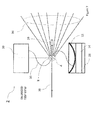
- Figure 1 to 3 shows a system 2 for resolving the crystal structure of a crystal 4 at atomic resolution by collecting X-ray diffraction images.
- the system 2 comprises an ultrasonic acoustic levitator 6 that levitates at least one droplet 8 of fluid containing one or multiple of spinning crystals 4.
- said ultrasonic acoustic levitator 6 comprises an ultrasonic transducer 10 for generating the ultrasonic acoustic wave and an ultrasonic reflector 12 and an ultrasonic transducer 14 to monitor the acoustic levitating pressure.
- the ultrasonic acoustic levitator 6 comprises an electrical source 16 that drives the driving power of the said ultrasonic transducer 10 to generate a stable constant pressure output by way of varying the driving frequency and voltage output by using the driving current as the feedback signal. Furthermore, the ultrasonic acoustic levitator 6 comprises a first X-ray window 18 for an incident X-ray beam 20 and a second X-ray window 22 for the diffracted X-ray beam 24.
- a mechanical shield 26 of the ultrasonic acoustic levitator 6 is provided to isolate the ultrasonic transmitting medium in the said ultrasonic acoustic levitator 6 and the said levitating droplet 8 from the air and the air turbulence in the surrounding. Further, an adjustment mechanism 28 is provided to adjust a gap, the parallelism and the displacement between said ultrasonic transducer 10 and the said ultrasonic reflector 12.
- the system 2 further comprises an ejector 30 for the ejection of one or multiple of droplets 32 of the said fluid 31 containing said single or multiples of crystals 4 into the said ultrasonic acoustic levitator 6.
- An apparatus for the visualization of the position and the spinning of the said crystal 4 in the levitating droplet 8 is provided as well as an X-ray source 34 and an X-ray detector 36 that detects the X-ray diffraction images 24 from the said single crystal 4 irradiated by the said X-ray source 34.
- a beam stop 29 stops the incident X-ray beam 20 between the said crystal 4 and the said X-ray detector.
- the acoustically levitated droplet 8 contains one or multiple of protein crystals 4 to be analysed by the X-ray diffraction.
- the high frame rate 2-dimensional detector 36 captures the diffracted X-ray 24.
- the condition of the acoustic levitator 6 is chosen such that the crystal orientation is rapidly varied via the internal circulation of the liquid droplet 8 by typically setting the acoustic levitating pressure in the range of 1 to 3 kPa.
- the droplet 8 containing single protein crystals 4 is placed in the levitator 6 via the droplet ejector 30 that can be automatically controlled.
- droplet ejectors 40 ejects one or multiple of droplets 42 of fluid 41 with certain chemical consistency into the levitating droplet 8 in the ultrasonic acoustic levitator 6.
- the purpose of the system 2 is to achieve the collection of X-ray diffraction dataset from single crystals 4 with the data acquisition rate of sub kHz to kHz or higher without freezing the sample and at increased maximum radiation dose.
- Another purpose of the system is to realize a fast X-ray protein structural analysis with a fully automated chain of the tasks from the harvesting of the protein crystals, their delivery to the X-ray beam path, and the acquisition of the dataset of X-ray diffraction images for solving the crystal structure at Angstrom resolution.
- the system 2 comprises the acoustic levitator 6, the two-dimensional X-ray detector 36, the X-ray source 34, and the ejector 30 of liquid droplets 8 containing single protein crystals 4.
- the acoustic levitator 6 comprises the ultrasonic transducer 10 of which output is stabilized by feedback the ultrasonic mirror reflector 12, and a transducer 14 to monitor the ultrasonic acoustic levitation pressure of the levitator 6.
- the ultrasonic mirror reflector 12 can be the levitator pressure sensor as well by attaching said transducer 14 to said mirror reflector 12.
- the two-dimensional X-ray detector 36 is capable to capture a series of X-ray images at high repetition rate of sub-kHz to kHz or higher with a minimal inactive time between the frames.
- a standing wave acoustic pressure distribution 30 is established between the said transducer 10 and said reflector 12.
- the peak pressure amplitude is of a sufficient amount, liquid droplets 8 can be levitated at a position close to any pressure nodes with small drift amplitude in both vertical and horizontal direction.
- the levitation acoustic pressure is to realize a stable levitation condition as well as fast spinning and/or orbiting of the protein crystal 4 inside the levitated droplet 8 via the internal circulation of the levitated droplet 8 by the acoustic streaming.
- This can be realized for example by adjusting the acoustic pressure lower than the pressure that can atomizes the droplet 8 but sufficiently (e.g. 40-60 %) larger than the pressure threshold below which value the levitation cannot be maintained due to the gravity by in the case of the embodiment of the levitator comprising the acoustic reflector 12 by adjusting the gap between said transducer 10 and said reflector 12.
- the ejector 30 may use an acoustic ejection using one or multiple of concentrated high frequency ultrasonic pulses into a crystallization tray or an acoustic ejection from a crystallization capillary by way of an application of pulsed acoustic pressure to the capillary.
Landscapes
- Chemical & Material Sciences (AREA)
- Health & Medical Sciences (AREA)
- Crystallography & Structural Chemistry (AREA)
- Physics & Mathematics (AREA)
- Clinical Laboratory Science (AREA)
- Chemical Kinetics & Catalysis (AREA)
- Biochemistry (AREA)
- Analytical Chemistry (AREA)
- Life Sciences & Earth Sciences (AREA)
- General Health & Medical Sciences (AREA)
- General Physics & Mathematics (AREA)
- Immunology (AREA)
- Pathology (AREA)
- Engineering & Computer Science (AREA)
- Acoustics & Sound (AREA)
- Multimedia (AREA)
- Analysing Materials By The Use Of Radiation (AREA)
Abstract
The present invention discloses a method and a system (2) for resolving a crystal structure of a crystal (4) at atomic resolution by collecting X-ray diffraction images, comprising:
a) ejecting a droplet (8) of fluid comprising single or multiple of crystal (4) into an ultrasonic acoustic levitator (6);
b) levitating said droplet (8) of fluid comprising said crystal (4) in an ultrasonic acoustic levitator (6);
b) monitoring the position and the spinning of the droplet (8) with a visualization apparatus;
c) applying X-ray (20) to said crystal (4), said X-ray (20) stemming from an X-ray source (34); and
d) detecting the X-ray diffraction images (24) from the said crystal (4) irradiated by the said X-ray source (34) by an X-ray detector (36) being capable to capture two dimensional diffraction patterns.
a) ejecting a droplet (8) of fluid comprising single or multiple of crystal (4) into an ultrasonic acoustic levitator (6);
b) levitating said droplet (8) of fluid comprising said crystal (4) in an ultrasonic acoustic levitator (6);
b) monitoring the position and the spinning of the droplet (8) with a visualization apparatus;
c) applying X-ray (20) to said crystal (4), said X-ray (20) stemming from an X-ray source (34); and
d) detecting the X-ray diffraction images (24) from the said crystal (4) irradiated by the said X-ray source (34) by an X-ray detector (36) being capable to capture two dimensional diffraction patterns.
Description
- The present invention is related to a system and a method for resolving a crystal structure of a crystal at atomic resolution by collecting X-ray diffraction images.
- X-ray diffraction is one of the best-established methods for the atomic-resolution structural analysis of macromolecules such as biological crystalline specimens and synthetic molecules. Combining synchrotron light sources with 2-dimensional high frame rate pixel detectors the collection time for a complete dataset has been reduced down to a few minutes. The acquisition rates, now routinely above 10 Hz, enable a continuous rotation of the protein crystal kept at freezing temperatures. Such an X-ray diffraction experiment setup, common to all major synchrotron beam lines, has accelerated the solution of important protein molecular structures during the last decade. It has been proposed that even faster data acquisition rate of 100-1000 Hz or higher leads to the minimization of the damage of fragile protein crystals by outrunning the diffusion of radicals generated by the X-ray absorption, thus drastically improving the quality of the diffraction dataset at room temperature. A faster data acquisition rate would also make it possible to perform a range of experiments that can only be conducted at room temperature, where the protein crystals is closer to physiological condition, such as in-situ structural dynamics studies and time-resolved experiments. Room temperature experiments will not only enable rapid screening of crystals but also facilitate the investigation of the conformational diversity of biomolecules that are difficult to study with frozen specimens.
- When a crystal is inside a droplet that is levitated by acoustic radiation pressure, internal circulation of the droplet due to the acoustic streaming induces fast spinning and orbiting of the crystal. Consequently, with careful alignment of the X-ray beam to the crystal, it is possible to acquire a dataset with various crystal orientations within a short time using a fast X-ray detector.
- In the conventional method as well as the acoustic levitation method of collecting the X-ray diffraction dataset, however, the single crystal samples have to be harvested and delivered manually. An automated method of the sample delivery such as the direct X-ray diffraction experiments through the crystallization plates and the acoustic ejection of the crystals on a solid sample holding mechanism as well as the use of continuous stream or a droplet train of crystallization liquid containing the single crystal samples have been reported previously. However, the efficiency of the sample usage of such methods is very low because of the difficulty to measure the X-ray diffraction images of each sample at multiple crystal orientations.
- The purpose of the current invention is to achieve the fast X-ray diffraction dataset acquisition at the frame rate of 100-1000 Hz or higher without the need to freeze the sample and with the high efficiency of the sample use by combining the ultrasonic acoustic levitation of droplets with an automatic sample harvest and delivery mechanism.
- It is therefore the objective of the present invention to provide a system and a method for resolving a crystal structure of a crystal at atomic resolution by collecting X-ray diffraction images thereby enabling the atomic-resolution solution of crystal structure of fragile macromolecular crystals, such as proteins.
- This objective is achieved with respect to the system according to the present invention by a system for resolving a crystal structure of a crystal at atomic resolution by collecting X-ray diffraction images, comprising:
- a) an ultrasonic acoustic levitator that levitates at least one droplet of fluid containing a single or multiple of spinning crystals, said ultrasonic acoustic levitator comprising:
- i) one or multiple of ultrasonic transducers;
- ii) one or multiple of electrical sources that supply the driving power for the said ultrasonic transducer;
- iii) at least one X-ray window;
- iv) a mechanical shield of the ultrasonic acoustic levitator that isolates the ultrasonic transmitting medium in which the droplet is levitated from the surrounding in terms of the air and the air turbulence and the at least one X-ray window;
- b) one or multiple of ejectors of one or multiple of droplets of said fluid containing said single or multiples of crystals into said ultrasonic acoustic levitator;
- c) an X-ray source; and
- d) an X-ray detector that detects the X-ray diffraction images from the said crystal irradiated by the said X-ray source.
- With respect to the method this objective is achieved according to the present invention by a method for resolving a crystal structure of a crystal at atomic resolution by collecting X-ray diffraction images, comprising the steps of:
- a) ejecting a droplet of fluid comprising one or multiple of crystals into an ultrasonic acoustic levitator;
- b) levitating said droplet comprising said crystal in said ultrasonic acoustic levitator;
- c) visually monitor the spatial position and spinning of said crystal in said levitating droplet;
- d) applying X-ray beam to said crystal in said levitating droplet, said X-ray stemming from an X-ray source; and
- e) detecting the X-ray diffraction images from said crystal irradiated by said X-ray source by an X-ray detector being capable to capture two dimensional diffraction patterns with a frame rate in the range of 100 to 3000 frames per sec or higher.
- The current invention realizes fully automated X-ray diffraction experiments with high data acquisition rate and high efficiency of sample use, thereby accelerating the structural analysis of biomolecules as well as structure-based drug development.
- A preferred embodiment of the present invention may comprise:
- a) an ultrasonic acoustic levitator that levitates at least one droplet of a fluid containing one or multiple of spinning crystals, said ultrasonic acoustic levitator comprising:
- i) a first ultrasonic transducer;
- ii) an ultrasonic reflector;
- iii) a second ultrasonic transducer to monitor a pressure of the acoustic standing wave in the acoustic cavity between said first ultrasonic transducer and said ultrasonic reflector;
- iv) an electrical source that supplies the driving power for the first ultrasonic transducer for a stable ultrasonic pressure output;
- v) at least one X-ray window;
- vi) a mechanical shield of the ultrasonic acoustic levitator that isolates the ultrasonic transmitting medium in which the droplet is levitated from the surrounding in terms of the air and the air turbulence and the at least one X-ray window;
- vii) an adjustment mechanism of the gap, the parallelism, and the displacement between said first ultrasonic transducer and the said ultrasonic reflector; and
- b) an ejector of a droplet of said fluid containing said single or multiples of crystals into said ultrasonic acoustic levitator;
- c) an apparatus for visualization of the position and the spinning of the said crystal in the said levitated droplet;
- d) an X-ray source;
- e) an X-ray detector that detects the X-ray diffraction images from the said crystal irradiated by said X-ray source;
- f) a beam stop that stops the incident X-ray beam between the said crystal and the said X-ray detector; and
- g) an apparatus for controlling the humidity and the temperature of the ultrasonic transmitting medium in which the said droplets are levitated.
- An additional measure could be to adjust the solvent content in the crystal by controllably evaporating the droplet fluid while levitating the droplet by way of controlling the humidity.
- Additionally or alternatively, the molecular conformation of the crystal can be varied by controlling the temperature of the droplet, the chemical consistency or pH of the solvent by ejecting additional droplets into the levitating droplets in the ultrasonic levitator.
- A further preferred embodiment of the present invention may provide the step of varying the molecular conformation of the crystal by applying optical pulses or laser pulses with specific wavelength to the droplet.
- Preferred embodiments of the present invention are hereinafter described in more detail with respect to the attached drawings which depict in:
- Figure 1
- schematically a graphical representation of X-ray diffraction imaging system for resolving the crystal structure of a crystal;
- Figure 2
- schematically an alternative system as compared to the system shown in
Figure 1 ; and - Figure 3
- schematically a further alternative system as compared to the systems shown in
Figures 1 and2 . -
Figure 1 to 3 shows asystem 2 for resolving the crystal structure of acrystal 4 at atomic resolution by collecting X-ray diffraction images. Thesystem 2 comprises an ultrasonicacoustic levitator 6 that levitates at least onedroplet 8 of fluid containing one or multiple ofspinning crystals 4. In one embodiment of the present invention, said ultrasonicacoustic levitator 6 comprises anultrasonic transducer 10 for generating the ultrasonic acoustic wave and anultrasonic reflector 12 and anultrasonic transducer 14 to monitor the acoustic levitating pressure. Further, the ultrasonicacoustic levitator 6 comprises anelectrical source 16 that drives the driving power of the saidultrasonic transducer 10 to generate a stable constant pressure output by way of varying the driving frequency and voltage output by using the driving current as the feedback signal. Furthermore, the ultrasonicacoustic levitator 6 comprises afirst X-ray window 18 for anincident X-ray beam 20 and asecond X-ray window 22 for thediffracted X-ray beam 24. Amechanical shield 26 of the ultrasonicacoustic levitator 6 is provided to isolate the ultrasonic transmitting medium in the said ultrasonicacoustic levitator 6 and the said levitatingdroplet 8 from the air and the air turbulence in the surrounding. Further, anadjustment mechanism 28 is provided to adjust a gap, the parallelism and the displacement between saidultrasonic transducer 10 and the saidultrasonic reflector 12. - The
system 2 further comprises anejector 30 for the ejection of one or multiple ofdroplets 32 of the saidfluid 31 containing said single or multiples ofcrystals 4 into the said ultrasonicacoustic levitator 6. An apparatus for the visualization of the position and the spinning of the saidcrystal 4 in the levitatingdroplet 8 is provided as well as anX-ray source 34 and anX-ray detector 36 that detects theX-ray diffraction images 24 from the saidsingle crystal 4 irradiated by the saidX-ray source 34. Abeam stop 29 stops theincident X-ray beam 20 between the saidcrystal 4 and the said X-ray detector. - The acoustically levitated
droplet 8 contains one or multiple ofprotein crystals 4 to be analysed by the X-ray diffraction. The high frame rate 2-dimensional detector 36 captures the diffractedX-ray 24. The condition of theacoustic levitator 6 is chosen such that the crystal orientation is rapidly varied via the internal circulation of theliquid droplet 8 by typically setting the acoustic levitating pressure in the range of 1 to 3 kPa. Thedroplet 8 containingsingle protein crystals 4 is placed in thelevitator 6 via thedroplet ejector 30 that can be automatically controlled. - Further additional one or multiple of
droplet ejectors 40 ejects one or multiple ofdroplets 42 offluid 41 with certain chemical consistency into the levitatingdroplet 8 in the ultrasonicacoustic levitator 6. - The purpose of the
system 2 is to achieve the collection of X-ray diffraction dataset fromsingle crystals 4 with the data acquisition rate of sub kHz to kHz or higher without freezing the sample and at increased maximum radiation dose. Another purpose of the system is to realize a fast X-ray protein structural analysis with a fully automated chain of the tasks from the harvesting of the protein crystals, their delivery to the X-ray beam path, and the acquisition of the dataset of X-ray diffraction images for solving the crystal structure at Angstrom resolution. - The
system 2 comprises theacoustic levitator 6, the two-dimensional X-ray detector 36, theX-ray source 34, and theejector 30 ofliquid droplets 8 containingsingle protein crystals 4. Theacoustic levitator 6 comprises theultrasonic transducer 10 of which output is stabilized by feedback theultrasonic mirror reflector 12, and atransducer 14 to monitor the ultrasonic acoustic levitation pressure of thelevitator 6. Theultrasonic mirror reflector 12 can be the levitator pressure sensor as well by attaching saidtransducer 14 to saidmirror reflector 12. The two-dimensional X-ray detector 36 is capable to capture a series of X-ray images at high repetition rate of sub-kHz to kHz or higher with a minimal inactive time between the frames. - In one embodiment of the
acoustic levitator 6, a standing waveacoustic pressure distribution 30 is established between the saidtransducer 10 and saidreflector 12. When the peak pressure amplitude is of a sufficient amount,liquid droplets 8 can be levitated at a position close to any pressure nodes with small drift amplitude in both vertical and horizontal direction. Of particular importance is to adjust the levitation acoustic pressure to realize a stable levitation condition as well as fast spinning and/or orbiting of theprotein crystal 4 inside the levitateddroplet 8 via the internal circulation of the levitateddroplet 8 by the acoustic streaming. This can be realized for example by adjusting the acoustic pressure lower than the pressure that can atomizes thedroplet 8 but sufficiently (e.g. 40-60 %) larger than the pressure threshold below which value the levitation cannot be maintained due to the gravity by in the case of the embodiment of the levitator comprising theacoustic reflector 12 by adjusting the gap between saidtransducer 10 and saidreflector 12. - The
ejector 30 may use an acoustic ejection using one or multiple of concentrated high frequency ultrasonic pulses into a crystallization tray or an acoustic ejection from a crystallization capillary by way of an application of pulsed acoustic pressure to the capillary.
Claims (15)
- A system (2) for resolving a crystal structure of a crystal (4) at atomic resolution by collecting X-ray diffraction images, comprising:a) an ultrasonic acoustic levitator (6) that levitates at least one droplet (8) of fluid containing one or multiple of spinning crystals (4), said ultrasonic acoustic levitator (6) comprising:i) one or multiple of ultrasonic transducers (10) that generate ultrasonic acoustic wave (30);ii) one or multiple of electrical sources (16) that supply the driving power for the said ultrasonic transducer (10);iii) a mechanical shield (26) that isolates the ultrasonic transmitting medium of the ultrasonic acoustic levitator (6) and the said levitating droplet (8) therein from the air and the air turbulence in surrounding with at least one X-ray window (18, 22);b) one or multiple of droplet ejectors (30) that eject one or multiple of the said droplets (8) of the said fluid (31) containing said single or multiples of the said crystals (4) into the said ultrasonic acoustic levitator (6);c) an X-ray source (34) to generate the X-ray beam (20);d) an X-ray detector (36) that detects the X-ray diffraction images (24) scattered by the said crystal (4) irradiated by the said X-ray beam (20);e) a beam stop (29) that stops the said incident X-ray beam (20) between the said crystal (4) and the said X-ray detector (36); andf) a apparatus to align the position of the said crystal (4) in the said levitating droplet (8) in the said ultrasonic levitator (6) position with respect to the X-ray beam (20).
- The system according to claim 1 wherein the acoustic levitation pressure is monitored by another ultrasonic transducer (14).
- The system according to any previous claims 1 to 2 wherein an apparatus is comprised to control the temperature and the humidity an ultrasonic transmitting medium in the said ultrasonic acoustic levitator (6).
- The system according to any previous claims 1 to 3 wherein the said electrical source (16) drives the said ultrasonic transducer (10) to generate a stable ultrasonic acoustic pressure output by way of varying the driving frequency and voltage output by using the driving current as the feedback signal.
- The system according to any previous claims 1 to 4 wherein the levitation force of the said droplet (8) is provided by the acoustic standing wave (30) in the acoustic cavity comprising of the said ultrasonic acoustic transducer (10) and an acoustic mirror reflector (12).
- The system according to any previous claims 1 to 5 wherein the said ultrasonic transducer (14) is in mechanical contact with the said acoustic reflector (12).
- The system according to any previous claims 1 to 6 wherein there is an adjustment mechanism (28) of the gap, the parallelism, and the displacement between the said ultrasonic transducer (10) and the said ultrasonic reflector (12).
- The system according to any previous claims 1 to 7 wherein additional one or multiple of droplet ejectors (40) eject one or multiple of droplets (42) of fluid (41) to the said levitating droplet (8) in the said ultrasonic acoustic levitator (10).
- The system according to any previous claims 1 to 8 wherein an apparatus for visualization of the said crystal (4) in the said levitating droplet (8) monitors the position and the spinning of the said crystal (4);
- A method for resolving a crystal structure of a crystal (4) at atomic resolution by collecting X-ray diffraction images, comprising the steps of:a) ejecting a droplet (8) of fluid (31) by the droplet ejector (30) into the ultrasound levitator (6) wherein the said droplet (8) comprising one or multiple of crystals (4);b) levitating the said droplet (8) comprising said crystal (4) in an ultrasonic acoustic levitator (6);c) applying X-ray beam (20) to the said crystal (4), wherein the said X-ray beam (20) stemming from an X-ray source (34); andd) detecting the X-ray diffraction (24) by an X-ray detector (36) wherein the said X-ray diffraction (24) generated by the scattering from the said single crystal (4) irradiated by the said X-ray beam (20).
- The method according to the claim 10 wherein the ultrasonic acoustic pressure within the ultrasonic acoustic levitator (6) is in the range of 1 to 4 kPa.
- The method according to any of the preceding claims 9 to 11 wherein the solvent content in the crystal (4) and/or the molecular conformation of the crystal (4) is adjusted by varying the chemical consistency or pH of the levitating droplet fluid by ejecting one or multiple of droplets (42) of appropriate fluid (41) by one or multiple of additional droplet ejector (40) into the said levitating droplet (8) in the said ultrasonic levitator (6).
- The method according to any of the preceding claims 9 to 12 wherein the solvent content in the crystal (4) and/or the molecular conformation of the crystal (4) is adjusted by controllably evaporating the solvent while levitating the droplet (8) by way of controlling the humidity and/or by controlling the temperature of the droplet (4).
- The method according to any of the preceding claims 9 to 13 wherein the molecular structure of the crystal (4) is varied by ornamenting it with some ligand by ejecting additional one or multiple of droplets (42) of fluids (41) comprising said ligand into the said levitating droplet (8) in the said ultrasonic levitator (6).
- The method according to any of the preceding claims 9 to 14 wherein the molecular conformation of the crystal (4) is varied by applying optical radiation of broadband wavelength or with specific wavelength or magnetic pulses to said crystal (4).
Priority Applications (7)
| Application Number | Priority Date | Filing Date | Title |
|---|---|---|---|
| EP15195946.7A EP3173774A1 (en) | 2015-11-24 | 2015-11-24 | A system and a method for resolving a crystal structure of a crystal at atomic resolution by collecting x-ray diffraction images |
| EP16788506.0A EP3380830B1 (en) | 2015-11-24 | 2016-10-28 | System and method for resolving a crystal structure of a biological crystalline macromolecule by collecting x-ray diffraction images |
| JP2018526792A JP6775581B2 (en) | 2015-11-24 | 2016-10-28 | Systems and methods for collecting X-ray diffraction images and elucidating the crystal structure of crystals by atomic decomposition |
| CN201680068238.5A CN108449987B (en) | 2015-11-24 | 2016-10-28 | Systems and methods for resolving crystal structures of crystals at atomic resolution |
| CA3005773A CA3005773C (en) | 2015-11-24 | 2016-10-28 | A system and a method for resolving a crystal structure of a crystal at atomic resolution by collecting x-ray diffraction images |
| US15/778,722 US10753888B2 (en) | 2015-11-24 | 2016-10-28 | System and a method for resolving a crystal structure of a crystal at atomic resolution by collecting X-ray diffraction images |
| PCT/EP2016/076032 WO2017089069A1 (en) | 2015-11-24 | 2016-10-28 | A system and a method for resolving a crystal structure of a crystal at atomic resolution by collecting x-ray diffraction images |
Applications Claiming Priority (1)
| Application Number | Priority Date | Filing Date | Title |
|---|---|---|---|
| EP15195946.7A EP3173774A1 (en) | 2015-11-24 | 2015-11-24 | A system and a method for resolving a crystal structure of a crystal at atomic resolution by collecting x-ray diffraction images |
Publications (1)
| Publication Number | Publication Date |
|---|---|
| EP3173774A1 true EP3173774A1 (en) | 2017-05-31 |
Family
ID=54705019
Family Applications (2)
| Application Number | Title | Priority Date | Filing Date |
|---|---|---|---|
| EP15195946.7A Withdrawn EP3173774A1 (en) | 2015-11-24 | 2015-11-24 | A system and a method for resolving a crystal structure of a crystal at atomic resolution by collecting x-ray diffraction images |
| EP16788506.0A Active EP3380830B1 (en) | 2015-11-24 | 2016-10-28 | System and method for resolving a crystal structure of a biological crystalline macromolecule by collecting x-ray diffraction images |
Family Applications After (1)
| Application Number | Title | Priority Date | Filing Date |
|---|---|---|---|
| EP16788506.0A Active EP3380830B1 (en) | 2015-11-24 | 2016-10-28 | System and method for resolving a crystal structure of a biological crystalline macromolecule by collecting x-ray diffraction images |
Country Status (6)
| Country | Link |
|---|---|
| US (1) | US10753888B2 (en) |
| EP (2) | EP3173774A1 (en) |
| JP (1) | JP6775581B2 (en) |
| CN (1) | CN108449987B (en) |
| CA (1) | CA3005773C (en) |
| WO (1) | WO2017089069A1 (en) |
Cited By (1)
| Publication number | Priority date | Publication date | Assignee | Title |
|---|---|---|---|---|
| CN115280139A (en) * | 2019-11-28 | 2022-11-01 | 保罗·谢勒学院 | Device for holding probe solutions of molecules in a plurality of independent units |
Families Citing this family (9)
| Publication number | Priority date | Publication date | Assignee | Title |
|---|---|---|---|---|
| JP7237373B2 (en) * | 2018-11-22 | 2023-03-13 | 株式会社リガク | Single crystal X-ray structure analysis device and sample holder mounting device |
| CN113302483B (en) * | 2018-11-23 | 2025-01-28 | 株式会社理学 | Device and method for absorbing sample for single crystal X-ray structure analysis |
| EP3828532A1 (en) | 2019-11-28 | 2021-06-02 | Paul Scherrer Institut | Device for hosting a probe solution of molecules in a plurality of independent cells |
| JP2023539007A (en) * | 2020-07-15 | 2023-09-13 | デンマークス、テクニスケ、ウニベルシテット | Laboratory-based 3D scanning X-ray Laue microdiffraction system and method (LAB3DμXRD) |
| CN113588697B (en) * | 2021-08-04 | 2024-04-19 | 中国科学院上海应用物理研究所 | High-viscosity extrusion injection sample loading device for protein crystal structure analysis |
| CN114295665A (en) * | 2021-12-10 | 2022-04-08 | 西安交通大学 | Water drop icing visual experiment device and application thereof |
| CN114441572B (en) * | 2021-12-30 | 2024-11-19 | 苏州青云瑞晶生物科技有限公司 | Method for continuous rotation precession electron diffraction tomography |
| CN115616011A (en) * | 2022-10-31 | 2023-01-17 | 中国科学院上海高等研究院 | An Ultrasonic Suspension Sample Stage for Synchrotron Radiation X-ray Scattering Test |
| CN116440972B (en) * | 2023-03-03 | 2026-03-24 | 厦门大学 | A non-contact ultrasonic pipette and its pipetting method |
Citations (3)
| Publication number | Priority date | Publication date | Assignee | Title |
|---|---|---|---|---|
| WO2002057520A1 (en) * | 2001-01-19 | 2002-07-25 | Chemical Holovoice Ab | System and method for screening of nucleation tendency of a molecule in a levitated droplet |
| DE102012101469A1 (en) * | 2012-02-23 | 2013-08-29 | BAM Bundesanstalt für Materialforschung und -prüfung | Apparatus for generating defined atmosphere in specimen chamber of acoustic levitator within mixing chamber, has gas outlet opening, which is arranged such that atmospheric gas discharging from gas outlet opening flows in specific direction |
| US20150191716A1 (en) * | 2014-01-08 | 2015-07-09 | Flodesign Sonics, Inc. | Acoustophoresis device with dual acoustophoretic chamber |
Family Cites Families (4)
| Publication number | Priority date | Publication date | Assignee | Title |
|---|---|---|---|---|
| US5036944A (en) * | 1986-03-24 | 1991-08-06 | Intersonics Incorporated | Method and apparatus for acoustic levitation |
| CN201886678U (en) * | 2010-12-20 | 2011-06-29 | 昆明理工大学 | Ultrasonic suspension demonstration device |
| US9327264B2 (en) | 2011-01-31 | 2016-05-03 | Uchicago Argonne, Llc | Containerless synthesis of amorphous and nanophase organic materials |
| CN106259268B (en) | 2016-08-17 | 2022-02-08 | 农业农村部南京农业机械化研究所 | Device and method for generating mist droplets |
-
2015
- 2015-11-24 EP EP15195946.7A patent/EP3173774A1/en not_active Withdrawn
-
2016
- 2016-10-28 EP EP16788506.0A patent/EP3380830B1/en active Active
- 2016-10-28 US US15/778,722 patent/US10753888B2/en active Active
- 2016-10-28 CN CN201680068238.5A patent/CN108449987B/en active Active
- 2016-10-28 JP JP2018526792A patent/JP6775581B2/en active Active
- 2016-10-28 CA CA3005773A patent/CA3005773C/en active Active
- 2016-10-28 WO PCT/EP2016/076032 patent/WO2017089069A1/en not_active Ceased
Patent Citations (3)
| Publication number | Priority date | Publication date | Assignee | Title |
|---|---|---|---|---|
| WO2002057520A1 (en) * | 2001-01-19 | 2002-07-25 | Chemical Holovoice Ab | System and method for screening of nucleation tendency of a molecule in a levitated droplet |
| DE102012101469A1 (en) * | 2012-02-23 | 2013-08-29 | BAM Bundesanstalt für Materialforschung und -prüfung | Apparatus for generating defined atmosphere in specimen chamber of acoustic levitator within mixing chamber, has gas outlet opening, which is arranged such that atmospheric gas discharging from gas outlet opening flows in specific direction |
| US20150191716A1 (en) * | 2014-01-08 | 2015-07-09 | Flodesign Sonics, Inc. | Acoustophoresis device with dual acoustophoretic chamber |
Non-Patent Citations (4)
| Title |
|---|
| CHRIS J. BENMORE ET AL: "Structural Characterization and Aging of Glassy Pharmaceuticals made Using Acoustic Levitation", JOURNAL OF PHARMACEUTICAL SCIENCES, vol. 102, no. 4, 4 February 2013 (2013-02-04), WASHINGTON, US, pages 1290 - 1300, XP055259365, ISSN: 0022-3549, DOI: 10.1002/jps.23464 * |
| GANGOPADHYAY A ET AL: "Beamline electrostatic levitator for in situ high energy x-ray diffraction studies of levitated solids and liquids", REVIEW OF SCIENTIFIC INSTRUMENTS, AIP, MELVILLE, NY, US, vol. 76, no. 7, 22 June 2005 (2005-06-22), pages 73901 - 073901, XP012079566, ISSN: 0034-6748, DOI: 10.1063/1.1932447 * |
| JORK LEITERER ET AL: "The use of an acoustic levitator to follow crystallization in small droplets by energy-dispersive X-ray diffraction", JOURNAL OF APPLIED CRYSTALLOGRAPHY., vol. 39, no. 5, 1 October 2006 (2006-10-01), DK, pages 771 - 773, XP055259234, ISSN: 0021-8898, DOI: 10.1107/S0021889806024915 * |
| MARCO A. B. ANDRADE ET AL: "Particle manipulation by a non-resonant acoustic levitator", APPLIED PHYSICS LETTERS, vol. 106, no. 1, 5 January 2015 (2015-01-05), 2 Huntington Quadrangle, Melville, NY 11747, pages 014101, XP055259528, ISSN: 0003-6951, DOI: 10.1063/1.4905130 * |
Cited By (1)
| Publication number | Priority date | Publication date | Assignee | Title |
|---|---|---|---|---|
| CN115280139A (en) * | 2019-11-28 | 2022-11-01 | 保罗·谢勒学院 | Device for holding probe solutions of molecules in a plurality of independent units |
Also Published As
| Publication number | Publication date |
|---|---|
| CA3005773C (en) | 2021-06-01 |
| CN108449987A (en) | 2018-08-24 |
| CA3005773A1 (en) | 2017-06-01 |
| EP3380830B1 (en) | 2023-06-07 |
| CN108449987B (en) | 2021-08-24 |
| EP3380830A1 (en) | 2018-10-03 |
| JP6775581B2 (en) | 2020-10-28 |
| US20180348149A1 (en) | 2018-12-06 |
| JP2019502102A (en) | 2019-01-24 |
| WO2017089069A1 (en) | 2017-06-01 |
| US10753888B2 (en) | 2020-08-25 |
Similar Documents
| Publication | Publication Date | Title |
|---|---|---|
| US10753888B2 (en) | System and a method for resolving a crystal structure of a crystal at atomic resolution by collecting X-ray diffraction images | |
| Grünbein et al. | Sample delivery for serial crystallography at free-electron lasers and synchrotrons | |
| JP6911254B2 (en) | Live cell identification and sorting system | |
| EP2157599A1 (en) | Method and apparatus for identification of biological material | |
| US10866182B2 (en) | Method and optical system for saturated illumination of analytes by a number of beams | |
| Tono et al. | Diverse application platform for hard X-ray diffraction in SACLA (DAPHNIS): application to serial protein crystallography using an X-ray free-electron laser | |
| US10908066B2 (en) | Use of vibrational spectroscopy for microfluidic liquid measurement | |
| JP2011206066A (en) | Method and device for selectively targeting cell within three-dimensional specimen | |
| US20060241395A1 (en) | Device and method for locating an instrument within a body | |
| US20120122084A1 (en) | System for identifying and sorting living cells | |
| EP2902773A1 (en) | Method and devices for X-ray crystallography, in particular with microcrystals of biological macromolecules | |
| CN107389627B (en) | A device for single-droplet single-cell culture and real-time detection of cellular metabolites | |
| US20060187533A1 (en) | Method and device for time-gated amplification of photons | |
| EP2584347B1 (en) | Facility and method for molecular structure determination | |
| JPS627837B2 (en) | ||
| Kang et al. | Compact Tape-Driven Sample Delivery System for Serial Femtosecond Crystallography | |
| Graziani et al. | A new experimental set-up for aerosol stability investigations in microgravity conditions | |
| Dörner et al. | Sample delivery for structural biology at the European XFEL | |
| WO2006002452A1 (en) | Raman spectroscopic device and method | |
| CN117007587A (en) | Optical precision measurement system for micro-nano particle quality based on light radiation force | |
| Mandelis | Focus on biotechnologies, biomedical optics, and microscopy |
Legal Events
| Date | Code | Title | Description |
|---|---|---|---|
| PUAI | Public reference made under article 153(3) epc to a published international application that has entered the european phase |
Free format text: ORIGINAL CODE: 0009012 |
|
| AK | Designated contracting states |
Kind code of ref document: A1 Designated state(s): AL AT BE BG CH CY CZ DE DK EE ES FI FR GB GR HR HU IE IS IT LI LT LU LV MC MK MT NL NO PL PT RO RS SE SI SK SM TR |
|
| AX | Request for extension of the european patent |
Extension state: BA ME |
|
| STAA | Information on the status of an ep patent application or granted ep patent |
Free format text: STATUS: THE APPLICATION IS DEEMED TO BE WITHDRAWN |
|
| 18D | Application deemed to be withdrawn |
Effective date: 20171201 |