EP3171152B1 - Process and device for isolating cells from biological tissue - Google Patents
Process and device for isolating cells from biological tissue Download PDFInfo
- Publication number
- EP3171152B1 EP3171152B1 EP16196789.8A EP16196789A EP3171152B1 EP 3171152 B1 EP3171152 B1 EP 3171152B1 EP 16196789 A EP16196789 A EP 16196789A EP 3171152 B1 EP3171152 B1 EP 3171152B1
- Authority
- EP
- European Patent Office
- Prior art keywords
- biological tissue
- casing
- tissue
- holder
- cells
- Prior art date
- Legal status (The legal status is an assumption and is not a legal conclusion. Google has not performed a legal analysis and makes no representation as to the accuracy of the status listed.)
- Active
Links
Images




Classifications
-
- C—CHEMISTRY; METALLURGY
- C12—BIOCHEMISTRY; BEER; SPIRITS; WINE; VINEGAR; MICROBIOLOGY; ENZYMOLOGY; MUTATION OR GENETIC ENGINEERING
- C12M—APPARATUS FOR ENZYMOLOGY OR MICROBIOLOGY; APPARATUS FOR CULTURING MICROORGANISMS FOR PRODUCING BIOMASS, FOR GROWING CELLS OR FOR OBTAINING FERMENTATION OR METABOLIC PRODUCTS, i.e. BIOREACTORS OR FERMENTERS
- C12M29/00—Means for introduction, extraction or recirculation of materials, e.g. pumps
- C12M29/16—Hollow fibers
-
- C—CHEMISTRY; METALLURGY
- C12—BIOCHEMISTRY; BEER; SPIRITS; WINE; VINEGAR; MICROBIOLOGY; ENZYMOLOGY; MUTATION OR GENETIC ENGINEERING
- C12M—APPARATUS FOR ENZYMOLOGY OR MICROBIOLOGY; APPARATUS FOR CULTURING MICROORGANISMS FOR PRODUCING BIOMASS, FOR GROWING CELLS OR FOR OBTAINING FERMENTATION OR METABOLIC PRODUCTS, i.e. BIOREACTORS OR FERMENTERS
- C12M23/00—Constructional details, e.g. recesses, hinges
- C12M23/48—Holding appliances; Racks; Supports
-
- C—CHEMISTRY; METALLURGY
- C12—BIOCHEMISTRY; BEER; SPIRITS; WINE; VINEGAR; MICROBIOLOGY; ENZYMOLOGY; MUTATION OR GENETIC ENGINEERING
- C12M—APPARATUS FOR ENZYMOLOGY OR MICROBIOLOGY; APPARATUS FOR CULTURING MICROORGANISMS FOR PRODUCING BIOMASS, FOR GROWING CELLS OR FOR OBTAINING FERMENTATION OR METABOLIC PRODUCTS, i.e. BIOREACTORS OR FERMENTERS
- C12M21/00—Bioreactors or fermenters specially adapted for specific uses
- C12M21/08—Bioreactors or fermenters specially adapted for specific uses for producing artificial tissue or for ex-vivo cultivation of tissue
-
- C—CHEMISTRY; METALLURGY
- C12—BIOCHEMISTRY; BEER; SPIRITS; WINE; VINEGAR; MICROBIOLOGY; ENZYMOLOGY; MUTATION OR GENETIC ENGINEERING
- C12M—APPARATUS FOR ENZYMOLOGY OR MICROBIOLOGY; APPARATUS FOR CULTURING MICROORGANISMS FOR PRODUCING BIOMASS, FOR GROWING CELLS OR FOR OBTAINING FERMENTATION OR METABOLIC PRODUCTS, i.e. BIOREACTORS OR FERMENTERS
- C12M21/00—Bioreactors or fermenters specially adapted for specific uses
- C12M21/18—Apparatus specially designed for the use of free, immobilized or carrier-bound enzymes
-
- C—CHEMISTRY; METALLURGY
- C12—BIOCHEMISTRY; BEER; SPIRITS; WINE; VINEGAR; MICROBIOLOGY; ENZYMOLOGY; MUTATION OR GENETIC ENGINEERING
- C12M—APPARATUS FOR ENZYMOLOGY OR MICROBIOLOGY; APPARATUS FOR CULTURING MICROORGANISMS FOR PRODUCING BIOMASS, FOR GROWING CELLS OR FOR OBTAINING FERMENTATION OR METABOLIC PRODUCTS, i.e. BIOREACTORS OR FERMENTERS
- C12M23/00—Constructional details, e.g. recesses, hinges
- C12M23/28—Constructional details, e.g. recesses, hinges disposable or single use
-
- C—CHEMISTRY; METALLURGY
- C12—BIOCHEMISTRY; BEER; SPIRITS; WINE; VINEGAR; MICROBIOLOGY; ENZYMOLOGY; MUTATION OR GENETIC ENGINEERING
- C12M—APPARATUS FOR ENZYMOLOGY OR MICROBIOLOGY; APPARATUS FOR CULTURING MICROORGANISMS FOR PRODUCING BIOMASS, FOR GROWING CELLS OR FOR OBTAINING FERMENTATION OR METABOLIC PRODUCTS, i.e. BIOREACTORS OR FERMENTERS
- C12M23/00—Constructional details, e.g. recesses, hinges
- C12M23/46—Means for fastening
-
- C—CHEMISTRY; METALLURGY
- C12—BIOCHEMISTRY; BEER; SPIRITS; WINE; VINEGAR; MICROBIOLOGY; ENZYMOLOGY; MUTATION OR GENETIC ENGINEERING
- C12M—APPARATUS FOR ENZYMOLOGY OR MICROBIOLOGY; APPARATUS FOR CULTURING MICROORGANISMS FOR PRODUCING BIOMASS, FOR GROWING CELLS OR FOR OBTAINING FERMENTATION OR METABOLIC PRODUCTS, i.e. BIOREACTORS OR FERMENTERS
- C12M29/00—Means for introduction, extraction or recirculation of materials, e.g. pumps
-
- C—CHEMISTRY; METALLURGY
- C12—BIOCHEMISTRY; BEER; SPIRITS; WINE; VINEGAR; MICROBIOLOGY; ENZYMOLOGY; MUTATION OR GENETIC ENGINEERING
- C12M—APPARATUS FOR ENZYMOLOGY OR MICROBIOLOGY; APPARATUS FOR CULTURING MICROORGANISMS FOR PRODUCING BIOMASS, FOR GROWING CELLS OR FOR OBTAINING FERMENTATION OR METABOLIC PRODUCTS, i.e. BIOREACTORS OR FERMENTERS
- C12M29/00—Means for introduction, extraction or recirculation of materials, e.g. pumps
- C12M29/04—Filters; Permeable or porous membranes or plates, e.g. dialysis
-
- C—CHEMISTRY; METALLURGY
- C12—BIOCHEMISTRY; BEER; SPIRITS; WINE; VINEGAR; MICROBIOLOGY; ENZYMOLOGY; MUTATION OR GENETIC ENGINEERING
- C12M—APPARATUS FOR ENZYMOLOGY OR MICROBIOLOGY; APPARATUS FOR CULTURING MICROORGANISMS FOR PRODUCING BIOMASS, FOR GROWING CELLS OR FOR OBTAINING FERMENTATION OR METABOLIC PRODUCTS, i.e. BIOREACTORS OR FERMENTERS
- C12M29/00—Means for introduction, extraction or recirculation of materials, e.g. pumps
- C12M29/10—Perfusion
-
- C—CHEMISTRY; METALLURGY
- C12—BIOCHEMISTRY; BEER; SPIRITS; WINE; VINEGAR; MICROBIOLOGY; ENZYMOLOGY; MUTATION OR GENETIC ENGINEERING
- C12M—APPARATUS FOR ENZYMOLOGY OR MICROBIOLOGY; APPARATUS FOR CULTURING MICROORGANISMS FOR PRODUCING BIOMASS, FOR GROWING CELLS OR FOR OBTAINING FERMENTATION OR METABOLIC PRODUCTS, i.e. BIOREACTORS OR FERMENTERS
- C12M41/00—Means for regulation, monitoring, measurement or control, e.g. flow regulation
- C12M41/30—Means for regulation, monitoring, measurement or control, e.g. flow regulation of concentration
- C12M41/32—Means for regulation, monitoring, measurement or control, e.g. flow regulation of concentration of substances in solution
-
- C—CHEMISTRY; METALLURGY
- C12—BIOCHEMISTRY; BEER; SPIRITS; WINE; VINEGAR; MICROBIOLOGY; ENZYMOLOGY; MUTATION OR GENETIC ENGINEERING
- C12M—APPARATUS FOR ENZYMOLOGY OR MICROBIOLOGY; APPARATUS FOR CULTURING MICROORGANISMS FOR PRODUCING BIOMASS, FOR GROWING CELLS OR FOR OBTAINING FERMENTATION OR METABOLIC PRODUCTS, i.e. BIOREACTORS OR FERMENTERS
- C12M45/00—Means for pre-treatment of biological substances
-
- C—CHEMISTRY; METALLURGY
- C12—BIOCHEMISTRY; BEER; SPIRITS; WINE; VINEGAR; MICROBIOLOGY; ENZYMOLOGY; MUTATION OR GENETIC ENGINEERING
- C12M—APPARATUS FOR ENZYMOLOGY OR MICROBIOLOGY; APPARATUS FOR CULTURING MICROORGANISMS FOR PRODUCING BIOMASS, FOR GROWING CELLS OR FOR OBTAINING FERMENTATION OR METABOLIC PRODUCTS, i.e. BIOREACTORS OR FERMENTERS
- C12M45/00—Means for pre-treatment of biological substances
- C12M45/09—Means for pre-treatment of biological substances by enzymatic treatment
-
- C—CHEMISTRY; METALLURGY
- C12—BIOCHEMISTRY; BEER; SPIRITS; WINE; VINEGAR; MICROBIOLOGY; ENZYMOLOGY; MUTATION OR GENETIC ENGINEERING
- C12N—MICROORGANISMS OR ENZYMES; COMPOSITIONS THEREOF; PROPAGATING, PRESERVING, OR MAINTAINING MICROORGANISMS; MUTATION OR GENETIC ENGINEERING; CULTURE MEDIA
- C12N5/00—Undifferentiated human, animal or plant cells, e.g. cell lines; Tissues; Cultivation or maintenance thereof; Culture media therefor
- C12N5/0081—Purging biological preparations of unwanted cells
-
- G—PHYSICS
- G01—MEASURING; TESTING
- G01N—INVESTIGATING OR ANALYSING MATERIALS BY DETERMINING THEIR CHEMICAL OR PHYSICAL PROPERTIES
- G01N1/00—Sampling; Preparing specimens for investigation
- G01N1/28—Preparing specimens for investigation including physical details of (bio-)chemical methods covered elsewhere, e.g. G01N33/50, C12Q
- G01N1/286—Preparing specimens for investigation including physical details of (bio-)chemical methods covered elsewhere, e.g. G01N33/50, C12Q involving mechanical work, e.g. chopping, disintegrating, compacting, homogenising
Definitions
- the present invention is directed to a process for isolating living target cells from biological tissue by delivering an appropriate release agent into the biological tissue through an array of hollow needles, thereby releasing the target cells from the tissue infrastructure.
- the procedure to isolate intact hepatocytes was first introduced by Howard and Pesch (1967) J Cell Biol 35, 675-684 and refined by Berry and Friend (1969) J Cell Biol 43, 506-520 ; Seglen (1976) Methods Cell Biol. 13, 29-34 and Klaunig (1981) In Vitro 17, 913-925 .
- the outcome was a two-stage perfusion protocol which, after decades, still represents the current standard procedure leading to high amounts of viable hepatocytes.
- a loose ligature is placed around the appropriate blood vessel (rat: vena portae; mouse: vena cava) near the liver, a cannula is inserted into the blood vessel, and the ligature is fixed by tightening the thread ends.
- a calcium depleting buffer containing EDTA or EGTA is delivered into the organ through the blood vessel at a flow rate of about 10-15ml/min for 10-15min in order to weaken calcium-depending cell-cell junctions and remove blood cells.
- a buffer comprising collagenase (at about 0,1 Wünsch Units per ml) is channeled (rat: 10-15min; mouse: 5-10min) through the organ at a flow rate of about 10-15ml/min for destruction of connective tissue further liberating cells without damaging target cells.
- the liver can now be removed from the animal and placed in an appropriate media on a petri dish.
- liver capsule Tearing apart the liver capsule leads to the release of liberated hepatocytes which are further purified by filtration through a 100 ⁇ m nylon mesh filter and an (optionally repeated) low-spin centrifugation step (5min 50g at 4°C) to enrich hepatocytes.
- a similar but ex-vivo process is used for hepatocyte isolation from livers of bigger vertebrates and/or for the isolation of adult cardiomyocytes.
- the target organ is excised before cannulating a blood vessel.
- the liver tissue is put on ice as fast as possible and inspected for large vessels. Cannulae are inserted into the largest vessels of the cut surface and fixed with tissue glue.
- the liver tissue is perfused extensively with an calcium-depleting buffer containing EGTA (or EDTA) at about 10ml/catheter.
- EGTA or EDTA
- it is perfused at the same flow rate with an about 0,2 U/ml collagenase-containing enzyme mix until the tissue shows irreversible deformation.
- the enzyme buffer is recirculated to reduce costs. After digestion, the liver tissue is gently dispersed with a spatula. Analogues to the rodent procedure, the cell suspension is filtered and hepatocytes are enriched by a (optionally repeated) low-spin centrifugation.
- Cardiomyocytes are generated by the so-called Langendorff perfusion system.
- the heart is rapidly excised from the body and ,under a microscope , a cannula is carefully inserted into the aorta and fixed with a ligature. Then, the heart is perfused with a calcium-free buffer to arrest contraction, followed by a collagenase-based enzyme solution to digest the extracellular matrix of the heart. After digestion, the heart is mechanically dissociated with forceps and cardiomyocytes are dispersed into a single-cell suspension.
- Lung alveolar epithelial cells are isolated in a similar procedure by inserting and fixing a cannula in the trachea and applying appropriate buffer and enzymes.
- US20110295149 discloses a device to solubilize tissue by an abrasive extraction of tissue fragments.
- the device is fixed on the tissue by vacuum and the cells are cut from the tissue with an abrasive component and further liquefied by appropriate enzymes.
- agents that release target cells from the biological tissue can be delivered by a device comprising a plurality of injection sites into a biological tissue, thereby releasing target cells as a single-cell suspension with good yield and viability of the target cells.
- Object of the invention is therefore an perfusion device for biological tissues as defined by the claims, comprising a casing having two parts, a first part (1) and a second part (9); a holder (7) for a plurality of hollow penetration structures (8), wherein the hollow penetration structures (8) are provided with at least one orifice having fluid communication through the holder (7); a support (5) for the biological tissue (6); wherein the support (5) for the biological tissue (6) is positioned in the casing at a distance to the holder (7) that by joining the first part (1) and the second part (9) to form the casing, the hollow penetration structures (8) are in proximity to the holder (7).
- the penetration process is triggered by closing the casing.
- the first and second part of the casing are each provided with threads as corresponding closing mechanism for joining the two parts, and configured such that by joining the first part (1) and the second part (9) to form the casing, the hollow penetration structures (8) penetrate at least in part into the biological tissue (6).
- Another object of the invention is a process for disaggregation of a biological tissue with a device as disclosed wherein the biological tissue is penetrated at least in part by at least one of the hollow penetration structures (8) by joining the first part (1) and the second part (9) to form the casing and at least one agent to disaggregate the biological tissue into target cells is administered through the hollow penetration structures (8) into the biological tissue (6).
- the term "disaggregation of a biological tissue into target cells” refers to any process where cell structures, cell aggregates or cell matrices are at least in part destroyed without killing, destroying or lysing the target cells.
- the target cells are obtained as single, isolated and living cells.
- an appropriate enzyme is administered into the liver with the device of the invention.
- the liver tissue is disaggregated to yield single liver cells which do not leave the liver.
- the epithelial cell sheet of the liver (the capsule of the liver) is mechanically opened and the liver cells can be washed from the remaining tissue.
- the cells achieved by the process and device of the present invention can be cultured, analyzed and/or transplanted into patients with methods known to a person skilled in the art.
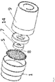
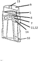
- the device of the invention is shown by way of example in several embodiments in Fig. 1 to Fig. 3 .
- first part (1) and the second part (9) of the casing are each provided with a corresponding closing mechanism (11, 12) for joining the two parts.
- first part (1) and/or the second part (9) of the casing are provided with at least one orifice for in- and/or output of fluids (10, 13).
- the hollow penetration structures (8) are provided with at least one orifice having fluid communication through the holder (7) to at least one of these orifices (10, 13).
- the casing of the device can be opened and closed in preferable a water-tight manner by the corresponding closing mechanism of the first and second part of the casing.
- the corresponding closing mechanisms (11,12) of the first part (1) and the second part (9) of the casing are threads, capable of interlocking or joining the first and second part to form the casing.
- the first and second part of the casing, the closing mechanism (11, 12) and the holder (7) may be combined to one or two separate pieces, but can also be provided separately. If the first and second part of the casing and one or both closing mechanisms (11, 12) are separate devices, they need to be provided with means like apertures to enable mechanically combination when closing the device.
- the biological tissue is placed on the support (5) which permeable for fluids and is then pressed between the support and the penetration needles (i.e. the holder), resulting in penetration of the biological tissue at least in part by the hollow penetration structures (8).
- Support (5) serves as counterpart for the holder (7) when the penetration structures are pressed against the tissue.
- the tissue may be placed on the support and pressed against the penetration structures or the tissue may be placed on the penetration structures and pressed against the support.
- the support (5) may be an integral part of the casing (i.e. the first or second part) or may be inserted and removed from the casing. In the latter case the support needs to have a smaller diameter that the casing.
- the support (5) has the function of providing a mechanical counterpart to the holder (7) when the needles penetrate into the tissue. Insofar, the design of support (5) is not essential and may be provided in form of a filter, mesh, rack, grid or even a plate with orifices (in order to prevent the tissue being soaked in fluids).
- the holder (7) can be inserted and removed from the casing i.e. should have a size or outer diameter fitting into the main casing.
- the holder (7) has a size or outer diameter to fit in a water-tight manner into the casing and/or is provided with an appropriate seal to prevent fluids (release agents) from circumventing the holder/the hollow penetration structures (8).
- hollow penetration structures refer to any elongated object having a needle-, cone- or pyramid-like shape, which is provided with a sharpened end.
- the term “hollow penetration structures” and “needle” is used as synonym.
- the hollow penetration structures and the holder may be produced separately or as one piece.
- the hollow needles (8) are provided with means to stop the flow of reagents through a needle when the opening of a needle is placed not within the biological tissue, i.e. in case a needle did not penetrate into the tissue or penetrated through the tissue.
- the hollow penetration structures (8) may have an outer diameter at the basis of 0,05 to 5 mm, preferably 0,2 to 1 mm, most preferably 0,3 to 0,7 mm and independently, an inner diameter at the basis of 0,02 to 4 mm, preferably 0,1 to 1mm, most preferably 0,1 to 0,6 mm and independently a length of 1 to 100 mm, preferably 2 to 20 mm, most preferably 4 to 5 mm.
- the number of hollow penetration structures (8) depends on the size of the biological tissue and may vary between 2 and 500, preferably between 5 and 100, and most preferably between 20 and 70.
- the hollow penetration structures (8) may be arranged in any geometry or array on the holder and may have the same or different length.
- the holder may be not mechanically fixed in main casing. This enables the use of different holders with different number of hollow penetration structures (8) and/or different length of hollow penetration structures (8) and/or different geometry or array of hollow needles on the holder, depending on the size and thickness of the biological tissue.
- the number, length, outer/inner diameter and area of hollow penetration structures (8) can be selected to achieve maximum effect of penetration.
- the device of the invention i.e. the casing comprising the first and second part, the holder (7) and the hollow penetration structures (8) may be produced from the same or different material like stainless steel, polyacrylamide, polystyrene, polyolefins like polyethylene and polypropylene, polycarbonate, polyoxymethylene, polymethylmethacrylate, poly lactic acid or polyamides.
- the device of the invention may be manufactured by any method known to a person skilled in the art. Preferred methods are injection molding and 3D printing, for example by extrusion deposition, fused deposition modeling, stereolithography or photopolymer digital light processing.
- the perfusion device according to the invention has the advantage that only the parts having mechanical contact to the biological tissue, i.e. the holder for the plurality of hollow needles and the support, are single-use disposables, whereas the main casing and the cap can, after appropriate cleaning, be used multiple times.
- the device is provided as disposable.
- the perfusion device may be used manually, i.e. with syringes and appropriate vessels, but can also be used in an automated environment comprising one or more pumps and tubing sets.
- the device is provided with at least one pump, like a gear pump as shown in Fig. 10 .
- the pump may be directly connected to the input/output orifices of the casing or may be provided with filter system or detection devices to monitor the release process of the cells.
- the pump may be integrated in the casing either in the first or the second part of the casing, or can provided externally and connected to the device with an appropriate tubing set. The position of the pump in view of the biological is not of importance.
- the pump may be driven by an internal or external motor. If the pump is integrated into the first or the second part of the casing, the pump and the casing are preferable manufactured by a 3D printing manufacturing process as disclosed for the holder (7).
- the process of the invention is characterized in that the biological tissue is penetrated at least in part by a plurality of hollow penetration structures (8).
- penetration means that the needles are placed into the biological tissue in order to administer the release agent into the biological tissue. It is not desired to pierce or puncture the needles through the biological tissue since the release agent would then not or not sufficiently enter the biological tissue to release the target cells. It should be taken care in the process of the invention that the majority of the needles are placed inside the biological tissue and do not pierce or puncture through the biological tissue. At best, all needles are placed into the biological tissue at 30-70 %, preferable approximately 50% of its thickness.
- the biological tissue is pressed between a support (5) permeable for fluids and the needles, resulting in penetration of the needles at least in part into the biological tissue.
- the support (5) can be for example the filter area of a filter cage (4), for example a "Smart Strainer” filter cage commercialized by Miltenyi Biotec GmbH.
- the biological tissue is penetrated by the plurality of hollow needles by placing the tissue on a support, e.g. a mesh of a filter cage of a device as already disclosed, closing the main casing with the cap and administering at least one agent to disaggregate the biological tissue through at least one orifice of the cap and collecting the flow-through from at least one orifice at another, preferably the lower end of the main casing.
- a support e.g. a mesh of a filter cage of a device as already disclosed
- At least one agent is administered into the biological tissue in an amount that an excess volume of release agent leaks from the biological tissue, and said excess volume of agent is administered again into the biological tissue.
- excess volume means that compared to the volume of the biological tissue, a much greater volume of agent is administered, and the volume of agent not taken up by the biological tissue leaks out.
- This embodiment may involve the administering of the agents in a first fresh stream which is recycled either as constant leak stream or batch-wise into the biological tissue.
- the leak stream may already contain target cells.
- the leak stream may be collected as cell suspension after one or more cycles.
- the process of the invention may be applied to a great variety of biological tissues.
- the target cells are disintegrated from the cellular infrastructure of the biological tissue by administering the agent, but cannot be released/extracted from the biological tissue and/or leave the biological tissue. This might occur if the outer structure of the biological tissue is still intact or if the biological tissue is provided with a capsule. e.g. an epithelial cell sheet. Extracting target cells from such biological tissue requires mechanically opening of the capsule.
- the biological tissue is mechanically opened and the target cells are extracted from the biological tissue.
- the biological tissue is mechanically opened by cutting, piercing, or rupturing the biological tissue manually by appropriate tools.
- the device of the invention is provided with means for mechanically opening the biological tissue, for example with a cutting device built in the main casing, the filter cage or the support for hollow needles.
- the hollow penetration structures are used as cutting device, for example by rotating the holder against the tissue.
- the process may comprise an additional step to remove unwanted "non-target cells", e.g. blood cells, before administering at least one agent to disaggregate the biological tissue.
- non-target cells are extracted from the biological tissue by administering at least one washing fluid into the biological tissue.
- the biological tissue (6) is first washed with buffer to release non-target cells from the biological tissue (6) and the fluid containing the non-target cells are stored in the storage cavity (14).
- the agent to disaggregate the biological tissue (6) and/or the buffer is repeatedly administered into the biological tissue (6), for example by a pump.
- the fluids are recycled several times over/into the biological tissue (6), thereby improving yield.
- the process of the invention can be applied to generate all type of target cells which are tissue-resident cells, especially cells from vertebrate or invertebrate tissue, preferably epithelial cells, endothelial cells, fibroblasts, myofibroblasts, hepatocytes, hepatic stellate cells, cardiomyocytes, podocytes, keratinocytes, melanocytes, neuronal cells including neurons, astrocytes, microglia and oligodendrocytes, leukocytes including dendritic cells, neutrophils, macrophages and lymphocytes, including T cells, B cells, NK cells, NKT cells and innate lymphoid type 1-3 cells, tissue stem cells including MSCs and progenitor cells of cells mentioned above.
- tissue-resident cells especially cells from vertebrate or invertebrate tissue, preferably epithelial cells, endothelial cells, fibroblasts, myofibroblasts, hepatocytes, hepatic stellate cells,
- the process of the invention can be applied to all types of biological tissue, like organs of vertebrates or invertebrates, preferably to spleen, heart, liver, brain and other neural tissues, kidney, lung, pancreas, breast, umbilical cord, skin, placenta, ovary, oviduct, uterus, prostate, tonsil, thymus, stomach, testis, trachea, cartilage, tendon, bone, skeletal muscle, smooth muscle, gut, colon, intestine, bladder, urethra, eye, gall bladder, organoids from cell cultures and tumors.
- biological tissue like organs of vertebrates or invertebrates, preferably to spleen, heart, liver, brain and other neural tissues, kidney, lung, pancreas, breast, umbilical cord, skin, placenta, ovary, oviduct, uterus, prostate, tonsil, thymus, stomach, testis, trachea, cartilage, tendon, bone, skeletal muscle, smooth muscle, gut
- the term "disaggregating agent” as used herein means a fluid like a buffer comprising a substance used to destroy the anchorage of target cells within the tissue without influencing the target cells itself.
- This anchorage derives from interactions of the cells with the extracellular matrix or with adjacent cells. These interactions, e.g. tight junctions, gap junctions, desmosomes, and hemidesmosomes, are built mainly by proteins, e.g. cadherins, connexins, claudins and integrins, mostly in a calcium-dependent manner. Therefore, the release agent which destroys the tissue integrity may contain a calcium-free and/or a calcium-depleting agent and/or enzymes that degrade the extracellular matrix or extracellular protein-protein interactions. The administration of the components of the release agent may be sequentially or simultaneously.
- the agent to disaggregate the biological tissue (6) is selected from the group consisting of trypsin, chymotrypsin, papain, collagenase, elastase, dispase, thermolysin, hyaluronidase, clostripain and neutral protease from clostridium histolyticum, pronase, DNase I, pepsin, proteinase K, lysozyme, chelating agents for bivalent ions (like EDTA or citrate) and mixtures thereof.
- the calcium-depleting reagent may be a buffer containing EDTA, EGTA or citrate.
- a calcium-free buffer or a calcium-depleting buffer is used in a first step and a buffer containing calcium ions, preferably at least 50 ⁇ M calcium ions, as well as a calcium-depending enzyme is used in a second step.
- a calcium-free buffer or a calcium-depleting buffer is used in a first step and a buffer concentrate containing calcium ions as well as a calcium-depending enzyme is spiked into the first buffer as a concentrate in a second step.
- any aqueous fluid with pH values, osmolality and ion concentrations in the physiological range can be used.
- Preferred buffers are PBS, D-PBS, HBSS (Hanks' balanced salt solution), EBSS, DMEM (Dulbecco's Modified Eagle Medium), DMEM/F12, IMDM, RPMI, RPMI-1640, either in a complete form or in variants thereof, with or without phenol red, with or without HEPES, with or without glucose, optionally including a protein component like FCS (fetal calf serum), FBS (fetal bovine serum), HSA (human serum albumin), or BSA (bovine serum albumin).
- FCS fetal calf serum
- FBS fetal bovine serum
- HSA human serum albumin
- BSA bovine serum albumin
- the first buffer administered to the tissue might contain an anticoagulant like EDTA, EGTA, citrate or heparin to avoid blood coagulation within the tissue.
- an anticoagulant like EDTA, EGTA, citrate or heparin to avoid blood coagulation within the tissue.
- Collagenase (EC 3.3.24.3) from Clostridium histolyticum, also called clostridiopeptidase A, is an enzyme mixture capable of causing hydrolytic cleavage of collagen molecules in their native conformation and at their helical region.
- Collagenase can also be isolated from many other bacterial sources (e.g. Achromobacter iophagus, Mycobacterium tuberculosis, Pseudomonas aeruginosa and other microorganisms).
- Clostridium histolyticum provides the main commercial source of collagenase. Crude preparations also contain contaminating protease activities like clostripain, neutral protease and further tryptic, caseinase and lipase activities. These can hydrolyse the portions of collagen which are not tightly wound in the helical form. Differences in the relative quantities of these contaminating activities are responsible for observed lot-to-lot variation.
- Hyaluronidase (EC 3.2.1.35), an endoglycosidase with a specificity for endo-N-acetylhexosaminic bonds, usually derives from bovine and sheep testis. It hydrolyses 1,4 linkages between 2-acetacido-2-deoxy- ⁇ -D-glucose and D-glucuronate residues in hyaluronic acid, a glycosaminoglycan found in the ground substance of virtually all connective tissue.
- Trypsin (EC 3.4.21.4), a pancreatic serine protease, is capable of hydrolyzing peptides, preferentially at bonds involving the carboxyl group of the basic amino acids, L-arginine or L-lysine, and also shows some esterase and amidase activity. Purified trypsin is ineffective against native collagen and, therefore, not suitable for use by itself in the dissociation of adult liver tissue.
- Clostripain (EC 3.4.22.8), like collagenase is derived from Clostridium histolyticum and highly specific for the carboxyl peptide bond of arginine. It is activated by calcium and found in commercial crude collagenase preparations.
- Lysozyme (EC 3.2.1.17), an endoglycosidase, catalyses the hydrolysis of 1,4- ⁇ -linkages between N-acetylmuramic acid and N-acetylglucosamine residues in glycosaminoglycans.
- Pronase (EC 3.3.24.4), a non-specific protease isolated from Streptomyces griseus, hydrolyses nearly all naturally occurring peptide bonds.
- Dispase (EC 3.4.24.28), a protease produced by Bacillus polymyxa, is capable of cleaving fibronectin, collagen IV, and to a lesser extent collagen I and hydrolyzing bonds involving leucine or phenylalanine.
- Pancreatic Elastase (EC 3.4.21.36), or elastase 1, a serin protease, degrades elastin, an elastic fibre which, together with collagen, determines the mechanical properties of connective tissue.
- Thermolysin (EC 3.4.24.27), a thermostable neutral metalloproteinase produced by Bacillus thermoproteolyticus, which requires Zn 2+ for enzyme activity and four Ca 2+ for structural stability, has a molecular weight of 34,6 kDa and specifically catalyzes the hydrolysis of peptide bonds containing hydrophobic amino acids.
- Papain (EC 3.4.22.2), a cysteine protease, is an enzyme derived from papaya.
- Chymotrypsin (EC 3.4.21.1), a digestive enzyme component of pancreatic juice, digests proteins and polypeptides in the duodenum and preferentially cleaves peptide amide bonds at aromatic amino acids (tyrosine, phenylalanine and tryptophan). After being synthesized as an inactive precursor (chymotrypsinogen), it is activated by trypsin cleavage.
- Proteinase K (EC 3.4.21.64), a 28,9 kDa broad-spectrum serine protease produced by Engyodontium album (formerly Tritirachium album ), is capable of digesting hair keratin. It is commonly used in molecular biology to digest proteins thus removing contaminations from nucleic acids preparations.
- Pepsin (EC 3.4.23.1), whose zymogen (pepsinogen) is released by the chief cells in the stomach, degrades food proteins into peptides. It is most efficient in cleaving peptide bonds between hydrophobic and preferably aromatic amino acids such as phenylalanine, tryptophan, and tyrosine.
- DNase I (EC 3.4.21.1) is a nuclease that cleaves DNA preferentially at phosphodiester linkages adjacent to a pyrimidine nucleotide, yielding 5'-phosphate-terminated polynucleotides with a free hydroxyl group on position 3', on average producing tetranucleotides. It acts on single-stranded DNA, double-stranded DNA, and chromatin. Regarding tissue dissociation, DNase I is useful for reducing cell aggregation caused by free DNA.
- a calcium-free buffer is used in a first perfusion cycle and a buffer containing significant amounts of calcium, preferably at least 50 ⁇ M, as well as a collagenase from Clostridium histolyticum, preferably between 0,05 and 1 Wünsch Units, is used in the second.
- a calcium-depleting buffer is used in a first cycle and a buffer containing significant amounts of calcium, preferably at least 50 ⁇ M, as well as a collagenase from Clostridium histolyticum, preferably between 0,05 and 1 Wünsch Units, is used in the second cycle.
- the washing fluid used in the process of the invention may comprise an aqueous buffer with pH, osmolality and ion concentrations which are in the physiological range.
- Preferred buffers are PBS, D-PBS, HBSS (Hanks' balanced salt solution), EBSS, DMEM (Dulbecco's Modified Eagle Medium), DMEM/F12, IMDM, RPMI, RPMI-1640, either in a complete form or in variants thereof, with or without phenol red, with or without glucose, optionally including a protein component like FCS (fetal calf serum), FBS (fetal bovine serum), HSA (human serum albumin), or BSA (bovine serum albumin).
- FCS fetal calf serum
- FBS fetal bovine serum
- HSA human serum albumin
- BSA bovine serum albumin
- the method and device of the invention can be used to isolate the above mentioned target cells from the appropriate biological tissues.
- the thus isolated target cells may be used for various applications in research, diagnostics and cell therapy.
- hepatocytes, cardiomyocytes or primary cells isolated with the device and the method of the invention can be used in assays to investigate either themselves or their reactions with other cell types or analytes.
- Hepatocytes and cardiomyocytes are useful sources for toxicological screenings of chemicals, especially potential drug compounds.
- the device and method of the invention may further be used to isolate living cells for transplantation into injured organs or to create artificial organs or organoid-like structures.
- a cannula was inserted into the vena cava and the suture was secured around the cannula. After removing the inner needle of the cannula, a tubing was connected to the cannula and perfusion medium I (PBS containing 10mM HEPES, 6.7mM KCl, 5mM glucose, and 0.2mM EDTA, pH7.4) was perfused through the liver. Immediately after the start of the perfusion, the portal vein was cut to allow drainage of the blood. After a few seconds the liver clear of blood.
- perfusion medium I PBS containing 10mM HEPES, 6.7mM KCl, 5mM glucose, and 0.2mM EDTA, pH7.4
- the tubing was transferred to the bottle of perfusion buffer II (PBS containing 30mM HEPES, 6.7mM KCl 5mM glucose, 1mM CaCl 2 and collagenase, pH7.4) without introducing air bubbles and the liver was further perfused for 15min. Then, the perfusion was stopped and the entire liver was carefully excised and transferred to a petri dish containing perfusion medium II. The liver capsule was torn apart with forceps. The sample was carefully triturated with a pipette several times and the cell suspension was transferred to a SmartStrainer, 100 ⁇ m. The flow-through was centrifuged for 5 min at 64g at 4°C to pellet the hepatocytes.
- perfusion buffer II PBS containing 30mM HEPES, 6.7mM KCl 5mM glucose, 1mM CaCl 2 and collagenase, pH7.4
- a part of the sample was further purified by a Percoll centrifugation step.
- the cell suspension was mixed with the same volume of 90% Percoll/10% PBS and centrifuged for 5 min at 64g. The pellet was washed once in PBS.
- the resulting single-cells were analyzed on the MACSQuant flow cytometer ( Fig. 6a and 6b ). Yield of hepatocytes (1 ⁇ 10 8 /0,8 ⁇ 10 8 per mouse liver before/after Percoll, respectively) and viability (85/94% before/after Percoll, respectively), measured by taking up propidiumiodid, showed a good performance. Viability was confirmed by microscopic analysis (trypan blue staining). Hepatocytes were successfully taken into culture and survived at least two days.
- Comparison Example 2 Combined mechanical and enzymatical liver dissociation without perfusion
- the liver was carefully excised and transferred to a petri dish containing perfusion medium I.
- the washed liver was transferred to a gentleMACS C Tube containing 5ml perfusion medium I and the program h_tumor_03 was started on the gentleMACS Octo to mechanically cut the tissue in smaller fragments.
- the sample was centrifuged and the pellet was once washed in perfusion buffer II. Then, the pellet was incubated for 15min in perfusion buffer II containing collagenase and DNase. After the incubation, the sample was transferred on a SmartStrainer filter cage available from Miltenyi Biotec GmbH.
- the flow-through was centrifuged for 5 min at 64g at 4°C to pellet the hepatocytes.
- the resulting single-cells were analyzed on the MACSQuant flow cytometer ( Fig. 7a and 7b ). Yield of hepatocytes (2 ⁇ 10 6 per mouse liver) and viability (2%), measured by taking up propidiumiodid, was very low. After a Percoll centrifugation step, no cell pellet was visible. Therefore, a combination of mechanical and enzymatical dissociation without perfusion is not useful for getting viable hepatocytes.
- Example 3 Perfusion of mouse liver with a hollow needle array
- a tissue perfusion tool as shown in Fig 1 with 64 hollow needles (stainless steel; length: 18mm; inner diameter: 0,19mm; outer diameter: 0,34mm) in a plastic holder in a parallel manner was utilized.
- a peristaltic pump was set to 75ml/min and a heating module was calibrated to allow 37°C at the tubing outlet. The outlet of the tubing was connected to the inlet of the pump.
- a tissue perfusion tool containing a hollow needle array was connected to the tubing outlet. After a CD1 mouse was sacrificed, the liver was dissected, washed with PBS on a petri dish and placed onto the nylon mesh of a SmartStrainer.
- the tissue perfusion tool was filled with perfusion buffer I (see comparative example 1) and simply placed onto the liver so that all hollow needle outlets were inserted into the tissue. After two minutes during which the tissue is freed from blood, a circulation was introduced to reduce the needed buffer volume. For this purpose, a second tubing was used to connect the flow-through and the flask containing the buffer. After 15 minutes, the circulation was stopped by removing the outlet of the second tubing from perfusion buffer I. The inlet of the first tubing was put into a second flask containing perfusion buffer II without introducing an air bubble. After two minutes, a circulation was introduced by connecting the flow-through and the flask containing perfusion buffer II. After 15 minutes, the pump was stopped and the tissue perfusion tool was removed. The perfused tissue was carefully transferred to a petri dish containing 5ml perfusion buffer II.
- the liver capsule was torn apart with forceps to release the cells.
- the sample was carefully triturated with a pipette several times and the cell suspension was transferred to a SmartStrainer, 100 ⁇ m.
- the flow-through was centrifuged for 5min at 64g at 4°C to pellet the hepatocytes.
- a part of the sample was further purified by Percoll gradient centrifugation.
- the resulting single-cells were analyzed on the MACSQuant flow cytometer ( Fig. 8a and 8b ).
- Example 4 Perfusion of a rat liver lobe with a hollow needle array
- a Wistar rat was sacrificed, and the liver was dissected into single lobes.
- One of the lobes was processed with the tissue perfusion tool identical to Example 3.
- the resulting single-cells were analyzed on the MACSQuant flow cytometer ( Fig. 9a and 9b ). Yield of hepatocytes (7,1 ⁇ 10 8 /3,7 ⁇ 10 8 per liver lobe before/after Percoll, respectively) and viability (90/87% before/after Percoll: respectively), calculated by propidiumiodid staining, showed excellent results.
- a comparable performance was reached with another rat strain (Sprague Dawley).
- results show that the new procedure using the tissue perfusion tool is an excellent replacement of the state-of-the-art procedure which is complex, inconvenient and can be executed only by staff which is highly trained to find and manipulate the small blood vessels.
Landscapes
- Health & Medical Sciences (AREA)
- Life Sciences & Earth Sciences (AREA)
- Engineering & Computer Science (AREA)
- Chemical & Material Sciences (AREA)
- Zoology (AREA)
- Organic Chemistry (AREA)
- Wood Science & Technology (AREA)
- Bioinformatics & Cheminformatics (AREA)
- Genetics & Genomics (AREA)
- Biomedical Technology (AREA)
- General Health & Medical Sciences (AREA)
- Biochemistry (AREA)
- Biotechnology (AREA)
- Microbiology (AREA)
- General Engineering & Computer Science (AREA)
- Sustainable Development (AREA)
- Molecular Biology (AREA)
- Clinical Laboratory Science (AREA)
- Analytical Chemistry (AREA)
- Pathology (AREA)
- Physics & Mathematics (AREA)
- Immunology (AREA)
- General Physics & Mathematics (AREA)
- Cell Biology (AREA)
- Micro-Organisms Or Cultivation Processes Thereof (AREA)
- Apparatus Associated With Microorganisms And Enzymes (AREA)
Description
- The present invention is directed to a process for isolating living target cells from biological tissue by delivering an appropriate release agent into the biological tissue through an array of hollow needles, thereby releasing the target cells from the tissue infrastructure.
- Cells which are strongly interconnected to form a biological tissue like epithelial cells are difficult to isolate into single, living cells from the tissue. While it is possible to mechanically destroy the infrastructure of the biological tissue and isolate single cells from the resulting debris, the thus obtained yield of living, unharmed cells is rather low.
- It is known to isolate cells from organs in a more gentle perfusion process, but this requires cumbersome perfusion of the organ through an appropriate blood vessel with a sequence of buffer solutions. Such processes are known for the isolation of cardiomyocytes, or hepatocytes etc.
- The procedure to isolate intact hepatocytes was first introduced by Howard and Pesch (1967) J Cell Biol 35, 675-684 and refined by Berry and Friend (1969) J Cell Biol 43, 506-520; Seglen (1976) Methods Cell Biol. 13, 29-34 and Klaunig (1981) In Vitro 17, 913-925. The outcome was a two-stage perfusion protocol which, after decades, still represents the current standard procedure leading to high amounts of viable hepatocytes. Usually, a loose ligature is placed around the appropriate blood vessel (rat: vena portae; mouse: vena cava) near the liver, a cannula is inserted into the blood vessel, and the ligature is fixed by tightening the thread ends. First, a calcium depleting buffer containing EDTA or EGTA is delivered into the organ through the blood vessel at a flow rate of about 10-15ml/min for 10-15min in order to weaken calcium-depending cell-cell junctions and remove blood cells. Second, a buffer comprising collagenase (at about 0,1 Wünsch Units per ml) is channeled (rat: 10-15min; mouse: 5-10min) through the organ at a flow rate of about 10-15ml/min for destruction of connective tissue further liberating cells without damaging target cells. The liver can now be removed from the animal and placed in an appropriate media on a petri dish. Tearing apart the liver capsule leads to the release of liberated hepatocytes which are further purified by filtration through a 100µm nylon mesh filter and an (optionally repeated) low-spin centrifugation step (5min 50g at 4°C) to enrich hepatocytes.
- A similar but ex-vivo process is used for hepatocyte isolation from livers of bigger vertebrates and/or for the isolation of adult cardiomyocytes. In these applications, the target organ is excised before cannulating a blood vessel.
- For the generation of human hepatocytes see review of Gomez-Lechon and Castell, "Isolation and cultre of human hepatocytes" in Berry and Edwards, The Hepatocyte Review, (2000), 11-15. The tissue is put on ice as fast as possible and inspected for large vessels. Cannulae are inserted into the largest vessels of the cut surface and fixed with tissue glue. In a first step, the liver tissue is perfused extensively with an calcium-depleting buffer containing EGTA (or EDTA) at about 10ml/catheter. In the second step, it is perfused at the same flow rate with an about 0,2 U/ml collagenase-containing enzyme mix until the tissue shows irreversible deformation. Mostly, the enzyme buffer is recirculated to reduce costs. After digestion, the liver tissue is gently dispersed with a spatula. Analogues to the rodent procedure, the cell suspension is filtered and hepatocytes are enriched by a (optionally repeated) low-spin centrifugation.
- Cardiomyocytes are generated by the so-called Langendorff perfusion system. The heart is rapidly excised from the body and ,under a microscope , a cannula is carefully inserted into the aorta and fixed with a ligature. Then, the heart is perfused with a calcium-free buffer to arrest contraction, followed by a collagenase-based enzyme solution to digest the extracellular matrix of the heart. After digestion, the heart is mechanically dissociated with forceps and cardiomyocytes are dispersed into a single-cell suspension.
- Lung alveolar epithelial cells are isolated in a similar procedure by inserting and fixing a cannula in the trachea and applying appropriate buffer and enzymes.
-
US20110295149 discloses a device to solubilize tissue by an abrasive extraction of tissue fragments. The device is fixed on the tissue by vacuum and the cells are cut from the tissue with an abrasive component and further liquefied by appropriate enzymes. - Similar devices are disclosed by
US2009023127A1 , andUS2007239099A1 . A device to supply perfusion solution to cells in a supporting structure is disclosed byUS5262168A . - In summary, the known isolation processes for cells from biological tissue are elaborate, time-consuming and are always executed by skilled personal.
- In another technical field, it is known to inject pharmacological active compounds into tissue like skin through a plurality of cannulas. The cannulas can be assembled in arrays having a common input lumen, as for example disclosed in
US6689103 ,US8349554B2 ,US8366677B2 ,US8708965B2 ,US2011/0213335A1 ,WO2014/047287A1 orEP2749306 . These publications are silent on the dissociation of the tissue to generate single-cells. - It was therefore an object of the invention to provide a device and a method for isolating living cells from biological tissue without the tedious and inconvenient need of cannulating a distinct lumen or cavity like a blood vessel.
- It was surprisingly found that agents that release target cells from the biological tissue can be delivered by a device comprising a plurality of injection sites into a biological tissue, thereby releasing target cells as a single-cell suspension with good yield and viability of the target cells.
- Object of the invention is therefore an perfusion device for biological tissues as defined by the claims, comprising a casing having two parts, a first part (1) and a second part (9); a holder (7) for a plurality of hollow penetration structures (8), wherein the hollow penetration structures (8) are provided with at least one orifice having fluid communication through the holder (7); a support (5) for the biological tissue (6); wherein the support (5) for the biological tissue (6) is positioned in the casing at a distance to the holder (7) that by joining the first part (1) and the second part (9) to form the casing, the hollow penetration structures (8) are in proximity to the holder (7).
- The penetration process is triggered by closing the casing. The first and second part of the casing are each provided with threads as corresponding closing mechanism for joining the two parts, and configured such that by joining the first part (1) and the second part (9) to form the casing, the hollow penetration structures (8) penetrate at least in part into the biological tissue (6).
- Another object of the invention is a process for disaggregation of a biological tissue with a device as disclosed wherein the biological tissue is penetrated at least in part by at least one of the hollow penetration structures (8) by joining the first part (1) and the second part (9) to form the casing and at least one agent to disaggregate the biological tissue into target cells is administered through the hollow penetration structures (8) into the biological tissue (6).
- The term "disaggregation of a biological tissue into target cells" refers to any process where cell structures, cell aggregates or cell matrices are at least in part destroyed without killing, destroying or lysing the target cells. At best, the target cells are obtained as single, isolated and living cells. For example, if liver is used as biological tissue, an appropriate enzyme is administered into the liver with the device of the invention. The liver tissue is disaggregated to yield single liver cells which do not leave the liver. To harvest the target cells, the epithelial cell sheet of the liver (the capsule of the liver) is mechanically opened and the liver cells can be washed from the remaining tissue.
-
-
Fig. 1 ,2 ,3 , show variants of a device of the invention, with casing having a first (1) and second part (9) each having a closing mechanism (11, 12); filter cage (4) and support (5) having a size or outer diameter fitting into the casing; holder (7) for a plurality of hollow penetration structures (8), also having a size or outer diameter fitting into the casing; hollow penetration structures (8) with fluidic connection through the holder (7); input / output port (10, 13). -
Fig. 4 ,5 and10 show further storage cavity (14) for fluids and an integrated pump (15) -
Fig. 6 shows the flow cytometry analysis of mouse liver cells isolated by the state-of-the-art perfusion technique, with further hepatocyte enrichment by Percoll centrifugation. -
Fig. 7 shows the flow cytometry analysis of mouse liver cells isolated by a mechanical/enzymatic protocol without a perfusion step, without further hepatocyte enrichment by Percoll centrifugation. -
Fig. 8 shows the flow cytometry analysis of mouse liver cells prepared according to the invention, with further hepatocyte enrichment by Percoll centrifugation. -
Fig. 9 shows the flow cytometry analysis of rat liver cells prepared according to the invention, with further hepatocyte enrichment by Percoll centrifugation. - The cells achieved by the process and device of the present invention can be cultured, analyzed and/or transplanted into patients with methods known to a person skilled in the art.
- The device of the invention is shown by way of example in several embodiments in
Fig. 1 to Fig. 3 . - In a preferred embodiment, the first part (1) and the second part (9) of the casing are each provided with a corresponding closing mechanism (11, 12) for joining the two parts.
- In another embodiment, the first part (1) and/or the second part (9) of the casing are provided with at least one orifice for in- and/or output of fluids (10, 13). In this embodiment, the the hollow penetration structures (8) are provided with at least one orifice having fluid communication through the holder (7) to at least one of these orifices (10, 13).
- The casing of the device can be opened and closed in preferable a water-tight manner by the corresponding closing mechanism of the first and second part of the casing. The corresponding closing mechanisms (11,12) of the first part (1) and the second part (9) of the casing are threads, capable of interlocking or joining the first and second part to form the casing.
- The first and second part of the casing, the closing mechanism (11, 12) and the holder (7) may be combined to one or two separate pieces, but can also be provided separately. If the first and second part of the casing and one or both closing mechanisms (11, 12) are separate devices, they need to be provided with means like apertures to enable mechanically combination when closing the device.
- The biological tissue is placed on the support (5) which permeable for fluids and is then pressed between the support and the penetration needles (i.e. the holder), resulting in penetration of the biological tissue at least in part by the hollow penetration structures (8).
- Support (5) serves as counterpart for the holder (7) when the penetration structures are pressed against the tissue. To this end, the tissue may be placed on the support and pressed against the penetration structures or the tissue may be placed on the penetration structures and pressed against the support.
- The support (5) may be an integral part of the casing (i.e. the first or second part) or may be inserted and removed from the casing. In the latter case the support needs to have a smaller diameter that the casing. The support (5) has the function of providing a mechanical counterpart to the holder (7) when the needles penetrate into the tissue. Insofar, the design of support (5) is not essential and may be provided in form of a filter, mesh, rack, grid or even a plate with orifices (in order to prevent the tissue being soaked in fluids).
- The holder (7) can be inserted and removed from the casing i.e. should have a size or outer diameter fitting into the main casing. Preferable, the holder (7) has a size or outer diameter to fit in a water-tight manner into the casing and/or is provided with an appropriate seal to prevent fluids (release agents) from circumventing the holder/the hollow penetration structures (8).
- The term "hollow penetration structures" refer to any elongated object having a needle-, cone- or pyramid-like shape, which is provided with a sharpened end. In the following, the term "hollow penetration structures" and "needle" is used as synonym.
- The hollow penetration structures and the holder may be produced separately or as one piece.
- Preferable, the hollow needles (8) are provided with means to stop the flow of reagents through a needle when the opening of a needle is placed not within the biological tissue, i.e. in case a needle did not penetrate into the tissue or penetrated through the tissue.
- The hollow penetration structures (8) may have an outer diameter at the basis of 0,05 to 5 mm, preferably 0,2 to 1 mm, most preferably 0,3 to 0,7 mm and independently, an inner diameter at the basis of 0,02 to 4 mm, preferably 0,1 to 1mm, most preferably 0,1 to 0,6 mm and independently a length of 1 to 100 mm, preferably 2 to 20 mm, most preferably 4 to 5 mm.
- The number of hollow penetration structures (8) depends on the size of the biological tissue and may vary between 2 and 500, preferably between 5 and 100, and most preferably between 20 and 70. The hollow penetration structures (8) may be arranged in any geometry or array on the holder and may have the same or different length. The holder may be not mechanically fixed in main casing. This enables the use of different holders with different number of hollow penetration structures (8) and/or different length of hollow penetration structures (8) and/or different geometry or array of hollow needles on the holder, depending on the size and thickness of the biological tissue.
- Depending on the size, thickness and the outer form of the biological tissue, the number, length, outer/inner diameter and area of hollow penetration structures (8) can be selected to achieve maximum effect of penetration.
- The device of the invention, i.e. the casing comprising the first and second part, the holder (7) and the hollow penetration structures (8) may be produced from the same or different material like stainless steel, polyacrylamide, polystyrene, polyolefins like polyethylene and polypropylene, polycarbonate, polyoxymethylene, polymethylmethacrylate, poly lactic acid or polyamides.
- The device of the invention may be manufactured by any method known to a person skilled in the art. Preferred methods are injection molding and 3D printing, for example by extrusion deposition, fused deposition modeling, stereolithography or photopolymer digital light processing.
- The perfusion device according to the invention has the advantage that only the parts having mechanical contact to the biological tissue, i.e. the holder for the plurality of hollow needles and the support, are single-use disposables, whereas the main casing and the cap can, after appropriate cleaning, be used multiple times. In a variant, of the invention, the device is provided as disposable.
- The perfusion device, according to the invention, may be used manually, i.e. with syringes and appropriate vessels, but can also be used in an automated environment comprising one or more pumps and tubing sets.
- In another embodiment, the device is provided with at least one pump, like a gear pump as shown in
Fig. 10 . The pump may be directly connected to the input/output orifices of the casing or may be provided with filter system or detection devices to monitor the release process of the cells. The pump may be integrated in the casing either in the first or the second part of the casing, or can provided externally and connected to the device with an appropriate tubing set. The position of the pump in view of the biological is not of importance. The pump may be driven by an internal or external motor. If the pump is integrated into the first or the second part of the casing, the pump and the casing are preferable manufactured by a 3D printing manufacturing process as disclosed for the holder (7). - The process of the invention is characterized in that the biological tissue is penetrated at least in part by a plurality of hollow penetration structures (8). The term "penetration" as used herein means that the needles are placed into the biological tissue in order to administer the release agent into the biological tissue. It is not desired to pierce or puncture the needles through the biological tissue since the release agent would then not or not sufficiently enter the biological tissue to release the target cells. It should be taken care in the process of the invention that the majority of the needles are placed inside the biological tissue and do not pierce or puncture through the biological tissue. At best, all needles are placed into the biological tissue at 30-70 %, preferable approximately 50% of its thickness.
- In the process of the invention, the biological tissue is pressed between a support (5) permeable for fluids and the needles, resulting in penetration of the needles at least in part into the biological tissue. The support (5) can be for example the filter area of a filter cage (4), for example a "Smart Strainer" filter cage commercialized by Miltenyi Biotec GmbH.
- In one embodiment of the process according to the invention, the biological tissue is penetrated by the plurality of hollow needles by placing the tissue on a support, e.g. a mesh of a filter cage of a device as already disclosed, closing the main casing with the cap and administering at least one agent to disaggregate the biological tissue through at least one orifice of the cap and collecting the flow-through from at least one orifice at another, preferably the lower end of the main casing.
- In a variant of the invention, at least one agent is administered into the biological tissue in an amount that an excess volume of release agent leaks from the biological tissue, and said excess volume of agent is administered again into the biological tissue. The term "excess volume" means that compared to the volume of the biological tissue, a much greater volume of agent is administered, and the volume of agent not taken up by the biological tissue leaks out. This embodiment may involve the administering of the agents in a first fresh stream which is recycled either as constant leak stream or batch-wise into the biological tissue.
- The leak stream may already contain target cells. In this variant, the leak stream may be collected as cell suspension after one or more cycles.
- The process of the invention may be applied to a great variety of biological tissues. However, depending on the preparation and the macroscopic nature of the biological tissue, the target cells are disintegrated from the cellular infrastructure of the biological tissue by administering the agent, but cannot be released/extracted from the biological tissue and/or leave the biological tissue. This might occur if the outer structure of the biological tissue is still intact or if the biological tissue is provided with a capsule. e.g. an epithelial cell sheet. Extracting target cells from such biological tissue requires mechanically opening of the capsule.
- In another embodiment of the invention, after administering of at least one release agent into the biological tissue, the biological tissue is mechanically opened and the target cells are extracted from the biological tissue. In variants of this embodiment, the biological tissue is mechanically opened by cutting, piercing, or rupturing the biological tissue manually by appropriate tools. In preferred variants, the device of the invention is provided with means for mechanically opening the biological tissue, for example with a cutting device built in the main casing, the filter cage or the support for hollow needles. In another variant, the hollow penetration structures are used as cutting device, for example by rotating the holder against the tissue.
- The process may comprise an additional step to remove unwanted "non-target cells", e.g. blood cells, before administering at least one agent to disaggregate the biological tissue. In this step non-target cells are extracted from the biological tissue by administering at least one washing fluid into the biological tissue. In this variant of the process according to the invention, the biological tissue (6) is first washed with buffer to release non-target cells from the biological tissue (6) and the fluid containing the non-target cells are stored in the storage cavity (14).
- In yet another variant, the agent to disaggregate the biological tissue (6) and/or the buffer is repeatedly administered into the biological tissue (6), for example by a pump. In this variant, the fluids are recycled several times over/into the biological tissue (6), thereby improving yield.
- The process of the invention can be applied to generate all type of target cells which are tissue-resident cells, especially cells from vertebrate or invertebrate tissue, preferably epithelial cells, endothelial cells, fibroblasts, myofibroblasts, hepatocytes, hepatic stellate cells, cardiomyocytes, podocytes, keratinocytes, melanocytes, neuronal cells including neurons, astrocytes, microglia and oligodendrocytes, leukocytes including dendritic cells, neutrophils, macrophages and lymphocytes, including T cells, B cells, NK cells, NKT cells and innate lymphoid type 1-3 cells, tissue stem cells including MSCs and progenitor cells of cells mentioned above.
- The process of the invention can be applied to all types of biological tissue, like organs of vertebrates or invertebrates, preferably to spleen, heart, liver, brain and other neural tissues, kidney, lung, pancreas, breast, umbilical cord, skin, placenta, ovary, oviduct, uterus, prostate, tonsil, thymus, stomach, testis, trachea, cartilage, tendon, bone, skeletal muscle, smooth muscle, gut, colon, intestine, bladder, urethra, eye, gall bladder, organoids from cell cultures and tumors.
- The term "disaggregating agent" as used herein means a fluid like a buffer comprising a substance used to destroy the anchorage of target cells within the tissue without influencing the target cells itself. This anchorage derives from interactions of the cells with the extracellular matrix or with adjacent cells. These interactions, e.g. tight junctions, gap junctions, desmosomes, and hemidesmosomes, are built mainly by proteins, e.g. cadherins, connexins, claudins and integrins, mostly in a calcium-dependent manner. Therefore, the release agent which destroys the tissue integrity may contain a calcium-free and/or a calcium-depleting agent and/or enzymes that degrade the extracellular matrix or extracellular protein-protein interactions. The administration of the components of the release agent may be sequentially or simultaneously.
- For example, the agent to disaggregate the biological tissue (6) is selected from the group consisting of trypsin, chymotrypsin, papain, collagenase, elastase, dispase, thermolysin, hyaluronidase, clostripain and neutral protease from clostridium histolyticum, pronase, DNase I, pepsin, proteinase K, lysozyme, chelating agents for bivalent ions (like EDTA or citrate) and mixtures thereof.
- Preferred is a sequestered application of a calcium-free or calcium-depleting buffer followed by an enzyme-containing buffer which degrades the extracellular matrix or extracellular protein-protein interactions. The calcium-depleting reagent may be a buffer containing EDTA, EGTA or citrate.
- In another embodiments of the process according to the invention, a calcium-free buffer or a calcium-depleting buffer is used in a first step and a buffer containing calcium ions, preferably at least 50µM calcium ions, as well as a calcium-depending enzyme is used in a second step.
- Most preferably, a calcium-free buffer or a calcium-depleting buffer is used in a first step and a buffer concentrate containing calcium ions as well as a calcium-depending enzyme is spiked into the first buffer as a concentrate in a second step.
- As buffer, any aqueous fluid with pH values, osmolality and ion concentrations in the physiological range can be used. Preferred buffers are PBS, D-PBS, HBSS (Hanks' balanced salt solution), EBSS, DMEM (Dulbecco's Modified Eagle Medium), DMEM/F12, IMDM, RPMI, RPMI-1640, either in a complete form or in variants thereof, with or without phenol red, with or without HEPES, with or without glucose, optionally including a protein component like FCS (fetal calf serum), FBS (fetal bovine serum), HSA (human serum albumin), or BSA (bovine serum albumin). The pH of the buffer is between 6,0 and 8,0, preferably between 7,0 and 7,5.
- The first buffer administered to the tissue might contain an anticoagulant like EDTA, EGTA, citrate or heparin to avoid blood coagulation within the tissue.
- In the process of the invention, a wide variety of enzymes, for example disclosed in Barry, Edwards and Barritt (1991) in Laboratory Techniques in Biochemistry and Molecular Biology Vol.21 can be used. The following enzymes may be used as component in the release agents:
Collagenase (EC 3.3.24.3) from Clostridium histolyticum, also called clostridiopeptidase A, is an enzyme mixture capable of causing hydrolytic cleavage of collagen molecules in their native conformation and at their helical region. These enzymes are very specific to collagen and have a specificity for the Pro-X- Gly-Pro (X = neutral amino acid) motive. They require Ca2+ for activity and usually contain Zn2+. Collagenase can also be isolated from many other bacterial sources (e.g. Achromobacter iophagus, Mycobacterium tuberculosis, Pseudomonas aeruginosa and other microorganisms). However, Clostridium histolyticum provides the main commercial source of collagenase. Crude preparations also contain contaminating protease activities like clostripain, neutral protease and further tryptic, caseinase and lipase activities. These can hydrolyse the portions of collagen which are not tightly wound in the helical form. Differences in the relative quantities of these contaminating activities are responsible for observed lot-to-lot variation. - Hyaluronidase (EC 3.2.1.35), an endoglycosidase with a specificity for endo-N-acetylhexosaminic bonds, usually derives from bovine and sheep testis. It hydrolyses 1,4 linkages between 2-acetacido-2-deoxy-β-D-glucose and D-glucuronate residues in hyaluronic acid, a glycosaminoglycan found in the ground substance of virtually all connective tissue.
- Trypsin (EC 3.4.21.4), a pancreatic serine protease, is capable of hydrolyzing peptides, preferentially at bonds involving the carboxyl group of the basic amino acids, L-arginine or L-lysine, and also shows some esterase and amidase activity. Purified trypsin is ineffective against native collagen and, therefore, not suitable for use by itself in the dissociation of adult liver tissue.
- Clostripain (EC 3.4.22.8), like collagenase is derived from Clostridium histolyticum and highly specific for the carboxyl peptide bond of arginine. It is activated by calcium and found in commercial crude collagenase preparations.
- Lysozyme (EC 3.2.1.17), an endoglycosidase, catalyses the hydrolysis of 1,4-β-linkages between N-acetylmuramic acid and N-acetylglucosamine residues in glycosaminoglycans.
- Pronase (EC 3.3.24.4), a non-specific protease isolated from Streptomyces griseus, hydrolyses nearly all naturally occurring peptide bonds.
- Dispase (EC 3.4.24.28), a protease produced by Bacillus polymyxa, is capable of cleaving fibronectin, collagen IV, and to a lesser extent collagen I and hydrolyzing bonds involving leucine or phenylalanine.
- Pancreatic Elastase (EC 3.4.21.36), or
elastase 1, a serin protease, degrades elastin, an elastic fibre which, together with collagen, determines the mechanical properties of connective tissue. - Thermolysin (EC 3.4.24.27), a thermostable neutral metalloproteinase produced by Bacillus thermoproteolyticus, which requires Zn2+ for enzyme activity and four Ca2+ for structural stability, has a molecular weight of 34,6 kDa and specifically catalyzes the hydrolysis of peptide bonds containing hydrophobic amino acids.
- Papain (EC 3.4.22.2), a cysteine protease, is an enzyme derived from papaya.
- Chymotrypsin (EC 3.4.21.1), a digestive enzyme component of pancreatic juice, digests proteins and polypeptides in the duodenum and preferentially cleaves peptide amide bonds at aromatic amino acids (tyrosine, phenylalanine and tryptophan). After being synthesized as an inactive precursor (chymotrypsinogen), it is activated by trypsin cleavage.
- Proteinase K (EC 3.4.21.64), a 28,9 kDa broad-spectrum serine protease produced by Engyodontium album (formerly Tritirachium album), is capable of digesting hair keratin. It is commonly used in molecular biology to digest proteins thus removing contaminations from nucleic acids preparations.
- Pepsin (EC 3.4.23.1), whose zymogen (pepsinogen) is released by the chief cells in the stomach, degrades food proteins into peptides. It is most efficient in cleaving peptide bonds between hydrophobic and preferably aromatic amino acids such as phenylalanine, tryptophan, and tyrosine.
- DNase I (EC 3.4.21.1) is a nuclease that cleaves DNA preferentially at phosphodiester linkages adjacent to a pyrimidine nucleotide, yielding 5'-phosphate-terminated polynucleotides with a free hydroxyl group on position 3', on average producing tetranucleotides. It acts on single-stranded DNA, double-stranded DNA, and chromatin. Regarding tissue dissociation, DNase I is useful for reducing cell aggregation caused by free DNA.
- In one embodiment of the process according to the invention, a calcium-free buffer is used in a first perfusion cycle and a buffer containing significant amounts of calcium, preferably at least 50 µM, as well as a collagenase from Clostridium histolyticum, preferably between 0,05 and 1 Wünsch Units, is used in the second. In another embodiment of the process according to the invention, a calcium-depleting buffer is used in a first cycle and a buffer containing significant amounts of calcium, preferably at least 50µM, as well as a collagenase from Clostridium histolyticum, preferably between 0,05 and 1 Wünsch Units, is used in the second cycle.
- The washing fluid used in the process of the invention may comprise an aqueous buffer with pH, osmolality and ion concentrations which are in the physiological range. Preferred buffers are PBS, D-PBS, HBSS (Hanks' balanced salt solution), EBSS, DMEM (Dulbecco's Modified Eagle Medium), DMEM/F12, IMDM, RPMI, RPMI-1640, either in a complete form or in variants thereof, with or without phenol red, with or without glucose, optionally including a protein component like FCS (fetal calf serum), FBS (fetal bovine serum), HSA (human serum albumin), or BSA (bovine serum albumin). The pH of the buffer is between 6,0 and 8,0, preferably between 7,2 and 7,4.
- The method and device of the invention can be used to isolate the above mentioned target cells from the appropriate biological tissues. The thus isolated target cells may be used for various applications in research, diagnostics and cell therapy. For example, hepatocytes, cardiomyocytes or primary cells isolated with the device and the method of the invention can be used in assays to investigate either themselves or their reactions with other cell types or analytes. Hepatocytes and cardiomyocytes are useful sources for toxicological screenings of chemicals, especially potential drug compounds. The device and method of the invention may further be used to isolate living cells for transplantation into injured organs or to create artificial organs or organoid-like structures.
- Although the following examples are restricted to certain target cells and tissues, especially hepatocytes from liver, the scope of the invention shall not be limited to the examples as the device and the process can be used for dissociating any biological tissue of invertebrate or vertebrate origin.
- Primary mouse hepatocytes were isolated according to the gold standard (e.g. procedure of Li et al. 2010 in Methods Mol Biol; 633, 185-196). A heating module and a peristaltic pump (flow
rate 3,2ml/min) were calibrated to allow 37°C at the tubing outlet. After a Balb/C mouse was sacrificed, the animal was secured by taping down the limbs. An incision was made through the skin of the lower abdomen to the lateral aspect of the rib cage and the skin was fold back over the chest. The intestines were moved to the right to reveal the portal vein and the vena cava. A suture were tied loosely around the vena cava near the liver. A cannula was inserted into the vena cava and the suture was secured around the cannula. After removing the inner needle of the cannula, a tubing was connected to the cannula and perfusion medium I (PBS containing 10mM HEPES, 6.7mM KCl, 5mM glucose, and 0.2mM EDTA, pH7.4) was perfused through the liver. Immediately after the start of the perfusion, the portal vein was cut to allow drainage of the blood. After a few seconds the liver clear of blood. After perfusing for 15min, the tubing was transferred to the bottle of perfusion buffer II (PBS containing 30mM HEPES, 6.7mM KCl 5mM glucose, 1mM CaCl2 and collagenase, pH7.4) without introducing air bubbles and the liver was further perfused for 15min. Then, the perfusion was stopped and the entire liver was carefully excised and transferred to a petri dish containing perfusion medium II. The liver capsule was torn apart with forceps. The sample was carefully triturated with a pipette several times and the cell suspension was transferred to a SmartStrainer, 100µm. The flow-through was centrifuged for 5 min at 64g at 4°C to pellet the hepatocytes. A part of the sample was further purified by a Percoll centrifugation step. For this purpose, the cell suspension was mixed with the same volume of 90% Percoll/10% PBS and centrifuged for 5 min at 64g. The pellet was washed once in PBS. The resulting single-cells were analyzed on the MACSQuant flow cytometer (Fig. 6a and 6b ). Yield of hepatocytes (1×10⁸/0,8×108 per mouse liver before/after Percoll, respectively) and viability (85/94% before/after Percoll, respectively), measured by taking up propidiumiodid, showed a good performance. Viability was confirmed by microscopic analysis (trypan blue staining). Hepatocytes were successfully taken into culture and survived at least two days. - After a CD1 mouse was sacrificed, the liver was carefully excised and transferred to a petri dish containing perfusion medium I. The washed liver was transferred to a gentleMACS C Tube containing 5ml perfusion medium I and the program h_tumor_03 was started on the gentleMACS Octo to mechanically cut the tissue in smaller fragments. After a 15min incubation step, the sample was centrifuged and the pellet was once washed in perfusion buffer II. Then, the pellet was incubated for 15min in perfusion buffer II containing collagenase and DNase. After the incubation, the sample was transferred on a SmartStrainer filter cage available from Miltenyi Biotec GmbH. The flow-through was centrifuged for 5 min at 64g at 4°C to pellet the hepatocytes. The resulting single-cells were analyzed on the MACSQuant flow cytometer (
Fig. 7a and 7b ). Yield of hepatocytes (2×10⁶ per mouse liver) and viability (2%), measured by taking up propidiumiodid, was very low. After a Percoll centrifugation step, no cell pellet was visible. Therefore, a combination of mechanical and enzymatical dissociation without perfusion is not useful for getting viable hepatocytes. - A tissue perfusion tool as shown in
Fig 1 with 64 hollow needles (stainless steel; length: 18mm; inner diameter: 0,19mm; outer diameter: 0,34mm) in a plastic holder in a parallel manner was utilized. A peristaltic pump was set to 75ml/min and a heating module was calibrated to allow 37°C at the tubing outlet. The outlet of the tubing was connected to the inlet of the pump. In contrast to Comparison Example 1, a tissue perfusion tool containing a hollow needle array was connected to the tubing outlet. After a CD1 mouse was sacrificed, the liver was dissected, washed with PBS on a petri dish and placed onto the nylon mesh of a SmartStrainer. The tissue perfusion tool was filled with perfusion buffer I (see comparative example 1) and simply placed onto the liver so that all hollow needle outlets were inserted into the tissue. After two minutes during which the tissue is freed from blood, a circulation was introduced to reduce the needed buffer volume. For this purpose, a second tubing was used to connect the flow-through and the flask containing the buffer. After 15 minutes, the circulation was stopped by removing the outlet of the second tubing from perfusion buffer I. The inlet of the first tubing was put into a second flask containing perfusion buffer II without introducing an air bubble. After two minutes, a circulation was introduced by connecting the flow-through and the flask containing perfusion buffer II. After 15 minutes, the pump was stopped and the tissue perfusion tool was removed. The perfused tissue was carefully transferred to a petri dish containing 5ml perfusion buffer II. - The liver capsule was torn apart with forceps to release the cells. The sample was carefully triturated with a pipette several times and the cell suspension was transferred to a SmartStrainer, 100µm. The flow-through was centrifuged for 5min at 64g at 4°C to pellet the hepatocytes. A part of the sample was further purified by Percoll gradient centrifugation. The resulting single-cells were analyzed on the MACSQuant flow cytometer (
Fig. 8a and 8b ). Yield of hepatocytes (8,6×10⁷/6,6×107 per liver before/after Percoll, respectively) as well as viability (77/92% before/after Percoll, respectively), calculated by propidiumiodid staining, showed excellent results. Hepatocytes were successfully taken into culture and survived at least two days. A comparable performance was achieved with other mouse strains (Balb/C and C57/B16). - A Wistar rat was sacrificed, and the liver was dissected into single lobes. One of the lobes was processed with the tissue perfusion tool identical to Example 3. The resulting single-cells were analyzed on the MACSQuant flow cytometer (
Fig. 9a and 9b ). Yield of hepatocytes (7,1×10⁸/3,7×108 per liver lobe before/after Percoll, respectively) and viability (90/87% before/after Percoll: respectively), calculated by propidiumiodid staining, showed excellent results. A comparable performance was reached with another rat strain (Sprague Dawley). - To sum up, results show that the new procedure using the tissue perfusion tool is an excellent replacement of the state-of-the-art procedure which is complex, inconvenient and can be executed only by staff which is highly trained to find and manipulate the small blood vessels.
Claims (13)
- Perfusion device for biological tissues comprising- a casing having two parts, a first part (1) and a second part (9),- a holder (7) for a plurality of hollow penetration structures (8), wherein the hollow penetration structures (8) are provided with at least one orifice having fluid communication through the holder (7)- a support (5) for the biological tissue (6)wherein the support (5) for the biological tissue (6) is positioned in the casing at a distance to the holder (7) that by joining the first part (1) and the second part (9) to form the casing, the hollow penetration structures (8) are in proximity to the holder (7), characterized in that the first part (1) and the second part (9) of the casing are each provided with threads as corresponding closing mechanism (11, 12) for joining the two parts and that by joining the first part (1) and the second part (9) to form the casing, the hollow penetration structures (8) penetrate at least in part into the biological tissue (6).
- Perfusion device according to any of the claim 1, characterized in that the hollow penetration structures (8) extend 1 to 100 mm from the holder (7) .
- Perfusion device according to claim 1 or 2, characterized in that the hollow penetration structures (8) are elongated objects having a needle-, cone- or pyramid-like shape.
- Perfusion device according to any of the claims 1 to 3, characterized in that the support (5) is a filter, mesh, rack, grid or a plate with orifices.
- Perfusion device according to any of the claims 1 to 4, characterized in that the holder (7) can be inserted and removed from the casing.
- Perfusion device according to any of the claims 1 to 5, characterized in that the first part (1) and the second part (9) of the casing are provided with a storage cavity (14) for fluids.
- Perfusion device according to any of the claims 1 to 6, characterized in that the first part (1) and/or the second part (9) of the casing are provided with at least one orifice for in- and/or output of fluids (10, 13).
- Process for disaggregation of a biological tissue with a device according to any of the claims 1 to 7, characterized in that the biological tissue is penetrated at least in part by at least one of the hollow penetration structures (8) by joining the first part (1) and the second part (9) to form the casing and at least one agent to disaggregate the biological tissue into target cells is administered through the hollow penetration structures (8) into the biological tissue (6).
- Process according to claim 8, characterized in that the biological tissue (6) is first washed with buffer to remove non-target cells from the biological tissue (6).
- Process according to claim 8 or 9, characterized in that the agent to disaggregate the biological tissue (6) and/or the buffer is repeatedly administered into the biological tissue (6).
- Process according to any of the claims 8 to 10, characterized in that the agent to disaggregate the biological tissue (6) is selected from the group consisting of trypsin, chymotrypsin, papain, collagenase, elastase, dispase, thermolysin, hyaluronidase, clostripain and neutral protease from clostridium histolyticum, pronase, DNase I, pepsin, proteinase K, lysozyme, chelating agents for bivalent ions and mixtures thereof.
- Process according to any of the claims 8 to 11, characterized in that the target cells are tissue-resident cells.
- Process according to any of the claims 8 to 12, characterized in that the biological tissue is selected from the group consisting of spleen, heart, liver, brain and other neural tissues, kidney, lung, pancreas, breast, umbilical cord, skin, placenta, ovary, oviduct, uterus, prostate, tonsil, thymus, stomach, testis, trachea, cartilage, tendon, bone, skeletal muscle, smooth muscle, gut, colon, intestine, bladder, urethra, eye, gall bladder, organoids from cell cultures and tumors.
Applications Claiming Priority (1)
| Application Number | Priority Date | Filing Date | Title |
|---|---|---|---|
| EP15195336 | 2015-11-19 |
Publications (2)
| Publication Number | Publication Date |
|---|---|
| EP3171152A1 EP3171152A1 (en) | 2017-05-24 |
| EP3171152B1 true EP3171152B1 (en) | 2023-01-25 |
Family
ID=54979363
Family Applications (1)
| Application Number | Title | Priority Date | Filing Date |
|---|---|---|---|
| EP16196789.8A Active EP3171152B1 (en) | 2015-11-19 | 2016-11-02 | Process and device for isolating cells from biological tissue |
Country Status (5)
| Country | Link |
|---|---|
| US (1) | US10287543B2 (en) |
| EP (1) | EP3171152B1 (en) |
| JP (1) | JP6919101B2 (en) |
| CN (1) | CN106893665A (en) |
| CA (1) | CA2948952C (en) |
Families Citing this family (24)
| Publication number | Priority date | Publication date | Assignee | Title |
|---|---|---|---|---|
| CN110799826B (en) | 2017-06-01 | 2023-02-28 | 贝克顿·迪金森公司 | Device for dissociating biological tissue samples and method of use thereof |
| CN107202725B (en) * | 2017-07-18 | 2018-10-02 | 杭州优思达生物技术有限公司 | Biological specimen collection handles bottle and its application method |
| US11732233B2 (en) * | 2017-07-18 | 2023-08-22 | Gid Bio, Inc. | Adipose tissue digestion system and tissue processing method |
| WO2019064653A1 (en) * | 2017-09-29 | 2019-04-04 | 凸版印刷株式会社 | Cell transplantation device and cell transplantation unit |
| KR102071476B1 (en) * | 2018-02-12 | 2020-01-30 | (의)삼성의료재단 | Container assembly for keeping tissue sample |
| EP3802777A4 (en) * | 2018-05-29 | 2022-02-23 | Vanderbilt University | MULTI-CHAMBER MICROFLUIDIC BIOREACTORS, ROTARY CYLINDRICAL VALVES AND APPLICATIONS OF SAME |
| CN108865975B (en) * | 2018-07-16 | 2021-11-23 | 浙江普罗亭健康科技有限公司 | Plaque tissue digestion kit and application thereof |
| JP2022514298A (en) * | 2018-12-19 | 2022-02-10 | リージェンツ オブ ザ ユニバーシティ オブ ミネソタ | Methods for Creating and Using Organoids and Their Tissues |
| US11639717B2 (en) * | 2019-04-09 | 2023-05-02 | Miltenyi Biotec B.V. & Co. KG | Perestaltic pump and device for isolating cells from biological tissue |
| CN110257330A (en) * | 2019-06-24 | 2019-09-20 | 浙江大学 | A method for efficiently obtaining whole immune cells from mouse lung |
| CN110333358A (en) * | 2019-06-24 | 2019-10-15 | 浙江大学 | A kind of method for building up of mice with acute lung injury lungs panimmunity cell characteristic map |
| US20230152303A1 (en) * | 2020-02-17 | 2023-05-18 | Nippon Becton Dickinson Company Ltd | Sample preservation composition |
| JP2021132541A (en) * | 2020-02-25 | 2021-09-13 | テルモ株式会社 | Method for chopping biological tissue |
| JP1687031S (en) * | 2020-06-30 | 2021-06-07 | ||
| US12440603B2 (en) | 2020-08-28 | 2025-10-14 | Axogen Corporation | Nerve grafts and methods of preparation thereof |
| CN112129598A (en) * | 2020-09-02 | 2020-12-25 | 西安交通大学第二附属医院 | Tissue Extraction Device for Breast Tumors |
| TW202233262A (en) * | 2020-10-22 | 2022-09-01 | 美商阿克松根股份有限公司 | Fasciculated nerve grafts, methods of making the same, and methods of treatment using the same |
| CN112391346B (en) * | 2020-11-30 | 2024-03-29 | 中国医学科学院医学生物学研究所 | Separation culture method for obtaining tree shrew brain astrocyte by non-enzymatic digestion method |
| CN112410179B (en) * | 2020-12-09 | 2023-09-29 | 桂林医学院 | Simple extracorporeal liver perfusion device for digestion and isolation of hepatocytes |
| CN113388599B (en) * | 2021-04-22 | 2022-10-28 | 浙江大学 | Enzyme kit for dispersing biological tissue and method for obtaining single cell suspension using the same |
| US12385002B1 (en) * | 2022-01-21 | 2025-08-12 | Sahar M. H. Jaffal | Automated primary cells' triturator |
| WO2024261083A2 (en) * | 2023-06-21 | 2024-12-26 | Global Life Sciences Solutions Operations Uk Limited | Enzymes |
| CN119792126A (en) * | 2025-03-13 | 2025-04-11 | 爱博睿美(成都)医疗科技有限公司 | A low molecular weight extracellular matrix active factor and its preparation method and use |
| KR102816093B1 (en) * | 2025-03-28 | 2025-05-30 | 최승준 | Intraosseous culture apparatus for stem cell or immune cell in body |
Citations (6)
| Publication number | Priority date | Publication date | Assignee | Title |
|---|---|---|---|---|
| GB1356794A (en) * | 1970-11-20 | 1974-06-12 | Defence Secratary Of State For | Apparatus and method for the production of a single cell suspen sion from tissue |
| US4350768A (en) * | 1980-09-19 | 1982-09-21 | Bristol Myers Company | Method for preparing single cell suspension |
| US5262128A (en) * | 1989-10-23 | 1993-11-16 | The United States Of America As Represented By The Department Of Health And Human Services | Array-type multiple cell injector |
| US20030215935A1 (en) * | 2002-05-14 | 2003-11-20 | Coon David James | Apparatus and method for isolating living cells from an encapsulated organ tissue sample |
| US20070239099A1 (en) * | 2006-03-03 | 2007-10-11 | Paul Goldfarb | Method and device for treating microscopic tumors remaining in tissues following surgical resection |
| EP2801610A1 (en) * | 2012-01-04 | 2014-11-12 | Japan Tissue Engineering Co., Ltd. | Cell separation container |
Family Cites Families (38)
| Publication number | Priority date | Publication date | Assignee | Title |
|---|---|---|---|---|
| US5197483A (en) * | 1990-08-06 | 1993-03-30 | Vitaly Rogalsky | Tissue disaggregator |
| US6286455B1 (en) * | 1998-01-12 | 2001-09-11 | Embrex, Inc. | Automated in ovo injection apparatus |
| US6689103B1 (en) | 1999-05-07 | 2004-02-10 | Scimed Life System, Inc. | Injection array apparatus and method |
| US6611707B1 (en) * | 1999-06-04 | 2003-08-26 | Georgia Tech Research Corporation | Microneedle drug delivery device |
| EP2343079B1 (en) * | 2001-02-07 | 2015-03-25 | Avita Medical Ltd | Cell suspension preparation device |
| US20030060780A1 (en) * | 2001-09-24 | 2003-03-27 | Shu Hang Chung | Epidermic injection device |
| DE10201259A1 (en) * | 2002-01-15 | 2003-08-28 | Augustinus Bader | Device for growing or culturing cells in a can-like container |
| US6780171B2 (en) * | 2002-04-02 | 2004-08-24 | Becton, Dickinson And Company | Intradermal delivery device |
| AU2003228950A1 (en) * | 2002-05-08 | 2003-11-11 | Scion Pharmaceuticals, Inc. | Oocyte recording chamber |
| US8162966B2 (en) * | 2002-10-25 | 2012-04-24 | Hydrocision, Inc. | Surgical devices incorporating liquid jet assisted tissue manipulation and methods for their use |
| US20090023127A1 (en) * | 2004-10-12 | 2009-01-22 | Agency For Science, Technology And Research | Tissue system and methods of use |
| US20070038181A1 (en) * | 2005-08-09 | 2007-02-15 | Alexander Melamud | Method, system and device for delivering a substance to tissue |
| JP5010867B2 (en) * | 2005-09-22 | 2012-08-29 | オリンパス株式会社 | Culture microscope equipment |
| US9057052B2 (en) * | 2005-10-21 | 2015-06-16 | Kaneka Corporation | Stem cell separating material and method of separation |
| ATE498005T1 (en) * | 2005-12-21 | 2011-02-15 | Univ Catholique Louvain | ISOLATED LIVER STEM CELLS |
| EP2518193A3 (en) | 2006-11-15 | 2013-01-23 | Invitrogen Dynal AS | Methods for reversibly binding a biotin compound to a support |
| US8609041B2 (en) | 2007-08-06 | 2013-12-17 | Samir Mitragotri | Apparatus for solubilizing tissue |
| CA2695731C (en) | 2007-08-06 | 2018-04-03 | Transderm, Inc. | Microneedle arrays formed from polymer films |
| CA2941187C (en) | 2007-08-14 | 2021-03-02 | Fred Hutchinson Cancer Research Center | Needle array assembly and method for delivering therapeutic agents |
| US8708965B2 (en) | 2008-11-17 | 2014-04-29 | Massachusetts Institute Of Technology | Scalable parallel gene therapy injector array |
| KR20110086854A (en) | 2008-11-18 | 2011-08-01 | 쓰리엠 이노베이티브 프로퍼티즈 컴파니 | Hollow Microneedle Arrays and Methods |
| ES2799891T3 (en) * | 2009-02-13 | 2020-12-22 | Univ California | Device, composition and method for tissue-based diagnosis |
| EP2480270A4 (en) * | 2009-09-21 | 2015-07-08 | Harvard Apparatus Regenerative Technology Inc | METHODS AND APPARATUSES FOR INTRODUCING CELLS INTO A TISSUE SITE |
| US8535239B2 (en) * | 2010-05-11 | 2013-09-17 | Ethicon Endo-Surgery, Inc. | Tissue harvesting device with manual dicing mechanism |
| US9017289B2 (en) * | 2010-11-03 | 2015-04-28 | Covidien Lp | Transdermal fluid delivery device |
| US20120202715A1 (en) * | 2011-02-09 | 2012-08-09 | Santiago Partida-Sanchez | Tissue dissaggregator device and methods of using the same |
| EP2540394B1 (en) * | 2011-06-27 | 2016-05-04 | Miltenyi Biotec GmbH | Device for fragmenting tissue |
| CN102327656B (en) | 2011-10-18 | 2013-05-08 | 清华大学 | Disposable array miniature injection needle head |
| US20140004086A1 (en) * | 2012-06-29 | 2014-01-02 | Tissue Genesis Inc. | Formation of cell aggregates |
| EP2897535A4 (en) | 2012-09-24 | 2016-06-22 | Cormatrix Cardiovascular Inc | Multi-needle injection apparatus and system for delivering pharmacological agents to biological tissue |
| EP2950851B1 (en) * | 2013-01-29 | 2017-01-25 | Sanofi-Aventis Deutschland GmbH | Drug delivery device |
| US20150368615A1 (en) * | 2013-02-01 | 2015-12-24 | Tohoku University | Method for separating cell from biological tissue |
| NL2013433B1 (en) * | 2014-09-08 | 2016-09-27 | Viscon Bv | Method and system of injecting. |
| CN104288129A (en) * | 2014-09-30 | 2015-01-21 | 奥思达干细胞有限公司 | Stem cell micro-needle patch for resisting wrinkles and removing freckles and preparation method thereof |
| CN104531524A (en) * | 2014-12-11 | 2015-04-22 | 国家纳米科学中心 | Micro-needle tip array chip for cell electroporation and application of micro-needle tip array chip |
| KR101749932B1 (en) * | 2015-09-25 | 2017-06-23 | 주식회사 엔바이오텍 | Kit for separating adipose tissue-derived stromal vascular fraction and the method for separating adipose tissue-derived using the same |
| CN105861277B (en) * | 2016-04-21 | 2017-12-15 | 同济大学 | Underwater injecting type deposit Culture in situ device |
| KR101982766B1 (en) * | 2018-10-22 | 2019-05-28 | 엑셀 메디컬 센드리안 버허드 | Device for separating the adipose stem cells |
-
2016
- 2016-10-25 US US15/333,337 patent/US10287543B2/en active Active
- 2016-11-02 EP EP16196789.8A patent/EP3171152B1/en active Active
- 2016-11-18 JP JP2016225131A patent/JP6919101B2/en active Active
- 2016-11-18 CA CA2948952A patent/CA2948952C/en active Active
- 2016-11-18 CN CN201611015386.9A patent/CN106893665A/en active Pending
Patent Citations (6)
| Publication number | Priority date | Publication date | Assignee | Title |
|---|---|---|---|---|
| GB1356794A (en) * | 1970-11-20 | 1974-06-12 | Defence Secratary Of State For | Apparatus and method for the production of a single cell suspen sion from tissue |
| US4350768A (en) * | 1980-09-19 | 1982-09-21 | Bristol Myers Company | Method for preparing single cell suspension |
| US5262128A (en) * | 1989-10-23 | 1993-11-16 | The United States Of America As Represented By The Department Of Health And Human Services | Array-type multiple cell injector |
| US20030215935A1 (en) * | 2002-05-14 | 2003-11-20 | Coon David James | Apparatus and method for isolating living cells from an encapsulated organ tissue sample |
| US20070239099A1 (en) * | 2006-03-03 | 2007-10-11 | Paul Goldfarb | Method and device for treating microscopic tumors remaining in tissues following surgical resection |
| EP2801610A1 (en) * | 2012-01-04 | 2014-11-12 | Japan Tissue Engineering Co., Ltd. | Cell separation container |
Also Published As
| Publication number | Publication date |
|---|---|
| CN106893665A (en) | 2017-06-27 |
| JP6919101B2 (en) | 2021-08-18 |
| US20170145369A1 (en) | 2017-05-25 |
| CA2948952A1 (en) | 2017-05-19 |
| CA2948952C (en) | 2022-10-18 |
| EP3171152A1 (en) | 2017-05-24 |
| US10287543B2 (en) | 2019-05-14 |
| JP2017099382A (en) | 2017-06-08 |
Similar Documents
| Publication | Publication Date | Title |
|---|---|---|
| US10287543B2 (en) | Process and device for isolating cells from biological tissue | |
| JP4904166B2 (en) | Device for removing enzymatic necrotic tissue from skin lesions | |
| KR101613478B1 (en) | Composition comprising mesenchymal stem cell-hydrogel and preparation method thereof | |
| Abramson et al. | Further studies on a collagenase in middle ear cholesteatoma | |
| US20070134792A1 (en) | Stem Cells and Signals Developed for Use in Tissue and Organ Repair and Replacement | |
| JP2003088358A (en) | Cell isolation device and cell isolation method | |
| US20150030571A1 (en) | Preparation and method for producing a preparation comprising mesenchymal stem cells | |
| US10329533B2 (en) | Regenerative cell and adipose-derived stem cell processing system and method | |
| KR101585032B1 (en) | Composition comprising mesenchymal stem cell-hydrogel and preparation method thereof | |
| CN111794947B (en) | Peristaltic pump and device for separating cells from biological tissue | |
| JP6475322B2 (en) | Three-dimensional cell culture system and cell culture method using the same | |
| CN104399122B (en) | A kind of acellular matrix and preparation method thereof | |
| CN109666644A (en) | Single celled method is separated from tumor sample | |
| EP3318300A1 (en) | Perfusion device for biological tissue | |
| CN109694845A (en) | For separating single celled kit and its application | |
| CN100497580C (en) | Cell handling device, tissue regeneration composition, and tissue regeneration method | |
| CN109536449A (en) | Single celled method is separated from abnormal structure | |
| EP3878512A1 (en) | Transplant and use thereof | |
| CN109609438A (en) | Methods for the isolation of single cells from non-tumor samples | |
| US20250288721A1 (en) | Tissue structure isolation using a micro-cutting device and uses of resulting micro-cubes of tissue | |
| CN109666624A (en) | Kit and its application containing clostridiopetidase A | |
| CN109694846A (en) | Kit and its application containing trypsin inhibitor | |
| RU2126832C1 (en) | Method of cell culture preparing | |
| CN109735515A (en) | Composition and its preparation method and application containing clostridiopetidase A | |
| CN109666626A (en) | Single celled method is separated from sample |
Legal Events
| Date | Code | Title | Description |
|---|---|---|---|
| PUAI | Public reference made under article 153(3) epc to a published international application that has entered the european phase |
Free format text: ORIGINAL CODE: 0009012 |
|
| STAA | Information on the status of an ep patent application or granted ep patent |
Free format text: STATUS: THE APPLICATION HAS BEEN PUBLISHED |
|
| AK | Designated contracting states |
Kind code of ref document: A1 Designated state(s): AL AT BE BG CH CY CZ DE DK EE ES FI FR GB GR HR HU IE IS IT LI LT LU LV MC MK MT NL NO PL PT RO RS SE SI SK SM TR |
|
| AX | Request for extension of the european patent |
Extension state: BA ME |
|
| STAA | Information on the status of an ep patent application or granted ep patent |
Free format text: STATUS: REQUEST FOR EXAMINATION WAS MADE |
|
| 17P | Request for examination filed |
Effective date: 20171114 |
|
| RBV | Designated contracting states (corrected) |
Designated state(s): AL AT BE BG CH CY CZ DE DK EE ES FI FR GB GR HR HU IE IS IT LI LT LU LV MC MK MT NL NO PL PT RO RS SE SI SK SM TR |
|
| RAP1 | Party data changed (applicant data changed or rights of an application transferred) |
Owner name: MILTENYI BIOTEC B.V. & CO. KG |
|
| STAA | Information on the status of an ep patent application or granted ep patent |
Free format text: STATUS: EXAMINATION IS IN PROGRESS |
|
| 17Q | First examination report despatched |
Effective date: 20210428 |
|
| REG | Reference to a national code |
Ref country code: DE Ref legal event code: R079 Ref document number: 602016077597 Country of ref document: DE Free format text: PREVIOUS MAIN CLASS: G01N0001280000 Ipc: C12M0001000000 |
|
| GRAP | Despatch of communication of intention to grant a patent |
Free format text: ORIGINAL CODE: EPIDOSNIGR1 |
|
| STAA | Information on the status of an ep patent application or granted ep patent |
Free format text: STATUS: GRANT OF PATENT IS INTENDED |
|
| RIC1 | Information provided on ipc code assigned before grant |
Ipc: C12M 3/00 20060101ALI20220922BHEP Ipc: G01N 1/28 20060101ALI20220922BHEP Ipc: C12M 1/00 20060101AFI20220922BHEP |
|
| INTG | Intention to grant announced |
Effective date: 20221011 |
|
| GRAS | Grant fee paid |
Free format text: ORIGINAL CODE: EPIDOSNIGR3 |
|
| GRAA | (expected) grant |
Free format text: ORIGINAL CODE: 0009210 |
|
| STAA | Information on the status of an ep patent application or granted ep patent |
Free format text: STATUS: THE PATENT HAS BEEN GRANTED |
|
| AK | Designated contracting states |
Kind code of ref document: B1 Designated state(s): AL AT BE BG CH CY CZ DE DK EE ES FI FR GB GR HR HU IE IS IT LI LT LU LV MC MK MT NL NO PL PT RO RS SE SI SK SM TR |
|
| REG | Reference to a national code |
Ref country code: GB Ref legal event code: FG4D |
|
| REG | Reference to a national code |
Ref country code: CH Ref legal event code: EP |
|
| REG | Reference to a national code |
Ref country code: AT Ref legal event code: REF Ref document number: 1545936 Country of ref document: AT Kind code of ref document: T Effective date: 20230215 Ref country code: IE Ref legal event code: FG4D |
|
| REG | Reference to a national code |
Ref country code: DE Ref legal event code: R096 Ref document number: 602016077597 Country of ref document: DE |
|
| REG | Reference to a national code |
Ref country code: LT Ref legal event code: MG9D |
|
| REG | Reference to a national code |
Ref country code: NL Ref legal event code: MP Effective date: 20230125 |
|
| REG | Reference to a national code |
Ref country code: AT Ref legal event code: MK05 Ref document number: 1545936 Country of ref document: AT Kind code of ref document: T Effective date: 20230125 |
|
| PG25 | Lapsed in a contracting state [announced via postgrant information from national office to epo] |
Ref country code: NL Free format text: LAPSE BECAUSE OF FAILURE TO SUBMIT A TRANSLATION OF THE DESCRIPTION OR TO PAY THE FEE WITHIN THE PRESCRIBED TIME-LIMIT Effective date: 20230125 |
|
| PG25 | Lapsed in a contracting state [announced via postgrant information from national office to epo] |
Ref country code: RS Free format text: LAPSE BECAUSE OF FAILURE TO SUBMIT A TRANSLATION OF THE DESCRIPTION OR TO PAY THE FEE WITHIN THE PRESCRIBED TIME-LIMIT Effective date: 20230125 Ref country code: PT Free format text: LAPSE BECAUSE OF FAILURE TO SUBMIT A TRANSLATION OF THE DESCRIPTION OR TO PAY THE FEE WITHIN THE PRESCRIBED TIME-LIMIT Effective date: 20230525 Ref country code: NO Free format text: LAPSE BECAUSE OF FAILURE TO SUBMIT A TRANSLATION OF THE DESCRIPTION OR TO PAY THE FEE WITHIN THE PRESCRIBED TIME-LIMIT Effective date: 20230425 Ref country code: LV Free format text: LAPSE BECAUSE OF FAILURE TO SUBMIT A TRANSLATION OF THE DESCRIPTION OR TO PAY THE FEE WITHIN THE PRESCRIBED TIME-LIMIT Effective date: 20230125 Ref country code: LT Free format text: LAPSE BECAUSE OF FAILURE TO SUBMIT A TRANSLATION OF THE DESCRIPTION OR TO PAY THE FEE WITHIN THE PRESCRIBED TIME-LIMIT Effective date: 20230125 Ref country code: HR Free format text: LAPSE BECAUSE OF FAILURE TO SUBMIT A TRANSLATION OF THE DESCRIPTION OR TO PAY THE FEE WITHIN THE PRESCRIBED TIME-LIMIT Effective date: 20230125 Ref country code: ES Free format text: LAPSE BECAUSE OF FAILURE TO SUBMIT A TRANSLATION OF THE DESCRIPTION OR TO PAY THE FEE WITHIN THE PRESCRIBED TIME-LIMIT Effective date: 20230125 Ref country code: AT Free format text: LAPSE BECAUSE OF FAILURE TO SUBMIT A TRANSLATION OF THE DESCRIPTION OR TO PAY THE FEE WITHIN THE PRESCRIBED TIME-LIMIT Effective date: 20230125 |
|
| PG25 | Lapsed in a contracting state [announced via postgrant information from national office to epo] |
Ref country code: SE Free format text: LAPSE BECAUSE OF FAILURE TO SUBMIT A TRANSLATION OF THE DESCRIPTION OR TO PAY THE FEE WITHIN THE PRESCRIBED TIME-LIMIT Effective date: 20230125 Ref country code: PL Free format text: LAPSE BECAUSE OF FAILURE TO SUBMIT A TRANSLATION OF THE DESCRIPTION OR TO PAY THE FEE WITHIN THE PRESCRIBED TIME-LIMIT Effective date: 20230125 Ref country code: IS Free format text: LAPSE BECAUSE OF FAILURE TO SUBMIT A TRANSLATION OF THE DESCRIPTION OR TO PAY THE FEE WITHIN THE PRESCRIBED TIME-LIMIT Effective date: 20230525 Ref country code: GR Free format text: LAPSE BECAUSE OF FAILURE TO SUBMIT A TRANSLATION OF THE DESCRIPTION OR TO PAY THE FEE WITHIN THE PRESCRIBED TIME-LIMIT Effective date: 20230426 Ref country code: FI Free format text: LAPSE BECAUSE OF FAILURE TO SUBMIT A TRANSLATION OF THE DESCRIPTION OR TO PAY THE FEE WITHIN THE PRESCRIBED TIME-LIMIT Effective date: 20230125 |
|
| REG | Reference to a national code |
Ref country code: DE Ref legal event code: R097 Ref document number: 602016077597 Country of ref document: DE |
|
| PG25 | Lapsed in a contracting state [announced via postgrant information from national office to epo] |
Ref country code: SM Free format text: LAPSE BECAUSE OF FAILURE TO SUBMIT A TRANSLATION OF THE DESCRIPTION OR TO PAY THE FEE WITHIN THE PRESCRIBED TIME-LIMIT Effective date: 20230125 Ref country code: RO Free format text: LAPSE BECAUSE OF FAILURE TO SUBMIT A TRANSLATION OF THE DESCRIPTION OR TO PAY THE FEE WITHIN THE PRESCRIBED TIME-LIMIT Effective date: 20230125 Ref country code: EE Free format text: LAPSE BECAUSE OF FAILURE TO SUBMIT A TRANSLATION OF THE DESCRIPTION OR TO PAY THE FEE WITHIN THE PRESCRIBED TIME-LIMIT Effective date: 20230125 Ref country code: DK Free format text: LAPSE BECAUSE OF FAILURE TO SUBMIT A TRANSLATION OF THE DESCRIPTION OR TO PAY THE FEE WITHIN THE PRESCRIBED TIME-LIMIT Effective date: 20230125 Ref country code: CZ Free format text: LAPSE BECAUSE OF FAILURE TO SUBMIT A TRANSLATION OF THE DESCRIPTION OR TO PAY THE FEE WITHIN THE PRESCRIBED TIME-LIMIT Effective date: 20230125 |
|
| PG25 | Lapsed in a contracting state [announced via postgrant information from national office to epo] |
Ref country code: SK Free format text: LAPSE BECAUSE OF FAILURE TO SUBMIT A TRANSLATION OF THE DESCRIPTION OR TO PAY THE FEE WITHIN THE PRESCRIBED TIME-LIMIT Effective date: 20230125 |
|
| PLBE | No opposition filed within time limit |
Free format text: ORIGINAL CODE: 0009261 |
|
| STAA | Information on the status of an ep patent application or granted ep patent |
Free format text: STATUS: NO OPPOSITION FILED WITHIN TIME LIMIT |
|
| 26N | No opposition filed |
Effective date: 20231026 |
|
| PG25 | Lapsed in a contracting state [announced via postgrant information from national office to epo] |
Ref country code: SI Free format text: LAPSE BECAUSE OF FAILURE TO SUBMIT A TRANSLATION OF THE DESCRIPTION OR TO PAY THE FEE WITHIN THE PRESCRIBED TIME-LIMIT Effective date: 20230125 |
|
| PG25 | Lapsed in a contracting state [announced via postgrant information from national office to epo] |
Ref country code: IT Free format text: LAPSE BECAUSE OF FAILURE TO SUBMIT A TRANSLATION OF THE DESCRIPTION OR TO PAY THE FEE WITHIN THE PRESCRIBED TIME-LIMIT Effective date: 20230125 |
|
| PG25 | Lapsed in a contracting state [announced via postgrant information from national office to epo] |
Ref country code: MC Free format text: LAPSE BECAUSE OF FAILURE TO SUBMIT A TRANSLATION OF THE DESCRIPTION OR TO PAY THE FEE WITHIN THE PRESCRIBED TIME-LIMIT Effective date: 20230125 |
|
| PG25 | Lapsed in a contracting state [announced via postgrant information from national office to epo] |
Ref country code: LU Free format text: LAPSE BECAUSE OF NON-PAYMENT OF DUE FEES Effective date: 20231102 |
|
| PG25 | Lapsed in a contracting state [announced via postgrant information from national office to epo] |
Ref country code: MC Free format text: LAPSE BECAUSE OF FAILURE TO SUBMIT A TRANSLATION OF THE DESCRIPTION OR TO PAY THE FEE WITHIN THE PRESCRIBED TIME-LIMIT Effective date: 20230125 Ref country code: LU Free format text: LAPSE BECAUSE OF NON-PAYMENT OF DUE FEES Effective date: 20231102 |
|
| REG | Reference to a national code |
Ref country code: BE Ref legal event code: MM Effective date: 20231130 |
|
| REG | Reference to a national code |
Ref country code: IE Ref legal event code: MM4A |
|
| PG25 | Lapsed in a contracting state [announced via postgrant information from national office to epo] |
Ref country code: IE Free format text: LAPSE BECAUSE OF NON-PAYMENT OF DUE FEES Effective date: 20231102 |
|
| PG25 | Lapsed in a contracting state [announced via postgrant information from national office to epo] |
Ref country code: BE Free format text: LAPSE BECAUSE OF NON-PAYMENT OF DUE FEES Effective date: 20231130 |
|
| PG25 | Lapsed in a contracting state [announced via postgrant information from national office to epo] |
Ref country code: IE Free format text: LAPSE BECAUSE OF NON-PAYMENT OF DUE FEES Effective date: 20231102 Ref country code: BE Free format text: LAPSE BECAUSE OF NON-PAYMENT OF DUE FEES Effective date: 20231130 |
|
| PG25 | Lapsed in a contracting state [announced via postgrant information from national office to epo] |
Ref country code: BG Free format text: LAPSE BECAUSE OF FAILURE TO SUBMIT A TRANSLATION OF THE DESCRIPTION OR TO PAY THE FEE WITHIN THE PRESCRIBED TIME-LIMIT Effective date: 20230125 |
|
| PG25 | Lapsed in a contracting state [announced via postgrant information from national office to epo] |
Ref country code: BG Free format text: LAPSE BECAUSE OF FAILURE TO SUBMIT A TRANSLATION OF THE DESCRIPTION OR TO PAY THE FEE WITHIN THE PRESCRIBED TIME-LIMIT Effective date: 20230125 |
|
| PG25 | Lapsed in a contracting state [announced via postgrant information from national office to epo] |
Ref country code: CY Free format text: LAPSE BECAUSE OF FAILURE TO SUBMIT A TRANSLATION OF THE DESCRIPTION OR TO PAY THE FEE WITHIN THE PRESCRIBED TIME-LIMIT; INVALID AB INITIO Effective date: 20161102 |
|
| PG25 | Lapsed in a contracting state [announced via postgrant information from national office to epo] |
Ref country code: HU Free format text: LAPSE BECAUSE OF FAILURE TO SUBMIT A TRANSLATION OF THE DESCRIPTION OR TO PAY THE FEE WITHIN THE PRESCRIBED TIME-LIMIT; INVALID AB INITIO Effective date: 20161102 |
|
| REG | Reference to a national code |
Ref country code: CH Ref legal event code: U11 Free format text: ST27 STATUS EVENT CODE: U-0-0-U10-U11 (AS PROVIDED BY THE NATIONAL OFFICE) Effective date: 20251201 |
|
| PG25 | Lapsed in a contracting state [announced via postgrant information from national office to epo] |
Ref country code: TR Free format text: LAPSE BECAUSE OF FAILURE TO SUBMIT A TRANSLATION OF THE DESCRIPTION OR TO PAY THE FEE WITHIN THE PRESCRIBED TIME-LIMIT Effective date: 20230125 |
|
| PGFP | Annual fee paid to national office [announced via postgrant information from national office to epo] |
Ref country code: DE Payment date: 20251119 Year of fee payment: 10 |
|
| PGFP | Annual fee paid to national office [announced via postgrant information from national office to epo] |
Ref country code: GB Payment date: 20251120 Year of fee payment: 10 |
|
| PGFP | Annual fee paid to national office [announced via postgrant information from national office to epo] |
Ref country code: FR Payment date: 20251126 Year of fee payment: 10 |
|
| PGFP | Annual fee paid to national office [announced via postgrant information from national office to epo] |
Ref country code: CH Payment date: 20251201 Year of fee payment: 10 |