EP3168227B1 - Glut-1 as a receptor for htlv envelopes and its uses - Google Patents
Glut-1 as a receptor for htlv envelopes and its uses Download PDFInfo
- Publication number
- EP3168227B1 EP3168227B1 EP16197090.0A EP16197090A EP3168227B1 EP 3168227 B1 EP3168227 B1 EP 3168227B1 EP 16197090 A EP16197090 A EP 16197090A EP 3168227 B1 EP3168227 B1 EP 3168227B1
- Authority
- EP
- European Patent Office
- Prior art keywords
- seq
- htlv
- glut1
- polypeptide
- polypeptides
- Prior art date
- Legal status (The legal status is an assumption and is not a legal conclusion. Google has not performed a legal analysis and makes no representation as to the accuracy of the status listed.)
- Expired - Lifetime
Links
Images


Classifications
-
- G—PHYSICS
- G01—MEASURING; TESTING
- G01N—INVESTIGATING OR ANALYSING MATERIALS BY DETERMINING THEIR CHEMICAL OR PHYSICAL PROPERTIES
- G01N33/00—Investigating or analysing materials by specific methods not covered by groups G01N1/00 - G01N31/00
- G01N33/48—Biological material, e.g. blood, urine; Haemocytometers
- G01N33/50—Chemical analysis of biological material, e.g. blood, urine; Testing involving biospecific ligand binding methods; Immunological testing
- G01N33/53—Immunoassay; Biospecific binding assay; Materials therefor
- G01N33/566—Immunoassay; Biospecific binding assay; Materials therefor using specific carrier or receptor proteins as ligand binding reagents where possible specific carrier or receptor proteins are classified with their target compounds
-
- A—HUMAN NECESSITIES
- A61—MEDICAL OR VETERINARY SCIENCE; HYGIENE
- A61K—PREPARATIONS FOR MEDICAL, DENTAL OR TOILETRY PURPOSES
- A61K38/00—Medicinal preparations containing peptides
- A61K38/16—Peptides having more than 20 amino acids; Gastrins; Somatostatins; Melanotropins; Derivatives thereof
- A61K38/162—Peptides having more than 20 amino acids; Gastrins; Somatostatins; Melanotropins; Derivatives thereof from virus
-
- A—HUMAN NECESSITIES
- A61—MEDICAL OR VETERINARY SCIENCE; HYGIENE
- A61P—SPECIFIC THERAPEUTIC ACTIVITY OF CHEMICAL COMPOUNDS OR MEDICINAL PREPARATIONS
- A61P19/00—Drugs for skeletal disorders
- A61P19/02—Drugs for skeletal disorders for joint disorders, e.g. arthritis, arthrosis
-
- A—HUMAN NECESSITIES
- A61—MEDICAL OR VETERINARY SCIENCE; HYGIENE
- A61P—SPECIFIC THERAPEUTIC ACTIVITY OF CHEMICAL COMPOUNDS OR MEDICINAL PREPARATIONS
- A61P25/00—Drugs for disorders of the nervous system
-
- A—HUMAN NECESSITIES
- A61—MEDICAL OR VETERINARY SCIENCE; HYGIENE
- A61P—SPECIFIC THERAPEUTIC ACTIVITY OF CHEMICAL COMPOUNDS OR MEDICINAL PREPARATIONS
- A61P29/00—Non-central analgesic, antipyretic or antiinflammatory agents, e.g. antirheumatic agents; Non-steroidal antiinflammatory drugs [NSAID]
-
- A—HUMAN NECESSITIES
- A61—MEDICAL OR VETERINARY SCIENCE; HYGIENE
- A61P—SPECIFIC THERAPEUTIC ACTIVITY OF CHEMICAL COMPOUNDS OR MEDICINAL PREPARATIONS
- A61P35/00—Antineoplastic agents
-
- A—HUMAN NECESSITIES
- A61—MEDICAL OR VETERINARY SCIENCE; HYGIENE
- A61P—SPECIFIC THERAPEUTIC ACTIVITY OF CHEMICAL COMPOUNDS OR MEDICINAL PREPARATIONS
- A61P37/00—Drugs for immunological or allergic disorders
- A61P37/02—Immunomodulators
-
- A—HUMAN NECESSITIES
- A61—MEDICAL OR VETERINARY SCIENCE; HYGIENE
- A61P—SPECIFIC THERAPEUTIC ACTIVITY OF CHEMICAL COMPOUNDS OR MEDICINAL PREPARATIONS
- A61P37/00—Drugs for immunological or allergic disorders
- A61P37/02—Immunomodulators
- A61P37/06—Immunosuppressants, e.g. drugs for graft rejection
-
- C—CHEMISTRY; METALLURGY
- C07—ORGANIC CHEMISTRY
- C07K—PEPTIDES
- C07K14/00—Peptides having more than 20 amino acids; Gastrins; Somatostatins; Melanotropins; Derivatives thereof
- C07K14/005—Peptides having more than 20 amino acids; Gastrins; Somatostatins; Melanotropins; Derivatives thereof from viruses
-
- C—CHEMISTRY; METALLURGY
- C07—ORGANIC CHEMISTRY
- C07K—PEPTIDES
- C07K14/00—Peptides having more than 20 amino acids; Gastrins; Somatostatins; Melanotropins; Derivatives thereof
- C07K14/435—Peptides having more than 20 amino acids; Gastrins; Somatostatins; Melanotropins; Derivatives thereof from animals; from humans
- C07K14/575—Hormones
- C07K14/62—Insulins
-
- C—CHEMISTRY; METALLURGY
- C07—ORGANIC CHEMISTRY
- C07K—PEPTIDES
- C07K14/00—Peptides having more than 20 amino acids; Gastrins; Somatostatins; Melanotropins; Derivatives thereof
- C07K14/435—Peptides having more than 20 amino acids; Gastrins; Somatostatins; Melanotropins; Derivatives thereof from animals; from humans
- C07K14/705—Receptors; Cell surface antigens; Cell surface determinants
-
- C—CHEMISTRY; METALLURGY
- C12—BIOCHEMISTRY; BEER; SPIRITS; WINE; VINEGAR; MICROBIOLOGY; ENZYMOLOGY; MUTATION OR GENETIC ENGINEERING
- C12N—MICROORGANISMS OR ENZYMES; COMPOSITIONS THEREOF; PROPAGATING, PRESERVING, OR MAINTAINING MICROORGANISMS; MUTATION OR GENETIC ENGINEERING; CULTURE MEDIA
- C12N2740/00—Reverse transcribing RNA viruses
- C12N2740/00011—Details
- C12N2740/10011—Retroviridae
- C12N2740/15011—Lentivirus, not HIV, e.g. FIV, SIV
- C12N2740/15022—New viral proteins or individual genes, new structural or functional aspects of known viral proteins or genes
-
- C—CHEMISTRY; METALLURGY
- C12—BIOCHEMISTRY; BEER; SPIRITS; WINE; VINEGAR; MICROBIOLOGY; ENZYMOLOGY; MUTATION OR GENETIC ENGINEERING
- C12N—MICROORGANISMS OR ENZYMES; COMPOSITIONS THEREOF; PROPAGATING, PRESERVING, OR MAINTAINING MICROORGANISMS; MUTATION OR GENETIC ENGINEERING; CULTURE MEDIA
- C12N2740/00—Reverse transcribing RNA viruses
- C12N2740/00011—Details
- C12N2740/10011—Retroviridae
- C12N2740/15011—Lentivirus, not HIV, e.g. FIV, SIV
- C12N2740/15041—Use of virus, viral particle or viral elements as a vector
- C12N2740/15045—Special targeting system for viral vectors
-
- C—CHEMISTRY; METALLURGY
- C12—BIOCHEMISTRY; BEER; SPIRITS; WINE; VINEGAR; MICROBIOLOGY; ENZYMOLOGY; MUTATION OR GENETIC ENGINEERING
- C12N—MICROORGANISMS OR ENZYMES; COMPOSITIONS THEREOF; PROPAGATING, PRESERVING, OR MAINTAINING MICROORGANISMS; MUTATION OR GENETIC ENGINEERING; CULTURE MEDIA
- C12N2810/00—Vectors comprising a targeting moiety
- C12N2810/50—Vectors comprising as targeting moiety peptide derived from defined protein
- C12N2810/60—Vectors comprising as targeting moiety peptide derived from defined protein from viruses
- C12N2810/6045—RNA rev transcr viruses
- C12N2810/6054—Retroviridae
Definitions
- the specification relates to the use of the ubiquitous vertebrate glucose transporter GLUT1 represented by SEQ ID NO: 2, or of fragments or sequences derived thereof, for the in vitro diagnosis of cancers, when used as a tumor marker, or for the screening of compounds useful for the preparation of drugs for the prevention or the treatment of pathologies linked to an infection of an individual with a PTLV, or pathologies linked to an overexpression of GLUT1 on cell surfaces, or the in vitro detection of GLUT1 on cell surfaces.
- the specification also relates to pharmaceutical compositions containing GLUT1, or fragments or sequences derived thereof, and their uses such as in the frame of the prevention or the treatment of pathologies linked to an infection of an individual with a PTLV.
- HTLV human T-cell leukemia virus
- HTLV envelope glycoproteins induce syncytium formation in vitro but their physiopathological effects are unclear. All vertebrate cell lines express functional HTLV envelope receptors, including cells resistant to HTLV envelope-mediated syncytium formation. We found that expression of the HTLV receptor-binding domain decreased lactate production due to diminished glucose consumption whereas binding-defective envelope mutants did not alter glucose metabolism. Glucose starvation increased HTLV receptor expression, reminiscent of nutrient sensing responses. Accordingly, overexpression of GLUT1, the ubiquitous vertebrate glucose transporter, specifically increased HTLV envelope binding and GLUT1 colocalized with HTLV envelopes.
- HTLV envelope binding was highest in human erythrocytes, where GLUT1 is abundantly expressed and is the sole glucose transporter isoform. These results demonstrate that GLUT1 is an HTLV envelope receptor, and that this ligand/receptor interaction likely participates in the immunological and neurological disorders associated with HTLV infection. Klepper and Voit (Eur. J. Pedriatr. 2002, vol 161, 295-304 ) discloses the diagnosis of GLUT1 deficiency syndrome by immunodetection or molecular analysis.
- the specification relates to the use of the ubiquitous vertebrate glucose transporter GLUT1 represented by SEQ ID NO: 2 or of fragments or sequences derived thereof, said fragments or derived sequences being able to bind to, the envelope proteins of the primate T-cell leukemia viruses (PTLV) or of cells expressing GLUT1, for:
- PTLV primate T-cell leukemia viruses
- a EGFP-tagged Glut1-binding component derived from PTLV RBD is applied onto live or fixed suspension or attached cells. After washes with appropriate buffer, cells are incubated for 30 min at RT, washed and analyzed or quantified as attached on an appropriate support on a fluorescent microscope or as individual cell suspension on a fluorescent analysis cell sorter (FACS).
- FACS fluorescent analysis cell sorter
- a non-fluorescent GLUT1-binding component derived from PTLV RBD is applied as described above and revealed with a secondary fluorochrome-tagged reagent such as a fluorochrome-tagged secondary antibody directed against the PTLV RBD or against a non-fluorochrome tag attached to the said PTLV RBD component.
- the specification also concerns the use of compounds selected for their ability to bind specifically to GLUT1 as defined above, for the preparation of drugs for the prevention or the treatment of pathologies linked to an infection of an individual with a PTLV, such as pathologies corresponding to adult T cell leukemia (ATL), HTLV-I-associated myelopathy/tropical spastic paraparesis (HAM/TSP), as well as other HTLV-associated syndromes such as large granular lymphocyte (LGL) leukemia ( Loughran, T. P., K. G. Hadlock, R. Perzova, T. C. Gentile, Q. Yang, S. K. Foung, and B. J. Poiesz. 1998.
- ATL adult T cell leukemia
- HAM/TSP HTLV-I-associated myelopathy/tropical spastic paraparesis
- LGL large granular lymphocyte
- the specification relates more particularly to the use for the preparation of drugs for the prevention or the treatment of pathologies linked to an infection of an individual with a PTLV, of compounds chosen among the followings:
- the specification also relates to the use of compounds selected for their ability to bind specifically to GLUT1 as defined above, for the preparation of drugs for the prevention or the treatment of pathologies linked to an overexpression of GLUT1 on cell surfaces, such as:
- the specification relates more particularly to the use for the preparation of drugs for the prevention or the treatment of pathologies linked to an overexpression of GLUT1 on cell surfaces, of compounds chosen among the followings:
- the specification relates more particularly to the use of polypeptides corresponding to the envelope proteins of PTLV, or fragments or sequences derived thereof, said polypeptides being selected for their ability to bind specifically to the ubiquitous vertebrate glucose transporter GLUT1 represented by SEQ ID NO: 2, for the preparation of drugs for the prevention or the treatment of pathologies linked to an overexpression of GLUT1 on cell surfaces, and the in vitro diagnosis of said pathologies.
- the invention relates to the in vitro diagnosis of GLUT1 deficiency syndrome.
- the invention concerns more particularly the use as defined above, of polypeptides able to bind to at least one of the above-mentioned fragments of GLUT1 corresponding to SEQ ID NO: 25, SEQ ID NO: 26, SEQ ID NO: 27, SEQ ID NO: 28, SEQ ID NO: 29, SEQ ID NO: 30, SEQ ID NO: 31, and SEQ ID NO: 32.
- the invention concerns more particularly the use as defined above, of polypeptides able to bind to at least the fragment of GLUT1 corresponding to SEQ ID NO: 32.
- the invention concerns more particularly the use as defined above, of GLUT1 binding polypeptides mentioned above chosen among the followings:
- the invention relates more particularly to the use of compounds selected for their ability to bind specifically to GLUT1 as mentioned above, and more particularly GLUT1 binding polypeptides as defined above, for the in vitro detection of GLUT1 on cell surfaces in the frame of processes for the in vitro diagnosis of GLUT1 deficiency syndrome, said processes comprising the following steps:
- the specification concerns more particularly the use of compounds as defined above for the in vitro diagnosis of cancers, characterized in that the compounds used are chosen among the compounds defined above selected for their ability to bind specifically to GLUT 1.
- the specification relates more particularly to the use as defined above, of GLUT1 binding polypeptides, for the preparation of drug vectors containing at their surface said polypeptides, said vectors being useful for targeting GLUT1 overexpressing cells for the prevention or the treatment of pathologies linked to an overexpression of GLUT1 on cell surfaces, said vectors containing molecules active against said pathologies, or containing genes in the frame of gene therapy of these pathologies.
- the specification relates more particularly to the use as defined above, of GLUT1 binding polypeptides, for the preparation of drug vectors containing at their surface GLUT1 binding polypeptides, said vectors being useful for targeting GLUT1 overexpressing tumor cells, or cells involved in the inflammatory mechanism, or activated cells of the immune system, or cells of the central nervous system, for the prevention or the treatment of related pathologies as defined above.
- the specification concerns more particularly the use of GLUT1 binding polypeptides, for the preparation of drug vectors as defined above, wherein the molecules active against the pathologies are antitumor molecules, or molecules against inflammatory conditions, immune or auto-immune diseases, or disorders of the central nervous system.
- the specification also relates to the use of nucleotide sequences encoding polypeptides compounds selected for their ability to bind specifically to GLUT1 as defined above, such as nucleotide sequences encoding the polypeptides defined above, or fragments thereof, for the preparation, by substitution of one or several nucleotides of said nucleotide sequences, of mutant nucleotide sequences encoding corresponding mutant polypeptides unable to bind to GLUT1.
- the specification also relates to the use of mutant nucleotide sequences encoding corresponding mutant polypeptides unable to bind to GLUT1 as defined above, for the preparation of transgenic mammal cells expressing said mutant polypeptides, said cells having a negative transdominant effect with regard to PTLV, thus preventing infection and dissemination of this latter in the organism.
- PTLV primate T-cell leukemia viruses
- the specification also concerns transgenic mammal cells expressing mutant polypeptides unable to bind to GLUT1 as defined above, said cells having a negative transdominant effect with regard to PTLV, thus preventing infection and dissemination of this latter in the organism.
- the specification relates more particularly to pharmaceutical compositions containing transgenic mammal cells as defined above, in association with a pharmaceutically acceptable carrier.
- the specification also concerns therapeutic vectors useful for targeting GLUT1 overexpressing cells in pathologies linked to an overexpression of GLUT1 on cell surfaces, such as defined above, said vectors containing at their surface GLUT1 binding polypeptides chosen among those defined above, and containing molecules active against said pathologies, as defined above, or containing genes in the frame of gene therapy.
- the specification relates more particularly to pharmaceutical compositions containing therapeutic vectors as described above, in association with a pharmaceutically acceptable carrier.
- the specification also relates to a method for the screening of compounds useful for:
- the specification relates more particularly to a method for the screening of compounds useful for the prevention or the treatment of pathologies linked to an overexpression of GLUT1 on cell surfaces, and the in vitro diagnosis of said pathologies, comprising the steps described above:
- the invention also concerns a method for the in vitro diagnosis of GLUT1 deficiency syndrome, characterized in that it comprises:
- the invention relates more particularly to a method as defined above for the in vitro diagnosis of pathologies mentioned above.
- the specification also concerns a kit for the in vitro diagnosis of pathologies linked to an overexpression of GLUT1 on cell surfaces as described above, comprising compounds, and more particularly polypeptides, selected for their ability to bind specifically to GLUT1 as defined above, said compounds or polypeptides being optionally labeled, and, if necessary reagents for the detection of the binding of said compounds or polypeptides to GLUT1 initially present on cell surfaces in the biological sample.
- HTLV human T-cell leukemia virus
- ATL adult T cell leukemia
- TSP/HAM tropical spastic paraparesis/HTLV-associated myelopathy
- HTLV Env-mediated syncytia formation Numerous cell surface components have been shown to play a role in HTLV Env-mediated syncytia formation [Niyogi, 2001; Daenke, 1999; Hildreth, 1997]. Nevertheless, HTLV Env-dependent cell membrane fusion and syncytia formation appear to be distinct from receptor binding per se [Denesvre, 1996; Daenke, 2000; Kim, 2000; Kim, 2003]. The search for HTLV Env receptor has been hindered in part by its ubiquitous presence [Sutton, 1996; Trejo, 2000; Jassal, 2001; Kim, 2003].
- HTLV envelopes alter lactate metabolism
- Cell proliferation in standard culture media is accompanied by acidification of the milieu that translates into a color change from red to yellow tones in the presence of the phenol-red pH indicator.
- culture medium did not readily acidify, and harbored red tones for several days post-transfection ( fig 1a ).
- expression of truncated soluble HTLV RBD proteins fused with either GFP, HA, or rFc tags also inhibited medium acidification.
- Lactate is the major byproduct of anaerobic glycolysis in vitro and its excretion is mediated by an H+/lactate symporter [Halestrap, 1999].
- H+/lactate symporter H+/lactate symporter
- we found that the decrease in lactate accumulation following transfection of HTLV RBD was apparent as early as 4 hours after the addition of fresh media.
- H1 RBD HTLV-1 RBD
- D106A and Y114A RBD mutants were expressed and secreted as efficiently as the wild-type H1 RBD ( fig 3a ), they exhibited significantly reduced (D106A) or non-detectable (Y114A) binding to the HTLV receptor as detected by FACS analysis ( fig 3b ).
- lactate accumulation was not reduced in cells expressing the non-binding Y114A RBD mutant and was minimally reduced in cells harboring the D106 RBD ( fig 3c ). Similar results were obtained with H2 RBD harboring the same allelic mutations. These data favor a direct association between lactate-related metabolic alterations and HTLV Env receptor binding.
- Extracellular lactate accumulates in cell cultures following its transport across cellular membranes by the MCT1 monocarboxylate transporter [Garcia, 1994]. Because HTLV and MLV share a common organization of the extracellular envelope [Kim, 2000] and the receptors for MLV Env are multispanning metabolite transporters [Overbaugh, 2001], we assessed whether the HTLV RBD bound to MCT1. Moreover, similar to our previous data concerning expression of the HTLV receptor on T cells [Manel, 2003], expression of MCT1 chaperone CD147 [Kirk, 2000] increases during T cell activation [Kasinrerk, 1992]. However, separate and combined overexpression of MCT1 and CD147 did not result in increased H1 RBD binding, arguing against a role for these molecules as receptors for HTLV Env.
- HTLV envelopes bind glucose transporter-1
- GLUT1 appears to be the only one encompassing all the known properties of the HTLV receptor. Indeed, GLUT1 expression is increased upon glucose deprivation and is transports glucose in all vertebrate cells [Mueckler, 1985], while fructose is transported by GLUT5. Furthermore, GLUT1 is not expressed on resting primary T cells and its expression is induced upon T cell activation [Rathmell, 2000; Chakrabarti, 1994] with kinetics that are strikingly similar to what we have reported for the HTLV receptor [Manel, 2003].
- GLUT1 could be readily detected upon immunoprecipitation with anti-rabbit-Fc-beads when it was co-expressed with H1 RBD , but could not be detected when expressed alone or with the H1 RBD Y114A mutant.
- a GFP-tagged HTLV-2 RBD colocalized with GLUT1 but not with PiT2 as assessed by fluorescence microscopy. Therefore, the GLUT1 glucose transporter is an essential component of the HTLV envelope receptor.
- cytochalasin B Interaction of GLUT1 with its ligand cytochalasin B inhibits glucose transport [Kasahara, 1977]. Since we showed that binding of HTLV envelopes to GLUT1 inhibits glucose consumption and uptake, we tested whether cytochalasin B would abrogate HTLV RBD binding. Indeed, cytochalasin B treatment of Jurkat T cells dramatically inhibited binding of H1 RBD , whereas binding of A RBD was not affected ( fig 5 ). Thus, GLUT1 directed glucose transport as well as binding of HTLV envelopes to GLUT1 are similarly inhibited by the cytochalasin B ligand. Altogether, these data demonstrate that GLUT1 is a receptor for HTLV envelopes.
- Viral receptor permits entry and thus infection.
- No cellular system currently exists that lacks GLUT1 expression.
- HTLV infection is specifically inhibited at the level of envelope-receptor interaction.
- overexpression of HTLV-2 RBD interferes with infecting incoming HTLV particles and specifically decreases HTLV titers by at least 2 logs, while no effect is detected on control A-MLV titers.
- GLUT1 is an entry receptor for HTLV
- GLUT3 or Pit2 in addition to the interfering H2 RBD .
- GLUT1 is an entry receptor for HTLV.
- HTLV-1 and -2 envelopes interact with GLUT1 through their receptor binding domains. This interaction strongly inhibits glucose consumption and glucose uptake, leading to decreased lactate production and a block in extracellular milieu acidification. Mutations that specifically altered receptor binding of both HTLV-1 and 2 envelopes released the block in glucose consumption, indicative of a direct correlation between receptor binding determinants in the HTLV envelopes and glucose transport. Glucose starvation was rapidly followed by increased binding of HTLV envelopes, highlighting a nutrient-sensing negative feedback loop between glucose availability and cell surface HTLV receptor expression.
- GLUT1 as the receptor, including increased binding of HTLV RBD upon overexpression of GLUT1 but not GLUT3, immunoprecipitation of GLUT1 by H1 RBD but not the receptor-binding mutant H1 RBD Y114A, uppermost binding of HTLV RBD on human erythrocytes, where GLUT1 is the major glucose transporter isoform, and no binding of HTLV RBD on human primary hepatocytes and murine erythrocytes, where GLUT1 is minimally expressed. Finally, GLUT1 could specifically alleviate interference to infection induced by HTLV RBD. GLUT1 fits all other known properties of the HTLV receptor.
- GLUT1 but not the GLUT 2-4 isoforms, is not expressed on resting T lymphocytes [Chakrabarti, 1994; Korgun, 2002] and is induced upon immunological [Frauwirth, 2002; Yu, 2003] or pharmacological [Chakrabarti, 1994] activation.
- GLUT1 orthologues are highly conserved among vertebrates, but are highly divergent between vertebrates and insects [Escher, 1999].
- GLUT1 is thus a new member of the multimembrane spanning metabolite transporters that serve as receptors for retroviral envelopes.
- all envelopes that recognize these receptors have been encoded by retroviruses that have a so-called simple genetic organization, such as MLV, feline leukemia viruses, porcine endogenous retrovirus and the gibbon ape leukemia virus [Overbaugh, 2001]
- HTLV belongs to the so-called complex retroviruses which code for several additional regulatory proteins.
- the envelopes of HTLV and MLV share a similar modular organization with some highly conserved amino acid motifs in their respective receptor binding domains [Kim, 2000].
- HTLV envelope receptor is associated to the cytoskeleton.
- a cytoplasmic-binding partner of GLUT1, GLUT1CBP which encodes a PDZ domain, has been reported to link GLUT1 to the cytoskeleton [Bunn, 1999]. It will therefore be interesting to evaluate the respective roles of the HTLV envelope, its cytoskeleton-associated cellular partners, such as GLUT1, GLUT1CBP and their immediate interacting cell components.
- HTLV receptor is induced upon glucose starvation
- transmission of HTLV may be more efficient in cells that are locally starved for glucose, such as lymphocytes in lymph nodes [Yu, 2003].
- lymphocytes in lymph nodes [Yu, 2003].
- the ability of circulating erythrocytes to dock HTLV might provide a means to distribute HTLV to such tissues.
- GLUT1 as a receptor for HTLV envelopes provides additional clues as to the ubiquitous in vitro expression of the receptor on cell lines and the paradoxical restriction of HTLV tropism to T lymphocytes in vivo. Rapid and dramatic metabolic alterations associated with the blockade of glucose consumption are likely to take place upon expression of the HTLV envelope in vivo, early after infection. Therefore, we propose that in vivo, HTLV infection initially spreads with a large tropism, however early after infection the vast majority of cells that are highly dependent on GLUT1 activity are rapidly eliminated. In contrast, resting T lymphocytes that have an extremely low metabolic rate and as such are much less dependent on glucose uptake, can tolerate this effect and are therefore maintained in vivo.
- DMEM Dulbecco's modified Eagle medium
- FBS fetal bovine serum
- cells were grown in either glucose-free DMEM (Life Technologies) or glucose-free RPMI - (Dutscher) with 10% dialyzed FBS (Life Technologies) and glucose (1 g/l) was supplemented when indicated.
- Full length envelope expression vectors for amphotropic MLV (pCSI.A), or devoid of its R peptide (pCSI.A ⁇ R), and H 183 FEnv that contains the N-terminal 183 amino acids of the HTLV-1 receptor-binding domain in the F-MLV envelope background, as well as truncated envelope expression vectors, derived from pCSI and encoding either of the first 215 residues of HTLV-1 SU (H1 RBD ), the first 178 residues of HTLV-2-SU (H2 RBD ) or the first 397 residues of the amphotropic murine leukemia virus (MLV) SU (A RBD ), fused to a C-terminal rabbit IgG Fc tag (rFc) or to EGFP (H2 RBD -GFP).
- H1 RBD the first 215 residues of HTLV-1 SU
- H2 RBD the first 178 residues of HTLV-2-SU
- a RBD amphotropic murine le
- HTLV-1 and -2 RBD constructs All point mutations introduced in HTLV-1 and -2 RBD constructs were generated using the quickchange site-directed mutagenesis method and mutations were verified by sequencing.
- Human Glut1 and Glut 3 cDNA were amplified by PCR from the pLib HeLa cDNA library (Clontech), and inserted into pCHIX, a modified version of the pCSI vector that contains a cassette comprising a factor Xa cleavage site, two copies of the hemagglutinin (HA) tag, and a histidine tag.
- the resulting construct (pCHIX.hGLUT1) encodes a GLUT1 protein with a HA-His tag at the C-terminal end.
- GLUT1 and GLUT3 were also inserted in a modified pCSI vector containing a DsRed2 C-terminal tag.
- human CD147 was amplified from 293T total RNA by RT-PCR and inserted into the pCHIX backbone in frame with the HA-His tag (pCHIX.hCD147).
- 293T cells were transfected with the various envelope expression vectors using a modified version of the calcium phosphate method. After an overnight transfection, cells were washed in phosphate-buffered saline (PBS) and fresh medium was added. Media were harvested at the indicated time points, filtered through a 0.45- ⁇ m pore-size filter, and lactate and glucose were measured with enzymatic diagnostic kits (Sigma).
- PBS phosphate-buffered saline
- Uptake was initiated by adding labeled hexoses to a final concentration of 0,1 mM (2 ⁇ Ci/ml for 2-2-deoxy-D[1- 3 H]glucose and 0,2 ⁇ Ci/ml for D[U- 14 C]fructose and 3-O-[ 14 C]methyl-D-glucose) and cells were incubated for 5' additional minutes. Cells were then resuspended in 500 ⁇ l cold serum-free glucose-free DMEM, wash one time in serum-free glucose-free DMEM, and solubilized in 400 ⁇ l of 0,1% SDS. 3 ⁇ l was used for Bradford normalization, while the rest was used for detection of either 3 H or 14 C by liquid scintillation in a Beckman counter.
- Binding assays were carried out as previously described [Manel, 2003]. Briefly, 5 x 10 5 cells (293T, HeLa, Jurkat or freshly isolated human erythrocytes) were incubated with 500 ⁇ l of H1 RBD , H2 RBD or A RBD supernatants for 30 min at 37°C, washed with PBA (1% BSA, 0.1% sodium azide in PBS), and incubated with a sheep anti-rabbit IgG antibody conjugated to fluorescein isothiocyanate (Sigma). When indicated, cytochalasin B (20 ⁇ M; Sigma) was added to cells for 1 hour prior to binding analyses. Binding was analyzed on a FACSCalibur (Becton Dickinson) and data analysis was performed using CellQuest (Becton Dickinson) and WinMDI (Scripps) softwares.
- Infections 293T cells were transfected in 6-wells plate, and one day after transfection, medium was replaced by high glucose DMEM supplemented with fructose (5 g/l) and non-essential amino acids. The next day, infection was initiated by adding supernatants containing MLV particles pseudotyped with either HTLV-2 or A-MLV envelopes. The following day, fresh medium was added, and 24 hours later cells were fixed and stained for alkaline phosphatase activity and dark focus of infection were counted. Viral particles were obtained by transfecting 293T cells with pLAPSN, pGagPol and either pCSIX.H2 or pCSI.A, and harvesting the 0.45 ⁇ m-filtered supernatants 24 hours later.
Landscapes
- Health & Medical Sciences (AREA)
- Life Sciences & Earth Sciences (AREA)
- Chemical & Material Sciences (AREA)
- Organic Chemistry (AREA)
- Medicinal Chemistry (AREA)
- General Health & Medical Sciences (AREA)
- Immunology (AREA)
- Engineering & Computer Science (AREA)
- Molecular Biology (AREA)
- Biochemistry (AREA)
- Proteomics, Peptides & Aminoacids (AREA)
- Gastroenterology & Hepatology (AREA)
- Pharmacology & Pharmacy (AREA)
- Animal Behavior & Ethology (AREA)
- Public Health (AREA)
- Veterinary Medicine (AREA)
- Bioinformatics & Cheminformatics (AREA)
- Genetics & Genomics (AREA)
- Biophysics (AREA)
- Nuclear Medicine, Radiotherapy & Molecular Imaging (AREA)
- General Chemical & Material Sciences (AREA)
- Chemical Kinetics & Catalysis (AREA)
- Biomedical Technology (AREA)
- Virology (AREA)
- Hematology (AREA)
- Urology & Nephrology (AREA)
- Zoology (AREA)
- Toxicology (AREA)
- Cell Biology (AREA)
- Diabetes (AREA)
- Physics & Mathematics (AREA)
- Endocrinology (AREA)
- Epidemiology (AREA)
- Pathology (AREA)
- General Physics & Mathematics (AREA)
- Biotechnology (AREA)
- Food Science & Technology (AREA)
- Microbiology (AREA)
- Analytical Chemistry (AREA)
- Rheumatology (AREA)
Description
- The specification relates to the use of the ubiquitous vertebrate glucose transporter GLUT1 represented by SEQ ID NO: 2, or of fragments or sequences derived thereof, for the in vitro diagnosis of cancers, when used as a tumor marker, or for the screening of compounds useful for the preparation of drugs for the prevention or the treatment of pathologies linked to an infection of an individual with a PTLV, or pathologies linked to an overexpression of GLUT1 on cell surfaces, or the in vitro detection of GLUT1 on cell surfaces. The specification also relates to pharmaceutical compositions containing GLUT1, or fragments or sequences derived thereof, and their uses such as in the frame of the prevention or the treatment of pathologies linked to an infection of an individual with a PTLV.
- The human T-cell leukemia virus (HTLV) is associated with leukemia and neurological syndromes. The role of viral envelopes in HTLV physiopathology is unclear and the envelope receptor, found in all vertebrate cell lines, remains unidentified.
- HTLV envelope glycoproteins induce syncytium formation in vitro but their physiopathological effects are unclear. All vertebrate cell lines express functional HTLV envelope receptors, including cells resistant to HTLV envelope-mediated syncytium formation. We found that expression of the HTLV receptor-binding domain decreased lactate production due to diminished glucose consumption whereas binding-defective envelope mutants did not alter glucose metabolism. Glucose starvation increased HTLV receptor expression, reminiscent of nutrient sensing responses. Accordingly, overexpression of GLUT1, the ubiquitous vertebrate glucose transporter, specifically increased HTLV envelope binding and GLUT1 colocalized with HTLV envelopes. Moreover, HTLV envelope binding was highest in human erythrocytes, where GLUT1 is abundantly expressed and is the sole glucose transporter isoform. These results demonstrate that GLUT1 is an HTLV envelope receptor, and that this ligand/receptor interaction likely participates in the immunological and neurological disorders associated with HTLV infection. Klepper and Voit (Eur. J. Pedriatr. 2002, vol 161, 295-304) discloses the diagnosis of GLUT1 deficiency syndrome by immunodetection or molecular analysis.
- Thus, the specification relates to the use of the ubiquitous vertebrate glucose transporter GLUT1 represented by SEQ ID NO: 2 or of fragments or sequences derived thereof, said fragments or derived sequences being able to bind to, the envelope proteins of the primate T-cell leukemia viruses (PTLV) or of cells expressing GLUT1, for:
- the screening of compounds useful for:
- * the preparation of drugs for the prevention or the treatment of pathologies linked to an infection of an individual with PTLV,
- * the preparation of drugs for the prevention or the treatment of pathologies linked to an overexpression of GLUT1 on cell surfaces,
- * the in vitro detection of GLUT1 on cell surfaces,
said compounds being selected for their ability to bind specifically to said GLUT1,
- the detection, concentration, and/or purification of PTLV or variants thereof, or of PTLV envelope proteins, or fragments thereof,
- the preparation of drugs for the prevention or the treatment of pathologies either linked to an infection of an individual or an animal with a PTLV, such as HTLV-1, HTLV-2, STLV-1, STLV-2, STLV-3, or their variants, or linked to the presence of PTLV SU-related sequences in such individuals or animals,
- the in vitro diagnosis of cancers, when used as a tumor marker.
For illustration purpose, screened compounds mentioned above can be selected for their ability to bind specifically to said GLUT1, or fragments of GLUT1, according to the following method using a EGFP-tagged GLUT1-binding component derived from PTLV RBD (receptor binding domain) as an example of such compound able to bind to GLUT1. - A EGFP-tagged Glut1-binding component derived from PTLV RBD is applied onto live or fixed suspension or attached cells. After washes with appropriate buffer, cells are incubated for 30 min at RT, washed and analyzed or quantified as attached on an appropriate support on a fluorescent microscope or as individual cell suspension on a fluorescent analysis cell sorter (FACS). Alternatively, a non-fluorescent GLUT1-binding component derived from PTLV RBD is applied as described above and revealed with a secondary fluorochrome-tagged reagent such as a fluorochrome-tagged secondary antibody directed against the PTLV RBD or against a non-fluorochrome tag attached to the said PTLV RBD component.
- The specification relates more particularly to the use as defined above, of fragments of GLUT1 chosen among the followings:
- SEQ ID NO: 25: NAPQKVIEEFY,
- SEQ ID NO: 26: NQTWVHRYGESILPTTLTTLWS,
- SEQ ID NO: 27: KSFEMLILGR,
- SEQ ID NO: 28: DSIMGNKDL,
- SEQ ID NO: 29: YSTSIFEKAGVQQP,
- SEQ ID NO: 30: EQLPWMSYLS,
- SEQ ID NO: 31: QYVEQLC,
- SEQ ID NO: 32: IVGMCFQYVEQLC.
- These fragments of GLUT1 correspond to the predicted extracellular loops of human GLUT1 as described by Mueckler, M., and C. Makepeace. 1997. Identification of an amino acid residue that lies between the exofacial vestibule and exofacial substrate-binding site of the GLUT1 sugar permeation pathway. J Biol Chem. 272(48):30141-6.
- The specification also concerns the use of compounds selected for their ability to bind specifically to GLUT1 as defined above, for the preparation of drugs for the prevention or the treatment of pathologies linked to an infection of an individual with a PTLV, such as pathologies corresponding to adult T cell leukemia (ATL), HTLV-I-associated myelopathy/tropical spastic paraparesis (HAM/TSP), as well as other HTLV-associated syndromes such as large granular lymphocyte (LGL) leukemia (Loughran, T. P., K. G. Hadlock, R. Perzova, T. C. Gentile, Q. Yang, S. K. Foung, and B. J. Poiesz. 1998. Epitope mapping of HTLV envelope seroreactivity in LGL leukemia. Br J Haematol. 101(2):318-24.), uveitis (Mochizuki, M., A. Ono, E. Ikeda, N. Hikita, T. Watanabe, K. Yamaguchi, K. Sagawa, and K. Ito. 1996. HTLV-I uveitis. J Acquir Immune Defic Syndr Hum Retrovirol. 13 Suppl 1:S50-6.), infective dermatitis (La Grenade, L., R. A. Schwartz, and C. K. Janniger. 1996. Childhood dermatitis in the tropics: with special emphasis on infective dermatitis, a marker for infection with human T-cell leukemia virus-I. Cutis. 58(2):115-8.), arthropathies (Nishioka, K., T. Sumida, and T. Hasunuma. 1996. Human T lymphotropic virus type I in arthropathy and autoimmune disorders. Arthritis Rheum. 39(8):1410-8.), cutaneous T cell lymphoma (mycosis fungoides) (1.Hall, W. W., C. R. Liu, O. Schneewind, H. Takahashi, M. H. Kaplan, G. Roupe, and A. Vahlne. 1991. Deleted HTLV-I provirus in blood and cutaneous lesions of patients with mycosis fungoides. Science. 253(5017):317-20. 2. Zucker-Franklin, D., B., A., Pancake, M. Marmor, and P. M. Legler. 1997. Reexamination of human T cell lymphotropic virus (HTLV-I/II) prevalence. Proc Natl Acad Sci USA. 94(12):6403-7), polymyositis (Saito M, Higuchi I, Saito A, Izumo S, Usuku K, Bangham CR, Osame M. Molecular analysis of T cell clonotypes in muscle-infiltrating lymphocytes from patients with human T ), and potentially other idiopathic diseases in which PTLV or PTLV sequences may be involved.
- The specification relates more particularly to the use for the preparation of drugs for the prevention or the treatment of pathologies linked to an infection of an individual with a PTLV, of compounds chosen among the followings:
- androgenic steroids (36: May JM, Danzo BJ. Photolabeling of the human erythrocyte glucose carrier with androgenic steroids. Biochim Biophys Acta. 1988 Aug 18;943(2):199-210),
- cytochalasin B, forskolin, dipyridamole, isobutylmethylxanthine (20: Hellwig B, Joost HG. Differentiation of erythrocyte-(GLUT1), liver-(GLUT2), and adipocyte-type (GLUT4) glucose transporters by binding of the inhibitory ligands cytochalasin B, forskolin, dipyridamole, and isobutylmethylxanthine. Mol Pharmacol. 1991 Sep;40(3):383-9),
- ethanol (Krauss SW, Diamond I, Gordon AS. Selective inhibition by ethanol of the ),
- genistein (Vera JC, Reyes AM, Carcamo JG, Velasquez FV, Rivas CI, Zhang RH, Strobel P, Iribarren R, Scher HI, Slebe JC, et al. Genistein is a natural inhibitor of hexose and dehydroascorbic acid transport through the glucose transporter, GLUT1. J Biol Chem. 1996 Apr 12;271(15):8719-24),
- cadmium (Lachaal M, Liu H, Kim S, Spangler RA, Jung CY. Cadmium increases GLUT1 substrate binding affinity in vitro while reducing its cytochalasin B binding affinity. Biochemistry. 1996 Nov 26;35 (47):14958-62),
- barbiturate (el-Barbary A, Fenstermacher JD, Haspel HC. Barbiturate inhibition of GLUT-1 mediated hexose transport in human erythrocytes exhibits substrate dependence for equilibrium exchange but not unidirectional sugar flux. Biochemistry. 1996 ),
- dehydroascorbic acid (Rumsey SC, Kwon O, Xu GW, Burant CF, Simpson I, Levine M. Glucose transporter isoforms GLUT1 and GLUT3 transport dehydroascorbic acid. J Biol Chem. 1997 ),
- tricyclic antidepressants (Pinkofsky HB, Dwyer DS, Bradley RJ. The inhibition of GLUT1 glucose transport and cytochalasin B binding activity by tricyclic antidepressants. Life Sci. 2000;66(3):271-8.),
- oestradiol, genistein and the anti-oestrogens, faslodex (ICI 182780), tamoxifen (Afzal I, Cunningham P, Naftalin RJ. Interactions of ATP, oestradiol, genistein and the anti-oestrogens, faslodex (ICI 182780) and tamoxifen, with the human erythrocyte glucose transporter, GLUT1. Biochem J. 2002 ),
- gamma agonists of peroxisome proliferator-activated receptors (PPAR) such as thiazolidinedione (troglitazone, pioglitazone, rosiglitazone) ("TZDs modify astrocyte metabolism and mitochondrial function, which could be beneficial in neurological conditions where glucose availability is reduced" from Dello Russo C, Gavrilyuk V, Weinberg G, Almeida A, Bolanos JP, Palmer J, Pelligrino D, Galea E, Feinstein DL. Peroxisome proliferator-activated receptor gamma thiazolidinedione agonists increase glucose metabolism in astrocytes. J Biol Chem. 2003 Feb 21;278(8):5828-36).
- The specification also relates to the use of compounds selected for their ability to bind specifically to GLUT1 as defined above, for the preparation of drugs for the prevention or the treatment of pathologies linked to an overexpression of GLUT1 on cell surfaces, such as:
- cancers, such as:
- squamous cell carcinoma (Kunkel M, Reichert TE, Benz P, Lehr HA, Jeong JH, Wieand S, Bartenstein P, Wagner W, Whiteside TL. Cancer. 2003 ),
- hypopharyngeal carcinoma (Mineta H, Miura K, Takebayashi S, Misawa K, Araki K, Misawa Y, Ueda Y. Anticancer Res. 2002 Nov-Dec;22(6B):3489-94),
- breast cancer (Brown RS, Wahl RL. Overexpression of Glut-1 glucose transporter in human breast cancer. An immunohistochemical study. Cancer. 1993 ),
- cervical carcinoma (Mendez LE, Manci N, Cantuaria G, Gomez-Marin O, Penalver M, Braunschweiger P, Nadji M. Expression of glucose transporter-1 in cervical cancer and its precursors. Gynecol Oncol. 2002 Aug;86(2): 138-43),
- ovarian carcinoma (Cantuaria G, Fagotti A, Ferrandina G, Magalhaes A, Nadji M, Angioli R, Penalver M, Mancuso S, Scambia G. GLUT-1 expression in ovarian carcinoma: association with survival and response to chemotherapy. Cancer. 2001 ),
- lung cancer (Ito T, Noguchi Y, Satoh S, Hayashi H, Inayama Y, Kitamura H. Expression of facilitative glucose transporter isoforms in lung carcinomas: its relation to histologic type, differentiation grade, and tumor stage. Mod Pathol. 1998 May;11(5):437-43. Younes M, Brown RW, Stephenson M, Gondo M, Cagle PT. Overexpression of Glut1 and Glut3 in stage I nonsmall cell lung carcinoma is associated with poor survival. Cancer. 1997 ),
- pancreatic cancer (Reske SN, Grillenberger KG, Glatting G, Port M, Hildebrandt M, Gansauge F, Beger HG. Overexpression of ),
- insulinoma (1: Boden G, Murer E, Mozzoli M. Glucose transporter proteins in human insulinoma. Ann Intern Med. 1994 ,
- inflammatory conditions,
- immune or auto-immune diseases, such as:
- autoimmune myocarditis (Tokita N, Hasegawa S, Tsujimura E, Yutani K, Izumi T, Nishimura T. Serial changes in 14C-deoxyglucose and 201T1 uptake in autoimmune myocarditis in rats. J Nucl Med. 2001 Feb;42(2):285-91),
- in the frame of CD28 T-cell activation (Frauwirth KA, Riley JL, Harris MH, Parry RV, Rathmell JC, Plas DR, Elstrom RL, June CH, Thompson CB. The CD28 signaling pathway regulates glucose metabolism. Immunity. 2002 Jun;16(6):769-77),
- in the frame of immunomodulation (Moriguchi S, Kato M, Sakai K, Yamamoto S, Shimizu E. Decreased mitogen response of splenic lymphocytes in obese Zucker rats is associated with the decreased expression of glucose transporter 1 (GLUT-1). Am J Clin Nutr. 1998 Jun;67(6): 1124-9),
- disorders of the central nervous system, such as facilitated glucose transporter protein type 1 (GLUT1) deficiency syndrome (review in Klepper J, Voit T, Eur J Pediatr. 2002 Jun; 161(6):295-304.).
- The specification relates more particularly to the use for the preparation of drugs for the prevention or the treatment of pathologies linked to an overexpression of GLUT1 on cell surfaces, of compounds chosen among the followings:
- polypeptides compounds corresponding to the envelope proteins of PTLV, or fragments or sequences derived thereof, said fragments or derived sequences being able to bind to GLUT1,
- glucose or derivatives such as galactose, 2-fluorodeoxyglucose, 2-deoxyglucose, 3-O-methylglucose,
- androgenic steroids, cytochalasin B, forskolin, dipyridamole, isobutylmethylxanthine, ethanol, genistein, cadmium, barbiturate, dehydroascorbic acid, tricyclic antidepressants, oestradiol, anti-oestrogens, faslodex (ICI 182780), tamoxifen, gamma agonists of peroxisome proliferator-activated receptors (PPAR) such as thiazolidinedione, troglitazone, pioglitazone, rosiglitazone, as mentioned above.
- The specification relates more particularly to the use of polypeptides corresponding to the envelope proteins of PTLV, or fragments or sequences derived thereof, said polypeptides being selected for their ability to bind specifically to the ubiquitous vertebrate glucose transporter GLUT1 represented by SEQ ID NO: 2, for the preparation of drugs for the prevention or the treatment of pathologies linked to an overexpression of GLUT1 on cell surfaces, and the in vitro diagnosis of said pathologies. The invention relates to the in vitro diagnosis of GLUT1 deficiency syndrome.
- The invention concerns more particularly the use as defined above, of polypeptides able to bind to at least one of the above-mentioned fragments of GLUT1 corresponding to SEQ ID NO: 25, SEQ ID NO: 26, SEQ ID NO: 27, SEQ ID NO: 28, SEQ ID NO: 29, SEQ ID NO: 30, SEQ ID NO: 31, and SEQ ID NO: 32.
- The invention concerns more particularly the use as defined above, of polypeptides able to bind to at least the fragment of GLUT1 corresponding to SEQ ID NO: 32.
- The invention concerns more particularly the use as defined above, of GLUT1 binding polypeptides mentioned above chosen among the followings:
- the envelope protein of HTLV-1 set forth by SEQ ID NO: 4, or of HTLV-2 forth by SEQ ID NO: 6, or of STLV-1 forth by SEQ ID NO: 8, or of STLV-2 forth by SEQ ID NO: 10, or of STLV-3 forth by SEQ ID NO: 12,
- fragments of the envelope proteins of PTLV, said fragments being polypeptides delimited in their N-terminal extremity by the amino acid located in
position 1 to 90, or in position 75 to 90, and in their C-terminal extremity by the amino acid located in position 135 to 245, or in position 135 to 150, of said envelope proteins of PTLV, forth by SEQ ID NO: 4, 6, 8, 10, 12, - fragments of the envelope proteins of PTLV, said fragments corresponding to the following polypeptides:
- * the polypeptide delimited in its N-terminal extremity by the amino acid located in position 83 to 89, and in its C-terminal extremity by the amino acid located in position 139 to 145, of the envelope protein of the strain MT-2 of HTLV-1 forth by SEQ ID NO: 4,
- * the polypeptide delimited in its N-terminal extremity by the amino acid located in position 79 to 85, and in its C-terminal extremity by the amino acid located in position 135 to 141, of the envelope protein of the strain NRA of HTLV-2 forth by SEQ ID NO: 6,
- * the polypeptide delimited in its N-terminal extremity by the amino acid located in position 83 to 89, and in its C-terminal extremity by the amino acid located in position 139 to 145, of the envelope protein of STLV-1 forth by SEQ ID NO: 8,
- * the polypeptide delimited in its N-terminal extremity by the amino acid located in position 79 to 85, and in its C-terminal extremity by the amino acid located in position 135 to 141, of the envelope protein of STLV-2 forth by SEQ ID NO: 10,
- * the polypeptide delimited in its N-terminal extremity by the amino acid located in position 82 to 88, and in its C-terminal extremity by the amino acid located in position 138 to 144, of the envelope protein of STLV-3 forth by SEQ ID NO: 12,
- * the polypeptide corresponding to the envelope protein of a variant of HTLV-1, said polypeptide having the following sequence SEQ ID NO: 14,

- * the polypeptide corresponding to the envelope protein of a variant of HTLV-1, said polypeptide having the following sequence SEQ ID NO: 16,

- * the polypeptide corresponding to the envelope protein of a variant of HTLV-1, said polypeptide having the following sequence SEQ ID NO: 18,

- * the polypeptide corresponding to the envelope protein of a variant of HTLV-1, said polypeptide having the following sequence SEQ ID NO: 20,

- * the polypeptide corresponding to the envelope protein of a variant of HTLV-1, said polypeptide having the following sequence SEQ ID NO: 22,

- * the polypeptide corresponding to the envelope protein of a variant of HTLV-2, said polypeptide having the following sequence SEQ ID NO: 24,

- The specification concerns more particularly the use of mentioned above of GLUT1 binding polypeptides as defined above, characterized in that the treated or detected pathologies are the followings:
- solid tumors, such as brain tumors, squamous cell carcinoma, hypopharyngeal carcinoma, breast cancer, cervical carcinoma, ovarian carcinoma, pancreatic cancer, insulinoma,
- inflammatory conditions, such as multiple sclerosis, rheumatoid arthritis,
- immune or auto-immune diseases, such as autoimmune myocarditis, or in the frame of CD28 T-cell activation, or in the frame of immunomodulation, or systemic lupus erythematosus,
- disorders of the central nervous system, such as facilitated glucose transporter protein type 1 (GLUT1) deficiency syndrome.
- The invention relates more particularly to the use of compounds selected for their ability to bind specifically to GLUT1 as mentioned above, and more particularly GLUT1 binding polypeptides as defined above, for the in vitro detection of GLUT1 on cell surfaces in the frame of processes for the in vitro diagnosis of GLUT1 deficiency syndrome, said processes comprising the following steps:
- contacting a biological sample (such as biopsies or cells or tissue manifesting or with a suspected aberrant GLUT1 expression profile) from an individual with compounds, and more particularly polypeptides, selected for their ability to bind specifically to GLUT1 as defined above, said compounds or polypeptides being optionally labeled, or susceptible to be recognized by a labeled molecule,
- determining the level of said compounds or polypeptides bound to the cells contained in the biological sample and comparison with the level of binding of said compound to cells contained in the biological sample from an healthy individual.
- The specification concerns more particularly the use of compounds as defined above for the in vitro diagnosis of cancers, characterized in that the compounds used are chosen among the compounds defined above selected for their ability to bind specifically to
GLUT 1. - The specification relates more particularly to the use as defined above, of GLUT1 binding polypeptides, for the preparation of drug vectors containing at their surface said polypeptides, said vectors being useful for targeting GLUT1 overexpressing cells for the prevention or the treatment of pathologies linked to an overexpression of GLUT1 on cell surfaces, said vectors containing molecules active against said pathologies, or containing genes in the frame of gene therapy of these pathologies.
- The specification relates more particularly to the use as defined above, of GLUT1 binding polypeptides, for the preparation of drug vectors containing at their surface GLUT1 binding polypeptides, said vectors being useful for targeting GLUT1 overexpressing tumor cells, or cells involved in the inflammatory mechanism, or activated cells of the immune system, or cells of the central nervous system, for the prevention or the treatment of related pathologies as defined above.
- The specification concerns more particularly the use of GLUT1 binding polypeptides, for the preparation of drug vectors as defined above, wherein the molecules active against the pathologies are antitumor molecules, or molecules against inflammatory conditions, immune or auto-immune diseases, or disorders of the central nervous system.
- The specification also relates to the use of nucleotide sequences encoding polypeptides compounds selected for their ability to bind specifically to GLUT1 as defined above, such as nucleotide sequences encoding the polypeptides defined above, or fragments thereof, for the preparation, by substitution of one or several nucleotides of said nucleotide sequences, of mutant nucleotide sequences encoding corresponding mutant polypeptides unable to bind to GLUT1.
- The specification also concerns the use of mutant polypeptides unable to bind to GLUT1 as defined above:
- as a negative control in the frame of the screening of compounds able to bind specifically to the non-mutated corresponding polypeptides, and thus liable to be used in the frame of the preparation of drugs for the prevention or the treatment of pathologies linked to an infection of an individual with a PTLV,
- for the preparation of drugs for the prevention or the treatment of pathologies linked to an infection of an individual with a PTLV.
- D in position 106 and/or Y in position 114 of the envelope protein of HTLV-1 corresponding to SEQ ID NO: 4,
- D in
position 102 and/or Y in position 110 or of HTLV-2 corresponding to SEQ ID NO: 6, - D in position 106 and/or Y in position 114 or of STLV-1 corresponding to SEQ ID NO: 8,
- D in
position 102 and/or Y in position 110 or of STLV-2 corresponding to SEQ ID NO: 10, - D in position 105 and/or Y in position 113 or of STLV-3 corresponding to SEQ ID NO: 12,
- D in position 18 and/or Y in position 26 of the polypeptides corresponding to SEQ ID NO: 14, 16, 18, 20, 22, and 24,
- The specification also relates to the use of mutant nucleotide sequences encoding corresponding mutant polypeptides unable to bind to GLUT1 as defined above, for the preparation of transgenic mammal cells expressing said mutant polypeptides, said cells having a negative transdominant effect with regard to PTLV, thus preventing infection and dissemination of this latter in the organism.
- The specification also concerns pharmaceutical compositions containing GLUT1 represented by SEQ ID NO: 2, or fragments or sequences derived thereof, said fragments or derived sequences being able to bind to the envelope proteins of the primate T-cell leukemia viruses (PTLV), in association with a pharmaceutically acceptable carrier.
- The specification relates more particularly to pharmaceutical compositions containing mutant polypeptides corresponding to the polypeptides defined above, wherein:
- D in position 106 and/or Y in position 114 of the envelope protein of HTLV-1 corresponding to SEQ ID NO: 4,
- D in
position 102 and/or Y in position 110 or of HTLV-2 corresponding to SEQ ID NO: 6, - D in position 105 and/or Y in position 113 or of STLV-3 corresponding to SEQ ID NO: 12,
- D in position 18 and/or Y in position 26, of the polypeptides corresponding to SEQ ID NO: 14, 16, 18, 20, 22, and 24,
- The specification also concerns transgenic mammal cells expressing mutant polypeptides unable to bind to GLUT1 as defined above, said cells having a negative transdominant effect with regard to PTLV, thus preventing infection and dissemination of this latter in the organism.
- The specification relates more particularly to pharmaceutical compositions containing transgenic mammal cells as defined above, in association with a pharmaceutically acceptable carrier.
- The specification also concerns therapeutic vectors useful for targeting GLUT1 overexpressing cells in pathologies linked to an overexpression of GLUT1 on cell surfaces, such as defined above, said vectors containing at their surface GLUT1 binding polypeptides chosen among those defined above, and containing molecules active against said pathologies, as defined above, or containing genes in the frame of gene therapy.
- The specification relates more particularly to pharmaceutical compositions containing therapeutic vectors as described above, in association with a pharmaceutically acceptable carrier.
- The specification also relates to a method for the screening of compounds useful for:
- * the preparation of drugs for the prevention or the treatment of pathologies linked to an infection of an individual with a PTLV,
- * the preparation of drugs for the prevention or the treatment of pathologies linked to an overexpression of GLUT1 on cell surfaces,
- * the in vitro detection of GLUT1 on cell surfaces,
said method comprising:- the contacting of GLUT1 represented by SEQ ID NO: 2, or of fragments or sequences derived thereof, said fragments or derived sequences being able to bind to the envelope proteins of the primate T-cell leukemia viruses (PTLV), or of cells expressing GLUT1, with compounds to be tested,
- the selection of compounds able to bind specifically to GLUT1, or fragments or sequences derived thereof, as for example according to the method mentioned above.
- The specification relates more particularly to a method for the screening of compounds useful for the prevention or the treatment of pathologies linked to an overexpression of GLUT1 on cell surfaces, and the in vitro diagnosis of said pathologies, comprising the steps described above:
The invention also concerns a method for the in vitro diagnosis of GLUT1 deficiency syndrome, characterized in that it comprises: - contacting a biological sample (such as biopsies or cells or tissue manifesting or with a suspected aberrant GLUT1 expression profile) from an individual with compounds, and more particularly polypeptides, selected for their ability to bind specifically to GLUT1 as defined above, said compounds or polypeptides being optionally labeled, or susceptible to be recognized by a labeled molecule,
- determining the level of said compounds or polypeptides bound to the cells contained in the biological sample and comparison with the level of binding of said compound to cells contained in the biological sample from an healthy individual.
- The invention relates more particularly to a method as defined above for the in vitro diagnosis of pathologies mentioned above.
- The specification also concerns a kit for the in vitro diagnosis of pathologies linked to an overexpression of GLUT1 on cell surfaces as described above, comprising compounds, and more particularly polypeptides, selected for their ability to bind specifically to GLUT1 as defined above, said compounds or polypeptides being optionally labeled, and, if necessary reagents for the detection of the binding of said compounds or polypeptides to GLUT1 initially present on cell surfaces in the biological sample.
- The invention is further illustrated with the detailed description hereafter of the determination of GLUT1 as a specific receptor for PTLV RBD.
- The human T-cell leukemia virus (HTLV)
type - Numerous cell surface components have been shown to play a role in HTLV Env-mediated syncytia formation [Niyogi, 2001; Daenke, 1999; Hildreth, 1997]. Nevertheless, HTLV Env-dependent cell membrane fusion and syncytia formation appear to be distinct from receptor binding per se [Denesvre, 1996; Daenke, 2000; Kim, 2000; Kim, 2003]. The search for HTLV Env receptor has been hindered in part by its ubiquitous presence [Sutton, 1996; Trejo, 2000; Jassal, 2001; Kim, 2003]. Additionally, the induction of rampant syncytium formation in cell culture upon expression of HTLV Env [Hoshino, 1983; Nagy, 1983] has prevented efficient and persistent Env expression. Based on our observation that the HTLV Env amino terminal domain shares striking structural and functional homology with that of murine leukemia viruses (MLV), we defined HTLV Env receptor-binding domain (RBD) and derived HTLV Env-based tools that overcome the problem of syncytia formation [Kim, 2000; Kim, 2003]. We were thus able to follow specific interactions between the Env RBD and a primary HTLV receptor. Using these tools, we have previously shown that the HTLV receptor is expressed on the surface on T lymphocytes, the major HTLV reservoir in vivo, only following T cell receptor activation [Manel, 2003].
- Here we describe striking metabolic alterations in cell cultures following expression of HTLV envelopes as well as HTLV receptor binding domains. These alterations are characterized by a defect in the acidification of the cell culture medium associated with a decreased lactate production and a decline in glucose consumption and uptake. These observations as well as the knowledge that Env receptors for the related MLV and most of the gammaretrovirus belong to the family of multiple-membrane spanning transporters [Overbaugh, 2001] prompted us to test ubiquitous lactate and glucose transport-associated molecules as receptors for HTLV Env. We show that the ubiquitous GLUT1 glucose transporter, present in all vertebrates, is an essential and specific component of the receptor for HTLV. Moreover, interaction of GLUT1 with the entire HTLV-1 and HTLV-2 envelopes as well as the truncated HTLV-1 and HTLV-2 RBDs alters glucose metabolism.
- Cell proliferation in standard culture media is accompanied by acidification of the milieu that translates into a color change from red to yellow tones in the presence of the phenol-red pH indicator. Upon transfection of either highly syncytial HTLV-1 and HTLV-2 envelopes, or a non-syncytial chimeric envelope that harbors the HTLV-1 RBD in a MLV Env backbone (H183FEnv), culture medium did not readily acidify, and harbored red tones for several days post-transfection (
fig 1a ). Moreover, expression of truncated soluble HTLV RBD proteins fused with either GFP, HA, or rFc tags also inhibited medium acidification. In contrast, no envelope construct that lacked HTLV RBD, including different MLV group envelopes, feline, porcine, lentiviral and Jaagsiekte retroviral Envs, as well as VSV-G and Ebola glycoproteins, had this effect. The lack of acidification associated with HTLV-1 or HTLV-2 Env expression was not an indirect consequence of their syncytial activity, since (i) medium acidification was observed in cells expressing a syncytial amphotropic-MLV Env (A-MLV devoid of the R peptide) (fig 1a ) and (ii) medium acidification was blocked when HTLV Env was expressed in cells that are resistant to HTLV-Env mediated syncytia formation (NIH3T3 TK- cells) [Kim, 2003]. - Decrease of pH in cell culture is primarily due to extracellular accumulation of lactate [Warburg, 1956]. Lactate is the major byproduct of anaerobic glycolysis in vitro and its excretion is mediated by an H+/lactate symporter [Halestrap, 1999]. We monitored lactate content in culture supernatants following transfection of various retroviral envelopes and RBD. Lactate accumulation was consistently 3-fold lower in H183FEnv- and HTLV RBD-transfected cells than in control- or MLV Env-transfected cells (
fig 1b ). This decrease in extracellular lactate accumulation after HTLV RBD transfection was DNA dose-dependent. Moreover, we found that the decrease in lactate accumulation following transfection of HTLV RBD was apparent as early as 4 hours after the addition of fresh media. - To examine whether a direct relationship exists between binding of the HTLV envelope receptor and diminished extracellular acidification and lactate accumulation, we attempted to generate HTLV-1 RBD (H1RBD) mutants with impaired receptor binding capacities. To this end, mutations resulting in single alanine substitutions were introduced at two different positions in H1RBD, D106 and Y114 which are highly conserved among primate T-lymphotropic viruses. Although both D106A and Y114A RBD mutants were expressed and secreted as efficiently as the wild-type H1RBD (
fig 3a ), they exhibited significantly reduced (D106A) or non-detectable (Y114A) binding to the HTLV receptor as detected by FACS analysis (fig 3b ). Moreover, perturbations in lactate metabolism correlated with binding to the HTLV receptor: lactate accumulation was not reduced in cells expressing the non-binding Y114A RBD mutant and was minimally reduced in cells harboring the D106 RBD (fig 3c ). Similar results were obtained with H2RBD harboring the same allelic mutations. These data favor a direct association between lactate-related metabolic alterations and HTLV Env receptor binding. - Extracellular lactate accumulates in cell cultures following its transport across cellular membranes by the MCT1 monocarboxylate transporter [Garcia, 1994]. Because HTLV and MLV share a common organization of the extracellular envelope [Kim, 2000] and the receptors for MLV Env are multispanning metabolite transporters [Overbaugh, 2001], we assessed whether the HTLV RBD bound to MCT1. Moreover, similar to our previous data concerning expression of the HTLV receptor on T cells [Manel, 2003], expression of MCT1 chaperone CD147 [Kirk, 2000] increases during T cell activation [Kasinrerk, 1992]. However, separate and combined overexpression of MCT1 and CD147 did not result in increased H1RBD binding, arguing against a role for these molecules as receptors for HTLV Env.
- In addition to a decrease in extracellular lactate accumulation, expression of the HTLV RBD also led to decreased intracellular lactate content, indicative of metabolic alterations upstream of lactate transport. In cell cultures, lactate accumulation results from the degradation of glucose during anaerobic glycolysis. Therefore, we assessed whether the decreased accumulation of lactate observed upon expression of HTLV RBD was linked to glucose metabolism. We measured glucose consumption as normalized to cellular protein content. Glucose consumption of cells expressing an HTLV RBD within the context of the H183FEnv entire envelope or the H1RBD was significantly decreased as compared to control cells (
fig 1b ) and this defect was detectable as early as 8 hours post transfection. To determine if this decrease in glucose consumption corresponded to a decrease in glucose transport across cellular membrane, we measured 2-deoxyglucose and fructose uptake in control cells and cells expressing HTLV RBD (fig 1c ). We observed that expression of either HTLV-1 or HTLV-2 RBD induced an approximatively 4-fold decrease in 2-deoxyglucose uptake, while A-MLV RBD had only a minor effect. Inhibitors of glucose uptake, cytochalasin B and phloretin, also inhibited glucose uptake. These results were also true for 3-O-methylglucose transport. Fructose uptake in the same cells was not altered by the presence of HTLV-1 nor HTLV-2 RBD however A-MLV RBD induced a slight decreased. We next evaluated the effect of glucose deprivation on the availability of the HTLV receptor in both adherent human 293T cells and suspension Jurkat T cells. After overnight culture of cells in the absence of glucose, binding of H1RBD was consistently increased by 2-fold in both cell types (fig 1d ). This effect of glucose deprivation was specific to HTLV as amphotropic MLV RBD (ARBD) binding was only marginally affected (fig 1d ). This phenomenon is reminiscent of a general metabolite transport feedback loop, whereby transporter availability at the cell surface increases upon substrate starvation [Martineau, 1972]. - A simple model whereby the HTLV envelope inhibits glucose consumption via direct binding to a glucose transporter can explain the metabolic effects described above. Upon evaluation of the different glucose transporter candidates, GLUT1 appears to be the only one encompassing all the known properties of the HTLV receptor. Indeed, GLUT1 expression is increased upon glucose deprivation and is transports glucose in all vertebrate cells [Mueckler, 1985], while fructose is transported by GLUT5. Furthermore, GLUT1 is not expressed on resting primary T cells and its expression is induced upon T cell activation [Rathmell, 2000; Chakrabarti, 1994] with kinetics that are strikingly similar to what we have reported for the HTLV receptor [Manel, 2003]. Since human but not murine erythrocytes have been described to be the cells exhibiting the highest concentration of GLUT1 [Mueckler, 1994], we evaluated HTLV receptor availability on freshly isolated red blood cells. Binding of H1RBD on human erythrocytes was strikingly efficient, reaching levels higher than those observed on any other tested cell type, whereas ARBD binding to erythrocytes was minimal (
fig 2a ). On murine erythrocytes however, no significant H1RBD binding could be detected, despite a similar ARBD binding on murine and human erythrocytes. Furthermore, primary human hepatocytes do not express GLUT1. Accordingly, we were unable to detect H1RBD binding to human primary hepatocytes, while ARBD binding could be readily detected. - In order to directly test the ability of HTLV envelopes to bind GLUT1, we derived a tagged GLUT1 expression vector and overexpressed this protein in HeLa cells. Both H1RBD and H2RBD binding was dramatically increased upon GLUT1 overexpression (
fig 4a ). This interaction was specific as the HTLV-2 binding-defective mutant, D102A, as well as its HTLV-1 counterpart, D106A, did not bind GLUT1 (fig 4a ). Furthermore, H1RBD and H2RBD binding remained at background levels upon overexpression of the amphotropic MLV envelope receptor, the inorganic phosphate transporter PiT2 [Miller, 1994]. Conversely, binding of ARBD was not increased after GLUT1 overexpression but as expected, this interaction was increased upon transfection of PiT2 (fig 4a ). GLUT3 is the closest isoform to GLUT1, and transports glucose with kinetics similar to that of GLUT1. Thus, we derived a tagged GLUT3 expression vector. Albeit similar overexpression levels of GLUT1 and GLUT3 in 293T cells, GLUT3 did not induce any increase in H1RBD binding (fig 4b ), suggesting that increase H1RBD binding in cells overexpressing GLUT1 is not an indirect consequence of increased glucose uptake. To determine if GLUT1 transfected cells were directly responsible for the observed increased in H1RBD binding, we derived fluorescent tagged GLUT1 and GLUT3 to unequivocally identity GLUT-overexpressing cells in the course of our FACS analysis. In this context, only cells overexpressing GLUT1-DsRed2 displayed a significant increase in H1RBD binding, while overexpressing GLUT3-DsRed2 had no effect on H1RBD binding. Consequently, we tested if HTLV glycoproteins directly interacts with GLUT1 proteins. To this end, we evaluated the ability of H1RBD to immunoprecipitate GLUT1. As shown onfig 4c , GLUT1 could be readily detected upon immunoprecipitation with anti-rabbit-Fc-beads when it was co-expressed with H1RBD, but could not be detected when expressed alone or with the H1RBD Y114A mutant. Moreover, a GFP-tagged HTLV-2 RBD colocalized with GLUT1 but not with PiT2 as assessed by fluorescence microscopy. Therefore, the GLUT1 glucose transporter is an essential component of the HTLV envelope receptor. - Interaction of GLUT1 with its ligand cytochalasin B inhibits glucose transport [Kasahara, 1977]. Since we showed that binding of HTLV envelopes to GLUT1 inhibits glucose consumption and uptake, we tested whether cytochalasin B would abrogate HTLV RBD binding. Indeed, cytochalasin B treatment of Jurkat T cells dramatically inhibited binding of H1RBD, whereas binding of ARBD was not affected (
fig 5 ). Thus, GLUT1 directed glucose transport as well as binding of HTLV envelopes to GLUT1 are similarly inhibited by the cytochalasin B ligand. Altogether, these data demonstrate that GLUT1 is a receptor for HTLV envelopes. - Viral receptor permits entry and thus infection. No cellular system currently exists that lacks GLUT1 expression. Thus, we developed a system in which HTLV infection is specifically inhibited at the level of envelope-receptor interaction. In this system, overexpression of HTLV-2 RBD interferes with infecting incoming HTLV particles and specifically decreases HTLV titers by at least 2 logs, while no effect is detected on control A-MLV titers. To determine if GLUT1 is an entry receptor for HTLV, we overexpressed GLUT1, GLUT3 or Pit2 in addition to the interfering H2RBD. While Pit2 and GLUT3 had no effect on HTLV titers, GLUT1 completely alleviated the interference to infection induced by H2RBD (
fig 5 ). Interestingly, both GLUT1 and GLUT3, but not Pit2, alleviated the alteration of glucose metabolism induced by the HTLV RBD. Thus, GLUT1 is an entry receptor for HTLV. - Here we show that HTLV-1 and -2 envelopes interact with GLUT1 through their receptor binding domains. This interaction strongly inhibits glucose consumption and glucose uptake, leading to decreased lactate production and a block in extracellular milieu acidification. Mutations that specifically altered receptor binding of both HTLV-1 and 2 envelopes released the block in glucose consumption, indicative of a direct correlation between receptor binding determinants in the HTLV envelopes and glucose transport. Glucose starvation was rapidly followed by increased binding of HTLV envelopes, highlighting a nutrient-sensing negative feedback loop between glucose availability and cell surface HTLV receptor expression. Further evidence converged to identify GLUT1 as the receptor, including increased binding of HTLV RBD upon overexpression of GLUT1 but not GLUT3, immunoprecipitation of GLUT1 by H1RBD but not the receptor-binding mutant H1RBD Y114A, uppermost binding of HTLV RBD on human erythrocytes, where GLUT1 is the major glucose transporter isoform, and no binding of HTLV RBD on human primary hepatocytes and murine erythrocytes, where GLUT1 is minimally expressed. Finally, GLUT1 could specifically alleviate interference to infection induced by HTLV RBD. GLUT1 fits all other known properties of the HTLV receptor. Indeed, as previously demonstrated for the HTLV receptor [Manel, 2003], GLUT1, but not the GLUT 2-4 isoforms, is not expressed on resting T lymphocytes [Chakrabarti, 1994; Korgun, 2002] and is induced upon immunological [Frauwirth, 2002; Yu, 2003] or pharmacological [Chakrabarti, 1994] activation. Moreover, GLUT1 orthologues are highly conserved among vertebrates, but are highly divergent between vertebrates and insects [Escher, 1999].
- GLUT1 is thus a new member of the multimembrane spanning metabolite transporters that serve as receptors for retroviral envelopes. Interestingly, until now, all envelopes that recognize these receptors have been encoded by retroviruses that have a so-called simple genetic organization, such as MLV, feline leukemia viruses, porcine endogenous retrovirus and the gibbon ape leukemia virus [Overbaugh, 2001], whereas HTLV belongs to the so-called complex retroviruses which code for several additional regulatory proteins. However, we have shown that in contrast to the wide phylogenetic divergence of their genomic RNA, the envelopes of HTLV and MLV share a similar modular organization with some highly conserved amino acid motifs in their respective receptor binding domains [Kim, 2000].
- Cell-to-cell contact appears to be required for HTLV transmission, and the cytoskeleton appears to play a major role in this process [Igakura, 2003]. Indeed, we observed that the HTLV receptor, despite pancellular expression, is specifically concentrated to mobile membrane regions and cell-to-cell contact areas. It should therefore be expected that the HTLV envelope receptor is associated to the cytoskeleton. Importantly, a cytoplasmic-binding partner of GLUT1, GLUT1CBP, which encodes a PDZ domain, has been reported to link GLUT1 to the cytoskeleton [Bunn, 1999]. It will therefore be interesting to evaluate the respective roles of the HTLV envelope, its cytoskeleton-associated cellular partners, such as GLUT1, GLUT1CBP and their immediate interacting cell components.
- Because expression of the HTLV receptor is induced upon glucose starvation, transmission of HTLV may be more efficient in cells that are locally starved for glucose, such as lymphocytes in lymph nodes [Yu, 2003]. Furthermore, the ability of circulating erythrocytes to dock HTLV, as shown here, might provide a means to distribute HTLV to such tissues.
- The identification of GLUT1 as a receptor for HTLV envelopes provides additional clues as to the ubiquitous in vitro expression of the receptor on cell lines and the paradoxical restriction of HTLV tropism to T lymphocytes in vivo. Rapid and dramatic metabolic alterations associated with the blockade of glucose consumption are likely to take place upon expression of the HTLV envelope in vivo, early after infection. Therefore, we propose that in vivo, HTLV infection initially spreads with a large tropism, however early after infection the vast majority of cells that are highly dependent on GLUT1 activity are rapidly eliminated. In contrast, resting T lymphocytes that have an extremely low metabolic rate and as such are much less dependent on glucose uptake, can tolerate this effect and are therefore maintained in vivo. Furthermore, local imbalances in the access to glucose following HTLV infection may lead to specific physiological alterations [Akaoka, 2001]. In this regard, it will be of interest to study the potential relationship between HTLV-associated neuropathologies and the specific dependence of neurons on GLUT1 mediated glucose consumption [Siegel, 1998].
- Cell culture. 293T human embryonic kidney and HeLa cervical carcinoma cells were grown in Dulbecco's modified Eagle medium (DMEM) with high glucose (4.5 g/l) and Jurkat T-cells were grown in RPMI supplemented with 10% fetal bovine serum (FBS) at 37°C in a 5% CO2-95% air atmosphere. For glucose starvation experiments, cells were grown in either glucose-free DMEM (Life Technologies) or glucose-free RPMI - (Dutscher) with 10% dialyzed FBS (Life Technologies) and glucose (1 g/l) was supplemented when indicated.
- Expression vectors. Full length envelope expression vectors for HTLV-1 (pCEL/2 [Denesvre, 1995]) and Friend ecotropic MLV (pCEL/F [Denesvre, 1995]), have been previously described. For the HTLV-2 envelope, a fragment from pHTE2 [Rosenberg, 1998] encompassing the tax, rex and env genes and the 3' LTR was inserted in the pCSI [Battini, 1999] vector (pCSIX.H2). Full length envelope expression vectors for amphotropic MLV (pCSI.A), or devoid of its R peptide (pCSI.AΔR), and H183FEnv that contains the N-terminal 183 amino acids of the HTLV-1 receptor-binding domain in the F-MLV envelope background, as well as truncated envelope expression vectors, derived from pCSI and encoding either of the first 215 residues of HTLV-1 SU (H1RBD), the first 178 residues of HTLV-2-SU (H2RBD) or the first 397 residues of the amphotropic murine leukemia virus (MLV) SU (ARBD), fused to a C-terminal rabbit IgG Fc tag (rFc) or to EGFP (H2RBD-GFP). All point mutations introduced in HTLV-1 and -2 RBD constructs were generated using the quickchange site-directed mutagenesis method and mutations were verified by sequencing. Human Glut1 and Glut3 cDNA were amplified by PCR from the pLib HeLa cDNA library (Clontech), and inserted into pCHIX, a modified version of the pCSI vector that contains a cassette comprising a factor Xa cleavage site, two copies of the hemagglutinin (HA) tag, and a histidine tag. The resulting construct (pCHIX.hGLUT1) encodes a GLUT1 protein with a HA-His tag at the C-terminal end. GLUT1 and GLUT3 were also inserted in a modified pCSI vector containing a DsRed2 C-terminal tag. Similarly, human CD147 was amplified from 293T total RNA by RT-PCR and inserted into the pCHIX backbone in frame with the HA-His tag (pCHIX.hCD147).
- Envelope expression and metabolic measurements. 293T cells were transfected with the various envelope expression vectors using a modified version of the calcium phosphate method. After an overnight transfection, cells were washed in phosphate-buffered saline (PBS) and fresh medium was added. Media were harvested at the indicated time points, filtered through a 0.45-µm pore-size filter, and lactate and glucose were measured with enzymatic diagnostic kits (Sigma). Values were normalized to cellular protein content using the Bradford assay (Sigma) after solubilization of cells in lysis buffer (50 mM Tris-HCl pH 8.0, 150 mM NaCl, 0.1% sodium dodecyl sulfate, 1.0% Nonidet P-40, 0.5% deoxycholate) and clarification by centrifugation.
- Assay of hexose uptake. 2-deoxy-D[1-3H]glucose, D[U-14C]fructose and 3-O-[14C]methyl-D-glucose were obtained from Amersham. Hexose uptake assay were adapted from Harrison et al. (REF HARRISON 1991). After transfection, approximatively 250,000 were seeded/well in 24-well plates. The next day, cells were washed two times in PBS, incubated in serum-free DMEM, washed one time in serum-free glucose-free DMEM, and incubated for 20' in 500 µl serum-free glucose-free DMEM modulo inhibitors (20 µM cytochalasin B, 300 µM phloretin; SIGMA). Uptake was initiated by adding labeled hexoses to a final concentration of 0,1 mM (2 µCi/ml for 2-2-deoxy-D[1-3H]glucose and 0,2 µCi/ml for D[U-14C]fructose and 3-O-[14C]methyl-D-glucose) and cells were incubated for 5' additional minutes. Cells were then resuspended in 500 µl cold serum-free glucose-free DMEM, wash one time in serum-free glucose-free DMEM, and solubilized in 400 µl of 0,1% SDS. 3 µl was used for Bradford normalization, while the rest was used for detection of either 3H or 14C by liquid scintillation in a Beckman counter.
- Western blots. Culture media (10 µl) from 293T cells expressing wild type or mutant HTLV-1 RBDs, and/or GLUT1 or GLUT3 expression vector. were subjected to electrophoresis on SDS-15% acrylamide gels, transferred onto nitrocellulose (Protran; Schleicher & Schuell), blocked in PBS containing 5% powdered milk and 0.5
% Tween 20, probed with either a 1:5000 dilution of horseradish peroxidase-conjugated anti-rabbit immunoglobulin or 1:2000 dilution of anti-HA 12CA5 (Roche) monoclonal antibody followed by a 1:5000 dilution of horseradish peroxidase-conjugated anti-mouse immunoglobulin, and visualized using an enhanced chemiluminescence kit (Amersham). - Binding assays. Binding assays were carried out as previously described [Manel, 2003]. Briefly, 5 x 10⁵ cells (293T, HeLa, Jurkat or freshly isolated human erythrocytes) were incubated with 500 µl of H1RBD, H2RBD or ARBD supernatants for 30 min at 37°C, washed with PBA (1% BSA, 0.1% sodium azide in PBS), and incubated with a sheep anti-rabbit IgG antibody conjugated to fluorescein isothiocyanate (Sigma). When indicated, cytochalasin B (20 µM; Sigma) was added to cells for 1 hour prior to binding analyses. Binding was analyzed on a FACSCalibur (Becton Dickinson) and data analysis was performed using CellQuest (Becton Dickinson) and WinMDI (Scripps) softwares.
- Infections. 293T cells were transfected in 6-wells plate, and one day after transfection, medium was replaced by high glucose DMEM supplemented with fructose (5 g/l) and non-essential amino acids. The next day, infection was initiated by adding supernatants containing MLV particles pseudotyped with either HTLV-2 or A-MLV envelopes. The following day, fresh medium was added, and 24 hours later cells were fixed and stained for alkaline phosphatase activity and dark focus of infection were counted. Viral particles were obtained by transfecting 293T cells with pLAPSN, pGagPol and either pCSIX.H2 or pCSI.A, and harvesting the 0.45µm-filtered supernatants 24 hours later.
-
-
Figure 1 Expression of the HTLV receptor-binding domain alters cellular metabolism. a, Medium acidification and syncytia formation in 293T cells one-day post-transfection with control DNA or Env expression vectors, including syncytial wild-type HTLV-1 Env and HTLV-2 Env, a non-syncytial chimeric H183FEnv, and syncytial A-MLV ΔR Env. b, Extracellular lactate and glucose in the culture medium of 293T cells were measured two days following transfection with an irrelevant DNA (control), F-MLV Env, H183FEnv, HTLV-1 RBD (H1RBD) or amphotropic MLV RBD (ARBD) expression vectors. Lactate and glucose concentrations were normalized to cellular protein content. c, 2-deoxyglucose and fructose uptake following transfection of 293T with an irrelevant DNA (control), H1RBD, H2RBD or ARBD expression vectors. Control cells were also incubated with glucose transporter inhibitors cytochalasin and phloretin. Data are the means of triplicate measures and are representative of two to three independent experiments. d, Expression of the HTLV and amphotropic-MLV receptors on 293T (1) and Jurkat T (2) cells cultured overnight in the presence or absence of glucose was monitored by binding of H1RBD and ARBD, respectively. -
Figure 2 HTLV receptor properties correlate with GLUT1 properties. a, Expression of the HTLV and amphotropic-MLV receptors at the surface of human and murine erythrocytes, as well as human primary hepatocytes. b, H1RBD and ARBD binding to Jurkat cells in the absence or presence of the GLUT1 inhibitor cytochalasin B. -
Figure 3 HTLV receptor-binding correlates with altered lactate metabolism. a, Expression of H1RBD and the derived mutants D106A and Y114A was monitored by Western blot analysis of the supernatants of 293T cells following transfection with the various expression plasmids. b, Binding of H1RBD and the D106A and Y114A mutants to the HTLV receptor on HeLa cells. c, Extracellular lactate in the medium of 293T cells one-day post transfection with an irrelevant DNA (control), H1RBD or the H1RBD D106A and Y114A mutants. Data are representative of three independent experiments. -
Figure 4 GLUT1 is a receptor for HTLV envelopes. a, Binding of H1RBD, H2RBD, H2RBD D102A mutant, and ARBD to control 293T cells or 293T cells overexpressing either GLUT1 or PiT2. b, Binding of H2RBD-EGFP to cells overexpressing GLUT1-HA or GLUT3-HA, and corresponding immunoblots using an anti-HA antibody. c, Immunoprecipitation of GLUT1-HA from 293T cells transfected with either an irrelevant construct, GLUT1 alone, H1RBD alone, H1RBD Y114A alone, GLUT1 with H1RBD or GLUT1 with H1RBD Y114A expression vectors. Immunoprecipitation was performed using anti-rabbit-Fc beads and probed with an anti-HA antibody. Total cell extracts were blotted using an anti-rabbit Fc or an anti-HA antibody. -
Figure 5 GLUT1 is an entry receptor for HTLV. Infections titer of MLV particles pseudotypes with HTLV-2 or A-MLV envelopes on 293T cells following transfection of an irrelevant or interfering H2RBD expression vectors alone or in addition to GLUT1, GLUT3 or Pit2 expression vectors.- 1. Slattery, J. P., Franchini, G. & Gessain, A. Genomic evolution, patterns of global dissemination, and interspecies transmission of human and simian T-cell leukemia/lymphotropic viruses. Genome Res 9, 525-40. (1999).
- 2. Poiesz, B. J. et al. Detection and isolation of type C retrovirus particles from fresh and cultured lymphocytes of a patient with cutaneous T-cell lymphoma. Proc Natl Acad Sci U S A 77, 7415-9. (1980).
- 3. Yoshida, M., Miyoshi, I. & Hinuma, Y. Isolation and characterization of retrovirus from cell lines of human adult T-cell leukemia and its implication in the disease. Proc Natl Acad Sci U S A 79, 2031-5. (1982).
- 4. Gessain, A. et al. Antibodies to human T-lymphotropic virus type-I in patients with tropical spastic paraparesis. ).
- 5. Osame, M. et al. HTLV-I associated myelopathy, a new clinical entity. ).
- 6. Cavrois, M., Gessain, A., Wain-Hobson, S. & Wattel, E. Proliferation of HTLV-1 infected circulating cells in vivo in all asymptomatic carriers and patients with TSP/HAM. Oncogene 12, 2419-23. (1996).
- 7. Hanon, E. et al. Fratricide among CD8(+) T lymphocytes naturally infected with human T cell lymphotropic virus type I. Immunity 13, 657-64. (2000).
- 8. Sutton, R. E. & Littman, D. R. Broad host range of human T-cell ).
- 9. Trejo, S. R. & Ratner, L. The HTLV receptor is a widely expressed protein. Virology 268, 41-8. (2000).
- 10. Kim, F. J., Manel, N., Boublik, Y., Battini, J. L. & Sitbon, M. Human T-cell ).
- 11. Berger, E. A., Murphy, P. M. & Farber, J. M. Chemokine receptors as HIV-1 coreceptors: roles in viral entry, tropism, and disease. Annu Rev Immunol 17, 657-700 (1999).
- 12. Clapham, P. R. & McKnight, A. HIV-1 receptors and cell tropism. Br Med Bull 58, 43-59 (2001).
- 13. Weiss, R. A. HIV receptors and cellular tropism. IUBMB Life 53, 201-5. (2002).
- 14. Niyogi, K. & Hildreth, J. E. Characterization of new syncytium-inhibiting monoclonal antibodies implicates lipid rafts in human T-cell ).
- 15. Daenke, S., McCracken, S. A. & Booth, S. Human T-cell leukaemia/).
- 16. Hildreth, J. E., Subramanium, A. & Hampton, R. A. Human T-cell lymphotropic virus type 1 (HTLV-1)-induced syncytium formation mediated by vascular cell adhesion molecule-1: evidence for involvement of cell adhesion molecules in HTLV-1 biology. J Virol 71, 1173-80. (1997).
- 17. Denesvre, C. et al. TM domain swapping of murine leukemia virus and human T-cell leukemia virus envelopes confers different infectious abilities despite similar incorporation into virions. J Virol 70, 4380-6. (1996).
- 18. Daenke, S. & Booth, S. HTLV-1-induced cell fusion is limited at two distinct steps in the fusion pathway after receptor binding. J Cell Sci 113, 37-44 (2000).
- 19. Kim, F. J. et al. Definition of an amino-terminal domain of the human T-cell ).
- 20. Jassal, S. R., Pohler, R. G. & Brighty, D. W. Human T-cell ).
- 21. Hoshino, H., Shimoyama, M., Miwa, M. & Sugimura, T. Detection of lymphocytes producing a human retrovirus associated with adult T-cell leukemia by syncytia induction assay. Proc Natl Acad Sci U S A 80, 7337-41. (1983).
- 22. Nagy, K., Clapham, P., Cheingsong-Popov, R. & Weiss, R. A. Human T-cell leukemia virus type I: induction of syncytia and inhibition by patients' sera. Int J Cancer 32, 321-8. (1983).
- 23. Kim, F. J., Manel, N., Garrido, E., Sitbon, M. & Battini, J. L. Truncated domains of the human T-cell leukemia virus envelope SU with receptor binding activity. In preparation (2003).
- 24. Manel, N. et al. The HTLV receptor is an early T-cell activation marker whose expression requires de novo protein synthesis. ).
- 25. Overbaugh, J., Miller, A. D. & Eiden, M. V. Receptors and entry cofactors for retroviruses include single and multiple transmembrane-spanning proteins as well as newly described glycophosphatidylinositol-anchored and secreted proteins. Microbiol Mol Biol Rev 65, 371-89, table of contents. (2001).
- 26. Warburg, O. On the origin of cancer cells. Science 123, 309-14. (1956).
- 27. Halestrap, A. P. & Price, N. T. The proton-linked monocarboxylate transporter (MCT) family: structure, function and regulation. Biochem J 343 ).
- 28. Garcia, C. K., Goldstein, J. L., Pathak, R. K., Anderson, R. G. & Brown, M. S. Molecular characterization of a membrane transporter for lactate, pyruvate, and other monocarboxylates: implications for the Cori cycle. Cell 76, 865-73. (1994).
- 29. Kirk, P. et al. CD147 is tightly associated with lactate transporters MCT1 and MCT4 and facilitates their cell surface expression. Embo J 19, 3896-904. (2000).
- 30. Kasinrerk, W. et al. Human leukocyte activation antigen M6, a member of the Ig superfamily, is the species homologue of rat OX-47, mouse basigin, and chicken HT7 molecule. J Immunol 149, 847-54. (1992).
- 31. Martineau, R., Kohlbacher, M., Shaw, S. N. & Amos, H. Enhancement of hexose entry into chick fibroblasts by starvation: differential effect on galactose and glucose. Proc Natl Acad Sci U S A 69, 3407-11. (1972).
- 32. Mueckler, M. et al. Sequence and structure of a human glucose transporter. Science 229, 941-5. (1985).
- 33. Rathmell, J. C., Vander Heiden, M. G., Harris, M. H., Frauwirth, K. A. & Thompson, C. B. In the absence of extrinsic signals, nutrient utilization by lymphocytes is insufficient to maintain either cell size or viability. Mol Cell 6, 683-92. (2000).
- 34. Chakrabarti, R., Jung, C. Y., Lee, T. P., Liu, H. & Mookerjee, B. K. Changes in glucose transport and transporter isoforms during the activation of human peripheral blood lymphocytes by phytohemagglutinin. J Immunol 152, 2660-8. (1994).
- 35. Mueckler, M. Facilitative glucose transporters. Eur J Biochem 219, 713-25. (1994).
- 36. Miller, D. G., Edwards, R. H. & Miller, A. D. Cloning of the cellular receptor for amphotropic murine retroviruses reveals homology to that for gibbon ape leukemia virus. Proc Natl Acad Sci U S A 91, 78-82. (1994).
- 37. Kasahara, M. & Hinkle, P. C. Reconstitution and purification of the D-glucose transporter from human erythrocytes. J Biol Chem 252, 7384-90. (1977).
- 38. Joost, H. G. & Thorens, B. The extended GLUT-family of sugar/polyol transport facilitators: nomenclature, sequence characteristics, and potential function of its novel members (review). Mol Membr Biol 18, 247-56. (2001).
- 39. Korgun, E. T. et al. Sustained hypoglycemia affects glucose transporter expression of human blood leukocytes. Blood Cells Mol Dis 28, 152-9. (2002).
- 40. Frauwirth, K. A. et al. The CD28 signaling pathway regulates glucose metabolism. Immunity 16, 769-77. (2002).
- 41. Yu, Q., Erman, B., Bhandoola, A., Sharrow, S. O. & Singer, A. In vitro evidence that cytokine receptor signals are required for differentiation of double positive thymocytes into functionally mature CD8(+) T cells. J Exp Med 197, 475-87. (2003).
- 42. Escher, S. A. & Rasmuson-Lestander, A. The Drosophila glucose transporter gene: cDNA sequence, phylogenetic comparisons, analysis of functional sites and secondary structures. Hereditas 130, 95-103 (1999).
- 43. Igakura, T. et al. Spread of HTLV-I Between Lymphocytes by Virus-Induced Polarization of the Cytoskeleton. Science 299, 1713-1716 (2003).
- 44. Bunn, R. C., Jensen, M. A. & Reed, B. C. Protein interactions with the glucose transporter binding protein GLUT1CBP that provide a link between GLUT1 and the cytoskeleton. ).
- 45. Akaoka, H. et al. Functional changes in astrocytes by human T-lymphotropic virus type-1 T- lymphocytes. Virus Res 78, 57-66. (2001).
- 46. Siegel, G. J., Agranoff, B. W., Wayne Albers, W., Fisher, S. K. & Uhler, M. D. Circulation and Energy Metabolism of the Brain. Basic Neurochemistry, Chapter 5-31 (1998).
- 47. Denesvre, C., Sonigo, P., Corbin, A., Ellerbrok, H. & Sitbon, M. Influence of transmembrane domains on the fusogenic abilities of human and murine leukemia retrovirus envelopes. J Virol 69, 4149-57. (1995).
- 48. Rosenberg, A. R., Delamarre, L., Preira, A. & Dokhelar, M. C. Analysis of functional conservation in the surface and transmembrane glycoprotein subunits of human T-cell leukemia virus type 1 (HTLV-1) and HTLV-2. J Virol 72, 7609-14. (1998).
- 49. Battini, J. L., Rasko, J. E. & Miller, A. D. A human cell-surface receptor for xenotropic and polytropic murine leukemia viruses: possible role in G protein-coupled signal transduction. Proc Natl Acad Sci U S A 96, 1385-90. (1999).
- 50. Rodrigues, P. & Heard, J. M. Modulation of phosphate uptake and amphotropic murine leukemia virus entry by posttranslational modifications of PIT-2. J Virol 73, 3789-99. (1999).
-
- <110> CENTRE NATIONAL DE LA RECHERCHE SCIENTIFIQUE (CNRS)
- <120> GLUT-1 AS A RECEPTOR FOR HTLV ENVELOPES AND ITS USES
- <130> CV - 251/EP/4
- <160> 32
- <170> BiSSAP 1.2
- <210> 1
<211> 1479
<212> DNA
<213> Homo sapiens - <220>
<221> source
<222> 1..1479
<223> /mol_type="unassigned DNA" /organism="Homo sapiens" - <220>
<221> CDS
<222> 1..1479
<223> /transl_table=1 - <400> 1


- <210> 2
<211> 492
<212> PRT
<213> Homo sapiens - <220>
<223> [CDS]:1..1479 fromSEQ ID NO 1 - <400> 2


- <210> 3
<211> 924
<212> DNA
<213> Human T-celllymphotropic virus type 1 - <220>
<221> source
<222> 1..924
<223> /mol_type="unassigned DNA" /organism="Human T-celllymphotropic virus type 1" - <220>
<221> CDS
<222> 1..924
<223> /transl_table=1 - <400> 3


- <210> 4
<211> 308
<212> PRT
<213> Human T-celllymphotropic virus type 1 - <220>
<223> [CDS]:1..924 fromSEQ ID NO 3 - <400> 4

- <210> 5
<211> 912
<212> DNA
<213> Human T-celllymphotropic virus type 2 - <220>
<221> source
<222> 1..912
<223> /mol_type="unassigned DNA" /organism="Human T-celllymphotropic virus type 2" - <220>
<221> CDS
<222> 1..912
<223> /transl_table=1 - <400> 5


- <210> 6
<211> 304
<212> PRT
<213> Human T-celllymphotropic virus type 2 - <220>
<223> [CDS]:1..912 fromSEQ ID NO 5 - <400> 6


- <210> 7
<211> 1467
<212> DNA
<213> Simian T-celllymphotropic virus type 1 - <220>
<221> source
<222> 1..1467
<223> /mol_type="unassigned DNA" /organism="Simian T-celllymphotropic virus type 1" - <220>
<221> CDS
<222> 1..1467
<223> /transl_table=1 - <400> 7


- <210> 8
<211> 488
<212> PRT
<213> Simian T-celllymphotropic virus type 1 - <220>
<223> [CDS]:1..1467 from SEQ ID NO 7 - <400> 8


- <210> 9
<211> 1461
<212> DNA
<213> Simian T-celllymphotropic virus type 2 - <220>
<221> source
<222> 1..1461
<223> /mol_type="unassigned DNA" /organism="Simian T-celllymphotropic virus type 2" - <220>
<221> CDS
<222> 1..1461
<223> /transl_table=1 - <400> 9


- <210> 10
<211> 486
<212> PRT
<213> Simian T-celllymphotropic virus type 2 - <220>
<223> [CDS]:1..1461 from SEQ ID NO 9 - <400> 10


- <210> 11
<211> 930
<212> DNA
<213> Simian T-celllymphotropic virus type 3 - <220>
<221> source
<222> 1..930
<223> /mol_type="unassigned DNA" /organism="Simian T-celllymphotropic virus type 3" - <220>
<221> CDS
<222> 1..930
<223> /transl_table=1 - <400> 11


- <210> 12
<211> 310
<212> PRT
<213> Simian T-celllymphotropic virus type 3 - <220>
<223> [CDS]:1..930 from SEQ ID NO 11 - <400> 12


- <210> 13
<211> 153
<212> DNA
<213> Human T-celllymphotropic virus type 1 - <220>
<221> source
<222> 1..153
<223> /mol_type="unassigned DNA" /organism="Human T-celllymphotropic virus type 1" - <220>
<221> CDS
<222> 1..153
<223> /transl_table=1 - <400> 13

- <210> 14
<211> 51
<212> PRT
<213> Human T-celllymphotropic virus type 1 - <220>
<223> [CDS]:1..153 from SEQ ID NO 13 - <400> 14


- <210> 15
<211> 153
<212> DNA
<213> Human T-celllymphotropic virus type 1 - <220>
<221> source
<222> 1..153
<223> /mol_type="unassigned DNA" /organism="Human T-celllymphotropic virus type 1" - <220>
<221> CDS
<222> 1..153
<223> /transl_table=1 - <400> 15

- <210> 16
<211> 51
<212> PRT
<213> Human T-celllymphotropic virus type 1 - <220>
<223> [CDS]:1..153 fromSEQ ID NO 15 - <400> 16

- <210> 17
<211> 153
<212> DNA
<213> Human T-celllymphotropic virus type 1 - <220>
<221> source
<222> 1..153
<223> /mol_type="unassigned DNA" /organism="Human T-celllymphotropic virus type 1" - <220>
<221> CDS
<222> 1..153
<223> /transl_table=1 - <400> 17

- <210> 18
<211> 51
<212> PRT
<213> Human T-celllymphotropic virus type 1 - <220>
<223> [CDS]:1..153 from SEQ ID NO 17 - <400> 18

- <210> 19
<211> 153
<212> DNA
<213> Human T-celllymphotropic virus type 1 - <220>
<221> source
<222> 1..153
<223> /mol_type="unassigned DNA" /organism="Human T-celllymphotropic virus type 1" - <220>
<221> CDS
<222> 1..153
<223> /transl_table=1 - <400> 19


- <210> 20
<211> 51
<212> PRT
<213> Human T-celllymphotropic virus type 1 - <220>
<223> [CDS]:1..153 from SEQ ID NO 19 - <400> 20
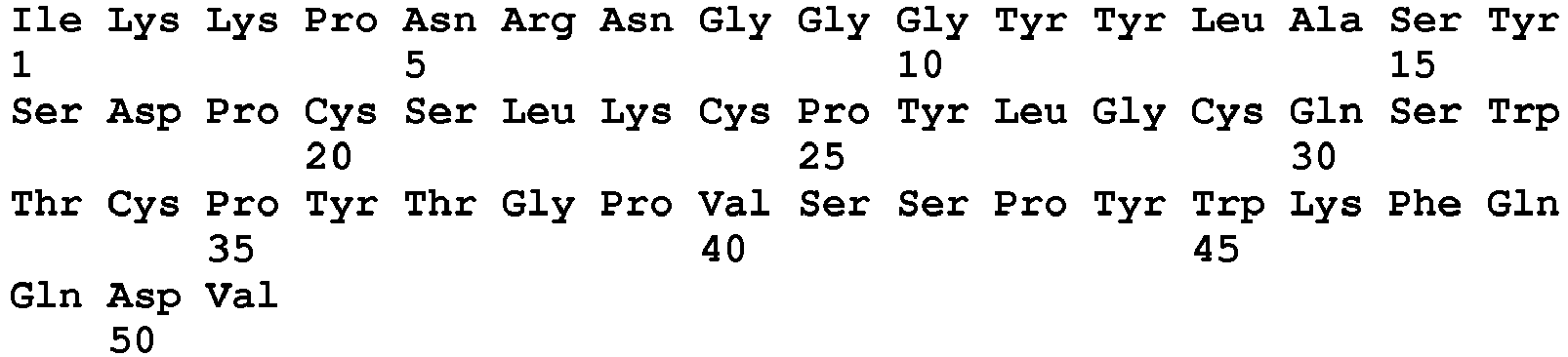
- <210> 21
<211> 171
<212> DNA
<213> Human T-celllymphotropic virus type 1 - <220>
<221> source
<222> 1..171
<223> /mol_type="unassigned DNA" /organism="Human T-celllymphotropic virus type 1" - <220>
<221> CDS
<222> 1..171
<223> /transl_table=1 - <400> 21


- <210> 22
<211> 57
<212> PRT
<213> Human T-celllymphotropic virus type 1 - <220>
<223> [CDS]:1..171 from SEQ ID NO 21 - <400> 22

- <210> 23
<211> 153
<212> DNA
<213> Human T-celllymphotropic virus type 2 - <220>
<221> source
<222> 1..153
<223> /mol_type="unassigned DNA" /organism="Human T-celllymphotropic virus type 2" - <220>
<221> CDS
<222> 1..153
<223> /transl_table=1 - <400> 23

- <210> 24
<211> 51
<212> PRT
<213> Human T-celllymphotropic virus type 2 - <220>
<223> [CDS]:1..153 from SEQ ID NO 23 - <400> 24


- <210> 25
<211> 11
<212> PRT
<213> Homo sapiens - <400> 25

- <210> 26
<211> 22
<212> PRT
<213> Homo sapiens - <400> 26

- <210> 27
<211> 10
<212> PRT
<213> Homo sapiens - <400> 27

- <210> 28
<211> 9
<212> PRT
<213> Homo sapiens - <400> 28

- <210> 29
<211> 14
<212> PRT
<213> Homo sapiens - <400> 29

- <210> 30
<211> 10
<212> PRT
<213> Homo sapiens - <400> 30

- <210> 31
<211> 7
<212> PRT
<213> Homo sapiens - <400> 31

- <210> 32
<211> 13
<212> PRT
<213> Homo sapiens - <400> 32

in association with a pharmaceutically acceptable carrier.
Claims (5)
- Use of polypeptides in an in vitro method for diagnosing glucose transporter protein type I (GLUT1) deficiency syndrome, wherein said polypeptides consist of the envelope proteins of PTLV, or fragments derived thereof, said polypeptides or fragments being selected for their ability to bind specifically to the ubiquitous vertebrate glucose transporter GLUT1 consisting of SEQ ID NO: 2.
- The use of polypeptides according to claim 1, wherein the in vitro method for diagnosing GLUT1 deficiency syndrome comprises the following steps:- contacting a biological sample from an individual with said polypeptide, said polypeptide being optionally labeled, or susceptible to be recognized by a labeled molecule, and- determining the level of said polypeptide bound to the cells contained in the biological sample and comparing said level with the level of binding of said polypeptide to cells contained in the biological sample from a healthy individual.
- The use of polypeptides according to claim 1 or 2, wherein the polypeptides are able to bind to at least one of the following fragments of GLUT1:- SEQ ID NO: 25: NAPQKVIEEFY,- SEQ ID NO: 26: NQTWVHRYGESILPTTLTTLWS,- SEQ ID NO: 27: KSFEMLILGR,- SEQ ID NO: 28: DSIMGNKDL,- SEQ ID NO: 29: YSTSIFEKAGVQQP,- SEQ ID NO: 30: EQLPWMSYLS,- SEQ ID NO: 31: QYVEQLC,- SEQ ID NO: 32: IVGMCFQYVEQLC.
- The use of polypeptides according to any one of claims 1 to 3, wherein the polypeptides are able to bind to at least the following fragment of GLUT1:- SEQ ID NO: 32: IVGMCFQYVEQLC.
- The use of polypeptides according to any one of claims 1 to 4, wherein the GLUT 1 binding polypeptides are chosen among the following:- the envelope protein of HTLV-1 consisting of SEQ ID NO: 4, or of HTLV-2 consisting of SEQ ID NO: 6, or of STLV-1 consisting of SEQ ID NO: 8, or of STLV-2 consisting of SEQ ID NO: 10, or of STLV-3 consisting of SEQ ID NO: 12,- fragments of the envelope proteins of PTLV, said fragments consisting of polypeptides delimited in their N-terminal extremity by the amino acid located in position 1 to 90, or in position 75 to 90, and in their C-terminal extremity by the amino acid located in position 135 to 245, or in position 135 to 150, of said envelope proteins of PTLV, consisting of SEQ ID NO: 4, 6, 8, 10, 12,- fragments of the envelope proteins of PTLV, said fragments consisting of the following polypeptides:∘ the polypeptide delimited in its N-terminal extremity by the amino acid located in position 83 to 89, and in its C-terminal extremity by the amino acid located in position 139 to 145, of the envelope protein of the strain MT-2 of HTLV-1 consisting of SEQ ID NO: 4,∘ the polypeptide delimited in its N-terminal extremity by the amino acid located in position 79 to 85, and in its C-terminal extremity by the amino acid located in position 135 to 141, of the envelope protein of the strain NRA of HTLV-2 consisting of SEQ ID NO: 6,∘ the polypeptide delimited in its N-terminal extremity by the amino acid located in position 83 to 89, and in its C-terminal extremity by the amino acid located in position 139 to 145, of the envelope protein of STLV-1 consisting of SEQ ID NO: 8,∘ the polypeptide delimited in its N-terminal extremity by the amino acid located in position 79 to 85, and in its C-terminal extremity by the amino acid located in position 135 to 141, of the envelope protein of STLV-2 consisting of SEQ ID NO: 10,∘ the polypeptide delimited in its N-terminal extremity by the amino acid located in position 82 to 88, and in its C-terminal extremity by the amino acid located in position 138 to 144, of the envelope protein of STLV-3 consisting of SEQ ID NO: 12,∘ the polypeptide consisting of the envelope protein of a variant of HTLV-1, said polypeptide having the following sequence SEQ ID NO: 14,∘ the polypeptide consisting of the envelope protein of a variant of HTLV-1, said polypeptide having the following sequence SEQ ID NO: 16,∘ the polypeptide consisting of the envelope protein of a variant of HTLV-1, said polypeptide having the following sequence SEQ ID NO: 18,∘ the polypeptide consisting of the envelope protein of a variant of HTLV-1, said polypeptide having the following sequence SEQ ID NO: 20,∘ the polypeptide consisting of the envelope protein of a variant of HTLV-1, said polypeptide having the following sequence SEQ ID NO: 22,∘ the polypeptide consisting of the envelope protein of a variant of HTLV-2, said polypeptide having the following sequence SEQ ID NO: 24.
Applications Claiming Priority (4)
| Application Number | Priority Date | Filing Date | Title |
|---|---|---|---|
| EP03291067 | 2003-05-02 | ||
| PCT/EP2004/004624 WO2004096841A1 (en) | 2003-05-02 | 2004-04-30 | Glut-1 as a receptor for htlv envelopes and its uses |
| EP12194035.7A EP2610265B1 (en) | 2003-05-02 | 2004-04-30 | Glut-1 as a receptor for HTLV envelopes and its uses |
| EP04730535.4A EP1648926B1 (en) | 2003-05-02 | 2004-04-30 | Glut-1 as a receptor for htlv envelopes and its uses |
Related Parent Applications (2)
| Application Number | Title | Priority Date | Filing Date |
|---|---|---|---|
| EP04730535.4A Division EP1648926B1 (en) | 2003-05-02 | 2004-04-30 | Glut-1 as a receptor for htlv envelopes and its uses |
| EP12194035.7A Division EP2610265B1 (en) | 2003-05-02 | 2004-04-30 | Glut-1 as a receptor for HTLV envelopes and its uses |
Publications (2)
| Publication Number | Publication Date |
|---|---|
| EP3168227A1 EP3168227A1 (en) | 2017-05-17 |
| EP3168227B1 true EP3168227B1 (en) | 2019-05-22 |
Family
ID=33396025
Family Applications (3)
| Application Number | Title | Priority Date | Filing Date |
|---|---|---|---|
| EP12194035.7A Expired - Lifetime EP2610265B1 (en) | 2003-05-02 | 2004-04-30 | Glut-1 as a receptor for HTLV envelopes and its uses |
| EP16197090.0A Expired - Lifetime EP3168227B1 (en) | 2003-05-02 | 2004-04-30 | Glut-1 as a receptor for htlv envelopes and its uses |
| EP04730535.4A Expired - Lifetime EP1648926B1 (en) | 2003-05-02 | 2004-04-30 | Glut-1 as a receptor for htlv envelopes and its uses |
Family Applications Before (1)
| Application Number | Title | Priority Date | Filing Date |
|---|---|---|---|
| EP12194035.7A Expired - Lifetime EP2610265B1 (en) | 2003-05-02 | 2004-04-30 | Glut-1 as a receptor for HTLV envelopes and its uses |
Family Applications After (1)
| Application Number | Title | Priority Date | Filing Date |
|---|---|---|---|
| EP04730535.4A Expired - Lifetime EP1648926B1 (en) | 2003-05-02 | 2004-04-30 | Glut-1 as a receptor for htlv envelopes and its uses |
Country Status (6)
| Country | Link |
|---|---|
| US (2) | US7642061B2 (en) |
| EP (3) | EP2610265B1 (en) |
| JP (1) | JP5612239B2 (en) |
| DK (1) | DK1648926T3 (en) |
| ES (1) | ES2740827T3 (en) |
| WO (1) | WO2004096841A1 (en) |
Families Citing this family (10)
| Publication number | Priority date | Publication date | Assignee | Title |
|---|---|---|---|---|
| EP2610265B1 (en) * | 2003-05-02 | 2016-11-16 | Centre National de la Recherche Scientifique (C.N.R.S.) | Glut-1 as a receptor for HTLV envelopes and its uses |
| US9777044B2 (en) * | 2003-05-02 | 2017-10-03 | Centre National De La Recherche Scientifique (Cnrs) | GLUT-1 as a receptor for HTLV envelopes and its uses |
| WO2007041268A2 (en) * | 2005-09-30 | 2007-04-12 | Xenoport, Inc. | Transporter-targeted methods of diagnosis and treatment |
| JP5624994B2 (en) * | 2009-01-09 | 2014-11-12 | サントル ナショナル ドゥ ラ ルシェルシュ シアンティフィク | New receptor binding ligands, their use in the detection of cells of biological interest |
| US8870950B2 (en) | 2009-12-08 | 2014-10-28 | Mitral Tech Ltd. | Rotation-based anchoring of an implant |
| WO2012035369A1 (en) * | 2010-09-17 | 2012-03-22 | Centre National De La Recherche Scientifique | Method for the diagnosis and/or prognosis of inflammatory states |
| ES2836258T3 (en) * | 2015-03-10 | 2021-06-24 | Univ Columbia | Recombinant GLUT1 Adeno-Associated Viral Vector Constructs and Related Methods for Restoring GLUT1 Expression |
| CN113508301A (en) | 2018-10-05 | 2021-10-15 | 梅塔福拉生物系统公司 | Use of ligands derived from receptor binding domains of endogenous porcine virus type B in the diagnosis of diseases associated with SMVT |
| CN113527429B (en) * | 2020-04-15 | 2024-04-16 | 辽宁中健医药科技有限公司 | Human liver cancer cell specific binding polypeptide and its use |
| WO2026041773A1 (en) * | 2024-08-23 | 2026-02-26 | Metafora Biosystems | Improved glut1-binding polypeptides |
Family Cites Families (10)
| Publication number | Priority date | Publication date | Assignee | Title |
|---|---|---|---|---|
| US5614366A (en) * | 1986-12-31 | 1997-03-25 | Genelabs Technologies, Inc. | HTLV-I peptide antigens and kit |
| JPH01501939A (en) | 1987-01-28 | 1989-07-06 | オーソ・フアーマシユーチカル・コーポレーシヨン | Immunosuppressive peptides and usage |
| WO1996021727A1 (en) | 1995-01-13 | 1996-07-18 | Virogenetics Corporation | Immunogenic compositions containing recombinant attenuated poxviruses expressing htlv antigens |
| US5897991A (en) * | 1995-06-07 | 1999-04-27 | The Mount Sinai Medical Center | Highly sensitive immunocytochemical method for diagnosis of malignant effusions |
| US5698410A (en) * | 1995-06-07 | 1997-12-16 | Mount Sinai School Of Medicine Of The City University Of New York | Highly sensitive immunocytochemical method for diagnosis of malignant effusions |
| WO1997015668A2 (en) | 1995-10-23 | 1997-05-01 | Board Of Regents, The University Of Texas System | Methods and compositions comprising glut-2 and glut-2 chimeras |
| EP0942748A4 (en) * | 1996-07-22 | 2004-12-08 | Univ Rockefeller | ENVELOPE GLYCOPROTEIN VACCINE FOR PROTECTION AGAINST HTLV-I AND HTLV-II INFECTION |
| EP1061913B1 (en) | 1998-03-13 | 2007-06-27 | The Johns Hopkins University School Of Medicine | The use of a protein tyrosine inhibitor such as genistein in the treatment of diabetic retinopathy |
| WO1999059559A1 (en) | 1998-05-15 | 1999-11-25 | Joslin Diabetes Center | Independent regulation of basal and insulin-stimulated glucose transport |
| EP2610265B1 (en) * | 2003-05-02 | 2016-11-16 | Centre National de la Recherche Scientifique (C.N.R.S.) | Glut-1 as a receptor for HTLV envelopes and its uses |
-
2004
- 2004-04-30 EP EP12194035.7A patent/EP2610265B1/en not_active Expired - Lifetime
- 2004-04-30 WO PCT/EP2004/004624 patent/WO2004096841A1/en not_active Ceased
- 2004-04-30 JP JP2006505346A patent/JP5612239B2/en not_active Expired - Lifetime
- 2004-04-30 US US10/555,289 patent/US7642061B2/en active Active
- 2004-04-30 ES ES16197090T patent/ES2740827T3/en not_active Expired - Lifetime
- 2004-04-30 EP EP16197090.0A patent/EP3168227B1/en not_active Expired - Lifetime
- 2004-04-30 DK DK04730535.4T patent/DK1648926T3/en active
- 2004-04-30 EP EP04730535.4A patent/EP1648926B1/en not_active Expired - Lifetime
-
2009
- 2009-08-26 US US12/547,924 patent/US8945583B2/en active Active
Non-Patent Citations (1)
| Title |
|---|
| None * |
Also Published As
| Publication number | Publication date |
|---|---|
| EP2610265A1 (en) | 2013-07-03 |
| DK1648926T3 (en) | 2013-10-14 |
| US7642061B2 (en) | 2010-01-05 |
| EP3168227A1 (en) | 2017-05-17 |
| EP2610265B1 (en) | 2016-11-16 |
| ES2740827T3 (en) | 2020-02-06 |
| JP5612239B2 (en) | 2014-10-22 |
| JP2007535293A (en) | 2007-12-06 |
| US20100056448A1 (en) | 2010-03-04 |
| WO2004096841A8 (en) | 2006-01-19 |
| EP1648926A1 (en) | 2006-04-26 |
| US8945583B2 (en) | 2015-02-03 |
| WO2004096841A1 (en) | 2004-11-11 |
| EP1648926B1 (en) | 2013-07-03 |
| US20080268424A1 (en) | 2008-10-30 |
Similar Documents
| Publication | Publication Date | Title |
|---|---|---|
| US8945583B2 (en) | GLUT-1 as a receptor for HTLV envelopes and its uses | |
| Manel et al. | The ubiquitous glucose transporter GLUT-1 is a receptor for HTLV | |
| Sandefur et al. | The I domain is required for efficient plasma membrane binding of human immunodeficiency virus type 1 Pr55Gag | |
| Jones et al. | Human T-cell leukemia virus type 1 (HTLV-1) and HTLV-2 use different receptor complexes to enter T cells | |
| Braaten et al. | Cyclophilin A is required for the replication of group M human immunodeficiency virus type 1 (HIV-1) and simian immunodeficiency virus SIV (CPZ) GAB but not group O HIV-1 or other primate immunodeficiency viruses | |
| Maurice et al. | Efficient gene transfer into human primary blood lymphocytes by surface-engineered lentiviral vectors that display a T cell–activating polypeptide | |
| Hoshino | Cellular factors involved in HTLV-1 entry and pathogenicit | |
| US6406841B1 (en) | Methods for the detection of HTLV-II antibodies employing novel HTLV-II NRA envelope peptides | |
| Sharma et al. | Efficient infection of a human T-cell line and of human primary peripheral blood leukocytes with a pseudotyped retrovirus vector. | |
| Constantinescu et al. | Activation of the erythropoietin receptor by the gp55‐P viral envelope protein is determined by a single amino acid in its transmembrane domain | |
| Oroszlan et al. | Primary structure and processing of gag and env gene products of human T-cell leukemia viruses HTLV-ICR and HTLV-IATK | |
| Coskun et al. | Expression of glucose transporter 1 confers susceptibility to human T-cell leukemia virus envelope-mediated fusion | |
| Le Blanc et al. | Multiple functions for the basic amino acids of the human T-cell leukemia virus type 1 matrix protein in viral transmission | |
| Alfadhli et al. | Capsid-specific nanobody effects on HIV-1 assembly and infectivity | |
| US20180002383A1 (en) | Glut-1 as a receptor for htlv envelopes and its uses | |
| Camaur et al. | Human immunodeficiency virus matrix tyrosine phosphorylation: characterization of the kinase and its substrate requirements | |
| WO1996014863A9 (en) | Suppression of proteolytic activity by dysfunctional protease formation | |
| Manel et al. | HTLV envelopes and their receptor GLUT1, the ubiquitous glucose transporter: a new vision on HTLV infection | |
| US6054281A (en) | Binding assays | |
| Rakotobe et al. | Human Polycomb group EED protein negatively affects HIV-1 assembly and release | |
| Hübner et al. | Inhibition of viral assembly in murine cells by HIV-1 matrix | |
| FR2897062A1 (en) | PEPTIDE DOMAIN NECESSARY FOR INTERACTION BETWEEN THE ENVELOPE OF A HERV-W INTERFERENCE GROUP VIRUS AND AN ASCT RECEPTOR | |
| Awasthi | Role of the human T cell lymphotropic virus type-1 (HTLV-1) accessory protein p30 (II) in adult T cell leukemogenesis | |
| Maldonado-Ortiz | Studies on the Assembly and Morphology of Human T-Cell Leukemia Virus Type 1 | |
| Murcia | Late restriction induced by an endogenous retrovirus |
Legal Events
| Date | Code | Title | Description |
|---|---|---|---|
| PUAI | Public reference made under article 153(3) epc to a published international application that has entered the european phase |
Free format text: ORIGINAL CODE: 0009012 |
|
| STAA | Information on the status of an ep patent application or granted ep patent |
Free format text: STATUS: THE APPLICATION HAS BEEN PUBLISHED |
|
| AC | Divisional application: reference to earlier application |
Ref document number: 2610265 Country of ref document: EP Kind code of ref document: P Ref document number: 1648926 Country of ref document: EP Kind code of ref document: P |
|
| AK | Designated contracting states |
Kind code of ref document: A1 Designated state(s): AT BE BG CH CY CZ DE DK EE ES FI FR GB GR HU IE IT LI LU MC NL PL PT RO SE SI SK TR |
|
| STAA | Information on the status of an ep patent application or granted ep patent |
Free format text: STATUS: REQUEST FOR EXAMINATION WAS MADE |
|
| 17P | Request for examination filed |
Effective date: 20171117 |
|
| RBV | Designated contracting states (corrected) |
Designated state(s): AT BE BG CH CY CZ DE DK EE ES FI FR GB GR HU IE IT LI LU MC NL PL PT RO SE SI SK TR |
|
| STAA | Information on the status of an ep patent application or granted ep patent |
Free format text: STATUS: EXAMINATION IS IN PROGRESS |
|
| 17Q | First examination report despatched |
Effective date: 20180123 |
|
| RIC1 | Information provided on ipc code assigned before grant |
Ipc: A61K 38/16 20060101ALI20180823BHEP Ipc: G01N 33/53 20060101ALI20180823BHEP Ipc: C07K 14/15 20060101AFI20180823BHEP Ipc: C07K 14/47 20060101ALI20180823BHEP Ipc: C07K 14/705 20060101ALI20180823BHEP Ipc: G01N 33/574 20060101ALI20180823BHEP Ipc: C07K 14/005 20060101ALI20180823BHEP |
|
| GRAP | Despatch of communication of intention to grant a patent |
Free format text: ORIGINAL CODE: EPIDOSNIGR1 |
|
| STAA | Information on the status of an ep patent application or granted ep patent |
Free format text: STATUS: GRANT OF PATENT IS INTENDED |
|
| INTG | Intention to grant announced |
Effective date: 20181005 |
|
| GRAS | Grant fee paid |
Free format text: ORIGINAL CODE: EPIDOSNIGR3 |
|
| GRAJ | Information related to disapproval of communication of intention to grant by the applicant or resumption of examination proceedings by the epo deleted |
Free format text: ORIGINAL CODE: EPIDOSDIGR1 |
|
| GRAL | Information related to payment of fee for publishing/printing deleted |
Free format text: ORIGINAL CODE: EPIDOSDIGR3 |
|
| STAA | Information on the status of an ep patent application or granted ep patent |
Free format text: STATUS: EXAMINATION IS IN PROGRESS |
|
| INTC | Intention to grant announced (deleted) | ||
| GRAR | Information related to intention to grant a patent recorded |
Free format text: ORIGINAL CODE: EPIDOSNIGR71 |
|
| STAA | Information on the status of an ep patent application or granted ep patent |
Free format text: STATUS: GRANT OF PATENT IS INTENDED |
|
| GRAA | (expected) grant |
Free format text: ORIGINAL CODE: 0009210 |
|
| STAA | Information on the status of an ep patent application or granted ep patent |
Free format text: STATUS: THE PATENT HAS BEEN GRANTED |
|
| 111L | Licence recorded |
Designated state(s): AT BE BG CH CY CZ DE DK EE ES FI FR GB GR HU IE IT LU MC NL PL PT RO SE SI SK TR Free format text: EXCLUSIVE LICENSE Name of requester: METAFORA BIOSYSTEMS, FR Effective date: 20190205 |
|
| R11L | Licence recorded (corrected) |
Designated state(s): AT BE BG CH CY CZ DE DK EE ES FI FR GB GR HU IE IT LU MC NL PL PT RO SE SI SK TR Free format text: EXCLUSIVE LICENSE Name of requester: METAFORA BIOSYSTEMS, FR Effective date: 20190205 |
|
| INTG | Intention to grant announced |
Effective date: 20190410 |
|
| 111L | Licence recorded |
Designated state(s): AT BE BG CH CY CZ DE DK EE ES FI FR GB GR HU IE IT LU MC NL PL PT RO SE SI SK TR Free format text: EXCLUSIVE LICENSE Name of requester: METAFORA BIOSYSTEMS, FR Effective date: 20190205 |
|
| AC | Divisional application: reference to earlier application |
Ref document number: 2610265 Country of ref document: EP Kind code of ref document: P Ref document number: 1648926 Country of ref document: EP Kind code of ref document: P |
|
| AK | Designated contracting states |
Kind code of ref document: B1 Designated state(s): AT BE BG CH CY CZ DE DK EE ES FI FR GB GR HU IE IT LI LU MC NL PL PT RO SE SI SK TR |
|
| REG | Reference to a national code |
Ref country code: GB Ref legal event code: FG4D |
|
| REG | Reference to a national code |
Ref country code: CH Ref legal event code: EP |
|
| REG | Reference to a national code |
Ref country code: IE Ref legal event code: FG4D |
|
| REG | Reference to a national code |
Ref country code: DE Ref legal event code: R096 Ref document number: 602004054001 Country of ref document: DE |
|
| REG | Reference to a national code |
Ref country code: AT Ref legal event code: REF Ref document number: 1135980 Country of ref document: AT Kind code of ref document: T Effective date: 20190615 |
|
| REG | Reference to a national code |
Ref country code: CH Ref legal event code: NV Representative=s name: TR-IP CONSULTING LLC, CH |
|
| REG | Reference to a national code |
Ref country code: NL Ref legal event code: MP Effective date: 20190522 |
|
| PG25 | Lapsed in a contracting state [announced via postgrant information from national office to epo] |
Ref country code: FI Free format text: LAPSE BECAUSE OF FAILURE TO SUBMIT A TRANSLATION OF THE DESCRIPTION OR TO PAY THE FEE WITHIN THE PRESCRIBED TIME-LIMIT Effective date: 20190522 Ref country code: NL Free format text: LAPSE BECAUSE OF FAILURE TO SUBMIT A TRANSLATION OF THE DESCRIPTION OR TO PAY THE FEE WITHIN THE PRESCRIBED TIME-LIMIT Effective date: 20190522 Ref country code: PT Free format text: LAPSE BECAUSE OF FAILURE TO SUBMIT A TRANSLATION OF THE DESCRIPTION OR TO PAY THE FEE WITHIN THE PRESCRIBED TIME-LIMIT Effective date: 20190922 Ref country code: SE Free format text: LAPSE BECAUSE OF FAILURE TO SUBMIT A TRANSLATION OF THE DESCRIPTION OR TO PAY THE FEE WITHIN THE PRESCRIBED TIME-LIMIT Effective date: 20190522 |
|
| PG25 | Lapsed in a contracting state [announced via postgrant information from national office to epo] |
Ref country code: BG Free format text: LAPSE BECAUSE OF FAILURE TO SUBMIT A TRANSLATION OF THE DESCRIPTION OR TO PAY THE FEE WITHIN THE PRESCRIBED TIME-LIMIT Effective date: 20190822 Ref country code: GR Free format text: LAPSE BECAUSE OF FAILURE TO SUBMIT A TRANSLATION OF THE DESCRIPTION OR TO PAY THE FEE WITHIN THE PRESCRIBED TIME-LIMIT Effective date: 20190823 |
|
| REG | Reference to a national code |
Ref country code: AT Ref legal event code: MK05 Ref document number: 1135980 Country of ref document: AT Kind code of ref document: T Effective date: 20190522 |
|
| PG25 | Lapsed in a contracting state [announced via postgrant information from national office to epo] |
Ref country code: RO Free format text: LAPSE BECAUSE OF FAILURE TO SUBMIT A TRANSLATION OF THE DESCRIPTION OR TO PAY THE FEE WITHIN THE PRESCRIBED TIME-LIMIT Effective date: 20190522 Ref country code: CZ Free format text: LAPSE BECAUSE OF FAILURE TO SUBMIT A TRANSLATION OF THE DESCRIPTION OR TO PAY THE FEE WITHIN THE PRESCRIBED TIME-LIMIT Effective date: 20190522 Ref country code: SK Free format text: LAPSE BECAUSE OF FAILURE TO SUBMIT A TRANSLATION OF THE DESCRIPTION OR TO PAY THE FEE WITHIN THE PRESCRIBED TIME-LIMIT Effective date: 20190522 Ref country code: AT Free format text: LAPSE BECAUSE OF FAILURE TO SUBMIT A TRANSLATION OF THE DESCRIPTION OR TO PAY THE FEE WITHIN THE PRESCRIBED TIME-LIMIT Effective date: 20190522 Ref country code: DK Free format text: LAPSE BECAUSE OF FAILURE TO SUBMIT A TRANSLATION OF THE DESCRIPTION OR TO PAY THE FEE WITHIN THE PRESCRIBED TIME-LIMIT Effective date: 20190522 Ref country code: EE Free format text: LAPSE BECAUSE OF FAILURE TO SUBMIT A TRANSLATION OF THE DESCRIPTION OR TO PAY THE FEE WITHIN THE PRESCRIBED TIME-LIMIT Effective date: 20190522 |
|
| REG | Reference to a national code |
Ref country code: ES Ref legal event code: FG2A Ref document number: 2740827 Country of ref document: ES Kind code of ref document: T3 Effective date: 20200206 |
|
| REG | Reference to a national code |
Ref country code: CH Ref legal event code: PCAR Free format text: NEW ADDRESS: ROUTE DU COUTSET 18, 1485 NUVILLY (CH) |
|
| REG | Reference to a national code |
Ref country code: DE Ref legal event code: R097 Ref document number: 602004054001 Country of ref document: DE |
|
| PLBE | No opposition filed within time limit |
Free format text: ORIGINAL CODE: 0009261 |
|
| STAA | Information on the status of an ep patent application or granted ep patent |
Free format text: STATUS: NO OPPOSITION FILED WITHIN TIME LIMIT |
|
| PG25 | Lapsed in a contracting state [announced via postgrant information from national office to epo] |
Ref country code: TR Free format text: LAPSE BECAUSE OF FAILURE TO SUBMIT A TRANSLATION OF THE DESCRIPTION OR TO PAY THE FEE WITHIN THE PRESCRIBED TIME-LIMIT Effective date: 20190522 |
|
| 26N | No opposition filed |
Effective date: 20200225 |
|
| PG25 | Lapsed in a contracting state [announced via postgrant information from national office to epo] |
Ref country code: PL Free format text: LAPSE BECAUSE OF FAILURE TO SUBMIT A TRANSLATION OF THE DESCRIPTION OR TO PAY THE FEE WITHIN THE PRESCRIBED TIME-LIMIT Effective date: 20190522 |
|
| PG25 | Lapsed in a contracting state [announced via postgrant information from national office to epo] |
Ref country code: SI Free format text: LAPSE BECAUSE OF FAILURE TO SUBMIT A TRANSLATION OF THE DESCRIPTION OR TO PAY THE FEE WITHIN THE PRESCRIBED TIME-LIMIT Effective date: 20190522 |
|
| PG25 | Lapsed in a contracting state [announced via postgrant information from national office to epo] |
Ref country code: MC Free format text: LAPSE BECAUSE OF FAILURE TO SUBMIT A TRANSLATION OF THE DESCRIPTION OR TO PAY THE FEE WITHIN THE PRESCRIBED TIME-LIMIT Effective date: 20190522 |
|
| PG25 | Lapsed in a contracting state [announced via postgrant information from national office to epo] |
Ref country code: LU Free format text: LAPSE BECAUSE OF NON-PAYMENT OF DUE FEES Effective date: 20200430 |
|
| REG | Reference to a national code |
Ref country code: BE Ref legal event code: MM Effective date: 20200430 |
|
| PG25 | Lapsed in a contracting state [announced via postgrant information from national office to epo] |
Ref country code: BE Free format text: LAPSE BECAUSE OF NON-PAYMENT OF DUE FEES Effective date: 20200430 |
|
| PG25 | Lapsed in a contracting state [announced via postgrant information from national office to epo] |
Ref country code: IE Free format text: LAPSE BECAUSE OF NON-PAYMENT OF DUE FEES Effective date: 20200430 |
|
| PG25 | Lapsed in a contracting state [announced via postgrant information from national office to epo] |
Ref country code: CY Free format text: LAPSE BECAUSE OF FAILURE TO SUBMIT A TRANSLATION OF THE DESCRIPTION OR TO PAY THE FEE WITHIN THE PRESCRIBED TIME-LIMIT Effective date: 20190522 |
|
| PG25 | Lapsed in a contracting state [announced via postgrant information from national office to epo] |
Ref country code: IT Free format text: LAPSE BECAUSE OF NON-PAYMENT OF DUE FEES Effective date: 20220430 |
|
| P01 | Opt-out of the competence of the unified patent court (upc) registered |
Effective date: 20230529 |
|
| PGFP | Annual fee paid to national office [announced via postgrant information from national office to epo] |
Ref country code: IT Payment date: 20230417 Year of fee payment: 20 Ref country code: FR Payment date: 20230425 Year of fee payment: 20 Ref country code: ES Payment date: 20230504 Year of fee payment: 20 Ref country code: DE Payment date: 20230412 Year of fee payment: 20 Ref country code: CH Payment date: 20230502 Year of fee payment: 20 |
|
| PGFP | Annual fee paid to national office [announced via postgrant information from national office to epo] |
Ref country code: GB Payment date: 20230424 Year of fee payment: 20 |
|
| REG | Reference to a national code |
Ref country code: DE Ref legal event code: R071 Ref document number: 602004054001 Country of ref document: DE Ref country code: CH Ref legal event code: PL |
|
| REG | Reference to a national code |
Ref country code: GB Ref legal event code: PE20 Expiry date: 20240429 |
|
| REG | Reference to a national code |
Ref country code: ES Ref legal event code: FD2A Effective date: 20240531 |
|
| PG25 | Lapsed in a contracting state [announced via postgrant information from national office to epo] |
Ref country code: GB Free format text: LAPSE BECAUSE OF EXPIRATION OF PROTECTION Effective date: 20240429 |
|
| PG25 | Lapsed in a contracting state [announced via postgrant information from national office to epo] |
Ref country code: ES Free format text: LAPSE BECAUSE OF EXPIRATION OF PROTECTION Effective date: 20240501 |
|
| PG25 | Lapsed in a contracting state [announced via postgrant information from national office to epo] |
Ref country code: GB Free format text: LAPSE BECAUSE OF EXPIRATION OF PROTECTION Effective date: 20240429 Ref country code: ES Free format text: LAPSE BECAUSE OF EXPIRATION OF PROTECTION Effective date: 20240501 |