EP3156101A1 - Radiation-therapy simulation apparatus - Google Patents
Radiation-therapy simulation apparatus Download PDFInfo
- Publication number
- EP3156101A1 EP3156101A1 EP15809175.1A EP15809175A EP3156101A1 EP 3156101 A1 EP3156101 A1 EP 3156101A1 EP 15809175 A EP15809175 A EP 15809175A EP 3156101 A1 EP3156101 A1 EP 3156101A1
- Authority
- EP
- European Patent Office
- Prior art keywords
- patient
- dose distribution
- type information
- body type
- radiation
- Prior art date
- Legal status (The legal status is an assumption and is not a legal conclusion. Google has not performed a legal analysis and makes no representation as to the accuracy of the status listed.)
- Granted
Links
- 238000001959 radiotherapy Methods 0.000 title claims abstract description 33
- 238000004088 simulation Methods 0.000 title claims abstract description 25
- 238000009826 distribution Methods 0.000 claims abstract description 96
- 239000000284 extract Substances 0.000 claims abstract description 7
- 230000005855 radiation Effects 0.000 claims description 19
- 230000001131 transforming effect Effects 0.000 claims description 7
- 239000002245 particle Substances 0.000 abstract description 18
- 238000004364 calculation method Methods 0.000 abstract description 15
- 239000011159 matrix material Substances 0.000 abstract description 7
- 238000002591 computed tomography Methods 0.000 description 15
- 238000002560 therapeutic procedure Methods 0.000 description 11
- 206010028980 Neoplasm Diseases 0.000 description 10
- 238000000342 Monte Carlo simulation Methods 0.000 description 9
- 238000000034 method Methods 0.000 description 9
- 238000010586 diagram Methods 0.000 description 6
- 230000001678 irradiating effect Effects 0.000 description 4
- 230000006399 behavior Effects 0.000 description 3
- 210000005013 brain tissue Anatomy 0.000 description 3
- 238000003384 imaging method Methods 0.000 description 3
- 230000010365 information processing Effects 0.000 description 3
- 210000000988 bone and bone Anatomy 0.000 description 2
- 210000003205 muscle Anatomy 0.000 description 2
- 210000000056 organ Anatomy 0.000 description 2
- 210000001519 tissue Anatomy 0.000 description 2
- ZOXJGFHDIHLPTG-UHFFFAOYSA-N Boron Chemical compound [B] ZOXJGFHDIHLPTG-UHFFFAOYSA-N 0.000 description 1
- 230000000903 blocking effect Effects 0.000 description 1
- 229910052796 boron Inorganic materials 0.000 description 1
- 238000004891 communication Methods 0.000 description 1
- 230000000694 effects Effects 0.000 description 1
- 239000004973 liquid crystal related substance Substances 0.000 description 1
- 238000002595 magnetic resonance imaging Methods 0.000 description 1
- 238000002600 positron emission tomography Methods 0.000 description 1
- 238000002661 proton therapy Methods 0.000 description 1
Images



Classifications
-
- A—HUMAN NECESSITIES
- A61—MEDICAL OR VETERINARY SCIENCE; HYGIENE
- A61N—ELECTROTHERAPY; MAGNETOTHERAPY; RADIATION THERAPY; ULTRASOUND THERAPY
- A61N5/00—Radiation therapy
- A61N5/10—X-ray therapy; Gamma-ray therapy; Particle-irradiation therapy
-
- A—HUMAN NECESSITIES
- A61—MEDICAL OR VETERINARY SCIENCE; HYGIENE
- A61N—ELECTROTHERAPY; MAGNETOTHERAPY; RADIATION THERAPY; ULTRASOUND THERAPY
- A61N5/00—Radiation therapy
- A61N5/10—X-ray therapy; Gamma-ray therapy; Particle-irradiation therapy
- A61N5/103—Treatment planning systems
- A61N5/1039—Treatment planning systems using functional images, e.g. PET or MRI
Definitions
- the present invention relates to a radiation-therapy simulation apparatus.
- An object of the present invention is to provide a radiation-therapy simulation apparatus capable of preventing an increase in a computation time.
- a radiation-therapy simulation apparatus which calculates a dose distribution based on irradiation applied to a target part of a patient, the apparatus including a storage unit that stores data regarding each of dose distributions obtained when a human body model for a predetermined body type is irradiated with radiation with respect to a plurality of irradiation conditions; an input unit that inputs an irradiation condition applied to the patient when the patient is irradiated with radiation, and body type information of the patient; and a correction unit that corrects data regarding a dose distribution stored in the storage unit on the basis of the body type information of the patient and body type information of the human body model.
- respective dose distributions are computed in advance by irradiating a human body model with radiation with respect to a plurality of irradiation conditions, and the dose distributions computed in advance are stored in the storage unit in correlation with the irradiation conditions.
- the radiation-therapy simulation apparatus inputs an irradiation condition applied when a patient is irradiated with radiation and body type information of a patient by using the input unit, and corrects data regarding the dose distribution stored in the storage unit on the basis of input body type information of the patient and body type information of the human body model. Consequently, the dose distribution computed in advance can be corrected according to the body type information of the patient, and corrected dose distribution can be used.
- it is not necessary to perform computation using a Monte Carlo method several times in order to find out an irradiation condition providing an optimal dose distribution it is possible to prevent an increase in a total computation time.
- the correction unit may compare CT image information including the body type information of the patient with the body type information of the human body model, transform position information in the body type information of the human body model into position information in the body type information of the patient, and correct the data regarding the dose distribution stored in the storage unit according to the transform of the position information. Consequently, the position information can be transformed so that the body type information of the human body model matches the CT image information including the body type information of the patient, and transform into position information corresponding to the body type information of the patient can be performed by applying the transformed position information to the data regarding the dose distribution stored in the storage unit. As a result, it is possible to calculate data regarding a dose distribution matching a body type of a patient and thus to obtain highly accurate data.
- the correction unit may transform position information regarding the irradiation condition applied to the patient into position information in the body type information of the human body model, extract data regarding a dose distribution corresponding to the transformed position information from the data regarding the dose distribution stored in the storage unit, and correct the extracted data regarding the dose distribution by transforming the data into position information based on the body type information of the patient. Consequently, it is possible to transform position information regarding an irradiation condition applied to a patient into position information in body type information of the human body model, extract data regarding the dose distribution obtained when the human body model is irradiated with radiation under the transformed irradiation condition, and perform correction by transforming the dose distribution into position information corresponding to the body type information of the patient. As a result, it is possible to obtain highly accurate data.
- the number of times of performing computation can be reduced, and thus it is possible to provide a radiation-therapy simulation apparatus which can prevent an increase in a total computation time.
- a radiation-therapy simulation apparatus 1 illustrated in FIG. 1 is an apparatus which can be used to create a therapy plan in a radiation therapy apparatus such as a boron neutron capture therapy (BNCT) apparatus.
- the radiation therapy apparatus includes, for example, an X-ray therapy apparatus, a proton therapy apparatus, and carbon-ion (heavy-ion) therapy apparatus.
- the radiation-therapy simulation apparatus 1 calculates a dose distribution of radiation applied to a target part of a patient in radiation therapy.
- the radiation-therapy simulation apparatus 1 includes a therapy plan terminal 2 for calculating a dose distribution, and the therapy plan terminal 2 is electrically connected to an input unit 3 and an output unit 4.
- the input unit 3 inputs various pieces of information required to calculate a dose distribution.
- the various pieces of information required to calculate a dose distribution include body type information of a patient and information regarding irradiation conditions.
- the body type information of a patient includes, for example, information regarding positions, sizes and shapes of muscles, bones, organs, tumors, and the like.
- the input unit 3 may input, for example, computed tomography (CT) image information as the body type information.
- CT computed tomography
- the input unit 3 may input image information captured by other imaging apparatuses instead of the CT image information.
- the input unit 3 may be connected to, for example, an imaging apparatus so as to input image information, and may be connected to a recording medium on which image information is recorded, so as to input the image information recorded on the recording medium.
- the information regarding irradiation conditions includes, for example, a coordinate (r, ⁇ ,z) indicating a position of the isocenter (irradiation target point), an irradiation angle ( ⁇ , ⁇ ), the kind of particle to be applied, particle energy, and a collimator diameter K.
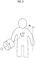
- FIG. 2 is a schematic diagram illustrating an exterior of a human body on which radiation therapy is performed
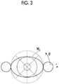
- FIG. 3 is a sectional view illustrating a section which is orthogonal to a longitudinal direction (body axis direction) of the human body on which radiation therapy is performed.
- a position of the isocenter is a position at which a particle advancing straight on an irradiation field central axis arrives.
- a target part such as a treated tumor is disposed at the isocenter.
- the position coordinate (r, ⁇ ,z) of the isocenter may be expressed in a cylindrical coordinate system, and is represented by a radius r, an angle ⁇ , and a height z as illustrated in FIGS. 2 and 3 .
- a process coordinate of the isocenter may be expressed by using other coordinate systems such as an orthogonal coordinate system.
- the irradiation angle ( ⁇ , ⁇ ) is an angle at which a particle advances toward a target part.
- the angle ⁇ is an azimuth angle which is an angle about an axis extending in a vertical direction
- the angle ⁇ is an elevation angle which is an angle about an axis extending in a horizontal direction.
- the kind of particle to be applied is, for example, a neutron, a proton, or a heavy ion.
- the energy of a particle to be applied is, for example, kinetic energy of the particle.
- the collimator is formed of members blocking advancing of a particle, and is used to form an irradiation field by restricting a particle irradiation range.
- the collimator diameter K is an inner diameter of an aperture through which a particle passes.
- the collimator diameter K is selected depending on a size of a tumor, and thus a particle irradiation range is restricted.
- the input unit 3 for example, a keyboard, a mouse, a touch panel, a scanner, and other information processing terminals and imaging apparatuses may be used.
- the output unit 4 outputs calculation results in the therapy plan terminal 2. As the calculation results, there may be image data indicating a dose distribution.
- the output unit 4 may include an image display apparatus, a printer, and the like. For example, the calculation results may be output to other information processing terminals.
- the therapy plan terminal 2 includes a central processing unit (CPU) performing a calculation process, a read only memory (ROM) and a random access memory (RAM) serving as a storage unit, an input signal circuit, an output signal circuit, a power supply circuit, and the like.
- the therapy plan terminal 2 includes a calculation unit 5 which calculates a dose distribution, a correction unit 6 which corrects the dose distribution calculated by the calculation unit 5, and a storage unit 7 which stores various pieces of information used to calculate or correct the dose distribution.
- respective dose distributions are calculated in advance by performing irradiation with particles under a plurality of irradiation conditions by using a standard human body model (human body atlas data), and data regarding the calculated dose distributions is stored in the storage unit 7.
- the storage unit 7 stores body type information of the standard human body model.
- the standard human body model is, for example, body type information which is set on the basis of body type information of many people, and includes information regarding shapes and positions of muscles, bones, organs, and the like of persons which are assumed to be standard.
- Results of calculating a dose distribution obtained by irradiating the human body model with particles in advance are stored in the storage unit 7.
- the data regarding the dose distribution is stored in correlation with each irradiation condition.
- the dose distribution for the standard human body model may be calculated by the calculation unit 5 of the radiation-therapy simulation apparatus 1, and may be calculated in advance by other information processing terminals.
- respective dose distributions are calculated by irradiating, with particles, a plurality of isocenters which are set, for example, at intervals of 5 cm and 5 degrees from a plurality of directions.
- the storage unit 7 stores, for example, data regarding dose distributions corresponding to several thousands of irradiation conditions.
- the calculation unit 5 calculates a dose distribution by using the Monte Carlo method which is known in the related art.
- a three-dimensional model of a human body is created on the basis of body type information of a human body (for example, CT image information of a patient), and a dose distribution is calculated by converting the three-dimensional model into a computation model.
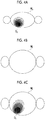
- the correction unit 6 corrects data regarding a dose distribution D 0 (standard dose distribution D 0 ) corresponding to an input irradiation condition on the basis of body type information M 1 (refer to FIG. 4B ) of a patient which is input from the input unit 3, and body type information M 0 (refer to FIG. 4A ) of the human body atlas data stored in the storage unit 7.
- the correction unit 6 compares CT image information including the body type information M 1 of the patient with the body type information M 0 of the human body atlas data, transforms position information in the body type information M 0 of the human body atlas data into position information in the body type information M 1 of the patient, and corrects the dose distribution Do stored in the storage unit 7 according to the transform of the position information.
- the correction unit 6 matches the body type information M 0 of the human body atlas data with the body type information M 1 included in a CT image of the patient, and obtains a transform matrix for transforming a position coordinate on the human body atlas data into a position coordinate on the CT image of the patient.
- the correction unit 6 corrects the data regarding the dose distribution Do for the human body atlas data stored in the storage unit 7 by transforming the data into data regarding a dose distribution D 1 corresponding to the body type information M 1 of the patient by using the transform matrix (refer to FIG. 4C ).
- the correction unit 6 transforms position information in an irradiation condition applied to the patient into position information on the human body atlas data.
- the correction unit 6 extracts data regarding a dose distribution corresponding to the transformed position information from the data regarding the dose distributions Do stored in the storage unit 7.
- the correction unit 6 corrects the extracted data regarding the dose distribution Do by performing transform into position information based on the body type information of the patient.
- a worker inputs irradiation conditions by using the input unit 3 (step S1).
- the input irradiation conditions are conditions applied when radiation therapy is performed on a patient.
- the worker inputs body type information of the patient to be treated (step S2).
- CT image information of the patient is input from a tomographic image capturing apparatus which functions as the input unit 3.
- the correction unit 6 reads body type information of the human body atlas data stored in the storage unit 7 (step S3). Next, the correction unit 6 performs positioning between the body type information of the patient and the human body atlas data so as to calculate a transform matrix (step S4). At this time, the body type information of the human body atlas data is matched with the body type information of the patient by enlarging or reducing the human body atlas data in any axial line direction.
- position information in the irradiation conditions which are input from the input unit 3 is transformed into position information in the human body atlas data (step S5).
- the coordinate (r, ⁇ ,z) indicating a position of the isocenter and the irradiation angle ( ⁇ , ⁇ ) as the position information in the irradiation conditions are transformed into position information in the human body atlas data by using the transform matrix.
- the correction unit 6 corrects the dose distribution D 0 corresponding to the irradiation conditions (step S6). Specifically, the dose distribution Do corresponding to the irradiation conditions transformed in step S5 is extracted, transform into position information corresponding to the body type information of the patient is performed by using the transform matrix with respect to the dose distribution Do, and thus corrected dose distribution D 1 is calculated.
- the calculation unit 5 creates an image of a dose distribution as illustrated in FIG. 6 on the basis of the corrected dose distribution D 1 .
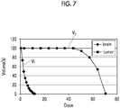
- the calculation unit 5 creates a graph (dose volume histogram) as illustrated in FIG. 7 on the basis of the corrected dose distribution.
- a transverse axis expresses a dose
- a longitudinal axis expresses a volume (%).
- FIG. 7 illustrates a relationship between a dose and a volume assuming that radiation therapy is performed on a tumor in the head of the patient as illustrated in FIG. 6 , in which V 1 indicates a volume proportion (%) occupied by normal brain tissues in a radiation irradiation region, and V 2 indicates a volume proportion (%) occupied by a tumor in the radiation irradiation region.
- the volume is 100% in a range of the dose from about 0 to about 40. In other words, it can be seen that radiation of the dose of about 40 or more is applied to 100% of the tumor portion.
- the volume is about 60% with respect to the dose of about 60. In other words, it can be seen that radiation of the dose of about 60 or more is applied to 60% of the tumor portion.
- the volume is almost 0% with respect to the dose of 10. In other words, it can be seen that radiation of the dose of about 10 or more is not applied to the normal brain tissues. It can be seen from the above description that the tumor portion is irradiated with radiation of a high dose, and the normal brain tissues are irradiated with only radiation of a low dose.
- a display unit (liquid crystal display unit) as the output unit 4 displays, for example, an image of the dose distribution illustrated in FIG. 6 or an image of the graph illustrated in FIG. 7 .
- the worker observes the image displayed on the display unit and determines whether or not the corrected dose distribution is appropriate.
- step S7 NO
- step S7 NO
- step S7 YES
- step S8 Irradiation conditions corresponding to the corrected dose distribution D 1 are determined as irradiation conditions used for actual treatment.
- the calculation unit 5 computes an accurate dose distribution by using the Monte Carlo method with respect to the determined irradiation conditions.
- a final result calculated here is output to the output unit 4.
- the respective dose distributions Do are computed in advance by irradiating the standard human body model (human body atlas data) with radiation with respect to a plurality of irradiation conditions, and the dose distributions Do computed in advance are stored in the storage unit 7 in correlation with the irradiation conditions.
- the radiation-therapy simulation apparatus 1 inputs an irradiation condition applied when a patient is irradiated with radiation and body type information of a patient by using the input unit 3, and corrects data regarding the dose distribution Do stored in the storage unit 7 on the basis of input body type information of the patient and body type information of the human body model.
- the dose distribution Do computed in advance can be corrected according to the body type information of the patient, and the corrected dose distribution D 1 can be used.
- the corrected dose distribution D 1 can be used.
- the correction unit 6 may compare CT image information including body type information of a patient with body type information of the human body atlas data, transform position information in the body type information of the human body atlas data into position information in the body type information of the patient, and correct data regarding the dose distribution stored in the storage unit 7 according to the transform of the position information. Consequently, the position information can be transformed so that the body type information of the human body atlas data matches the CT image information including the body type information of the patient, and transform into position information corresponding to the body type information of the patient can be performed by applying the transformed position information to the data regarding the dose distribution Do stored in the storage unit 7. As a result, it is possible to calculate data regarding a dose distribution matching a body type of a patient and thus to obtain highly accurate data.
- the correction unit 6 may transform position information regarding an irradiation condition applied to a patient into position information in body type information of the human body atlas data, extract data regarding the dose distribution D 0 corresponding to the transformed position information from the data regarding the dose distribution Do stored in the storage unit 7, and correct the extracted data regarding the dose distribution Do by transforming the data into position information based on the body type information of the patient.
- an appropriate dose distribution can be obtained under an irradiation condition by correcting data regarding the dose distribution Do corresponding to the human body atlas data by using the irradiation condition and body type information of a patient, and then data regarding a dose distribution at the time of applying radiation under the irradiation condition may be obtained by performing computation using the Monte Carlo method. Consequently, it is possible to obtain data regarding a more accurate dose distribution while preventing an increase in a total computation time.
- a dose distribution in the human body atlas data of a standard body type is calculated in advance, but a human body model may not be a human body model for a non-standard body type.
- Human body models for a plurality of kinds of body types may be set, and a dose distribution in each human body model may be stored in the storage unit.
- CT image information is used as body type information of a patient, but other examination data may be used as body type information of a patient.
- the process procedures illustrated in FIG. 5 may be executed by changing order thereof as appropriate.
- an irradiation condition may be input after patient body type information is input.
- An irradiation condition may be input before the irradiation condition is transformed into a coordinate on a human body model.
- 1 radiation-therapy simulation apparatus 2 therapy plan terminal, 3 input unit, 4 output unit, 5 calculation unit, 6 correction unit, 7 storage unit
Landscapes
- Health & Medical Sciences (AREA)
- Engineering & Computer Science (AREA)
- Biomedical Technology (AREA)
- Pathology (AREA)
- Nuclear Medicine, Radiotherapy & Molecular Imaging (AREA)
- Radiology & Medical Imaging (AREA)
- Life Sciences & Earth Sciences (AREA)
- Animal Behavior & Ethology (AREA)
- General Health & Medical Sciences (AREA)
- Public Health (AREA)
- Veterinary Medicine (AREA)
- Radiation-Therapy Devices (AREA)
- Nuclear Medicine (AREA)
Abstract
Description
- The present invention relates to a radiation-therapy simulation apparatus.
- When radiation therapy is performed, it is necessary to plan an optimal irradiation condition for reducing an irradiation dose for a normal tissue in the vicinity of a tumor and thus increasing an irradiation dose for the tumor prior to the therapy. Therefore, in recent years, simulation using a Monte Carlo method capable of accurately modeling behaviors of particles has been performed. Regarding computation of a dose distribution using the Monte Carlo method in radiation therapy of the related art, a three-dimensional model of a human body is created on the basis of medical image data such as computed tomography (CT), magnetic resonance imaging (MRI), or positron emission tomography (PET) of a patient, and is converted into a computation model so that a dose distribution is calculated. Citation List
- [PTL 1] Japanese Unexamined Patent Application Publication No.
2012-88771 - Since there are parts (body tissues) whose densities are greatly different from each other in the body, a particle which is incident into the human body shows a complex behavior. In the simulation using the Monte Carlo method, an accurate dose distribution can be computed since a behavior of each particle is computed, but a computation time increases. Thus, if an irradiation condition for providing a suitable dose distribution is to be obtained by performing simulation several times after changing irradiation conditions, a total computation time required therefor also necessarily increases.
- Therefore, it is necessary to determine an appropriate irradiation condition while preventing an increase in a total computation time. An object of the present invention is to provide a radiation-therapy simulation apparatus capable of preventing an increase in a computation time.
- According to an aspect of the present invention, there is provided a radiation-therapy simulation apparatus which calculates a dose distribution based on irradiation applied to a target part of a patient, the apparatus including a storage unit that stores data regarding each of dose distributions obtained when a human body model for a predetermined body type is irradiated with radiation with respect to a plurality of irradiation conditions; an input unit that inputs an irradiation condition applied to the patient when the patient is irradiated with radiation, and body type information of the patient; and a correction unit that corrects data regarding a dose distribution stored in the storage unit on the basis of the body type information of the patient and body type information of the human body model.
- In the radiation-therapy simulation apparatus of the present invention, respective dose distributions are computed in advance by irradiating a human body model with radiation with respect to a plurality of irradiation conditions, and the dose distributions computed in advance are stored in the storage unit in correlation with the irradiation conditions. The radiation-therapy simulation apparatus inputs an irradiation condition applied when a patient is irradiated with radiation and body type information of a patient by using the input unit, and corrects data regarding the dose distribution stored in the storage unit on the basis of input body type information of the patient and body type information of the human body model. Consequently, the dose distribution computed in advance can be corrected according to the body type information of the patient, and corrected dose distribution can be used. Thus, since it is not necessary to perform computation using a Monte Carlo method several times in order to find out an irradiation condition providing an optimal dose distribution, it is possible to prevent an increase in a total computation time.
- The correction unit may compare CT image information including the body type information of the patient with the body type information of the human body model, transform position information in the body type information of the human body model into position information in the body type information of the patient, and correct the data regarding the dose distribution stored in the storage unit according to the transform of the position information. Consequently, the position information can be transformed so that the body type information of the human body model matches the CT image information including the body type information of the patient, and transform into position information corresponding to the body type information of the patient can be performed by applying the transformed position information to the data regarding the dose distribution stored in the storage unit. As a result, it is possible to calculate data regarding a dose distribution matching a body type of a patient and thus to obtain highly accurate data.
- The correction unit may transform position information regarding the irradiation condition applied to the patient into position information in the body type information of the human body model, extract data regarding a dose distribution corresponding to the transformed position information from the data regarding the dose distribution stored in the storage unit, and correct the extracted data regarding the dose distribution by transforming the data into position information based on the body type information of the patient. Consequently, it is possible to transform position information regarding an irradiation condition applied to a patient into position information in body type information of the human body model, extract data regarding the dose distribution obtained when the human body model is irradiated with radiation under the transformed irradiation condition, and perform correction by transforming the dose distribution into position information corresponding to the body type information of the patient. As a result, it is possible to obtain highly accurate data.
- According to the present invention, the number of times of performing computation can be reduced, and thus it is possible to provide a radiation-therapy simulation apparatus which can prevent an increase in a total computation time.
-
-
FIG. 1 is a block diagram illustrating a radiation-therapy simulation apparatus according to an embodiment of the present invention. -
FIG. 2 is a schematic diagram illustrating an exterior of a human body on which radiation therapy is performed. -
FIG. 3 is a sectional view illustrating a section which is orthogonal to a longitudinal direction of the human body on which radiation therapy is performed. -
FIG. 4A is a schematic diagram illustrating a dose distribution for a human body model stored in a storage unit. -
FIG. 4B is a schematic diagram illustrating body type information of a patient. -
FIG. 4C is a schematic diagram illustrating a dose distribution after being corrected. -
FIG. 5 is a flowchart illustrating process procedures in the radiation-therapy simulation apparatus. -
FIG. 6 is an image illustrating a dose distribution in the head of a patient.FIG. 7 is a graph illustrating a relationship between an irradiation dose and a volume. - Hereinafter, a preferred embodiment of the present invention will be described in detail with reference to the drawings. Similar or corresponding portions are given the same reference numerals throughout the drawings, and repeated description will be omitted.
- A radiation-therapy simulation apparatus 1 illustrated in
FIG. 1 is an apparatus which can be used to create a therapy plan in a radiation therapy apparatus such as a boron neutron capture therapy (BNCT) apparatus. The radiation therapy apparatus includes, for example, an X-ray therapy apparatus, a proton therapy apparatus, and carbon-ion (heavy-ion) therapy apparatus. - The radiation-therapy simulation apparatus 1 calculates a dose distribution of radiation applied to a target part of a patient in radiation therapy. The radiation-therapy simulation apparatus 1 includes a
therapy plan terminal 2 for calculating a dose distribution, and thetherapy plan terminal 2 is electrically connected to aninput unit 3 and an output unit 4. - The
input unit 3 inputs various pieces of information required to calculate a dose distribution. The various pieces of information required to calculate a dose distribution include body type information of a patient and information regarding irradiation conditions. The body type information of a patient includes, for example, information regarding positions, sizes and shapes of muscles, bones, organs, tumors, and the like. Theinput unit 3 may input, for example, computed tomography (CT) image information as the body type information. Theinput unit 3 may input image information captured by other imaging apparatuses instead of the CT image information. Theinput unit 3 may be connected to, for example, an imaging apparatus so as to input image information, and may be connected to a recording medium on which image information is recorded, so as to input the image information recorded on the recording medium. - The information regarding irradiation conditions includes, for example, a coordinate (r,θ,z) indicating a position of the isocenter (irradiation target point), an irradiation angle (φ,ψ), the kind of particle to be applied, particle energy, and a collimator diameter K.
FIG. 2 is a schematic diagram illustrating an exterior of a human body on which radiation therapy is performed, andFIG. 3 is a sectional view illustrating a section which is orthogonal to a longitudinal direction (body axis direction) of the human body on which radiation therapy is performed. A position of the isocenter is a position at which a particle advancing straight on an irradiation field central axis arrives. For example, a target part such as a treated tumor is disposed at the isocenter. The position coordinate (r,θ,z) of the isocenter may be expressed in a cylindrical coordinate system, and is represented by a radius r, an angle θ, and a height z as illustrated inFIGS. 2 and3 . A process coordinate of the isocenter may be expressed by using other coordinate systems such as an orthogonal coordinate system. - The irradiation angle (φ,ψ) is an angle at which a particle advances toward a target part. For example, the angle φ is an azimuth angle which is an angle about an axis extending in a vertical direction, and the angle ψ is an elevation angle which is an angle about an axis extending in a horizontal direction. The kind of particle to be applied is, for example, a neutron, a proton, or a heavy ion. The energy of a particle to be applied is, for example, kinetic energy of the particle. The collimator is formed of members blocking advancing of a particle, and is used to form an irradiation field by restricting a particle irradiation range. The collimator diameter K is an inner diameter of an aperture through which a particle passes. For example, the collimator diameter K is selected depending on a size of a tumor, and thus a particle irradiation range is restricted.
- As the
input unit 3, for example, a keyboard, a mouse, a touch panel, a scanner, and other information processing terminals and imaging apparatuses may be used. - The output unit 4 outputs calculation results in the
therapy plan terminal 2. As the calculation results, there may be image data indicating a dose distribution. The output unit 4 may include an image display apparatus, a printer, and the like. For example, the calculation results may be output to other information processing terminals. - The
therapy plan terminal 2 includes a central processing unit (CPU) performing a calculation process, a read only memory (ROM) and a random access memory (RAM) serving as a storage unit, an input signal circuit, an output signal circuit, a power supply circuit, and the like. Thetherapy plan terminal 2 includes acalculation unit 5 which calculates a dose distribution, acorrection unit 6 which corrects the dose distribution calculated by thecalculation unit 5, and astorage unit 7 which stores various pieces of information used to calculate or correct the dose distribution. - In the radiation-therapy simulation apparatus 1, respective dose distributions are calculated in advance by performing irradiation with particles under a plurality of irradiation conditions by using a standard human body model (human body atlas data), and data regarding the calculated dose distributions is stored in the
storage unit 7. Thestorage unit 7 stores body type information of the standard human body model. - The standard human body model is, for example, body type information which is set on the basis of body type information of many people, and includes information regarding shapes and positions of muscles, bones, organs, and the like of persons which are assumed to be standard. Results of calculating a dose distribution obtained by irradiating the human body model with particles in advance are stored in the
storage unit 7. The data regarding the dose distribution is stored in correlation with each irradiation condition. The dose distribution for the standard human body model may be calculated by thecalculation unit 5 of the radiation-therapy simulation apparatus 1, and may be calculated in advance by other information processing terminals. - Regarding the dose distribution for the standard human body model, respective dose distributions are calculated by irradiating, with particles, a plurality of isocenters which are set, for example, at intervals of 5 cm and 5 degrees from a plurality of directions. The
storage unit 7 stores, for example, data regarding dose distributions corresponding to several thousands of irradiation conditions. - The
calculation unit 5 calculates a dose distribution by using the Monte Carlo method which is known in the related art. In the Monte Carlo method, a three-dimensional model of a human body is created on the basis of body type information of a human body (for example, CT image information of a patient), and a dose distribution is calculated by converting the three-dimensional model into a computation model. - The
correction unit 6 corrects data regarding a dose distribution D0 (standard dose distribution D0) corresponding to an input irradiation condition on the basis of body type information M1 (refer toFIG. 4B ) of a patient which is input from theinput unit 3, and body type information M0 (refer toFIG. 4A ) of the human body atlas data stored in thestorage unit 7. Thecorrection unit 6 compares CT image information including the body type information M1 of the patient with the body type information M0 of the human body atlas data, transforms position information in the body type information M0 of the human body atlas data into position information in the body type information M1 of the patient, and corrects the dose distribution Do stored in thestorage unit 7 according to the transform of the position information. Thecorrection unit 6 matches the body type information M0 of the human body atlas data with the body type information M1 included in a CT image of the patient, and obtains a transform matrix for transforming a position coordinate on the human body atlas data into a position coordinate on the CT image of the patient. Thecorrection unit 6 corrects the data regarding the dose distribution Do for the human body atlas data stored in thestorage unit 7 by transforming the data into data regarding a dose distribution D1 corresponding to the body type information M1 of the patient by using the transform matrix (refer toFIG. 4C ). - The
correction unit 6 transforms position information in an irradiation condition applied to the patient into position information on the human body atlas data. Thecorrection unit 6 extracts data regarding a dose distribution corresponding to the transformed position information from the data regarding the dose distributions Do stored in thestorage unit 7. Thecorrection unit 6 corrects the extracted data regarding the dose distribution Do by performing transform into position information based on the body type information of the patient. - Next, a description will be made of process procedures in the radiation-therapy simulation apparatus 1 with reference to a flowchart illustrated in
FIG. 5 . - First, a worker inputs irradiation conditions by using the input unit 3 (step S1). Here, the input irradiation conditions are conditions applied when radiation therapy is performed on a patient. Next, the worker inputs body type information of the patient to be treated (step S2). Here, CT image information of the patient is input from a tomographic image capturing apparatus which functions as the
input unit 3. - Next, the
correction unit 6 reads body type information of the human body atlas data stored in the storage unit 7 (step S3). Next, thecorrection unit 6 performs positioning between the body type information of the patient and the human body atlas data so as to calculate a transform matrix (step S4). At this time, the body type information of the human body atlas data is matched with the body type information of the patient by enlarging or reducing the human body atlas data in any axial line direction. - Next, position information in the irradiation conditions which are input from the
input unit 3 is transformed into position information in the human body atlas data (step S5). Here, the coordinate (r,θ,z) indicating a position of the isocenter and the irradiation angle (φ,ψ) as the position information in the irradiation conditions are transformed into position information in the human body atlas data by using the transform matrix. - Next, the
correction unit 6 corrects the dose distribution D0 corresponding to the irradiation conditions (step S6). Specifically, the dose distribution Do corresponding to the irradiation conditions transformed in step S5 is extracted, transform into position information corresponding to the body type information of the patient is performed by using the transform matrix with respect to the dose distribution Do, and thus corrected dose distribution D1 is calculated. - Next, it is determined whether or not the corrected dose distribution D1 is appropriate (step S7). For example, the
calculation unit 5 creates an image of a dose distribution as illustrated inFIG. 6 on the basis of the corrected dose distribution D1. Thecalculation unit 5 creates a graph (dose volume histogram) as illustrated inFIG. 7 on the basis of the corrected dose distribution. In the graph illustrated inFIG. 7 , a transverse axis expresses a dose, and a longitudinal axis expresses a volume (%).FIG. 7 illustrates a relationship between a dose and a volume assuming that radiation therapy is performed on a tumor in the head of the patient as illustrated inFIG. 6 , in which V1 indicates a volume proportion (%) occupied by normal brain tissues in a radiation irradiation region, and V2 indicates a volume proportion (%) occupied by a tumor in the radiation irradiation region. - In a line of V2, the volume is 100% in a range of the dose from about 0 to about 40. In other words, it can be seen that radiation of the dose of about 40 or more is applied to 100% of the tumor portion. In the line of V2, the volume is about 60% with respect to the dose of about 60. In other words, it can be seen that radiation of the dose of about 60 or more is applied to 60% of the tumor portion. In a line of V1, the volume is almost 0% with respect to the dose of 10. In other words, it can be seen that radiation of the dose of about 10 or more is not applied to the normal brain tissues. It can be seen from the above description that the tumor portion is irradiated with radiation of a high dose, and the normal brain tissues are irradiated with only radiation of a low dose.
- A display unit (liquid crystal display unit) as the output unit 4 displays, for example, an image of the dose distribution illustrated in
FIG. 6 or an image of the graph illustrated inFIG. 7 . The worker observes the image displayed on the display unit and determines whether or not the corrected dose distribution is appropriate. - In a case where the corrected dose distribution D1 is not appropriate (step S7: NO), the flow returns to step S1, and the processes in steps S1 to S7 are repeatedly performed. In a case where the corrected dose distribution D1 is appropriate (step S7: YES), the flow proceeds to step S8. Irradiation conditions corresponding to the corrected dose distribution D1 are determined as irradiation conditions used for actual treatment.
- In the subsequent step S8, the
calculation unit 5 computes an accurate dose distribution by using the Monte Carlo method with respect to the determined irradiation conditions. A final result calculated here is output to the output unit 4. - In the radiation-therapy simulation apparatus 1, the respective dose distributions Do are computed in advance by irradiating the standard human body model (human body atlas data) with radiation with respect to a plurality of irradiation conditions, and the dose distributions Do computed in advance are stored in the
storage unit 7 in correlation with the irradiation conditions. The radiation-therapy simulation apparatus 1 inputs an irradiation condition applied when a patient is irradiated with radiation and body type information of a patient by using theinput unit 3, and corrects data regarding the dose distribution Do stored in thestorage unit 7 on the basis of input body type information of the patient and body type information of the human body model. Consequently, the dose distribution Do computed in advance can be corrected according to the body type information of the patient, and the corrected dose distribution D1 can be used. Thus, since it is not necessary to perform computation using the Monte Carlo method several times in order to find out an irradiation condition providing an optimal dose distribution, it is possible to prevent an increase in a total computation time. - The
correction unit 6 may compare CT image information including body type information of a patient with body type information of the human body atlas data, transform position information in the body type information of the human body atlas data into position information in the body type information of the patient, and correct data regarding the dose distribution stored in thestorage unit 7 according to the transform of the position information. Consequently, the position information can be transformed so that the body type information of the human body atlas data matches the CT image information including the body type information of the patient, and transform into position information corresponding to the body type information of the patient can be performed by applying the transformed position information to the data regarding the dose distribution Do stored in thestorage unit 7. As a result, it is possible to calculate data regarding a dose distribution matching a body type of a patient and thus to obtain highly accurate data. - The
correction unit 6 may transform position information regarding an irradiation condition applied to a patient into position information in body type information of the human body atlas data, extract data regarding the dose distribution D0 corresponding to the transformed position information from the data regarding the dose distribution Do stored in thestorage unit 7, and correct the extracted data regarding the dose distribution Do by transforming the data into position information based on the body type information of the patient. Consequently, it is possible to transform position information in an irradiation condition applied to the patient into position information in body type information of the human body atlas data, extract data regarding the dose distribution D0 obtained when the human body atlas data is irradiated with radiation under the transformed irradiation condition, and perform correction by transforming the dose distribution Do into position information corresponding to the body type information of the patient. As a result, it is possible to obtain highly accurate data. - It may be checked that an appropriate dose distribution can be obtained under an irradiation condition by correcting data regarding the dose distribution Do corresponding to the human body atlas data by using the irradiation condition and body type information of a patient, and then data regarding a dose distribution at the time of applying radiation under the irradiation condition may be obtained by performing computation using the Monte Carlo method. Consequently, it is possible to obtain data regarding a more accurate dose distribution while preventing an increase in a total computation time.
- The present invention is not limited to the above-described embodiment, and may be variously modified as follows within the scope without departing from the concept of the present invention.
- In the above-described embodiment, a dose distribution in the human body atlas data of a standard body type is calculated in advance, but a human body model may not be a human body model for a non-standard body type. Human body models for a plurality of kinds of body types may be set, and a dose distribution in each human body model may be stored in the storage unit.
- In the above-described embodiment, CT image information is used as body type information of a patient, but other examination data may be used as body type information of a patient.
- The process procedures illustrated in
FIG. 5 may be executed by changing order thereof as appropriate. For example, an irradiation condition may be input after patient body type information is input. An irradiation condition may be input before the irradiation condition is transformed into a coordinate on a human body model. - There may be a configuration in which a terminal including a calculation unit and a terminal including a correction unit are separately provided, and data communication is performed between the terminals.
- 1 radiation-therapy simulation apparatus, 2 therapy plan terminal, 3 input unit, 4 output unit, 5 calculation unit, 6 correction unit, 7 storage unit
Claims (3)
- A radiation-therapy simulation apparatus which calculates a dose distribution based on irradiation applied to a target part of a patient, the apparatus comprising:a storage unit that stores data regarding each of dose distributions obtained when a human body model for a predetermined body type is irradiated with radiation with respect to a plurality of irradiation conditions;an input unit that inputs an irradiation condition applied to the patient when the patient is irradiated with radiation, and body type information of the patient; anda correction unit that corrects data regarding a dose distribution stored in the storage unit on the basis of the body type information of the patient and body type information of the human body model.
- The radiation-therapy simulation apparatus according to claim 1,
wherein the correction unit compares CT image information including the body type information of the patient with the body type information of the human body model, transforms position information in the body type information of the human body model into position information in the body type information of the patient, and corrects the data regarding the dose distribution stored in the storage unit according to the transform of the position information. - The radiation-therapy simulation apparatus according to claim 2,
wherein the correction unit transforms position information regarding the irradiation condition applied to the patient into position information in the body type information of the human body model, extracts data regarding a dose distribution corresponding to the transformed position information from the data regarding the dose distribution stored in the storage unit, and corrects the extracted data regarding the dose distribution by transforming the data into position information based on the body type information of the patient.
Applications Claiming Priority (2)
| Application Number | Priority Date | Filing Date | Title |
|---|---|---|---|
| JP2014123243A JP6211469B2 (en) | 2014-06-16 | 2014-06-16 | Radiation therapy simulation device |
| PCT/JP2015/067315 WO2015194554A1 (en) | 2014-06-16 | 2015-06-16 | Radiation-therapy simulation apparatus |
Publications (3)
| Publication Number | Publication Date |
|---|---|
| EP3156101A1 true EP3156101A1 (en) | 2017-04-19 |
| EP3156101A4 EP3156101A4 (en) | 2018-03-07 |
| EP3156101B1 EP3156101B1 (en) | 2019-05-22 |
Family
ID=54935537
Family Applications (1)
| Application Number | Title | Priority Date | Filing Date |
|---|---|---|---|
| EP15809175.1A Not-in-force EP3156101B1 (en) | 2014-06-16 | 2015-06-16 | Radiation-therapy simulation apparatus |
Country Status (5)
| Country | Link |
|---|---|
| EP (1) | EP3156101B1 (en) |
| JP (1) | JP6211469B2 (en) |
| CN (1) | CN106456993B (en) |
| TW (1) | TWI545521B (en) |
| WO (1) | WO2015194554A1 (en) |
Families Citing this family (8)
| Publication number | Priority date | Publication date | Assignee | Title |
|---|---|---|---|---|
| CN108778420A (en) * | 2016-03-31 | 2018-11-09 | 住友重机械工业株式会社 | Neutron capture cure treatment planning systems |
| JP6797555B2 (en) * | 2016-05-09 | 2020-12-09 | キヤノンメディカルシステムズ株式会社 | Medical information processing device |
| JP6565120B2 (en) * | 2016-09-23 | 2019-08-28 | 住友重機械工業株式会社 | Neutron capture therapy system and treatment planning system for neutron capture therapy |
| CN110709132B (en) * | 2017-04-05 | 2022-08-05 | 加利福尼亚大学董事会 | Method for user-adaptive radiation therapy planning and system using the same |
| CN110013613A (en) * | 2019-04-16 | 2019-07-16 | 东莞东阳光高能医疗设备有限公司 | Boron neutron capture treatment planning system |
| CN113827876B (en) * | 2020-06-08 | 2025-05-09 | 中硼(厦门)医疗器械有限公司 | Working steps of radiation therapy system and irradiation parameter verification device |
| CN112346109A (en) * | 2020-09-08 | 2021-02-09 | 中国辐射防护研究院 | Model and method for calibrating eye crystal dosimeter |
| CN115798725B (en) * | 2022-10-27 | 2024-03-26 | 佛山读图科技有限公司 | Method for manufacturing human body simulation image data with lesion for nuclear medicine |
Family Cites Families (5)
| Publication number | Priority date | Publication date | Assignee | Title |
|---|---|---|---|---|
| US8688618B2 (en) * | 2009-06-23 | 2014-04-01 | The Johns Hopkins University | Method and system for determining treatment plans |
| JP5641503B2 (en) * | 2010-10-15 | 2014-12-17 | 国立大学法人 筑波大学 | Multi-step lattice voxel method |
| WO2012075577A1 (en) * | 2010-12-08 | 2012-06-14 | Gregory Couch | Generating a suitable model for estimating patient radiation dose resulting from medical imaging scans |
| CN103702716B (en) * | 2011-08-17 | 2016-03-30 | 三菱电机株式会社 | Skin dose assessment aid and treatment planning device |
| JP2014054392A (en) * | 2012-09-12 | 2014-03-27 | Toshiba Corp | Radiotherapy planning device |
-
2014
- 2014-06-16 JP JP2014123243A patent/JP6211469B2/en not_active Expired - Fee Related
-
2015
- 2015-06-09 TW TW104118602A patent/TWI545521B/en not_active IP Right Cessation
- 2015-06-16 CN CN201580032442.7A patent/CN106456993B/en not_active Expired - Fee Related
- 2015-06-16 EP EP15809175.1A patent/EP3156101B1/en not_active Not-in-force
- 2015-06-16 WO PCT/JP2015/067315 patent/WO2015194554A1/en not_active Ceased
Also Published As
| Publication number | Publication date |
|---|---|
| CN106456993A (en) | 2017-02-22 |
| JP2016002178A (en) | 2016-01-12 |
| TW201601116A (en) | 2016-01-01 |
| JP6211469B2 (en) | 2017-10-11 |
| CN106456993B (en) | 2019-04-05 |
| WO2015194554A1 (en) | 2015-12-23 |
| EP3156101A4 (en) | 2018-03-07 |
| EP3156101B1 (en) | 2019-05-22 |
| TWI545521B (en) | 2016-08-11 |
Similar Documents
| Publication | Publication Date | Title |
|---|---|---|
| EP3156101B1 (en) | Radiation-therapy simulation apparatus | |
| US9179982B2 (en) | Method and system for automatic patient identification | |
| EP3655101B1 (en) | Triggered treatment systems | |
| RU2696428C2 (en) | Target object tracking system and method using a quality indicator during radiation therapy | |
| US10675486B2 (en) | Systems, devices, and methods for quality assurance of radiation therapy | |
| EP3250288B1 (en) | Three dimensional localization and tracking for adaptive radiation therapy | |
| Ehler et al. | Patient specific 3D printed phantom for IMRT quality assurance | |
| EP3251089B1 (en) | Three dimensional localization of a moving target for adaptive radiation therapy | |
| EP2745874A1 (en) | Skin dose evaluation-assisting device and therapeutic planning device | |
| Wen et al. | Evaluation of the deformation and corresponding dosimetric implications in prostate cancer treatment | |
| US9892526B2 (en) | Radiotherapy apparatus and radiotherapy method | |
| US9855443B2 (en) | Determining soft-tissue shift taking into account bony shift | |
| Roggen et al. | Deep Learning model for markerless tracking in spinal SBRT | |
| CN108883298A (en) | For the spectrum modeling system of Part Ther treatment plan, method and apparatus | |
| Wegener et al. | Feasibility of Ethos adaptive treatments of lung tumors and associated quality assurance | |
| WO2018051557A1 (en) | Medical image processing apparatus, treatment system, and medical image processing program | |
| EP4461349A1 (en) | System and method for radiation treatment planning | |
| Nakamoto et al. | A computerized framework for monitoring four-dimensional dose distributions during stereotactic body radiation therapy using a portal dose image-based 2D/3D registration approach | |
| Radonic et al. | The ROSMA-PTV concept and the impact of rotational setup uncertainties on the tumor control probability in canine brain tumors | |
| Hyka et al. | Comparison Between Different Dosimetric Tools Applications in Radiotherapy | |
| Carillo et al. | 186 CORRELATING SURROGATES FOR BLADDER DOSIMETRY WITH THE DOSE-VOLUME HISTOGRAM OF THE BLADDER WALL DEFINED ON T2W-MRI IMAGING |
Legal Events
| Date | Code | Title | Description |
|---|---|---|---|
| STAA | Information on the status of an ep patent application or granted ep patent |
Free format text: STATUS: THE INTERNATIONAL PUBLICATION HAS BEEN MADE |
|
| PUAI | Public reference made under article 153(3) epc to a published international application that has entered the european phase |
Free format text: ORIGINAL CODE: 0009012 |
|
| STAA | Information on the status of an ep patent application or granted ep patent |
Free format text: STATUS: REQUEST FOR EXAMINATION WAS MADE |
|
| 17P | Request for examination filed |
Effective date: 20170116 |
|
| AK | Designated contracting states |
Kind code of ref document: A1 Designated state(s): AL AT BE BG CH CY CZ DE DK EE ES FI FR GB GR HR HU IE IS IT LI LT LU LV MC MK MT NL NO PL PT RO RS SE SI SK SM TR |
|
| AX | Request for extension of the european patent |
Extension state: BA ME |
|
| DAV | Request for validation of the european patent (deleted) | ||
| DAX | Request for extension of the european patent (deleted) | ||
| A4 | Supplementary search report drawn up and despatched |
Effective date: 20180206 |
|
| RIC1 | Information provided on ipc code assigned before grant |
Ipc: A61N 5/10 20060101AFI20180131BHEP |
|
| GRAP | Despatch of communication of intention to grant a patent |
Free format text: ORIGINAL CODE: EPIDOSNIGR1 |
|
| STAA | Information on the status of an ep patent application or granted ep patent |
Free format text: STATUS: GRANT OF PATENT IS INTENDED |
|
| INTG | Intention to grant announced |
Effective date: 20181219 |
|
| GRAS | Grant fee paid |
Free format text: ORIGINAL CODE: EPIDOSNIGR3 |
|
| GRAA | (expected) grant |
Free format text: ORIGINAL CODE: 0009210 |
|
| STAA | Information on the status of an ep patent application or granted ep patent |
Free format text: STATUS: THE PATENT HAS BEEN GRANTED |
|
| AK | Designated contracting states |
Kind code of ref document: B1 Designated state(s): AL AT BE BG CH CY CZ DE DK EE ES FI FR GB GR HR HU IE IS IT LI LT LU LV MC MK MT NL NO PL PT RO RS SE SI SK SM TR |
|
| REG | Reference to a national code |
Ref country code: GB Ref legal event code: FG4D |
|
| REG | Reference to a national code |
Ref country code: CH Ref legal event code: EP |
|
| REG | Reference to a national code |
Ref country code: IE Ref legal event code: FG4D |
|
| REG | Reference to a national code |
Ref country code: AT Ref legal event code: REF Ref document number: 1135371 Country of ref document: AT Kind code of ref document: T Effective date: 20190615 |
|
| REG | Reference to a national code |
Ref country code: DE Ref legal event code: R096 Ref document number: 602015030895 Country of ref document: DE |
|
| REG | Reference to a national code |
Ref country code: NL Ref legal event code: MP Effective date: 20190522 |
|
| REG | Reference to a national code |
Ref country code: LT Ref legal event code: MG4D |
|
| PG25 | Lapsed in a contracting state [announced via postgrant information from national office to epo] |
Ref country code: NO Free format text: LAPSE BECAUSE OF FAILURE TO SUBMIT A TRANSLATION OF THE DESCRIPTION OR TO PAY THE FEE WITHIN THE PRESCRIBED TIME-LIMIT Effective date: 20190822 Ref country code: LT Free format text: LAPSE BECAUSE OF FAILURE TO SUBMIT A TRANSLATION OF THE DESCRIPTION OR TO PAY THE FEE WITHIN THE PRESCRIBED TIME-LIMIT Effective date: 20190522 Ref country code: NL Free format text: LAPSE BECAUSE OF FAILURE TO SUBMIT A TRANSLATION OF THE DESCRIPTION OR TO PAY THE FEE WITHIN THE PRESCRIBED TIME-LIMIT Effective date: 20190522 Ref country code: ES Free format text: LAPSE BECAUSE OF FAILURE TO SUBMIT A TRANSLATION OF THE DESCRIPTION OR TO PAY THE FEE WITHIN THE PRESCRIBED TIME-LIMIT Effective date: 20190522 Ref country code: PT Free format text: LAPSE BECAUSE OF FAILURE TO SUBMIT A TRANSLATION OF THE DESCRIPTION OR TO PAY THE FEE WITHIN THE PRESCRIBED TIME-LIMIT Effective date: 20190922 Ref country code: HR Free format text: LAPSE BECAUSE OF FAILURE TO SUBMIT A TRANSLATION OF THE DESCRIPTION OR TO PAY THE FEE WITHIN THE PRESCRIBED TIME-LIMIT Effective date: 20190522 Ref country code: AL Free format text: LAPSE BECAUSE OF FAILURE TO SUBMIT A TRANSLATION OF THE DESCRIPTION OR TO PAY THE FEE WITHIN THE PRESCRIBED TIME-LIMIT Effective date: 20190522 Ref country code: SE Free format text: LAPSE BECAUSE OF FAILURE TO SUBMIT A TRANSLATION OF THE DESCRIPTION OR TO PAY THE FEE WITHIN THE PRESCRIBED TIME-LIMIT Effective date: 20190522 |
|
| PG25 | Lapsed in a contracting state [announced via postgrant information from national office to epo] |
Ref country code: GR Free format text: LAPSE BECAUSE OF FAILURE TO SUBMIT A TRANSLATION OF THE DESCRIPTION OR TO PAY THE FEE WITHIN THE PRESCRIBED TIME-LIMIT Effective date: 20190823 Ref country code: RS Free format text: LAPSE BECAUSE OF FAILURE TO SUBMIT A TRANSLATION OF THE DESCRIPTION OR TO PAY THE FEE WITHIN THE PRESCRIBED TIME-LIMIT Effective date: 20190522 Ref country code: LV Free format text: LAPSE BECAUSE OF FAILURE TO SUBMIT A TRANSLATION OF THE DESCRIPTION OR TO PAY THE FEE WITHIN THE PRESCRIBED TIME-LIMIT Effective date: 20190522 Ref country code: BG Free format text: LAPSE BECAUSE OF FAILURE TO SUBMIT A TRANSLATION OF THE DESCRIPTION OR TO PAY THE FEE WITHIN THE PRESCRIBED TIME-LIMIT Effective date: 20190822 |
|
| REG | Reference to a national code |
Ref country code: AT Ref legal event code: MK05 Ref document number: 1135371 Country of ref document: AT Kind code of ref document: T Effective date: 20190522 |
|
| PG25 | Lapsed in a contracting state [announced via postgrant information from national office to epo] |
Ref country code: SK Free format text: LAPSE BECAUSE OF FAILURE TO SUBMIT A TRANSLATION OF THE DESCRIPTION OR TO PAY THE FEE WITHIN THE PRESCRIBED TIME-LIMIT Effective date: 20190522 Ref country code: RO Free format text: LAPSE BECAUSE OF FAILURE TO SUBMIT A TRANSLATION OF THE DESCRIPTION OR TO PAY THE FEE WITHIN THE PRESCRIBED TIME-LIMIT Effective date: 20190522 Ref country code: CZ Free format text: LAPSE BECAUSE OF FAILURE TO SUBMIT A TRANSLATION OF THE DESCRIPTION OR TO PAY THE FEE WITHIN THE PRESCRIBED TIME-LIMIT Effective date: 20190522 Ref country code: AT Free format text: LAPSE BECAUSE OF FAILURE TO SUBMIT A TRANSLATION OF THE DESCRIPTION OR TO PAY THE FEE WITHIN THE PRESCRIBED TIME-LIMIT Effective date: 20190522 Ref country code: EE Free format text: LAPSE BECAUSE OF FAILURE TO SUBMIT A TRANSLATION OF THE DESCRIPTION OR TO PAY THE FEE WITHIN THE PRESCRIBED TIME-LIMIT Effective date: 20190522 Ref country code: DK Free format text: LAPSE BECAUSE OF FAILURE TO SUBMIT A TRANSLATION OF THE DESCRIPTION OR TO PAY THE FEE WITHIN THE PRESCRIBED TIME-LIMIT Effective date: 20190522 |
|
| REG | Reference to a national code |
Ref country code: CH Ref legal event code: PL |
|
| REG | Reference to a national code |
Ref country code: DE Ref legal event code: R097 Ref document number: 602015030895 Country of ref document: DE |
|
| PG25 | Lapsed in a contracting state [announced via postgrant information from national office to epo] |
Ref country code: IT Free format text: LAPSE BECAUSE OF FAILURE TO SUBMIT A TRANSLATION OF THE DESCRIPTION OR TO PAY THE FEE WITHIN THE PRESCRIBED TIME-LIMIT Effective date: 20190522 Ref country code: SM Free format text: LAPSE BECAUSE OF FAILURE TO SUBMIT A TRANSLATION OF THE DESCRIPTION OR TO PAY THE FEE WITHIN THE PRESCRIBED TIME-LIMIT Effective date: 20190522 Ref country code: MC Free format text: LAPSE BECAUSE OF FAILURE TO SUBMIT A TRANSLATION OF THE DESCRIPTION OR TO PAY THE FEE WITHIN THE PRESCRIBED TIME-LIMIT Effective date: 20190522 |
|
| PLBE | No opposition filed within time limit |
Free format text: ORIGINAL CODE: 0009261 |
|
| REG | Reference to a national code |
Ref country code: BE Ref legal event code: MM Effective date: 20190630 |
|
| STAA | Information on the status of an ep patent application or granted ep patent |
Free format text: STATUS: NO OPPOSITION FILED WITHIN TIME LIMIT |
|
| PG25 | Lapsed in a contracting state [announced via postgrant information from national office to epo] |
Ref country code: TR Free format text: LAPSE BECAUSE OF FAILURE TO SUBMIT A TRANSLATION OF THE DESCRIPTION OR TO PAY THE FEE WITHIN THE PRESCRIBED TIME-LIMIT Effective date: 20190522 |
|
| 26N | No opposition filed |
Effective date: 20200225 |
|
| GBPC | Gb: european patent ceased through non-payment of renewal fee |
Effective date: 20190822 |
|
| PG25 | Lapsed in a contracting state [announced via postgrant information from national office to epo] |
Ref country code: IE Free format text: LAPSE BECAUSE OF NON-PAYMENT OF DUE FEES Effective date: 20190616 Ref country code: PL Free format text: LAPSE BECAUSE OF FAILURE TO SUBMIT A TRANSLATION OF THE DESCRIPTION OR TO PAY THE FEE WITHIN THE PRESCRIBED TIME-LIMIT Effective date: 20190522 |
|
| PG25 | Lapsed in a contracting state [announced via postgrant information from national office to epo] |
Ref country code: BE Free format text: LAPSE BECAUSE OF NON-PAYMENT OF DUE FEES Effective date: 20190630 Ref country code: CH Free format text: LAPSE BECAUSE OF NON-PAYMENT OF DUE FEES Effective date: 20190630 Ref country code: LI Free format text: LAPSE BECAUSE OF NON-PAYMENT OF DUE FEES Effective date: 20190630 Ref country code: SI Free format text: LAPSE BECAUSE OF FAILURE TO SUBMIT A TRANSLATION OF THE DESCRIPTION OR TO PAY THE FEE WITHIN THE PRESCRIBED TIME-LIMIT Effective date: 20190522 Ref country code: LU Free format text: LAPSE BECAUSE OF NON-PAYMENT OF DUE FEES Effective date: 20190616 |
|
| PG25 | Lapsed in a contracting state [announced via postgrant information from national office to epo] |
Ref country code: FR Free format text: LAPSE BECAUSE OF NON-PAYMENT OF DUE FEES Effective date: 20190722 |
|
| PGFP | Annual fee paid to national office [announced via postgrant information from national office to epo] |
Ref country code: DE Payment date: 20200602 Year of fee payment: 6 Ref country code: FI Payment date: 20200609 Year of fee payment: 6 |
|
| PG25 | Lapsed in a contracting state [announced via postgrant information from national office to epo] |
Ref country code: GB Free format text: LAPSE BECAUSE OF NON-PAYMENT OF DUE FEES Effective date: 20190822 |
|
| PG25 | Lapsed in a contracting state [announced via postgrant information from national office to epo] |
Ref country code: CY Free format text: LAPSE BECAUSE OF FAILURE TO SUBMIT A TRANSLATION OF THE DESCRIPTION OR TO PAY THE FEE WITHIN THE PRESCRIBED TIME-LIMIT Effective date: 20190522 |
|
| PG25 | Lapsed in a contracting state [announced via postgrant information from national office to epo] |
Ref country code: IS Free format text: LAPSE BECAUSE OF FAILURE TO SUBMIT A TRANSLATION OF THE DESCRIPTION OR TO PAY THE FEE WITHIN THE PRESCRIBED TIME-LIMIT Effective date: 20190922 |
|
| PG25 | Lapsed in a contracting state [announced via postgrant information from national office to epo] |
Ref country code: HU Free format text: LAPSE BECAUSE OF FAILURE TO SUBMIT A TRANSLATION OF THE DESCRIPTION OR TO PAY THE FEE WITHIN THE PRESCRIBED TIME-LIMIT; INVALID AB INITIO Effective date: 20150616 Ref country code: MT Free format text: LAPSE BECAUSE OF FAILURE TO SUBMIT A TRANSLATION OF THE DESCRIPTION OR TO PAY THE FEE WITHIN THE PRESCRIBED TIME-LIMIT Effective date: 20190522 |
|
| REG | Reference to a national code |
Ref country code: DE Ref legal event code: R119 Ref document number: 602015030895 Country of ref document: DE |
|
| REG | Reference to a national code |
Ref country code: FI Ref legal event code: MAE |
|
| PG25 | Lapsed in a contracting state [announced via postgrant information from national office to epo] |
Ref country code: FI Free format text: LAPSE BECAUSE OF NON-PAYMENT OF DUE FEES Effective date: 20210616 |
|
| PG25 | Lapsed in a contracting state [announced via postgrant information from national office to epo] |
Ref country code: DE Free format text: LAPSE BECAUSE OF NON-PAYMENT OF DUE FEES Effective date: 20220101 |
|
| PG25 | Lapsed in a contracting state [announced via postgrant information from national office to epo] |
Ref country code: MK Free format text: LAPSE BECAUSE OF FAILURE TO SUBMIT A TRANSLATION OF THE DESCRIPTION OR TO PAY THE FEE WITHIN THE PRESCRIBED TIME-LIMIT Effective date: 20190522 |