EP3151011A1 - Methods of detecting therapeutic exosomes - Google Patents
Methods of detecting therapeutic exosomes Download PDFInfo
- Publication number
- EP3151011A1 EP3151011A1 EP16194158.8A EP16194158A EP3151011A1 EP 3151011 A1 EP3151011 A1 EP 3151011A1 EP 16194158 A EP16194158 A EP 16194158A EP 3151011 A1 EP3151011 A1 EP 3151011A1
- Authority
- EP
- European Patent Office
- Prior art keywords
- activity
- exosome
- exosomes
- cells
- msc
- Prior art date
- Legal status (The legal status is an assumption and is not a legal conclusion. Google has not performed a legal analysis and makes no representation as to the accuracy of the status listed.)
- Granted
Links
- 210000001808 exosome Anatomy 0.000 title claims abstract description 474
- 230000001225 therapeutic effect Effects 0.000 title claims abstract description 113
- 238000000034 method Methods 0.000 title claims abstract description 111
- 230000000694 effects Effects 0.000 claims abstract description 239
- 230000000295 complement effect Effects 0.000 claims abstract description 27
- 102100022464 5'-nucleotidase Human genes 0.000 claims abstract description 25
- 101000678236 Homo sapiens 5'-nucleotidase Proteins 0.000 claims abstract description 25
- 210000002901 mesenchymal stem cell Anatomy 0.000 claims description 223
- 108090000623 proteins and genes Proteins 0.000 claims description 187
- 102000004169 proteins and genes Human genes 0.000 claims description 169
- 239000003636 conditioned culture medium Substances 0.000 claims description 57
- 239000012528 membrane Substances 0.000 claims description 29
- 230000004071 biological effect Effects 0.000 claims description 28
- 239000000203 mixture Substances 0.000 claims description 25
- 201000010099 disease Diseases 0.000 claims description 24
- 208000037265 diseases, disorders, signs and symptoms Diseases 0.000 claims description 24
- 206010028980 Neoplasm Diseases 0.000 claims description 21
- 206010061216 Infarction Diseases 0.000 claims description 15
- 201000011510 cancer Diseases 0.000 claims description 15
- 230000007574 infarction Effects 0.000 claims description 15
- 230000001404 mediated effect Effects 0.000 claims description 15
- 230000036542 oxidative stress Effects 0.000 claims description 14
- 238000002296 dynamic light scattering Methods 0.000 claims description 13
- 208000027418 Wounds and injury Diseases 0.000 claims description 10
- 208000031225 myocardial ischemia Diseases 0.000 claims description 10
- MHAJPDPJQMAIIY-UHFFFAOYSA-N Hydrogen peroxide Chemical compound OO MHAJPDPJQMAIIY-UHFFFAOYSA-N 0.000 claims description 9
- 206010019280 Heart failures Diseases 0.000 claims description 8
- 230000006037 cell lysis Effects 0.000 claims description 8
- 230000006378 damage Effects 0.000 claims description 8
- 208000024827 Alzheimer disease Diseases 0.000 claims description 7
- 206010061218 Inflammation Diseases 0.000 claims description 7
- 208000007201 Myocardial reperfusion injury Diseases 0.000 claims description 7
- 230000003293 cardioprotective effect Effects 0.000 claims description 7
- 230000030833 cell death Effects 0.000 claims description 7
- 206010012601 diabetes mellitus Diseases 0.000 claims description 7
- 230000001939 inductive effect Effects 0.000 claims description 7
- 230000004054 inflammatory process Effects 0.000 claims description 7
- 238000010172 mouse model Methods 0.000 claims description 7
- 208000015122 neurodegenerative disease Diseases 0.000 claims description 7
- 208000018737 Parkinson disease Diseases 0.000 claims description 6
- 208000000453 Skin Neoplasms Diseases 0.000 claims description 6
- 208000015322 bone marrow disease Diseases 0.000 claims description 6
- 230000001684 chronic effect Effects 0.000 claims description 6
- 208000014674 injury Diseases 0.000 claims description 6
- 238000013310 pig model Methods 0.000 claims description 6
- 208000017520 skin disease Diseases 0.000 claims description 6
- 201000004681 Psoriasis Diseases 0.000 claims description 5
- 230000001575 pathological effect Effects 0.000 claims description 5
- 201000000849 skin cancer Diseases 0.000 claims description 5
- 206010059313 Anogenital warts Diseases 0.000 claims description 4
- 208000027496 Behcet disease Diseases 0.000 claims description 4
- 208000009137 Behcet syndrome Diseases 0.000 claims description 4
- 201000003883 Cystic fibrosis Diseases 0.000 claims description 4
- 102100025621 Cytochrome b-245 heavy chain Human genes 0.000 claims description 4
- 201000004624 Dermatitis Diseases 0.000 claims description 4
- 206010012438 Dermatitis atopic Diseases 0.000 claims description 4
- 206010014982 Epidermal and dermal conditions Diseases 0.000 claims description 4
- 206010016654 Fibrosis Diseases 0.000 claims description 4
- 208000002260 Keloid Diseases 0.000 claims description 4
- 206010023330 Keloid scar Diseases 0.000 claims description 4
- 208000031673 T-Cell Cutaneous Lymphoma Diseases 0.000 claims description 4
- 208000025865 Ulcer Diseases 0.000 claims description 4
- 241000934136 Verruca Species 0.000 claims description 4
- 208000000260 Warts Diseases 0.000 claims description 4
- 206010052428 Wound Diseases 0.000 claims description 4
- 201000008937 atopic dermatitis Diseases 0.000 claims description 4
- 208000016532 chronic granulomatous disease Diseases 0.000 claims description 4
- 201000007241 cutaneous T cell lymphoma Diseases 0.000 claims description 4
- 230000004761 fibrosis Effects 0.000 claims description 4
- 201000011066 hemangioma Diseases 0.000 claims description 4
- 230000008938 immune dysregulation Effects 0.000 claims description 4
- 238000000099 in vitro assay Methods 0.000 claims description 4
- 208000037906 ischaemic injury Diseases 0.000 claims description 4
- 210000001117 keloid Anatomy 0.000 claims description 4
- 230000003902 lesion Effects 0.000 claims description 4
- 201000005962 mycosis fungoides Diseases 0.000 claims description 4
- 208000010125 myocardial infarction Diseases 0.000 claims description 4
- 208000025638 primary cutaneous T-cell non-Hodgkin lymphoma Diseases 0.000 claims description 4
- 206010039083 rhinitis Diseases 0.000 claims description 4
- 201000009890 sinusitis Diseases 0.000 claims description 4
- 201000010153 skin papilloma Diseases 0.000 claims description 4
- 230000007838 tissue remodeling Effects 0.000 claims description 4
- 230000036269 ulceration Effects 0.000 claims description 4
- 230000002401 inhibitory effect Effects 0.000 claims description 3
- 230000014759 maintenance of location Effects 0.000 claims description 3
- 238000001542 size-exclusion chromatography Methods 0.000 claims description 3
- 238000000108 ultra-filtration Methods 0.000 claims description 3
- 108090000708 Proteasome Endopeptidase Complex Proteins 0.000 abstract description 42
- 102000004245 Proteasome Endopeptidase Complex Human genes 0.000 abstract description 42
- 102000004190 Enzymes Human genes 0.000 abstract description 40
- 108090000790 Enzymes Proteins 0.000 abstract description 40
- 102000010834 Extracellular Matrix Proteins Human genes 0.000 abstract description 26
- 108010037362 Extracellular Matrix Proteins Proteins 0.000 abstract description 26
- 210000002744 extracellular matrix Anatomy 0.000 abstract description 26
- 230000002414 glycolytic effect Effects 0.000 abstract description 24
- 230000005764 inhibitory process Effects 0.000 abstract description 18
- 230000019948 ion homeostasis Effects 0.000 abstract description 15
- 230000012846 protein folding Effects 0.000 abstract description 15
- 230000003078 antioxidant effect Effects 0.000 abstract description 14
- 230000000051 modifying effect Effects 0.000 abstract description 13
- 210000004027 cell Anatomy 0.000 description 201
- 235000018102 proteins Nutrition 0.000 description 165
- 238000003556 assay Methods 0.000 description 32
- WEVYAHXRMPXWCK-UHFFFAOYSA-N Acetonitrile Chemical compound CC#N WEVYAHXRMPXWCK-UHFFFAOYSA-N 0.000 description 30
- 230000001413 cellular effect Effects 0.000 description 28
- 241000699666 Mus <mouse, genus> Species 0.000 description 27
- 102000002689 Toll-like receptor Human genes 0.000 description 26
- 108020000411 Toll-like receptor Proteins 0.000 description 26
- 210000004379 membrane Anatomy 0.000 description 26
- 230000004913 activation Effects 0.000 description 24
- 239000000047 product Substances 0.000 description 24
- 101000716102 Homo sapiens T-cell surface glycoprotein CD4 Proteins 0.000 description 21
- 102100036011 T-cell surface glycoprotein CD4 Human genes 0.000 description 21
- 238000001514 detection method Methods 0.000 description 21
- 108090000765 processed proteins & peptides Proteins 0.000 description 21
- 239000002609 medium Substances 0.000 description 20
- 102000003974 Fibroblast growth factor 2 Human genes 0.000 description 19
- 108090000379 Fibroblast growth factor 2 Proteins 0.000 description 19
- 239000000427 antigen Substances 0.000 description 19
- 238000004519 manufacturing process Methods 0.000 description 19
- 210000001519 tissue Anatomy 0.000 description 19
- 210000001744 T-lymphocyte Anatomy 0.000 description 18
- 108091007433 antigens Proteins 0.000 description 18
- 102000036639 antigens Human genes 0.000 description 18
- 230000031018 biological processes and functions Effects 0.000 description 18
- 102000004196 processed proteins & peptides Human genes 0.000 description 18
- 238000011282 treatment Methods 0.000 description 18
- 210000001671 embryonic stem cell Anatomy 0.000 description 17
- 229930027945 nicotinamide-adenine dinucleotide Natural products 0.000 description 16
- 101000831567 Homo sapiens Toll-like receptor 2 Proteins 0.000 description 15
- 101000669447 Homo sapiens Toll-like receptor 4 Proteins 0.000 description 15
- OIRDTQYFTABQOQ-KQYNXXCUSA-N adenosine Chemical compound C1=NC=2C(N)=NC=NC=2N1[C@@H]1O[C@H](CO)[C@@H](O)[C@H]1O OIRDTQYFTABQOQ-KQYNXXCUSA-N 0.000 description 15
- 238000010790 dilution Methods 0.000 description 15
- 239000012895 dilution Substances 0.000 description 15
- 230000002519 immonomodulatory effect Effects 0.000 description 15
- 238000004949 mass spectrometry Methods 0.000 description 15
- BOPGDPNILDQYTO-NNYOXOHSSA-N nicotinamide-adenine dinucleotide Chemical compound C1=CCC(C(=O)N)=CN1[C@H]1[C@H](O)[C@H](O)[C@@H](COP(O)(=O)OP(O)(=O)OC[C@@H]2[C@H]([C@@H](O)[C@@H](O2)N2C3=NC=NC(N)=C3N=C2)O)O1 BOPGDPNILDQYTO-NNYOXOHSSA-N 0.000 description 15
- 239000000872 buffer Substances 0.000 description 14
- 238000010586 diagram Methods 0.000 description 14
- 230000004069 differentiation Effects 0.000 description 14
- 239000000463 material Substances 0.000 description 14
- 239000000758 substrate Substances 0.000 description 14
- 102100024333 Toll-like receptor 2 Human genes 0.000 description 13
- 102100039360 Toll-like receptor 4 Human genes 0.000 description 13
- 230000034659 glycolysis Effects 0.000 description 13
- 108091032973 (ribonucleotides)n+m Proteins 0.000 description 12
- 102000013009 Pyruvate Kinase Human genes 0.000 description 12
- 108020005115 Pyruvate Kinase Proteins 0.000 description 12
- 230000015572 biosynthetic process Effects 0.000 description 12
- 230000006870 function Effects 0.000 description 12
- BDAGIHXWWSANSR-UHFFFAOYSA-N methanoic acid Natural products OC=O BDAGIHXWWSANSR-UHFFFAOYSA-N 0.000 description 12
- 102100022002 CD59 glycoprotein Human genes 0.000 description 11
- 101000897400 Homo sapiens CD59 glycoprotein Proteins 0.000 description 11
- 102100033810 RAC-alpha serine/threonine-protein kinase Human genes 0.000 description 11
- 239000002158 endotoxin Substances 0.000 description 11
- 229920006008 lipopolysaccharide Polymers 0.000 description 11
- 108010000685 platelet-derived growth factor AB Proteins 0.000 description 11
- 239000004017 serum-free culture medium Substances 0.000 description 11
- 102000004127 Cytokines Human genes 0.000 description 10
- 108090000695 Cytokines Proteins 0.000 description 10
- 241000283973 Oryctolagus cuniculus Species 0.000 description 10
- 206010063837 Reperfusion injury Diseases 0.000 description 10
- 210000000068 Th17 cell Anatomy 0.000 description 10
- 238000003149 assay kit Methods 0.000 description 10
- 238000004113 cell culture Methods 0.000 description 10
- 230000001143 conditioned effect Effects 0.000 description 10
- 230000002255 enzymatic effect Effects 0.000 description 10
- 230000008569 process Effects 0.000 description 10
- 210000003289 regulatory T cell Anatomy 0.000 description 10
- 230000019491 signal transduction Effects 0.000 description 10
- ZFXYFBGIUFBOJW-UHFFFAOYSA-N theophylline Chemical compound O=C1N(C)C(=O)N(C)C2=C1NC=N2 ZFXYFBGIUFBOJW-UHFFFAOYSA-N 0.000 description 10
- 238000001262 western blot Methods 0.000 description 10
- MNULEGDCPYONBU-WMBHJXFZSA-N (1r,4s,5e,5'r,6'r,7e,10s,11r,12s,14r,15s,16s,18r,19s,20r,21e,25s,26r,27s,29s)-4-ethyl-11,12,15,19-tetrahydroxy-6'-[(2s)-2-hydroxypropyl]-5',10,12,14,16,18,20,26,29-nonamethylspiro[24,28-dioxabicyclo[23.3.1]nonacosa-5,7,21-triene-27,2'-oxane]-13,17,23-trio Polymers O([C@@H]1CC[C@@H](/C=C/C=C/C[C@H](C)[C@@H](O)[C@](C)(O)C(=O)[C@H](C)[C@@H](O)[C@H](C)C(=O)[C@H](C)[C@@H](O)[C@H](C)/C=C/C(=O)O[C@H]([C@H]2C)[C@H]1C)CC)[C@]12CC[C@@H](C)[C@@H](C[C@H](C)O)O1 MNULEGDCPYONBU-WMBHJXFZSA-N 0.000 description 9
- MNULEGDCPYONBU-DJRUDOHVSA-N (1s,4r,5z,5'r,6'r,7e,10s,11r,12s,14r,15s,18r,19r,20s,21e,26r,27s)-4-ethyl-11,12,15,19-tetrahydroxy-6'-(2-hydroxypropyl)-5',10,12,14,16,18,20,26,29-nonamethylspiro[24,28-dioxabicyclo[23.3.1]nonacosa-5,7,21-triene-27,2'-oxane]-13,17,23-trione Polymers O([C@H]1CC[C@H](\C=C/C=C/C[C@H](C)[C@@H](O)[C@](C)(O)C(=O)[C@H](C)[C@@H](O)C(C)C(=O)[C@H](C)[C@H](O)[C@@H](C)/C=C/C(=O)OC([C@H]2C)C1C)CC)[C@]12CC[C@@H](C)[C@@H](CC(C)O)O1 MNULEGDCPYONBU-DJRUDOHVSA-N 0.000 description 9
- MNULEGDCPYONBU-YNZHUHFTSA-N (4Z,18Z,20Z)-22-ethyl-7,11,14,15-tetrahydroxy-6'-(2-hydroxypropyl)-5',6,8,10,12,14,16,28,29-nonamethylspiro[2,26-dioxabicyclo[23.3.1]nonacosa-4,18,20-triene-27,2'-oxane]-3,9,13-trione Polymers CC1C(C2C)OC(=O)\C=C/C(C)C(O)C(C)C(=O)C(C)C(O)C(C)C(=O)C(C)(O)C(O)C(C)C\C=C/C=C\C(CC)CCC2OC21CCC(C)C(CC(C)O)O2 MNULEGDCPYONBU-YNZHUHFTSA-N 0.000 description 9
- MNULEGDCPYONBU-VVXVDZGXSA-N (5e,5'r,7e,10s,11r,12s,14s,15r,16r,18r,19s,20r,21e,26r,29s)-4-ethyl-11,12,15,19-tetrahydroxy-6'-[(2s)-2-hydroxypropyl]-5',10,12,14,16,18,20,26,29-nonamethylspiro[24,28-dioxabicyclo[23.3.1]nonacosa-5,7,21-triene-27,2'-oxane]-13,17,23-trione Polymers C([C@H](C)[C@@H](O)[C@](C)(O)C(=O)[C@@H](C)[C@H](O)[C@@H](C)C(=O)[C@H](C)[C@@H](O)[C@H](C)/C=C/C(=O)OC([C@H]1C)[C@H]2C)\C=C\C=C\C(CC)CCC2OC21CC[C@@H](C)C(C[C@H](C)O)O2 MNULEGDCPYONBU-VVXVDZGXSA-N 0.000 description 9
- MNULEGDCPYONBU-UHFFFAOYSA-N 4-ethyl-11,12,15,19-tetrahydroxy-6'-(2-hydroxypropyl)-5',10,12,14,16,18,20,26,29-nonamethylspiro[24,28-dioxabicyclo[23.3.1]nonacosa-5,7,21-triene-27,2'-oxane]-13,17,23-trione Polymers CC1C(C2C)OC(=O)C=CC(C)C(O)C(C)C(=O)C(C)C(O)C(C)C(=O)C(C)(O)C(O)C(C)CC=CC=CC(CC)CCC2OC21CCC(C)C(CC(C)O)O2 MNULEGDCPYONBU-UHFFFAOYSA-N 0.000 description 9
- 108010034753 Complement Membrane Attack Complex Proteins 0.000 description 9
- 108040008097 MAP kinase activity proteins Proteins 0.000 description 9
- 102000019149 MAP kinase activity proteins Human genes 0.000 description 9
- 239000002270 dispersing agent Substances 0.000 description 9
- 238000001943 fluorescence-activated cell sorting Methods 0.000 description 9
- 238000004128 high performance liquid chromatography Methods 0.000 description 9
- 150000002632 lipids Chemical class 0.000 description 9
- 229930191479 oligomycin Natural products 0.000 description 9
- MNULEGDCPYONBU-AWJDAWNUSA-N oligomycin A Polymers O([C@H]1CC[C@H](/C=C/C=C/C[C@@H](C)[C@H](O)[C@@](C)(O)C(=O)[C@@H](C)[C@H](O)[C@@H](C)C(=O)[C@@H](C)[C@H](O)[C@@H](C)/C=C/C(=O)O[C@@H]([C@@H]2C)[C@@H]1C)CC)[C@@]12CC[C@H](C)[C@H](C[C@@H](C)O)O1 MNULEGDCPYONBU-AWJDAWNUSA-N 0.000 description 9
- 239000002245 particle Substances 0.000 description 9
- -1 proton Chemical compound 0.000 description 9
- VBEQCZHXXJYVRD-GACYYNSASA-N uroanthelone Chemical compound C([C@@H](C(=O)N[C@H](C(=O)N[C@@H](CS)C(=O)N[C@@H](CC(N)=O)C(=O)N[C@@H](CS)C(=O)N[C@H](C(=O)N[C@@H]([C@@H](C)CC)C(=O)NCC(=O)N[C@@H](CC=1C=CC(O)=CC=1)C(=O)N[C@@H](CO)C(=O)NCC(=O)N[C@@H](CC(O)=O)C(=O)N[C@@H](CCCNC(N)=N)C(=O)N[C@@H](CS)C(=O)N[C@@H](CCC(N)=O)C(=O)N[C@@H]([C@@H](C)O)C(=O)N[C@@H](CCCNC(N)=N)C(=O)N[C@@H](CC(O)=O)C(=O)N[C@@H](CC(C)C)C(=O)N[C@@H](CCCNC(N)=N)C(=O)N[C@@H](CC=1C2=CC=CC=C2NC=1)C(=O)N[C@@H](CC=1C2=CC=CC=C2NC=1)C(=O)N[C@@H](CCC(O)=O)C(=O)N[C@@H](CC(C)C)C(=O)N[C@@H](CCCNC(N)=N)C(O)=O)C(C)C)[C@@H](C)O)NC(=O)[C@H](CO)NC(=O)[C@H](CC(O)=O)NC(=O)[C@H](CC(C)C)NC(=O)[C@H](CO)NC(=O)[C@H](CCC(O)=O)NC(=O)[C@@H](NC(=O)[C@H](CC=1NC=NC=1)NC(=O)[C@H](CCSC)NC(=O)[C@H](CS)NC(=O)[C@@H](NC(=O)CNC(=O)CNC(=O)[C@H](CC(N)=O)NC(=O)[C@H](CC(C)C)NC(=O)[C@H](CS)NC(=O)[C@H](CC=1C=CC(O)=CC=1)NC(=O)CNC(=O)[C@H](CC(O)=O)NC(=O)[C@H](CC=1C=CC(O)=CC=1)NC(=O)[C@H](CO)NC(=O)[C@H](CO)NC(=O)[C@H]1N(CCC1)C(=O)[C@H](CS)NC(=O)CNC(=O)[C@H]1N(CCC1)C(=O)[C@H](CC=1C=CC(O)=CC=1)NC(=O)[C@H](CO)NC(=O)[C@@H](N)CC(N)=O)C(C)C)[C@@H](C)CC)C1=CC=C(O)C=C1 VBEQCZHXXJYVRD-GACYYNSASA-N 0.000 description 9
- 239000002126 C01EB10 - Adenosine Substances 0.000 description 8
- 102400001368 Epidermal growth factor Human genes 0.000 description 8
- 101800003838 Epidermal growth factor Proteins 0.000 description 8
- 102100022629 Fructose-2,6-bisphosphatase Human genes 0.000 description 8
- 101000823463 Homo sapiens Fructose-2,6-bisphosphatase Proteins 0.000 description 8
- 108010026552 Proteome Proteins 0.000 description 8
- 102000004142 Trypsin Human genes 0.000 description 8
- 108090000631 Trypsin Proteins 0.000 description 8
- 229960005305 adenosine Drugs 0.000 description 8
- 230000008436 biogenesis Effects 0.000 description 8
- 238000003501 co-culture Methods 0.000 description 8
- LOKCTEFSRHRXRJ-UHFFFAOYSA-I dipotassium trisodium dihydrogen phosphate hydrogen phosphate dichloride Chemical compound P(=O)(O)(O)[O-].[K+].P(=O)(O)([O-])[O-].[Na+].[Na+].[Cl-].[K+].[Cl-].[Na+] LOKCTEFSRHRXRJ-UHFFFAOYSA-I 0.000 description 8
- 229940116977 epidermal growth factor Drugs 0.000 description 8
- 230000014509 gene expression Effects 0.000 description 8
- 239000003102 growth factor Substances 0.000 description 8
- 238000004895 liquid chromatography mass spectrometry Methods 0.000 description 8
- 239000002953 phosphate buffered saline Substances 0.000 description 8
- 229920001184 polypeptide Polymers 0.000 description 8
- 238000002360 preparation method Methods 0.000 description 8
- 210000002966 serum Anatomy 0.000 description 8
- 239000012679 serum free medium Substances 0.000 description 8
- 239000012588 trypsin Substances 0.000 description 8
- 230000002407 ATP formation Effects 0.000 description 7
- 108700031361 Brachyury Proteins 0.000 description 7
- 108010062580 Concanavalin A Proteins 0.000 description 7
- 102000001253 Protein Kinase Human genes 0.000 description 7
- LCTONWCANYUPML-UHFFFAOYSA-M Pyruvate Chemical compound CC(=O)C([O-])=O LCTONWCANYUPML-UHFFFAOYSA-M 0.000 description 7
- 230000004721 adaptive immunity Effects 0.000 description 7
- 230000005961 cardioprotection Effects 0.000 description 7
- 239000006143 cell culture medium Substances 0.000 description 7
- 239000001963 growth medium Substances 0.000 description 7
- 210000002865 immune cell Anatomy 0.000 description 7
- 230000001965 increasing effect Effects 0.000 description 7
- 230000015788 innate immune response Effects 0.000 description 7
- 230000004048 modification Effects 0.000 description 7
- 238000012986 modification Methods 0.000 description 7
- 230000026731 phosphorylation Effects 0.000 description 7
- 238000006366 phosphorylation reaction Methods 0.000 description 7
- 108060006633 protein kinase Proteins 0.000 description 7
- 102000005962 receptors Human genes 0.000 description 7
- 108020003175 receptors Proteins 0.000 description 7
- 230000002829 reductive effect Effects 0.000 description 7
- 230000017423 tissue regeneration Effects 0.000 description 7
- LJQLQCAXBUHEAZ-UWTATZPHSA-N 3-phospho-D-glyceroyl dihydrogen phosphate Chemical compound OP(=O)(O)OC[C@@H](O)C(=O)OP(O)(O)=O LJQLQCAXBUHEAZ-UWTATZPHSA-N 0.000 description 6
- OSWFIVFLDKOXQC-UHFFFAOYSA-N 4-(3-methoxyphenyl)aniline Chemical compound COC1=CC=CC(C=2C=CC(N)=CC=2)=C1 OSWFIVFLDKOXQC-UHFFFAOYSA-N 0.000 description 6
- 108020004414 DNA Proteins 0.000 description 6
- AOJJSUZBOXZQNB-TZSSRYMLSA-N Doxorubicin Chemical compound O([C@H]1C[C@@](O)(CC=2C(O)=C3C(=O)C=4C=CC=C(C=4C(=O)C3=C(O)C=21)OC)C(=O)CO)[C@H]1C[C@H](N)[C@H](O)[C@H](C)O1 AOJJSUZBOXZQNB-TZSSRYMLSA-N 0.000 description 6
- 108010038512 Platelet-Derived Growth Factor Proteins 0.000 description 6
- 102000010780 Platelet-Derived Growth Factor Human genes 0.000 description 6
- FAPWRFPIFSIZLT-UHFFFAOYSA-M Sodium chloride Chemical compound [Na+].[Cl-] FAPWRFPIFSIZLT-UHFFFAOYSA-M 0.000 description 6
- 210000004413 cardiac myocyte Anatomy 0.000 description 6
- HVYWMOMLDIMFJA-DPAQBDIFSA-N cholesterol Chemical compound C1C=C2C[C@@H](O)CC[C@]2(C)[C@@H]2[C@@H]1[C@@H]1CC[C@H]([C@H](C)CCCC(C)C)[C@@]1(C)CC2 HVYWMOMLDIMFJA-DPAQBDIFSA-N 0.000 description 6
- GLNDAGDHSLMOKX-UHFFFAOYSA-N coumarin 120 Chemical compound C1=C(N)C=CC2=C1OC(=O)C=C2C GLNDAGDHSLMOKX-UHFFFAOYSA-N 0.000 description 6
- 229940127089 cytotoxic agent Drugs 0.000 description 6
- 230000001419 dependent effect Effects 0.000 description 6
- 239000003814 drug Substances 0.000 description 6
- 239000006274 endogenous ligand Substances 0.000 description 6
- 238000001914 filtration Methods 0.000 description 6
- 235000019253 formic acid Nutrition 0.000 description 6
- 239000000499 gel Substances 0.000 description 6
- 208000028867 ischemia Diseases 0.000 description 6
- 239000003446 ligand Substances 0.000 description 6
- 108020004999 messenger RNA Proteins 0.000 description 6
- 230000009467 reduction Effects 0.000 description 6
- 239000012465 retentate Substances 0.000 description 6
- 238000001228 spectrum Methods 0.000 description 6
- 238000012360 testing method Methods 0.000 description 6
- 102000040650 (ribonucleotides)n+m Human genes 0.000 description 5
- 108091006112 ATPases Proteins 0.000 description 5
- 102000057290 Adenosine Triphosphatases Human genes 0.000 description 5
- 102000009346 Adenosine receptors Human genes 0.000 description 5
- 108050000203 Adenosine receptors Proteins 0.000 description 5
- 101710163595 Chaperone protein DnaK Proteins 0.000 description 5
- 102100029722 Ectonucleoside triphosphate diphosphohydrolase 1 Human genes 0.000 description 5
- 101710178376 Heat shock 70 kDa protein Proteins 0.000 description 5
- 101710152018 Heat shock cognate 70 kDa protein Proteins 0.000 description 5
- 101001134621 Homo sapiens Programmed cell death 6-interacting protein Proteins 0.000 description 5
- DAQAKHDKYAWHCG-UHFFFAOYSA-N Lactacystin Natural products CC(=O)NC(C(O)=O)CSC(=O)C1(C(O)C(C)C)NC(=O)C(C)C1O DAQAKHDKYAWHCG-UHFFFAOYSA-N 0.000 description 5
- 102100024193 Mitogen-activated protein kinase 1 Human genes 0.000 description 5
- 101000574441 Mus musculus Alkaline phosphatase, germ cell type Proteins 0.000 description 5
- 102000010168 Myeloid Differentiation Factor 88 Human genes 0.000 description 5
- 108010077432 Myeloid Differentiation Factor 88 Proteins 0.000 description 5
- 108010057466 NF-kappa B Proteins 0.000 description 5
- 102000003945 NF-kappa B Human genes 0.000 description 5
- 108091000080 Phosphotransferase Proteins 0.000 description 5
- 102100033344 Programmed cell death 6-interacting protein Human genes 0.000 description 5
- 108700008625 Reporter Genes Proteins 0.000 description 5
- 238000004458 analytical method Methods 0.000 description 5
- 230000003110 anti-inflammatory effect Effects 0.000 description 5
- 239000002246 antineoplastic agent Substances 0.000 description 5
- 239000007771 core particle Substances 0.000 description 5
- 238000005516 engineering process Methods 0.000 description 5
- 238000001952 enzyme assay Methods 0.000 description 5
- 210000003743 erythrocyte Anatomy 0.000 description 5
- 238000000605 extraction Methods 0.000 description 5
- 238000005194 fractionation Methods 0.000 description 5
- 239000012634 fragment Substances 0.000 description 5
- 229920000159 gelatin Polymers 0.000 description 5
- 235000019322 gelatine Nutrition 0.000 description 5
- 238000011534 incubation Methods 0.000 description 5
- 208000015181 infectious disease Diseases 0.000 description 5
- DAQAKHDKYAWHCG-RWTHQLGUSA-N lactacystin Chemical compound CC(=O)N[C@H](C(O)=O)CSC(=O)[C@]1([C@@H](O)C(C)C)NC(=O)[C@H](C)[C@@H]1O DAQAKHDKYAWHCG-RWTHQLGUSA-N 0.000 description 5
- 210000002487 multivesicular body Anatomy 0.000 description 5
- 230000037361 pathway Effects 0.000 description 5
- 230000010412 perfusion Effects 0.000 description 5
- 102000020233 phosphotransferase Human genes 0.000 description 5
- 230000035790 physiological processes and functions Effects 0.000 description 5
- 230000002265 prevention Effects 0.000 description 5
- 230000000770 proinflammatory effect Effects 0.000 description 5
- 230000035755 proliferation Effects 0.000 description 5
- 239000011535 reaction buffer Substances 0.000 description 5
- 230000028327 secretion Effects 0.000 description 5
- 239000011780 sodium chloride Substances 0.000 description 5
- 229960000278 theophylline Drugs 0.000 description 5
- 102100038222 60 kDa heat shock protein, mitochondrial Human genes 0.000 description 4
- 102000000412 Annexin Human genes 0.000 description 4
- 108050008874 Annexin Proteins 0.000 description 4
- 208000026310 Breast neoplasm Diseases 0.000 description 4
- 108010058432 Chaperonin 60 Proteins 0.000 description 4
- 206010009944 Colon cancer Diseases 0.000 description 4
- 108010010803 Gelatin Proteins 0.000 description 4
- WQZGKKKJIJFFOK-GASJEMHNSA-N Glucose Natural products OC[C@H]1OC(O)[C@H](O)[C@@H](O)[C@@H]1O WQZGKKKJIJFFOK-GASJEMHNSA-N 0.000 description 4
- 101000831496 Homo sapiens Toll-like receptor 3 Proteins 0.000 description 4
- 241001465754 Metazoa Species 0.000 description 4
- 241001494479 Pecora Species 0.000 description 4
- 102000035195 Peptidases Human genes 0.000 description 4
- 108091005804 Peptidases Proteins 0.000 description 4
- 239000004365 Protease Substances 0.000 description 4
- 108020004459 Small interfering RNA Proteins 0.000 description 4
- 102000043977 Tetraspanins Human genes 0.000 description 4
- 108700031126 Tetraspanins Proteins 0.000 description 4
- 102100024324 Toll-like receptor 3 Human genes 0.000 description 4
- 102000046299 Transforming Growth Factor beta1 Human genes 0.000 description 4
- 101800002279 Transforming growth factor beta-1 Proteins 0.000 description 4
- 238000002835 absorbance Methods 0.000 description 4
- 230000033289 adaptive immune response Effects 0.000 description 4
- 230000000903 blocking effect Effects 0.000 description 4
- 230000015556 catabolic process Effects 0.000 description 4
- 210000000170 cell membrane Anatomy 0.000 description 4
- 230000005754 cellular signaling Effects 0.000 description 4
- 238000003776 cleavage reaction Methods 0.000 description 4
- 230000024203 complement activation Effects 0.000 description 4
- 238000011109 contamination Methods 0.000 description 4
- 230000016396 cytokine production Effects 0.000 description 4
- 230000009089 cytolysis Effects 0.000 description 4
- 230000002950 deficient Effects 0.000 description 4
- 210000002304 esc Anatomy 0.000 description 4
- 239000008273 gelatin Substances 0.000 description 4
- 235000011852 gelatine desserts Nutrition 0.000 description 4
- 239000008103 glucose Substances 0.000 description 4
- 238000009396 hybridization Methods 0.000 description 4
- 230000007062 hydrolysis Effects 0.000 description 4
- 238000006460 hydrolysis reaction Methods 0.000 description 4
- 238000003119 immunoblot Methods 0.000 description 4
- 230000002458 infectious effect Effects 0.000 description 4
- 210000004020 intracellular membrane Anatomy 0.000 description 4
- 238000003670 luciferase enzyme activity assay Methods 0.000 description 4
- 210000004698 lymphocyte Anatomy 0.000 description 4
- 239000003550 marker Substances 0.000 description 4
- 239000011859 microparticle Substances 0.000 description 4
- 230000002438 mitochondrial effect Effects 0.000 description 4
- 230000003647 oxidation Effects 0.000 description 4
- 238000007254 oxidation reaction Methods 0.000 description 4
- 244000052769 pathogen Species 0.000 description 4
- 102000007863 pattern recognition receptors Human genes 0.000 description 4
- 108010089193 pattern recognition receptors Proteins 0.000 description 4
- NBIIXXVUZAFLBC-UHFFFAOYSA-K phosphate Chemical compound [O-]P([O-])([O-])=O NBIIXXVUZAFLBC-UHFFFAOYSA-K 0.000 description 4
- 239000013641 positive control Substances 0.000 description 4
- 230000000644 propagated effect Effects 0.000 description 4
- 238000011069 regeneration method Methods 0.000 description 4
- 230000010410 reperfusion Effects 0.000 description 4
- 230000007017 scission Effects 0.000 description 4
- 230000011664 signaling Effects 0.000 description 4
- 239000004055 small Interfering RNA Substances 0.000 description 4
- 210000004988 splenocyte Anatomy 0.000 description 4
- 238000010186 staining Methods 0.000 description 4
- 239000006228 supernatant Substances 0.000 description 4
- 239000013589 supplement Substances 0.000 description 4
- 238000004885 tandem mass spectrometry Methods 0.000 description 4
- 208000037816 tissue injury Diseases 0.000 description 4
- 108700020469 14-3-3 Proteins 0.000 description 3
- 102000004899 14-3-3 Proteins Human genes 0.000 description 3
- VDABVNMGKGUPEY-UHFFFAOYSA-N 6-carboxyfluorescein succinimidyl ester Chemical compound C=1C(O)=CC=C2C=1OC1=CC(O)=CC=C1C2(C1=C2)OC(=O)C1=CC=C2C(=O)ON1C(=O)CCC1=O VDABVNMGKGUPEY-UHFFFAOYSA-N 0.000 description 3
- 108020004774 Alkaline Phosphatase Proteins 0.000 description 3
- 102000002260 Alkaline Phosphatase Human genes 0.000 description 3
- 101100339431 Arabidopsis thaliana HMGB2 gene Proteins 0.000 description 3
- 208000023275 Autoimmune disease Diseases 0.000 description 3
- 108090001138 Biglycan Proteins 0.000 description 3
- 102000004954 Biglycan Human genes 0.000 description 3
- 206010006187 Breast cancer Diseases 0.000 description 3
- 201000009030 Carcinoma Diseases 0.000 description 3
- 208000001333 Colorectal Neoplasms Diseases 0.000 description 3
- 101000836492 Dictyostelium discoideum ALG-2 interacting protein X Proteins 0.000 description 3
- 102100039328 Endoplasmin Human genes 0.000 description 3
- YXWOAJXNVLXPMU-ZXXMMSQZSA-N Fructose 2,6-diphosphate Chemical compound OP(=O)(O)O[C@]1(CO)O[C@H](COP(O)(O)=O)[C@@H](O)[C@@H]1O YXWOAJXNVLXPMU-ZXXMMSQZSA-N 0.000 description 3
- 108700010013 HMGB1 Proteins 0.000 description 3
- 101150021904 HMGB1 gene Proteins 0.000 description 3
- 206010018910 Haemolysis Diseases 0.000 description 3
- 108010054147 Hemoglobins Proteins 0.000 description 3
- 102000001554 Hemoglobins Human genes 0.000 description 3
- 102100037907 High mobility group protein B1 Human genes 0.000 description 3
- 101001057504 Homo sapiens Interferon-stimulated gene 20 kDa protein Proteins 0.000 description 3
- 101001055144 Homo sapiens Interleukin-2 receptor subunit alpha Proteins 0.000 description 3
- 101001052493 Homo sapiens Mitogen-activated protein kinase 1 Proteins 0.000 description 3
- 101001124792 Homo sapiens Proteasome subunit beta type-10 Proteins 0.000 description 3
- 101001136986 Homo sapiens Proteasome subunit beta type-8 Proteins 0.000 description 3
- 101001136981 Homo sapiens Proteasome subunit beta type-9 Proteins 0.000 description 3
- 101000884271 Homo sapiens Signal transducer CD24 Proteins 0.000 description 3
- 102100027268 Interferon-stimulated gene 20 kDa protein Human genes 0.000 description 3
- 208000008839 Kidney Neoplasms Diseases 0.000 description 3
- 206010061535 Ovarian neoplasm Diseases 0.000 description 3
- 206010061902 Pancreatic neoplasm Diseases 0.000 description 3
- 108010022181 Phosphopyruvate Hydratase Proteins 0.000 description 3
- 208000000236 Prostatic Neoplasms Diseases 0.000 description 3
- 102100029081 Proteasome subunit beta type-10 Human genes 0.000 description 3
- 102100035760 Proteasome subunit beta type-8 Human genes 0.000 description 3
- 102100035764 Proteasome subunit beta type-9 Human genes 0.000 description 3
- 108010029485 Protein Isoforms Proteins 0.000 description 3
- 102000001708 Protein Isoforms Human genes 0.000 description 3
- 102100038081 Signal transducer CD24 Human genes 0.000 description 3
- 229930006000 Sucrose Natural products 0.000 description 3
- CZMRCDWAGMRECN-UGDNZRGBSA-N Sucrose Chemical compound O[C@H]1[C@H](O)[C@@H](CO)O[C@@]1(CO)O[C@@H]1[C@H](O)[C@@H](O)[C@H](O)[C@@H](CO)O1 CZMRCDWAGMRECN-UGDNZRGBSA-N 0.000 description 3
- 239000013504 Triton X-100 Substances 0.000 description 3
- 229920004890 Triton X-100 Polymers 0.000 description 3
- 208000006105 Uterine Cervical Neoplasms Diseases 0.000 description 3
- 230000001464 adherent effect Effects 0.000 description 3
- 230000001580 bacterial effect Effects 0.000 description 3
- 239000011575 calcium Substances 0.000 description 3
- 229910052791 calcium Inorganic materials 0.000 description 3
- 230000034303 cell budding Effects 0.000 description 3
- 230000010261 cell growth Effects 0.000 description 3
- 230000003833 cell viability Effects 0.000 description 3
- 239000003795 chemical substances by application Substances 0.000 description 3
- 235000012000 cholesterol Nutrition 0.000 description 3
- 238000004587 chromatography analysis Methods 0.000 description 3
- 208000029742 colonic neoplasm Diseases 0.000 description 3
- 238000012258 culturing Methods 0.000 description 3
- 230000007812 deficiency Effects 0.000 description 3
- 238000006731 degradation reaction Methods 0.000 description 3
- 210000004443 dendritic cell Anatomy 0.000 description 3
- 230000004064 dysfunction Effects 0.000 description 3
- 210000002242 embryoid body Anatomy 0.000 description 3
- 108010022937 endoplasmin Proteins 0.000 description 3
- 210000001163 endosome Anatomy 0.000 description 3
- VJJPUSNTGOMMGY-MRVIYFEKSA-N etoposide Chemical compound COC1=C(O)C(OC)=CC([C@@H]2C3=CC=4OCOC=4C=C3[C@@H](O[C@H]3[C@@H]([C@@H](O)[C@@H]4O[C@H](C)OC[C@H]4O3)O)[C@@H]3[C@@H]2C(OC3)=O)=C1 VJJPUSNTGOMMGY-MRVIYFEKSA-N 0.000 description 3
- 229960005420 etoposide Drugs 0.000 description 3
- 230000007717 exclusion Effects 0.000 description 3
- 238000002474 experimental method Methods 0.000 description 3
- 210000002216 heart Anatomy 0.000 description 3
- 230000008588 hemolysis Effects 0.000 description 3
- 210000000987 immune system Anatomy 0.000 description 3
- 239000012678 infectious agent Substances 0.000 description 3
- 230000002757 inflammatory effect Effects 0.000 description 3
- 238000002347 injection Methods 0.000 description 3
- 239000007924 injection Substances 0.000 description 3
- 210000005007 innate immune system Anatomy 0.000 description 3
- 208000032839 leukemia Diseases 0.000 description 3
- 238000004020 luminiscence type Methods 0.000 description 3
- 208000020816 lung neoplasm Diseases 0.000 description 3
- 230000007246 mechanism Effects 0.000 description 3
- 230000004060 metabolic process Effects 0.000 description 3
- 239000003226 mitogen Substances 0.000 description 3
- 230000009456 molecular mechanism Effects 0.000 description 3
- 210000004165 myocardium Anatomy 0.000 description 3
- 238000010899 nucleation Methods 0.000 description 3
- 210000000056 organ Anatomy 0.000 description 3
- 238000007747 plating Methods 0.000 description 3
- 230000022532 regulation of transcription, DNA-dependent Effects 0.000 description 3
- 230000001105 regulatory effect Effects 0.000 description 3
- 230000008439 repair process Effects 0.000 description 3
- 238000011160 research Methods 0.000 description 3
- 238000002415 sodium dodecyl sulfate polyacrylamide gel electrophoresis Methods 0.000 description 3
- 239000000243 solution Substances 0.000 description 3
- 210000000130 stem cell Anatomy 0.000 description 3
- 230000035882 stress Effects 0.000 description 3
- 239000005720 sucrose Substances 0.000 description 3
- 231100000331 toxic Toxicity 0.000 description 3
- 230000002588 toxic effect Effects 0.000 description 3
- 238000010200 validation analysis Methods 0.000 description 3
- 230000003612 virological effect Effects 0.000 description 3
- YBJHBAHKTGYVGT-ZKWXMUAHSA-N (+)-Biotin Chemical compound N1C(=O)N[C@@H]2[C@H](CCCCC(=O)O)SC[C@@H]21 YBJHBAHKTGYVGT-ZKWXMUAHSA-N 0.000 description 2
- VQPIHIGAMRSAAN-WMUFLLRFSA-N (3S)-3-[[(2S)-6-amino-2-[[2-[[(2S)-2-[[(2S)-6-amino-2-[[2-[[(2S)-2-[[(2S)-2-[[(2S)-2-[[2-[[(2S)-5-amino-2-[[(2S)-2-[[(2S)-6-amino-2-[[(2S)-2-[[(2S)-2-[[(2S)-2-amino-5-carbamimidamidopentanoyl]amino]-4-methylpentanoyl]amino]-3-(4-hydroxyphenyl)propanoyl]amino]hexanoyl]amino]-3-phenylpropanoyl]amino]-5-oxopentanoyl]amino]acetyl]amino]propanoyl]amino]-4-methylpentanoyl]amino]-4-methylpentanoyl]amino]acetyl]amino]hexanoyl]amino]-3-phenylpropanoyl]amino]acetyl]amino]hexanoyl]amino]-4-[[(2S)-1-[[(2S)-4-amino-1-[[(2S)-1-[[(2S)-1-[[(2S)-6-amino-1-[[(2S)-6-amino-1-[[(2S)-1-[[(2S)-1-[[(2S)-1-[[(2S)-1-[[(2S)-1-[[(2S)-1-[[(2S)-1-[[2-[[(2S)-1-[[(2S)-1-[[(2S)-1-[[(2S)-1-[[2-[[2-[[(2S)-1-[[(2S)-1-[[(2S,3R)-1-[[(2S)-5-amino-1-[[(2S)-1-[[(2S,3R)-1-[[(1S)-1-carboxy-2-phenylethyl]amino]-3-hydroxy-1-oxobutan-2-yl]amino]-3-methyl-1-oxobutan-2-yl]amino]-1,5-dioxopentan-2-yl]amino]-3-hydroxy-1-oxobutan-2-yl]amino]-3-hydroxy-1-oxopropan-2-yl]amino]-1-oxopropan-2-yl]amino]-2-oxoethyl]amino]-2-oxoethyl]amino]-4-methyl-1-oxopentan-2-yl]amino]-3-carboxy-1-oxopropan-2-yl]amino]-4-methyl-1-oxopentan-2-yl]amino]-1-oxopropan-2-yl]amino]-2-oxoethyl]amino]-3-(4-hydroxyphenyl)-1-oxopropan-2-yl]amino]-4-methyl-1-oxopentan-2-yl]amino]-3-carboxy-1-oxopropan-2-yl]amino]-3-hydroxy-1-oxopropan-2-yl]amino]-3-methyl-1-oxobutan-2-yl]amino]-3-methyl-1-oxobutan-2-yl]amino]-3-methyl-1-oxobutan-2-yl]amino]-1-oxohexan-2-yl]amino]-1-oxohexan-2-yl]amino]-3-(4-hydroxyphenyl)-1-oxopropan-2-yl]amino]-3-methyl-1-oxobutan-2-yl]amino]-1,4-dioxobutan-2-yl]amino]-3-(4-hydroxyphenyl)-1-oxopropan-2-yl]amino]-4-oxobutanoic acid Chemical compound CC(C)C[C@H](NC(=O)[C@H](CC(C)C)NC(=O)[C@H](C)NC(=O)CNC(=O)[C@H](CCC(N)=O)NC(=O)[C@H](Cc1ccccc1)NC(=O)[C@H](CCCCN)NC(=O)[C@H](Cc1ccc(O)cc1)NC(=O)[C@H](CC(C)C)NC(=O)[C@@H](N)CCCNC(N)=N)C(=O)NCC(=O)N[C@@H](CCCCN)C(=O)N[C@@H](Cc1ccccc1)C(=O)NCC(=O)N[C@@H](CCCCN)C(=O)N[C@@H](CC(O)=O)C(=O)N[C@@H](Cc1ccc(O)cc1)C(=O)N[C@@H](CC(N)=O)C(=O)N[C@@H](C(C)C)C(=O)N[C@@H](Cc1ccc(O)cc1)C(=O)N[C@@H](CCCCN)C(=O)N[C@@H](CCCCN)C(=O)N[C@@H](C(C)C)C(=O)N[C@@H](C(C)C)C(=O)N[C@@H](C(C)C)C(=O)N[C@@H](CO)C(=O)N[C@@H](CC(O)=O)C(=O)N[C@@H](CC(C)C)C(=O)N[C@@H](Cc1ccc(O)cc1)C(=O)NCC(=O)N[C@@H](C)C(=O)N[C@@H](CC(C)C)C(=O)N[C@@H](CC(O)=O)C(=O)N[C@@H](CC(C)C)C(=O)NCC(=O)NCC(=O)N[C@@H](C)C(=O)N[C@@H](CO)C(=O)N[C@@H]([C@@H](C)O)C(=O)N[C@@H](CCC(N)=O)C(=O)N[C@@H](C(C)C)C(=O)N[C@@H]([C@@H](C)O)C(=O)N[C@@H](Cc1ccccc1)C(O)=O VQPIHIGAMRSAAN-WMUFLLRFSA-N 0.000 description 2
- LXJXRIRHZLFYRP-VKHMYHEASA-L (R)-2-Hydroxy-3-(phosphonooxy)-propanal Natural products O=C[C@H](O)COP([O-])([O-])=O LXJXRIRHZLFYRP-VKHMYHEASA-L 0.000 description 2
- 229940126695 20S proteasome inhibitor Drugs 0.000 description 2
- OSJPPGNTCRNQQC-UWTATZPHSA-N 3-phospho-D-glyceric acid Chemical compound OC(=O)[C@H](O)COP(O)(O)=O OSJPPGNTCRNQQC-UWTATZPHSA-N 0.000 description 2
- 102000004008 5'-Nucleotidase Human genes 0.000 description 2
- STQGQHZAVUOBTE-UHFFFAOYSA-N 7-Cyan-hept-2t-en-4,6-diinsaeure Natural products C1=2C(O)=C3C(=O)C=4C(OC)=CC=CC=4C(=O)C3=C(O)C=2CC(O)(C(C)=O)CC1OC1CC(N)C(O)C(C)O1 STQGQHZAVUOBTE-UHFFFAOYSA-N 0.000 description 2
- 206010000871 Acute monocytic leukaemia Diseases 0.000 description 2
- 102100038910 Alpha-enolase Human genes 0.000 description 2
- 101710165425 Alpha-enolase Proteins 0.000 description 2
- IJGRMHOSHXDMSA-UHFFFAOYSA-N Atomic nitrogen Chemical compound N#N IJGRMHOSHXDMSA-UHFFFAOYSA-N 0.000 description 2
- 101710125298 Beta-defensin 2 Proteins 0.000 description 2
- 102100038326 Beta-defensin 4A Human genes 0.000 description 2
- 101710176951 Beta-defensin 4A Proteins 0.000 description 2
- 102000004506 Blood Proteins Human genes 0.000 description 2
- 108010017384 Blood Proteins Proteins 0.000 description 2
- COXVTLYNGOIATD-HVMBLDELSA-N CC1=C(C=CC(=C1)C1=CC(C)=C(C=C1)\N=N\C1=C(O)C2=C(N)C(=CC(=C2C=C1)S(O)(=O)=O)S(O)(=O)=O)\N=N\C1=CC=C2C(=CC(=C(N)C2=C1O)S(O)(=O)=O)S(O)(=O)=O Chemical compound CC1=C(C=CC(=C1)C1=CC(C)=C(C=C1)\N=N\C1=C(O)C2=C(N)C(=CC(=C2C=C1)S(O)(=O)=O)S(O)(=O)=O)\N=N\C1=CC=C2C(=CC(=C(N)C2=C1O)S(O)(=O)=O)S(O)(=O)=O COXVTLYNGOIATD-HVMBLDELSA-N 0.000 description 2
- 108010080422 CD39 antigen Proteins 0.000 description 2
- 102100025222 CD63 antigen Human genes 0.000 description 2
- 102100027221 CD81 antigen Human genes 0.000 description 2
- 102100027217 CD82 antigen Human genes 0.000 description 2
- 102100037904 CD9 antigen Human genes 0.000 description 2
- OYPRJOBELJOOCE-UHFFFAOYSA-N Calcium Chemical compound [Ca] OYPRJOBELJOOCE-UHFFFAOYSA-N 0.000 description 2
- 241000283707 Capra Species 0.000 description 2
- 102000013602 Cardiac Myosins Human genes 0.000 description 2
- 108010051609 Cardiac Myosins Proteins 0.000 description 2
- 206010008342 Cervix carcinoma Diseases 0.000 description 2
- 208000011231 Crohn disease Diseases 0.000 description 2
- UHDGCWIWMRVCDJ-CCXZUQQUSA-N Cytarabine Chemical compound O=C1N=C(N)C=CN1[C@H]1[C@@H](O)[C@H](O)[C@@H](CO)O1 UHDGCWIWMRVCDJ-CCXZUQQUSA-N 0.000 description 2
- LXJXRIRHZLFYRP-VKHMYHEASA-N D-glyceraldehyde 3-phosphate Chemical compound O=C[C@H](O)COP(O)(O)=O LXJXRIRHZLFYRP-VKHMYHEASA-N 0.000 description 2
- WEAHRLBPCANXCN-UHFFFAOYSA-N Daunomycin Natural products CCC1(O)CC(OC2CC(N)C(O)C(C)O2)c3cc4C(=O)c5c(OC)cccc5C(=O)c4c(O)c3C1 WEAHRLBPCANXCN-UHFFFAOYSA-N 0.000 description 2
- 101710088194 Dehydrogenase Proteins 0.000 description 2
- 101000876610 Dictyostelium discoideum Extracellular signal-regulated kinase 2 Proteins 0.000 description 2
- 101710184673 Enolase 1 Proteins 0.000 description 2
- 241000283074 Equus asinus Species 0.000 description 2
- 241000588724 Escherichia coli Species 0.000 description 2
- 108010007457 Extracellular Signal-Regulated MAP Kinases Proteins 0.000 description 2
- 102100020903 Ezrin Human genes 0.000 description 2
- 238000004252 FT/ICR mass spectrometry Methods 0.000 description 2
- LLQPHQFNMLZJMP-UHFFFAOYSA-N Fentrazamide Chemical compound N1=NN(C=2C(=CC=CC=2)Cl)C(=O)N1C(=O)N(CC)C1CCCCC1 LLQPHQFNMLZJMP-UHFFFAOYSA-N 0.000 description 2
- 102000008946 Fibrinogen Human genes 0.000 description 2
- 108010049003 Fibrinogen Proteins 0.000 description 2
- 108010067306 Fibronectins Proteins 0.000 description 2
- 102000016359 Fibronectins Human genes 0.000 description 2
- 238000012413 Fluorescence activated cell sorting analysis Methods 0.000 description 2
- GHASVSINZRGABV-UHFFFAOYSA-N Fluorouracil Chemical compound FC1=CNC(=O)NC1=O GHASVSINZRGABV-UHFFFAOYSA-N 0.000 description 2
- 101710113864 Heat shock protein 90 Proteins 0.000 description 2
- 102100034051 Heat shock protein HSP 90-alpha Human genes 0.000 description 2
- 101000934368 Homo sapiens CD63 antigen Proteins 0.000 description 2
- 101000914479 Homo sapiens CD81 antigen Proteins 0.000 description 2
- 101000914469 Homo sapiens CD82 antigen Proteins 0.000 description 2
- 101000738354 Homo sapiens CD9 antigen Proteins 0.000 description 2
- 101001018020 Homo sapiens Lymphocyte antigen 96 Proteins 0.000 description 2
- 101000669513 Homo sapiens Metalloproteinase inhibitor 1 Proteins 0.000 description 2
- 101000779418 Homo sapiens RAC-alpha serine/threonine-protein kinase Proteins 0.000 description 2
- 101000798015 Homo sapiens RAC-beta serine/threonine-protein kinase Proteins 0.000 description 2
- 101000798007 Homo sapiens RAC-gamma serine/threonine-protein kinase Proteins 0.000 description 2
- 101000655352 Homo sapiens Telomerase reverse transcriptase Proteins 0.000 description 2
- 101000669402 Homo sapiens Toll-like receptor 7 Proteins 0.000 description 2
- 101000800483 Homo sapiens Toll-like receptor 8 Proteins 0.000 description 2
- 101000637726 Homo sapiens Toll/interleukin-1 receptor domain-containing adapter protein Proteins 0.000 description 2
- 108010001336 Horseradish Peroxidase Proteins 0.000 description 2
- 108090000144 Human Proteins Proteins 0.000 description 2
- 102000003839 Human Proteins Human genes 0.000 description 2
- 108090001005 Interleukin-6 Proteins 0.000 description 2
- FFEARJCKVFRZRR-BYPYZUCNSA-N L-methionine Chemical compound CSCC[C@H](N)C(O)=O FFEARJCKVFRZRR-BYPYZUCNSA-N 0.000 description 2
- FBOZXECLQNJBKD-ZDUSSCGKSA-N L-methotrexate Chemical compound C=1N=C2N=C(N)N=C(N)C2=NC=1CN(C)C1=CC=C(C(=O)N[C@@H](CCC(O)=O)C(O)=O)C=C1 FBOZXECLQNJBKD-ZDUSSCGKSA-N 0.000 description 2
- 102100039648 Lactadherin Human genes 0.000 description 2
- 101710191666 Lactadherin Proteins 0.000 description 2
- 239000000232 Lipid Bilayer Substances 0.000 description 2
- 206010058467 Lung neoplasm malignant Diseases 0.000 description 2
- 206010025323 Lymphomas Diseases 0.000 description 2
- 108091054438 MHC class II family Proteins 0.000 description 2
- 102100039364 Metalloproteinase inhibitor 1 Human genes 0.000 description 2
- 108700011259 MicroRNAs Proteins 0.000 description 2
- 102000002151 Microfilament Proteins Human genes 0.000 description 2
- 102100027869 Moesin Human genes 0.000 description 2
- 208000035489 Monocytic Acute Leukemia Diseases 0.000 description 2
- 101100120552 Mus musculus Foxp3 gene Proteins 0.000 description 2
- 241000699670 Mus sp. Species 0.000 description 2
- NWIBSHFKIJFRCO-WUDYKRTCSA-N Mytomycin Chemical compound C1N2C(C(C(C)=C(N)C3=O)=O)=C3[C@@H](COC(N)=O)[C@@]2(OC)[C@@H]2[C@H]1N2 NWIBSHFKIJFRCO-WUDYKRTCSA-N 0.000 description 2
- BAWFJGJZGIEFAR-NNYOXOHSSA-O NAD(+) Chemical compound NC(=O)C1=CC=C[N+]([C@H]2[C@@H]([C@H](O)[C@@H](COP(O)(=O)OP(O)(=O)OC[C@@H]3[C@H]([C@@H](O)[C@@H](O3)N3C4=NC=NC(N)=C4N=C3)O)O2)O)=C1 BAWFJGJZGIEFAR-NNYOXOHSSA-O 0.000 description 2
- 108010025020 Nerve Growth Factor Proteins 0.000 description 2
- 102100029560 Nicotinamide riboside kinase 2 Human genes 0.000 description 2
- 239000000020 Nitrocellulose Substances 0.000 description 2
- 206010033128 Ovarian cancer Diseases 0.000 description 2
- 229930012538 Paclitaxel Natural products 0.000 description 2
- 208000037273 Pathologic Processes Diseases 0.000 description 2
- 108010033276 Peptide Fragments Proteins 0.000 description 2
- 101800001442 Peptide pr Proteins 0.000 description 2
- 108700020962 Peroxidase Proteins 0.000 description 2
- 102000003992 Peroxidases Human genes 0.000 description 2
- 108010069341 Phosphofructokinases Proteins 0.000 description 2
- 102000001105 Phosphofructokinases Human genes 0.000 description 2
- 102000009569 Phosphoglucomutase Human genes 0.000 description 2
- 102000012288 Phosphopyruvate Hydratase Human genes 0.000 description 2
- 102100029751 Plasma membrane calcium-transporting ATPase 1 Human genes 0.000 description 2
- 102100029743 Plasma membrane calcium-transporting ATPase 4 Human genes 0.000 description 2
- 108010039918 Polylysine Proteins 0.000 description 2
- 206010060862 Prostate cancer Diseases 0.000 description 2
- 229940079156 Proteasome inhibitor Drugs 0.000 description 2
- 108010042687 Pyruvate Oxidase Proteins 0.000 description 2
- 102100032315 RAC-beta serine/threonine-protein kinase Human genes 0.000 description 2
- 102100032314 RAC-gamma serine/threonine-protein kinase Human genes 0.000 description 2
- 102100022127 Radixin Human genes 0.000 description 2
- 206010038389 Renal cancer Diseases 0.000 description 2
- 208000005718 Stomach Neoplasms Diseases 0.000 description 2
- 241000282887 Suidae Species 0.000 description 2
- NKANXQFJJICGDU-QPLCGJKRSA-N Tamoxifen Chemical compound C=1C=CC=CC=1C(/CC)=C(C=1C=CC(OCCN(C)C)=CC=1)/C1=CC=CC=C1 NKANXQFJJICGDU-QPLCGJKRSA-N 0.000 description 2
- 102000007000 Tenascin Human genes 0.000 description 2
- 108010008125 Tenascin Proteins 0.000 description 2
- 108010060818 Toll-Like Receptor 9 Proteins 0.000 description 2
- 102100039390 Toll-like receptor 7 Human genes 0.000 description 2
- 102100033110 Toll-like receptor 8 Human genes 0.000 description 2
- 102100033117 Toll-like receptor 9 Human genes 0.000 description 2
- 102100032120 Toll/interleukin-1 receptor domain-containing adapter protein Human genes 0.000 description 2
- 102000004887 Transforming Growth Factor beta Human genes 0.000 description 2
- 108090001012 Transforming Growth Factor beta Proteins 0.000 description 2
- 239000007983 Tris buffer Substances 0.000 description 2
- 108091000387 actin binding proteins Proteins 0.000 description 2
- 102000025816 actinin binding proteins Human genes 0.000 description 2
- 108091009126 actinin binding proteins Proteins 0.000 description 2
- RJURFGZVJUQBHK-UHFFFAOYSA-N actinomycin D Natural products CC1OC(=O)C(C(C)C)N(C)C(=O)CN(C)C(=O)C2CCCN2C(=O)C(C(C)C)NC(=O)C1NC(=O)C1=C(N)C(=O)C(C)=C2OC(C(C)=CC=C3C(=O)NC4C(=O)NC(C(N5CCCC5C(=O)N(C)CC(=O)N(C)C(C(C)C)C(=O)OC4C)=O)C(C)C)=C3N=C21 RJURFGZVJUQBHK-UHFFFAOYSA-N 0.000 description 2
- 230000009471 action Effects 0.000 description 2
- 230000003213 activating effect Effects 0.000 description 2
- 239000012190 activator Substances 0.000 description 2
- 230000003044 adaptive effect Effects 0.000 description 2
- 229940009456 adriamycin Drugs 0.000 description 2
- 238000011166 aliquoting Methods 0.000 description 2
- 230000003281 allosteric effect Effects 0.000 description 2
- 230000033115 angiogenesis Effects 0.000 description 2
- 239000003242 anti bacterial agent Substances 0.000 description 2
- 230000001640 apoptogenic effect Effects 0.000 description 2
- 238000013459 approach Methods 0.000 description 2
- 238000003491 array Methods 0.000 description 2
- 125000001769 aryl amino group Chemical group 0.000 description 2
- 210000003719 b-lymphocyte Anatomy 0.000 description 2
- 238000004166 bioassay Methods 0.000 description 2
- 238000010256 biochemical assay Methods 0.000 description 2
- 230000003115 biocidal effect Effects 0.000 description 2
- 239000013060 biological fluid Substances 0.000 description 2
- 230000001851 biosynthetic effect Effects 0.000 description 2
- 210000002459 blastocyst Anatomy 0.000 description 2
- 210000001185 bone marrow Anatomy 0.000 description 2
- 238000000423 cell based assay Methods 0.000 description 2
- 230000024245 cell differentiation Effects 0.000 description 2
- 238000001516 cell proliferation assay Methods 0.000 description 2
- 201000010881 cervical cancer Diseases 0.000 description 2
- 238000006243 chemical reaction Methods 0.000 description 2
- 239000003153 chemical reaction reagent Substances 0.000 description 2
- 150000001875 compounds Chemical class 0.000 description 2
- 210000004351 coronary vessel Anatomy 0.000 description 2
- 238000009295 crossflow filtration Methods 0.000 description 2
- XUJNEKJLAYXESH-UHFFFAOYSA-N cysteine Natural products SCC(N)C(O)=O XUJNEKJLAYXESH-UHFFFAOYSA-N 0.000 description 2
- 235000018417 cysteine Nutrition 0.000 description 2
- 210000004292 cytoskeleton Anatomy 0.000 description 2
- 230000001086 cytosolic effect Effects 0.000 description 2
- 239000002254 cytotoxic agent Substances 0.000 description 2
- 231100000599 cytotoxic agent Toxicity 0.000 description 2
- STQGQHZAVUOBTE-VGBVRHCVSA-N daunorubicin Chemical compound O([C@H]1C[C@@](O)(CC=2C(O)=C3C(=O)C=4C=CC=C(C=4C(=O)C3=C(O)C=21)OC)C(C)=O)[C@H]1C[C@H](N)[C@H](O)[C@H](C)O1 STQGQHZAVUOBTE-VGBVRHCVSA-N 0.000 description 2
- 238000004925 denaturation Methods 0.000 description 2
- 230000036425 denaturation Effects 0.000 description 2
- 238000009795 derivation Methods 0.000 description 2
- 229940079593 drug Drugs 0.000 description 2
- 108010047482 ectoATPase Proteins 0.000 description 2
- 230000002526 effect on cardiovascular system Effects 0.000 description 2
- 238000010828 elution Methods 0.000 description 2
- 230000001159 endocytotic effect Effects 0.000 description 2
- 210000002472 endoplasmic reticulum Anatomy 0.000 description 2
- 229960003699 evans blue Drugs 0.000 description 2
- 229940012952 fibrinogen Drugs 0.000 description 2
- 239000000706 filtrate Substances 0.000 description 2
- 108060000864 flotillin Proteins 0.000 description 2
- 102000010660 flotillin Human genes 0.000 description 2
- MHMNJMPURVTYEJ-UHFFFAOYSA-N fluorescein-5-isothiocyanate Chemical compound O1C(=O)C2=CC(N=C=S)=CC=C2C21C1=CC=C(O)C=C1OC1=CC(O)=CC=C21 MHMNJMPURVTYEJ-UHFFFAOYSA-N 0.000 description 2
- 229960002949 fluorouracil Drugs 0.000 description 2
- 239000012737 fresh medium Substances 0.000 description 2
- 230000004927 fusion Effects 0.000 description 2
- 102000006602 glyceraldehyde-3-phosphate dehydrogenase Human genes 0.000 description 2
- 108020004445 glyceraldehyde-3-phosphate dehydrogenase Proteins 0.000 description 2
- PCHJSUWPFVWCPO-UHFFFAOYSA-N gold Chemical compound [Au] PCHJSUWPFVWCPO-UHFFFAOYSA-N 0.000 description 2
- 229910052737 gold Inorganic materials 0.000 description 2
- 239000010931 gold Substances 0.000 description 2
- 208000024908 graft versus host disease Diseases 0.000 description 2
- 230000012010 growth Effects 0.000 description 2
- 230000003394 haemopoietic effect Effects 0.000 description 2
- 230000004217 heart function Effects 0.000 description 2
- 230000011132 hemopoiesis Effects 0.000 description 2
- 108091006093 heterotrimeric G proteins Proteins 0.000 description 2
- 102000034345 heterotrimeric G proteins Human genes 0.000 description 2
- 102000045711 human LY96 Human genes 0.000 description 2
- 102000045718 human TLR2 Human genes 0.000 description 2
- 102000045717 human TLR4 Human genes 0.000 description 2
- 229940099552 hyaluronan Drugs 0.000 description 2
- KIUKXJAPPMFGSW-MNSSHETKSA-N hyaluronan Chemical compound CC(=O)N[C@H]1[C@H](O)O[C@H](CO)[C@@H](O)C1O[C@H]1[C@H](O)[C@@H](O)[C@H](O[C@H]2[C@@H](C(O[C@H]3[C@@H]([C@@H](O)[C@H](O)[C@H](O3)C(O)=O)O)[C@H](O)[C@@H](CO)O2)NC(C)=O)[C@@H](C(O)=O)O1 KIUKXJAPPMFGSW-MNSSHETKSA-N 0.000 description 2
- 229920002674 hyaluronan Polymers 0.000 description 2
- 230000028993 immune response Effects 0.000 description 2
- 238000009169 immunotherapy Methods 0.000 description 2
- 230000001976 improved effect Effects 0.000 description 2
- 238000000338 in vitro Methods 0.000 description 2
- 239000003112 inhibitor Substances 0.000 description 2
- 230000035992 intercellular communication Effects 0.000 description 2
- 230000003834 intracellular effect Effects 0.000 description 2
- 238000005040 ion trap Methods 0.000 description 2
- 150000002500 ions Chemical class 0.000 description 2
- 210000003734 kidney Anatomy 0.000 description 2
- 201000010982 kidney cancer Diseases 0.000 description 2
- 238000000021 kinase assay Methods 0.000 description 2
- 238000002372 labelling Methods 0.000 description 2
- 239000010410 layer Substances 0.000 description 2
- 208000014018 liver neoplasm Diseases 0.000 description 2
- 201000005202 lung cancer Diseases 0.000 description 2
- 238000007898 magnetic cell sorting Methods 0.000 description 2
- 238000012423 maintenance Methods 0.000 description 2
- 208000015486 malignant pancreatic neoplasm Diseases 0.000 description 2
- 210000004962 mammalian cell Anatomy 0.000 description 2
- 239000011159 matrix material Substances 0.000 description 2
- SGDBTWWWUNNDEQ-LBPRGKRZSA-N melphalan Chemical compound OC(=O)[C@@H](N)CC1=CC=C(N(CCCl)CCCl)C=C1 SGDBTWWWUNNDEQ-LBPRGKRZSA-N 0.000 description 2
- 229960001924 melphalan Drugs 0.000 description 2
- 230000034217 membrane fusion Effects 0.000 description 2
- 230000008172 membrane trafficking Effects 0.000 description 2
- 230000009061 membrane transport Effects 0.000 description 2
- 230000002503 metabolic effect Effects 0.000 description 2
- 229930182817 methionine Natural products 0.000 description 2
- 229960000485 methotrexate Drugs 0.000 description 2
- 239000002679 microRNA Substances 0.000 description 2
- 230000004898 mitochondrial function Effects 0.000 description 2
- 108010071525 moesin Proteins 0.000 description 2
- NAFSTSRULRIERK-UHFFFAOYSA-M monosodium urate Chemical class [Na+].N1C([O-])=NC(=O)C2=C1NC(=O)N2 NAFSTSRULRIERK-UHFFFAOYSA-M 0.000 description 2
- 230000000877 morphologic effect Effects 0.000 description 2
- 230000003472 neutralizing effect Effects 0.000 description 2
- 229920001220 nitrocellulos Polymers 0.000 description 2
- 230000005937 nuclear translocation Effects 0.000 description 2
- 239000002773 nucleotide Substances 0.000 description 2
- 125000003729 nucleotide group Chemical group 0.000 description 2
- 239000003960 organic solvent Substances 0.000 description 2
- 229960001592 paclitaxel Drugs 0.000 description 2
- 201000002528 pancreatic cancer Diseases 0.000 description 2
- 208000008443 pancreatic carcinoma Diseases 0.000 description 2
- 230000008506 pathogenesis Effects 0.000 description 2
- 230000001717 pathogenic effect Effects 0.000 description 2
- 230000009054 pathological process Effects 0.000 description 2
- 239000008194 pharmaceutical composition Substances 0.000 description 2
- 108091000115 phosphomannomutase Proteins 0.000 description 2
- 229920002401 polyacrylamide Polymers 0.000 description 2
- 229920000656 polylysine Polymers 0.000 description 2
- 229910052700 potassium Inorganic materials 0.000 description 2
- 239000013615 primer Substances 0.000 description 2
- 230000001902 propagating effect Effects 0.000 description 2
- 108010043671 prostatic acid phosphatase Proteins 0.000 description 2
- 239000003207 proteasome inhibitor Substances 0.000 description 2
- 238000000575 proteomic method Methods 0.000 description 2
- 238000010791 quenching Methods 0.000 description 2
- 230000000171 quenching effect Effects 0.000 description 2
- 238000011536 re-plating Methods 0.000 description 2
- 239000011541 reaction mixture Substances 0.000 description 2
- 238000011084 recovery Methods 0.000 description 2
- 230000001172 regenerating effect Effects 0.000 description 2
- 230000008929 regeneration Effects 0.000 description 2
- 238000007634 remodeling Methods 0.000 description 2
- 210000001995 reticulocyte Anatomy 0.000 description 2
- 238000005204 segregation Methods 0.000 description 2
- 230000012488 skeletal system development Effects 0.000 description 2
- 150000003384 small molecules Chemical class 0.000 description 2
- 239000011734 sodium Substances 0.000 description 2
- 229910052708 sodium Inorganic materials 0.000 description 2
- 241000894007 species Species 0.000 description 2
- 239000000126 substance Substances 0.000 description 2
- 230000007755 survival signaling Effects 0.000 description 2
- 230000008685 targeting Effects 0.000 description 2
- RCINICONZNJXQF-MZXODVADSA-N taxol Chemical compound O([C@@H]1[C@@]2(C[C@@H](C(C)=C(C2(C)C)[C@H](C([C@]2(C)[C@@H](O)C[C@H]3OC[C@]3([C@H]21)OC(C)=O)=O)OC(=O)C)OC(=O)[C@H](O)[C@@H](NC(=O)C=1C=CC=CC=1)C=1C=CC=CC=1)O)C(=O)C1=CC=CC=C1 RCINICONZNJXQF-MZXODVADSA-N 0.000 description 2
- ZRKFYGHZFMAOKI-QMGMOQQFSA-N tgfbeta Chemical compound C([C@H](NC(=O)[C@H](C(C)C)NC(=O)CNC(=O)[C@H](CCC(O)=O)NC(=O)[C@H](CCCNC(N)=N)NC(=O)[C@H](CC(N)=O)NC(=O)[C@H](CC(C)C)NC(=O)[C@H]([C@@H](C)O)NC(=O)[C@H](CCC(O)=O)NC(=O)[C@H]([C@@H](C)O)NC(=O)[C@H](CC(C)C)NC(=O)CNC(=O)[C@H](C)NC(=O)[C@H](CO)NC(=O)[C@H](CCC(N)=O)NC(=O)[C@@H](NC(=O)[C@H](C)NC(=O)[C@H](C)NC(=O)[C@@H](NC(=O)[C@H](CC(C)C)NC(=O)[C@@H](N)CCSC)C(C)C)[C@@H](C)CC)C(=O)N[C@@H]([C@@H](C)O)C(=O)N[C@@H](C(C)C)C(=O)N[C@@H](CC=1C=CC=CC=1)C(=O)N[C@@H](C)C(=O)N1[C@@H](CCC1)C(=O)N[C@@H]([C@@H](C)O)C(=O)N[C@@H](CC(N)=O)C(=O)N[C@@H](CCC(O)=O)C(=O)N[C@@H](C)C(=O)N[C@@H](CC=1C=CC=CC=1)C(=O)N[C@@H](CCCNC(N)=N)C(=O)N[C@@H](C)C(=O)N[C@@H](CC(C)C)C(=O)N1[C@@H](CCC1)C(=O)N1[C@@H](CCC1)C(=O)N[C@@H](CCCNC(N)=N)C(=O)N[C@@H](CCC(O)=O)C(=O)N[C@@H](CCCNC(N)=N)C(=O)N[C@@H](CO)C(=O)N[C@@H](CCCNC(N)=N)C(=O)N[C@@H](CC(C)C)C(=O)N[C@@H](CC(C)C)C(O)=O)C1=CC=C(O)C=C1 ZRKFYGHZFMAOKI-QMGMOQQFSA-N 0.000 description 2
- 238000002560 therapeutic procedure Methods 0.000 description 2
- 239000003053 toxin Substances 0.000 description 2
- 231100000765 toxin Toxicity 0.000 description 2
- 108700012359 toxins Proteins 0.000 description 2
- LENZDBCJOHFCAS-UHFFFAOYSA-N tris Chemical compound OCC(N)(CO)CO LENZDBCJOHFCAS-UHFFFAOYSA-N 0.000 description 2
- 102000024173 tubulin binding proteins Human genes 0.000 description 2
- 108091012783 tubulin binding proteins Proteins 0.000 description 2
- 229940124549 vasodilator Drugs 0.000 description 2
- 239000003071 vasodilator agent Substances 0.000 description 2
- 229960004528 vincristine Drugs 0.000 description 2
- OGWKCGZFUXNPDA-XQKSVPLYSA-N vincristine Chemical compound C([N@]1C[C@@H](C[C@]2(C(=O)OC)C=3C(=CC4=C([C@]56[C@H]([C@@]([C@H](OC(C)=O)[C@]7(CC)C=CCN([C@H]67)CC5)(O)C(=O)OC)N4C=O)C=3)OC)C[C@@](C1)(O)CC)CC1=C2NC2=CC=CC=C12 OGWKCGZFUXNPDA-XQKSVPLYSA-N 0.000 description 2
- OGWKCGZFUXNPDA-UHFFFAOYSA-N vincristine Natural products C1C(CC)(O)CC(CC2(C(=O)OC)C=3C(=CC4=C(C56C(C(C(OC(C)=O)C7(CC)C=CCN(C67)CC5)(O)C(=O)OC)N4C=O)C=3)OC)CN1CCC1=C2NC2=CC=CC=C12 OGWKCGZFUXNPDA-UHFFFAOYSA-N 0.000 description 2
- 239000003643 water by type Substances 0.000 description 2
- 230000029663 wound healing Effects 0.000 description 2
- WMLBMYGMIFJTCS-HUROMRQRSA-N (2r,3s,5r)-2-[(9-phenylxanthen-9-yl)oxymethyl]-5-purin-9-yloxolan-3-ol Chemical compound C([C@H]1O[C@H](C[C@@H]1O)N1C2=NC=NC=C2N=C1)OC1(C2=CC=CC=C2OC2=CC=CC=C21)C1=CC=CC=C1 WMLBMYGMIFJTCS-HUROMRQRSA-N 0.000 description 1
- MMLYERLRGHVBEK-XYOKQWHBSA-N (4e)-5,9-dimethyldeca-4,8-dienal Chemical compound CC(C)=CCC\C(C)=C\CCC=O MMLYERLRGHVBEK-XYOKQWHBSA-N 0.000 description 1
- TZCPCKNHXULUIY-RGULYWFUSA-N 1,2-distearoyl-sn-glycero-3-phosphoserine Chemical compound CCCCCCCCCCCCCCCCCC(=O)OC[C@H](COP(O)(=O)OC[C@H](N)C(O)=O)OC(=O)CCCCCCCCCCCCCCCCC TZCPCKNHXULUIY-RGULYWFUSA-N 0.000 description 1
- NHBKXEKEPDILRR-UHFFFAOYSA-N 2,3-bis(butanoylsulfanyl)propyl butanoate Chemical compound CCCC(=O)OCC(SC(=O)CCC)CSC(=O)CCC NHBKXEKEPDILRR-UHFFFAOYSA-N 0.000 description 1
- TVZGACDUOSZQKY-LBPRGKRZSA-N 4-aminofolic acid Chemical compound C1=NC2=NC(N)=NC(N)=C2N=C1CNC1=CC=C(C(=O)N[C@@H](CCC(O)=O)C(O)=O)C=C1 TVZGACDUOSZQKY-LBPRGKRZSA-N 0.000 description 1
- ZEWWMQPOPGVFGX-UHFFFAOYSA-N 6,7-diamino-2h-phthalazin-1-one Chemical class C1=NNC(=O)C2=C1C=C(N)C(N)=C2 ZEWWMQPOPGVFGX-UHFFFAOYSA-N 0.000 description 1
- GOZMBJCYMQQACI-UHFFFAOYSA-N 6,7-dimethyl-3-[[methyl-[2-[methyl-[[1-[3-(trifluoromethyl)phenyl]indol-3-yl]methyl]amino]ethyl]amino]methyl]chromen-4-one;dihydrochloride Chemical compound Cl.Cl.C=1OC2=CC(C)=C(C)C=C2C(=O)C=1CN(C)CCN(C)CC(C1=CC=CC=C11)=CN1C1=CC=CC(C(F)(F)F)=C1 GOZMBJCYMQQACI-UHFFFAOYSA-N 0.000 description 1
- 102100026802 72 kDa type IV collagenase Human genes 0.000 description 1
- HCAJQHYUCKICQH-VPENINKCSA-N 8-Oxo-7,8-dihydro-2'-deoxyguanosine Chemical compound C1=2NC(N)=NC(=O)C=2NC(=O)N1[C@H]1C[C@H](O)[C@@H](CO)O1 HCAJQHYUCKICQH-VPENINKCSA-N 0.000 description 1
- ZKRFOXLVOKTUTA-KQYNXXCUSA-N 9-(5-phosphoribofuranosyl)-6-mercaptopurine Chemical compound O[C@@H]1[C@H](O)[C@@H](COP(O)(O)=O)O[C@H]1N1C(NC=NC2=S)=C2N=C1 ZKRFOXLVOKTUTA-KQYNXXCUSA-N 0.000 description 1
- 108010085238 Actins Proteins 0.000 description 1
- 102000007469 Actins Human genes 0.000 description 1
- 102100032197 Alpha-crystallin A chain Human genes 0.000 description 1
- 102100022749 Aminopeptidase N Human genes 0.000 description 1
- 102400000068 Angiostatin Human genes 0.000 description 1
- 108010079709 Angiostatins Proteins 0.000 description 1
- 208000032467 Aplastic anaemia Diseases 0.000 description 1
- 201000001320 Atherosclerosis Diseases 0.000 description 1
- 208000037260 Atherosclerotic Plaque Diseases 0.000 description 1
- 206010005003 Bladder cancer Diseases 0.000 description 1
- 108010006654 Bleomycin Proteins 0.000 description 1
- 208000018084 Bone neoplasm Diseases 0.000 description 1
- 108091003079 Bovine Serum Albumin Proteins 0.000 description 1
- 238000009010 Bradford assay Methods 0.000 description 1
- 208000003174 Brain Neoplasms Diseases 0.000 description 1
- COVZYZSDYWQREU-UHFFFAOYSA-N Busulfan Chemical compound CS(=O)(=O)OCCCCOS(C)(=O)=O COVZYZSDYWQREU-UHFFFAOYSA-N 0.000 description 1
- 108090000342 C-Type Lectins Proteins 0.000 description 1
- 102000003930 C-Type Lectins Human genes 0.000 description 1
- YDNKGFDKKRUKPY-JHOUSYSJSA-N C16 ceramide Natural products CCCCCCCCCCCCCCCC(=O)N[C@@H](CO)[C@H](O)C=CCCCCCCCCCCCCC YDNKGFDKKRUKPY-JHOUSYSJSA-N 0.000 description 1
- 108010049990 CD13 Antigens Proteins 0.000 description 1
- 190000008236 Carboplatin Chemical compound 0.000 description 1
- 208000005623 Carcinogenesis Diseases 0.000 description 1
- 102100035882 Catalase Human genes 0.000 description 1
- 108010053835 Catalase Proteins 0.000 description 1
- 206010008354 Cervix neoplasm Diseases 0.000 description 1
- 108091006146 Channels Proteins 0.000 description 1
- 108010062745 Chloride Channels Proteins 0.000 description 1
- 102000011045 Chloride Channels Human genes 0.000 description 1
- 108010019874 Clathrin Proteins 0.000 description 1
- 102000005853 Clathrin Human genes 0.000 description 1
- 102100031162 Collagen alpha-1(XVIII) chain Human genes 0.000 description 1
- 102000008130 Cyclic AMP-Dependent Protein Kinases Human genes 0.000 description 1
- 108010049894 Cyclic AMP-Dependent Protein Kinases Proteins 0.000 description 1
- CMSMOCZEIVJLDB-UHFFFAOYSA-N Cyclophosphamide Chemical compound ClCCN(CCCl)P1(=O)NCCCO1 CMSMOCZEIVJLDB-UHFFFAOYSA-N 0.000 description 1
- GSXOAOHZAIYLCY-UHFFFAOYSA-N D-F6P Natural products OCC(=O)C(O)C(O)C(O)COP(O)(O)=O GSXOAOHZAIYLCY-UHFFFAOYSA-N 0.000 description 1
- 238000007399 DNA isolation Methods 0.000 description 1
- 239000003155 DNA primer Substances 0.000 description 1
- 108010092160 Dactinomycin Proteins 0.000 description 1
- 206010061818 Disease progression Diseases 0.000 description 1
- 239000006144 Dulbecco’s modified Eagle's medium Substances 0.000 description 1
- 241000196324 Embryophyta Species 0.000 description 1
- 206010014733 Endometrial cancer Diseases 0.000 description 1
- 206010014759 Endometrial neoplasm Diseases 0.000 description 1
- 108010079505 Endostatins Proteins 0.000 description 1
- 108090000394 Erythropoietin Proteins 0.000 description 1
- 102000003951 Erythropoietin Human genes 0.000 description 1
- 229930189413 Esperamicin Natural products 0.000 description 1
- 101150021185 FGF gene Proteins 0.000 description 1
- 208000013452 Fallopian tube neoplasm Diseases 0.000 description 1
- 108010040721 Flagellin Proteins 0.000 description 1
- 102100028652 Gamma-enolase Human genes 0.000 description 1
- 239000001828 Gelatine Substances 0.000 description 1
- 206010018338 Glioma Diseases 0.000 description 1
- ZWZWYGMENQVNFU-UHFFFAOYSA-N Glycerophosphorylserin Natural products OC(=O)C(N)COP(O)(=O)OCC(O)CO ZWZWYGMENQVNFU-UHFFFAOYSA-N 0.000 description 1
- 108010027814 HSP72 Heat-Shock Proteins Proteins 0.000 description 1
- 102100040352 Heat shock 70 kDa protein 1A Human genes 0.000 description 1
- 102100023043 Heat shock protein beta-8 Human genes 0.000 description 1
- 208000002250 Hematologic Neoplasms Diseases 0.000 description 1
- 229920002971 Heparan sulfate Polymers 0.000 description 1
- 101150094793 Hes3 gene Proteins 0.000 description 1
- 102000018713 Histocompatibility Antigens Class II Human genes 0.000 description 1
- 208000017604 Hodgkin disease Diseases 0.000 description 1
- 208000010747 Hodgkins lymphoma Diseases 0.000 description 1
- 101000627872 Homo sapiens 72 kDa type IV collagenase Proteins 0.000 description 1
- 101000854648 Homo sapiens Ezrin Proteins 0.000 description 1
- 101001066129 Homo sapiens Glyceraldehyde-3-phosphate dehydrogenase Proteins 0.000 description 1
- 101001047341 Homo sapiens Heat shock protein beta-8 Proteins 0.000 description 1
- 101000994375 Homo sapiens Integrin alpha-4 Proteins 0.000 description 1
- 101000994369 Homo sapiens Integrin alpha-5 Proteins 0.000 description 1
- 101000935043 Homo sapiens Integrin beta-1 Proteins 0.000 description 1
- 101001011382 Homo sapiens Interferon regulatory factor 3 Proteins 0.000 description 1
- 101001013150 Homo sapiens Interstitial collagenase Proteins 0.000 description 1
- 101000645296 Homo sapiens Metalloproteinase inhibitor 2 Proteins 0.000 description 1
- 101001052490 Homo sapiens Mitogen-activated protein kinase 3 Proteins 0.000 description 1
- 101000633314 Homo sapiens Nicotinamide riboside kinase 2 Proteins 0.000 description 1
- 101000979342 Homo sapiens Nuclear factor NF-kappa-B p105 subunit Proteins 0.000 description 1
- 101001131990 Homo sapiens Peroxidasin homolog Proteins 0.000 description 1
- 101001124867 Homo sapiens Peroxiredoxin-1 Proteins 0.000 description 1
- 101000619805 Homo sapiens Peroxiredoxin-5, mitochondrial Proteins 0.000 description 1
- 101000619708 Homo sapiens Peroxiredoxin-6 Proteins 0.000 description 1
- 101000583553 Homo sapiens Phosphoglucomutase-1 Proteins 0.000 description 1
- 101000728095 Homo sapiens Plasma membrane calcium-transporting ATPase 1 Proteins 0.000 description 1
- 101000728117 Homo sapiens Plasma membrane calcium-transporting ATPase 4 Proteins 0.000 description 1
- 101001126417 Homo sapiens Platelet-derived growth factor receptor alpha Proteins 0.000 description 1
- 101000595198 Homo sapiens Podocalyxin Proteins 0.000 description 1
- 101000904173 Homo sapiens Progonadoliberin-1 Proteins 0.000 description 1
- 101000736929 Homo sapiens Proteasome subunit alpha type-1 Proteins 0.000 description 1
- 101000610781 Homo sapiens Proteasome subunit alpha type-2 Proteins 0.000 description 1
- 101001136888 Homo sapiens Proteasome subunit alpha type-3 Proteins 0.000 description 1
- 101000577424 Homo sapiens Proteasome subunit alpha type-4 Proteins 0.000 description 1
- 101001124667 Homo sapiens Proteasome subunit alpha type-5 Proteins 0.000 description 1
- 101001090813 Homo sapiens Proteasome subunit alpha type-6 Proteins 0.000 description 1
- 101001105486 Homo sapiens Proteasome subunit alpha type-7 Proteins 0.000 description 1
- 101000706678 Homo sapiens Proteasome subunit beta type-1 Proteins 0.000 description 1
- 101000611053 Homo sapiens Proteasome subunit beta type-2 Proteins 0.000 description 1
- 101001089120 Homo sapiens Proteasome subunit beta type-3 Proteins 0.000 description 1
- 101000592466 Homo sapiens Proteasome subunit beta type-4 Proteins 0.000 description 1
- 101000735881 Homo sapiens Proteasome subunit beta type-5 Proteins 0.000 description 1
- 101000735893 Homo sapiens Proteasome subunit beta type-6 Proteins 0.000 description 1
- 101001136954 Homo sapiens Proteasome subunit beta type-7 Proteins 0.000 description 1
- 101000653788 Homo sapiens Protein S100-A11 Proteins 0.000 description 1
- 101000693054 Homo sapiens Protein S100-A13 Proteins 0.000 description 1
- 101000990915 Homo sapiens Stromelysin-1 Proteins 0.000 description 1
- 101000577874 Homo sapiens Stromelysin-2 Proteins 0.000 description 1
- 101000874179 Homo sapiens Syndecan-1 Proteins 0.000 description 1
- 101000595548 Homo sapiens TIR domain-containing adapter molecule 1 Proteins 0.000 description 1
- 101000763579 Homo sapiens Toll-like receptor 1 Proteins 0.000 description 1
- 101000669460 Homo sapiens Toll-like receptor 5 Proteins 0.000 description 1
- 101000669406 Homo sapiens Toll-like receptor 6 Proteins 0.000 description 1
- 206010020751 Hypersensitivity Diseases 0.000 description 1
- DGAQECJNVWCQMB-PUAWFVPOSA-M Ilexoside XXIX Chemical compound C[C@@H]1CC[C@@]2(CC[C@@]3(C(=CC[C@H]4[C@]3(CC[C@@H]5[C@@]4(CC[C@@H](C5(C)C)OS(=O)(=O)[O-])C)C)[C@@H]2[C@]1(C)O)C)C(=O)O[C@H]6[C@@H]([C@H]([C@@H]([C@H](O6)CO)O)O)O.[Na+] DGAQECJNVWCQMB-PUAWFVPOSA-M 0.000 description 1
- 108090000723 Insulin-Like Growth Factor I Proteins 0.000 description 1
- 102000004218 Insulin-Like Growth Factor I Human genes 0.000 description 1
- 102000048143 Insulin-Like Growth Factor II Human genes 0.000 description 1
- 108090001117 Insulin-Like Growth Factor II Proteins 0.000 description 1
- 102100032818 Integrin alpha-4 Human genes 0.000 description 1
- 102100032817 Integrin alpha-5 Human genes 0.000 description 1
- 102100025304 Integrin beta-1 Human genes 0.000 description 1
- 102100029843 Interferon regulatory factor 3 Human genes 0.000 description 1
- 102000014150 Interferons Human genes 0.000 description 1
- 108010050904 Interferons Proteins 0.000 description 1
- 102000003777 Interleukin-1 beta Human genes 0.000 description 1
- 108090000193 Interleukin-1 beta Proteins 0.000 description 1
- 102000014158 Interleukin-12 Subunit p40 Human genes 0.000 description 1
- 108010011429 Interleukin-12 Subunit p40 Proteins 0.000 description 1
- 102000013691 Interleukin-17 Human genes 0.000 description 1
- 108090000862 Ion Channels Proteins 0.000 description 1
- 102000004310 Ion Channels Human genes 0.000 description 1
- 108090001030 Lipoproteins Proteins 0.000 description 1
- 102000004895 Lipoproteins Human genes 0.000 description 1
- 108091054455 MAP kinase family Proteins 0.000 description 1
- 102000043136 MAP kinase family Human genes 0.000 description 1
- 102000043131 MHC class II family Human genes 0.000 description 1
- FYYHWMGAXLPEAU-UHFFFAOYSA-N Magnesium Chemical compound [Mg] FYYHWMGAXLPEAU-UHFFFAOYSA-N 0.000 description 1
- 241000124008 Mammalia Species 0.000 description 1
- 102000000380 Matrix Metalloproteinase 1 Human genes 0.000 description 1
- 108010000684 Matrix Metalloproteinases Proteins 0.000 description 1
- 102000002274 Matrix Metalloproteinases Human genes 0.000 description 1
- 102000012750 Membrane Glycoproteins Human genes 0.000 description 1
- 108010090054 Membrane Glycoproteins Proteins 0.000 description 1
- 108010052285 Membrane Proteins Proteins 0.000 description 1
- 102000003939 Membrane transport proteins Human genes 0.000 description 1
- 108090000301 Membrane transport proteins Proteins 0.000 description 1
- 102100026262 Metalloproteinase inhibitor 2 Human genes 0.000 description 1
- 102100026261 Metalloproteinase inhibitor 3 Human genes 0.000 description 1
- 102100024192 Mitogen-activated protein kinase 3 Human genes 0.000 description 1
- 229930192392 Mitomycin Natural products 0.000 description 1
- 241001529936 Murinae Species 0.000 description 1
- CRJGESKKUOMBCT-VQTJNVASSA-N N-acetylsphinganine Chemical compound CCCCCCCCCCCCCCC[C@@H](O)[C@H](CO)NC(C)=O CRJGESKKUOMBCT-VQTJNVASSA-N 0.000 description 1
- ZDZOTLJHXYCWBA-VCVYQWHSSA-N N-debenzoyl-N-(tert-butoxycarbonyl)-10-deacetyltaxol Chemical compound O([C@H]1[C@H]2[C@@](C([C@H](O)C3=C(C)[C@@H](OC(=O)[C@H](O)[C@@H](NC(=O)OC(C)(C)C)C=4C=CC=CC=4)C[C@]1(O)C3(C)C)=O)(C)[C@@H](O)C[C@H]1OC[C@]12OC(=O)C)C(=O)C1=CC=CC=C1 ZDZOTLJHXYCWBA-VCVYQWHSSA-N 0.000 description 1
- ACFIXJIJDZMPPO-NNYOXOHSSA-N NADPH Chemical compound C1=CCC(C(=O)N)=CN1[C@H]1[C@H](O)[C@H](O)[C@@H](COP(O)(=O)OP(O)(=O)OC[C@@H]2[C@H]([C@@H](OP(O)(O)=O)[C@@H](O2)N2C3=NC=NC(N)=C3N=C2)O)O1 ACFIXJIJDZMPPO-NNYOXOHSSA-N 0.000 description 1
- 206010061309 Neoplasm progression Diseases 0.000 description 1
- 102000007072 Nerve Growth Factors Human genes 0.000 description 1
- 208000009905 Neurofibromatoses Diseases 0.000 description 1
- 101710134507 Nicotinamide riboside kinase 2 Proteins 0.000 description 1
- 102100023050 Nuclear factor NF-kappa-B p105 subunit Human genes 0.000 description 1
- 108700020796 Oncogene Proteins 0.000 description 1
- 102000043276 Oncogene Human genes 0.000 description 1
- 229910019142 PO4 Inorganic materials 0.000 description 1
- 208000000821 Parathyroid Neoplasms Diseases 0.000 description 1
- 102000002508 Peptide Elongation Factors Human genes 0.000 description 1
- 108010068204 Peptide Elongation Factors Proteins 0.000 description 1
- 102100034601 Peroxidasin homolog Human genes 0.000 description 1
- 102100029139 Peroxiredoxin-1 Human genes 0.000 description 1
- 102100022078 Peroxiredoxin-5, mitochondrial Human genes 0.000 description 1
- 206010057249 Phagocytosis Diseases 0.000 description 1
- 229940122907 Phosphatase inhibitor Drugs 0.000 description 1
- 102100030999 Phosphoglucomutase-1 Human genes 0.000 description 1
- 102000011755 Phosphoglycerate Kinase Human genes 0.000 description 1
- 108050001987 Plasma membrane calcium-transporting ATPase 1 Proteins 0.000 description 1
- 108050003586 Plasma membrane calcium-transporting ATPase 4 Proteins 0.000 description 1
- 102100030485 Platelet-derived growth factor receptor alpha Human genes 0.000 description 1
- 206010035664 Pneumonia Diseases 0.000 description 1
- 102100036031 Podocalyxin Human genes 0.000 description 1
- ZLMJMSJWJFRBEC-UHFFFAOYSA-N Potassium Chemical compound [K] ZLMJMSJWJFRBEC-UHFFFAOYSA-N 0.000 description 1
- 102000004257 Potassium Channel Human genes 0.000 description 1
- 108091000054 Prion Proteins 0.000 description 1
- 102000029797 Prion Human genes 0.000 description 1
- 102100024028 Progonadoliberin-1 Human genes 0.000 description 1
- 229940124158 Protease/peptidase inhibitor Drugs 0.000 description 1
- 102100036042 Proteasome subunit alpha type-1 Human genes 0.000 description 1
- 102100040364 Proteasome subunit alpha type-2 Human genes 0.000 description 1
- 102100035908 Proteasome subunit alpha type-3 Human genes 0.000 description 1
- 102100028813 Proteasome subunit alpha type-4 Human genes 0.000 description 1
- 102100029270 Proteasome subunit alpha type-5 Human genes 0.000 description 1
- 102100034664 Proteasome subunit alpha type-6 Human genes 0.000 description 1
- 102100021201 Proteasome subunit alpha type-7 Human genes 0.000 description 1
- 102100031566 Proteasome subunit beta type-1 Human genes 0.000 description 1
- 102100040400 Proteasome subunit beta type-2 Human genes 0.000 description 1
- 102100033755 Proteasome subunit beta type-3 Human genes 0.000 description 1
- 102100033190 Proteasome subunit beta type-4 Human genes 0.000 description 1
- 102100036127 Proteasome subunit beta type-5 Human genes 0.000 description 1
- 102100036128 Proteasome subunit beta type-6 Human genes 0.000 description 1
- 102100035763 Proteasome subunit beta type-7 Human genes 0.000 description 1
- 108010009736 Protein Hydrolysates Proteins 0.000 description 1
- 102000003923 Protein Kinase C Human genes 0.000 description 1
- 108090000315 Protein Kinase C Proteins 0.000 description 1
- 102100029811 Protein S100-A11 Human genes 0.000 description 1
- 102100025670 Protein S100-A13 Human genes 0.000 description 1
- 102100032442 Protein S100-A8 Human genes 0.000 description 1
- 102100032420 Protein S100-A9 Human genes 0.000 description 1
- 102000004022 Protein-Tyrosine Kinases Human genes 0.000 description 1
- 108090000412 Protein-Tyrosine Kinases Proteins 0.000 description 1
- 102000007615 Pulmonary Surfactant-Associated Protein A Human genes 0.000 description 1
- 108010007100 Pulmonary Surfactant-Associated Protein A Proteins 0.000 description 1
- 108090000944 RNA Helicases Proteins 0.000 description 1
- 102000004409 RNA Helicases Human genes 0.000 description 1
- 238000011530 RNeasy Mini Kit Methods 0.000 description 1
- 108020004511 Recombinant DNA Proteins 0.000 description 1
- 208000015634 Rectal Neoplasms Diseases 0.000 description 1
- 108010047909 Resistin Proteins 0.000 description 1
- 102000007156 Resistin Human genes 0.000 description 1
- 108010083644 Ribonucleases Proteins 0.000 description 1
- 102000006382 Ribonucleases Human genes 0.000 description 1
- 102000013674 S-100 Human genes 0.000 description 1
- 108700021018 S100 Proteins 0.000 description 1
- 206010061934 Salivary gland cancer Diseases 0.000 description 1
- 206010039491 Sarcoma Diseases 0.000 description 1
- 206010040070 Septic Shock Diseases 0.000 description 1
- 102100023085 Serine/threonine-protein kinase mTOR Human genes 0.000 description 1
- 206010041067 Small cell lung cancer Diseases 0.000 description 1
- 241000191967 Staphylococcus aureus Species 0.000 description 1
- 102100030416 Stromelysin-1 Human genes 0.000 description 1
- 102100028848 Stromelysin-2 Human genes 0.000 description 1
- 208000003734 Supraventricular Tachycardia Diseases 0.000 description 1
- 101000996723 Sus scrofa Gonadotropin-releasing hormone receptor Proteins 0.000 description 1
- 102100035721 Syndecan-1 Human genes 0.000 description 1
- 108091008874 T cell receptors Proteins 0.000 description 1
- 230000005867 T cell response Effects 0.000 description 1
- 102000016266 T-Cell Antigen Receptors Human genes 0.000 description 1
- 102100036073 TIR domain-containing adapter molecule 1 Human genes 0.000 description 1
- 108010065917 TOR Serine-Threonine Kinases Proteins 0.000 description 1
- 206010043276 Teratoma Diseases 0.000 description 1
- 208000024313 Testicular Neoplasms Diseases 0.000 description 1
- 101001099217 Thermotoga maritima (strain ATCC 43589 / DSM 3109 / JCM 10099 / NBRC 100826 / MSB8) Triosephosphate isomerase Proteins 0.000 description 1
- FOCVUCIESVLUNU-UHFFFAOYSA-N Thiotepa Chemical compound C1CN1P(N1CC1)(=S)N1CC1 FOCVUCIESVLUNU-UHFFFAOYSA-N 0.000 description 1
- 208000024770 Thyroid neoplasm Diseases 0.000 description 1
- 108010031429 Tissue Inhibitor of Metalloproteinase-3 Proteins 0.000 description 1
- 102100027010 Toll-like receptor 1 Human genes 0.000 description 1
- 102100039357 Toll-like receptor 5 Human genes 0.000 description 1
- 102100039387 Toll-like receptor 6 Human genes 0.000 description 1
- 108010033576 Transferrin Receptors Proteins 0.000 description 1
- 102100026144 Transferrin receptor protein 1 Human genes 0.000 description 1
- 206010052779 Transplant rejections Diseases 0.000 description 1
- GLNADSQYFUSGOU-GPTZEZBUSA-J Trypan blue Chemical compound [Na+].[Na+].[Na+].[Na+].C1=C(S([O-])(=O)=O)C=C2C=C(S([O-])(=O)=O)C(/N=N/C3=CC=C(C=C3C)C=3C=C(C(=CC=3)\N=N\C=3C(=CC4=CC(=CC(N)=C4C=3O)S([O-])(=O)=O)S([O-])(=O)=O)C)=C(O)C2=C1N GLNADSQYFUSGOU-GPTZEZBUSA-J 0.000 description 1
- 108700030796 Tsg101 Proteins 0.000 description 1
- 101150072717 Tsg101 gene Proteins 0.000 description 1
- 102000004243 Tubulin Human genes 0.000 description 1
- 108090000704 Tubulin Proteins 0.000 description 1
- 206010067584 Type 1 diabetes mellitus Diseases 0.000 description 1
- 208000007097 Urinary Bladder Neoplasms Diseases 0.000 description 1
- 241000251539 Vertebrata <Metazoa> Species 0.000 description 1
- JXLYSJRDGCGARV-WWYNWVTFSA-N Vinblastine Natural products O=C(O[C@H]1[C@](O)(C(=O)OC)[C@@H]2N(C)c3c(cc(c(OC)c3)[C@]3(C(=O)OC)c4[nH]c5c(c4CCN4C[C@](O)(CC)C[C@H](C3)C4)cccc5)[C@@]32[C@H]2[C@@]1(CC)C=CCN2CC3)C JXLYSJRDGCGARV-WWYNWVTFSA-N 0.000 description 1
- 241000700605 Viruses Species 0.000 description 1
- 206010047741 Vulval cancer Diseases 0.000 description 1
- 230000006682 Warburg effect Effects 0.000 description 1
- DFPAKSUCGFBDDF-ZQBYOMGUSA-N [14c]-nicotinamide Chemical compound N[14C](=O)C1=CC=CN=C1 DFPAKSUCGFBDDF-ZQBYOMGUSA-N 0.000 description 1
- 230000001594 aberrant effect Effects 0.000 description 1
- 238000009825 accumulation Methods 0.000 description 1
- 239000002253 acid Substances 0.000 description 1
- RJURFGZVJUQBHK-IIXSONLDSA-N actinomycin D Chemical compound C[C@H]1OC(=O)[C@H](C(C)C)N(C)C(=O)CN(C)C(=O)[C@@H]2CCCN2C(=O)[C@@H](C(C)C)NC(=O)[C@H]1NC(=O)C1=C(N)C(=O)C(C)=C2OC(C(C)=CC=C3C(=O)N[C@@H]4C(=O)N[C@@H](C(N5CCC[C@H]5C(=O)N(C)CC(=O)N(C)[C@@H](C(C)C)C(=O)O[C@@H]4C)=O)C(C)C)=C3N=C21 RJURFGZVJUQBHK-IIXSONLDSA-N 0.000 description 1
- 208000037892 acute myocardial injury Diseases 0.000 description 1
- 210000005006 adaptive immune system Anatomy 0.000 description 1
- 229940121359 adenosine receptor antagonist Drugs 0.000 description 1
- 230000002411 adverse Effects 0.000 description 1
- 230000032683 aging Effects 0.000 description 1
- 229930013930 alkaloid Natural products 0.000 description 1
- 208000026935 allergic disease Diseases 0.000 description 1
- 230000007815 allergy Effects 0.000 description 1
- 230000000961 alloantigen Effects 0.000 description 1
- 108010055905 alpha-Crystallin A Chain Proteins 0.000 description 1
- 235000001014 amino acid Nutrition 0.000 description 1
- 150000001413 amino acids Chemical class 0.000 description 1
- 229960003896 aminopterin Drugs 0.000 description 1
- 206010002026 amyotrophic lateral sclerosis Diseases 0.000 description 1
- 230000001195 anabolic effect Effects 0.000 description 1
- 239000002870 angiogenesis inducing agent Substances 0.000 description 1
- 230000002491 angiogenic effect Effects 0.000 description 1
- 238000010171 animal model Methods 0.000 description 1
- 238000000137 annealing Methods 0.000 description 1
- 230000003042 antagnostic effect Effects 0.000 description 1
- 230000000340 anti-metabolite Effects 0.000 description 1
- 230000000708 anti-progestin effect Effects 0.000 description 1
- 230000030741 antigen processing and presentation Effects 0.000 description 1
- 229940100197 antimetabolite Drugs 0.000 description 1
- 239000002256 antimetabolite Substances 0.000 description 1
- 239000003418 antiprogestin Substances 0.000 description 1
- 210000000709 aorta Anatomy 0.000 description 1
- 238000000149 argon plasma sintering Methods 0.000 description 1
- FZCSTZYAHCUGEM-UHFFFAOYSA-N aspergillomarasmine B Natural products OC(=O)CNC(C(O)=O)CNC(C(O)=O)CC(O)=O FZCSTZYAHCUGEM-UHFFFAOYSA-N 0.000 description 1
- 208000006673 asthma Diseases 0.000 description 1
- 230000002238 attenuated effect Effects 0.000 description 1
- 150000001558 benzoic acid derivatives Chemical class 0.000 description 1
- BGWGXPAPYGQALX-ARQDHWQXSA-N beta-D-fructofuranose 6-phosphate Chemical compound OC[C@@]1(O)O[C@H](COP(O)(O)=O)[C@@H](O)[C@@H]1O BGWGXPAPYGQALX-ARQDHWQXSA-N 0.000 description 1
- 125000002619 bicyclic group Chemical group 0.000 description 1
- 210000003445 biliary tract Anatomy 0.000 description 1
- 230000033228 biological regulation Effects 0.000 description 1
- 230000008512 biological response Effects 0.000 description 1
- 229960002685 biotin Drugs 0.000 description 1
- 235000020958 biotin Nutrition 0.000 description 1
- 239000011616 biotin Substances 0.000 description 1
- 230000002051 biphasic effect Effects 0.000 description 1
- 201000000053 blastoma Diseases 0.000 description 1
- 229960001561 bleomycin Drugs 0.000 description 1
- OYVAGSVQBOHSSS-UAPAGMARSA-O bleomycin A2 Chemical compound N([C@H](C(=O)N[C@H](C)[C@@H](O)[C@H](C)C(=O)N[C@@H]([C@H](O)C)C(=O)NCCC=1SC=C(N=1)C=1SC=C(N=1)C(=O)NCCC[S+](C)C)[C@@H](O[C@H]1[C@H]([C@@H](O)[C@H](O)[C@H](CO)O1)O[C@@H]1[C@H]([C@@H](OC(N)=O)[C@H](O)[C@@H](CO)O1)O)C=1N=CNC=1)C(=O)C1=NC([C@H](CC(N)=O)NC[C@H](N)C(N)=O)=NC(N)=C1C OYVAGSVQBOHSSS-UAPAGMARSA-O 0.000 description 1
- 210000004369 blood Anatomy 0.000 description 1
- 239000008280 blood Substances 0.000 description 1
- 230000023555 blood coagulation Effects 0.000 description 1
- 210000000988 bone and bone Anatomy 0.000 description 1
- 230000014461 bone development Effects 0.000 description 1
- 210000004271 bone marrow stromal cell Anatomy 0.000 description 1
- 238000010322 bone marrow transplantation Methods 0.000 description 1
- 210000004556 brain Anatomy 0.000 description 1
- 229960002092 busulfan Drugs 0.000 description 1
- 230000036952 cancer formation Effects 0.000 description 1
- 229960004562 carboplatin Drugs 0.000 description 1
- 231100000504 carcinogenesis Toxicity 0.000 description 1
- 230000000747 cardiac effect Effects 0.000 description 1
- 229930188550 carminomycin Natural products 0.000 description 1
- XREUEWVEMYWFFA-CSKJXFQVSA-N carminomycin Chemical compound C1[C@H](N)[C@H](O)[C@H](C)O[C@H]1O[C@@H]1C2=C(O)C(C(=O)C3=C(O)C=CC=C3C3=O)=C3C(O)=C2C[C@@](O)(C(C)=O)C1 XREUEWVEMYWFFA-CSKJXFQVSA-N 0.000 description 1
- XREUEWVEMYWFFA-UHFFFAOYSA-N carminomycin I Natural products C1C(N)C(O)C(C)OC1OC1C2=C(O)C(C(=O)C3=C(O)C=CC=C3C3=O)=C3C(O)=C2CC(O)(C(C)=O)C1 XREUEWVEMYWFFA-UHFFFAOYSA-N 0.000 description 1
- 229950001725 carubicin Drugs 0.000 description 1
- 230000001925 catabolic effect Effects 0.000 description 1
- 230000008568 cell cell communication Effects 0.000 description 1
- 230000005779 cell damage Effects 0.000 description 1
- 230000032823 cell division Effects 0.000 description 1
- 239000013592 cell lysate Substances 0.000 description 1
- 230000009087 cell motility Effects 0.000 description 1
- 210000002583 cell-derived microparticle Anatomy 0.000 description 1
- 230000010267 cellular communication Effects 0.000 description 1
- 229940106189 ceramide Drugs 0.000 description 1
- ZVEQCJWYRWKARO-UHFFFAOYSA-N ceramide Natural products CCCCCCCCCCCCCCC(O)C(=O)NC(CO)C(O)C=CCCC=C(C)CCCCCCCCC ZVEQCJWYRWKARO-UHFFFAOYSA-N 0.000 description 1
- 238000012512 characterization method Methods 0.000 description 1
- 230000035605 chemotaxis Effects 0.000 description 1
- 229910052801 chlorine Inorganic materials 0.000 description 1
- DQLATGHUWYMOKM-UHFFFAOYSA-L cisplatin Chemical compound N[Pt](N)(Cl)Cl DQLATGHUWYMOKM-UHFFFAOYSA-L 0.000 description 1
- 229960004316 cisplatin Drugs 0.000 description 1
- 229930193282 clathrin Natural products 0.000 description 1
- 239000011248 coating agent Substances 0.000 description 1
- 238000000576 coating method Methods 0.000 description 1
- 229940000425 combination drug Drugs 0.000 description 1
- 238000004891 communication Methods 0.000 description 1
- 230000000052 comparative effect Effects 0.000 description 1
- 230000004154 complement system Effects 0.000 description 1
- 239000002299 complementary DNA Substances 0.000 description 1
- 238000010205 computational analysis Methods 0.000 description 1
- 210000002808 connective tissue Anatomy 0.000 description 1
- 230000030944 contact inhibition Effects 0.000 description 1
- 238000007796 conventional method Methods 0.000 description 1
- 210000004748 cultured cell Anatomy 0.000 description 1
- 208000035250 cutaneous malignant susceptibility to 1 melanoma Diseases 0.000 description 1
- 229960004397 cyclophosphamide Drugs 0.000 description 1
- 229960000640 dactinomycin Drugs 0.000 description 1
- 238000007405 data analysis Methods 0.000 description 1
- 230000007423 decrease Effects 0.000 description 1
- 230000007547 defect Effects 0.000 description 1
- 230000007123 defense Effects 0.000 description 1
- 230000004665 defense response Effects 0.000 description 1
- 230000002939 deleterious effect Effects 0.000 description 1
- 238000002716 delivery method Methods 0.000 description 1
- 238000013461 design Methods 0.000 description 1
- 230000014155 detection of activity Effects 0.000 description 1
- 238000011161 development Methods 0.000 description 1
- 230000018109 developmental process Effects 0.000 description 1
- RGLYKWWBQGJZGM-ISLYRVAYSA-N diethylstilbestrol Chemical compound C=1C=C(O)C=CC=1C(/CC)=C(\CC)C1=CC=C(O)C=C1 RGLYKWWBQGJZGM-ISLYRVAYSA-N 0.000 description 1
- 239000000539 dimer Substances 0.000 description 1
- 230000005750 disease progression Effects 0.000 description 1
- 239000012153 distilled water Substances 0.000 description 1
- 230000009429 distress Effects 0.000 description 1
- 229960004679 doxorubicin Drugs 0.000 description 1
- 238000012377 drug delivery Methods 0.000 description 1
- 238000007877 drug screening Methods 0.000 description 1
- 238000002651 drug therapy Methods 0.000 description 1
- 238000001493 electron microscopy Methods 0.000 description 1
- 239000003480 eluent Substances 0.000 description 1
- 201000008184 embryoma Diseases 0.000 description 1
- 210000001174 endocardium Anatomy 0.000 description 1
- 238000009261 endocrine therapy Methods 0.000 description 1
- 230000002121 endocytic effect Effects 0.000 description 1
- 230000012202 endocytosis Effects 0.000 description 1
- 201000003914 endometrial carcinoma Diseases 0.000 description 1
- 210000002919 epithelial cell Anatomy 0.000 description 1
- 229940105423 erythropoietin Drugs 0.000 description 1
- 230000028023 exocytosis Effects 0.000 description 1
- 208000024519 eye neoplasm Diseases 0.000 description 1
- 108010055671 ezrin Proteins 0.000 description 1
- 239000012894 fetal calf serum Substances 0.000 description 1
- 210000002950 fibroblast Anatomy 0.000 description 1
- 238000000684 flow cytometry Methods 0.000 description 1
- 239000012530 fluid Substances 0.000 description 1
- 230000002538 fungal effect Effects 0.000 description 1
- 206010017758 gastric cancer Diseases 0.000 description 1
- 238000010353 genetic engineering Methods 0.000 description 1
- 230000030414 genetic transfer Effects 0.000 description 1
- 208000005017 glioblastoma Diseases 0.000 description 1
- 230000006692 glycolytic flux Effects 0.000 description 1
- 210000002288 golgi apparatus Anatomy 0.000 description 1
- XLXSAKCOAKORKW-UHFFFAOYSA-N gonadorelin Chemical compound C1CCC(C(=O)NCC(N)=O)N1C(=O)C(CCCN=C(N)N)NC(=O)C(CC(C)C)NC(=O)CNC(=O)C(NC(=O)C(CO)NC(=O)C(CC=1C2=CC=CC=C2NC=1)NC(=O)C(CC=1NC=NC=1)NC(=O)C1NC(=O)CC1)CC1=CC=C(O)C=C1 XLXSAKCOAKORKW-UHFFFAOYSA-N 0.000 description 1
- 210000004524 haematopoietic cell Anatomy 0.000 description 1
- 201000010536 head and neck cancer Diseases 0.000 description 1
- 208000014829 head and neck neoplasm Diseases 0.000 description 1
- 210000005003 heart tissue Anatomy 0.000 description 1
- 230000009033 hematopoietic malignancy Effects 0.000 description 1
- 230000002440 hepatic effect Effects 0.000 description 1
- 206010073071 hepatocellular carcinoma Diseases 0.000 description 1
- 239000000833 heterodimer Substances 0.000 description 1
- 238000010842 high-capacity cDNA reverse transcription kit Methods 0.000 description 1
- 239000000710 homodimer Substances 0.000 description 1
- 230000007236 host immunity Effects 0.000 description 1
- 102000047486 human GAPDH Human genes 0.000 description 1
- 235000003642 hunger Nutrition 0.000 description 1
- HOMGKSMUEGBAAB-UHFFFAOYSA-N ifosfamide Chemical compound ClCCNP1(=O)OCCCN1CCCl HOMGKSMUEGBAAB-UHFFFAOYSA-N 0.000 description 1
- 229960001101 ifosfamide Drugs 0.000 description 1
- 230000005965 immune activity Effects 0.000 description 1
- 208000026278 immune system disease Diseases 0.000 description 1
- 238000003018 immunoassay Methods 0.000 description 1
- 238000012744 immunostaining Methods 0.000 description 1
- 230000001506 immunosuppresive effect Effects 0.000 description 1
- 238000007901 in situ hybridization Methods 0.000 description 1
- 238000001727 in vivo Methods 0.000 description 1
- 238000005462 in vivo assay Methods 0.000 description 1
- 210000003000 inclusion body Anatomy 0.000 description 1
- 230000006698 induction Effects 0.000 description 1
- 210000004969 inflammatory cell Anatomy 0.000 description 1
- 230000028709 inflammatory response Effects 0.000 description 1
- 238000001802 infusion Methods 0.000 description 1
- 229940079322 interferon Drugs 0.000 description 1
- 210000000936 intestine Anatomy 0.000 description 1
- 238000001990 intravenous administration Methods 0.000 description 1
- 208000012947 ischemia reperfusion injury Diseases 0.000 description 1
- 238000002955 isolation Methods 0.000 description 1
- 230000006517 limb development Effects 0.000 description 1
- 230000000670 limiting effect Effects 0.000 description 1
- 239000013554 lipid monolayer Substances 0.000 description 1
- 239000007788 liquid Substances 0.000 description 1
- 238000004811 liquid chromatography Methods 0.000 description 1
- 201000007270 liver cancer Diseases 0.000 description 1
- 238000011068 loading method Methods 0.000 description 1
- 230000007774 longterm Effects 0.000 description 1
- 210000004072 lung Anatomy 0.000 description 1
- 230000002934 lysing effect Effects 0.000 description 1
- 210000002540 macrophage Anatomy 0.000 description 1
- 239000011777 magnesium Substances 0.000 description 1
- 229910052749 magnesium Inorganic materials 0.000 description 1
- 230000035800 maturation Effects 0.000 description 1
- 201000001441 melanoma Diseases 0.000 description 1
- 210000003071 memory t lymphocyte Anatomy 0.000 description 1
- 244000000010 microbial pathogen Species 0.000 description 1
- 238000000520 microinjection Methods 0.000 description 1
- 210000003470 mitochondria Anatomy 0.000 description 1
- 230000006676 mitochondrial damage Effects 0.000 description 1
- 230000004065 mitochondrial dysfunction Effects 0.000 description 1
- 210000001700 mitochondrial membrane Anatomy 0.000 description 1
- 230000002297 mitogenic effect Effects 0.000 description 1
- 229960004857 mitomycin Drugs 0.000 description 1
- KKZJGLLVHKMTCM-UHFFFAOYSA-N mitoxantrone Chemical compound O=C1C2=C(O)C=CC(O)=C2C(=O)C2=C1C(NCCNCCO)=CC=C2NCCNCCO KKZJGLLVHKMTCM-UHFFFAOYSA-N 0.000 description 1
- 229960001156 mitoxantrone Drugs 0.000 description 1
- 238000010369 molecular cloning Methods 0.000 description 1
- 210000001616 monocyte Anatomy 0.000 description 1
- RZJSUWQGFCHNFS-UHFFFAOYSA-N monoisobutyl phthalate Chemical compound CC(C)COC(=O)C1=CC=CC=C1C(O)=O RZJSUWQGFCHNFS-UHFFFAOYSA-N 0.000 description 1
- 230000004899 motility Effects 0.000 description 1
- 201000006417 multiple sclerosis Diseases 0.000 description 1
- 239000010813 municipal solid waste Substances 0.000 description 1
- DAZSWUUAFHBCGE-KRWDZBQOSA-N n-[(2s)-3-methyl-1-oxo-1-pyrrolidin-1-ylbutan-2-yl]-3-phenylpropanamide Chemical compound N([C@@H](C(C)C)C(=O)N1CCCC1)C(=O)CCC1=CC=CC=C1 DAZSWUUAFHBCGE-KRWDZBQOSA-N 0.000 description 1
- 239000007922 nasal spray Substances 0.000 description 1
- 229940097496 nasal spray Drugs 0.000 description 1
- 210000000822 natural killer cell Anatomy 0.000 description 1
- MRWXACSTFXYYMV-FDDDBJFASA-N nebularine Chemical compound O[C@@H]1[C@H](O)[C@@H](CO)O[C@H]1N1C2=NC=NC=C2N=C1 MRWXACSTFXYYMV-FDDDBJFASA-N 0.000 description 1
- 239000013642 negative control Substances 0.000 description 1
- 230000025020 negative regulation of T cell proliferation Effects 0.000 description 1
- 230000004770 neurodegeneration Effects 0.000 description 1
- 201000004931 neurofibromatosis Diseases 0.000 description 1
- 239000003900 neurotrophic factor Substances 0.000 description 1
- 238000006386 neutralization reaction Methods 0.000 description 1
- 210000000440 neutrophil Anatomy 0.000 description 1
- VVGIYYKRAMHVLU-UHFFFAOYSA-N newbouldiamide Natural products CCCCCCCCCCCCCCCCCCCC(O)C(O)C(O)C(CO)NC(=O)CCCCCCCCCCCCCCCCC VVGIYYKRAMHVLU-UHFFFAOYSA-N 0.000 description 1
- 229910052757 nitrogen Inorganic materials 0.000 description 1
- 208000002154 non-small cell lung carcinoma Diseases 0.000 description 1
- 238000012758 nuclear staining Methods 0.000 description 1
- 235000015097 nutrients Nutrition 0.000 description 1
- 238000002515 oligonucleotide synthesis Methods 0.000 description 1
- 238000011369 optimal treatment Methods 0.000 description 1
- 210000003463 organelle Anatomy 0.000 description 1
- 230000008520 organization Effects 0.000 description 1
- 230000011164 ossification Effects 0.000 description 1
- 238000004806 packaging method and process Methods 0.000 description 1
- 230000003076 paracrine Effects 0.000 description 1
- 230000004108 pentose phosphate pathway Effects 0.000 description 1
- 210000003516 pericardium Anatomy 0.000 description 1
- 230000000737 periodic effect Effects 0.000 description 1
- 230000035699 permeability Effects 0.000 description 1
- 230000008782 phagocytosis Effects 0.000 description 1
- 239000010452 phosphate Substances 0.000 description 1
- 239000008363 phosphate buffer Substances 0.000 description 1
- 229940085991 phosphate ion Drugs 0.000 description 1
- 150000003905 phosphatidylinositols Chemical class 0.000 description 1
- 150000003904 phospholipids Chemical class 0.000 description 1
- 230000004962 physiological condition Effects 0.000 description 1
- 230000001817 pituitary effect Effects 0.000 description 1
- 229910052697 platinum Inorganic materials 0.000 description 1
- BASFCYQUMIYNBI-UHFFFAOYSA-N platinum Substances [Pt] BASFCYQUMIYNBI-UHFFFAOYSA-N 0.000 description 1
- 208000015768 polyposis Diseases 0.000 description 1
- 239000011148 porous material Substances 0.000 description 1
- 239000011591 potassium Substances 0.000 description 1
- 108020001213 potassium channel Proteins 0.000 description 1
- OXCMYAYHXIHQOA-UHFFFAOYSA-N potassium;[2-butyl-5-chloro-3-[[4-[2-(1,2,4-triaza-3-azanidacyclopenta-1,4-dien-5-yl)phenyl]phenyl]methyl]imidazol-4-yl]methanol Chemical compound [K+].CCCCC1=NC(Cl)=C(CO)N1CC1=CC=C(C=2C(=CC=CC=2)C2=N[N-]N=N2)C=C1 OXCMYAYHXIHQOA-UHFFFAOYSA-N 0.000 description 1
- 230000003389 potentiating effect Effects 0.000 description 1
- 230000001376 precipitating effect Effects 0.000 description 1
- 239000002243 precursor Substances 0.000 description 1
- 230000001686 pro-survival effect Effects 0.000 description 1
- 239000000583 progesterone congener Substances 0.000 description 1
- 230000026447 protein localization Effects 0.000 description 1
- 230000018883 protein targeting Effects 0.000 description 1
- 239000002212 purine nucleoside Substances 0.000 description 1
- 239000000296 purinergic P1 receptor antagonist Substances 0.000 description 1
- JFINOWIINSTUNY-UHFFFAOYSA-N pyrrolidin-3-ylmethanesulfonamide Chemical compound NS(=O)(=O)CC1CCNC1 JFINOWIINSTUNY-UHFFFAOYSA-N 0.000 description 1
- 230000002285 radioactive effect Effects 0.000 description 1
- 108010048484 radixin Proteins 0.000 description 1
- 238000003753 real-time PCR Methods 0.000 description 1
- 230000010837 receptor-mediated endocytosis Effects 0.000 description 1
- 238000003757 reverse transcription PCR Methods 0.000 description 1
- 230000002441 reversible effect Effects 0.000 description 1
- 201000003804 salivary gland carcinoma Diseases 0.000 description 1
- 231100000241 scar Toxicity 0.000 description 1
- 238000007790 scraping Methods 0.000 description 1
- 238000012216 screening Methods 0.000 description 1
- 230000036303 septic shock Effects 0.000 description 1
- 238000012163 sequencing technique Methods 0.000 description 1
- 239000002356 single layer Substances 0.000 description 1
- 210000003491 skin Anatomy 0.000 description 1
- 210000004927 skin cell Anatomy 0.000 description 1
- 208000000587 small cell lung carcinoma Diseases 0.000 description 1
- 210000002460 smooth muscle Anatomy 0.000 description 1
- 210000000952 spleen Anatomy 0.000 description 1
- 206010041823 squamous cell carcinoma Diseases 0.000 description 1
- 208000017572 squamous cell neoplasm Diseases 0.000 description 1
- 239000007858 starting material Substances 0.000 description 1
- 230000037351 starvation Effects 0.000 description 1
- 201000011549 stomach cancer Diseases 0.000 description 1
- 238000003860 storage Methods 0.000 description 1
- 238000003786 synthesis reaction Methods 0.000 description 1
- 230000009897 systematic effect Effects 0.000 description 1
- 229960001603 tamoxifen Drugs 0.000 description 1
- 229940063683 taxotere Drugs 0.000 description 1
- NRUKOCRGYNPUPR-QBPJDGROSA-N teniposide Chemical compound COC1=C(O)C(OC)=CC([C@@H]2C3=CC=4OCOC=4C=C3[C@@H](O[C@H]3[C@@H]([C@@H](O)[C@@H]4O[C@@H](OC[C@H]4O3)C=3SC=CC=3)O)[C@@H]3[C@@H]2C(OC3)=O)=C1 NRUKOCRGYNPUPR-QBPJDGROSA-N 0.000 description 1
- 229960001278 teniposide Drugs 0.000 description 1
- 229960001196 thiotepa Drugs 0.000 description 1
- 210000000779 thoracic wall Anatomy 0.000 description 1
- 201000002510 thyroid cancer Diseases 0.000 description 1
- 239000003104 tissue culture media Substances 0.000 description 1
- 231100000167 toxic agent Toxicity 0.000 description 1
- 239000003440 toxic substance Substances 0.000 description 1
- 210000003412 trans-golgi network Anatomy 0.000 description 1
- 230000002103 transcriptional effect Effects 0.000 description 1
- 238000001890 transfection Methods 0.000 description 1
- 238000013519 translation Methods 0.000 description 1
- 208000029387 trophoblastic neoplasm Diseases 0.000 description 1
- 230000004614 tumor growth Effects 0.000 description 1
- 208000029729 tumor suppressor gene on chromosome 11 Diseases 0.000 description 1
- 229940121358 tyrosine kinase inhibitor Drugs 0.000 description 1
- 239000005483 tyrosine kinase inhibitor Substances 0.000 description 1
- 201000005112 urinary bladder cancer Diseases 0.000 description 1
- 210000003462 vein Anatomy 0.000 description 1
- 230000002861 ventricular Effects 0.000 description 1
- 230000028973 vesicle-mediated transport Effects 0.000 description 1
- 229960003048 vinblastine Drugs 0.000 description 1
- JXLYSJRDGCGARV-XQKSVPLYSA-N vincaleukoblastine Chemical compound C([C@@H](C[C@]1(C(=O)OC)C=2C(=CC3=C([C@]45[C@H]([C@@]([C@H](OC(C)=O)[C@]6(CC)C=CCN([C@H]56)CC4)(O)C(=O)OC)N3C)C=2)OC)C[C@@](C2)(O)CC)N2CCC2=C1NC1=CC=CC=C21 JXLYSJRDGCGARV-XQKSVPLYSA-N 0.000 description 1
- GBABOYUKABKIAF-GHYRFKGUSA-N vinorelbine Chemical compound C1N(CC=2C3=CC=CC=C3NC=22)CC(CC)=C[C@H]1C[C@]2(C(=O)OC)C1=CC([C@]23[C@H]([C@]([C@H](OC(C)=O)[C@]4(CC)C=CCN([C@H]34)CC2)(O)C(=O)OC)N2C)=C2C=C1OC GBABOYUKABKIAF-GHYRFKGUSA-N 0.000 description 1
- 229960002066 vinorelbine Drugs 0.000 description 1
- 201000005102 vulva cancer Diseases 0.000 description 1
- 238000005406 washing Methods 0.000 description 1
- XLYOFNOQVPJJNP-UHFFFAOYSA-N water Chemical compound O XLYOFNOQVPJJNP-UHFFFAOYSA-N 0.000 description 1
Images


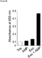


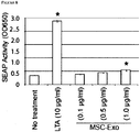


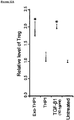
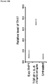
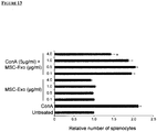
Classifications
-
- G—PHYSICS
- G01—MEASURING; TESTING
- G01N—INVESTIGATING OR ANALYSING MATERIALS BY DETERMINING THEIR CHEMICAL OR PHYSICAL PROPERTIES
- G01N33/00—Investigating or analysing materials by specific methods not covered by groups G01N1/00 - G01N31/00
- G01N33/48—Biological material, e.g. blood, urine; Haemocytometers
- G01N33/50—Chemical analysis of biological material, e.g. blood, urine; Testing involving biospecific ligand binding methods; Immunological testing
- G01N33/5005—Chemical analysis of biological material, e.g. blood, urine; Testing involving biospecific ligand binding methods; Immunological testing involving human or animal cells
-
- A—HUMAN NECESSITIES
- A61—MEDICAL OR VETERINARY SCIENCE; HYGIENE
- A61P—SPECIFIC THERAPEUTIC ACTIVITY OF CHEMICAL COMPOUNDS OR MEDICINAL PREPARATIONS
- A61P11/00—Drugs for disorders of the respiratory system
-
- A—HUMAN NECESSITIES
- A61—MEDICAL OR VETERINARY SCIENCE; HYGIENE
- A61P—SPECIFIC THERAPEUTIC ACTIVITY OF CHEMICAL COMPOUNDS OR MEDICINAL PREPARATIONS
- A61P11/00—Drugs for disorders of the respiratory system
- A61P11/02—Nasal agents, e.g. decongestants
-
- A—HUMAN NECESSITIES
- A61—MEDICAL OR VETERINARY SCIENCE; HYGIENE
- A61P—SPECIFIC THERAPEUTIC ACTIVITY OF CHEMICAL COMPOUNDS OR MEDICINAL PREPARATIONS
- A61P17/00—Drugs for dermatological disorders
-
- A—HUMAN NECESSITIES
- A61—MEDICAL OR VETERINARY SCIENCE; HYGIENE
- A61P—SPECIFIC THERAPEUTIC ACTIVITY OF CHEMICAL COMPOUNDS OR MEDICINAL PREPARATIONS
- A61P17/00—Drugs for dermatological disorders
- A61P17/02—Drugs for dermatological disorders for treating wounds, ulcers, burns, scars, keloids, or the like
-
- A—HUMAN NECESSITIES
- A61—MEDICAL OR VETERINARY SCIENCE; HYGIENE
- A61P—SPECIFIC THERAPEUTIC ACTIVITY OF CHEMICAL COMPOUNDS OR MEDICINAL PREPARATIONS
- A61P17/00—Drugs for dermatological disorders
- A61P17/06—Antipsoriatics
-
- A—HUMAN NECESSITIES
- A61—MEDICAL OR VETERINARY SCIENCE; HYGIENE
- A61P—SPECIFIC THERAPEUTIC ACTIVITY OF CHEMICAL COMPOUNDS OR MEDICINAL PREPARATIONS
- A61P25/00—Drugs for disorders of the nervous system
- A61P25/14—Drugs for disorders of the nervous system for treating abnormal movements, e.g. chorea, dyskinesia
- A61P25/16—Anti-Parkinson drugs
-
- A—HUMAN NECESSITIES
- A61—MEDICAL OR VETERINARY SCIENCE; HYGIENE
- A61P—SPECIFIC THERAPEUTIC ACTIVITY OF CHEMICAL COMPOUNDS OR MEDICINAL PREPARATIONS
- A61P25/00—Drugs for disorders of the nervous system
- A61P25/28—Drugs for disorders of the nervous system for treating neurodegenerative disorders of the central nervous system, e.g. nootropic agents, cognition enhancers, drugs for treating Alzheimer's disease or other forms of dementia
-
- A—HUMAN NECESSITIES
- A61—MEDICAL OR VETERINARY SCIENCE; HYGIENE
- A61P—SPECIFIC THERAPEUTIC ACTIVITY OF CHEMICAL COMPOUNDS OR MEDICINAL PREPARATIONS
- A61P29/00—Non-central analgesic, antipyretic or antiinflammatory agents, e.g. antirheumatic agents; Non-steroidal antiinflammatory drugs [NSAID]
-
- A—HUMAN NECESSITIES
- A61—MEDICAL OR VETERINARY SCIENCE; HYGIENE
- A61P—SPECIFIC THERAPEUTIC ACTIVITY OF CHEMICAL COMPOUNDS OR MEDICINAL PREPARATIONS
- A61P3/00—Drugs for disorders of the metabolism
- A61P3/08—Drugs for disorders of the metabolism for glucose homeostasis
- A61P3/10—Drugs for disorders of the metabolism for glucose homeostasis for hyperglycaemia, e.g. antidiabetics
-
- A—HUMAN NECESSITIES
- A61—MEDICAL OR VETERINARY SCIENCE; HYGIENE
- A61P—SPECIFIC THERAPEUTIC ACTIVITY OF CHEMICAL COMPOUNDS OR MEDICINAL PREPARATIONS
- A61P35/00—Antineoplastic agents
-
- A—HUMAN NECESSITIES
- A61—MEDICAL OR VETERINARY SCIENCE; HYGIENE
- A61P—SPECIFIC THERAPEUTIC ACTIVITY OF CHEMICAL COMPOUNDS OR MEDICINAL PREPARATIONS
- A61P37/00—Drugs for immunological or allergic disorders
- A61P37/02—Immunomodulators
-
- A—HUMAN NECESSITIES
- A61—MEDICAL OR VETERINARY SCIENCE; HYGIENE
- A61P—SPECIFIC THERAPEUTIC ACTIVITY OF CHEMICAL COMPOUNDS OR MEDICINAL PREPARATIONS
- A61P37/00—Drugs for immunological or allergic disorders
- A61P37/08—Antiallergic agents
-
- A—HUMAN NECESSITIES
- A61—MEDICAL OR VETERINARY SCIENCE; HYGIENE
- A61P—SPECIFIC THERAPEUTIC ACTIVITY OF CHEMICAL COMPOUNDS OR MEDICINAL PREPARATIONS
- A61P9/00—Drugs for disorders of the cardiovascular system
-
- A—HUMAN NECESSITIES
- A61—MEDICAL OR VETERINARY SCIENCE; HYGIENE
- A61P—SPECIFIC THERAPEUTIC ACTIVITY OF CHEMICAL COMPOUNDS OR MEDICINAL PREPARATIONS
- A61P9/00—Drugs for disorders of the cardiovascular system
- A61P9/10—Drugs for disorders of the cardiovascular system for treating ischaemic or atherosclerotic diseases, e.g. antianginal drugs, coronary vasodilators, drugs for myocardial infarction, retinopathy, cerebrovascula insufficiency, renal arteriosclerosis
-
- G—PHYSICS
- G01—MEASURING; TESTING
- G01N—INVESTIGATING OR ANALYSING MATERIALS BY DETERMINING THEIR CHEMICAL OR PHYSICAL PROPERTIES
- G01N33/00—Investigating or analysing materials by specific methods not covered by groups G01N1/00 - G01N31/00
- G01N33/48—Biological material, e.g. blood, urine; Haemocytometers
- G01N33/50—Chemical analysis of biological material, e.g. blood, urine; Testing involving biospecific ligand binding methods; Immunological testing
- G01N33/68—Chemical analysis of biological material, e.g. blood, urine; Testing involving biospecific ligand binding methods; Immunological testing involving proteins, peptides or amino acids
-
- G—PHYSICS
- G01—MEASURING; TESTING
- G01N—INVESTIGATING OR ANALYSING MATERIALS BY DETERMINING THEIR CHEMICAL OR PHYSICAL PROPERTIES
- G01N33/00—Investigating or analysing materials by specific methods not covered by groups G01N1/00 - G01N31/00
- G01N33/48—Biological material, e.g. blood, urine; Haemocytometers
- G01N33/50—Chemical analysis of biological material, e.g. blood, urine; Testing involving biospecific ligand binding methods; Immunological testing
- G01N33/68—Chemical analysis of biological material, e.g. blood, urine; Testing involving biospecific ligand binding methods; Immunological testing involving proteins, peptides or amino acids
- G01N33/6803—General methods of protein analysis not limited to specific proteins or families of proteins
- G01N33/6848—Methods of protein analysis involving mass spectrometry
-
- G—PHYSICS
- G01—MEASURING; TESTING
- G01N—INVESTIGATING OR ANALYSING MATERIALS BY DETERMINING THEIR CHEMICAL OR PHYSICAL PROPERTIES
- G01N2570/00—Omics, e.g. proteomics, glycomics or lipidomics; Methods of analysis focusing on the entire complement of classes of biological molecules or subsets thereof, i.e. focusing on proteomes, glycomes or lipidomes
Definitions
- the present invention relates to the fields of medicine, cell biology, molecular biology and genetics. This invention relates to the field of medicine.
- Exosomes were once thought to be "trash bags” for cells to discard unwanted proteins 1 .
- exosomes are increasingly viewed as having important physiological function particularly in cellular communication.
- Exosomes are bi-lipid membrane vesicles of 50-100 ⁇ m that are secreted by many cell types 2 . They belong to a class of secreted cellular products known as microparticles which broadly encompasses all secreted membrane vesicles. Other than exosomes, microparticles include microvesicles (100-1000 ⁇ m), ectosomes (50-200 ⁇ m), membrane particles (50-80 ⁇ m), exosome-like vesicles (20-50 ⁇ m) and apoptotic vesicles (50-500 ⁇ m). The major distinguishing parameter for these different classes of microparticles is their size and the best defined class is the exosomes.
- Exosomes have a density in sucrose of 1.10 to 1.19 g/ml, sedimented at 100,000 g, has a cholesterol-rich lipid membrane containing sphingomyelin, ceramide, lipid rafts and exposed phosphatidylserine.
- the process of exosome biogenesis is complex and involves complex intracellular membrane trafficking and cargo sorting through the biosynthetic and endocytotic pathways.
- the hallmark features of exosomes are markers of the endoplasmic reticulum and the endosomes such as Alix, Tsg101, Rab proteins, etc.
- Exosomes are stored in multivesicular bodies prior to release via fusion of the multivesicular bodies (MVBs) with the plasma membrane.
- Exosomes have been shown to mediate intercellular communication particularly in immune or tumor cells 3-8 . Recently we extended this function to include tissue repair when we reported that exosomes secreted by human ESC-derived MSCs reduced infarct size by about 50% in a mouse model of myocardial ischemia reperfusion (MI/R) injury 9,10 . These exosomes were purified as a population of homogenously sized microparticles of about 50-100 ⁇ m in diameter by size exclusion on HPLC and they carry both protein and RNA load 9,11,12 .
- MI/R myocardial ischemia reperfusion
- the proteins investigated here include glycolytic enzymes for the breakdown of glucose to generate ATP and NADH, PFKB3 that increases glycolysis, CD73 that hydrolyses AMP to adenosine capable of activating signaling cascades through adenosine receptors, CD59 that inhibits the formation of membrane attack complex (MAC) and 20S proteasome.
- MAC membrane attack complex
- the method may comprise detecting an activity of an exosome.
- the method may comprise immunodulatory activity. It may comprise complement inhibition activity. It may comprise proteasome activity. It may comprise glycolytic enzyme activity. It may comprise anti-oxidative activity. It may comprise extracellular matrix (ECM) modifying activity. It may comprise NT5E (CD73) ecto-5'-ectonucleotidase activity. It may comprise ion homeostasis activity. It may comprise chaperone activity. It may comprise any one or more of the above.
- the activity may comprise immunodulatory activity.
- the immunodulatory activity may be detected by detecting activity of a protein set out in Table D10.
- the activity may comprise complement inhibition activity.
- the complement inhibition activity may be detected by detecting activity of a protein set out in Table D1.
- the activity may comprise proteasome activity.
- the proteasome activity may be detected by detecting activity of one or more, preferably all, proteins set out in Table D2.
- the activity may comprise glycolytic enzyme activity.
- the glycolytic enzyme activity may be detected by detecting activity of one or more, preferably all, proteins set out in Table D3.
- the activity may comprise anti-oxidative activity.
- the anti-oxidative activity may be detected by detecting activity of one or more, preferably all, proteins set out in Table D4.
- the activity may comprise extracellular matrix (ECM) modifying activity.
- ECM extracellular matrix
- the extracellular matrix (ECM) modifying activity may be detected by detecting activity of one or more, preferably all, proteins set out in Table D5.
- the activity may comprise NT5E (CD73) ecto-5'-ectonucleotidase activity.
- the NT5E (CD73) ecto-5'-ectonucleotidase activity may be detected by detecting activity of one or more, preferably all, proteins set out in Table D6.
- the activity may comprise ion homeostasis activity.
- the ion homeostasis activity may be detected by detecting activity of one or more, preferably all, proteins set out in Table D7.
- the activity may comprise chaperone activity.
- the chaperone activity may be detected by detecting activity of one or more, preferably all, proteins set out in Table D8.
- the activity may comprise immunomodulatory activity.
- the immunomodulatory activity may be detected by detecting activity of one or more, preferably all, proteins set out in Table D10.
- the exosome may be likely to be capable of inhibiting complement mediated cell lysis. It may also, alternatively or in addition, be capable of reducing infarct size for example as assayed in a mouse or pig model of myocardial ischemia and reperfusion injury, or which is capable of reducing oxidative stress for example as assayed in an in vitro assay of hydrogen peroxide (H 2 O 2 )-induced cell death. It may have both the above activities.
- H 2 O 2 hydrogen peroxide
- the exosome may be likely to comprise a therapeutic exosome having therapeutic activity.
- the therapeutic activity may comprise cardioprotective activity.
- the therapeutic activity may be against one or more diseases selected from the group consisting of: cardiac failure, bone marrow disease, skin disease, bums and degenerative diseases such as diabetes, Alzheimer's disease, Parkinson's disease, cancer, myocardial infarction, a cutaneous wound, a dermatologic disorder, a dermatological lesion, dermatitis, psoriasis, condyloma, verruca, hemangioma, keloid, skin cancer, atopic dermatitis, Behcet disease, chronic granulomatous disease, cutaneous T cell lymphoma, ulceration, a pathological condition characterised by initial injury inducing inflammation and immune dysregulation leading to chronic tissue remodeling including fibrosis and loss of function, renal ischemic injury, cystic fibrosis, sinusitis and rhinitis or an orthopaedic disease.
- diseases selected from the group consisting of: cardiac failure, bone marrow disease, skin disease, bums and degenerative diseases such as diabetes, Alzheimer's disease
- a method comprising the steps of: (a) isolating an exosome from a mesenchymal stem cell conditioned medium (MSC-CM), optionally comprising separating the exosome from other components based on molecular weight, size, shape, composition or biological activity, and (b) detecting an activity as set out above in the isolated exosome, preferably by detecting the activity of one or more proteins as set out above.
- MSC-CM mesenchymal stem cell conditioned medium
- a method of producing an exosome comprising: (a) obtaining a mesenchymal stem cell conditioned medium (MSC-CM); (b) concentrating the mesenchymal stem cell conditioned medium, for example by ultrafiltration over a >1000 kDa membrane; (c) subjecting the concentrated mesenchymal stem cell conditioned medium to size exclusion chromatography, such as using a a TSK Guard column SWXL, 6 x 40 mm or a TSK gel G4000 SWXL, 7.8 x 300 mm column; (d) selecting UV absorbant fractions, for example, at 220 nm, that exhibit dynamic light scattering, such as detected by a quasi-elastic light scattering (QELS); in which step (d) for example comprises collecting fractions which elute with a retention time of 11-13 minutes, such as 12 minutes; and detecting an activity as set out above in the exosome, preferably by detecting the activity
- MSC-CM mesenchymal stem cell conditioned medium
- a method of detecting whether an exosome comprises therapeutic activity comprising detecting an enzymatic activity as set out above, in which if the enzymatic activity is detected, then the exosome comprises therapeutic activity.
- biochemical properties of therapeutic exosomes include
- proteins for these biochemical activies should not only have the proteins for these biochemical activies, the proteins must be enzymatically active. The enzymatic activity of these proteins could therefore be surrogate markers for the therapeutic efficacy of an exosome preparation.
- MSC mesenchymal stem cell
- an exosome which comprises one or more of: (a) complement inhibition activity; (b) proteasome activity; (c) glycolytic enzyme activity; (d) anti-oxidative activity; (e) extracellular matrix (ECM) modifying activity; (f) NT5E (CD73) ecto-5'-ectonucleotidase activity; (g) ion homeostasis activity; (h) chaperone activity, may comprise therapeutic activity; and (i) immunodulatory activity.
- exosome comprising such properties may be therapeutic in nature against a number of diseases, as described below, in other words be a therapeutic exosome.
- exosome is used in this document, this should be understood to include “therapeutic exosome” as well, where the context permits.
- the exosome may be derivable from the MSC by any of several means, for example by secretion, budding or dispersal from the MSC.
- the exosome may be produced, exuded, emitted or shed from the MSC.
- the exosome may be secreted into the cell culture medium.
- the exosome may in particular comprise a vesicle.
- the therapeutic exosomes described here may comprise any one or more of the properties of the exosomes described herein.
- the exosomes may comprise vesicles or a flattened sphere limited by a lipid bilayer.
- the exosomes may comprise diameters of 40-100 nm.
- the exosomes may be formed by inward budding of the endosomal membrane.
- the exosomes may have a density of ⁇ 1.13 - 1.19 g/ml and may float on sucrose gradients.
- the exosomes may be enriched in cholesterol and sphingomyelin, and lipid raft markers such as GM1, GM3, flotillin and the src protein kinase Lyn.
- the exosomes may comprise one or more proteins present in mesenchymal stem cells or mesenchymal stem cell conditioned medium (MSC-CM), such as a protein characteristic or specific to the MSC or MSC-CM.
- MSC-CM mesenchymal stem cell conditioned medium
- exosomes which comprises one or more genes found in MSCs or medium which is conditioned by culture of MSCs, and which comprises therapeutic activity.
- Such therapeutic exosomes may be identified by detecting exosomes which display one or more activities selected from the group consisting of: (a) complement inhibition activity; (b) proteasome activity; (c) glycolytic enzyme activity; (d) anti-oxidative activity; (e) extracellular matrix (ECM) modifying activity; (f) NT5E (CD73) ecto-5'-ectonucleotidase activity; (g) ion homeostasis activity; (h) chaperone activity; and (i) immunodulatory activity.
- the exosomes may comprise molecules secreted by the MSC.
- Such an exosome, and combinations of any of the molecules comprised therein, including in particular proteins or polypeptides, may be used to supplement the activity of, or in place of, the MSCs or medium conditioned by the MSCs for the purpose of for example treating or preventing a disease.
- the exosomes may comprise a cytosolic protein found in cytoskeleton e.g. tubulin, actin and actin-binding proteins, intracellular membrane fusions and transport e.g. annexins and rab proteins, signal transduction proteins e.g. protein kinases, 14-3-3 and heterotrimeric G proteins, metabolic enzymes e.g. peroxidases, pyruvate and lipid kinases, and enolase-1 and the family of tetraspanins e.g. CD9, CD63, CD81 and CD82.
- the exosomes may comprise one or more tetraspanins.
- the exosomes may comprise mRNA and/or microRNA.
- the term "particle”, which includes an exosome, as used in this document may be taken to mean a discrete entity.
- the particle may be something that is isolatable from a mesenchymal stem cell (MSC) or mesenchymal stem cell conditioned medium (MSC-CM).
- the particle may be responsible for at least an activity of the MSC or MSC-CM.
- the particle may be responsible for, and carry out, substantially most or all of the functions of the MSC or MSC-CM.
- the particle may be a substitute (or biological substitute) for the MSC or MSC-CM.
- the exosomes may be used for any of the therapeutic purposes that the MSC or MSC-CM may be put to use.
- the exosome preferably has at least one property of a mesenchymal stem cell, such as a therapeutic property.
- the exosome may have a biological property, such as a biological activity.
- the exosome may have any of the biological activities of an MSC.
- the exosome may for example have a therapeutic or restorative activity of an MSC.
- MSCs such as mesenchymal stem cell conditioned media or MSC-CM, as described below
- MSC-CM mesenchymal stem cell conditioned media
- the biological property or biological activity of an MSC may therefore correspond to a biological property or activity of an mesenchymal stem cell conditioned medium.
- the exosome may comprise one or more biological properties or activities of a mesenchymal stem cell conditioned medium (MSC-CM).
- Therapeutic exosomes described here may be isolated from Mesenchymal Stem Cell Conditioned Medium (MSC-CM) and tested for the presence of one or more of the following activities: (a) complement inhibition activity; (b) proteasome activity; (c) glycolytic enzyme activity; (d) anti-oxidative activity; (e) extracellular matrix (ECM) modifying activity; (f) NT5E (CD73) ecto-5'-ectonucleotidase activity; (g) ion homeostasis activity; (h) chaperone activity; and (i) immunodulatory activity.
- MSC-CM Mesenchymal Stem Cell Conditioned Medium
- each of the above activities may be detected in therapeutic exosomes by a number of ways, known in the art.
- the relevant activity may be detected by detection of the presence of the protein or polypeptide responsible for the activity in the therapeutic exosome. This may be achieved for example by use of cognate antibodies and detection of binding between the antibody and the protein, by immunoassays and Western blots, etc as known in the art.
- 1 unit (U) of enzyme activity is defined as the activity required for the production of 1 ⁇ mole of product per minute.
- the blot was then incubated with a horseradish peroxidase-coupled secondary antibody.
- the secondary antibody used was goat anti-mouse IgG (1:1250) or donkey anti-rabbit IgG (1:1250). All antibodies were obtained from Santa Cruz Biotechnology, Santa Cruz, CA except mouse anti-PK which was from Abcam Inc., Cambridge, MA.
- the blot was then incubated with HRP-enhanced Chemiluminescent substrate (Thermo Fisher Scientific Inc., Waltham, MA) and then exposed to X-ray film.
- LC MS is described in detail in the document "Basics of LC/MS Primer” (Agilent Technologies) at http://www.chem.agilent.com/Library/Support/Documents/a05296.pdf
- Proteins in two ml of dialyzed exosomes were reduced, alkylated and tryptic digested as described (20).
- the samples were then desalted by passing the digested mixture through a conditioned Sep-Pak C-18 SPE cartridge (Waters, Milford, MA, USA), washed twice with a 3% acetonitrile (ACN) (JT Baker, Phillipsburg, NJ) and 0.1% formic acid (FA) buffer, and eluted with a 70% ACN and 0.1% FA buffer.
- ACN acetonitrile
- F formic acid
- the eluted samples were then dried to about 10% of their initial volumes by removing organic solvent in a vacuum centrifuge.
- a 90 l/min with a splitter to anmminute gradient at a constant flow rate of 20 effective flow rate of 200 ⁇ l/min was used to elute the peptides into the mass spectrometer.
- the LTQ was operated in a data-dependent mode by performing MS/MS scans for the 8 of the most intense peaks from each MS scan in the FTMS.
- MS/MS (dta) spectra of the eight SCX fractions were combined into a single mascot generic file by a home-written program. Protein identification was achieved by searching the combined data against the IPI human protein database (version 3.34; 69,164 sequences, 29,064,825 residues) via an in-house Mascot server (Version 2.2.04, Matrix Science, UK).
- the search parameters were: a maximum of 2 missed cleavages using trypsin; fixed modification was carbaminomethylation of cysteine and variable modification was oxidation of methionine.
- the mass tolerances were set to 10 ppm and 0.8 Da for peptide precursor and fragment ions respectively. Protein identification was accepted as true positive if two different peptides were found to have scores greater than the homology scores.
- Complement inhibition activity may be detected in therapeutic exosomes by detection of any one or more of the proteins or their activities, as set out in Table D1, thereby identifying them as therapeutic exosomes.
- Table D1 Table D1.Complement Inhibition Activity Protein GenBank Accession Number/ NCBI Reference Sequence (RefSeq) Synonyms CD59 NM_203329.2, NP_976074.1
- complement-mediated cell lysis activity may be detected by conducting the assay set out below.
- SRBCs sheep red blood cells
- PBS phosphate buffered saline
- C5b6, C8 and C9 were purchased from Calbiochem (San Diego, CA), and C7 from Sigma-Aldrich (St Louis, MO). Assembly of intact C5b-9 on SRBCs was initiated by a 15 min incubation (37 °C) with 1ml each of C5b6 (0.1 ⁇ g/ml) and C7 (0.4 ⁇ g/ml) in the presence or absence of exosomes at a final concentration of 0.1 ⁇ g/ml.
- the SRBCs were then washed and incubated with 1ml each of C8 (0.4 ⁇ g/ml,) plus C9 (0.4 ⁇ g/ml) with or without a blocking CD59 antibody at a final concentration of 0.05 ⁇ g/ml for an additional 30 min. After that the SRBCs were centrifuged and the amount of hemoglobin released by the lysed SRBCs in the supernatant was measured by absorbance at 415 nm. Total (100%) hemolysis was obtained by treating the cells with 1% (w/v) Triton X-100.
- Proteasome activity may be detected in therapeutic exosomes by detection of any one or more of the proteins or their activities, as set out in Table D2, thereby identifying them as therapeutic exosomes.
- Proteasome Activity Protein GenBank Accession Number/ Ref seq Synonyms PSMA1 X61969.1, CAA43961.1 PSMA2 D00760.1, BAA00657.1 PSMA3 NM_002788.2, NP_002779.1 PSMA4 BC005361.1, AAH05361.1 PSMA5 X61970.1, CAA43962.1 PSMA6 X59417.1, CAA42052.1 PSMA7 AF022815.1, AAB81515.1 PSMB1 D00761.1, BAA00658.1 PSMB2 D26599.1, BAA05646.1 PSMB3 BC013008.2, AAH13008.1 PSMB4 D26600.1, BAA05647.1 PSMB5 D29011.1, BAA06097.1 PSMB6 BC000835.2, AAH
- proteasome activity may be detected by conducting the assay set out below.
- AMC fluorophore 7-Amino-4 methylcoumarin
- 1 unit enzyme activity of 20S core particle is defined as the activity required for the production of 1 ⁇ mole of product per minute.
- Exosomes are found to contain 5.0 ⁇ U 20S core particle activity / ⁇ g protein.
- Glycolytic enzyme activity may be detected in exosomes by detection of any one or more of the proteins or their activities, as set out in Table D3, thereby identifying them as therapeutic exosomes.
- Table D3 Table D3.
- Glycolytic Enzyme Activity Protein GenBank Accession Number/ NCBI Reference Sequence (RefSeq) Synonyms GAPDH AF261085.1, AAF99678.1 PGK L00159.1, AAA60078.1 PGM1 BC019920.1, AAH19920.1 Enolase2 BC022545.1, AAH22545.1 PKm2 M23725.1, AAA36449.1 PFKFB3 CAH73611 PFKFB3 - phosphorylated form ATP * NADH * * It is understood that ATP and NADH are not proteins.
- protein or “polypeptide” may encompass small molecules such as ATP and NADH.
- ATP ATP
- NADH a small molecule.
- table headings and other references in this document may for the sake of convenience refer to "proteins” but may include references to ATP, NADH, etc (where these are not in fact proteins). These may be detected, as noted above, for determination of therapeutic exosomes.
- glycolytic enzyme activity may be transferred into cells exposed to the exosomes, resulting in increased ATP and NADH production
- detection of ATP or NADH production (or both) by the cell may be used as a means of detecting glycolytic enzyme activity in a produced exosome, thereby identifying it as a therapeutic exosome.
- GAPDH and PGK activity were measured based on their downstream product, ATP in the glycolysis reaction using 2 commercially available kits, KDalert GAPDH assay kit (Ambion Inc., Austin, TX) and ApoSENSOR ADP/ATP ratio assay kit (Biovision). Briefly, exosomes were lysed using a cell extraction kit (Biovision). To measure GAPDH activity, 10 ⁇ g of lysed exosomes was added to KDalert reaction buffer containing D-glyceraldehyde-3-phosphate, NAD+ and Pi to form 1, 3-bisphosphoglycerate + NADH+ H+.
- PGK and 60 ⁇ M ADP were then added to convert 1, 3-bisphosphoglycerate and ADP to 3-phosphoglycerate and ATP.
- 10 ⁇ g of lysed exosomes was added to the 1, 3-bisphosphoglycerate produced from the above assay.
- ADP was added to allow the formation of ATP.
- ATP amount which is proportional to PGK activity was then quantified using ATP luciferase assay.
- H9C2 cardiomyocytes were plated onto a 96 well plate (poly-lysine coated) at 30,000 cells per well. After 5 hours, the cells were washed twice with Tyrode's buffer before incubating in Tyrode's buffer containing 20 ⁇ mol oligomycin (Sigma-Aldrich), 6 mmol glucose, and with or without 0.1 ⁇ g/ml exosomes for 15, 30 and 60 minutes. Cellular ATP concentration was measured using ATPlite 1step luminescence ATP detection assay system (PerkinElmer, Zaventem, Belgium).
- 1 unit enzyme activity of GAPDH (AF261085.1) is defined as the activity required for the production of 1 ⁇ mole of product per minute.
- Exosomes are found to contain 1.1 ⁇ U GAPDH activity / ⁇ g protein, 3.59 ⁇ U PGK (L00159.1) activity / ⁇ g protein and 5.5 ⁇ U PKm2 (M23725.1) activity / ⁇ g protein.
- Anti-oxidative activity may be detected in therapeutic exosomes by detection of any one or more of the proteins or their activities, as set out in Table D4, thereby identifying them as therapeutic exosomes.
- Table D4 Table D4.
- EXTRACELLULAR MATRIX MODIFYING ACTIVITY
- Extracellular matrix (ECM) modifying activity may be detected in therapeutic exosomes by detection of any one or more of the proteins or their activities, as set out in Table D5, thereby identifying them as therapeutic exosomes.
- Table D5 Table D5.
- NT5E (CD73) ecto-5'-ectonucleotidase activity may be detected in therapeutic exosomes by detection of any one or more of the proteins or their activities, as set out in Table D6, thereby identifying them as therapeutic exosomes.
- NT5E (CD73) ecto-5'-ectonucleotidase activity in the presence of AMP will induce phosphorylation of AKT and ERK1/2
- detection of phosphorylated AKT i.e., phosphorylated AKT1, phosphorylated AKT2 or phosphorylated AKT3 or phosphorylated ERK1 or phosphorylated ERK2 (or any combination of these) in or of the exosome, may be used as a means of detecting NT5E (CD73) ecto-5'-ectonucleotidase enzyme activity in the exosome, thereby identifying it as a therapeutic exosome.
- NT5E CD73
- ecto-5'-ectonucleotidase Activity Protein GenBank Accession Number/ NCBI Reference Sequence (RefSeq) Synonyms Ecto-5'-ectonucleotidas X55740.1, CAA39271.1 NT5E, CD73 Ecto-apyrase S73813.1, AAB32152.1 CD39 AKT1, phosphorylated form NM_001014432, NP_001014431 AKT2, phosphorylated form NM_001626, NP_001617 AKT3, phosphorylated form NM_005465, NP_005456 ERK1, phosphorylated form NM_001040056, NP_001035145 MAPK3 ERK2, phosphorylated form NM_002745, NP_002736 MAPK1
- CD73 (NT5E) enzymatic activity in exosome was determined by incubating 2.5 ⁇ g of exosome in 100 ⁇ l Tris buffer pH 7.4 containing 50 ⁇ M AMP (Sigma-Aldrich, St Louis, MO). The amount of phosphate ions released from the hydrolysis of AMP was then determined by Colorlock Gold kit (Innova Biosciences, Cambridge, UK) as per manufacturer's instruction.
- Ecto-5'- ectonucleotidase enzyme activity is defined as the activity required for the production of 1 ⁇ mole of product per minute.
- Exosomes are found to contain 22.04 ⁇ U Ecto-5'- ectonucleotidase activity / ⁇ g protein.
- Ion homeostasis activity may be detected in therapeutic exosomes by detection of any one or more of the proteins or their activities, as set out in Table D7, thereby identifying them as therapeutic exosomes.
- Table D7 Table D7. Ion Homeostasis Activity Protein GenBank Accession Number/ NCBI Reference Sequence (RefSeq) Synonyms PMCA1 P20020 PMCA4 P23634 Ca2+-dependent ATPase activity ATP2B1 J04027.1, AAA74511.1 Ca2+-dependent ATPase activity ATP2B4 M25874.1, AAA50819.1
- Chaperone activity may be detected in therapeutic exosomes by detection of any one or more of the proteins or their activities, as set out in Table D8, thereby identifying them as therapeutic exosomes.
- Table D8 Table D8. Chaperone Activity Protein GenBank Accession Number/ NCBI Reference Sequence (RefSeq) Synonyms HSP60 M34664.1, AAA36022.1 HSP70 AB023420.1, BAA75062.1 HSP90 M27024.1, AAA63194.1
- a therapeutic exosome may display immunomodulatory activity.
- the immune system exists primarily to protect the organism against disease by destroying non-self cells or tissues, neutralizing infectious or toxic materials and removing cell debris e.g. dead or dying cells.
- the immune system consists of the innate and adaptive immune systems.
- Innate immune cells include dendritic cells, macrophages and neutrophils and these cells are genetically programmed to recognize invariant features of infectious/toxic materials, stress signals, dead/dying cells, or non-self cells. It is evolutionarily conserved and provides the first line of defense.
- Adaptive immune cells are the T and B lymphocytes. They adapt to each foreign, toxic or non-self agent by generating receptors specific against these materials de novo to target them for neutralization or destruction.
- innate immune cells recognize conserved molecular signatures within microbial pathogens referred to commonly as pathogen-associated molecular patterns and stress signals through germline-encoded pattern recognition receptors (PPRs) such as include Toll-like receptors (TLRs), retinoic acid-inducible gene 1-like RNA helicases, nucleotide binding oligomerisation domain-like receptors, and C-type lectin receptors.
- TLRs Toll-like receptors
- retinoic acid-inducible gene 1-like RNA helicases include nucleotide binding oligomerisation domain-like receptors, and C-type lectin receptors.
- the TLRs are the best characterized PPRs with regards to modulation of adaptive immune responses such as production of IgM, IgG and IgA antibody, and activation of T H 1, T H 17 CD4 + , and CD8 + T cells.
- TLRs are highly conserved type 1 integral membrane glycoproteins. In human, ten TLRs have been identified to date. All TLRs, except TLR3, signal through the MyD88-dependent pathway either directly or in combination with TIRAP, ultimately leading to activation and nuclear translocation of NFB, activating protein 1 (AP1), or both of these, leading to the production of inflammatory cytokines. TLR3 and TLR4 initiate the MyD88-independent, TRIF-dependent signalling pathway leading to the activation and nuclear translocation of either NF ⁇ B or IRF3 to produce inflammatory cytokines and type 1 interferon, respectively. Different TLRs recognise different classes of pathogen-associated molecular patterns, e.g.
- TLR4 recognises bacterial cell-surface lipopolysaccharides
- TLR1 and TLR2 recognise peptidoglycans
- TLR2 and TLR6 recognise lipoproteins
- TLR3 recognises viral double-stranded RNA
- TLR7 and TLR8 recognise viral single-stranded RNA
- TLR9 recognises bacterial and viral CpG
- TLR5 recognises flagellin.
- TLRs Despite the overwhemingly importance of TLRs in mediating host immunity against infectious agents and their products, activation of TLR signaling by endogenous ligands to trigger a sterile inflammatory response is increasingly viewed as an important function of TLRs.
- numerous endogenous ligands for the different TLRs have been identified (Table D9 below). TLRs have been implicated in ischemia and reperfusion injury, tissue repair and regeneration, autoimmune diseases, and tumorigenesis and tumour progression. Table D9. TLRs and their endogenous ligands. Highlighted ligands are also found to be present in MSC exosomes.
- MSCs display immunomodulatory properties in vitro such as inhibition of T-cell proliferation induced by alloantigens, mitogens and CD3-ligation, inhibit the proliferation of B cells, and the activity of natural killer cells 5 .
- Clinical trials testing the therapeutic efficacy of MSC-based intervention for human GVHD 6 and Crohn's disease 7 have been encouraging.
- the underlying mechanism for the immunomodulatory properties of MSCs remains poorly understood. Increasingly, it is thought that these properties are mediated by paracrine secretion from MSCs 8 .
- Immunomodulatory activity may be detected in therapeutic exosomes by detection of any one or more of the proteins or their activities, as set out in Table D10, thereby identifying them as therapeutic exosomes.
- Table D10 Variant GenBank Accession Number NCBI Reference Sequence (RefSeq) Biglycan AK092954 NM_001711 Endoplasmin AY040226 NM_003299 HSP60 M34664.1, AAA36022.1 HSP70 AB023420.1, BAA75062.1 ⁇ -defensin 2 AJ000152 NM_004942 Fibrinogen alpha NM_021871 beta NM_001184741 gamma NM_000509 Fibronectin NM_212482
- Tenascin-C NM_002160 S100 proteins S100A11 D38583 NM_005620 S100A13 AK097132 NM_001024212 S100A8 BC005928 NM_002964 S100A9 BC
- the conditioned cell culture medium such as a Mesenchymal Stem Cell Conditioned Medium (MSC-CM) may be obtained by culturing a mesenchymal stem cell (MSC), a descendent thereof or a cell line derived therefrom in a cell culture medium; and isolating the cell culture medium.
- the mesenchymal stem cell may be produced by a process comprising obtaining a cell by dispersing a embryonic stem (ES) cell colony.
- ES embryonic stem
- the cell, or a descendent thereof may be propagated in the absence of co-culture in a serum free medium comprising FGF2.
- the exosome may be produced or isolated in a number of ways. Such a method may comprise isolating the exosome from a mesenchymal stem cell (MSC). Such a method may comprise isolating the exosome from an mesenchymal stem cell conditioned medium (MSC-CM).
- the exosome may be isolated for example by being separated from non-associated components based on any property of the exosome.
- the exosome may be isolated based on molecular weight, size, shape, composition or biological activity. For example, filtration with a membrane of a suitable molecular weight or size cutoff, as described in the Assays for Molecular Weight elsewhere in this document, may be used.
- One or more biological activities of the exosome may be used to track its activity during fractionation of the mesenchymal stem cell conditioned medium (MSC-CM).
- Immunomodulatory activity of exosomes may be assessed by using THP-1 cells and genetically modified THP- cell line/HEK reporter cell lines. Such cell lines are described in the Examples below.
- Immunomodulatory activity of exosomes may be assessed using unmodified THP-1, by assaying the ability of the exosome to induce cytokine production. This is described in detail at Example 23 below.
- cytokine production may be assessed using surrogate reporter cell lines as described in the Examples.
- a THP1-XBlue with a stably transfected secreted embryonic alkaline phosphatase (SEAP) reporter gene under the transcriptional control of NF-kB promoter or a THP1-XBlue-defMYD that is deficient in MyD88 activity may be used, as described in Example 19.
- Immunomodulatory activity of exosomes may also be assayed by their ability to inhibit proliferation of Concanavalin A-activated lymphocytes. This is described in detail in Example 28.
- the property of a mesenchymal stem cell may comprise a property of a medium conditioned by a mesenchymal stem cell (MSC-CM).
- MSC-CM mesenchymal stem cell
- the property may comprise a biological property such as a biological activity.
- biological activities include cardioprotection, reduction of oxidative stress and reduction of infarct size.
- the exosome in particular a therapeutic exosome, may have a property of mesenchymal stem cells and/or mesenchymal stem cell conditioned medium (MSC-CM) comprising cardioprotection
- the cardioprotection may comprise restoration or maintenance of cardiac function during ischemia and/or reperfusion.
- Cardioprotection may for example be assayed using any one or more of the methods described in Examples 5, 10, 14 and 20 of WO 2009/105044 .
- the exosome in particular a therapeutic exosome, may have a property of mesenchymal stem cells and/or mesenchymal stem cell conditioned medium (MSC-CM) comprising the ability to reduce oxidative stress (or cytoprotection).
- MSC-CM mesenchymal stem cell conditioned medium
- the reduction of oxidative stress may for example be assayed using an in vitro assay of hydrogen peroxide (H 2 O 2 )-induced cell death.
- hydrogen peroxide (H 2 O 2 )-mediated oxidative stress is induced in human leukemic CEM cells and cell viability is monitored by Trypan blue-exclusion.
- Human leukemic CEM cells are incubated with exosome, conditioned medium or mesenchymal stem cell (with saline as a control) and treated with 50 ⁇ M H 2 O 2 to induce oxidative stress.
- Cell viability is assessed using Trypan Blue exclusion at 12, 24, 36 and 48 hours after H 2 O 2 treatment.
- the reduction of oxidative stress may further be assayed using an in vivo assay of DNA oxidation.
- In vivo oxidative stress may also be assayed as follows. Pigs are treated with the exosome, conditioned medium or mesenchymal stem cell (with saline as a control). Tissue sections of pig heart are obtained. Nuclear oxidative stress in tissue sections of treated and untreated pigs is quantified by 8-OHdG immunostaining for oxidized DNA. The tissue sections are assayed for intense nuclear staining indicative of DNA oxidation and oxidative stress.
- the exosome in particular a therapeutic exosome, may have a property of mesenchymal stem cells and/or mesenchymal stem cell conditioned medium (MSC-CM) comprising the ability to reduce infarct size.
- MSC-CM mesenchymal stem cell conditioned medium
- Infarct size may for example be assayed using any one or more of the methods described in Examples 6 and 13 of WO 2009/105044 .
- the exosome may have a molecular weight of greater than 100 kDa. It may have a molecular weight of greater than 500 kDa. For example, it may have a molecular weight of greater than 1000 kDa.
- the molecular weight may be determined by various means. In principle, the molecular weight may be determined by size fractionation and filtration through a membrane with the relevant molecular weight cut-off. The exosome size may then be determined by tracking segregation of component proteins with SDS-PAGE or by a biological assay.
- the instrument setup consisted of a liquid chromatography system with a binary pump, an auto injector, a thermostated column oven and a UV-visible detector operated by the Class VP software from Shimadzu Corporation (Kyoto, Japan).
- the Chromatography columns used were TSK Guard column SWXL, 6 x 40 mm and TSK gel G4000 SWXL, 7.8 x 300 mm from Tosoh Corporation (Tokyo, Japan).
- the following detectors, Dawn 8 (light scattering), Optilab (refractive index) and QELS (dynamic light scattering) were connected in series following the UV-visible detector.
- the last three detectors were from Wyatt Technology Corporation (California, USA) and were operated by the ASTRA software.
- the components of the sample were separated by size exclusion i.e. the larger molecules will elute before the smaller molecules.
- the eluent buffer used was 20 mM phosphate buffer with 150 mM of NaCl at pH 7.2. This buffer was filtered through a pore size of 0.1 ⁇ m and degassed for 15 minutes before use.
- the chromatography system was equilibrated at a flow rate of 0.5 ml/min until the signal in Dawn 8 stabilized at around 0.3 detector voltage units.
- the UV-visible detector was set at 220 ⁇ m and the column was oven equilibrated to 25oC.
- the elution mode was isocratic and the run time was 40 minutes.
- the volume of sample injected ranged from 50 to 100 ml.
- the % area of the exosome peak vs. all other peaks was integrated from the UV-visible detector.
- the hydrodynamic radius, Rh was computed by the QELS and Dawn 8 detectors.
- the highest count rate (Hz) at the peak apex was taken as the Rh. Peaks of the separated components visualized at 220 ⁇ m were collected as fractions for further characterization studies.
- the exosome may have a molecular weight of greater than 100 kDa.
- the exosome may be such that most proteins of the exosome with less than 100 kDa molecular weight segregate into the greater than 100 kDa molecular weight retentate fraction, when subject to filtration.
- most proteins of the exosome with less than 500 kDa molecular weight may segregate into the greater than 500 kDa molecular weight retentate fraction. This indicates that the exosome may have a molecular weight of more than 500 kDa.
- the exosome may have a molecular weight of more than 1000 kDa.
- the exosome may be such that when subject to filtration with a membrane with a molecular weight cutoff of 1000 kDa, the relevant biological activity substantially or predominantly remains in the retentate fraction.
- biological activity may be absent in the filtrate fraction.
- the biological activity may comprise any of the biological activities of the exosome described elsewhere in this document.
- the biological activity may comprise reduction of infarct size, as assayed in any suitable model of myocardia ischemia and reperfusion injury.
- the biological activity may be assayed in a mouse or pig model, such as described in the Examples of WO 2009/105044 .
- myocardial ischemia is induced by 30 minutes left coronary artery (LCA) occlusion by suture ligation and reperfusion is initiated by removal of suture.
- Mice are treated with liquid containing the exosome (such as unfractionated MSC-CM), filtrate (such as ⁇ 100 or 1,000 kD fraction), retentate (such as >1000 kD retentate) or saline intravenously via the tail vein, 5 minutes before reperfusion. 24 hours later, the hearts are excised. Before excision, the A rea A t R isk (AAR) is determined by religating the LCA and then perfusing Evans blue through the aorta.
- AAR A rea A t R isk
- AAR is defined as the area not stained by the dye and is expressed as a percentage of the left ventricular wall area. Infarct size is assessed 24 hours later using Evans blue and TTC. Where the relative infarct size is significantly reduced in animals treated with mesenchymal stem cell conditioned medium (MSC-CM) and the retentate (such as a >1000 kD) fraction when compared to saline, this indicates that the exosome has a molecular weight which is higher than the relevant cutoff of the membrane (e.g., greater than 1000 kDa).
- MSC-CM mesenchymal stem cell conditioned medium
- retentate such as a >1000 kD
- the exosome may have a size of greater than 2 ⁇ m.
- the exosome may have a size of greater than 5 ⁇ m, 10 ⁇ m, 20 ⁇ m, 30 ⁇ m, 40 ⁇ m or 50 ⁇ m.
- the exosome may have a size of greater than 100 ⁇ m, such as greater than 150 ⁇ m.
- the exosome may have a size of substantially 200 ⁇ m or greater.
- the exosome or exosomes may have a range of sizes, such as between 2 ⁇ m to 20 ⁇ m, 2 ⁇ m to 50 ⁇ m, 2 ⁇ m to 100 ⁇ m, 2 ⁇ m to 150 ⁇ m or 2 ⁇ m to 200 ⁇ m.
- the exosome or exosomes may have a size between 20 ⁇ m to 50 ⁇ m, 20 ⁇ m to 100 ⁇ m, 20 ⁇ m to 150 ⁇ m or 20 ⁇ m to 200 ⁇ m.
- the exosome or exosomes may have a size between 50 ⁇ m to 100 ⁇ m, 50 ⁇ m to 150 ⁇ m or 50 ⁇ m to 200 ⁇ m.
- the exosome or exosomes may have a size between 100 ⁇ m to 150 ⁇ m or 100 ⁇ m to 200 ⁇ m.
- the exosome or exosomes may have a size between 150 ⁇ m to 200 ⁇ m.
- the size may be determined by various means. In principle, the size may be determined by size fractionation and filtration through a membrane with the relevant size cut- off. The exosome size may then be determined by tracking segregation of component proteins with SDS-PAGE or by a biological assay.
- the size may also be determined by electron microscopy, as described in Example 21 of WO 2009/105044 .
- the exosome may comprise one or more proteins secreted by a mesenchymal stem cell.
- the exosome may comprise one or more proteins present in mesenchymal stem cell conditioned medium (MSC-CM).
- the exosome may comprise 10% or more, 20% or more, 30% or more, 40% or more, 50% or more, 60% or more or 70% or more of these proteins.
- the exosome may comprise substantially about 75% of these proteins.
- the proteins may be defined by reference to a list of proteins or gene products of a list of genes.
- Proteins and gene products which may be comprised in an exosome are described in for example WO 2009/105044 .
- the active component in the secretion such as an exosome that confers cardioprotection against the reperfusion injury may be isolated by means known in the art and described for example in WO 2009/105044 .
- the active component may comprise an exosome secreted by the mesenchymal stem cells (MSCs).
- Exosomes are small membrane vesicles formed in late endocytic compartments (multivesicular bodies) first described to be secreted by reticulocytes in 1983 and subsequently found to be secreted by many cells types including various haematopoietic cells, tumours of haematopoietic or non-haematopoietic origin and epithelial cells. They are distinct entities from the more recently described 'ribonuclease complex' also named exosome.
- Exosomes may be defined by morphological and biochemical parameters. Accordingly, the exosomes described here may comprise one or more of these morphological or biochemical parameters.
- Exosomes are classically defined as "saucer-like" vesicles or a flattened sphere limited by a lipid bilayer with diameters of 40-100 nm and are formed by inward budding of the endosomal membrane. Like all lipid vesicles and unlike protein aggregates or nucleosomal fragments that are released by apoptotic cells, exosomes have a density of ⁇ 1.13 - 1.19 g/ml and float on sucrose gradients. Exosomes are enriched in cholesterol and sphingomyelin, and lipid raft markers such as GM1, GM3, flotillin and the src protein kinase Lyn suggesting that their membranes are enriched in lipid rafts.
- exosomes contain ubiquitous proteins that appear to be common to all exosomes and proteins that are cell-type specific. Also, proteins in exosomes from the same cell-type but of different species are highly conserved.
- the ubiquitous exosome-associated proteins include cytosolic proteins found in cytoskeleton e.g. tubulin, actin and actin-binding proteins, intracellular membrane fusions and transport e.g. annexins and rab proteins, signal transduction proteins e.g. protein kinases, 14-3-3 and heterotrimeric G proteins, metabolic enzymes e.g.
- tetraspanins e.g. CD9, CD63, CD81 and CD82.
- the tetraspannins are highly enriched in exosomes and are known to be involved in the organization of large molecular complexes and membrane subdomains.
- exosomes examples include MHC class II molecules in exosomes from MHC class II-expressing cells, CD86 in dendritic cell-derived exosomes, T-cell receptors on T-cell-derived exosomes etc.
- exosomes do not contain proteins of nuclear, mitochondrial, endoplasmic-reticulum or Golgi-apparatus origin.
- highly abundant plasma membrane proteins are absent in exosomes suggesting that they are not simply fragments of the plasma membrane.
- Many of the reported ubiquitous exosome-associated proteins are also present in the proteomic profile of the hESC-MSC secretion.
- Exosomes are also known to contain mRNA and microRNA, which can be delivered to another cell, and can be functional in this new location. The physiological functions of exosome remain poorly defined. It is thought to help eradicate obsolete proteins, recycle proteins, mediate tramission of infectious particles such as prions and viruses, induce complement resistance, facilitate immune cell-cell communication and transmit cell signaling. Exosomes have been used in immunotherapy for treatment of cancer.
- exosomes including therapeutic exosomes may be used as a substitute for an MSC or MSC-CM, as described above and below.
- the therapeutic exosomes may be used for any of the therapeutic purposes that MSCs or MSC-CMs are currently being used, or in the future may be used.
- Mesenchymal stem cells and differentiated cells produced by the methods and compositions described here may be used for, or for the preparation of a pharmaceutical composition for, the treatment of a disease.
- a disease may comprise a disease treatable by regenerative therapy, including cardiac failure, bone marrow disease, skin disease, bums, degenerative disease such as diabetes, Alzheimer's disease, Parkinson's disease, etc and cancer. Accordingly, therapeutic exosomes from MSCs may be used to treat such diseases.
- Therapeutic exosomes from mesenchymal stem cells such as those made according to the methods and compositions described here may be used for a variety of commercially important research, diagnostic, and therapeutic purposes.
- the therapeutic exosomes from mesenchymal stem cells may in particular be used for the preparation of a pharmaceutical composition for the treatment of disease.
- disease may comprise a disease treatable by regenerative therapy, including cardiac failure, bone marrow disease, skin disease, bums, degenerative disease such as diabetes, Alzheimer's disease, Parkinson's disease, etc and cancer.
- BM-MSCs bone marrow derived mesenchymal stem cells
- the proteins expressed are involved in three biological processes: metabolism, defense response, and tissue differentiation including vascularization, hematopoiesis and skeletal development. Accordingly, the therapeutic exosomes from MSCs described here may be used to treat diseases which these functions may have a role in, or whose repair or treatment involves any one or more of these biological processes. Similarly, the proteins expressed by the MSCs, singly or in combination, preferably in the form of therapeutic exosomes as described here, may be used to supplement the activity of, or in place of, the MSCs, or media conditioned by the MSCs, for the purpose of for example treating or preventing such diseases.
- the 201 gene products expressed by the MSCs are shown to activate important signalling pathways in cardiovascular biology, bone development and hematopoiesis such as Jak-STAT, MAPK, Toll-like receptor, TGF-beta signalling and mTOR signaling pathways. Accordingly, the therapeutic exosomes from the MSCs, etc, may be used to prevent or treat a disease in which any of these signalling pathways is involved, or whose aetiology involves one or more defects in any one or more of these signalling pathways.
- such therapeutic exosomes may be used to treat cardiac failure, bone marrow disease, skin disease, bums and degenerative diseases such as diabetes, Alzheimer's disease, Parkinson's disease and cancer.
- Such therapeutic exosomes may also be used to treat myocardial infarction, a cutaneous wound, a dermatologic disorder, a dermatological lesion, dermatitis, psoriasis, condyloma, verruca, hemangioma, keloid, skin cancer, atopic dermatitis, Behcet disease, chronic granulomatous disease, cutaneous T cell lymphoma, ulceration, a pathological condition characterised by initial injury inducing inflammation and immune dysregulation leading to chronic tissue remodeling including fibrosis and loss of function, renal ischemic injury, cystic fibrosis, sinusitis and rhinitis or an orthopaedic disease.
- the therapeutic exosomes may be used to aid wound healing, scar reduction, bone formation, a bone graft or bone marrow transplantation in an individual.
- conditioned medium should be taken to include not only cell culture medium exposed to MSCs as well as such a composition comprising one or more, preferably substantially all, the polypeptides which are present in the conditioned medium.
- the therapeutic exosomes may also be used as sources for any of the proteins secreted or expressed by the MSCs.
- the therapeutic exosomes as described in this document may be delivered to the human or animal body by any suitable means.
- the delivery system may comprise a source of therapeutic exosomes, such as a container containing the therapeutic exosomes.
- the delivery system may comprise a dispenser for dispensing the therapeutic exosomes to a target.
- adelivery system for delivering a therapeutic exosome comprising a source of therapeutic exosomes as described in this document together with a dispenser operable to deliver the therapeutic exosomes to a target.
- Delivery systems for delivering fluid into the body include injection, surgical drips, cathethers (including perfusion cathethers) such as those described in United States Patent 6,139,524 , for example, drug delivery catheters such as those described in United States Patent 7,122,019 .
- Delivery to the lungs or nasal passages may be achieved using for example a nasal spray, puffer, inhaler, etc as known in the art (for example as shown in United States Design Patent D544,957 .
- Delivery to the kidneys may be achieved using an intra-aortic renal delivery catheter, such as that described in United States Patent 7,241,273 .
- the therapeutic exosomes may for example be used for the treatment or prevention of atherosclerosis.
- perfusion of therapeutic exosomes may be done intravenously to stabilize atherosclerotic plaques or reduce inflammation in the plaques.
- the therapeutic exosomes may be used for the treatment or prevention of septic shock by intravenous perfusion.
- the therapeutic exosomes may be used for the treatment or prevention of heart failure. This may be achieved by chronic intracoronary or intramyocardially perfusion of therapeutic exosomes to retard remodeling or retard heart failure.
- the therapeutic exosomes may be used for the treatment or prevention of lung inflammation by intranasal delivery.
- the therapeutic exosomes may be used for the treatment or prevention of dermatological conditions e.g. psoriasis. Long term delivery of therapeutic exosomes may be employed using transdermal microinjection needles until the condition is resolved.
- the therapeutic exosomes may be delivered for example to the cardiac tissue (i.e., myocardium, pericardium, or endocardium) by direct intracoronary injection through the chest wall or using standard percutaneous catheter based methods under fluoroscopic guidance for direct injection into tissue such as the myocardium or infusion of an inhibitor from a stent or catheter which is inserted into a bodily lumen.
- cardiac tissue i.e., myocardium, pericardium, or endocardium
- standard percutaneous catheter based methods under fluoroscopic guidance for direct injection into tissue such as the myocardium or infusion of an inhibitor from a stent or catheter which is inserted into a bodily lumen.
- any variety of coronary catheter, or a perfusion catheter, may be used to administer the compound.
- the therapeutic exosomes may be coated or impregnated on a stent that is placed in a coronary vessel.
- Mesenchymal stem cells and differentiated cells made according to the methods and compositions described here, and therapeutic exosomes derived therefrom, may also be used for tissue reconstitution or regeneration in a human patient in need thereof.
- the cells are administered in a manner that permits them to graft to the intended tissue site and reconstitute or regenerate the functionally deficient area.
- the methods and compositions described here may be used to modulate the differentiation of stem cells.
- Mesenchymal stem cells and differentiated cells and therapeutic exosomes derived therefrom may be used for tissue engineering, such as for the growing of skin grafts.
- Modulation of stem cell differentiation may be used for the bioengineering of artificial organs or tissues, or for prosthetics, such as stents.
- Mesenchymal stem cells and differentiated cells made by the methods and compositions described here and therapeutic exosomes derived therefrom may be used for the treatment of cancer.
- cancer and “cancerous” refer to or describe the physiological condition in mammals that is typically characterized by unregulated cell growth.
- Examples of cancer include but are not limited to, carcinoma, lymphoma, blastoma, sarcoma, and leukemia.
- cancers include squamous cell cancer, small-cell lung cancer, non-small cell lung cancer, gastric cancer, pancreatic cancer, glial cell tumors such as glioblastoma and neurofibromatosis, cervical cancer, ovarian cancer, liver cancer, bladder cancer, hepatoma, breast cancer, colon cancer, colorectal cancer, endometrial carcinoma, salivary gland carcinoma, kidney cancer, renal cancer, prostate cancer, vulval cancer, thyroid cancer, hepatic carcinoma and various types of head and neck cancer.
- glial cell tumors such as glioblastoma and neurofibromatosis
- cervical cancer ovarian cancer
- liver cancer bladder cancer
- hepatoma hepatoma
- breast cancer colon cancer
- colorectal cancer endometrial carcinoma
- salivary gland carcinoma salivary gland carcinoma
- kidney cancer renal cancer
- prostate cancer prostate cancer
- vulval cancer thyroid cancer
- hepatic carcinoma various types of head and neck cancer.
- solid tumor cancer including colon cancer, breast cancer, lung cancer and prostrate cancer
- hematopoietic malignancies including leukemias and lymphomas
- Hodgkin's disease aplastic anemia
- skin cancer and familiar adenomatous polyposis.
- Further examples include brain neoplasms, colorectal neoplasms, breast neoplasms, cervix neoplasms, eye neoplasms, liver neoplasms, lung neoplasms, pancreatic neoplasms, ovarian neoplasms, prostatic neoplasms, skin neoplasms, testicular neoplasms, neoplasms, bone neoplasms, trophoblastic neoplasms, fallopian tube neoplasms, rectal neoplasms, colonic neoplasms, kidney neoplasms, stomach neoplasms, and parathyroid neoplasms.
- Breast cancer, prostate cancer, pancreatic cancer, colorectal cancer, lung cancer, malignant melanoma, leukaemia, lympyhoma, ovarian cancer, cervical cancer and biliary tract carcinoma are also included
- the mesenchymal stem cells and differentiated cells made according to the methods and compositions described here may also be used in combination with anticancer agents such as endostatin and angiostatin or cytotoxic agents or chemotherapeutic agent.
- anticancer agents such as endostatin and angiostatin or cytotoxic agents or chemotherapeutic agent.
- drugs such as such as adriamycin, daunomycin, cis-platinum, etoposide, taxol, taxotere and alkaloids, such as vincristine, and antimetabolites such as methotrexate.
- cytotoxic agent refers to a substance that inhibits or prevents the function of cells and/or causes destruction of cells.
- the term is intended to include radioactive isotopes (e.g. I, Y, Pr), chemotherapeutic agents, and toxins such as enzymatically active toxins of bacterial, fungal, plant or animal origin, or fragments thereof.
- the term includes oncogene product/tyrosine kinase inhibitors, such as the bicyclic ansamycins disclosed in WO 94/22867 ; 1,2-bis(arylamino) benzoic acid derivatives disclosed in EP 600832 ; 6,7-diamino-phthalazin-1-one derivatives disclosed in EP 600831 ; 4,5-bis(arylamino)-phthalimide derivatives as disclosed in EP 516598 ; or peptides which inhibit binding of a tyrosine kinase to a SH2-containing substrate protein (see WO 94/07913 , for example).
- a "chemotherapeutic agent” is a chemical compound useful in the treatment of cancer.
- chemotherapeutic agents include Adriamycin, Doxorubicin, 5-Fluorouracil (5-FU), Cytosine arabinoside (Ara-C), Cyclophosphamide, Thiotepa, Busulfan, Cytoxin, Taxol, Methotrexate, Cisplatin, Melphalan, Vinblastine, Bleomycin, Etoposide, Ifosfamide, Mitomycin C, Mitoxantrone, Vincristine, VP-16, Vinorelbine, Carboplatin, Teniposide, Daunomycin, Carminomycin, Aminopterin, Dactinomycin, Mitomycins, Nicotinamide, Esperamicins (see U.S. Pat.
- exosomes including therapeutic exosomes, described here may be isolated or produced from mesenchymal stem cell conditioned medium (MSC-CM).
- MSC-CM mesenchymal stem cell conditioned medium
- MSCs suitable for use in the production of conditioned media and exosomes may be made by any method known in the art.
- MSCs may be made by propagating a cell obtained by dispersing a embryonic stem (ES) cell colony, or a descendent thereof, in the absence of co-culture in a serum free medium comprising FGF2. This is described in detail in the sections below.
- ES embryonic stem
- MSC mesenchymal stem cells
- MSC-like cells from hESCs
- hTERT human telomerase reverse transcriptase
- the exosomes may therefore be made from MSCs derived by the use of a clinically relevant and reproducible protocol for isolating similar or identical (such as homogenous) MSC populations from differentiating hESCs.
- the method comprises dispersing a embryonic stem (ES) cell colony into cells. The cells are then plated out and propagated. The cells are propagated in the absence of co-culture in a serum free medium comprising fibroblast growth factor 2 (FGF2), in order to obtain mesenchymal stem cells (MSCs).
- FGF2 fibroblast growth factor 2
- the protocol does not require serum, use of mouse cells or genetic manipulations and requires less manipulations and time, and is therefore highly scalable.
- the protocol may be used for the isolation of MSCs from two different hESC lines, HuES9 and H-1 and also a third one, Hes-3.
- Human ES cell derived MSCs (hESC-MSCs) obtained by the methods and compositions described here are remarkably similar to bone-marrow derived MSCs (BM-MSCs).
- the embryonic stem cell culture may comprise a human embryonic stem cell (hESC) culture.
- hESC human embryonic stem cell
- a method of generating mesenchymal stem cells comprises trypsinizing and propagating hESCs without feeder support in media supplemented with FGF2 and optionally PDGF AB before sorting for CD105+CD24- cells.
- the method may comprise sorting for CD105+, CD24- cells from trypsinized hESCs one week after feeder-free propagation in a media supplemented with FGF2 and optionally PDGF AB will generate to generate a hESC-MSC cell culture in which at least some, such as substantially all, or all cells are similar or identical (such as homogenous) to each other.
- the MSCs produced by this method may be used to produce mesenchymal stem cell conditioned medium (MSC-CM), from which the exosomes may be isolated.
- MSC-CM mesenchymal stem cell conditioned medium
- One method of producing mesenchymal stem cells may comprise dispersing or disaggregating an embryonic stem cell colony into cells.
- the embryonic stem cell colony may comprise a huES9 colony ( Cowan CA, Klimanskaya I, McMahon J, Atienza J, Witmyer J, et al. (2004) Derivation of embryonic stem-cell lines from human blastocysts. N Engl J Med 350: 1353-1356 ) or a H1 ESC colony ( Thomson JA, Itskovitz-Eldor J, Shapiro SS, Waknitz MA, Swiergiel JJ, et al. (1998) Embryonic Stem Cell Lines Derived from Human Blastocysts. Science 282: 1145-1147 .).
- the cells in the colony may be disaggregated or dispersed to a substantial extent, i.e., at least into clumps.
- the colony may be disaggregated or dispersed to the extent that all the cells in the colony are single, i.e., the colony is completely disaggregated.
- the disaggregation may be achieved with a dispersing agent.
- the dispersing agent may be anything that is capable of detaching at least some embryonic stem cells in a colony from each other.
- the dispersing agent may comprise a reagent which disrupts the adhesion between cells in a colony, or between cells and a substrate, or both.
- the dispersing agent may comprise a protease.
- the dispersing agent may comprise trypsin.
- the treatment with trypsin may last for example for 3 minutes or thereabouts at 37 degrees C.
- the cells may then be neutralised, centrifuged and resuspended in medium before plating out.
- the method may comprise dispersing a confluent plate of human embryonic stem cells with trypsin and plating the cells out.
- the disaggregation may comprise at least some of the following sequence of steps: aspiration, rinsing, trypsinization, incubation, dislodging, quenching , re-seeding and aliquoting.
- aspiration rinsing
- trypsinization trypsinization
- incubation dislodging
- quenching quenching
- re-seeding aliquoting.
- the following protocol is adapted from the Hedrick Lab, UC San Diego (http://hedricklab.ucsd.edu/Protocol/COSCell.html).
- the media is aspirated or generally removed from the vessel, such as a flask.
- the cells are rinsed with a volume, for example 5-10 mls, of a buffered medium, which is may be free from Ca 2+ and Mg 2+ .
- the cells may be rinsed with calcium and magnesium free PBS.
- an amount of dispersing agent in buffer is added to the vessel, and the vessel rolled to coat the growing surface with the dispersing agent solution. For example, 1 ml of trypsin in Hank's BSS may be added to a flask.
- the cells are left for some time at a maintained temperature.
- the cells may be left at 37°C for a few minutes (e.g., 2 to 5 minutes).
- the cells may be dislodged by mechanical action, for example by scraping or by whacking the side of the vessel with a hand. The cells should come off in sheets and slide down the surface.
- a volume of medium is added to the flask.
- the medium may comprise a neutralising agent to stop the action of the dispersing agent.
- the dispersing agent is a protease such as trypsin
- the medium may contain a protein, such as a serum protein, which will mop up the activity of the protease.
- 3 ml of serum containing cell culture medium is added to the flask to make up a total of 4 mls.
- the cells may be pipetted to dislodge or disperse the cells.
- the cells are re-seeded into fresh culture vessels and fresh medium added.
- a number of re-seedings may be made at different split ratios.
- the cells may be reseeded at 1/15 dilution and 1/5 dilution.
- the cells may be re-seeded by adding 1 drop of cells into a 25cm 2 flask and 3 drops into another to reseed the culture, and 7-8 mls media is then added to each to provide for 1/15 dilution and 1/5 dilution from for example a 75cm 2 flask.
- the cells may be aliquoted into new dishes or whatever split ratio is desired, and media added.
- the method includes the following steps: human ES cells are first grown suspended in non-adherent manner to form embryoid bodies (EBs). 5-10 day old EBs are then trypsinized before plating as adherent cells on gelatine coated tissue culture plates.
- EBs embryoid bodies
- the disaggregated cells may be plated and maintained as a cell culture.
- the cells may be plated onto a culture vessel or substrate such as a gelatinized plate.
- a culture vessel or substrate such as a gelatinized plate.
- the cells are grown and propagated without the presence of co-culture, e.g., in the absence of feeder cells.
- the cells in the cell culture may be grown in a serum-free medium which is supplemented by one or more growth factors such as fibroblast growth factor 2 (FGF2) and optionally platelet-derived growth factor AB (PDGF AB), at for example 5ng/ml.
- FGF2 fibroblast growth factor 2
- PDGF AB platelet-derived growth factor AB
- the cells in the cell culture may be split or subcultured 1:4 when confluent, by treatment with trypsin, washing and replating.
- the cells may be cultured in the absence of co-culture.
- co-culture refers to a mixture of two or more different kinds of cells that are grown together, for example, stromal feeder cells.
- the inner surface of the culture dish is usually coated with a feeder layer of mouse embryonic skin cells that have been treated so they will not divide.
- the feeder layer provides an adherent surface to enable the ES cells to attach and grow.
- the feeder cells release nutrients into the culture medium which are required for ES cell growth.
- the ES and MSC cells may be cultured in the absence of such co-culture.
- the cells may be cultured as a monolayer or in the absence of feeder cells.
- the embryonic stem cells may be cultured in the absence of feeder cells to establish mesenchymal stem cells (MSC).
- the dissociated or disaggregated embryonic stem cells may be plated directly onto a culture substrate.
- the culture substrate may comprise a tissue culture vessel, such as a Petri dish.
- the vessel may be pre-treated.
- the cells may be plated onto, and grow on, a gelatinised tissue culture plate.
- An example protocol for the gelatin coating of dishes follows. A solution of 0.1% gelatin in distilled water is made and autoclaved. This may be stored at room temp. The bottom of a tissue culture dish is covered with the gelatin solution and incubated for 5-15min. Remove gelatin and plates are ready to use. Medium should be added before adding cells to prevent hypotonic lysis.
- the dissociated or disaggregated embryonic stem cells may be cultured in a medium which may comprise a serum-free medium.
- serum-free media may comprise cell culture media which is free of serum proteins, e.g., fetal calf serum.
- Serum-free media are known in the art, and are described for example in US Patents 5,631,159 and 5,661,034 . Serum-free media are commercially available from, for example, Gibco-BRL (Invitrogen).
- the serum-free media may be protein free, in that it may lack proteins, hydrolysates, and components of unknown composition.
- the serum-free media may comprise chemically-defined media in which all components have a known chemical structure. Chemically-defined serum-free media is advantageous as it provides a completely defined system which eliminates variability allows for improved reproducibility and more consistent performance, and decreases possibility of contamination by adventitious agents.
- the serum-free media may comprise Knockout DMEM media (Invitrogen-Gibco, Grand Island, New York).
- the serum-free media may be supplemented with one or more components, such as serum replacement media, at a concentration of for example, 5%, 10%, 15%, etc.
- the serum-free media may comprise or be supplemented with 10% serum replacement media from Invitrogen- Gibco (Grand Island, New York).
- the serum-free medium in which the dissociated or disaggregated embryonic stem cells are cultured may comprise one or more growth factors.
- growth factors include PDGF, EGF, TGF-a, FGF, NGF, Erythropoietin, TGF-b, IGF-I and IGF-II.
- the growth factor may comprise fibroblast growth factor 2 (FGF2).
- the medium may also contain other growth factors such as platelet-derived growth factor AB (PDGF AB). Both of these growth factors are known in the art.
- the method may comprise culturing cells in a medium comprising both FGF2 and PDGF AB.
- the medium may comprise or further comprise epidermal growth factor (EGF).
- EGF epidermal growth factor
- Use of EGF may enhance growth of MSCs.
- EGF may be used at any suitable concentration, for example 5-10 ng/ml EGF.
- EGF may be used in place of PDGF.
- EGF is a protein well known in the art, and is referred to as symbol EGF, Alt. Symbols URG, Entrez 1950, HUGO 3229, OMIM 131530, RefSeq NM_001963, UniProt P01133.
- FGF2 is a wide-spectrum mitogenic, angiogenic, and neurotrophic factor that is expressed at low levels in many tissues and cell types and reaches high concentrations in brain and pituitary. FGF2 has been implicated in a multitude of physiologic and pathologic processes, including limb development, angiogenesis, wound healing, and tumor growth. FGF2 may be obtained commercially, for example from Invitrogen-Gibco (Grand Island, New York).
- Platelet Derived Growth Factor is a potent mitogen for a wide range of cell types including fibroblasts, smooth muscle and connective tissue.
- PDGF which is composed of a dimer of two chains termed the A chain and B chain, can be present as AA or BB homodimers or as an AB heterodimer.
- Human PDGF-AB is a 25.5 kDa homodimer protein consisting of 13.3 kDa A chain and 12.2 B chain.
- PDGF AB may be obtained commercially, for example from Peprotech (Rocky Hill, New Jersey).
- the growth factor(s), such as FGF2 and optionally PDGF AB, may be present in the medium at concentrations of about 100pg/ml, such as about 500pg/ml, such as about 1ng/ml, such as about 2ng/ml, such as about 3ng/ml, such as about 4ng/ml, such as about 5ng/ml.
- the medium contains FGF2 at about 5ng/ml.
- the medium may also contain PDGF AB, such as at about 5ng/ml.
- Cells in culture will generally continue growing until confluence, when contact inhibition causes cessation of cell division and growth. Such cells may then be dissociated from the substrate or flask, and "split", subcultured or passaged, by dilution into tissue culture medium and replating.
- the methods and compositions described here may therefore comprise passaging, or splitting during culture.
- the cells in the cell culture may be split at a ratio of 1:2 or more, such as 1:3, such as 1:4, 1:5 or more.
- Passage designates the process consisting in taking an aliquot of a confluent culture of a cell line, in inoculating into fresh medium, and in culturing the line until confluence or saturation is obtained.
- the method may further comprise a selection or sorting step, to further isolate or select for mesenchymal stem cells.
- the selection or sorting step may comprise selecting mesenchymal stem cells (MSC) from the cell culture by means of one or more surface antigen markers.
- MSC mesenchymal stem cells
- the use of a selection or sorting step further enhances the stringency of sorting and selection specificity for MSCs and furthermore potentially reduces possible contamination from embryonic stem cells such as hESCs and other hESC-derivatives from the starting material. This would then further reduce the risk of teratoma formation and further increase the clinical relevance of the protocol we describe.
- FACS fluorescence activated cell sorting
- MSCs a number of candidate surface antigens known to be associated with MSCs e.g. CD105, CD73, ANPEP, ITGA4 (CD49d), PDGFRA
- some of the MSC associated surface antigens e.g. CD29 and CD49e are also highly expressed in ES cells such as hESCs and their expression are verified by FACS analysis.
- the association of a surface antigen with MSCs may not be sufficient to qualify the antigen as a selectable marker for isolating MSCs from ES cells such as hESC. Accordingly, the selection or sorting step may employ antigens which are differentially expressed between MSCs and ES cells.
- the selection or sorting step of our method may positively select for mesenchymal stem cells based on the expression of antigens.
- antigens may be identified by, for example, comparing the gene expression profiles of hESCs and hESCMSCs.
- the selection or sorting may specifically make use of any of the antigens shown in Table E1A and E1B below.
- the selection or sorting step of our method may positively select for mesenchymal stem cells based on the expression of antigens which are identified as expressed on MSCs, but not expressed on ES cells such as hESCs.
- CD73 is highly expressed on MSCs, while being not highly expressed on hESCs. Both CD73 and CD105 are highly expressed surface antigens in MSCs and are among the top 20 highly expressed surface antigens in hESC-MSCs relative to hESC, the use of either CD73 or CD 105 (or both) as selectable marker for putative MSCs will be equally effective in sorting for putative MSCs generated by differentiating hESCs.
- the selection or sorting step may negatively select against antigens based on surface antigens that are highly expressed as surface antigen on embryonic stem cells (ES cells) such as hESCs, and not mesenchymal stem cells e.g., hESC-MSC. Selection or sorting may be based on known or previously identified hESC-specific surface antigens such as MIBP, ITGB1BP3 and PODXL, and CD24.
- ES cells embryonic stem cells
- hESC-MSC mesenchymal stem cells
- CD24 may be used as a negative selection or sorting marker either on its own, or in conjunction with CD105 as a positive selectable marker for isolating putative MSCs from differentiating hESC cultures.
- CM huES9.E1 derived MSCs conditioned media
- HPLC HPLC as described earlier.
- CM collected from MSCs culture was concentrated 50x by tangential flow filtration (TFF) using a membrane with a 100 kDa MWCO (Sartorius, Goettingen, Germany).
- CM was passed through chromatography columns (TSK Guard column SWXL, 6x40 mm and TSK gel G4000 SWXL, 7.8x300 mm, Tosoh Corp., Tokyo, Japan).
- Exosomes were collected from the first peak of the elution, concentrated using 100 kDa MWCO filter (Sartorius). Exosomes were filtered with a 0.22 ⁇ m filter before storage or use.
- Proteins in two ml of dialyzed exosomes were reduced, alkylated and tryptic digested as described (20).
- the samples were then desalted by passing the digested mixture through a conditioned Sep-Pak C-18 SPE cartridge (Waters, Milford, MA, USA), washed twice with a 3% acetonitrile (ACN) (JT Baker, Phillipsburg, NJ) and 0.1% formic acid (FA) buffer, and eluted with a 70% ACN and 0.1% FA buffer.
- ACN acetonitrile
- F formic acid
- the eluted samples were then dried to about 10% of their initial volumes by removing organic solvent in a vacuum centrifuge.
- Mobile phase A (5 mM KH4PO4 + 30% acetonitrile) and mobile phase B (5 mM KH4PO4 + 30% acetonitrile + 350 mM KCl) at 1 ml/min.
- Injected peptides were trapped and desalted in a Zorvax 300SB-C18 enrichment column (5 mm x03 mm, Agilent Technologies, Germany) and eluted into a nano-bored C18 packed column (75 ⁇ m x 100 ⁇ , Michrom Bioresources, Auburn, CA).
- a 90 minute gradient at a constant flow rate of 20 ⁇ l/min with a splitter to an effective flow rate of 200 ⁇ l/min was used to elute the peptides into the mass spectrometer.
- the LTQ was operated in a data-dependent mode by performing MS/MS scans for the 8 of the most intense peaks from each MS scan in the FTMS.
- Protein identification was achieved by searching the combined data against the IPI human protein database (version 3.34; 69,164 sequences, 29,064,825 residues) via an in-house Mascot server (Version 2.2.04, Matrix Science, UK).
- the search parameters were: a maximum of 2 missed cleavages using trypsin; fixed modification was carbaminomethylation of cysteine and variable modification was oxidation of methionine.
- the mass tolerances were set to 10 ppm and 0.8 Da for peptide precursor and fragment ions respectively.
- Protein identification was accepted as true positive if two different peptides were found to have scores greater than the homology scores.
- cytokines and other proteins were considered to be present in the exosomes if the signal intensity is 2 fold higher (p ⁇ 0.05) than that in non-conditioned medium.
- the membrane was transferred to the membrane holder of SNAP i.d. system (Millipore, Billerica, MA), blocked and incubated with primary anti-human antibodies, which included mouse anti-GAPDH (1:100 dilution), mouse anti-PGK (1:60), mouse anti-PGD (1:60), rabbit anti-PFKFB3 (1:60), mouse anti-pyruvate kinase (PK, 1:200), mouse anti- 20S proteasome ⁇ 1-7 (1:200), mouse anti-CD73 (1:60) and mouse anti-CD59 (1:200).
- primary anti-human antibodies included mouse anti-GAPDH (1:100 dilution), mouse anti-PGK (1:60), mouse anti-PGD (1:60), rabbit anti-PFKFB3 (1:60), mouse anti-pyruvate kinase (PK, 1:200), mouse anti- 20S proteasome ⁇ 1-7 (1:200), mouse anti-CD73 (1:60) and mouse anti-CD59 (1:200).
- the blot was then incubated with a horseradish peroxidase-coupled secondary antibody.
- the secondary antibody used was goat anti-mouse IgG (1:1250) or donkey anti-rabbit IgG (1:1250).
- the blot was then incubated with HRP-enhanced Chemiluminescent substrate (Thermo Fisher Scientific Inc., Waltham, MA) and then exposed to X-ray film.
- HRP-enhanced Chemiluminescent substrate Thermo Fisher Scientific Inc., Waltham, MA
- the lysed exosome extract was incubated with 50 ⁇ l of reaction mix from a commercially available PK assay kit (Biovision).
- the increase in fluorescence intensity is therefore proportional to the amount of pyruvate produced.
- GAPDH and PGK activity were measured based on their downstream product, ATP in the glycolysis reaction using 2 commercially available kits, KDalert GAPDH assay kit (Ambion Inc., Austin, TX) and ApoSENSOR ADP/ATP ratio assay kit (Biovision).
- exosomes were lysed using a cell extraction kit (Biovision).
- ADP was added to allow the formation of ATP.
- ATP amount which is proportional to PGK activity was then quantified using ATP luciferase assay.
- the proteasome activity was measured using a 20S proteasome activity assay kit (Millipore) based on detection of the fluorophore 7-Amino-4 methylcoumarin (AMC) after cleavage from the labeled substrate LLVY-AMC by 20S proteasome in the presence or absence of lactacystin, a specific 20S proteasome inhibitor.
- AMC fluorophore 7-Amino-4 methylcoumarin
- CD73 (NT5E) enzymatic activity in exosome was determined by incubating 2.5 ⁇ g of exosome in 100 ⁇ l Tris buffer pH 7.4 containing 50 ⁇ M AMP (Sigma-Aldrich, St Louis, MO).
- the amount of phosphate ions released from the hydrolysis of AMP was then determined by Colorlock Gold kit (Innova Biosciences, Cambridge, UK) as per manufacturer's instruction.
- H9C2 cardiomyocytes were plated onto a 96 well plate (poly-lysine coated) at 30,000 cells per well.
- the cells were washed twice with Tyrode's buffer before incubating in Tyrode's buffer containing 20 ⁇ mol oligomycin (Sigma-Aldrich), 6 mmol glucose, and with or without 0.1 ⁇ g/ml exosomes for 15, 30 and 60 minutes.
- Tyrode's buffer containing 20 ⁇ mol oligomycin (Sigma-Aldrich), 6 mmol glucose, and with or without 0.1 ⁇ g/ml exosomes for 15, 30 and 60 minutes.
- H9C2 cardiomyocytes were plated onto a 6 well plate at 200,000 cells per well and serum starved overnight.
- the cells were then incubated with fresh serum-free medium with or without 1mM theophylline for another hour.
- One series contained no supplement, 50 ⁇ M AMP alone, 0.1 ⁇ g/ml exosomes alone or a combination of 50 ⁇ M AMP and 0.1 ⁇ g/ml exosomes.
- the second series was similar to the first except that the media contained 1mM theophylline.
- the first series was used to replace medium of the cells that were being cultured in serum-free medium alone and the second series to those cells that were being cultured in theophylline-containing medium.
- the cells were harvested and lysed using a commercially available mammalian cell extraction kit (BioVision) in the presence of a protease and phosphatase inhibitor cocktail 1 (Sigma Aldrich).
- Protein concentration was determined by standard Bradford assay.
- SRBCs sheep red blood cells
- PBS phosphate buffered saline
- C5b6, C8 and C9 were purchased from Calbiochem (San Diego, CA), and C7 from Sigma-Aldrich (St Louis, MO).
- the SRBCs were then washed and incubated with 1ml each of C8 (0.4 ⁇ g/ml,) plus C9 (0.4 ⁇ g/ml) with or without a blocking CD59 antibody at a final concentration of 0.05 ⁇ g/ml for an additional 30 min.
- Exosome was purified from culture medium conditioned by HuES9.E1, a human ESC-derived mesenchymal stem cells 13 by HPLC as previously described 9,10 .
- Proteomic profiling using mass spectrometry and antibody approaches was performed as previously described 9-11 on 3 independently prepared batches of HPLC-purified exosomes.
- a total of 866 proteins were detected and these proteins were denoted by their gene symbol to facilitate analysis (Table E1). Of these, 318 gene products were found in the 739 proteins previously identified in the unfractionated conditioned medium 9,10 ( Figure 1 ).
- GPDH glyceraldehyde 3-phosphate dehydrogenase
- PK pyruvate kinase
- EEF1A1 eukaryotic translation elongation factor 1A1
- MFGE8 milk fat globule EGF factor 8 protein
- G ⁇ proteins G ⁇ proteins, clathrin, Alix (PDCD6IP), MHC class1, annexins (ANX), Rab proteins, ezrin(VIL2), radixin(RDX) and moesin (MSN)(ERM), actin, tubulin, HSP70 and HSP90 were found to be present in at least 50% of the exosomes that were characterized 2 .
- Table E1 Proteomic profile of 3 independently prepared exosomes as determined by LC MS/MS and antibody arrays. Black font - Identified by LC MS/MS. Underline - Identified by antibody arrays. Grey shade - Identified by LC MS/MS and was found to be present in at least 50% of exosomes characterized 2
- exosome biology e.g. communication, cellular motility, inflammation and exosome biogenesis.
- exosomes are generally postulated to function as vehicles of intercellular communication and morphogen signaling 4,16 and mediators of immune activity 2 , the involvement of exosome proteins in signal transduction pathways, cell structure, cell motility and immune responses was not unexpected.
- a fourth class of processes that we tentatively termed as "exosome biogenesis” essentially reflected the biogenesis and release of exosomes through the involvement of endosomes, ESCRT-mediated sorting and the formation of multivesicular bodies, and fusion with the plasma membrane.
- proteins such as those involved in trans-golgi network, intracellular protein traffic, general vesicle transport, endocytosis, receptor-mediated endocytosis, other protein targeting and localization and exocytosis are not unexpectedly enriched in exosomes.
- Other biological processes that we classified as tissue repair and regeneration include those that are involved in the development and differentiation of mesodermal and ectodermal tissues including skeletal development and angiogenesis. These biological processes are consistent with the differentiation potential of mesenchymal stem cells 17 and reflected the cellular origin of the exosomes.
- Blood clotting was also a significantly over-represented biological process that we postulated may be important in ameliorating tissue injury. All three under-represented biological processes were not unexpectedly involved in regulating gene expression at the transcriptional level. Most proteins involved in these processes tend to be transported efficiently into the nucleus, making them less likely to be associated with exosomes. We also observed that 421 proteins found in the conditioned medium (CM) were not present in the exosome ( Figure 1 ) and these proteins could be functionally clustered into eleven over-represented and two under-represented biological processes.
- CM conditioned medium
- Example 14 Results - Exosome Enhanced Cellular ATP Production Through Glycolysis
- exosome proteome was the presence of all five enzymes in the ATP generating stage of the glycolysis ( Figure 3A ): glyceraldehyde 3-phosphate dehydrogenase (GAPDH), phosphoglycerate kinase (PGK), phosphoglucomutase (PGM), enolase (ENO) and pyruvate kinase m2 isoform (PKm2).
- GAPDH phosphoglycerate kinase
- PGM phosphoglucomutase
- ENO enolase
- PKm2 pyruvate kinase m2 isoform
- the three enzymes that generate either ATP or NADH namely GAPDH, PGK, and PKm2 were further confirmed to be present by immunoblotting ( Figure 3B ).
- exosome contains PFKFB3 which converts fructose 6-phosphate to fructose 2, 6-bisphosphate.
- PFKFB3 is one of four PFKFB isoforms encoded by four different genes, PFKFKB1, 2, 3 and 4.
- PFKFBs are responsible for maintaining the cellular level of fructose-2,6-bisphosphate, a powerful allosteric activator of phosphofructokinase 18 which catalyses the commitment to glycolysis. They are thought to be responsible for the high glycolytic rate or "Warburg effect" in cancer cells 19 .
- PFKFB3 The kinase activity of PFKFB3 is upregulated by phosphorylation by protein kinases such as cAMP-dependent protein kinase and protein kinase C. Mass spectrometry analysis revealed the presence of PFKFB3 in the exosome and immunoblotting further demonstrated that this enzyme was phosphorylated ( Figure 3A ).
- oligomycin-treated H9C2 cells Since oligomycin inhibits mitochondrial ATPase 20 , oligomycin-treated cells would have to utilize glycolysis as their major source of cellular ATP.
- exosomes increased ATP level in oligomycin-treated cells by 75.5 ⁇ 28.8% or 55.8 ⁇ 16.5% in 15 to 30 minutes of exposure to exosomes respectively through increased glycolysis.
- Mass spectrometry analysis of MSC exosomes not only detected the presence of all seven ⁇ - (PMSA1-7) and all seven ⁇ -subunits (PMSB1-7) of the 20S core particle, but also the three beta subunits of "immunoproteasome", PMSB8 ( ⁇ 5i or LMP7), PMSB9 ( ⁇ 1i or LMP2), PMSB10 ( ⁇ 2i or LMP10) gene product 21 .
- the presence of some of the 20S proteasome peptides was further confirmed by western blot hybridization ( Figure 4A ).
- the presence of all seven ⁇ - and all seven ⁇ -subunits of the 20S core particle suggest that MSC exosomes contain intact 20S proteasome complexes and therefore potentially possess 20S proteasome enzymatic activity.
- MSC exosome was able to degrade short fluorogenic peptides with an enzymatic activity of 5.00 ⁇ U/ ⁇ g protein and this degradation was inhibited by lactacystin, a specific proteasome inhibitor ( Figure 4B ).
- lactacystin a specific proteasome inhibitor
- 20S proteasome activity in the exosomes could be detected without lysing the exosomes. This suggested that the 20S proteasome may be present on the surface and not in the lumen of exosomes
- Ecto-5'-nucleotidase (NT5E or CD73) together with ecto-apyrase (CD39) enzymatically convert precursor nucleotides into adenosine.
- N5E or CD73 ecto-apyrase
- CD39 hydrolyzes extracellular ATP and ADP to AMP which is then degraded to adenosine by ecto-5'-nucleotidase.
- Adenosine has a half-life of ten seconds in human blood. It is a powerful vasodilator but is not used clinically as a vasodilator as it is very short acting. It is used for the rapid treatment of supraventricular tachycardias.
- Adenosine is an endogenous purine nucleoside that modulates many physiological processes through four known adenosine receptor subtypes (A1, A2A, A2B, and A3) 24 .
- NT5E was found to be present in the MSC exosomes by mass spectrometry analysis and confirmed by immunoblotting ( Figure 5A ). Their enzyme activities were determined to be 22.04 ⁇ U/ ⁇ g protein.
- H9C2 cardiomyocytes were serum starved overnight and then exposed to exosomes and AMP. Cell lysates were then analyzed for phosphorylation of ERK1/2 and AKT ( Figure 5B , Figure 5C ).
- ERK1/2 and AKT After overnight serum starvation, exposure to exosomes and AMP induced phosphorylation of ERK1/2 and AKT. The phosphorylation of ERK1/2 and AKT was abolished in the presence of theophylline, a non-selective adenosine receptor antagonist, antagonized A1, A2A, A2B, and A3 receptors 25 .
- the complement system is a part of the innate immune system which complements the function of antibodies.
- a biochemical cascade is initiated to generate several key products: C3b which binds to the surface of pathogens and enhance phagocytosis of these pathogens; C5a which helps to recruit inflammatory cells by chemotaxis; and C5b which initiates formation of the MAC consisting of C5b, C6, C7, C8, and polymeric C9.
- MAC deposited on the target cell forms a transmembrane channel which causes subsequent cell lysis.
- Aberrant activation of the complement pathway is thought to play a deleterious role in ischemia reperfusion injury 26 .
- MSC exosome was found to contain CD59 by mass spectrometry analysis and this was confirmed by immunoblotting ( Figure 6A ). Since CD59 inhibits formation of MAC 27 , this suggested that exosomes may inhibit complement activation and subsequent complement-mediated cell lysis. Consistent with this hypothesis, exosome inhibited complement-mediated lysis of sheep red blood cells (SRBCs) ( Figure 6B ). This inhibition was abolished when a CD59 blocking antibody was used to pre-treat the exosomes, showing that CD59 of exosomes is directly involved in the inhibition of complement lysis.
- SRBCs sheep red blood cells
- the proteome of MSC exosomes contained a diverse array of proteins. A significant fraction are proteins that are involved in the highly regulated and complex intracellular membrane trafficking and sorting through the biosynthetic and endocytotic pathways 28 and the presence of these proteins are probably a reflection of the biogenesis of exosomes which are essentially small bi-lipid membrane vesicles secreted by many cell types. It was previously observed that exosomes from different cellular sources carry a common set of proteins, many of which are reflective of their biogenesis 29 . The complexity in the biogenesis of exosome and selective loading of the protein and RNA cargo load suggest a heavy investment of cellular resources and such commitment must be underpinned by important physiological functions.
- exosomes have been discovered for more than 30 years, the biological significance of exosome is just starting to be uncovered. It has been implicated in an increasing number of important physiological and pathological processes such as disposal of unwanted protein 1 , antigen presentation 30 , genetic exchange 31 , immune responses 32,33 and tumor metastasis 32-37 . Together, these observations suggest that as a group or individually, exosomes may modulate many biological processes. The latter possibility is supported by the wide array of proteins and RNAs that has been found in exosomes 12,38 . In addition to the functions of exosomes listed above, we have recently observed that exosomes secreted by MSCs could reduce infarct size in a mouse model of acute myocardial ischemia/reperfusion injury 9 .
- ATP deficiency a major index of cell viability, is the key consequence of mitochondrial dysfunction. Since MSC exosomes could be internalized by cells 12 , exosome has the potential to restore cellular ATP level by increasing cellular content of glycolytic enzymes or phosphorylated PFKB3 in cells with mitochondrial damage. Indeed, when mitochondrial ATPase in H9C2 cells was inhibited by oligomycin, exosomes could increase ATP production in these cells.
- 20S proteasome is responsible for the degradation of about 90% of all intracellular oxidatively damaged proteins 40 and reduced proteasomal activity has been postulated to be a contributing factor in the pathogenesis of aging-related neurodegenerative diseases such as Alzheimer's disease and Parkinson's disease 41,42 or cardiovascular disease 43-45 .
- the biochemical potential of MSC exosomes to hydrolyze AMP to adenosine by CD73 and to subsequently induce phosphorylation of AKT and ERK1/2 through the adenosine receptor illustrated the capacity of MSC exosomes to hydrolyze AMP released by distressed cells and stimulated survival signaling pathways.
- a recent position paper from the European Society of Cardiology highlighted the activation of adenosine receptor and phosphorylation of the pro-survival kinases such as PI3 kinase-AKT and ERK1/2 as possibly having a role in the limiting reperfusion injury 46 . Therefore, the activation of adenosine receptor which has been shown to be cardioprotective 46,47 may also be a molecular mechanism mediating the amelioration of reperfusion injury by exosomes.
- Complement activation is a known mediator of ischemia/reperfusion injury in tissues such as intestines, heart and kidney, and its attenuation or inhibition has been shown to ameliorate tissue injury 48-50 .
- exosome-based therapeutics could be activated or attenuated in proportion to the severity of disease-precipitating microenvironment. Consequently, the efficacy of exosome-based therapeutics could be highly responsive to and yet limited by the disease precipitating micro-environment. Together, these features could render exosome-based therapeutics intrinsically safer and more efficacious.
- MSC exosomes were first assessed for their potential to activate TLRs using two cell lines derived from THP-1, a human acute monocytic leukemia cell line known to express most of the human TLRs.
- the first line is THP1-XBlue with a stably transfected secreted embryonic alkaline phosphatase (SEAP) reporter gene under the transcriptional control of NF-kB promoter.
- SEAP embryonic alkaline phosphatase
- the second line is THP1-XBlue-defMYD that is deficient in MyD88 activity such that the activation of THP1-XBlue and not THP1-XBlue-defMYD indicates the presence of TLR ligands.
- MSC exosomes activated THP1-XBlue to the same extent as 0.01 ⁇ g/ml LPS and this activation unlike that of LPS was not abolished by polymyoxin B, ruling out LPS contamination in the MSC exosome preparation. Both MSC exosomes and LPS failed to activate THP1-XBlue-defMYD indicating that the activation of THP1-XBlue was mediated solely by activation of some of theTLRs present on THP-1 cells ( Figure 6 ).
- MSC exosomes contain putative ligands for TLR2 and TLR4, they were tested for the potential to activate these receptors using HEK reporter cell lines that are stably transfected with either human TLR2 or TLR4, and a SEAP reporter system ( Figure 7 and Figure 8 ).
- MSC exosomes contained endogenous ligands for both TLR2 and TLR4, only TLR4 was activated at 0.1 ⁇ g/ml exosomes.
- TLR2 There were some small but significant (p ⁇ 0.01) activation of TLR2 at 1.0 ⁇ g/ml exosomes indicating that the capacity for TLR2 activation by MSC exosomes was at least 10 times lower.
- TLR4 activation by exosome was not abolished by polymyoxin B, this ruled out LPS contamination in the MSC exosome preparation. Therefore MSC exosomes can activate TLR4 but not TLR2.
- Example 20 MSC Exosome has TLR Ligands - Method
- TLR ligands on MSC exosomes was determined using two cell lines derived from THP-1, a human acute monocytic leukemia cell line known to express most of the human TLRs.
- the first line is THP1-XBlue with a stably transfected secreted embryonic alkaline phosphatase (SEAP) reporter gene under the transcriptional control of NF-kB promoter.
- SEAP embryonic alkaline phosphatase
- the second line is THP1-XBlue-defMYD that is deficient in MyD88 activity.
- 105 cells/well were seeded in a 96-well plate and incubated for 24 hours with 10 ⁇ g/ml LPS (E.
- MSC-Exo MSC derived exosome
- SEAP level was assayed using a Quanti-Blue (InvivoGen), a fluorescent substrate for alkaline phosphatase.
- the TLR2 activation potential of MSC exosome was assessed using a commercially available HEK293 cell line that has been stably co-transfected with human TLR2, MD2 and CD 14 and an optimized secreted embryonic alkaline phosphatase (SEAP) reporter gene under the control of the NF- ⁇ B promoter.
- This cell line, HEK-Blue-hTLR2 (Invitrogen) was seeded in a 96-well plate at 104 cells/well and incubated for 24 hours with 10 ⁇ g/ml lipoteichoic acid (LTA, Staphylococcus aureus, Sigma) or 0.1, 0.5 and 1.0 ⁇ g/ml MSC derived exosome (MSC-Exo).
- SEAP level was assayed using a Quanti-Blue (InvivoGen), a fluorescent substrate for alkaline phosphatase.
- the TLR4 activation potential of MSC exosome was assessed using a commercially available HEK293 cell line that has been stably co-transfected with human TLR4, MD2 and CD 14 and an optimized secreted embryonic alkaline phosphatase (SEAP) reporter gene under the control of the NF- ⁇ B promoter.
- This cell line, HEK-Blue-hTLR4 (Invitrogen) was seeded in a 96-well plate at 104 cells/well and incubated for 24 hours with 10 ⁇ g/ml LPS (E.
- MSC-Exo MSC derived exosome
- SEAP level was assayed using a Quanti-Blue (InvivoGen), a fluorescent substrate for alkaline phosphatase.
- THP-1 cells were seeded into 24-well culture plates at a concentration of 1x10⁶ cells/ml, stimulated with 10 ⁇ g/ml LPS or 0.1 ⁇ g/ml MSC-Exo for 0.5, 1, 3, 6, 12, 24, 48, 72, 96, and 120 hours. The cells were harvested and analysed for cytokine production by real-time quantitative RT-PCR. Total RNA was extracted using the RNeasy Mini kit (QIAGEN) and reverse transcribed using the High Capacity cDNA Reverse Transcription Kit (ABI P/N. 4368813). PCR was performed on a StepOnePlusTM Real-Time PCR Systems (ABI P/N.
- the primers used were as follows: IL-1 ⁇ (FW: 5' -CCTGTCCTGCGTGTTGAAAGA-3' ; RV: 5' - GGGAACTGGGCAGACTCAAA-3' ), TNF- ⁇ (FW: 5' - CCCCAGGGACCTCTCTCTAATC-3' ; RV: 5' -GGTTTGCTACAACATGGGCTACA-3' ), IL-6 (FW: 5' -TCGAGCCCACCGGGAACGAA-3'; RV:5' - GCAACTGGACCGAAGGCGCT-3' ), IL-12p40 (FW: 5' -CATGGTGGATGCCGTTCA-3'; RV:5' -ACCTCCACCTGCCGAGAAT-3' ), IL-10 (FW: 5'-GTGATGCCCCAAGCTGAGA-3'; RV:5' -CACGGCCTTGCTCTTGTTTT-3' ), TGF- ⁇ 1 (FW: 5' -CAGCAACAATTCCTGGCGATA
- mRNA content was normalized to that of human GAPDH mRNA and is illustrated as n-fold expression in comparison with untreated controls.
- Each sample was tested in triplicate. Data were analyzed using the comparative ⁇ CT method and Applied Biosystems StepOne software Version 2.0.1, according to the manufacturer's instructions.
- TLRs are a major class of receptors that regulate activation of the innate immunity and MSC exosomes can activate TLRs in human monocytic cells, we determined if this activation leads to production of cytokines ( Figure 10 ).
- MSC exosomes induced a biphasic production of pro- and anti-inflammatory cytokines that peaked at 3 and 72-96 hours, respectively.
- Treg refers to a subpopulation of T cells known as Regulatory T cells or suppressor T cells 12 . They downregulate the immune system and maintain tolerance to self-antigens. These cells have been used in immunotherapy for immune diseases such as allergy and asthma, diabetes, transplant rejection etc 13 14.
- Th17 cells are CD4+ memory T cells and are defined by their signature secretion of IL-17 cytokines. They are thought to be central to the pathogenesis of autoimmune disease 15 .
- CD4+ mouse cells were incubated with MSC exosomes.
- CD4+ mouse cells were induced to differentiate into Treg cells by TGF- ⁇ 1 and into Th17 cells by IL-6 & TGF- ⁇ 1 either exerted no effects on the differentiation of CD4+ mouse T cells into either CD4+CD25+Foxp3+ Treg or Th17 cells ( Figure 11 ).
- Example 25 Inducing Differentiation of CD4+ Mouse T cells into Either Treg (a) or Th17 cells (b)
- Murine splenocytes were prepared from 6 to 8-week-old BALB/C mice as previously described 16 . Briefly, mouse spleens were removed, minced and strained. Erythrocytes were then lysed. CD4+ cells were harvested using a FITC-conjugated Rat anti-mouse CD4 antibody with the EasySep FITC Selection Kit (StemCell Technologies) and the purity was verified by flow cytometry to be >95%. Purified CD4 + T cells were activated using anti-CD3 mAb and 5 ⁇ g/ml anti-CD28 mAb.
- Treg and Th17 cells in a and b were normalized to the untreated cells.
- Example 27 Modulating Adaptive Immunity via Innate Immunity
- CD4 + mouse T cells were purified and activated as described in Figure 11 .
- Treg and Th17 cells in a and b were normalized to the untreated cells.
- Example 28 MSC Exosome Concanavalin A-activated Lymphocyte Proliferation Assay
- MSC exosomes may be assessed by their ability to inhibit proliferation of Concanavalin A-activated lymphocytes. See Figure 13 .
- Mouse splenocytes were isolated and labeled with carboxyfluorescein succinimidyl ester (CFSE).
- CFSE carboxyfluorescein succinimidyl ester
- the CFSE-labeled splenocytes were incubated with different concentration of MSC exosomes (0.1, 0.5, 1 and 4 ⁇ g/ml) in the presence or absence of 5 ⁇ g/ml Concanavalin A (ConA) for 3 days.
- ConA Concanavalin A
- MSC exosomes were prepared as previously described (Lai et al, below). The number of fluorescent cells for each treatment group was quantitated by FACS. Data were normalized to the untreated control and presented as mean ( ⁇ SD) of triplicate samples.
- MSC-Exo did not affect proliferation of splenocytes (p > 0.05) and 4.0 ⁇ g/ml MSC-Exo significantly inhibited ConA-stimulated splenocytic proliferation (* p ⁇ 0.01).
- MSC exosomes can activate pro- and anti-inflammatory innate immune responses but can induce suppressive adaptive immune responses only indirectly via the innate immune system. They do not activate inflammatory adaptive immune responses directly or indirectly.
- MSC exosomes could be used to enhance tissue repair and inhibit disease progression in autoimmune diseases such as GVHD, Crohn's disease, SLE, Diabetes Type 1, Multiple Sclerosis, ALS .
Landscapes
- Health & Medical Sciences (AREA)
- Life Sciences & Earth Sciences (AREA)
- Engineering & Computer Science (AREA)
- Chemical & Material Sciences (AREA)
- Medicinal Chemistry (AREA)
- General Health & Medical Sciences (AREA)
- Immunology (AREA)
- Bioinformatics & Cheminformatics (AREA)
- Molecular Biology (AREA)
- Biomedical Technology (AREA)
- Urology & Nephrology (AREA)
- Hematology (AREA)
- Organic Chemistry (AREA)
- Animal Behavior & Ethology (AREA)
- Pharmacology & Pharmacy (AREA)
- Public Health (AREA)
- Veterinary Medicine (AREA)
- Nuclear Medicine, Radiotherapy & Molecular Imaging (AREA)
- General Chemical & Material Sciences (AREA)
- Chemical Kinetics & Catalysis (AREA)
- Physics & Mathematics (AREA)
- Food Science & Technology (AREA)
- Analytical Chemistry (AREA)
- Cell Biology (AREA)
- Microbiology (AREA)
- Pathology (AREA)
- Biotechnology (AREA)
- Biochemistry (AREA)
- General Physics & Mathematics (AREA)
- Bioinformatics & Computational Biology (AREA)
- Proteomics, Peptides & Aminoacids (AREA)
- Neurosurgery (AREA)
- Neurology (AREA)
- Tropical Medicine & Parasitology (AREA)
- Diabetes (AREA)
- Pulmonology (AREA)
- Dermatology (AREA)
- Biophysics (AREA)
- Spectroscopy & Molecular Physics (AREA)
- Heart & Thoracic Surgery (AREA)
Abstract
Description
- The present invention relates to the fields of medicine, cell biology, molecular biology and genetics. This invention relates to the field of medicine.
- Exosomes were once thought to be "trash bags" for cells to discard unwanted proteins1. However, exosomes are increasingly viewed as having important physiological function particularly in cellular communication.
- Exosomes are bi-lipid membrane vesicles of 50-100 ηm that are secreted by many cell types2. They belong to a class of secreted cellular products known as microparticles which broadly encompasses all secreted membrane vesicles. Other than exosomes, microparticles include microvesicles (100-1000 ηm), ectosomes (50-200 ηm), membrane particles (50-80 ηm), exosome-like vesicles (20-50 ηm) and apoptotic vesicles (50-500 ηm). The major distinguishing parameter for these different classes of microparticles is their size and the best defined class is the exosomes.
- Exosomes have a density in sucrose of 1.10 to 1.19 g/ml, sedimented at 100,000 g, has a cholesterol-rich lipid membrane containing sphingomyelin, ceramide, lipid rafts and exposed phosphatidylserine. The process of exosome biogenesis is complex and involves complex intracellular membrane trafficking and cargo sorting through the biosynthetic and endocytotic pathways. As evidence of this complex biogenesis, the hallmark features of exosomes are markers of the endoplasmic reticulum and the endosomes such as Alix, Tsg101, Rab proteins, etc. Exosomes are stored in multivesicular bodies prior to release via fusion of the multivesicular bodies (MVBs) with the plasma membrane.
- Exosomes have been shown to mediate intercellular communication particularly in immune or tumor cells3-8. Recently we extended this function to include tissue repair when we reported that exosomes secreted by human ESC-derived MSCs reduced infarct size by about 50% in a mouse model of myocardial ischemia reperfusion (MI/R) injury9,10. These exosomes were purified as a population of homogenously sized microparticles of about 50-100 ηm in diameter by size exclusion on HPLC and they carry both protein and RNA load9,11,12.
- However, the mechanism/s underlying this cardioprotective effect by MSC exosomes has yet to be elucidated. Part of the reason could be attributed to a deficiency in our understanding of the biological potency of exosomes in general. Despite extensive proteomic and RNA profiling of exosomes from various cell types and biological fluids, the biological potency of the proteins and RNAs in exosomes remains largely uninvestigated. Most studies to date on the biological potency are limited to the immune responses that are elicited by exosomes from immune cells, particularly the dendritic cells2 but the molecular or biochemical basis of these biological responses have yet to be elucidated.
- To address this deficiency, we performed a comprehensive proteomic profiling of HPLC-purified exosome to first identify proteins present in these exosomes and then using these proteins to predict the types of biological activity or potential in the exosomes. This was then validated by either biochemical or cellular assays.
- A total of 866 unique gene products were detected and they could be functionally clustered into 32 over-represented biological processes indicating that exosomes have the potential to exert a wide spectrum of biochemical or cellular effects. To evaluate this potential, we selected proteins for which assays to assess either their biochemical and/or cellular activities are available and that together, would demonstrate the wide spectrum of biochemical and cellular potential in exosomes, and provide candidate molecular mechanisms for the cardioprotective properties of MSC exosomes.
- The proteins investigated here include glycolytic enzymes for the breakdown of glucose to generate ATP and NADH, PFKB3 that increases glycolysis, CD73 that hydrolyses AMP to adenosine capable of activating signaling cascades through adenosine receptors, CD59 that inhibits the formation of membrane attack complex (MAC) and 20S proteasome.
- According to a 1st aspect of the present invention, we provide a method of detecting a therapeutic exosome. The method may comprise detecting an activity of an exosome. The method may comprise immunodulatory activity. It may comprise complement inhibition activity. It may comprise proteasome activity. It may comprise glycolytic enzyme activity. It may comprise anti-oxidative activity. It may comprise extracellular matrix (ECM) modifying activity. It may comprise NT5E (CD73) ecto-5'-ectonucleotidase activity. It may comprise ion homeostasis activity. It may comprise chaperone activity. It may comprise any one or more of the above.
- The activity may comprise immunodulatory activity. The immunodulatory activity may be detected by detecting activity of a protein set out in Table D10.
- The activity may comprise complement inhibition activity. The complement inhibition activity may be detected by detecting activity of a protein set out in Table D1.
- The activity may comprise proteasome activity. The proteasome activity may be detected by detecting activity of one or more, preferably all, proteins set out in Table D2.
- The activity may comprise glycolytic enzyme activity. The glycolytic enzyme activity may be detected by detecting activity of one or more, preferably all, proteins set out in Table D3.
- The activity may comprise anti-oxidative activity. The anti-oxidative activity may be detected by detecting activity of one or more, preferably all, proteins set out in Table D4.
- The activity may comprise extracellular matrix (ECM) modifying activity. The extracellular matrix (ECM) modifying activity may be detected by detecting activity of one or more, preferably all, proteins set out in Table D5.
- The activity may comprise NT5E (CD73) ecto-5'-ectonucleotidase activity. The NT5E (CD73) ecto-5'-ectonucleotidase activity may be detected by detecting activity of one or more, preferably all, proteins set out in Table D6.
- The activity may comprise ion homeostasis activity. The ion homeostasis activity may be detected by detecting activity of one or more, preferably all, proteins set out in Table D7.
- The activity may comprise chaperone activity. The chaperone activity may be detected by detecting activity of one or more, preferably all, proteins set out in Table D8.
- The activity may comprise immunomodulatory activity. The immunomodulatory activity may be detected by detecting activity of one or more, preferably all, proteins set out in Table D10.
- If an activity is detected, the exosome may be likely to be capable of inhibiting complement mediated cell lysis. It may also, alternatively or in addition, be capable of reducing infarct size for example as assayed in a mouse or pig model of myocardial ischemia and reperfusion injury, or which is capable of reducing oxidative stress for example as assayed in an in vitro assay of hydrogen peroxide (H2O2)-induced cell death. It may have both the above activities.
- If an activity is detected, the exosome may be likely to comprise a therapeutic exosome having therapeutic activity.
- The therapeutic activity may comprise cardioprotective activity.
- The therapeutic activity may be against one or more diseases selected from the group consisting of: cardiac failure, bone marrow disease, skin disease, bums and degenerative diseases such as diabetes, Alzheimer's disease, Parkinson's disease, cancer, myocardial infarction, a cutaneous wound, a dermatologic disorder, a dermatological lesion, dermatitis, psoriasis, condyloma, verruca, hemangioma, keloid, skin cancer, atopic dermatitis, Behcet disease, chronic granulomatous disease, cutaneous T cell lymphoma, ulceration, a pathological condition characterised by initial injury inducing inflammation and immune dysregulation leading to chronic tissue remodeling including fibrosis and loss of function, renal ischemic injury, cystic fibrosis, sinusitis and rhinitis or an orthopaedic disease.
- There is provided, according to a 2nd aspect of the present invention, a method comprising the steps of: (a) isolating an exosome from a mesenchymal stem cell conditioned medium (MSC-CM), optionally comprising separating the exosome from other components based on molecular weight, size, shape, composition or biological activity, and (b) detecting an activity as set out above in the isolated exosome, preferably by detecting the activity of one or more proteins as set out above.
- We provide, according to a 3rd aspect of the present invention, a method of producing an exosome, the method comprising: (a) obtaining a mesenchymal stem cell conditioned medium (MSC-CM); (b) concentrating the mesenchymal stem cell conditioned medium, for example by ultrafiltration over a >1000 kDa membrane; (c) subjecting the concentrated mesenchymal stem cell conditioned medium to size exclusion chromatography, such as using a a TSK Guard column SWXL, 6 x 40 mm or a TSK gel G4000 SWXL, 7.8 x 300 mm column; (d) selecting UV absorbant fractions, for example, at 220 nm, that exhibit dynamic light scattering, such as detected by a quasi-elastic light scattering (QELS); in which step (d) for example comprises collecting fractions which elute with a retention time of 11-13 minutes, such as 12 minutes; and detecting an activity as set out above in the exosome, preferably by detecting the activity of one or more proteins as set out above.
- As a 4th aspect of the present invention, there is provided a method of detecting whether an exosome comprises therapeutic activity, the method comprising detecting an enzymatic activity as set out above, in which if the enzymatic activity is detected, then the exosome comprises therapeutic activity.
- The practice of the present invention will employ, unless otherwise indicated, conventional techniques of chemistry, molecular biology, microbiology, recombinant DNA and immunology, which are within the capabilities of a person of ordinary skill in the art. Such techniques are explained in the literature. See, for example, J. Sambrook, E. F. Fritsch, and T. Maniatis, 1989, Molecular Cloning: A Laboratory Manual, Second Edition, Books 1-3, Cold Spring Harbor Laboratory Press; Ausubel, F. M. et al. (1995 and periodic supplements; Current Protocols in Molecular Biology, ch. 9, 13, and 16, John Wiley & Sons, New York, N.Y.); B. Roe, J. Crabtree, and A. Kahn, 1996, DNA Isolation and Sequencing: Essential Techniques, John Wiley & Sons; J. M. Polak and James O'D. McGee, 1990, In Situ Hybridization: Principles and Practice; Oxford University Press; M. J. Gait (Editor), 1984, Oligonucleotide Synthesis: A Practical Approach, Irl Press; D. M. J. Lilley and J. E. Dahlberg, 1992, Methods of Enzymology: DNA Structure Part A: Synthesis and Physical Analysis of DNA Methods in Enzymology, Academic Press; Using Antibodies : A Laboratory Manual: Portable Protocol NO. I by Edward Harlow, David Lane, Ed Harlow (1999, Cold Spring Harbor Laboratory Press, ISBN 0-87969-544-7); Antibodies : A Laboratory Manual by Ed Harlow (Editor), David Lane (Editor) (1988, Cold Spring Harbor Laboratory Press, ISBN 0-87969-314-2), 1855. Handbook of Drug Screening, edited by Ramakrishna Seethala, Prabhavathi B. Fernandes (2001, New York, NY, Marcel Dekker, ISBN 0-8247-0562-9); and Lab Ref: A Handbook of Recipes, Reagents, and Other Reference Tools for Use at the Bench, Edited Jane Roskams and Linda Rodgers, 2002, Cold Spring Harbor Laboratory, ISBN 0-87969-630-3. Each of these general texts is herein incorporated by reference.
-
-
Figure 1 is a diagram showing the intersection of the 739 proteins previously identified in MSC conditioned medium versus the 866 proteins identified in purified exosomes. -
Figure 2 is a diagram showing proteomic analysis of exosome proteins. The 866 gene products in exosomes were functionally clustered into 32 over-represented and three under-represented biological processes (p<0.001). The 421 proteins found in the conditioned medium (CM) but not in the exosome were functionally clustered into 11 over-represented and 2 under-represented biological processes. Black bars represent processes for gene products in the exosomes and white bars represent processes for gene products in the CM but not exosomes. -
Figures 3A to 3D are diagrams showing that exosomes regulate glycolysis. -
Figure 3A is a schematic diagram of glycolysis. -
Figure 3B is a Western blot analysis of conditioned medium (CM) and exosome (Exo) for geraldehyde phosphate dehydrogenase (GAPDH), phosphoclycerate kinase (PGK), pyruvate kinase m2 isoform (PKm2) and pPFKFB3. -
Figure 3C is a Enzymatic activities of GAPDH, PGK and PKm2 in MSC exosome were determined by the production of ATP or pyruvate using commercially available assay kit. One unit (U) enzyme activity is defined as the activity to generate 1 µmole product per minute at 37°C. -
Figure 3D . Effect of exosome on ATP production in oligomycin-treated cells. H9C2 cardiomyocytes were washed twice with Tyrode's buffer and then incubated in Tyrode's buffer containing 20 µmol of a mitochondrial inhibitor, oligomycin, 6 mmol glucose, and with or without 0.1 µg/ml exosomes for 15, 30 and 60 minutes. Cellular ATP concentration was measured using ATPlite 1step luminescence ATP detection assay system and normalized to that of sample without exosomes at 15 minutes. *p=0.0173, **p=0.0090 -
Figures 4A and4B are diagrams showing 20S proteasome in exosome. -
Figure 4A . Western blot analysis of MSC conditioned medium (CM) and exosome (Exo) using an antibody specific for PMSA 1-7 peptides. -
Figure 4B . Proteasome activity in MSC exosome was determined using a commercially available proteasome activity assay kit (Millipore). Proteasome activity was measured by the rate of degradation of a fluorogenic peptide in the absence or presence of lactacystin, a proteasome inhibitor. One unit (U) enzyme activity is defined as the activity to generate 1 µmole product per minute at 37°C. *p=0.00023 -
Figures 5A to 5C are diagrams showing exosome phosphorylated ERK and AKT via NT5E (ecto-5'-ectonucleotidase CD73). -
Figure 5A . Western blot analysis of MSC CM and exosome for CD73 using a specific antibody. -
Figure 5B . CD73 activity in conditioned medium (CM) and exosomes (Exo) was measured by the production of phosphate ion from the hydrolysis of AMP. -
Figure 5C . H9C2 cells were serum starved overnight and then incubated with medium with or without 1mM theophylline for one hour. The cells were then exposed for 5 min to medium that had been pre-incubated for 30 minutes with 50 µM AMP, 0.1µg/mL exosome or AMP and exosome. The cells were then harvested and lysed. 10 µg total proteins were immunoblotted using 1:2000 dilution ofrabbit anti-pERK 1/2, 1:2000 dilution of rabbit anti ERK1/2, 1:500 dilution rabbit anti-pAKT or 1:500 dilution of rabbit anti AKT. -
Figures 6A to 6B are diagrams showing exosome inhibited the formation of membrane attack complex (MAC). -
Figure 6A . Western blot analysis of MSC conditioned medium (CM) and exosome (Exo) using a CD59-specific antibody. -
Figure 6B . SRBCs were washed and then re-suspended in PBS with C5b6 and C7 in the presence or absence of exosome. The mixture was incubated at 37°C for 15 min before C8 and C9 were added with or without a blocking CD59 antibody for additional 30 min incubation. The cells were centrifuged and the amount of hemoglobin released by the lysed SRBC in the supernatant was measured by absorbance at 415 nm. The positive control (total hemolysis) was supernatant from cells completely lysed with Triton X-100. The negative control is the sample without addition of complement components. The absorbance value of positive control was normalized to 100%*p=2.8E-06, **p=3.51E-08. -
Figure 7 is a diagram showing that MSC exosome has TLR ligands. -
Figure 8 is a diagram showing that MSC exosome does not activate TLR2. -
Figure 9 is a diagram showing that MSC exosome activates TLR4. -
Figure 10 is a diagram showing that MSC exosome induce production of pro- and anti-inflammatory cytokines by THP-1 cells. -
Figure 11 is a diagram showing that inducing differentiation of CD4+ mouse T cells into either Treg (a) or Th17 cells (b). -
Figure 12A andFigure 12B are diagrams showing modulating adaptive immunity via innate immunity. -
Figure 13 is a diagram showing a concanavalin A-activated lymphocyte proliferation assay for exosomes. - Mammalian cells secrete exosomes that contain proteins and RNAs. Hypothetically, these exosomes could be harvested and purified from healthy cells to be used to replenish and bolster the cellular resources in diseased or distressed cells, and enable these cells to initiate recovery and repair. For example, we have demonstrated that exosomes from human embryonic stem cell-derived mesenchymal stem cells could rescue cardiomyocytes from cell death after acute myocardial ischemia-reperfusion injury. Here we describe the biochemical properties of these exosomes. These properties could be used to identify, select or assess exosome preparation for therapeutic potential in treating diseased or distressed cells.
- Cells in distress as a consequence of injury or disease often share similar cellular dysfunctions. They have increased mitochondrial membrane permeability resulting in reduced ATP and NADH production, increased ROS level and loss in ion homeostasis including Na, Ca, K and Cl. There are also accumulation of denatured proteins leading to ER stress and formation of inclusion bodies and excessive innate immune response. To begin the process of recovery and repair, cells must first correct these cellular dysfunctions. Since exosomes are secreted phospholipid vesicles containing proteins and RNAs, they could potentially deliver enzymes and RNA that could rapidly initiate the necessary cellular activities to correct these cellular dysfunctions.
- Here we describe the biochemical properties of therapeutic exosomes. These biochemical properties include
- a. Inhibition of complement activity (Table D1 below)
- b. proteasome activity to degrade denature proteins (Table D2 below)
- c. glycolytic enzymes to generate ATP and NADH (Table D3below)
- d. pentose phosphate pathway enzymes to generate NADPH
- e. anti-oxidative activity (Table D4 below)
- f. MMP/TIMP activity (Table D5 below)
- g. CD73 ecto-5'-ectonucleotidase activity to activate survival signaling pathway (Table D6 below)
- h. ATPase membrane channels or transporters to restore ion homeostasis e.g. calcium, sodium, proton, potassium and chloride channels (Table D7 below)
- i. chaperone activity to maintain protein structural integrity (Table D8 below)
- Potentially therapeutic exoosmes should not only have the proteins for these biochemical activies, the proteins must be enzymatically active. The enzymatic activity of these proteins could therefore be surrogate markers for the therapeutic efficacy of an exosome preparation.
- We therefore provide a number of biochemical assays to define, identify and assess therapeutic exosomes.
- We describe biological properties, in particular biochemical properties of a particle which is derivable from a mesenchymal stem cell (MSC), for example an exosome.
- According to the methods and compositions described here, an exosome which comprises one or more of: (a) complement inhibition activity; (b) proteasome activity; (c) glycolytic enzyme activity; (d) anti-oxidative activity; (e) extracellular matrix (ECM) modifying activity; (f) NT5E (CD73) ecto-5'-ectonucleotidase activity; (g) ion homeostasis activity; (h) chaperone activity, may comprise therapeutic activity; and (i) immunodulatory activity.
- An exosome comprising such properties may be therapeutic in nature against a number of diseases, as described below, in other words be a therapeutic exosome. Where the term "exosome" is used in this document, this should be understood to include "therapeutic exosome" as well, where the context permits.
- The exosome may be derivable from the MSC by any of several means, for example by secretion, budding or dispersal from the MSC. For example, the exosome may be produced, exuded, emitted or shed from the MSC. Where the MSC is in cell culture, the exosome may be secreted into the cell culture medium.
- The exosome may in particular comprise a vesicle. The therapeutic exosomes described here may comprise any one or more of the properties of the exosomes described herein.
- The exosomes may comprise vesicles or a flattened sphere limited by a lipid bilayer. The exosomes may comprise diameters of 40-100 nm. The exosomes may be formed by inward budding of the endosomal membrane. The exosomes may have a density of ∼1.13 - 1.19 g/ml and may float on sucrose gradients. The exosomes may be enriched in cholesterol and sphingomyelin, and lipid raft markers such as GM1, GM3, flotillin and the src protein kinase Lyn. The exosomes may comprise one or more proteins present in mesenchymal stem cells or mesenchymal stem cell conditioned medium (MSC-CM), such as a protein characteristic or specific to the MSC or MSC-CM.
- We provide a exosomes which comprises one or more genes found in MSCs or medium which is conditioned by culture of MSCs, and which comprises therapeutic activity. Such therapeutic exosomes may be identified by detecting exosomes which display one or more activities selected from the group consisting of: (a) complement inhibition activity; (b) proteasome activity; (c) glycolytic enzyme activity; (d) anti-oxidative activity; (e) extracellular matrix (ECM) modifying activity; (f) NT5E (CD73) ecto-5'-ectonucleotidase activity; (g) ion homeostasis activity; (h) chaperone activity; and (i) immunodulatory activity.
- The exosomes may comprise molecules secreted by the MSC. Such an exosome, and combinations of any of the molecules comprised therein, including in particular proteins or polypeptides, may be used to supplement the activity of, or in place of, the MSCs or medium conditioned by the MSCs for the purpose of for example treating or preventing a disease.
- The exosomes may comprise a cytosolic protein found in cytoskeleton e.g. tubulin, actin and actin-binding proteins, intracellular membrane fusions and transport e.g. annexins and rab proteins, signal transduction proteins e.g. protein kinases, 14-3-3 and heterotrimeric G proteins, metabolic enzymes e.g. peroxidases, pyruvate and lipid kinases, and enolase-1 and the family of tetraspanins e.g. CD9, CD63, CD81 and CD82. In particular, the exosomes may comprise one or more tetraspanins. The exosomes may comprise mRNA and/or microRNA.
- The term "particle", which includes an exosome, as used in this document may be taken to mean a discrete entity. The particle may be something that is isolatable from a mesenchymal stem cell (MSC) or mesenchymal stem cell conditioned medium (MSC-CM). The particle may be responsible for at least an activity of the MSC or MSC-CM. The particle may be responsible for, and carry out, substantially most or all of the functions of the MSC or MSC-CM. For example, the particle may be a substitute (or biological substitute) for the MSC or MSC-CM.
- Where the term "particle" is used in this document, it should be understood as including (and where the context permits) synonymous with, an exosome.
- The exosomes may be used for any of the therapeutic purposes that the MSC or MSC-CM may be put to use.
- The exosome preferably has at least one property of a mesenchymal stem cell, such as a therapeutic property. The exosome may have a biological property, such as a biological activity. The exosome may have any of the biological activities of an MSC. The exosome may for example have a therapeutic or restorative activity of an MSC.
- Media conditioned by MSCs (such as mesenchymal stem cell conditioned media or MSC-CM, as described below) are known to comprise biological activities of MSC and are capable of substituting for the MSCs themselves - see for example
WO 2009/105044 . The biological property or biological activity of an MSC may therefore correspond to a biological property or activity of an mesenchymal stem cell conditioned medium. Accordingly, the exosome may comprise one or more biological properties or activities of a mesenchymal stem cell conditioned medium (MSC-CM). - Therapeutic exosomes described here may be isolated from Mesenchymal Stem Cell Conditioned Medium (MSC-CM) and tested for the presence of one or more of the following activities: (a) complement inhibition activity; (b) proteasome activity; (c) glycolytic enzyme activity; (d) anti-oxidative activity; (e) extracellular matrix (ECM) modifying activity; (f) NT5E (CD73) ecto-5'-ectonucleotidase activity; (g) ion homeostasis activity; (h) chaperone activity; and (i) immunodulatory activity.
- Each of the above activities may be detected in therapeutic exosomes by a number of ways, known in the art. For example, the relevant activity may be detected by detection of the presence of the protein or polypeptide responsible for the activity in the therapeutic exosome. This may be achieved for example by use of cognate antibodies and detection of binding between the antibody and the protein, by immunoassays and Western blots, etc as known in the art.
- In general, 1 unit (U) of enzyme activity is defined as the activity required for the production of 1 µmole of product per minute.
- An example protocol using Western blot hybridization to detect activity via proteins or polypeptides is set out below.
- 12 µg exosomes were separated on 4-12% SDS-polyacrylamide gels and electroblotted onto a nitrocellulose membrane. The membrane was transferred to the membrane holder of SNAP i.d. system (Millipore, Billerica, MA), blocked and incubated with primary anti-human antibodies, which included mouse anti-GAPDH (1:100 dilution), mouse anti-PGK (1:60), mouse anti-PGD (1:60), rabbit anti-PFKFB3 (1:60), mouse anti-pyruvate kinase (PK, 1:200), mouse anti- 20S proteasome α1-7 (1:200), mouse anti-CD73 (1:60) and mouse anti-CD59 (1:200). The blot was then incubated with a horseradish peroxidase-coupled secondary antibody. The secondary antibody used was goat anti-mouse IgG (1:1250) or donkey anti-rabbit IgG (1:1250). All antibodies were obtained from Santa Cruz Biotechnology, Santa Cruz, CA except mouse anti-PK which was from Abcam Inc., Cambridge, MA. The blot was then incubated with HRP-enhanced Chemiluminescent substrate (Thermo Fisher Scientific Inc., Waltham, MA) and then exposed to X-ray film.
- Liquid Chromatography-Mass Spectrometry (LC MS) or Mass Spectrometry (MS)
- Other methods such as mass spectrometry may also be used to detect presence of the protein or polypeptide in the exosome. LC MS is described in detail in the document "Basics of LC/MS Primer" (Agilent Technologies) at http://www.chem.agilent.com/Library/Support/Documents/a05296.pdf
- An example protocol using liquid chromatography-mass spectrometry (LC MS) or mass spectrometry (MS) is set out below.
- Proteins in two ml of dialyzed exosomes were reduced, alkylated and tryptic digested as described (20). The samples were then desalted by passing the digested mixture through a conditioned Sep-Pak C-18 SPE cartridge (Waters, Milford, MA, USA), washed twice with a 3% acetonitrile (ACN) (JT Baker, Phillipsburg, NJ) and 0.1% formic acid (FA) buffer, and eluted with a 70% ACN and 0.1% FA buffer. The eluted samples were then dried to about 10% of their initial volumes by removing organic solvent in a vacuum centrifuge. To reduce sample complexity, offline peptide fractionation was carried out with a HPLC system (Shimadzu, Japan) through a Polysulfoethyl SCX column (200 mm x 4.6 mm) (PolyLC, USA). Mobile phase A (5 mM KH4PO4 + 30% acetonitrile) and mobile phase B (5 mM KH4PO4 + 30% acetonitrile + 350 mM KCl) at 1 ml/min. Eight fractions were collected and dried with a vacuum centrifuge. Fractionated samples were loaded into the auto sampler of a Shimadzu micro HPLC system coupled online to a LTQ-FT Ultra linear ion trap mass spectrometer (Thermo Electron, Bremem, Germany) fitted with a nanospray source. Injected peptides were trapped and desalted in a Zorvax 300SB-C18 enrichment column (5 mm x03 mm, Agilent Technologies, Germany) and eluted into a nano-bored C18 packed column (75 µm x 100 Å, Michrom Bioresources, Auburn, CA). A 90 l/min with a splitter to anmminute gradient at a constant flow rate of 20 effective flow rate of 200 ηl/min was used to elute the peptides into the mass spectrometer. The LTQ was operated in a data-dependent mode by performing MS/MS scans for the 8 of the most intense peaks from each MS scan in the FTMS. For each experiment, MS/MS (dta) spectra of the eight SCX fractions were combined into a single mascot generic file by a home-written program. Protein identification was achieved by searching the combined data against the IPI human protein database (version 3.34; 69,164 sequences, 29,064,825 residues) via an in-house Mascot server (Version 2.2.04, Matrix Science, UK). The search parameters were: a maximum of 2 missed cleavages using trypsin; fixed modification was carbaminomethylation of cysteine and variable modification was oxidation of methionine. The mass tolerances were set to 10 ppm and 0.8 Da for peptide precursor and fragment ions respectively. Protein identification was accepted as true positive if two different peptides were found to have scores greater than the homology scores.
- Complement inhibition activity may be detected in therapeutic exosomes by detection of any one or more of the proteins or their activities, as set out in Table D1, thereby identifying them as therapeutic exosomes.
Table D1Table D1.Complement Inhibition Activity Protein GenBank Accession Number/ NCBI Reference Sequence (RefSeq) Synonyms CD59 NM_203329.2, NP_976074.1 - Alternatively, or in addition, complement-mediated cell lysis activity may be detected by conducting the assay set out below.
- Briefly, sheep red blood cells (SRBCs) were purchased (Innovative Research, Southfield, MI) and washed three times with phosphate buffered saline (PBS) before resuspending at 1×10⁸ cell/ml PBS. Purified complement components, C5b6, C8 and C9 were purchased from Calbiochem (San Diego, CA), and C7 from Sigma-Aldrich (St Louis, MO). Assembly of intact C5b-9 on SRBCs was initiated by a 15 min incubation (37 °C) with 1ml each of C5b6 (0.1 µg/ml) and C7 (0.4µg/ml) in the presence or absence of exosomes at a final concentration of 0.1 µg/ml. The SRBCs were then washed and incubated with 1ml each of C8 (0.4µg/ml,) plus C9 (0.4µg/ml) with or without a blocking CD59 antibody at a final concentration of 0.05 µg/ml for an additional 30 min. After that the SRBCs were centrifuged and the amount of hemoglobin released by the lysed SRBCs in the supernatant was measured by absorbance at 415 nm. Total (100%) hemolysis was obtained by treating the cells with 1% (w/v) Triton X-100.
- Proteasome activity may be detected in therapeutic exosomes by detection of any one or more of the proteins or their activities, as set out in Table D2, thereby identifying them as therapeutic exosomes.
Table D2Table D2. Proteasome Activity Protein GenBank Accession Number/ Ref seq Synonyms PSMA1 X61969.1, CAA43961.1 PSMA2 D00760.1, BAA00657.1 PSMA3 NM_002788.2, NP_002779.1 PSMA4 BC005361.1, AAH05361.1 PSMA5 X61970.1, CAA43962.1 PSMA6 X59417.1, CAA42052.1 PSMA7 AF022815.1, AAB81515.1 PSMB1 D00761.1, BAA00658.1 PSMB2 D26599.1, BAA05646.1 PSMB3 BC013008.2, AAH13008.1 PSMB4 D26600.1, BAA05647.1 PSMB5 D29011.1, BAA06097.1 PSMB6 BC000835.2, AAH00835.1 PSMB7 AJ420455.1 20S core particle PSMB8 NM_004159.4, NP_004150.1 PSMB9 NM_148954.2, NP_683756.1 PSMB10 Y13640.1, CAA73982.1 - Alternatively, or in addition, proteasome activity may be detected by conducting the assay set out below.
- The proteasome activity was measured using a 20S proteasome activity assay kit (Millipore) based on detection of the fluorophore 7-Amino-4 methylcoumarin (AMC) after cleavage from the labeled substrate LLVY-AMC by 20S proteasome in the presence or absence of lactacystin, a specific 20S proteasome inhibitor. Briefly, 4 µg of exosome was incubated with a reaction buffer containing LLVY-AMC in the presence or absence of 25 µM lactacystin. The samples and AMC standards were incubated at 37 °C and fluorescense intensity at Ex/Em= 380/460 nm was monitored for 2 hours.
- 1 unit enzyme activity of 20S core particle is defined as the activity required for the production of 1 µmole of product per minute.
- Exosomes are found to contain 5.0
µU 20S core particle activity / µg protein. - Glycolytic enzyme activity may be detected in exosomes by detection of any one or more of the proteins or their activities, as set out in Table D3, thereby identifying them as therapeutic exosomes.
Table D3Table D3. Glycolytic Enzyme Activity Protein GenBank Accession Number/ NCBI Reference Sequence (RefSeq) Synonyms GAPDH AF261085.1, AAF99678.1 PGK L00159.1, AAA60078.1 PGM1 BC019920.1, AAH19920.1 Enolase2 BC022545.1, AAH22545.1 PKm2 M23725.1, AAA36449.1 PFKFB3 CAH73611 PFKFB3 - phosphorylated form ATP * NADH * * It is understood that ATP and NADH are not proteins. They may however be detected in order to detect glycolytic enzyme activity in exosomes and accordingly may be used to establish whether an exosome is a therapeutic exosome. Similarly, detection of ATP and NADH roduction (or both) in a cell may be used (see above). - Accordingly, it will be understood in the context of this document, that the term "protein" or "polypeptide" may encompass small molecules such as ATP and NADH. No adverse significance should be attached to such a reference to detection of an entity under a table header "protein" in such a table (or elsewhere in this document), where the entity is not a protein but is in fact some other type of entity, e.g., a small molecule. Accordingly, some table headings and other references in this document (e.g., Table D3) may for the sake of convenience refer to "proteins" but may include references to ATP, NADH, etc (where these are not in fact proteins). These may be detected, as noted above, for determination of therapeutic exosomes.
- Furthermore, as glycolytic enzyme activity may be transferred into cells exposed to the exosomes, resulting in increased ATP and NADH production, detection of ATP or NADH production (or both) by the cell may be used as a means of detecting glycolytic enzyme activity in a produced exosome, thereby identifying it as a therapeutic exosome.
- 5 µg of exosome (in 12 µl) was lysed using a cell extraction kit (Biovision, Mountain view, CA). The lysed exosome extract was incubated with 50 µl of reaction mix from a commercially available PK assay kit (Biovision). In this assay, pyruvate produced by PK was oxidized by pyruvate oxidase to produce fluorescences (Ex/Em= 535/587 nm). The increase in fluorescence intensity is therefore proportional to the amount of pyruvate produced.
- GAPDH and PGK activity were measured based on their downstream product, ATP in the glycolysis reaction using 2 commercially available kits, KDalert GAPDH assay kit (Ambion Inc., Austin, TX) and ApoSENSOR ADP/ATP ratio assay kit (Biovision). Briefly, exosomes were lysed using a cell extraction kit (Biovision). To measure GAPDH activity, 10 µg of lysed exosomes was added to KDalert reaction buffer containing D-glyceraldehyde-3-phosphate, NAD+ and Pi to form 1, 3-bisphosphoglycerate + NADH+ H+. 250 U/ml of PGK and 60 µM ADP were then added to convert 1, 3-bisphosphoglycerate and ADP to 3-phosphoglycerate and ATP. The amount of ATP produced which was proportional to GAPDH activity, was then measured using ATP luciferase assay. To measure PGK activity, 10 µg of lysed exosomes was added to the 1, 3-bisphosphoglycerate produced from the above assay. ADP was added to allow the formation of ATP. ATP amount which is proportional to PGK activity was then quantified using ATP luciferase assay.
- H9C2 cardiomyocytes were plated onto a 96 well plate (poly-lysine coated) at 30,000 cells per well. After 5 hours, the cells were washed twice with Tyrode's buffer before incubating in Tyrode's buffer containing 20 µmol oligomycin (Sigma-Aldrich), 6 mmol glucose, and with or without 0.1 µg/ml exosomes for 15, 30 and 60 minutes. Cellular ATP concentration was measured using ATPlite 1step luminescence ATP detection assay system (PerkinElmer, Zaventem, Belgium).
- 1 unit enzyme activity of GAPDH (AF261085.1) is defined as the activity required for the production of 1 µmole of product per minute.
- Exosomes are found to contain 1.1 µU GAPDH activity / µg protein, 3.59 µU PGK (L00159.1) activity / µg protein and 5.5 µU PKm2 (M23725.1) activity / µg protein.
- Anti-oxidative activity may be detected in therapeutic exosomes by detection of any one or more of the proteins or their activities, as set out in Table D4, thereby identifying them as therapeutic exosomes.
Table D4Table D4. Anti-Oxidative Activity Protein GenBank Accession Number/ NCBI Reference Sequence (RefSeq) Synonyms GAPDH AF261085.1, AAF99678.1 PGD (6-phosphoglucona te dehydrogenase) BC000368.2, AAH00368.1 PXDN AF200348.1, AAF06354.1 PRDX1 BC021683.1, AAH21683.1 PRDX6 D14662.1, BAA03496.1 Catalase AY028632.1, AAK29181.1 - Extracellular matrix (ECM) modifying activity may be detected in therapeutic exosomes by detection of any one or more of the proteins or their activities, as set out in Table D5, thereby identifying them as therapeutic exosomes.
Table D5Table D5. Extracellular Matrix (ECM) Modifying Activity Protein GenBank Accession Number/ NCBI Reference Sequence (RefSeq) Synonyms MMP1 X54925.1, CAA38691.1 MMP2 NM_001127891.1, NP_001121363.1 MMP3 X05232.1, X05232.1 MMP10 X07820.1, CAA30679.1 TIMP1 NM_003254.2, NP_003245.1 TIMP2 NM_003255.4, NP_003246.1 TIMP3 NM_000362.4, NP_000353.1 - NT5E (CD73) ecto-5'-ectonucleotidase activity may be detected in therapeutic exosomes by detection of any one or more of the proteins or their activities, as set out in Table D6, thereby identifying them as therapeutic exosomes.
- Furthermore, as exposing cells to exosomes having NT5E (CD73) ecto-5'-ectonucleotidase activity in the presence of AMP will induce phosphorylation of AKT and ERK1/2, detection of phosphorylated AKT (i.e., phosphorylated AKT1, phosphorylated AKT2 or phosphorylated AKT3) or phosphorylated ERK1 or phosphorylated ERK2 (or any combination of these) in or of the exosome, may be used as a means of detecting NT5E (CD73) ecto-5'-ectonucleotidase enzyme activity in the exosome, thereby identifying it as a therapeutic exosome.
Table D6Table D6. NT5E (CD73) ecto-5'-ectonucleotidase Activity Protein GenBank Accession Number/ NCBI Reference Sequence (RefSeq) Synonyms Ecto-5'-ectonucleotidas X55740.1, CAA39271.1 NT5E, CD73 Ecto-apyrase S73813.1, AAB32152.1 CD39 AKT1, phosphorylated form NM_001014432, NP_001014431 AKT2, phosphorylated form NM_001626, NP_001617 AKT3, phosphorylated form NM_005465, NP_005456 ERK1, phosphorylated form NM_001040056, NP_001035145 MAPK3 ERK2, phosphorylated form NM_002745, NP_002736 MAPK1 - CD73 (NT5E) enzymatic activity in exosome was determined by incubating 2.5 µg of exosome in 100 µl Tris buffer pH 7.4 containing 50 µM AMP (Sigma-Aldrich, St Louis, MO). The amount of phosphate ions released from the hydrolysis of AMP was then determined by Colorlock Gold kit (Innova Biosciences, Cambridge, UK) as per manufacturer's instruction.
- 1 unit Ecto-5'- ectonucleotidase enzyme activity is defined as the activity required for the production of 1 µmole of product per minute.
- Exosomes are found to contain 22.04 µU Ecto-5'- ectonucleotidase activity / µg protein.
- Ion homeostasis activity may be detected in therapeutic exosomes by detection of any one or more of the proteins or their activities, as set out in Table D7, thereby identifying them as therapeutic exosomes.
Table D7Table D7. Ion Homeostasis Activity Protein GenBank Accession Number/ NCBI Reference Sequence (RefSeq) Synonyms PMCA1 P20020 PMCA4 P23634 Ca2+-dependent ATPase activity ATP2B1 J04027.1, AAA74511.1 Ca2+-dependent ATPase activity ATP2B4 M25874.1, AAA50819.1 - Chaperone activity may be detected in therapeutic exosomes by detection of any one or more of the proteins or their activities, as set out in Table D8, thereby identifying them as therapeutic exosomes.
Table D8Table D8. Chaperone Activity Protein GenBank Accession Number/ NCBI Reference Sequence (RefSeq) Synonyms HSP60 M34664.1, AAA36022.1 HSP70 AB023420.1, BAA75062.1 HSP90 M27024.1, AAA63194.1 - A therapeutic exosome may display immunomodulatory activity.
- The immune system exists primarily to protect the organism against disease by destroying non-self cells or tissues, neutralizing infectious or toxic materials and removing cell debris e.g. dead or dying cells. In vertebrates, the immune system consists of the innate and adaptive immune systems. Innate immune cells include dendritic cells, macrophages and neutrophils and these cells are genetically programmed to recognize invariant features of infectious/toxic materials, stress signals, dead/dying cells, or non-self cells. It is evolutionarily conserved and provides the first line of defense. Adaptive immune cells are the T and B lymphocytes. They adapt to each foreign, toxic or non-self agent by generating receptors specific against these materials de novo to target them for neutralization or destruction. On the other hand, innate immune cells recognize conserved molecular signatures within microbial pathogens referred to commonly as pathogen-associated molecular patterns and stress signals through germline-encoded pattern recognition receptors (PPRs) such as include Toll-like receptors (TLRs), retinoic acid-inducible gene 1-like RNA helicases, nucleotide binding oligomerisation domain-like receptors, and C-type lectin receptors. The activation of adaptive immune cells to generate specific receptors against infectious or toxic agents is complex and modulated by the innate immune cells. Among the PPRs, the TLRs are the best characterized PPRs with regards to modulation of adaptive immune responses such as production of IgM, IgG and IgA antibody, and activation of
T H1, TH17 CD4+, and CD8+ T cells. - TLRs are highly conserved
type 1 integral membrane glycoproteins. In human, ten TLRs have been identified to date. All TLRs, except TLR3, signal through the MyD88-dependent pathway either directly or in combination with TIRAP, ultimately leading to activation and nuclear translocation of NFB, activating protein 1 (AP1), or both of these, leading to the production of inflammatory cytokines. TLR3 and TLR4 initiate the MyD88-independent, TRIF-dependent signalling pathway leading to the activation and nuclear translocation of either NFκB or IRF3 to produce inflammatory cytokines andtype 1 interferon, respectively. Different TLRs recognise different classes of pathogen-associated molecular patterns, e.g. TLR4 recognises bacterial cell-surface lipopolysaccharides, TLR1 and TLR2 recognise peptidoglycans, TLR2 and TLR6 recognise lipoproteins, TLR3 recognises viral double-stranded RNA, TLR7 and TLR8 recognise viral single-stranded RNA, TLR9 recognises bacterial and viral CpG, TLR5 recognises flagellin. - Despite the overwhemingly importance of TLRs in mediating host immunity against infectious agents and their products, activation of TLR signaling by endogenous ligands to trigger a sterile inflammatory response is increasingly viewed as an important function of TLRs. To date, numerous endogenous ligands for the different TLRs have been identified (Table D9 below). TLRs have been implicated in ischemia and reperfusion injury, tissue repair and regeneration, autoimmune diseases, and tumorigenesis and tumour progression.
Table D9. TLRs and their endogenous ligands. Highlighted ligands are also found to be present in MSC exosomes. TLR2 Biglycan, endoplasmin, HMGB1, HSP60, HSP70, human cardiac myosin, hyaluronan and monosodium urate crystals TLR3 mRNA TLR4 Biglycan, CD138, α-crystallin A chain, β- defensin 2, endoplasmin, fibrinogen, fibronectin, heparan sulphate, HMGB1, HSP22, HSP60, HSP70, HSP72, hyaluronan, monosodium urate crystals, OxPAPC, resistin, S100 proteins, surfactant protein A and tenascin-CTLR7 RNA and small interfering RNA (siRNA) TLR8 Human cardiac myosin and small interfering RNA (siRNA) TLR9 DNA and HMGB1 - MSCs display immunomodulatory properties in vitro such as inhibition of T-cell proliferation induced by alloantigens, mitogens and CD3-ligation, inhibit the proliferation of B cells, and the activity of natural killer cells5. Clinical trials testing the therapeutic efficacy of MSC-based intervention for human GVHD6 and Crohn's disease7 have been encouraging. However the underlying mechanism for the immunomodulatory properties of MSCs remains poorly understood. Increasingly, it is thought that these properties are mediated by paracrine secretion from MSCs8.
- In 2008, our group demonstrated that culture medium conditioned by human embryonic stem cell-derived MSCs (hESC-MSCs) reduced relative infarct size in a pig and mouse model of ischemia/reperfusion injury9 and improved cardiac function in pig model of chronic ischemia10, and the active component has a presumptive diameter of 50-200 nm. This component was subsequently purified by HPLC and identified as an exosome11. Proteomic analysis revealed that these exosomes contained endogenous ligands for TLR2 and TLR4 (table 1). Therefore, MSC exosomes have the potential to activate TLR2 and TLR4 and contribute to the immunomodulatory activities of MSCs.
- Immunomodulatory activity may be detected in therapeutic exosomes by detection of any one or more of the proteins or their activities, as set out in Table D10, thereby identifying them as therapeutic exosomes.
Table D10 Variant GenBank Accession Number NCBI Reference Sequence (RefSeq) Biglycan AK092954 NM_001711 Endoplasmin AY040226 NM_003299 HSP60 M34664.1, AAA36022.1 HSP70 AB023420.1, BAA75062.1 β- defensin 2AJ000152 NM_004942 Fibrinogen alpha NM_021871 beta NM_001184741 gamma NM_000509 Fibronectin NM_212482 Tenascin-C NM_002160 S100 proteins S100A11 D38583 NM_005620 S100A13 AK097132 NM_001024212 S100A8 BC005928 NM_002964 S100A9 BC047681 NM_002965 S100P X65614 NM_005980 - The conditioned cell culture medium such as a Mesenchymal Stem Cell Conditioned Medium (MSC-CM) may be obtained by culturing a mesenchymal stem cell (MSC), a descendent thereof or a cell line derived therefrom in a cell culture medium; and isolating the cell culture medium. The mesenchymal stem cell may be produced by a process comprising obtaining a cell by dispersing a embryonic stem (ES) cell colony. The cell, or a descendent thereof, may be propagated in the absence of co-culture in a serum free medium comprising FGF2.
- The exosome may be produced or isolated in a number of ways. Such a method may comprise isolating the exosome from a mesenchymal stem cell (MSC). Such a method may comprise isolating the exosome from an mesenchymal stem cell conditioned medium (MSC-CM). The exosome may be isolated for example by being separated from non-associated components based on any property of the exosome. For example, the exosome may be isolated based on molecular weight, size, shape, composition or biological activity. For example, filtration with a membrane of a suitable molecular weight or size cutoff, as described in the Assays for Molecular Weight elsewhere in this document, may be used. One or more biological activities of the exosome may be used to track its activity during fractionation of the mesenchymal stem cell conditioned medium (MSC-CM).
- Immunomodulatory activity of exosomes may be assessed by using THP-1 cells and genetically modified THP- cell line/HEK reporter cell lines. Such cell lines are described in the Examples below.
- Immunomodulatory activity of exosomes may be assessed using unmodified THP-1, by assaying the ability of the exosome to induce cytokine production. This is described in detail at Example 23 below. Alternatively cytokine production may be assessed using surrogate reporter cell lines as described in the Examples. For example, a THP1-XBlue with a stably transfected secreted embryonic alkaline phosphatase (SEAP) reporter gene under the transcriptional control of NF-kB promoter or a THP1-XBlue-defMYD that is deficient in MyD88 activity may be used, as described in Example 19.
- Immunomodulatory activity of exosomes may also be assayed by their ability to inhibit proliferation of Concanavalin A-activated lymphocytes. This is described in detail in Example 28.
- Other methods of assaying immunomodulatory activity of exosomes may be used. These include exposing CD4+ T cells to exosome-treated monocytes e.g. THP-1 to determine if they enhance Tregs or Th17 production. This is described in detail in Example 26.
- The property of a mesenchymal stem cell may comprise a property of a medium conditioned by a mesenchymal stem cell (MSC-CM). Methods of producing such a mesenchymal stem cell conditioned medium are described known in the art and are described for example in Example 1 of
WO 2009/105044 . - The property may comprise a biological property such as a biological activity. Examples of biological activities include cardioprotection, reduction of oxidative stress and reduction of infarct size.
- The exosome, in particular a therapeutic exosome, may have a property of mesenchymal stem cells and/or mesenchymal stem cell conditioned medium (MSC-CM) comprising cardioprotection The cardioprotection may comprise restoration or maintenance of cardiac function during ischemia and/or reperfusion.
- Cardioprotection may for example be assayed using any one or more of the methods described in Examples 5, 10, 14 and 20 of
WO 2009/105044 . - The exosome, in particular a therapeutic exosome, may have a property of mesenchymal stem cells and/or mesenchymal stem cell conditioned medium (MSC-CM) comprising the ability to reduce oxidative stress (or cytoprotection).
- The reduction of oxidative stress may for example be assayed using an in vitro assay of hydrogen peroxide (H2O2)-induced cell death. In summary, hydrogen peroxide (H2O2)-mediated oxidative stress is induced in human leukemic CEM cells and cell viability is monitored by Trypan blue-exclusion. Human leukemic CEM cells are incubated with exosome, conditioned medium or mesenchymal stem cell (with saline as a control) and treated with 50 µM H2O2 to induce oxidative stress. Cell viability is assessed using Trypan Blue exclusion at 12, 24, 36 and 48 hours after H2O2 treatment.
- The reduction of oxidative stress may further be assayed using an in vivo assay of DNA oxidation. In vivo oxidative stress may also be assayed as follows. Pigs are treated with the exosome, conditioned medium or mesenchymal stem cell (with saline as a control). Tissue sections of pig heart are obtained. Nuclear oxidative stress in tissue sections of treated and untreated pigs is quantified by 8-OHdG immunostaining for oxidized DNA. The tissue sections are assayed for intense nuclear staining indicative of DNA oxidation and oxidative stress.
- The exosome, in particular a therapeutic exosome, may have a property of mesenchymal stem cells and/or mesenchymal stem cell conditioned medium (MSC-CM) comprising the ability to reduce infarct size.
- Infarct size may for example be assayed using any one or more of the methods described in Examples 6 and 13 of
WO 2009/105044 . - The exosome may have a molecular weight of greater than 100 kDa. It may have a molecular weight of greater than 500 kDa. For example, it may have a molecular weight of greater than 1000 kDa.
- The molecular weight may be determined by various means. In principle, the molecular weight may be determined by size fractionation and filtration through a membrane with the relevant molecular weight cut-off. The exosome size may then be determined by tracking segregation of component proteins with SDS-PAGE or by a biological assay.
- The instrument setup consisted of a liquid chromatography system with a binary pump, an auto injector, a thermostated column oven and a UV-visible detector operated by the Class VP software from Shimadzu Corporation (Kyoto, Japan). The Chromatography columns used were TSK Guard column SWXL, 6 x 40 mm and TSK gel G4000 SWXL, 7.8 x 300 mm from Tosoh Corporation (Tokyo, Japan). The following detectors, Dawn 8 (light scattering), Optilab (refractive index) and QELS (dynamic light scattering) were connected in series following the UV-visible detector. The last three detectors were from Wyatt Technology Corporation (California, USA) and were operated by the ASTRA software. The components of the sample were separated by size exclusion i.e. the larger molecules will elute before the smaller molecules. The eluent buffer used was 20 mM phosphate buffer with 150 mM of NaCl at pH 7.2. This buffer was filtered through a pore size of 0.1 µm and degassed for 15 minutes before use. The chromatography system was equilibrated at a flow rate of 0.5 ml/min until the signal in
Dawn 8 stabilized at around 0.3 detector voltage units. The UV-visible detector was set at 220 ηm and the column was oven equilibrated to 25oC. The elution mode was isocratic and the run time was 40 minutes. The volume of sample injected ranged from 50 to 100 ml. The % area of the exosome peak vs. all other peaks was integrated from the UV-visible detector. The hydrodynamic radius, Rh was computed by the QELS andDawn 8 detectors. The highest count rate (Hz) at the peak apex was taken as the Rh. Peaks of the separated components visualized at 220 ηm were collected as fractions for further characterization studies. - The exosome may have a molecular weight of greater than 100 kDa. For example, the exosome may be such that most proteins of the exosome with less than 100 kDa molecular weight segregate into the greater than 100 kDa molecular weight retentate fraction, when subject to filtration. Similarly, when subjected to filtration with a membrane with a 500 kDa cut off, most proteins of the exosome with less than 500 kDa molecular weight may segregate into the greater than 500 kDa molecular weight retentate fraction. This indicates that the exosome may have a molecular weight of more than 500 kDa.
- The exosome may have a molecular weight of more than 1000 kDa. For example, the exosome may be such that when subject to filtration with a membrane with a molecular weight cutoff of 1000 kDa, the relevant biological activity substantially or predominantly remains in the retentate fraction. Alternatively or in addition, biological activity may be absent in the filtrate fraction. The biological activity may comprise any of the biological activities of the exosome described elsewhere in this document.
- For example, the biological activity may comprise reduction of infarct size, as assayed in any suitable model of myocardia ischemia and reperfusion injury. For example, the biological activity may be assayed in a mouse or pig model, such as described in the Examples of
WO 2009/105044 . - In summary, myocardial ischemia is induced by 30 minutes left coronary artery (LCA) occlusion by suture ligation and reperfusion is initiated by removal of suture. Mice are treated with liquid containing the exosome (such as unfractionated MSC-CM), filtrate (such as < 100 or 1,000 kD fraction), retentate (such as >1000 kD retentate) or saline intravenously via the tail vein, 5 minutes before reperfusion. 24 hours later, the hearts are excised. Before excision, the Area At Risk (AAR) is determined by religating the LCA and then perfusing Evans blue through the aorta.
- AAR is defined as the area not stained by the dye and is expressed as a percentage of the left ventricular wall area. Infarct size is assessed 24 hours later using Evans blue and TTC. Where the relative infarct size is significantly reduced in animals treated with mesenchymal stem cell conditioned medium (MSC-CM) and the retentate (such as a >1000 kD) fraction when compared to saline, this indicates that the exosome has a molecular weight which is higher than the relevant cutoff of the membrane (e.g., greater than 1000 kDa).
- The exosome may have a size of greater than 2ηm. The exosome may have a size of greater than 5ηm, 10ηm, 20ηm, 30ηm, 40ηm or 50ηm. The exosome may have a size of greater than 100ηm, such as greater than 150ηm. The exosome may have a size of substantially 200ηm or greater.
- The exosome or exosomes may have a range of sizes, such as between 2ηm to 20ηm, 2ηm to 50ηm, 2ηm to 100ηm, 2ηm to 150ηm or 2ηm to 200ηm. The exosome or exosomes may have a size between 20ηm to 50ηm, 20ηm to 100ηm, 20ηm to 150ηm or 20ηm to 200ηm. The exosome or exosomes may have a size between 50ηm to 100ηm, 50ηm to 150ηm or 50ηm to 200ηm. The exosome or exosomes may have a size between 100ηm to 150ηm or 100ηm to 200ηm. The exosome or exosomes may have a size between 150ηm to 200ηm.
- The size may be determined by various means. In principle, the size may be determined by size fractionation and filtration through a membrane with the relevant size cut- off. The exosome size may then be determined by tracking segregation of component proteins with SDS-PAGE or by a biological assay.
- The size may also be determined by electron microscopy, as described in Example 21 of
WO 2009/105044 . - The exosome may comprise one or more proteins secreted by a mesenchymal stem cell. The exosome may comprise one or more proteins present in mesenchymal stem cell conditioned medium (MSC-CM).
- For example, the exosome may comprise 10% or more, 20% or more, 30% or more, 40% or more, 50% or more, 60% or more or 70% or more of these proteins. The exosome may comprise substantially about 75% of these proteins. The proteins may be defined by reference to a list of proteins or gene products of a list of genes.
- Proteins and gene products which may be comprised in an exosome are described in for example
WO 2009/105044 . - The active component in the secretion such as an exosome that confers cardioprotection against the reperfusion injury may be isolated by means known in the art and described for example in
WO 2009/105044 . The active component may comprise an exosome secreted by the mesenchymal stem cells (MSCs). - Exosomes are small membrane vesicles formed in late endocytic compartments (multivesicular bodies) first described to be secreted by reticulocytes in 1983 and subsequently found to be secreted by many cells types including various haematopoietic cells, tumours of haematopoietic or non-haematopoietic origin and epithelial cells. They are distinct entities from the more recently described 'ribonuclease complex' also named exosome.
- Exosomes may be defined by morphological and biochemical parameters. Accordingly, the exosomes described here may comprise one or more of these morphological or biochemical parameters.
- Exosomes are classically defined as "saucer-like" vesicles or a flattened sphere limited by a lipid bilayer with diameters of 40-100 nm and are formed by inward budding of the endosomal membrane. Like all lipid vesicles and unlike protein aggregates or nucleosomal fragments that are released by apoptotic cells, exosomes have a density of ∼1.13 - 1.19 g/ml and float on sucrose gradients. Exosomes are enriched in cholesterol and sphingomyelin, and lipid raft markers such as GM1, GM3, flotillin and the src protein kinase Lyn suggesting that their membranes are enriched in lipid rafts.
- The molecular composition of exosomes from different cell types and of different species has been examined. In general, exosomes contain ubiquitous proteins that appear to be common to all exosomes and proteins that are cell-type specific. Also, proteins in exosomes from the same cell-type but of different species are highly conserved. The ubiquitous exosome-associated proteins include cytosolic proteins found in cytoskeleton e.g. tubulin, actin and actin-binding proteins, intracellular membrane fusions and transport e.g. annexins and rab proteins, signal transduction proteins e.g. protein kinases, 14-3-3 and heterotrimeric G proteins, metabolic enzymes e.g. peroxidases, pyruvate and lipid kinases, and enolase-1 and the family of tetraspanins e.g. CD9, CD63, CD81 and CD82. The tetraspannins are highly enriched in exosomes and are known to be involved in the organization of large molecular complexes and membrane subdomains.
- Examples of cell-type specific proteins in exosomes are MHC class II molecules in exosomes from MHC class II-expressing cells, CD86 in dendritic cell-derived exosomes, T-cell receptors on T-cell-derived exosomes etc. Notably, exosomes do not contain proteins of nuclear, mitochondrial, endoplasmic-reticulum or Golgi-apparatus origin. Also, highly abundant plasma membrane proteins are absent in exosomes suggesting that they are not simply fragments of the plasma membrane. Many of the reported ubiquitous exosome-associated proteins are also present in the proteomic profile of the hESC-MSC secretion.
- Exosomes are also known to contain mRNA and microRNA, which can be delivered to another cell, and can be functional in this new location. The physiological functions of exosome remain poorly defined. It is thought to help eradicate obsolete proteins, recycle proteins, mediate tramission of infectious particles such as prions and viruses, induce complement resistance, facilitate immune cell-cell communication and transmit cell signaling. Exosomes have been used in immunotherapy for treatment of cancer.
- The exosomes including therapeutic exosomes may be used as a substitute for an MSC or MSC-CM, as described above and below.
- In particular, the therapeutic exosomes may be used for any of the therapeutic purposes that MSCs or MSC-CMs are currently being used, or in the future may be used.
- It will be evident that the methods and compositions described here enable the production of therapeutic exosomes from mesenchymal stem cells. Thus, any uses of mesenchymal stem cells will equally attach to therapeutic exosomes from mesenchymal stem cells.
- Mesenchymal stem cells and differentiated cells produced by the methods and compositions described here may be used for, or for the preparation of a pharmaceutical composition for, the treatment of a disease. Such disease may comprise a disease treatable by regenerative therapy, including cardiac failure, bone marrow disease, skin disease, bums, degenerative disease such as diabetes, Alzheimer's disease, Parkinson's disease, etc and cancer. Accordingly, therapeutic exosomes from MSCs may be used to treat such diseases.
- Therapeutic exosomes from mesenchymal stem cells such as those made according to the methods and compositions described here may be used for a variety of commercially important research, diagnostic, and therapeutic purposes.
- The therapeutic exosomes from mesenchymal stem cells may in particular be used for the preparation of a pharmaceutical composition for the treatment of disease. Such disease may comprise a disease treatable by regenerative therapy, including cardiac failure, bone marrow disease, skin disease, bums, degenerative disease such as diabetes, Alzheimer's disease, Parkinson's disease, etc and cancer.
- Mesenchymal stem cells made by the methods and compositions described here have similar or identical properties to bone marrow derived mesenchymal stem cells (BM-MSCs). Therefore, the mesenchymal stem cells, and any differentiated cells made from these, as well as therapeutic exosomes derived therefrom, may be used in any of the applications for which BM-MSCs are known to be used, or in which it is possible for them to be used.
- Analysis of the proteome of MSCs shows that the proteins expressed are involved in three biological processes: metabolism, defense response, and tissue differentiation including vascularization, hematopoiesis and skeletal development. Accordingly, the therapeutic exosomes from MSCs described here may be used to treat diseases which these functions may have a role in, or whose repair or treatment involves any one or more of these biological processes. Similarly, the proteins expressed by the MSCs, singly or in combination, preferably in the form of therapeutic exosomes as described here, may be used to supplement the activity of, or in place of, the MSCs, or media conditioned by the MSCs, for the purpose of for example treating or preventing such diseases.
- The 201 gene products expressed by the MSCs are shown to activate important signalling pathways in cardiovascular biology, bone development and hematopoiesis such as Jak-STAT, MAPK, Toll-like receptor, TGF-beta signalling and mTOR signaling pathways. Accordingly, the therapeutic exosomes from the MSCs, etc, may be used to prevent or treat a disease in which any of these signalling pathways is involved, or whose aetiology involves one or more defects in any one or more of these signalling pathways.
- Accordingly, such therapeutic exosomes may be used to treat cardiac failure, bone marrow disease, skin disease, bums and degenerative diseases such as diabetes, Alzheimer's disease, Parkinson's disease and cancer.
- Such therapeutic exosomes may also be used to treat myocardial infarction, a cutaneous wound, a dermatologic disorder, a dermatological lesion, dermatitis, psoriasis, condyloma, verruca, hemangioma, keloid, skin cancer, atopic dermatitis, Behcet disease, chronic granulomatous disease, cutaneous T cell lymphoma, ulceration, a pathological condition characterised by initial injury inducing inflammation and immune dysregulation leading to chronic tissue remodeling including fibrosis and loss of function, renal ischemic injury, cystic fibrosis, sinusitis and rhinitis or an orthopaedic disease.
- The therapeutic exosomes may be used to aid wound healing, scar reduction, bone formation, a bone graft or bone marrow transplantation in an individual.
- Unless the context dictates otherwise, the term "conditioned medium" should be taken to include not only cell culture medium exposed to MSCs as well as such a composition comprising one or more, preferably substantially all, the polypeptides which are present in the conditioned medium.
- The therapeutic exosomes may also be used as sources for any of the proteins secreted or expressed by the MSCs. We therefore provide for a method of producing a polypeptide as comprised in an therapeutic exosome, the method comprising obtaining a therapeutic exosome as described, and isolating the polypeptide from the therapeutic exosome.
- The therapeutic exosomes as described in this document may be delivered to the human or animal body by any suitable means.
- We therefore describe a delivery system for delivering a therapeutic exosome as described in this document to a target cell, tissue, organ, animal body or human body, and methods for using the delivery system to deliver therapeutic exosomes to a target.
- The delivery system may comprise a source of therapeutic exosomes, such as a container containing the therapeutic exosomes. The delivery system may comprise a dispenser for dispensing the therapeutic exosomes to a target.
- Accordingly, we provide adelivery system for delivering a therapeutic exosome, comprising a source of therapeutic exosomes as described in this document together with a dispenser operable to deliver the therapeutic exosomes to a target.
- We further provide for the use of such a delivery system in a method of delivering a therapeutic exosome to a target.
- Delivery systems for delivering fluid into the body are known in the art, and include injection, surgical drips, cathethers (including perfusion cathethers) such as those described in United States Patent
6,139,524 , for example, drug delivery catheters such as those described in United States Patent7,122,019 . - Delivery to the lungs or nasal passages, including intranasal delivery, may be achieved using for example a nasal spray, puffer, inhaler, etc as known in the art (for example as shown in United States Design Patent
D544,957 . - Delivery to the kidneys may be achieved using an intra-aortic renal delivery catheter, such as that described in United States Patent
7,241,273 . - It will be evident that the particular delivery should be configurable to deliver the required amount of therapeutic exosomes at the appropriate interval, in order to achieve optimal treatment.
- The therapeutic exosomes may for example be used for the treatment or prevention of atherosclerosis. Here, perfusion of therapeutic exosomes may be done intravenously to stabilize atherosclerotic plaques or reduce inflammation in the plaques. The therapeutic exosomes may be used for the treatment or prevention of septic shock by intravenous perfusion.
- The therapeutic exosomes may be used for the treatment or prevention of heart failure. This may be achieved by chronic intracoronary or intramyocardially perfusion of therapeutic exosomes to retard remodeling or retard heart failure. The therapeutic exosomes may be used for the treatment or prevention of lung inflammation by intranasal delivery.
- The therapeutic exosomes may be used for the treatment or prevention of dermatological conditions e.g. psoriasis. Long term delivery of therapeutic exosomes may be employed using transdermal microinjection needles until the condition is resolved.
- It will be evident that the delivery method will depend on the particular organ to which the therapeutic exosomes is to be delivered, and the skilled person will be able to determine which means to employ accordingly.
- As an example, in the treatment of cardiac inflammation, the therapeutic exosomes may be delivered for example to the cardiac tissue (i.e., myocardium, pericardium, or endocardium) by direct intracoronary injection through the chest wall or using standard percutaneous catheter based methods under fluoroscopic guidance for direct injection into tissue such as the myocardium or infusion of an inhibitor from a stent or catheter which is inserted into a bodily lumen.
- Any variety of coronary catheter, or a perfusion catheter, may be used to administer the compound. Alternatively the therapeutic exosomes may be coated or impregnated on a stent that is placed in a coronary vessel.
- Mesenchymal stem cells and differentiated cells made according to the methods and compositions described here, and therapeutic exosomes derived therefrom, may also be used for tissue reconstitution or regeneration in a human patient in need thereof. The cells are administered in a manner that permits them to graft to the intended tissue site and reconstitute or regenerate the functionally deficient area.
- For example, the methods and compositions described here may be used to modulate the differentiation of stem cells. Mesenchymal stem cells and differentiated cells and therapeutic exosomes derived therefrom may be used for tissue engineering, such as for the growing of skin grafts. Modulation of stem cell differentiation may be used for the bioengineering of artificial organs or tissues, or for prosthetics, such as stents.
- Mesenchymal stem cells and differentiated cells made by the methods and compositions described here and therapeutic exosomes derived therefrom may be used for the treatment of cancer.
- The terms "cancer" and "cancerous" refer to or describe the physiological condition in mammals that is typically characterized by unregulated cell growth. Examples of cancer include but are not limited to, carcinoma, lymphoma, blastoma, sarcoma, and leukemia.
- More particular examples of such cancers include squamous cell cancer, small-cell lung cancer, non-small cell lung cancer, gastric cancer, pancreatic cancer, glial cell tumors such as glioblastoma and neurofibromatosis, cervical cancer, ovarian cancer, liver cancer, bladder cancer, hepatoma, breast cancer, colon cancer, colorectal cancer, endometrial carcinoma, salivary gland carcinoma, kidney cancer, renal cancer, prostate cancer, vulval cancer, thyroid cancer, hepatic carcinoma and various types of head and neck cancer. Further examples are solid tumor cancer including colon cancer, breast cancer, lung cancer and prostrate cancer, hematopoietic malignancies including leukemias and lymphomas, Hodgkin's disease, aplastic anemia, skin cancer and familiar adenomatous polyposis. Further examples include brain neoplasms, colorectal neoplasms, breast neoplasms, cervix neoplasms, eye neoplasms, liver neoplasms, lung neoplasms, pancreatic neoplasms, ovarian neoplasms, prostatic neoplasms, skin neoplasms, testicular neoplasms, neoplasms, bone neoplasms, trophoblastic neoplasms, fallopian tube neoplasms, rectal neoplasms, colonic neoplasms, kidney neoplasms, stomach neoplasms, and parathyroid neoplasms. Breast cancer, prostate cancer, pancreatic cancer, colorectal cancer, lung cancer, malignant melanoma, leukaemia, lympyhoma, ovarian cancer, cervical cancer and biliary tract carcinoma are also included.
- The mesenchymal stem cells and differentiated cells made according to the methods and compositions described here may also be used in combination with anticancer agents such as endostatin and angiostatin or cytotoxic agents or chemotherapeutic agent. For example, drugs such as such as adriamycin, daunomycin, cis-platinum, etoposide, taxol, taxotere and alkaloids, such as vincristine, and antimetabolites such as methotrexate. The term "cytotoxic agent" as used herein refers to a substance that inhibits or prevents the function of cells and/or causes destruction of cells. The term is intended to include radioactive isotopes (e.g. I, Y, Pr), chemotherapeutic agents, and toxins such as enzymatically active toxins of bacterial, fungal, plant or animal origin, or fragments thereof.
- Also, the term includes oncogene product/tyrosine kinase inhibitors, such as the bicyclic ansamycins disclosed in
WO 94/22867 EP 600832 EP 600831 EP 516598 WO 94/07913 U.S. Pat. No. 4,675,187 ), Melphalan and other related nitrogen mustards, and endocrine therapies (such as diethylstilbestrol (DES), Tamoxifen, LHRH antagonizing drugs, progestins, anti-progestins etc). - The exosomes, including therapeutic exosomes, described here may be isolated or produced from mesenchymal stem cell conditioned medium (MSC-CM). MSCs suitable for use in the production of conditioned media and exosomes may be made by any method known in the art.
- In particular, MSCs may be made by propagating a cell obtained by dispersing a embryonic stem (ES) cell colony, or a descendent thereof, in the absence of co-culture in a serum free medium comprising FGF2. This is described in detail in the sections below.
- The prior art methods of obtaining mesenchymal stem cells (MSC) or MSC-like cells from hESCs involve either transfection of a human telomerase reverse transcriptase (hTERT) gene into differentiating hESCs (Xu et al., 2004) or coculture with mouse OP9 cell line (Barberi et al., 2005). The use of exogenous genetic material and mouse cells in these derivation protocols introduces unacceptable risks of tumorigenicity or infection of xenozootic infectious agents.
- The exosomes may therefore be made from MSCs derived by the use of a clinically relevant and reproducible protocol for isolating similar or identical (such as homogenous) MSC populations from differentiating hESCs. In general, the method comprises dispersing a embryonic stem (ES) cell colony into cells. The cells are then plated out and propagated. The cells are propagated in the absence of co-culture in a serum free medium comprising fibroblast growth factor 2 (FGF2), in order to obtain mesenchymal stem cells (MSCs).
- Thus, the protocol does not require serum, use of mouse cells or genetic manipulations and requires less manipulations and time, and is therefore highly scalable. The protocol may be used for the isolation of MSCs from two different hESC lines, HuES9 and H-1 and also a third one, Hes-3. Human ES cell derived MSCs (hESC-MSCs) obtained by the methods and compositions described here are remarkably similar to bone-marrow derived MSCs (BM-MSCs).
- The embryonic stem cell culture may comprise a human embryonic stem cell (hESC) culture.
- In a one embodiment, a method of generating mesenchymal stem cells (MSC) comprises trypsinizing and propagating hESCs without feeder support in media supplemented with FGF2 and optionally PDGF AB before sorting for CD105+CD24- cells.
- The method may comprise sorting for CD105+, CD24- cells from trypsinized hESCs one week after feeder-free propagation in a media supplemented with FGF2 and optionally PDGF AB will generate to generate a hESC-MSC cell culture in which at least some, such as substantially all, or all cells are similar or identical (such as homogenous) to each other.
- The MSCs produced by this method may be used to produce mesenchymal stem cell conditioned medium (MSC-CM), from which the exosomes may be isolated.
- One method of producing mesenchymal stem cells may comprise dispersing or disaggregating an embryonic stem cell colony into cells.
- The embryonic stem cell colony may comprise a huES9 colony (Cowan CA, Klimanskaya I, McMahon J, Atienza J, Witmyer J, et al. (2004) Derivation of embryonic stem-cell lines from human blastocysts. N Engl J Med 350: 1353-1356) or a H1 ESC colony (Thomson JA, Itskovitz-Eldor J, Shapiro SS, Waknitz MA, Swiergiel JJ, et al. (1998) Embryonic Stem Cell Lines Derived from Human Blastocysts. Science 282: 1145-1147.).
- The cells in the colony may be disaggregated or dispersed to a substantial extent, i.e., at least into clumps. The colony may be disaggregated or dispersed to the extent that all the cells in the colony are single, i.e., the colony is completely disaggregated.
- The disaggregation may be achieved with a dispersing agent.
- The dispersing agent may be anything that is capable of detaching at least some embryonic stem cells in a colony from each other. The dispersing agent may comprise a reagent which disrupts the adhesion between cells in a colony, or between cells and a substrate, or both. The dispersing agent may comprise a protease.
- The dispersing agent may comprise trypsin. The treatment with trypsin may last for example for 3 minutes or thereabouts at 37 degrees C. The cells may then be neutralised, centrifuged and resuspended in medium before plating out.
- The method may comprise dispersing a confluent plate of human embryonic stem cells with trypsin and plating the cells out.
- The disaggregation may comprise at least some of the following sequence of steps: aspiration, rinsing, trypsinization, incubation, dislodging, quenching , re-seeding and aliquoting. The following protocol is adapted from the Hedrick Lab, UC San Diego (http://hedricklab.ucsd.edu/Protocol/COSCell.html).
- In the aspiration step, the media is aspirated or generally removed from the vessel, such as a flask. In the rinsing step, the cells are rinsed with a volume, for example 5-10 mls, of a buffered medium, which is may be free from Ca2+ and Mg2+. For example, the cells may be rinsed with calcium and magnesium free PBS. In the trypsinization step, an amount of dispersing agent in buffer is added to the vessel, and the vessel rolled to coat the growing surface with the dispersing agent solution. For example, 1 ml of trypsin in Hank's BSS may be added to a flask.
- In the incubation step, the cells are left for some time at a maintained temperature. For example, the cells may be left at 37°C for a few minutes (e.g., 2 to 5 minutes). In the dislodging step, the cells may be dislodged by mechanical action, for example by scraping or by whacking the side of the vessel with a hand. The cells should come off in sheets and slide down the surface.
- In the quenching step, a volume of medium is added to the flask. The medium may comprise a neutralising agent to stop the action of the dispersing agent. For example, if the dispersing agent is a protease such as trypsin, the medium may contain a protein, such as a serum protein, which will mop up the activity of the protease. In a particular example, 3 ml of serum containing cell culture medium is added to the flask to make up a total of 4 mls. The cells may be pipetted to dislodge or disperse the cells.
- In the re-seeding step, the cells are re-seeded into fresh culture vessels and fresh medium added. A number of re-seedings may be made at different split ratios. For example, the cells may be reseeded at 1/15 dilution and 1/5 dilution. In a particular example, the cells may be re-seeded by adding 1 drop of cells into a 25cm2 flask and 3 drops into another to reseed the culture, and 7-8 mls media is then added to each to provide for 1/15 dilution and 1/5 dilution from for example a 75cm2 flask. In the aliquoting step, the cells may be aliquoted into new dishes or whatever split ratio is desired, and media added.
- In a specific embodiment, the method includes the following steps: human ES cells are first grown suspended in non-adherent manner to form embryoid bodies (EBs). 5-10 day old EBs are then trypsinized before plating as adherent cells on gelatine coated tissue culture plates.
- The disaggregated cells may be plated and maintained as a cell culture.
- The cells may be plated onto a culture vessel or substrate such as a gelatinized plate. Crucially, the cells are grown and propagated without the presence of co-culture, e.g., in the absence of feeder cells.
- The cells in the cell culture may be grown in a serum-free medium which is supplemented by one or more growth factors such as fibroblast growth factor 2 (FGF2) and optionally platelet-derived growth factor AB (PDGF AB), at for example 5ng/ml. The cells in the cell culture may be split or subcultured 1:4 when confluent, by treatment with trypsin, washing and replating.
- The cells may be cultured in the absence of co-culture. The term "co-culture" refers to a mixture of two or more different kinds of cells that are grown together, for example, stromal feeder cells.
- Thus, in typical ES cell culture, the inner surface of the culture dish is usually coated with a feeder layer of mouse embryonic skin cells that have been treated so they will not divide. The feeder layer provides an adherent surface to enable the ES cells to attach and grow. In addition, the feeder cells release nutrients into the culture medium which are required for ES cell growth. In the methods and compositions described here, the ES and MSC cells may be cultured in the absence of such co-culture.
- The cells may be cultured as a monolayer or in the absence of feeder cells. The embryonic stem cells may be cultured in the absence of feeder cells to establish mesenchymal stem cells (MSC).
- The dissociated or disaggregated embryonic stem cells may be plated directly onto a culture substrate. The culture substrate may comprise a tissue culture vessel, such as a Petri dish. The vessel may be pre-treated. The cells may be plated onto, and grow on, a gelatinised tissue culture plate.
- An example protocol for the gelatin coating of dishes follows. A solution of 0.1% gelatin in distilled water is made and autoclaved. This may be stored at room temp. The bottom of a tissue culture dish is covered with the gelatin solution and incubated for 5-15min. Remove gelatin and plates are ready to use. Medium should be added before adding cells to prevent hypotonic lysis.
- The dissociated or disaggregated embryonic stem cells may be cultured in a medium which may comprise a serum-free medium.
- The term "serum-free media" may comprise cell culture media which is free of serum proteins, e.g., fetal calf serum. Serum-free media are known in the art, and are described for example in
US Patents 5,631,159 and5,661,034 . Serum-free media are commercially available from, for example, Gibco-BRL (Invitrogen). - The serum-free media may be protein free, in that it may lack proteins, hydrolysates, and components of unknown composition. The serum-free media may comprise chemically-defined media in which all components have a known chemical structure. Chemically-defined serum-free media is advantageous as it provides a completely defined system which eliminates variability allows for improved reproducibility and more consistent performance, and decreases possibility of contamination by adventitious agents.
- The serum-free media may comprise Knockout DMEM media (Invitrogen-Gibco, Grand Island, New York).
- The serum-free media may be supplemented with one or more components, such as serum replacement media, at a concentration of for example, 5%, 10%, 15%, etc. The serum-free media may comprise or be supplemented with 10% serum replacement media from Invitrogen- Gibco (Grand Island, New York).
- The serum-free medium in which the dissociated or disaggregated embryonic stem cells are cultured may comprise one or more growth factors. A number of growth factors are known in the art, including PDGF, EGF, TGF-a, FGF, NGF, Erythropoietin, TGF-b, IGF-I and IGF-II.
- The growth factor may comprise fibroblast growth factor 2 (FGF2). The medium may also contain other growth factors such as platelet-derived growth factor AB (PDGF AB). Both of these growth factors are known in the art. The method may comprise culturing cells in a medium comprising both FGF2 and PDGF AB.
- Alternatively, or in addition, the medium may comprise or further comprise epidermal growth factor (EGF). Use of EGF may enhance growth of MSCs. EGF may be used at any suitable concentration, for example 5-10 ng/ml EGF. EGF may be used in place of PDGF. EGF is a protein well known in the art, and is referred to as symbol EGF, Alt. Symbols URG, Entrez 1950, HUGO 3229, OMIM 131530, RefSeq NM_001963, UniProt P01133.
- Thus, we disclose the use of media comprising (i) FGF2, (ii) FGF2 and PDGF and (iii) FGF2 and EGF and other combinations.
- FGF2 is a wide-spectrum mitogenic, angiogenic, and neurotrophic factor that is expressed at low levels in many tissues and cell types and reaches high concentrations in brain and pituitary. FGF2 has been implicated in a multitude of physiologic and pathologic processes, including limb development, angiogenesis, wound healing, and tumor growth. FGF2 may be obtained commercially, for example from Invitrogen-Gibco (Grand Island, New York).
- Platelet Derived Growth Factor (PDGF) is a potent mitogen for a wide range of cell types including fibroblasts, smooth muscle and connective tissue. PDGF, which is composed of a dimer of two chains termed the A chain and B chain, can be present as AA or BB homodimers or as an AB heterodimer. Human PDGF-AB is a 25.5 kDa homodimer protein consisting of 13.3 kDa A chain and 12.2 B chain. PDGF AB may be obtained commercially, for example from Peprotech (Rocky Hill, New Jersey).
- The growth factor(s), such as FGF2 and optionally PDGF AB, may be present in the medium at concentrations of about 100pg/ml, such as about 500pg/ml, such as about 1ng/ml, such as about 2ng/ml, such as about 3ng/ml, such as about 4ng/ml, such as about 5ng/ml. In some embodiments, the medium contains FGF2 at about 5ng/ml. The medium may also contain PDGF AB, such as at about 5ng/ml.
- Cells in culture will generally continue growing until confluence, when contact inhibition causes cessation of cell division and growth. Such cells may then be dissociated from the substrate or flask, and "split", subcultured or passaged, by dilution into tissue culture medium and replating.
- The methods and compositions described here may therefore comprise passaging, or splitting during culture. The cells in the cell culture may be split at a ratio of 1:2 or more, such as 1:3, such as 1:4, 1:5 or more. The term "passage" designates the process consisting in taking an aliquot of a confluent culture of a cell line, in inoculating into fresh medium, and in culturing the line until confluence or saturation is obtained.
- The method may further comprise a selection or sorting step, to further isolate or select for mesenchymal stem cells.
- The selection or sorting step may comprise selecting mesenchymal stem cells (MSC) from the cell culture by means of one or more surface antigen markers. The use of a selection or sorting step further enhances the stringency of sorting and selection specificity for MSCs and furthermore potentially reduces possible contamination from embryonic stem cells such as hESCs and other hESC-derivatives from the starting material. This would then further reduce the risk of teratoma formation and further increase the clinical relevance of the protocol we describe.
- A number of methods are known for selection or sorting based on antigen expression, and any of these may be used in the selection or sorting step described here. The selection or sorting may be achieved by means of fluorescence activated cell sorting (FACS). Thus, as known in the art, FACS involves exposing cells to a reporter, such as a labelled antibody, which binds to and labels antigens expressed by the cell. Methods of production of antibodies and labelling thereof to form reporters are known in the art, and described for example in Harlow and Lane. The cells are then passed through a FACS machine, which sorts the cells from each other based on the labelling. Alternatively or in addition, magnetic cell sorting (MACS) may be employed to sort the cells.
- We have realised that while a number of candidate surface antigens known to be associated with MSCs e.g. CD105, CD73, ANPEP, ITGA4 (CD49d), PDGFRA, some of the MSC associated surface antigens e.g. CD29 and CD49e are also highly expressed in ES cells such as hESCs and their expression are verified by FACS analysis. The association of a surface antigen with MSCs may not be sufficient to qualify the antigen as a selectable marker for isolating MSCs from ES cells such as hESC. Accordingly, the selection or sorting step may employ antigens which are differentially expressed between MSCs and ES cells.
- The selection or sorting step of our method may positively select for mesenchymal stem cells based on the expression of antigens. Such antigens may be identified by, for example, comparing the gene expression profiles of hESCs and hESCMSCs. In particular embodiments, the selection or sorting may specifically make use of any of the antigens shown in Table E1A and E1B below.
- The selection or sorting step of our method may positively select for mesenchymal stem cells based on the expression of antigens which are identified as expressed on MSCs, but not expressed on ES cells such as hESCs.
- CD73 is highly expressed on MSCs, while being not highly expressed on hESCs. Both CD73 and CD105 are highly expressed surface antigens in MSCs and are among the top 20 highly expressed surface antigens in hESC-MSCs relative to hESC, the use of either CD73 or CD 105 (or both) as selectable marker for putative MSCs will be equally effective in sorting for putative MSCs generated by differentiating hESCs.
- Alternatively, or in addition, the selection or sorting step may negatively select against antigens based on surface antigens that are highly expressed as surface antigen on embryonic stem cells (ES cells) such as hESCs, and not mesenchymal stem cells e.g., hESC-MSC. Selection or sorting may be based on known or previously identified hESC-specific surface antigens such as MIBP, ITGB1BP3 and PODXL, and CD24.
- FACS analysis confirms the expression of CD24 on hESC but not hESC-MSCs. Therefore, CD24 may be used as a negative selection or sorting marker either on its own, or in conjunction with CD105 as a positive selectable marker for isolating putative MSCs from differentiating hESC cultures.
- Exosomes were purified from huES9.E1 derived MSCs conditioned media (CM) using HPLC as described earlier. In brief, CM collected from MSCs culture was concentrated 50x by tangential flow filtration (TFF) using a membrane with a 100 kDa MWCO (Sartorius, Goettingen, Germany).
- After that, CM was passed through chromatography columns (TSK Guard column SWXL, 6x40 mm and TSK gel G4000 SWXL, 7.8x300 mm, Tosoh Corp., Tokyo, Japan).
- Exosomes were collected from the first peak of the elution, concentrated using 100 kDa MWCO filter (Sartorius). Exosomes were filtered with a 0.22 µm filter before storage or use.
- Proteins in two ml of dialyzed exosomes were reduced, alkylated and tryptic digested as described (20).
- The samples were then desalted by passing the digested mixture through a conditioned Sep-Pak C-18 SPE cartridge (Waters, Milford, MA, USA), washed twice with a 3% acetonitrile (ACN) (JT Baker, Phillipsburg, NJ) and 0.1% formic acid (FA) buffer, and eluted with a 70% ACN and 0.1% FA buffer.
- The eluted samples were then dried to about 10% of their initial volumes by removing organic solvent in a vacuum centrifuge.
- To reduce sample complexity, offline peptide fractionation was carried out with a HPLC system (Shimadzu, Japan) through a Polysulfoethyl SCX column (200 mm x 4.6 mm) (PolyLC, USA).
- Mobile phase A (5 mM KH4PO4 + 30% acetonitrile) and mobile phase B (5 mM KH4PO4 + 30% acetonitrile + 350 mM KCl) at 1 ml/min.
- Eight fractions were collected and dried with a vacuum centrifuge.
- Fractionated samples were loaded into the auto sampler of a Shimadzu micro HPLC system coupled online to a LTQ-FT Ultra linear ion trap mass spectrometer (Thermo Electron, Bremem, Germany) fitted with a nanospray source.
- Injected peptides were trapped and desalted in a Zorvax 300SB-C18 enrichment column (5 mm x03 mm, Agilent Technologies, Germany) and eluted into a nano-bored C18 packed column (75 µm x 100 Å, Michrom Bioresources, Auburn, CA).
- A 90 minute gradient at a constant flow rate of 20 µl/min with a splitter to an effective flow rate of 200 ηl/min was used to elute the peptides into the mass spectrometer.
- The LTQ was operated in a data-dependent mode by performing MS/MS scans for the 8 of the most intense peaks from each MS scan in the FTMS.
- For each experiment, MS/MS (dta) spectra of the eight SCX fractions were combined into a single mascot generic file by a home-written program.
- Protein identification was achieved by searching the combined data against the IPI human protein database (version 3.34; 69,164 sequences, 29,064,825 residues) via an in-house Mascot server (Version 2.2.04, Matrix Science, UK).
- The search parameters were: a maximum of 2 missed cleavages using trypsin; fixed modification was carbaminomethylation of cysteine and variable modification was oxidation of methionine.
- The mass tolerances were set to 10 ppm and 0.8 Da for peptide precursor and fragment ions respectively.
- Protein identification was accepted as true positive if two different peptides were found to have scores greater than the homology scores.
- 500 µl of non-conditioned media and exosomes from 3 independent preparations were assayed for the presence of cytokines and other proteins using RayBio® Biotin Label-based Human Antibody Array I according to manufacturer's instructions (RayBio, Norcross, GA).
- The cytokines and other proteins were considered to be present in the exosomes if the signal intensity is 2 fold higher (p<0.05) than that in non-conditioned medium.
- 12 µg exosomes were separated on 4-12% SDS-polyacrylamide gels and electroblotted onto a nitrocellulose membrane.
- The membrane was transferred to the membrane holder of SNAP i.d. system (Millipore, Billerica, MA), blocked and incubated with primary anti-human antibodies, which included mouse anti-GAPDH (1:100 dilution), mouse anti-PGK (1:60), mouse anti-PGD (1:60), rabbit anti-PFKFB3 (1:60), mouse anti-pyruvate kinase (PK, 1:200), mouse anti- 20S proteasome α1-7 (1:200), mouse anti-CD73 (1:60) and mouse anti-CD59 (1:200).
- The blot was then incubated with a horseradish peroxidase-coupled secondary antibody.
- The secondary antibody used was goat anti-mouse IgG (1:1250) or donkey anti-rabbit IgG (1:1250).
- All antibodies were obtained from Santa Cruz Biotechnology, Santa Cruz, CA except mouse anti-PK which was from Abcam Inc., Cambridge, MA.
- The blot was then incubated with HRP-enhanced Chemiluminescent substrate (Thermo Fisher Scientific Inc., Waltham, MA) and then exposed to X-ray film.
- 5 µg of exosome (in 12 µl) was lysed using a cell extraction kit (Biovision, Mountain view, CA).
- The lysed exosome extract was incubated with 50 µl of reaction mix from a commercially available PK assay kit (Biovision).
- In this assay, pyruvate produced by PK was oxidized by pyruvate oxidase to produce fluorescences (Ex/Em= 535/587 nm).
- The increase in fluorescence intensity is therefore proportional to the amount of pyruvate produced.
- GAPDH and PGK activity were measured based on their downstream product, ATP in the glycolysis reaction using 2 commercially available kits, KDalert GAPDH assay kit (Ambion Inc., Austin, TX) and ApoSENSOR ADP/ATP ratio assay kit (Biovision).
- Briefly, exosomes were lysed using a cell extraction kit (Biovision).
- To measure GAPDH activity, 10 µg of lysed exosomes was added to KDalert reaction buffer containing D-glyceraldehyde-3-phosphate, NAD+ and Pi to form 1, 3-bisphosphoglycerate + NADH+ H+.
- 250 U/ml of PGK and 60 µM ADP were then added to convert 1, 3-bisphosphoglycerate and ADP to 3-phosphoglycerate and ATP.
- The amount of ATP produced which was proportional to GAPDH activity, was then measured using ATP luciferase assay.
- To measure PGK activity, 10 µg of lysed exosomes was added to the 1, 3-bisphosphoglycerate produced from the above assay.
- ADP was added to allow the formation of ATP.
- ATP amount which is proportional to PGK activity was then quantified using ATP luciferase assay.
- The proteasome activity was measured using a 20S proteasome activity assay kit (Millipore) based on detection of the fluorophore 7-Amino-4 methylcoumarin (AMC) after cleavage from the labeled substrate LLVY-AMC by 20S proteasome in the presence or absence of lactacystin, a specific 20S proteasome inhibitor.
- Briefly, 4 µg of exosome was incubated with a reaction buffer containing LLVY-AMC in the presence or absence of 25µM actacystin.
- The samples and AMC standards were incubated at 37 °C and fluorescense intensity at Ex/Em= 380/460 nm was monitored for 2 hours.
- CD73 (NT5E) enzymatic activity in exosome was determined by incubating 2.5 µg of exosome in 100 µl Tris buffer pH 7.4 containing 50 µM AMP (Sigma-Aldrich, St Louis, MO).
- The amount of phosphate ions released from the hydrolysis of AMP was then determined by Colorlock Gold kit (Innova Biosciences, Cambridge, UK) as per manufacturer's instruction.
- H9C2 cardiomyocytes were plated onto a 96 well plate (poly-lysine coated) at 30,000 cells per well.
- After 5 hours, the cells were washed twice with Tyrode's buffer before incubating in Tyrode's buffer containing 20 µmol oligomycin (Sigma-Aldrich), 6 mmol glucose, and with or without 0.1 µg/ml exosomes for 15, 30 and 60 minutes.
- Cellular ATP concentration was measured using ATPlite 1step luminescence ATP detection assay system (PerkinElmer, Zaventem, Belgium).
- H9C2 cardiomyocytes were plated onto a 6 well plate at 200,000 cells per well and serum starved overnight.
- The cells were then incubated with fresh serum-free medium with or without 1mM theophylline for another hour.
- In the meantime, two series of serum-free media were prepared.
- One series contained no supplement, 50 µM AMP alone, 0.1 µg/ml exosomes alone or a combination of 50 µM AMP and 0.1 µg/ml exosomes.
- The second series was similar to the first except that the media contained 1mM theophylline.
- Both series were incubated at 37°C for 30 minutes.
- The first series was used to replace medium of the cells that were being cultured in serum-free medium alone and the second series to those cells that were being cultured in theophylline-containing medium.
- After five minutes, the cells were harvested and lysed using a commercially available mammalian cell extraction kit (BioVision) in the presence of a protease and phosphatase inhibitor cocktail 1 (Sigma Aldrich).
- Protein concentration was determined by standard Bradford assay.
- 10 µg of the total proteins were analysed by western blot hybridization using 1:2000 dilution of
rabbit anti-pERK 1/2 (Cell Signaling, 9101S), 1:2000 dilution of rabbit anti-AKT (Cell Signaling, 9271S), 1:500 dilution of rabbit anti-ERKI (Santa Cruz,sc-94) and 1:500 dilution of rabbit anti-AKT (Cell Signaling, 9272S). - Briefly, sheep red blood cells (SRBCs) were purchased (Innovative Research, Southfield, MI) and washed three times with phosphate buffered saline (PBS) before resuspending at 1×10⁸ cell/ml PBS.
- Purified complement components, C5b6, C8 and C9 were purchased from Calbiochem (San Diego, CA), and C7 from Sigma-Aldrich (St Louis, MO).
- Assembly of intact C5b-9 on SRBCs was initiated by a 15 min incubation (37 °C) with 1ml each of C5b6 (0.1 µg/ml) and C7 (0.4µg/ml) in the presence or absence of exosomes at a final concentration of 0.1 µg/ml.
- The SRBCs were then washed and incubated with 1ml each of C8 (0.4µg/ml,) plus C9 (0.4µg/ml) with or without a blocking CD59 antibody at a final concentration of 0.05 µg/ml for an additional 30 min.
- After that the SRBCs were centrifuged and the amount of hemoglobin released by the lysed SRBCs in the supernatant was measured by absorbance at 415 nm.
- Total (100%) hemolysis was obtained by treating the cells with 1% (w/v) Triton X-100.
- Exosome was purified from culture medium conditioned by HuES9.E1, a human ESC-derived mesenchymal stem cells13 by HPLC as previously described9,10. Proteomic profiling using mass spectrometry and antibody approaches was performed as previously described9-11 on 3 independently prepared batches of HPLC-purified exosomes. A total of 866 proteins were detected and these proteins were denoted by their gene symbol to facilitate analysis (Table E1). Of these, 318 gene products were found in the 739 proteins previously identified in the unfractionated conditioned medium9,10 (
Figure 1 ). - Based on data analysis of 15 proteomic analyses carried out on exosomes purified from cultured cells and from biological fluids, Thery et al. had observed that a set of about 17 proteins, namely glyceraldehyde 3-phosphate dehydrogenase (GAPDH), pyruvate kinase (PK), eukaryotic translation elongation factor 1A1 (EEF1A1), milk fat
globule EGF factor 8 protein (MFGE8), tetraspanins, 14-3-3 proteins, Gα proteins, clathrin, Alix (PDCD6IP), MHC class1, annexins (ANX), Rab proteins, ezrin(VIL2), radixin(RDX) and moesin (MSN)(ERM), actin, tubulin, HSP70 and HSP90 were found to be present in at least 50% of the exosomes that were characterized2. Not unexpectedly, most of these proteins were also found in the proteome of the HPLC-purified MSC exosomes (Table E1). Also consistent with the endosomal origin of exosomes, we detected the presence of endosome-associated proteins such as Alix (PDCD6IP) and Rab (Table E1).


- Table E1: Proteomic profile of 3 independently prepared exosomes as determined by LC MS/MS and antibody arrays. Black font - Identified by LC MS/MS. Underline - Identified by antibody arrays. Grey shade - Identified by LC MS/MS and was found to be present in at least 50% of exosomes characterized2
- To better understand the biological significance of the proteins in the exosomes, functional clustering of the 866 proteins into biological processes was performed using PANTHER (Protein ANalysis THrough Evolutionary Relationships) analytical software14,15. The observed frequency of genes from the exosome proteome in each biological process was compared with the reference frequency of genes in the NCBI database for that biological process. The 866 gene products could be clustered into 32 biological processes that were over-represented (p<0.001) and 3 that were under-represented (p<0.001) (
Figure 2 ). - These biological processes could be further classified into several activities associated with exosome biology e.g. communication, cellular motility, inflammation and exosome biogenesis. As exosomes are generally postulated to function as vehicles of intercellular communication and morphogen signaling4,16 and mediators of immune activity2, the involvement of exosome proteins in signal transduction pathways, cell structure, cell motility and immune responses was not unexpected. A fourth class of processes that we tentatively termed as "exosome biogenesis" essentially reflected the biogenesis and release of exosomes through the involvement of endosomes, ESCRT-mediated sorting and the formation of multivesicular bodies, and fusion with the plasma membrane. Therefore, proteins such as those involved in trans-golgi network, intracellular protein traffic, general vesicle transport, endocytosis, receptor-mediated endocytosis, other protein targeting and localization and exocytosis are not unexpectedly enriched in exosomes. Other biological processes that we classified as tissue repair and regeneration include those that are involved in the development and differentiation of mesodermal and ectodermal tissues including skeletal development and angiogenesis. These biological processes are consistent with the differentiation potential of mesenchymal stem cells17 and reflected the cellular origin of the exosomes. In our classification of metabolism, we included processes involved in catabolic or anabolic metabolisms that produce energy and building blocks for growth and regeneration. Blood clotting was also a significantly over-represented biological process that we postulated may be important in ameliorating tissue injury. All three under-represented biological processes were not unexpectedly involved in regulating gene expression at the transcriptional level. Most proteins involved in these processes tend to be transported efficiently into the nucleus, making them less likely to be associated with exosomes. We also observed that 421 proteins found in the conditioned medium (CM) were not present in the exosome (
Figure 1 ) and these proteins could be functionally clustered into eleven over-represented and two under-represented biological processes. Of the over-represented processes, six were also found in the clustering of the 866 proteins found in exosomes while the remaining five, namely amino acid activation, protein folding, chromatic packaging and remodeling, protein complex assembly and mRNA splicing were not. The two under-represented biological processes were also found to be under-represented for the exosome proteins. The diverse array of proteins in MSC exosomes indicated that they have the potential to participate in a wide spectrum of biochemical and cellular activities. To test this hypothesis, we selected those proteins for which assays to test their biochemical and cellular activities are available, and that together would provide an indication for the wide diversity in activities of exosomes. - One prominent feature of the exosome proteome was the presence of all five enzymes in the ATP generating stage of the glycolysis (
Figure 3A ): glyceraldehyde 3-phosphate dehydrogenase (GAPDH), phosphoglycerate kinase (PGK), phosphoglucomutase (PGM), enolase (ENO) and pyruvate kinase m2 isoform (PKm2). Of these, the three enzymes that generate either ATP or NADH namely GAPDH, PGK, and PKm2 were further confirmed to be present by immunoblotting (Figure 3B ). Their enzymatic activities were determined to be 1.1 µU, 3.59 µU and 5.5 µU per ug protein respectively (Figure 3C ) where 1 unit (U) of enzyme activity is defined as the activity required for the production of 1 µmole of product per minute. - In addition, exosome contains PFKFB3 which converts fructose 6-phosphate to
fructose 2, 6-bisphosphate. PFKFB3 is one of four PFKFB isoforms encoded by four different genes, PFKFKB1, 2, 3 and 4. PFKFBs are responsible for maintaining the cellular level of fructose-2,6-bisphosphate, a powerful allosteric activator of phosphofructokinase18 which catalyses the commitment to glycolysis. They are thought to be responsible for the high glycolytic rate or "Warburg effect" in cancer cells19. The kinase activity of PFKFB3 is upregulated by phosphorylation by protein kinases such as cAMP-dependent protein kinase and protein kinase C. Mass spectrometry analysis revealed the presence of PFKFB3 in the exosome and immunoblotting further demonstrated that this enzyme was phosphorylated (Figure 3A ). - The presence of ATP-generating glycolytic enzymes and phosphorylated PFKFB3 in exosomes predicted that exposure of cells to exosome could increase glycolytic flux and increase ATP production. To test this, we determined if these exosomes could increase ATP synthesis in oligomycin-treated H9C2 cells. Since oligomycin inhibits mitochondrial ATPase20, oligomycin-treated cells would have to utilize glycolysis as their major source of cellular ATP. Consistent with the presence of glycolytic enzymes and PFKFB3, and our previous demonstration that H9C2 cells could internalize MSC exosomes12, exosomes increased ATP level in oligomycin-treated cells by 75.5 ± 28.8% or 55.8 ± 16.5% in 15 to 30 minutes of exposure to exosomes respectively through increased glycolysis.
- Mass spectrometry analysis of MSC exosomes not only detected the presence of all seven α- (PMSA1-7) and all seven β-subunits (PMSB1-7) of the 20S core particle, but also the three beta subunits of "immunoproteasome", PMSB8 (β5i or LMP7), PMSB9 (β1i or LMP2), PMSB10 (β2i or LMP10) gene product 21. The presence of some of the 20S proteasome peptides was further confirmed by western blot hybridization (
Figure 4A ). The presence of all seven α- and all seven β-subunits of the 20S core particle suggest that MSC exosomes contain intact 20S proteasome complexes and therefore potentially possess 20S proteasome enzymatic activity. - Consistent with this, MSC exosome was able to degrade short fluorogenic peptides with an enzymatic activity of 5.00 µU/µg protein and this degradation was inhibited by lactacystin, a specific proteasome inhibitor (
Figure 4B ). We also observed that unlike the glycolytic enzyme assays described above, 20S proteasome activity in the exosomes could be detected without lysing the exosomes. This suggested that the 20S proteasome may be present on the surface and not in the lumen of exosomes - It was previously observed that 20S proteasome preferentially assembles in an "end-on" configuration on phosphatidylinositol lipid monolayer, ER and Golgi lipid films22 such that entrance to 20S proteasome is perpendicular to membrane. Therefore, this together with our observations suggests that 20S proteasomes in the exosomes were attached to external membrane surface and their entrances were perpendicular to the exosome membrane.
- Ecto-5'-nucleotidase (NT5E or CD73) together with ecto-apyrase (CD39) enzymatically convert precursor nucleotides into adenosine. During cellular injury, cells release ATP and ADP23. CD39 hydrolyzes extracellular ATP and ADP to AMP which is then degraded to adenosine by ecto-5'-nucleotidase.
- Adenosine has a half-life of ten seconds in human blood. It is a powerful vasodilator but is not used clinically as a vasodilator as it is very short acting. It is used for the rapid treatment of supraventricular tachycardias. Adenosine is an endogenous purine nucleoside that modulates many physiological processes through four known adenosine receptor subtypes (A1, A2A, A2B, and A3)24.
- NT5E was found to be present in the MSC exosomes by mass spectrometry analysis and confirmed by immunoblotting (
Figure 5A ). Their enzyme activities were determined to be 22.04 µU/µg protein. To determine if exosomes could activate adenosine signaling in cells via CD73-mediated hydrolysis of AMP, H9C2 cardiomyocytes were serum starved overnight and then exposed to exosomes and AMP. Cell lysates were then analyzed for phosphorylation of ERK1/2 and AKT (Figure 5B ,Figure 5C ). - After overnight serum starvation, exposure to exosomes and AMP induced phosphorylation of ERK1/2 and AKT. The phosphorylation of ERK1/2 and AKT was abolished in the presence of theophylline, a non-selective adenosine receptor antagonist, antagonized A1, A2A, A2B, and A3 receptors25.
- The complement system is a part of the innate immune system which complements the function of antibodies. Upon activation, a biochemical cascade is initiated to generate several key products: C3b which binds to the surface of pathogens and enhance phagocytosis of these pathogens; C5a which helps to recruit inflammatory cells by chemotaxis; and C5b which initiates formation of the MAC consisting of C5b, C6, C7, C8, and polymeric C9. MAC deposited on the target cell forms a transmembrane channel which causes subsequent cell lysis. Aberrant activation of the complement pathway is thought to play a deleterious role in ischemia reperfusion injury26.
- MSC exosome was found to contain CD59 by mass spectrometry analysis and this was confirmed by immunoblotting (
Figure 6A ). Since CD59 inhibits formation of MAC27, this suggested that exosomes may inhibit complement activation and subsequent complement-mediated cell lysis. Consistent with this hypothesis, exosome inhibited complement-mediated lysis of sheep red blood cells (SRBCs) (Figure 6B ). This inhibition was abolished when a CD59 blocking antibody was used to pre-treat the exosomes, showing that CD59 of exosomes is directly involved in the inhibition of complement lysis. - In this report, we profiled the proteome of 3 independently prepared, HPLC-purified ESC-derived MSC exosome using mass spectrometry and cytokine array and identified 866 proteins. These proteins included many proteins commonly found in other exosomes.
- Clustering of these proteins according to their functions suggested that the exosome has the potential to drive many biological processes. To evaluate this hypothetical biological potential, we examined a set of proteins selected on the basis that assays for their biochemical and cellular activities are available, and that as a group, they would illustrate the wide ranging diversity in activities. More importantly, the biochemical and cellular activities of these selected proteins could potentially ameliorate tissue injury in acute myocardial ischemia/reperfusion injury.
- The proteome of MSC exosomes contained a diverse array of proteins. A significant fraction are proteins that are involved in the highly regulated and complex intracellular membrane trafficking and sorting through the biosynthetic and endocytotic pathways28 and the presence of these proteins are probably a reflection of the biogenesis of exosomes which are essentially small bi-lipid membrane vesicles secreted by many cell types. It was previously observed that exosomes from different cellular sources carry a common set of proteins, many of which are reflective of their biogenesis29. The complexity in the biogenesis of exosome and selective loading of the protein and RNA cargo load suggest a heavy investment of cellular resources and such commitment must be underpinned by important physiological functions.
- Although exosomes have been discovered for more than 30 years, the biological significance of exosome is just starting to be uncovered. It has been implicated in an increasing number of important physiological and pathological processes such as disposal of unwanted protein1, antigen presentation30, genetic exchange31, immune responses32,33 and tumor metastasis32-37. Together, these observations suggest that as a group or individually, exosomes may modulate many biological processes. The latter possibility is supported by the wide array of proteins and RNAs that has been found in exosomes12,38. In addition to the functions of exosomes listed above, we have recently observed that exosomes secreted by MSCs could reduce infarct size in a mouse model of acute myocardial ischemia/reperfusion injury9. To investigate and elucidate the underlying molecular mechanism, we undertook a systematic proteomic interrogation of the MSC exosomes to reveal the cellular and biochemical potential of these exosomes and to identify candidate biological activities that could ameliorate tissue injury in acute myocardial ischemia/reperfusion injury9.
- We first determined that the glycolytic enzymes that are responsible for generating ATP and NADH18, were not only present in the exosomes but were also biochemically active. In addition, the exosomes also contained the active phosphorylated form of PFKB3 that catalyzed the formation of fructose-2,6-bisphosphate, a powerful allosteric activator of phosphofructokinase These observations indicated that MSC exosomes have the potential to restore cellular ATP and NADH through glycolysis, independent of mitochondrial function. Besides being the major site of ATP production, mitochondria is also the major organelle in regulating cell death39. Loss of mitochondrial function and subsequent depletion of ATP generally represent the early steps in the cascade leading to cell death during pathological conditions such as acute myocardial ischemia/reperfusion injury. ATP deficiency, a major index of cell viability, is the key consequence of mitochondrial dysfunction. Since MSC exosomes could be internalized by cells12, exosome has the potential to restore cellular ATP level by increasing cellular content of glycolytic enzymes or phosphorylated PFKB3 in cells with mitochondrial damage. Indeed, when mitochondrial ATPase in H9C2 cells was inhibited by oligomycin, exosomes could increase ATP production in these cells.
- The presence of all seven α and β subunits that together constitute the 20S proteasome and the subsequent validation of 20S proteasome enzymatic activity in the exosomes suggested that the therapeutic activity of MSC exosomes could be partly attributed to the presence of 20S proteasome. 20S proteasome is responsible for the degradation of about 90% of all intracellular oxidatively damaged proteins40 and reduced proteasomal activity has been postulated to be a contributing factor in the pathogenesis of aging-related neurodegenerative diseases such as Alzheimer's disease and Parkinson's disease41,42 or cardiovascular disease43-45.
- The biochemical potential of MSC exosomes to hydrolyze AMP to adenosine by CD73 and to subsequently induce phosphorylation of AKT and ERK1/2 through the adenosine receptor illustrated the capacity of MSC exosomes to hydrolyze AMP released by distressed cells and stimulated survival signaling pathways. A recent position paper from the European Society of Cardiology highlighted the activation of adenosine receptor and phosphorylation of the pro-survival kinases such as PI3 kinase-AKT and ERK1/2 as possibly having a role in the limiting reperfusion injury46. Therefore, the activation of adenosine receptor which has been shown to be cardioprotective46,47 may also be a molecular mechanism mediating the amelioration of reperfusion injury by exosomes.
- The inhibition of complement-mediated lysis of red blood cells by CD59 on exosomes represents yet another candidate mechanism for the cardioprotective effect of the exosome. Complement activation is a known mediator of ischemia/reperfusion injury in tissues such as intestines, heart and kidney, and its attenuation or inhibition has been shown to ameliorate tissue injury48-50.
- In summary, our interrogation and biochemical validation of the exosome proteome have uncovered a diverse range biochemical and cellular activities, and identified several candidate pathways for the cardioprotective effect of the exosome. Further validation studies in appropriate animal models will be required to determine if one or more of these candidate pathways contributed to the efficacy of MSC exosome in reducing reperfusion injury in the treatment of acute myocardial injury51. The multitude of biochemical potentials in MSC exosomes provides for the possibility of simultaneously targeting more than one mediator of tissue injury and potentially inducing a therapeutic synergy similar to that observed in combination drug therapy. This possibility of being able to target multiple mediators of injury is further enhanced by the use of enzymes to drive these targeting activities. Since enzyme activities are dictated by their microenvironment e.g. substrate concentration or pH, the enzyme-based therapeutic activities of exosomes could be activated or attenuated in proportion to the severity of disease-precipitating microenvironment. Consequently, the efficacy of exosome-based therapeutics could be highly responsive to and yet limited by the disease precipitating micro-environment. Together, these features could render exosome-based therapeutics intrinsically safer and more efficacious.
- MSC exosomes were first assessed for their potential to activate TLRs using two cell lines derived from THP-1, a human acute monocytic leukemia cell line known to express most of the human TLRs. The first line is THP1-XBlue with a stably transfected secreted embryonic alkaline phosphatase (SEAP) reporter gene under the transcriptional control of NF-kB promoter. The second line is THP1-XBlue-defMYD that is deficient in MyD88 activity such that the activation of THP1-XBlue and not THP1-XBlue-defMYD indicates the presence of TLR ligands. At 0.1 µg/ml, MSC exosomes activated THP1-XBlue to the same extent as 0.01 µg/ml LPS and this activation unlike that of LPS was not abolished by polymyoxin B, ruling out LPS contamination in the MSC exosome preparation. Both MSC exosomes and LPS failed to activate THP1-XBlue-defMYD indicating that the activation of THP1-XBlue was mediated solely by activation of some of theTLRs present on THP-1 cells (
Figure 6 ). - Since MSC exosomes contain putative ligands for TLR2 and TLR4, they were tested for the potential to activate these receptors using HEK reporter cell lines that are stably transfected with either human TLR2 or TLR4, and a SEAP reporter system (
Figure 7 andFigure 8 ). Although MSC exosomes contained endogenous ligands for both TLR2 and TLR4, only TLR4 was activated at 0.1 µg/ml exosomes. There were some small but significant (p<0.01) activation of TLR2 at 1.0 µg/ml exosomes indicating that the capacity for TLR2 activation by MSC exosomes was at least 10 times lower. As TLR4 activation by exosome was not abolished by polymyoxin B, this ruled out LPS contamination in the MSC exosome preparation. Therefore MSC exosomes can activate TLR4 but not TLR2. - The presence of TLR ligands on MSC exosomes was determined using two cell lines derived from THP-1, a human acute monocytic leukemia cell line known to express most of the human TLRs. The first line is THP1-XBlue with a stably transfected secreted embryonic alkaline phosphatase (SEAP) reporter gene under the transcriptional control of NF-kB promoter. The second line is THP1-XBlue-defMYD that is deficient in MyD88 activity. 105 cells/well were seeded in a 96-well plate and incubated for 24 hours with 10 ηg/ml LPS (E. coli 026:B6, Sigma) or 0.1 µg/ml MSC derived exosome (MSC-Exo) with or without polymyoxin B, an antibiotic that binds and inactivates LPS. SEAP level was assayed using a Quanti-Blue (InvivoGen), a fluorescent substrate for alkaline phosphatase.
- The results are shown in
Figure 7 . - The TLR2 activation potential of MSC exosome was assessed using a commercially available HEK293 cell line that has been stably co-transfected with human TLR2, MD2 and CD 14 and an optimized secreted embryonic alkaline phosphatase (SEAP) reporter gene under the control of the NF-κB promoter. This cell line, HEK-Blue-hTLR2 (Invitrogen) was seeded in a 96-well plate at 104 cells/well and incubated for 24 hours with 10 µg/ml lipoteichoic acid (LTA, Staphylococcus aureus, Sigma) or 0.1, 0.5 and 1.0 µg/ml MSC derived exosome (MSC-Exo). SEAP level was assayed using a Quanti-Blue (InvivoGen), a fluorescent substrate for alkaline phosphatase.
- The results are shown in
Figure 8 . - The TLR4 activation potential of MSC exosome was assessed using a commercially available HEK293 cell line that has been stably co-transfected with human TLR4, MD2 and CD 14 and an optimized secreted embryonic alkaline phosphatase (SEAP) reporter gene under the control of the NF-κB promoter. This cell line, HEK-Blue-hTLR4 (Invitrogen) was seeded in a 96-well plate at 104 cells/well and incubated for 24 hours with 10 ηg/ml LPS (E. coli 026:B6, Sigma) or 0.1 µg/ml MSC derived exosome (MSC-Exo) with or without polymyoxin B, an antibiotic that binds and inactivates LPS. SEAP level was assayed using a Quanti-Blue (InvivoGen), a fluorescent substrate for alkaline phosphatase.
- The results are shown in
Figure 9 . - THP-1 cells were seeded into 24-well culture plates at a concentration of 1x10⁶ cells/ml, stimulated with 10 ηg/ml LPS or 0.1 µg/ml MSC-Exo for 0.5, 1, 3, 6, 12, 24, 48, 72, 96, and 120 hours. The cells were harvested and analysed for cytokine production by real-time quantitative RT-PCR. Total RNA was extracted using the RNeasy Mini kit (QIAGEN) and reverse transcribed using the High Capacity cDNA Reverse Transcription Kit (ABI P/N. 4368813). PCR was performed on a StepOnePlus™ Real-Time PCR Systems (ABI P/N. 4376592) at a total volume of 20 µl in the presence of 10 µl of 2 × Fast SYBR® Green Master Mix (ABI, P/N 4385612) reaction buffer, 1 µl of cDNA, and 1 µ 1 Oligonucleotide primers with an initial denaturation step at 95° C for 10 min followed by 40 cycles of denaturation at 95° C for 3 s, annealing and elongation at 60° C for 30 s. The primers used were as follows: IL-1 β (FW: 5' -CCTGTCCTGCGTGTTGAAAGA-3' ; RV: 5' - GGGAACTGGGCAGACTCAAA-3' ), TNF- α (FW: 5' - CCCCAGGGACCTCTCTCTAATC-3' ; RV: 5' -GGTTTGCTACAACATGGGCTACA-3' ), IL-6 (FW: 5' -TCGAGCCCACCGGGAACGAA-3'; RV:5' - GCAACTGGACCGAAGGCGCT-3' ), IL-12p40 (FW: 5' -CATGGTGGATGCCGTTCA-3'; RV:5' -ACCTCCACCTGCCGAGAAT-3' ), IL-10 (FW: 5'-GTGATGCCCCAAGCTGAGA-3'; RV:5' -CACGGCCTTGCTCTTGTTTT-3' ), TGF-β 1 (FW: 5' -CAGCAACAATTCCTGGCGATA-3' ; RV: 5' - AAGGCGAAAGCCCTCAATTT-3' ), GAPDH (FW: 5'-GTCTTCACCACCATGGAGAAGGCT-3'; RV: 5'-CATGCCAGTGAGCTTCCCGTTCA-3' ). For each sample, mRNA content was normalized to that of human GAPDH mRNA and is illustrated as n-fold expression in comparison with untreated controls. Each sample was tested in triplicate. Data were analyzed using the comparative ΔCT method and Applied Biosystems StepOne software Version 2.0.1, according to the manufacturer's instructions.
- Effect of MSC exosomes on cytokine production by THP-1 cells.
- Since TLRs are a major class of receptors that regulate activation of the innate immunity and MSC exosomes can activate TLRs in human monocytic cells, we determined if this activation leads to production of cytokines (
Figure 10 ). - MSC exosomes induced a biphasic production of pro- and anti-inflammatory cytokines that peaked at 3 and 72-96 hours, respectively.
- The activation of TLRs and induction of pro- and anti-inflammatory cytokines by MSC exosome suggest that MSC exosomes could modulate adaptive immunity. Specifically, we examine if MSC exosomes have any effects on Treg or Th17 differentiation. Treg refers to a subpopulation of T cells known as Regulatory T cells or suppressor T cells12. They downregulate the immune system and maintain tolerance to self-antigens. These cells have been used in immunotherapy for immune diseases such as allergy and asthma, diabetes, transplant rejection etc 13 14. Th17 cells are CD4+ memory T cells and are defined by their signature secretion of IL-17 cytokines. They are thought to be central to the pathogenesis of autoimmune disease15.
- To test the effects of MSC exosomes on differentiation of CD4+ mouse T cells into either Treg or Th17 cells, CD4+ mouse cells were incubated with MSC exosomes. As positive controls, CD4+ mouse cells were induced to differentiate into Treg cells by TGF-β1 and into Th17 cells by IL-6 & TGF-β1 either exerted no effects on the differentiation of CD4+ mouse T cells into either CD4+CD25+Foxp3+ Treg or Th17 cells (
Figure 11 ). - Murine splenocytes were prepared from 6 to 8-week-old BALB/C mice as previously described16. Briefly, mouse spleens were removed, minced and strained. Erythrocytes were then lysed. CD4+ cells were harvested using a FITC-conjugated Rat anti-mouse CD4 antibody with the EasySep FITC Selection Kit (StemCell Technologies) and the purity was verified by flow cytometry to be >95%. Purified CD4+ T cells were activated using anti-CD3 mAb and 5 µg/ml anti-CD28 mAb.
- a) For Treg differentiation: the culture medium was supplemented with 10 ηg/ml rhTGF-β1 or 0.1 µg/ml MSC-Exo. After 6 days, the number of Treg cells defined as CD4+, CD25+ and mouse Foxp3+ cells were determined by FACS using standard antibody staining techniques.
- b) For Th17 differentiation, the culture medium was supplemented with 20 ηg/ml rIL-6 & 1 ηg/ml rhTGF-β1 or 0.1 µg/ml MSC-Exo. After 6 days, the number of Th17 cells defined as CD4+, IL17a+ cells were determined by FACS using standard antibody staining techniques.
- Treg and Th17 cells in a and b were normalized to the untreated cells.
- Since MSC exosomes exert a profound activation effect on THP-1 cells and innate immunity plays a central role in modulating adaptive immune response , we hypothesized that exosome could modulate adaptive immunity via its effects on innate immunity. To test this hypothesis, we repeated the experiment described in
Figure 11 with some modifications. During differentiation of the CD4+ T cells to either Treg or TH17 cells, the T cells were incubated with THP-1 cells that had been exposed to exosomes for 6 days (Figure 12 ). As positive controls, the T cells were incubated with TGF-β1 or rIL-6+ TGF-β1, respectively. THP-1 cells that had been exposed to MSC exosomes for 48 hours induced T cell differentiation into Tregs but not TH17 cells, suggesting that MSC exosomes could induce an immunosuppressive but not a pro-inflammatory T cell response. - CD4+ mouse T cells were purified and activated as described in
Figure 11 . - a) For Treg differentiation: the culture medium was supplemented with 10 ηg/ml rhTGF-β1, THP-1 cells or THP-1 cells exposed to 0.1 µg/ml MSC exosomes for 48 hours. The THP-1 cells added at at a ratio of 1 THP-1 to 1000 CD4+ mouse T cells. After 6 days, the number of Treg cells defined as CD4+, CD25+ and mouse Foxp3+ cells were determined by FACS using standard antibody staining techniques.
- b) For Th17 differentiation, the culture medium was supplemented with 20 ηg/ml rIL-6 & 1 ηg/ml rhTGF-β1 or or THP-1 cells exposed to 0.1 ug/ml MSC exosomes for 48 hours. The THP-1 cells added at at a ratio of 1 THP-1 to 1000 CD4+ mouse T cells. After 6 days, the number of Th17 cells defined as CD4+, IL17a+ cells were determined by FACS using standard antibody staining techniques.
- Treg and Th17 cells in a and b were normalized to the untreated cells.
- MSC exosomes may be assessed by their ability to inhibit proliferation of Concanavalin A-activated lymphocytes. See
Figure 13 . - Mouse splenocytes were isolated and labeled with carboxyfluorescein succinimidyl ester (CFSE). The CFSE-labeled splenocytes were incubated with different concentration of MSC exosomes (0.1, 0.5, 1 and 4 µg/ml) in the presence or absence of 5 µg/ml Concanavalin A (ConA) for 3 days.
- MSC exosomes were prepared as previously described (Lai et al, below). The number of fluorescent cells for each treatment group was quantitated by FACS. Data were normalized to the untreated control and presented as mean (± SD) of triplicate samples.
- MSC-Exo did not affect proliferation of splenocytes (p > 0.05) and 4.0 µg/ml MSC-Exo significantly inhibited ConA-stimulated splenocytic proliferation (* p < 0.01).
- Reference is made to R.C. Lai, F. Arslan, M.M. Lee, N.S. Sze, A. Choo, T.S. Chen, M. Salto-Tellez, L. Timmers, C.N. Lee, R.M. El Oakley, G. Pasterkamp, D.P. de Kleijn, S.K. Lim, Exosome secreted by MSC reduces myocardial ischemia/reperfusion injury, Stem Cell Res, 4 (2010) 214-222.
- MSC exosomes can activate pro- and anti-inflammatory innate immune responses but can induce suppressive adaptive immune responses only indirectly via the innate immune system. They do not activate inflammatory adaptive immune responses directly or indirectly.
- Therefore MSC exosomes could be used to enhance tissue repair and inhibit disease progression in autoimmune diseases such as GVHD, Crohn's disease, SLE,
Diabetes Type 1, Multiple Sclerosis, ALS . -
- 1 Kawai, T. & Akira, S. The roles of TLRs, RLRs and NLRs in pathogen recognition ARTICLE. International Immunology 21, 317-337, doi:10.1093/intimm/dxp017 (2009).
- 2 Devitt, A. & Marshall, L. J. The innate immune system and the clearance of apoptotic cells. Journal of Leukocyte Biology 90, 447-457, doi:10.1189/jlb.0211095 (2011).
- 3 Iwasaki, A. & Medzhitov, R. Regulation of Adaptive Immunity by the Innate Immune System. Science 327, 291-295, doi:10.1126/science.1183021 (2010).
- 4 Yu, L., Wang, L. & Chen, S. Endogenous toll-like receptor ligands and their biological significance. Journal of Cellular and Molecular Medicine 14, 2592-2603, doi:10.1111/j.1582-4934.2010.01127.x (2010).
- 5 Nauta, A. J. & Fibbe, W. E. Immunomodulatory properties of mesenchymal stromal cells. Blood 110, 3499-3506, doi:10.1182/blood-2007-02-069716 (2007).
- 6 Le Blanc, K. et al. Mesenchymal stem cells for treatment of steroid-resistant, severe, acute graft-versus-host disease: a phase II study. The Lancet 371, 1579-1586 (2008).
- 7 García-Olmo, D. et al. A Phase I Clinical Trial of the Treatment of Crohn's Fistula by Adipose Mesenchymal Stem Cell Transplantation. Diseases of the Colon & Rectum 48, 1416-1423 1410.1007/s10350-10005-10052-10356 (2005).
- 8 Singer, N. G. & Caplan, A. I. Mesenchymal Stem Cells: Mechanisms of Inflammation. Annual Review of Pathology: Mechanisms of ).
- 9 Timmers, L. et al. Reduction of myocardial infarct size by human mesenchymal stem cell conditioned medium. ).
- 10 Timmers, L. et al. Human mesenchymal stem cell-conditioned medium improves cardiac function following myocardial infarction. ).
- 11 Lai, R. C. et al. Exosome secreted by MSC reduces myocardial ischemia/reperfusion injury. ).
- 12 Vignali, D. A. A., Collison, L. W. & Workman, C. J. How regulatory T cells work. ).
- 13 Riley, J. L., June, C. H. & Blazar, B. R. Human T Regulatory Cell Therapy: Take a Billion or So and Call Me in the Morning. ).
- 14 Nandakumar, S., Miller, C. & Kumaraguru, U. T regulatory cells: an overview and intervention techniques to modulate allergy outcome. Clinical and Molecular Allergy 7, 5 (2009).
- 15 Perry, D., Peck, A. B., Carcamo, W. C., Morel, L. & Nguyen, C. Q. The Current Concept of T H 17 Cells and Their Expanding Role in Systemic Lupus Erythematosus. Arthritis 2011, doi:10.1155/2011/810649 (2011).
- 16 Su, P. F. et al. Dioscorea phytocompounds enhance murine splenocyte proliferation ex vivo and improve regeneration of bone marrow cells in vivo. Evid Based Complement Alternat Med 2011, 731308, doi:10.1093/ecam/neq032 (2011).
-
- 1. Pan, B.T. & Johnstone, R.M. Fate of the transferrin receptor during maturation of sheep reticulocytes in vitro: selective externalization of the receptor. Cell 33, 967-978 (1983).
- 2. Thery, C., Ostrowski, M. & Segura, E. Membrane vesicles as conveyors of immune responses. ).
- 3. Fevrier, B. & Raposo, G. Exosomes: endosomal-derived vesicles shipping extracellular messages. Curr ).
- 4. Keller, S., Sanderson, M.P., Stoeck, A. & Altevogt, P. Exosomes: from biogenesis and secretion to biological function. Immunol Lett 107, 102-108 (2006).
- 5. Zitvogel, L., et al. Eradication of established murine tumors using a novel cell-free vaccine: Dendritic cell-derived exosomes. ).
- 6. Wolfers, J., et al. Tumor-derived exosomes are a source of shared tumor rejection antigens for CTL cross-priming. Nature Medicine 7, 297-303 (2001).
- 7. Skokos, D., et al. Mast cell-derived exosomes induce phenotypic and functional maturation of dendritic cells and elicit specific immune responses in vivo. Journal of Immunology 170, 3037-3045 (2003).
- 8. Taylor, D.D. & Gercel-Taylor, C. Tumour-derived exosomes and their role in cancer-associated T-cell signalling defects. Brutish Journal of Cancer 92, 305-311 (2005).
- 9. Lai, R.C., et al. Exosome secreted by MSC reduces myocardial ischemia/reperfusion injury. ).
- 10. Lai, R.C., et al. Derivation and characterization of human fetal MSCs: an alternative cell source for large-scale production of cardioprotective microparticles. J mol Cell Cardiol 48, 1215-1224 (2010).
- 11. Sze, S.K., et al. Elucidating the secretion proteome of human embryonic stem cell-derived mesenchymal stem cells. ).
- 12. Chen, T.S., et al. Mesenchymal stem cell secretes microparticles enriched in pre-microRNAs. Nucleic Acids Res 38, 215-224 (2010).
- 13. Lian, Q., et al. Derivation of Clinically Compliant MSCs from CD105+, CD24- Differentiated Human ESCs. ).
- 14. Thomas, P.D., et al. PANTHER: a library of protein families and subfamilies indexed by function. Genome Res 13, 2129-2141 (2003).
- 15. Thomas, P.D., et al. Applications for protein sequence-function evolution data: mRNA/protein expression analysis and coding SNP scoring tools. Nucleic Acids Res 34, W645-650 (2006).
- 16. Simons, M. & Raposo, G. Exosomes--vesicular carriers for intercellular communication. Curr Opin Cell Biol 21, 575-581 (2009).
- 17. Barry, F.P. & Murphy, J.M. Mesenchymal stem cells: clinical applications and biological characterization. IntJBiochem Cell Biol 36, 568-584 (2004).
- 18. Pilkis, S.J., El-Maghrabi, M.R., Pilkis, J. & Claus, T. Inhibition of fructose-1,6-bisphosphatase by ).
- 19. Bando, H., et al. Phosphorylation of the 6-phosphofructo-2-kinase/).
- 20. Penefsky, H.S. Mechanism of inhibition of mitochondrial adenosine triphosphatase by dicyclohexylcarbodiimide and oligomycin: relationship to ATP synthesis. Proc Natl Acad Sci U S A 82, 1589-1593 (1985).
- 21. Tanaka, K. The proteasome: overview of structure and functions. Proc Jpn Acad Ser B Phys Biol Sci 85, 12-36 (2009).
- 22. Newman, R.H., Whitehead, P., Lally, J., Coffer, A. & Freemont, P. 20S human proteasomes bind with a specific orientation to lipid monolayers in vitro. Biochim Biophys Acta 1281, 111-116 (1996).
- 23. Evgenia Gerasimovskaya, E.K. Extracellular ATP and adenosine as regulators of endothelial cell function. Implications for health and disease (2010).
- 24. Hasko, G., Linden, J., Cronstein, B. & Pacher, P. Adenosine receptors: therapeutic aspects for inflammatory and immune diseases. Nat Rev Drug Discov 7, 759-770 (2008).
- 25. Jacobson, K.A. Introduction to adenosine receptors as therapeutic targets. Handbook of experimental pharmacology, 1-24 (2009).
- 26. Frangogiannis, N.G., Smith, C.W. & Entman, M.L. The inflammatory response in myocardial infarction. Cardiovasc Res 53, 31-47 (2002).
- 27. Davies, A. & Lachmann, P.J. Membrane defence against complement lysis: the structure and biological properties of CD59. ).
- 28. Simons, M. & Raposo, G. Exosomes - vesicular carriers for intercellular communication. Current Opinion in Cell Biology 21, 575-581 (2009).
- 29. Simpson, R.J., Lim, J.W., Moritz, R.L. & Mathivanan, S. Exosomes: proteomic insights and diagnostic potential. ).
- 30. Raposo, G., et al. B lymphocytes secrete antigen-presenting vesicles. Journal of Experimental Medicine 183, 1161-1172 (1996).
- 31. Valadi, H., et al. Exosome-mediated transfer of mRNAs and microRNAs is a novel mechanism of genetic exchange between cells. ).
- 32. Schorey, J.S. & Bhatnagar, S. Exosome function: From tumor immunology to pathogen biology. ).
- 33. Théry, C., Ostrowski, M. & Segura, E. Membrane vesicles as conveyors of immune responses. ).
- 34. Huber, V., Filipazzi, P., Iero, M., Fais, S. & Rivoltini, L. More insights into the immunosuppressive potential of tumor exosomes. Journal of Translational Medicine 6(2008).
- 35. Muntasell, A., Berger, A.C. & Roche, P.A. T cell-induced secretion of MHC class II-peptide complexes on B cell exosomes. EMBO Journal 26, 4263-4272 (2007).
- 36. Zeelenberg, I.S., et al. Targeting tumor antigens to secreted membrane vesicles in vivo induces efficient antitumor immune responses. Cancer Research 68, 1228-1235 (2008).
- 37. Zöller, M. Tetraspanins: Push and pull in suppressing and promoting metastasis. ).
- 38. Simpson, R.J., Jensen, S.S. & Lim, J.W.E. Proteomic profiling of exosomes: Current perspectives. ).
- 39. Orrenius, S., Gogvadze, V. & Zhivotovsky, B. Mitochondrial Oxidative Stress: Implications for Cell Death. Annual Review of Pharmacology and Toxicology 47, 143-183 (2007).
- 40. Jung, T. & Grune, T. The proteasome and its role in the degradation of oxidized proteins. ).
- 41. Stadtman, E.R. & Levine, R.L. Protein oxidation. Ann N Y Acad Sci 899, 191-208 (2000).
- 42. Dunlop, R.A., Brunk, U.T. & Rodgers, K.J. Oxidized proteins: mechanisms of removal and consequences of accumulation. IUBMB Life 61, 522-527 (2009).
- 43. Sohns, W., van Veen, T.A. & van der Heyden, M.A. Regulatory roles of the ubiquitin-proteasome system in cardiomyocyte apoptosis. .
- 44. Willis, M.S., Townley-Tilson, W.H., Kang, E.Y., Homeister, J.W. & Patterson, C. Sent to destroy: the ubiquitin proteasome system regulates cell signaling and protein quality control in cardiovascular development and disease. Circ Res 106, 463-478.
- 45. Tsukamoto, O., Minamino, T. & Kitakaze, M. Functional alterations of cardiac proteasomes under physiological and pathological conditions. Cardiovasc Res 85, 339-346 (2010).
- 46. Ovize, M., et al. Postconditioning and protection from reperfusion injury: where do we stand? Position paper from the Working Group of Cellular Biology of the Heart of the European Society of Cardiology. Cardiovasc Res 87, 406-423 (2010).
- 47. Hausenloy, D.J. & Yellon, D.M. New directions for protecting the heart against ischaemia-reperfusion injury: targeting the Reperfusion Injury Salvage Kinase (RISK)-pathway. Cardiovasc Res 61, 448-460 (2004).
- 48. Pamuk, O.N., et al. Spleen tyrosine kinase inhibition prevents tissue damage after ischemia-reperfusion. Am JPhysiol Gastrointest Liver Physiol 299, G391-399 (2010).
- 49. Castellano, G., et al. Therapeutic targeting of classical and lectin pathways of complement protects from ischemia-reperfusion-induced renal damage. Am J Pathol 176, 1648-1659 (2010).
- 50. Busche, M.N., Pavlov, V., Takahashi, K. & Stahl, G.L. Myocardial ischemia and reperfusion injury is dependent on both IgM and mannose-binding lectin. Am JPhysiol Heart Circ Physiol 297, H1853-1859 (2009).
- 51. Lai, R.C., et al. Exosome secreted by MSC reduces myocardial ischemia/reperfusion injury. ).
- Each of the applications and patents mentioned in this document, and each document cited or referenced in each of the above applications and patents, including during the prosecution of each of the applications and patents ("application cited documents") and any manufacturer's instructions or catalogues for any products cited or mentioned in each of the applications and patents and in any of the application cited documents, are hereby incorporated herein by reference. Furthermore, all documents cited in this text, and all documents cited or referenced in documents cited in this text, and any manufacturer's instructions or catalogues for any products cited or mentioned in this text, are hereby incorporated herein by reference.
- Various modifications and variations of the described methods and system of the invention will be apparent to those skilled in the art without departing from the scope and spirit of the invention. Although the invention has been described in connection with specific preferred embodiments, it should be understood that the invention as claimed should not be unduly limited to such specific embodiments. Indeed, various modifications of the described modes for carrying out the invention which are obvious to those skilled in molecular biology or related fields are intended to be within the scope of the claims.
- The invention is further defined by the following clauses:
- 1. A method of detecting a therapeutic exosome, the method comprising detecting an activity of an exosome selected from the group consisting of:
- (a) immunodulatory activity; (b) complement inhibition activity; (c) proteasome activity; (d) glycolytic enzyme activity; (e) anti-oxidative activity; (f) extracellular matrix (ECM) modifying activity; (g) NT5E (CD73) ecto-5'-ectonucleotidase activity; (h) ion homeostasis activity; and (i) chaperone activity.
- 2. A method according to
clause 1, in which the activity comprises immunodulatory activity, the immunomodulatory activity being detected by detecting activity of a protein set out in Table D10. - 3. A method according to
clause 1, in which the activity comprises complement inhibition activity, the complement inhibition activity being detected by detecting activity of a protein set out in Table D1. - 4. A method according to
clause 1, in which the activity comprises proteasome activity, the proteasome activity being detected by detecting activity of one or more, preferably all, proteins set out in Table D2. - 5. A method according to
clause 1, in which the activity comprises glycolytic enzyme activity, the glycolytic enzyme activity being detected by detecting activity of one or more, preferably all, proteins set out in Table D3. - 6. A method according to
clause 1, in which the activity comprises anti-oxidative activity, the anti-oxidative activity being detected by detecting activity of one or more, preferably all, proteins set out in Table D4. - 7. A method according to
clause 1, in which the activity comprises extracellular matrix (ECM) modifying activity, the extracellular matrix (ECM) modifying activity being detected by detecting activity of one or more, preferably all, proteins set out in Table D5. - 8. A method according to
clause 1, in which the activity comprises NT5E (CD73) ecto-5'-ectonucleotidase activity, the NT5E (CD73) ecto-5'-ectonucleotidase activity being detected by detecting activity of one or more, preferably all, proteins set out in Table D6. - 9. A method according to
clause 1, in which the activity comprises ion homeostasis activity, the ion homeostasis activity being detected by detecting activity of one or more, preferably all, proteins set out in Table D7. - 10. A method according to
clause 1, in which the activity comprises chaperone activity, the chaperone activity being detected by detecting activity of one or more, preferably all, proteins set out in Table D8. - 11. A method according to any preceding clause, in which if an activity is detected, the exosome is likely to be capable of: (i) inhibiting complement mediated cell lysis; or (ii) reducing infarct size for example as assayed in a mouse or pig model of myocardial ischemia and reperfusion injury, or which is capable of reducing oxidative stress for example as assayed in an in vitro assay of hydrogen peroxide (H2O2)-induced cell death, or both (i) and (ii).
- 12. A method according to clause 11, in which the therapeutic activity comprises cardioprotective activity.
- 13. A method according to clause 11, in which the therapeutic activity is against one or more diseases selected from the group consisting of: cardiac failure, bone marrow disease, skin disease, bums and degenerative diseases such as diabetes, Alzheimer's disease, Parkinson's disease, cancer, myocardial infarction, a cutaneous wound, a dermatologic disorder, a dermatological lesion, dermatitis, psoriasis, condyloma, verruca, hemangioma, keloid, skin cancer, atopic dermatitis, Behcet disease, chronic granulomatous disease, cutaneous T cell lymphoma, ulceration, a pathological condition characterised by initial injury inducing inflammation and immune dysregulation leading to chronic tissue remodeling including fibrosis and loss of function, renal ischemic injury, cystic fibrosis, sinusitis and rhinitis or an orthopaedic disease.
- 14. A method comprising the steps of: (a) isolating an exosome from a mesenchymal stem cell conditioned medium (MSC-CM), optionally comprising separating the exosome from other components based on molecular weight, size, shape, composition or biological activity, and (b) detecting an activity set out in
clause 1 in the isolated exosome, preferably by detecting the activity of one or more proteins set out in any ofclauses 2 to 9. - 15. A method of producing an exosome, the method comprising: (a) obtaining a mesenchymal stem cell conditioned medium (MSC-CM); (b) concentrating the mesenchymal stem cell conditioned medium, for example by ultrafiltration over a >1000 kDa membrane; (c) subjecting the concentrated mesenchymal stem cell conditioned medium to size exclusion chromatography, such as using a a TSK Guard column SWXL, 6 x 40 mm or a TSK gel G4000 SWXL, 7.8 x 300 mm column; (d) selecting UV absorbant fractions, for example, at 220 nm, that exhibit dynamic light scattering, such as detected by a quasi-elastic light scattering (QELS); in which step (d) for example comprises collecting fractions which elute with a retention time of 11-13 minutes, such as 12 minutes; and detecting an activity set out in
clause 1 in the exosome, preferably by detecting the activity of one or more proteins set out in any ofclauses 2 to 9.
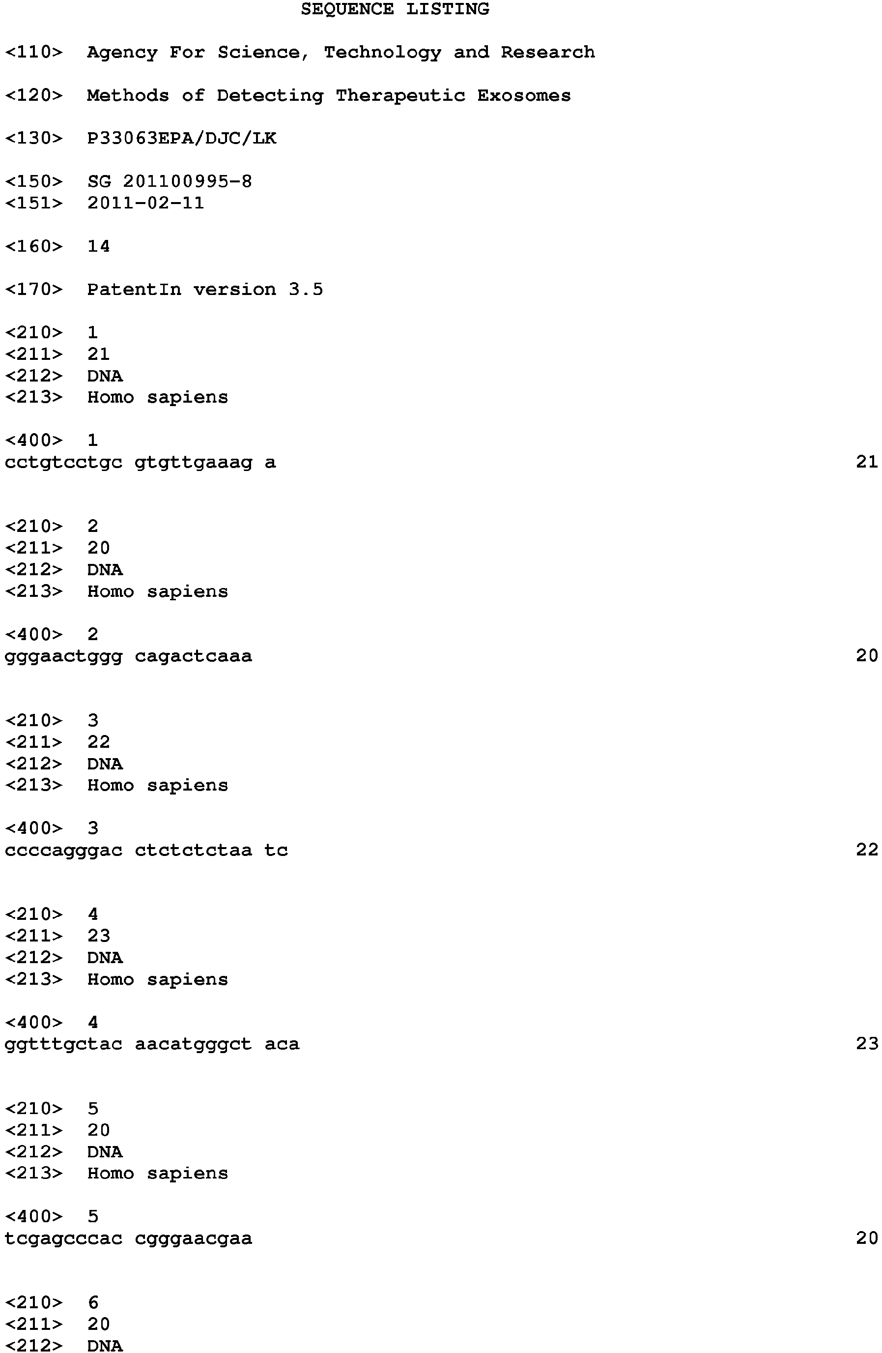
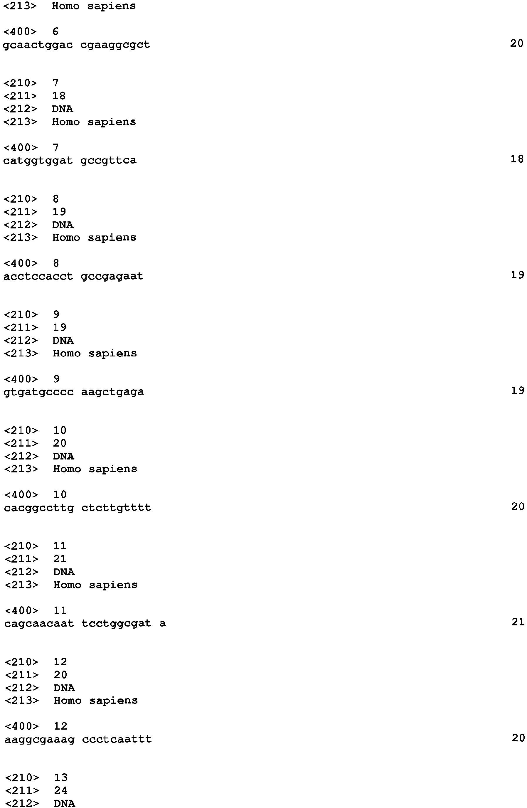

Claims (7)
- A method of detecting a therapeutic exosome, the method comprising detecting an activity of an exosome comprising NT5E (CD73) ecto-5'-ectonucleotidase activity.
- A method according to Claim 1, in which the NT5E (CD73) ecto-5'-ectonucleotidase activity is detected by detecting activity of one or more, preferably all, proteins set out in Table D6.
- A method according to any preceding claim, in which if an activity is detected, the exosome is likely to be capable of: (i) inhibiting complement mediated cell lysis; or (ii) reducing infarct size for example as assayed in a mouse or pig model of myocardial ischemia and reperfusion injury, or which is capable of reducing oxidative stress for example as assayed in an in vitro assay of hydrogen peroxide (H2O2)-induced cell death, or both (i) and (ii).
- A method according to Claim 3, in which the therapeutic activity comprises cardioprotective activity.
- A method according to Claim 3, in which the therapeutic activity is against one or more diseases selected from the group consisting of: cardiac failure, bone marrow disease, skin disease, bums and degenerative diseases such as diabetes, Alzheimer's disease, Parkinson's disease, cancer, myocardial infarction, a cutaneous wound, a dermatologic disorder, a dermatological lesion, dermatitis, psoriasis, condyloma, verruca, hemangioma, keloid, skin cancer, atopic dermatitis, Behcet disease, chronic granulomatous disease, cutaneous T cell lymphoma, ulceration, a pathological condition characterised by initial injury inducing inflammation and immune dysregulation leading to chronic tissue remodeling including fibrosis and loss of function, renal ischemic injury, cystic fibrosis, sinusitis and rhinitis or an orthopaedic disease.
- A method comprising the steps of: (a) isolating an exosome from a mesenchymal stem cell conditioned medium (MSC-CM), optionally comprising separating the exosome from other components based on molecular weight, size, shape, composition or biological activity, and (b) detecting an activity set out in Claim 1 in the isolated exosome, preferably by detecting the activity of one or more proteins set out in any of Claims 2 to 5.
- A method of producing an exosome, the method comprising: (a) obtaining a mesenchymal stem cell conditioned medium (MSC-CM); (b) concentrating the mesenchymal stem cell conditioned medium, for example by ultrafiltration over a >1000 kDa membrane; (c) subjecting the concentrated mesenchymal stem cell conditioned medium to size exclusion chromatography, such as using a a TSK Guard column SWXL, 6 x 40 mm or a TSK gel G4000 SWXL, 7.8 x 300 mm column; (d) selecting UV absorbant fractions, for example, at 220 nm, that exhibit dynamic light scattering, such as detected by a quasi-elastic light scattering (QELS); in which step (d) for example comprises collecting fractions which elute with a retention time of 11-13 minutes, such as 12 minutes; and detecting an activity set out in Claim 1 in the exosome, preferably by detecting the activity of one or more proteins set out in any of Claims 2 to 5.
Applications Claiming Priority (3)
| Application Number | Priority Date | Filing Date | Title |
|---|---|---|---|
| SG2011009958A SG183579A1 (en) | 2011-02-11 | 2011-02-11 | Methods of detecting therapeutic exosomes |
| PCT/SG2012/000039 WO2012108842A1 (en) | 2011-02-11 | 2012-02-10 | Methods of detecting therapeutic exosomes |
| EP12744853.8A EP2673636B1 (en) | 2011-02-11 | 2012-02-10 | Methods of detecting therapeutic exosomes |
Related Parent Applications (2)
| Application Number | Title | Priority Date | Filing Date |
|---|---|---|---|
| EP12744853.8A Division-Into EP2673636B1 (en) | 2011-02-11 | 2012-02-10 | Methods of detecting therapeutic exosomes |
| EP12744853.8A Division EP2673636B1 (en) | 2011-02-11 | 2012-02-10 | Methods of detecting therapeutic exosomes |
Publications (2)
| Publication Number | Publication Date |
|---|---|
| EP3151011A1 true EP3151011A1 (en) | 2017-04-05 |
| EP3151011B1 EP3151011B1 (en) | 2019-11-13 |
Family
ID=46638849
Family Applications (2)
| Application Number | Title | Priority Date | Filing Date |
|---|---|---|---|
| EP12744853.8A Active EP2673636B1 (en) | 2011-02-11 | 2012-02-10 | Methods of detecting therapeutic exosomes |
| EP16194158.8A Active EP3151011B1 (en) | 2011-02-11 | 2012-02-10 | Methods of detecting therapeutic exosomes |
Family Applications Before (1)
| Application Number | Title | Priority Date | Filing Date |
|---|---|---|---|
| EP12744853.8A Active EP2673636B1 (en) | 2011-02-11 | 2012-02-10 | Methods of detecting therapeutic exosomes |
Country Status (6)
| Country | Link |
|---|---|
| US (1) | US11156616B2 (en) |
| EP (2) | EP2673636B1 (en) |
| JP (2) | JP6046640B2 (en) |
| CN (2) | CN103635800B (en) |
| SG (2) | SG183579A1 (en) |
| WO (1) | WO2012108842A1 (en) |
Families Citing this family (54)
| Publication number | Priority date | Publication date | Assignee | Title |
|---|---|---|---|---|
| US11660317B2 (en) | 2004-11-08 | 2023-05-30 | The Johns Hopkins University | Compositions comprising cardiosphere-derived cells for use in cell therapy |
| CA2760840C (en) | 2009-08-24 | 2014-12-02 | Board Of Trustees Of Michigan State University | Pretreated densified biomass products and methods of making and using same |
| US11286463B2 (en) | 2012-03-08 | 2022-03-29 | Advanced ReGen Medical Technologies, LLC | Reprogramming of aged adult stem cells |
| AU2013302799B2 (en) | 2012-08-13 | 2018-03-01 | Cedars-Sinai Medical Center | Exosomes and micro-ribonucleic acids for tissue regeneration |
| CA2906502A1 (en) | 2013-03-13 | 2014-10-02 | University Of Miami | Method for isolation and purification of microvesicles from cell culture supernatants and biological fluids |
| US12558316B2 (en) | 2013-03-13 | 2026-02-24 | University Of Miami | Methods and compositions for the treatment of epidermolysis bullosa |
| SG11201508433TA (en) | 2013-04-12 | 2015-11-27 | Andaloussi Samir El | Therapeutic delivery vesicles |
| WO2016043654A1 (en) | 2014-09-15 | 2016-03-24 | Agency For Science, Technology And Research | Methods of treating graft versus host disease (gvhd) or epidermolysis bullosa (eb) with exosomes |
| SG11201601939VA (en) * | 2013-09-16 | 2016-04-28 | Agency Science Tech & Res | Method |
| US10772911B2 (en) | 2013-12-20 | 2020-09-15 | Advanced ReGen Medical Technologies, LLC | Cell free compositions for cellular restoration and methods of making and using same |
| CR20160307A (en) | 2013-12-20 | 2016-11-08 | Advanced Regen Medical Tech Llc | Compositions for cellular restoration and methods of making and using same |
| WO2016054591A1 (en) | 2014-10-03 | 2016-04-07 | Cedars-Sinai Medical Center | Cardiosphere-derived cells and exosomes secreted by such cells in the treatment of muscular dystrophy |
| US20180042847A1 (en) * | 2015-03-16 | 2018-02-15 | Duncan ROSS | Method of treatment comprising membrane-enclosed vesicle |
| WO2016152786A1 (en) * | 2015-03-20 | 2016-09-29 | 国立大学法人三重大学 | Therapeutic agent associated with suppression of proliferation and metastasis of tumor, which comprises exosomes released from cytotoxic t cells and targets cancer stromal/mesenchymal cells |
| US10702581B2 (en) | 2015-05-04 | 2020-07-07 | Ilias Biologics Inc. | Compositions containing protein loaded exosome and methods for preparing and delivering the same |
| WO2016196822A1 (en) * | 2015-06-02 | 2016-12-08 | Cedars-Sinai Medical Center | Urodele exosomes as therapeutic agents |
| AU2016276750A1 (en) * | 2015-06-09 | 2018-01-18 | The Board Of Regents Of The University Of Texas System | Diagnostic test for early stage cancer |
| WO2017064688A1 (en) * | 2015-10-15 | 2017-04-20 | Cells For Cells, S.P.A. | Neurite regeneration therapy based on exosomes derived from menstrual stem cells |
| SG11201804117XA (en) * | 2015-11-18 | 2018-06-28 | Univ Georgia | Neural cell extracellular vessicles |
| US11253551B2 (en) | 2016-01-11 | 2022-02-22 | Cedars-Sinai Medical Center | Cardiosphere-derived cells and exosomes secreted by such cells in the treatment of heart failure with preserved ejection fraction |
| KR102805970B1 (en) * | 2016-03-02 | 2025-05-09 | 유니버시티 오브 피츠버그 - 오브 더 커먼웰쓰 시스템 오브 하이어 에듀케이션 | Matrix bound nanovesicles and their use |
| EP3427060B1 (en) | 2016-03-07 | 2025-08-13 | Father Flanagan's Boys' Home doing Business as Boys Town National Research Hospital | Noninvasive molecular controls |
| WO2017190000A1 (en) | 2016-04-29 | 2017-11-02 | Advanced ReGen Medical Technologies, LLC | Microrna compositions and methods of making and using same |
| WO2017204187A1 (en) | 2016-05-24 | 2017-11-30 | 公益財団法人がん研究会 | Method of recovering extracellular vesicles and container for extracellular vesicles |
| WO2017210652A1 (en) | 2016-06-03 | 2017-12-07 | Cedars-Sinai Medical Center | Cdc-derived exosomes for treatment of ventricular tachyarrythmias |
| CA3027970A1 (en) * | 2016-06-17 | 2017-12-21 | United Therapeutics Corporation | Extracellular vesicles with enhanced potency |
| US10946047B2 (en) | 2016-06-17 | 2021-03-16 | United Therapeutics Corporation | Extracellular vesicles with enhanced potency |
| EP3515459A4 (en) | 2016-09-20 | 2020-08-05 | Cedars-Sinai Medical Center | CARDIOSPHERIC CELLS AND THEIR EXTRACELLULAR VESICLES TO DELAY OR REVERSE THE AGING PROCESS AND AGE-RELATED DISEASES |
| CA3002520A1 (en) * | 2016-09-30 | 2018-04-05 | Ilias Biologics Inc. | Compositions containing protein loaded exosome and methods for preparing and delivering the same |
| CA3042360A1 (en) * | 2016-11-03 | 2018-05-11 | Exostem Biotec Ltd. | Mesenchymal stem cells populations, their products, and use thereof |
| JP6981619B2 (en) * | 2017-03-14 | 2021-12-15 | 株式会社ダイセル | Exosome production promoter |
| CA3059910A1 (en) | 2017-04-19 | 2018-10-25 | Cedars-Sinai Medical Center | Methods and compositions for treating skeletal muscular dystrophy |
| US11638724B2 (en) | 2017-05-05 | 2023-05-02 | University of Pittsburgh—of the Commonwealth System of Higher Education | Ocular applications of matrix bound vesicles (MBVs) |
| KR20190003316A (en) | 2017-06-30 | 2019-01-09 | 주식회사 엑소코바이오 | A composition comprising an exosome and/or extracellular vesicle derived from adipose-derived stem cell as an active ingredient and its application for improving dermatitis |
| MX2020002085A (en) * | 2017-08-24 | 2020-07-13 | Texas A & M Univ Sys | Mesenchymal stem/stromal cell-derived extracellular vesicles and uses thereof in autoimmune diseases. |
| CN107583699A (en) * | 2017-09-27 | 2018-01-16 | 河南科技大学第附属医院 | A kind of heparin tube examined for clinical medicine and preparation method thereof |
| KR20190060646A (en) | 2017-11-24 | 2019-06-03 | 주식회사 엑소코바이오 | A composition comprising an exosome and/or extracellular vesicle derived from stem cell as an active ingredient and its application for reinforcing or improving skin barrier |
| WO2019103381A1 (en) * | 2017-11-24 | 2019-05-31 | 주식회사 엑소코바이오 | Use of composition comprising stem cell-derived exosome as effective ingredient in reinforcement and functional improvement of skin barrier |
| WO2019118817A1 (en) * | 2017-12-14 | 2019-06-20 | Mayo Foundation For Medical Education And Research | Purified exosome products, method of making, and methods of using |
| US11660355B2 (en) | 2017-12-20 | 2023-05-30 | Cedars-Sinai Medical Center | Engineered extracellular vesicles for enhanced tissue delivery |
| US10717981B2 (en) | 2018-01-18 | 2020-07-21 | Advanced ReGen Medical Technologies, LLC | Therapeutic compositions and methods of making and using the same |
| WO2019152549A1 (en) | 2018-02-05 | 2019-08-08 | Cedars-Sinai Medical Center | Methods for therapeutic use of exosomes and y-rnas |
| EP3787642A4 (en) | 2018-05-03 | 2022-09-21 | University of Pittsburgh - of the Commonwealth System of Higher Education | MATRIX-BOUND VESICLES (MBV) CONTAINING IL-33 AND THEIR USE |
| KR102233530B1 (en) * | 2018-11-22 | 2021-03-31 | (주)프로스테믹스 | Exomsome and various uses thereof |
| US20220193211A1 (en) * | 2019-04-18 | 2022-06-23 | Intellistem Technologies Inc. | Overexpression of immunoproteasome in host cells for generating antigen-presenting cells |
| KR20220087483A (en) * | 2019-10-23 | 2022-06-24 | 유니버시티 오브 피츠버그-오브 더 커먼웰쓰 시스템 오브 하이어 에듀케이션 | Use of MBV for the treatment of autoimmune diseases |
| WO2021113299A1 (en) * | 2019-12-04 | 2021-06-10 | United Therapeutics Corporation | Extracellular vesicles and their uses |
| IL295102B2 (en) | 2020-02-28 | 2026-04-01 | Joyce H Ma | Formulations and uses thereof |
| CN112206311B (en) * | 2020-09-23 | 2021-12-28 | 南方医科大学 | Application of S100A11 protein as diabetes biomarker and therapeutic target |
| CN112444629B (en) * | 2020-12-09 | 2023-09-29 | 青岛科技大学 | Exosome detection method and application based on hairpin nucleic acid molecule signal amplification circuit |
| CN114848674A (en) * | 2022-03-25 | 2022-08-05 | 四川大学华西医院 | Application of stem cell particles in preparation of medicine for treating Parkinson's disease |
| WO2024089180A1 (en) * | 2022-10-26 | 2024-05-02 | Eximmium Biotechnologies Gmbh | Method to determine extracellular vesicle recovery |
| CN116286623A (en) * | 2023-02-13 | 2023-06-23 | 中国医学科学院基础医学研究所 | Metabolic markers and screening methods for screening exosomes derived from young and aged mesenchymal stem cells |
| EP4578454A1 (en) | 2023-12-28 | 2025-07-02 | Lysatpharma GmbH | Compositions comprising platelet derived extracellular vesicles |
Citations (12)
| Publication number | Priority date | Publication date | Assignee | Title |
|---|---|---|---|---|
| US4675187A (en) | 1983-05-16 | 1987-06-23 | Bristol-Myers Company | BBM-1675, a new antibiotic complex |
| EP0516598A1 (en) | 1991-05-27 | 1992-12-02 | Weidmüller Interface GmbH & Co. | Pliers with selective front and side introduction |
| WO1994007913A1 (en) | 1992-09-25 | 1994-04-14 | Warner-Lambert Company | Peptide antagonists of sh2 binding and therapeutic uses thereof |
| EP0600831A1 (en) | 1992-11-27 | 1994-06-08 | Ciba-Geigy Ag | Phtalazinon derivatives |
| EP0600832A1 (en) | 1992-11-27 | 1994-06-08 | Ciba-Geigy Ag | Derivatives of diamino benzoic and diamino phthalic acid und their use as protein linase inhibitors |
| WO1994022867A1 (en) | 1993-04-07 | 1994-10-13 | Pfizer Inc. | Bicyclic ansamycins and their use as antitumor agents |
| US5631159A (en) | 1993-09-22 | 1997-05-20 | Creative Biomolecules, Inc. | Lipid-modified serum free media |
| US5661034A (en) | 1991-07-18 | 1997-08-26 | Fuji Yakuhin Kogyo Kabushiki Kaisha | Serum-free medium for tissue culture containing tissue inhibitor of metalloproteinases and method for growing cells using the medium |
| US6139524A (en) | 1998-10-16 | 2000-10-31 | Scimed Life Systems, Inc. | Stent delivery system with perfusion |
| US7122019B1 (en) | 2000-11-28 | 2006-10-17 | Flowmedica Inc. | Intra-aortic renal drug delivery catheter |
| US7241273B2 (en) | 2002-09-20 | 2007-07-10 | Flowmedica, Inc. | Intra-aortic renal delivery catheter |
| WO2009105044A1 (en) | 2008-02-22 | 2009-08-27 | Agency For Science, Technology And Research (A*Star) | Mesenchymal stem cell particles |
Family Cites Families (7)
| Publication number | Priority date | Publication date | Assignee | Title |
|---|---|---|---|---|
| US20040241176A1 (en) | 2000-04-27 | 2004-12-02 | Ap Cells. Inc. | Method of producing membrane vesicles |
| WO2006007529A2 (en) * | 2004-07-01 | 2006-01-19 | University Of Pittsburgh Of The Commonwealth System For Higher Education | Immunosuppressive exosomes |
| USD544957S1 (en) | 2005-03-23 | 2007-06-19 | Rexam Dispensing Systems | Nasal spray dispenser |
| WO2007015174A2 (en) * | 2005-07-08 | 2007-02-08 | Exothera L.L.C. | Exosome-specific ligands, their preparartion and uses |
| ES2936256T3 (en) * | 2008-02-01 | 2023-03-15 | Massachusetts Gen Hospital | Use of microvesicles in the diagnosis, and prognosis of diseases and medical conditions |
| CA2743211A1 (en) * | 2008-11-12 | 2010-05-20 | Caris Life Sciences Luxembourg Holdings, S.A.R.L. | Methods and systems of using exosomes for determining phenotypes |
| KR20120081972A (en) * | 2009-07-02 | 2012-07-20 | 아이티에이치 임뮨 세라피 홀딩스 에이비 | Exosome based treatment of cancer |
-
2011
- 2011-02-11 SG SG2011009958A patent/SG183579A1/en unknown
-
2012
- 2012-02-10 JP JP2013553403A patent/JP6046640B2/en not_active Expired - Fee Related
- 2012-02-10 EP EP12744853.8A patent/EP2673636B1/en active Active
- 2012-02-10 US US13/984,203 patent/US11156616B2/en not_active Expired - Fee Related
- 2012-02-10 CN CN201280017773.XA patent/CN103635800B/en not_active Expired - Fee Related
- 2012-02-10 EP EP16194158.8A patent/EP3151011B1/en active Active
- 2012-02-10 CN CN201610952298.5A patent/CN107102128B/en not_active Expired - Fee Related
- 2012-02-10 WO PCT/SG2012/000039 patent/WO2012108842A1/en not_active Ceased
- 2012-02-10 SG SG2013057450A patent/SG192191A1/en unknown
-
2016
- 2016-11-16 JP JP2016223270A patent/JP6444963B2/en not_active Expired - Fee Related
Patent Citations (12)
| Publication number | Priority date | Publication date | Assignee | Title |
|---|---|---|---|---|
| US4675187A (en) | 1983-05-16 | 1987-06-23 | Bristol-Myers Company | BBM-1675, a new antibiotic complex |
| EP0516598A1 (en) | 1991-05-27 | 1992-12-02 | Weidmüller Interface GmbH & Co. | Pliers with selective front and side introduction |
| US5661034A (en) | 1991-07-18 | 1997-08-26 | Fuji Yakuhin Kogyo Kabushiki Kaisha | Serum-free medium for tissue culture containing tissue inhibitor of metalloproteinases and method for growing cells using the medium |
| WO1994007913A1 (en) | 1992-09-25 | 1994-04-14 | Warner-Lambert Company | Peptide antagonists of sh2 binding and therapeutic uses thereof |
| EP0600831A1 (en) | 1992-11-27 | 1994-06-08 | Ciba-Geigy Ag | Phtalazinon derivatives |
| EP0600832A1 (en) | 1992-11-27 | 1994-06-08 | Ciba-Geigy Ag | Derivatives of diamino benzoic and diamino phthalic acid und their use as protein linase inhibitors |
| WO1994022867A1 (en) | 1993-04-07 | 1994-10-13 | Pfizer Inc. | Bicyclic ansamycins and their use as antitumor agents |
| US5631159A (en) | 1993-09-22 | 1997-05-20 | Creative Biomolecules, Inc. | Lipid-modified serum free media |
| US6139524A (en) | 1998-10-16 | 2000-10-31 | Scimed Life Systems, Inc. | Stent delivery system with perfusion |
| US7122019B1 (en) | 2000-11-28 | 2006-10-17 | Flowmedica Inc. | Intra-aortic renal drug delivery catheter |
| US7241273B2 (en) | 2002-09-20 | 2007-07-10 | Flowmedica, Inc. | Intra-aortic renal delivery catheter |
| WO2009105044A1 (en) | 2008-02-22 | 2009-08-27 | Agency For Science, Technology And Research (A*Star) | Mesenchymal stem cell particles |
Non-Patent Citations (80)
| Title |
|---|
| "A Handbook of Recipes, Reagents, and Other Reference Tools for Use at the Bench", 2002, COLD SPRING HARBOR LABORATORY |
| "Antibodies : A Laboratory Manual", 1988, COLD SPRING HARBOR LABORATORY PRESS, pages: 1855 |
| "Current Protocols in Molecular Biology", JOHN WILEY & SONS |
| "Handbook of Drug Screening", 2001, MARCEL DEKKER |
| "Oligonucleotide Synthesis: A Practical Approach", 1984, IRL PRESS |
| B. ROE; J. CRABTREE; A. KAHN: "DNA Isolation and Sequencing: Essential Techniques", 1996, JOHN WILEY & SONS |
| BANDO, H. ET AL.: "Phosphorylation of the 6-phosphofructo-2-kinase/fructose 2,6-bisphosphatase/PFKFB3 family of glycolytic regulators in human cancer", CLIN CANCER RES, vol. 11, 2005, pages 5784 - 5792, XP009079337, DOI: doi:10.1158/1078-0432.CCR-05-0149 |
| BARRY, F.P.; MURPHY, J.M: "Mesenchymal stem cells: clinical applications and biological characterization", INT J BIOCHEM CELL BIOL, vol. 36, 2004, pages 568 - 584, XP002360961, DOI: doi:10.1016/j.biocel.2003.11.001 |
| CASTELLANO, G. ET AL.: "Therapeutic targeting of classical and lectin pathways of complement protects from ischemia-reperfusion-induced renal damage", AM JPATHOL, vol. 176, 2010, pages 1648 - 1659 |
| CHEN, T.S. ET AL.: "Mesenchymal stem cell secretes microparticles enriched in pre-microRNAs", NUCLEIC ACIDS RES, vol. 38, 2010, pages 215 - 224, XP002756861, DOI: doi:10.1093/nar/gkp857 |
| CLAUDIA CHAVEZ-MUÑOZ ET AL: "Profile of exosomes related proteins released by differentiated and undifferentiated human keratinocytes", JOURNAL OF CELLULAR PHYSIOLOGY, vol. 221, no. 1, 1 October 2009 (2009-10-01), pages 221 - 231, XP055195376, ISSN: 0021-9541, DOI: 10.1002/jcp.21847 * |
| COWAN CA; KLIMANSKAYA I; MCMAHON J; ATIENZA J; WITMYER J ET AL.: "Derivation of embryonic stem-cell lines from human blastocysts", N ENGL J MED, vol. 350, 2004, pages 1353 - 1356, XP002404211 |
| D. M. J. LILLEY; D J. E. DAHLBERG: "Methods in Enzymology", 1992, ACADEMIC PRESS, article "Methods of Enzymology: DNA Structure Part A: Synthesis and Physical Analysis of DNA" |
| DAVIES, A.; LACHMANN, P.J.: "Membrane defence against complement lysis: the structure and biological properties of CD59", IMMUNOL RES, vol. 12, 1993, pages 258 - 275 |
| DEVITT, A.; MARSHALL, L. J.: "The innate immune system and the clearance of apoptotic cells", JOURNAL OF LEUKOCYTE BIOLOGY, vol. 90, 2011, pages 447 - 457 |
| DUNLOP, R.A.; BRUNK, U.T.; RODGERS, K.J: "Oxidized proteins: mechanisms of removal and consequences of accumulation", IUBMB LIFE, vol. 61, 2009, pages 522 - 527 |
| EDWARD HARLOW; DAVID LANE; ED HARLOW: "Using Antibodies : A Laboratory Manual: Portable Protocol NO. I", 1999, COLD SPRING HARBOR LABORATORY PRESS |
| EVGENIA GERASIMOVSKAYA, E.K: "Extracellular ATP and adenosine as regulators of endothelial cell function", IMPLICATIONS FOR HEALTH AND DISEASE, 2010 |
| FEVRIER, B; RAPOSO, G: "Exosomes: endosomal-derived vesicles shipping extracellular messages", CURR OPIN CELL BIOL, vol. 16, 2004, pages 415 - 421 |
| FRANGOGIANNIS, N.G.; SMITH, C.W.; ENTMAN, M.L.: "The inflammatory response in myocardial infarction", CARDIOVASC RES, vol. 53, 2002, pages 31 - 47 |
| GARCIA-OLMO, D. ET AL.: "A Phase I Clinical Trial of the Treatment of Crohn's Fistula by Adipose Mesenchymal Stem Cell Transplantation", DISEASES OF THE COLON & RECTUM, vol. 48, 2005, pages 1416 - 1423, XP002407256, DOI: doi:10.1007/s10350-005-0052-6 |
| HASKO, G.; LINDEN, J.; CRONSTEIN, B.; PACHER, P: "Adenosine receptors: therapeutic aspects for inflammatory and immune diseases", NAT REV DRUG DISCOV, vol. 7, 2008, pages 759 - 770 |
| HAUSENLOY, D.J.; YELLON, D.M.: "New directions for protecting the heart against ischaemia-reperfusion injury: targeting the Reperfusion Injury Salvage Kinase (RISK)-pathway.", CARDIOVASC RES, vol. 61, 2004, pages 448 - 460, XP002407967, DOI: doi:10.1016/j.cardiores.2003.09.024 |
| HUBER, V.; FILIPAZZI, P.; IERO, M.; FAIS, S; RIVOLTINI, L: "More insights into the immunosuppressive potential of tumor exosomes", JOURNAL OF TRANSLATIONAL MEDICINE, vol. 6, 2008, XP021044053, DOI: doi:10.1186/1479-5876-6-63 |
| IWASAKI, A.; MEDZHITOV, R.: "Regulation of Adaptive Immunity by the Innate Immune System", SCIENCE, vol. 327, 2010, pages 291 - 295 |
| J. M. POLAK; JAMES O'D. MCGEE: "In Situ Hybridization: Principles and Practice", 1990, OXFORD UNIVERSITY PRESS |
| J. SAMBROOK; E. F. FRITSCH; T. MANIATIS: "Molecular Cloning: A Laboratory Manual", 1989, COLD SPRING HARBOR LABORATORY PRESS |
| JACOBSON, K.A: "Handbook of experimental pharmacology", 2009, article "Introduction to adenosine receptors as therapeutic targets.", pages: 1 - 24 |
| JUNG, T.; GRUNE, T.: "The proteasome and its role in the degradation of oxidized proteins", IUBMB LIFE, vol. 60, 2008, pages 743 - 752 |
| KAWAI, T.; AKIRA, S.: "The roles of TLRs, RLRs and NLRs in pathogen recognition ARTICLE", INTERNATIONAL IMMUNOLOGY, vol. 21, 2009, pages 317 - 337, XP055112188, DOI: doi:10.1093/intimm/dxp017 |
| KELLER, S.; SANDERSON, M.P.; STOECK, A.; ALTEVOGT, P.: "Exosomes: from biogenesis and secretion to biological function", IMMUNOL LETT, vol. 107, 2006, pages 102 - 108, XP024999091, DOI: doi:10.1016/j.imlet.2006.09.005 |
| LAI, R. C. ET AL.: "Exosome secreted by MSC reduces myocardial ischemia/reperfusion injury", STEM CELL RES, vol. 4, 2010, pages 214 - 222, XP027054731 |
| LAI, R.C. ET AL.: "Derivation and characterization of human fetal MSCs: an alternative cell source for large-scale production of cardioprotective microparticles", J MOL CELL CARDIOL, vol. 48, 2010, pages 1215 - 1224, XP055464739, DOI: doi:10.1016/j.yjmcc.2009.12.021 |
| LAI, R.C. ET AL.: "Exosome secreted by MSC reduces myocardial ischemia/reperfusion injury", STEM CELL RES, vol. 4, 2010, pages 214 - 222, XP027054731 |
| LE BLANC, K ET AL.: "Mesenchymal stem cells for treatment of steroid-resistant, severe, acute graft-versus-host disease: a phase II study", THE LANCET, vol. 371, 2008, pages 1579 - 1586, XP026859188, DOI: doi:10.1016/S0140-6736(08)60690-X |
| LIAN, Q. ET AL.: "Derivation of Clinically Compliant MSCs from CD105+, CD24- Differentiated Human ESCs", STEM CELLS, vol. 25, 2007, pages 425 - 436, XP002611381, DOI: doi:10.1634/stemcells.2006-0420 |
| MUNTASELL, A.; BERGER, A.C.; ROCHE, P.A: "T cell-induced secretion of MHC class II-peptide complexes on B cell exosomes", EMBO JOURNAL, vol. 26, 2007, pages 4263 - 4272 |
| NANDAKUMAR, S.; MILLER, C.; KUMARAGURU, U: "T regulatory cells: an overview and intervention techniques to modulate allergy outcome", CLINICAL AND MOLECULAR ALLERGY, vol. 7, 2009, pages 5, XP021050246, DOI: doi:10.1186/1476-7961-7-5 |
| NAUTA, A. J.; FIBBE, W. E: "Immunomodulatory properties of mesenchymal stromal cells", BLOOD, vol. 110, 2007, pages 3499 - 3506 |
| NEWMAN, R.H.; WHITEHEAD, P.; LALLY, J.; COFFER, A; FREEMONT, P: "20S human proteasomes bind with a specific orientation to lipid monolayers in vitro", BIOCHIM BIOPHYS ACTA, vol. 1281, 1996, pages 111 - 116 |
| ORRENIUS, S.; GOGVADZE, V.; ZHIVOTOVSKY, B: "Mitochondrial Oxidative Stress: Implications for Cell Death", ANNUAL REVIEW OF PHARMACOLOGY AND TOXICOLOGY, vol. 47, 2007, pages 143 - 183 |
| OVIZE, M. ET AL.: "Postconditioning and protection from reperfusion injury: where do we stand? Position paper from the Working Group of Cellular Biology of the Heart of the European Society of Cardiology", CARDIOVASC RES, vol. 87, 2010, pages 406 - 423 |
| PAMUK, O.N. ET AL.: "Spleen tyrosine kinase inhibition prevents tissue damage after ischemia-reperfusion", AM J PHYSIOL GASTROINTEST LIVER PHYSIOL, vol. 299, 2010, pages G391 - 399 |
| PAN, B.T.; JOHNSTONE, R.M: "Fate of the transferrin receptor during maturation of sheep reticulocytes in vitro: selective externalization of the receptor", CELL, vol. 33, 1983, pages 967 - 978, XP027462733, DOI: doi:10.1016/0092-8674(83)90040-5 |
| PENEFSKY, H.S: "Mechanism of inhibition of mitochondrial adenosine triphosphatase by dicyclohexylcarbodiimide and oligomycin: relationship to ATP synthesis", PROC NATL ACAD SCI USA, vol. 82, 1985, pages 1589 - 1593 |
| PERRY, D.; PECK, A. B.; CARCAMO, W. C.; MOREL, L.; NGUYEN, C. Q: "The Current Concept of T H 17 Cells and Their Expanding Role in Systemic Lupus Erythematosus", ARTHRITIS, 2011 |
| PILKIS, S.J.; EL-MAGHRABI, M.R.; PILKIS, J.; CLAUS, T.: "Inhibition of fructose-1,6-bisphosphatase by fructose 2,6-bisphosphate", JBIOL CHEM, vol. 256, 1981, pages 3619 - 3622 |
| R.C. LAI; F. ARSLAN; M.M. LEE; N.S. SZE; A. CHOO; T.S. CHEN; M. SALTO-TELLEZ; L. TIMMERS; C.N. LEE; R.M. EL OAKLEY: "Exosome secreted by MSC reduces myocardial ischemia/reperfusion injury", STEM CELL RES, vol. 4, 2010, pages 214 - 222, XP027054731 |
| RAPOSO, G. ET AL.: "B lymphocytes secrete antigen-presenting vesicles", JOURNAL OF EXPERIMENTAL MEDICINE, vol. 183, 1996, pages 1161 - 1172, XP002060486, DOI: doi:10.1084/jem.183.3.1161 |
| RILEY, J. L.; JUNE, C. H.; BLAZAR, B. R.: "Human T Regulatory Cell Therapy: Take a Billion or So and Call Me in the Morning", IMMUNITY, vol. 30, 2009, pages 656 - 665, XP055159519, DOI: doi:10.1016/j.immuni.2009.04.006 |
| SCHOREY, J.S; BHATNAGAR, S: "Exosome function: From tumor immunology to pathogen biology", TRAFFIC, vol. 9, 2008, pages 871 - 881, XP002644382, DOI: doi:10.1111/j.1600-0854.2008.00734.x |
| SIMONS, M.; RAPOSO, G.: "Exosomes - vesicular carriers for intercellular communication", CURRENT OPINION IN CELL BIOLOGY, vol. 21, 2009, pages 575 - 581, XP026434725 |
| SIMONS, M.; RAPOSO, G.: "Exosomes--vesicular carriers for intercellular communication", CURR OPIN CELL BIOL, vol. 21, 2009, pages 575 - 581, XP026434725 |
| SIMPSON, R.J.; JENSEN, S.S; LIM, J.W.E: "Proteomic profiling of exosomes: Current perspectives", PROTEOMICS, vol. 8, 2008, pages 4083 - 4099, XP008139622, DOI: doi:10.1002/pmic.200800109 |
| SIMPSON, R.J.; LIM, J.W.; MORITZ, R.L.; MATHIVANAN, S: "Exosomes: proteomic insights and diagnostic potential", EXPERT REV PROTEOMICS, vol. 6, 2009, pages 267 - 283, XP009125412, DOI: doi:10.1586/epr.09.17 |
| SINGER, N. G.; CAPLAN, A. I.: "Mesenchymal Stem Cells: Mechanisms of Inflammation", ANNUAL REVIEW OF PATHOLOGY: MECHANISMS OF DISEASE, vol. 6, 2011, pages 457 - 478 |
| SKOKOS, D. ET AL.: "Mast cell-derived exosomes induce phenotypic and functional maturation of dendritic cells and elicit specific immune responses in vivo", JOURNAL OF IMMUNOLOGY, vol. 170, 2003, pages 3037 - 3045 |
| SOHNS, W.; VAN VEEN, T.A.; VAN DER HEYDEN, M.A.: "Regulatory roles of the ubiquitin-proteasome system in cardiomyocyte apoptosis", CURR MOL MED, vol. 10, pages 1 - 13 |
| STADTMAN, E.R.; LEVINE, R.L.: "Protein oxidation", ANN N YACAD SCI, vol. 899, 2000, pages 191 - 208 |
| SU, P. F. ET AL.: "Dioscorea phytocompounds enhance murine splenocyte proliferation ex vivo and improve regeneration of bone marrow cells in vivo", EVID BASED COMPLEMENT ALTERNAT MED, 2011, pages 731308 |
| SZE, S.K. ET AL.: "Elucidating the secretion proteome of human embryonic stem cell-derived mesenchymal stem cells", MOL CELL PROTEOMICS, vol. 6, 2007, pages 1680 - 1689, XP002531526, DOI: doi:10.1074/mcp.M600393-MCP200 |
| TANAKA, K.: "The proteasome: overview of structure and functions", PROC JPN ACAD SER B PHYS BIOL SCI, vol. 85, 2009, pages 12 - 36 |
| TAYLOR, D.D.; GERCEL-TAYLOR, C.: "Tumour-derived exosomes and their role in cancer-associated T-cell signalling defects", BRITISH JOURNAL OF CANCER, vol. 92, 2005, pages 305 - 311, XP002623150, DOI: doi:10.1038/SJ.BJC.6602316 |
| THERY, C.; OSTROWSKI, M.; SEGURA, E: "Membrane vesicles as conveyors of immune responses", NATURE REVIEWS IMMUNOLOGY, vol. 9, 2009, pages 581 - 593, XP055018445, DOI: doi:10.1038/nri2567 |
| THERY, C.; OSTROWSKI, M; SEGURA, E.: "Membrane vesicles as conveyors of immune responses", NAT REV IMMUNOL, vol. 9, 2009, pages 581 - 593, XP055018445, DOI: doi:10.1038/nri2567 |
| THOMAS, P.D ET AL.: "PANTHER: a library of protein families and subfamilies indexed by function", GENOME RES, vol. 13, 2003, pages 2129 - 2141, XP002327041, DOI: doi:10.1101/gr.772403 |
| THOMAS, P.D. ET AL.: "Applications for protein sequence-function evolution data: mRNA/protein expression analysis and coding SNP scoring tools", NUCLEIC ACIDS RES, vol. 34, 2006, pages W645 - 650 |
| THOMSON JA; ITSKOVITZ-ELDOR J; SHAPIRO SS; WAKNITZ MA; SWIERGIEL JJ ET AL.: "Embryonic Stem Cell Lines Derived from Human Blastocysts", SCIENCE, vol. 282, 1998, pages 1145 - 1147, XP002933311, DOI: doi:10.1126/science.282.5391.1145 |
| TIMMERS, L ET AL.: "Reduction of myocardial infarct size by human mesenchymal stem cell conditioned medium", STEM CELL RESEARCH, vol. 1, 2008, pages 129 - 137 |
| TIMMERS, L. ET AL.: "Human mesenchymal stem cell-conditioned medium improves cardiac function following myocardial infarction", STEM CELL RESEARCH, vol. 6, 2011, pages 206 - 214, XP028597231, DOI: doi:10.1016/j.scr.2011.01.001 |
| TSUKAMOTO, O.; MINAMINO, T.; KITAKAZE, M: "Functional alterations of cardiac proteasomes under physiological and pathological conditions", CARDIOVASC RES, vol. 85, 2010, pages 339 - 346 |
| USCHE, M.N.; PAVLOV, V.; TAKAHASHI, K; STAHL, G.L.: "Myocardial ischemia and reperfusion injury is dependent on both IgM and mannose-binding lectin", AM J PHYSIOL HEART CIRC PHYSIOL, vol. 297, 2009, pages H1853 - 1859 |
| VALADI, H. ET AL.: "Exosome-mediated transfer of mRNAs and microRNAs is a novel mechanism of genetic exchange between cells", NAT CELL BIOL, vol. 9, 2007, pages 654 - 659 |
| VIGNALI, D. A. A.; COLLISON, L. W.; WORKMAN, C. J.: "How regulatory T cells work", NAT REV IMMUNOL, vol. 8, 2008, pages 523 - 532, XP009133754 |
| WILLIS, M.S.; TOWNLEY-TILSON, W.H.; KANG, E.Y.; HOMEISTER, J.W.; PATTERSON, C.: "Sent to destroy: the ubiquitin proteasome system regulates cell signaling and protein quality control in cardiovascular development and disease", CIRC RES, vol. 106, pages 463 - 478 |
| WOLFERS, J. ET AL.: "Tumor-derived exosomes are a source of shared tumor rejection antigens for CTL cross-priming", NATURE MEDICINE, vol. 7, 2001, pages 297 - 303, XP002202776, DOI: doi:10.1038/85438 |
| YU, L.; WANG, L.; CHEN, S.: "Endogenous toll-like receptor ligands and their biological significance", JOURNAL OF CELLULAR AND MOLECULAR MEDICINE, vol. 14, 2010, pages 2592 - 2603 |
| ZEELENBERG, I.S. ET AL.: "Targeting tumor antigens to secreted membrane vesicles in vivo induces efficient antitumor immune responses", CANCER RESEARCH, vol. 68, 2008, pages 1228 - 1235, XP002508134, DOI: doi:10.1158/0008-5472.CAN-07-3163 |
| ZITVOGEL, L. ET AL.: "Eradication of established murine tumors using a novel cell-free vaccine: Dendritic cell-derived exosomes", NATURE MEDICINE, vol. 4, 1998, pages 594 - 600, XP002085387, DOI: doi:10.1038/nm0598-594 |
| ZOLLER, M.: "Tetraspanins: Push and pull in suppressing and promoting metastasis", NATURE REVIEWS CANCER, vol. 9, 2009, pages 40 - 55, XP007915967 |
Also Published As
| Publication number | Publication date |
|---|---|
| JP6046640B2 (en) | 2016-12-21 |
| CN107102128A (en) | 2017-08-29 |
| CN107102128B (en) | 2019-07-05 |
| CN103635800A (en) | 2014-03-12 |
| US11156616B2 (en) | 2021-10-26 |
| JP2014507140A (en) | 2014-03-27 |
| EP2673636B1 (en) | 2017-08-09 |
| WO2012108842A1 (en) | 2012-08-16 |
| JP6444963B2 (en) | 2018-12-26 |
| EP2673636A1 (en) | 2013-12-18 |
| EP2673636A4 (en) | 2015-07-22 |
| SG192191A1 (en) | 2013-08-30 |
| US20140031256A1 (en) | 2014-01-30 |
| CN103635800B (en) | 2017-02-15 |
| JP2017093429A (en) | 2017-06-01 |
| HK1243488A1 (en) | 2018-07-13 |
| SG183579A1 (en) | 2012-09-27 |
| EP3151011B1 (en) | 2019-11-13 |
Similar Documents
| Publication | Publication Date | Title |
|---|---|---|
| EP2673636B1 (en) | Methods of detecting therapeutic exosomes | |
| Lee et al. | Caveolin-1 selectively regulates microRNA sorting into microvesicles after noxious stimuli | |
| DK2254586T3 (en) | Mesenchymal stem cell particles | |
| Sluijter et al. | Microvesicles and exosomes for intracardiac communication | |
| Greening et al. | Human endometrial exosomes contain hormone-specific cargo modulating trophoblast adhesive capacity: insights into endometrial-embryo interactions | |
| SG190450A1 (en) | Method of purifying exosomes | |
| US20250121010A1 (en) | A biomarker indicating the therapeutic efficacy of extracellular vesicle (ev)- preparations | |
| HK1243488B (en) | Methods of detecting therapeutic exosomes | |
| Balestra | NOVEL ASPECTS OF EGFR NON-CLATHRIN ENDOCYTOSIS REGULATION: CELL CONTEXT DEPENDENCY AND ROLE OF RNA AND RNA-BINDING PROTEINS. | |
| US20250208046A1 (en) | Method for predicting gene transfer rate | |
| Park et al. | Trogocytic-molting of T-cell microvilli controls T-cell clonal expansion | |
| Nazari et al. | Impact of acute myeloid leukemia-derived extracellular vesicles on expression of CTNNB1, TGF-β, and VEGF genes in bone marrow mesenchymal stem cells: insights into leukemia pathogenesis | |
| Kalvala et al. | Extracellular vesicles modulate integrin signaling and subcellular energetics to promote pulmonary lymphangioleiomyomatosis metastasis | |
| Joshi et al. | The IRE1α/XBP1/Myomaker axis drives myoblast fusion in adult skeletal muscle | |
| Breuninger | Utilizing Heat Shock Protein 70 (Hsp70) as a Tumor-Specific Blood Biomarker and for the Isolation of Circulating Tumor Cells (CTCs) | |
| Kholia | The role of PADs in the biogenesis of prostate cancer microvesicles which play a dysfunctional role on DC biology and the therapeutic potential of skeletal muscle microvesicles on prostate cancer cells | |
| Linder | The role of the tumor suppressor p53 in the biogenesis and function of extracellular vesicles. | |
| Wee Yeh Yeo et al. | Mesenchymal stromal cell extracellular vesicles/exosomes | |
| 매낸더 et al. | Inhibitory Effects of Heparin Derivatives on A549-derived Exosomes | |
| General et al. | Organized by ISEV and ISEV-Americas | |
| AU2014200380A1 (en) | Mesenchymal stem cell particles |
Legal Events
| Date | Code | Title | Description |
|---|---|---|---|
| PUAI | Public reference made under article 153(3) epc to a published international application that has entered the european phase |
Free format text: ORIGINAL CODE: 0009012 |
|
| STAA | Information on the status of an ep patent application or granted ep patent |
Free format text: STATUS: THE APPLICATION HAS BEEN PUBLISHED |
|
| AC | Divisional application: reference to earlier application |
Ref document number: 2673636 Country of ref document: EP Kind code of ref document: P |
|
| AK | Designated contracting states |
Kind code of ref document: A1 Designated state(s): AL AT BE BG CH CY CZ DE DK EE ES FI FR GB GR HR HU IE IS IT LI LT LU LV MC MK MT NL NO PL PT RO RS SE SI SK SM TR |
|
| STAA | Information on the status of an ep patent application or granted ep patent |
Free format text: STATUS: REQUEST FOR EXAMINATION WAS MADE |
|
| 17P | Request for examination filed |
Effective date: 20170922 |
|
| RBV | Designated contracting states (corrected) |
Designated state(s): AL AT BE BG CH CY CZ DE DK EE ES FI FR GB GR HR HU IE IS IT LI LT LU LV MC MK MT NL NO PL PT RO RS SE SI SK SM TR |
|
| STAA | Information on the status of an ep patent application or granted ep patent |
Free format text: STATUS: EXAMINATION IS IN PROGRESS |
|
| 17Q | First examination report despatched |
Effective date: 20180105 |
|
| GRAP | Despatch of communication of intention to grant a patent |
Free format text: ORIGINAL CODE: EPIDOSNIGR1 |
|
| STAA | Information on the status of an ep patent application or granted ep patent |
Free format text: STATUS: GRANT OF PATENT IS INTENDED |
|
| INTG | Intention to grant announced |
Effective date: 20190531 |
|
| GRAS | Grant fee paid |
Free format text: ORIGINAL CODE: EPIDOSNIGR3 |
|
| GRAA | (expected) grant |
Free format text: ORIGINAL CODE: 0009210 |
|
| STAA | Information on the status of an ep patent application or granted ep patent |
Free format text: STATUS: THE PATENT HAS BEEN GRANTED |
|
| AC | Divisional application: reference to earlier application |
Ref document number: 2673636 Country of ref document: EP Kind code of ref document: P |
|
| AK | Designated contracting states |
Kind code of ref document: B1 Designated state(s): AL AT BE BG CH CY CZ DE DK EE ES FI FR GB GR HR HU IE IS IT LI LT LU LV MC MK MT NL NO PL PT RO RS SE SI SK SM TR |
|
| REG | Reference to a national code |
Ref country code: CH Ref legal event code: EP Ref country code: CH Ref legal event code: NV Representative=s name: REUTELER AND CIE S.A., CH Ref country code: AT Ref legal event code: REF Ref document number: 1202227 Country of ref document: AT Kind code of ref document: T Effective date: 20191115 |
|
| REG | Reference to a national code |
Ref country code: DE Ref legal event code: R096 Ref document number: 602012065749 Country of ref document: DE |
|
| REG | Reference to a national code |
Ref country code: IE Ref legal event code: FG4D |
|
| REG | Reference to a national code |
Ref country code: NL Ref legal event code: MP Effective date: 20191113 |
|
| REG | Reference to a national code |
Ref country code: LT Ref legal event code: MG4D |
|
| PG25 | Lapsed in a contracting state [announced via postgrant information from national office to epo] |
Ref country code: FI Free format text: LAPSE BECAUSE OF FAILURE TO SUBMIT A TRANSLATION OF THE DESCRIPTION OR TO PAY THE FEE WITHIN THE PRESCRIBED TIME-LIMIT Effective date: 20191113 Ref country code: BG Free format text: LAPSE BECAUSE OF FAILURE TO SUBMIT A TRANSLATION OF THE DESCRIPTION OR TO PAY THE FEE WITHIN THE PRESCRIBED TIME-LIMIT Effective date: 20200213 Ref country code: LV Free format text: LAPSE BECAUSE OF FAILURE TO SUBMIT A TRANSLATION OF THE DESCRIPTION OR TO PAY THE FEE WITHIN THE PRESCRIBED TIME-LIMIT Effective date: 20191113 Ref country code: SE Free format text: LAPSE BECAUSE OF FAILURE TO SUBMIT A TRANSLATION OF THE DESCRIPTION OR TO PAY THE FEE WITHIN THE PRESCRIBED TIME-LIMIT Effective date: 20191113 Ref country code: NL Free format text: LAPSE BECAUSE OF FAILURE TO SUBMIT A TRANSLATION OF THE DESCRIPTION OR TO PAY THE FEE WITHIN THE PRESCRIBED TIME-LIMIT Effective date: 20191113 Ref country code: GR Free format text: LAPSE BECAUSE OF FAILURE TO SUBMIT A TRANSLATION OF THE DESCRIPTION OR TO PAY THE FEE WITHIN THE PRESCRIBED TIME-LIMIT Effective date: 20200214 Ref country code: NO Free format text: LAPSE BECAUSE OF FAILURE TO SUBMIT A TRANSLATION OF THE DESCRIPTION OR TO PAY THE FEE WITHIN THE PRESCRIBED TIME-LIMIT Effective date: 20200213 Ref country code: PT Free format text: LAPSE BECAUSE OF FAILURE TO SUBMIT A TRANSLATION OF THE DESCRIPTION OR TO PAY THE FEE WITHIN THE PRESCRIBED TIME-LIMIT Effective date: 20200313 Ref country code: PL Free format text: LAPSE BECAUSE OF FAILURE TO SUBMIT A TRANSLATION OF THE DESCRIPTION OR TO PAY THE FEE WITHIN THE PRESCRIBED TIME-LIMIT Effective date: 20191113 Ref country code: LT Free format text: LAPSE BECAUSE OF FAILURE TO SUBMIT A TRANSLATION OF THE DESCRIPTION OR TO PAY THE FEE WITHIN THE PRESCRIBED TIME-LIMIT Effective date: 20191113 |
|
| PG25 | Lapsed in a contracting state [announced via postgrant information from national office to epo] |
Ref country code: IS Free format text: LAPSE BECAUSE OF FAILURE TO SUBMIT A TRANSLATION OF THE DESCRIPTION OR TO PAY THE FEE WITHIN THE PRESCRIBED TIME-LIMIT Effective date: 20200313 Ref country code: HR Free format text: LAPSE BECAUSE OF FAILURE TO SUBMIT A TRANSLATION OF THE DESCRIPTION OR TO PAY THE FEE WITHIN THE PRESCRIBED TIME-LIMIT Effective date: 20191113 Ref country code: RS Free format text: LAPSE BECAUSE OF FAILURE TO SUBMIT A TRANSLATION OF THE DESCRIPTION OR TO PAY THE FEE WITHIN THE PRESCRIBED TIME-LIMIT Effective date: 20191113 |
|
| PG25 | Lapsed in a contracting state [announced via postgrant information from national office to epo] |
Ref country code: AL Free format text: LAPSE BECAUSE OF FAILURE TO SUBMIT A TRANSLATION OF THE DESCRIPTION OR TO PAY THE FEE WITHIN THE PRESCRIBED TIME-LIMIT Effective date: 20191113 |
|
| PG25 | Lapsed in a contracting state [announced via postgrant information from national office to epo] |
Ref country code: DK Free format text: LAPSE BECAUSE OF FAILURE TO SUBMIT A TRANSLATION OF THE DESCRIPTION OR TO PAY THE FEE WITHIN THE PRESCRIBED TIME-LIMIT Effective date: 20191113 Ref country code: EE Free format text: LAPSE BECAUSE OF FAILURE TO SUBMIT A TRANSLATION OF THE DESCRIPTION OR TO PAY THE FEE WITHIN THE PRESCRIBED TIME-LIMIT Effective date: 20191113 Ref country code: ES Free format text: LAPSE BECAUSE OF FAILURE TO SUBMIT A TRANSLATION OF THE DESCRIPTION OR TO PAY THE FEE WITHIN THE PRESCRIBED TIME-LIMIT Effective date: 20191113 Ref country code: RO Free format text: LAPSE BECAUSE OF FAILURE TO SUBMIT A TRANSLATION OF THE DESCRIPTION OR TO PAY THE FEE WITHIN THE PRESCRIBED TIME-LIMIT Effective date: 20191113 Ref country code: CZ Free format text: LAPSE BECAUSE OF FAILURE TO SUBMIT A TRANSLATION OF THE DESCRIPTION OR TO PAY THE FEE WITHIN THE PRESCRIBED TIME-LIMIT Effective date: 20191113 |
|
| REG | Reference to a national code |
Ref country code: DE Ref legal event code: R097 Ref document number: 602012065749 Country of ref document: DE |
|
| REG | Reference to a national code |
Ref country code: AT Ref legal event code: MK05 Ref document number: 1202227 Country of ref document: AT Kind code of ref document: T Effective date: 20191113 |
|
| PG25 | Lapsed in a contracting state [announced via postgrant information from national office to epo] |
Ref country code: SK Free format text: LAPSE BECAUSE OF FAILURE TO SUBMIT A TRANSLATION OF THE DESCRIPTION OR TO PAY THE FEE WITHIN THE PRESCRIBED TIME-LIMIT Effective date: 20191113 Ref country code: SM Free format text: LAPSE BECAUSE OF FAILURE TO SUBMIT A TRANSLATION OF THE DESCRIPTION OR TO PAY THE FEE WITHIN THE PRESCRIBED TIME-LIMIT Effective date: 20191113 |
|
| PLBE | No opposition filed within time limit |
Free format text: ORIGINAL CODE: 0009261 |
|
| STAA | Information on the status of an ep patent application or granted ep patent |
Free format text: STATUS: NO OPPOSITION FILED WITHIN TIME LIMIT |
|
| 26N | No opposition filed |
Effective date: 20200814 |
|
| PG25 | Lapsed in a contracting state [announced via postgrant information from national office to epo] |
Ref country code: AT Free format text: LAPSE BECAUSE OF FAILURE TO SUBMIT A TRANSLATION OF THE DESCRIPTION OR TO PAY THE FEE WITHIN THE PRESCRIBED TIME-LIMIT Effective date: 20191113 Ref country code: SI Free format text: LAPSE BECAUSE OF FAILURE TO SUBMIT A TRANSLATION OF THE DESCRIPTION OR TO PAY THE FEE WITHIN THE PRESCRIBED TIME-LIMIT Effective date: 20191113 |
|
| PG25 | Lapsed in a contracting state [announced via postgrant information from national office to epo] |
Ref country code: IT Free format text: LAPSE BECAUSE OF FAILURE TO SUBMIT A TRANSLATION OF THE DESCRIPTION OR TO PAY THE FEE WITHIN THE PRESCRIBED TIME-LIMIT Effective date: 20191113 |
|
| PGFP | Annual fee paid to national office [announced via postgrant information from national office to epo] |
Ref country code: LU Payment date: 20210205 Year of fee payment: 10 Ref country code: MC Payment date: 20210202 Year of fee payment: 10 Ref country code: IE Payment date: 20210129 Year of fee payment: 10 |
|
| PGFP | Annual fee paid to national office [announced via postgrant information from national office to epo] |
Ref country code: BE Payment date: 20210217 Year of fee payment: 10 |
|
| PG25 | Lapsed in a contracting state [announced via postgrant information from national office to epo] |
Ref country code: TR Free format text: LAPSE BECAUSE OF FAILURE TO SUBMIT A TRANSLATION OF THE DESCRIPTION OR TO PAY THE FEE WITHIN THE PRESCRIBED TIME-LIMIT Effective date: 20191113 Ref country code: MT Free format text: LAPSE BECAUSE OF FAILURE TO SUBMIT A TRANSLATION OF THE DESCRIPTION OR TO PAY THE FEE WITHIN THE PRESCRIBED TIME-LIMIT Effective date: 20191113 Ref country code: CY Free format text: LAPSE BECAUSE OF FAILURE TO SUBMIT A TRANSLATION OF THE DESCRIPTION OR TO PAY THE FEE WITHIN THE PRESCRIBED TIME-LIMIT Effective date: 20191113 |
|
| PG25 | Lapsed in a contracting state [announced via postgrant information from national office to epo] |
Ref country code: MK Free format text: LAPSE BECAUSE OF FAILURE TO SUBMIT A TRANSLATION OF THE DESCRIPTION OR TO PAY THE FEE WITHIN THE PRESCRIBED TIME-LIMIT Effective date: 20191113 |
|
| PG25 | Lapsed in a contracting state [announced via postgrant information from national office to epo] |
Ref country code: MC Free format text: LAPSE BECAUSE OF NON-PAYMENT OF DUE FEES Effective date: 20220228 |
|
| REG | Reference to a national code |
Ref country code: BE Ref legal event code: MM Effective date: 20220228 |
|
| PG25 | Lapsed in a contracting state [announced via postgrant information from national office to epo] |
Ref country code: LU Free format text: LAPSE BECAUSE OF NON-PAYMENT OF DUE FEES Effective date: 20220210 |
|
| PG25 | Lapsed in a contracting state [announced via postgrant information from national office to epo] |
Ref country code: IE Free format text: LAPSE BECAUSE OF NON-PAYMENT OF DUE FEES Effective date: 20220210 |
|
| PG25 | Lapsed in a contracting state [announced via postgrant information from national office to epo] |
Ref country code: BE Free format text: LAPSE BECAUSE OF NON-PAYMENT OF DUE FEES Effective date: 20220228 |
|
| PGFP | Annual fee paid to national office [announced via postgrant information from national office to epo] |
Ref country code: FR Payment date: 20230118 Year of fee payment: 12 Ref country code: CH Payment date: 20230307 Year of fee payment: 12 |
|
| PGFP | Annual fee paid to national office [announced via postgrant information from national office to epo] |
Ref country code: GB Payment date: 20230227 Year of fee payment: 12 Ref country code: DE Payment date: 20230222 Year of fee payment: 12 |
|
| REG | Reference to a national code |
Ref country code: DE Ref legal event code: R119 Ref document number: 602012065749 Country of ref document: DE |
|
| REG | Reference to a national code |
Ref country code: CH Ref legal event code: PL |
|
| PG25 | Lapsed in a contracting state [announced via postgrant information from national office to epo] |
Ref country code: CH Free format text: LAPSE BECAUSE OF NON-PAYMENT OF DUE FEES Effective date: 20240229 |
|
| GBPC | Gb: european patent ceased through non-payment of renewal fee |
Effective date: 20240210 |
|
| PG25 | Lapsed in a contracting state [announced via postgrant information from national office to epo] |
Ref country code: CH Free format text: LAPSE BECAUSE OF NON-PAYMENT OF DUE FEES Effective date: 20240229 |
|
| PG25 | Lapsed in a contracting state [announced via postgrant information from national office to epo] |
Ref country code: DE Free format text: LAPSE BECAUSE OF NON-PAYMENT OF DUE FEES Effective date: 20240903 |
|
| PG25 | Lapsed in a contracting state [announced via postgrant information from national office to epo] |
Ref country code: GB Free format text: LAPSE BECAUSE OF NON-PAYMENT OF DUE FEES Effective date: 20240210 |
|
| PG25 | Lapsed in a contracting state [announced via postgrant information from national office to epo] |
Ref country code: FR Free format text: LAPSE BECAUSE OF NON-PAYMENT OF DUE FEES Effective date: 20240229 |
|
| PG25 | Lapsed in a contracting state [announced via postgrant information from national office to epo] |
Ref country code: GB Free format text: LAPSE BECAUSE OF NON-PAYMENT OF DUE FEES Effective date: 20240210 Ref country code: FR Free format text: LAPSE BECAUSE OF NON-PAYMENT OF DUE FEES Effective date: 20240229 Ref country code: DE Free format text: LAPSE BECAUSE OF NON-PAYMENT OF DUE FEES Effective date: 20240903 |