EP3149481B1 - Multiplex assay for improved scoring of tumor tissues stained for pd-l1 - Google Patents
Multiplex assay for improved scoring of tumor tissues stained for pd-l1 Download PDFInfo
- Publication number
- EP3149481B1 EP3149481B1 EP15727610.6A EP15727610A EP3149481B1 EP 3149481 B1 EP3149481 B1 EP 3149481B1 EP 15727610 A EP15727610 A EP 15727610A EP 3149481 B1 EP3149481 B1 EP 3149481B1
- Authority
- EP
- European Patent Office
- Prior art keywords
- positive
- detectable signal
- cells
- tumor
- staining
- Prior art date
- Legal status (The legal status is an assumption and is not a legal conclusion. Google has not performed a legal analysis and makes no representation as to the accuracy of the status listed.)
- Revoked
Links
Images
Classifications
-
- G—PHYSICS
- G01—MEASURING; TESTING
- G01N—INVESTIGATING OR ANALYSING MATERIALS BY DETERMINING THEIR CHEMICAL OR PHYSICAL PROPERTIES
- G01N33/00—Investigating or analysing materials by specific methods not covered by groups G01N1/00 - G01N31/00
- G01N33/48—Biological material, e.g. blood, urine; Haemocytometers
- G01N33/50—Chemical analysis of biological material, e.g. blood, urine; Testing involving biospecific ligand binding methods; Immunological testing
- G01N33/58—Chemical analysis of biological material, e.g. blood, urine; Testing involving biospecific ligand binding methods; Immunological testing involving labelled substances
-
- G—PHYSICS
- G01—MEASURING; TESTING
- G01N—INVESTIGATING OR ANALYSING MATERIALS BY DETERMINING THEIR CHEMICAL OR PHYSICAL PROPERTIES
- G01N1/00—Sampling; Preparing specimens for investigation
- G01N1/28—Preparing specimens for investigation including physical details of (bio-)chemical methods covered elsewhere, e.g. G01N33/50, C12Q
- G01N1/30—Staining; Impregnating ; Fixation; Dehydration; Multistep processes for preparing samples of tissue, cell or nucleic acid material and the like for analysis
-
- G—PHYSICS
- G01—MEASURING; TESTING
- G01N—INVESTIGATING OR ANALYSING MATERIALS BY DETERMINING THEIR CHEMICAL OR PHYSICAL PROPERTIES
- G01N33/00—Investigating or analysing materials by specific methods not covered by groups G01N1/00 - G01N31/00
- G01N33/48—Biological material, e.g. blood, urine; Haemocytometers
- G01N33/50—Chemical analysis of biological material, e.g. blood, urine; Testing involving biospecific ligand binding methods; Immunological testing
- G01N33/53—Immunoassay; Biospecific binding assay; Materials therefor
- G01N33/575—Immunoassay; Biospecific binding assay; Materials therefor for cancer
- G01N33/5752—Immunoassay; Biospecific binding assay; Materials therefor for cancer of the lungs
-
- G—PHYSICS
- G01—MEASURING; TESTING
- G01N—INVESTIGATING OR ANALYSING MATERIALS BY DETERMINING THEIR CHEMICAL OR PHYSICAL PROPERTIES
- G01N33/00—Investigating or analysing materials by specific methods not covered by groups G01N1/00 - G01N31/00
- G01N33/48—Biological material, e.g. blood, urine; Haemocytometers
- G01N33/50—Chemical analysis of biological material, e.g. blood, urine; Testing involving biospecific ligand binding methods; Immunological testing
- G01N33/53—Immunoassay; Biospecific binding assay; Materials therefor
- G01N33/575—Immunoassay; Biospecific binding assay; Materials therefor for cancer
- G01N33/5758—Immunoassay; Biospecific binding assay; Materials therefor for cancer involving compounds serving as markers for tumours, cancers or neoplasias, e.g. cellular determinants, receptors, heat shock/stress proteins, A-protein, oligosaccharides or metabolites
-
- G—PHYSICS
- G01—MEASURING; TESTING
- G01N—INVESTIGATING OR ANALYSING MATERIALS BY DETERMINING THEIR CHEMICAL OR PHYSICAL PROPERTIES
- G01N2333/00—Assays involving biological materials from specific organisms or of a specific nature
- G01N2333/435—Assays involving biological materials from specific organisms or of a specific nature from animals; from humans
- G01N2333/705—Assays involving receptors, cell surface antigens or cell surface determinants
- G01N2333/70596—Molecules with a "CD"-designation not provided for elsewhere in G01N2333/705
Definitions
- the present disclosure relates to methods for histochemically scoring PD-L1 expression in tumor tissues.
- PD-1 is a member of the CD28 family of receptors, which includes CD28, CTLA-4, ICOS, PD-1, and BTLA.
- Two cell surface glycoprotein ligands for PD-1 have been identified, PD-L1 and PD-L2, and have been shown to downregulate T cell activation and cytokine secretion upon binding to PD-1 ( Freeman et al., J Exp Med 192:1027-34 (2000 ); Latchman et al., Nat Immunol 2:261-8 (2001 ); Carter et al., Eur J Immunol 32:634-43 (2002 ); Ohigashi et al., Clin Cancer Res 11:2947-53 (2005 )).
- Both PD-L1 (B7-H1) and PD-L2 (B7-DC) are B7 homologs that bind to PD-1, but do not bind to other CD28 family members.
- the PD-L1-PD1 pathway is involved in the negative regulation of some immune responses and may play an important role in the regulation of peripheral tolerance. Interaction of PD-L1 with PD1 results in inhibition of TCR-mediated proliferation and cytokine production. PD-L1 has been suggested to play a role in tumor immunity by increasing apoptosis of antigen-specific T-cell clones ( Dong et al. Nat Med 8:793-800 (2002 )). Indeed, PD-L1 expression has been found in several murine and human cancers, including human lung, ovarian and colon carcinoma and various myelomas ( Iwai et al. PNAS 99:12293-7 (2002 ); Ohigashi et al.
- measuring the amount of PD-L1 protein in biological samples may aid in the early detection of cancer pathologies and may help assess the efficacy and durability of investigational drugs that inhibit the binding of the PD-L1 protein.
- Ghebeh et al. disclose chromogenic immunohistochemistry (IHC) assays wherein snap-frozen, acetone-fixed breast cancer tissue samples are double-stained in separate assays for B7-H1 (PD-L1) and one of FoxP3, CD3, CD 4, CD 8 and pan-Cytokeratin.
- IHC immunohistochemistry
- Afanasiev et al. disclose the analysis of PD-L1 protein expression in Merkel cell carcinoma (MCC) samples.
- MCC Merkel cell carcinoma
- Formalin-fixed paraffin-embedded MCC samples are analyzed in a chromogenic single-marker IHC assay using an anti-PD-Ll antibody.
- Powderly et al. http://carolinabiooncology.org/wp-content/uploads/ASCO-2013-Presentation-Anti-PD-L-1Biomarkers.pdf
- PD-L1 expression data that were obtained in chromogenic single-marker IHC assays.
- Iwai et al. disclose data from fluorescence IHC assays with cryosections of liver tissue that are double-stained for PD-L1 and CD11b. Also disclosed are fluorescence assays that show PD-L1 staining and, in addition, counterstained nuclei.
- PerkinElmer ( https:/ /www.perkinelmer.comllab-solutions/resources/docs/ FLY_Opal-Multiplex-Staining.pdf) is concerned with TSA-based fluorescence multiplex assays and presents one photomicrograph of a breast cancer tissue microarray with different fluorescence signals for HER2, ER, PR, Ki-67 and nuclei, and another photomicrograph of a breast cancer tissue section showing different fluorescence signals for CD8, CD4, CD20, cytokeratin and nuclei.
- Lyford-Pike et al. disclose the analysis of PD-L1 and CD4 protein expression in formalin-fixed paraffin-embedded tissue samples from human papillomavirus-associated head and neck squamous cell carcinomas (HPV-HNSCC).
- the samples are analyzed in separate chromogenic single-marker IHC assays using an anti-PD-Ll antibody or antibodies against various immune cell-specific markers such as CD3, CD4, CD68 or CD1a.
- PD-L1 protein expression as an accurate predictor for cancer and/or the efficacy of anti-PD-1 and anti-PD-Ll directed therapies remains challenging.
- many tumor samples show PD-L1 staining in both tumor cells and immune cells. Differentiation of these two cell types may be difficult for pathologists, especially when both are present in the same sample.
- the present invention features multiplex assays for improved scoring of tumor tissues stained with PD-L1.
- the assays feature PD-L1 staining in a first color plus staining of a differentiating marker specific for tumor cells or immune cells, in a second color, and optionally, a second differentiating marker specific for tumor cells or immune cells, in a third color.
- the assays of the present invention help to differentiate between the PD-L1 positive tumor cells and the PD-L1 positive immune cells. This may improve the ability of samples to be scored more quickly, accurately, and with a greater degree of reproducibility as compared to scoring samples stained with PD-L1 alone.
- a method of scoring PD-L1 expression in a tumor sample comprising labeling the tumor tissue sample, the labeling comprising contacting the tissue sample with an anti-PD-Ll primary antibody, and contacting the same tissue sample with a primary antibody directed to a tumor cell-specific marker and an antibody directed to an immune cell-specific marker, and visualizing each of the antibodies in the tissue sample with a reagent that generates a detectable signal corresponding to each of the primary antibodies, wherein the anti-PD-Ll antibody has a first detectable signal, the antibody directed to the tumor cell-specific marker has a second detectable signal distinguishable from the first detectable signal, and the antibody directed to an immune cell-specific marker has a third detectable signal distinguishable from the first detectable signal and the second detectable signal, wherein the first, second, and third detectable signals are generated by chromogens; and the method of scoring PD-L1 expression in a tumor sample further comprising scoring PD-L1 expression in tumor cells, immune cells,
- the tumor cell-specific marker is selected from the group consisting of a cytokeratin, chromogranin, synaptophysin, CD56, thyroid transcription factor-1 (TTF-1), p53, leukocyte common antigen (LCA), vimentin, and smooth muscle actin
- the immune cell-specific marker is selected from the group consisting of CD3, CD4, CD8, CD19, CD20, CD11c, CD123, CD56, CD14, CD33, or CD66b.
- the immune cell-specific marker is a T-cell marker or a B-cell marker.
- the antibody directed to the tumor cell-specific marker is a pan-keratin antibody and the antibody directed to the immune cell-specific marker is an anti-CD4 antibody.
- the first detectable signal is generated by contacting the tissue sample with a horseradish peroxidase (HRP)-conjugated secondary antibody that recognizes the anti-PD-Ll primary antibody, reacting the HRP with 3,3'-Diaminobenzidine (DAB) to produce a brown color;
- the second detectable signal is generated by contacting the sample with an alkaline phosphatase (AP) labeled antibody that recognizes the primary antibody directed to a tumor cell-specific marker, reacting the AP with a Fast Red chromogen and naphthol to produce a red color;
- the third detectable signal is generated by contacting the sample with a HRP-conjugated secondary antibody that recognizes the primary antibody directed to an immune cell-specific marker, and reacting the HRP with HRP-green chromogen to produce a green color.
- the first, second, and/or third detectable signal is an amplified signal, optionally wherein the amplified signal is generated by tyramide signal amplification.
- the sample is contacted simultaneously with the primary antibodies or the sample is contacted sequentially with the primary antibodies.
- the method further comprises counterstaining the tissue sample, the counterstain producing a fourth detectable signal that is distinguishable from the first, second, and the third detectable signals, optionally the counterstain comprises hematoxylin.
- a fifth detectable signal is produced by overlap of the first detectable signal and the second detectable signal.
- a sixth detectable signal is produced by overlap of the first detectable signal and the third detectable signal.
- the total number of PD-L1 positive and PD-L1-negative tumor cells is quantitated, and the tumor is scored as PD-L1 positive if staining for PD-L1 is detected in greater than about 10% of tumor cells, or the tumor is scored as PD-L1 positive if staining for PD-L1 is detected in greater than about 50% of tumor cells.
- the total number of PD-L1 positive and PD-L1-negative immune cells is quantitated, and the tumor is scored as PD-L1 positive if staining for PD-L1 is detected in greater than about 10% of immune cells, or the tumor is scored as PD-L1 positive if staining for PD-L1 is detected in greater than about 50% of immune cells.
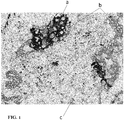
- FIG. 1 shows staining of a NSCLC tumor sample.
- PD-L1 staining is indicated by (a), and would show as brown in a color image.
- Cytokeratins from pan keratin antibody are indicated by (b), and would be red in a color image.
- CD4 of immune cells are indicated by (c) and would be green/blue in a color image.
- Counterstain is diluted hematoxylin.
- the present disclosure relates generally to histochemical or cytochemical methods of labeling tumor samples to facilitate scoring of PD-L1 expression in tumor cells, immune cells, or both.
- cells are labeled with binding entities specific for PD-L1 and: (1) at least one tumor cell marker; (2) at least one immune cell marker; or (3) at least one tumor cell marker and at least one immune cell marker.
- the binding entities are then visualized in the tumor samples by generating at least three distinct detectable signals: a first detectable signal that correlates with the location of PD-L1 binding entity; a second detectable signal that correlates with the location of the tumor cell-specific binding entity; and a third detectable signal that correlates with the location of the immune cell marker.
- Each of the detectable signals are distinguishable from one another.
- a counterstain may be provided in a fourth detectable signal and/or a fifth detectable signal may be generated from co-localization of any two of the first, second, and third detectable signals.
- the present methods are compatible with tumor samples suitable for histochemical or cytochemical analysis, including, for example, fresh frozen, formalin-fixed paraffin-embedded (FFPE)) samples, cytological smears (such as cervical smears), isolates of circulating tumor cells, etc.
- FFPE formalin-fixed paraffin-embedded
- the sample is a FFPE sample of tumor tissue.
- tumor cell-specific biomarkers may include but are not limited to: cytokeratins detectable with the pan keratin antibody (e.g., basic cytokeratins, many of the acidic cytokeratins), other cytokeratins such as cytokeratin 7 (CK7) and cytokeratin 20 (CK20), chromogranin, synaptophysin, CD56, thyroid transcription factor-1 (TTF-1), p53, leukocyte common antigen (LCA), vimentin, smooth muscle actin, or the like (e.g., see Capelozzi, V., J Bras Pneumol. 2009;35(4):375-382 ).
- cytokeratins detectable with the pan keratin antibody e.g., basic cytokeratins, many of the acidic cytokeratins
- other cytokeratins such as cytokeratin 7 (CK7) and cytokeratin 20 (CK20)
- chromogranin synaptophy
- immune cell-specific biomarkers may include but are not limited to: CD3, CD4, CD8, CD19, CD20, CD11c, CD123, CD56, CD14, CD33, or CD66b.
- a lymphocyte-specific marker is used.
- a T-cell specific marker such as CD3, CD4, or CD8, or a B-cell specific marker, such as CD19 or CD20 may be used.
- the immune cell marker is CD4 and the tumor cell marker is a cytokeratin detectable by a pan cytokeratin antibody.
- Histochemistry and cytochemistry are techniques often used to identify biomarkers within the context of intact cells by labeling the samples with molecules that bind specifically to the biomarker in a manner that can be visualized on a microscope.
- Immunohistochemistry (IHC) and immunocytochemistry (ICC) are types of histochemistry and cytochemistry that use antibodies to label the biomarkers.
- In situ hybridization (ISH) is a type of histochemistry or cytochemistry that uses nucleic acid probes to label specific nucleotide sequences in the tissue or cell sample.
- biomarker By identifying the biomarker in the context of a tissue environment or cellular environment, spatial relationships between the biomarkers and other morphological or molecular features of the cell or tissue sample can be elucidated, which may reveal information that is not apparent from other molecular or cellular techniques.
- binding entity shall refer to any compound or composition that is capable of specifically binding to a specific molecular structure in a tumor sample suitable for histochemical or cytochemical analysis.
- examples include antibodies and antigen binding fragments thereof, as well as engineered specific binding structures, including ADNECTINs (scaffold based on 10 th FN3 fibronectin; Bristol-Myers-Squibb Co.), AFFIBODYs (scaffold based on Z domain of protein A from S.
- the binding entities are antibodies or antigen-binding fragments thereof.
- antibody refers to any form of antibody that exhibits the desired biological or binding activity. Thus, it is used in the broadest sense and specifically covers, but is not limited to, monoclonal antibodies (including full length monoclonal antibodies), polyclonal antibodies, multispecific antibodies (e.g., bispecific antibodies), humanized antibodies, fully human antibodies, chimeric antibodies and camelized single domain antibodies.
- antibody fragment or “antigen binding fragment” refers to antigen binding fragments of antibodies, i.e. antibody fragments that retain the ability to bind specifically to the antigen bound by the full-length antibody, e.g. fragments that retain one or more CDR regions.
- antibody binding fragments include, but are not limited to, Fab, Fab', F(ab')2, and Fv fragments; diabodies; linear antibodies; single-chain antibody molecules, e.g., sc-Fv; nanobodies and multispecific antibodies formed from antibody fragments.
- Exemplary anti-PD-Ll antibodies include SP263 (fully described in U.S. Provisional Patent Application Serial Number 62/004572 , Docket Number 32151 US, and filed May 29, 2014), SP142 (Cat. # M4420, Spring Biosciences, Inc., Pleasanton, CA), and PD-L1 (E1L3N®) XP® Rabbit mAb (Cat. # #13684; Cell Signaling Technologies, Inc., Danvers, MA).
- the PD-L1 binding entity is SP263 or SP142
- the tumor cell-specific binding entity is a pan keratin antibody
- the immune cell specific binding entity is an anti-CD4 antibody.
- the PD-L1 binding entity is SP142
- the tumor cell-specific binding entity is a pan keratin antibody
- the immune cell specific binding entity is an anti-CD4 antibody.
- the assays of the present invention feature staining of PD-L1 as well as staining of one or more of tumor and immune cell markers to generate a detectable signal that correlates with the location at which an exogenous binding entity has bound to the sample.
- Histochemical and cytochemical methods of generating detectable signals from exogenous binding entities in samples are well known to one of ordinary skill in the art and typically involve application of one or more labels.
- Exemplary labels include chromogenic labels, fluorescent labels, luminescent labels, radiometric labels, etc., are used for recognition of the markers or targets (e.g., PD-L1, tumor cell-specific marker, immune cell-specific marker, etc.). Labels are well known to one of ordinary skill in the art and are not limited to the labels described herein.
- the detectable signals are generated through use of chromogens.
- the label is applied through the use of a secondary antibody.
- the binding entity may be a primary antibody specific for the PD-L1, immune cell marker, or tumor cell marker. If antibodies derived from different species of animal are used as the primary antibody, a secondary antibody specific for that species of antibody can be used to apply the label.
- the primary antibody can be modified to contain a separate moiety that can be bound by a specific binding entity.
- a primary antibody is a haptenized antibody (i.e., and antibody modified to contain a specific hapten). Many different haptens are known, so each of the primary antibodies can be modified to contain a different hapten, and different anti-hapten antibodies may be used to specifically label the different primary antibodies.
- the detectable signal may be amplified.
- a signal is "amplified" when more label is deposited per primary antibody than by using a standard primary-secondary antibody arrangement.
- One commonly used method of amplification is tyramide signal amplification, which is described in Bobrow, M. N., Harris, T. D., Shaughnessy, K. J., and Litt, G. J. (1989) J. Immunol. Methods 125, 279-285 .
- a modified form of tyramide signal amplification as described in WO 2013148498 is used.
- the present invention features multiplex assays for improved scoring of tumor tissues stained with PD-L1.
- the assays feature steps for staining PD-L1 in a first color (PD-L1 is shown in brown in FIG. 1 ), as well as steps for staining a tumor cell-specific marker and/or immune cell-specific marker with a second and/or third differentiating color.
- FIG. 1 For example, FIG. 1
- the assays of the present invention help to differentiate between the PD-L1 positive tumor cells and the PD-L1 positive immune cells. This may improve the ability of samples to be scored (manual/ visual, machine/image analysis) more quickly, accurately, and with a greater degree of reproducibility as compared to scoring samples stained with PD-L1 alone.
- Table 1 illustrates the use of differentiating markers/colors for differentiating between the PD-L1 positive tumor cells and the PD-L1 positive immune cells.
- PD-L1 can be detected using the anti-PD-Ll antibody, and the PD-L1 is visible as a first color (e.g., brown in the example shown in FIG. 1 ).
- Tumor cells with PD-L1 will show the first color (PD-L1), as indicated by the "+” sign in Column 4 of Table 1 (note that the tumor cells without PD-L1 do not have the first color, as indicated by the "-" sign in Column 4 of Table 1).
- Immune cells also exhibit PD-L1 expression and can be shown as the first color (PD-L1), as indicated by the "+" sign in Column 1 of Table 1.
- PD-L1 the first color
- the sample is stained for a tumor-specific marker (e.g., cytokeratins detected by the pan keratin antibody), which is visible as a second color (a color different from the first color).
- a tumor-specific marker e.g., cytokeratins detected by the pan keratin antibody
- the sample may also be stained for an immune cell-specific marker (e.g., CD4 or other marker), which is visible as a third color (a color different from the first and second colors).
- Cells that have the third color but not the first color and second color are PD-L1 negative immune cells (Column 2).
- the present invention is not limited to staining in any particular order.
- Example 1 describes staining first for PD-L1, then staining for the tumor cell-specific marker, then staining for the immune cell-specific marker, and finally using a counterstain.
- the order of the staining is different.
- the immune cell-specific marker is stained before the tumor cell-specific marker is stained, etc.
- the assays of the present invention feature staining of PD-L1 as well as staining of one or more differentiating markers.
- Methods of staining may include immunohistochemisty (IHC). Staining techniques may be performed on various biological samples, such as tissue (e.g., fresh frozen, formalin-fixed paraffin-embedded (FFPE)) and cytological samples. Chromogenic labels are used for recognition of the markers or targets (e.g., PD-L1, tumor cell-specific marker, immune cell-specific marker, etc.). Labels are well known to one of ordinary skill in the art and are not limited to the labels described herein.
- Example 1 A non-limiting example of a detailed protocol is described in Example 1 below. Briefly, samples of interest are stained for PD-L1. The sample is incubated first with an anti-PD-Ll primary antibody. The anti-PD-Ll primary antibody is detected with a first color. In Example 1, the sample is incubated with a horseradish peroxidase (HRP)-conjugated secondary antibody against the primary anti-PD-Ll antibody and a substrate (3,3'-diaminobenzidine (DAB)) is added, producing the first color (e.g., brown). Alternative enzymes and substrates (and resulting colors are described below).
- HRP horseradish peroxidase
- DAB diaminobenzidine
- tumor cell-specific biomarkers may include but are not limited to: cytokeratins detectable with the pan keratin antibody (e.g., basic cytokeratins, many of the acidic cytokeratins), other cytokeratins such as cytokeratin 7 (CK7) and cytokeratin 20 (CK20), chromogranin, synaptophysin, CD56, thyroid transcription factor-1 (TTF-1), p53, leukocyte common antigen (LCA), vimentin, smooth muscle actin, or the like (e.g., see Capelozzi, V., J Bras Pneumol.
- cytokeratins detectable with the pan keratin antibody e.g., basic cytokeratins, many of the acidic cytokeratins
- other cytokeratins such as cytokeratin 7 (CK7) and cytokeratin 20 (CK20)
- CK7 cytokeratin 7
- CK20 cytokeratin
- Example 1 One of ordinary skill in the art can substitute another appropriate tumor cell-specific biomarker for the cytokeratins as described in Example 1.
- the sample is incubated first with a primary antibody (e.g., anti-pan keratin antibody) against the first differentiating marker (tumor cell-specific marker).
- the anti-differentiating marker primary antibody is detected with a second color.
- the sample is incubated with a haptenized antibody against the anti-differentiating marker primary antibody, and then the sample is incubated with an alkaline phosphatase (AP)-conjugated anti-hapten antibody.
- the substrate Fast Red Chromogen produces the second color (e.g., red).
- the sample may then be stained for an immune cell-specific marker.
- immune cell-specific biomarkers include CD4 or any other CD marker.
- Immune cell-specific biomarkers are well known to one of ordinary skill in the art.
- One of ordinary skill in the art could substitute another appropriate immune cell-specific biomarker for CD4 as described in Example 1.
- the sample is incubated first with a primary antibody (e.g., anti-CD4 antibody) against the second differentiating marker (immune cell-specific marker).
- the anti-differentiating marker primary antibody is detected with a third color.
- the sample is incubated with a HRP-conjugated secondary antibody against the primary anti-differentiating marker antibody and the substrate HRP-Green Chromogen is added, producing the third color (e.g., green/blue).
- the samples are then counterstained, producing a fourth color (the fourth color being different from the first, second, and third colors).
- the counterstain comprises hematoxyline; however, the counterstain is not limited to hematoxyline.
- Alternative counterstains are well known to one of ordinary skill in the art.
- the counterstain may comprise methylene blue, nuclear red, toluidine blue, eosin, methyl green, or the like. The particular counterstain is generally selected to produce contrast so as to enhance visibility.
- the samples are then interpreted and scored (scoring is described below).
- the results of the staining may be interpreted as described in Table 1.
- a tumor cell-specific marker e.g., cytokeratins
- an immune cell-specific marker e.g., cytokeratins
- the cells that score for the first color and third color but not the second color are PD-L1 positive immune cells
- cells that score for both the first color and the second color (but not the third color) are PD-L1 positive cancer cells
- cells that score for the second color but not the first color nor the third color are PD-L1 negative cancer cells.
- the first color and second color overlap (or others overlap), producing a fifth (different) color. This overlap color may help scoring.
- the staining (e.g., PD-L1 and the differentiating marker) occurs sequentially. In some embodiments, the staining occurs simultaneously.
- Example 1 describes staining first for PD-L1, then staining for the tumor cell-specific marker, then staining for the immune cell-specific marker, and finally using a counterstain.
- the tumor cell-specific marker or immune cell-specific marker
- the tumor cell-specific marker is stained first, followed by the PD-L1 staining, etc.
- the present invention is not limited to the signaling conjugates (e.g., enzymes and chromogenic substrates) used in Example 1 nor to the other signaling conjugates described herein.
- Alternative enzyme-chromogenic substrate pairs for detection methods e.g., immunohistochemistry, etc. are well known to one of ordinary skill in the art.
- Chromogenic substrates precipitate when activated by the appropriate enzyme. That is, the traditional chromogenic substance is converted from a soluble reagent into an insoluble, colored precipitate upon contacting the enzyme.
- Chromogenic substrates used for the present invention may be compatible with automated slide staining instruments and processes and/or automated detection and analysis instruments and software. This may enable high detection sensitivity and multiplexing capability.
- the enzyme of the secondary antibody comprises HRP, alkaline phosphatase (AP), glucose oxidase, beta-galactosidase, the like, and/or others described in WO Patent Application No. 20131484498 .
- the substrate comprises DAB, Fast Red and Fast Blue, Fast Red and Black (silver), nitro blue tetrazolium chloride (NBT), 5-bromo-4-chloro-3-indolyl phosphate (BCIP), x-gal, 3-amino-9-ethylcarbazole (AEC), 5-bromo-4-chloro-3-indoyl-beta-D-galactopyranoside (BCIG), p-nitrophenyl phosphate (PNPP), 2,2'-azinobis [3-ethylbenzothiazoline-6-sulfonic acid] (ABTS), 3,3',5,5'-tetramethylbenzidine (TMB), HRP-Green Chromogen, the
- the present invention is also not limited to any particular colors or color combinations.
- the first color (PD-L1) is brown; however, in some examples, the first color (PD-L1) may be any other appropriate color, e.g., red, blue, yellow, green, etc., depending on the enzyme-substrate combination.
- the second color e.g., cytokeratins, other markers
- the second color may be red/pink; however, in some examples, the second color (e.g., cytokeratins, other markers) may be any other appropriate color, e.g., brown, blue, yellow, green, etc., depending on the enzyme-substrate combination.
- the third color (e.g., CD4, other markers) may be green/blue; however, in some examples, the third color (e.g., CD4, other markers) may be any other appropriate color, e.g., brown, red/pink, yellow, etc., depending on the enzyme-substrate combination.
- the fourth color (counterstain) may be blue; however, the fourth color (counterstain) may be any other appropriate color, e.g., red, green, etc.
- the fifth color overlap of the two colors
- the second color (and third color) directs scoring of PD-L1 in either the tumor cells or the immune cells.
- the use of the third color may improve scoring.
- the use of the third color may help to clarify which cell type (tumor vs. immune) is PD-L1 positive. This can help the accuracy of the calculation of the number of PD-L1 positive immune cells, PD-L1 negative tumor cells, and PD-L1 positive tumor cells.
- a positive result (e.g., a "PD-L1 positive result) may be calculated in a variety of ways and is not limited to the examples described herein.
- the number of PD-L1 positive tumor cells or the area of PD-L1 that is associated with tumor cells may be calculated and then factored into an equation to calculate and H score. For example, if the H score is above a threshold for PD-L1 positivity then the sample is PD-L1 positive, and if the H score is below the threshold for PD-L1 positivity then the sample is PD-L1 negative.
- a positive result is determined by calculating the percentage of PD-L1 positive tumor cells and determining if that percentage is above the threshold for positivity or a predetermined cut-off.
- the minimum percentage of PD-L1 cells that confers PD-L1 positivity e.g., 5% or more PD-L1 positive tumor cells confers PD-L1 positivity, 10% or more PD-L1 positive tumor cells confers PD-L1 positivity, 25% or more PD-L1 positive tumor cells confers PD-L1 positivity, 50% or more PD-L1 positive tumor cells confers PD-L1 positivity, etc.
- a positive result is determined by calculating the number of PD-L1 positive tumor cells divided by the total number of cells (e.g., number of tumor plus immune cells) and determining if that value is above the threshold for positivity (e.g., value greater than 0.15, value greater than 0.25, value greater than 0.5, etc.).
- a positive result is determined by calculating the sum of the percentage of PD-L1 positive tumor cells and PD-L1 positive immune cells and determining if that value is above the threshold for positivity (e.g., value greater than 40, value greater than 50, value greater than 60, etc.).
- a positive result is determined by calculating the number of PD-L1 positive tumor cells divided by the number of PD-L1 negative tumor cells and determining if that value is above the threshold for positivity (e.g., value greater than 1.5, value greater than 1.8, value greater than 2, etc.).
- a positive result is determined by calculating the number of PD-L1 positive tumor cells divided by the sum of the number of PD-L1 negative tumor cells and the number of PD-L1 negative immune cells and determining if that value is above the threshold for positivity (e.g., greater than 1.1, greater than 1.3, etc.).
- a positive result is determined by calculating the number of PD-L1 positive tumor cells divided by the sum of the number of PD-L1 negative tumor cells and the number of PD-L1 positive immune cells and determining if that value is above the threshold for positivity (e.g., greater than 1.1, greater than 1.3, etc.).
- a positive result is determined by calculating the number of PD-L1 positive tumor cells divided by the number of PD-L1 positive immune cells and determining if that value is above the threshold for positivity. In some embodiments, a positive result is determined by calculating the number of PD-L1 positive tumor cells divided by the number of PD-L1 negative immune cells and determining if that value is above the threshold for positivity.
- PD-L1 positivity may be determined by calculating the percentage of PD-L1 positive tumor cells. For example, staining in greater than about 1% of cells (e.g., tumor cells) is scored as PD-L1 positive. In another example, staining in greater than about 5% of cells (e.g., tumor cells) is scored as PD-L1 positive. In some embodiments, staining in greater than about 10% of cells (e.g., tumor cells) is scored as PD-L1 positive. In some examples, staining in greater than about 15% of cells (e.g., tumor cells) is scored as PD-L1 positive.
- staining in greater than about 20% of cells is scored as PD-L1 positive. In some examples, staining in greater than about 25% of cells (e.g., tumor cells) is scored as PD-L1 positive. In some examples, staining in greater than about 30% of cells (e.g., tumor cells) is scored as PD-L1 positive. In some examples, staining in greater than about 35% of cells (e.g., tumor cells) is scored as PD-L1 positive. In some examples, staining in greater than about 40% of cells (e.g., tumor cells) is scored as PD-L1 positive.
- staining in greater than about 45% of cells is scored as PD-L1 positive. In some embodiments, staining in greater than about 50% of cells (e.g., tumor cells) is scored as PD-L1 positive. In some examples, staining in greater than about 55% of cells (e.g., tumor cells) is scored as PD-L1 positive. In some examples, staining in greater than about 60% of cells (e.g., tumor cells) is scored as PD-L1 positive. In some examples, staining in greater than about 65% of cells (e.g., tumor cells) is scored as PD-L1 positive.
- staining in greater than about 70% of cells is scored as PD-L1 positive. In some examples, staining in greater than about 75% of cells (e.g., tumor cells) is scored as PD-L1 positive. In some examples, staining in greater than about 80% of cells (e.g., tumor cells) is scored as PD-L1 positive. In some examples, staining in greater than about 90% of cells (e.g., tumor cells) is scored as PD-L1 positive.
- the number or percentage of PD-L1 positive immune cells may be relevant to the determination of PD-L1 positivity. For example, staining in greater than about 5% of immune cells is associated with scoring as PD-L1 positive. In some embodiments, staining in greater than about 10% of immune cells is associated with scoring as PD-L1 positive. In some examples, staining in greater than about 15% of immune cells is associated with scoring as PD-L1 positive. In some examples, staining in greater than about 25% of immune cells is associated with scoring as PD-L1 positive. In some embodiments, staining in greater than about 50% of immune cells is associated with scoring as PD-L1 positive.
- the positivity of the sample may also be determined by the degree or intensity of staining (e.g., heavy staining may be positive and light staining may be negative).
- scoring methods may feature scoring samples on an intensity scale, e.g., of 0 to 3, for PD-L1 expression (see for example, U.S. Provisional Patent Application No. 61/875,334 (Scoring Method For Mesothelin Protein Expression).
- samples are scored based on intensity and percentages of cells staining. For example, as described in U.S. Provisional Patent Application No.
- Example 1 describes a non-limiting example of a multiplex IHC assay of the present invention.
- a NSCLC sample slide is prepared according to standard protocols.
- methods of detecting a target in a biological sample include contacting the biological sample with a detection probe, contacting the biological sample with a labeling conjugate, and contacting the biological sample with a signaling conjugate.
- the labeling conjugate includes an enzyme.
- the signaling conjugate includes a latent reactive moiety and a chromogenic moiety.
- the enzyme catalyzes conversion of the latent reactive moiety into a reactive moiety, which covalently binds to the biological sample proximally to or directly on the target.
- the method further includes illuminating the biological sample with light and detecting the target through absorbance of the light by the chromogenic moiety of the signaling conjugate.
- the reactive moiety reacts with a tyrosine residue of the biological sample, the enzyme conjugate, the detection probe, or combinations thereof.
- the detection probe is an antibody probe.
- the labeling conjugate includes an antibody coupled to the enzyme. Enzymes may include oxidoreductases, peroxidases, or hydrolases. An antibody for the labeling conjugate may be an anti-species or an anti-hapten antibody.
- the detection probe may include a hapten selected from the group consisting an oxazole hapten, pyrazole hapten, thiazole hapten, nitroaryl hapten, benzofuran hapten, triterpene hapten, urea hapten, thiourea hapten, rotenoid hapten, coumarin hapten, cyclolignan hapten, di-nitrophenyl hapten, biotin hapten, digoxigenin hapten, fluorescein hapten, and rhodamine hapten.
- the detection probe is monoclonal antibody derived from a second species such as goat, rabbit, mouse, or the like.
- the labeling conjugate is configured, through its inclusion of an anti-species or an anti-hapten antibody to bind selectively to the detection probe.
- Chromogen conjugates used for the present invention may be configured to absorb light more selectively than traditionally available chromogens. Detection is realized by absorbance of the light by the signaling conjugate; for example, absorbance of at least about 5% of incident light would facilitate detection of the target. In other darker stains, at least about 20% of incident light would be absorbed. Non-uniform absorbance of light within the visible spectra results in the chromophore moiety appearing colored.
- the signaling conjugates disclosed herein may appear colored due to their absorbance; the signaling conjugates may appear to provide any color when used in the assay, with certain particular colors including red, orange, yellow, green, indigo, or violet depending on the spectral absorbance associated with the chomophore moiety.
- the chromophore moieties may have narrower spectral absorbances than those absorbances of traditionally used chromogens (e.g. DAB, Fast Red, Fast Blue).
- the spectral absorbance associated with the first chromophore moiety of the first signaling conjugate has a full-width half-max (FWHM) of between about 30 nm and about 250 nm, between about 30 nm and about 150 nm, between about 30 nm and about 100 nm, or between about 20 nm and about 60 nm.
- FWHM full-width half-max
- Narrow spectral absorbances enable the signaling conjugate chromophore moiety to be analyzed differently than traditional chromogens. While having enhanced features compared to traditionally chromogens, detecting the signaling conjugates remains simple. In illustrative embodiments, detecting comprises using a bright-field microscope or an equivalent digital scanner. The narrow spectral absorbances enable chromogenic multi-plexing at level beyond the capability of traditional chromogens. For example, traditional chromogens are somewhat routinely duplexed (e.g. Fast Red and Fast Blue, Fast Red and Black (silver), Fast Red and DAB).
- traditional chromogens are somewhat routinely duplexed (e.g. Fast Red and Fast Blue, Fast Red and Black (silver), Fast Red and DAB).
- the method includes detecting from two to at least about six different targets using different signaling conjugates or combinations thereof.
- illuminating the biological sample with light comprises illuminating the biological sample with a spectrally narrow light source, the spectrally narrow light source having a spectral emission with a second full-width half-max (FWHM) of between about 30 nm and about 250 nm, between about 30 nm and about 150 nm, between about 30 nm and about 100 nm, or between about 20 nm and about 60 nm.
- illuminating the biological sample with light includes illuminating the biological sample with an LED light source.
- illuminating the biological sample with light includes illuminating the biological sample with a filtered light source.
- detecting targets within the sample includes contacting the biological sample with a first amplifying conjugate that is covalently deposited proximally to or directly on the first labeling conjugate.
- the first amplifying conjugate may be followed by contacting the biological sample with a secondary labeling conjugate.
- the amplification of signal using amplifying conjugates enhances the deposition of signaling conjugate.
- the enhanced deposition of signaling conjugate enables easier visual identification of the chromogenic signal, that is, the amplification makes the color darker and easier to see. For low expressing targets, this amplification may result in the signal becoming sufficiently dark to be visible, whereas without amplification, the target would not be apparent.
- the signaling conjugate is covalently deposited proximally to the target at a concentration of greater than about 1x10 11 molecules per cm 2 * ⁇ m to about 1x10 16 molecules per cm 2 * ⁇ m of the biological sample.
- the first target and the second target are genetic nucleic acids. Detecting the first target through absorbance of the light by the first signaling conjugate includes detecting a first colored signal selected from red, orange, yellow, green, indigo, or violet, the first colored signal associated with spectral absorbance associated with the first chromogenic moiety of the first signaling conjugate.
- Detecting the second target through absorbance of the light by the second signaling conjugate includes detecting a second colored signal selected from red, orange, yellow, green, indigo, or violet, the second colored signal associated with spectral absorbance associated with the second chromogenic moiety of the second signaling conjugate. Detecting an overlap in proximity through absorbance of the light by the first signaling conjugate overlapping in proximity with the second signaling conjugate so that a third colored signal associated with overlapping spectral absorbance of the first spectral absorbance and the second spectral absorbance. According to one example, this third color signals a normal genetic arrangement and the first and second colors signal a genetic rearrangement or translocation.
- PD ⁇ L 1 Value Percentage of PD ⁇ L 1 positive tumor cells
- Threshold for positivity PD-L1 Value > 40% is PD-L1 positive
- a pathologist views Sample 3A and calculates the percentage of PD-L1 positive tumor cells as being 48%. Based on the threshold for positivity, Sample 3A is labeled PD-L1 positive.
- PD ⁇ L 1 Value # of PD ⁇ L 1 positive tumor cells / total # of cells
- Threshold for positivity PD-L1 Value > 0.25 is PD-L1 positive
- a pathologist views Sample 3B.
- the number of PD-L1 positive tumor cells is 68, and the total number of cells is 460.
- Sample 3B is labeled PD-L1 negative.
- PD ⁇ L 1 Value # of PD ⁇ L 1 positive tumor cells + % PD ⁇ L 1 positive immune cells
- Threshold for positivity PD-L1 Value > 60 is PD-L1 positive
- a pathologist views Sample 3C.
- the percent of PD-L1 positive tumor cells is 50, and the percent of PD-L1 positive immune cells is 20.
- Sample 3C is labeled PD-L1 positive.
- PD ⁇ L 1 Value # of PD ⁇ L 1 positive tumor cells / ( # of PD ⁇ L 1 negative tumor cells + # of PD - L 1 positive immune cells )
- Threshold for positivity PD-L1 Value > 0.8 is PD-L1 positive
- a pathologist views Sample 3D.
- the number of PD-L1 positive tumor cells is 68
- the number of PD-L1 negative tumor cells is 45
- the number of PD-L1 positive immune cells is 210.
- H score 1 * ( percentage of tumor cells staining at 1 + intensity ) + 2 * percentage of tumor cells staining at 2 + intensity + 3 * percentage of cells staining at 3 + intensity
- Threshold for positivity H score > 125 is PD-L1 positive
- a pathologist views Sample 3E.
- the percentage of PD-L1 positive tumor cells staining at 1+ intensity is 5%
- the percentage of PD-L1 positive tumor cells staining at 2+ intensity is 35%
- the percentage of PD-L1 positive tumor cells staining at 3+ intensity is 20%.
- an "antibody” used in accordance with the present invention may be a whole antibody or a fragment of an antibody that is effective in binding to a desired target site.
- an "antibody” of the present invention may be substituted with a targeting moiety (e.g., ligand peptide, small molecule, etc.).
- a targeting moiety e.g., ligand peptide, small molecule, etc.
Landscapes
- Health & Medical Sciences (AREA)
- Life Sciences & Earth Sciences (AREA)
- Engineering & Computer Science (AREA)
- Immunology (AREA)
- Molecular Biology (AREA)
- Biomedical Technology (AREA)
- Chemical & Material Sciences (AREA)
- Hematology (AREA)
- Urology & Nephrology (AREA)
- Analytical Chemistry (AREA)
- Physics & Mathematics (AREA)
- Biochemistry (AREA)
- General Health & Medical Sciences (AREA)
- General Physics & Mathematics (AREA)
- Pathology (AREA)
- Cell Biology (AREA)
- Microbiology (AREA)
- Food Science & Technology (AREA)
- Medicinal Chemistry (AREA)
- Biotechnology (AREA)
- Investigating Or Analysing Biological Materials (AREA)
Description
- The present disclosure relates to methods for histochemically scoring PD-L1 expression in tumor tissues.
- Programmed death 1 (PD-1) is a member of the CD28 family of receptors, which includes CD28, CTLA-4, ICOS, PD-1, and BTLA. Two cell surface glycoprotein ligands for PD-1 have been identified, PD-L1 and PD-L2, and have been shown to downregulate T cell activation and cytokine secretion upon binding to PD-1 (Freeman et al., J Exp Med 192:1027-34 (2000); Latchman et al., Nat Immunol 2:261-8 (2001); Carter et al., Eur J Immunol 32:634-43 (2002); Ohigashi et al., Clin Cancer Res 11:2947-53 (2005)). Both PD-L1 (B7-H1) and PD-L2 (B7-DC) are B7 homologs that bind to PD-1, but do not bind to other CD28 family members.
- The PD-L1-PD1 pathway is involved in the negative regulation of some immune responses and may play an important role in the regulation of peripheral tolerance. Interaction of PD-L1 with PD1 results in inhibition of TCR-mediated proliferation and cytokine production. PD-L1 has been suggested to play a role in tumor immunity by increasing apoptosis of antigen-specific T-cell clones (Dong et al. Nat Med 8:793-800 (2002)). Indeed, PD-L1 expression has been found in several murine and human cancers, including human lung, ovarian and colon carcinoma and various myelomas (Iwai et al. PNAS 99:12293-7 (2002); Ohigashi et al. Clin Cancer Res 11:2947-53 (2005)). Thus, measuring the amount of PD-L1 protein in biological samples may aid in the early detection of cancer pathologies and may help assess the efficacy and durability of investigational drugs that inhibit the binding of the PD-L1 protein.
- Ghebeh et al. (Neoplasia. 2006 Mar;8(3):190-8) disclose chromogenic immunohistochemistry (IHC) assays wherein snap-frozen, acetone-fixed breast cancer tissue samples are double-stained in separate assays for B7-H1 (PD-L1) and one of FoxP3, CD3, CD 4, CD 8 and pan-Cytokeratin.
- Ghebeh et al. (BMC Cancer. 2008 Feb 23;8:57) are also concerned with chromogenic IHC double-staining assays using, amongst others, anti-B7-H1 (PD-L1) and anti-FoxP3 antibodies, and anti-B7-H1 and anti-CD8 antibodies.
- Afanasiev et al. (Clin Cancer Res. 2013 Oct 1;19(19):5351-60) disclose the analysis of PD-L1 protein expression in Merkel cell carcinoma (MCC) samples. Formalin-fixed paraffin-embedded MCC samples are analyzed in a chromogenic single-marker IHC assay using an anti-PD-Ll antibody.
- Powderly et al. (http://carolinabiooncology.org/wp-content/uploads/ASCO-2013-Presentation-Anti-PD-L-1Biomarkers.pdf) also present PD-L1 expression data that were obtained in chromogenic single-marker IHC assays.
- Iwai et al. (Int Immunol. 2005 Feb;17(2):133-44) disclose data from fluorescence IHC assays with cryosections of liver tissue that are double-stained for PD-L1 and CD11b. Also disclosed are fluorescence assays that show PD-L1 staining and, in addition, counterstained nuclei.
- PerkinElmer (https:/ /www.perkinelmer.comllab-solutions/resources/docs/ FLY_Opal-Multiplex-Staining.pdf) is concerned with TSA-based fluorescence multiplex assays and presents one photomicrograph of a breast cancer tissue microarray with different fluorescence signals for HER2, ER, PR, Ki-67 and nuclei, and another photomicrograph of a breast cancer tissue section showing different fluorescence signals for CD8, CD4, CD20, cytokeratin and nuclei.
- Lyford-Pike et al. (Cancer Res. 2013 Mar 15;73(6)) disclose the analysis of PD-L1 and CD4 protein expression in formalin-fixed paraffin-embedded tissue samples from human papillomavirus-associated head and neck squamous cell carcinomas (HPV-HNSCC). In particular, the samples are analyzed in separate chromogenic single-marker IHC assays using an anti-PD-Ll antibody or antibodies against various immune cell-specific markers such as CD3, CD4, CD68 or CD1a.
- Lloyd et al. (www.poster-submission.com/cdrom/download_poster/ 37/27814/1062P) discuss multispectral imaging and linear unmixing of signals in a multiplex chromogenic IHC assay with a tissue microarray stained for CD3, FoxP3 and CD69. No staining of PD-L1 is shown. Instead, the marker is mentioned as an example for fluorescence IHC.
- However, the use of PD-L1 protein expression as an accurate predictor for cancer and/or the efficacy of anti-PD-1 and anti-PD-Ll directed therapies remains challenging. For example, many tumor samples show PD-L1 staining in both tumor cells and immune cells. Differentiation of these two cell types may be difficult for pathologists, especially when both are present in the same sample.
- The present invention features multiplex assays for improved scoring of tumor tissues stained with PD-L1. The assays feature PD-L1 staining in a first color plus staining of a differentiating marker specific for tumor cells or immune cells, in a second color, and optionally, a second differentiating marker specific for tumor cells or immune cells, in a third color. The assays of the present invention help to differentiate between the PD-L1 positive tumor cells and the PD-L1 positive immune cells. This may improve the ability of samples to be scored more quickly, accurately, and with a greater degree of reproducibility as compared to scoring samples stained with PD-L1 alone.
In one aspect of the present disclosure is a method of scoring PD-L1 expression in a tumor sample, the method comprising labeling the tumor tissue sample, the labeling comprising contacting the tissue sample with an anti-PD-Ll primary antibody, and contacting the same tissue sample with a primary antibody directed to a tumor cell-specific marker and an antibody directed to an immune cell-specific marker, and visualizing each of the antibodies in the tissue sample with a reagent that generates a detectable signal corresponding to each of the primary antibodies, wherein the anti-PD-Ll antibody has a first detectable signal, the antibody directed to the tumor cell-specific marker has a second detectable signal distinguishable from the first detectable signal, and the antibody directed to an immune cell-specific marker has a third detectable signal distinguishable from the first detectable signal and the second detectable signal, wherein the first, second, and third detectable signals are generated by chromogens; and the method of scoring PD-L1 expression in a tumor sample further comprising scoring PD-L1 expression in tumor cells, immune cells, or both, wherein co-localization of the first and second detectable signals indicates the presence of PD-L1-positive tumor cells and co-localization of the first and third detectable signals indicates the presence of PD-L1-positive immune cells. In some embodiments, the tumor cell-specific marker is selected from the group consisting of a cytokeratin, chromogranin, synaptophysin, CD56, thyroid transcription factor-1 (TTF-1), p53, leukocyte common antigen (LCA), vimentin, and smooth muscle actin, and the immune cell-specific marker is selected from the group consisting of CD3, CD4, CD8, CD19, CD20, CD11c, CD123, CD56, CD14, CD33, or CD66b. In some embodiments, the immune cell-specific marker is a T-cell marker or a B-cell marker. In some embodiments, the antibody directed to the tumor cell-specific marker is a pan-keratin antibody and the antibody directed to the immune cell-specific marker is an anti-CD4 antibody. In some embodiments, the first detectable signal is generated by contacting the tissue sample with a horseradish peroxidase (HRP)-conjugated secondary antibody that recognizes the anti-PD-Ll primary antibody, reacting the HRP with 3,3'-Diaminobenzidine (DAB) to produce a brown color; the second detectable signal is generated by contacting the sample with an alkaline phosphatase (AP) labeled antibody that recognizes the primary antibody directed to a tumor cell-specific marker, reacting the AP with a Fast Red chromogen and naphthol to produce a red color; and the third detectable signal is generated by contacting the sample with a HRP-conjugated secondary antibody that recognizes the primary antibody directed to an immune cell-specific marker, and reacting the HRP with HRP-green chromogen to produce a green color. In some embodiments, the first, second, and/or third detectable signal is an amplified signal, optionally wherein the amplified signal is generated by tyramide signal amplification. In some embodiments, the sample is contacted simultaneously with the primary antibodies or the sample is contacted sequentially with the primary antibodies. In some embodiments, the method further comprises counterstaining the tissue sample, the counterstain producing a fourth detectable signal that is distinguishable from the first, second, and the third detectable signals, optionally the counterstain comprises hematoxylin. In some embodiments, a fifth detectable signal is produced by overlap of the first detectable signal and the second detectable signal. In some embodiments, a sixth detectable signal is produced by overlap of the first detectable signal and the third detectable signal. In some embodiments, the total number of PD-L1 positive and PD-L1-negative tumor cells is quantitated, and the tumor is scored as PD-L1 positive if staining for PD-L1 is detected in greater than about 10% of tumor cells, or the tumor is scored as PD-L1 positive if staining for PD-L1 is detected in greater than about 50% of tumor cells. In some embodiments, the total number of PD-L1 positive and PD-L1-negative immune cells is quantitated, and the tumor is scored as PD-L1 positive if staining for PD-L1 is detected in greater than about 10% of immune cells, or the tumor is scored as PD-L1 positive if staining for PD-L1 is detected in greater than about 50% of immune cells. In some embodiments, PD-L1-positive immune cells, PD-L1 positive tumor cells, and PD-L1 negative tumor cells are quantitated to generate a PD-L1 Value, wherein PD-L1 Value = PD-L1 positive tumor cells / (PD-L1 negative tumor cells + PD-L1 positive immune cells), wherein PD-L1 positive tumor cells is calculated either by counting the number of cells staining for both the first and second detectable signals or by calculating the area of the tissue sample in which the first detectable signal is associated with the second detectable signal, PD-L1 negative tumor cells is calculated either by counting the number of cells staining for the second detectable signal only or by calculating the area of the tissue sample in which the second detectable signal is not associated with the first detectable signal, and PD-L1 positive immune cells is calculated either by counting the number of cells staining for both the first and third detectable signals or by calculating the area of the tissue sample in which the first detectable signal is associated with the third detectable signal. In some embodiments, the method further comprises scoring intensity of PD-L1 staining in PD-L1 positive tumor cells and calculating an H score, wherein H score = 1 * (percentage of PD-L1 positive tumor cells staining at 1+ intensity) + 2 * (percentage of PD-L1 positive tumor cells staining at 2+ intensity) + 3 * (percentage ofPD-L1 positive tumor cells staining at 3+ intensity). - Any feature or combination of features described herein are included within the scope of the present invention provided that the features included in any such combination are not mutually inconsistent as will be apparent from the context, this specification, and the knowledge of one of ordinary skill in the art. Additional advantages and aspects of the present invention are apparent in the following detailed description and claims.
-
FIG. 1 shows staining of a NSCLC tumor sample. PD-L1 staining is indicated by (a), and would show as brown in a color image. Cytokeratins from pan keratin antibody are indicated by (b), and would be red in a color image. CD4 of immune cells are indicated by (c) and would be green/blue in a color image. Counterstain is diluted hematoxylin. - The present disclosure relates generally to histochemical or cytochemical methods of labeling tumor samples to facilitate scoring of PD-L1 expression in tumor cells, immune cells, or both. Briefly, cells are labeled with binding entities specific for PD-L1 and: (1) at least one tumor cell marker; (2) at least one immune cell marker; or (3) at least one tumor cell marker and at least one immune cell marker. The binding entities are then visualized in the tumor samples by generating at least three distinct detectable signals: a first detectable signal that correlates with the location of PD-L1 binding entity; a second detectable signal that correlates with the location of the tumor cell-specific binding entity; and a third detectable signal that correlates with the location of the immune cell marker. Each of the detectable signals are distinguishable from one another. Optionally, a counterstain may be provided in a fourth detectable signal and/or a fifth detectable signal may be generated from co-localization of any two of the first, second, and third detectable signals.
- The present methods are compatible with tumor samples suitable for histochemical or cytochemical analysis, including, for example, fresh frozen, formalin-fixed paraffin-embedded (FFPE)) samples, cytological smears (such as cervical smears), isolates of circulating tumor cells, etc. In a specific aspect, the sample is a FFPE sample of tumor tissue.
- Any marker capable of distinguishing tumor cells from non-tumor cells may be used. Examples of tumor cell-specific biomarkers may include but are not limited to: cytokeratins detectable with the pan keratin antibody (e.g., basic cytokeratins, many of the acidic cytokeratins), other cytokeratins such as cytokeratin 7 (CK7) and cytokeratin 20 (CK20), chromogranin, synaptophysin, CD56, thyroid transcription factor-1 (TTF-1), p53, leukocyte common antigen (LCA), vimentin, smooth muscle actin, or the like (e.g., see Capelozzi, V., J Bras Pneumol. 2009;35(4):375-382).
- Any marker capable of distinguishing immune cells from non-immune cells may be used. Examples of immune cell-specific biomarkers may include but are not limited to: CD3, CD4, CD8, CD19, CD20, CD11c, CD123, CD56, CD14, CD33, or CD66b. In one example, a lymphocyte-specific marker is used. For example, a T-cell specific marker, such as CD3, CD4, or CD8, or a B-cell specific marker, such as CD19 or CD20 may be used.
- In a specific embodiment, the immune cell marker is CD4 and the tumor cell marker is a cytokeratin detectable by a pan cytokeratin antibody.
- Histochemistry and cytochemistry are techniques often used to identify biomarkers within the context of intact cells by labeling the samples with molecules that bind specifically to the biomarker in a manner that can be visualized on a microscope. Immunohistochemistry (IHC) and immunocytochemistry (ICC) are types of histochemistry and cytochemistry that use antibodies to label the biomarkers. In situ hybridization (ISH) is a type of histochemistry or cytochemistry that uses nucleic acid probes to label specific nucleotide sequences in the tissue or cell sample. By identifying the biomarker in the context of a tissue environment or cellular environment, spatial relationships between the biomarkers and other morphological or molecular features of the cell or tissue sample can be elucidated, which may reveal information that is not apparent from other molecular or cellular techniques.
- As used herein, the term "binding entity" shall refer to any compound or composition that is capable of specifically binding to a specific molecular structure in a tumor sample suitable for histochemical or cytochemical analysis. Examples include antibodies and antigen binding fragments thereof, as well as engineered specific binding structures, including ADNECTINs (scaffold based on 10th FN3 fibronectin; Bristol-Myers-Squibb Co.), AFFIBODYs (scaffold based on Z domain of protein A from S. aureus; Affibody AB, Solna, Sweden), AVIMERs (scaffold based on domain A/LDL receptor; Amgen, Thousand Oaks, CA), dAbs (scaffold based on VH or VL antibody domain; GlaxoSmithKline PLC, Cambridge, UK), DARPins (scaffold based on Ankyrin repeat proteins; Molecular Partners AG, Zürich, CH), ANTICALINs (scaffold based on lipocalins; Pieris AG, Freising, DE), NANOBODYs (scaffold based on VHH (camelid Ig); Ablynx N/V, Ghent, BE), TRANS-BODYs (scaffold based on Transferrin; Pfizer Inc., New York, NY), SMIPs (Emergent Biosolutions, Inc., Rockville, MD), and TETRANECTINs (scaffold based on C-type lectin domain (CTLD), tetranectin; Borean Pharma A/S, Aarhus, DK). Descriptions of such engineered specific binding structures are reviewed by Wurch et al., Development of Novel Protein Scaffolds as Alternatives to Whole Antibodies for Imaging and Therapy: Status on Discovery Research and Clinical Validation, Current Pharmaceutical Biotechnology, Vol. 9, pp. 502-509 (2008).
- In an example, the binding entities are antibodies or antigen-binding fragments thereof. As used herein, the term "antibody" refers to any form of antibody that exhibits the desired biological or binding activity. Thus, it is used in the broadest sense and specifically covers, but is not limited to, monoclonal antibodies (including full length monoclonal antibodies), polyclonal antibodies, multispecific antibodies (e.g., bispecific antibodies), humanized antibodies, fully human antibodies, chimeric antibodies and camelized single domain antibodies.
- As used herein, unless otherwise indicated, "antibody fragment" or "antigen binding fragment" refers to antigen binding fragments of antibodies, i.e. antibody fragments that retain the ability to bind specifically to the antigen bound by the full-length antibody, e.g. fragments that retain one or more CDR regions. Examples of antibody binding fragments include, but are not limited to, Fab, Fab', F(ab')2, and Fv fragments; diabodies; linear antibodies; single-chain antibody molecules, e.g., sc-Fv; nanobodies and multispecific antibodies formed from antibody fragments.
- Exemplary anti-PD-Ll antibodies include SP263 (fully described in
U.S. Provisional Patent Application Serial Number 62/004572 - As previously described, the assays of the present invention feature staining of PD-L1 as well as staining of one or more of tumor and immune cell markers to generate a detectable signal that correlates with the location at which an exogenous binding entity has bound to the sample. Histochemical and cytochemical methods of generating detectable signals from exogenous binding entities in samples are well known to one of ordinary skill in the art and typically involve application of one or more labels. Exemplary labels include chromogenic labels, fluorescent labels, luminescent labels, radiometric labels, etc., are used for recognition of the markers or targets (e.g., PD-L1, tumor cell-specific marker, immune cell-specific marker, etc.). Labels are well known to one of ordinary skill in the art and are not limited to the labels described herein. In a specific embodiment, the detectable signals are generated through use of chromogens.
- In an embodiment, the label is applied through the use of a secondary antibody. For example, the binding entity may be a primary antibody specific for the PD-L1, immune cell marker, or tumor cell marker. If antibodies derived from different species of animal are used as the primary antibody, a secondary antibody specific for that species of antibody can be used to apply the label. In another example, the primary antibody can be modified to contain a separate moiety that can be bound by a specific binding entity. One example of such a primary antibody is a haptenized antibody (i.e., and antibody modified to contain a specific hapten). Many different haptens are known, so each of the primary antibodies can be modified to contain a different hapten, and different anti-hapten antibodies may be used to specifically label the different primary antibodies.
- In another embodiment, the detectable signal may be amplified. As used herein, a signal is "amplified" when more label is deposited per primary antibody than by using a standard primary-secondary antibody arrangement. One commonly used method of amplification is tyramide signal amplification, which is described in Bobrow, M. N., Harris, T. D., Shaughnessy, K. J., and Litt, G. J. (1989) J. Immunol. Methods 125, 279-285. In an exemplary embodiment, a modified form of tyramide signal amplification as described in
WO 2013148498 is used. - Referring now to
FIG. 1 , the present invention features multiplex assays for improved scoring of tumor tissues stained with PD-L1. The assays feature steps for staining PD-L1 in a first color (PD-L1 is shown in brown inFIG. 1 ), as well as steps for staining a tumor cell-specific marker and/or immune cell-specific marker with a second and/or third differentiating color. For example,FIG. 1 shows PD-L1 stained brown (as indicated by (a)), cytokeratins targeted by pan keratin antibodies (cytokeratins specific for the epithelial cancer cells) stained in red/pink (as indicated by (b)), and CD4 (of immune cells) stained in green/blue (as indicated by (c)). The differentiating colors (red/pink and green/blue) allows one to determine if the PD-L1 that is detected is present in tumor cells or immune cells. Thus, the assays of the present invention help to differentiate between the PD-L1 positive tumor cells and the PD-L1 positive immune cells. This may improve the ability of samples to be scored (manual/ visual, machine/image analysis) more quickly, accurately, and with a greater degree of reproducibility as compared to scoring samples stained with PD-L1 alone. - Table 1 illustrates the use of differentiating markers/colors for differentiating between the PD-L1 positive tumor cells and the PD-L1 positive immune cells. PD-L1 can be detected using the anti-PD-Ll antibody, and the PD-L1 is visible as a first color (e.g., brown in the example shown in
FIG. 1 ). Tumor cells with PD-L1 will show the first color (PD-L1), as indicated by the "+" sign in Column 4 of Table 1 (note that the tumor cells without PD-L1 do not have the first color, as indicated by the "-" sign in Column 4 of Table 1). Immune cells also exhibit PD-L1 expression and can be shown as the first color (PD-L1), as indicated by the "+" sign in Column 1 of Table 1. To differentiate between the two cell types that are positive for PD-L1, the sample is stained for a tumor-specific marker (e.g., cytokeratins detected by the pan keratin antibody), which is visible as a second color (a color different from the first color). The sample may also be stained for an immune cell-specific marker (e.g., CD4 or other marker), which is visible as a third color (a color different from the first and second colors).TABLE 1 (see summary below) Column 1 Column 2 Column 3 Column 4 Immune Cell with PD-L1 Immune Cell without PD-L1 Tumor Cell with PD-L1 Tumor Cell Without PD-L1 PD-L1 (first color) + - + - Tumor Cell-Specific Differentiating Marker (second color) - - + + Immune Cell-Specific Differentiating Marker (third color) + + - - - Table 1 Summary: Cells that have both the first color (PD-L1) and the second color (tumor cell-specific differentiating marker) but not the third color (immune cell-specific differentiating marker) are PD-L1 positive tumor cells (Column 3); cells that have both the first color (PD-L1) and third color (immune cell-specific differentiating marker) but not the second color are PD-L1 positive immune cells (Column 1); and cells that have the second color (tumor cell-specific differentiating marker) but not the first color (PD-L1) nor the third color (immune cell-specific differentiating marker) are PD-L1 negative tumor cells (Column 4). Cells that have the third color but not the first color and second color are PD-L1 negative immune cells (Column 2). The present invention is not limited to staining in any particular order. For example, Example 1 describes staining first for PD-L1, then staining for the tumor cell-specific marker, then staining for the immune cell-specific marker, and finally using a counterstain. However, in some embodiments, the order of the staining is different. For example, in some embodiments, the immune cell-specific marker is stained before the tumor cell-specific marker is stained, etc.
- As previously described, the assays of the present invention feature staining of PD-L1 as well as staining of one or more differentiating markers. Methods of staining may include immunohistochemisty (IHC). Staining techniques may be performed on various biological samples, such as tissue (e.g., fresh frozen, formalin-fixed paraffin-embedded (FFPE)) and cytological samples. Chromogenic labels are used for recognition of the markers or targets (e.g., PD-L1, tumor cell-specific marker, immune cell-specific marker, etc.). Labels are well known to one of ordinary skill in the art and are not limited to the labels described herein.
- A non-limiting example of a detailed protocol is described in Example 1 below. Briefly, samples of interest are stained for PD-L1. The sample is incubated first with an anti-PD-Ll primary antibody. The anti-PD-Ll primary antibody is detected with a first color. In Example 1, the sample is incubated with a horseradish peroxidase (HRP)-conjugated secondary antibody against the primary anti-PD-Ll antibody and a substrate (3,3'-diaminobenzidine (DAB)) is added, producing the first color (e.g., brown). Alternative enzymes and substrates (and resulting colors are described below).
- The sample is then stained for a first differentiating marker, e.g., a marker that is tumor cell-specific. Examples of tumor cell-specific biomarkers may include but are not limited to: cytokeratins detectable with the pan keratin antibody (e.g., basic cytokeratins, many of the acidic cytokeratins), other cytokeratins such as cytokeratin 7 (CK7) and cytokeratin 20 (CK20), chromogranin, synaptophysin, CD56, thyroid transcription factor-1 (TTF-1), p53, leukocyte common antigen (LCA), vimentin, smooth muscle actin, or the like (e.g., see Capelozzi, V., J Bras Pneumol. 2009;35(4):375-382). One of ordinary skill in the art can substitute another appropriate tumor cell-specific biomarker for the cytokeratins as described in Example 1. The sample is incubated first with a primary antibody (e.g., anti-pan keratin antibody) against the first differentiating marker (tumor cell-specific marker). The anti-differentiating marker primary antibody is detected with a second color. In Example, 1, the sample is incubated with a haptenized antibody against the anti-differentiating marker primary antibody, and then the sample is incubated with an alkaline phosphatase (AP)-conjugated anti-hapten antibody. The substrate Fast Red Chromogen produces the second color (e.g., red).
- The sample may then be stained for an immune cell-specific marker. Non-limiting examples of immune cell-specific biomarkers include CD4 or any other CD marker. Immune cell-specific biomarkers are well known to one of ordinary skill in the art. One of ordinary skill in the art could substitute another appropriate immune cell-specific biomarker for CD4 as described in Example 1. The sample is incubated first with a primary antibody (e.g., anti-CD4 antibody) against the second differentiating marker (immune cell-specific marker). The anti-differentiating marker primary antibody is detected with a third color. In Example 1, the sample is incubated with a HRP-conjugated secondary antibody against the primary anti-differentiating marker antibody and the substrate HRP-Green Chromogen is added, producing the third color (e.g., green/blue).
- In some embodiments, the samples are then counterstained, producing a fourth color (the fourth color being different from the first, second, and third colors). In some embodiments, the counterstain comprises hematoxyline; however, the counterstain is not limited to hematoxyline. Alternative counterstains are well known to one of ordinary skill in the art. For example, the counterstain may comprise methylene blue, nuclear red, toluidine blue, eosin, methyl green, or the like. The particular counterstain is generally selected to produce contrast so as to enhance visibility.
- Following the staining procedure, the samples are then interpreted and scored (scoring is described below). In some embodiments, the results of the staining may be interpreted as described in Table 1. In the case of an assay that stains for PD-L1, a tumor cell-specific marker (e.g., cytokeratins), and an immune cell-specific marker, the cells that score for the first color and third color but not the second color are PD-L1 positive immune cells, cells that score for both the first color and the second color (but not the third color) are PD-L1 positive cancer cells, and cells that score for the second color but not the first color nor the third color are PD-L1 negative cancer cells. In some embodiments, the first color and second color overlap (or others overlap), producing a fifth (different) color. This overlap color may help scoring.
- In some embodiments, the staining (e.g., PD-L1 and the differentiating marker) occurs sequentially. In some embodiments, the staining occurs simultaneously.
- The present invention is not limited to staining in any particular order. For example, Example 1 describes staining first for PD-L1, then staining for the tumor cell-specific marker, then staining for the immune cell-specific marker, and finally using a counterstain. However, in some examples, the tumor cell-specific marker (or immune cell-specific marker) is stained first, followed by the PD-L1 staining, etc.
- The present invention is not limited to the signaling conjugates (e.g., enzymes and chromogenic substrates) used in Example 1 nor to the other signaling conjugates described herein. Alternative enzyme-chromogenic substrate pairs for detection methods (e.g., immunohistochemistry, etc.) are well known to one of ordinary skill in the art.
- Traditionally, chromogenic substrates precipitate when activated by the appropriate enzyme. That is, the traditional chromogenic substance is converted from a soluble reagent into an insoluble, colored precipitate upon contacting the enzyme. Chromogenic substrates used for the present invention may be compatible with automated slide staining instruments and processes and/or automated detection and analysis instruments and software. This may enable high detection sensitivity and multiplexing capability.
- For example, the enzyme of the secondary antibody comprises HRP, alkaline phosphatase (AP), glucose oxidase, beta-galactosidase, the like, and/or others described in
WO Patent Application No. 20131484498 WO Patent Application No. 20131484498 - The present invention is also not limited to any particular colors or color combinations. For example, in some embodiments, the first color (PD-L1) is brown; however, in some examples, the first color (PD-L1) may be any other appropriate color, e.g., red, blue, yellow, green, etc., depending on the enzyme-substrate combination. In some embodiments, the second color (e.g., cytokeratins, other markers) may be red/pink; however, in some examples, the second color (e.g., cytokeratins, other markers) may be any other appropriate color, e.g., brown, blue, yellow, green, etc., depending on the enzyme-substrate combination. In some embodiments, the third color (e.g., CD4, other markers) may be green/blue; however, in some examples, the third color (e.g., CD4, other markers) may be any other appropriate color, e.g., brown, red/pink, yellow, etc., depending on the enzyme-substrate combination. In some examples, the fourth color (counterstain) may be blue; however, the fourth color (counterstain) may be any other appropriate color, e.g., red, green, etc. In some examples, the fifth color (overlap of the two colors) may be purple (e.g., if the two colors are red and blue), green (e.g., if the two colors are yellow and blue), orange (e.g., if the two colors are red and yellow), etc., depending on the combination of the two colors.
- Samples are then scored. The second color (and third color) directs scoring of PD-L1 in either the tumor cells or the immune cells. The use of the third color may improve scoring. For example, the use of the third color may help to clarify which cell type (tumor vs. immune) is PD-L1 positive. This can help the accuracy of the calculation of the number of PD-L1 positive immune cells, PD-L1 negative tumor cells, and PD-L1 positive tumor cells.
- A positive result (e.g., a "PD-L1 positive result) may be calculated in a variety of ways and is not limited to the examples described herein.
- In some examples, the number of PD-L1 positive tumor cells or the area of PD-L1 that is associated with tumor cells (e.g., the area of the slide covered with PD-L1 due to tumor cells) or the percentage of PD-L1 positive tumor cells may be calculated and then factored into an equation to calculate and H score. For example, if the H score is above a threshold for PD-L1 positivity then the sample is PD-L1 positive, and if the H score is below the threshold for PD-L1 positivity then the sample is PD-L1 negative.
- Non-limiting examples of scoring calculations are presented in Example 3. Some examples are briefly described below:
- In some examples, a positive result is determined by calculating the percentage of PD-L1 positive tumor cells and determining if that percentage is above the threshold for positivity or a predetermined cut-off. For example, in some embodiments, the minimum percentage of PD-L1 cells that confers PD-L1 positivity, e.g., 5% or more PD-L1 positive tumor cells confers PD-L1 positivity, 10% or more PD-L1 positive tumor cells confers PD-L1 positivity, 25% or more PD-L1 positive tumor cells confers PD-L1 positivity, 50% or more PD-L1 positive tumor cells confers PD-L1 positivity, etc.
- In some examples, a positive result is determined by calculating the number of PD-L1 positive tumor cells divided by the total number of cells (e.g., number of tumor plus immune cells) and determining if that value is above the threshold for positivity (e.g., value greater than 0.15, value greater than 0.25, value greater than 0.5, etc.).
- In some examples, a positive result is determined by calculating the sum of the percentage of PD-L1 positive tumor cells and PD-L1 positive immune cells and determining if that value is above the threshold for positivity (e.g., value greater than 40, value greater than 50, value greater than 60, etc.).
- In some examples, a positive result is determined by calculating the number of PD-L1 positive tumor cells divided by the number of PD-L1 negative tumor cells and determining if that value is above the threshold for positivity (e.g., value greater than 1.5, value greater than 1.8, value greater than 2, etc.).
- In some examples, a positive result is determined by calculating the number of PD-L1 positive tumor cells divided by the sum of the number of PD-L1 negative tumor cells and the number of PD-L1 negative immune cells and determining if that value is above the threshold for positivity (e.g., greater than 1.1, greater than 1.3, etc.).
- In some examples, a positive result is determined by calculating the number of PD-L1 positive tumor cells divided by the sum of the number of PD-L1 negative tumor cells and the number of PD-L1 positive immune cells and determining if that value is above the threshold for positivity (e.g., greater than 1.1, greater than 1.3, etc.).
- In some examples, a positive result is determined by calculating the number of PD-L1 positive tumor cells divided by the number of PD-L1 positive immune cells and determining if that value is above the threshold for positivity. In some embodiments, a positive result is determined by calculating the number of PD-L1 positive tumor cells divided by the number of PD-L1 negative immune cells and determining if that value is above the threshold for positivity.
- As discussed above, PD-L1 positivity may be determined by calculating the percentage of PD-L1 positive tumor cells. For example, staining in greater than about 1% of cells (e.g., tumor cells) is scored as PD-L1 positive. In another example, staining in greater than about 5% of cells (e.g., tumor cells) is scored as PD-L1 positive. In some embodiments, staining in greater than about 10% of cells (e.g., tumor cells) is scored as PD-L1 positive. In some examples, staining in greater than about 15% of cells (e.g., tumor cells) is scored as PD-L1 positive. In some examples, staining in greater than about 20% of cells (e.g., tumor cells) is scored as PD-L1 positive. In some examples, staining in greater than about 25% of cells (e.g., tumor cells) is scored as PD-L1 positive. In some examples, staining in greater than about 30% of cells (e.g., tumor cells) is scored as PD-L1 positive. In some examples, staining in greater than about 35% of cells (e.g., tumor cells) is scored as PD-L1 positive. In some examples, staining in greater than about 40% of cells (e.g., tumor cells) is scored as PD-L1 positive. In some examples, staining in greater than about 45% of cells (e.g., tumor cells) is scored as PD-L1 positive. In some embodiments, staining in greater than about 50% of cells (e.g., tumor cells) is scored as PD-L1 positive. In some examples, staining in greater than about 55% of cells (e.g., tumor cells) is scored as PD-L1 positive. In some examples, staining in greater than about 60% of cells (e.g., tumor cells) is scored as PD-L1 positive. In some examples, staining in greater than about 65% of cells (e.g., tumor cells) is scored as PD-L1 positive. In some examples, staining in greater than about 70% of cells (e.g., tumor cells) is scored as PD-L1 positive. In some examples, staining in greater than about 75% of cells (e.g., tumor cells) is scored as PD-L1 positive. In some examples, staining in greater than about 80% of cells (e.g., tumor cells) is scored as PD-L1 positive. In some examples, staining in greater than about 90% of cells (e.g., tumor cells) is scored as PD-L1 positive.
- The number or percentage of PD-L1 positive immune cells may be relevant to the determination of PD-L1 positivity. For example, staining in greater than about 5% of immune cells is associated with scoring as PD-L1 positive. In some embodiments, staining in greater than about 10% of immune cells is associated with scoring as PD-L1 positive. In some examples, staining in greater than about 15% of immune cells is associated with scoring as PD-L1 positive. In some examples, staining in greater than about 25% of immune cells is associated with scoring as PD-L1 positive. In some embodiments, staining in greater than about 50% of immune cells is associated with scoring as PD-L1 positive.
- The positivity of the sample may also be determined by the degree or intensity of staining (e.g., heavy staining may be positive and light staining may be negative). In some embodiments, scoring methods may feature scoring samples on an intensity scale, e.g., of 0 to 3, for PD-L1 expression (see for example,
U.S. Provisional Patent Application No. 61/875,334 U.S. Provisional Patent Application No. 61/875334 - Example 1 describes a non-limiting example of a multiplex IHC assay of the present invention. A NSCLC sample slide is prepared according to standard protocols.
- 1. Apply 1 drop of PD-L1 SP142 antibody (Ventana Medical System, Tucson, Arizona) to the slide and incubate for 16 minutes. Rinse slide with reaction buffer.
- 2. Apply 1 drop of OptiView HQ Universal Linker (Catalog No. 760-700, Ventana Medical Systems, Tucson, Arizona) and incubate for 8 minutes. Rinse slide with Reaction Buffer.
- 3. Apply 1 drop of OptiView HRP Multimer (Catalog No. 760-700, Ventana Medical System, Tucson, Arizona) and incubate for 8 minutes. Rinse slide with Reaction Buffer.
- 4. Apply 1 drop each of OptiView Amplifier H202 and OptiView Amplifier (Catalog No. 760-700, Ventana Medical System, Tucson, Arizona) and incubate for 8 minutes. Rinse slide with Reaction Buffer.
- 5. Apply 1 drop of OptiView Amplifier Multimer (Catalog No. 760-700, Ventana Medical System, Tucson, Arizona) and incubate for 8 minutes. Rinse slide with Reaction Buffer.
- 6. Apply 1 drop of OptiView H202 and 1 drop of OptiView DAB (Catalog No. 760-700, Ventana Medical Systems, Tucson, Arizona) and incubate for 8 minutes. Rinse slide with Reaction Buffer.
- 7. Apply 1 drop of OptiView Copper (Catalog No. 760-700, Ventana Medical Systems, Tucson, Arizona) and incubate for 4 minutes. Rinse slide with Reaction Buffer.
- 8. Apply 1 drop of Pan Keratin Antibody (AE1/AE3/PCK26) Primary Antibody (Catalog No. 760-2595, Ventana Medical Systems, Tucson, Arizona). Incubate for 8 minutes. Rinse slide with Reaction Buffer.
- 9. Apply 1 drop of Haptenized anti-mouse antibody and incubate for 8 minutes. Rinse slide with reaction buffer.
- 10. Apply 1 drop of AP-conjugated anti-hapten antibody and incubate for 8 minutes. Rinse slide with Reaction Buffer.
- 11. Apply Fast Red chromogen and incubate for 8 minutes. Rinse with Reaction Buffer.
- 12. Apply 1 drop of anti-CD4 (SP35) rabbit monoclonal primary antibody (Catalog No. 790-4423, Ventana Medical Systems, Tucson, Arizona) and incubate for 16 minutes. Rinse slide with Reaction Buffer.
- 13. Apply 1 drop of HRP-conjugated anti-rabbit antibody and incubate for 16 minutes. Rinse slide with Reaction Buffer.
- 14. Apply 2 drops of HRP-Green Chromogen Detection 1 and incubate for 4 minutes.
- 15. Apply 2 drops of HRP-Green Chromogen Detection 2 and incubate for 12 minutes. Rinse slide with Reaction Buffer.
- 16. Apply 1 drop of Mayer's Hematoxyline (1:5) and incubate for 4 minutes. Rinse slide with Reaction Buffer.
- The following example describes alternative signaling conjugates described in
WO Patent Application No. 2013148498 . - In some embodiments, methods of detecting a target in a biological sample include contacting the biological sample with a detection probe, contacting the biological sample with a labeling conjugate, and contacting the biological sample with a signaling conjugate. The labeling conjugate includes an enzyme. The signaling conjugate includes a latent reactive moiety and a chromogenic moiety. The enzyme catalyzes conversion of the latent reactive moiety into a reactive moiety, which covalently binds to the biological sample proximally to or directly on the target. The method further includes illuminating the biological sample with light and detecting the target through absorbance of the light by the chromogenic moiety of the signaling conjugate. In one embodiment, the reactive moiety reacts with a tyrosine residue of the biological sample, the enzyme conjugate, the detection probe, or combinations thereof.
- In some embodiments, the detection probe is an antibody probe. In some embodiments, the labeling conjugate includes an antibody coupled to the enzyme. Enzymes may include oxidoreductases, peroxidases, or hydrolases. An antibody for the labeling conjugate may be an anti-species or an anti-hapten antibody. The detection probe may include a hapten selected from the group consisting an oxazole hapten, pyrazole hapten, thiazole hapten, nitroaryl hapten, benzofuran hapten, triterpene hapten, urea hapten, thiourea hapten, rotenoid hapten, coumarin hapten, cyclolignan hapten, di-nitrophenyl hapten, biotin hapten, digoxigenin hapten, fluorescein hapten, and rhodamine hapten. In other examples, the detection probe is monoclonal antibody derived from a second species such as goat, rabbit, mouse, or the like. The labeling conjugate is configured, through its inclusion of an anti-species or an anti-hapten antibody to bind selectively to the detection probe.
- Chromogen conjugates used for the present invention may be configured to absorb light more selectively than traditionally available chromogens. Detection is realized by absorbance of the light by the signaling conjugate; for example, absorbance of at least about 5% of incident light would facilitate detection of the target. In other darker stains, at least about 20% of incident light would be absorbed. Non-uniform absorbance of light within the visible spectra results in the chromophore moiety appearing colored. The signaling conjugates disclosed herein may appear colored due to their absorbance; the signaling conjugates may appear to provide any color when used in the assay, with certain particular colors including red, orange, yellow, green, indigo, or violet depending on the spectral absorbance associated with the chomophore moiety. According to another aspect, the chromophore moieties may have narrower spectral absorbances than those absorbances of traditionally used chromogens (e.g. DAB, Fast Red, Fast Blue). In illustrative embodiments, the spectral absorbance associated with the first chromophore moiety of the first signaling conjugate has a full-width half-max (FWHM) of between about 30 nm and about 250 nm, between about 30 nm and about 150 nm, between about 30 nm and about 100 nm, or between about 20 nm and about 60 nm.
- Narrow spectral absorbances enable the signaling conjugate chromophore moiety to be analyzed differently than traditional chromogens. While having enhanced features compared to traditionally chromogens, detecting the signaling conjugates remains simple. In illustrative embodiments, detecting comprises using a bright-field microscope or an equivalent digital scanner. The narrow spectral absorbances enable chromogenic multi-plexing at level beyond the capability of traditional chromogens. For example, traditional chromogens are somewhat routinely duplexed (e.g. Fast Red and Fast Blue, Fast Red and Black (silver), Fast Red and DAB). However, triplexed or three-color applications, or greater, are atypical, as it becomes difficult to discern one chromophore from another. In illustrative embodiments of the presently disclosed technology, the method includes detecting from two to at least about six different targets using different signaling conjugates or combinations thereof. In one embodiment, illuminating the biological sample with light comprises illuminating the biological sample with a spectrally narrow light source, the spectrally narrow light source having a spectral emission with a second full-width half-max (FWHM) of between about 30 nm and about 250 nm, between about 30 nm and about 150 nm, between about 30 nm and about 100 nm, or between about 20 nm and about 60 nm. In another embodiment, illuminating the biological sample with light includes illuminating the biological sample with an LED light source. In another embodiment, illuminating the biological sample with light includes illuminating the biological sample with a filtered light source.
- In illustrative embodiments, detecting targets within the sample includes contacting the biological sample with a first amplifying conjugate that is covalently deposited proximally to or directly on the first labeling conjugate. The first amplifying conjugate may be followed by contacting the biological sample with a secondary labeling conjugate. Illustratively, the amplification of signal using amplifying conjugates enhances the deposition of signaling conjugate. The enhanced deposition of signaling conjugate enables easier visual identification of the chromogenic signal, that is, the amplification makes the color darker and easier to see. For low expressing targets, this amplification may result in the signal becoming sufficiently dark to be visible, whereas without amplification, the target would not be apparent. In one embodiment, the signaling conjugate is covalently deposited proximally to the target at a concentration of greater than about 1x10¹¹ molecules per cm2*µm to about 1x10¹⁶ molecules per cm2*µm of the biological sample. In one embodiment, the first target and the second target are genetic nucleic acids. Detecting the first target through absorbance of the light by the first signaling conjugate includes detecting a first colored signal selected from red, orange, yellow, green, indigo, or violet, the first colored signal associated with spectral absorbance associated with the first chromogenic moiety of the first signaling conjugate. Detecting the second target through absorbance of the light by the second signaling conjugate includes detecting a second colored signal selected from red, orange, yellow, green, indigo, or violet, the second colored signal associated with spectral absorbance associated with the second chromogenic moiety of the second signaling conjugate. Detecting an overlap in proximity through absorbance of the light by the first signaling conjugate overlapping in proximity with the second signaling conjugate so that a third colored signal associated with overlapping spectral absorbance of the first spectral absorbance and the second spectral absorbance. According to one example, this third color signals a normal genetic arrangement and the first and second colors signal a genetic rearrangement or translocation.
- The following example describes various calculations (3A-3E) for determining PD-L1 positivity.
-

- A pathologist views Sample 3A and calculates the percentage of PD-L1 positive tumor cells as being 48%. Based on the threshold for positivity, Sample 3A is labeled PD-L1 positive.
-

- A pathologist views Sample 3B. The number of PD-L1 positive tumor cells is 68, and the total number of cells is 460. The PD-L1 value based on the above calculation is 68/460 = 0.147. Based on the threshold for positivity, Sample 3B is labeled PD-L1 negative.
-

- A pathologist views Sample 3C. The percent of PD-L1 positive tumor cells is 50, and the percent of PD-L1 positive immune cells is 20. The PD-L1 value based on the above calculation is 50 + 20 = 70. Based on the threshold for positivity, Sample 3C is labeled PD-L1 positive.
-

- A pathologist views Sample 3D. The number of PD-L1 positive tumor cells is 68, the number of PD-L1 negative tumor cells is 45, and the number of PD-L1 positive immune cells is 210. The PD-L1 value based on the above calculation is 68/(45+210) = 0.266. Based on the threshold for positivity, Sample 3D is labeled PD-L1 negative.
-

- A pathologist views Sample 3E. The percentage of PD-L1 positive tumor cells staining at 1+ intensity is 5%, the percentage of PD-L1 positive tumor cells staining at 2+ intensity is 35%, and the percentage of PD-L1 positive tumor cells staining at 3+ intensity is 20%. The H score is 5(1) + 2(35) + 3 (20) = 135. Based on the threshold for positivity, Sample 3E is labeled PD-L1 positive.
- As used herein, the term "about" refers to plus or minus 10% of the referenced number.
- Various modifications of the invention, in addition to those described herein, will be apparent to those skilled in the art from the foregoing description. Such modifications are also intended to fall within the scope of the appended claims. For example, an "antibody" used in accordance with the present invention may be a whole antibody or a fragment of an antibody that is effective in binding to a desired target site. Also, when appropriate, an "antibody" of the present invention may be substituted with a targeting moiety (e.g., ligand peptide, small molecule, etc.). For example, if the tumor cell or the immune cell has a specific, differentiating and unique cell surface receptor, then a corresponding targeting moiety may be used in accordance with the present invention to differentiate tumor cells from immune cells.
Claims (14)
- A method of scoring PD-L1 expression in a tumor sample, the method comprising:• labeling the tumor tissue sample, the labeling comprising:∘ contacting the tissue sample with an anti-PD-Ll primary antibody; and∘ contacting the same tissue sample with∘ a primary antibody directed to a tumor cell-specific marker and an antibody directed to an immune cell-specific marker; and∘ visualizing each of the antibodies in the tissue sample with a reagent that generates a detectable signal corresponding to each of the primary antibodies, wherein the anti-PD-Ll antibody has a first detectable signal, the antibody directed to the tumor cell-specific marker has a second detectable signal distinguishable from the first detectable signal, and the antibody directed to an immune cell-specific marker has a third detectable signal distinguishable from the first detectable signal and the second detectable signal;wherein the first, second, and third detectable signals are generated by chromogens; and• scoring PD-L1 expression in tumor cells, immune cells, or both, wherein co-localization of the first and second detectable signals indicates the presence of PD-L1-positive tumor cells and co-localization of the first and third detectable signals indicates the presence of PD-L1-positive immune cells.
- The method of claim 1, wherein the tumor cell-specific marker is selected from the group consisting of a cytokeratin, chromogranin, synaptophysin, CD56, thyroid transcription factor-1 (TTF-1), p53, leukocyte common antigen (LCA), vimentin, and smooth muscle actin, and wherein the immune cell-specific marker is selected from the group consisting of CD3, CD4, CD8, CD19, CD20, CD11c, CD123, CD56, CD14, CD33, or CD66b.
- The method of claim 1 or 2, wherein the immune cell-specific marker is a T-cell marker or a B-cell marker.
- The method of claim 1, wherein the antibody directed to the tumor cell-specific marker is a pan-keratin antibody and the antibody directed to the immune cell-specific marker is an anti-CD4 antibody.
- The method of any of claims 1 to 4, wherein:• the first detectable signal is generated by∘ contacting the tissue sample with a horseradish peroxidase (HRP)-conjugated secondary antibody that recognizes the anti-PD-L1 primary antibody;∘ reacting the HRP with 3,3'-Diaminobenzidine (DAB) to produce a brown color;• the second detectable signal is generated by:∘ contacting the sample with an alkaline phosphatase (AP) labeled antibody that recognizes the primary antibody directed to a tumor cell-specific marker;∘ reacting the AP with a Fast Red chromogen and naphthol to produce a red color; and• the third detectable signal is generated by∘ contacting the sample with a HRP-conjugated secondary antibody that recognizes the primary antibody directed to an immune cell-specific marker;∘ reacting the HRP with HRP-green chromogen to produce a green color.
- The method of any of claims 1 to 5, wherein the first, second, and/or third detectable signal is an amplified signal, optionally wherein the amplified signal is generated by tyramide signal amplification.
- The method of any of claims 1 to 6, wherein contacting the sample with the primary antibodies is performed simultaneously, or wherein contacting the sample with the primary antibodies is performed sequentially.
- The method of any of claims 1 to 7, further comprising counterstaining the tissue sample, the counterstain producing a fourth detectable signal that is distinguishable from the first, second, and the third detectable signals, optionally wherein the counterstain comprises hematoxylin.
- The method of any of claims 1 to 8, wherein a fifth detectable signal is produced by overlap of the first detectable signal and the second detectable signal.
- The method of claim 9, wherein a sixth detectable signal is produced by overlap of the first detectable signal and the third detectable signal.
- The method of any of claims 1 to 10, wherein the total number of PD-L1 positive and PD-L1-negative tumor cells is quantitated, and wherein the tumor is scored as PD-L1 positive if staining for PD-L1 is detected in greater than about 10% of tumor cells, or the tumor is scored as PD-L1 positive if staining for PD-L1 is detected in greater than about 50% of tumor cells.
- The method of any of claims 1 to 10, wherein the total number of PD-L1 positive and PD-L1-negative immune cells is quantitated, and wherein the tumor is scored as PD-L1 positive if staining for PD-L1 is detected in greater than about 10% of immune cells, or the tumor is scored as PD-L1 positive if staining for PD-L1 is detected in greater than about 50% of immune cells.
- The method of claims 1 to 10, wherein PD-L1-positive immune cells, PD-L1 positive tumor cells, and PD-L1 negative tumor cells are quantitated to generate a PD-L1 Value, wherein:
 • PD-L1 positive tumor cells is calculated either by counting the number of cells staining for both the first and second detectable signals or by calculating the area of the tissue sample in which the first detectable signal is associated with the second detectable signal;• PD-L1 negative tumor cells is calculated either by counting the number of cells staining for the second detectable signal only or by calculating the area of the tissue sample in which the second detectable signal is not associated with the first detectable signal; and• PD-L1 positive immune cells is calculated either by counting the number of cells staining for both the first and third detectable signals or by calculating the area of the tissue sample in which the first detectable signal is associated with the third detectable signal.
 - The method of any of claims 1 to 10, further comprising scoring intensity of PD-L1 staining in PD-L1 positive tumor cells and calculating an H score, wherein:

Applications Claiming Priority (2)
| Application Number | Priority Date | Filing Date | Title |
|---|---|---|---|
| US201462005701P | 2014-05-30 | 2014-05-30 | |
| PCT/EP2015/061922 WO2015181343A2 (en) | 2014-05-30 | 2015-05-29 | Multiplex assay for improved scoring of tumor tissues stained for pd-l1 |
Publications (2)
| Publication Number | Publication Date |
|---|---|
| EP3149481A2 EP3149481A2 (en) | 2017-04-05 |
| EP3149481B1 true EP3149481B1 (en) | 2019-01-09 |
Family
ID=53366001
Family Applications (1)
| Application Number | Title | Priority Date | Filing Date |
|---|---|---|---|
| EP15727610.6A Revoked EP3149481B1 (en) | 2014-05-30 | 2015-05-29 | Multiplex assay for improved scoring of tumor tissues stained for pd-l1 |
Country Status (8)
| Country | Link |
|---|---|
| US (2) | US20150346210A1 (en) |
| EP (1) | EP3149481B1 (en) |
| JP (1) | JP6652557B2 (en) |
| AU (1) | AU2015265871B2 (en) |
| CA (1) | CA2947687C (en) |
| DK (1) | DK3149481T3 (en) |
| ES (1) | ES2715018T3 (en) |
| WO (1) | WO2015181343A2 (en) |
Families Citing this family (30)
| Publication number | Priority date | Publication date | Assignee | Title |
|---|---|---|---|---|
| KR20250091304A (en) | 2013-03-15 | 2025-06-20 | 제넨테크, 인크. | Biomarkers and methods of treating pd-1 and pd-l1 related conditions |
| ES2753360T3 (en) | 2014-05-29 | 2020-04-08 | Spring Bioscience Corp | Antibodies to PD-L1 and uses thereof |
| BR112017000497B1 (en) * | 2014-07-11 | 2023-12-26 | Ventana Medical Systems, Inc | ISOLATED ANTIBODY, PROKARYOTIC HOST CELL, IMMUNOCONJUGATE AND METHOD FOR DETECTING THE PRESENCE OR LEVEL OF PD-L1 EXPRESSION |
| EP3254110B1 (en) | 2015-02-03 | 2020-03-18 | Ventana Medical Systems, Inc. | Histochemical assay for evaluating expression of programmed death ligand 1 (pd-l1) |
| AU2016233495B2 (en) | 2015-03-13 | 2022-02-24 | Cytomx Therapeutics, Inc | Anti-PDL1 antibodies, activatable anti-PDL1 antibodies, and methods of use thereof |
| AR107303A1 (en) | 2016-01-08 | 2018-04-18 | Hoffmann La Roche | METHODS OF TREATMENT OF POSITIVE CANCER FOR ACE USING ANTAGONISTS OF AXISION TO AXIS PD-1 AND ANTI-ACE / ANTI-CD3, USE, COMPOSITION, KIT |
| JP6847120B2 (en) * | 2016-03-01 | 2021-03-24 | コーセプト セラピューティクス, インコーポレイテッド | Use of glucocorticoid receptor modulators to enhance checkpoint inhibitors |
| JP2019507180A (en) * | 2016-03-04 | 2019-03-14 | アイオー バイオテック エーピーエスIO Biotech ApS | Combination therapy for cancer |
| US10613092B2 (en) | 2016-04-01 | 2020-04-07 | Agilent Technologies, Inc. | Scoring methods for anti-PD therapy eligibility and compositions for performing same |
| CN112274637A (en) | 2016-08-17 | 2021-01-29 | 康姆普根有限公司 | Anti-TIGIT antibodies, anti-PVRIG antibodies and combinations thereof |
| EP3523434A1 (en) | 2016-10-07 | 2019-08-14 | Secarna Pharmaceuticals GmbH & Co. KG | Novel approach for treating cancer |
| CN110662758A (en) | 2017-03-16 | 2020-01-07 | 高山免疫科学股份有限公司 | CD80 variant immunomodulatory proteins and uses thereof |
| US20200268703A1 (en) * | 2017-03-22 | 2020-08-27 | Takahito Jomori | Medicine for treating advanced or recurrent cancer patient being non-responsive or non-tolerant to standard chemotherapy and incurable and unresectable |
| BR112019025188A2 (en) | 2017-06-01 | 2020-06-23 | Cytomx Therapeutics, Inc. | ACTIVABLE ANTI-PDL1 ANTIBODIES AND METHODS OF USE OF THE SAME |
| WO2018220446A1 (en) * | 2017-06-01 | 2018-12-06 | Compugen Ltd. | Triple combination antibody therapies |
| JP7198269B2 (en) | 2017-08-08 | 2022-12-28 | ケモセントリックス,インコーポレイティド | Macrocycle immunomodulator |
| WO2019089858A2 (en) | 2017-11-01 | 2019-05-09 | Juno Therapeutics, Inc. | Methods of assessing or monitoring a response to a cell therapy |
| WO2019241758A1 (en) | 2018-06-15 | 2019-12-19 | Alpine Immune Sciences, Inc. | Pd-1 variant immunomodulatory proteins and uses thereof |
| KR20210089146A (en) | 2018-09-19 | 2021-07-15 | 알파인 이뮨 사이언시즈, 인코포레이티드 | Methods and uses of variant CD80 proteins and related constructs |
| GB201819853D0 (en) * | 2018-12-05 | 2019-01-23 | Innovation Ulster Ltd | Therapy |
| WO2020132046A1 (en) | 2018-12-19 | 2020-06-25 | Corcept Therapeutics Incorporated | Methods of treating cancer comprising administration of a glucocorticoid receptor modulator and a cancer chemotherapy agent |
| CN117281790A (en) | 2018-12-19 | 2023-12-26 | 科塞普特治疗公司 | Pharmaceutical preparations containing relacolan, heteroaryl-keto fused azadecalin compounds |
| US11234971B2 (en) | 2018-12-19 | 2022-02-01 | Corcept Therapeutics Incorporated | Methods of treating cancer comprising administration of a glucocorticoid receptor modulator and a cancer chemotherapy agent |
| BR112022022524A2 (en) | 2020-05-08 | 2022-12-13 | Alpine Immune Sciences Inc | IMMUNOMODULATORY INHIBITORY PROTEINS OF APRIL AND BAFF WITH AND WITHOUT A T-CELL INHIBITORY PROTEIN AND METHODS OF THEIR USE |
| CN111638341A (en) * | 2020-07-01 | 2020-09-08 | 山东凯歌智能机器有限公司 | A kit and detection method for detecting PD-L1 gene mutation in peripheral blood circulating tumor cells of patients with small cell lung cancer |
| US20250243260A1 (en) | 2022-03-07 | 2025-07-31 | Alpine Immune Sciences, Inc. | Immunomodulatory proteins of variant cd80 polypeptides, cell therapies and related methods and uses |
| CN114636820B (en) * | 2022-05-07 | 2022-10-21 | 珠海圣美生物诊断技术有限公司 | Kit and method for detecting circulating PD-L1 positive cells |
| CN115047192A (en) * | 2022-06-20 | 2022-09-13 | 苏州百道医疗科技有限公司 | Multiple fluorescence immunohistochemical analysis kit for non-small cell lung cancer and use method thereof |
| EP4608402A1 (en) | 2022-10-28 | 2025-09-03 | Corcept Therapeutics Incorporated | Treatments for amyotrophic lateral sclerosis using dazucorilant |
| CN119510762A (en) * | 2024-08-16 | 2025-02-25 | 北京市肿瘤防治研究所 | A marker combination for predicting the efficacy of anti-PD-1 immunotherapy for melanoma and its application |
Citations (4)
| Publication number | Priority date | Publication date | Assignee | Title |
|---|---|---|---|---|
| US20060083744A1 (en) | 2004-10-06 | 2006-04-20 | Lieping Chen | B7-H1 and methods of diagnosis, prognosis, and treatment of cancer |
| WO2007082154A2 (en) | 2006-01-05 | 2007-07-19 | Mayo Foundation For Medical Education And Research | B7-h1 and b7-h4 in cancer |
| WO2013173223A1 (en) | 2012-05-15 | 2013-11-21 | Bristol-Myers Squibb Company | Cancer immunotherapy by disrupting pd-1/pd-l1 signaling |
| WO2015061668A1 (en) | 2013-10-25 | 2015-04-30 | Dana-Farber Cancer Institute, Inc. | Anti-pd-l1 monoclonal antibodies and fragments thereof |
-
2015
- 2015-05-29 AU AU2015265871A patent/AU2015265871B2/en active Active
- 2015-05-29 JP JP2017514967A patent/JP6652557B2/en active Active
- 2015-05-29 ES ES15727610T patent/ES2715018T3/en active Active
- 2015-05-29 CA CA2947687A patent/CA2947687C/en active Active
- 2015-05-29 WO PCT/EP2015/061922 patent/WO2015181343A2/en not_active Ceased
- 2015-05-29 US US14/725,288 patent/US20150346210A1/en not_active Abandoned
- 2015-05-29 EP EP15727610.6A patent/EP3149481B1/en not_active Revoked
- 2015-05-29 DK DK15727610.6T patent/DK3149481T3/en active
-
2017
- 2017-10-05 US US15/726,377 patent/US20180031567A1/en not_active Abandoned
Patent Citations (4)
| Publication number | Priority date | Publication date | Assignee | Title |
|---|---|---|---|---|
| US20060083744A1 (en) | 2004-10-06 | 2006-04-20 | Lieping Chen | B7-H1 and methods of diagnosis, prognosis, and treatment of cancer |
| WO2007082154A2 (en) | 2006-01-05 | 2007-07-19 | Mayo Foundation For Medical Education And Research | B7-h1 and b7-h4 in cancer |
| WO2013173223A1 (en) | 2012-05-15 | 2013-11-21 | Bristol-Myers Squibb Company | Cancer immunotherapy by disrupting pd-1/pd-l1 signaling |
| WO2015061668A1 (en) | 2013-10-25 | 2015-04-30 | Dana-Farber Cancer Institute, Inc. | Anti-pd-l1 monoclonal antibodies and fragments thereof |
Non-Patent Citations (7)
| Title |
|---|
| "Modern surgical pathology", 2009, article DEBRA HAWES ET AL.: "Immunohistochemistry", pages: 48 - 70, XP055624588 |
| ELMER PERKIN: "MULTIPLEX TISSUE BIOMARKERS IN CONTEXT", 6 May 2014 (2014-05-06), XP055433617, Retrieved from the Internet <URL:https://www.perkinelmer.com/lab-solutions/resources/docs/FLY_Opal-Multiplex-Staining.pdf> [retrieved on 20171208] * |
| GHEBEH ET AL.: "The B7-H7 (PD-L1) T Lymphocyte-Inhibitory Molecule Is Expressed in Breast Cancer Patients with Infiltrating Ductal Carcinoma: Correlation with Important High-Risk Prognostic Factors", NEOPLASIA, vol. 8, no. 3, 2006, pages 190 - 198, XP008091677, DOI: 10.1593/neo.05733 |
| PERKINELMER: "get more insight from tissue with opal multiplex staining", MULTIPLEX TISSUE BIOMARKERS IN CONTEXT, 6 May 2014 (2014-05-06), XP055650650, Retrieved from the Internet <URL:https://www.perkinelmer.com/lab- solution s/resources/docs/FLY_Opal-Multiplex-Stain ing. pdf> |
| R C LLOYD ET AL: "Phenotyping immune cells in-situ. An investigation of the spatial heterogeneity of specific immune cell phenotypes in the tumour microenvironment", 8 March 2014 (2014-03-08), XP055433943, Retrieved from the Internet <URL:www.poster-submission.com/cdrom/download_poster/37/27814/1062P> [retrieved on 20171211] * |
| S. LYFORD-PIKE ET AL: "Evidence for a Role of the PD-1:PD-L1 Pathway in Immune Resistance of HPV-Associated Head and Neck Squamous Cell Carcinoma", CANCER RESEARCH, vol. 73, no. 6, 3 January 2013 (2013-01-03), pages 1733 - 1741, XP055179352, ISSN: 0008-5472, DOI: 10.1158/0008-5472.CAN-12-2384 * |
| TAUBE ET AL.: "Association of PD-1, PD-1 ligands, and other features of the tumor immune microenvironment with response to anti-PD-L1 therapy", CLIN CANCER RES, vol. 20, no. 19, 2014, pages 5064 - 74, XP055291958, DOI: 10.1158/1078-0432.CCR-13-3271 |
Also Published As
| Publication number | Publication date |
|---|---|
| JP2017518516A (en) | 2017-07-06 |
| ES2715018T3 (en) | 2019-05-31 |
| US20150346210A1 (en) | 2015-12-03 |
| EP3149481A2 (en) | 2017-04-05 |
| DK3149481T3 (en) | 2019-04-08 |
| CA2947687C (en) | 2021-07-06 |
| CA2947687A1 (en) | 2015-12-03 |
| AU2015265871A1 (en) | 2016-10-13 |
| US20180031567A1 (en) | 2018-02-01 |
| JP6652557B2 (en) | 2020-02-26 |
| WO2015181343A2 (en) | 2015-12-03 |
| AU2015265871B2 (en) | 2020-01-23 |
| WO2015181343A3 (en) | 2016-02-11 |
Similar Documents
| Publication | Publication Date | Title |
|---|---|---|
| EP3149481B1 (en) | Multiplex assay for improved scoring of tumor tissues stained for pd-l1 | |
| US20250054144A1 (en) | Methods and systems for evaluation of immune cell infiltrate in tumor samples | |
| US11899016B2 (en) | Methods of identifying immune cells in PD-L1 positive tumor tissue | |
| US12086985B2 (en) | Methods and systems for predicting response to PD-1 axis directed therapeutics | |
| JP2022169540A (en) | Methods and systems for quantitative immunohistochemistry | |
| US11988673B2 (en) | Multiplexed immunohistochemistry using recombinant antibodies with epitope tags | |
| US12422435B2 (en) | Methods and systems for evaluation of immune cell infiltrate in stage III colorectal cancer | |
| JP2023113659A (en) | peptide nucleic acid conjugate | |
| CN112236677A (en) | Direct immunohistochemical and immunocytochemical methods | |
| US20230204585A1 (en) | Histochemical systems and methods for evaluating egfr and egfr ligand expression in tumor samples | |
| US20240043904A1 (en) | Morphological marker staining | |
| US20260016475A1 (en) | Materials and methods for evaluation of antigen presentation machinery components and uses thereof | |
| CA3016555C (en) | Multiplexed immunohistochemistry using recombinant antibodies with epitope tags | |
| CN117836627A (en) | Use of single cell ELISA starting from deparaffinized cells for detection of molecules of interest |
Legal Events
| Date | Code | Title | Description |
|---|---|---|---|
| STAA | Information on the status of an ep patent application or granted ep patent |
Free format text: STATUS: THE INTERNATIONAL PUBLICATION HAS BEEN MADE |
|
| PUAI | Public reference made under article 153(3) epc to a published international application that has entered the european phase |
Free format text: ORIGINAL CODE: 0009012 |
|
| STAA | Information on the status of an ep patent application or granted ep patent |
Free format text: STATUS: REQUEST FOR EXAMINATION WAS MADE |
|
| 17P | Request for examination filed |
Effective date: 20170102 |
|
| AK | Designated contracting states |
Kind code of ref document: A2 Designated state(s): AL AT BE BG CH CY CZ DE DK EE ES FI FR GB GR HR HU IE IS IT LI LT LU LV MC MK MT NL NO PL PT RO RS SE SI SK SM TR |
|
| AX | Request for extension of the european patent |
Extension state: BA ME |
|
| DAV | Request for validation of the european patent (deleted) | ||
| DAX | Request for extension of the european patent (deleted) | ||
| PUAG | Search results despatched under rule 164(2) epc together with communication from examining division |
Free format text: ORIGINAL CODE: 0009017 |
|
| STAA | Information on the status of an ep patent application or granted ep patent |
Free format text: STATUS: EXAMINATION IS IN PROGRESS |
|
| 17Q | First examination report despatched |
Effective date: 20171219 |
|
| B565 | Issuance of search results under rule 164(2) epc |
Effective date: 20171219 |
|
| RIC1 | Information provided on ipc code assigned before grant |
Ipc: G01N 33/574 20060101AFI20171214BHEP |
|
| GRAP | Despatch of communication of intention to grant a patent |
Free format text: ORIGINAL CODE: EPIDOSNIGR1 |
|
| STAA | Information on the status of an ep patent application or granted ep patent |
Free format text: STATUS: GRANT OF PATENT IS INTENDED |
|
| INTG | Intention to grant announced |
Effective date: 20180806 |
|
| GRAS | Grant fee paid |
Free format text: ORIGINAL CODE: EPIDOSNIGR3 |
|
| GRAA | (expected) grant |
Free format text: ORIGINAL CODE: 0009210 |
|
| STAA | Information on the status of an ep patent application or granted ep patent |
Free format text: STATUS: THE PATENT HAS BEEN GRANTED |
|
| AK | Designated contracting states |
Kind code of ref document: B1 Designated state(s): AL AT BE BG CH CY CZ DE DK EE ES FI FR GB GR HR HU IE IS IT LI LT LU LV MC MK MT NL NO PL PT RO RS SE SI SK SM TR |
|
| REG | Reference to a national code |
Ref country code: GB Ref legal event code: FG4D |
|
| REG | Reference to a national code |
Ref country code: CH Ref legal event code: EP Ref country code: AT Ref legal event code: REF Ref document number: 1087972 Country of ref document: AT Kind code of ref document: T Effective date: 20190115 |
|
| REG | Reference to a national code |
Ref country code: DE Ref legal event code: R096 Ref document number: 602015023185 Country of ref document: DE |
|
| REG | Reference to a national code |
Ref country code: IE Ref legal event code: FG4D |
|
| REG | Reference to a national code |
Ref country code: CH Ref legal event code: NV Representative=s name: LATSCHA SCHOELLHORN PARTNER AG, CH |
|
| REG | Reference to a national code |
Ref country code: DK Ref legal event code: T3 Effective date: 20190401 |
|
| REG | Reference to a national code |
Ref country code: NL Ref legal event code: MP Effective date: 20190109 |
|
| REG | Reference to a national code |
Ref country code: LT Ref legal event code: MG4D |
|
| PG25 | Lapsed in a contracting state [announced via postgrant information from national office to epo] |
Ref country code: NL Free format text: LAPSE BECAUSE OF FAILURE TO SUBMIT A TRANSLATION OF THE DESCRIPTION OR TO PAY THE FEE WITHIN THE PRESCRIBED TIME-LIMIT Effective date: 20190109 |
|
| REG | Reference to a national code |
Ref country code: AT Ref legal event code: MK05 Ref document number: 1087972 Country of ref document: AT Kind code of ref document: T Effective date: 20190109 |
|
| PG25 | Lapsed in a contracting state [announced via postgrant information from national office to epo] |
Ref country code: PT Free format text: LAPSE BECAUSE OF FAILURE TO SUBMIT A TRANSLATION OF THE DESCRIPTION OR TO PAY THE FEE WITHIN THE PRESCRIBED TIME-LIMIT Effective date: 20190509 Ref country code: SE Free format text: LAPSE BECAUSE OF FAILURE TO SUBMIT A TRANSLATION OF THE DESCRIPTION OR TO PAY THE FEE WITHIN THE PRESCRIBED TIME-LIMIT Effective date: 20190109 Ref country code: FI Free format text: LAPSE BECAUSE OF FAILURE TO SUBMIT A TRANSLATION OF THE DESCRIPTION OR TO PAY THE FEE WITHIN THE PRESCRIBED TIME-LIMIT Effective date: 20190109 Ref country code: PL Free format text: LAPSE BECAUSE OF FAILURE TO SUBMIT A TRANSLATION OF THE DESCRIPTION OR TO PAY THE FEE WITHIN THE PRESCRIBED TIME-LIMIT Effective date: 20190109 Ref country code: NO Free format text: LAPSE BECAUSE OF FAILURE TO SUBMIT A TRANSLATION OF THE DESCRIPTION OR TO PAY THE FEE WITHIN THE PRESCRIBED TIME-LIMIT Effective date: 20190409 Ref country code: LT Free format text: LAPSE BECAUSE OF FAILURE TO SUBMIT A TRANSLATION OF THE DESCRIPTION OR TO PAY THE FEE WITHIN THE PRESCRIBED TIME-LIMIT Effective date: 20190109 |
|
| PG25 | Lapsed in a contracting state [announced via postgrant information from national office to epo] |
Ref country code: HR Free format text: LAPSE BECAUSE OF FAILURE TO SUBMIT A TRANSLATION OF THE DESCRIPTION OR TO PAY THE FEE WITHIN THE PRESCRIBED TIME-LIMIT Effective date: 20190109 Ref country code: GR Free format text: LAPSE BECAUSE OF FAILURE TO SUBMIT A TRANSLATION OF THE DESCRIPTION OR TO PAY THE FEE WITHIN THE PRESCRIBED TIME-LIMIT Effective date: 20190410 Ref country code: LV Free format text: LAPSE BECAUSE OF FAILURE TO SUBMIT A TRANSLATION OF THE DESCRIPTION OR TO PAY THE FEE WITHIN THE PRESCRIBED TIME-LIMIT Effective date: 20190109 Ref country code: IS Free format text: LAPSE BECAUSE OF FAILURE TO SUBMIT A TRANSLATION OF THE DESCRIPTION OR TO PAY THE FEE WITHIN THE PRESCRIBED TIME-LIMIT Effective date: 20190509 Ref country code: BG Free format text: LAPSE BECAUSE OF FAILURE TO SUBMIT A TRANSLATION OF THE DESCRIPTION OR TO PAY THE FEE WITHIN THE PRESCRIBED TIME-LIMIT Effective date: 20190409 Ref country code: RS Free format text: LAPSE BECAUSE OF FAILURE TO SUBMIT A TRANSLATION OF THE DESCRIPTION OR TO PAY THE FEE WITHIN THE PRESCRIBED TIME-LIMIT Effective date: 20190109 |
|
| REG | Reference to a national code |
Ref country code: DE Ref legal event code: R026 Ref document number: 602015023185 Country of ref document: DE |
|
| PLBI | Opposition filed |
Free format text: ORIGINAL CODE: 0009260 |
|
| PLAX | Notice of opposition and request to file observation + time limit sent |
Free format text: ORIGINAL CODE: EPIDOSNOBS2 |
|
| PG25 | Lapsed in a contracting state [announced via postgrant information from national office to epo] |
Ref country code: CZ Free format text: LAPSE BECAUSE OF FAILURE TO SUBMIT A TRANSLATION OF THE DESCRIPTION OR TO PAY THE FEE WITHIN THE PRESCRIBED TIME-LIMIT Effective date: 20190109 Ref country code: AT Free format text: LAPSE BECAUSE OF FAILURE TO SUBMIT A TRANSLATION OF THE DESCRIPTION OR TO PAY THE FEE WITHIN THE PRESCRIBED TIME-LIMIT Effective date: 20190109 Ref country code: RO Free format text: LAPSE BECAUSE OF FAILURE TO SUBMIT A TRANSLATION OF THE DESCRIPTION OR TO PAY THE FEE WITHIN THE PRESCRIBED TIME-LIMIT Effective date: 20190109 Ref country code: EE Free format text: LAPSE BECAUSE OF FAILURE TO SUBMIT A TRANSLATION OF THE DESCRIPTION OR TO PAY THE FEE WITHIN THE PRESCRIBED TIME-LIMIT Effective date: 20190109 Ref country code: AL Free format text: LAPSE BECAUSE OF FAILURE TO SUBMIT A TRANSLATION OF THE DESCRIPTION OR TO PAY THE FEE WITHIN THE PRESCRIBED TIME-LIMIT Effective date: 20190109 Ref country code: SK Free format text: LAPSE BECAUSE OF FAILURE TO SUBMIT A TRANSLATION OF THE DESCRIPTION OR TO PAY THE FEE WITHIN THE PRESCRIBED TIME-LIMIT Effective date: 20190109 |
|
| 26 | Opposition filed |
Opponent name: CMS CAMERON MCKENNA NABARRO OLSWANG LLP Effective date: 20191009 |
|
| PG25 | Lapsed in a contracting state [announced via postgrant information from national office to epo] |
Ref country code: SM Free format text: LAPSE BECAUSE OF FAILURE TO SUBMIT A TRANSLATION OF THE DESCRIPTION OR TO PAY THE FEE WITHIN THE PRESCRIBED TIME-LIMIT Effective date: 20190109 |
|
| PG25 | Lapsed in a contracting state [announced via postgrant information from national office to epo] |
Ref country code: MC Free format text: LAPSE BECAUSE OF FAILURE TO SUBMIT A TRANSLATION OF THE DESCRIPTION OR TO PAY THE FEE WITHIN THE PRESCRIBED TIME-LIMIT Effective date: 20190109 |
|
| REG | Reference to a national code |
Ref country code: BE Ref legal event code: MM Effective date: 20190531 |
|
| PG25 | Lapsed in a contracting state [announced via postgrant information from national office to epo] |
Ref country code: LU Free format text: LAPSE BECAUSE OF NON-PAYMENT OF DUE FEES Effective date: 20190529 Ref country code: SI Free format text: LAPSE BECAUSE OF FAILURE TO SUBMIT A TRANSLATION OF THE DESCRIPTION OR TO PAY THE FEE WITHIN THE PRESCRIBED TIME-LIMIT Effective date: 20190109 |
|
| PLBB | Reply of patent proprietor to notice(s) of opposition received |
Free format text: ORIGINAL CODE: EPIDOSNOBS3 |
|
| PG25 | Lapsed in a contracting state [announced via postgrant information from national office to epo] |
Ref country code: TR Free format text: LAPSE BECAUSE OF FAILURE TO SUBMIT A TRANSLATION OF THE DESCRIPTION OR TO PAY THE FEE WITHIN THE PRESCRIBED TIME-LIMIT Effective date: 20190109 |
|
| PG25 | Lapsed in a contracting state [announced via postgrant information from national office to epo] |
Ref country code: IE Free format text: LAPSE BECAUSE OF NON-PAYMENT OF DUE FEES Effective date: 20190529 |
|
| PG25 | Lapsed in a contracting state [announced via postgrant information from national office to epo] |
Ref country code: BE Free format text: LAPSE BECAUSE OF NON-PAYMENT OF DUE FEES Effective date: 20190531 |
|
| PLCK | Communication despatched that opposition was rejected |
Free format text: ORIGINAL CODE: EPIDOSNREJ1 |
|
| PG25 | Lapsed in a contracting state [announced via postgrant information from national office to epo] |
Ref country code: CY Free format text: LAPSE BECAUSE OF FAILURE TO SUBMIT A TRANSLATION OF THE DESCRIPTION OR TO PAY THE FEE WITHIN THE PRESCRIBED TIME-LIMIT Effective date: 20190109 |
|
| APBM | Appeal reference recorded |
Free format text: ORIGINAL CODE: EPIDOSNREFNO |
|
| APBP | Date of receipt of notice of appeal recorded |
Free format text: ORIGINAL CODE: EPIDOSNNOA2O |
|
| APAH | Appeal reference modified |
Free format text: ORIGINAL CODE: EPIDOSCREFNO |
|
| PG25 | Lapsed in a contracting state [announced via postgrant information from national office to epo] |
Ref country code: HU Free format text: LAPSE BECAUSE OF FAILURE TO SUBMIT A TRANSLATION OF THE DESCRIPTION OR TO PAY THE FEE WITHIN THE PRESCRIBED TIME-LIMIT; INVALID AB INITIO Effective date: 20150529 Ref country code: MT Free format text: LAPSE BECAUSE OF FAILURE TO SUBMIT A TRANSLATION OF THE DESCRIPTION OR TO PAY THE FEE WITHIN THE PRESCRIBED TIME-LIMIT Effective date: 20190109 |
|
| APBQ | Date of receipt of statement of grounds of appeal recorded |
Free format text: ORIGINAL CODE: EPIDOSNNOA3O |
|
| PG25 | Lapsed in a contracting state [announced via postgrant information from national office to epo] |
Ref country code: MK Free format text: LAPSE BECAUSE OF FAILURE TO SUBMIT A TRANSLATION OF THE DESCRIPTION OR TO PAY THE FEE WITHIN THE PRESCRIBED TIME-LIMIT Effective date: 20190109 |
|
| APAH | Appeal reference modified |
Free format text: ORIGINAL CODE: EPIDOSCREFNO |
|
| REG | Reference to a national code |
Ref country code: FR Ref legal event code: PLFP Year of fee payment: 9 |
|
| REG | Reference to a national code |
Ref country code: DE Ref legal event code: R103 Ref document number: 602015023185 Country of ref document: DE Ref country code: DE Ref legal event code: R064 Ref document number: 602015023185 Country of ref document: DE |
|
| APBU | Appeal procedure closed |
Free format text: ORIGINAL CODE: EPIDOSNNOA9O |
|
| PGFP | Annual fee paid to national office [announced via postgrant information from national office to epo] |
Ref country code: IT Payment date: 20230512 Year of fee payment: 9 Ref country code: FR Payment date: 20230412 Year of fee payment: 9 Ref country code: ES Payment date: 20230608 Year of fee payment: 9 Ref country code: DK Payment date: 20230426 Year of fee payment: 9 Ref country code: DE Payment date: 20230412 Year of fee payment: 9 Ref country code: CH Payment date: 20230602 Year of fee payment: 9 |
|
| PGFP | Annual fee paid to national office [announced via postgrant information from national office to epo] |
Ref country code: GB Payment date: 20230412 Year of fee payment: 9 |
|
| RDAF | Communication despatched that patent is revoked |
Free format text: ORIGINAL CODE: EPIDOSNREV1 |
|
| STAA | Information on the status of an ep patent application or granted ep patent |
Free format text: STATUS: PATENT REVOKED |
|
| RDAG | Patent revoked |
Free format text: ORIGINAL CODE: 0009271 |
|
| REG | Reference to a national code |
Ref country code: CH Ref legal event code: PL |
|
| 27W | Patent revoked |
Effective date: 20230713 |
|
| GBPR | Gb: patent revoked under art. 102 of the ep convention designating the uk as contracting state |
Effective date: 20230713 |