EP3146334B1 - Methods for determining whether a patient will achieve a response after radiation therapy - Google Patents
Methods for determining whether a patient will achieve a response after radiation therapy Download PDFInfo
- Publication number
- EP3146334B1 EP3146334B1 EP15724627.3A EP15724627A EP3146334B1 EP 3146334 B1 EP3146334 B1 EP 3146334B1 EP 15724627 A EP15724627 A EP 15724627A EP 3146334 B1 EP3146334 B1 EP 3146334B1
- Authority
- EP
- European Patent Office
- Prior art keywords
- malignant
- carcinoma
- tumor
- cell
- adenocarcinoma
- Prior art date
- Legal status (The legal status is an assumption and is not a legal conclusion. Google has not performed a legal analysis and makes no representation as to the accuracy of the status listed.)
- Active
Links
Images



Classifications
-
- G—PHYSICS
- G01—MEASURING; TESTING
- G01N—INVESTIGATING OR ANALYSING MATERIALS BY DETERMINING THEIR CHEMICAL OR PHYSICAL PROPERTIES
- G01N33/00—Investigating or analysing materials by specific methods not covered by groups G01N1/00 - G01N31/00
- G01N33/48—Biological material, e.g. blood, urine; Haemocytometers
- G01N33/50—Chemical analysis of biological material, e.g. blood, urine; Testing involving biospecific ligand binding methods; Immunological testing
- G01N33/5005—Chemical analysis of biological material, e.g. blood, urine; Testing involving biospecific ligand binding methods; Immunological testing involving human or animal cells
- G01N33/5008—Chemical analysis of biological material, e.g. blood, urine; Testing involving biospecific ligand binding methods; Immunological testing involving human or animal cells for testing or evaluating the effect of chemical or biological compounds, e.g. drugs, cosmetics
- G01N33/5011—Chemical analysis of biological material, e.g. blood, urine; Testing involving biospecific ligand binding methods; Immunological testing involving human or animal cells for testing or evaluating the effect of chemical or biological compounds, e.g. drugs, cosmetics for testing antineoplastic activity
-
- C—CHEMISTRY; METALLURGY
- C12—BIOCHEMISTRY; BEER; SPIRITS; WINE; VINEGAR; MICROBIOLOGY; ENZYMOLOGY; MUTATION OR GENETIC ENGINEERING
- C12Q—MEASURING OR TESTING PROCESSES INVOLVING ENZYMES, NUCLEIC ACIDS OR MICROORGANISMS; COMPOSITIONS OR TEST PAPERS THEREFOR; PROCESSES OF PREPARING SUCH COMPOSITIONS; CONDITION-RESPONSIVE CONTROL IN MICROBIOLOGICAL OR ENZYMOLOGICAL PROCESSES
- C12Q1/00—Measuring or testing processes involving enzymes, nucleic acids or microorganisms; Compositions therefor; Processes of preparing such compositions
- C12Q1/48—Measuring or testing processes involving enzymes, nucleic acids or microorganisms; Compositions therefor; Processes of preparing such compositions involving transferase
-
- G—PHYSICS
- G01—MEASURING; TESTING
- G01N—INVESTIGATING OR ANALYSING MATERIALS BY DETERMINING THEIR CHEMICAL OR PHYSICAL PROPERTIES
- G01N33/00—Investigating or analysing materials by specific methods not covered by groups G01N1/00 - G01N31/00
- G01N33/48—Biological material, e.g. blood, urine; Haemocytometers
- G01N33/50—Chemical analysis of biological material, e.g. blood, urine; Testing involving biospecific ligand binding methods; Immunological testing
- G01N33/5005—Chemical analysis of biological material, e.g. blood, urine; Testing involving biospecific ligand binding methods; Immunological testing involving human or animal cells
- G01N33/5008—Chemical analysis of biological material, e.g. blood, urine; Testing involving biospecific ligand binding methods; Immunological testing involving human or animal cells for testing or evaluating the effect of chemical or biological compounds, e.g. drugs, cosmetics
-
- G—PHYSICS
- G01—MEASURING; TESTING
- G01N—INVESTIGATING OR ANALYSING MATERIALS BY DETERMINING THEIR CHEMICAL OR PHYSICAL PROPERTIES
- G01N33/00—Investigating or analysing materials by specific methods not covered by groups G01N1/00 - G01N31/00
- G01N33/48—Biological material, e.g. blood, urine; Haemocytometers
- G01N33/50—Chemical analysis of biological material, e.g. blood, urine; Testing involving biospecific ligand binding methods; Immunological testing
- G01N33/5005—Chemical analysis of biological material, e.g. blood, urine; Testing involving biospecific ligand binding methods; Immunological testing involving human or animal cells
- G01N33/5008—Chemical analysis of biological material, e.g. blood, urine; Testing involving biospecific ligand binding methods; Immunological testing involving human or animal cells for testing or evaluating the effect of chemical or biological compounds, e.g. drugs, cosmetics
- G01N33/502—Chemical analysis of biological material, e.g. blood, urine; Testing involving biospecific ligand binding methods; Immunological testing involving human or animal cells for testing or evaluating the effect of chemical or biological compounds, e.g. drugs, cosmetics for testing non-proliferative effects
-
- G—PHYSICS
- G01—MEASURING; TESTING
- G01N—INVESTIGATING OR ANALYSING MATERIALS BY DETERMINING THEIR CHEMICAL OR PHYSICAL PROPERTIES
- G01N33/00—Investigating or analysing materials by specific methods not covered by groups G01N1/00 - G01N31/00
- G01N33/48—Biological material, e.g. blood, urine; Haemocytometers
- G01N33/50—Chemical analysis of biological material, e.g. blood, urine; Testing involving biospecific ligand binding methods; Immunological testing
- G01N33/92—Chemical analysis of biological material, e.g. blood, urine; Testing involving biospecific ligand binding methods; Immunological testing involving lipids, e.g. cholesterol, lipoproteins, or their receptors
-
- G—PHYSICS
- G01—MEASURING; TESTING
- G01N—INVESTIGATING OR ANALYSING MATERIALS BY DETERMINING THEIR CHEMICAL OR PHYSICAL PROPERTIES
- G01N2560/00—Chemical aspects of mass spectrometric analysis of biological material
Definitions
- the present invention relates to methods for determining whether a patient will achieve a response after radiation therapy.
- Radiation therapy is one of the most common therapeutic and palliative anti-cancer treatments. Its main limitation is due to the intrinsic radiation resistance of the tumor, limiting its efficacy ( Bernier et al., 2004. Nat Rev Cancer. 4(9):737-47 ). Because of the improvement of tumor imaging and medical physic researches new stereotaxic radiation therapy devices have been developed with a better targeting of the radiation into the tumor. Those stereotaxic accelerators are changing irradiation plans by increasing the dose within a limited number of fractions ( Bernier et al., 2004. Nat Rev Cancer. 4(9):737-47 ). If they demonstrated a strong efficacy in oligometastases and small solid tumors, these hypofractionated radiation therapy protocols have to be validated for most of the tumor type in function of their localization and their radiation resistance.
- Biomarkers can be classified in three categories: omics from tumor biopsies, phenotypic imaging and secretory factors ( Kumar et al., 2006. Biomarkers. 11(5):385-405 ).
- Tumor markers by omics are essentially obtained by genomics and proteomic assays. If they have the advantage to quest markers in a very large broad of molecular events, the need for tumor biopsies limits their studies to specific tumor localization and the number of samples. Usually, those studies are dedicated to prognostic studies grading and assessing the treatment.
- Phenotypic imaging allows the evaluation of some physiologic change in the tumor such as hypoxia, cell proliferation index, necrosis or immune cell infiltration ( Malviya & Nayak, 2013.
- Phenotypic imaging has the advantage to be non-invasive. However, the heterogeneity of the tumor response and the consistent quantification of the molecular biomarkers remain under investigation.
- secretory factors coming from blood, saliva and urine samples have the advantage to be easily obtained from any patient and can be provided before and all along the treatment ( Ganepola et al., 2014. World J Gastrointest Oncol. 6(4):83-97 ). Those secretory factors can include pro-inflammatory cytokines, peptides LDL or circulating tumor cells. If some of them have been investigated, none of them has been validated as biomarker of the radiation therapy efficacy.
- Sphingolipid ceramide also represents a potential and interesting secreted biomarker.
- ceramide is a pro-apoptotic factor, rapidly generated into the outer layer of the cell membrane through hydrolysis of sphingomyelin by acidic or neutral sphingomyelinase (respectively ASM and NSM), but also in reticulum through a de novo synthesis pathway dependent of the ceramide synthase ( Corre et al., 2010. Mutat Res. 704(1-3):61-7 ).
- ceramide is a pro-apoptotic factor, rapidly generated into the outer layer of the cell membrane through hydrolysis of sphingomyelin by acidic or neutral sphingomyelinase (respectively ASM and NSM), but also in reticulum through a de novo synthesis pathway dependent of the ceramide synthase ( Corre et al., 2010. Mutat Res. 704(1-3):61-7 ).
- Several studies demonstrate the involvement of
- ceramides in the extracellular medium are also playing important biological roles in physiological and pathophysiological processes.
- High levels of ceramides have been observed in plasma and serum from patients with several physiopathologies, including lung emphysema ( Petrache et al., 2005. Nat Med. 11(5):491-8 ), Wilson disease ( Lang et al., 2007. Nat Med. 13(2):164-70 ) and multiple organ failure ( Delogu et al., 1999. Crit Care Med. 27(11):2413-7 ).
- Plasma ceramide levels are increased during lipid infusion in humans and rats, and in obese, insulin-resistant mice ( Watt et al., 2012. Diabetologia.
- ceramide and its enzyme ASM have also been quantified in serum from 11 patients with gross tumors from different origins after spatial fractionated grid radiation therapy (SFGRT) including a first irradiation at 15 Gy followed by 30 fractions of 2 Gy ( Sathishkumar et al., 2005. Cancer Biol Ther. 4(9):979-86 ).
- FSGRT spatial fractionated grid radiation therapy
- the few number of patients and the diverse origin of the tumors diluted the strength of their results and do not allow to establish strong statistical evidence the correlation of ceramide and radiation therapy efficacy.
- Ogretmen et al. discloses that ceramide induce antiproliferative and apoptotic responses in cancer cells, and that radiotherapy causes formation of ceramides which accumulate in the cells.
- US patent US6,274,309 discloses a method for evaluating a cell sensitivity to sphingomyelinase-related apoptosis in the course of a radiation stress stimulus exposure. More specifically, US6,274,309 discloses a method relying on the comparison of both sphingomyelin and ceramide levels within the cells.
- Ogretmen et al. (2001. Drug Resist Updat. 4(6):368-77 ) discloses that anti-cancer agents cause elevation of endogenous ceramide levels, accompanied by apoptotic cell death in human cancer cells.
- the present invention relates to methods for determining whether a patient will achieve a response after radiation therapy.
- the present invention is defined by the claims.
- the inventors studied the ability of plasmic ceramides, a known pro-apoptotic bioactive sphingolipid, to be correlated to the tumor control in a clinical phase II study combining hypofractionated radiation therapy and irinotecan in liver and lung metastases. Liver and lung metastases were indeed treated with 4 times 10 Gy at day 1, 3, 7 and 10 combined to 40 mg/m 2 of irrinitocan at day 1 and 7. Plasma from patients was harvested before the first treatment and after the second and fourth treatments.
- ceramides were quantified by LC-MS/MS and correlated to radiation-induced tumor response.
- plasmic ceramide concentration was measured before irradiation and was not found to be related to the potential radiation therapy response. Then, the fold of ceramide concentrations was measured at day 3 or 10 versus the unirradiated baseline. Ceramide concentrations in patients responding to the radiation therapy were significantly up-regulated as compared to the non-responder patients.
- evaluation of the different subclasses of ceramides (in function of the number of carbons of the fatty acid chains), were estimated and demonstrated that the 4 major subspecies of ceramides (C16, C22, C24 and C24:1) were also upregulated in responders as compared to non-responders. In this present study, the inventors demonstrate that elevation of ceramides secreted in the plasma is correlated to the efficacy of the hypofractionated treatment.
- a first object of the present invention relates to a method for determining whether a patient suffering from a cancer will achieve a response after radiation therapy comprising the steps of
- Cancers to be treated include primary tumors and metastatic tumors.
- Examples of cancers that may be treated include, but are not limited to, cancer cells from the bladder, blood, bone, bone marrow, brain, breast, colon, esophagus, gastrointestine, gum, head, kidney, liver, lung, nasopharynx, neck, ovary, prostate, skin, stomach, testis, tongue, or uterus.
- the cancer may specifically be of the following histological type, though it is not limited to these: neoplasm, malignant; carcinoma; carcinoma, undifferentiated; giant and spindle cell carcinoma; small cell carcinoma; papillary carcinoma; squamous cell carcinoma; lymphoepithelial carcinoma; basal cell carcinoma; pilomatrix carcinoma; transitional cell carcinoma; papillary transitional cell carcinoma; adenocarcinoma; gastrinoma, malignant; cholangiocarcinoma; hepatocellular carcinoma; combined hepatocellular carcinoma and cholangiocarcinoma; trabecular adenocarcinoma; adenoid cystic carcinoma; adenocarcinoma in adenomatous polyp; adenocarcinoma, familial polyposis coli; solid carcinoma; carcinoid tumor, malignant; branchiolo-alveolar adenocarcinoma; papillary adenocarcinoma; chromophobe carcinoma; acid
- radiation therapy has its general meaning in the art and refers the treatment of cancer with ionizing radiation.
- Ionizing radiation deposits energy that injures or destroys cells in the area being treated (the target tissue) by damaging their genetic material, making it impossible for these cells to continue to grow.
- One type of radiation therapy commonly used involves photons, e.g ., X-rays. Depending on the amount of energy they possess, the rays can be used to destroy cancer cells on the surface of or deeper in the body. The higher the energy of the X-ray beam, the deeper the X-rays can go into the target tissue. Linear accelerators and betatrons produce X-rays of increasingly greater energy.
- the use of machines to focus radiation (such as X-rays) on a cancer site is called external beam radiation therapy.
- Gamma rays are another form of photons used in radiation therapy.
- Gamma rays are produced spontaneously as certain elements (such as radium, uranium, and cobalt 60) release radiation as they decompose, or decay.
- the radiation therapy is external radiation therapy.
- external radiation therapy include, but are not limited to, conventional external beam radiation therapy; three-dimensional conformal radiation therapy (3D-CRT), which delivers shaped beams to closely fit the shape of a tumor from different directions; intensity modulated radiation therapy (IMRT), e.g.
- 3D-CRT three-dimensional conformal radiation therapy
- IMRT intensity modulated radiation therapy
- helical tomotherapy which shapes the radiation beams to closely fit the shape of a tumor and also alters the radiation dose according to the shape of the tumor; conformal proton beam radiation therapy; image-guided radiation therapy (IGRT), which combines scanning and radiation technologies to provide real time images of a tumor to guide the radiation treatment; intraoperative radiation therapy (IORT), which delivers radiation directly to a tumor during surgery; stereotactic radiosurgery, which delivers a large, precise radiation dose to a small tumor area in a single session; hyperfractionated radiation therapy, e.g ., continuous hyperfractionated accelerated radiation therapy (CHART), in which more than one treatment (fraction) of radiation therapy are given to a subject per day; and hypofractionated radiation therapy, in which larger doses of radiation therapy per fraction is given but fewer fractions.
- IGRT image-guided radiation therapy
- IORT intraoperative radiation therapy
- the method of the present invention is particularly suitable in the context of a hypofractionated radiation therapy.
- hypofractionated radiation therapy has its general meaning in the art and refers to radiation therapy in which the total dose of radiation is divided into large doses and treatments are given less than once a day.
- a treatment course comprises 1, 2, 3, 4 or 5 regimens of ionizing radiations.
- the regimen of ionizing radiations is combined with the administration of at least one chemotherapeutic agent.
- Chemotherapeutic agent include those compounds with anti-cancer activity, e.g ., compounds that induce apoptosis, compounds that reduce lifespan or compounds that render cells sensitive to stress and include, but are not limited to, aminoglutethimide, amsacrine, anastrozole, asparaginase, beg, bicalutamide, bleomycin, buserelin, busulfan, campothecin, capecitabine, carboplatin, carmustine, chlorambucil, cisplatin, cladribine, clodronate, colchicine, cyclophosphamide, cyproterone, cytarabine, dacarbazine, dactinomycin, daunorubicin, dienestrol, diethy
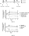
- the protocol of radiation therapy is performed on the patient as described in Figure 1 .
- 4 sessions of 10 Gy (total dose: 40 Gy) were spread over 2 weeks at day 1, 3, 8 and 10 using a Novalis stereotaxic accelerator device.
- a dose of 40 mg/m 2 irinotecan (Pfizer) in 250 mL of physiological saline or glucose isotonic was intravenously injected 30 to 90 minutes before the first and third radiotherapy sessions.
- the second blood sample is obtained at day 3.
- blood sample is meant a volume of whole blood or fraction thereof, e.g ., serum, plasma, etc.
- Ceramide has its general meaning in the art and refers to any N -acylsphingosine. Ceramides include sphingolipids in which the sphingosine is acylated with a fatty acid acyl-CoA derivative to form an N -acylsphingosine.
- the carbon chain is saturated or unsaturated.
- the carbon chain comprises 16, 18, 20, 22 or 24 carbons. In some embodiments, the carbon chain is a C16, C16:1, C18, C18:1, C20, C20:1, C22, C22:1, C24, or C24:1 carbon chain.
- the level of total ceramides is determined. In some embodiments, the level of at least one ceramide subspecies is determined. In some embodiments, the subspecies is selected from the group consisting of C16, C16:1, C18, C18:1, C20, C20:1, C22, C22:1, C24, or C24:1 ceramides. In some embodiments, the level of C24 ceramides is determined.
- the method of the present invention is particularly suitable for discriminating responder from non-responder.
- the term "responder" in the context of the present disclosure refers to a patient that will achieve a response, i.e ., a patient in whom the cancer is eradicated, reduced or improved.
- the responders have an objective response and therefore the term does not encompass patients having a stabilized cancer such that the disease is not progressing after radiation therapy.
- a non-responder or refractory patient includes patients in whom the cancer does not show reduction or improvement after radiation therapy.
- the term "non-responder” also includes patients having a stabilized cancer.
- the characterization of the patient as a responder or non-responder can be performed by reference to a standard or a training set.
- the standard may be the profile of a patient who is known to be a responder or non-responder or alternatively may be a numerical value.
- Such predetermined standards may be provided in any suitable form, such as a printed list or diagram, computer software program, or other media.
- hematologic cell pool over 1.5x10 9 white cells, 10 11 platelets and 90 g/L hemoglobin
- adequate hepatic and renal functions serum bilirubin less than 1.5 fold and transaminase and alkaline phosphatase less than 5-fold over the upper limit of normal.
- Exclusion criteria were defined as a performance index according to the World Health Organization (WHO) scale greater than 2, prior thoraco-abdominal irradiation, a contraindication to irinotecan prior (within 5 years) or concomitant treatment of an invasive cancer, diffuse metastatic disease, or more than 3 metastases. All institutional ethics committees approved the protocol, and signed informed consents were obtained from all patients.
- WHO World Health Organization
- Fig. 1 The complete treatment protocol is described in Fig. 1 . Briefly, 4 fractions of 10 Gy were spread at day (D)1, 3, 8 and 10 using Novalis (Brain Lab, Feldmün, D) or Cyberknife (Accuray, Sunnyvaley, CA) stereotactic accelerator devices. Whatever the type of accelerator, 99% CTV was encompassed by 75-95% isodose corresponding to a dose from 42 to 53 Gy at the center. 40 mg/m 2 irinotecan (Pfizer, New York, NY) was intravenously injected 30 to 90 minutes before the first and third radiotherapy sessions. Because of the absence of toxicity, the 35 patients received the complete treatment.
- the tumor response to the protocol was assessed using RECIST 1.1 (Response Evaluation Criteria In Solid Tumors) on the thoracic or liver tomodensitometry (TDM) ( Eisenhauer et al., 2009. Eur J Cancer. 45(2):228-47 ).
- the first evaluation was performed 6-8 weeks after the end of treatment, then at 3, 6, and 12 months.
- a complete response (CR) was defined by the complete disappearance of all lesions.
- a partial response (PR) and a progression (P) were respectively characterized by a reduction greater than 30% and an increase greater than 20% of the largest diameters of each lesion.
- Stability (S) was declared when tumor reduction or progression was respectively insufficient to define a PR or a P.
- the upper phase was collected, combined with the first, dried under nitrogen at room temperature and resuspended in 150 ⁇ L of hexane/propan-2-ol (60:40 v/v).
- Lipid extract purification was optimized from a previous method ( Bodennec et al., 2000. J Lipid Res. 41(9):1524-31 ). Briefly, samples were loaded on LC-NH 2 cartridges (Interchim, Montluzzo, F) preconditioned with 2 mL of hexane. The 100 mg cartridge was washed with 1.4 mL of ethyl acetate-hexane 15:85 (v/v) eluting neutral lipids in a single fraction. A second wash with 1.6 mL of chloroform/methanol 23:1 (v/v) eluted free Cer. Cer fraction was dried down under nitrogen and redissolved in 300 ⁇ L of MeOH containing 10 mM highest grade ammonium acetate (Fluka, Buchs, CH) and 0.2% formic acid. Samples were stored at -20°C until analysis.
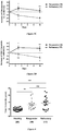
- Concentration of plasma Cer was monitored by LC-ESI-MS/MS during the SBRT protocol with irinotecan, at D3 and D10 and then, compared to the basal level at D0 ( Fig. 2 ).
- mean increase of Cer concentration in the different patient groups was monitored depending on their tumor response ( Fig. 2A ).
- CR was observed in 10, PR in 8, S in 8 and P in 9 patients.
- Cer dose response at D3 and D10 correlated with treatment efficacy.
- the total Cer amount increased significantly at D3 in plasma from patients exhibiting a diminution of tumor volume (CR: 18.5% ⁇ 8.92, P ⁇ 0.05 and PR: 10.7% ⁇ 2.27; P ⁇ 0.01; both vs D0).
- total Cer in S group remained stable at D3, then decreased significantly at D10 (D3: -1.70% ⁇ 4.36; P > 0.05 and D10: -15% ⁇ 2.40; both P ⁇ 0.01 vs D0) and was below the basal level for the P group (D3: -19.06% ⁇ 5.72; P ⁇ 0.05 and D10: -20.16% ⁇ 3.71; both P ⁇ 0.01 vs D0).
- CR and PR were defined as the objective response where S and P were considered as refractory to the treatment ( Fig. 2C ).
- S and P were considered as refractory to the treatment ( Fig. 2C ).
- 18/35 patients showed an objective response whereas 17/35 patients were considered refractory, due to tumor stabilization or progression or the emergence of new pulmonary or liver metastases.
- the total Cer level of the objective responder group was significantly higher in comparison to the basal Cer level objective group at D3 (15.07% ⁇ 5.02; P ⁇ 0.01 vs D0), and significantly lower for refractory group at D3 and D10 (D3: -9.79% ⁇ 4.12; P ⁇ 0.05 vs D0 and D10: - 17.95% ⁇ 2.27; P ⁇ 0.01 vs D0).
- the increase of plasma Cer concentration during the SBRT combined with irinotecan represents a promising endpoint that many serve as a harbinger tumor regression.
- the decrease of plasma Cer concentration is associated with the lack of tumor response of the treatment.
- Basal Cer level does not correlated with the outcome of the radiotherapy
- Plasma Cer appears to hold promise as a surrogate marker of the tumor response
- Basal Cer concentration appears as a marker of tumor presence in patients.
- Cer modulation during the treatment discriminate tumor control as a function of time
- ASM and Cer are secreted into the extracellular medium, by endothelial cells activated by pro-inflammatory cytokines including Il- ⁇ or TNF- ⁇ ( Marathe et al., 1998. J Biol Chem. 273(7):4081-8 ).
- pro-inflammatory cytokines including Il- ⁇ or TNF- ⁇
- HMVEC-L irradiation of primary microvascular endothelial cells HMVEC-L induces secretion of ASM and Cer into the extracellular medium (data not shown).
- Cer release by endothelial activation may lead to subsequent Cer-dependent radiosensitization of the tumor cells.
- the elevation of plasma Cer may also due to the high level of tumor apoptotic bodies enriched in Cer induced by the SBRT with irinotecan.
- Non-regulated of increase Cer may appear during late non-reversible stage of DNA damage-induced cell death ( Tepper et al., 1999. J Clin Invest. 103(7):971-8 ).
- the high level of Cer in the blood stream could be a marker of this form of cell death.
- plasma Cer concentration represents an early biomarker of response efficacy SBRT and irinotecan. This statement is supported by the fact that neither sex, age, location nor tumor volume was a co-variance factor correlated with the increase of ceramide during treatment. Preliminary data shows that tumor irradiation in mice induces plasma Cer in a dose-dependent manner (data not shown). We must reproduce the experiments using irinotecan to estimate its ability to induce plasma Cer.
- the Cer generation might be occurring slowly over the course of several weeks of the treatment and thus changes over baseline may not reach the level of significance.
- Further clinical studies must validate or refute Cer as a tumor response surrogate marker after conventional radiotherapies.
- the new radiotherapy devices stereotaxic X-ray accelerator, intrabeam, protontherapy
- clinical protocols are being re-evaluated and redesigned for some tumor exposures, with a dose escalation and a decrease in the number of fractions.
- intracellular Cer generation and/or tumor cell death is enhanced.
- plasma Cer levels would be expected acutely after irradiation improving its detection. Because of those specificities, increase of plasma Cer might only be observable and quantifiable after high dose radiation.
Landscapes
- Health & Medical Sciences (AREA)
- Life Sciences & Earth Sciences (AREA)
- Engineering & Computer Science (AREA)
- Immunology (AREA)
- Chemical & Material Sciences (AREA)
- Biomedical Technology (AREA)
- Molecular Biology (AREA)
- Urology & Nephrology (AREA)
- Hematology (AREA)
- General Health & Medical Sciences (AREA)
- Biotechnology (AREA)
- Microbiology (AREA)
- Analytical Chemistry (AREA)
- Physics & Mathematics (AREA)
- Biochemistry (AREA)
- Bioinformatics & Cheminformatics (AREA)
- General Physics & Mathematics (AREA)
- Cell Biology (AREA)
- Pathology (AREA)
- Medicinal Chemistry (AREA)
- Food Science & Technology (AREA)
- Toxicology (AREA)
- Tropical Medicine & Parasitology (AREA)
- Organic Chemistry (AREA)
- Wood Science & Technology (AREA)
- Proteomics, Peptides & Aminoacids (AREA)
- Zoology (AREA)
- Biophysics (AREA)
- Genetics & Genomics (AREA)
- General Engineering & Computer Science (AREA)
- Endocrinology (AREA)
- Medicines That Contain Protein Lipid Enzymes And Other Medicines (AREA)
- Acyclic And Carbocyclic Compounds In Medicinal Compositions (AREA)
- Pharmaceuticals Containing Other Organic And Inorganic Compounds (AREA)
- Other Investigation Or Analysis Of Materials By Electrical Means (AREA)
- Investigating Or Analysing Biological Materials (AREA)
Description
- The present invention relates to methods for determining whether a patient will achieve a response after radiation therapy.
- Radiation therapy is one of the most common therapeutic and palliative anti-cancer treatments. Its main limitation is due to the intrinsic radiation resistance of the tumor, limiting its efficacy (Bernier et al., 2004. Nat Rev Cancer. 4(9):737-47). Because of the improvement of tumor imaging and medical physic researches new stereotaxic radiation therapy devices have been developed with a better targeting of the radiation into the tumor. Those stereotaxic accelerators are changing irradiation plans by increasing the dose within a limited number of fractions (Bernier et al., 2004. Nat Rev Cancer. 4(9):737-47). If they demonstrated a strong efficacy in oligometastases and small solid tumors, these hypofractionated radiation therapy protocols have to be validated for most of the tumor type in function of their localization and their radiation resistance.
- Currently, the most common way to validate the radiation therapy efficacy is obtained through the visualization of the tumor volume control or regression by CT-scan or by other non-invasive imaging techniques. Unfortunately, tumor volume response can be estimated within months after the end of the radiation therapy delaying any alternative treatment. Discovering biological markers allowing for the discrimination between responding and refractory patients to the radiation therapy represents a major issue to improve anti-tumor treatment.
- Biomarkers can be classified in three categories: omics from tumor biopsies, phenotypic imaging and secretory factors (Kumar et al., 2006. Biomarkers. 11(5):385-405). Tumor markers by omics are essentially obtained by genomics and proteomic assays. If they have the advantage to quest markers in a very large broad of molecular events, the need for tumor biopsies limits their studies to specific tumor localization and the number of samples. Usually, those studies are dedicated to prognostic studies grading and assessing the treatment. Phenotypic imaging allows the evaluation of some physiologic change in the tumor such as hypoxia, cell proliferation index, necrosis or immune cell infiltration (Malviya & Nayak, 2013. Curr Pharm Biotechnol. 14(7):669-82). Phenotypic imaging has the advantage to be non-invasive. However, the heterogeneity of the tumor response and the consistent quantification of the molecular biomarkers remain under investigation. Finally, secretory factors coming from blood, saliva and urine samples have the advantage to be easily obtained from any patient and can be provided before and all along the treatment (Ganepola et al., 2014. World J Gastrointest Oncol. 6(4):83-97). Those secretory factors can include pro-inflammatory cytokines, peptides LDL or circulating tumor cells. If some of them have been investigated, none of them has been validated as biomarker of the radiation therapy efficacy.
- Sphingolipid ceramide also represents a potential and interesting secreted biomarker. Indeed, ceramide is a pro-apoptotic factor, rapidly generated into the outer layer of the cell membrane through hydrolysis of sphingomyelin by acidic or neutral sphingomyelinase (respectively ASM and NSM), but also in reticulum through a de novo synthesis pathway dependent of the ceramide synthase (Corre et al., 2010. Mutat Res. 704(1-3):61-7). Several studies demonstrate the involvement of ceramide in cell and tumor radiosensisitivity. Exogenous ceramide treatment enhances radiation-induced LNCaP cell death and tumor regression (Kumar et al., 2006. Biomarkers. 11(5):385-405). In the same manner, increasing endogenous ceramide through DL-PDMP and D-MAPP, respectively an inhibitor of glucosyl-ceramide synthase and an inhibitor of ceramidase, enhances Jurkat radiosensitivity (Rodriguez-Lafrasse et al., 2002. Int J Cancer. 101(6):589-98). Besides their involvement in tumor cell death, ceramides have been observed in endothelial cell apoptosis in response to high-dose radiation therapy which is modulating tumor regression. In fact, fibrosarcoma or melanoma tumor cells transplanted in mice, then irradiated, rapidly induced a massive endothelial cell apoptosis via ASM activation and ceramide generation participating to tumor regression (Garcia-Barros et al., 2003. Science. 300(5622):1155-9). Invalidation of ASM blocks endothelial cell apoptosis and tumor regression induced by high-dose radiation therapy.
- Besides their intracellular form, secreted ceramides in the extracellular medium are also playing important biological roles in physiological and pathophysiological processes. High levels of ceramides have been observed in plasma and serum from patients with several physiopathologies, including lung emphysema (Petrache et al., 2005. Nat Med. 11(5):491-8), Wilson disease (Lang et al., 2007. Nat Med. 13(2):164-70) and multiple organ failure (Delogu et al., 1999. Crit Care Med. 27(11):2413-7). Plasma ceramide levels are increased during lipid infusion in humans and rats, and in obese, insulin-resistant mice (Watt et al., 2012. Diabetologia. 55(10):2741-6) which may correlated with insulin sensitivity, inflammation and atherosclerotic risk. Interestingly, ceramide and its enzyme ASM have also been quantified in serum from 11 patients with gross tumors from different origins after spatial fractionated grid radiation therapy (SFGRT) including a first irradiation at 15 Gy followed by 30 fractions of 2 Gy (Sathishkumar et al., 2005. Cancer Biol Ther. 4(9):979-86). Three days after treatment, an increase of secreted ceramides was quantified in the serum of 5 of the 7 patients responding to this specific radiation therapy protocol. However, the few number of patients and the diverse origin of the tumors diluted the strength of their results and do not allow to establish strong statistical evidence the correlation of ceramide and radiation therapy efficacy.
- Ogretmen et al. (2004. Nat Rev Cancer. 4(8):604-16) discloses that ceramide induce antiproliferative and apoptotic responses in cancer cells, and that radiotherapy causes formation of ceramides which accumulate in the cells.
-
US patent US6,274,309 (Kilesnick et al. ) discloses a method for evaluating a cell sensitivity to sphingomyelinase-related apoptosis in the course of a radiation stress stimulus exposure. More specifically,US6,274,309 discloses a method relying on the comparison of both sphingomyelin and ceramide levels within the cells. - Ogretmen et al. (2001. Drug Resist Updat. 4(6):368-77) discloses that anti-cancer agents cause elevation of endogenous ceramide levels, accompanied by apoptotic cell death in human cancer cells.
- The present invention relates to methods for determining whether a patient will achieve a response after radiation therapy. In particular, the present invention is defined by the claims.
- Discovering biological markers of tumor regression induced by ionizing radiation will permit a better discrimination between responding and refractory patients to the radiation therapy. In this present invention, the inventors studied the ability of plasmic ceramides, a known pro-apoptotic bioactive sphingolipid, to be correlated to the tumor control in a clinical phase II study combining hypofractionated radiation therapy and irinotecan in liver and lung metastases. Liver and lung metastases were indeed treated with 4
times 10 Gy atday day day - Accordingly, a first object of the present invention relates to a method for determining whether a patient suffering from a cancer will achieve a response after radiation therapy comprising the steps of
- i) determining the level of ceramide in a first blood sample obtained from the patient before radiation therapy,
- ii) determining the level of ceramide in a second blood sample obtained from the patient during or just after radiation therapy,
- iii) comparing the level determined at step i) with the level determined at step ii), and
- iv) concluding that the patient will achieve response when the level determined at step ii) is higher than the level determined at step i) or concluding that the patient will not achieve a response when the level determined at step ii) is lower than the level determined at step i).
- Cancers to be treated include primary tumors and metastatic tumors. Examples of cancers that may be treated include, but are not limited to, cancer cells from the bladder, blood, bone, bone marrow, brain, breast, colon, esophagus, gastrointestine, gum, head, kidney, liver, lung, nasopharynx, neck, ovary, prostate, skin, stomach, testis, tongue, or uterus. In addition, the cancer may specifically be of the following histological type, though it is not limited to these: neoplasm, malignant; carcinoma; carcinoma, undifferentiated; giant and spindle cell carcinoma; small cell carcinoma; papillary carcinoma; squamous cell carcinoma; lymphoepithelial carcinoma; basal cell carcinoma; pilomatrix carcinoma; transitional cell carcinoma; papillary transitional cell carcinoma; adenocarcinoma; gastrinoma, malignant; cholangiocarcinoma; hepatocellular carcinoma; combined hepatocellular carcinoma and cholangiocarcinoma; trabecular adenocarcinoma; adenoid cystic carcinoma; adenocarcinoma in adenomatous polyp; adenocarcinoma, familial polyposis coli; solid carcinoma; carcinoid tumor, malignant; branchiolo-alveolar adenocarcinoma; papillary adenocarcinoma; chromophobe carcinoma; acidophil carcinoma; oxyphilic adenocarcinoma; basophil carcinoma; clear cell adenocarcinoma; granular cell carcinoma; follicular adenocarcinoma; papillary and follicular adenocarcinoma; non-encapsulating sclerosing carcinoma; adrenal cortical carcinoma; endometroid carcinoma; skin appendage carcinoma; apocrine adenocarcinoma; sebaceous adenocarcinoma; ceruminous; adenocarcinoma; mucoepidermoid carcinoma; cystadenocarcinoma; papillary cystadenocarcinoma; papillary serous cystadenocarcinoma; mucinous cystadenocarcinoma; mucinous adenocarcinoma; signet ring cell carcinoma; infiltrating duct carcinoma; medullary carcinoma; lobular carcinoma; inflammatory carcinoma; Paget's disease, mammary; acinar cell carcinoma; adenosquamous carcinoma; adenocarcinoma w/squamous metaplasia; thymoma, malignant; ovarian stromal tumor, malignant; thecoma, malignant; granulosa cell tumor, malignant; and roblastoma, malignant; Sertoli cell carcinoma; Leydig cell tumor, malignant; lipid cell tumor, malignant; paraganglioma, malignant; extra-mammary paraganglioma, malignant; pheochromocytoma; glomangiosarcoma; malignant melanoma; amelanotic melanoma; superficial spreading melanoma; malig melanoma in giant pigmented nevus; epithelioid cell melanoma; blue nevus, malignant; sarcoma; fibrosarcoma; fibrous histiocytoma, malignant; myxosarcoma; liposarcoma; leiomyosarcoma; rhabdomyosarcoma; embryonal rhabdomyosarcoma; alveolar rhabdomyosarcoma; stromal sarcoma; mixed tumor, malignant; mullerian mixed tumor; nephroblastoma; hepatoblastoma; carcinosarcoma; mesenchymoma, malignant; brenner tumor, malignant; phyllodes tumor, malignant; synovial sarcoma; mesothelioma, malignant; dysgerminoma; embryonal carcinoma; teratoma, malignant; struma ovarii, malignant; choriocarcinoma; mesonephroma, malignant; hemangiosarcoma; hemangioendothelioma, malignant; kaposi's sarcoma; hemangiopericytoma, malignant; lymphangiosarcoma; osteosarcoma; juxtacortical osteosarcoma; chondrosarcoma; chondroblastoma, malignant; mesenchymal chondrosarcoma; giant cell tumor of bone; Ewing's sarcoma; odontogenic tumor, malignant; ameloblastic odontosarcoma; ameloblastoma, malignant; ameloblastic fibrosarcoma; pinealoma, malignant; chordoma; glioma, malignant; ependymoma; astrocytoma; protoplasmic astrocytoma; fibrillary astrocytoma; astroblastoma; glioblastoma; oligodendroglioma; oligodendroblastoma; primitive neuroectodermal; cerebellar sarcoma; ganglioneuroblastoma; neuroblastoma; retinoblastoma; olfactory neurogenic tumor; meningioma, malignant; neurofibrosarcoma; neurilemmoma, malignant; granular cell tumor, malignant; malignant lymphoma; Hodgkin's disease; Hodgkin's lymphoma; paragranuloma; malignant lymphoma, small lymphocytic; malignant lymphoma, large cell, diffuse; malignant lymphoma, follicular; mycosis fungoides; other specified non-Hodgkin's lymphomas; malignant histiocytosis; multiple myeloma; mast cell sarcoma; immunoproliferative small intestinal disease; leukemia; lymphoid leukemia; plasma cell leukemia; erythroleukemia; lymphosarcoma cell leukemia; myeloid leukemia; basophilic leukemia; eosinophilic leukemia; monocytic leukemia; mast cell leukemia; megakaryoblastic leukemia; myeloid sarcoma; and hairy cell leukemia.
- The term "radiation therapy" has its general meaning in the art and refers the treatment of cancer with ionizing radiation. Ionizing radiation deposits energy that injures or destroys cells in the area being treated (the target tissue) by damaging their genetic material, making it impossible for these cells to continue to grow. One type of radiation therapy commonly used involves photons, e.g., X-rays. Depending on the amount of energy they possess, the rays can be used to destroy cancer cells on the surface of or deeper in the body. The higher the energy of the X-ray beam, the deeper the X-rays can go into the target tissue. Linear accelerators and betatrons produce X-rays of increasingly greater energy. The use of machines to focus radiation (such as X-rays) on a cancer site is called external beam radiation therapy. Gamma rays are another form of photons used in radiation therapy. Gamma rays are produced spontaneously as certain elements (such as radium, uranium, and cobalt 60) release radiation as they decompose, or decay. In some embodiments, the radiation therapy is external radiation therapy. Examples of external radiation therapy include, but are not limited to, conventional external beam radiation therapy; three-dimensional conformal radiation therapy (3D-CRT), which delivers shaped beams to closely fit the shape of a tumor from different directions; intensity modulated radiation therapy (IMRT), e.g., helical tomotherapy, which shapes the radiation beams to closely fit the shape of a tumor and also alters the radiation dose according to the shape of the tumor; conformal proton beam radiation therapy; image-guided radiation therapy (IGRT), which combines scanning and radiation technologies to provide real time images of a tumor to guide the radiation treatment; intraoperative radiation therapy (IORT), which delivers radiation directly to a tumor during surgery; stereotactic radiosurgery, which delivers a large, precise radiation dose to a small tumor area in a single session; hyperfractionated radiation therapy, e.g., continuous hyperfractionated accelerated radiation therapy (CHART), in which more than one treatment (fraction) of radiation therapy are given to a subject per day; and hypofractionated radiation therapy, in which larger doses of radiation therapy per fraction is given but fewer fractions.
- In some embodiments, the method of the present invention is particularly suitable in the context of a hypofractionated radiation therapy. As used herein, the term "hypofractionated radiation therapy" has its general meaning in the art and refers to radiation therapy in which the total dose of radiation is divided into large doses and treatments are given less than once a day.
- Typically, a treatment course comprises 1, 2, 3, 4 or 5 regimens of ionizing radiations. In some embodiments, the regimen of ionizing radiations is combined with the administration of at least one chemotherapeutic agent. Chemotherapeutic agent include those compounds with anti-cancer activity, e.g., compounds that induce apoptosis, compounds that reduce lifespan or compounds that render cells sensitive to stress and include, but are not limited to, aminoglutethimide, amsacrine, anastrozole, asparaginase, beg, bicalutamide, bleomycin, buserelin, busulfan, campothecin, capecitabine, carboplatin, carmustine, chlorambucil, cisplatin, cladribine, clodronate, colchicine, cyclophosphamide, cyproterone, cytarabine, dacarbazine, dactinomycin, daunorubicin, dienestrol, diethylstilbestrol, docetaxel, doxorubicin, epirubicin, estradiol, estramustine, etoposide, exemestane, filgrastim, fludarabine, fludrocortisone, fluorouracil, fluoxymesterone, flutamide, gemcitabine, genistein, goserelin, hydroxyurea, idarubicin, ifosfamide, imatinib, interferon, irinotecan, ironotecan, letrozole, leucovorin, leuprolide, levamisole, lomustine, mechlorethamine, medroxyprogesterone, megestrol, melphalan, mercaptopurine, mesna, methotrexate, mitomycin, mitotane, mitoxantrone, nilutamide, nocodazole, octreotide, oxaliplatin, paclitaxel, pamidronate, pentostatin, plicamycin, porfimer, procarbazine, raltitrexed, rituximab, streptozocin, suramin, tamoxifen, temozolomide, teniposide, testosterone, thioguanine, thiotepa, titanocene dichloride, topotecan, trastuzumab, tretinoin, vinblastine, vincristine, vindesine, and vinorelbine.
- In some embodiments, the protocol of radiation therapy is performed on the patient as described in
Figure 1 . Briefly, 4 sessions of 10 Gy (total dose: 40 Gy) were spread over 2 weeks atday day 3. - By "blood sample" is meant a volume of whole blood or fraction thereof, e.g., serum, plasma, etc.
- As used herein, the term "ceramide" has its general meaning in the art and refers to any N-acylsphingosine. Ceramides include sphingolipids in which the sphingosine is acylated with a fatty acid acyl-CoA derivative to form an N-acylsphingosine. Typically, the carbon chain is saturated or unsaturated. Furthermore, the carbon chain comprises 16, 18, 20, 22 or 24 carbons. In some embodiments, the carbon chain is a C16, C16:1, C18, C18:1, C20, C20:1, C22, C22:1, C24, or C24:1 carbon chain.
- Methods to determine the level of ceramides in biological samples are known in the art, for example, as provided in Kasumov et al. (2010. Anal. Biochem. 401(1):154-161) or Hu et al. (2009. J Lipid Res. 50(9):1852-1862). Typically, quantitative analyses of ceramides is performed by Ultra Performance Liquid Chromatography coupled to a mass spectrometer. Immunoassays are also possible and generally involve contacting the blood sample with an antibody to ceramide, under conditions effective to allow the formation of immunocomplexes. In this regard, the skilled artisan will be able to assess the level of ceramides in the blood sample.
- In some embodiments, the level of total ceramides is determined. In some embodiments, the level of at least one ceramide subspecies is determined. In some embodiments, the subspecies is selected from the group consisting of C16, C16:1, C18, C18:1, C20, C20:1, C22, C22:1, C24, or C24:1 ceramides. In some embodiments, the level of C24 ceramides is determined.
- The method of the present invention is particularly suitable for discriminating responder from non-responder. As used herein, the term "responder" in the context of the present disclosure refers to a patient that will achieve a response, i.e., a patient in whom the cancer is eradicated, reduced or improved. According to the invention, the responders have an objective response and therefore the term does not encompass patients having a stabilized cancer such that the disease is not progressing after radiation therapy. A non-responder or refractory patient includes patients in whom the cancer does not show reduction or improvement after radiation therapy. According to the invention, the term "non-responder" also includes patients having a stabilized cancer. Typically, the characterization of the patient as a responder or non-responder can be performed by reference to a standard or a training set. The standard may be the profile of a patient who is known to be a responder or non-responder or alternatively may be a numerical value. Such predetermined standards may be provided in any suitable form, such as a printed list or diagram, computer software program, or other media. When it is concluded that the patient is a non-responder, the physician could take the decision to stop the protocol of radiation therapy to avoid any further adverse sides effects.
- The invention will be further illustrated by the following figures and examples. However, these examples and figures should not be interpreted in any way as limiting the scope of the present invention.
-
-
Figure 1 . Treatment plan of the phase II clinical protocol combining SBRT with irinotecan. Irradiations (10 Gy) were applied at D1, D3, D7 and D10. Irinotecan (40 mg/m2) was administered at D1 and D7. Blood samples were harvested before treatment (D0) and after the second and fourth irradiation (D3 and D10). -
Figure 2 . Variation of total Cer during the treatment is correlated with the tumor response. A: Ratio of Cer concentration at D3 and D10 vs D0 in complete responder (CR), partial responder (PR), tumor stabilization (S) and progression (P) groups. B: Ratio of the Cer concentration at D3 and D10 vs D0 in tumor responding (CR, PR, and S) group as compared to tumor progression (P) group. C: Ratio of the Cer concentration at D3 and D10 vs D0 in tumor shrinking (CR, PR) group as compared to the group where tumors do not regressed (S, P). Measurements were performed in triplicate (Number of patients in parentheses, mean ± SEM, ns = P > 0.05, * = P < 0.05). -
Figure 3 . Evolution of major Cer subspecies during the treatment is correlated with the tumor response. Ratio of C16:0 (A), C22:0 (B), C24:0 (C), and C24:1 Cer (D) concentrations at D3 and D10 vs D0 in objective response (CR, PR) group as compared to the refractory group (S, P). Measurements were performed in triplicate (Number of patients in parentheses, mean ± SEM, ns = P > 0.05, * = P < 0.05). -
Figure 4 . Basal Cer level does not correlated with the outcome of the radiotherapy. Basal plasma Cer was measured in responder and refractory patients and healthy population were performed in triplicate (mean ± SEM, ns = P > 0.05, * = P < 0.05). -
Figure 5 . Classification of Cer variation clusters patients depending on their tumor response. CER increases and decreases at D3 and D10, respectively, are represented in red and green where median equal to 0 are in black. The position of each patient on the hierarchy is presented after cluster 3.0 analysis and tree view visualization depending on tumor response to the treatment (CR, PR, S, P). -
Figure 6 . Cer modulation during the treatment discriminates tumor control in function of time. Kaplan-Meier curves for patients without tumor volume worsening in function of time are shown for patients with either an increase or decrease of Cer at D3 (A) and D10 (B). Number of patients are in parentheses. P < 0.05 between groups Cer increase and Cer decrease for both figures. - A multicentric phase II clinical study with SBRT and concomitant irinotecan for colorectal adenocarcinoma lung and liver metastases relapsing from fluorouracil (5-FU) with or without eloxatin or irinotecan, was performed from 2008 to 2013 between 3 French oncology centers (Nantes, Lyon, and Lille). Patients (mean age: 64 years; range: 32-80 years), with a life expectancy over 6 months and with inoperable or recurrent hepatic and/or lung metastases after surgery were selected. Metastases should be measurable with the largest diameter under or equal to 6 cm. The sum of the maximum diameter of multiple metastases must be under or equal to 6 cm. Clinical target volume (CTV) should be located more than 12 mm laterally or 15 mm in the cranio-caudal direction of stomach, small intestine esophagus, trachea, and pulmonary arteries.
- Patients must have an adequate hematologic cell pool (over 1.5x10⁹ white cells, 1011 platelets and 90 g/L hemoglobin), and adequate hepatic and renal functions (serum bilirubin less than 1.5 fold and transaminase and alkaline phosphatase less than 5-fold over the upper limit of normal). Exclusion criteria were defined as a performance index according to the World Health Organization (WHO) scale greater than 2, prior thoraco-abdominal irradiation, a contraindication to irinotecan prior (within 5 years) or concomitant treatment of an invasive cancer, diffuse metastatic disease, or more than 3 metastases. All institutional ethics committees approved the protocol, and signed informed consents were obtained from all patients.
- The complete treatment protocol is described in
Fig. 1 . Briefly, 4 fractions of 10 Gy were spread at day (D)1, 3, 8 and 10 using Novalis (Brain Lab, Feldkirchen, D) or Cyberknife (Accuray, Sunnyvaley, CA) stereotactic accelerator devices. Whatever the type of accelerator, 99% CTV was encompassed by 75-95% isodose corresponding to a dose from 42 to 53 Gy at the center. 40 mg/m2 irinotecan (Pfizer, New York, NY) was intravenously injected 30 to 90 minutes before the first and third radiotherapy sessions. Because of the absence of toxicity, the 35 patients received the complete treatment. 20 mL of blood was collected in tubes with citrate before the first (D0) and after second (D3) and fourth (D10) irradiations, then stored at 4°C for 30 minutes. Blood samples were centrifuged at 1000 g for 5 minutes at 4°C to recover the plasma. Plasma aliquots were stored at -80°C until further analysis. Whole blood from healthy donors over 45 years, were collected at the French blood institute (EFS, Nantes, F), to recover the plasma using the same protocol as previously described. - The tumor response to the protocol was assessed using RECIST 1.1 (Response Evaluation Criteria In Solid Tumors) on the thoracic or liver tomodensitometry (TDM) (Eisenhauer et al., 2009. Eur J Cancer. 45(2):228-47). The first evaluation was performed 6-8 weeks after the end of treatment, then at 3, 6, and 12 months. A complete response (CR) was defined by the complete disappearance of all lesions. A partial response (PR) and a progression (P) were respectively characterized by a reduction greater than 30% and an increase greater than 20% of the largest diameters of each lesion. Stability (S) was declared when tumor reduction or progression was respectively insufficient to define a PR or a P.
- Ultrapure standards of Cer subspecies (C14:0, C16:0, C18:0, C18:1, C20:0, C24:0 and C24:1) and non-natural C17:0 Cer used as an internal standard (IS) were purchased from Avanti Polar Lipids (Alabaster, AL). UPLC grade methanol and analytical grade organic solvents were purchased from Fisher Scientific (Pittsburgh, PA).
- Forty microliters from 1 µM C17 Cer were added to each sample. Lipid extraction was carried out in two steps with minor modifications of previously described procedures (Hara & Radin, 1978. Anal Biochem. 90(1):420-6). First extraction was performed by adding 1.5 mL of hexane/propan-2-ol mixture (60:40, V/V) on 100 µL of plasma. The sample was vortexed, centrifuged at 3000 g for 5 minutes at 4°C and the upper phase was collected. A second extraction was then performed with 1.5 mL of methanol. After homogenization and centrifugation at 8000 g for 5 minutes at 4°C, the upper phase was collected, combined with the first, dried under nitrogen at room temperature and resuspended in 150 µL of hexane/propan-2-ol (60:40 v/v).
- Lipid extract purification was optimized from a previous method (Bodennec et al., 2000. J Lipid Res. 41(9):1524-31). Briefly, samples were loaded on LC-NH2 cartridges (Interchim, Montluçon, F) preconditioned with 2 mL of hexane. The 100 mg cartridge was washed with 1.4 mL of ethyl acetate-hexane 15:85 (v/v) eluting neutral lipids in a single fraction. A second wash with 1.6 mL of chloroform/methanol 23:1 (v/v) eluted free Cer. Cer fraction was dried down under nitrogen and redissolved in 300 µL of MeOH containing 10 mM highest grade ammonium acetate (Fluka, Buchs, CH) and 0.2% formic acid. Samples were stored at -20°C until analysis.
- Purified Cer fractions were analyzed by LC-ESI-MS/MS on an Acquity H-Class UPLC system combined with a Xevo TQD triple quadrupole mass spectrometer (Waters Corporation, Milford, CT). Gradient chromatographic separation was performed on Waters C18 BEH column (2.1 mm x 50 mm) with 1.8 µM particle size equipped with a 0.5 µM prefilter. The column heater was set at 43°C. The mobile phases consisted of MiliQ water containing 0.2% formic acid and 10 mM ammonium acetate (Eluent A) and methanol containing 0.2% formic acid and 10 mM ammonium acetate (Eluent B). The injection volume was 5 µL. Purified Cer were eluted in 4 minutes with a linear gradient to 98% of Eluent B. Before the next run, a reequilibration from 4.00 to 4.10 minutes and stabilization from 4.10 to 6 minutes with 95% of Eluent B were performed. The flow rate was set to 0.6 mL/min. All analyses were performed using electrospray ionization in the positive ion mode with multiple reactions monitoring (MRM). Measurement and data analysis were collected by Mass-Lynx software version 4.1. Integration and quantification were performed using the Waters Target Links™ software.
- Three independent measurements were performed per patient sample. Wilcoxon signed-rank test and ANOVA with 95% confidence estimation were performed with StatView 6.0 package. For hierarchical clustering, Cer ratios between D3 or D10 and D0 were estimated according to the expression profiling of other patients (Eisen et al., 1998. Proc Natl Acad Sci USA. 95(25):14863-8) by cluster 3.0 and displayed by Java TreeView (both softwares, http://bonsai.hgc.jp/∼mdehoon/software/cluster/software.htm). Independence of each group was tested by chi-squared test. The probability of tumor control in function of time in patient with an increase or decrease of ceramide were obtained by Kaplan-Meier method (Kaplan, 1958. J Am Stat Assoc. 53:457-480) and compared by the log-rank test, giving 95% confidence intervals (CI). The prognostic value of variables (sex, age, tumor location and volume) was calculated using the Cox multivariate regression model (Cox, 1972. J R Stat Soc. 34:187-220).
- Concentration of plasma Cer was monitored by LC-ESI-MS/MS during the SBRT protocol with irinotecan, at D3 and D10 and then, compared to the basal level at D0 (
Fig. 2 ). First, no correlation was shown between the level of Cer increase at D3 and D10 after SBRT with irinotecan and any covariance factors (sex, age, tumor location and tumor volume; data not shown). Then, mean increase of Cer concentration in the different patient groups was monitored depending on their tumor response (Fig. 2A ). One year after treatment, CR was observed in 10, PR in 8, S in 8 and P in 9 patients. We observed that Cer dose response at D3 and D10 correlated with treatment efficacy. The total Cer amount increased significantly at D3 in plasma from patients exhibiting a diminution of tumor volume (CR: 18.5% ± 8.92, P < 0.05 and PR: 10.7% ± 2.27; P < 0.01; both vs D0). In contrast, total Cer in S group remained stable at D3, then decreased significantly at D10 (D3: -1.70% ± 4.36; P > 0.05 and D10: -15% ± 2.40; both P < 0.01 vs D0) and was below the basal level for the P group (D3: -19.06% ± 5.72; P < 0.05 and D10: -20.16% ± 3.71; both P < 0.01 vs D0). - Because the tumor growth arrest is a hallmark of the response to the treatment, we first decided to determine a potential correlation between responders including CR, PR and S groups, and refractory patients including P group (
Fig. 2B ). The total Cer level of this responding group (CR, PR, and S) was significantly higher in comparison to the basal level at D3 (9.91% ± 3.99; P < 0.05 vs D0), but not to D10. Interestingly, the total Cer level of this refractory group (P) decreased significantly at D3 and D10 compared to the basal rate (D3: -19.06% ± 5.72; P < 0.05 vs D0; and D10: -20.17% ± 3.71; P < 0.01 vs D0). As proposed in the present clinical phase II study, CR and PR were defined as the objective response where S and P were considered as refractory to the treatment (Fig. 2C ). One year after treatment, 18/35 patients showed an objective response whereas 17/35 patients were considered refractory, due to tumor stabilization or progression or the emergence of new pulmonary or liver metastases. Using this classification group, the total Cer level of the objective responder group was significantly higher in comparison to the basal Cer level objective group at D3 (15.07% ± 5.02; P < 0.01 vs D0), and significantly lower for refractory group at D3 and D10 (D3: -9.79% ± 4.12; P < 0.05 vs D0 and D10: - 17.95% ± 2.27; P < 0.01 vs D0). The increase of plasma Cer concentration during the SBRT combined with irinotecan represents a promising endpoint that many serve as a harbinger tumor regression. On the other hand, the decrease of plasma Cer concentration is associated with the lack of tumor response of the treatment. - Twelve Cer subspecies have been sought in order to characterize more specifically the composition of the total Cer. The most abundant compounds were those containing the fatty acid C24:0 (45.46% ± 1.06), C24:1 (23.43% ± 0.90), C22:0 (15.74% ± 0.34), and C16:0 (7.10% ± 0.39). The other compounds were present in very small amount. Because of a potential discrepancy of their response to the treatment, those major Cer subspecies were separately quantified, compared to their basal level at D0 and correlated with the tumor response (
Fig. 3A to D ). In fact, the levels of these 4 subspecies followed a similar profile as that of the total Cer levels during the treatment. The ratio of C24:0 Cer, the most abundant compound, increased significantly in the objective responder group from 11.9% ± 5.17 at D3 (P < 0.05 vs D0). These subspecies decreased significantly in the refractory group from -11.08% ± 4.33 at D3 and -19.19% ± 2.39 at D10 (both P < 0.05 vs D0). The three other main Cer subspecies exhibited similar significant changes in their ratios as observed for C24:0 Cer. These results establish that all the major plasma Cer subspecies, as well as the total Cer, are evolving following a similar pattern of change after SBRT combined with irinotecan, with rising levels correlated with the tumor response. - Because plasma Cer appears to hold promise as a surrogate marker of the tumor response, correlation of basal level of Cer in patients with treatment efficacy was investigated. Plasma Cer concentration from healthy donors was very homogenous and significantly lower (mean: 1.98 µM ± 0.09) than that of the patients with high individual variability (responder group: 3.34 µM ± 0.32 and refractory group 3.82 µM ± 0.45; both P < 0.01 vs healthy donors). Basal Cer concentration appears as a marker of tumor presence in patients. However, the comparison of the total CER concentration between the two patient groups did not permit a discrimination between the responder group and the refractory group (P = 0.67). Thus, the amount of total Cer in patients before any treatment cannot be regarded as a prognostic factor of the tumor response to SBRT combined to irinotecan.
- Total Cer and its subspecies modulation during treatment were evaluated for each patient and hierarchical clustering was established. Cer increased and decreased at D3 and D10 are represented respectively in red and green when median equal to 0 are in black (
Fig. 5 ). Clustering of the individual Cer evolution demonstrated a hierarchy between patients with objective and refractory responses. Hierarchy of total Cer modulation at D3 and D10 showed clearly that 8/10 CR and 6/8 PR patients are clustered above the median response equal to 0 (both P < 0.001). In contrast, 5/8 S and 8/9 P patients were grouped below this median (both P < 0.001). For every Cer subspecies, a similar discrimination between the objective and refractory patients were obtained (data not shown) without improvement of the patients' segregation as compared to the cluster analysis with total Cer. - Finally, we evaluated the tumor volume worsening measured by CT-
Scan Fig. 6 ). Kaplan-Meier curves clearly demonstrate that patients with an increase of Cer either at D3 or D10 get high probability of tumor control during the first year. Interestingly, no patients with a Cer increase at D10 show an aggravation of tumor. Patients with a decrease of Cer have a 50% of chance to tumor worsening during the first year. These results clearly and statistically discriminates the ability of tumor control for treated patients depending on early Cer increase or decrease (D3 or D10: p < 0.01). - In the present well-defined phase II study combining SBRT with irinotecan, we clearly correlated the elevation of Cer concentration in the blood plasma with the tumor response rate. Similarly, a decrease of Cer concentration in the blood plasma was correlated with stabilization or a tumor progression, and therefore within effective treatment. Our results defined plasma Cer as an early surrogate marker of the tumor response, detectable early during the radiotherapy treatment.
- The results presented in
Fig. 3 , show that patients with liver or lung metastases of colorectal cancer, have a Cer concentration in blood plasma higher than healthy patients. These results are in agreement with the literature showing a modulation of Cer in the patient's blood suffering from diverse pathologies (Delogu et al., 1999. Crit Care Med. 27(11):2413-7; Lang et al., 2007. Nat Med. 13(2):164-70; Petrache et al., 2005. Nat Med. 11(5):491-8; Watt et al., 2012. Diabetologia. 55(10):2741-6). Moreover, Cer in the blood flow has also been quantified after SFGRT scheme including a fraction of 15 Gy then 30 of 2 Gy (Sathishkumar et al., 2005. Cancer Biol Ther. 4(9):979-86). Plasma Cer concentrations were significantly increased 72 hours after SFGRT in 3/3 CR and 2/4 PR patients. However, no correlation was found in the no-responder group where one patient showed an increase of Cer level, and the other one a decrease. Indeed, significant discrimination Cer increase and decrease groups was impossible because of the small size of the cohort. Furthermore, the diverse tumor origins diluted the strength of the results. This promising study was not allowing statistical evidence establishing the secreted Cer as a biomarker of radiotherapy efficacy. - In the present study, those two weaknesses have been solved. First, our study includes a larger cohort of 35 patients with almost an equal number of responders and refractory (respectively 18 and 17). Secondly, all metastases derived from primary colorectal carcinoma and were treated by SBRT with irinotecan. Finally, the tumor volumes were equivalent, below 6 cm for the largest diameter limiting potential inconsistency due to the volume size. Because of the strict patient inclusion criteria and clinical follow-up, we were able to extend previous results on the correlation between modulation of the total Cer into the blood stream and the tumor response after radiotherapy. Moreover, our work sheds new light on the Cer subspecies enhanced after irradiation. In fact, not only total Cer, but all the abundant Cer subspecies (C16:0, C18:0, C22:0, C22:1, C24:0, and C24:1, Cer) were increased in the responder group (
Fig. 4 ). Surprisingly, the decreases of all Cer subspecies in the plasma from the refractory group were observed mainly at D10 and not D3. Interestingly, total Cer evolution is sufficient to evaluate the efficacy response after SBRT with irinotecan. The quantification and the analysis of the different Cer subspecies do not improve the strength of the biomarker properties. This finding was confirmed by clustering analysis. In fact, 8/10 CR and 6/8 PR patients were above the median of the total Cer modulation at D3 and D10, when 7/8 S and 8/9 P patients were below (Fig. 5 ). Chi-squared statistic test demonstrated that the increase of plasma Cer is higher in objective responder than refractory patients (P < 0.01). No advantage was gained by measuring the different subspecies individually. So, total Cer represents a reliable early biomarker for individual response to radiotherapy. - The high concentration of Cer into the blood stream may be explained by different mechanisms. ASM and Cer are secreted into the extracellular medium, by endothelial cells activated by pro-inflammatory cytokines including Il-β or TNF-α (Marathe et al., 1998. J Biol Chem. 273(7):4081-8). We also found that irradiation of primary microvascular endothelial cells HMVEC-L induces secretion of ASM and Cer into the extracellular medium (data not shown). In this hypothesis, Cer release by endothelial activation may lead to subsequent Cer-dependent radiosensitization of the tumor cells. The elevation of plasma Cer may also due to the high level of tumor apoptotic bodies enriched in Cer induced by the SBRT with irinotecan. Non-regulated of increase Cer may appear during late non-reversible stage of DNA damage-induced cell death (Tepper et al., 1999. J Clin Invest. 103(7):971-8). The high level of Cer in the blood stream could be a marker of this form of cell death. In fact, we showed a correlation between the strength of tumor regression resulting from cell death and the increase of plasma Cer after SBRT and irinotecan (
Fig. 2A ). We are presently looking for a correct biological explanation for elevation of plasma Cer. Further studies must also define the specific role of SBRT and irinotecan treatment in the secretion of Cer. - Because of our findings, we proposed that plasma Cer concentration represents an early biomarker of response efficacy SBRT and irinotecan. This statement is supported by the fact that neither sex, age, location nor tumor volume was a co-variance factor correlated with the increase of ceramide during treatment. Preliminary data shows that tumor irradiation in mice induces plasma Cer in a dose-dependent manner (data not shown). We must reproduce the experiments using irinotecan to estimate its ability to induce plasma Cer.
- So, finding early surrogate markers may allow physicians to adapt or stop the treatment, limiting potential complications associated with treatments that do not have clinical benefits. Furthermore, early biomarkers allowing adaptation of the tumor treatment will permit a personalized therapy by reducing the cost of the treatment and the arrest of expensive targeted therapy, if necessary. Further investigations will be required to demonstrate if our finding can be generalized to other radiotherapy protocols. Common radiotherapy protocols are designed using a fractionated dosing schedule of 2 Gy daily for several weeks. It is still not clear whether conventional fractionation is inducing intracellular Cer inside the irradiated cells. The 2 Gy per day dose of radiation may not be enough to generate Cer. Furthermore, as already seen on the tumor cell death, the Cer generation might be occurring slowly over the course of several weeks of the treatment and thus changes over baseline may not reach the level of significance. Further clinical studies must validate or refute Cer as a tumor response surrogate marker after conventional radiotherapies. The new radiotherapy devices (stereotaxic X-ray accelerator, intrabeam, protontherapy) allow better tumor targeting. As a consequence, clinical protocols are being re-evaluated and redesigned for some tumor exposures, with a dose escalation and a decrease in the number of fractions. By increasing the dose, intracellular Cer generation and/or tumor cell death is enhanced. In this case, plasma Cer levels would be expected acutely after irradiation improving its detection. Because of those specificities, increase of plasma Cer might only be observable and quantifiable after high dose radiation.
- Finally, this study providing an important and timely insight of the plasma ceramide impact in tumor response to SBRT with irinotecan might be translated into an improvement in the clinical management of similar patients. Our Kaplan-Meier analyses shows a statistical discrimination of tumor control in patients, defined by CT-Scan over the first year of treatment, and the plasma Cer elevation or diminution (
Fig. 6 ). Moreover, plasma Cer concentration at D10 seems more truthful to estimate the probability of patient to prolong tumor control over the year. When Cer decrease at D3 or D10, tumor volume in 50% of patients will increase proving a failure of the therapy during or just at the end of the treatment. Tumor volume assessment by MRI, CT-Scan or PET-SCAN may be observed but only months after treatment. This limits the usage of new treatments for refractory patients and increases the risk of tumor progression and complications of ineffective treatments. Early diagnostic biomarkers of the tumor response during the radiotherapy may influence physicians to adapt or to stop inefficient treatments. Moreover, patients will be reassured rapidly of their treatment efficacy. In this circumstance, the modulation of secreted Cer in blood flow represents a new and interesting early biomarker of tumor response to clinical radiotherapy protocols using high dose per fraction. -
- Bernier J, Hall EJ, Giaccia A (2004) Radiation oncology: a century of achievements. Nat Rev Cancer 4(9): 737-47
- Bibault JE, Ceugnart L, Prevost B, Mirabel X, Lartigau E (2013) CT appearance of pulmonary carcinomas after stereotactic radiation therapy. Diagnostic and interventional imaging 94(3): 255-62
- Bodennec J, Koul O, Aguado I, Brichon G, Zwingelstein G, Portoukalian J (2000) A procedure for fractionation of sphingolipid classes by solid-phase extraction on aminopropyl cartridges. J Lipid Res 41(9): 1524-31
- Corre I, Niaudet C, Paris F (2010) Plasma membrane signaling induced by ionizing radiation. Mutat Res 704(1-3): 61-7
- Cox DR (1972) Regression models and life tables. J R Stat Soc 34: 187-220
- Delogu G, Famularo G, Amati F, Signore L, Antonucci A, Trinchieri V, Di Marzio L, Cifone MG (1999) Ceramide concentrations in septic patients: a possible marker of multiple organ dysfunction syndrome. Crit Care Med 27(11): 2413-7
- Eisen MB, Spellman PT, Brown PO, Botstein D (1998) Cluster analysis and display of genome-wide expression patterns. Proc Natl Acad Sci U S A 95(25): 14863-8
- Eisenhauer EA, Therasse P, Bogaerts J, Schwartz LH, Sargent D, Ford R, Dancey J, Arbuck S, Gwyther S, Mooney M, Rubinstein L, Shankar L, Dodd L, Kaplan R, Lacombe D, Verweij J (2009) New response evaluation criteria in solid tumours: revised RECIST guideline (version 1.1). Eur J Cancer 45(2): 228-47
- Ganepola GA, Nizin J, Rutledge JR, Chang DH (2014) Use of blood-based biomarkers for early diagnosis and surveillance of colorectal cancer. World J Gastrointest Oncol 6(4): 83-97
- Garcia-Barros M, Paris F, Cordon-Cardo C, Lyden D, Rafii S, Haimovitz-Friedman A, Fuks Z, Kolesnick R (2003) Tumor response to radiotherapy regulated by endothelial cell apoptosis. Science 300(5622): 1155-9
- Hara A, Radin NS (1978) Lipid extraction of tissues with a low-toxicity solvent. Anal Biochem 90(1): 420-6
- Kaplan ESM, P. (1958) Non parametric estimation from incomplete observation. J Am Stat Assoc 53: 457- 480
- Kavanagh BD, Miften M, Rabinovitch RA (2011) Advances in treatment techniques: stereotactic body radiation therapy and the spread of hypofractionation. Cancer J 17(3): 177-81
- Kimura K, Markowski M, Edsall LC, Spiegel S, Gelmann EP (2003) Role of ceramide in mediating apoptosis of irradiated LNCaP prostate cancer cells. Cell Death Differ 10(2): 240-8
- Kumar S, Mohan A, Guleria R (2006) Biomarkers in cancer screening, research and detection: present and future: a review. Biomarkers 11(5): 385-405
- Lang PA, Schenck M, Nicolay JP, Becker JU, Kempe DS, Lupescu A, Koka S, Eisele K, Klarl BA, Rubben H, Schmid KW, Mann K, Hildenbrand S, Hefter H, Huber SM, Wieder T, Erhardt A, Haussinger D, Gulbins E, Lang F (2007) Liver cell death and anemia in Wilson disease involve acid sphingomyelinase and ceramide. Nat Med 13(2): 164-70
- Malviya G, Nayak TK (2013) PET imaging to monitor cancer therapy. Curr Pharm Biotechnol 14(7): 669-82
- Marathe S, Schissel SL, Yellin MJ, Beatini N, Mintzer R, Williams KJ, Tabas I (1998) Human vascular endothelial cells are a rich and regulatable source of secretory sphingomyelinase. Implications for early atherogenesis and ceramide-mediated cell signaling. J Biol Chem 273(7): 4081-8
- Petrache I, Natarajan V, Zhen L, Medler TR, Richter AT, Cho C, Hubbard WC, Berdyshev EV, Tuder RM (2005) Ceramide upregulation causes pulmonary cell apoptosis and emphysema-like disease in mice. Nat Med 11(5): 491-8
- Rodriguez-Lafrasse C, Alphonse G, Aloy MT, Ardail D, Gerard JP, Louisot P, Rousson R (2002) Increasing endogenous ceramide using inhibitors of sphingolipid metabolism maximizes ionizing radiation-induced mitochondrial injury and apoptotic cell killing. Int J Cancer 101(6): 589-98
- Sathishkumar S, Boyanovsky B, Karakashian AA, Rozenova K, Giltiay NV, Kudrimoti M, Mohiuddin M, Ahmed MM, Nikolova-Karakashian M (2005) Elevated sphingomyelinase activity and ceramide concentration in serum of patients undergoing high dose spatially fractionated radiation treatment: implications for endothelial apoptosis. Cancer Biol Ther 4(9): 979-86
- Tepper AD, de Vries E, van Blitterswijk WJ, Borst J (1999) Ordering of ceramide formation, caspase activation, and mitochondrial changes during CD95- and DNA damage-induced apoptosis. J Clin Invest 103(7): 971-8
- Watt MJ, Barnett AC, Bruce CR, Schenk S, Horowitz JF, Hoy AJ (2012) Regulation of plasma ceramide levels with fatty acid oversupply: evidence that the liver detects and secretes de novo synthesised ceramide. Diabetologia 55(10): 2741-6
Claims (14)
- A method for determining whether a patient suffering from a cancer will achieve a response after radiation therapy comprising the steps of:i) determining the level of ceramide in a first blood sample obtained from the patient before radiation therapy,ii) determining the level of ceramide in a second blood sample obtained from the patient during or just after radiation therapy,iii) comparing the level determined at step i) with the level determined at step ii), andiv) concluding that the patient will achieve response when the level determined at step ii) is higher than the level determined at step i) or concluding that the patient will not achieve a response when the level determined at step ii) is lower than the level determined at step i).
- The method according to claim 1, wherein the cancer to be treated include primary tumors and metastatic tumors.
- The method according to claim 1 or claim 2, wherein the cancer is selected from the group consisting of cancer cells from the bladder, blood, bone, bone marrow, brain, breast, colon, esophagus, gastrointestine, gum, head, kidney, liver, lung, nasopharynx, neck, ovary, prostate, skin, stomach, testis, tongue, and uterus.
- The method according to any one of claims 1 to 3, wherein the cancer is selected from the group consisting of malignant neoplasm; carcinoma; undifferentiated carcinoma; giant and spindle cell carcinoma; small cell carcinoma; papillary carcinoma; squamous cell carcinoma; lymphoepithelial carcinoma; basal cell carcinoma; pilomatrix carcinoma; transitional cell carcinoma; papillary transitional cell carcinoma; adenocarcinoma; malignant gastrinoma; cholangiocarcinoma; hepatocellular carcinoma; combined hepatocellular carcinoma and cholangiocarcinoma; trabecular adenocarcinoma; adenoid cystic carcinoma; adenocarcinoma in adenomatous polyp; familial polyposis coli adenocarcinoma; solid carcinoma; malignant carcinoid tumor; branchiolo-alveolar adenocarcinoma; papillary adenocarcinoma; chromophobe carcinoma; acidophil carcinoma; oxyphilic adenocarcinoma; basophil carcinoma; clear cell adenocarcinoma; granular cell carcinoma; follicular adenocarcinoma; papillary and follicular adenocarcinoma; nonencapsulating sclerosing carcinoma; adrenal cortical carcinoma; endometroid carcinoma; skin appendage carcinoma; apocrine adenocarcinoma; sebaceous adenocarcinoma; ceruminous; adenocarcinoma; mucoepidermoid carcinoma; cystadenocarcinoma; papillary cystadenocarcinoma; papillary serous cystadenocarcinoma; mucinous cystadenocarcinoma; mucinous adenocarcinoma; signet ring cell carcinoma; infiltrating duct carcinoma; medullary carcinoma; lobular carcinoma; inflammatory carcinoma; mammary Paget's disease; acinar cell carcinoma; adenosquamous carcinoma; adenocarcinoma w/squamous metaplasia; malignant thymoma; malignant ovarian stromal tumor; malignant thecoma; malignant granulosa cell tumor; malignant roblastoma; Sertoli cell carcinoma; malignant Leydig cell tumor; malignant lipid cell tumor; malignant paraganglioma; malignant extra-mammary paraganglioma; pheochromocytoma; glomangio sarcoma; malignant melanoma; amelanotic melanoma; superficial spreading melanoma; malignant melanoma in giant pigmented nevus; epithelioid cell melanoma; malignant blue nevus; sarcoma; fibrosarcoma; malignant fibrous histiocytoma; myxosarcoma; liposarcoma; leiomyosarcoma; rhabdomyosarcoma; embryonal rhabdomyosarcoma; alveolar rhabdomyosarcoma; stromal sarcoma; malignant mixed tumor; mullerian mixed tumor; nephroblastoma; hepatoblastoma; carcinosarcoma; malignant mesenchymoma; malignant Brenner tumor; malignant phyllodes tumor; synovial sarcoma; malignant mesothelioma; dysgerminoma; embryonal carcinoma; malignant teratoma; malignant struma ovarii; choriocarcinoma; malignant mesonephroma; hemangiosarcoma; malignant hemangioendothelioma; Kaposi's sarcoma; malignant hemangiopericytoma; lymphangiosarcoma; osteosarcoma; juxtacortical osteosarcoma; chondrosarcoma; malignant chondroblastoma; mesenchymal chondrosarcoma; giant cell tumor of bone; Ewing's sarcoma; malignant odontogenic tumor; ameloblastic odontosarcoma; malignant ameloblastoma; ameloblastic fibrosarcoma; malignant pinealoma; chordoma; malignant glioma; ependymoma; astrocytoma; protoplasmic astrocytoma; fibrillary astrocytoma; astroblastoma; glioblastoma; oligodendroglioma; oligodendroblastoma; primitive neuroectodermal tumor; cerebellar sarcoma; ganglioneuroblastoma; neuroblastoma; retinoblastoma; olfactory neurogenic tumor; malignant meningioma; neurofibrosarcoma; malignant neurilemmoma; malignant granular cell tumor; malignant lymphoma; Hodgkin's disease; Hodgkin's lymphoma; paragranuloma; small lymphocytic malignant lymphoma; large cell, diffuse malignant lymphoma; follicular malignant lymphoma; mycosis fungoides; other specified non-Hodgkin's lymphomas; malignant histiocytosis; multiple myeloma; mast cell sarcoma; immunoproliferative small intestinal disease; leukemia; lymphoid leukemia; plasma cell leukemia; erythroleukemia; lymphosarcoma cell leukemia; myeloid leukemia; basophilic leukemia; eosinophilic leukemia; monocytic leukemia; mast cell leukemia; megakaryoblastic leukemia; myeloid sarcoma; and hairy cell leukemia.
- The method according to any one of claims 1 to 4, wherein the radiation therapy consists of a hypofractionated radiation therapy.
- The method according to any one of claims 1 to 5, wherein the radiation therapy comprises 1, 2, 3, 4 or 5 regimens of ionizing radiations.
- The method according to claim 6, wherein the regimen of ionizing radiations is combined with the administration of at least one chemotherapeutic agent.
- The method according to claim 7, wherein the chemotherapeutic agent is selected from the group consisting of aminoglutethimide, amsacrine, anastrozole, asparaginase, BEG, bicalutamide, bleomycin, buserelin, busulfan, camptothecin, capecitabine, carboplatin, carmustine, chlorambucil, cisplatin, cladribine, clodronate, colchicine, cyclophosphamide, cyproterone, cytarabine, dacarbazine, dactinomycin, daunorubicin, dienestrol, diethylstilbestrol, docetaxel, doxorubicin, epirubicin, estradiol, estramustine, etoposide, exemestane, filgrastim, fludarabine, fludrocortisone, fluorouracil, fluoxymesterone, flutamide, gemcitabine, genistein, goserelin, hydroxyurea, idarubicin, ifosfamide, imatinib, interferon, irinotecan, ironotecan, letrozole, leucovorin, leuprolide, levamisole, lomustine, mechlorethamine, medroxyprogesterone, megestrol, melphalan, mercaptopurine, mesna, methotrexate, mitomycin, mitotane, mitoxantrone, nilutamide, nocodazole, octreotide, oxaliplatin, paclitaxel, pamidronate, pentostatin, plicamycin, porfimer, procarbazine, raltitrexed, rituximab, streptozocin, suramin, tamoxifen, temozolomide, teniposide, testosterone, thioguanine, thiotepa, titanocene dichloride, topotecan, trastuzumab, tretinoin, vinblastine, vincristine, vindesine, and vinorelbine.
- The method according to any one of claims 1 to 8, wherein the protocol of radiation therapy consists of 4 sessions of 10 Gy spread over 2 weeks at day 1, 3, 8 and 10 combined with the administration of a dose of irinotecan injected 30 to 90 minutes before the first and third radiotherapy sessions.
- The method according to claim 9, wherein the second blood sample was obtained at day 3.
- The method according to any one of claims 1 to 10, wherein the level of ceramide is determined by Ultra Performance Liquid Chromatography coupled to a mass spectrometer
- The method according to any one of claims 1 to 11, wherein the level of total ceramide is determined.
- The method according to any one of claims 1 to 12, wherein the level of at least one ceramide subspecies is determined, wherein the subspecies is selected from the group consisting of C16, C16:1, C18, C18:1, C20, C20:1, C22, C22:1, C24, and C24:1 ceramides.
- The method according to any one of claims 1 to 13, wherein the level of C24 ceramides is determined.
Applications Claiming Priority (3)
| Application Number | Priority Date | Filing Date | Title |
|---|---|---|---|
| EP14305764 | 2014-05-23 | ||
| EP15305426 | 2015-03-24 | ||
| PCT/EP2015/061354 WO2015177329A1 (en) | 2014-05-23 | 2015-05-22 | Methods for determining whether a patient will achieve a response after radiation therapy |
Publications (2)
| Publication Number | Publication Date |
|---|---|
| EP3146334A1 EP3146334A1 (en) | 2017-03-29 |
| EP3146334B1 true EP3146334B1 (en) | 2019-08-14 |
Family
ID=53267358
Family Applications (1)
| Application Number | Title | Priority Date | Filing Date |
|---|---|---|---|
| EP15724627.3A Active EP3146334B1 (en) | 2014-05-23 | 2015-05-22 | Methods for determining whether a patient will achieve a response after radiation therapy |
Country Status (6)
| Country | Link |
|---|---|
| US (1) | US10386359B2 (en) |
| EP (1) | EP3146334B1 (en) |
| JP (1) | JP6545718B2 (en) |
| CA (1) | CA2949574C (en) |
| ES (1) | ES2753580T3 (en) |
| WO (1) | WO2015177329A1 (en) |
Families Citing this family (10)
| Publication number | Priority date | Publication date | Assignee | Title |
|---|---|---|---|---|
| KR20180062063A (en) * | 2016-11-30 | 2018-06-08 | (주) 메티메디제약 | An extended release pharmaceutical formulation of anti-cnacer drug |
| CN107014921B (en) * | 2017-04-07 | 2019-06-25 | 上海药明康德新药开发有限公司 | The efficient liquid phase detection method of naganol |
| CN109387579A (en) * | 2017-08-14 | 2019-02-26 | 内蒙古蒙牛乳业(集团)股份有限公司 | The method for measuring levamisol in dairy products |
| CN109030653B (en) * | 2018-02-12 | 2021-05-07 | 南京海纳医药科技股份有限公司 | A kind of detection method of related substances in levofolic acid |
| CN109115922A (en) * | 2018-11-12 | 2019-01-01 | 沈阳和合医学检验所有限公司 | A kind of method of Raltitrexed drug concentration in measurement human plasma |
| CN111678998B (en) * | 2020-04-30 | 2021-11-09 | 扬子江药业集团有限公司 | Separation and detection method for enantiomers in raltitrexed |
| CN111579679A (en) * | 2020-05-29 | 2020-08-25 | 南京品生医疗科技有限公司 | Antitumor drug detection kit and application thereof |
| CN111812220A (en) * | 2020-05-29 | 2020-10-23 | 南京品生医学检验实验室有限公司 | Method for detecting concentration of antitumor drug in blood plasma |
| KR20260037060A (en) * | 2024-09-09 | 2026-03-17 | 재단법인 아산사회복지재단 | Gene Panel for the Prediction of Radiation Response in patients with Renal Cell Carcinoma |
| CN119943275B (en) * | 2024-12-13 | 2026-02-17 | 南方医科大学南方医院 | Application of ceramides in predicting the risk of microvascular invasion in hepatocellular carcinoma |
Family Cites Families (11)
| Publication number | Priority date | Publication date | Assignee | Title |
|---|---|---|---|---|
| US6274309B1 (en) * | 1996-07-26 | 2001-08-14 | Richard Kolesnick | Method for the modulation of acid-sphingomyelinase-related apoptosis |
| US6982142B2 (en) * | 1997-12-01 | 2006-01-03 | John Wayne Cancer Institute | Methods for screening therapeutically effective agents |
| JP2000072749A (en) * | 1998-08-24 | 2000-03-07 | Mitsui Chemicals Inc | Apoptosis inducer using quinoline derivative |
| AU2001233086A1 (en) * | 2000-01-28 | 2001-08-07 | Musc Foundation For Research Development | Ceramidase compositions and methods based thereon |
| JP2004201681A (en) * | 2002-12-11 | 2004-07-22 | Sankyo Co Ltd | New lipid kinase |
| US20060167027A1 (en) * | 2003-07-10 | 2006-07-27 | Wedge Stephen R | Use of the quinazoline derivative zd6474 combined with platinum compounds and optionally ionising radiation in the treatment of diseases associated with angiogenesis and/or increased vascular permeability |
| AU2010277554B2 (en) * | 2009-07-30 | 2015-02-19 | Antisense Pharma Gmbh | Combination of a chemotherapeutic agent and an inhibitor of the TGF-beta system |
| WO2011059776A2 (en) * | 2009-10-28 | 2011-05-19 | Revision Therapeutics, Inc | Prophylaxis of skin cancer with retinamides |
| CA2857401A1 (en) * | 2011-11-30 | 2013-06-06 | Metanomics Health Gmbh | Device and methods to diagnose pancreatic cancer |
| US9119874B2 (en) * | 2012-05-04 | 2015-09-01 | Trustees Of Boston College | Cell-penetrating markers of apoptosis |
| WO2013173595A1 (en) * | 2012-05-16 | 2013-11-21 | Stefan Worgall | Methods to decrease susceptibility to asthmatic bronchoconstriction |
-
2015
- 2015-05-22 US US15/313,338 patent/US10386359B2/en active Active
- 2015-05-22 CA CA2949574A patent/CA2949574C/en active Active
- 2015-05-22 EP EP15724627.3A patent/EP3146334B1/en active Active
- 2015-05-22 WO PCT/EP2015/061354 patent/WO2015177329A1/en not_active Ceased
- 2015-05-22 ES ES15724627T patent/ES2753580T3/en active Active
- 2015-05-22 JP JP2016568962A patent/JP6545718B2/en not_active Expired - Fee Related
Non-Patent Citations (1)
| Title |
|---|
| CHANG H Y ET AL: "Proteases for cell suicide: functions and regulation of caspases", MICROBIOLOGY AND MOLECULAR BIOLOGY REVIEWS, AMERICAN SOCIETY FOR MICROBIOLOGY, US, vol. 64, no. 4, 1 December 2000 (2000-12-01), pages 821 - 846, XP002244707, ISSN: 1092-2172, DOI: 10.1128/MMBR.64.4.821-846.2000 * |
Also Published As
| Publication number | Publication date |
|---|---|
| JP2017528684A (en) | 2017-09-28 |
| ES2753580T3 (en) | 2020-04-13 |
| EP3146334A1 (en) | 2017-03-29 |
| US10386359B2 (en) | 2019-08-20 |
| WO2015177329A1 (en) | 2015-11-26 |
| US20170184566A1 (en) | 2017-06-29 |
| JP6545718B2 (en) | 2019-07-17 |
| CA2949574C (en) | 2022-06-07 |
| CA2949574A1 (en) | 2015-11-26 |
Similar Documents
| Publication | Publication Date | Title |
|---|---|---|
| US10386359B2 (en) | Methods for determining whether a patient will achieve a response after radiation therapy | |
| Brunkhorst-Kanaan et al. | Targeted lipidomics reveal derangement of ceramides in major depression and bipolar disorder | |
| Stojakovic et al. | Partial inhibition of mitochondrial complex I ameliorates Alzheimer’s disease pathology and cognition in APP/PS1 female mice | |
| Lu et al. | Integrated analysis of transcriptomics and metabonomics profiles in aflatoxin B1-induced hepatotoxicity in rat | |
| Dubois et al. | Plasma ceramide, a real-time predictive marker of pulmonary and hepatic metastases response to stereotactic body radiation therapy combined with irinotecan | |
| CN105308189A (en) | Tumor cell markers in response to anticancer therapy | |
| Li et al. | Study of plasma metabolic profiling and biomarkers of chronic unpredictable mild stress rats based on gas chromatography/mass spectrometry | |
| Ajith et al. | Mass spectrometry imaging deciphers dysregulated lipid metabolism in the human hippocampus affected by temporal lobe epilepsy | |
| Jiang et al. | An integrated multi-omics approach revealed the regulation of melatonin on age-dependent mitochondrial function impair and lipid dyshomeostasis in mice hippocampus | |
| JP2010518379A (en) | Biomarkers of ionizing radiation response | |
| Udumula et al. | Intermittent fasting induced ketogenesis inhibits mouse epithelial ovarian cancer by promoting antitumor T cell response | |
| Wang et al. | Isoliquiritigenin alleviates neuropathic pain by reducing microglia inflammation through inhibition of the ERK signaling pathway and decreasing CEBPB transcription expression | |
| Linghu et al. | Novel targets for ameliorating energy metabolism disorders in depression through stable isotope-resolved metabolomics | |
| Cao et al. | Lycium ruthenicum Murray anthocyanin-driven neuroprotection modulates the gut microbiome and metabolome of MPTP-treated mice | |
| US20250340941A1 (en) | Assay and methods for drug discovery | |
| Grill et al. | DIPG-35. Biological medicine for diffuse intrinsic pontine glioma (DIPG) eradication: Results of the three arm biomarker-driven randomized BIOMEDE 1.0 trial | |
| Amer et al. | Rosuvastatin restores liver tissue-resident NK cell activation in aged mice by improving mitochondrial function | |
| Goshadrou et al. | Effect of ghrelin on serum metabolites in Alzheimer’s disease model rats; a metabolomics studies based on 1H-NMR technique | |
| Jia et al. | Intermittent fasting mitigates cognitive deficits in Alzheimer’s disease via the gut-brain axis | |
| Mohammed et al. | Targeting cholecystokinin‐2 receptor for pancreatic cancer chemoprevention | |
| Abrantes et al. | Measuring the mitochondrial outer membrane potential in live cells | |
| Wang et al. | Near‐Infrared Fluorescent GABA‐Trp CDs with BBB Penetration for Treatment of Inflammatory Depression | |
| AU2007323782A1 (en) | Personalized therapeutic treatment process | |
| WO2024097388A1 (en) | Systems and methods for monitoring fatty acid metabolism | |
| Bora et al. | Omics: a holistic approach in cancer treatment |
Legal Events
| Date | Code | Title | Description |
|---|---|---|---|
| STAA | Information on the status of an ep patent application or granted ep patent |
Free format text: STATUS: THE INTERNATIONAL PUBLICATION HAS BEEN MADE |
|
| PUAI | Public reference made under article 153(3) epc to a published international application that has entered the european phase |
Free format text: ORIGINAL CODE: 0009012 |
|
| STAA | Information on the status of an ep patent application or granted ep patent |
Free format text: STATUS: REQUEST FOR EXAMINATION WAS MADE |
|
| 17P | Request for examination filed |
Effective date: 20161223 |
|
| AK | Designated contracting states |
Kind code of ref document: A1 Designated state(s): AL AT BE BG CH CY CZ DE DK EE ES FI FR GB GR HR HU IE IS IT LI LT LU LV MC MK MT NL NO PL PT RO RS SE SI SK SM TR |
|
| AX | Request for extension of the european patent |
Extension state: BA ME |
|
| DAV | Request for validation of the european patent (deleted) | ||
| DAX | Request for extension of the european patent (deleted) | ||
| STAA | Information on the status of an ep patent application or granted ep patent |
Free format text: STATUS: EXAMINATION IS IN PROGRESS |
|
| 17Q | First examination report despatched |
Effective date: 20171102 |
|
| GRAP | Despatch of communication of intention to grant a patent |
Free format text: ORIGINAL CODE: EPIDOSNIGR1 |
|
| STAA | Information on the status of an ep patent application or granted ep patent |
Free format text: STATUS: GRANT OF PATENT IS INTENDED |
|
| INTG | Intention to grant announced |
Effective date: 20190102 |
|
| GRAS | Grant fee paid |
Free format text: ORIGINAL CODE: EPIDOSNIGR3 |
|
| GRAJ | Information related to disapproval of communication of intention to grant by the applicant or resumption of examination proceedings by the epo deleted |
Free format text: ORIGINAL CODE: EPIDOSDIGR1 |
|
| GRAL | Information related to payment of fee for publishing/printing deleted |
Free format text: ORIGINAL CODE: EPIDOSDIGR3 |
|
| STAA | Information on the status of an ep patent application or granted ep patent |
Free format text: STATUS: EXAMINATION IS IN PROGRESS |
|
| INTC | Intention to grant announced (deleted) | ||
| GRAR | Information related to intention to grant a patent recorded |
Free format text: ORIGINAL CODE: EPIDOSNIGR71 |
|
| STAA | Information on the status of an ep patent application or granted ep patent |
Free format text: STATUS: GRANT OF PATENT IS INTENDED |
|
| GRAA | (expected) grant |
Free format text: ORIGINAL CODE: 0009210 |
|
| STAA | Information on the status of an ep patent application or granted ep patent |
Free format text: STATUS: THE PATENT HAS BEEN GRANTED |
|
| AK | Designated contracting states |
Kind code of ref document: B1 Designated state(s): AL AT BE BG CH CY CZ DE DK EE ES FI FR GB GR HR HU IE IS IT LI LT LU LV MC MK MT NL NO PL PT RO RS SE SI SK SM TR |
|
| INTG | Intention to grant announced |
Effective date: 20190705 |
|
| REG | Reference to a national code |
Ref country code: GB Ref legal event code: FG4D |
|
| REG | Reference to a national code |
Ref country code: CH Ref legal event code: EP Ref country code: AT Ref legal event code: REF Ref document number: 1167641 Country of ref document: AT Kind code of ref document: T Effective date: 20190815 |
|
| REG | Reference to a national code |
Ref country code: IE Ref legal event code: FG4D |
|
| REG | Reference to a national code |
Ref country code: DE Ref legal event code: R096 Ref document number: 602015035795 Country of ref document: DE |
|
| REG | Reference to a national code |
Ref country code: CH Ref legal event code: NV Representative=s name: STOLMAR AND PARTNER INTELLECTUAL PROPERTY S.A., CH |
|
| REG | Reference to a national code |
Ref country code: NL Ref legal event code: MP Effective date: 20190814 |
|
| REG | Reference to a national code |
Ref country code: LT Ref legal event code: MG4D |
|
| PG25 | Lapsed in a contracting state [announced via postgrant information from national office to epo] |
Ref country code: PT Free format text: LAPSE BECAUSE OF FAILURE TO SUBMIT A TRANSLATION OF THE DESCRIPTION OR TO PAY THE FEE WITHIN THE PRESCRIBED TIME-LIMIT Effective date: 20191216 Ref country code: NO Free format text: LAPSE BECAUSE OF FAILURE TO SUBMIT A TRANSLATION OF THE DESCRIPTION OR TO PAY THE FEE WITHIN THE PRESCRIBED TIME-LIMIT Effective date: 20191114 Ref country code: BG Free format text: LAPSE BECAUSE OF FAILURE TO SUBMIT A TRANSLATION OF THE DESCRIPTION OR TO PAY THE FEE WITHIN THE PRESCRIBED TIME-LIMIT Effective date: 20191114 Ref country code: NL Free format text: LAPSE BECAUSE OF FAILURE TO SUBMIT A TRANSLATION OF THE DESCRIPTION OR TO PAY THE FEE WITHIN THE PRESCRIBED TIME-LIMIT Effective date: 20190814 Ref country code: LT Free format text: LAPSE BECAUSE OF FAILURE TO SUBMIT A TRANSLATION OF THE DESCRIPTION OR TO PAY THE FEE WITHIN THE PRESCRIBED TIME-LIMIT Effective date: 20190814 Ref country code: HR Free format text: LAPSE BECAUSE OF FAILURE TO SUBMIT A TRANSLATION OF THE DESCRIPTION OR TO PAY THE FEE WITHIN THE PRESCRIBED TIME-LIMIT Effective date: 20190814 Ref country code: FI Free format text: LAPSE BECAUSE OF FAILURE TO SUBMIT A TRANSLATION OF THE DESCRIPTION OR TO PAY THE FEE WITHIN THE PRESCRIBED TIME-LIMIT Effective date: 20190814 Ref country code: SE Free format text: LAPSE BECAUSE OF FAILURE TO SUBMIT A TRANSLATION OF THE DESCRIPTION OR TO PAY THE FEE WITHIN THE PRESCRIBED TIME-LIMIT Effective date: 20190814 |
|
| REG | Reference to a national code |
Ref country code: AT Ref legal event code: MK05 Ref document number: 1167641 Country of ref document: AT Kind code of ref document: T Effective date: 20190814 |
|
| PG25 | Lapsed in a contracting state [announced via postgrant information from national office to epo] |
Ref country code: AL Free format text: LAPSE BECAUSE OF FAILURE TO SUBMIT A TRANSLATION OF THE DESCRIPTION OR TO PAY THE FEE WITHIN THE PRESCRIBED TIME-LIMIT Effective date: 20190814 Ref country code: GR Free format text: LAPSE BECAUSE OF FAILURE TO SUBMIT A TRANSLATION OF THE DESCRIPTION OR TO PAY THE FEE WITHIN THE PRESCRIBED TIME-LIMIT Effective date: 20191115 Ref country code: IS Free format text: LAPSE BECAUSE OF FAILURE TO SUBMIT A TRANSLATION OF THE DESCRIPTION OR TO PAY THE FEE WITHIN THE PRESCRIBED TIME-LIMIT Effective date: 20191214 Ref country code: RS Free format text: LAPSE BECAUSE OF FAILURE TO SUBMIT A TRANSLATION OF THE DESCRIPTION OR TO PAY THE FEE WITHIN THE PRESCRIBED TIME-LIMIT Effective date: 20190814 Ref country code: LV Free format text: LAPSE BECAUSE OF FAILURE TO SUBMIT A TRANSLATION OF THE DESCRIPTION OR TO PAY THE FEE WITHIN THE PRESCRIBED TIME-LIMIT Effective date: 20190814 |
|
| PG25 | Lapsed in a contracting state [announced via postgrant information from national office to epo] |
Ref country code: TR Free format text: LAPSE BECAUSE OF FAILURE TO SUBMIT A TRANSLATION OF THE DESCRIPTION OR TO PAY THE FEE WITHIN THE PRESCRIBED TIME-LIMIT Effective date: 20190814 |
|
| REG | Reference to a national code |
Ref country code: ES Ref legal event code: FG2A Ref document number: 2753580 Country of ref document: ES Kind code of ref document: T3 Effective date: 20200413 |
|
| PG25 | Lapsed in a contracting state [announced via postgrant information from national office to epo] |
Ref country code: RO Free format text: LAPSE BECAUSE OF FAILURE TO SUBMIT A TRANSLATION OF THE DESCRIPTION OR TO PAY THE FEE WITHIN THE PRESCRIBED TIME-LIMIT Effective date: 20190814 Ref country code: AT Free format text: LAPSE BECAUSE OF FAILURE TO SUBMIT A TRANSLATION OF THE DESCRIPTION OR TO PAY THE FEE WITHIN THE PRESCRIBED TIME-LIMIT Effective date: 20190814 Ref country code: DK Free format text: LAPSE BECAUSE OF FAILURE TO SUBMIT A TRANSLATION OF THE DESCRIPTION OR TO PAY THE FEE WITHIN THE PRESCRIBED TIME-LIMIT Effective date: 20190814 Ref country code: PL Free format text: LAPSE BECAUSE OF FAILURE TO SUBMIT A TRANSLATION OF THE DESCRIPTION OR TO PAY THE FEE WITHIN THE PRESCRIBED TIME-LIMIT Effective date: 20190814 Ref country code: EE Free format text: LAPSE BECAUSE OF FAILURE TO SUBMIT A TRANSLATION OF THE DESCRIPTION OR TO PAY THE FEE WITHIN THE PRESCRIBED TIME-LIMIT Effective date: 20190814 |
|
| PG25 | Lapsed in a contracting state [announced via postgrant information from national office to epo] |
Ref country code: CZ Free format text: LAPSE BECAUSE OF FAILURE TO SUBMIT A TRANSLATION OF THE DESCRIPTION OR TO PAY THE FEE WITHIN THE PRESCRIBED TIME-LIMIT Effective date: 20190814 Ref country code: SK Free format text: LAPSE BECAUSE OF FAILURE TO SUBMIT A TRANSLATION OF THE DESCRIPTION OR TO PAY THE FEE WITHIN THE PRESCRIBED TIME-LIMIT Effective date: 20190814 Ref country code: IS Free format text: LAPSE BECAUSE OF FAILURE TO SUBMIT A TRANSLATION OF THE DESCRIPTION OR TO PAY THE FEE WITHIN THE PRESCRIBED TIME-LIMIT Effective date: 20200224 Ref country code: SM Free format text: LAPSE BECAUSE OF FAILURE TO SUBMIT A TRANSLATION OF THE DESCRIPTION OR TO PAY THE FEE WITHIN THE PRESCRIBED TIME-LIMIT Effective date: 20190814 |
|
| REG | Reference to a national code |
Ref country code: DE Ref legal event code: R097 Ref document number: 602015035795 Country of ref document: DE |
|
| PLBE | No opposition filed within time limit |
Free format text: ORIGINAL CODE: 0009261 |
|
| STAA | Information on the status of an ep patent application or granted ep patent |
Free format text: STATUS: NO OPPOSITION FILED WITHIN TIME LIMIT |
|
| PG2D | Information on lapse in contracting state deleted |
Ref country code: IS |
|
| 26N | No opposition filed |
Effective date: 20200603 |
|
| PG25 | Lapsed in a contracting state [announced via postgrant information from national office to epo] |
Ref country code: SI Free format text: LAPSE BECAUSE OF FAILURE TO SUBMIT A TRANSLATION OF THE DESCRIPTION OR TO PAY THE FEE WITHIN THE PRESCRIBED TIME-LIMIT Effective date: 20190814 |
|
| PG25 | Lapsed in a contracting state [announced via postgrant information from national office to epo] |
Ref country code: MC Free format text: LAPSE BECAUSE OF FAILURE TO SUBMIT A TRANSLATION OF THE DESCRIPTION OR TO PAY THE FEE WITHIN THE PRESCRIBED TIME-LIMIT Effective date: 20190814 |
|
| PG25 | Lapsed in a contracting state [announced via postgrant information from national office to epo] |
Ref country code: LU Free format text: LAPSE BECAUSE OF NON-PAYMENT OF DUE FEES Effective date: 20200522 |
|
| PG25 | Lapsed in a contracting state [announced via postgrant information from national office to epo] |
Ref country code: IE Free format text: LAPSE BECAUSE OF NON-PAYMENT OF DUE FEES Effective date: 20200522 |
|
| PG25 | Lapsed in a contracting state [announced via postgrant information from national office to epo] |
Ref country code: MT Free format text: LAPSE BECAUSE OF FAILURE TO SUBMIT A TRANSLATION OF THE DESCRIPTION OR TO PAY THE FEE WITHIN THE PRESCRIBED TIME-LIMIT Effective date: 20190814 Ref country code: CY Free format text: LAPSE BECAUSE OF FAILURE TO SUBMIT A TRANSLATION OF THE DESCRIPTION OR TO PAY THE FEE WITHIN THE PRESCRIBED TIME-LIMIT Effective date: 20190814 |
|
| PG25 | Lapsed in a contracting state [announced via postgrant information from national office to epo] |
Ref country code: MK Free format text: LAPSE BECAUSE OF FAILURE TO SUBMIT A TRANSLATION OF THE DESCRIPTION OR TO PAY THE FEE WITHIN THE PRESCRIBED TIME-LIMIT Effective date: 20190814 |
|
| PGFP | Annual fee paid to national office [announced via postgrant information from national office to epo] |
Ref country code: IT Payment date: 20220511 Year of fee payment: 8 Ref country code: GB Payment date: 20220519 Year of fee payment: 8 Ref country code: FR Payment date: 20220530 Year of fee payment: 8 Ref country code: ES Payment date: 20220602 Year of fee payment: 8 Ref country code: DE Payment date: 20220511 Year of fee payment: 8 |
|
| PGFP | Annual fee paid to national office [announced via postgrant information from national office to epo] |
Ref country code: CH Payment date: 20220530 Year of fee payment: 8 Ref country code: BE Payment date: 20220520 Year of fee payment: 8 |
|
| REG | Reference to a national code |
Ref country code: DE Ref legal event code: R119 Ref document number: 602015035795 Country of ref document: DE |
|
| REG | Reference to a national code |
Ref country code: CH Ref legal event code: PL |
|
| GBPC | Gb: european patent ceased through non-payment of renewal fee |
Effective date: 20230522 |
|
| REG | Reference to a national code |
Ref country code: BE Ref legal event code: MM Effective date: 20230531 |
|
| PG25 | Lapsed in a contracting state [announced via postgrant information from national office to epo] |
Ref country code: LI Free format text: LAPSE BECAUSE OF NON-PAYMENT OF DUE FEES Effective date: 20230531 Ref country code: CH Free format text: LAPSE BECAUSE OF NON-PAYMENT OF DUE FEES Effective date: 20230531 |
|
| PG25 | Lapsed in a contracting state [announced via postgrant information from national office to epo] |
Ref country code: IT Free format text: LAPSE BECAUSE OF NON-PAYMENT OF DUE FEES Effective date: 20230522 Ref country code: DE Free format text: LAPSE BECAUSE OF NON-PAYMENT OF DUE FEES Effective date: 20231201 Ref country code: GB Free format text: LAPSE BECAUSE OF NON-PAYMENT OF DUE FEES Effective date: 20230522 |
|
| PG25 | Lapsed in a contracting state [announced via postgrant information from national office to epo] |
Ref country code: FR Free format text: LAPSE BECAUSE OF NON-PAYMENT OF DUE FEES Effective date: 20230531 Ref country code: BE Free format text: LAPSE BECAUSE OF NON-PAYMENT OF DUE FEES Effective date: 20230531 |
|
| REG | Reference to a national code |
Ref country code: ES Ref legal event code: FD2A Effective date: 20240701 |
|
| PG25 | Lapsed in a contracting state [announced via postgrant information from national office to epo] |
Ref country code: ES Free format text: LAPSE BECAUSE OF NON-PAYMENT OF DUE FEES Effective date: 20230523 |
|
| PG25 | Lapsed in a contracting state [announced via postgrant information from national office to epo] |
Ref country code: ES Free format text: LAPSE BECAUSE OF NON-PAYMENT OF DUE FEES Effective date: 20230523 |