EP3145949B1 - Lentiviral vectors - Google Patents
Lentiviral vectors Download PDFInfo
- Publication number
- EP3145949B1 EP3145949B1 EP15720762.2A EP15720762A EP3145949B1 EP 3145949 B1 EP3145949 B1 EP 3145949B1 EP 15720762 A EP15720762 A EP 15720762A EP 3145949 B1 EP3145949 B1 EP 3145949B1
- Authority
- EP
- European Patent Office
- Prior art keywords
- vector
- expression
- siv
- transgene
- cells
- Prior art date
- Legal status (The legal status is an assumption and is not a legal conclusion. Google has not performed a legal analysis and makes no representation as to the accuracy of the status listed.)
- Active
Links
Images















Classifications
-
- C—CHEMISTRY; METALLURGY
- C12—BIOCHEMISTRY; BEER; SPIRITS; WINE; VINEGAR; MICROBIOLOGY; ENZYMOLOGY; MUTATION OR GENETIC ENGINEERING
- C12N—MICROORGANISMS OR ENZYMES; COMPOSITIONS THEREOF; PROPAGATING, PRESERVING, OR MAINTAINING MICROORGANISMS; MUTATION OR GENETIC ENGINEERING; CULTURE MEDIA
- C12N15/00—Mutation or genetic engineering; DNA or RNA concerning genetic engineering, vectors, e.g. plasmids, or their isolation, preparation or purification; Use of hosts therefor
- C12N15/09—Recombinant DNA-technology
- C12N15/63—Introduction of foreign genetic material using vectors; Vectors; Use of hosts therefor; Regulation of expression
- C12N15/79—Vectors or expression systems specially adapted for eukaryotic hosts
- C12N15/85—Vectors or expression systems specially adapted for eukaryotic hosts for animal cells
- C12N15/86—Viral vectors
-
- A—HUMAN NECESSITIES
- A61—MEDICAL OR VETERINARY SCIENCE; HYGIENE
- A61K—PREPARATIONS FOR MEDICAL, DENTAL OR TOILETRY PURPOSES
- A61K38/00—Medicinal preparations containing peptides
- A61K38/16—Peptides having more than 20 amino acids; Gastrins; Somatostatins; Melanotropins; Derivatives thereof
- A61K38/17—Peptides having more than 20 amino acids; Gastrins; Somatostatins; Melanotropins; Derivatives thereof from animals; from humans
- A61K38/36—Blood coagulation or fibrinolysis factors
- A61K38/37—Factors VIII
-
- A—HUMAN NECESSITIES
- A61—MEDICAL OR VETERINARY SCIENCE; HYGIENE
- A61K—PREPARATIONS FOR MEDICAL, DENTAL OR TOILETRY PURPOSES
- A61K48/00—Medicinal preparations containing genetic material which is inserted into cells of the living body to treat genetic diseases; Gene therapy
-
- A—HUMAN NECESSITIES
- A61—MEDICAL OR VETERINARY SCIENCE; HYGIENE
- A61K—PREPARATIONS FOR MEDICAL, DENTAL OR TOILETRY PURPOSES
- A61K48/00—Medicinal preparations containing genetic material which is inserted into cells of the living body to treat genetic diseases; Gene therapy
- A61K48/005—Medicinal preparations containing genetic material which is inserted into cells of the living body to treat genetic diseases; Gene therapy characterised by an aspect of the 'active' part of the composition delivered, i.e. the nucleic acid delivered
- A61K48/0066—Manipulation of the nucleic acid to modify its expression pattern, e.g. enhance its duration of expression, achieved by the presence of particular introns in the delivered nucleic acid
-
- A—HUMAN NECESSITIES
- A61—MEDICAL OR VETERINARY SCIENCE; HYGIENE
- A61P—SPECIFIC THERAPEUTIC ACTIVITY OF CHEMICAL COMPOUNDS OR MEDICINAL PREPARATIONS
- A61P11/00—Drugs for disorders of the respiratory system
-
- A—HUMAN NECESSITIES
- A61—MEDICAL OR VETERINARY SCIENCE; HYGIENE
- A61P—SPECIFIC THERAPEUTIC ACTIVITY OF CHEMICAL COMPOUNDS OR MEDICINAL PREPARATIONS
- A61P7/00—Drugs for disorders of the blood or the extracellular fluid
- A61P7/04—Antihaemorrhagics; Procoagulants; Haemostatic agents; Antifibrinolytic agents
-
- C—CHEMISTRY; METALLURGY
- C07—ORGANIC CHEMISTRY
- C07K—PEPTIDES
- C07K14/00—Peptides having more than 20 amino acids; Gastrins; Somatostatins; Melanotropins; Derivatives thereof
- C07K14/435—Peptides having more than 20 amino acids; Gastrins; Somatostatins; Melanotropins; Derivatives thereof from animals; from humans
- C07K14/46—Peptides having more than 20 amino acids; Gastrins; Somatostatins; Melanotropins; Derivatives thereof from animals; from humans from vertebrates
- C07K14/47—Peptides having more than 20 amino acids; Gastrins; Somatostatins; Melanotropins; Derivatives thereof from animals; from humans from vertebrates from mammals
- C07K14/4701—Peptides having more than 20 amino acids; Gastrins; Somatostatins; Melanotropins; Derivatives thereof from animals; from humans from vertebrates from mammals not used
- C07K14/4712—Cystic fibrosis
-
- C—CHEMISTRY; METALLURGY
- C12—BIOCHEMISTRY; BEER; SPIRITS; WINE; VINEGAR; MICROBIOLOGY; ENZYMOLOGY; MUTATION OR GENETIC ENGINEERING
- C12N—MICROORGANISMS OR ENZYMES; COMPOSITIONS THEREOF; PROPAGATING, PRESERVING, OR MAINTAINING MICROORGANISMS; MUTATION OR GENETIC ENGINEERING; CULTURE MEDIA
- C12N7/00—Viruses; Bacteriophages; Compositions thereof; Preparation or purification thereof
-
- C—CHEMISTRY; METALLURGY
- C12—BIOCHEMISTRY; BEER; SPIRITS; WINE; VINEGAR; MICROBIOLOGY; ENZYMOLOGY; MUTATION OR GENETIC ENGINEERING
- C12N—MICROORGANISMS OR ENZYMES; COMPOSITIONS THEREOF; PROPAGATING, PRESERVING, OR MAINTAINING MICROORGANISMS; MUTATION OR GENETIC ENGINEERING; CULTURE MEDIA
- C12N7/00—Viruses; Bacteriophages; Compositions thereof; Preparation or purification thereof
- C12N7/02—Recovery or purification
-
- C—CHEMISTRY; METALLURGY
- C12—BIOCHEMISTRY; BEER; SPIRITS; WINE; VINEGAR; MICROBIOLOGY; ENZYMOLOGY; MUTATION OR GENETIC ENGINEERING
- C12N—MICROORGANISMS OR ENZYMES; COMPOSITIONS THEREOF; PROPAGATING, PRESERVING, OR MAINTAINING MICROORGANISMS; MUTATION OR GENETIC ENGINEERING; CULTURE MEDIA
- C12N7/00—Viruses; Bacteriophages; Compositions thereof; Preparation or purification thereof
- C12N7/04—Inactivation or attenuation; Producing viral sub-units
-
- C—CHEMISTRY; METALLURGY
- C07—ORGANIC CHEMISTRY
- C07K—PEPTIDES
- C07K14/00—Peptides having more than 20 amino acids; Gastrins; Somatostatins; Melanotropins; Derivatives thereof
- C07K14/435—Peptides having more than 20 amino acids; Gastrins; Somatostatins; Melanotropins; Derivatives thereof from animals; from humans
- C07K14/745—Blood coagulation or fibrinolysis factors
- C07K14/755—Factors VIII, e.g. factor VIII C (AHF), factor VIII Ag (VWF)
-
- C—CHEMISTRY; METALLURGY
- C07—ORGANIC CHEMISTRY
- C07K—PEPTIDES
- C07K2319/00—Fusion polypeptide
- C07K2319/01—Fusion polypeptide containing a localisation/targetting motif
- C07K2319/03—Fusion polypeptide containing a localisation/targetting motif containing a transmembrane segment
-
- C—CHEMISTRY; METALLURGY
- C07—ORGANIC CHEMISTRY
- C07K—PEPTIDES
- C07K2319/00—Fusion polypeptide
- C07K2319/33—Fusion polypeptide fusions for targeting to specific cell types, e.g. tissue specific targeting, targeting of a bacterial subspecies
-
- C—CHEMISTRY; METALLURGY
- C12—BIOCHEMISTRY; BEER; SPIRITS; WINE; VINEGAR; MICROBIOLOGY; ENZYMOLOGY; MUTATION OR GENETIC ENGINEERING
- C12N—MICROORGANISMS OR ENZYMES; COMPOSITIONS THEREOF; PROPAGATING, PRESERVING, OR MAINTAINING MICROORGANISMS; MUTATION OR GENETIC ENGINEERING; CULTURE MEDIA
- C12N2740/00—Reverse transcribing RNA viruses
- C12N2740/00011—Details
- C12N2740/10011—Retroviridae
- C12N2740/15011—Lentivirus, not HIV, e.g. FIV, SIV
- C12N2740/15032—Use of virus as therapeutic agent, other than vaccine, e.g. as cytolytic agent
-
- C—CHEMISTRY; METALLURGY
- C12—BIOCHEMISTRY; BEER; SPIRITS; WINE; VINEGAR; MICROBIOLOGY; ENZYMOLOGY; MUTATION OR GENETIC ENGINEERING
- C12N—MICROORGANISMS OR ENZYMES; COMPOSITIONS THEREOF; PROPAGATING, PRESERVING, OR MAINTAINING MICROORGANISMS; MUTATION OR GENETIC ENGINEERING; CULTURE MEDIA
- C12N2740/00—Reverse transcribing RNA viruses
- C12N2740/00011—Details
- C12N2740/10011—Retroviridae
- C12N2740/15011—Lentivirus, not HIV, e.g. FIV, SIV
- C12N2740/15041—Use of virus, viral particle or viral elements as a vector
-
- C—CHEMISTRY; METALLURGY
- C12—BIOCHEMISTRY; BEER; SPIRITS; WINE; VINEGAR; MICROBIOLOGY; ENZYMOLOGY; MUTATION OR GENETIC ENGINEERING
- C12N—MICROORGANISMS OR ENZYMES; COMPOSITIONS THEREOF; PROPAGATING, PRESERVING, OR MAINTAINING MICROORGANISMS; MUTATION OR GENETIC ENGINEERING; CULTURE MEDIA
- C12N2740/00—Reverse transcribing RNA viruses
- C12N2740/00011—Details
- C12N2740/10011—Retroviridae
- C12N2740/15011—Lentivirus, not HIV, e.g. FIV, SIV
- C12N2740/15041—Use of virus, viral particle or viral elements as a vector
- C12N2740/15043—Use of virus, viral particle or viral elements as a vector viral genome or elements thereof as genetic vector
-
- C—CHEMISTRY; METALLURGY
- C12—BIOCHEMISTRY; BEER; SPIRITS; WINE; VINEGAR; MICROBIOLOGY; ENZYMOLOGY; MUTATION OR GENETIC ENGINEERING
- C12N—MICROORGANISMS OR ENZYMES; COMPOSITIONS THEREOF; PROPAGATING, PRESERVING, OR MAINTAINING MICROORGANISMS; MUTATION OR GENETIC ENGINEERING; CULTURE MEDIA
- C12N2740/00—Reverse transcribing RNA viruses
- C12N2740/00011—Details
- C12N2740/10011—Retroviridae
- C12N2740/15011—Lentivirus, not HIV, e.g. FIV, SIV
- C12N2740/15041—Use of virus, viral particle or viral elements as a vector
- C12N2740/15045—Special targeting system for viral vectors
-
- C—CHEMISTRY; METALLURGY
- C12—BIOCHEMISTRY; BEER; SPIRITS; WINE; VINEGAR; MICROBIOLOGY; ENZYMOLOGY; MUTATION OR GENETIC ENGINEERING
- C12N—MICROORGANISMS OR ENZYMES; COMPOSITIONS THEREOF; PROPAGATING, PRESERVING, OR MAINTAINING MICROORGANISMS; MUTATION OR GENETIC ENGINEERING; CULTURE MEDIA
- C12N2740/00—Reverse transcribing RNA viruses
- C12N2740/00011—Details
- C12N2740/10011—Retroviridae
- C12N2740/15011—Lentivirus, not HIV, e.g. FIV, SIV
- C12N2740/15051—Methods of production or purification of viral material
-
- C—CHEMISTRY; METALLURGY
- C12—BIOCHEMISTRY; BEER; SPIRITS; WINE; VINEGAR; MICROBIOLOGY; ENZYMOLOGY; MUTATION OR GENETIC ENGINEERING
- C12N—MICROORGANISMS OR ENZYMES; COMPOSITIONS THEREOF; PROPAGATING, PRESERVING, OR MAINTAINING MICROORGANISMS; MUTATION OR GENETIC ENGINEERING; CULTURE MEDIA
- C12N2740/00—Reverse transcribing RNA viruses
- C12N2740/00011—Details
- C12N2740/10011—Retroviridae
- C12N2740/16011—Human Immunodeficiency Virus, HIV
- C12N2740/16041—Use of virus, viral particle or viral elements as a vector
-
- C—CHEMISTRY; METALLURGY
- C12—BIOCHEMISTRY; BEER; SPIRITS; WINE; VINEGAR; MICROBIOLOGY; ENZYMOLOGY; MUTATION OR GENETIC ENGINEERING
- C12N—MICROORGANISMS OR ENZYMES; COMPOSITIONS THEREOF; PROPAGATING, PRESERVING, OR MAINTAINING MICROORGANISMS; MUTATION OR GENETIC ENGINEERING; CULTURE MEDIA
- C12N2760/00—MICROORGANISMS OR ENZYMES; COMPOSITIONS THEREOF; PROPAGATING, PRESERVING, OR MAINTAINING MICROORGANISMS; MUTATION OR GENETIC ENGINEERING; CULTURE MEDIA ssRNA viruses negative-sense
- C12N2760/00011—Details
- C12N2760/18011—Paramyxoviridae
- C12N2760/18811—Sendai virus
- C12N2760/18841—Use of virus, viral particle or viral elements as a vector
- C12N2760/18845—Special targeting system for viral vectors
-
- C—CHEMISTRY; METALLURGY
- C12—BIOCHEMISTRY; BEER; SPIRITS; WINE; VINEGAR; MICROBIOLOGY; ENZYMOLOGY; MUTATION OR GENETIC ENGINEERING
- C12N—MICROORGANISMS OR ENZYMES; COMPOSITIONS THEREOF; PROPAGATING, PRESERVING, OR MAINTAINING MICROORGANISMS; MUTATION OR GENETIC ENGINEERING; CULTURE MEDIA
- C12N2810/00—Vectors comprising a targeting moiety
- C12N2810/50—Vectors comprising as targeting moiety peptide derived from defined protein
- C12N2810/60—Vectors comprising as targeting moiety peptide derived from defined protein from viruses
- C12N2810/6072—Vectors comprising as targeting moiety peptide derived from defined protein from viruses negative strand RNA viruses
-
- C—CHEMISTRY; METALLURGY
- C12—BIOCHEMISTRY; BEER; SPIRITS; WINE; VINEGAR; MICROBIOLOGY; ENZYMOLOGY; MUTATION OR GENETIC ENGINEERING
- C12N—MICROORGANISMS OR ENZYMES; COMPOSITIONS THEREOF; PROPAGATING, PRESERVING, OR MAINTAINING MICROORGANISMS; MUTATION OR GENETIC ENGINEERING; CULTURE MEDIA
- C12N2830/00—Vector systems having a special element relevant for transcription
- C12N2830/15—Vector systems having a special element relevant for transcription chimeric enhancer/promoter combination
Definitions
- the present invention relates to lentiviral gene transfer vectors pseudotyped with hemagglutinin-neuraminidase (HN) and fusion (F) proteins from a respiratory paramyxovirus, comprising a promoter and a transgene; and methods of making the same.
- the present invention also relates to the use of said vectors in gene therapy, particularly for the treatment of respiratory tract diseases such as Cystic Fibrosis (CF).
- CF Cystic Fibrosis
- Lentiviruses belong to a genus of viruses of the Retroviridae family, and are characterised by a long incubation period. Lentiviruses can deliver a significant amount of viral RNA into the DNA of the host cell and have the unique ability among retroviruses of being able to infect non-dividing cells, so they are one of the most efficient methods of a gene delivery vector.
- Lentiviral vectors especially those derived from HIV-1, are widely studied and frequently used vectors.
- the evolution of the lentiviral vectors backbone and the ability of viruses to deliver recombinant DNA molecules (transgenes) into target cells have led to their use in many applications.
- Two possible applications of viral vectors include restoration of functional genes in genetic therapy and in vitro recombinant protein production.
- Pseudotyping is the process of producing viruses or viral vectors in combination with foreign viral envelope proteins.
- the foreign viral envelope proteins can be used to alter host tropism or an increased/decreased stability of the virus particles.
- pseudotyping allows one to specify the character of the envelope proteins.
- a frequently used protein is the glycoprotein G of the Vesicular stomatitis virus (VSV), short VSV-G.
- VSV Vesicular stomatitis virus
- Efficient and controllable retroviral expression of a transgene is understood to require the presence of intron sequences.
- incorporation of such introns into retroviral vectors involves elaborate and time-consuming methods owing to the multi-step processes employed.
- viral gene transfer agents have not been useful for the treatment of diseases, without the transduction of stem cell populations, because of the host adaptive immune response, which prevents successful repeat administration.
- CF Cystic Fibrosis
- CFTR CF transmembrane conductance regulator
- A1AT deficiency is an inherited disorder that may cause lung disease and liver disease. Symptoms include shortness of breath/wheezing, reduced ability to exercise, weight loss, recurring respiratory infections, fatigue and rapid heartbeat upon standing. Affected individuals often develop emphysema. About 10-15% percent of patients with A1AT deficiency develop liver disease. Individuals with A1AT deficiency are also at risk of developing a hepatocellular carcinoma.
- A1AT is a secreted protein, produced mainly in the liver and then trafficked to the lung, with smaller amounts also being produced in the lung itself.
- the main function of A1AT is to bind and neutralise neutrophil elastase.
- A1AT gene therapy is likely to be of therapeutic value in patients with A1AT deficiency, CF and chronic obstructive pulmonary disease (COPD), where increasing or introducing A1AT may improve lung function.
- COPD chronic obstructive pulmonary disease
- A1AT therapy is also potentially valuable for the treatment of non-respiratory/non-pulmonary diseases, such as type 1 and type 2 diabetes, acute myocardial infarction, rheumatoid arthritis, inflammatory bowel disease, transplant rejection, graft versus host (GvH) disease, multiple sclerosis and infections, particularly viral infections, due to the effect of A1AT deficiency on other tissues/organs, such as the liver and pancreas (see, for example, Lewis Mol. Med. 2012; 18:957-970 .)
- non-respiratory/non-pulmonary diseases such as type 1 and type 2 diabetes, acute myocardial infarction, rheumatoid arthritis, inflammatory bowel disease, transplant rejection, graft versus host (GvH) disease, multiple sclerosis and infections, particularly viral infections, due to the effect of A1AT deficiency on other tissues/organs, such as the liver and pancreas (see, for example, Lewis Mol. Med. 2012; 18:95
- A1AT deficiency is an attractive target disease for gene therapy because the therapeutic threshold levels are well defined.
- a comparison of A1AT levels in subjects with the risk of developing emphysema/COPD determined a protective threshold level of 11 ⁇ M in serum, with levels below 11 ⁇ M are used as threshold for initiating protein augmentation therapy where available.
- A1AT levels in airway lining fluid are only ⁇ 10% of serum level, because the lung epithelium constitutes a barrier and the therapeutic threshold in airway surface lining fluid is therefore considered to be 1.1 ⁇ M (see Ferraroti et al. Thorax. 2012 Aug;67(8):669-74 and Abusriwil & Stockley 2006 Current Opinion in Pulmonary Medicine 12:125-131 ).
- A1AT ERT is currently not accessible in all countries and currently not available in the UK.
- cardiovascular diseases and blood disorders particularly blood clotting deficiencies such as Haemophilia (A and B), van Willebrand disease and Factor VII deficiency.
- Haemophilia is an attractive target for gene therapy.
- Haemophilia A is an inherited bleeding disorder caused by a deficiency or mutation of Factor VIII (FVIII). Its inheritance is sex-linked, with almost all patients being male. Bleeding is typically into the joints. Bleeding into the muscle, mucosal tissue and central nervous system (CNS) is uncommon but can occur.
- FVIII Factor VIII
- Disease severity is inversely proportional to the level of FVIII: less than 1% ( ⁇ 0.01 IU/ml) results in severe disease, with bleeding after minimal injury; between 1-5% (0.01 IU/ml-0.05 IU/ml) causes moderate disease, with bleeding after mild injury; and greater than 5% (>0.05 IU/ml) causes mild disease, with bleeding only after significant trauma or surgery.
- EP 1950307 describes lentiviral vectors pseudotyped with RNA or DNA virus spike proteins for gene transfer into airway epithelial stem cells.
- WO 2007/110628 describes hCEFI promoters which comprise a human CMV enhancer operably linked to a human EF1a promoter. These promoters were found to have high and sustained levels of expression and were useful for constructs for gene expression.
- the present inventors have developed a lentiviral vector, which has been pseudotyped with hemagglutinin-neuraminidase (HN) and fusion (F) proteins from a respiratory paramyxovirus, comprising a promoter and a transgene.
- HN hemagglutinin-neuraminidase
- F fusion proteins from a respiratory paramyxovirus
- the backbone of the vector is from a simian immunodeficiency virus (SIV), such as SIV1 or African green monkey SIV (SIV-AGM).
- SIV-AGM African green monkey SIV
- the backbone of a viral vector of the invention is from SIV-AGM.
- the HN and F proteins function, respectively, to attach to sialic acids and mediate cell fusion for vector entry to target cells.
- the present inventors have discovered that this specifically F/HN-pseudotyped lentiviral vector can efficiently transduce airway epithelium, resulting in transgene expression sustained for periods beyond the proposed lifespan of airway epithelial cells. Importantly, the present inventors have also found that re-administration does not result in a loss of efficacy. These features make the vectors of the present invention attractive candidates for treating diseases via their use in expressing therapeutic proteins: (i) within the cells of the respiratory tract; (ii) secreted into the lumen of the respiratory tract; and (iii) secreted into the circulatory system.
- the present invention addresses one or more of the above needs by providing lentiviral vectors pseudotyped with hemagglutinin-neuraminidase (HN) and fusion (F) proteins from a respiratory paramyxovirus, comprising a promoter and a transgene.
- the promoter is a hybrid human CMV enhancer/EF1a (hCEF) promoter.
- the present invention also provides methods of manufacturing said vectors, compositions comprising said vectors, and said vectors for use in therapy.
- the vectors of the present invention enable higher and sustained gene expression through efficient gene transfer.
- the above-identified problems are addressed by the present invention which provides F/HN-pseudotyped lentiviral vectors which are capable of: (i) airway transduction without disruption of epithelial integrity; (ii) persistent gene expression; (iii) lack of chronic toxicity; and (iv) efficient repeat administration.
- Long term/persistent stable gene expression preferably at a therapeutically-effective level, may be achieved using repeat doses of a vector of the present invention. Alternatively, a single dose may be used to achieve the desired long-term expression.
- the lentiviral vectors of the invention exhibit efficient airway cell uptake, enhanced transgene expression, and suffer no loss of efficacy upon repeated administration.
- the lentiviral vectors of the present invention can be used in gene therapy.
- the efficient airway cell uptake properties of the vectors of the invention make them highly suitable for treating respiratory tract diseases.
- the lentiviral vectors of the invention can also be used in methods of gene therapy to promote secretion of therapeutic proteins.
- vectors of the invention provide secretion of therapeutic proteins into the lumen of the respiratory tract or the circulatory system.
- a vector of the invention may enable the use of the lungs (or nose or airways) as a "factory" to produce a therapeutic protein that is then secreted and enters the general circulation at therapeutic levels, where it can travel to cells/tissues of interest to elicit a therapeutic effect.
- the production of such secreted proteins does not rely on specific disease target cells being transduced, which is a significant advantage and achieves high levels of protein expression.
- other diseases which are not respiratory tract diseases such as cardiovascular diseases and blood disorders, particularly blood clotting deficiencies, can also be treated by the vectors of the present invention.
- Alpha-1 Antitrypsin is a secreted anti-protease that is produced mainly in the liver and then trafficked to the lung, with smaller amounts also being produced in the lung itself.
- the main function of A1AT is to bind and neutralise/inhibit neutrophil elastase.
- Gene therapy with A1AT according to the present invention is relevant to A1AT deficient patient, as well as in other lung diseases such as cystic fibrosis or chronic obstructive pulmonary disease (COPD), and offers the opportunity to overcome some of the problems encountered by enzyme replacement therapy.
- COPD chronic obstructive pulmonary disease
- the present inventors have previously demonstrated that there is a significant correlation between neutrophil elastase (NE) and A1AT in sputum samples from cystic fibrosis patients, showing that the body produces A1AT in response to NE challenge.
- the present inventors have also shown that there is a statistically significant correlation between NE and lung clearance index, a marker of small airways disease, implying that increased NE has a negative impact on lung function.
- the inventors have now surprisingly demonstrated that the lentiviral vectors of the invention can achieve high concentrations of A1AT and long term (at least 90 days) A1AT expression in vivo.
- gene therapy with A1AT may neutralise NE, improving the lung function of patients with cystic fibrosis and/or COPD (and having a therapeutic effect in other indications as described herein).
- the present invention relates to the use of a lentiviral vector as described herein for the administration of an A1AT transgene and gene therapy of conditions including, but not limited to, A1AT deficiency, cystic fibrosis and/or COPD.
- lentiviral A1AT directly to the nasal epithelium and/or lung may overcome some of the limitations currently faced by enzyme replacement therapy (A1AT isolated from human blood and administered intravenously every week), providing stable, long-lasting expression in the target tissue (lung/nasal epithelium), ease of administration and unlimited availability.
- enzyme replacement therapy A1AT isolated from human blood and administered intravenously every week
- transduction with a lentiviral vector of the invention leads to secretion of the recombinant protein into the lumen of the lung as well as into the circulation.
- One benefit of this is that the therapeutic protein reaches the interstitium. In the case of A1AT deficiency, this is advantageous because NE inhibition is also required at this site.
- A1AT gene therapy may therefore also be beneficial in other disease indications, non-limiting examples of which include type 1 and type 2 diabetes, acute myocardial infarction, ischemic heart disease, rheumatoid arthritis, inflammatory bowel disease, transplant rejection, graft versus host (GvH) disease, multiple sclerosis, liver disease, cirrhosis, vasculitides and infections, such as bacterial and/or viral infections.
- type 1 and type 2 diabetes include type 1 and type 2 diabetes, acute myocardial infarction, ischemic heart disease, rheumatoid arthritis, inflammatory bowel disease, transplant rejection, graft versus host (GvH) disease, multiple sclerosis, liver disease, cirrhosis, vasculitides and infections, such as bacterial and/or viral infections.
- A1AT has numerous other anti-inflammatory and tissue-protective effects, for example in pre-clinical models of diabetes, graft versus host disease and inflammatory bowel disease.
- the production of A1AT in the lung and/or nose following transduction according to the present invention may, therefore, be more widely applicable, including to these indications.
- cardiovascular diseases and blood disorders particularly blood clotting deficiencies such as haemophilia (A and B), von Willebrand disease and Factor VII deficiency.
- Haemophilia A may be treated according to the present invention.
- Disease severity is inversely proportional to the level of FVIII, and an increase in FVIII of 2-5% (0.02-0.05 IU/ml) is enough to be therapeutically effective.
- the nose is a preferred production site for a therapeutic protein using a gene therapy vector of the invention for at least one of the following reasons: (i) extracellular barriers such as inflammatory cells and sputum are less pronounced in the nose; (ii) ease of vector administration; (iii) smaller quantities of vector required; and (iv) ethical considerations.
- transduction of nasal epithelial cells with a lentiviral vector of the invention may result in efficient (high-level) and long-lasting expression of the therapeutic transgene of interest.
- the vectors of the present invention enable long term gene expression, resulting in long term expression of a therapeutic protein.
- long term expression means expression of a therapeutic gene and/or protein, preferably at therapeutic levels, for at least 45 days, at least 60 days, at least 90 days, at least 120 days, at least 180 days, at least 250 days, at least 360 days, at least 450 days, at least 730 days or more.
- long term expression means expression for at least 90 days, at least 120 days, at least 180 days, at least 250 days, at least 360 days, at least 450 days, at least 720 days or more, more preferably at least 360 days, at least 450 days, at least 720 days or more.
- This long term expression may be achieved by repeated doses or by a single dose.
- Repeated doses may be administered twice-daily, daily, twice-weekly, weekly, monthly, every two months, every three months, every four months, every six months, yearly, every two years, or more. Dosing may be continued for as long as required, for example, for at least six months, at least one year, two years, three years, four years, five years, ten years, fifteen years, twenty years, or more, up to for the lifetime of the patient to be treated.
- Lentiviral vectors such as those of the invention, can integrate into the genome of transduced cells and lead to long-lasting expression, making them suitable for transduction of stem/progenitor cells.
- stem/progenitor cells In the lung, several cell types with regenerative capacity have been identified as responsible for maintaining specific cell lineages in the conducting airways and alveoli. These include basal cells and submucosal gland duct cells in the upper airways, Clara cells and neuroendocrine cells in the bronchiolar airways, bronchioalveolar stem cells in the terminal bronchioles and type II pneumocytes in the alveoli.
- the vectors of the present invention bring about long term gene expression of the transgene of interest by introducing the transgene into one or more long-lived airway epithelial cells or cell types, such as basal cells and submucosal gland duct cells in the upper airways, Clara cells and neuroendocrine cells in the bronchiolar airways, bronchioalveolar stem cells in the terminal bronchioles and type II pneumocytes in the alveoli.
- epithelial cells or cell types such as basal cells and submucosal gland duct cells in the upper airways, Clara cells and neuroendocrine cells in the bronchiolar airways, bronchioalveolar stem cells in the terminal bronchioles and type II pneumocytes in the alveoli.
- the lentiviral vectors of the invention may transduce one or more cells or cell lines with regenerative potential within the lung (including the airways and respiratory tract) to achieve long term gene expression.
- the lentiviral vector of the invention transduces basal cells, such as those in the upper airways/respiratory tract. Basal cells have a central role in processes of epithelial maintenance and repair following injury. In addition, basal cells are widely distributed along the human respiratory epithelium, with a relative distribution ranging from 30% (larger airways) to 6% (smaller airways).
- the lentiviral vectors of the invention may be used to transduce isolated and expanded stem/progenitor cells ex vivo prior administration to a patient.
- the lentiviral vectors of the invention are used to transduce cells within the lung (or airways/respiratory tract) in vivo.
- the vectors of the present invention enable high levels of gene expression, resulting in high levels (preferably therapeutic levels) of expression of a therapeutic protein. Expression may be measured by any appropriate method (qualitative or quantitative, preferably quantitative), and concentrations given in any appropriate unit of measurement, for example ng/ml.
- a high level of expression according to the present invention may mean expression of a therapeutic gene and/or protein at a concentration of at least 10 ng/ml, at least 20 ng/ml, at least 30 ng/ml, at least 40 ng/ml, at least 50 ng/ml, at least 60 ng/ml, at least 70 ng/ml, at least 80 ng/ml, at least 90 ng/ml, at least 100 ng/ml, at least 200 ng/ml, at least 300 ng/ml, at least 400 ng/ml, at least 500 ng/ml, at least 600 ng/ml, at least 700 ng/ml, at least 800 ng/ml, at least 900 ng/ml, at least 1,000 ng/ml, at least 2,000 ng/ml, at least 3,000 ng/ml, at least 4,000 ng/ml, at least 5,000 ng/ml, at least 10,000, at least 15,000 ng/ml, at
- the lentiviral vectors of the present invention typically provide high expression levels of a transgene when administered to a patient.
- high expression and therapeutic expression are used interchangeably herein.
- a high level of expression according to the present invention may mean expression of a therapeutic gene and/or protein at a concentration of at least about 100nM, at least about 200nM, at least about 300nM, at least about 400nM, at least about 500nM, at least about 600nM, at least about 700nM, at least about 800nM, at least about 900nM, at least about 1 ⁇ M, at least about 1.1 ⁇ M, at least about 1.2 ⁇ M, at least about 1.3 ⁇ M, at least about 1.4 ⁇ M, at least about 1.5 ⁇ M, at least about 2 ⁇ M, at least about 3 ⁇ M, at least about 4 ⁇ M, at least about 5 ⁇ M, at least about 6 ⁇ M, at least about 7 ⁇ M, at least about 8 ⁇ M, at least about 9 ⁇ M, at least about 10 ⁇ M, at least about 11 ⁇ M, at least about 12 ⁇ M, at least about 13 ⁇ M, at least about 14 ⁇ M, at least about 15 ⁇ M, at least about 20 ⁇ M, at least about 25 ⁇ M, at least about 30 ⁇
- a high level of expression according to the present invention may mean expression of a therapeutic gene (typically measured by mRNA expression) at least about 1%, at least about 2%, at least about 3%, at least about 4%, at least about 5%, at least about 6%, at least about 7%, at least about 8%, at least about 9%, at least about 10%, at least about 15%, at least about 20% or more compared with the expression level of the corresponding endogenous (defective) mRNA. Therapeutic expression may be defined using these same values.
- a typical expression level of endogenous CFTR mRNA may be quantified in terms of the number of copies of the mRNA per lung cell, for example one copy of the endogenous CFTR mRNA per lung cell, two copies of the endogenous CFTR mRNA per lung cell, three copies of the endogenous CFTR mRNA per lung cell, four copies of the endogenous CFTR mRNA per lung cell, five copies of the endogenous CFTR mRNA per lung cell, or more, preferably two copies of the endogenous CFTR mRNA per lung cell.
- the expression of the therapeutic gene of the invention such as a functional CFTR gene, may be quantified relative to the endogenous gene, such as the endogenous (dysfunctional) CFTR genes in terms of mRNA copies per cell or any other appropriate unit.
- a high level of expression according to the present invention may mean expression of a therapeutic gene and/or protein at a concentration of at least about 0.5%, at least about 1%, at least about 2%, at least about 3%, at least about 4%, at least about 5%, at least about 6%, at least about 7%, at least about 8%, at least about 9%, at least about 10%, at least about 15%, at least about 20%, at least about 25%, at least about 30%, at least about 40%, at least about 50%, at least about 60%, at least about 70%, at least about 80%, at least about 90%, at least 95%, at least about 96%, at least about 97%, at least about 98%, at least about 99%, or more compared with the wild type level of the therapeutic gene and/or protein, wherein the wild type level is the level in a normal individual without the disease.
- wild type expression is given as 100%, with any improvement in gene expression measured relative to that.
- a therapeutic level of expression of the gene or protein may be at least about 0.5%, at least about 1%, at least about 2%, at least about 3%, at least about 4%, at least about 5%, at least about 6%, at least about 7%, at least about 8%, at least about 9%, at least about 10%, at least about 15%, at least about 20%, at least about 25%, at least about 30%, at least about 40%, at least about 50%, or more compared with the wild type level of the therapeutic gene and/or protein.
- a therapeutic level of expression of the gene or protein may be at least about 55%, at least about 60%, at least about 70%, at least about 80%, at least about 90%, at least 95%, at least about 96%, at least about 97%, at least about 98%, at least about 99%, or more compared with the wild type level of the therapeutic gene and/or protein.
- the concentration in the lung or epithelial lining fluid is approximately ten times that in serum.
- the concentration of secreted protein in the lung or epithelial lining fluid is in the region of 750 ng/ml, the serum concentration of the protein is in the region of 75 ng/ml.
- Expression levels of a therapeutic gene and/or protein of the invention may be measured in the lung tissue, epithelial lining fluid and/or serum/plasma as appropriate.
- a high and/or therapeutic expression level may therefore refer to the concentration in the lung, epithelial lining fluid and/or serum/plasma.
- a therapeutic expression level of CFTR is typically 1-5% of the therapeutic CFTR mRNA compared with the expression level of the endogenous (defective) CFTR mRNA.
- a therapeutic expression level of A1AT is typically at least about 1 ⁇ M in the epithelial lining fluid, and/or at least about 0.1 ⁇ M in the serum.
- a therapeutic expression level of A1AT in the epithelial lining fluid is at least about 1.1 ⁇ M, and/or a therapeutic serum expression level of A1AT according to the present invention is at least about 11 ⁇ M.
- a therapeutic expression level of A1AT in the epithelial lining fluid is 70 ⁇ g/ml (compared with a "normal" target level of ATT (A1AT) in the ELF of 200 ⁇ g/ml).
- a therapeutic expression level of FVIII protein is typically at least about 1-3% or at least about 1-6% of the expression level in a normal individual who does not suffer from haemophilia.
- the therapeutic gene included in the vector of the invention may be modified to facilitate expression.
- the gene sequence may be in CpG-depleted and/or codon-optimised form to facilitate gene expression. Standard techniques for modifying the gene sequence in this way are known in the art.
- the promoter included in a vector may be specifically selected and/or modified to further refine regulation of expression of the therapeutic gene.
- suitable promoters and standard techniques for their modification are known in the art.
- a number of suitable (CpG-free) promoters are described in Pringle et al. (J. Mol. Med. Berl. 2012, 90(12): 1487-96 ).
- the promoter of the invention is a hCEF promoter.
- the vector of the invention may be modified to allow shut down of gene expression.
- Standard techniques for modifying the vector in this way are known in the art.
- Tet-responsive promoters are widely used.
- the vectors of the present invention also demonstrate remarkable resistance to shear forces with only modest reduction in transduction ability when passaged through clinically-relevant delivery devices such as bronchoscopes, spray bottles and nebulisers.
- the invention provides F/HN lentiviral vectors comprising a promoter and a transgene, having no intron positioned between the promoter and the transgene.
- the vector of the present invention is delivered to cells of the respiratory tract.
- the lentivirus is SIV.
- the promoter is a hybrid human CMV enhancer/EF1 a (hCEF) promoter.
- hCEF human CMV enhancer/EF1 a
- said promoter of the invention lacks the intron corresponding to nucleotides 570-709 and the exon corresponding to nucleotides 728-733 of the hCEF promoter.
- a preferred example of an hCEF promoter sequence of the invention is provided by SEQ ID NO: 6. Also described are CMV promoters.
- CMV promoter sequence An example of a CMV promoter sequence is provided by SEQ ID NO: 17.
- Other promoters for transgene expression are known in the art and their suitability for lentiviral vectors determined using routine techniques known in the art.
- Non-limiting examples of other promoters include UbC and UCOE.
- the promoter may be modified to further regulate expression of the transgene of the invention.
- the transgene encodes a CFTR.
- An example of a CFTR cDNA is provided by SEQ ID NO: 7.
- the transgene encodes an A1AT.
- An example of an A1AT transgene is provided by SEQ ID NO: 15, or by the complementary sequence of SEQ ID NO: 26.
- SEQ ID NO: 15 is a codon-optimized CpG depleted A1AT transgene designed by the present inventors to enhance translation in human cells. Such optimisation has been shown to enhance gene expression by up to 15-fold.
- the invention provides a vector comprising a polynucleotide comprising or consisting of the nucleotide sequence of SEQ ID NO: 15. Variants of same sequence (as defined herein) which possess the same technical effect of enhancing translation compared with the unmodified (wild-type) A1AT gene sequence are also encompassed.
- the present disclosure further provides a polypeptide encoded by said A1AT transgene, as exemplified by the polypeptide of SEQ ID NO: 27, plasmids (particularly vector genome plasmids as defined herein) .
- the invention further provides lentiviral vectors comprising said A1AT transgene.
- aspects of the invention relating to A1AT gene therapy according to the present invention use the A1AT transgene sequence of SEQ ID NO: 15.
- the transgene encodes a FVIII.
- FVIII transgene examples are provided by SEQ ID NOs: 16 and 30, or by the respective complementary sequences of SEQ ID NO: 28 and 31.
- Lentiviral vectors suitable for use in the present invention include Human immunodeficiency virus (HIV), Simian immunodeficiency virus (SIV), Feline immunodeficiency virus (FIV), Equine infectious anaemia virus (EIAV), and Visna/maedi virus.
- HIV Human immunodeficiency virus
- SIV Simian immunodeficiency virus
- FV Feline immunodeficiency virus
- EIAV Equine infectious anaemia virus
- Visna/maedi virus preferably SIV-AGM.
- an HIV vector is used.
- the lentiviral vectors of the present invention are pseudotyped with hemagglutinin-neuraminidase (HN) and fusion (F) proteins from a respiratory paramyxovirus.
- the respiratory paramyxovirus is a Sendai virus (murine parainfluenza virus type 1).
- the lentiviral vector is integrase-competent (IC). In an alternative embodiment, the lentiviral vector is integrase-deficient (ID).
- the transgene of the invention is any one or more of DNAH5, DNAH11, DNAI1, and DNAI2 , or other known related gene.
- the respiratory tract epithelium is targeted for delivery of the vector.
- the transgene encodes Alpha-1 Antitrypsin (A1AT), Surfactant Protein B (SFTPB), or Granulocyte-Macrophage Colony-Stimulating Factor (GM-CSF).
- A1AT Alpha-1 Antitrypsin
- SFTPB Surfactant Protein B
- GM-CSF Granulocyte-Macrophage Colony-Stimulating Factor
- the transgene encodes a monoclonal antibody (mAb) against an infectious agent.
- transgene encodes anti-TNF alpha.
- the transgene encodes a therapeutic protein implicated in an inflammatory, immune or metabolic condition.
- the vector is delivered to the cells of the respiratory tract to allow production of proteins to be secreted into circulatory system.
- the transgene encodes for Factor VII, Factor VIII, Factor IX, Factor X, Factor XI and/or von Willebrand's factor.
- Such a vector may be used in the treatment of diseases, particularly cardiovascular diseases and blood disorders, preferably blood clotting deficiencies such as Haemophilia.
- the transgene encodes a monoclonal antibody (mAb) against an infectious agent.
- the transgene encodes a protein implicated in an inflammatory, immune or metabolic condition, such as, lysosomal storage disease.
- an F/HN-SIV lentiviral vector comprising an hCEF promoter and a CFTR transgene, having no intron positioned between the promoter and the transgene.
- pDNA1 plasmid for example, pGM326 as described herein, illustrated in Figure 1A and with the sequence of SEQ ID NO: 1.
- the invention also provides an F/HN-SIV lentiviral vector comprising an hCEF promoter and an A1AT transgene, having no intron positioned between the promoter and the transgene.
- a lentiviral vector may be produced by the method described herein, using a plasmid carrying the A1AT transgene and a promoter. Similarly, there is no intron between the promoter and the A1AT transgene in the vector genome (pDNA1) plasmid.
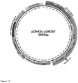
- An exemplary sequence of such a plasmid is given in SEQ ID NO: 9 (F/HN-SIV-hCEF-soA1AT, illustrated in Figure 15A ).
- the invention also provides an F/HN-SIV lentiviral vector comprising (i) an hCEF promoter; and (ii) an FVIII transgene; wherein no intron is positioned between the promoter and the transgene.
- a lentiviral vector may be produced by the method described herein, using a plasmid carrying the FVIII transgene and a promoter.
- pDNA1 vector genome
- Exemplary sequences of such plasmids are given in SEQ ID NO: 11 to 14 (illustrated in Figure 22A to E ).
- the lentiviral vector as described above comprises a transgene.
- the transgene comprises a nucleic acid sequence encoding a gene product, e.g., a protein.
- the nucleic acid sequence encoding a CFTR, A1AT or FVIII comprises (or consists of) a nucleic acid sequence having at least 90% (such as at least 90, 92, 94, 95, 96, 97, 98, 99 or 100%) sequence identity to the CFTR, A1AT or FVIII nucleic acid sequence respectively.
- the nucleic acid sequence encoding CFTR, A1AT or FVIII comprises (or consists of) a nucleic acid sequence having at least 95% (such as at least 95, 96, 97, 98, 99 or 100%) sequence identity to the CFTR, A1AT or FVIII nucleic acid sequence respectively.
- the nucleic acid sequence encoding CFTR is provided by SEQ ID NO: 7
- the nucleic acid sequence encoding A1AT is provided by SEQ ID NO: 15, or by the complementary sequence of SEQ ID NO: 26
- the nucleic acid sequence encoding FVIII is provided by SEQ ID NO: 16 and 30, or by the respective complementary sequences of SEQ ID NO: 28 and 31, or variants thereof.
- polypeptide as used herein also encompasses variant sequences.
- the polypeptide encoded by the transgene of the invention may have at least 90% (such as at least 90, 92, 94, 95, 96, 97, 98, 99 or 100%) sequence identity to a functional CFTR, A1AT or FVIII polypeptide sequence respectively.
- the amino acid sequence of the CFTR, A1AT or FVIII transgene comprises (or consists of) an amino acid sequence having at least 95% (such as at least 95, 96, 97, 98, 99 or 100%) sequence identity to the functional CFTR, A1AT or FVIII polypeptide sequence respectively.
- the amino acid sequence of the A1AT protein encoded by the transgene of the invention comprises (or consists of) the amino acid sequence of SEQ ID NO: 27, or variants thereof.
- said variant A1AT proteins of the invention have at least 90% (such as at least 90, 92, 94, 95, 96, 97, 98, 99 or 100%), more preferably at least 95% or more sequence identity with SEQ ID NO: 27.
- the nucleic acid sequence encoding CFTR, A1AT or FVIII comprises (or consists of) the CFTR, A1AT or FVII complementary DNA sequence respectively.
- the CFTR, A1AT or FVIII transgene is a sequence-optimised CFTR, A1AT or FVIII ( soCFTR2, soA1AT or FVIII ).
- SEQ ID NOS: 7, 15 and 16 An example is provided by SEQ ID NOS: 7, 15 and 16 respectively.
- An exemplary complementary sequence-optimised A1AT sequence is given by SEQ ID NO: 26.
- Exemplary complementary sequence optimised FVIII sequences are given by SEQ ID NOs: 28 and 31.
- the F/HN vector transgene expression is driven by cytomegalovirus (CMV) promoter.
- CMV cytomegalovirus
- EF1a elongation factor 1a
- the vector transgene expression is driven by hybrid human CMV enhancer/EF1a (hCEF) promoter.
- the hCEF promoter has all CG dinucleotides replaced with any one of AG, TG or GT.
- the hCEF promoter is CpG-free.
- the lentiviral vector may be produced using the F/HN-SIV-hCEF-soCFTR2-IC plasmid.
- CFTR is expressed under control of the hCEF promoter.
- This lentiviral vector may be described as comprising F/HN-SIV-hCEF-soCFTR2-IC, as it comprises the SIV F/HN elements, as well as an integrase competent expression cassette comprising CFTR under the control of the hCEF promoter.
- This lentiviral vector of the invention is capable of producing long-lasting, repeatable, high-level expression in airway cells without inducing an undue immune response. Consequently, the invention provides an efficient means of in vivo gene therapy, for example, CFTR gene transfer into the CF lung for the treatment of CF lung disease.
- the lentiviral vector may be produced using the F/HN-SIV-hCEF-soA1AT plasmid.
- A1AT is expressed under control of the hCEF promoter.
- This lentiviral vector may be described as comprising F/HN-SIV-hCEF-soA1AT, as it comprises the SIV F/HN elements, as well as an expression cassette comprising A1AT under the control of the hCEF promoter.
- This lentiviral vector of the invention is capable of producing long-lasting, repeatable, high-level expression in airway cells without inducing an undue immune response.
- the invention provides an efficient means of in vivo gene therapy, for example, A1AT gene transfer into a patient's lung or nose for the production of A1AT which is then secreted into the circulatory system (as described herein).
- this vector and other vectors of the invention comprising the A1AT transgene may be used in the treatment of A1AT deficiency, or other indications as described herein.
- the lentiviral vectors may be produced using the F/HN-SIV-CMV-HFVIII-V3, F/HN-SIV-hCEF-HFVIII-V3, F/HN-SIV-CMV-HFVIII-N6-co and/or F/HN-SIV-hCEF-HFVIII-N6-co plasmids.
- the invention provides only the hCEF containing plasmids.
- HFVIII refers to human FVIII. In this instance, FVIII is expressed under control of the hCEF (as claimed) or CMV promoter.
- lentiviral vectors may be described as comprising F/HN-SIV-CMV-HFVIII-V3, F/HN-SIV-hCEF-HFVIII-V3, F/HN-SIV-CMV-HFVIII-N6-co and F/HN-SIV-hCEF-HFVIII-N6-co respectively, as they comprise the SIV F/HN elements, as well as an expression cassette comprising FVIII under the control of the hCEF/CMV promoter.
- Viral vector products produced using the F/HN-SIV-CMV-HFVIII-V3, F/HN-SIV-hCEF-HFVIII-V3, F/HN-SIV-CMV-HFVIII-N6-co and/or F/HN-SIV-hCEF-HFVIII-N6-co plasmids are also known as vGM126, vGM127, vGM142 and vGM129 (see Figure 22 ). These lentiviral vectors are capable of producing long-lasting, repeatable, high-level expression in airway cells without inducing an undue immune response.
- the invention provides an efficient means of in vivo gene therapy, for example, FVIII gene transfer into a patient's lung or nose for the production of FVIII which is then secreted into the circulatory system (as described herein).
- vectors of the invention comprising the FVIII transgene may be used in the treatment of haemophilia, or other indications as described herein.
- the lentiviral vectors of the invention do not contain an intron between the promoter and the transgene.
- the vector genome plasmids of the invention (used to generate said lentiviral vectors as described herein) also do not contain an intron between the promoter and the transgene.
- the invention therefore provides, in one embodiment, no intron between the hCEF promoter and the coding sequences to be expressed.
- the coding sequence to be expressed is a CFTR, A1AT and / or FVIII nucleic acid sequence.
- the vectors of the invention comprise central polypurine tract (cPPT) and the Woodchuck hepatitis virus posttranscriptional regulatory elements (WPRE).
- cPPT central polypurine tract
- WPRE Woodchuck hepatitis virus posttranscriptional regulatory elements
- the WPRE sequence is provided by SEQ ID NO: 8.
- the vector of the invention is used for gene therapy.
- the disease to be treated is CF.
- the disease to be treated is Primary Ciliary Dyskinesia (PCD).
- the vector is used to treat acute lung injury.
- the disease to be treated is Surfactant Protein B (SP-B) deficiency, Alpha 1-antitrypsin Deficiency (A1AD), Pulmonary Alveolar Proteinosis (PAP), Chronic obstructive pulmonary disease (COPD).
- the disease is an inflammatory, immune or metabolic condition.
- the disease to be treated may be a cardiovascular disease or blood disorder, particularly a blood clotting deficiency.
- the disease to be treated is Haemophilia A, Haemophilia B, or Haemophilia C, Factor VII deficiency and/or von Willebrand disease.
- the disease to be treated is an inflammatory disease, infectious disease or metabolic condition, such as, lysosomal storage disease.
- Non-limiting examples of diseases which may be treated using A1AT gene therapy according to the present invention include type 1 and type 2 diabetes, acute myocardial infarction, ischemic heart disease, rheumatoid arthritis, inflammatory bowel disease, transplant rejection, graft versus host (GvH) disease, multiple sclerosis, liver disease, cirrhosis, vasculitides and infections, such as bacterial and/or viral infections.
- the vector can effectively treat a disease by providing a transgene for the correction of the disease. For example, inserting a functional copy of the CFTR gene to ameliorate or prevent lung disease in CF patients, independent of the underlying mutation.
- a production method for the lentiviral vectors of the invention is provided.
- the method of the invention is a scalable GMP-compatible method.
- the method of the invention allows the generation of high titre purified F/HN vectors.
- the method of the invention comprises the following steps:
- the one or more plasmids provide the vector genome, the Gag-Pol, Rev, F and HN.
- the vector genome plasmid encodes all the genetic material that is packaged into final lentiviral vector, including the transgene.
- the vector genome plasmid may be designated herein as "pDNA1”.
- the other four plasmids are manufacturing plasmids encoding the Gag-Pol, Rev, F and HN proteins. These plasmids may be designated "pDNA2a", “pDNA2b”, “pDNA3a” and "pDNA3b” respectively.
- the lentivirus is SIV, such as SIV1, preferably SIV-AGM.
- the F and HN proteins are derived from a Paramyxovirus, such as Sendai virus.
- the vector genome plasmid (pDNA1) comprises the transgene and the transgene promoter.
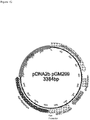
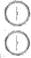
- the five plasmids are characterised by Figures 1A-1E , thus pDNA1 is the pGM326 plasmid of Figure 1A , pDNA2a is the pGM299 plasmid of Figure 1B , pDNA2b is the pGM299 plasmid of Figure 1C , pDNA3a is the pGM301 plasmid of Figure 1D and pDNA3b is the pGM303 plasmid of Figure 1E .
- the final CFTR containing lentiviral vector may be referred to as vGM058 (see the Examples).
- the vGM058 vector is a preferred embodiment of the invention.
- the five plasmids may be characterised by Figures 15A (thus plasmid pDNA1 may be pGM407) and 1B-E (as above for the specific CFTR embodiment).
- the five plasmids may be characterised by Figures 22C-F (thus plasmid pDNA1 may be pGM411, pGM412, pGM413 or pGM414) and 1BE. According to the invention, pDNA1 may be pGM413 or pGM414.
- the plasmid as defined in Figure 1A is represented by SEQ ID NO: 1; the plasmid as defined in Figure 1B is represented by SEQ ID NO: 2; the plasmid as defined in Figure 1C is represented by SEQ ID NO: 3; the plasmid as defined in Figure 1D is represented by SEQ ID NO: 4; the plasmid as defined in Figure 1E is represented by SEQ ID NO: 5; the plasmid as defined in Figure 15A is represented by SEQ ID NO: 9.
- F/HN-SIV-CMV-HFVIII-V3, F/HN-SIV-hCEF-HFVIII-V3, F/HN-SIV-CMV-HFVIII-N6-co and/or F/HN-SIV-hCEF-HFVIII-N6-co plasmids as defined in Figure 22B are represented by SEQ ID NOs: 11 to 14 respectively.
- the vector genome plasmid (pDNA1) provides the enhancer/promoter, Psi, RRE, cPPT, mWPRE, SIN LTR, SV40 polyA (see Figure 1A ), which are important for viral manufacture.
- the CMV enhancer/promoter, SV40 polyA, colE1 Ori and KanR are involved in manufacture of the lentiviral vector of the invention (e.g. vGM058), but are not found in the final viral vector.
- the RRE, cPPT (central polypurine tract), hCEF, soCFTR2 (transgene) and mWPRE from pGM326 are found in the final viral vector.
- SIN LTR (long terminal repeats, SIN/IN self inactivating) and Psi (packaging signal) may be found in the final viral vector.
- corresponding elements from the other vector genome plasmids are required for manufacture (but not found in the final vector), or are present in the final viral vector.
- the F and HN proteins from pDNA3a and pDNA3b are important for infection of target cells with the final lentiviral vector, i.e. for entry of a patients epithelial cells (typically lung or nasal cells as described herein).
- the products of the pDNA2a and pDNA2b plasmids are important for virus transduction, i.e. for inserting the lentiviral DNA into the host's genome.
- the promoter, regulatory elements (such as WPRE) and transgene are important for transgene expression within the target cell(s).
- steps (a)-(f) are carried out sequentially.
- the cells are HEK293 cells or 293T/17 cells.
- the cells are grown in animal-component free (serum-free) media.
- the transfection is carried out by the use of PEIProTM.
- the nuclease is an endonuclease, for example, Benzonase®.
- the trypsin activity is provided by an animal origin free, recombinant enzyme such as TrypLE SelectTM.
- the addition of the nuclease is at the pre-harvest stage. In an alternative embodiment, the addition of the nuclease is at the post-harvest stage. In another embodiment, the addition of trypsin is at the pre-harvest stage. In another embodiment, the addition of the trypsin is at the post-harvest stage.
- the purification step comprises a chromatography step.
- mixed-mode size exclusion chromatography SEC
- anion exchange chromatography is used.
- no salt gradient is used for the elution step.
- this method is used to produce the lentiviral vectors of the invention.
- the vector of the invention comprises the CFTR, A1AT and / or FVIII gene.
- the vector of the invention comprises any of the above-mentioned genes, or the genes encoding the above-mentioned proteins.
- any combination of one or more of the specific plasmid constructs provided by Figures 1A-1E , Figure 15A and/or Figure 22C-22F is used to provide a vector of the invention, provided the resulting vector comprises the hCEF promoter.
- the invention further provides the lentiviral vectors of the invention for use in a method of treating a disease, the method comprising administering a lentiviral vector of the invention to a subject.
- the invention provides a lentiviral vector of the invention for use in treatment of a lung disease.
- disease is a chronic disease.
- a method of treating CF is provided.
- a method of treating Primary Ciliary Dyskinesia (PCD), Surfactant Protein B (SP-B) deficiency, Alpha 1-antitrypsin Deficiency (A1AD), Pulmonary Alveolar Proteinosis (PAP), Chronic obstructive pulmonary disease (COPD) is provided.
- the disease is an inflammatory, immune or metabolic condition.
- the disease may be a cardiovascular disease or blood disorder, particularly a blood clotting deficiency, such as Haemophilia A, Haemophilia B, Haemophilia C, Factor VII deficiency and/or von Willebrand disease, an inflammatory disease, infectious disease or metabolic condition, such as, lysosomal storage disease.
- a blood clotting deficiency such as Haemophilia A, Haemophilia B, Haemophilia C, Factor VII deficiency and/or von Willebrand disease
- an inflammatory disease such as, infectious disease or metabolic condition, such as, lysosomal storage disease.
- the disease may be type 1 and type 2 diabetes, acute myocardial infarction, ischemic heart disease, rheumatoid arthritis, inflammatory bowel disease, transplant rejection, graft versus host (GvH) disease, multiple sclerosis, liver disease, cirrhosis, vasculitides and infections, such as bacterial and/or viral infections.
- the lentiviral vectors of the invention may be administered in any dosage appropriate for achieving the desired therapeutic effect.
- Appropriate dosages may be determined by a clinician or other medical practitioner using standard techniques and within the normal course of their work.
- suitable dosages include 1x10 8 transduction units (TU), 1x10 9 TU, 1x10 10 TU, 1x10 11 TU or more.
- compositions comprising the lentiviral vectors described above, and a pharmaceutically-acceptable carrier.
- pharmaceutically acceptable carriers include water, saline, and phosphate-buffered saline.
- the composition is in lyophilized form, in which case it may include a stabilizer, such as bovine serum albumin (BSA).
- BSA bovine serum albumin
- the vectors of the invention may be administered by any appropriate route. It may be desired to direct the compositions of the present invention (as described above) to the respiratory system of a subject. Efficient transmission of a therapeutic/prophylactic composition or medicament to the site of infection in the respiratory tract may be achieved by oral or intra-nasal administration, for example, as aerosols (e.g. nasal sprays), or by catheters. Typically the lentiviral vectors of the invention are stable in clinically relevant nebulisers, catheters and aerosols, etc.
- Formulations for intra-nasal administration may be in the form of nasal droplets or a nasal spray.
- An intra-nasal formulation may comprise droplets having approximate diameters in the range of 100-5000 ⁇ m, such as 500-4000 ⁇ m, 1000-3000 ⁇ m or 100-1000 ⁇ m.
- the droplets may be in the range of about 0.001-100 ⁇ l, such as 0.1-50 ⁇ l or 1.0-25 ⁇ l, or such as 0.001-1 ⁇ l.
- the aerosol formulation may take the form of a powder, suspension or solution.
- the size of aerosol particles is relevant to the delivery capability of an aerosol. Smaller particles may travel further down the respiratory airway towards the alveoli than would larger particles.
- the aerosol particles have a diameter distribution to facilitate delivery along the entire length of the bronchi, bronchioles, and alveoli.
- the particle size distribution may be selected to target a particular section of the respiratory airway, for example the alveoli.
- the particles may have diameters in the approximate range of 0.1-50 ⁇ m, preferably 1-25 ⁇ m, more preferably 1-5 ⁇ m.
- Aerosol particles may be for delivery using a nebulizer (e.g. via the mouth) or nasal spray.
- An aerosol formulation may optionally contain a propellant and/or surfactant.
- nucleic acid sequence and “polynucleotide” are used interchangeably and do not imply any length restriction.
- nucleic acid and “nucleotide” are used interchangeably.
- nucleic acid sequence and “polynucleotide” embrace DNA (including cDNA) and RNA sequences.
- transgene and “gene” are also used interchangeably and both terms encompass fragments or variants thereof encoding the target protein.
- transgenes of the present invention include nucleic acid sequences that have been removed from their naturally occurring environment, recombinant or cloned DNA isolates, and chemically synthesized analogues or analogues biologically synthesized by heterologous systems.
- the polynucleotides of the present invention may be prepared by any means known in the art. For example, large amounts of the polynucleotides may be produced by replication in a suitable host cell.
- the natural or synthetic DNA fragments coding for a desired fragment will be incorporated into recombinant nucleic acid constructs, typically DNA constructs, capable of introduction into and replication in a prokaryotic or eukaryotic cell.
- DNA constructs will be suitable for autonomous replication in a unicellular host, such as yeast or bacteria, but may also be intended for introduction to and integration within the genome of a cultured insect, mammalian, plant or other eukaryotic cell lines.
- the polynucleotides of the present invention may also be produced by chemical synthesis, e.g. by the phosphoramidite method or the tri-ester method, and may be performed on commercial automated oligonucleotide synthesizers.
- a double-stranded fragment may be obtained from the single stranded product of chemical synthesis either by synthesizing the complementary strand and annealing the strand together under appropriate conditions or by adding the complementary strand using DNA polymerase with an appropriate primer sequence.
- isolated in the context of the present invention denotes that the polynucleotide sequence has been removed from its natural genetic milieu and is thus free of other extraneous or unwanted coding sequences (but may include naturally occurring 5' and 3' untranslated regions such as promoters and terminators), and is in a form suitable for use within genetically engineered protein production systems.
- isolated molecules are those that are separated from their natural environment.
- degenerate codon representative of all possible codons encoding each amino acid.
- some polynucleotides encompassed by the degenerate sequence may encode variant amino acid sequences, but one of ordinary skill in the art can easily identify such variant sequences by reference to the amino acid sequences of the present invention.
- a “variant" nucleic acid sequence has substantial homology or substantial similarity to a reference nucleic acid sequence (or a fragment thereof).
- a nucleic acid sequence or fragment thereof is “substantially homologous" (or “substantially identical") to a reference sequence if, when optimally aligned (with appropriate nucleotide insertions or deletions) with the other nucleic acid (or its complementary strand), there is nucleotide sequence identity in at least about 70%, 75%, 80%, 82, 84, 86, 88, 90, 92, 94, 96, 98 or 99% of the nucleotide bases. Methods for homology determination of nucleic acid sequences are known in the art.
- a "variant" nucleic acid sequence is substantially homologous with (or substantially identical to) a reference sequence (or a fragment thereof) if the "variant" and the reference sequence they are capable of hybridizing under stringent (e.g. highly stringent) hybridization conditions.
- Nucleic acid sequence hybridization will be affected by such conditions as salt concentration (e.g. NaCl), temperature, or organic solvents, in addition to the base composition, length of the complementary strands, and the number of nucleotide base mismatches between the hybridizing nucleic acids, as will be readily appreciated by those skilled in the art.
- Stringent temperature conditions are preferably employed, and generally include temperatures in excess of 30°C, typically in excess of 37°C and preferably in excess of 45°C.
- Stringent salt conditions will ordinarily be less than 1000 mM, typically less than 500 mM, and preferably less than 200 mM.
- the pH is typically between 7.0 and 8.3. The combination of parameters is much more important than any single parameter.
- nucleic acid percentage sequence identity Methods of determining nucleic acid percentage sequence identity are known in the art.
- a sequence having a defined number of contiguous nucleotides may be aligned with a nucleic acid sequence (having the same number of contiguous nucleotides) from the corresponding portion of a nucleic acid sequence of the present invention.
- Tools known in the art for determining nucleic acid percentage sequence identity include Nucleotide BLAST.
- preferential codon usage refers to codons that are most frequently used in cells of a certain species, thus favouring one or a few representatives of the possible codons encoding each amino acid.
- the amino acid threonine (Thr) may be encoded by ACA, ACC, ACG, or ACT, but in mammalian host cells ACC is the most commonly used codon; in other species, different codons may be preferential.
- Preferential codons for a particular host cell species can be introduced into the polynucleotides of the present invention by a variety of methods known in the art. Introduction of preferential codon sequences into recombinant DNA can, for example, enhance production of the protein by making protein translation more efficient within a particular cell type or species.
- the nucleic acid sequence is codon optimized for expression in a host cell.
- a "fragment" of a polynucleotide of interest comprises a series of consecutive nucleotides from the sequence of said full-length polynucleotide.
- a “fragment" of a polynucleotide of interest may comprise (or consist of) at least 30 consecutive nucleotides from the sequence of said polynucleotide (e.g. at least 35, 50, 75, 100, 150, 200, 250, 300, 350, 400, 450, 500, 550, 600, 650, 700, 750, 800 850, 900, 950 or 1000 consecutive nucleic acid residues of said polynucleotide).
- a fragment may include at least one antigenic determinant and/or may encode at least one antigenic epitope of the corresponding polypeptide of interest.
- a fragment as defined herein retains the same function as the full-length polynucleotide or polypeptide.
- vectors comprising a hCEF promoter and plasmids/tools for the production of such vectors are encompassed by the invention. Disclosure relating to vectors comprising other promoters and vector genome plasmids/other tools specifically for the production of vectors not comprising a hCEF promoter is included for comparative purposes only.
- HEK293T, Freestyle 293F (Life Technologies, Paisley, UK) and 293T/17 cells (CRL-11268; ATCC, Manassas, VA) were maintained in Dulbecco's minimal Eagle's medium (Invitrogen, Carlsbad, CA) containing 10% fetal bovine serum and supplemented with penicillin (100 U/ml) and streptomycin (100 ⁇ g/ml) or FreestyleTM 293 Expression Medium (Life Technologies).
- pCAGGS-Fct4 and pCAGGS-SIVct+HN were constructed as follows:
- pCAGGS-SIVct+HN For construction of pCAGGS-SIVct+HN, the genes encoding the cytoplasmic tail of SIVagm TMP and the N terminus of HN protein were first amplified by PCR from pCAGGS-SIVct/HN with the primer pair 5'-GAGACTCGAGATGTGGTCTGAGTTAAAAATCAGG-3' (SEQ ID NO: 24) and 5'-AGAGGTAGACCAGTACGAGTCACGTTTGCCCCTATCACCATCCCTAACCCTCTGTC ATAAAC-3' (SEQ ID NO: 25).
- the resulting PCR fragment was cloned into the Xho I and Acc I sites of pKS+SIVct to generate pKS+SIVct-H. Then a Xho I- Dra III fragment from pKS+SIVct-H and a Dra III -Bsu 36I fragment from pCAGGS-HN were cloned into the Xho I and Bsu 36I sites of pCAGGS to yield pCAGGS-SIVct+HN.
- the cPPT and WPRE sequences were inserted in the SIV-derived gene transfer plasmid.
- An example of the WPRE sequence used is provided in SEQ ID NO: 8.
- the plasmid pGM101 contains the colE1 origin of replication, kanamycin resistance gene and promoter and was created by synthetic gene synthesis (GeneArt, Regensburg, Germany; now LifeTechnologies Ltd).
- the hybrid CMV/SIV R U5 LTR, partial Gag, RRE, cPPT, SIN U3 and R sequences from pBS/CG2-Rc/s-CMV-D U were amplified by PCR and inserted along with the hCEF enhancer/Promoter sequence amplified by PCR from pGM169 ( Hyde et al. Nature Biotechnology 26:549 ) and the soCFTR2 cDNA isolated from pGM169 on a Nhel-Apal restriction enzyme fragment into pGM101 to create pGM326.
- the CMV enhancer/chicken beta actin promoter along with associated exon/intron sequences, SIV GagPol and RRE sequences and the SV40 polyA/origin of replication were amplified by PCR from pCAGGS/Sagm-gtr ( Nakajima et al. 2000 Human Gene Therapy 11:1863 ) to create pGM297.
- the CMV enhancer/ promoter along with associated exon/intron sequences and SV40 polyA sequence from pCI were isolated on a BgIII-BamHI restriction enzyme fragment and the SIV Rev sequence derived from pCAGGS/Sagm-gtr amplified by PCR were inserted into pGM101 to create pGM299.
- the CMV enhancer/chicken beta actin promoter along with associated exon/intron sequences, the Fct4 cDNA and SV40 polyA/origin from pCAGGS-Fct4 were isolated on a SaII-HindIII restriction enzyme fragment by a combination of gene synthesis, PCR and restriction enzyme fragment recombination and inserted into pGM101 to create pGM301.
- the CMV enhancer/chicken beta actin promoter along with associated exon/intron sequences, the SIVct+HN cDNA and SV40 polyA/origin from pCAGGS-SIVct+HN were isolated on a SaII-HindIII restriction enzyme fragment by a combination of gene synthesis, PCR and restriction enzyme fragment recombination and inserted into pGM101 to create pGM303.
- pGM plasmids of the invention were made using standard techniques and in accordance with the above disclosure.
- restriction enzymes and PCR polymerases were supplied by New England Biolabs (Ipswich, MA, USA) and DNA purification reagents were supplied by Qiagen (Limburg, Netherlands).
- SeV-F/HN-pseudotyped SIV vector was produced by transfecting 293T/17 cells (15 cm diameter culture dishes) with four plasmids complexed to Lipofectamine/Plus reagents (Invitrogen) according to the manufacturer's recommendations [Plasmid-1: 10 ⁇ g SIV-derived transfer plasmid carrying a GFP, a luciferase (lux) reporter gene, or a GFP-CFTR fusion construct, Plasmid-2: 3 ⁇ g packaging plasmid, Plasmid-3: 2 ⁇ g pCAGGS-Fct4, Plasmid 4: 2 ⁇ g pCAGGS-SIVct+HN; Figures 1A-E show schematic drawings of plasmids used for the production of vectors of the invention].
- VSV-G pseudotyped SIV vector was produced using a similar protocol, but a pVSV-G plasmid (2 ⁇ g; Clontech, Mountain View, CA) was used instead of pCAGGS-Fct4 and pCAGGS-SIVct+HN.
- a pVSV-G plasmid (2 ⁇ g; Clontech, Mountain View, CA) was used instead of pCAGGS-Fct4 and pCAGGS-SIVct+HN.
- the culture medium was replaced with 30 ml serum-free Dulbecco's modified Eagle medium containing 5 mmol/l sodium butyrate. Sodium butyrate stimulates the vector production to inhibit histone deacetylase.
- the culture supernatant containing the SIV vector was harvested 48 hours after transfection, filtered through a 0.45 ⁇ m filter membrane, and further concentrated by high-speed centrifugation (20,000 g for 4 hours at 4 °C, Avanti JA18 rotor; Beckman Coulter, Brea, CA).
- the vector pellets were suspended in PBS (Invitrogen) to 100- to 200-fold concentration and stored at -80 °C.
- SeV-F/HN-pseudotyped SIV vector was produced by transfecting HEK293T or 293T/17 cells cultured in FreeStyleTM 293 Expression Medium with a mixture of five plasmids with the following characteristics: pDNA1 (for example pGM326; Figure 1A ) encodes the lentiviral vector mRNA; pDNA2a (for example pGM297; Figure 1B ) encodes SIV Gag and Pol proteins; pDNA2b (for example pGM299: Figure 1C ) encodes SIV Rev proteins; pDNA3a (for example pGM301; Figure 1D ) encodes the Sendai virus-derived Fct4 protein [ Kobayashi et al., 2003 J.
- Virol. 77:2607 pDNA3b (for example pGM303; Figure 1E ) encodes the Sendai virus-derived SIVct+HN [ Kobayashi et al., 2003 J. Virol. 77:2607 ] complexed with PEIpro (Polyplus, Illkirch, France).
- Cell culture media was supplemented at 12-24 post-transfection with sodium butyrate. Sodium butyrate stimulates vector production via inhibiting histone deacetylase resulting in increasing expression of the SIV and Sendai virus fusion protein components encoded by the five plasmids.
- Cell culture media was supplemented at 44-52 hours and/or 68-76 hours post-transfection with 5 units/mL Benzonase Nuclease (Merck Millipore, Nottingham, UK). The culture supernatant containing the SIV vector was harvested 68-76.5 hours after transfection, and clarified by filtration through a 0.45 ⁇ m membrane.
- the SIV vector is treated by digestion with a protease containing trypsin activity - for example an animal origin free, recombinant enzyme such as TrypLE SelectTM. Subsequently, SIV vector is typically further purified and concentrated by anion-exchange chromatography and/or tangential flow filtration ultra-filtration/dia-filtration.
- the particle titre was determined using real-time reverse transcriptase-PCR.
- Virus RNA was purified using a QIAamp viral RNA mini-kit (QIAGEN, Strasse, Germany), and reverse transcribed using Superscript II (Invitrogen).
- QIAGEN QuantiTect probe PCR system
- primers for amplifying 131 nucleotides (bp) spanning the WPRE sequence forward primer: 5'-ggatacgctgctttaatgcc-3', reverse primer: 5'-acgccacgttgcctgacaac-3'
- forward primer 5'-ggatacgctgctttaatgcc-3'
- reverse primer 5'-acgccacgttgcctgacaac-3'
- SIV gene transfer plasmid DNA (3 ⁇ 10 4 to 2 ⁇ 10 6 molecules) was used as standard.
- Transduction units were determined by transducing 293T cells with serial dilutions of vector stock and quantification of transduced cells by GFP fluorescence (for F/HN-SIV-GFP and VSV-G-SIV-GFP) or staining with anti-luciferase antibody (for F/HN-SIV-lux).
- the particle titre (VP/mL) was typically determined using real-time reverse transcriptase-PCR.
- Virus RNA was purified using a QIAamp viral RNA mini-kit (QIAGEN, Strasse, Germany), and reverse transcribed using reverse transcriptase (Life Technologies).
- Transduction units were determined by transducing 293T/17 or Freestyle 293F cells with serial dilutions of SIV vector and quantification of WPRE containing provirus DNA by TaqMan quantitative PCR system (Life Technologies) using primers amplifying a portion of the WPRE sequence in an ABI PRISM 7700 Sequence Detector System (Life Technologies). Plasmid DNA molecules containing WPRE sequences were used as quantitative standards.
- Murine tracheal epithelial cells were isolated as follows. C57BL/6N Mice were culled and the tracheas were excised from the larynx to the main bronchial branches using sterile surgical instruments. The tissues were placed in a tube containing cold Ham's F-12 medium with 100 U/ml penicillin (P), 100 ⁇ g/ml streptomycin (S) and 2.5 mg/ml amphotericin B (A) (Ham's F12/PSA medium) and kept on ice.
- P penicillin
- S 100 ⁇ g/ml streptomycin
- A amphotericin B
- the tracheas were cleaned from adherent muscles and connective tissue, cut longitudinally to expose the internal respiratory epithelium, and placed in 0.15% pronase solution in F-12 medium ( ⁇ 5 ml in 15 ml tube). Tissue digestion was performed overnight (15-18 hr) at 4 °C. To block the enzymatic reaction, 10% fetal bovine serum (FBS) was added to the tissue digest. After gently inverting the tube to detach more cells, the tracheas were placed into a new tube containing 10% FBS/Ham's F-12/PS solution, and inverted as before. This step was repeated two more times.
- FBS fetal bovine serum
- the content of the four tubes was pooled together and centrifuged at 500g for 10 min at 4°C.
- the pellet was re-suspended in DNase solution (0.5 mg/ml crude pancreatic DNase plus 10 mg/ml BSA in FBS/Ham's F-12/PS solution, about 200 ⁇ l/trachea), incubated on ice for 5 minutes, and centrifuged as before.
- DNase solution 0.5 mg/ml crude pancreatic DNase plus 10 mg/ml BSA in FBS/Ham's F-12/PS solution, about 200 ⁇ l/trachea
- tEC were resuspended in Progenitor Cell Targeted (PCT) medium (CnT-17, CELLnTEC, Bern, Switzerland), an antibiotics and antimycotics-free formulation specifically designed for human and mouse airways progenitor cells isolation and proliferation.
- PCT Progenitor Cell Targeted
- tEC were then plated in a Primaria tissue culture dish (Becton Dickinson Labwere, Franklin Lakes, NJ, USA) and incubated for 3-4 hr in 5% CO 2 at 37 °C. Non-adherent cells were collected and centrifuged at 500g for 5 min at 4°C and counted in a haemocytometer.
- tEC were suspended in PCT medium and seeded on a NunclonTM ⁇ plate (Nunc A/S, Roskilde, Denmark), coated with 50 ⁇ g/ml type 1 rat tail collagen at a recommended seeding density of 4x10 3 cells/cm 2 .
- tEC were also cultured in a control basic medium, containing DMEM/Ham's F12 supplemented with L-glutamine (4 mM), HEPES (15 mM) and NaHCO3 (3.4 mM). Plates were incubated at 37°C with 5% CO 2 . To determine the proportion of basal cells in the tEC population before and after expansion in PCT medium, cytospin preparations were stained with the anti-KRT5 antibody and the appropriate secondary antibody.
- the pool of tEC produced comprised both fully differentiated cells (Clara and ciliated cells) and basal cells. Once seeded onto permeable membranes tEC were able to generate an air liquid interface (ALI) culture system as a result of basal cell proliferation and redifferentiation into secretory and ciliated cells.
- ALI air liquid interface
- 2D tEC cultures using a commercially available proprietary Progenitor Cell Targeted (PCT) medium specifically formulated to support the proliferation of airway progenitor cells while maintaining them in an undifferentiated status were assessed.
- PCT Progenitor Cell Targeted
- tEC seeded on collagen-coated plastic surfaces (4x10 3 cells/cm 2 ) and exposed to PCT medium were able to grow rapidly and became confluent within 5-8 days whereas tEC exposed to the growth factor-deficient control medium were unable to adhere and propagate.
- Example 6 Ex vivo transduction of basal cells-enriched tEC cultures with F/HN-SIV-GFP
- tEC prepared in Example 5 were grown to approximately 70% confluence over 7 days in PCT medium and transduced with F/HN-SIV carrying a green fluorescent protein reporter gene (F/HN-SIV-GFP) at an MOI 100 and incubated at 37°C with 5% CO 2 for 3 days.
- F/HN-SIV-GFP green fluorescent protein reporter gene
- basal cells-enriched tEC cultures were detached with the enzyme accutase (CELLnTEC), re-suspended in PBS/1%BSA and subjected to FACS analysis, counting an average of 20.277 ⁇ 2.478 cells/group.
- the F/HN-SIV vector transduced 26% ⁇ 0.9% of basal cells-enriched tEC (p ⁇ 0.0001 when compared to untransduced controls).
- Example 7 In vivo administration to the mouse nose
- mice female, 6-8 weeks were used. Mice were anesthetized, placed horizontally on their backs onto a heated board, and a thin catheter ( ⁇ 0.5 mm outer diameter) was inserted ⁇ 2.5 mm from the tip of nose into the left nostril. Using a syringe pump (Cole-Parmer, Vernon Hills, IL), vector (100 ⁇ l) was then slowly perfused onto the nasal epithelium (1.3 ⁇ l/min) for 75 minutes. Despite perfusion of virus into the left nostril, we routinely observe transfection in both left and right nostrils, which is due to dispersion of the solutions throughout the entire nasal cavity.
- a syringe pump Cold-Parmer, Vernon Hills, IL
- mice preconditioned with 1% lysophosphatidylcholine as described by Limberis et al ., 2002, were used as controls. At indicated time points (3-360 days after transduction), mice were culled to visualize GFP expression. As shown in Figure 2 , GFP expression was observed for at least 449 days post-transduction, whereas the negative control showed no GFP expression. As shown in Figure 3 , transgene expression with the F/NH-SIV vector was consistent, with observable GFP expression at least 360 days post-transduction in 10 independently tested mice.
- transduction with an F/HN-SIV vector of the invention comprising an hCEF promoter resulted in long-term expression of a CpG rich reporter gene (luciferase).
- CpG rich reporter gene luciferase
- mice were transduced with either one dose of F/HN-SIV-lux (single-dose group), or two doses of F/HN-SIV-GFP (day 0, day 28), followed by F/HN-SIV-lux on day 56 (repeat-dose group).
- mice receiving F/HN-SIV-lux (single-dose group) and F/HN-SIV-lux on day 56 (repeat-dose group) were of similar age and were transduced at the same time. Gene expression was analysed 30 days after F/HN-SIV-lux administration.
- mice were transfected with the cationic lipid GL67A complexed to a luciferase reporter gene as previously described ( Griesenbach, U. et al., Methods Mol Biol. 2008; 433:229-42 ) and luciferase expression was measured 2 days after transfection.
- Insertion site profiling was conducted on transduced mice, and survival time investigated as set out in the description of Figure 12 above. Transduction using the F/HN-SIV vector of the invention was not observed to have any adverse effect on mouse survival compared with an existing F/HN-SIV vector or negative (buffer only) control (see Figure 12G ).
- Example 8 Induced regeneration of nasal epithelial cells by polidocanol treatment
- Nasal epithelial cells were stripped by polidocanol treatment according to the method described ( Borthwick et al., Am J Respir Cell Mol Biol. 2001 Jun; 24(6):662-70 ), with some modification.
- mice were anesthetized and 10 ⁇ l polidocanol (2%) was administered to the nose as a bolus by "nasal sniffing".
- F/HN-SIV-GFP 4 ⁇ 10 8 TU/mouse vector to mouse nasal epithelium. Seven days after transduction, nasal tissue was perfused with 10 ⁇ l of 2% (vol/vol in PBS) polidocanol, and this treatment was repeated again 3 weeks later. Histological sections were analysed 58 days after vector administration (30 days after the last polidocanol treatment).
- Example 10 Tissue preparation for histological assessment of GFP expression and/or basal cell detection
- mice were culled and the skin was removed. The head was cut at eye level and skin, jaw, tongue, and the soft tip of the nose were carefully removed.
- GFP fluorescence was detected using fluorescence stereoscopic microscopy (Leica, Ernst Leitz Optician Werke, Germany). Subsequently, the tissue was fixed in 4% paraformaldehyde (pH 7.4) overnight at room temperature and was then submerged in 20% EDTA (pH 7.5 for 5 days) for decalcification. The EDTA solution was changed at least every second day. After decalcification, the tissue was incubated in 15% sucrose overnight at room temperature and was then embedded in Tissue Mount (Chiba Medical, Soka, Japan).
- EGFP expression was determined in histological sections of the mouse nasal cavity (2mm from the tip of the nose) at 30 days post treatment.
- Figure 4 shows the location of EGFP expression ( Figure 4 , white punctate signal).
- Figure 5 shows the cell types transduced by /NH-SIV transgene treatment of the mouse nose. 69% of the cells transduced in the mouse nasal cavity were ciliated respiratory epithelial cells. Other transduced cell types included neuronal cells in the olfactory epithelium (21%) and squamous cells (7%).
- HRP horseradish peroxidase
- Sections were then washed in PBS and incubated with the peroxidase substrate 3-amino-9-ethylcarbazole (AEC) (provided with the kit) for 5 min. Finally, slides were washed in distilled H 2 O, counterstained with aqueous Harris' hematoxylin (BDH) for 15 seconds, washed in tap water, and then in distilled H 2 O.
- AEC peroxidase substrate 3-amino-9-ethylcarbazole
- Immunofluorescence detection of GFP-positive transduced nasal epithelial cells and Krt5 positive basal cells was performed using the following primary and secondary antibodies: goat polyclonal anti-GFP antibody (1:250) (Abcam), rabbit monoclonal anti-KRT5 antibody (1:500) (Abcam), Alexa Fluor 488 donkey anti-Goat IgG (1:200) (Invitrogen, Paisley, UK) and Alexa Fluor 594 goat anti-rabbit IgG (1:200) (Invitrogen).
- To improve antibody performance sections were subjected to heat-mediated antigen retrieval in citrate buffer (10 mM citric acid, 0.05% Tween20, pH 6.0) for 20 min on a water bath at 100 °C.
- ALIs Fully differentiated airway epithelial cells grown as ALI cultures were purchased from Epithelix (Geneva, Switzerland). ALIs were transfected with F/HN-SIV-lux at a multiplicity of infection ranging from ⁇ 25 to ⁇ 300 TU/cell. After 6 hours, the virus was removed and ALIs were incubated for 10-26 days. The basolateral medium was changed every 48 hours during this incubation period. At specified time points, the ALIs were lysed in 100 ⁇ l reporter lysis buffer and luciferase expression was quantified using the Luciferase Assay System (Promega, Victoria, UK) according to the manufacturer's instructions.
- the total protein content of the cultures was quantified using the BioRad protein assay kit (BioRad, Hemel Hempstead, UK). Each sample was assayed in duplicate. Luciferase expression was then presented as relative light units/mg total protein. For bioluminescence imaging 100 ⁇ g luciferin in PBS were added to the apical membrane.
- Luciferase expression was greater at MOI 250 compared with MOI 25.
- HEK293T cells were transfected with F/HN-SIV-GFP-CFTR or an F/HN-SIV-GFP control virus at a multiplicity of infection of 500 TU/cell and cultured for 2 days.
- CFTR chloride channel activity was assayed by measuring the rate of 125 iodide efflux as previously described (Derand, R., et al ., 2003).
- the 125 iodide efflux rates were normalized to the time of forskolin/IBMX addition (time 0). Curves were constructed by plotting rates of 125 iodide efflux against time.
- F/HN-SIV can direct functional CFTR expression, with a relative 125 iodide efflux of 0.65.
- the GL67A plasmid vector achieved a lower 125 iodide efflux value of 0.33.
- Example 13 Transduction of sheep and human primary lung cells and mouse lung
- F/HN-SIV efficiently transduces sheep & human primary lung cells and mouse lung.
- F/HN-SIV-CMV-Lux was used to transduce human nasal brushings (MOI 250) and human and sheep lung slices cultured ex vivo (1x10 7 TU/slice).
- transduction of both the human nasal brushing cells and human and sheep lung slices resulted in substantial luciferase transgene expression (average values in the region of 2x10 2 RLU/mg protein for the human nasal brushings, 1x10 7 RLU/mg protein for the human lung slices and 2 x 10 7 RLU/mg protein for the sheep lung slices) 24-48 hours post-transduction.
- CFTR mRNA expression level (%VE: copies of CFTR mRNA per copy of endogenous CFTR mRNA x 100) at 6-8 days post-transduction was also measured.
- the horizontal dotted line in Figure 9C represents a target expression level of 5% VE, which is thought to represent the therapeutic threshold.
- vGM012 CMW, vGM014 EF1a, vGM020 hCEF and vGM076 hCEF ID achieved expression in the nose above the target level for the whole time course of the experiment.
- the fifth vector, vGM074 CMV ID achieved expression in the nose above the accepted expression level for the whole time course of the experiment.
- vGM014 EF1a and vGM020 hCEF Two of the five vectors tested (vGM014 EF1a and vGM020 hCEF) achieved expression in the lung above the target level for the whole time course of the experiment.
- the vGM020 hCEF vector achieved the highest level of in vivo expression out of the five vectors tested.
- Figure 9H shows EGFP expression at 14 days post transduction in the murine nasal epithelium following delivery of 1.6 x 10 8 TU of F/HN-SIV-hCEF-EGFPLux (vGM020), as visualised by immunohistochemistry (nuclei stained with DAPI).
- F/HN-SIV also efficiently transduces sheep lung i n vivo.
- Acriflavine was instilled (3x100 ⁇ L aliquots over ⁇ 5 minutes) to a proximal airway under direct bronchoscopic visualisation.
- the distribution of the acriflavine can be appreciated by the orange colouration of the dissected airway at postmortem ( Figure 10A ).
- the acriflavine was largely restricted to the conducting airways and absent from the alveolar regions.
- the arrow in Figure 10A indicates the approximate site of instillation.
- the numbers on the ruler are in cm.
- Figure 10B is a diagrammatic representation of the sheep lung (trachea centre/top). The circle represents the region in ( Figure 10A ).
- 5-6 tissue sample blocks were taken at post-mortem at ⁇ 1 cm intervals from the site of instillation.
- FIG. 10C luciferase assays normalised to protein content
- Figure 10D quantitative RT-PCR normalised to endogenous CFTR mRNA levels.
- the horizontal line in Figure 10C represents the highest luciferase activity noted in any sample treated with a non-viral gene transfer vector
- Figure 10D the target expression level of 5% VE (thought to represent the therapeutic threshold, see above).
- the average was higher than the non-viral vector comparison ( Figure 10C ), and also achieved expression above the target 5% VE threshold.
- Example 14 CFTR expression and function measured using human CF intestinal organoids
- Example 15 Generation of lentiviral vectors for A1AT
- Lentiviral vectors were prepared using the SIV backbone and 5 plasmid method described above in Examples 2 and 3 (for the CFTR lentivirus) and using the hCEF promoter as described herein. Two separate lentiviral constructs were generated: one with a human alpha-1-antitrypsin (hAAT) transgene; one with a Gaussia luciferase (Glux) transgene (see Figure 15A and B , SEQ ID NOs: 9 and 10 respectively). The cDNAs contained within these vectors were codon-optimised and CpG-depleted.
- hAAT human alpha-1-antitrypsin
- Glux Gaussia luciferase
- Example 16 Air-liquid interface (ALI) culture using lux reporter gene lentiviral vector
- ALIs Fully differentiated wild-type human ALI cultures (MucilAir) were purchased from Epithelix SARL (Geneva, CH). ALIs were cultured at 37°C and 5% CO 2 and the basolateral culture medium changed every 2-3 days. The culture medium was stored at -20°C until further analysis.
- ALIs were transduced (on day 0) by pipetting 100 ⁇ l of virus (1x10 7 Tagman transfection units (TTU) onto the apical surface. The virus was removed after 4 hours incubation at 37°C, and the basolateral medium replaced.
- TTU Tagman transfection units
- the apical surface of the ALIs was washed by incubating with sterile PBS for one hour. The washings were removed and stored at - 20°C until further analysis.
- Example 17 Transgene expression in lung slice cultures using A1AT and lux reporter gene lentiviral vectors
- Gaussia luciferase expression was determined as described above (Example 16). As shown in Figure 17A , high levels of expression of secreted reporter protein Gaussia luciferase followed lentiviral-mediated gene transfer in human lung slices. AAT (also referred to herein as A1AT) expression was determined using a sandwich ELISA (Abcam, Cambridge, UK), performed according to the manufacturer's recommendations. 50 ⁇ l of sample was assayed in duplicate, and measured on a microplate reader at 450nm. As shown in Figure 17B , high levels of expression of alpha-1-antitrypsin (AAT/A1AT) followed lentiviral-mediated gene transfer in human lung slices.
- AAT also referred to herein as A1AT
- Example 18 In vivo administration of A1AT and lux reporter gene lentiviral vectors to the mouse nose
- mice Female C57BL/6 mice (Charles River, UK) were anaesthetised with isoflurane and given 100ul of virus by nasal instillation as described in Xenariou S et al, Gene Ther 2007 May; 14(9): 768-75 . Animals were given between 1 and 5 doses and observed daily for signs of toxicity.
- mice Female C57BL/6 mice were anaesthetised on day 0 using isoflurane and given a single 100ul dose of the SIV1 hCEF-soGLux virus (1x10 6 TTU) by nasal instillation .
- Control animals were treated with DMEM (tissue culture medium), the main constituent of the viral preparation used in the study.
- PBS the main constituent of the lentivirus production batch used in the study
- mice were sacrificed at the indicated time-points post transduction. Blood was collected by puncturing the left ventricle, and centrifuged at 760g av for 10 minutes to prepare serum. Serum was subsequently frozen at -80°C.
- a bronco-alveolar lavage was performed by dissecting the neck, inserting a cannula into the trachea and securing it in place with suture thread. 500 ⁇ l of PBS was instilled into the lung, and aspirated three times to obtain thorough washing of the epithelial lining. The sample was immediately snap frozen in liquid nitrogen, and stored at -80°C for further analysis.
- Lungs were then dissected and snap-frozen in liquid nitrogen, and subsequently homogenised in lysing matrix D tubes (MP Biomedicals), centrifuged in a FastPrep machine (ThermoFisher Scientific, Waltham, MA, USA) at 4m/s for 45 seconds, and stored at -80 °C for further analysis.
- lysing matrix D tubes MP Biomedicals
- FastPrep machine ThermoFisher Scientific, Waltham, MA, USA
- AAT A1AT
- Glux Gaussia luciferase
- a urea assay (Abcam, Cambridge, UK) was performed according to the manufacturer's instructions.
- the concentration of ATT (A1AT) in the epithelial lining fluid was calculated. Specifically, the concentration of AAT in the broncho-alveolar lavage fluid was multiplied by the dilution factor, to provide an estimate of the 'true' AAT concentration in epithelial lining fluid.
- a "protective" target level of ATT (A1AT) in the epithelial lining fluid (ELF, i.e. the fluid lining the airways and airspaces in the lungs) is 70 ⁇ g/ml (compared with a "normal" target level of ATT (A1AT) in the ELF of 200 ⁇ g/ml).
- ATT epithelial lining fluid
- FIG. 21 therapeutic levels of alpha-1-antitrypsin in epithelial lining fluid followed ATT (A1AT) lentiviral-mediated gene transfer in vivo.
- FVIII lentiviral vectors were prepared using the SIV backbone and 5 plasmid method described above in Examples 2 and 3 (for the CFTR lentivirus) and 15 (for the A1AT lentivirus).
- the promoter-transgene plasmids have SEQ ID NOs: 11 to 14 respectively.
- the SIV sequence was identical to the CFTR constructs (Examples 2 and 3) except for the promoter and cDNA.
- the human cytomegalovirus promotor (CMV) or tissue specific hCEFI promotor/enhancer was used as indicated ( Figure 22 ) to drive expression of FVIII transgenes.
- SIV-F/HN-FVIII-N6-co contained the wild type human FVIII cDNA from which the BDD domain has been deleted and replaced with codon optimised 226 amino acid 6N-glycosylation fragment.
- SIV-FVIII-V3 contains the wild type human FVIII cDNA from which the 226 amino acid glycosylation site has been deleted and replaced with 17 amino acid peptide which expresses 6N-glycosylation triples within the B domain ( McIntosh et al., Blood 2013 121(17); 3335-3344 ).
- Example 21 Quantification of hFVIII antigen and activity levels in in vivo and in vitro models
- FVIII antigen levels in a murine model were quantified by enzyme-linked immunosorbent assay (ELISA) according to the manufacturer's protocol. Briefly, plasma, BAL and lung were analysed for the presence of FVIII antigen using Asserachrom (FVIII:Ag) Elisa (Stago Diagnostics, France).
- ELISA enzyme-linked immunosorbent assay
- FVIII activity was evaluated in an in vitro HEK293T model (FVIII:C, Affinity Biological, Canada). Supernatants were collected 48 and 72 hours after HEK293T transduction with SIV-F/HN-FVIII-N6 or SIV-F/HN-FVIII-N3. FVIII activity was evaluated by following the manufacturer's instructions using 50 ⁇ l supernatants assayed in duplicate. As a negative control the supernatant from untreated HEK293T cells was tested. hFVIII activity was calculated from a standard curve generated using a series of dilutions of normal human pooled plasma (13th British Standard for blood coagulation Factor VIII concentrate, Human; NIBSC).
- HEK293T cells were transduced with two different batches of vGM142 (Batch 1 - 5.9x10 8 TTU/ml and Batch 2 - 2.8x10 8 TTU/ml).
- HEK293T cells were transduced with vGM142 Batch 1 vector ( Figure 23A ) and vGM142 Batch 2 vector ( Figure 23B) at 3 different MOIs (MOI 1; 10; 100), and collected 48 and 72 hours post-transduction.
- Example 22 In vivo administration of FVIII and lux reporter gene lentiviral vectors to the mouse nose
- Wild type C57BL/6 female mice aged 6-8 weeks old (Charles River, UK were anaesthetised using isofluorane and given 100 ⁇ l of virus in Dulbecco's phosphate-buffered saline (D-PBS), as described previously (Griesenbach et al., 2012) and the presence of FVIII antigen was assessed.
- D-PBS Dulbecco's phosphate-buffered saline
- mice received 3 doses (every other day) of SIV-F/HN-FVIII-N6 (vGM142) and were culled 10 days after the first dose.
- mice were treated with 12 doses (every other day) of SIV-F/HN-FVIII-N6 (vGM142) and culled 28 days after the first dose.
- Plasma, BAL fluid and Lung were collected (as described in Example 14). Briefly, the mice were sacrificed at the indicated time-points post transduction. Blood was then collected from heart into the 3.2 trisodium citrate anticoagulant collection tubes, before being centrifuged at 2000 - 2500 x g to obtain plasma. BAL fluid was collected by applying 3 consecutive installations of PBS (500 ⁇ l) into mouse lung at room temperature. Supernatants were stored at - 80°C. Lungs were collected and stored at - 80°C prior to tissue homogenisation.
- FVIII expression was then assessed. FVIII levels were assessed in lung tissue homogenates ( Figure 24A ), BAL fluid ( Figure 24B ) and plasma ( Figure 24C ) collected separately in 3 independent experiments at 10 and/or 28 days post SIV-F/HN-FVIII-N6 treatment. Analysis was performed using One-way Anova (GraphPad Prism) with multiple comparisons between treated groups (**** p ⁇ 0.0001).
- Example 23 Manufacturing method for lentiviral vectors in accordance with the invention
- HEK293 cells are grown in suspension, in Freestyle Expression Media (chemically defined, animal & protein-free), and the cell count is monitored. Glucose concentration is determined and titrated to ⁇ 35mM. A transfection mixture of pDNA/PEIProTM is prepared and the cells are transfected at 0.33mg pDNA/1E9 cells.
- Glucose concentration is again determined and titrated to ⁇ 35mM 5u/mL of Benzonase® can be added and three-stage inline virus clarification is carried out. Benzonase® is added followed by TrypLE SelectTM. The virus is cooled to 0°C and kept on wet ice for all subsequent steps. After filtering any non-virus particulate matter (mPES 0.45 ⁇ m filter), the virus is loaded onto Mustang® Q XT (3mL membrane / L clarified virus) followed by washing with 0.15M NaCl Tris pH7.5 and elution with 1.0M NaCl Tris pH7.5. The virus fraction is collected and diluted to 0.1-0.2 initial volume with Freestyle media. TrypLE SelectTM can be added here if not added above and Benzonase® can be also be added at this stage in addition to or instead of above.
- TrypLE SelectTM can be added here if not added above and Benzonase® can be also be added at this stage in addition to
- TFF Tangential flow filtration
- FIG 11 depicts the production and purification of F/HN SIV Vectors.
- F/HN SIV Vectors were produced by 5 plasmid (pDNA) PEI-mediated transient transfection of 293T cells grown in suspension at 1L scale in pH controlled WAVE Bioreactors (GE), using scalable methods of the invention. Vectors were clarified by depth/end-filtration (GE/Pall), contaminating nucleic acids were removed with Benzonase® (Merck), vectors were activated with TrypLE SelectTM (Life Technology), purified and concentrated by anion exchange membrane chromatography (Pall) and tangential-flow filtration (Spectrum). All process vessels, containers and columns were single-use cGMP compliant.
- plasmid DNA All reagents except plasmid DNA were animal-free cGMP compliant. Data from a variety of vector configurations (transgene promoter, transgene, integrase status) are shown. Physical and functional titres were determined using Q-PCR.
- SEQ ID NO: 1 Plasmid as defined in Figure 1A (pDNA1 pGM326) SEQ ID NO: 2 Plasmid as defined in Figure 1B (pDNA2a pGM297) SEQ ID NO: 3 Plasmid as defined in Figure 1C (pDNA2b pGM299) SEQ ID NO: 4 Plasmid as defined in Figure 1D (pDNA3a pGM301) SEQ ID NO: 5 Plasmid as defined in Figure 1E (pDNA3b pGM303) SEQ ID NO: 6 Exemplified hCEF promoter SEQ ID NO: 7 Exemplified CFTR transgene ( soCFTR2 ) SEQ ID NO: 8 Exemplified WPRE component (mWPRE) SEQ ID NO: 9 F/HN-SIV-hCEF-soA1AT plasmid as defined in Figure 15 (pDNA1 pGM407)
Landscapes
- Health & Medical Sciences (AREA)
- Life Sciences & Earth Sciences (AREA)
- Chemical & Material Sciences (AREA)
- Genetics & Genomics (AREA)
- Engineering & Computer Science (AREA)
- Organic Chemistry (AREA)
- Zoology (AREA)
- Bioinformatics & Cheminformatics (AREA)
- General Health & Medical Sciences (AREA)
- Wood Science & Technology (AREA)
- Biotechnology (AREA)
- Medicinal Chemistry (AREA)
- Biochemistry (AREA)
- General Engineering & Computer Science (AREA)
- Biomedical Technology (AREA)
- Molecular Biology (AREA)
- Microbiology (AREA)
- Public Health (AREA)
- Virology (AREA)
- Veterinary Medicine (AREA)
- Pharmacology & Pharmacy (AREA)
- Animal Behavior & Ethology (AREA)
- Immunology (AREA)
- Biophysics (AREA)
- Epidemiology (AREA)
- Gastroenterology & Hepatology (AREA)
- Proteomics, Peptides & Aminoacids (AREA)
- Plant Pathology (AREA)
- Physics & Mathematics (AREA)
- Hematology (AREA)
- Pulmonology (AREA)
- Toxicology (AREA)
- Chemical Kinetics & Catalysis (AREA)
- Nuclear Medicine, Radiotherapy & Molecular Imaging (AREA)
- General Chemical & Material Sciences (AREA)
- Diabetes (AREA)
- Medicines That Contain Protein Lipid Enzymes And Other Medicines (AREA)
- Medicines Containing Material From Animals Or Micro-Organisms (AREA)
- Micro-Organisms Or Cultivation Processes Thereof (AREA)
- Enzymes And Modification Thereof (AREA)
Description
- The present invention relates to lentiviral gene transfer vectors pseudotyped with hemagglutinin-neuraminidase (HN) and fusion (F) proteins from a respiratory paramyxovirus, comprising a promoter and a transgene; and methods of making the same. The present invention also relates to the use of said vectors in gene therapy, particularly for the treatment of respiratory tract diseases such as Cystic Fibrosis (CF).
- Lentiviruses belong to a genus of viruses of the Retroviridae family, and are characterised by a long incubation period. Lentiviruses can deliver a significant amount of viral RNA into the DNA of the host cell and have the unique ability among retroviruses of being able to infect non-dividing cells, so they are one of the most efficient methods of a gene delivery vector.
- Lentiviral vectors, especially those derived from HIV-1, are widely studied and frequently used vectors. The evolution of the lentiviral vectors backbone and the ability of viruses to deliver recombinant DNA molecules (transgenes) into target cells have led to their use in many applications. Two possible applications of viral vectors include restoration of functional genes in genetic therapy and in vitro recombinant protein production.
- Pseudotyping is the process of producing viruses or viral vectors in combination with foreign viral envelope proteins. As such, the foreign viral envelope proteins can be used to alter host tropism or an increased/decreased stability of the virus particles. For example, pseudotyping allows one to specify the character of the envelope proteins. A frequently used protein is the glycoprotein G of the Vesicular stomatitis virus (VSV), short VSV-G.
- Efficient and controllable retroviral expression of a transgene is understood to require the presence of intron sequences. However, incorporation of such introns into retroviral vectors involves elaborate and time-consuming methods owing to the multi-step processes employed.
- To date, viral gene transfer agents have not been useful for the treatment of diseases, without the transduction of stem cell populations, because of the host adaptive immune response, which prevents successful repeat administration.
- Moreover, gene transfer to the airway epithelium has proven more difficult than originally anticipated. For example, the use of lentiviral pseudotypes that require disruption of epithelial integrity to transduce the airways, for example by the use of detergents such as lysophosphatidylcholine or ethylene glycol bis(2-aminoethyl ether)-N,N,N'N'-tetraacetic acid, has been linked to an increased risk of sepsis.
- One example of a clinical setting which would benefit from gene transfer to the airway epithelium is treatment of Cystic Fibrosis (CF). CF is a fatal genetic disorder caused by mutations in the CF transmembrane conductance regulator (CFTR) gene, which acts as a chloride channel in airway epithelial cells. CF is characterised by recurrent chest infections, increased airway secretions, and eventually respiratory failure. In the UK, the current median age at death is ∼25 years. For most genotypes, there are no treatments targeting the basic defect; current treatments for symptomatic relief require hours of self-administered therapy daily. Gene therapy, unlike small molecule drugs, is independent of CFTR mutational class and is thus applicable to all affected CF individuals. However, to date no viral vector has met the requirements for clinical use, and the same applies to other diseases, particularly many other respiratory tract diseases.
- In this regard, at least three major problems have been encountered. Gene transfer efficiency is generally poor, at least in part because the respective receptors for many viral vectors appear to be predominantly localised to the basolateral surface of the airway epithelium. Second, penetration of the respiratory tract mucus layer is generally poor. Finally, the ability to administer viral vectors repeatedly, mandatory for the life-long treatment of a self-renewing epithelium, is limited.
- Administration of the vectors for clinical application is another pertinent factor. Therefore, viral stability through use of clinically relevant devices (e.g. bronchoscope and nebuliser) must be maintained for treatment efficacy.
- Another example of a potential target for gene therapy is α1-antitrypsin (A1AT) deficiency. A1AT deficiency is an inherited disorder that may cause lung disease and liver disease. Symptoms include shortness of breath/wheezing, reduced ability to exercise, weight loss, recurring respiratory infections, fatigue and rapid heartbeat upon standing. Affected individuals often develop emphysema. About 10-15% percent of patients with A1AT deficiency develop liver disease. Individuals with A1AT deficiency are also at risk of developing a hepatocellular carcinoma.
- A1AT is a secreted protein, produced mainly in the liver and then trafficked to the lung, with smaller amounts also being produced in the lung itself. The main function of A1AT is to bind and neutralise neutrophil elastase. A1AT gene therapy is likely to be of therapeutic value in patients with A1AT deficiency, CF and chronic obstructive pulmonary disease (COPD), where increasing or introducing A1AT may improve lung function.
- A1AT therapy is also potentially valuable for the treatment of non-respiratory/non-pulmonary diseases, such as
type 1 andtype 2 diabetes, acute myocardial infarction, rheumatoid arthritis, inflammatory bowel disease, transplant rejection, graft versus host (GvH) disease, multiple sclerosis and infections, particularly viral infections, due to the effect of A1AT deficiency on other tissues/organs, such as the liver and pancreas (see, for example, Lewis Mol. Med. 2012; 18:957-970.) - A1AT deficiency is an attractive target disease for gene therapy because the therapeutic threshold levels are well defined. A comparison of A1AT levels in subjects with the risk of developing emphysema/COPD determined a protective threshold level of 11 µM in serum, with levels below 11 µM are used as threshold for initiating protein augmentation therapy where available. A1AT levels in airway lining fluid are only ∼10% of serum level, because the lung epithelium constitutes a barrier and the therapeutic threshold in airway surface lining fluid is therefore considered to be 1.1 µM (see Ferraroti et al. Thorax. 2012 Aug;67(8):669-74 and Abusriwil & Stockley 2006 Current Opinion in Pulmonary Medicine 12:125-131).
- Six FDA-approved commercial formulations of A1AT protein isolated from pooled human blood are in clinical use in the US for the treatment of patients with severe A1AT deficiency (via weekly intravenous injections). Enzyme replacement therapy (ERT) is expensive (∼$100,000/year) and although biochemical efficacy for ERT protein augmentation therapy has been proven clinical efficacy has been more difficult to prove.
- A1AT ERT is currently not accessible in all countries and currently not available in the UK. In addition, it is difficult to achieve sufficiently sustained tissue levels using current therapies, which may in part be responsible for the modest clinical efficacy observed so far.
- Other attractive targets for gene therapy include cardiovascular diseases and blood disorders, particularly blood clotting deficiencies such as Haemophilia (A and B), van Willebrand disease and Factor VII deficiency.
- Haemophilia, particularly Haemophilia A, is an attractive target for gene therapy. Haemophilia A is an inherited bleeding disorder caused by a deficiency or mutation of Factor VIII (FVIII). Its inheritance is sex-linked, with almost all patients being male. Bleeding is typically into the joints. Bleeding into the muscle, mucosal tissue and central nervous system (CNS) is uncommon but can occur. Disease severity is inversely proportional to the level of FVIII: less than 1% (<0.01 IU/ml) results in severe disease, with bleeding after minimal injury; between 1-5% (0.01 IU/ml-0.05 IU/ml) causes moderate disease, with bleeding after mild injury; and greater than 5% (>0.05 IU/ml) causes mild disease, with bleeding only after significant trauma or surgery.
-
EP 1950307 describes lentiviral vectors pseudotyped with RNA or DNA virus spike proteins for gene transfer into airway epithelial stem cells. -
WO 2007/110628 describes hCEFI promoters which comprise a human CMV enhancer operably linked to a human EF1a promoter. These promoters were found to have high and sustained levels of expression and were useful for constructs for gene expression. - There is accordingly a need for a gene therapy vector that is able to circumvent one or more of the problems described above.
- The present inventors have developed a lentiviral vector, which has been pseudotyped with hemagglutinin-neuraminidase (HN) and fusion (F) proteins from a respiratory paramyxovirus, comprising a promoter and a transgene. Typically the backbone of the vector is from a simian immunodeficiency virus (SIV), such as SIV1 or African green monkey SIV (SIV-AGM). Preferably the backbone of a viral vector of the invention is from SIV-AGM. The HN and F proteins function, respectively, to attach to sialic acids and mediate cell fusion for vector entry to target cells. The present inventors have discovered that this specifically F/HN-pseudotyped lentiviral vector can efficiently transduce airway epithelium, resulting in transgene expression sustained for periods beyond the proposed lifespan of airway epithelial cells. Importantly, the present inventors have also found that re-administration does not result in a loss of efficacy. These features make the vectors of the present invention attractive candidates for treating diseases via their use in expressing therapeutic proteins: (i) within the cells of the respiratory tract; (ii) secreted into the lumen of the respiratory tract; and (iii) secreted into the circulatory system.
- The present invention addresses one or more of the above needs by providing lentiviral vectors pseudotyped with hemagglutinin-neuraminidase (HN) and fusion (F) proteins from a respiratory paramyxovirus, comprising a promoter and a transgene. As claimed, the promoter is a hybrid human CMV enhancer/EF1a (hCEF) promoter. The present invention also provides methods of manufacturing said vectors, compositions comprising said vectors, and said vectors for use in therapy.
- The vectors of the present invention enable higher and sustained gene expression through efficient gene transfer. The above-identified problems are addressed by the present invention which provides F/HN-pseudotyped lentiviral vectors which are capable of: (i) airway transduction without disruption of epithelial integrity; (ii) persistent gene expression; (iii) lack of chronic toxicity; and (iv) efficient repeat administration. Long term/persistent stable gene expression, preferably at a therapeutically-effective level, may be achieved using repeat doses of a vector of the present invention. Alternatively, a single dose may be used to achieve the desired long-term expression.
- By contrast with known lentiviral vectors, the lentiviral vectors of the invention exhibit efficient airway cell uptake, enhanced transgene expression, and suffer no loss of efficacy upon repeated administration.
- Thus, advantageously, the lentiviral vectors of the present invention can be used in gene therapy. By way of example, the efficient airway cell uptake properties of the vectors of the invention make them highly suitable for treating respiratory tract diseases. The lentiviral vectors of the invention can also be used in methods of gene therapy to promote secretion of therapeutic proteins. By way of further example, vectors of the invention provide secretion of therapeutic proteins into the lumen of the respiratory tract or the circulatory system. Thus, administration of a vector of the invention and its uptake by airway cells may enable the use of the lungs (or nose or airways) as a "factory" to produce a therapeutic protein that is then secreted and enters the general circulation at therapeutic levels, where it can travel to cells/tissues of interest to elicit a therapeutic effect. In contrast to intracellular or membrane proteins, the production of such secreted proteins does not rely on specific disease target cells being transduced, which is a significant advantage and achieves high levels of protein expression. Thus, other diseases which are not respiratory tract diseases, such as cardiovascular diseases and blood disorders, particularly blood clotting deficiencies, can also be treated by the vectors of the present invention.
- As an example, Alpha-1 Antitrypsin (A1AT) is a secreted anti-protease that is produced mainly in the liver and then trafficked to the lung, with smaller amounts also being produced in the lung itself. The main function of A1AT is to bind and neutralise/inhibit neutrophil elastase. Gene therapy with A1AT according to the present invention is relevant to A1AT deficient patient, as well as in other lung diseases such as cystic fibrosis or chronic obstructive pulmonary disease (COPD), and offers the opportunity to overcome some of the problems encountered by enzyme replacement therapy.
- The present inventors have previously demonstrated that there is a significant correlation between neutrophil elastase (NE) and A1AT in sputum samples from cystic fibrosis patients, showing that the body produces A1AT in response to NE challenge. The present inventors have also shown that there is a statistically significant correlation between NE and lung clearance index, a marker of small airways disease, implying that increased NE has a negative impact on lung function. As presented herein, the inventors have now surprisingly demonstrated that the lentiviral vectors of the invention can achieve high concentrations of A1AT and long term (at least 90 days) A1AT expression in vivo. Thus, gene therapy with A1AT may neutralise NE, improving the lung function of patients with cystic fibrosis and/or COPD (and having a therapeutic effect in other indications as described herein). Accordingly, the present invention relates to the use of a lentiviral vector as described herein for the administration of an A1AT transgene and gene therapy of conditions including, but not limited to, A1AT deficiency, cystic fibrosis and/or COPD. Administration of lentiviral A1AT directly to the nasal epithelium and/or lung may overcome some of the limitations currently faced by enzyme replacement therapy (A1AT isolated from human blood and administered intravenously every week), providing stable, long-lasting expression in the target tissue (lung/nasal epithelium), ease of administration and unlimited availability.
- In some embodiments, transduction with a lentiviral vector of the invention leads to secretion of the recombinant protein into the lumen of the lung as well as into the circulation. One benefit of this is that the therapeutic protein reaches the interstitium. In the case of A1AT deficiency, this is advantageous because NE inhibition is also required at this site. A1AT gene therapy may therefore also be beneficial in other disease indications, non-limiting examples of which include
type 1 andtype 2 diabetes, acute myocardial infarction, ischemic heart disease, rheumatoid arthritis, inflammatory bowel disease, transplant rejection, graft versus host (GvH) disease, multiple sclerosis, liver disease, cirrhosis, vasculitides and infections, such as bacterial and/or viral infections. - A1AT has numerous other anti-inflammatory and tissue-protective effects, for example in pre-clinical models of diabetes, graft versus host disease and inflammatory bowel disease. The production of A1AT in the lung and/or nose following transduction according to the present invention may, therefore, be more widely applicable, including to these indications.
- Other examples of diseases that may be treated with gene therapy of a secreted protein according to the present invention include cardiovascular diseases and blood disorders, particularly blood clotting deficiencies such as haemophilia (A and B), von Willebrand disease and Factor VII deficiency.
- In some embodiments, Haemophilia A may be treated according to the present invention. Disease severity is inversely proportional to the level of FVIII, and an increase in FVIII of 2-5% (0.02-0.05 IU/ml) is enough to be therapeutically effective.
- In some embodiments the nose is a preferred production site for a therapeutic protein using a gene therapy vector of the invention for at least one of the following reasons: (i) extracellular barriers such as inflammatory cells and sputum are less pronounced in the nose; (ii) ease of vector administration; (iii) smaller quantities of vector required; and (iv) ethical considerations. Thus, transduction of nasal epithelial cells with a lentiviral vector of the invention may result in efficient (high-level) and long-lasting expression of the therapeutic transgene of interest.
- The vectors of the present invention enable long term gene expression, resulting in long term expression of a therapeutic protein. As described herein, the phrases "long term expression", "sustained expression" and "persistent expression" are used interchangeably. Long term expression according to the present invention means expression of a therapeutic gene and/or protein, preferably at therapeutic levels, for at least 45 days, at least 60 days, at least 90 days, at least 120 days, at least 180 days, at least 250 days, at least 360 days, at least 450 days, at least 730 days or more. Preferably long term expression means expression for at least 90 days, at least 120 days, at least 180 days, at least 250 days, at least 360 days, at least 450 days, at least 720 days or more, more preferably at least 360 days, at least 450 days, at least 720 days or more. This long term expression may be achieved by repeated doses or by a single dose.
- Repeated doses may be administered twice-daily, daily, twice-weekly, weekly, monthly, every two months, every three months, every four months, every six months, yearly, every two years, or more. Dosing may be continued for as long as required, for example, for at least six months, at least one year, two years, three years, four years, five years, ten years, fifteen years, twenty years, or more, up to for the lifetime of the patient to be treated.
- Lentiviral vectors, such as those of the invention, can integrate into the genome of transduced cells and lead to long-lasting expression, making them suitable for transduction of stem/progenitor cells. In the lung, several cell types with regenerative capacity have been identified as responsible for maintaining specific cell lineages in the conducting airways and alveoli. These include basal cells and submucosal gland duct cells in the upper airways, Clara cells and neuroendocrine cells in the bronchiolar airways, bronchioalveolar stem cells in the terminal bronchioles and type II pneumocytes in the alveoli. Therefore, and without being bound by theory, it is believed that the vectors of the present invention bring about long term gene expression of the transgene of interest by introducing the transgene into one or more long-lived airway epithelial cells or cell types, such as basal cells and submucosal gland duct cells in the upper airways, Clara cells and neuroendocrine cells in the bronchiolar airways, bronchioalveolar stem cells in the terminal bronchioles and type II pneumocytes in the alveoli.
- Accordingly, the lentiviral vectors of the invention may transduce one or more cells or cell lines with regenerative potential within the lung (including the airways and respiratory tract) to achieve long term gene expression. In a preferred embodiment the lentiviral vector of the invention transduces basal cells, such as those in the upper airways/respiratory tract. Basal cells have a central role in processes of epithelial maintenance and repair following injury. In addition, basal cells are widely distributed along the human respiratory epithelium, with a relative distribution ranging from 30% (larger airways) to 6% (smaller airways).
- The lentiviral vectors of the invention may be used to transduce isolated and expanded stem/progenitor cells ex vivo prior administration to a patient. Preferably, the lentiviral vectors of the invention are used to transduce cells within the lung (or airways/respiratory tract) in vivo.
- The vectors of the present invention enable high levels of gene expression, resulting in high levels (preferably therapeutic levels) of expression of a therapeutic protein. Expression may be measured by any appropriate method (qualitative or quantitative, preferably quantitative), and concentrations given in any appropriate unit of measurement, for example ng/ml. A high level of expression according to the present invention may mean expression of a therapeutic gene and/or protein at a concentration of at least 10 ng/ml, at least 20 ng/ml, at least 30 ng/ml, at least 40 ng/ml, at least 50 ng/ml, at least 60 ng/ml, at least 70 ng/ml, at least 80 ng/ml, at least 90 ng/ml, at least 100 ng/ml, at least 200 ng/ml, at least 300 ng/ml, at least 400 ng/ml, at least 500 ng/ml, at least 600 ng/ml, at least 700 ng/ml, at least 800 ng/ml, at least 900 ng/ml, at least 1,000 ng/ml, at least 2,000 ng/ml, at least 3,000 ng/ml, at least 4,000 ng/ml, at least 5,000 ng/ml, at least 10,000, at least 15,000 ng/ml, at least 20,000 ng/ml or more. Therapeutic expression may be defined using these same values.
- The lentiviral vectors of the present invention typically provide high expression levels of a transgene when administered to a patient. The terms high expression and therapeutic expression are used interchangeably herein.
- A high level of expression according to the present invention may mean expression of a therapeutic gene and/or protein at a concentration of at least about 100nM, at least about 200nM, at least about 300nM, at least about 400nM, at least about 500nM, at least about 600nM, at least about 700nM, at least about 800nM, at least about 900nM, at least about 1µM, at least about 1.1µM, at least about 1.2µM, at least about 1.3µM, at least about 1.4µM, at least about 1.5µM, at least about 2µM, at least about 3µM, at least about 4µM, at least about 5µM, at least about 6µM, at least about 7µM, at least about 8µM, at least about 9µM, at least about 10µM, at least about 11µM, at least about 12µM, at least about 13µM, at least about 14µM, at least about 15µM, at least about 20µM, at least about 25µM, at least about 30µM, at least about 40µM, at least about 50µM, at least about 75µM, or at least about 100µM or more. Therapeutic expression may be defined using these same values.
- A high level of expression according to the present invention may mean expression of a therapeutic gene (typically measured by mRNA expression) at least about 1%, at least about 2%, at least about 3%, at least about 4%, at least about 5%, at least about 6%, at least about 7%, at least about 8%, at least about 9%, at least about 10%, at least about 15%, at least about 20% or more compared with the expression level of the corresponding endogenous (defective) mRNA. Therapeutic expression may be defined using these same values. For example, a typical expression level of endogenous CFTR mRNA may be quantified in terms of the number of copies of the mRNA per lung cell, for example one copy of the endogenous CFTR mRNA per lung cell, two copies of the endogenous CFTR mRNA per lung cell, three copies of the endogenous CFTR mRNA per lung cell, four copies of the endogenous CFTR mRNA per lung cell, five copies of the endogenous CFTR mRNA per lung cell, or more, preferably two copies of the endogenous CFTR mRNA per lung cell. The expression of the therapeutic gene of the invention, such as a functional CFTR gene, may be quantified relative to the endogenous gene, such as the endogenous (dysfunctional) CFTR genes in terms of mRNA copies per cell or any other appropriate unit.
- A high level of expression according to the present invention may mean expression of a therapeutic gene and/or protein at a concentration of at least about 0.5%, at least about 1%, at least about 2%, at least about 3%, at least about 4%, at least about 5%, at least about 6%, at least about 7%, at least about 8%, at least about 9%, at least about 10%, at least about 15%, at least about 20%, at least about 25%, at least about 30%, at least about 40%, at least about 50%, at least about 60%, at least about 70%, at least about 80%, at least about 90%, at least 95%, at least about 96%, at least about 97%, at least about 98%, at least about 99%, or more compared with the wild type level of the therapeutic gene and/or protein, wherein the wild type level is the level in a normal individual without the disease. In some embodiments, wild type expression is given as 100%, with any improvement in gene expression measured relative to that. As a non-limiting example, if in a normal individual without the disease the expression of the functional gene is given as 100%, and in an individual with the disease, the expression of the functional gene is 0%, a therapeutic level of expression of the gene or protein may be at least about 0.5%, at least about 1%, at least about 2%, at least about 3%, at least about 4%, at least about 5%, at least about 6%, at least about 7%, at least about 8%, at least about 9%, at least about 10%, at least about 15%, at least about 20%, at least about 25%, at least about 30%, at least about 40%, at least about 50%, or more compared with the wild type level of the therapeutic gene and/or protein. As another non-limiting example, if in a normal individual without the disease the expression of the functional gene is given as 100%, and in an individual with the disease, the expression of the functional gene is 50%, a therapeutic level of expression of the gene or protein may be at least about 55%, at least about 60%, at least about 70%, at least about 80%, at least about 90%, at least 95%, at least about 96%, at least about 97%, at least about 98%, at least about 99%, or more compared with the wild type level of the therapeutic gene and/or protein.
- For secreted proteins such as A1AT, typically the concentration in the lung or epithelial lining fluid (as measured using BAL) is approximately ten times that in serum. As a non-limiting example, if the concentration of secreted protein in the lung or epithelial lining fluid is in the region of 750 ng/ml, the serum concentration of the protein is in the region of 75 ng/ml.
- Expression levels of a therapeutic gene and/or protein of the invention may be measured in the lung tissue, epithelial lining fluid and/or serum/plasma as appropriate. A high and/or therapeutic expression level may therefore refer to the concentration in the lung, epithelial lining fluid and/or serum/plasma.
- As a non-limiting example, a therapeutic expression level of CFTR is typically 1-5% of the therapeutic CFTR mRNA compared with the expression level of the endogenous (defective) CFTR mRNA.
- As another non-limiting example, a therapeutic expression level of A1AT is typically at least about 1µM in the epithelial lining fluid, and/or at least about 0.1µM in the serum. In a preferred embodiment, a therapeutic expression level of A1AT in the epithelial lining fluid is at least about 1.1 µM, and/or a therapeutic serum expression level of A1AT according to the present invention is at least about 11µM. As another non-limiting example, a therapeutic expression level of A1AT in the epithelial lining fluid (ELF, i.e. the fluid lining the airways and airspaces in the lungs) is 70µg/ml (compared with a "normal" target level of ATT (A1AT) in the ELF of 200µg/ml).
- As another non-limiting example, a therapeutic expression level of FVIII protein is typically at least about 1-3% or at least about 1-6% of the expression level in a normal individual who does not suffer from haemophilia.
- The therapeutic gene included in the vector of the invention may be modified to facilitate expression. For example, the gene sequence may be in CpG-depleted and/or codon-optimised form to facilitate gene expression. Standard techniques for modifying the gene sequence in this way are known in the art.
- The promoter included in a vector may be specifically selected and/or modified to further refine regulation of expression of the therapeutic gene. Again, suitable promoters and standard techniques for their modification are known in the art. As a non-limiting example, a number of suitable (CpG-free) promoters are described in Pringle et al. (J. Mol. Med. Berl. 2012, 90(12): 1487-96). As claimed, the promoter of the invention is a hCEF promoter.
- The vector of the invention may be modified to allow shut down of gene expression. Standard techniques for modifying the vector in this way are known in the art. As a non-limiting example, Tet-responsive promoters are widely used.
- The vectors of the present invention also demonstrate remarkable resistance to shear forces with only modest reduction in transduction ability when passaged through clinically-relevant delivery devices such as bronchoscopes, spray bottles and nebulisers.
- In one embodiment, the invention provides F/HN lentiviral vectors comprising a promoter and a transgene, having no intron positioned between the promoter and the transgene. In one embodiment, the vector of the present invention is delivered to cells of the respiratory tract. In embodiment, the lentivirus is SIV. As claimed, the promoter is a hybrid human CMV enhancer/EF1 a (hCEF) promoter. Typically said promoter of the invention lacks the intron corresponding to nucleotides 570-709 and the exon corresponding to nucleotides 728-733 of the hCEF promoter. A preferred example of an hCEF promoter sequence of the invention is provided by SEQ ID NO: 6. Also described are CMV promoters. An example of a CMV promoter sequence is provided by SEQ ID NO: 17. Other promoters for transgene expression are known in the art and their suitability for lentiviral vectors determined using routine techniques known in the art. Non-limiting examples of other promoters include UbC and UCOE. As described herein, the promoter may be modified to further regulate expression of the transgene of the invention.
- In one embodiment, the transgene encodes a CFTR. An example of a CFTR cDNA is provided by SEQ ID NO: 7.
- In one embodiment, the transgene encodes an A1AT. An example of an A1AT transgene is provided by SEQ ID NO: 15, or by the complementary sequence of SEQ ID NO: 26. SEQ ID NO: 15 is a codon-optimized CpG depleted A1AT transgene designed by the present inventors to enhance translation in human cells. Such optimisation has been shown to enhance gene expression by up to 15-fold. Thus, in one embodiment, the invention provides a vector comprising a polynucleotide comprising or consisting of the nucleotide sequence of SEQ ID NO: 15. Variants of same sequence (as defined herein) which possess the same technical effect of enhancing translation compared with the unmodified (wild-type) A1AT gene sequence are also encompassed. The present disclosure further provides a polypeptide encoded by said A1AT transgene, as exemplified by the polypeptide of SEQ ID NO: 27, plasmids (particularly vector genome plasmids as defined herein) . The invention further provides lentiviral vectors comprising said A1AT transgene. In a preferred embodiment, aspects of the invention relating to A1AT gene therapy according to the present invention use the A1AT transgene sequence of SEQ ID NO: 15.
- In one embodiment, the transgene encodes a FVIII. Examples of a FVIII transgene are provided by SEQ ID NOs: 16 and 30, or by the respective complementary sequences of SEQ ID NO: 28 and 31.
- Lentiviral vectors suitable for use in the present invention include Human immunodeficiency virus (HIV), Simian immunodeficiency virus (SIV), Feline immunodeficiency virus (FIV), Equine infectious anaemia virus (EIAV), and Visna/maedi virus. In one embodiment of the invention, an SIV vector is used, preferably SIV-AGM. In another embodiment, an HIV vector is used.
- The lentiviral vectors of the present invention are pseudotyped with hemagglutinin-neuraminidase (HN) and fusion (F) proteins from a respiratory paramyxovirus. In one embodiment, the respiratory paramyxovirus is a Sendai virus (murine parainfluenza virus type 1).
- In one embodiment of the invention, the lentiviral vector is integrase-competent (IC). In an alternative embodiment, the lentiviral vector is integrase-deficient (ID).
- In another embodiment of the invention, the transgene of the invention is any one or more of DNAH5, DNAH11, DNAI1, and DNAI2, or other known related gene.
- In one embodiment of the invention, the respiratory tract epithelium is targeted for delivery of the vector. In this embodiment, the transgene encodes Alpha-1 Antitrypsin (A1AT), Surfactant Protein B (SFTPB), or Granulocyte-Macrophage Colony-Stimulating Factor (GM-CSF). In another embodiment, the transgene encodes a monoclonal antibody (mAb) against an infectious agent. In one embodiment, transgene encodes anti-TNF alpha. In a further embodiment, the transgene encodes a therapeutic protein implicated in an inflammatory, immune or metabolic condition.
- In one embodiment of the invention, the vector is delivered to the cells of the respiratory tract to allow production of proteins to be secreted into circulatory system. In this embodiment, the transgene encodes for Factor VII, Factor VIII, Factor IX, Factor X, Factor XI and/or von Willebrand's factor. Such a vector may be used in the treatment of diseases, particularly cardiovascular diseases and blood disorders, preferably blood clotting deficiencies such as Haemophilia. In another embodiment, the transgene encodes a monoclonal antibody (mAb) against an infectious agent. In one embodiment, the transgene encodes a protein implicated in an inflammatory, immune or metabolic condition, such as, lysosomal storage disease.
- In accordance with the invention, there is provided an F/HN-SIV lentiviral vector comprising an hCEF promoter and a CFTR transgene, having no intron positioned between the promoter and the transgene. Similarly, there is no intron between the promoter and the transgene in the vector genome (pDNA1) plasmid (for example, pGM326 as described herein, illustrated in
Figure 1A and with the sequence of SEQ ID NO: 1). - The invention also provides an F/HN-SIV lentiviral vector comprising an hCEF promoter and an A1AT transgene, having no intron positioned between the promoter and the transgene. Such a lentiviral vector may be produced by the method described herein, using a plasmid carrying the A1AT transgene and a promoter. Similarly, there is no intron between the promoter and the A1AT transgene in the vector genome (pDNA1) plasmid. An exemplary sequence of such a plasmid is given in SEQ ID NO: 9 (F/HN-SIV-hCEF-soA1AT, illustrated in
Figure 15A ). - The invention also provides an F/HN-SIV lentiviral vector comprising (i) an hCEF promoter; and (ii) an FVIII transgene; wherein no intron is positioned between the promoter and the transgene. Such a lentiviral vector may be produced by the method described herein, using a plasmid carrying the FVIII transgene and a promoter. Similarly, there is no intron between the promoter and the FVIII transgene in the vector genome (pDNA1) plasmid. Exemplary sequences of such plasmids are given in SEQ ID NO: 11 to 14 (illustrated in
Figure 22A to E ). - The lentiviral vector as described above comprises a transgene. The transgene comprises a nucleic acid sequence encoding a gene product, e.g., a protein.
- For example, in one embodiment, the nucleic acid sequence encoding a CFTR, A1AT or FVIII comprises (or consists of) a nucleic acid sequence having at least 90% (such as at least 90, 92, 94, 95, 96, 97, 98, 99 or 100%) sequence identity to the CFTR, A1AT or FVIII nucleic acid sequence respectively. In a further embodiment, the nucleic acid sequence encoding CFTR, A1AT or FVIII comprises (or consists of) a nucleic acid sequence having at least 95% (such as at least 95, 96, 97, 98, 99 or 100%) sequence identity to the CFTR, A1AT or FVIII nucleic acid sequence respectively. In one embodiment, the nucleic acid sequence encoding CFTR is provided by SEQ ID NO: 7, the nucleic acid sequence encoding A1AT is provided by SEQ ID NO: 15, or by the complementary sequence of SEQ ID NO: 26 and/or the nucleic acid sequence encoding FVIII is provided by SEQ ID NO: 16 and 30, or by the respective complementary sequences of SEQ ID NO: 28 and 31, or variants thereof.
- The term "polypeptide" as used herein also encompasses variant sequences. Thus, the polypeptide encoded by the transgene of the invention may have at least 90% (such as at least 90, 92, 94, 95, 96, 97, 98, 99 or 100%) sequence identity to a functional CFTR, A1AT or FVIII polypeptide sequence respectively. In a further embodiment, the amino acid sequence of the CFTR, A1AT or FVIII transgene comprises (or consists of) an amino acid sequence having at least 95% (such as at least 95, 96, 97, 98, 99 or 100%) sequence identity to the functional CFTR, A1AT or FVIII polypeptide sequence respectively. In one embodiment, the amino acid sequence of the A1AT protein encoded by the transgene of the invention comprises (or consists of) the amino acid sequence of SEQ ID NO: 27, or variants thereof. Preferably said variant A1AT proteins of the invention have at least 90% (such as at least 90, 92, 94, 95, 96, 97, 98, 99 or 100%), more preferably at least 95% or more sequence identity with SEQ ID NO: 27.
- In one embodiment, the nucleic acid sequence encoding CFTR, A1AT or FVIII comprises (or consists of) the CFTR, A1AT or FVII complementary DNA sequence respectively. In one embodiment, the CFTR, A1AT or FVIII transgene is a sequence-optimised CFTR, A1AT or FVIII (soCFTR2, soA1AT or FVIII). An example is provided by SEQ ID NOS: 7, 15 and 16 respectively. An exemplary complementary sequence-optimised A1AT sequence is given by SEQ ID NO: 26. Exemplary complementary sequence optimised FVIII sequences are given by SEQ ID NOs: 28 and 31.
- In one instance of the disclosure, the F/HN vector transgene expression is driven by cytomegalovirus (CMV) promoter. In another instance, the vector transgene expression is driven by elongation factor 1a (EF1a) promoter. As claimed, the vector transgene expression is driven by hybrid human CMV enhancer/EF1a (hCEF) promoter. In one embodiment, the hCEF promoter has all CG dinucleotides replaced with any one of AG, TG or GT. Thus, in one embodiment, the hCEF promoter is CpG-free.
- In one embodiment, the lentiviral vector may be produced using the F/HN-SIV-hCEF-soCFTR2-IC plasmid. In this embodiment, CFTR is expressed under control of the hCEF promoter. This lentiviral vector may be described as comprising F/HN-SIV-hCEF-soCFTR2-IC, as it comprises the SIV F/HN elements, as well as an integrase competent expression cassette comprising CFTR under the control of the hCEF promoter. This lentiviral vector of the invention is capable of producing long-lasting, repeatable, high-level expression in airway cells without inducing an undue immune response. Consequently, the invention provides an efficient means of in vivo gene therapy, for example, CFTR gene transfer into the CF lung for the treatment of CF lung disease.
- In a preferred embodiment, the lentiviral vector may be produced using the F/HN-SIV-hCEF-soA1AT plasmid. In this embodiment, A1AT is expressed under control of the hCEF promoter. This lentiviral vector may be described as comprising F/HN-SIV-hCEF-soA1AT, as it comprises the SIV F/HN elements, as well as an expression cassette comprising A1AT under the control of the hCEF promoter. This lentiviral vector of the invention is capable of producing long-lasting, repeatable, high-level expression in airway cells without inducing an undue immune response. Consequently, the invention provides an efficient means of in vivo gene therapy, for example, A1AT gene transfer into a patient's lung or nose for the production of A1AT which is then secreted into the circulatory system (as described herein). Thus, this vector and other vectors of the invention comprising the A1AT transgene may be used in the treatment of A1AT deficiency, or other indications as described herein.
- In another preferred instance of the present disclosure, the lentiviral vectors may be produced using the F/HN-SIV-CMV-HFVIII-V3, F/HN-SIV-hCEF-HFVIII-V3, F/HN-SIV-CMV-HFVIII-N6-co and/or F/HN-SIV-hCEF-HFVIII-N6-co plasmids. The invention provides only the hCEF containing plasmids. HFVIII refers to human FVIII. In this instance, FVIII is expressed under control of the hCEF (as claimed) or CMV promoter. These lentiviral vectors may be described as comprising F/HN-SIV-CMV-HFVIII-V3, F/HN-SIV-hCEF-HFVIII-V3, F/HN-SIV-CMV-HFVIII-N6-co and F/HN-SIV-hCEF-HFVIII-N6-co respectively, as they comprise the SIV F/HN elements, as well as an expression cassette comprising FVIII under the control of the hCEF/CMV promoter. Viral vector products produced using the F/HN-SIV-CMV-HFVIII-V3, F/HN-SIV-hCEF-HFVIII-V3, F/HN-SIV-CMV-HFVIII-N6-co and/or F/HN-SIV-hCEF-HFVIII-N6-co plasmids are also known as vGM126, vGM127, vGM142 and vGM129 (see
Figure 22 ). These lentiviral vectors are capable of producing long-lasting, repeatable, high-level expression in airway cells without inducing an undue immune response. Consequently, the invention provides an efficient means of in vivo gene therapy, for example, FVIII gene transfer into a patient's lung or nose for the production of FVIII which is then secreted into the circulatory system (as described herein). Thus, vectors of the invention comprising the FVIII transgene may be used in the treatment of haemophilia, or other indications as described herein. - The lentiviral vectors of the invention do not contain an intron between the promoter and the transgene. Similarly, the vector genome plasmids of the invention (used to generate said lentiviral vectors as described herein) also do not contain an intron between the promoter and the transgene. The invention therefore provides, in one embodiment, no intron between the hCEF promoter and the coding sequences to be expressed. In one preferred embodiment, the coding sequence to be expressed is a CFTR, A1AT and/or FVIII nucleic acid sequence.
- In one embodiment, the vectors of the invention comprise central polypurine tract (cPPT) and the Woodchuck hepatitis virus posttranscriptional regulatory elements (WPRE). In one embodiment, the WPRE sequence is provided by SEQ ID NO: 8.
- In one embodiment the vector of the invention is used for gene therapy. In one embodiment the disease to be treated is CF. In another embodiment of the invention, the disease to be treated is Primary Ciliary Dyskinesia (PCD). In one embodiment, the vector is used to treat acute lung injury. In one embodiment of the invention, the disease to be treated is Surfactant Protein B (SP-B) deficiency, Alpha 1-antitrypsin Deficiency (A1AD), Pulmonary Alveolar Proteinosis (PAP), Chronic obstructive pulmonary disease (COPD). In another embodiment, the disease is an inflammatory, immune or metabolic condition.
- The disease to be treated may be a cardiovascular disease or blood disorder, particularly a blood clotting deficiency. Thus, in some embodiments, the disease to be treated is Haemophilia A, Haemophilia B, or Haemophilia C, Factor VII deficiency and/or von Willebrand disease. In yet another embodiment, the disease to be treated is an inflammatory disease, infectious disease or metabolic condition, such as, lysosomal storage disease.
- Non-limiting examples of diseases which may be treated using A1AT gene therapy according to the present invention include
type 1 andtype 2 diabetes, acute myocardial infarction, ischemic heart disease, rheumatoid arthritis, inflammatory bowel disease, transplant rejection, graft versus host (GvH) disease, multiple sclerosis, liver disease, cirrhosis, vasculitides and infections, such as bacterial and/or viral infections. - In one aspect of the invention, the vector can effectively treat a disease by providing a transgene for the correction of the disease. For example, inserting a functional copy of the CFTR gene to ameliorate or prevent lung disease in CF patients, independent of the underlying mutation.
- In another embodiment of the invention, a production method for the lentiviral vectors of the invention is provided. In this embodiment, the method of the invention is a scalable GMP-compatible method.
- Thus, the method of the invention allows the generation of high titre purified F/HN vectors.
- The method of the invention comprises the following steps:
- (a) growing cells in suspension;
- (b) transfecting the cells with one or more plasmids;
- (c) adding a nuclease;
- (d) harvesting the lentivirus;
- (e) adding trypsin; and
- (f) purification.
- In one embodiment of the method of the invention, the one or more plasmids provide the vector genome, the Gag-Pol, Rev, F and HN. Thus, there can be five plasmids for each of the vector genome, the Gag-Pol, Rev, F and HN, respectively. In the preferred 5 plasmid method of the invention, the vector genome plasmid encodes all the genetic material that is packaged into final lentiviral vector, including the transgene. Typically only a portion of the genetic material found in the vector genome plasmid ends up in the virus. The vector genome plasmid may be designated herein as "pDNA1". The other four plasmids are manufacturing plasmids encoding the Gag-Pol, Rev, F and HN proteins. These plasmids may be designated "pDNA2a", "pDNA2b", "pDNA3a" and "pDNA3b" respectively.
- In one embodiment of the invention, the lentivirus is SIV, such as SIV1, preferably SIV-AGM. In one embodiment, the F and HN proteins are derived from a Paramyxovirus, such as Sendai virus. In one embodiment, the vector genome plasmid (pDNA1) comprises the transgene and the transgene promoter.
- In a specific embodiment relating to CFTR, the five plasmids are characterised by
Figures 1A-1E , thus pDNA1 is the pGM326 plasmid ofFigure 1A , pDNA2a is the pGM299 plasmid ofFigure 1B , pDNA2b is the pGM299 plasmid ofFigure 1C , pDNA3a is the pGM301 plasmid ofFigure 1D and pDNA3b is the pGM303 plasmid ofFigure 1E . In this embodiment, the final CFTR containing lentiviral vector may be referred to as vGM058 (see the Examples). The vGM058 vector is a preferred embodiment of the invention. - In an embodiment relating to A1AT, the five plasmids may be characterised by
Figures 15A (thus plasmid pDNA1 may be pGM407) and 1B-E (as above for the specific CFTR embodiment). - Relating to FVIII, the five plasmids may be characterised by
Figures 22C-F (thus plasmid pDNA1 may be pGM411, pGM412, pGM413 or pGM414) and 1BE. According to the invention, pDNA1 may be pGM413 or pGM414. - In these embodiments of the invention, the plasmid as defined in
Figure 1A is represented by SEQ ID NO: 1; the plasmid as defined inFigure 1B is represented by SEQ ID NO: 2; the plasmid as defined inFigure 1C is represented by SEQ ID NO: 3; the plasmid as defined inFigure 1D is represented by SEQ ID NO: 4; the plasmid as defined inFigure 1E is represented by SEQ ID NO: 5; the plasmid as defined inFigure 15A is represented by SEQ ID NO: 9. The F/HN-SIV-CMV-HFVIII-V3, F/HN-SIV-hCEF-HFVIII-V3, F/HN-SIV-CMV-HFVIII-N6-co and/or F/HN-SIV-hCEF-HFVIII-N6-co plasmids as defined inFigure 22B are represented by SEQ ID NOs: 11 to 14 respectively. - In the 5 plasmid method of the invention all five plasmids contribute to the formation of the final lentiviral vector. During manufacture of the lentiviral vector, the vector genome plasmid (pDNA1) provides the enhancer/promoter, Psi, RRE, cPPT, mWPRE, SIN LTR, SV40 polyA (see
Figure 1A ), which are important for viral manufacture. Using pGM326 as a non-limiting example of a pDNA1, the CMV enhancer/promoter, SV40 polyA, colE1 Ori and KanR are involved in manufacture of the lentiviral vector of the invention (e.g. vGM058), but are not found in the final viral vector. The RRE, cPPT (central polypurine tract), hCEF, soCFTR2 (transgene) and mWPRE from pGM326 are found in the final viral vector. SIN LTR (long terminal repeats, SIN/IN self inactivating) and Psi (packaging signal) may be found in the final viral vector. - For other lentiviral vectors of the invention, corresponding elements from the other vector genome plasmids (pDNA1) are required for manufacture (but not found in the final vector), or are present in the final viral vector.
- The F and HN proteins from pDNA3a and pDNA3b (preferably Sendai F and HN proteins) are important for infection of target cells with the final lentiviral vector, i.e. for entry of a patients epithelial cells (typically lung or nasal cells as described herein). The products of the pDNA2a and pDNA2b plasmids are important for virus transduction, i.e. for inserting the lentiviral DNA into the host's genome. The promoter, regulatory elements (such as WPRE) and transgene are important for transgene expression within the target cell(s).
- In one embodiment, steps (a)-(f) are carried out sequentially. In one embodiment, the cells are HEK293 cells or 293T/17 cells. In one embodiment, the cells are grown in animal-component free (serum-free) media. In one embodiment, the transfection is carried out by the use of PEIPro™. In one embodiment, the nuclease is an endonuclease, for example, Benzonase®. In one embodiment, the trypsin activity is provided by an animal origin free, recombinant enzyme such as TrypLE Select™.
- In one embodiment of the invention, the addition of the nuclease is at the pre-harvest stage. In an alternative embodiment, the addition of the nuclease is at the post-harvest stage. In another embodiment, the addition of trypsin is at the pre-harvest stage. In another embodiment, the addition of the trypsin is at the post-harvest stage.
- In one embodiment, the purification step comprises a chromatography step. In this embodiment, mixed-mode size exclusion chromatography (SEC) is used. In one embodiment, anion exchange chromatography is used. In this embodiment, no salt gradient is used for the elution step.
- In one embodiment, this method is used to produce the lentiviral vectors of the invention. In this embodiment, the vector of the invention comprises the CFTR, A1AT and/or FVIII gene. In an alternative embodiment, the vector of the invention comprises any of the above-mentioned genes, or the genes encoding the above-mentioned proteins.
- In one embodiment of the method of the invention, any combination of one or more of the specific plasmid constructs provided by
Figures 1A-1E ,Figure 15A and/orFigure 22C-22F is used to provide a vector of the invention, provided the resulting vector comprises the hCEF promoter. - The invention further provides the lentiviral vectors of the invention for use in a method of treating a disease, the method comprising administering a lentiviral vector of the invention to a subject. In this embodiment, the invention provides a lentiviral vector of the invention for use in treatment of a lung disease. In one embodiment, disease is a chronic disease. In a specific embodiment, a method of treating CF is provided. In other embodiments, a method of treating Primary Ciliary Dyskinesia (PCD), Surfactant Protein B (SP-B) deficiency, Alpha 1-antitrypsin Deficiency (A1AD), Pulmonary Alveolar Proteinosis (PAP), Chronic obstructive pulmonary disease (COPD) is provided. In another embodiment, the disease is an inflammatory, immune or metabolic condition.
- In another embodiment, the disease may be a cardiovascular disease or blood disorder, particularly a blood clotting deficiency, such as Haemophilia A, Haemophilia B, Haemophilia C, Factor VII deficiency and/or von Willebrand disease, an inflammatory disease, infectious disease or metabolic condition, such as, lysosomal storage disease.
- The disease may be
type 1 andtype 2 diabetes, acute myocardial infarction, ischemic heart disease, rheumatoid arthritis, inflammatory bowel disease, transplant rejection, graft versus host (GvH) disease, multiple sclerosis, liver disease, cirrhosis, vasculitides and infections, such as bacterial and/or viral infections. - The lentiviral vectors of the invention may be administered in any dosage appropriate for achieving the desired therapeutic effect. Appropriate dosages may be determined by a clinician or other medical practitioner using standard techniques and within the normal course of their work. Non-limiting examples of suitable dosages include 1x10⁸ transduction units (TU), 1x10⁹ TU, 1x10¹⁰ TU, 1x10¹¹ TU or more.
- The invention also provides compositions comprising the lentiviral vectors described above, and a pharmaceutically-acceptable carrier. Non-limiting examples of pharmaceutically acceptable carriers include water, saline, and phosphate-buffered saline. In some embodiments, however, the composition is in lyophilized form, in which case it may include a stabilizer, such as bovine serum albumin (BSA). In some embodiments, it may be desirable to formulate the composition with a preservative, such as thiomersal or sodium azide, to facilitate long-term storage.
- The vectors of the invention may be administered by any appropriate route. It may be desired to direct the compositions of the present invention (as described above) to the respiratory system of a subject. Efficient transmission of a therapeutic/prophylactic composition or medicament to the site of infection in the respiratory tract may be achieved by oral or intra-nasal administration, for example, as aerosols (e.g. nasal sprays), or by catheters. Typically the lentiviral vectors of the invention are stable in clinically relevant nebulisers, catheters and aerosols, etc.
- Formulations for intra-nasal administration may be in the form of nasal droplets or a nasal spray. An intra-nasal formulation may comprise droplets having approximate diameters in the range of 100-5000 µm, such as 500-4000 µm, 1000-3000 µm or 100-1000 µm. Alternatively, in terms of volume, the droplets may be in the range of about 0.001-100 µl, such as 0.1-50 µl or 1.0-25 µl, or such as 0.001-1 µl.
- The aerosol formulation may take the form of a powder, suspension or solution. The size of aerosol particles is relevant to the delivery capability of an aerosol. Smaller particles may travel further down the respiratory airway towards the alveoli than would larger particles. In one embodiment, the aerosol particles have a diameter distribution to facilitate delivery along the entire length of the bronchi, bronchioles, and alveoli. Alternatively, the particle size distribution may be selected to target a particular section of the respiratory airway, for example the alveoli. In the case of aerosol delivery of the medicament, the particles may have diameters in the approximate range of 0.1-50 µm, preferably 1-25 µm, more preferably 1-5 µm.
- Aerosol particles may be for delivery using a nebulizer (e.g. via the mouth) or nasal spray. An aerosol formulation may optionally contain a propellant and/or surfactant.
- As used herein, the terms "nucleic acid sequence" and "polynucleotide" are used interchangeably and do not imply any length restriction. As used herein, the terms "nucleic acid" and "nucleotide" are used interchangeably. The terms "nucleic acid sequence" and "polynucleotide" embrace DNA (including cDNA) and RNA sequences. The terms "transgene" and "gene" are also used interchangeably and both terms encompass fragments or variants thereof encoding the target protein.
- The transgenes of the present invention include nucleic acid sequences that have been removed from their naturally occurring environment, recombinant or cloned DNA isolates, and chemically synthesized analogues or analogues biologically synthesized by heterologous systems.
- The polynucleotides of the present invention may be prepared by any means known in the art. For example, large amounts of the polynucleotides may be produced by replication in a suitable host cell. The natural or synthetic DNA fragments coding for a desired fragment will be incorporated into recombinant nucleic acid constructs, typically DNA constructs, capable of introduction into and replication in a prokaryotic or eukaryotic cell. Usually the DNA constructs will be suitable for autonomous replication in a unicellular host, such as yeast or bacteria, but may also be intended for introduction to and integration within the genome of a cultured insect, mammalian, plant or other eukaryotic cell lines.
- The polynucleotides of the present invention may also be produced by chemical synthesis, e.g. by the phosphoramidite method or the tri-ester method, and may be performed on commercial automated oligonucleotide synthesizers. A double-stranded fragment may be obtained from the single stranded product of chemical synthesis either by synthesizing the complementary strand and annealing the strand together under appropriate conditions or by adding the complementary strand using DNA polymerase with an appropriate primer sequence.
- When applied to a nucleic acid sequence, the term "isolated" in the context of the present invention denotes that the polynucleotide sequence has been removed from its natural genetic milieu and is thus free of other extraneous or unwanted coding sequences (but may include naturally occurring 5' and 3' untranslated regions such as promoters and terminators), and is in a form suitable for use within genetically engineered protein production systems. Such isolated molecules are those that are separated from their natural environment.
- In view of the degeneracy of the genetic code, considerable sequence variation is possible among the polynucleotides of the present invention. Degenerate codons encompassing all possible codons for a given amino acid are set forth below:
Amino Acid Codons Degenerate Codon Cys TGC TGT TGY Ser AGC AGT TCA TCC TCG TCT WSN Thr ACA ACC ACG ACT ACN Pro CCA CCC CCG CCT CCN Ala GCA GCC GCG GCT GCN Gly GGA GGC GGG GGT GGN Asn AAC AAT AAY Asp GAC GAT GAY Glu GAA GAG GAR Gln CAA CAG CAR His CAC CAT CAY Arg AGA AGG CGA CGC CGG CGT MGN Lys AAA AAG AAR Met ATG ATG Ile ATA ATC ATT ATH Leu CTA CTC CTG CTT TTA TTG YTN Val GTA GTC GTG GTT GTN Phe TTC TTT TTY Tyr TAC TAT TAY Trp TGG TGG Ter TAA TAG TGA TRR Asn/ Asp RAY Glu/ Gln SAR Any NNN - One of ordinary skill in the art will appreciate that flexibility exists when determining a degenerate codon, representative of all possible codons encoding each amino acid. For example, some polynucleotides encompassed by the degenerate sequence may encode variant amino acid sequences, but one of ordinary skill in the art can easily identify such variant sequences by reference to the amino acid sequences of the present invention.
- A "variant" nucleic acid sequence has substantial homology or substantial similarity to a reference nucleic acid sequence (or a fragment thereof). A nucleic acid sequence or fragment thereof is "substantially homologous" (or "substantially identical") to a reference sequence if, when optimally aligned (with appropriate nucleotide insertions or deletions) with the other nucleic acid (or its complementary strand), there is nucleotide sequence identity in at least about 70%, 75%, 80%, 82, 84, 86, 88, 90, 92, 94, 96, 98 or 99% of the nucleotide bases. Methods for homology determination of nucleic acid sequences are known in the art.
- Alternatively, a "variant" nucleic acid sequence is substantially homologous with (or substantially identical to) a reference sequence (or a fragment thereof) if the "variant" and the reference sequence they are capable of hybridizing under stringent (e.g. highly stringent) hybridization conditions. Nucleic acid sequence hybridization will be affected by such conditions as salt concentration (e.g. NaCl), temperature, or organic solvents, in addition to the base composition, length of the complementary strands, and the number of nucleotide base mismatches between the hybridizing nucleic acids, as will be readily appreciated by those skilled in the art. Stringent temperature conditions are preferably employed, and generally include temperatures in excess of 30°C, typically in excess of 37°C and preferably in excess of 45°C. Stringent salt conditions will ordinarily be less than 1000 mM, typically less than 500 mM, and preferably less than 200 mM. The pH is typically between 7.0 and 8.3. The combination of parameters is much more important than any single parameter.
- Methods of determining nucleic acid percentage sequence identity are known in the art. By way of example, when assessing nucleic acid sequence identity, a sequence having a defined number of contiguous nucleotides may be aligned with a nucleic acid sequence (having the same number of contiguous nucleotides) from the corresponding portion of a nucleic acid sequence of the present invention. Tools known in the art for determining nucleic acid percentage sequence identity include Nucleotide BLAST.
- One of ordinary skill in the art appreciates that different species exhibit "preferential codon usage". As used herein, the term "preferential codon usage" refers to codons that are most frequently used in cells of a certain species, thus favouring one or a few representatives of the possible codons encoding each amino acid. For example, the amino acid threonine (Thr) may be encoded by ACA, ACC, ACG, or ACT, but in mammalian host cells ACC is the most commonly used codon; in other species, different codons may be preferential. Preferential codons for a particular host cell species can be introduced into the polynucleotides of the present invention by a variety of methods known in the art. Introduction of preferential codon sequences into recombinant DNA can, for example, enhance production of the protein by making protein translation more efficient within a particular cell type or species.
- Thus, in one embodiment of the invention, the nucleic acid sequence is codon optimized for expression in a host cell.
- A "fragment" of a polynucleotide of interest comprises a series of consecutive nucleotides from the sequence of said full-length polynucleotide. By way of example, a "fragment" of a polynucleotide of interest may comprise (or consist of) at least 30 consecutive nucleotides from the sequence of said polynucleotide (e.g. at least 35, 50, 75, 100, 150, 200, 250, 300, 350, 400, 450, 500, 550, 600, 650, 700, 750, 800 850, 900, 950 or 1000 consecutive nucleic acid residues of said polynucleotide). A fragment may include at least one antigenic determinant and/or may encode at least one antigenic epitope of the corresponding polypeptide of interest. Typically a fragment as defined herein retains the same function as the full-length polynucleotide or polypeptide.
- The present invention will now be described by way of example only with reference to the accompanying drawings, in which:
-
Figure 1 shows exemplary plasmids utilised in the invention.Figures 1A-1E show schematic drawings of plasmids used for production of the vectors of the invention. In one embodiment of the invention,Figure 1A provides a tool of the invention. -
Figure 2 shows the duration of F/HN-SIV transgene expression in mouse nasal tissue, which was perfused with 4x10⁸ TU F/HN-SIV-CMV-EGFP and EGFP expression determined at the indicated number of days post-treatment. A negative control is shown where nasal tissue was perfused with vector diluent (PBS). -
Figure 3 demonstrates consistency of F/HN-SIV transgene expression, by showing at 1 year post-treatment. EGFP expression was determined in 10 independent mice at 360 days post-treatment. -
Figure 4 shows cellular distribution of F/HN-SIV transgene expression. EGFP expression was determined in histological sections of the mouse nasal cavity (2 mm from tip of nose) at 30 days post-treatment. EGFP positive cells produce a white punctate signal. -
Figure 5 shows cell types transduced by F/HN-SIV treatment of the mouse nose. 69% of the cells transduced in the mouse nasal cavity were ciliated respiratory epithelial cells. Other cell types transduced included neuronal cells in the olfactory epithelium (21%) and squamous cells (7%). -
Figure 6 shows repeat administration of F/HN-SIV to the mouse nose. Mouse nasal tissue which was transduced (asFigure 1 ) with one dose of F/HN-SIV-CMV-Lux or, two doses of F/HN-SIV-CMV-EGFP followed by one dose of F/HN-SIV-CMV-Lux at 28 day intervals. Thus, repeat administration of F/HN-SIV to the mouse nose does not alter gene expression levels. Transgene expression is compared to a leading non-viral gene transfer formulation (CMV-Lux plasmid complexed with GL67A). -
Figure 7 displays transduction of human air liquid interface (ALI) respiratory cell cultures. Human ALI cultures were transduced with F/HN-SIV-Lux at the indicated multiplicity of infection (MOI) and imaged for Lux expression at 5 days post-treatment. -
Figure 8 demonstrates that F/HN-SIV can direct functional CFTR expression. HEK293T cells were transduced with F/HN-SIV-CMV-EGFP-CFTR at 500 MOI and CFTR functional activity was determined by iodide efflux. F/HN-SIV-CMV-EGFP served as a negative control. -
Figure 9 exhibits that F/HN-SIV efficiently transduces sheep & human primary lung cells and mouse lung.Figure 9A shows that transduction of human nasal brushing cells (MOI 250) and human and sheep lung slices cultured ex vivo (1E7 TU/slice) with F/HN-SIV-CMV-Lux results in substantial luciferase transgene expression 24-48 hours post-transduction.Figures 9B and 9C display transduction of (∼1x10⁵) primary human CF lung cells cultured at the air-liquid interface (CF hALIs) with (3E7 TU) F/HN-SIV-soCFTR2 vectors containing CMV- and hCEF transgene promoters. (B) Vector copy number (copies of pro-viral DNA per copy of endogenous CFTR DNA) at 6-8 days post-transduction. (C) CFTR mRNA expression level (%VE: copies of CFTR mRNA per copy of endogenous CFTR mRNA x 100) at 6-8 days post-transduction. The horizontal line in (C) represents target expression level of 5% VE - thought to represent the therapeutic threshold. Following in vivo delivery of F/HN-SIV-EGFPLux vectors containing CMV-, EF1aS and hCEF promoters in integrase defective (ID) or integrase competent form (IC or no label) airway cells transgene expression was determined in the nasal (D) and lung (E) murine epithelium (n=6-10/group). Time course of luciferase transgene expression was monitored by repeated in vivo bioluminescence imaging and as normalised to delivered dose.Figure 9F shows representative bioluminescence images following in vivo murine transduction atday 14 post transduction.Figure 9G portrays representative bioluminescence images following in vitro transduction of non-CF hALI at day 5-6 post transduction.Figure 9H represents EGFP expression at 14 days post transduction in the murine nasal epithelium following delivery of 1.6E8 TU of F/HN-SIV-hCEF-EGFPLux (vGM020). EGFP visualised by immunohistochemistry, nuclei visualised by DAPI.Figure 9I shows time-course of luciferase transgene expression in non-CF ALIs was monitored by repeated bioluminescence imaging and was normalised to delivered dose. -
Figure 10 shows that F/HN-SIV efficiently transduces sheep lung in vivo. Figure A shows that to model virus delivery to the sheep lung, we instilled (3x100µL aliquots over ∼5 minutes) acriflavine to a proximal airway under direct bronchoscopic visualisation. The distribution of the acriflavine can be appreciated by the orange colouration of the dissected airway at postmortem. Note the acriflavine is largely restricted to the conducting airways and absent from the alveolar regions. Arrow indicates the approximate site of instillation. Numbers on ruler are cm.Figure 10B is a diagrammatic representation of the sheep lung (trachea centre/top). Green circle represents region in (A). In (B), line indicates passage of bronchoscope to deliver 3x100µL aliquots of 2.2E9 TU/mL (6.6E8 TU total) F/HN SIV CMV- EGFPLux to n=3 individual sheep (animal codes T121, T156 & T251). At seven days post-delivery, 5-6 tissue sample blocks were taken at post-mortem at ∼1 cm intervals from the site of instillation. Blocks were divided into 2-3 approximately equivalent samples and analysed for transgene expression by (C) luciferase assays normalised to protein content; and (D) quantitative RT-PCR normalised to endogenous CFTR mRNA levels. Horizontal line in (C) represents highest luciferase activity noted in any sample treated with a non-viral gene transfer vector, and (D) target expression level of 5% VE - thought to represent the therapeutic threshold. -
Figure 11 depicts the production and purification of F/HN SIV Vectors. F/HN SIV Vectors were produced by 5 plasmid (pDNA) PEI-mediated transient transfection of 293T cells grown in suspension at 1L scale in pH controlled WAVE Bioreactors (GE), using scalable methods of the invention. Vectors were clarified by depth/end-filtration (GE/Pall), contaminating nucleic acids were removed with Benzonase® (Merck), vectors were activated with TrypLE Select™ (Life Technology), purified and concentrated by anion exchange membrane chromatography (Pall) and tangential-flow filtration (Spectrum). All process vessels, containers and columns were single-use cGMP compliant. All reagents except plasmid DNA were animal-free cGMP compliant. Data from a variety of vector configurations (transgene promoter, transgene, integrase status) are shown. Physical and functional titres were determined using Q-PCR. (A) Physical titre from initial clarified harvest and final purified product. Median process yield is ∼44%. (B) Functional titre of final product. Median functional titre is ∼2x10⁹ TU/mL product and ∼3x10⁹ TU/L bioreactor volume. Target productivity and yields were exceeded. Lower yielding CFTR vectors utilise CMV transgene promoter. Higher yielding CFTR vectors utilise EF1aS and hCEF transgene promoters. (C) Final product particle: infectivity ratio is tightly clustered and similar to values from other high quality vector manufacturers (Oxford BioMedica, BlueBirdBio). Median particle: infectivity ratio is ∼300. Product consistency has been achieved with the use of "Design of Experiments" methods supporting ultimate transition to QbD-based regulatory agency approval of manufacturing process. (D) Final product functional titre is strongly correlated with initial physical titre indicating non-saturating process conditions with no vector concentration limiting process steps - suggests purification scale-up will be efficient. -
Figure 12 displays insertion site (IS) profiling and survival of transduced mice.Figures 12A-F provide a comparison of IS profiles from F/HN-SIV transduced mouse lung and VSV-G-HIV vector transduced mouse retina (Bartholomae et al, Mol Ther (2011) 19:703-710). IS profiles for (A,C,E) were derived from deep sequencing of lung DNA from two mice transduced with F/HN-SIV, and for (B,D,F) from retinal VSV-G-HIV IS sequences (Schmidt laboratory). (A,B) Aggregated IS sites (lung, 2862; retina, 262) plotted on karyograms generated via the Ensembl genome browser. (C,D) IS distances to transcription start sites (TSS). Numbers of IS in each distance bin are shown above bars. The sum exceeds the total number of IS analysed because a typical IS is located near to several TSS. Graphs were generated by use of the UCSC genome browser and the GREAT genome analyser (great.stanford.edu). (E,F) QuickMap (www.gtsg.org) comparison between random (diamond) and observed (square) insertion frequencies per chromosome. (G) Survival of mice treated with progenitor F/HN-SIV vector (virus vector manufactured in accordance with known method using adherent cells: black line, 24 months of data) and current generation vector (GTC: dark grey line, 8 months of data) compared with buffer treated mice (light grey line, 24 months of data). Data aggregated from various experiments involving mice treated with buffer or ∼1E7 TU virus by nasal sniffing. -
Figure 13 shows hCEF mediated respiratory transgene expression - using lentivirus gene transfer with a CpG rich transgene - transgene expression (average radiance p/s/cm2/sr) against days post dose. High levels of Gaussia luciferase reporter gene expression compared with the control (naive lung) was observed in both the lung (square) and nose (circle) for at least 168 days after dosing. -
Figure 14 shows the effect of transduction of human intestinal organoids with a CFTR lentiviral vector (vGM058) of the invention. The left-hand panel shows that forskolin induced swelling was significantly (p<0.001) reduced in vGM058 transduced organoids. In the right-hand panel, A549 cells were transduced with vGM058 or a control virus and CFTR function quantified using a radioactive iodide-efflux assay. Significant (p<0.05) levels of CFTR-mediated iodide efflux were detected in vGM058 transduced cells. -
Figures 15A shows a schematic drawings of a plasmid used for production of the A1AT vectors of the invention. In one embodiment of the invention,Figure 15A provides a tool of the invention.Figure 15B shows a control plasmid encoding the Gaussia luciferase reporter gene. -
Figure 16 shows that F/HN-SIV efficiently transduces human primary lung cells in ALI culture. In particular, transduction of human ALI cultures results in substantial luciferase transgene expression for at least 80 days post-transduction. Each point represents the mean value of RLU/µl in the media from n = 6 ALIs at the timepoint shown. Vertical bars represent the standard error of the mean. -
Figure 17 shows gene expression following transduction of human lung slices (n=6 per group) with SIV1 hCEF-sogLux (Figure 17A ) and SIV1 hCEF-sohAAT (Figure 17B ). High levels of expression were observed. Each point represents the mean value of RLU/µl in the media from n = 6 lung slices at the timepoint shown. Vertical bars represent the standard error of the mean. -
Figure 18 shows long term expression (>12 months) of Gaussia luciferase following lentiviral-mediated gene transfer (SIV1 hCEF-soGLux) in vivo. A: lung tissue homogenate; B: broncho-alveolar lavage (BAL) fluid; C: serum. Each point represents the mean value of RLU/ µl in one group of animals (n = 5 or 6 per group) harvested at the timepoint shown. Vertical bars represent standard error of the mean. -
Figure 19 shows high levels of expression of A1AT expression following lentiviral-mediated gene transfer (SIV1 hCEF-sohAAT) in vivo. Each point represents one animal. Horizontal bars represent the median of each group. -
Figure 21 shows the level of A1A1 in the epithelial lining fluid following lentiviral mediated gene transfer (SIV1 hCEF-sohAAT) in vivo. lentiviral mediated gene transfer (SIV1 hCEF-sohAAT) in vivo. -
Figure 22A shows schematic drawings of FVIII cDNA constructs used for production of the FVIII vectors.Figure 22B shows viral vectors of the invention (with hCEF) and other FVIII vectors. In one embodiment of the invention,Figure 22B provides a tool.Figures 22C-F show schematic drawings of pDNA1 plasmids used for production of the FVIII vectors. In one embodiment of the invention,Figures 22C-F provide tools. The invention relates to plasmids/vectors/tools in which FVIII is expressed under a hCEF promoter. -
Figure 23 shows HEK293T transduction efficiency with vGM142 (SIV-F/HN-FVIII-N6-co) for batch 1 (A) and batch 2 (B). The graphs show FVIII activity for each MOI at 48 and 72 hours post-transduction. Each symbol represents an independent experiment (n=5-6 experiments). The horizontal bar indicates group mean +/- SD. Analysis was performed using One-way Anova (GraphPad Prism) with multiple comparisons to untreated control ** p<0.01; *** p<0.001; **** p<0.0001. -
Figure 24 shows the assessment of vGM142 in an in vivo system (murine model). A: lung; B: BAL fluid; C: plasma. The graphs show the level of hFVIII (as a percentage of the normal level) against the different treatments forGroups 1 to 3 (Groups 1 and 2 - 10 day treatment, 3 doses/week of 100µl vector per mouse (total amount of doses was equal 3 per animal); Group 3 - 28 day treatment, 3 doses/week of 100µl vector per mouse (total amount of doses was equal 12 per animal)). Each symbol represents an individual mouse (Group 1 (n=4) treated with total vector dose of 1.4x10⁶ TTU/mouse; Group 2 (n=3) treated with total vector dose of 1.57x10⁸ TTU/mouse and Group 3 (n=4) treated with total vector dose of 3.36x10⁸ TTU/mouse). The horizontal bar indicates mean FVIII:Ag levels +/- SD. Analysis was performed using One-way Anova (GraphPad Prism) with multiple comparisons between treated groups **** p<0.0001. - The invention is now described with reference to the Examples below. These are not limiting on the scope of the invention, and a person skilled in the art would be appreciate that suitable equivalents could be used within the scope of the present invention. Thus, the Examples may be considered component parts of the invention, and the individual aspects described therein may be considered as disclosed independently, or in any combination.
- Only vectors comprising a hCEF promoter and plasmids/tools for the production of such vectors are encompassed by the invention. Disclosure relating to vectors comprising other promoters and vector genome plasmids/other tools specifically for the production of vectors not comprising a hCEF promoter is included for comparative purposes only.
- HEK293T, Freestyle 293F (Life Technologies, Paisley, UK) and 293T/17 cells (CRL-11268; ATCC, Manassas, VA) were maintained in Dulbecco's minimal Eagle's medium (Invitrogen, Carlsbad, CA) containing 10% fetal bovine serum and supplemented with penicillin (100 U/ml) and streptomycin (100 µg/ml) or Freestyle™ 293 Expression Medium (Life Technologies).
- pCAGGS-Fct4 and pCAGGS-SIVct+HN were constructed as follows:
- (i) Plasmid SIVct/HN contains the gene encoding the cytoplasmic tail of SIVagm TMP (reversed) fused to the ectodomain and transmembrane regions of SeV HN protein. Three oligonucleotide pairs were synthesized:
pair 1, 5'-TCGAGATGTGGTCTGAGTTAAAAATCAGGAGCAACGACGGAGGTGAAGGACCAGA CGCCAACGACCC-3' (SEQ ID NO: 18) and 5'-CCGGGGGTCGTTGGCGTCTGGTCCTTCACCTCCGTCGTTGCTCCTGATTTTTAACT CAGACCACATC-3' (SEQ ID NO: 19);pair 2, 5'-CCGGGGAAAGGGGGTGCAACACATCCATATCCAGCCATCTCTACCTGTTTATGGAC AGA-3' (SEQ ID NO: 20) and 5'-ACCCTCTGTCCATAAACAGGTAGAGATGGCTGGATATGGATGTGTTGCACCCCTTT CC-3' (SEQ ID NO: 21); andpair 3, 5'-GGGTTAGGTGGTTGCTGATTCTCTCATTCACCCAGTGGG-3' (SEQ ID NO: 22) and 5'-GATCCCCACTGGGTGAATGAGAGAATCAGCAACCACCTA-3' (SEQ ID NO: 23). These oligonucleotide pairs were annealed and cloned into the XhoI and BamHI sites of pBluescript KS+ (Stratagene) to yield pKS+SIVct. pCAGGS-SIVct/HN was constructed by cloning the 160-bp XhoI-DraIII fragment from pKS+SIVct and a 1.5-kbp DraIII-Bsu36I fragment from pCAGGS-HN, which carries the wild-type HN gene (HNwt), in the XhoI site of pCAGGS vector, into the XhoI and Bsu36I sites of pCAGGS. This plasmid was constructed so that the cytoplasmic tail of the HN protein was replaced with the cytoplasmic tail of SIVagm TMP. - For construction of pCAGGS-SIVct+HN, the genes encoding the cytoplasmic tail of SIVagm TMP and the N terminus of HN protein were first amplified by PCR from pCAGGS-SIVct/HN with the primer pair 5'-GAGACTCGAGATGTGGTCTGAGTTAAAAATCAGG-3' (SEQ ID NO: 24) and 5'-AGAGGTAGACCAGTACGAGTCACGTTTGCCCCTATCACCATCCCTAACCCTCTGTC ATAAAC-3' (SEQ ID NO: 25). The resulting PCR fragment was cloned into the XhoI and AccI sites of pKS+SIVct to generate pKS+SIVct-H. Then a XhoI-DraIII fragment from pKS+SIVct-H and a DraIII-Bsu36I fragment from pCAGGS-HN were cloned into the XhoI and Bsu36I sites of pCAGGS to yield pCAGGS-SIVct+HN.
- The cPPT and WPRE sequences were inserted in the SIV-derived gene transfer plasmid. An example of the WPRE sequence used is provided in SEQ ID NO: 8.
- The plasmid pGM101 contains the colE1 origin of replication, kanamycin resistance gene and promoter and was created by synthetic gene synthesis (GeneArt, Regensburg, Germany; now LifeTechnologies Ltd).
- The hybrid CMV/SIV R U5 LTR, partial Gag, RRE, cPPT, SIN U3 and R sequences from pBS/CG2-Rc/s-CMV-D U (Nakajima et al. 2000 Human Gene Therapy 11:1863) were amplified by PCR and inserted along with the hCEF enhancer/Promoter sequence amplified by PCR from pGM169 (Hyde et al. Nature Biotechnology 26:549) and the soCFTR2 cDNA isolated from pGM169 on a Nhel-Apal restriction enzyme fragment into pGM101 to create pGM326.
- The CMV enhancer/chicken beta actin promoter along with associated exon/intron sequences, SIV GagPol and RRE sequences and the SV40 polyA/origin of replication were amplified by PCR from pCAGGS/Sagm-gtr (Nakajima et al. 2000 Human Gene Therapy 11:1863) to create pGM297. The CMV enhancer/ promoter along with associated exon/intron sequences and SV40 polyA sequence from pCI (Promega, Madison, WI, USA) were isolated on a BgIII-BamHI restriction enzyme fragment and the SIV Rev sequence derived from pCAGGS/Sagm-gtr amplified by PCR were inserted into pGM101 to create pGM299.
- The CMV enhancer/chicken beta actin promoter along with associated exon/intron sequences, the Fct4 cDNA and SV40 polyA/origin from pCAGGS-Fct4 were isolated on a SaII-HindIII restriction enzyme fragment by a combination of gene synthesis, PCR and restriction enzyme fragment recombination and inserted into pGM101 to create pGM301.
- The CMV enhancer/chicken beta actin promoter along with associated exon/intron sequences, the SIVct+HN cDNA and SV40 polyA/origin from pCAGGS-SIVct+HN were isolated on a SaII-HindIII restriction enzyme fragment by a combination of gene synthesis, PCR and restriction enzyme fragment recombination and inserted into pGM101 to create pGM303.
- Other pGM plasmids of the invention were made using standard techniques and in accordance with the above disclosure.
- Throughout these plasmid DNA assembly approaches, restriction enzymes and PCR polymerases were supplied by New England Biolabs (Ipswich, MA, USA) and DNA purification reagents were supplied by Qiagen (Limburg, Netherlands).
- Replication-defective self-inactivating SIV vector was constructed with minor modifications. Briefly, the SeV-F/HN-pseudotyped SIV vector was produced by transfecting 293T/17 cells (15 cm diameter culture dishes) with four plasmids complexed to Lipofectamine/Plus reagents (Invitrogen) according to the manufacturer's recommendations [Plasmid-1: 10 µg SIV-derived transfer plasmid carrying a GFP, a luciferase (lux) reporter gene, or a GFP-CFTR fusion construct, Plasmid-2: 3 µg packaging plasmid, Plasmid-3: 2 µg pCAGGS-Fct4, Plasmid 4: 2 µg pCAGGS-SIVct+HN;
Figures 1A-E show schematic drawings of plasmids used for the production of vectors of the invention]. The VSV-G pseudotyped SIV vector was produced using a similar protocol, but a pVSV-G plasmid (2 µg; Clontech, Mountain View, CA) was used instead of pCAGGS-Fct4 and pCAGGS-SIVct+HN. At 12 hours after transfection the culture medium was replaced with 30 ml serum-free Dulbecco's modified Eagle medium containing 5 mmol/l sodium butyrate. Sodium butyrate stimulates the vector production to inhibit histone deacetylase. The culture supernatant containing the SIV vector was harvested 48 hours after transfection, filtered through a 0.45 µm filter membrane, and further concentrated by high-speed centrifugation (20,000 g for 4 hours at 4 °C, Avanti JA18 rotor; Beckman Coulter, Brea, CA). The vector pellets were suspended in PBS (Invitrogen) to 100- to 200-fold concentration and stored at -80 °C. - SeV-F/HN-pseudotyped SIV vector was produced by transfecting HEK293T or 293T/17 cells cultured in FreeStyle™ 293 Expression Medium with a mixture of five plasmids with the following characteristics: pDNA1 (for example pGM326;
Figure 1A ) encodes the lentiviral vector mRNA; pDNA2a (for example pGM297;Figure 1B ) encodes SIV Gag and Pol proteins; pDNA2b (for example pGM299:Figure 1C ) encodes SIV Rev proteins; pDNA3a (for example pGM301;Figure 1D ) encodes the Sendai virus-derived Fct4 protein [Kobayashi et al., 2003 J. Virol. 77:2607]; and pDNA3b (for example pGM303;Figure 1E ) encodes the Sendai virus-derived SIVct+HN [Kobayashi et al., 2003 J. Virol. 77:2607] complexed with PEIpro (Polyplus, Illkirch, France). - Cell culture media was supplemented at 12-24 post-transfection with sodium butyrate. Sodium butyrate stimulates vector production via inhibiting histone deacetylase resulting in increasing expression of the SIV and Sendai virus fusion protein components encoded by the five plasmids. Cell culture media was supplemented at 44-52 hours and/or 68-76 hours post-transfection with 5 units/mL Benzonase Nuclease (Merck Millipore, Nottingham, UK). The culture supernatant containing the SIV vector was harvested 68-76.5 hours after transfection, and clarified by filtration through a 0.45 µm membrane. The SIV vector is treated by digestion with a protease containing trypsin activity - for example an animal origin free, recombinant enzyme such as TrypLE Select™. Subsequently, SIV vector is typically further purified and concentrated by anion-exchange chromatography and/or tangential flow filtration ultra-filtration/dia-filtration.
- This same method was used to generate lentiviral vectors comprising the A1AT and FVIII transgenes, with the plasmids of
Figure 15A andFigure 22B replacing that ofFigure 1A to provide the appropriate transgene (see Examples 15 and 20). - The particle titre was determined using real-time reverse transcriptase-PCR. Virus RNA was purified using a QIAamp viral RNA mini-kit (QIAGEN, Strasse, Germany), and reverse transcribed using Superscript II (Invitrogen). The QuantiTect probe PCR system (QIAGEN) and primers for amplifying 131 nucleotides (bp) spanning the WPRE sequence (forward primer: 5'-ggatacgctgctttaatgcc-3', reverse primer: 5'-acgccacgttgcctgacaac-3') were used according to the manufacturer's protocol in an ABI PRISM 7700 Sequence Detector System (PE Applied Biosystems, Foster City, CA). SIV gene transfer plasmid DNA (3 × 10⁴ to 2 × 10⁶ molecules) was used as standard.
- Transduction units (TU/ml) were determined by transducing 293T cells with serial dilutions of vector stock and quantification of transduced cells by GFP fluorescence (for F/HN-SIV-GFP and VSV-G-SIV-GFP) or staining with anti-luciferase antibody (for F/HN-SIV-lux).
- The particle titre (VP/mL) was typically determined using real-time reverse transcriptase-PCR. Virus RNA was purified using a QIAamp viral RNA mini-kit (QIAGEN, Strasse, Germany), and reverse transcribed using reverse transcriptase (Life Technologies).
- TaqMan quantitative PCR system (Life Technologies) using primers amplifying a portion of the WPRE sequence in an ABI PRISM 7700 Sequence Detector System (Life Technologies). In vitro transcribed WPRE RNA molecules were used as quantitative standards.
- Transduction units (TU/mL) were determined by transducing 293T/17 or Freestyle 293F cells with serial dilutions of SIV vector and quantification of WPRE containing provirus DNA by TaqMan quantitative PCR system (Life Technologies) using primers amplifying a portion of the WPRE sequence in an ABI PRISM 7700 Sequence Detector System (Life Technologies). Plasmid DNA molecules containing WPRE sequences were used as quantitative standards.
- Murine tracheal epithelial cells (tEC) were isolated as follows. C57BL/6N Mice were culled and the tracheas were excised from the larynx to the main bronchial branches using sterile surgical instruments. The tissues were placed in a tube containing cold Ham's F-12 medium with 100 U/ml penicillin (P), 100 µg/ml streptomycin (S) and 2.5 mg/ml amphotericin B (A) (Ham's F12/PSA medium) and kept on ice. In a sterile tissue culture hood, the tracheas were cleaned from adherent muscles and connective tissue, cut longitudinally to expose the internal respiratory epithelium, and placed in 0.15% pronase solution in F-12 medium (∼5 ml in 15 ml tube). Tissue digestion was performed overnight (15-18 hr) at 4 °C. To block the enzymatic reaction, 10% fetal bovine serum (FBS) was added to the tissue digest. After gently inverting the tube to detach more cells, the tracheas were placed into a new tube containing 10% FBS/Ham's F-12/PS solution, and inverted as before. This step was repeated two more times. The content of the four tubes was pooled together and centrifuged at 500g for 10 min at 4°C. The pellet was re-suspended in DNase solution (0.5 mg/ml crude pancreatic DNase plus 10 mg/ml BSA in FBS/Ham's F-12/PS solution, about 200 µl/trachea), incubated on ice for 5 minutes, and centrifuged as before.
- After removing the supernatant, tEC were resuspended in Progenitor Cell Targeted (PCT) medium (CnT-17, CELLnTEC, Bern, Switzerland), an antibiotics and antimycotics-free formulation specifically designed for human and mouse airways progenitor cells isolation and proliferation. tEC were then plated in a Primaria tissue culture dish (Becton Dickinson Labwere, Franklin Lakes, NJ, USA) and incubated for 3-4 hr in 5% CO2 at 37 °C. Non-adherent cells were collected and centrifuged at 500g for 5 min at 4°C and counted in a haemocytometer. To generate basal cells-enriched cultures, tEC were suspended in PCT medium and seeded on a Nunclon™Δ plate (Nunc A/S, Roskilde, Denmark), coated with 50 µg/
ml type 1 rat tail collagen at a recommended seeding density of 4x10³ cells/cm2. tEC were also cultured in a control basic medium, containing DMEM/Ham's F12 supplemented with L-glutamine (4 mM), HEPES (15 mM) and NaHCO3 (3.4 mM). Plates were incubated at 37°C with 5% CO2. To determine the proportion of basal cells in the tEC population before and after expansion in PCT medium, cytospin preparations were stained with the anti-KRT5 antibody and the appropriate secondary antibody. - The pool of tEC produced comprised both fully differentiated cells (Clara and ciliated cells) and basal cells. Once seeded onto permeable membranes tEC were able to generate an air liquid interface (ALI) culture system as a result of basal cell proliferation and redifferentiation into secretory and ciliated cells. To establish an alternative and rapid protocol for basal cell expansion, two-dimensional (2D) tEC cultures using a commercially available proprietary Progenitor Cell Targeted (PCT) medium, specifically formulated to support the proliferation of airway progenitor cells while maintaining them in an undifferentiated status were assessed. As a negative control, tEC were exposed to a basic media formulation without addition of specific growth factors. tEC seeded on collagen-coated plastic surfaces (4x10³ cells/cm2) and exposed to PCT medium were able to grow rapidly and became confluent within 5-8 days whereas tEC exposed to the growth factor-deficient control medium were unable to adhere and propagate. To establish whether the use of PCT medium resulted in a substantial enrichment of the basal cell population, tEC were harvested at approximately 80% confluence (n=6 wells), fixed and treated with an anti-keratin 5 (Krt5) antibody, a specific marker of basal cells. Freshly isolated tEC were used as controls (n=3 unique preparations). The proportion of Krt5 positive basal cells after expansion in PCT medium was higher (78±1.4%) than in freshly isolated pools of tEC (33±0.6%), demonstrating that murine airway basal cells can be selectively and rapidly expanded from a mixed pool of tEC using a commercial medium.
- To determine whether the F/HN-SIV vector can effectively transduce basal cells ex vivo, tEC prepared in Example 5 were grown to approximately 70% confluence over 7 days in PCT medium and transduced with F/HN-SIV carrying a green fluorescent protein reporter gene (F/HN-SIV-GFP) at an
MOI 100 and incubated at 37°C with 5% CO2 for 3 days. tEC derived from wild-type and GFP transgenic animals were cultured under the same conditions and used as negative (no viral transduction) and positive control groups, respectively (n=3-6 wells/group). - To quantify the proportion of GFP-positive cells, basal cells-enriched tEC cultures were detached with the enzyme accutase (CELLnTEC), re-suspended in PBS/1%BSA and subjected to FACS analysis, counting an average of 20.277±2.478 cells/group. The F/HN-SIV vector transduced 26%±0.9% of basal cells-enriched tEC (p<0.0001 when compared to untransduced controls).
- To assess whether transduced GFP-expressing cells were basal cells, three days post-infection cells were double stained with antibodies against Krt5 and GFP. Immunofluorescence staining of cultured cells showed that approximately 40% of Krt5-expressing cells also expressed the GFP reporter gene, showing that the F/HN-SIV vector can transduce progenitor basal cells ex vivo.
- C57BL/6N mice (female, 6-8 weeks) were used. Mice were anesthetized, placed horizontally on their backs onto a heated board, and a thin catheter (<0.5 mm outer diameter) was inserted ∼2.5 mm from the tip of nose into the left nostril. Using a syringe pump (Cole-Parmer, Vernon Hills, IL), vector (100 µl) was then slowly perfused onto the nasal epithelium (1.3 µl/min) for 75 minutes. Despite perfusion of virus into the left nostril, we routinely observe transfection in both left and right nostrils, which is due to dispersion of the solutions throughout the entire nasal cavity. PBS and VSV-G-SIV transduced mice preconditioned with 1% lysophosphatidylcholine as described by Limberis et al., 2002, were used as controls. At indicated time points (3-360 days after transduction), mice were culled to visualize GFP expression. As shown in
Figure 2 , GFP expression was observed for at least 449 days post-transduction, whereas the negative control showed no GFP expression. As shown inFigure 3 , transgene expression with the F/NH-SIV vector was consistent, with observable GFP expression at least 360 days post-transduction in 10 independently tested mice. - Similarly, as shown in
Figure 13 , transduction with an F/HN-SIV vector of the invention comprising an hCEF promoter resulted in long-term expression of a CpG rich reporter gene (luciferase). High levels of expression relative to the control were observed in both the lung and the nose for at least 169 days post-transduction. - In the repeat administration experiments groups of mice were transduced with either one dose of F/HN-SIV-lux (single-dose group), or two doses of F/HN-SIV-GFP (
day 0, day 28), followed by F/HN-SIV-lux on day 56 (repeat-dose group). Importantly, mice receiving F/HN-SIV-lux (single-dose group) and F/HN-SIV-lux on day 56 (repeat-dose group) were of similar age and were transduced at the same time. Gene expression was analysed 30 days after F/HN-SIV-lux administration. For comparison, mice were transfected with the cationic lipid GL67A complexed to a luciferase reporter gene as previously described (Griesenbach, U. et al., Methods Mol Biol. 2008; 433:229-42) and luciferase expression was measured 2 days after transfection. - As shown in
Figure 6 , repeat administration of F/HN-SIV to the mouse nose does not alter gene expression levels. Transgene expression is compared to a leading non-viral gene transfer formulation (CMV-Lux plasmid complexed with GL67A). - Insertion site profiling was conducted on transduced mice, and survival time investigated as set out in the description of
Figure 12 above. Transduction using the F/HN-SIV vector of the invention was not observed to have any adverse effect on mouse survival compared with an existing F/HN-SIV vector or negative (buffer only) control (seeFigure 12G ). - Nasal epithelial cells were stripped by polidocanol treatment according to the method described (Borthwick et al., Am J Respir Cell Mol Biol. 2001 Jun; 24(6):662-70), with some modification. In brief, mice were anesthetized and 10 µl polidocanol (2%) was administered to the nose as a bolus by "nasal sniffing". To confirm the stripping and regeneration of nasal epithelial cells, nasal tissue was perfused with 10 µl of 2% (vol/vol in PBS) polidocanol (nonaethylene glycol mono-dodecyl ether; SIGMA, St Louis, MO) and histological analysis undertaken 24 hours and 7 days after treatment (n = 3/group).
- To analyse transduction of possible progenitor or stem cells, we first administered F/HN-SIV-GFP (4 × 10⁸ TU/mouse) vector to mouse nasal epithelium. Seven days after transduction, nasal tissue was perfused with 10 µl of 2% (vol/vol in PBS) polidocanol, and this treatment was repeated again 3 weeks later. Histological sections were analysed 58 days after vector administration (30 days after the last polidocanol treatment).
- Mice were injected intraperitoneally with 150 mg/kg of D-luciferin (Xenogen, Alameda, CA) 10 minutes before imaging and were anesthetized with isoflurane. Bioluminescence (photons/s/cm2/sr) from living mice was measured using an IVIS50 system (Xenogen) at a binning of 4 for 10 minutes, using the software programme Living Image (Xenogen). For anatomical localization a pseudocolor image representing light intensity (blue: least intense, red: most intense) was generated using Living Image software and superimposed over the grayscale reference image. To quantify bioluminescence in the nose, photon emission in a defined area (red box) was measured by marking a standardized area for quantification. The size of the red box was kept constant and was placed over the heads of the animals as indicated in the figure. Importantly, the areas were marked using the grayscale reference image to avoid bias.
- Mice were culled and the skin was removed. The head was cut at eye level and skin, jaw, tongue, and the soft tip of the nose were carefully removed. For in situ imaging of GFP expression in the nasal cavity, GFP fluorescence was detected using fluorescence stereoscopic microscopy (Leica, Ernst Leitz Optische Werke, Germany). Subsequently, the tissue was fixed in 4% paraformaldehyde (pH 7.4) overnight at room temperature and was then submerged in 20% EDTA (pH 7.5 for 5 days) for decalcification. The EDTA solution was changed at least every second day. After decalcification, the tissue was incubated in 15% sucrose overnight at room temperature and was then embedded in Tissue Mount (Chiba Medical, Soka, Japan). Ten micrometer sections were cut at six different positions in each mouse head (∼0-6 mm from the tip of nasal bone). GFP expression was observed using a fluorescent microscope (Leica). Quantification and identification of cell types were carried out on six levels per mouse using a ×40 or ×63 objective. Prolonged image exposure was necessary to capture the structure of the nasal epithelium using fluorescent microscopy. This led to pixel saturation of GFP-positive cells and caused GFP-positive cells to appear almost white rather than the common green appearance that we, and others, observe under higher magnification.
- Cellular distribution of F/NH-SIV transgene expression was investigated in histological sections. Specifically, EGFP expression was determined in histological sections of the mouse nasal cavity (2mm from the tip of the nose) at 30 days post treatment.
Figure 4 shows the location of EGFP expression (Figure 4 , white punctate signal). -
Figure 5 shows the cell types transduced by /NH-SIV transgene treatment of the mouse nose. 69% of the cells transduced in the mouse nasal cavity were ciliated respiratory epithelial cells. Other transduced cell types included neuronal cells in the olfactory epithelium (21%) and squamous cells (7%). - To detect basal cells following polidocanol treatment horseradish peroxidase (HRP)-based immunostaining was performed using the Envision kit (Dako, Glostrup, Denmark). Briefly, slides were treated with 0.6% hydrogen peroxide in methanol for 15 min, washed in tap water and incubated with 1.5% normal goat serum (Abcam) for 30 min. Slides were then incubated with a rabbit polyclonal anti-Cytokeratin 5 antibody (1:500) (Abcam) for 1 hr following a Goat anti-rabbit IgG conjugated to HRP (provided with the kit) for 30 min. Sections were then washed in PBS and incubated with the peroxidase substrate 3-amino-9-ethylcarbazole (AEC) (provided with the kit) for 5 min. Finally, slides were washed in distilled H2O, counterstained with aqueous Harris' hematoxylin (BDH) for 15 seconds, washed in tap water, and then in distilled H2O.
- Immunofluorescence detection of GFP-positive transduced nasal epithelial cells and Krt5 positive basal cells was performed using the following primary and secondary antibodies: goat polyclonal anti-GFP antibody (1:250) (Abcam), rabbit monoclonal anti-KRT5 antibody (1:500) (Abcam), Alexa Fluor 488 donkey anti-Goat IgG (1:200) (Invitrogen, Paisley, UK) and Alexa Fluor 594 goat anti-rabbit IgG (1:200) (Invitrogen). To improve antibody performance, sections were subjected to heat-mediated antigen retrieval in citrate buffer (10 mM citric acid, 0.05% Tween20, pH 6.0) for 20 min on a water bath at 100 °C. Stained sections were mounted in ProLong® Gold Antifade Reagent with DAPI (Invitrogen) and analysed with a confocal microscope as before (all Zeiss). GFP-positive basal cells (identification based on morphology and location within the epithelial layer) were quantified on a total of 13 sections/mouse. Sections that displayed putative GFP positive basal cells were selected for double staining with the anti-KRT5 and anti-GFP antibodies to confirm the basal cell phenotype.
- Fully differentiated airway epithelial cells grown as ALI cultures were purchased from Epithelix (Geneva, Switzerland). ALIs were transfected with F/HN-SIV-lux at a multiplicity of infection ranging from ∼25 to ∼300 TU/cell. After 6 hours, the virus was removed and ALIs were incubated for 10-26 days. The basolateral medium was changed every 48 hours during this incubation period. At specified time points, the ALIs were lysed in 100 µl reporter lysis buffer and luciferase expression was quantified using the Luciferase Assay System (Promega, Southampton, UK) according to the manufacturer's instructions. The total protein content of the cultures was quantified using the BioRad protein assay kit (BioRad, Hemel Hempstead, UK). Each sample was assayed in duplicate. Luciferase expression was then presented as relative light units/mg total protein. For
bioluminescence imaging 100 µg luciferin in PBS were added to the apical membrane. - As shown in
Figure 7 , cells in ALI cultures were successfully transduced with F/HN-SIV-Lux, as evidenced by luciferase expression. Luciferase expression was greater atMOI 250 compared withMOI 25. - HEK293T cells were transfected with F/HN-SIV-GFP-CFTR or an F/HN-SIV-GFP control virus at a multiplicity of infection of 500 TU/cell and cultured for 2 days. CFTR chloride channel activity was assayed by measuring the rate of 125iodide efflux as previously described (Derand, R., et al., 2003). The 125iodide efflux rates were normalized to the time of forskolin/IBMX addition (time 0). Curves were constructed by plotting rates of 125iodide efflux against time. To reflect the cumulative levels of 125iodide efflux following agonist-stimulation, all comparisons are based on areas under the time-125iodide efflux curves. The area under the curve was calculated by the trapezium rule. Experiments were carried out in duplicate (n = 6 wells/group/experiment).
- As shown in
Figure 8 , F/HN-SIV can direct functional CFTR expression, with a relative 125iodide efflux of 0.65. In contrast, the GL67A plasmid vector achieved a lower 125iodide efflux value of 0.33. - F/HN-SIV efficiently transduces sheep & human primary lung cells and mouse lung.
- F/HN-SIV-CMV-Lux was used to transduce human nasal brushings (MOI 250) and human and sheep lung slices cultured ex vivo (1x10⁷ TU/slice). As shown in
Figure 9A , transduction of both the human nasal brushing cells and human and sheep lung slices resulted in substantial luciferase transgene expression (average values in the region of 2x10² RLU/mg protein for the human nasal brushings, 1x10⁷ RLU/mg protein for the human lung slices and 2 x 10⁷ RLU/mg protein for the sheep lung slices) 24-48 hours post-transduction. - Primary human CF lung cells cultured at the air-liquid interface (CF hALIs, ∼1x10⁵) were transduced with (3 x 10⁷ TU) F/HN-SIV-soCFTR2 vectors containing CMV- and hCEF transgene promoters. Vector copy number (copies of pro-viral DNA per copy of endogenous CFTR DNA) was measured at 6-8 days post-transduction. Both the CMW and hCEF promoters were able to achieve a vector copy number of at least 1 x 10 1 (
Figure 9B ). - CFTR mRNA expression level (%VE: copies of CFTR mRNA per copy of endogenous CFTR mRNA x 100) at 6-8 days post-transduction was also measured. The horizontal dotted line in
Figure 9C represents a target expression level of 5% VE, which is thought to represent the therapeutic threshold. Both the F/HN-SIV-soCFTR-CMV and F/HN-SIV-soCFTR2-hCEF induced expression significantly above this target (in the region of 40583±10687 and 18509±13588 respectively, mean ± SD, n=4). - Following in vivo delivery of F/HN-SIV-EGFPLux vectors containing CMV, EF1a and hCEF promoters in integrase defective (ID) or integrase competent form (IC or no label) airway cells transgene expression was determined in the nasal (
Figure 9D ) and lung (Figure 9E ) murine epithelium (n=6-10/group). The time course of luciferase transgene expression was monitored by repeated in vivo bioluminescence imaging and was normalised to delivered dose. Four of the five vectors tested (vGM012 CMW, vGM014 EF1a, vGM020 hCEF and vGM076 hCEF ID) achieved expression in the nose above the target level for the whole time course of the experiment. The fifth vector, vGM074 CMV ID, achieved expression in the nose above the accepted expression level for the whole time course of the experiment. - Two of the five vectors tested (vGM014 EF1a and vGM020 hCEF) achieved expression in the lung above the target level for the whole time course of the experiment. One vector, vGM012 CMW, achieved expression in the lung above the accepted expression level for the whole time course of the experiment.
- Bioluminescence was detected following in vivo murine transduction at
day 14 post transduction. Representative images are shown inFigure 9F . The vGM020 hCEF vector achieved the highest level of in vivo expression out of the five vectors tested. - Bioluminescence was also detected following in vitro transduction of non-CF hALI at day 5-6 post transduction. Representative images are shown
Figure 9G . Again, the vGM020 hCEF vector achieved the highest level of expression out of the five vectors tested. -
Figure 9H shows EGFP expression at 14 days post transduction in the murine nasal epithelium following delivery of 1.6 x 10 8 TU of F/HN-SIV-hCEF-EGFPLux (vGM020), as visualised by immunohistochemistry (nuclei stained with DAPI). - The time-course of luciferase transgene expression in non-CF ALIs was monitored by repeated bioluminescence imaging and was normalised to the delivered dose. As shown in
Figure 9I , the vGM014 EF1a and vGM020 hCEF vectors achieved the highest level of expression. - F/HN-SIV also efficiently transduces sheep lung in vivo. Acriflavine was instilled (3x100µL aliquots over ∼5 minutes) to a proximal airway under direct bronchoscopic visualisation. The distribution of the acriflavine can be appreciated by the orange colouration of the dissected airway at postmortem (
Figure 10A ). - The acriflavine was largely restricted to the conducting airways and absent from the alveolar regions. The arrow in
Figure 10A indicates the approximate site of instillation. The numbers on the ruler are in cm. -
Figure 10B is a diagrammatic representation of the sheep lung (trachea centre/top). The circle represents the region in (Figure 10A ). InFigure 10B , the arrow indicates passage of bronchoscope to deliver 3x100µL aliquots of 2.2E9 TU/mL (6.6E8 TU total) F/HN SIV CMV- EGFPLux to n=3 individual sheep (animal codes T121, T156 & T251). At seven days post-delivery, 5-6 tissue sample blocks were taken at post-mortem at ∼1 cm intervals from the site of instillation. - The sample blocks were divided into 2-3 approximately equivalent samples and analysed for transgene expression, the results of which are shown in
Figure 10C as luciferase assays normalised to protein content; and InFigure 10D as quantitative RT-PCR normalised to endogenous CFTR mRNA levels. The horizontal line inFigure 10C represents the highest luciferase activity noted in any sample treated with a non-viral gene transfer vector, and inFigure 10D the target expression level of 5% VE (thought to represent the therapeutic threshold, see above). For each treatment group the average was higher than the non-viral vector comparison (Figure 10C ), and also achieved expression above thetarget 5% VE threshold. - Human CF intestinal organoids were generated as described by Dekkers JF et al, (Nature Medicine 2013, 19(7): 939-945). Briefly, intestinal biopsies were washed in EDTA containing solutions to dissociate crypt cells. Crypt cells were then transduced with vGM058 (approximately 1x10⁷ transduction units) or a control virus (n=3 wells/condition) and embedded in Matrigel and allowed to form organoids for 3-4 days. CFTR function was assessed by exposing the organoids to forskolin (approximately 5 µM forskolin) which increases intracellular cAMP levels and thereby activates CFTR. In response the CFTR activation the organoids increase chloride transport which leads to water uptake and swelling. Organoids (minimum 10/well) were directly analysed by confocal live-cell microscopy (LSM710, Zeiss, ×5 objective). Forskolin-stimulated organoid swelling was automatically quantified using Volocity imaging software (Improvision). The total organoid area (xy plane) increase relative to that at t = 0 of forskolin treatment was calculated and averaged. Forskolin induced swelling was significantly (p<0.001) increased in vGM058 transduced organoids compared to controls (see
Figure 14A ). Significant levels of CFTR-mediated iodide efflux (p<0.05) were also detected in vGM058 transduced cells (Figure 14B ) using the iodide efflux assay disclosed herein (see Example 12). - Lentiviral vectors were prepared using the SIV backbone and 5 plasmid method described above in Examples 2 and 3 (for the CFTR lentivirus) and using the hCEF promoter as described herein. Two separate lentiviral constructs were generated: one with a human alpha-1-antitrypsin (hAAT) transgene; one with a Gaussia luciferase (Glux) transgene (see
Figure 15A and B , SEQ ID NOs: 9 and 10 respectively). The cDNAs contained within these vectors were codon-optimised and CpG-depleted. - Fully differentiated wild-type human ALI cultures (MucilAir) were purchased from Epithelix SARL (Geneva, CH). ALIs were cultured at 37°C and 5% CO2 and the basolateral culture medium changed every 2-3 days. The culture medium was stored at -20°C until further analysis.
- ALIs were transduced (on day 0) by pipetting 100µl of virus (1x10⁷ Tagman transfection units (TTU) onto the apical surface. The virus was removed after 4 hours incubation at 37°C, and the basolateral medium replaced.
- At indicated timepoints post-transduction, the apical surface of the ALIs was washed by incubating with sterile PBS for one hour. The washings were removed and stored at - 20°C until further analysis.
- A Gaussia luciferase assay (New England Biolabs, Ipswich, USA) was performed according to manufacturer's recommendations. 15µl of sample was analysed in duplicate, and luminescence determined in an Appliskan plate reader. Glux expression was expressed as RLU/µl fluid (RUL = relative light units).
-
Figure 16 provides the results, with each point representing the mean value of RLU/µl in the media from n=6 ALIs at the time point shown (standard error indicated by error bars). - Lentiviral-mediated gene transfer in human air-liquid interfaces resulted in the long-term expression of secreted reporter protein Gaussia luciferase.
- Precision-cut human lung slices were prepared as described in Moreno L et al, Respir Res 2006 Aug 21;7:111. Lung slices were placed in 12-well tissue culture plates (1 slice per well) in 1ml of media and incubated at 37°C and 5% CO2. The media was changed daily and stored at -20°C until further analysis.
- On
day 0 lung slices (n=6 per group) were transduced with SIV hCEF-sogLux (1 x 10⁶ TTU) or SIV1 hCEF-sohAAT (2 x 10⁶ TTU) virus diluted in medium to a final volume of 1000µl and incubated for 4 hours. After the incubation, medium was replaced and stored at -20°C until further analysis. - Gaussia luciferase expression was determined as described above (Example 16). As shown in
Figure 17A , high levels of expression of secreted reporter protein Gaussia luciferase followed lentiviral-mediated gene transfer in human lung slices. AAT (also referred to herein as A1AT) expression was determined using a sandwich ELISA (Abcam, Cambridge, UK), performed according to the manufacturer's recommendations. 50µl of sample was assayed in duplicate, and measured on a microplate reader at 450nm. As shown inFigure 17B , high levels of expression of alpha-1-antitrypsin (AAT/A1AT) followed lentiviral-mediated gene transfer in human lung slices. - Female C57BL/6 mice (Charles River, UK) were anaesthetised with isoflurane and given 100ul of virus by nasal instillation as described in Xenariou S et al, Gene Ther 2007 May; 14(9): 768-75. Animals were given between 1 and 5 doses and observed daily for signs of toxicity.
- For Gaussia luciferase, female C57BL/6 mice were anaesthetised on
day 0 using isoflurane and given a single 100ul dose of the SIV1 hCEF-soGLux virus (1x10⁶ TTU) by nasal instillation . Control animals were treated with DMEM (tissue culture medium), the main constituent of the viral preparation used in the study. - For A1AT, female C57bl/6 mice (n = 5 per group) were treated with 3 doses of SIV1 hCEF-sohAAT at 10-day intervals (100µl per dose, 6.8x10⁷ TTU; total dose 2.4x10⁸ TTU). Control animals were instilled with 100ul of sterilised PBS (the main constituent of the lentivirus production batch used in the study) at each dosing point.
- 10 days after the third dose, animals were sacrificed and lung tissue homogenate, broncho-alveolar lavage fluid and serum analysed for AAT expression.
- In addition, long term expression of A1AT was investigated. On
days 1 to 5 of the experiment, C57bl/6 mice were treated with 100µl of SIV1 hCEF-sohAAT by nasal instillation (5 doses of 4 x 10⁵ TTU, i.e. 2x10e6 TTU per animal in total). Control animals were instilled with 100ul of DMEM (tissue culture medium), the main constituent of the lentivirus production batch used in the study. Animals were sacrificed at various timepoints post-transduction and lung tissue homogenate, broncho-alveolar lavage fluid and serum were analysed for AAT expression. - Mice were sacrificed at the indicated time-points post transduction. Blood was collected by puncturing the left ventricle, and centrifuged at 760gav for 10 minutes to prepare serum. Serum was subsequently frozen at -80°C.
- A bronco-alveolar lavage (BAL) was performed by dissecting the neck, inserting a cannula into the trachea and securing it in place with suture thread. 500 µl of PBS was instilled into the lung, and aspirated three times to obtain thorough washing of the epithelial lining. The sample was immediately snap frozen in liquid nitrogen, and stored at -80°C for further analysis.
- Lungs were then dissected and snap-frozen in liquid nitrogen, and subsequently homogenised in lysing matrix D tubes (MP Biomedicals), centrifuged in a FastPrep machine (ThermoFisher Scientific, Waltham, MA, USA) at 4m/s for 45 seconds, and stored at -80 °C for further analysis.
- AAT (A1AT) and Gaussia luciferase (Glux) expression was determined as described in Examples 16 and 17 above. In vivo transduction of mouse airway cells with a single dose of the lux reporter gene lentiviral vector of Example 15 resulted in long-term expression (at least 12 months) of the secreted reporter protein Gaussia luciferase, in lung homogenate (
Figure 18A ), bronco-alveolar lavage fluid (BAL,Figure 18B ) and serum (Figure 18C ). - High levels of expression of A1AT were observed in lung homogenate, BAL and serum following lentiviral-mediated transfer of the AAT (A1AT) gene in vivo (
Figure 19 ), with over a 100-fold increase in ATT (A1AT) expression in the lung homogenate and BAL observed compared with the corresponding negative (PBS) controls. A significant increase (at least one order of magnitude) in ATT (A1AT) expression was also observed in the serum. - In addition, long-term expression (at least 90 days) of alpha-1-antitrypsin was observed in lung homogenate (
Figure 20A ), BAL (Figure 20B ) and serum (Figure 20C ) following lentiviral-mediated gene transfer of the AAT (A1AT) gene in vivo. - C57bl/6 mice (n = 5 per group) were treated with 3 doses of SIV1 hCEF-sohAAT at 10-day intervals (100µl per dose, 6.8x10⁷ TTU; total dose 2.4x10⁸ TTU). Control animals were instilled with 100ul of sterilised PBS (the main constituent of the lentivirus production batch used in the study) at each dosing point.
- 10 days after the third dose, animals were sacrificed and lung tissue homogenate, broncho-alveloar lavage fluid and serum analysed for A1AT expression.
- A urea assay (Abcam, Cambridge, UK) was performed according to the manufacturer's instructions.
- Firstly, serial dilutions of murine serum and BAL fluid samples were prepared and analysed to determine the appropriate dilution to use in further experiments.
- Secondly, corresponding serum and BAL fluid samples from single mice (n =14) were analysed to calculate the fold-difference between urea concentration in serum and BAL fluid, equivalent to the dilutional effect of BAL on epithelial lining fluid (as per Rennard SI et al, J Appl Physiol (1985). 1986 Feb;60(2):532-8). The mean dilution of BAL was 41-fold (range 24-88).
- Taking into account this dilutional effect, the concentration of ATT (A1AT) in the epithelial lining fluid was calculated. Specifically, the concentration of AAT in the broncho-alveolar lavage fluid was multiplied by the dilution factor, to provide an estimate of the 'true' AAT concentration in epithelial lining fluid.
- A "protective" target level of ATT (A1AT) in the epithelial lining fluid (ELF, i.e. the fluid lining the airways and airspaces in the lungs) is 70µg/ml (compared with a "normal" target level of ATT (A1AT) in the ELF of 200µg/ml). As shown in
Figure 21 , therapeutic levels of alpha-1-antitrypsin in epithelial lining fluid followed ATT (A1AT) lentiviral-mediated gene transfer in vivo. - Four different FVIII lentiviral vectors were prepared using the SIV backbone and 5 plasmid method described above in Examples 2 and 3 (for the CFTR lentivirus) and 15 (for the A1AT lentivirus). The promoter-transgene plasmids have SEQ ID NOs: 11 to 14 respectively.
- The SIV sequence was identical to the CFTR constructs (Examples 2 and 3) except for the promoter and cDNA. The human cytomegalovirus promotor (CMV) or tissue specific hCEFI promotor/enhancer was used as indicated (
Figure 22 ) to drive expression of FVIII transgenes. - SIV-F/HN-FVIII-N6-co contained the wild type human FVIII cDNA from which the BDD domain has been deleted and replaced with codon optimised 226 amino acid 6N-glycosylation fragment.
- SIV-FVIII-V3 contains the wild type human FVIII cDNA from which the 226 amino acid glycosylation site has been deleted and replaced with 17 amino acid peptide which expresses 6N-glycosylation triples within the B domain (McIntosh et al., Blood 2013 121(17); 3335-3344).
- Human FVIII antigen levels in a murine model were quantified by enzyme-linked immunosorbent assay (ELISA) according to the manufacturer's protocol. Briefly, plasma, BAL and lung were analysed for the presence of FVIII antigen using Asserachrom (FVIII:Ag) Elisa (Stago Diagnostics, France).
- Samples were diluted 1:2 and incubated on a mouse monoclonal anti-human factor VIII fragment-coated 96-well plate for 2 hours at room temperature. Following washing, antimouse secondary antibody coupled with peroxidase was added to the plate and incubation was carried out for 2 hours at RT. hFVIII:Ag levels were determined spectrophotometric at 450 nm using TMB substrate (data not shown).
- Another ELISA assay was used to evaluate FVIII activity in an in vitro HEK293T model (FVIII:C, Affinity Biological, Canada). Supernatants were collected 48 and 72 hours after HEK293T transduction with SIV-F/HN-FVIII-N6 or SIV-F/HN-FVIII-N3. FVIII activity was evaluated by following the manufacturer's instructions using 50µl supernatants assayed in duplicate. As a negative control the supernatant from untreated HEK293T cells was tested. hFVIII activity was calculated from a standard curve generated using a series of dilutions of normal human pooled plasma (13th British Standard for blood coagulation Factor VIII concentrate, Human; NIBSC).
- HEK293T cells were transduced with two different batches of vGM142 (Batch 1 - 5.9x10⁸ TTU/ml and Batch 2 - 2.8x10⁸ TTU/ml). HEK293T cells were transduced with
vGM142 Batch 1 vector (Figure 23A ) andvGM142 Batch 2 vector (Figure 23B) at 3 different MOIs (MOI 1; 10; 100), and collected 48 and 72 hours post-transduction. - As is clear from
Figure 23 , increasing FVIII activity was observed with increasing MOI for bothBatch 1 andBatch 2 of vGM142 at both 48 and 72 hours post-transduction. Furthermore, FVIII activity increased from 48 hours to 72 hours for each MOI tested. - All animal procedures were performed in accordance with the conditions and limitation of the UK Home Office Project and Personal licence regulations under the Animal Scientific Procedure Act (1986).
- Wild type C57BL/6 female mice aged 6-8 weeks old (Charles River, UK were anaesthetised using isofluorane and given 100µl of virus in Dulbecco's phosphate-buffered saline (D-PBS), as described previously (Griesenbach et al., 2012) and the presence of FVIII antigen was assessed.
- In two experiments (
Group 1 and 2) mice received 3 doses (every other day) of SIV-F/HN-FVIII-N6 (vGM142) and were culled 10 days after the first dose. Group 1 (n=4) were treated with a total vector dose of 1.4x10⁶ TTU/mouse. Group 2 (n=3) were treated with a total vector dose of 1.57x10⁸ TTU/mouse - In one experiment (Group 3) mice were treated with 12 doses (every other day) of SIV-F/HN-FVIII-N6 (vGM142) and culled 28 days after the first dose. Group 3 (n=4) were treated with a total vector dose of 3.36x10⁸ TTU/mouse)
- Plasma, BAL fluid and Lung were collected (as described in Example 14). Briefly, the mice were sacrificed at the indicated time-points post transduction. Blood was then collected from heart into the 3.2 trisodium citrate anticoagulant collection tubes, before being centrifuged at 2000 - 2500 x g to obtain plasma. BAL fluid was collected by applying 3 consecutive installations of PBS (500µl) into mouse lung at room temperature. Supernatants were stored at - 80°C. Lungs were collected and stored at - 80°C prior to tissue homogenisation.
- The presence of FVIII expression was then assessed. FVIII levels were assessed in lung tissue homogenates (
Figure 24A ), BAL fluid (Figure 24B ) and plasma (Figure 24C ) collected separately in 3 independent experiments at 10 and/or 28 days post SIV-F/HN-FVIII-N6 treatment. Analysis was performed using One-way Anova (GraphPad Prism) with multiple comparisons between treated groups (**** p<0.0001). - As is clear from
Figure 24A , all three treatment groups produced an observable increase in hFVIII levels within the lung tissue compared with the corresponding control (D-PBS). The 28 day treatment ofGroup 3 resulted in a significant increase in hFVIII expression compared with the 10 day treatments ofGroups Figure 24B ), although in these samples there was also a significant increase in hFVIII levels inGroup 2 compared withGroup 1.Group 3 treatment resulted in a significant increase in hFVIII levels in plasma (Figure 24C ). - HEK293 cells are grown in suspension, in Freestyle Expression Media (chemically defined, animal & protein-free), and the cell count is monitored. Glucose concentration is determined and titrated to ∼35mM. A transfection mixture of pDNA/PEIPro™ is prepared and the cells are transfected at 0.33mg pDNA/1E9 cells.
- Cell count is monitored again and further Freestyle Expression Media is added. Glucose concentration is again determined and titrated to ∼35mM 5u/mL of Benzonase® can be added and three-stage inline virus clarification is carried out. Benzonase® is added followed by TrypLE Select™. The virus is cooled to 0°C and kept on wet ice for all subsequent steps. After filtering any non-virus particulate matter (mPES 0.45µm filter), the virus is loaded onto Mustang® Q XT (3mL membrane / L clarified virus) followed by washing with 0.15M NaCl Tris pH7.5 and elution with 1.0M NaCl Tris pH7.5. The virus fraction is collected and diluted to 0.1-0.2 initial volume with Freestyle media. TrypLE Select™ can be added here if not added above and Benzonase® can be also be added at this stage in addition to or instead of above.
- Spectrum Tangential flow filtration (TFF) is carried out (UF to ∼0.1-0.05 initial volume = HV; DF retentate x5 HV against formulation buffer; UF to ∼0.001 - 0.002 initial volume) and retentate is collected. A second TFF step may be carried out and a smaller TFF unit for DF and/or final UF can be used. Additional steps can include mixed-mode/SEC and 0.45µm or 0.2µm sterile filtration.
-
Figure 11 depicts the production and purification of F/HN SIV Vectors. F/HN SIV Vectors were produced by 5 plasmid (pDNA) PEI-mediated transient transfection of 293T cells grown in suspension at 1L scale in pH controlled WAVE Bioreactors (GE), using scalable methods of the invention. Vectors were clarified by depth/end-filtration (GE/Pall), contaminating nucleic acids were removed with Benzonase® (Merck), vectors were activated with TrypLE Select™ (Life Technology), purified and concentrated by anion exchange membrane chromatography (Pall) and tangential-flow filtration (Spectrum). All process vessels, containers and columns were single-use cGMP compliant. All reagents except plasmid DNA were animal-free cGMP compliant. Data from a variety of vector configurations (transgene promoter, transgene, integrase status) are shown. Physical and functional titres were determined using Q-PCR. - See the results of this exemplary method of the invention are discussed in the description of
Figure 11 above.Key to SEQ ID NOs SEQ ID NO: 1 Plasmid as defined in Figure 1A (pDNA1 pGM326)SEQ ID NO: 2 Plasmid as defined in Figure 1B (pDNA2a pGM297)SEQ ID NO: 3 Plasmid as defined in Figure 1C (pDNA2b pGM299)SEQ ID NO: 4 Plasmid as defined in Figure 1D (pDNA3a pGM301)SEQ ID NO: 5 Plasmid as defined in Figure 1E (pDNA3b pGM303)SEQ ID NO: 6 Exemplified hCEF promoter SEQ ID NO: 7 Exemplified CFTR transgene (soCFTR2) SEQ ID NO: 8 Exemplified WPRE component (mWPRE) SEQ ID NO: 9 F/HN-SIV-hCEF-soA1AT plasmid as defined in Figure 15 (pDNA1 pGM407)SEQ ID NO: 10 F/HN-SIV-hCEF-sogLux plasmid as defined in Figure 15 (pDNA1 pGM358)SEQ ID NO: 11 F/HN-SIV-CMV-HFVIII-V3 plasmid as defined in Figure 22C (pDNA1 pGM411)SEQ ID NO: 12 F/HN-SIV-hCEF-HFVIII-V3 plasmid as defined in Figure 22D (pDNA1 pGM413)SEQ ID NO: 13 F/HN-SIV-CMV-HFVIII-N6-co plasmid as defined in Figure 22E (pDNA1 pGM412)SEQ ID NO: 14 F/HN-SIV-hCEF-HFVIII-N6-co plasmid as defined in Figure 22F (pDNA1 pGM414)SEQ ID NO: 15 Exemplified A1AT transgene SEQ ID NO: 16 Exemplified FVIII transgene (N6) SEQ ID NO: 17 Exemplified CMV promoter SEQ ID NO: 18 Primer for the construction of pCAGGS-Fct4 SEQ ID NO: 19 Primer for the construction of pCAGGS-Fct4 SEQ ID NO: 20 Primer for the construction of pCAGGS-Fct4 SEQ ID NO: 21 Primer for the construction of pCAGGS-Fct4 SEQ ID NO: 22 Primer for the construction of pCAGGS-Fct4 SEQ ID NO: 23 Primer for the construction of pCAGGS-Fct4 SEQ ID NO: 24 Primer for the construction of pCAGGS-SIVct+HN SEQ ID NO: 25 Primer for the construction of pCAGGS-SIVct+HN SEQ ID NO: 26 Complementary strand to the exemplified A1AT transgene SEQ ID NO: 27 Exemplified A1A1 peptide SEQ ID NO: 28 Complementary strand to the exemplified FVIII transgene (N6) SEQ ID NO: 29 Exemplified FVIII peptide (N6) SEQ ID NO: 30 Exemplified FVIII transgene (V3) SEQ ID NO: 31 Complementary strand to the exemplified FVIII transgene (V3) SEQ ID NO: 32 Exemplified FVIII peptide (V3) SEQ ID NO: 33 Complementary strand to the exemplified CMV promoter -


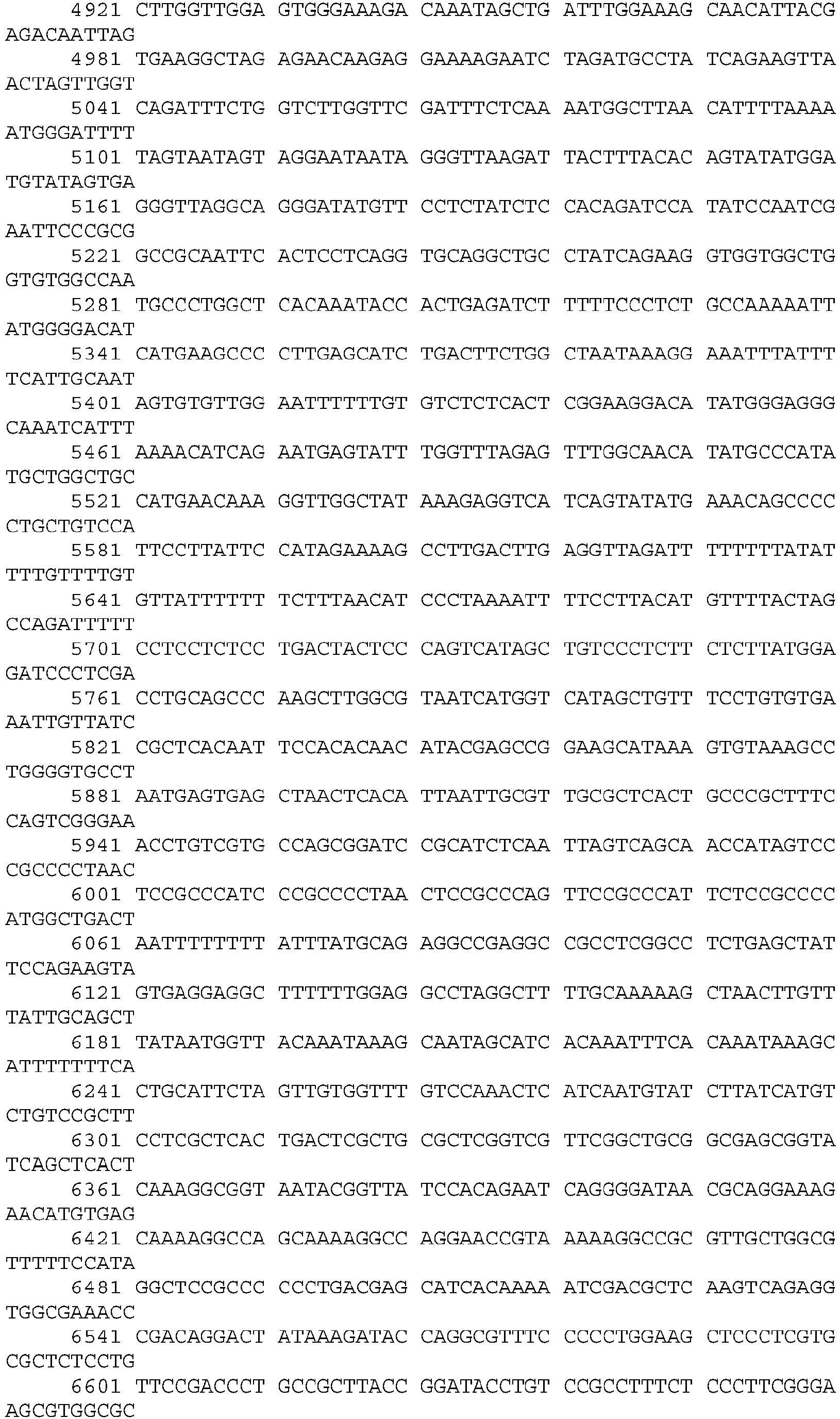


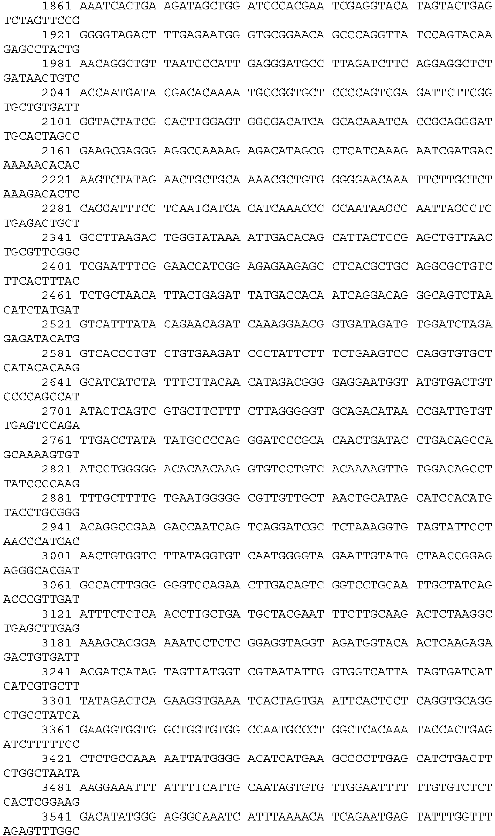


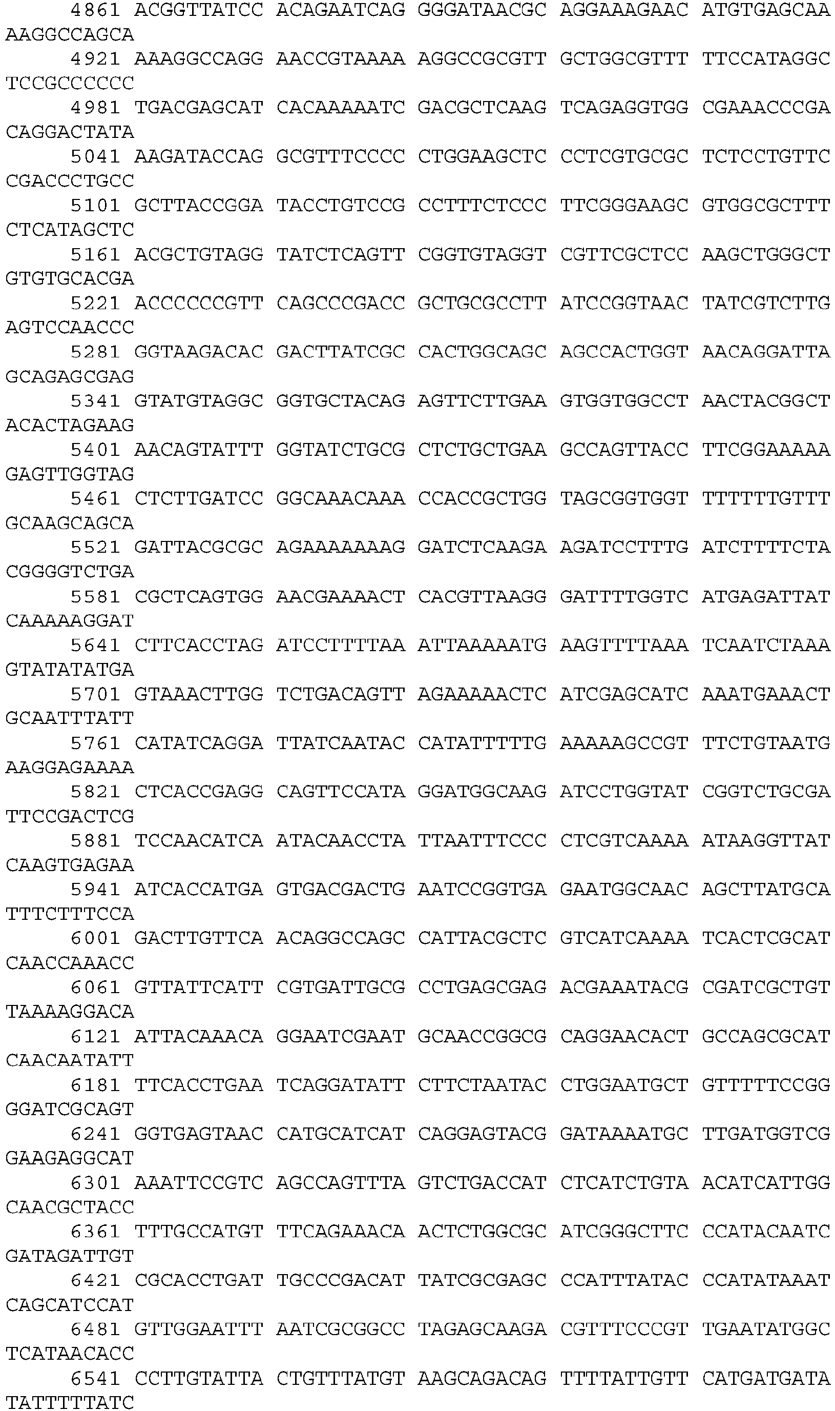

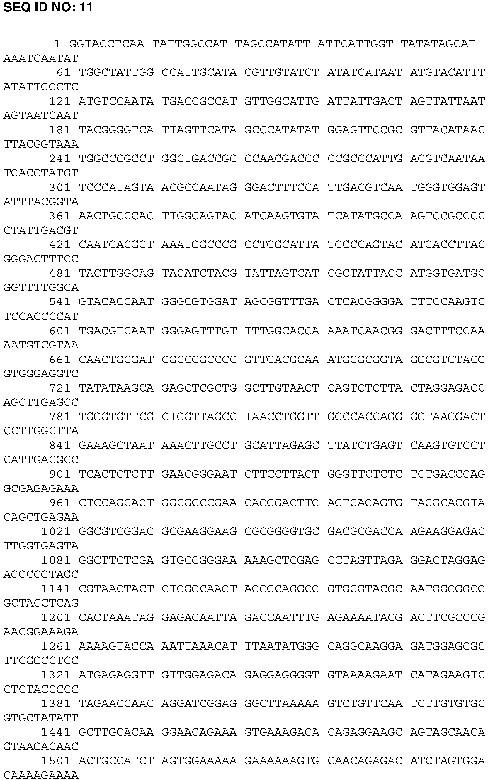


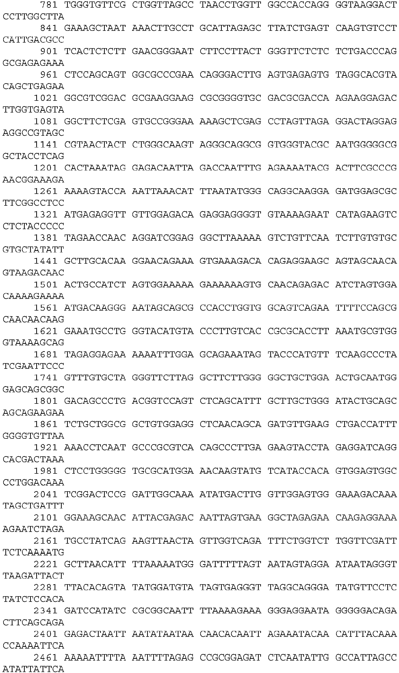





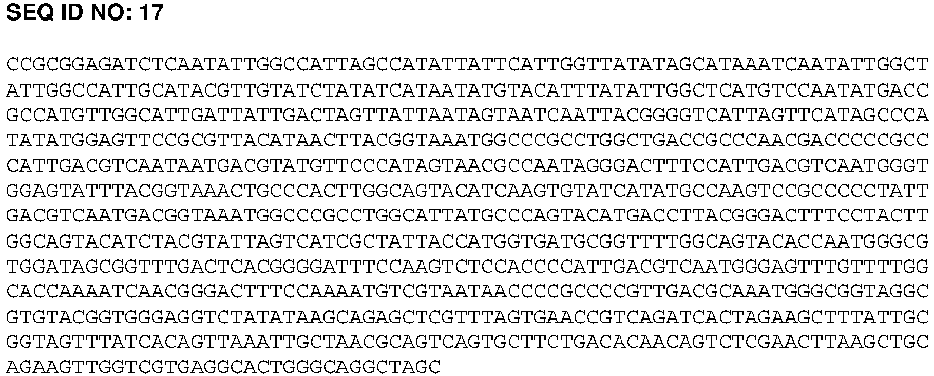
- SEQ ID NO: 18
TCGAGATGTGGTCTGAGTTAAAAATCAGGAGCAACGACGGAGGTGAAGGACCAGACGCCAACGACCC - SEQ ID NO: 19
CCGGGGGTCGTTGGCGTCTGGTCCTTCACCTCCGTCGTTGCTCCTGATTTTTAACTCAGACCACATC - SEQ ID NO: 20
CCGGGGAAAGGGGGTGCAACACATCCATATCCAGCCATCTCTACCTGTTTATGGACA - SEQ ID NO: 21
ACCCTCTGTCCATAAACAGGTAGAGATGGCTGGATATGGATGTGTTGCACCCCTTTCC - SEQ ID NO: 22
GGGTTAGGTGGTTGCTGATTCTCTCATTCACCCAGTGGG - SEQ ID NO: 23
GATCCCCACTGGGTGAATGAGAGAATCAGCAACCACCTA - SEQ ID NO: 24
GAGACTCGAGATGTGGTCTGAGTTAAAAATCAGG - SEQ ID NO: 25
AGAGGTAGACCAGTACGAGTCACGTTTGCCCCTATCACCATCCCTAACCCTCTGTCATAAAC





Claims (13)
- A lentiviral vector pseudotyped with hemagglutinin-neuraminidase (HN) and fusion (F) proteins from a respiratory paramyxovirus, wherein said lentiviral vector comprises a hybrid human CMV enhancer/EF1a (hCEF) promoter and a transgene, and wherein the lentiviral vector lacks an(y) intron positioned between said promoter and said transgene.
- The lentiviral vector according to Claim 1, wherein the lentiviral vector is selected from the group consisting of a Human immunodeficiency virus (HIV) vector, a Simian immunodeficiency virus (SIV) vector, a Feline immunodeficiency virus (FIV) vector, an Equine infectious anaemia virus (EIAV) vector, and a Visna/maedi virus vector; wherein preferably the lentiviral vector is a SIV vector.
- The lentiviral vector according to Claim 1 or 2, wherein the respiratory paramyxovirus is a Sendai virus.
- The lentiviral vector according to any of the preceding claims, wherein the transgene is selected from:(a) a secreted therapeutic protein, optionally, Alpha-1 Antitrypsin (A1AT), Factor VIII, Surfactant Protein B (SFTPB), Factor VII, Factor IX, Factor X, Factor XI, von Willebrand Factor, Granulocyte-Macrophage Colony-Stimulating Factor (GM-CSF), or a monoclonal antibody against an infectious agent; or(b) CFTR, DNAH5, DNAH11, DNAI1 or DNAI2.
- The lentiviral vector according to any one of Claims 1 to 4, wherein:(a) the transgene encodes CFTR;(b) the transgene encodes A1AT; or(c) the transgene encodes FVIII.
- The lentiviral vector according to any one of Claims 1 to 5, wherein the transgene encodes CFTR.
- A method of producing lentivirus as defined in any one of Claims 1 to 6, said method comprising the following steps:(a) growing cells in suspension;(b) transfecting the cells with one or more plasmids;(c) adding a nuclease;(d) harvesting the lentivirus;(e) adding trypsin; and(f) purification.
- The method according to Claim 7, wherein:(i) steps (a)-(f) are carried out sequentially;(ii) the cells are HEK293T or 293T/17 cells;(iii) the addition of the nuclease is at the pre-harvest stage;(iv) the addition of trypsin is at the post-harvest stage; and/or(v) the purification step comprises a chromatography step.
- A lentiviral vector as defined in any one of Claims 1 to 6 for use in a method of treating a disease.
- The lentiviral vector for the use according to Claim 9, wherein the disease is a lung disease selected from: cystic fibrosis (CF); Primary Ciliary Dyskinesia (PCD); Surfactant Protein B (SP-B) deficiency; Alpha 1-antitrypsin Deficiency (A1AD); Pulmonary Alveolar Proteinosis (PAP); and Chronic obstructive pulmonary disease (COPD).
- The lentiviral vector for the use according to Claim 9, wherein:(a) the disease to be treated is CF;(b) the disease to be treated is A1AT deficiency; or(c) the disease to be treated is Haemophilia, wherein optionally the transgene is Factor VIII.
- A host cell comprising the vector according to any one of Claims 1-6.
- A composition comprising a vector according to any one of Claims 1-6, and a pharmaceutically-acceptable carrier.
Priority Applications (6)
| Application Number | Priority Date | Filing Date | Title |
|---|---|---|---|
| HRP20200930TT HRP20200930T1 (en) | 2014-05-21 | 2015-04-23 | LENTIVIRUS VECTORS |
| MEP-2020-114A ME03789B (en) | 2014-05-21 | 2015-04-23 | LENTIVIRAL VECTORS |
| SI201531275T SI3145949T1 (en) | 2014-05-21 | 2015-04-23 | Lentiviral vectors |
| SM20200360T SMT202000360T1 (en) | 2014-05-21 | 2015-04-23 | Lentiviral vectors |
| RS20200724A RS60586B1 (en) | 2014-05-21 | 2015-04-23 | Lentiviral vectors |
| PL15720762T PL3145949T3 (en) | 2014-05-21 | 2015-04-23 | Lentiviral vectors |
Applications Claiming Priority (2)
| Application Number | Priority Date | Filing Date | Title |
|---|---|---|---|
| GB1409089.8A GB2526339A (en) | 2014-05-21 | 2014-05-21 | Lentiviral vectors |
| PCT/GB2015/051201 WO2015177501A1 (en) | 2014-05-21 | 2015-04-23 | Lentiviral vectors |
Publications (2)
| Publication Number | Publication Date |
|---|---|
| EP3145949A1 EP3145949A1 (en) | 2017-03-29 |
| EP3145949B1 true EP3145949B1 (en) | 2020-04-08 |
Family
ID=51135270
Family Applications (1)
| Application Number | Title | Priority Date | Filing Date |
|---|---|---|---|
| EP15720762.2A Active EP3145949B1 (en) | 2014-05-21 | 2015-04-23 | Lentiviral vectors |
Country Status (21)
| Country | Link |
|---|---|
| US (1) | US10704061B2 (en) |
| EP (1) | EP3145949B1 (en) |
| JP (1) | JP6637444B2 (en) |
| AU (1) | AU2015263150B2 (en) |
| CA (1) | CA2947125C (en) |
| CY (1) | CY1123413T1 (en) |
| DK (1) | DK3145949T3 (en) |
| EA (1) | EA038966B1 (en) |
| ES (1) | ES2805045T3 (en) |
| GB (1) | GB2526339A (en) |
| HR (1) | HRP20200930T1 (en) |
| HU (1) | HUE051269T2 (en) |
| LT (1) | LT3145949T (en) |
| ME (1) | ME03789B (en) |
| PL (1) | PL3145949T3 (en) |
| PT (1) | PT3145949T (en) |
| RS (1) | RS60586B1 (en) |
| SI (1) | SI3145949T1 (en) |
| SM (1) | SMT202000360T1 (en) |
| UA (1) | UA122127C2 (en) |
| WO (1) | WO2015177501A1 (en) |
Families Citing this family (38)
| Publication number | Priority date | Publication date | Assignee | Title |
|---|---|---|---|---|
| BR102015012334A2 (en) * | 2015-05-27 | 2016-11-29 | Fundação Hemoct De Ribeirão Preto Fundherp | production process of blood coagulation factor vii and blood coagulation factor vii |
| SI3377637T1 (en) | 2016-04-08 | 2020-08-31 | Krystal Biotech, Inc. | Compositions for use in procedures for the treatment of wounds, disorders and skin diseases |
| JP2018174874A (en) * | 2017-04-19 | 2018-11-15 | 国立大学法人 東京医科歯科大学 | Method of measuring water secretion function of epithelial cells |
| JP7511469B2 (en) | 2017-09-27 | 2024-07-05 | イノザイム ファーマ, インコーポレイテッド | Methods for improving cardiovascular function and treating cardiovascular disease using recombinant ectonucleotide pyrophosphatase/phosphodiesterase (NPP1). |
| GB201814590D0 (en) * | 2018-09-07 | 2018-10-24 | Oxford Biomedica Ltd | Viral vector production system |
| WO2019200163A1 (en) | 2018-04-12 | 2019-10-17 | Krystal Biotech, Inc. | Compositions and methods for the treatment of autosomal recessive congenital ichthyosis |
| CN112041434A (en) | 2018-04-27 | 2020-12-04 | 克里斯托生物技术股份有限公司 | Recombinant nucleic acids encoding one or more cosmetic proteins for cosmetic applications |
| CN112384247B (en) * | 2018-07-02 | 2024-09-27 | 库拉米斯公司 | Gene and cell therapy agents using cell fusion technology and uses thereof |
| WO2020009444A1 (en) * | 2018-07-02 | 2020-01-09 | 서울대학교산학협력단 | Gene and cell therapy product using cell fusion technology and use thereof |
| CA3112627A1 (en) | 2018-09-24 | 2020-04-02 | Krystal Biotech, Inc. | Compositions and methods for the treatment of netherton syndrome |
| MX2021008832A (en) | 2019-02-08 | 2021-09-08 | Krystal Biotech Inc | COMPOSITIONS AND METHODS FOR ADMINISTRATION OF CFTR POLYPEPTIDES. |
| BR112021017853A2 (en) * | 2019-03-13 | 2021-11-30 | Generation Bio Co | Non-viral dna vectors and uses thereof to express fviii therapeutics |
| CN112575034B (en) * | 2019-09-29 | 2023-04-25 | 济南赛尔生物科技股份有限公司 | Product for treating hemophilia A and application thereof |
| US20210189427A1 (en) * | 2019-12-20 | 2021-06-24 | Krystal Biotech, Inc. | Compositions and methods for gene delivery to the airways and/or lungs |
| WO2021222222A1 (en) * | 2020-04-27 | 2021-11-04 | Mccray Jr Paul B | Compositions and methods for the treatment of cystic fibrosis |
| US20230190871A1 (en) | 2020-05-20 | 2023-06-22 | Sana Biotechnology, Inc. | Methods and compositions for treatment of viral infections |
| CN116157145A (en) | 2020-06-09 | 2023-05-23 | 依诺兹梅制药公司 | Soluble ENPP1 or ENPP3 protein and uses thereof |
| KR20230074703A (en) * | 2020-06-24 | 2023-05-31 | 바이오버라티브 테라퓨틱스 인크. | Method for removing free factor VIII from preparations of lentiviral vectors modified to express the protein |
| US20230303665A1 (en) | 2020-08-28 | 2023-09-28 | Sana Biotechnology, Inc. | Modified anti-viral binding agents |
| MX2023003021A (en) * | 2020-09-16 | 2023-05-24 | Generation Bio Co | Non-viral dna vectors and uses thereof for expressing fviii therapeutics. |
| GB202100688D0 (en) * | 2021-01-19 | 2021-03-03 | Autolus Ltd | Process |
| GB202102832D0 (en) * | 2021-02-26 | 2021-04-14 | Ip2Ipo Innovations Ltd | Retroviral vectors |
| AU2022246917A1 (en) | 2021-04-02 | 2023-10-12 | Krystal Biotech, Inc. | Viral vectors for cancer therapy |
| GB202105278D0 (en) | 2021-04-13 | 2021-05-26 | Imperial College Innovations Ltd | Cell therapy |
| GB202105276D0 (en) * | 2021-04-13 | 2021-05-26 | Imperial College Innovations Ltd | Delivery of gene therapy vectors |
| EP4373951A1 (en) * | 2021-07-19 | 2024-05-29 | 2seventy bio, Inc. | Vector manufacturing processes |
| WO2023114949A1 (en) | 2021-12-16 | 2023-06-22 | Sana Biotechnology, Inc. | Methods and systems of particle production |
| WO2023133595A2 (en) | 2022-01-10 | 2023-07-13 | Sana Biotechnology, Inc. | Methods of ex vivo dosing and administration of lipid particles or viral vectors and related systems and uses |
| WO2023150647A1 (en) | 2022-02-02 | 2023-08-10 | Sana Biotechnology, Inc. | Methods of repeat dosing and administration of lipid particles or viral vectors and related systems and uses |
| JP2025513059A (en) | 2022-04-11 | 2025-04-22 | アイピー2アイピーオー イノベーションズ リミテッド | Combination Therapy |
| US20260027145A1 (en) | 2022-07-27 | 2026-01-29 | Sana Biotechnology, Inc. | Methods of transduction using a viral vector and inhibitors of antiviral restriction factors |
| US20260055146A1 (en) | 2022-08-24 | 2026-02-26 | Sana Biotechnology, Inc. | Delivery of heterologous proteins |
| GB202212472D0 (en) * | 2022-08-26 | 2022-10-12 | Ip2Ipo Innovations Ltd | Retroviral vectors |
| US20260053948A1 (en) | 2022-09-15 | 2026-02-26 | Yeda Research And Development Co. Ltd. | Compositions and methods for virotherapy |
| WO2024119157A1 (en) | 2022-12-02 | 2024-06-06 | Sana Biotechnology, Inc. | Lipid particles with cofusogens and methods of producing and using the same |
| WO2024220597A2 (en) | 2023-04-18 | 2024-10-24 | Sana Biotechnology, Inc. | Digital droplet based assay for detecting replication competent lentiviral vector |
| AU2024274792A1 (en) | 2023-05-23 | 2025-10-23 | Sana Biotechnology, Inc. | Tandem fusogens and related lipid particles |
| WO2025184529A1 (en) | 2024-03-01 | 2025-09-04 | Sana Biotechnology, Inc. | Viral particles with fusogen display and related compositions and methods |
Family Cites Families (8)
| Publication number | Priority date | Publication date | Assignee | Title |
|---|---|---|---|---|
| EP1980617A1 (en) * | 1998-12-31 | 2008-10-15 | Novartis Vaccines and Diagnostics, Inc. | Improved expression of HIV polypeptides and production of virus-like particles |
| AU2001262684A1 (en) * | 2000-06-01 | 2001-12-11 | Dnavec Research Inc. | Pseudo-type retrovirus vector containing membrane protein having hemagglutinin activity |
| WO2003029274A2 (en) | 2001-09-28 | 2003-04-10 | University Of North Carolina At Chapel Hill | Paramyxoviruses as gene transfer vectors to lung cells |
| CA2462628C (en) * | 2001-10-02 | 2019-08-20 | Institut Clayton De La Recherche | Restricted expression lentiviral vectors |
| DK1950307T3 (en) | 2005-10-28 | 2016-02-15 | Id Pharma Co Ltd | Luftvejsepitelstamcelle for gene transfer using a lentiviral vector, which is pseudo typified by RNA virus spike protein |
| GB0606190D0 (en) | 2006-03-28 | 2006-05-10 | Isis Innovation | Construct |
| EP1950303A1 (en) | 2007-01-26 | 2008-07-30 | Bayer CropScience AG | Genetically modified plants which synthesise a starch with low amylase content and higher swelling ability |
| HUE030719T2 (en) * | 2007-04-09 | 2017-05-29 | Univ Florida | RAAV vector formulations and methods comprising tyrosine-modified capsid proteins for their use |
-
2014
- 2014-05-21 GB GB1409089.8A patent/GB2526339A/en not_active Withdrawn
-
2015
- 2015-04-23 EA EA201692361A patent/EA038966B1/en unknown
- 2015-04-23 WO PCT/GB2015/051201 patent/WO2015177501A1/en not_active Ceased
- 2015-04-23 HU HUE15720762A patent/HUE051269T2/en unknown
- 2015-04-23 EP EP15720762.2A patent/EP3145949B1/en active Active
- 2015-04-23 AU AU2015263150A patent/AU2015263150B2/en active Active
- 2015-04-23 SM SM20200360T patent/SMT202000360T1/en unknown
- 2015-04-23 JP JP2016567082A patent/JP6637444B2/en active Active
- 2015-04-23 HR HRP20200930TT patent/HRP20200930T1/en unknown
- 2015-04-23 SI SI201531275T patent/SI3145949T1/en unknown
- 2015-04-23 PL PL15720762T patent/PL3145949T3/en unknown
- 2015-04-23 RS RS20200724A patent/RS60586B1/en unknown
- 2015-04-23 CA CA2947125A patent/CA2947125C/en active Active
- 2015-04-23 PT PT157207622T patent/PT3145949T/en unknown
- 2015-04-23 DK DK15720762.2T patent/DK3145949T3/en active
- 2015-04-23 LT LTEP15720762.2T patent/LT3145949T/en unknown
- 2015-04-23 ES ES15720762T patent/ES2805045T3/en active Active
- 2015-04-23 US US15/312,026 patent/US10704061B2/en active Active
- 2015-04-23 ME MEP-2020-114A patent/ME03789B/en unknown
- 2015-04-23 UA UAA201612986A patent/UA122127C2/en unknown
-
2020
- 2020-07-03 CY CY20201100617T patent/CY1123413T1/en unknown
Non-Patent Citations (1)
| Title |
|---|
| None * |
Also Published As
| Publication number | Publication date |
|---|---|
| SMT202000360T1 (en) | 2020-09-10 |
| GB2526339A (en) | 2015-11-25 |
| LT3145949T (en) | 2020-08-25 |
| RS60586B1 (en) | 2020-08-31 |
| CA2947125A1 (en) | 2015-11-26 |
| GB201409089D0 (en) | 2014-07-02 |
| JP6637444B2 (en) | 2020-01-29 |
| EA038966B1 (en) | 2021-11-16 |
| UA122127C2 (en) | 2020-09-25 |
| HUE051269T2 (en) | 2021-03-01 |
| PL3145949T3 (en) | 2021-01-25 |
| CY1123413T1 (en) | 2021-12-31 |
| WO2015177501A1 (en) | 2015-11-26 |
| PT3145949T (en) | 2020-07-13 |
| DK3145949T3 (en) | 2020-06-29 |
| JP2017521049A (en) | 2017-08-03 |
| CA2947125C (en) | 2023-02-28 |
| EA201692361A1 (en) | 2017-05-31 |
| SI3145949T1 (en) | 2020-08-31 |
| HRP20200930T1 (en) | 2020-09-18 |
| EP3145949A1 (en) | 2017-03-29 |
| AU2015263150A1 (en) | 2016-11-10 |
| ME03789B (en) | 2021-04-20 |
| US10704061B2 (en) | 2020-07-07 |
| ES2805045T3 (en) | 2021-02-10 |
| AU2015263150B2 (en) | 2020-04-16 |
| US20170096684A1 (en) | 2017-04-06 |
Similar Documents
| Publication | Publication Date | Title |
|---|---|---|
| JP6637444B2 (en) | Lentivirus vector | |
| JP7208231B2 (en) | Use of syncytin for targeted drug and gene delivery to lung tissue | |
| JP2021530985A (en) | Fososome composition and its use | |
| CN110573523A (en) | AAV vector-based influenza vaccine | |
| US20220323612A1 (en) | Gene therapy of neuronal ceroid lipofuscinoses | |
| EP3662066A1 (en) | Cellular models of and therapies for ocular diseases | |
| US20220339296A1 (en) | Gene therapy for mucopolysaccharidosis, type i | |
| JP2024112876A (en) | Treatment of mucopolysaccharidosis type I with fully human glycosylated human alpha-L-iduronidase (idua) | |
| US20200071721A1 (en) | Gene therapy for mucopolysaccharidosis, type ii | |
| EP3824095A1 (en) | Gene therapy for macular degeneration | |
| JP2014533961A (en) | Modified stem cells and their therapeutic use | |
| US20240325567A1 (en) | Cell therapy | |
| EP4323516A1 (en) | Signal peptides | |
| AU2020211080B2 (en) | Methods of producing and using recombinant alpha 1-antitrypsin (AAT) and compositions thereof | |
| US20250304996A1 (en) | Pseudotyped lentiviral vectors | |
| EP4676545A1 (en) | Synthetic promoters | |
| CA3208936A1 (en) | Retroviral vectors |
Legal Events
| Date | Code | Title | Description |
|---|---|---|---|
| STAA | Information on the status of an ep patent application or granted ep patent |
Free format text: STATUS: THE INTERNATIONAL PUBLICATION HAS BEEN MADE |
|
| PUAI | Public reference made under article 153(3) epc to a published international application that has entered the european phase |
Free format text: ORIGINAL CODE: 0009012 |
|
| STAA | Information on the status of an ep patent application or granted ep patent |
Free format text: STATUS: REQUEST FOR EXAMINATION WAS MADE |
|
| 17P | Request for examination filed |
Effective date: 20161216 |
|
| AK | Designated contracting states |
Kind code of ref document: A1 Designated state(s): AL AT BE BG CH CY CZ DE DK EE ES FI FR GB GR HR HU IE IS IT LI LT LU LV MC MK MT NL NO PL PT RO RS SE SI SK SM TR |
|
| AX | Request for extension of the european patent |
Extension state: BA ME |
|
| DAV | Request for validation of the european patent (deleted) | ||
| STAA | Information on the status of an ep patent application or granted ep patent |
Free format text: STATUS: EXAMINATION IS IN PROGRESS |
|
| 17Q | First examination report despatched |
Effective date: 20180404 |
|
| RIC1 | Information provided on ipc code assigned before grant |
Ipc: A61K 38/37 20060101ALI20190828BHEP Ipc: C07K 14/755 20060101ALI20190828BHEP Ipc: A61K 48/00 20060101ALI20190828BHEP Ipc: C12N 15/86 20060101ALI20190828BHEP Ipc: C07K 14/47 20060101AFI20190828BHEP |
|
| GRAP | Despatch of communication of intention to grant a patent |
Free format text: ORIGINAL CODE: EPIDOSNIGR1 |
|
| STAA | Information on the status of an ep patent application or granted ep patent |
Free format text: STATUS: GRANT OF PATENT IS INTENDED |
|
| INTG | Intention to grant announced |
Effective date: 20191018 |
|
| RAP1 | Party data changed (applicant data changed or rights of an application transferred) |
Owner name: IP2IPO INNOVATIONS LIMITED Owner name: ID PHARMA CO., LTD. |
|
| GRAS | Grant fee paid |
Free format text: ORIGINAL CODE: EPIDOSNIGR3 |
|
| GRAA | (expected) grant |
Free format text: ORIGINAL CODE: 0009210 |
|
| STAA | Information on the status of an ep patent application or granted ep patent |
Free format text: STATUS: THE PATENT HAS BEEN GRANTED |
|
| AK | Designated contracting states |
Kind code of ref document: B1 Designated state(s): AL AT BE BG CH CY CZ DE DK EE ES FI FR GB GR HR HU IE IS IT LI LT LU LV MC MK MT NL NO PL PT RO RS SE SI SK SM TR |
|
| AX | Request for extension of the european patent |
Extension state: BA ME |
|
| REG | Reference to a national code |
Ref country code: AT Ref legal event code: REF Ref document number: 1254259 Country of ref document: AT Kind code of ref document: T Effective date: 20200415 Ref country code: CH Ref legal event code: EP |
|
| REG | Reference to a national code |
Ref country code: DE Ref legal event code: R096 Ref document number: 602015050234 Country of ref document: DE |
|
| REG | Reference to a national code |
Ref country code: IE Ref legal event code: FG4D |
|
| REG | Reference to a national code |
Ref country code: HR Ref legal event code: TUEP Ref document number: P20200930T Country of ref document: HR |
|
| REG | Reference to a national code |
Ref country code: DK Ref legal event code: T3 Effective date: 20200625 |
|
| REG | Reference to a national code |
Ref country code: NL Ref legal event code: FP |
|
| REG | Reference to a national code |
Ref country code: FI Ref legal event code: FGE |
|
| REG | Reference to a national code |
Ref country code: PT Ref legal event code: SC4A Ref document number: 3145949 Country of ref document: PT Date of ref document: 20200713 Kind code of ref document: T Free format text: AVAILABILITY OF NATIONAL TRANSLATION Effective date: 20200706 |
|
| REG | Reference to a national code |
Ref country code: SE Ref legal event code: TRGR |
|
| REG | Reference to a national code |
Ref country code: CH Ref legal event code: NV Representative=s name: SERVOPATENT GMBH, CH |
|
| REG | Reference to a national code |
Ref country code: SK Ref legal event code: T3 Ref document number: E 34658 Country of ref document: SK |
|
| REG | Reference to a national code |
Ref country code: NO Ref legal event code: T2 Effective date: 20200408 |
|
| REG | Reference to a national code |
Ref country code: EE Ref legal event code: FG4A Ref document number: E019462 Country of ref document: EE Effective date: 20200625 |
|
| REG | Reference to a national code |
Ref country code: HR Ref legal event code: T1PR Ref document number: P20200930 Country of ref document: HR |
|
| REG | Reference to a national code |
Ref country code: GR Ref legal event code: EP Ref document number: 20200401863 Country of ref document: GR Effective date: 20200916 |
|
| REG | Reference to a national code |
Ref country code: HR Ref legal event code: ODRP Ref document number: P20200930 Country of ref document: HR Payment date: 20200610 Year of fee payment: 6 |
|
| REG | Reference to a national code |
Ref country code: DE Ref legal event code: R097 Ref document number: 602015050234 Country of ref document: DE |
|
| REG | Reference to a national code |
Ref country code: ES Ref legal event code: FG2A Ref document number: 2805045 Country of ref document: ES Kind code of ref document: T3 Effective date: 20210210 |
|
| PLBE | No opposition filed within time limit |
Free format text: ORIGINAL CODE: 0009261 |
|
| STAA | Information on the status of an ep patent application or granted ep patent |
Free format text: STATUS: NO OPPOSITION FILED WITHIN TIME LIMIT |
|
| REG | Reference to a national code |
Ref country code: AT Ref legal event code: UEP Ref document number: 1254259 Country of ref document: AT Kind code of ref document: T Effective date: 20200408 |
|
| REG | Reference to a national code |
Ref country code: HU Ref legal event code: AG4A Ref document number: E051269 Country of ref document: HU |
|
| 26N | No opposition filed |
Effective date: 20210112 |
|
| REG | Reference to a national code |
Ref country code: HR Ref legal event code: ODRP Ref document number: P20200930 Country of ref document: HR Payment date: 20210409 Year of fee payment: 7 |
|
| REG | Reference to a national code |
Ref country code: HR Ref legal event code: ODRP Ref document number: P20200930 Country of ref document: HR Payment date: 20220419 Year of fee payment: 8 |
|
| REG | Reference to a national code |
Ref country code: HR Ref legal event code: ODRP Ref document number: P20200930 Country of ref document: HR Payment date: 20230419 Year of fee payment: 9 |
|
| P01 | Opt-out of the competence of the unified patent court (upc) registered |
Effective date: 20230515 |
|
| REG | Reference to a national code |
Ref country code: HR Ref legal event code: ODRP Ref document number: P20200930 Country of ref document: HR Payment date: 20240411 Year of fee payment: 10 |
|
| PGFP | Annual fee paid to national office [announced via postgrant information from national office to epo] |
Ref country code: SM Payment date: 20250327 Year of fee payment: 11 Ref country code: LT Payment date: 20250324 Year of fee payment: 11 |
|
| PGFP | Annual fee paid to national office [announced via postgrant information from national office to epo] |
Ref country code: SI Payment date: 20250410 Year of fee payment: 11 |
|
| PGFP | Annual fee paid to national office [announced via postgrant information from national office to epo] |
Ref country code: MT Payment date: 20250325 Year of fee payment: 11 |
|
| REG | Reference to a national code |
Ref country code: HR Ref legal event code: ODRP Ref document number: P20200930 Country of ref document: HR Payment date: 20250411 Year of fee payment: 11 |
|
| PGFP | Annual fee paid to national office [announced via postgrant information from national office to epo] |
Ref country code: NL Payment date: 20250418 Year of fee payment: 11 |
|
| PGFP | Annual fee paid to national office [announced via postgrant information from national office to epo] |
Ref country code: LU Payment date: 20250418 Year of fee payment: 11 |
|
| PGFP | Annual fee paid to national office [announced via postgrant information from national office to epo] |
Ref country code: MC Payment date: 20250425 Year of fee payment: 11 |
|
| PGFP | Annual fee paid to national office [announced via postgrant information from national office to epo] |
Ref country code: FI Payment date: 20250424 Year of fee payment: 11 |
|
| PGFP | Annual fee paid to national office [announced via postgrant information from national office to epo] |
Ref country code: PL Payment date: 20250410 Year of fee payment: 11 Ref country code: DE Payment date: 20250422 Year of fee payment: 11 |
|
| PGFP | Annual fee paid to national office [announced via postgrant information from national office to epo] |
Ref country code: GB Payment date: 20250423 Year of fee payment: 11 Ref country code: ES Payment date: 20250529 Year of fee payment: 11 Ref country code: DK Payment date: 20250429 Year of fee payment: 11 |
|
| PGFP | Annual fee paid to national office [announced via postgrant information from national office to epo] |
Ref country code: RS Payment date: 20250411 Year of fee payment: 11 Ref country code: IS Payment date: 20250410 Year of fee payment: 11 Ref country code: NO Payment date: 20250424 Year of fee payment: 11 |
|
| PGFP | Annual fee paid to national office [announced via postgrant information from national office to epo] |
Ref country code: AL Payment date: 20250421 Year of fee payment: 11 Ref country code: BE Payment date: 20250418 Year of fee payment: 11 Ref country code: IT Payment date: 20250424 Year of fee payment: 11 |
|
| PGFP | Annual fee paid to national office [announced via postgrant information from national office to epo] |
Ref country code: HR Payment date: 20250411 Year of fee payment: 11 |
|
| PGFP | Annual fee paid to national office [announced via postgrant information from national office to epo] |
Ref country code: PT Payment date: 20250410 Year of fee payment: 11 Ref country code: LV Payment date: 20250417 Year of fee payment: 11 |
|
| PGFP | Annual fee paid to national office [announced via postgrant information from national office to epo] |
Ref country code: FR Payment date: 20250425 Year of fee payment: 11 Ref country code: EE Payment date: 20250425 Year of fee payment: 11 |
|
| PGFP | Annual fee paid to national office [announced via postgrant information from national office to epo] |
Ref country code: BG Payment date: 20250424 Year of fee payment: 11 Ref country code: GR Payment date: 20250423 Year of fee payment: 11 |
|
| PGFP | Annual fee paid to national office [announced via postgrant information from national office to epo] |
Ref country code: CH Payment date: 20250501 Year of fee payment: 11 |
|
| PGFP | Annual fee paid to national office [announced via postgrant information from national office to epo] |
Ref country code: RO Payment date: 20250414 Year of fee payment: 11 Ref country code: AT Payment date: 20250423 Year of fee payment: 11 |
|
| PGFP | Annual fee paid to national office [announced via postgrant information from national office to epo] |
Ref country code: TR Payment date: 20250415 Year of fee payment: 11 Ref country code: SK Payment date: 20250416 Year of fee payment: 11 |
|
| PGFP | Annual fee paid to national office [announced via postgrant information from national office to epo] |
Ref country code: CZ Payment date: 20250414 Year of fee payment: 11 |
|
| PGFP | Annual fee paid to national office [announced via postgrant information from national office to epo] |
Ref country code: IE Payment date: 20250422 Year of fee payment: 11 |
|
| PGFP | Annual fee paid to national office [announced via postgrant information from national office to epo] |
Ref country code: SE Payment date: 20250429 Year of fee payment: 11 |
|
| PGFP | Annual fee paid to national office [announced via postgrant information from national office to epo] |
Ref country code: MK Payment date: 20250320 Year of fee payment: 11 |
|
| PGFP | Annual fee paid to national office [announced via postgrant information from national office to epo] |
Ref country code: HU Payment date: 20251008 Year of fee payment: 11 |
|
| PGFP | Annual fee paid to national office [announced via postgrant information from national office to epo] |
Ref country code: CY Payment date: 20250324 Year of fee payment: 11 |
|
| REG | Reference to a national code |
Ref country code: HR Ref legal event code: ODRP Ref document number: P20200930 Country of ref document: HR Payment date: 20260420 Year of fee payment: 12 |