EP3142764B1 - System and apparatus for isolating or enriching agents using floatation - Google Patents
System and apparatus for isolating or enriching agents using floatation Download PDFInfo
- Publication number
- EP3142764B1 EP3142764B1 EP15793428.2A EP15793428A EP3142764B1 EP 3142764 B1 EP3142764 B1 EP 3142764B1 EP 15793428 A EP15793428 A EP 15793428A EP 3142764 B1 EP3142764 B1 EP 3142764B1
- Authority
- EP
- European Patent Office
- Prior art keywords
- instances
- microbubbles
- sample
- biological agents
- container
- Prior art date
- Legal status (The legal status is an assumption and is not a legal conclusion. Google has not performed a legal analysis and makes no representation as to the accuracy of the status listed.)
- Active
Links
Images




Classifications
-
- G—PHYSICS
- G01—MEASURING; TESTING
- G01N—INVESTIGATING OR ANALYSING MATERIALS BY DETERMINING THEIR CHEMICAL OR PHYSICAL PROPERTIES
- G01N1/00—Sampling; Preparing specimens for investigation
- G01N1/28—Preparing specimens for investigation including physical details of (bio-)chemical methods covered elsewhere, e.g. G01N33/50, C12Q
- G01N1/40—Concentrating samples
- G01N1/4077—Concentrating samples by other techniques involving separation of suspended solids
-
- A—HUMAN NECESSITIES
- A61—MEDICAL OR VETERINARY SCIENCE; HYGIENE
- A61P—SPECIFIC THERAPEUTIC ACTIVITY OF CHEMICAL COMPOUNDS OR MEDICINAL PREPARATIONS
- A61P35/00—Antineoplastic agents
-
- A—HUMAN NECESSITIES
- A61—MEDICAL OR VETERINARY SCIENCE; HYGIENE
- A61P—SPECIFIC THERAPEUTIC ACTIVITY OF CHEMICAL COMPOUNDS OR MEDICINAL PREPARATIONS
- A61P37/00—Drugs for immunological or allergic disorders
- A61P37/02—Immunomodulators
- A61P37/04—Immunostimulants
-
- B—PERFORMING OPERATIONS; TRANSPORTING
- B03—SEPARATION OF SOLID MATERIALS USING LIQUIDS OR USING PNEUMATIC TABLES OR JIGS; MAGNETIC OR ELECTROSTATIC SEPARATION OF SOLID MATERIALS FROM SOLID MATERIALS OR FLUIDS; SEPARATION BY HIGH-VOLTAGE ELECTRIC FIELDS
- B03D—FLOTATION; DIFFERENTIAL SEDIMENTATION
- B03D1/00—Flotation
- B03D1/14—Flotation machines
-
- C—CHEMISTRY; METALLURGY
- C12—BIOCHEMISTRY; BEER; SPIRITS; WINE; VINEGAR; MICROBIOLOGY; ENZYMOLOGY; MUTATION OR GENETIC ENGINEERING
- C12M—APPARATUS FOR ENZYMOLOGY OR MICROBIOLOGY; APPARATUS FOR CULTURING MICROORGANISMS FOR PRODUCING BIOMASS, FOR GROWING CELLS OR FOR OBTAINING FERMENTATION OR METABOLIC PRODUCTS, i.e. BIOREACTORS OR FERMENTERS
- C12M47/00—Means for after-treatment of the produced biomass or of the fermentation or metabolic products, e.g. storage of biomass
- C12M47/04—Cell isolation or sorting
-
- G—PHYSICS
- G01—MEASURING; TESTING
- G01N—INVESTIGATING OR ANALYSING MATERIALS BY DETERMINING THEIR CHEMICAL OR PHYSICAL PROPERTIES
- G01N33/00—Investigating or analysing materials by specific methods not covered by groups G01N1/00 - G01N31/00
- G01N33/48—Biological material, e.g. blood, urine; Haemocytometers
- G01N33/50—Chemical analysis of biological material, e.g. blood, urine; Testing involving biospecific ligand binding methods; Immunological testing
- G01N33/53—Immunoassay; Biospecific binding assay; Materials therefor
- G01N33/536—Immunoassay; Biospecific binding assay; Materials therefor with immune complex formed in liquid phase
-
- B—PERFORMING OPERATIONS; TRANSPORTING
- B03—SEPARATION OF SOLID MATERIALS USING LIQUIDS OR USING PNEUMATIC TABLES OR JIGS; MAGNETIC OR ELECTROSTATIC SEPARATION OF SOLID MATERIALS FROM SOLID MATERIALS OR FLUIDS; SEPARATION BY HIGH-VOLTAGE ELECTRIC FIELDS
- B03D—FLOTATION; DIFFERENTIAL SEDIMENTATION
- B03D2203/00—Specified materials treated by the flotation agents; Specified applications
- B03D2203/003—Biotechnological applications, e.g. separation or purification of enzymes, hormones, vitamins, viruses
-
- G—PHYSICS
- G01—MEASURING; TESTING
- G01N—INVESTIGATING OR ANALYSING MATERIALS BY DETERMINING THEIR CHEMICAL OR PHYSICAL PROPERTIES
- G01N1/00—Sampling; Preparing specimens for investigation
- G01N1/28—Preparing specimens for investigation including physical details of (bio-)chemical methods covered elsewhere, e.g. G01N33/50, C12Q
- G01N1/40—Concentrating samples
- G01N1/4055—Concentrating samples by solubility techniques
- G01N2001/4066—Concentrating samples by solubility techniques using difference of solubility between liquid and gas, e.g. bubbling, scrubbing or sparging
Definitions
- centrifugation the use of centrifugal force for the sedimentation of heterogeneous mixtures with a centrifuge, is a common method for isolating cells in animal blood.
- the prior art describes a number of centrifuge apparati, but these apparati are expensive and complex, such that, primarily, only laboratories can afford them, and only laboratory technicians can operate them. This limits the clinical and consumer applications of centrifugal isolation.
- centrifugation-based methods separate cells based on physical properties (e.g. density, size, shape), and do little to separate cell based on protein expression and biochemical profile.
- Magnetic force is an alternative to centrifugal force for isolating cells in animal blood.
- An example of this application from the prior art is the use of immunomagnetic nanobeads and lateral magnetophoresis for isolation of circulating tumor cells (CTC's). While effective, the application of this isolation method is costly and requires the expertise of a laboratory-trained technician. This limits the clinical and consumer applications of magnetic force isolation.
- Microbubbles are bubbles less than can be less than a millimeter in diameter to greater than a micrometer in diameter.
- Microbubbles can be filled inside with a gas, such as air or perfluorocarbon. Adjacent to cells or other biological agents or non-biological agents, microbubbles can cause those agents to float in a liquid.
- biological or non-biological agents can float in the presence or absence of microbubbles adjacent thereto by including a liquid or gas, or combination thereof, that cause the agents to float, such as solutions that are known in the art.
- the microbubbles, adjacent thereto biological agents or non-biological agents that are isolated or enriched, are disrupted.
- Cells from animal blood are not the only biological agents isolated through centrifugal and magnetic force technology.
- Other potentially life-saving applications include the isolation of cells (such as stem cells, immune cells, circulating tumor cell cells (CTC's), tumor cells, or other cells), cell fragments, bacteria, viruses, parasites, and others.
- CTC's circulating tumor cell cells
- the development of alternatives to centrifugal and magnetic force for isolating biological agents is critical in order to apply the life- saving properties of those isolated biological agents to the home and to the clinic.
- WO 99/55837 a separation method is known, separating a target material such as human or animal cells from a liquid sample by encapsulating the target material in gas microbubbles and accomplishing separation by flotation. Further, a separation device including two opposing syringes interconnected through a valve is known from WO 99/55837 .
- the acute angle is between about 5 degrees and about 40 degrees. According to a preferred embodiment, the acute angle is between about 5 degrees and about 10 degrees. According to a preferred embodiment, the acute angle is about 35 degrees.
- the apparatus comprises a container spout directionally orienting one or more droplets that form from the tip comprising the one or more biological agents.
- the apparatus comprises a collection container to collect the one or more droplets.
- the collection container is adjacent to the outer surface of the sample container.
- the collection container is mounted onto the outer surface of the sample container close to the bottom end of the container spout.
- the plunger of the apparatus is operated manually by a user.
- the apparatus comprises a drive screw mechanically coupled to the plunger, wherein rotation of the drive screw is mechanically converted to linear translation of the plunger.
- the apparatus comprises a container mount to house the drive screw.
- a bottom flange of the sample container is secured to the container mount by inserting the bottom flange into a locking flange slot on the container mount.
- the drive screw is operated manually by a user.
- the drive screw is operated automatically by a drive motor.
- the drive screw is operated automatically by a drive motor remotely controlled by a microprocessor.
- the apparatus comprises an apparatus enclosure to house the drive motor.
- the apparatus comprises a plunger adapter joint and a plunger engagement adapter, wherein the plunger adapter joint is adjacent to the plunger and the plunger engagement adapter, and the plunger engagement adapter is adjacent to the plunger adapter joint and the drive screw.
- the container mount comprises a sensor depressor and sensor mount.
- the container mount further comprises a wire port.
- the container mount further comprises a screw port.
- the sample container is formed of a material to provide the inner volume of the sample container hydrophilicity.
- the sample container is formed of a material to provide the inner volume of the sample container hydrophobicity.
- the material is glass.
- a hydrophilic layer is adjacent thereto the inner volume of the sample container.
- a hydrophobic layer is adjacent thereto the tapered end of the inner volume of the sample container.
- a hydrophilic layer is adjacent thereto the inner volume of the sample container and a hydrophobic layer is adjacent thereto the tapered end of the inner volume.
- the sample container is formed of a material to provide hydrophilicity to the inner volume and a hydrophobic layer is adjacent thereto the tapered end of the inner volume.
- the hydrophilic layer is bovine serum albumin.
- the hydrophobic layer is SURFASIL TM .
- the hydrophobic layer is paraffin, polytetrafluoroethylene, poloaxmer, or combinations thereof.
- the sample container holds a sample volume of between about 1mL and about 10mL. According to a preferred embodiment, the sample container holds a sample volume of between about 10mL and about 50mL. In some instances, the sample container holds a sample volume of between about 50mL and about 200mL. According to a preferred embodiment, the sample is diluted.
- the sample is tissue, blood, bone marrow, urine, saliva, cerebrospinal fluid, seminal fluid, sputum, stool, joint fluid, lymph, amniotic fluid, bile, ascites, or pleural effusion, or combinations thereof.
- the sample is umbilical cord blood.
- the sample is circulating blood.
- the sample is tumor tissue.
- the sample is fluid within and surrounding a tumor tissue.
- the biological agents are cells.
- the cells are circulating tumor cells (CTCs).
- the cells are bone marrow cells.
- the cells are fetal cells from a mother's blood.
- a ratio of the cells to the plurality of microbubbles is about 1 : 100. According to an example, a ratio of the cells to the plurality of microbubbles is about 1 : 500. According to an example, a ratio of the cells to the plurality of microbubbles is about 1 : 1000.
- the biological agents isolated or enriched are less than 0.5% of the total sample. According to a preferred embodiment, the biological agents isolated or enriched are less than 0.1% of the total sample. According to an example, the biological agents isolated or enriched are less than 0.01% of the total sample. According to a preferred embodiment, the biological agents isolated or enriched are less than 0.001% of the total sample.
- the microbubbles are lipid shell microbubbles, albumin shell microbubbles, polymer shell microbubbles, lipopolymer shell microbubbles or glass microbubbles, or combinations thereof.
- the shell of each microbubble comprises lipid, protein, polymer, lipopolymer, or glass or combinations thereof.
- the plurality of microbubbles have an outer diameter of between about 1 micron and about 10 microns. According to an example, the plurality of microbubbles have an outer diameter of between about 2 microns and about 8 microns. According to an example, the plurality of microbubbles have an outer diameter of between about 2 microns and about 20 microns.
- a plurality of selection markers are adjacent to the outer surface of the plurality of microbubbles.
- the plurality of selection markers bind the outer surface by nucleophilic conjugate addition.
- the plurality of selection markers bind the outer surface by Michael addition.
- the plurality of selection markers per microbubble is between about 200,000 and about 500,000.
- a surface density of the plurality of selection markers adjacent to the outer surface of a single microbubble of the plurality of microbubbles is between about 3,000 and about 6,000 selection markers per square micron.
- a surface density of the plurality of selection markers adjacent to the outer surface of a single microbubble of the plurality of microbubbles is between about 1,000 and about 3,000 selection markers per square micron. According to an example, a surface density of the plurality of selection markers adjacent to the outer surface of a single microbubble of the plurality of microbubbles is less than about 6,000 selection markers per square micron. According to an example, a surface density of the plurality of selection markers adjacent to the outer surface of a single microbubble of the plurality of microbubbles is less than about 3,000 selection markers per square micron. According to an example, the plurality of selection markers per microbubble is between about 300,000 and about 400,000.
- the plurality of selection markers comprise a first set and a second set.
- each of the plurality of microbubbles comprise the first set and the second set of selection markers.
- a first set of the plurality of microbubbles comprise the first set of selection markers and a second set of the plurality of microbubbles comprise the second set of selection markers.
- cells attach the plurality of microbubbles by receptor ligand interactions.
- one or more selection markers bind cell surface markers.
- one or more selection markers are different.
- one or more selection markers are antibodies.
- one or more selection markers are molecules that bind tumor markers.
- one or more selection markers are molecules that bind stem cell markers.
- one or more selection markers are molecules that bind cell surface marker, EpCAM.
- one or more selection markers are molecules that bind cell surface marker, CD 133.
- one or more selection markers are molecules that bind cell surface marker, CD34.
- At least about 80% of the cells bind at least one of the plurality of microbubbles. According to an example, at least about 85% of the cells bind at least one of the plurality of microbubbles. According to an example, at least about 90% of the cells bind at least one of the plurality of microbubbles. According to an example, at least about 95% of the cells bind at least one of the plurality of microbubbles.
- a biological agent of the sample floats when at least about 2 microbubbles are adjacent thereto. According to an example, a biological agent of the sample floats when at least about 3 microbubbles are adjacent thereto. According to an example, a biological agent of the sample floats when between about 2 to about 20 microbubbles are adjacent thereto.
- the apparatus is compatible with downstream assays.
- the downstream assays comprise cell staining, molecular analysis, or cell culture, or combinations thereof.
- the apparatus is sterile.
- the apparatus comprises a sterile liquid in the inner volume to enhance viability of the sample.
- the apparatus comprises a sterile liquid in the inner volume to cause the plurality of microbubbles to float.
- the density of the liquid is between about 0.9 and about 1.1 grams per cubic centimeter.
- the apparatus is used in a clinical or laboratory setting. According to an example, the apparatus is used in a residential setting. According to an example, the apparatus is utilized for personal use.
- the apparatus comprises a bottom stop sensor, a stepper motor, a top drip detector, a power supply, a microcontroller board, or combinations thereof.
- a method for isolation or enrichment of biological agents from a sample comprises (a) obtaining a sample and a plurality of microbubbles, (b) adding the sample and the plurality of microbubbles to an inner volume of a sample container, (c) orienting the tapered end of the sample container away from ground wherein the plurality of microbubbles float one or more biological agents towards a tip of the tapered end, and (d) moving a plunger from an open end of the sample container towards the tapered end to isolate or enrich one or more biological agents from the sample.
- the method comprises disrupting the plurality of microbubbles after (d).
- ambient pressure is increased to disrupt the plurality of microbubbles.
- ambient pressure is increased to between about 2 atm and about 4 atm for between about 2 minutes and about 10 minutes to disrupt the plurality of microbubbles.
- ambient pressure is increased to about 3 atm for about 5 minutes to disrupt the plurality of microbubbles.
- the method comprises incubating the plurality of microbubbles and the sample in the inner volume for at least about 1 minute before (c) or (d). According to an example, the method comprises incubating the plurality of microbubbles and the sample in the inner volume for at least about 5 minutes before (c) or (d). According to an example, the method comprises incubating the plurality of microbubbles and the sample in the inner volume for at least about 10 minutes before (c) or (d). According to an example, the method comprises sterilizing the inner volume of the sample container prior to adding of (b).
- kits for isolation or enrichment of biological agents from a sample comprising one or more sample containers, an apparatus enclosure, a container mounts, one or more selection markers, components to form microbubbles, instructions for forming microbubbles, and instructions for using the sample container.
- kits for isolation or enrichment of biological agents from a sample comprising one or more sample containers, an apparatus enclosure, a container mount, microbubbles with one or more selection markers adjacent thereto, and instructions for using the sample container.
- kits for isolation or enrichment of biological agents from a sample comprising one or more sample containers, one or more selection markers, components to form microbubbles, instructions for forming microbubbles, and instructions for using the sample container.
- the apparatus is used in a remote geographical area. According to an example, the apparatus is used in an impoverished or low-income area. According to an example, the apparatus is used in the absence of sterilization equipment. According to an example, the apparatus is used in the absence of centrifugation equipment. According to an example, the apparatus is used by a clinician. According to an example, the apparatus is used by a researcher. According to an example, the apparatus is used by a lay person. According to an example, the apparatus is used by a consumer. According to an example, the apparatus is used for cord blood banking. According to an example, the apparatus is used for isolation of immune cells for immunotherapy in cancer treatment.
- the apparatus is used for isolation of hematopoietic stem cells from umbilical cord blood following a live birth.
- the apparatus is used for isolation of one or more narcotics from a blood sample or urine sample.
- the apparatus is used for depletion of one or more biological agents from a sample.
- the apparatus is used for depletion of one or more non-biological agents from a sample.
- This present disclosure comprises a sterilized sample container apparatus, existing as part of an open or closed system.
- the apparatus can isolate biological including animal cells, animal cell fragments, bacteria, viruses, fungi, parasites, plant cells, or non-biological agents including chemical agents, industrial agents, agricultural agents, and other biological or non-biological agents.
- the present disclosure can comprise mechanisms to keep the apparatus sterile, plunging systems, collection containers for isolating biological agents, hydrophobic materials, hydrophilic materials, non-specific interaction blocking agents, syringe needle systems, sterile syringes, infusion needles, or liquids or combinations thereof.
- the present disclosure may be applied to a number of applications including clinical, research, diagnostic, industrial, institutional, or consumer, or uses or combinations thereof.
- the present disclosure is used for the isolation or enrichment of biological or non-biological agents.
- the present disclosure is used from the depletion of biological or non-biological agents.
- the present disclosure is used for the collection, storage, research, application, or others, or combinations thereof biological agents or non-biological agents isolated or enriched from a sample.
- the term “about” means an acceptable error for a particular value as determined by one of ordinary skill in the art, which depends in part on how the value is measured or determined. In certain embodiments, the term “about” means within 1, 2, 3, or 4 standard deviations. In certain embodiments, the term “about” means within 30%, 25%, 20%, 15%, 10%, 9%, 8%, 7%, 6%, 5%, 4%, 3%, 2%, 1%, 0.5%, 0.1%, or 0.05% of a given value or range.
- microbubbles adjacent to a biological agent to cause the biological agent to be buoyant in a liquid.
- microbubbles can be surfactant microbubbles, lipid or phospholipid shell microbubbles, protein shell microbubbles (i.e. albumin shell), polymer microbubbles, polyelectrolyte microbubbles, or glass microbubbles, or combinations thereof.
- microbubbles can be solid. In some instances, microbubbles can be hollow. In some instances, microbubbles can be porous. In some instances, microbubbles can comprise one or more layers or shells. In some instances, microbubbles can be composite, for example, comprising one or more individual particulates of a material or comprising one or more nanobubbles within a microbubble, or others. In some instances, the core of the microbubble comprising a gas can provide the buoyant property. In some instances, the material of the microbubble can provide the buoyant property. In some instances, microbubbles can be formed using methods known in the art.
- surfactant microbubbles can be formed from surfactants such as SPAN-40, TWEEN-40, or others.
- lipid or phospholipid microbubbles can be formed from natural lipids, phospholipids, sphingolipids, fatty acid modified lipids, head group modified lipids, fluorescent lipids, polymer lipids, cationic lipids, neutral lipids, or others.
- microbubbles can be formed from DSPC/PEG40 stearate / DSPE-PEG3400-maleimide.
- polymeric microbubbles can be formed from poly-lactic coglycolic acid (PLGA), poly-caprolactone (PCL), polyvinylalcohol (PVA), combinations thereof or others.
- microbubbles can also be formed from polysaccharides such as chitosan.
- microbubbles can be utilized in a clinical setting.
- the core of the microbubbles can comprise gas or liquid.
- the core of the microbubbles can comprise perfluorohexane gas.
- the core of the microbubble can comprise air.
- microbubbles can have surface hydrophobicity. In some instances, microbubbles can be durable under pressure. In some instances, microbubbles can disrupt at pressures much greater than ambient. In some instances, microbubbles can aggregate in a liquid. In some instances, microbubble aggregation improves isolation or enrichment from a sample of biological agents that are adjacent thereto microbubbles. In some instances, microbubbles can be buoyant in a liquid. In some instances, microbubble buoyancy improves isolation or enrichment from a sample of biological agents that are adjacent thereto microbubbles.
- microbubbles can have an outer diameter of about 2, 3, 4, 5, 6, 7, 8, 9, 10, 11, 12, 13, 14, 15, 16, 17, 18, 19, or about 20 microns. In some instances, microbubbles can have an outer diameter of about 1, 5, 10, 15, 20, 25, 30, 35, or about 40 microns. In some instances, microbubbles can have an outer diameter of about 0.15, 0.2, 0.25, 0.3, 0.35, 0.4, 0.45, 0.5, 0.55, 0.6, 0.65, or about 0.7 microns. In some instances, microbubbles can have an outer diameter of between about 1 micrometer and about 1 millimeter. In some instances, microbubbles can have an outer diameter of between about 0.2 microns and about 0.6 microns.
- microbubbles can have an outer diameter of between about 1 micron and about 10 microns. In some instances, microbubbles can have an outer diameter of between about 2 microns and about 8 microns. In some instances, microbubbles can have an outer diameter of between about 2 microns and about 20 microns. In some instances, microbubbles can have an outer diameter of between about 2 microns and about 6 microns. In some instances, microbubbles can be monodisperse.
- microbubbles can be hollow. In some instances, microbubbles can be porous. In some instances, microbubbles can comprise a gaseous core. In some instances, microbubbles can comprise a liquid core with a density lower than the density of the liquid the microbubbles are dispersed in.
- Selection markers can be adjacent to microbubbles such that specific biological agents in a sample will become adjacent thereto the microbubbles by interactions with one or more of the selection markers.
- selection markers can be antibodies, antibody fragments, antigens, hormone, growth factors, recombinant soluble receptors, transferrin, LDL, neurotransmitters, toxins, carbohydrates, lipids, nucleic acids, small molecules, aptamers, aptamer derivatives, dendrimers, proteins, peptides, polypeptides, nucleotides, polynucleotides or others or combinations thereof.
- a selection marker can be any molecule known to bind a cell surface marker. In some instances, a selection marker can be any known molecule to bind a stem cell surface marker. In some instances, a selection marker can be anti-CD34 or anti-CD 133.
- a selection marker can be any molecule known to bind AA4, AA4.1, P-gp, ABCB5, ABCG2, ALDH, alkaline phosphatase, alpha6-integrin, WNT2B, antithrombin III, asialo GM1, Bcl-2, betal-integrin, c-kit (CD117), c-met, NCAM (CD56), CD105, CD133, CD166, CD29, CD30, CD31, siglec-3 (CD33), CD73, CD9, CD90, CK19, CLV3, ECMA-7, EDR1, EEC, FGF-4, Flk1, Flt3/Flk2, CD115, Glia2, Gli3, GSTA1, Her5, HSA, hsp25, Id2, IL-3Ralpha, KDR, keratin 15, keratin 19, kit, 1-selectin, lamin A/C, Lewis X antigen, LeX, Lgr5, Lrp4, MCM2, MCSP
- a selection marker can be any known molecule to bind tumor or cancer cell surface marker. In some instances, a selection marker can be anti-EpCAM. In some instances, a selection marker can be anti-Her2. In some instances, a selection marker can be anti-EGFR. In some instances, a selection marker can be anti-CEA. In some instances, a selection marker can be any molecule known to bind alpha-fetoprotein (AFP), beta-2-microglobulin (B2M), beta-human chorionic gonadotropin (Beta-hCG), carcinoembryonic antigen (CEA), CD20, chromogranin A (CgA), Her-2, Kit, or prostate-specific antigen (PSA) or combinations thereof or others.
- AFP alpha-fetoprotein
- B2M beta-2-microglobulin
- Beta-hCG beta-human chorionic gonadotropin
- CEA carcinoembryonic antigen
- CD20 chromogranin A
- CgA chro
- a selection marker can be any known molecule to bind an immune cell surface marker.
- a selection marker can be anti-CD3, anti-CD4, anti-CD8, or others.
- a selection marker can be any molecule known to bind CD3, CD4, CD8, CD25, CD45, CD2, CD5, CD6, CD27, CD31, CD25, CD69, CD28, CD152, CD154, CD19, CD20, CD40, CD134, CD83, CMRF-44, CMRF-56, OX40L, DEC-205, CD11c, F4/80, CD11b, MHCII, BDCA-1, CD68, or DC-SIGN, or combinations thereof, or others.
- a selection marker known to bind an immune cell surface marker can be used to isolate or enrich cells for immunotherapy.
- cells for immunotherapy can be used to induce, enhance, or suppress an immune response.
- cells for immunotherapy in cancer can be used to induce an immune response towards cancer cells, such that the immune system can reject or destroy tumor or cancer cells.
- cells for immunotherapy in cancer can reject or destroy tumor or cancer cells.
- cells for immunotherapy in cancer can stimulate an immune response to reject or destroy tumor or cancer cells.
- One or more selection markers can be adjacent thereto to the surface of one or more microbubbles. In some instances, one or more selection markers can be attached to the surface of one or more microbubbles. In some instances, one or more selection markers can be inserted into the outer shell of the microbubble. In some instances, one or more selection markers can be inserted into the core of the microbubble. In some instances, one or more selection markers can be inserted into both the outer shell and core of the microbubble. In some instances, one or more selection markers can be attached to the outer surface of the microbubble by nucleophilic conjugate addition. In some instances, one or more selection markers can be attached to the outer surface of the microbubble by Michael addition.
- one or more selection markers can be attached to the outer surface of the microbubble by a linking group that can be directly attached to the one or more microbubbles.
- the linking group can be an antibody fragment, such as Fc fragment specific IgG.
- more than one linking group can attach the one or more selection markers to the outer surface of the microbubble.
- selection markers can be adjacent to the surface of one or more microbubbles. In some instances, selection markers can be uniformly distributed adjacent to the outer surface of one or more microbubbles. In some instances, selection markers can be adjacent to one or more discrete regions of the outer surface of microbubbles. In some instances, selection markers can be patterned adjacent to the surface of microbubbles.
- about 1, 100, 500, 1,000, 5,000, 10,000, 50,000, 100,000, 125,000, 150,000, 175,000, 200,000, 225,000, 250,000, 275,000, 300,000, 325,000, 350,000, 375,000, 400,000, 425,000, 450,000, 475,000, 500,000, 525,000, 550,000, 575,000, 600,000 or more selection markers can be adjacent to the outer surface of a single microbubble. In some instances, between about 200,000 and about 500,000 selection markers can be adjacent to the outer surface of a single molecule. In some instances, between about 300,000 and about 400,000 selection markers can be adjacent to the outer surface of a single microbubble. In some instances, at least about 5,000 selection markers can be adjacent to the outer surface of a single microbubble. In some instances, at least about 10,000 selection markers can be adjacent to the outer surface of a single microbubble. In some instances, at least about 100,000 selection markers can be adjacent to the outer surface of a single microbubble.
- Isolation, enrichment, or depletion of distinct biological agents can include microbubbles with a first set and a second set of selection markers on each microbubble.
- the first set and the second set of selection markers are different.
- a first set of selection markers for CD133 and a second set of selection markers for CD34 can both be adjacent to the same microbubble.
- Simultaneous isolation or enrichment of two distinct biological agents can include microbubbles with a first set and a second set of selection markers.
- the first set and the second set of selection markers are different.
- a first set of selection markers for CD45 can be adjacent to a first microbubble and a second set of selection markers for CD34 can be adjacent to a second microbubble.
- more than one type of selection marker can be adjacent to the surface of each of the one or more microbubbles.
- Biological agents or non-biological agents can be isolated or enriched from a sample. In some instances, biological agents or non-biological agents can be depleted from a sample. In some instances, the sample can be a clinical sample. In some instances, the sample can be a laboratory sample. In some instances, a sample can be an industrial sample. In some instances, the sample can be obtained from a subject. In some instances, the sample can be obtained from the user. In some instances, the sample can be previously manipulated such as diluted, frozen, sterilized, or others.
- a sample can be tissue, blood, bone marrow, urine, saliva, cerebrospinal fluid, seminal fluid, sputum, stool, joint fluid, lymph, amniotic fluid, bile, ascites, pleural effusion, or others.
- a sample can be a tumor or cancer tissue.
- a sample can be fluid within and around a tumor or cancer tissue.
- a sample can be a blood sample.
- a blood sample can be umbilical cord blood.
- Non-biological agents isolated or enriched from a sample can include but are not limited to chemical agents, industrial agents, agricultural agents, manufacturing agents or others, or combinations thereof.
- non-biological agents can be polymers, organic compounds, inorganic compounds, or others.
- non-biological agents can be a solid, liquid, or gas.
- the non-biological agent isolated can be an impurity.
- the non-biological agent can be an elemental metal or a metal alloy.
- the non-biological agent can be a pharmaceutical composition.
- the non-biological agent can be a narcotic particle in a blood sample or urine sample obtained from a subject.
- the sample from the subject can be analyzed by a law enforcement person, a crime lab person, or other criminal justice personnel.
- the narcotic particle can be cocaine or amphetamine, or others.
- specific antibodies or other ligands to narcotics can be adjacent thereto microbubbles to capture the narcotic particles from the blood or urine sample.
- ELISA assays can determine the amount of narcotic particles adjacent thereto the microbubbles.
- Biological agents isolated or enriched from a sample can include but are not limited to cells, cell fragments, organelles, bacteria, viruses, fungi, parasites, proteins, peptides, polypeptides, nucleotides, polynucleotides, DNA, RNA, or amino acids.
- biological agents can be cells.
- biological agents can be cell lines.
- biological agents can be primary cells.
- biological agents can be rare cells in a sample, such as less than 0.001%, 0.01%, or 0.1% the total cell population in the sample.
- the biological agents can be immune cells for immunotherapy.
- biological agents can be hematopoietic stem cells.
- biological agents can be circulating tumor cells.
- cells can be 4T1 mouse cells, BxPC3 pancreatic adenocarcinoma cells, ASPC1 pancreatic adenocarcinoma cells, A549 lung cancer cells, PC3 prostate cancer cells, KG1a cells, or others.
- Selection markers can be adjacent to microbubbles such that specific biological agents in a sample will become adjacent thereto the microbubbles by interactions with one or more of the selection markers.
- FIG. 1A-B show the synthesis and characterization of microbubbles with selection marker adjacent thereto with microscopic images to show microbubbles adjacent thereto cells after incubation with microbubbles.
- Microbubbles can be adjacent thereto biological agents of a sample. Microbubbles can attach to a biological agent by a selection marker adjacent thereto the outer surface of the microbubble. Microbubbles can attach to a biological agent by binding a selection marker to a surface marker on the surface of the biological agent. Microbubbles can attach to a biological agent by binding a selection marker to a binding region on the surface of the biological agent. In some instances, a selection marker binds a cell surface marker, thereby attaching the microbubble to a cell. In some instances, a selection marker binds a binding region of a protein, polypeptide or peptide, thereby attaching the microbubble to the protein, polypeptide, or peptide.
- the biological agents to be enriched or isolated from a sample can comprise other components.
- a sample comprising biological agents such as stem cells to be enriched or isolated may also comprise blood cells, antibodies, and others.
- the biological agents to be enriched or isolated can comprise 0.001%, 0.01%, 0.1%, 0.2%, 0.3%, 0.4%, 0.5%, 0.6%, 0.7%, 0.8%, 0.9%, 1%, 2%, 3%, 4%, 5%, 6%, 7%, 8%, 9%, 10%, 11%, 12% 13%, 14%, 15%, 16%, 17%, 18%, 19%, 20%, 21%, 22%, 23%, 24%, 25% or less the total number of components in a sample.
- the biological agents to be enriched or isolated can comprise less than 20% of the sample. In some instances, the biological agents to be enriched or isolated can comprise less than 10% of the sample. In some instances, the biological agents to be enriched or isolated can comprise less than 5% of the sample. In some instances, the biological agents to be enriched or isolated can comprise less than 1% of the sample. In some instances, the biological agents to be enriched or isolated can be about 100 biological agents or less per 10 mL of sample. In some instances, the biological agents to be enriched or isolated can be about 100 biological agents or less per 9 mL of sample. In some instances, the biological agents to be enriched or isolated can be about 100 biological agents or less per 8 mL of sample.
- the biological agents to be enriched or isolated can be about 100 biological agents or less per 7 mL of sample. In some instances, the biological agents to be enriched or isolated can be about 100 biological agents or less per 6 mL of sample. In some instances, the biological agents to be enriched or isolated can be about 100 biological agents or less per 5 mL of sample.
- the percent of biological agents with one or more microbubbles adjacent thereto following incubation can be about 60%, 65%, 70%, 75%, 80%, 85%, 90%, 95%, 96%, 97%, 98%, 99% or more. In some instances, the percent of biological agents with one or more microbubbles adjacent thereto can be 100%. In some instances, the percent of biological agents with one or more microbubbles adjacent thereto can be about 85% to about 95%. In some instances, the percent of biological agents with one or more microbubbles adjacent thereto can be about 80% to about 95%. In some instances, the percent of biological agents with one or more microbubbles adjacent thereto can be at least about 85%. In some instances, the percent of biological agents with one or more microbubbles adjacent thereto can be at least about 90%.
- one or more biological agents can be incubated with one or more microbubbles such that following a period of incubation the one or more microbubbles can be adjacent thereto one or more biological agents.
- the period of incubation can be about 1, 2, 3, 4, 5, 6, 7, 8, 9, 10, 11, 12, 13, 14, or about 15 minutes or more.
- the period of incubation can be about 1 minute or less.
- the period of incubation can be about 5 minutes or less.
- the period of incubation can be about 10 minutes or less.
- the period of incubation can be about 15 minute or less.
- the period of incubation can be between about 1 minute and about 5 minutes.
- the period of incubation can be between about 1 minute and about 10 minutes.
- one or more microbubbles adjacent to a biological agent can cause the biological agent to be sufficiently buoyant in a liquid.
- sufficiently buoyant can be biological agents adjacent to microbubbles that are floating on the top of a liquid at a liquid atmosphere interface.
- sufficiently buoyant can be biological agents adjacent to microbubbles that are floating within a liquid at a depth above complete sedimentation to the bottom of the liquid.
- sufficiently buoyant can be biological agents adjacent to microbubbles that are floating within a liquid away from a liquid-liquid interface with a second liquid of different density.
- microbubbles alone can be buoyant.
- biological agents adjacent to one or more microbubbles can be buoyant.
- the buoyancy of a biological agent depends of the number of microbubbles adjacent thereto.
- microbubbles can be buoyant in a first liquid but not in a second liquid.
- one or more microbubbles adjacent to a biological agent can cause the biological agent to be buoyant in a liquid.
- the number of microbubbles needed to be adjacent to a biological agent to cause the biological agent to be buoyant in a liquid varies by the radius of the microbubble. The smaller the radius of the microbubble, the more microbubbles needed to cause a biological agent to be buoyant. In some instances, the number of microbubbles needed to be adjacent to a biological agent to cause the biological agent to be buoyant in a liquid varies by the size, shape, composition, surface area, weight, or charge of the biological agent.
- about 1, 2, 3, 4, 5, 6, 7, 8, 9, 10, 11, 12, 13, 14, 15, 16, 17, 18, 19, or about 20 microbubbles or more can cause a biological agent to be buoyant. In some instances, more than about 20 microbubbles can cause a biological agent to be buoyant. In some instances, at least 2 microbubbles can cause a biological agent to be buoyant. In some instances, between about 2 and about 8 microbubbles can cause a biological agent to be buoyant. In some instances, between about 10 and about 20 microbubbles can cause a biological agent to be buoyant. In some instances, about 2 microbubbles with an outer diameter of about 3 microns can cause a biological agent to be buoyant. In some instances, about 20 microbubbles with an outer diameter of about 1.5 microns can cause a biological agent to be buoyant.
- the liquid can an aqueous solution.
- the aqueous solution can be phosphate buffered saline.
- the liquid can be blood.
- the liquid can be sterile.
- a sample can be diluted with a liquid.
- a liquid can be added to a sample.
- a sample can be diluted with a liquid to enhance the viability of the biological agents within the sample.
- addition of liquid to a sample causes microbubbles to float.
- the liquid can be the sample itself.
- the liquid can be blood, urine, saliva, cerebrospinal fluid, seminal fluid, sputum, stool, joint fluid, lymph, amniotic fluid, bile, ascites, pleural effusion, or other bodily fluids.
- the liquid has a density of between about 0.9 and about 1.1 grams per cubic centimeter. In some instances, the liquid has a density of between about 0.95 and about 1.05 grams per cubic centimeter. In some instances, the liquid has a density of about 1 gram per cubic centimeter.
- one or more microbubbles can be adjacent thereto the one or more biological agents of a sample.
- the ratio of a single biological agent adjacent thereto microbubbles can be about 1:1000. In some instances, the ratio can be about 1:100. In some instances, the ratio can be about 1:500. In some instances, the ratio can be about 1:2000.
- the ratio can be about 1:1, 1:10, 1:25, 1:50, 1:55, 1:60, 1:65, 1:70, 1:75, 1:80, 1:85, 1:90, 1:95, 1:100, 1:105, 1:110, 1:115, 1:120, 1:125, 1:130, 1:135, 1:140, 1:145, 1:150, 1:155, 1:160, 1:165, 1:170, 1:175, 1:180, 1:185, 1:190, 1:195, 1:200, 1:250, 1:300, 1:350, 1:400, 1:450, 1:500, 1:550, 1:600, 1:650, 1:700, 1:750, 1:800, 1:850, 1:900, 1:950, 1:1000, 1:1100, 1:1200, 1:1300, 1:1400, 1:1500, or about 1:2000. In some instances, the ratio can be between about 1:900 and about 1:1100. In some instances, the ratio can be between about 1:500 and about 1:2000. In some instances, the ratio can be between about 1:500 and about 1:1500
- One or more biological agents can be isolated using the apparatus, methods, and kits disclosed herein.
- a single set of biological agents can be isolated.
- the single set can be homogeneous, such as a cell population wherein each cell expresses the sample level of a cell surface marker.
- the single set can be heterogeneous, such as a cell population wherein each cell expresses the same cell surface marker, but at different levels.
- a single set of biological agents such as circulating tumor cells expressing surface marker EpCAM can be isolated from a sample that comprises the circulating tumor cells and other cell types in a blood sample using the apparatus, methods, and kits disclosed herein.
- a single set of biological agents such as bovine serum albumin can be isolated from a sample that comprises the bovine serum albumin and other proteins using the apparatus, methods, and kits disclosed herein.
- a first set and a second set of biological agents can be isolated from a sample using the apparatus, methods, and kits disclosed herein.
- the first and second set of biological agents can be different.
- 2, 3, 4, 5 or more sets of biological agents can be isolated using the apparatus, methods, and kits disclosed herein.
- 3 sets of biological agents can be isolated using the apparatus, methods, and kits disclosed herein.
- a first set of tumor cells expressing cell surface marker EpCAM and a second set of tumor cells expressing cell surface marker CEA (carcinoembryonic antigen) or a third set of biological agents expressing both EpCAM and CEA can be isolated from a sample using the apparatus, methods, and kits disclosed herein.
- Microbubbles that are adjacent to biological agents can be dissociated from the biological agents by microbubble disruption.
- microbubbles are disrupted after enrichment or isolation of biological agents.
- microbubbles are disrupted at any point after becoming adjacent to biological agents.
- all microbubbles are disrupted.
- a portion of microbubbles are disrupted.
- at least 60%, 65%, 70%, 75%, 80%, 85%, 90%, 95% or more of the microbubbles adjacent to the biological agents are disrupted.
- more than 95% of microbubbles adjacent to the biological agents are disrupted.
- between about 90% to about 95% of the microbubbles adjacent to the biological agents are disrupted.
- microbubbles can be disrupted chemically or thermally. In some instances, microbubbles can be disrupted by sonication, vacuum application, application of organic solvents, application of surfactants, or increasing ambient pressure, or others. In some instances, microbubbles are disrupted by degradation, such as hydrolytic degradation or bio-erosion. In some instances, microbubbles can be disrupted by pH changes or temperature changes.
- microbubbles can be disrupted by sonication. In some instances, microbubbles can be disrupted by sonication for at least about 1, 2, 3, 4, 5, 6, 7, 8, 9, or about 10 seconds or more. In some instances, microbubbles can be disrupted by sonication for at least about 2 seconds. In some instances, microbubbles can be disrupted by sonication for at least about 5 seconds. In some instances, microbubbles can be disrupted by sonication for at least about 10 seconds. In some instances, microbubbles can be disrupted by sonication using a water bath sonicator.
- microbubbles can be disrupted by increasing ambient pressure. In some instances, increasing ambient pressure can be done by air compression. In some instances, the apparatus, methods, and kits disclosed herein can provide air compression of 3 atm for 5 minutes. In some instances, the apparatus, methods, and kits disclosed herein can provide air compression of 3 atm for 8 minutes. In some instances, the apparatus, methods, and kits disclosed herein can provide air compression of 3 atm for 10 minutes. In some instances, the apparatus, methods, and kits disclosed herein can provide air compression of 2 atm for 5 minutes. In some instances, the apparatus, methods, and kits disclosed herein can provide air compression of 2 atm for 8 minutes. In some instances, the apparatus, methods, and kits disclosed herein can provide air compression of 2 atm for 10 minutes.
- the apparatus, methods, and kits disclosed herein can provide air compression of 4 atm for 5 minutes. In some instances, the apparatus, methods, and kits disclosed herein can provide air compression of 4 atm for 8 minutes. In some instances, the apparatus, methods, and kits disclosed herein can provide air compression of 4 atm for 10 minutes. In some instances, the apparatus, methods, and kits disclosed herein can provide air compression of about 1.5, 2, 2.5, 3, 3.5, 4, 4.5, 5atm or more. In some instances, the apparatus can provide air compression of between about 2 atm and about 4 atm. In some instances, the apparatus can provide air compression of between about 2 atm and about 4 atm for about 3 minutes to about 10 minutes. In some instances, the apparatus can provide air compression for about 1, 2, 3, 4, 5, 6, 7, 8, 9, 10, 11, 12, 13, 14, 15 minutes or more.
- the air compression provided for disruption of microbubbles does not affect cell viability. In some instances, more than about 99% of cells remain viable following air compression. In some instances, more than about 98% of cells remain viable following air compression. In some instances, more than about 97% of cells remain viable following air compression. In some instances, cells remain viable for at least 2 hours following air compression. In some instances, cells remain viable for at least 8 hours following air compression. In some instances, cells remain viable for at least 18 hours following air compression. In some instances, cells remain viable for at least 24 hours following air compression.
- the apparatus, methods, and kits disclosed herein can provide a recovery efficiency of at least about 90%.
- the recovery efficiency can be at least about 95%. In some instances, the recovery efficiency can be at least about 98%. In some instances when biological agents can be cells, the recovery efficiency can be at least about 90% with cell viability at least about 95%. In some instances, the recovery efficiency can be at least about 80% with cell viability at least about 90%. In some instances, the recovery efficiency can be at least about 95% with cell viability at least about 90%. In some instances, the recovery efficiency can be at least about 95% with cell viability at least about 85%.
- FIG. 2 shows the percent recovery of CD34+ KG1a cells incubated with either microbubbles lacking any selection marker, or incubated with CD34-microbubbles demonstrating 87% recovery of CD34+ KG1a cells using the CD34-microbubbles.
- FIG. 3 shows cell viability following microbubble disruption using high ambient pressure.
- FIG. 3A shows a sample container with a microbubble layer before and after applying pressure.
- FIG. 3B shows the time of high pressure application and the resulting cell viability in both the control and pressure-treated group.
- isolated or enriched cells reattach to a tissue culture dish and proliferate following isolation from blood using the methods disclosed herein.
- the apparatus, methods, and kits disclosed herein provide for enrichment or isolation of biological agents from a sample using a sample container.
- the sample container can comprise an inner volume to hold the sample and microbubbles.
- the inner volume can be smooth and curved.
- the sample container can comprise a length longer than a width.
- the sample container can comprise a cylinder with two ends.
- the cylindrical sample container can comprise an open end and opposite the open end can be a tapered end. In some instances, the tapered end can be a cone-shaped end.
- the sample container can be a test tube or a centrifuge tube with an open end and a tapered end with a conical, flat, or rounded tip.
- the sample container can be a 50 mL tube.
- the sample container can be a 15 mL tube.
- the sample container can be a 10 mL tube.
- the sample container can be a 1 mL tube.
- the sample container can be glass, such as borosilicate glass, fused silica, quartz glass, KIMAX ® , PYREX ® , or ZERDOUR ® , or others.
- the sample container can be plastic, such as polyethylene, polystyrene, polypropylene, polytetrafluoroethylene (PTFE), polyurethane (PU), polyvinylchloride (PVC), EPDM, fluoroelastomer, neoprene, nitrile, or nylon or others.
- the sample container can be fabricated by 3D printing.
- the sample container can be fabricated by injection molding.
- the sample container can be reusable.
- the sample container can be disposable.
- the sample container can be a one-time use container.
- the sample container can be sterile.
- the inner volume of the sample container can be sterilized.
- the angle of the container spout can be determined by measuring the angle relative to the horizontal axis of the sample container. In some instances, the angle of the container spout relative to horizontal axis of the sample container can be less than 90 degrees. In some instances, the angle can be about 85, 80, 75, 70, 65, 60, 55, 50, 45, 40, 35, 30, 25, 20, 15, 10, or 5 degrees. In some instances, the angle can be between about 15 degrees to about 50 degrees. In some instances, the angle can be between about 15 degrees to about 35 degrees. In some instances, the angle can be between about 20 degrees to about 50 degrees. In some instances, the angle can be more acute than the angle of the angled tip of the tapered end.
- the open end of the sample container opposite the tapered end can be flat wherein the entire circumference of the open end can be equidistant to a flat tapered end.
- the tip of the tapered end of the sample container can form a perpendicular angle relative to the horizontal axis of the sample container.
- a plunger can be inserted into the open end of the sample container.
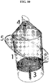
- the tapered end can be open. In some instances, the tapered end can be adjacent to a needle. In some instances, the tapered end can be adjacent to a container spout. In some instances, the tapered end can be flat wherein the entire circumference of the tapered end can be equidistant to the flat open end. In some instances, the tip of the tapered end of the sample container can form a perpendicular angle relative to the horizontal axis of the sample container. In some instances, the tapered end can comprise an angled tip, FIG. 10 .
- the angle of the angled tip on the tapered end can be determined by measuring the angle relative to the horizontal axis of the sample container. In some instances, the angle of the angled tip on the tapered end can be less than about 90 degrees. In some instances, the angle can be less than about 80 degrees. In some instances, the angle can be less than about 70 degrees. In some instances, the angle can be less than about 60 degrees. In some instances, the angle can be less than about 50 degrees. In some instances, the angle can be less than about 40 degrees. In some instances, the angle can be less than about 30 degrees. In some instances, the angle can be less than about 20 degrees. In some instances, the angle can be less than about 10 degrees. In some instances, the angle can be less than about 5 degrees.
- the angle can be between about 1 degree and about 5 degrees. In some instances, the angle can be between about 1 degree and about 10 degrees. In some instances, the angle can be between about 5 degrees and about 10 degrees. In some instances, the angle can be between about 1 degree and about 20 degrees. In some instances, the angle can be between about 1 degree and about 30 degrees. In some instances, the angle can be between about 1 degree and about 40 degrees. In some instances, the angle can be between about 1 degree and about 50 degrees. In some instances, the angle can be between about 5 degrees to about 40 degrees. In some instances, the angle can be between about 5 degrees to about 10 degrees. In some instances, the angle can be about 35 degrees. In some instances, the angle can be about 30 degrees. In some instances, the angle can be about 40 degrees. In some instances, the angle can be about 1, 5, 10, 15, 20, 25, 30, 35, 40, 45, 50, 55, 60, 65, 70, 75, 80, or about 85 degrees.
- the angle of the sample container shaft can be 90 degrees. In some instances, the angle of the sample container shaft can be between about 85 and about 95 degrees.
- the sample can be added directly to the inner volume of the sample container for enrichment or isolation of the biological agents.
- the sample can be diluted before or after being added to the sample container.
- the sample can be added directly to the inner volume of the sample container without centrifugal concentration.
- the total volume of a sample can be added to the sample container.
- the sample can be divided into separate subvolumes that can be separated added to separate sample containers.
- the sample can be further diluted with a liquid prior to being added to the sample container.
- the sample volume that can be accommodated by the sample container can be about 0.1, 0.2, 0.3, 0.4, 0.5, 0.6, 0.7, 0.8, 0.9, 1, 2, 3, 4, 5, 6, 7, 8, 9, 10, 11, 12, 13, 14, 15, 16, 17, 18, 19, 20, 21, 22, 23, 24, 25, 26, 27, 28, 29, 30, 31, 32, 33, 34, 35, 36, 37, 38, 39, 40, 41, 42, 43, 44, 45, 46, 47, 48, 49, 50, 51, 52, 53, 54, 55, 56, 57, 58, 59, 60, 61, 62, 63, 64, 65, 66, 67, 68, 69, 70, 71, 72, 73, 74, 75, 80, 85, 90, 95, 100, 105, 110, 115, 120, 125, 130, 135, 140, 145, 150, 155, 160, 165, 170, 175, 200, 250, 300, 350, 400, 450, 500 mL or more.
- the total sample volume can be less than about 1mL. In some instances, the total sample volume can be less than about 5mL. In some instances, the total sample volume can be less than about 10mL. In some instances, the total sample volume can be less than about 15mL. In some instances, the total sample volume can be less than about 50mL. In some instances, the total sample volume can be less than about 100mL. In some instances, the total sample volume can be less than about 200mL. In some instances, the total sample volume can be less than about 500mL.
- the total sample volume can be between about 1mL and about 5mL. In some instances, the total sample volume can be between about 1mL and about 10mL. In some instances, the total sample volume can be between about 1mL and about 15mL. In some instances, the total sample volume can be between about 1mL and about 50mL. In some instances, the total sample volume can be between about 1mL and about 100mL.
- the inner surface of the sample container can be hydrophobic to promote droplet formation at the tip of the tapered end during collection of the biological agents from the sample container.
- a hydrophobic layer can be adjacent to the inner surface of the sample container.
- a hydrophobic layer can be coated onto the inner surface of the sample container.
- the inner surface of the sample container comprises a material making the inner surface hydrophobic.
- an amphiphilic agent can be coated on the inner surface of the sample container to provide a hydrophobic layer.
- the hydrophobic layer can comprise a siliconizing fluid such as SURFASIL TM OR AQUASIL TM .
- the hydrophobic layer can comprise paraffin, such as paraffin wax, kerosene, liquid paraffin, mineral oil, or petroleum jelly, or combinations thereof.
- the hydrophobic layer can comprise one or more of polytetrafluoroethylene (PTFE) (such as TEFLON ® ), perfluoroalkoxy (PFA), or fluorinated ethylene propylene (FEP), or others.
- PTFE polytetrafluoroethylene
- PFA perfluoroalkoxy
- FEP fluorinated ethylene propylene
- the hydrophobic layer can comprise one or more poloaxmers such as SYNPERONICS ® , KOLLIPHOR ® , or PLURONIC ® (such as F68 or F127 or others).
- the entire inner surface of the surface container can be hydrophobic. In some instances, a portion of the inner surface can be hydrophobic. In some instances, about 1%, 2%, 3%, 4%, 5%, 6%, 7%, 8%, 9%, 10%, 15%, or about 20% of the inner surface can be hydrophobic. In some instances, the portion of the inner surface that can be hydrophobic can be the tapered neck. In some instances, the portion of the inner surface that can be hydrophobic can be the container top opening. In some instances, the portion of the inner surface that can be hydrophobic can be the tapered neck and container top opening.
- the inner surface of the sample container can be hydrophilic to prevent microbubbles from attaching, sticking, or aggregating at the inner surface.
- a sample container comprises a material such as glass to provide an inner surface that can be hydrophilic.
- the inner surface of the sample container can have a hydrophilic layer adjacent thereto. The adjacent hydrophilic layer can attach to the sample container by covalent bond, ionic bond, electrostatic interactions, van de Waal forces, or others.
- an amphiphilic agent can be coated on the inner surface of the sample container to provide a hydrophilic layer.
- bovine serum albumin polyurethane resin, polyvinylpyrrolidone (PVP), hyaluronic acid polyacrylate, or polyurethane polyacrylate, or combinations thereof, or others can provide a hydrophilic layer adjacent to the inner surface of the sample container.
- PVP polyvinylpyrrolidone
- hyaluronic acid polyacrylate or polyurethane polyacrylate, or combinations thereof, or others can provide a hydrophilic layer adjacent to the inner surface of the sample container.
- the entire inner surface of the sample container can be hydrophilic. In some instances, a portion of the inner surface can be hydrophilic. In some instances, about 75%, 80%, 85%, 90%, 95%, 96%, 97%, 98%, 99% or more of the inner surface can be hydrophilic. In some instances, the portion of the inner surface that can be the tapered neck can be not hydrophilic. In some instances, the portion of the inner surface that can be the container top opening can be not hydrophilic. In some instances, the portion of the inner surface that can be the tapered neck and the container top opening can be not hydrophilic.
- the apparatus, methods, and kits disclosed herein can localize one or more biological agents adjacent to one or more microbubbles such that the biological agents can be isolated or enriched.
- the sample container can separate the biological agents adjacent to the microbubbles by separation into a removable harvesting cap assembly.
- FIG. 4 shows one embodiment of the apparatus.
- a sample container 300 can contain an inner volume, 301, comprising a sample, one or more microbubbles with one or more selection markers adjacent thereto, a liquid, or combinations thereof, 310.
- a plunger, 401 can be inserted into the sample container 300 at the open end, 304, opposite the tapered end, 303.
- the plunger, 401 can be driven by a drive screw, 402, that can be housed in a container mount, 600, by contacting the drive screw 402 to the plunger engagement adapter, 404.
- the bottom flange, 308, of the sample container can also be housed in the container mount, 600.
- a mechanical convertor, 503, converting motor shaft rotation into linear translation of the plunger, can be housed in the apparatus closure, 501.
- a drive motor, 502, housed in the apparatus enclosure can automatically drive the plunger via the mechanical convertor, 503.
- the drive screw can be metal. In some instances, the drive screw can be non-metal. In some instances, the drive screw can be plastic. In some instances, the drive screw can be fabricated by 3D printing.
- FIG. 5 shows the sample container, 300.
- the sample container can be formed of any container material, 302, as disclosed herein.
- the sample container can have a tapered end, 303, and an open end 304 into which a plunger can be inserted.
- the sample container can comprise a container spout, 305 at the tip of the opening, 306, of the tapered end.
- the container spout, 305 can form an acute angle relative to the horizontal axis of the sample container, 314.
- the tip of the tapered end can form an acute angle, 311, relative to the horizontal axis of the sample container.
- the acute angle, 311, of the tip can be the same as the acute angle of the container spout, 314.
- the acute angle, 311, of the tip can be different than the acute angle of the container spout, 314.
- the acute angle, 311, of the tip can be more acute than the acute angle of the container spout, 314.
- the sample container can have a collection container, 307, to collect 310 from the container spout, 305.
- the collection container can be mounted onto the sample container.
- the collection container can be mounted on the outer surface of the sample container close to the bottom end of the container spout.
- the sample container can comprise a bottom inner diameter, 316, as disclosed herein.
- the sample container can comprise a top inner diameter, 317, as disclosed herein.
- the sample container can comprise a bottom outer diameter including the bottom flange, 308, as disclosed herein.
- the sample container can comprise a wall thickness, 312, as disclosed herein.
- the sample container can comprise a container height, 313, as disclosed herein.
- the sample container can comprise a spout angle, 314, as disclosed herein.
- the sample container can comprise a main container shaft angle, 315, as disclosed herein.
- the transition from the inner diameter, 316 at the open end 304 to the tapered end, 303 can be a curved inner surface, 309, for smooth angle transition between the two ends.
- the bottom inner diameter, 316, of the sample container can be between about 10mm and about 20 mm. In some instances, the bottom inner diameter of the sample container can be about 5, 5.5, 6, 6.5, 7, 7.5, 8, 8.5, 9, 9.5, 10, 10.5, 11, 11.5, 12, 12.5, 13, 13.5, 14, 14.5, 15, 15.5, 16, 16.5, 17, 17.5, 18, 18.5, 19, 19.5, 20, 20.5, 21, 21.5, 22, 22.5, 23, 23.5, 24, 24.5, or about 25mm or more. In some instances, the bottom inner diameter of the sample container can be about 10 mm. In some instances, the bottom inner diameter of the sample container can be about 15 mm. In some instances, the bottom inner diameter of the sample container can be about 18 mm. In some instances, the bottom inner diameter of the sample container can be about 20 mm.
- the top inner diameter, 317, of the sample container is less than the bottom inner diameter.
- the top inner diameter can be about 3 mm. In some instances, the top inner diameter can be about 4 mm. In some instances, the top inner diameter can be about 2 mm. In some instances, the top inner diameter can be between about 2mm and about 5mm. In some instances, the top inner diameter can be between about 1mm and about 5mm. In some instances, the top inner diameter can be between about 1mm and about 3mm. In some instances, the top inner diameter can be about 1, 1.25, 1.5, 1.75, 2, 2.25, 2.5, 2.75, 3, 3.25, 3.5, 3.75, 4, 4.25, 4.5, 4.75, 5, 5.25, or 5.5 mm. In some instances, the top inner diameter can be at least 1mm.
- the bottom outer diameter, 318, of the sample container can be more than the bottom inner diameter. In some instances, the bottom outer diameter can be adapted to the inner volume of the sample container. In some instances, the bottom outer diameter can be adapted to the diameter of the plunging system.
- the wall thickness of the sample container can be between about 1 millimeter (mm) and about 1.5mm. In some instances, the wall thickness of the sample container can be between about 1 mm and about 2mm. In some instances, the wall thickness of the sample container can be about 1 mm. In some instances, the wall thickness of the sample container can be about 1.5 mm. In some instances, the wall thickness of the sample container can be about 1.1 mm.
- the wall thickness of the sample container can be about 0.1, 1.0, 1.1, 1.2, 1.3, 1.4, 1.5, 1.6, 1.7, 1.8, 1.9, 2.0, 2.5, 3.0, 3.5, 4.0, 4.5, 5.0, 5.5, 6.0, 6.5, 7.0, 7.5, 8.0, 8.5, 9.0, 9.5, or about 10mm.
- the height of the sample container can be between about 50mm and about 200 mm. In some instances, the height of the sample container can be about 20, 25, 30, 35, 40, 45, 50, 55, 60, 65, 70, 75, 80, 85, 90, 95, 100, 105, 110, 115, 120, 125, 130, 135, 140, 145, 150, 155, 160, 165, 170, 175, 180, 185, 190, 195, 200, 210, 220, 230, 240, 250, 260, 270, 280, 290, or about 300 mm. In some instances, the height of the sample container can be about 50mm. In some instances, the height of the sample container can be about 100mm. In some instances, the height of the sample container can be about 150mm. In some instances, the height of the sample container can be about 200mm.
- FIG. 6 shows the plunger, 401, can fit inside the sample container, adjacent to an adapter joint, 403.
- the adapter joint can be adjacent to the plunger on one side and adjacent to the plunger engagement adapter, 404, on the other side.
- the plunger engagement adapter 404 can engage the drive screw, 402.
- FIG. 7 shows the container mount, 600.
- the container mount can comprise a locking flange slot, 601, and a flange slot, 602.
- the bottom flange 308 of the sample container can be inserted into the flange slot, 602, and moved into the locking flange slot, 601, to secure the sample container to the container mount by the locking flange, 607.
- the container mount can comprise a lower sensor depressor, 603.
- the container mount can comprise a lower sensor mount, 604.
- the container mount can comprise a wire port, 605.
- the container mount can comprise a screw port, 606.
- the container mount can be metal.
- the container mount can be non-metal.
- the container mount can be plastic.
- the container mount can be fabricated by 3D printing.
- FIG. 8 shows three different plunger engagement joints, an adapter-side plunger engagement joint, 701, a plunger-side plunger engagement joint, 702, and a plunger engagement joint, 703.
- the plunger engagement joint can be metal.
- the plunger engagement joint can be non-metal.
- the plunger engagement joint can be plastic.
- the plunger engagement joint can be fabricated by 3D printing.
- FIG. 9 shows two different alignment flanges, 319 and 320.
- the alignment flange can be metal.
- the alignment flange can be non-metal.
- the alignment flange can be plastic.
- the alignment flange can be fabricated by 3D printing.
- the alignment flange can be the bottom flange.
- the plunging system can be manually operated by a user. In some instances, the plunging system can be mechanically driven by a user. In some instances, the plunging system can be automated. In some instances, the plunging system can be computerized. In some instances, the plunging system can be automated and operated remotely by a microprocessor. In some instances, the plunging system can be a geared plunging system. In some instances, the plunger system can move the biological agents adjacent to microbubbles to the tapered end of the sample container for collection at the tip. In some instances, the plunger system can move the biological agents adjacent to microbubbles to the tapered end of the sample container such that droplets form at the tip of the tapered end.
- the apparatus can comprise a vacuum system.
- the vacuum system can pull the biological agents adjacent to microbubbles to the tapered end by applying a vacuum.
- a filter can be inserted to the apparatus for collection of biological agents and microbubbles adjacent thereto during application of a vacuum.
- a standard filter pipette tip can be modified to act as a filter membrane for collection of cells and microbubbles during application of a vacuum, FIG. 16 .
- a custom filter system can be constructed.
- the apparatus can comprise a collection container. In some instances, the apparatus does not comprise a collection container. In some instances, when the tapered end angle is acute, microbubbles collect into a targeted region of the collection container. In some instances, the collection container can be modified for biological agent assays, such as cell staining, molecular analysis, or cell culture, or combinations thereof, or others.
- the apparatus or parts thereof can be fabricated using 3D printing technology.
- the apparatus can be operated manually by a user.
- the apparatus can be automated.
- the apparatus can be operated remotely by a microprocessor.
- the apparatus can be an open system.
- the apparatus can be a closed system.
- a closed system apparatus can maintain sterility.
- a closed system can minimize user manipulation of the apparatus.
- a closed system can be automated.
- a closed system can be automated to maintain sterility of the contents, such as the sample and microbubbles.
- An open system can be sterilized.
- An open system can be automated.
- the apparatus further comprises a collection container that has an opening at the lowest point for the final sample collection using a special container, dependent on the downstream applications, attached to the apparatus.
- the collection container can be modified for downstream applications, including cell staining, molecular analysis, or cell culture, or combinations thereof.
- the collection container can collect droplets of a liquid comprising one or more biological agent adjacent thereto one or more microbubbles.
- the tapered end of the sample container comprises a flat tip, FIG. 11 , rather than an angled tip, FIG. 10 .
- droplets that fall from the flat tip can fall at any direction.
- droplets that fall from the flat tip can fall to any part of the stand.
- the tapered end of the sample container comprises a flat tip, FIG. 11 .
- the apparatus does not comprise a collection container.
- the outer surface area of the sample container can be coated with one or more hydrophobic materials.
- the inner surface area of the sample container can be coated with one or more hydrophilic materials.
- an open system apparatus can isolate biological agents from a sample. For example, this system can isolate or enrich cells harvested from umbilical cord blood and other body fluid. In some instances, the open system apparatus can isolate CTC's, bone marrow cells, stem cells, immune cells, infectious agents, or fetal cells from mother's blood. In some instances, an open system can comprise one or more plunging systems, one or more collection container systems, one or more hydrophobic outer coatings, one or more hydrophilic or blocking agent inner coatings, one or more solutions causing the biological agents to float, or combinations thereof. In some instances, an open system, when enclosed, can maintain sterility of the biological agents enriched or isolated from a sample.
- an open system apparatus of the present disclosure can comprise a collection container attached to the sample container.
- the collection container can collect droplets formed at the tip of the tapered end comprising the isolated or enriched biological agents with microbubbles adjacent thereto, a liquid, or combinations thereof.
- the collection container can be modified for downstream applications, including cell staining, molecular analysis, or cell culture, or combinations thereof.
- the tip of the tapered end of the sample container can form an acute angle relative to the horizontal axis of the sample container. In such instances, the droplets that form and fall from the angled tip, fall into a targeted section of the collection container.
- the angled tip, FIG. 10 can be used for optimizing the direction of falling droplets.
- FIG. 12 illustrates a closed system apparatus of the present disclosure.
- this apparatus can comprise a sterile sample container with an open end and a tapered end.
- an inner surface area of the sample container can be coated with a hydrophilic material, a hydrophobic material, or combinations thereof.
- a closed system apparatus can further comprise one or more collection containers.
- a closed system apparatus can further comprise a needle and syringe system at the open end of the sample container wherein one or more biological agents, one or more microbubbles, or a liquid, or combinations thereof can be added to the inner volume of the sample container.
- samples comprising biological agents can be added to the sample container through a sterile syringe and needle from the open end of the sample container.
- the same sterile syringe and needle can be used to inject a liquid to cause microbubbles with biological agents adjacent thereto to float.
- floating microbubbles with biological agents adjacent thereto can reach the tapered end after the incubation period.
- a different sterile syringe and needle can be used to inject the liquid, such as to cause the agents and microbubbles adjacent thereto to reach the tapered end without a plunger system.
- a needle and syringe system for injecting a liquid can be replaced with a generic intravenous fluid delivery tubing system.
- the closed system apparatus further comprises blocking agents or hydrophobic materials, collection containers, or combinations thereof.
- a closed system apparatus does not comprise one or more plunging systems.
- a closed system apparatus can be substantially the same as an open system apparatus.
- a closed system apparatus can differ from an open system apparatus in that the closed system apparatus comprises a plunging system at the open end of the sample container and the open system apparatus comprises a needle and syringe system at the open end of the sample container.
- FIG. 13 illustrates an open system apparatus of the present disclosure.
- the apparatus comprise a sterile sample container with a flat tip FIG. 11 , on the tapered end of the sample container.
- the sample container can be placed atop a mounting base or container mount.
- Biological agents with microbubbles adjacent thereto can be moved towards the tapered end of the sample container using a plunging system, such as a geared plunger.
- a removable harvesting cap assembly can be adjacent to the tapered end of the sample container and the biological agents isolated or enriched can be collected into the removable harvesting cap.
- FIG. 14A-F illustrates use of the open system apparatus for isolating microbubbles from cord blood by adding a liquid comprising microbubbles to the sample container.
- the illustrated apparatus comprises a sterile, sample container, coated inside with non-specific interaction blocking agents and outside with hydrophobic material, a tapered end with angled tip and a collection container.
- FIG. 14A-D illustrates that the acute angle of the tip of the tapered end can cause each droplet that forms at the tip to fall in a single direction.
- FIG. 14E-F illustrates aggregation of microbubbles within the top portion of the beaded droplet that forms at the angled tip of the tapered end. Microbubbles within the top portion of the beaded droplet can stay on the top surface of the droplet as the droplet falls into the collection tube.
- the apparatus can be configured so that the tapered end can be facing ground and the agents with microbubbles adjacent thereto rise to the plunger insert at the open end.
- the liquid from the inner volume of the sample container can exit through the tapered end as the plunger can be moved from the open end towards the tapered end, as the agents with microbubbles adjacent thereto continue to aggregate along the plunger. Additional liquid can be draw into the tapered end of the sample container for washing, staining, introducing nutrients or others. Purging the liquid from the tapered end and drawing additional liquid into the tapered end can be repeated one or more times.
- the apparatus following washing, can be reoriented with the tapered end facing away from ground so that the agents adjacent thereto microbubbles can be collected from the droplets that can form on the tip of the tapered end.
- the tapered end of the sample container can be inverted away from ground.
- biological agents adjacent thereto microbubbles can rise away from ground towards the tapered end of the sample container.
- biological agents adjacent thereto microbubbles can pass through the tip of the tapered end and collect in the first of one or more droplets that are formed.
- the tapered end of the sample container can be pointed towards the ground, with the open end inverted away from ground, FIG. 15 .
- biological agents adjacent thereto microbubbles can rise away from the tapered end and towards the plunger inserted into the open end of the sample container.
- the plunger from the open end can be moved towards the tapered end, liquid from the inner volume of the sample container can pass through the tip of the tapered end first.
- the tapered end can also comprise a multi-valve at the tip of the tapered end.
- the multi-valve can permit excess liquid to leave the sample container.
- the multi-valve can permit additional liquid or wash fluid to enter the multi-valve.
- the biological agents adjacent thereto microbubbles can exit the tip of the tapered end after the bulk of the liquid and can be further washed with additional liquid or wash fluid in the multi-valve.
- the multi-valve can be a microfluidic component.
- the apparatus can be automated.
- An automated apparatus can comprise a sample container, a plunging system, one or more collection tubes, one or more sensors, one or more motors, a motor driver, a top drip detector, a power supply, a microcontroller board, or others, or combinations thereof.
- the one or more sensors can be a bottom sensor (i.e. bottom stop sensor), a top detector (i.e. top drip detector), or combinations thereof.
- the one or more motors can be a stepper motor.
- the motor driver can be a stepper motor driver.
- the power supply can be a DC power supply.
- the power supply can provide power to a microcontroller board, a motor driver, or combinations thereof.
- the microcontroller board can control the motor driver. In some instances, the microcontroller can control the motor driver based on feedback from the one or more detectors. In some instances, the motor driver drives the motor to move the plunger system. In some instances, the motor moves the plunger system incrementally, continuously, or in a step-wise. In some instances, the top drip detector can detect when a droplet is formed at the tip of the tapered end of the sample container. In some instances, the bottom stop sensor can detect when the plunging system cannot move further based on physical constraints of the sample container.
- the apparatus can be utilized in a laboratory or clinical setting. In some instances, the apparatus can be utilized in a non-laboratory setting. In some instances, the apparatus can be utilized in a home or residential setting. In some instances, the apparatus can be utilized in remote geographical areas. In some instances, the apparatus can be utilized in impoverished or low-income areas. In some instances, the apparatus can be utilized in the absence of sterilization equipment or centrifugation equipment. In some instances, the apparatus can be utilized for personal use. In some instances, the user can be a clinician or researcher. In some instances, the user can be a lay person. In some instances, the user can be a consumer.
- hematopoietic stem cells can be isolated from umbilical cord blood following a live birth using the apparatus, methods, and kits disclosed herein.
- immune cells can be isolated from for immunotherapy or cancer therapy using the apparatus, methods, and kits disclosed herein.
- the apparatus can be utilized for blood cord banking.
- a kit can comprise one or more sample containers, one or more apparatus enclosures, one or more container mounts, one or more selection markers, components to form microbubbles, instructions for forming microbubbles and using the sample container.
- a kit can comprise one or more sample containers, one or more apparatus enclosures, one or more container mounts, microbubbles with one or more selection markers adjacent thereto, and instructions for using the sample container.
- a kit can comprise one or more sample container and instructions for using the sample container.
- a kit can comprise one or more sample containers, one or more selection markers, components to form microbubbles, instructions for forming microbubbles and using the sample container.
Landscapes
- Health & Medical Sciences (AREA)
- Life Sciences & Earth Sciences (AREA)
- Engineering & Computer Science (AREA)
- Chemical & Material Sciences (AREA)
- Immunology (AREA)
- Biotechnology (AREA)
- General Health & Medical Sciences (AREA)
- Organic Chemistry (AREA)
- Bioinformatics & Cheminformatics (AREA)
- Biochemistry (AREA)
- Molecular Biology (AREA)
- Biomedical Technology (AREA)
- Wood Science & Technology (AREA)
- Zoology (AREA)
- Cell Biology (AREA)
- Microbiology (AREA)
- Medicinal Chemistry (AREA)
- Analytical Chemistry (AREA)
- Physics & Mathematics (AREA)
- Pathology (AREA)
- General Physics & Mathematics (AREA)
- Hematology (AREA)
- Urology & Nephrology (AREA)
- Genetics & Genomics (AREA)
- Sustainable Development (AREA)
- General Engineering & Computer Science (AREA)
- Pharmacology & Pharmacy (AREA)
- Nuclear Medicine, Radiotherapy & Molecular Imaging (AREA)
- Animal Behavior & Ethology (AREA)
- General Chemical & Material Sciences (AREA)
- Chemical Kinetics & Catalysis (AREA)
- Public Health (AREA)
- Veterinary Medicine (AREA)
- Food Science & Technology (AREA)
- Investigating Or Analysing Biological Materials (AREA)
- Apparatus Associated With Microorganisms And Enzymes (AREA)
- Sampling And Sample Adjustment (AREA)
- Measuring Or Testing Involving Enzymes Or Micro-Organisms (AREA)
- Micro-Organisms Or Cultivation Processes Thereof (AREA)
- Medicines Containing Material From Animals Or Micro-Organisms (AREA)
Description
- There is an unmet need for cost-efficient technologies for high-purity isolation and analysis of cells in animal blood and tissue solutions. The prior art describes several methods for isolating these cells through centrifugation and through the application of magnetic force. Other methods of cell isolation also include use of microfluidics, filtration, and others.
- Centrifugation, the use of centrifugal force for the sedimentation of heterogeneous mixtures with a centrifuge, is a common method for isolating cells in animal blood. The prior art describes a number of centrifuge apparati, but these apparati are expensive and complex, such that, primarily, only laboratories can afford them, and only laboratory technicians can operate them. This limits the clinical and consumer applications of centrifugal isolation. Furthermore, centrifugation-based methods separate cells based on physical properties (e.g. density, size, shape), and do little to separate cell based on protein expression and biochemical profile.
- Magnetic force is an alternative to centrifugal force for isolating cells in animal blood. An example of this application from the prior art is the use of immunomagnetic nanobeads and lateral magnetophoresis for isolation of circulating tumor cells (CTC's). While effective, the application of this isolation method is costly and requires the expertise of a laboratory-trained technician. This limits the clinical and consumer applications of magnetic force isolation.
- Centrifugal and magnetic force isolation of cells in animal blood entails infusing the cells in a solution causing them to fall. An innovation is to use microbubbles with selection markers adjacent thereto for cell isolation. Microbubbles are bubbles less than can be less than a millimeter in diameter to greater than a micrometer in diameter. Microbubbles can be filled inside with a gas, such as air or perfluorocarbon. Adjacent to cells or other biological agents or non-biological agents, microbubbles can cause those agents to float in a liquid. In some instances, biological or non-biological agents can float in the presence or absence of microbubbles adjacent thereto by including a liquid or gas, or combination thereof, that cause the agents to float, such as solutions that are known in the art. In some instances, the microbubbles, adjacent thereto biological agents or non-biological agents that are isolated or enriched, are disrupted.
- Generally, cells with microbubble adjacent thereto are placed into standard centrifuge tubes for the application of centrifugal or magnetic force for isolation or enrichment. The prior art uses certain inverted tubes and other container apparati to increase microbubble effectiveness, but none of these apparati allow for the isolation of cells absent a centrifugal force, and a laboratory-trained technician to properly apply this force to the agents. This creates high costs and inefficient isolation results.
- These high costs and inefficient results limit the application of the prior art's isolation methods. Large research institutions, along with well-funded private companies, can afford to apply these isolation methods, but smaller entities or personal or consumer users and clinics lacking laboratory equipment and know-how, cannot. Furthermore, the prior art has no practically implementable consumer applications, as ordinary consumers lack the level of skill needed to operate a centrifuge or a magnetic force application, and the resources needed to purchase expensive equipment.
- While the prior art illustrates methods and devices for improving existing cell isolation techniques, it provides no viable alternative for isolating cells, whether micro bubble-adjacent thereto or not, without the application of centrifugal or magnetic force, and the necessary laboratory expenses and know-how to apply such forces. Life-saving applications of isolating cells from animal blood, in clinical and consumer environments, are limited as a result.
- Cells from animal blood are not the only biological agents isolated through centrifugal and magnetic force technology. Other potentially life-saving applications include the isolation of cells (such as stem cells, immune cells, circulating tumor cell cells (CTC's), tumor cells, or other cells), cell fragments, bacteria, viruses, parasites, and others. The development of alternatives to centrifugal and magnetic force for isolating biological agents is critical in order to apply the life- saving properties of those isolated biological agents to the home and to the clinic.
- From the United States patent application publication
US 2008/166786 A1 a pump unit including a piston, feeding particles in a liquid with particle dispersed there in is known. The pump in the unit is designed to inhibit mixture of bubbles. - From the international patent application publication
WO 99/55837 WO 99/55837 - It is an object of the invention to create a system for flotation separation including microbubbles and a method for operating said system.
- This and other objects are achieved by the features in
claim 1 and a method of operating the same according to claim 12. Advantageous embodiments are claimed in the dependent claims. - According to a preferred embodiment, the acute angle is between about 5 degrees and about 40 degrees. According to a preferred embodiment, the acute angle is between about 5 degrees and about 10 degrees. According to a preferred embodiment, the acute angle is about 35 degrees.
- According to a preferred embodiment, the apparatus comprises a container spout directionally orienting one or more droplets that form from the tip comprising the one or more biological agents. According to an example, the apparatus comprises a collection container to collect the one or more droplets. According to an example, the collection container is adjacent to the outer surface of the sample container. According to an example, the collection container is mounted onto the outer surface of the sample container close to the bottom end of the container spout.
- According to a preferred embodiment, the plunger of the apparatus is operated manually by a user.
- According to a preferred embodiment, the apparatus comprises a drive screw mechanically coupled to the plunger, wherein rotation of the drive screw is mechanically converted to linear translation of the plunger. According to a preferred embodiment, the apparatus comprises a container mount to house the drive screw. In some instances, a bottom flange of the sample container is secured to the container mount by inserting the bottom flange into a locking flange slot on the container mount. According to a preferred embodiment, the drive screw is operated manually by a user. According to a preferred embodiment, the drive screw is operated automatically by a drive motor. According to a preferred embodiment, the drive screw is operated automatically by a drive motor remotely controlled by a microprocessor. According to an example, the apparatus comprises an apparatus enclosure to house the drive motor.
- According to a preferred embodiment, the apparatus comprises a plunger adapter joint and a plunger engagement adapter, wherein the plunger adapter joint is adjacent to the plunger and the plunger engagement adapter, and the plunger engagement adapter is adjacent to the plunger adapter joint and the drive screw. According to an example, the container mount comprises a sensor depressor and sensor mount. According to an example, the container mount further comprises a wire port. According to an example, the container mount further comprises a screw port.
- According to an example, the sample container is formed of a material to provide the inner volume of the sample container hydrophilicity. According to an example, the sample container is formed of a material to provide the inner volume of the sample container hydrophobicity. According to an example, the material is glass. According to an example, a hydrophilic layer is adjacent thereto the inner volume of the sample container. According to an example, a hydrophobic layer is adjacent thereto the tapered end of the inner volume of the sample container. According to an example, a hydrophilic layer is adjacent thereto the inner volume of the sample container and a hydrophobic layer is adjacent thereto the tapered end of the inner volume. According to an example, the sample container is formed of a material to provide hydrophilicity to the inner volume and a hydrophobic layer is adjacent thereto the tapered end of the inner volume. According to an example, the hydrophilic layer is bovine serum albumin. In some instances, the hydrophobic layer is SURFASIL™. According to an example, the hydrophobic layer is paraffin, polytetrafluoroethylene, poloaxmer, or combinations thereof.
- In some instances According to a preferred embodiment, the sample container holds a sample volume of between about 1mL and about 10mL. According to a preferred embodiment, the sample container holds a sample volume of between about 10mL and about 50mL. In some instances, the sample container holds a sample volume of between about 50mL and about 200mL. According to a preferred embodiment, the sample is diluted.
- According to an example, the sample is tissue, blood, bone marrow, urine, saliva, cerebrospinal fluid, seminal fluid, sputum, stool, joint fluid, lymph, amniotic fluid, bile, ascites, or pleural effusion, or combinations thereof. According to an example, the sample is umbilical cord blood. According to an example, the sample is circulating blood. According to an example, the sample is tumor tissue. According to an example, the sample is fluid within and surrounding a tumor tissue. According to an example, the biological agents are cells. According to an example, the cells are circulating tumor cells (CTCs). According to an example, the cells are bone marrow cells. According to an example, the cells are fetal cells from a mother's blood.
- According to an example, a ratio of the cells to the plurality of microbubbles is about 1 : 100. According to an example, a ratio of the cells to the plurality of microbubbles is about 1 : 500. According to an example, a ratio of the cells to the plurality of microbubbles is about 1 : 1000.
- According to a preferred embodiment, the biological agents isolated or enriched are less than 0.5% of the total sample. According to a preferred embodiment, the biological agents isolated or enriched are less than 0.1% of the total sample. According to an example, the biological agents isolated or enriched are less than 0.01% of the total sample. According to a preferred embodiment, the biological agents isolated or enriched are less than 0.001% of the total sample.
- According to a preferred embodiment, the microbubbles are lipid shell microbubbles, albumin shell microbubbles, polymer shell microbubbles, lipopolymer shell microbubbles or glass microbubbles, or combinations thereof. According to a preferred embodiment, the shell of each microbubble comprises lipid, protein, polymer, lipopolymer, or glass or combinations thereof.
- According to an example, the plurality of microbubbles have an outer diameter of between about 1 micron and about 10 microns. According to an example, the plurality of microbubbles have an outer diameter of between about 2 microns and about 8 microns. According to an example, the plurality of microbubbles have an outer diameter of between about 2 microns and about 20 microns.
- According to an example, a plurality of selection markers are adjacent to the outer surface of the plurality of microbubbles. According to an example, the plurality of selection markers bind the outer surface by nucleophilic conjugate addition. According to an example, the plurality of selection markers bind the outer surface by Michael addition. According to a preferred embodiment, the plurality of selection markers per microbubble is between about 200,000 and about 500,000. According to an example, a surface density of the plurality of selection markers adjacent to the outer surface of a single microbubble of the plurality of microbubbles is between about 3,000 and about 6,000 selection markers per square micron. According to an example, a surface density of the plurality of selection markers adjacent to the outer surface of a single microbubble of the plurality of microbubbles is between about 1,000 and about 3,000 selection markers per square micron. According to an example, a surface density of the plurality of selection markers adjacent to the outer surface of a single microbubble of the plurality of microbubbles is less than about 6,000 selection markers per square micron. According to an example, a surface density of the plurality of selection markers adjacent to the outer surface of a single microbubble of the plurality of microbubbles is less than about 3,000 selection markers per square micron. According to an example, the plurality of selection markers per microbubble is between about 300,000 and about 400,000.
- According to an example, the plurality of selection markers comprise a first set and a second set. According to an example, each of the plurality of microbubbles comprise the first set and the second set of selection markers. According to an example, a first set of the plurality of microbubbles comprise the first set of selection markers and a second set of the plurality of microbubbles comprise the second set of selection markers.
- According to an example, cells attach the plurality of microbubbles by receptor ligand interactions. According to an example, one or more selection markers bind cell surface markers. According to an example, one or more selection markers are different. According to an example, one or more selection markers are antibodies. According to an example, one or more selection markers are molecules that bind tumor markers. According to an example, one or more selection markers are molecules that bind stem cell markers. According to an example, one or more selection markers are molecules that bind cell surface marker, EpCAM. According to an example, one or more selection markers are molecules that bind cell surface marker, CD 133. According to an example, one or more selection markers are molecules that bind cell surface marker, CD34.
- According to an example, at least about 80% of the cells bind at least one of the plurality of microbubbles. According to an example, at least about 85% of the cells bind at least one of the plurality of microbubbles. According to an example, at least about 90% of the cells bind at least one of the plurality of microbubbles. According to an example, at least about 95% of the cells bind at least one of the plurality of microbubbles.
- According to an example, a biological agent of the sample floats when at least about 2 microbubbles are adjacent thereto. According to an example, a biological agent of the sample floats when at least about 3 microbubbles are adjacent thereto. According to an example, a biological agent of the sample floats when between about 2 to about 20 microbubbles are adjacent thereto.
- According to an embodiment, the apparatus is compatible with downstream assays. According to a preferred embodiment, the downstream assays comprise cell staining, molecular analysis, or cell culture, or combinations thereof.
- According to an example, the apparatus is sterile. According to an example, the apparatus comprises a sterile liquid in the inner volume to enhance viability of the sample. According to an example, the apparatus comprises a sterile liquid in the inner volume to cause the plurality of microbubbles to float. According to an example, the density of the liquid is between about 0.9 and about 1.1 grams per cubic centimeter.
- According to an example, the apparatus is used in a clinical or laboratory setting. According to an example, the apparatus is used in a residential setting. According to an example, the apparatus is utilized for personal use.
- According to an example, the apparatus comprises a bottom stop sensor, a stepper motor, a top drip detector, a power supply, a microcontroller board, or combinations thereof.
- According to an example, a method for isolation or enrichment of biological agents from a sample comprises (a) obtaining a sample and a plurality of microbubbles, (b) adding the sample and the plurality of microbubbles to an inner volume of a sample container, (c) orienting the tapered end of the sample container away from ground wherein the plurality of microbubbles float one or more biological agents towards a tip of the tapered end, and (d) moving a plunger from an open end of the sample container towards the tapered end to isolate or enrich one or more biological agents from the sample.
- According to an example, the method comprises disrupting the plurality of microbubbles after (d). According to an example, ambient pressure is increased to disrupt the plurality of microbubbles. According to an example, ambient pressure is increased to between about 2 atm and about 4 atm for between about 2 minutes and about 10 minutes to disrupt the plurality of microbubbles. According to an example, ambient pressure is increased to about 3 atm for about 5 minutes to disrupt the plurality of microbubbles.
- According to an example, the method comprises incubating the plurality of microbubbles and the sample in the inner volume for at least about 1 minute before (c) or (d). According to an example, the method comprises incubating the plurality of microbubbles and the sample in the inner volume for at least about 5 minutes before (c) or (d). According to an example, the method comprises incubating the plurality of microbubbles and the sample in the inner volume for at least about 10 minutes before (c) or (d). According to an example, the method comprises sterilizing the inner volume of the sample container prior to adding of (b).
- Another aspect of the disclosure provides for a kit for isolation or enrichment of biological agents from a sample comprising one or more sample containers, an apparatus enclosure, a container mounts, one or more selection markers, components to form microbubbles, instructions for forming microbubbles, and instructions for using the sample container.
- Another example provides for a kit for isolation or enrichment of biological agents from a sample comprising one or more sample containers, an apparatus enclosure, a container mount, microbubbles with one or more selection markers adjacent thereto, and instructions for using the sample container.
- Another example provides for a kit for isolation or enrichment of biological agents from a sample comprising one or more sample containers, one or more selection markers, components to form microbubbles, instructions for forming microbubbles, and instructions for using the sample container.
- According to an example, the apparatus is used in a remote geographical area. According to an example, the apparatus is used in an impoverished or low-income area. According to an example, the apparatus is used in the absence of sterilization equipment. According to an example, the apparatus is used in the absence of centrifugation equipment. According to an example, the apparatus is used by a clinician. According to an example, the apparatus is used by a researcher. According to an example, the apparatus is used by a lay person. According to an example, the apparatus is used by a consumer.
According to an example, the apparatus is used for cord blood banking. According to an example, the apparatus is used for isolation of immune cells for immunotherapy in cancer treatment. According to an example, the apparatus is used for isolation of hematopoietic stem cells from umbilical cord blood following a live birth. According to an example, the apparatus is used for isolation of one or more narcotics from a blood sample or urine sample. According to an example, the apparatus is used for depletion of one or more biological agents from a sample. According to an example, the apparatus is used for depletion of one or more non-biological agents from a sample. - The following the drawings and description are to be regarded as illustrative in nature, and not as restrictive, as the scope of protection is determined in terms of the claims.
- The novel features of the invention are set forth with particularity in the appended claims. A better understanding of the features and advantages of the present invention will be obtained by reference to the following detailed description that sets forth illustrative embodiments, in which the principles of the invention are utilized, and the accompanying drawings (also "figure" and "FIG." herein), of which:
-
FIG. 1A illustrates synthesis of microbubbles that are adjacent thereto cells, FIG. IB. -
FIG. 2 illustrates the percent recovery of CD34+ KG la cells incubated with either a) microbubbles lacking any selection marker (control MBs) or with b) CD34-microbubbles (CD34 MBs). -
FIG. 3A illustrates the microbubble layer before applying pressure and the microbubble layer disrupted after applying pressure and 3B illustrates cell viability following microbubble disruption using high ambient pressure. -
FIG. 4 illustrates one embodiment of the apparatus of the present disclosure. -
FIG. 5 illustrates one embodiment of the sample container of the apparatus. -
FIG. 6 illustrates one embodiment of the plunging system of the apparatus. -
FIG. 7 illustrates one embodiment of the container mount of the apparatus. -
FIG. 8 illustrates three embodiments of the plunger engagement joint. -
FIG. 9 illustrates two embodiments of the alignment flange. -
FIG. 10 illustrates an angled tip of the tapered end of a sample container. -
FIG. 11 illustrates a flat tip of the tapered end of a sample container. -
FIG. 12 illustrates a closed system apparatus of the present disclosure. -
FIG. 13 illustrates an open system apparatus of the present disclosure with a manual plunger control knob. -
FIG. 14A-H illustrates use of the open system apparatus for isolating microbubbles -
FIG. 15 illustrates one embodiment of the sample container of the apparatus. -
FIG. 16 illustrates one embodiment of a vacuum system of the apparatus. -
FIG. 17 illustrates an automated apparatus. - This present disclosure comprises a sterilized sample container apparatus, existing as part of an open or closed system. The apparatus can isolate biological including animal cells, animal cell fragments, bacteria, viruses, fungi, parasites, plant cells, or non-biological agents including chemical agents, industrial agents, agricultural agents, and other biological or non-biological agents.
- The present disclosure can comprise mechanisms to keep the apparatus sterile, plunging systems, collection containers for isolating biological agents, hydrophobic materials, hydrophilic materials, non-specific interaction blocking agents, syringe needle systems, sterile syringes, infusion needles, or liquids or combinations thereof.
- The present disclosure may be applied to a number of applications including clinical, research, diagnostic, industrial, institutional, or consumer, or uses or combinations thereof. In some instances, the present disclosure is used for the isolation or enrichment of biological or non-biological agents. In some instances, the present disclosure is used from the depletion of biological or non-biological agents. In some instances, the present disclosure is used for the collection, storage, research, application, or others, or combinations thereof biological agents or non-biological agents isolated or enriched from a sample.
- As used herein, and unless otherwise specified, the term "about" means an acceptable error for a particular value as determined by one of ordinary skill in the art, which depends in part on how the value is measured or determined. In certain embodiments, the term "about" means within 1, 2, 3, or 4 standard deviations. In certain embodiments, the term "about" means within 30%, 25%, 20%, 15%, 10%, 9%, 8%, 7%, 6%, 5%, 4%, 3%, 2%, 1%, 0.5%, 0.1%, or 0.05% of a given value or range.
- The apparatus, methods, and kits disclosed herein provide for one or more microbubbles adjacent to a biological agent to cause the biological agent to be buoyant in a liquid. In some instances, microbubbles can be surfactant microbubbles, lipid or phospholipid shell microbubbles, protein shell microbubbles (i.e. albumin shell), polymer microbubbles, polyelectrolyte microbubbles, or glass microbubbles, or combinations thereof.
- In some instances microbubbles can be solid. In some instances, microbubbles can be hollow. In some instances, microbubbles can be porous. In some instances, microbubbles can comprise one or more layers or shells. In some instances, microbubbles can be composite, for example, comprising one or more individual particulates of a material or comprising one or more nanobubbles within a microbubble, or others. In some instances, the core of the microbubble comprising a gas can provide the buoyant property. In some instances, the material of the microbubble can provide the buoyant property. In some instances, microbubbles can be formed using methods known in the art.
- In some instances, surfactant microbubbles can be formed from surfactants such as SPAN-40, TWEEN-40, or others. In some instances, lipid or phospholipid microbubbles can be formed from natural lipids, phospholipids, sphingolipids, fatty acid modified lipids, head group modified lipids, fluorescent lipids, polymer lipids, cationic lipids, neutral lipids, or others. In some instances, microbubbles can be formed from DSPC/PEG40 stearate / DSPE-PEG3400-maleimide. In some instances, polymeric microbubbles can be formed from poly-lactic coglycolic acid (PLGA), poly-caprolactone (PCL), polyvinylalcohol (PVA), combinations thereof or others. In some instances, microbubbles can also be formed from polysaccharides such as chitosan. In some instances, microbubbles can be utilized in a clinical setting. In some instances, the core of the microbubbles can comprise gas or liquid. In some instances, the core of the microbubbles can comprise perfluorohexane gas. In some instances, the core of the microbubble can comprise air.
- In some instances, microbubbles can have surface hydrophobicity. In some instances, microbubbles can be durable under pressure. In some instances, microbubbles can disrupt at pressures much greater than ambient. In some instances, microbubbles can aggregate in a liquid. In some instances, microbubble aggregation improves isolation or enrichment from a sample of biological agents that are adjacent thereto microbubbles. In some instances, microbubbles can be buoyant in a liquid. In some instances, microbubble buoyancy improves isolation or enrichment from a sample of biological agents that are adjacent thereto microbubbles.
- In some instances, microbubbles can have an outer diameter of about 2, 3, 4, 5, 6, 7, 8, 9, 10, 11, 12, 13, 14, 15, 16, 17, 18, 19, or about 20 microns. In some instances, microbubbles can have an outer diameter of about 1, 5, 10, 15, 20, 25, 30, 35, or about 40 microns. In some instances, microbubbles can have an outer diameter of about 0.15, 0.2, 0.25, 0.3, 0.35, 0.4, 0.45, 0.5, 0.55, 0.6, 0.65, or about 0.7 microns. In some instances, microbubbles can have an outer diameter of between about 1 micrometer and about 1 millimeter. In some instances, microbubbles can have an outer diameter of between about 0.2 microns and about 0.6 microns. In some instances, microbubbles can have an outer diameter of between about 1 micron and about 10 microns. In some instances, microbubbles can have an outer diameter of between about 2 microns and about 8 microns. In some instances, microbubbles can have an outer diameter of between about 2 microns and about 20 microns. In some instances, microbubbles can have an outer diameter of between about 2 microns and about 6 microns. In some instances, microbubbles can be monodisperse.
- In some instances, microbubbles can be hollow. In some instances, microbubbles can be porous. In some instances, microbubbles can comprise a gaseous core. In some instances, microbubbles can comprise a liquid core with a density lower than the density of the liquid the microbubbles are dispersed in.
- Selection markers can be adjacent to microbubbles such that specific biological agents in a sample will become adjacent thereto the microbubbles by interactions with one or more of the selection markers.
- In some instances, selection markers can be antibodies, antibody fragments, antigens, hormone, growth factors, recombinant soluble receptors, transferrin, LDL, neurotransmitters, toxins, carbohydrates, lipids, nucleic acids, small molecules, aptamers, aptamer derivatives, dendrimers, proteins, peptides, polypeptides, nucleotides, polynucleotides or others or combinations thereof.
- In some instances, a selection marker can be any molecule known to bind a cell surface marker. In some instances, a selection marker can be any known molecule to bind a stem cell surface marker. In some instances, a selection marker can be anti-CD34 or anti-CD 133. In some instances, a selection marker can be any molecule known to bind AA4, AA4.1, P-gp, ABCB5, ABCG2, ALDH, alkaline phosphatase, alpha6-integrin, WNT2B, antithrombin III, asialo GM1, Bcl-2, betal-integrin, c-kit (CD117), c-met, NCAM (CD56), CD105, CD133, CD166, CD29, CD30, CD31, siglec-3 (CD33), CD73, CD9, CD90, CK19, CLV3, ECMA-7, EDR1, EEC, FGF-4, Flk1, Flt3/Flk2, CD115, Glia2, Gli3, GSTA1, Her5, HSA, hsp25, Id2, IL-3Ralpha, KDR, keratin 15, keratin 19, kit, 1-selectin, lamin A/C, Lewis X antigen, LeX, Lgr5, Lrp4, MCM2, MCSP, nestin, neurofilament, NG2, NRP-1, nucleostemin, OC.3, Oct-4, OST-PTP, p21, p63, p75, PCLP, PCNA, PECAM, procalcitonin, RC1 antigen, Rex-1, Sca-1, SCF, Sox-9, SSEA-1, SSEA-3, SSEA-4, Stat3, Stat5, Stra8, Stro-1, Thy-1, Tra-1-60, VEGFR-2, or Zacl or combinations thereof, or others.
- In some instances, a selection marker can be any known molecule to bind tumor or cancer cell surface marker. In some instances, a selection marker can be anti-EpCAM. In some instances, a selection marker can be anti-Her2. In some instances, a selection marker can be anti-EGFR. In some instances, a selection marker can be anti-CEA. In some instances, a selection marker can be any molecule known to bind alpha-fetoprotein (AFP), beta-2-microglobulin (B2M), beta-human chorionic gonadotropin (Beta-hCG), carcinoembryonic antigen (CEA), CD20, chromogranin A (CgA), Her-2, Kit, or prostate-specific antigen (PSA) or combinations thereof or others.
- In some instances, a selection marker can be any known molecule to bind an immune cell surface marker. In some instances, a selection marker can be anti-CD3, anti-CD4, anti-CD8, or others. In some instances, a selection marker can be any molecule known to bind CD3, CD4, CD8, CD25, CD45, CD2, CD5, CD6, CD27, CD31, CD25, CD69, CD28, CD152, CD154, CD19, CD20, CD40, CD134, CD83, CMRF-44, CMRF-56, OX40L, DEC-205, CD11c, F4/80, CD11b, MHCII, BDCA-1, CD68, or DC-SIGN, or combinations thereof, or others. In some instances, a selection marker known to bind an immune cell surface marker can be used to isolate or enrich cells for immunotherapy. In these instances, cells for immunotherapy can be used to induce, enhance, or suppress an immune response. In these instances, cells for immunotherapy in cancer can be used to induce an immune response towards cancer cells, such that the immune system can reject or destroy tumor or cancer cells. In these instances, cells for immunotherapy in cancer can reject or destroy tumor or cancer cells. In these instances, cells for immunotherapy in cancer can stimulate an immune response to reject or destroy tumor or cancer cells.
- One or more selection markers can be adjacent thereto to the surface of one or more microbubbles. In some instances, one or more selection markers can be attached to the surface of one or more microbubbles. In some instances, one or more selection markers can be inserted into the outer shell of the microbubble. In some instances, one or more selection markers can be inserted into the core of the microbubble. In some instances, one or more selection markers can be inserted into both the outer shell and core of the microbubble. In some instances, one or more selection markers can be attached to the outer surface of the microbubble by nucleophilic conjugate addition. In some instances, one or more selection markers can be attached to the outer surface of the microbubble by Michael addition. In some instances, one or more selection markers can be attached to the outer surface of the microbubble by a linking group that can be directly attached to the one or more microbubbles. In some instances, the linking group can be an antibody fragment, such as Fc fragment specific IgG. In some instances, more than one linking group can attach the one or more selection markers to the outer surface of the microbubble.
- In some instances, selection markers can be adjacent to the surface of one or more microbubbles. In some instances, selection markers can be uniformly distributed adjacent to the outer surface of one or more microbubbles. In some instances, selection markers can be adjacent to one or more discrete regions of the outer surface of microbubbles. In some instances, selection markers can be patterned adjacent to the surface of microbubbles.
- In some instances, about 1, 100, 500, 1,000, 5,000, 10,000, 50,000, 100,000, 125,000, 150,000, 175,000, 200,000, 225,000, 250,000, 275,000, 300,000, 325,000, 350,000, 375,000, 400,000, 425,000, 450,000, 475,000, 500,000, 525,000, 550,000, 575,000, 600,000 or more selection markers can be adjacent to the outer surface of a single microbubble. In some instances, between about 200,000 and about 500,000 selection markers can be adjacent to the outer surface of a single molecule. In some instances, between about 300,000 and about 400,000 selection markers can be adjacent to the outer surface of a single microbubble. In some instances, at least about 5,000 selection markers can be adjacent to the outer surface of a single microbubble. In some instances, at least about 10,000 selection markers can be adjacent to the outer surface of a single microbubble. In some instances, at least about 100,000 selection markers can be adjacent to the outer surface of a single microbubble.
- Isolation, enrichment, or depletion of distinct biological agents can include microbubbles with a first set and a second set of selection markers on each microbubble. In some instances, the first set and the second set of selection markers are different. For example, a first set of selection markers for CD133 and a second set of selection markers for CD34 can both be adjacent to the same microbubble.
- Simultaneous isolation or enrichment of two distinct biological agents can include microbubbles with a first set and a second set of selection markers. In some instances, the first set and the second set of selection markers are different. For example, a first set of selection markers for CD45 can be adjacent to a first microbubble and a second set of selection markers for CD34 can be adjacent to a second microbubble. In some instances, more than one type of selection marker can be adjacent to the surface of each of the one or more microbubbles.
- Biological agents or non-biological agents can be isolated or enriched from a sample. In some instances, biological agents or non-biological agents can be depleted from a sample. In some instances, the sample can be a clinical sample. In some instances, the sample can be a laboratory sample. In some instances, a sample can be an industrial sample. In some instances, the sample can be obtained from a subject. In some instances, the sample can be obtained from the user. In some instances, the sample can be previously manipulated such as diluted, frozen, sterilized, or others.
- A sample can be tissue, blood, bone marrow, urine, saliva, cerebrospinal fluid, seminal fluid, sputum, stool, joint fluid, lymph, amniotic fluid, bile, ascites, pleural effusion, or others. In some instances, a sample can be a tumor or cancer tissue. In some instances, a sample can be fluid within and around a tumor or cancer tissue. In some instances, a sample can be a blood sample. In some instances, a blood sample can be umbilical cord blood.
- Non-biological agents isolated or enriched from a sample can include but are not limited to chemical agents, industrial agents, agricultural agents, manufacturing agents or others, or combinations thereof. In some instances, non-biological agents can be polymers, organic compounds, inorganic compounds, or others. In some instances, non-biological agents can be a solid, liquid, or gas. In some instances, the non-biological agent isolated can be an impurity. In some instances, the non-biological agent can be an elemental metal or a metal alloy. In some instances, the non-biological agent can be a pharmaceutical composition.
- In some instances, the non-biological agent can be a narcotic particle in a blood sample or urine sample obtained from a subject. In instances wherein the non-biological agent can be a narcotic particle, the sample from the subject can be analyzed by a law enforcement person, a crime lab person, or other criminal justice personnel. In some instances, the narcotic particle can be cocaine or amphetamine, or others. In some instances, specific antibodies or other ligands to narcotics can be adjacent thereto microbubbles to capture the narcotic particles from the blood or urine sample. In some instances, ELISA assays can determine the amount of narcotic particles adjacent thereto the microbubbles.
- Biological agents isolated or enriched from a sample can include but are not limited to cells, cell fragments, organelles, bacteria, viruses, fungi, parasites, proteins, peptides, polypeptides, nucleotides, polynucleotides, DNA, RNA, or amino acids. In some instances, biological agents can be cells. In some instances, biological agents can be cell lines. In some instances, biological agents can be primary cells. In some instances, biological agents can be rare cells in a sample, such as less than 0.001%, 0.01%, or 0.1% the total cell population in the sample. In some instances, the biological agents can be immune cells for immunotherapy. In some instances, biological agents can be hematopoietic stem cells. In some instances, biological agents can be circulating tumor cells. In some instances, cells can be 4T1 mouse cells, BxPC3 pancreatic adenocarcinoma cells, ASPC1 pancreatic adenocarcinoma cells, A549 lung cancer cells, PC3 prostate cancer cells, KG1a cells, or others.
- Selection markers can be adjacent to microbubbles such that specific biological agents in a sample will become adjacent thereto the microbubbles by interactions with one or more of the selection markers.
FIG. 1A-B show the synthesis and characterization of microbubbles with selection marker adjacent thereto with microscopic images to show microbubbles adjacent thereto cells after incubation with microbubbles. - Microbubbles can be adjacent thereto biological agents of a sample. Microbubbles can attach to a biological agent by a selection marker adjacent thereto the outer surface of the microbubble. Microbubbles can attach to a biological agent by binding a selection marker to a surface marker on the surface of the biological agent. Microbubbles can attach to a biological agent by binding a selection marker to a binding region on the surface of the biological agent. In some instances, a selection marker binds a cell surface marker, thereby attaching the microbubble to a cell. In some instances, a selection marker binds a binding region of a protein, polypeptide or peptide, thereby attaching the microbubble to the protein, polypeptide, or peptide.
- In some instances, the biological agents to be enriched or isolated from a sample can comprise other components. For example, a sample comprising biological agents such as stem cells to be enriched or isolated may also comprise blood cells, antibodies, and others. In some instances, the biological agents to be enriched or isolated can comprise 0.001%, 0.01%, 0.1%, 0.2%, 0.3%, 0.4%, 0.5%, 0.6%, 0.7%, 0.8%, 0.9%, 1%, 2%, 3%, 4%, 5%, 6%, 7%, 8%, 9%, 10%, 11%, 12% 13%, 14%, 15%, 16%, 17%, 18%, 19%, 20%, 21%, 22%, 23%, 24%, 25% or less the total number of components in a sample. In some instances, the biological agents to be enriched or isolated can comprise less than 20% of the sample. In some instances, the biological agents to be enriched or isolated can comprise less than 10% of the sample. In some instances, the biological agents to be enriched or isolated can comprise less than 5% of the sample. In some instances, the biological agents to be enriched or isolated can comprise less than 1% of the sample. In some instances, the biological agents to be enriched or isolated can be about 100 biological agents or less per 10 mL of sample. In some instances, the biological agents to be enriched or isolated can be about 100 biological agents or less per 9 mL of sample. In some instances, the biological agents to be enriched or isolated can be about 100 biological agents or less per 8 mL of sample. In some instances, the biological agents to be enriched or isolated can be about 100 biological agents or less per 7 mL of sample. In some instances, the biological agents to be enriched or isolated can be about 100 biological agents or less per 6 mL of sample. In some instances, the biological agents to be enriched or isolated can be about 100 biological agents or less per 5 mL of sample.
- In some instances, the percent of biological agents with one or more microbubbles adjacent thereto following incubation can be about 60%, 65%, 70%, 75%, 80%, 85%, 90%, 95%, 96%, 97%, 98%, 99% or more. In some instances, the percent of biological agents with one or more microbubbles adjacent thereto can be 100%. In some instances, the percent of biological agents with one or more microbubbles adjacent thereto can be about 85% to about 95%. In some instances, the percent of biological agents with one or more microbubbles adjacent thereto can be about 80% to about 95%. In some instances, the percent of biological agents with one or more microbubbles adjacent thereto can be at least about 85%. In some instances, the percent of biological agents with one or more microbubbles adjacent thereto can be at least about 90%.
- In some instances, one or more biological agents can be incubated with one or more microbubbles such that following a period of incubation the one or more microbubbles can be adjacent thereto one or more biological agents. In some instances, the period of incubation can be about 1, 2, 3, 4, 5, 6, 7, 8, 9, 10, 11, 12, 13, 14, or about 15 minutes or more. In some instances, the period of incubation can be about 1 minute or less. In some instances, the period of incubation can be about 5 minutes or less. In some instances, the period of incubation can be about 10 minutes or less. In some instances, the period of incubation can be about 15 minute or less. In some instances, the period of incubation can be between about 1 minute and about 5 minutes. In some instances, the period of incubation can be between about 1 minute and about 10 minutes.
- In some instances, one or more microbubbles adjacent to a biological agent can cause the biological agent to be sufficiently buoyant in a liquid. In some instances, sufficiently buoyant can be biological agents adjacent to microbubbles that are floating on the top of a liquid at a liquid atmosphere interface. In some instances, sufficiently buoyant can be biological agents adjacent to microbubbles that are floating within a liquid at a depth above complete sedimentation to the bottom of the liquid. In some instances, sufficiently buoyant can be biological agents adjacent to microbubbles that are floating within a liquid away from a liquid-liquid interface with a second liquid of different density. In some instances, microbubbles alone can be buoyant. In some instances, biological agents adjacent to one or more microbubbles can be buoyant. In some instances, the buoyancy of a biological agent depends of the number of microbubbles adjacent thereto. In some instances, microbubbles can be buoyant in a first liquid but not in a second liquid.
- In some instances, one or more microbubbles adjacent to a biological agent can cause the biological agent to be buoyant in a liquid. In some instances, the number of microbubbles needed to be adjacent to a biological agent to cause the biological agent to be buoyant in a liquid varies by the radius of the microbubble. The smaller the radius of the microbubble, the more microbubbles needed to cause a biological agent to be buoyant. In some instances, the number of microbubbles needed to be adjacent to a biological agent to cause the biological agent to be buoyant in a liquid varies by the size, shape, composition, surface area, weight, or charge of the biological agent.
- In some instances, about 1, 2, 3, 4, 5, 6, 7, 8, 9, 10, 11, 12, 13, 14, 15, 16, 17, 18, 19, or about 20 microbubbles or more can cause a biological agent to be buoyant. In some instances, more than about 20 microbubbles can cause a biological agent to be buoyant. In some instances, at least 2 microbubbles can cause a biological agent to be buoyant. In some instances, between about 2 and about 8 microbubbles can cause a biological agent to be buoyant. In some instances, between about 10 and about 20 microbubbles can cause a biological agent to be buoyant. In some instances, about 2 microbubbles with an outer diameter of about 3 microns can cause a biological agent to be buoyant. In some instances, about 20 microbubbles with an outer diameter of about 1.5 microns can cause a biological agent to be buoyant.
- In some instances, the liquid can an aqueous solution. In some instances, the aqueous solution can be phosphate buffered saline. In some instances, the liquid can be blood. In some instances, the liquid can be sterile. In some instances, a sample can be diluted with a liquid. In some instances, a liquid can be added to a sample. In some instances, a sample can be diluted with a liquid to enhance the viability of the biological agents within the sample. In some instances, addition of liquid to a sample causes microbubbles to float.
- In some instances, the liquid can be the sample itself. In some instances, the liquid can be blood, urine, saliva, cerebrospinal fluid, seminal fluid, sputum, stool, joint fluid, lymph, amniotic fluid, bile, ascites, pleural effusion, or other bodily fluids. In some instances, the liquid has a density of between about 0.9 and about 1.1 grams per cubic centimeter. In some instances, the liquid has a density of between about 0.95 and about 1.05 grams per cubic centimeter. In some instances, the liquid has a density of about 1 gram per cubic centimeter.
- Following a period of incubation, one or more microbubbles can be adjacent thereto the one or more biological agents of a sample. In some instances, the ratio of a single biological agent adjacent thereto microbubbles can be about 1:1000. In some instances, the ratio can be about 1:100. In some instances, the ratio can be about 1:500. In some instances, the ratio can be about 1:2000. In some instances, the ratio can be about 1:1, 1:10, 1:25, 1:50, 1:55, 1:60, 1:65, 1:70, 1:75, 1:80, 1:85, 1:90, 1:95, 1:100, 1:105, 1:110, 1:115, 1:120, 1:125, 1:130, 1:135, 1:140, 1:145, 1:150, 1:155, 1:160, 1:165, 1:170, 1:175, 1:180, 1:185, 1:190, 1:195, 1:200, 1:250, 1:300, 1:350, 1:400, 1:450, 1:500, 1:550, 1:600, 1:650, 1:700, 1:750, 1:800, 1:850, 1:900, 1:950, 1:1000, 1:1100, 1:1200, 1:1300, 1:1400, 1:1500, or about 1:2000. In some instances, the ratio can be between about 1:900 and about 1:1100. In some instances, the ratio can be between about 1:500 and about 1:2000. In some instances, the ratio can be between about 1:500 and about 1:1500.
- One or more biological agents can be isolated using the apparatus, methods, and kits disclosed herein. In some instances, a single set of biological agents can be isolated. In some instances, the single set can be homogeneous, such as a cell population wherein each cell expresses the sample level of a cell surface marker. In some instances, the single set can be heterogeneous, such as a cell population wherein each cell expresses the same cell surface marker, but at different levels. In a first example, a single set of biological agents such as circulating tumor cells expressing surface marker EpCAM can be isolated from a sample that comprises the circulating tumor cells and other cell types in a blood sample using the apparatus, methods, and kits disclosed herein. In a second example, a single set of biological agents such as bovine serum albumin can be isolated from a sample that comprises the bovine serum albumin and other proteins using the apparatus, methods, and kits disclosed herein.
- In some instances, a first set and a second set of biological agents can be isolated from a sample using the apparatus, methods, and kits disclosed herein. In some instances, the first and second set of biological agents can be different. In some instances, 2, 3, 4, 5 or more sets of biological agents can be isolated using the apparatus, methods, and kits disclosed herein. In some instances, 3 sets of biological agents can be isolated using the apparatus, methods, and kits disclosed herein. For example, a first set of tumor cells expressing cell surface marker EpCAM and a second set of tumor cells expressing cell surface marker CEA (carcinoembryonic antigen) or a third set of biological agents expressing both EpCAM and CEA can be isolated from a sample using the apparatus, methods, and kits disclosed herein.
- Microbubbles that are adjacent to biological agents can be dissociated from the biological agents by microbubble disruption. In some instances, microbubbles are disrupted after enrichment or isolation of biological agents. In some instances, microbubbles are disrupted at any point after becoming adjacent to biological agents. In some instances, all microbubbles are disrupted. In some instances, a portion of microbubbles are disrupted. In some instances, at least 60%, 65%, 70%, 75%, 80%, 85%, 90%, 95% or more of the microbubbles adjacent to the biological agents are disrupted. In some instances, more than 95% of microbubbles adjacent to the biological agents are disrupted. In some instances, between about 90% to about 95% of the microbubbles adjacent to the biological agents are disrupted.
- In some instances, microbubbles can be disrupted chemically or thermally. In some instances, microbubbles can be disrupted by sonication, vacuum application, application of organic solvents, application of surfactants, or increasing ambient pressure, or others. In some instances, microbubbles are disrupted by degradation, such as hydrolytic degradation or bio-erosion. In some instances, microbubbles can be disrupted by pH changes or temperature changes.
- In some instances, microbubbles can be disrupted by sonication. In some instances, microbubbles can be disrupted by sonication for at least about 1, 2, 3, 4, 5, 6, 7, 8, 9, or about 10 seconds or more. In some instances, microbubbles can be disrupted by sonication for at least about 2 seconds. In some instances, microbubbles can be disrupted by sonication for at least about 5 seconds. In some instances, microbubbles can be disrupted by sonication for at least about 10 seconds. In some instances, microbubbles can be disrupted by sonication using a water bath sonicator.
- In some instances, microbubbles can be disrupted by increasing ambient pressure. In some instances, increasing ambient pressure can be done by air compression. In some instances, the apparatus, methods, and kits disclosed herein can provide air compression of 3 atm for 5 minutes. In some instances, the apparatus, methods, and kits disclosed herein can provide air compression of 3 atm for 8 minutes. In some instances, the apparatus, methods, and kits disclosed herein can provide air compression of 3 atm for 10 minutes. In some instances, the apparatus, methods, and kits disclosed herein can provide air compression of 2 atm for 5 minutes. In some instances, the apparatus, methods, and kits disclosed herein can provide air compression of 2 atm for 8 minutes. In some instances, the apparatus, methods, and kits disclosed herein can provide air compression of 2 atm for 10 minutes. In some instances, the apparatus, methods, and kits disclosed herein can provide air compression of 4 atm for 5 minutes. In some instances, the apparatus, methods, and kits disclosed herein can provide air compression of 4 atm for 8 minutes. In some instances, the apparatus, methods, and kits disclosed herein can provide air compression of 4 atm for 10 minutes. In some instances, the apparatus, methods, and kits disclosed herein can provide air compression of about 1.5, 2, 2.5, 3, 3.5, 4, 4.5, 5atm or more. In some instances, the apparatus can provide air compression of between about 2 atm and about 4 atm. In some instances, the apparatus can provide air compression of between about 2 atm and about 4 atm for about 3 minutes to about 10 minutes. In some instances, the apparatus can provide air compression for about 1, 2, 3, 4, 5, 6, 7, 8, 9, 10, 11, 12, 13, 14, 15 minutes or more.
- In some instances, the air compression provided for disruption of microbubbles does not affect cell viability. In some instances, more than about 99% of cells remain viable following air compression. In some instances, more than about 98% of cells remain viable following air compression. In some instances, more than about 97% of cells remain viable following air compression. In some instances, cells remain viable for at least 2 hours following air compression. In some instances, cells remain viable for at least 8 hours following air compression. In some instances, cells remain viable for at least 18 hours following air compression. In some instances, cells remain viable for at least 24 hours following air compression.
- Of the total biological agents in a sample, the apparatus, methods, and kits disclosed herein can provide a recovery efficiency of at least about 90%. In some instances, the recovery efficiency can be at least about 95%. In some instances, the recovery efficiency can be at least about 98%. In some instances when biological agents can be cells, the recovery efficiency can be at least about 90% with cell viability at least about 95%. In some instances, the recovery efficiency can be at least about 80% with cell viability at least about 90%. In some instances, the recovery efficiency can be at least about 95% with cell viability at least about 90%. In some instances, the recovery efficiency can be at least about 95% with cell viability at least about 85%.
-
FIG. 2 shows the percent recovery of CD34+ KG1a cells incubated with either microbubbles lacking any selection marker, or incubated with CD34-microbubbles demonstrating 87% recovery of CD34+ KG1a cells using the CD34-microbubbles. -
FIG. 3 shows cell viability following microbubble disruption using high ambient pressure.FIG. 3A shows a sample container with a microbubble layer before and after applying pressure.FIG. 3B shows the time of high pressure application and the resulting cell viability in both the control and pressure-treated group. - In some instances, following microbubble disruption, isolated or enriched cells reattach to a tissue culture dish and proliferate following isolation from blood using the methods disclosed herein.
- The apparatus, methods, and kits disclosed herein provide for enrichment or isolation of biological agents from a sample using a sample container. The sample container can comprise an inner volume to hold the sample and microbubbles. The inner volume can be smooth and curved. The sample container can comprise a length longer than a width. The sample container can comprise a cylinder with two ends. The cylindrical sample container can comprise an open end and opposite the open end can be a tapered end. In some instances, the tapered end can be a cone-shaped end. In some instances, the sample container can be a test tube or a centrifuge tube with an open end and a tapered end with a conical, flat, or rounded tip. In some instances, the sample container can be a 50 mL tube. In some instances, the sample container can be a 15 mL tube. In some instances, the sample container can be a 10 mL tube. In some instances, the sample container can be a 1 mL tube.
- In some instances, the sample container can be glass, such as borosilicate glass, fused silica, quartz glass, KIMAX®, PYREX®, or ZERDOUR®, or others. In some instances, the sample container can be plastic, such as polyethylene, polystyrene, polypropylene, polytetrafluoroethylene (PTFE), polyurethane (PU), polyvinylchloride (PVC), EPDM, fluoroelastomer, neoprene, nitrile, or nylon or others. In some instances, the sample container can be fabricated by 3D printing. In some instances, the sample container can be fabricated by injection molding. In some instances, the sample container can be reusable. In some instances, the sample container can be disposable. In some instances, the sample container can be a one-time use container. In some instances, the sample container can be sterile. In some instances, the inner volume of the sample container can be sterilized.
- The angle of the container spout can be determined by measuring the angle relative to the horizontal axis of the sample container. In some instances, the angle of the container spout relative to horizontal axis of the sample container can be less than 90 degrees. In some instances, the angle can be about 85, 80, 75, 70, 65, 60, 55, 50, 45, 40, 35, 30, 25, 20, 15, 10, or 5 degrees. In some instances, the angle can be between about 15 degrees to about 50 degrees. In some instances, the angle can be between about 15 degrees to about 35 degrees. In some instances, the angle can be between about 20 degrees to about 50 degrees. In some instances, the angle can be more acute than the angle of the angled tip of the tapered end.
- In some instances, the open end of the sample container opposite the tapered end can be flat wherein the entire circumference of the open end can be equidistant to a flat tapered end. In some instances, the tip of the tapered end of the sample container can form a perpendicular angle relative to the horizontal axis of the sample container. In some instances, a plunger can be inserted into the open end of the sample container.
- In some instances, the tapered end can be open. In some instances, the tapered end can be adjacent to a needle. In some instances, the tapered end can be adjacent to a container spout. In some instances, the tapered end can be flat wherein the entire circumference of the tapered end can be equidistant to the flat open end. In some instances, the tip of the tapered end of the sample container can form a perpendicular angle relative to the horizontal axis of the sample container. In some instances, the tapered end can comprise an angled tip,
FIG. 10 . - The angle of the angled tip on the tapered end can be determined by measuring the angle relative to the horizontal axis of the sample container. In some instances, the angle of the angled tip on the tapered end can be less than about 90 degrees. In some instances, the angle can be less than about 80 degrees. In some instances, the angle can be less than about 70 degrees. In some instances, the angle can be less than about 60 degrees. In some instances, the angle can be less than about 50 degrees. In some instances, the angle can be less than about 40 degrees. In some instances, the angle can be less than about 30 degrees. In some instances, the angle can be less than about 20 degrees. In some instances, the angle can be less than about 10 degrees. In some instances, the angle can be less than about 5 degrees. In some instances, the angle can be between about 1 degree and about 5 degrees. In some instances, the angle can be between about 1 degree and about 10 degrees. In some instances, the angle can be between about 5 degrees and about 10 degrees. In some instances, the angle can be between about 1 degree and about 20 degrees. In some instances, the angle can be between about 1 degree and about 30 degrees. In some instances, the angle can be between about 1 degree and about 40 degrees. In some instances, the angle can be between about 1 degree and about 50 degrees. In some instances, the angle can be between about 5 degrees to about 40 degrees. In some instances, the angle can be between about 5 degrees to about 10 degrees. In some instances, the angle can be about 35 degrees. In some instances, the angle can be about 30 degrees. In some instances, the angle can be about 40 degrees. In some instances, the angle can be about 1, 5, 10, 15, 20, 25, 30, 35, 40, 45, 50, 55, 60, 65, 70, 75, 80, or about 85 degrees.
- In some instances, the angle of the sample container shaft can be 90 degrees. In some instances, the angle of the sample container shaft can be between about 85 and about 95 degrees.
- In some instances, the sample can be added directly to the inner volume of the sample container for enrichment or isolation of the biological agents. In some instances, the sample can be diluted before or after being added to the sample container. In some instances, the sample can be added directly to the inner volume of the sample container without centrifugal concentration. In some instances, the total volume of a sample can be added to the sample container. In some instances, the sample can be divided into separate subvolumes that can be separated added to separate sample containers. In some instances, the sample can be further diluted with a liquid prior to being added to the sample container.
- In some instances, the sample volume that can be accommodated by the sample container can be about 0.1, 0.2, 0.3, 0.4, 0.5, 0.6, 0.7, 0.8, 0.9, 1, 2, 3, 4, 5, 6, 7, 8, 9, 10, 11, 12, 13, 14, 15, 16, 17, 18, 19, 20, 21, 22, 23, 24, 25, 26, 27, 28, 29, 30, 31, 32, 33, 34, 35, 36, 37, 38, 39, 40, 41, 42, 43, 44, 45, 46, 47, 48, 49, 50, 51, 52, 53, 54, 55, 56, 57, 58, 59, 60, 61, 62, 63, 64, 65, 66, 67, 68, 69, 70, 71, 72, 73, 74, 75, 80, 85, 90, 95, 100, 105, 110, 115, 120, 125, 130, 135, 140, 145, 150, 155, 160, 165, 170, 175, 200, 250, 300, 350, 400, 450, 500 mL or more. In some instances, the total sample volume can be less than about 1mL. In some instances, the total sample volume can be less than about 5mL. In some instances, the total sample volume can be less than about 10mL. In some instances, the total sample volume can be less than about 15mL. In some instances, the total sample volume can be less than about 50mL. In some instances, the total sample volume can be less than about 100mL. In some instances, the total sample volume can be less than about 200mL. In some instances, the total sample volume can be less than about 500mL.
- In some instances, the total sample volume can be between about 1mL and about 5mL. In some instances, the total sample volume can be between about 1mL and about 10mL. In some instances, the total sample volume can be between about 1mL and about 15mL. In some instances, the total sample volume can be between about 1mL and about 50mL. In some instances, the total sample volume can be between about 1mL and about 100mL.
- In some instances, the inner surface of the sample container can be hydrophobic to promote droplet formation at the tip of the tapered end during collection of the biological agents from the sample container. In some instances, a hydrophobic layer can be adjacent to the inner surface of the sample container. In some instances, a hydrophobic layer can be coated onto the inner surface of the sample container.
- In some instances, the inner surface of the sample container comprises a material making the inner surface hydrophobic. In some instances, an amphiphilic agent can be coated on the inner surface of the sample container to provide a hydrophobic layer. In some instances, the hydrophobic layer can comprise a siliconizing fluid such as SURFASIL™ OR AQUASIL™. In some instances, the hydrophobic layer can comprise paraffin, such as paraffin wax, kerosene, liquid paraffin, mineral oil, or petroleum jelly, or combinations thereof. In some instances, the hydrophobic layer can comprise one or more of polytetrafluoroethylene (PTFE) (such as TEFLON®), perfluoroalkoxy (PFA), or fluorinated ethylene propylene (FEP), or others. In some instances, the hydrophobic layer can comprise one or more poloaxmers such as SYNPERONICS®, KOLLIPHOR®, or PLURONIC® (such as F68 or F127 or others).
- In some instances, the entire inner surface of the surface container can be hydrophobic. In some instances, a portion of the inner surface can be hydrophobic. In some instances, about 1%, 2%, 3%, 4%, 5%, 6%, 7%, 8%, 9%, 10%, 15%, or about 20% of the inner surface can be hydrophobic. In some instances, the portion of the inner surface that can be hydrophobic can be the tapered neck. In some instances, the portion of the inner surface that can be hydrophobic can be the container top opening. In some instances, the portion of the inner surface that can be hydrophobic can be the tapered neck and container top opening.
- In some instances, the inner surface of the sample container can be hydrophilic to prevent microbubbles from attaching, sticking, or aggregating at the inner surface. In some instances, a sample container comprises a material such as glass to provide an inner surface that can be hydrophilic. In some instances, the inner surface of the sample container can have a hydrophilic layer adjacent thereto. The adjacent hydrophilic layer can attach to the sample container by covalent bond, ionic bond, electrostatic interactions, van de Waal forces, or others. In some instances, an amphiphilic agent can be coated on the inner surface of the sample container to provide a hydrophilic layer. In some instances, bovine serum albumin, polyurethane resin, polyvinylpyrrolidone (PVP), hyaluronic acid polyacrylate, or polyurethane polyacrylate, or combinations thereof, or others can provide a hydrophilic layer adjacent to the inner surface of the sample container.
- In some instances, the entire inner surface of the sample container can be hydrophilic. In some instances, a portion of the inner surface can be hydrophilic. In some instances, about 75%, 80%, 85%, 90%, 95%, 96%, 97%, 98%, 99% or more of the inner surface can be hydrophilic. In some instances, the portion of the inner surface that can be the tapered neck can be not hydrophilic. In some instances, the portion of the inner surface that can be the container top opening can be not hydrophilic. In some instances, the portion of the inner surface that can be the tapered neck and the container top opening can be not hydrophilic.
- The apparatus, methods, and kits disclosed herein can localize one or more biological agents adjacent to one or more microbubbles such that the biological agents can be isolated or enriched. In some instances, the sample container can separate the biological agents adjacent to the microbubbles by separation into a removable harvesting cap assembly.
-
FIG. 4 shows one embodiment of the apparatus. Asample container 300 can contain an inner volume, 301, comprising a sample, one or more microbubbles with one or more selection markers adjacent thereto, a liquid, or combinations thereof, 310. A plunger, 401, can be inserted into thesample container 300 at the open end, 304, opposite the tapered end, 303. The plunger, 401, can be driven by a drive screw, 402, that can be housed in a container mount, 600, by contacting thedrive screw 402 to the plunger engagement adapter, 404. The bottom flange, 308, of the sample container can also be housed in the container mount, 600. A mechanical convertor, 503, converting motor shaft rotation into linear translation of the plunger, can be housed in the apparatus closure, 501. A drive motor, 502, housed in the apparatus enclosure, can automatically drive the plunger via the mechanical convertor, 503. - In some instances, the drive screw can be metal. In some instances, the drive screw can be non-metal. In some instances, the drive screw can be plastic. In some instances, the drive screw can be fabricated by 3D printing.
-
FIG. 5 shows the sample container, 300. The sample container can be formed of any container material, 302, as disclosed herein. The sample container can have a tapered end, 303, and anopen end 304 into which a plunger can be inserted. The sample container can comprise a container spout, 305 at the tip of the opening, 306, of the tapered end. The container spout, 305 can form an acute angle relative to the horizontal axis of the sample container, 314. The tip of the tapered end can form an acute angle, 311, relative to the horizontal axis of the sample container. The acute angle, 311, of the tip can be the same as the acute angle of the container spout, 314. The acute angle, 311, of the tip can be different than the acute angle of the container spout, 314. The acute angle, 311, of the tip can be more acute than the acute angle of the container spout, 314. The sample container can have a collection container, 307, to collect 310 from the container spout, 305. The collection container can be mounted onto the sample container. The collection container can be mounted on the outer surface of the sample container close to the bottom end of the container spout. - The sample container can comprise a bottom inner diameter, 316, as disclosed herein. The sample container can comprise a top inner diameter, 317, as disclosed herein. The sample container can comprise a bottom outer diameter including the bottom flange, 308, as disclosed herein. The sample container can comprise a wall thickness, 312, as disclosed herein. The sample container can comprise a container height, 313, as disclosed herein. The sample container can comprise a spout angle, 314, as disclosed herein. The sample container can comprise a main container shaft angle, 315, as disclosed herein. The transition from the inner diameter, 316 at the
open end 304 to the tapered end, 303, can be a curved inner surface, 309, for smooth angle transition between the two ends. - In some instances, the bottom inner diameter, 316, of the sample container can be between about 10mm and about 20 mm. In some instances, the bottom inner diameter of the sample container can be about 5, 5.5, 6, 6.5, 7, 7.5, 8, 8.5, 9, 9.5, 10, 10.5, 11, 11.5, 12, 12.5, 13, 13.5, 14, 14.5, 15, 15.5, 16, 16.5, 17, 17.5, 18, 18.5, 19, 19.5, 20, 20.5, 21, 21.5, 22, 22.5, 23, 23.5, 24, 24.5, or about 25mm or more. In some instances, the bottom inner diameter of the sample container can be about 10 mm. In some instances, the bottom inner diameter of the sample container can be about 15 mm. In some instances, the bottom inner diameter of the sample container can be about 18 mm. In some instances, the bottom inner diameter of the sample container can be about 20 mm.
- The top inner diameter, 317, of the sample container is less than the bottom inner diameter. In some instances, the top inner diameter can be about 3 mm. In some instances, the top inner diameter can be about 4 mm. In some instances, the top inner diameter can be about 2 mm. In some instances, the top inner diameter can be between about 2mm and about 5mm. In some instances, the top inner diameter can be between about 1mm and about 5mm. In some instances, the top inner diameter can be between about 1mm and about 3mm. In some instances, the top inner diameter can be about 1, 1.25, 1.5, 1.75, 2, 2.25, 2.5, 2.75, 3, 3.25, 3.5, 3.75, 4, 4.25, 4.5, 4.75, 5, 5.25, or 5.5 mm. In some instances, the top inner diameter can be at least 1mm.
- In some instances, the bottom outer diameter, 318, of the sample container can be more than the bottom inner diameter. In some instances, the bottom outer diameter can be adapted to the inner volume of the sample container. In some instances, the bottom outer diameter can be adapted to the diameter of the plunging system.
- In some instances, the wall thickness of the sample container can be between about 1 millimeter (mm) and about 1.5mm. In some instances, the wall thickness of the sample container can be between about 1 mm and about 2mm. In some instances, the wall thickness of the sample container can be about 1 mm. In some instances, the wall thickness of the sample container can be about 1.5 mm. In some instances, the wall thickness of the sample container can be about 1.1 mm. In some instances, the wall thickness of the sample container can be about 0.1, 1.0, 1.1, 1.2, 1.3, 1.4, 1.5, 1.6, 1.7, 1.8, 1.9, 2.0, 2.5, 3.0, 3.5, 4.0, 4.5, 5.0, 5.5, 6.0, 6.5, 7.0, 7.5, 8.0, 8.5, 9.0, 9.5, or about 10mm.
- In some instances, the height of the sample container can be between about 50mm and about 200 mm. In some instances, the height of the sample container can be about 20, 25, 30, 35, 40, 45, 50, 55, 60, 65, 70, 75, 80, 85, 90, 95, 100, 105, 110, 115, 120, 125, 130, 135, 140, 145, 150, 155, 160, 165, 170, 175, 180, 185, 190, 195, 200, 210, 220, 230, 240, 250, 260, 270, 280, 290, or about 300 mm. In some instances, the height of the sample container can be about 50mm. In some instances, the height of the sample container can be about 100mm. In some instances, the height of the sample container can be about 150mm. In some instances, the height of the sample container can be about 200mm.
-
FIG. 6 shows the plunger, 401, can fit inside the sample container, adjacent to an adapter joint, 403. The adapter joint can be adjacent to the plunger on one side and adjacent to the plunger engagement adapter, 404, on the other side. Theplunger engagement adapter 404 can engage the drive screw, 402. -
FIG. 7 shows the container mount, 600. The container mount can comprise a locking flange slot, 601, and a flange slot, 602. Thebottom flange 308 of the sample container can be inserted into the flange slot, 602, and moved into the locking flange slot, 601, to secure the sample container to the container mount by the locking flange, 607. The container mount can comprise a lower sensor depressor, 603. The container mount can comprise a lower sensor mount, 604. The container mount can comprise a wire port, 605. The container mount can comprise a screw port, 606. In some instances, the container mount can be metal. In some instances, the container mount can be non-metal. In some instances, the container mount can be plastic. In some instances, the container mount can be fabricated by 3D printing. -
FIG. 8 shows three different plunger engagement joints, an adapter-side plunger engagement joint, 701, a plunger-side plunger engagement joint, 702, and a plunger engagement joint, 703. In some instances, the plunger engagement joint can be metal. In some instances, the plunger engagement joint can be non-metal. In some instances, the plunger engagement joint can be plastic. In some instances, the plunger engagement joint can be fabricated by 3D printing. -
FIG. 9 shows two different alignment flanges, 319 and 320. In some instances, the alignment flange can be metal. In some instances, the alignment flange can be non-metal. In some instances, the alignment flange can be plastic. In some instances, the alignment flange can be fabricated by 3D printing. In some instances, the alignment flange can be the bottom flange. - In some instances, the plunging system can be manually operated by a user. In some instances, the plunging system can be mechanically driven by a user. In some instances, the plunging system can be automated. In some instances, the plunging system can be computerized. In some instances, the plunging system can be automated and operated remotely by a microprocessor. In some instances, the plunging system can be a geared plunging system. In some instances, the plunger system can move the biological agents adjacent to microbubbles to the tapered end of the sample container for collection at the tip. In some instances, the plunger system can move the biological agents adjacent to microbubbles to the tapered end of the sample container such that droplets form at the tip of the tapered end.
- In some instances, the apparatus can comprise a vacuum system. In some instances, the vacuum system can pull the biological agents adjacent to microbubbles to the tapered end by applying a vacuum. In some instances, a filter can be inserted to the apparatus for collection of biological agents and microbubbles adjacent thereto during application of a vacuum. In some instances, a standard filter pipette tip can be modified to act as a filter membrane for collection of cells and microbubbles during application of a vacuum,
FIG. 16 . In some instances, a custom filter system can be constructed. - In some instances, the apparatus can comprise a collection container. In some instances, the apparatus does not comprise a collection container. In some instances, when the tapered end angle is acute, microbubbles collect into a targeted region of the collection container. In some instances, the collection container can be modified for biological agent assays, such as cell staining, molecular analysis, or cell culture, or combinations thereof, or others.
- In some instances, the apparatus or parts thereof can be fabricated using 3D printing technology. In some instances, the apparatus can be operated manually by a user. In some instances, the apparatus can be automated. In some instances, the apparatus can be operated remotely by a microprocessor. In some instances, the apparatus can be an open system. In some instances, the apparatus can be a closed system. A closed system apparatus can maintain sterility. A closed system can minimize user manipulation of the apparatus. A closed system can be automated. A closed system can be automated to maintain sterility of the contents, such as the sample and microbubbles. An open system can be sterilized. An open system can be automated.
- In some instances, the apparatus further comprises a collection container that has an opening at the lowest point for the final sample collection using a special container, dependent on the downstream applications, attached to the apparatus. In some instances, the collection container can be modified for downstream applications, including cell staining, molecular analysis, or cell culture, or combinations thereof. In some instances, the collection container can collect droplets of a liquid comprising one or more biological agent adjacent thereto one or more microbubbles. In some instances, the tapered end of the sample container comprises a flat tip,
FIG. 11 , rather than an angled tip,FIG. 10 . In some instances, droplets that fall from the flat tip can fall at any direction. In some instances, droplets that fall from the flat tip can fall to any part of the stand. - In some instances, the tapered end of the sample container comprises a flat tip,
FIG. 11 . In some instances, the apparatus does not comprise a collection container. In some instances, the outer surface area of the sample container can be coated with one or more hydrophobic materials. In some instances, the inner surface area of the sample container can be coated with one or more hydrophilic materials. - In some instances, an open system apparatus can isolate biological agents from a sample. For example, this system can isolate or enrich cells harvested from umbilical cord blood and other body fluid. In some instances, the open system apparatus can isolate CTC's, bone marrow cells, stem cells, immune cells, infectious agents, or fetal cells from mother's blood. In some instances, an open system can comprise one or more plunging systems, one or more collection container systems, one or more hydrophobic outer coatings, one or more hydrophilic or blocking agent inner coatings, one or more solutions causing the biological agents to float, or combinations thereof. In some instances, an open system, when enclosed, can maintain sterility of the biological agents enriched or isolated from a sample.
- In some instances, an open system apparatus of the present disclosure can comprise a collection container attached to the sample container. The collection container can collect droplets formed at the tip of the tapered end comprising the isolated or enriched biological agents with microbubbles adjacent thereto, a liquid, or combinations thereof. In some instances, the collection container can be modified for downstream applications, including cell staining, molecular analysis, or cell culture, or combinations thereof. In some instances, the tip of the tapered end of the sample container can form an acute angle relative to the horizontal axis of the sample container. In such instances, the droplets that form and fall from the angled tip, fall into a targeted section of the collection container. In some instances, the angled tip,
FIG. 10 , can be used for optimizing the direction of falling droplets. -
FIG. 12 illustrates a closed system apparatus of the present disclosure. In some instances, this apparatus can comprise a sterile sample container with an open end and a tapered end. In some instances, an inner surface area of the sample container can be coated with a hydrophilic material, a hydrophobic material, or combinations thereof. In some instances, a closed system apparatus can further comprise one or more collection containers. In some instances, a closed system apparatus can further comprise a needle and syringe system at the open end of the sample container wherein one or more biological agents, one or more microbubbles, or a liquid, or combinations thereof can be added to the inner volume of the sample container. In some instances, samples comprising biological agents can be added to the sample container through a sterile syringe and needle from the open end of the sample container. In some instances, the same sterile syringe and needle can be used to inject a liquid to cause microbubbles with biological agents adjacent thereto to float. In some instances, floating microbubbles with biological agents adjacent thereto can reach the tapered end after the incubation period. In some instances, a different sterile syringe and needle can be used to inject the liquid, such as to cause the agents and microbubbles adjacent thereto to reach the tapered end without a plunger system. In some instances, a needle and syringe system for injecting a liquid can be replaced with a generic intravenous fluid delivery tubing system. In some instances, the closed system apparatus further comprises blocking agents or hydrophobic materials, collection containers, or combinations thereof. In some instances, a closed system apparatus does not comprise one or more plunging systems. In some instances, a closed system apparatus can be substantially the same as an open system apparatus. In some instances, a closed system apparatus can differ from an open system apparatus in that the closed system apparatus comprises a plunging system at the open end of the sample container and the open system apparatus comprises a needle and syringe system at the open end of the sample container. -
FIG. 13 illustrates an open system apparatus of the present disclosure. In some instances, the apparatus comprise a sterile sample container with a flat tipFIG. 11 , on the tapered end of the sample container. In some instances, the sample container can be placed atop a mounting base or container mount. Biological agents with microbubbles adjacent thereto can be moved towards the tapered end of the sample container using a plunging system, such as a geared plunger. In some instances, a removable harvesting cap assembly can be adjacent to the tapered end of the sample container and the biological agents isolated or enriched can be collected into the removable harvesting cap. -
FIG. 14A-F illustrates use of the open system apparatus for isolating microbubbles from cord blood by adding a liquid comprising microbubbles to the sample container. The illustrated apparatus comprises a sterile, sample container, coated inside with non-specific interaction blocking agents and outside with hydrophobic material, a tapered end with angled tip and a collection container.FIG. 14A-D illustrates that the acute angle of the tip of the tapered end can cause each droplet that forms at the tip to fall in a single direction.FIG. 14E-F illustrates aggregation of microbubbles within the top portion of the beaded droplet that forms at the angled tip of the tapered end. Microbubbles within the top portion of the beaded droplet can stay on the top surface of the droplet as the droplet falls into the collection tube. - In some instances, the apparatus can be configured so that the tapered end can be facing ground and the agents with microbubbles adjacent thereto rise to the plunger insert at the open end. In such instances, the liquid from the inner volume of the sample container can exit through the tapered end as the plunger can be moved from the open end towards the tapered end, as the agents with microbubbles adjacent thereto continue to aggregate along the plunger. Additional liquid can be draw into the tapered end of the sample container for washing, staining, introducing nutrients or others. Purging the liquid from the tapered end and drawing additional liquid into the tapered end can be repeated one or more times. In some instances, following washing, the apparatus can be reoriented with the tapered end facing away from ground so that the agents adjacent thereto microbubbles can be collected from the droplets that can form on the tip of the tapered end.
- In some instances, the tapered end of the sample container can be inverted away from ground. In this instance, biological agents adjacent thereto microbubbles can rise away from ground towards the tapered end of the sample container. In this instance, when a plunger from the open end can be moved towards the tapered end, biological agents adjacent thereto microbubbles can pass through the tip of the tapered end and collect in the first of one or more droplets that are formed.
- In other instances, the tapered end of the sample container can be pointed towards the ground, with the open end inverted away from ground,
FIG. 15 . In this instance, biological agents adjacent thereto microbubbles can rise away from the tapered end and towards the plunger inserted into the open end of the sample container. In this instance, when the plunger from the open end can be moved towards the tapered end, liquid from the inner volume of the sample container can pass through the tip of the tapered end first. In this instance, the tapered end can also comprise a multi-valve at the tip of the tapered end. In this instance, the multi-valve can permit excess liquid to leave the sample container. In this instance, the multi-valve can permit additional liquid or wash fluid to enter the multi-valve. In this instance, the biological agents adjacent thereto microbubbles can exit the tip of the tapered end after the bulk of the liquid and can be further washed with additional liquid or wash fluid in the multi-valve. In some instances, the multi-valve can be a microfluidic component. - In some instances, the apparatus can be automated. An automated apparatus can comprise a sample container, a plunging system, one or more collection tubes, one or more sensors, one or more motors, a motor driver, a top drip detector, a power supply, a microcontroller board, or others, or combinations thereof. In some instances, the one or more sensors can be a bottom sensor (i.e. bottom stop sensor), a top detector (i.e. top drip detector), or combinations thereof. In some instances, the one or more motors can be a stepper motor. In some instances, the motor driver can be a stepper motor driver. In some instances, the power supply can be a DC power supply. In some instances, the power supply can provide power to a microcontroller board, a motor driver, or combinations thereof. In some instances, the microcontroller board can control the motor driver. In some instances, the microcontroller can control the motor driver based on feedback from the one or more detectors. In some instances, the motor driver drives the motor to move the plunger system. In some instances, the motor moves the plunger system incrementally, continuously, or in a step-wise. In some instances, the top drip detector can detect when a droplet is formed at the tip of the tapered end of the sample container. In some instances, the bottom stop sensor can detect when the plunging system cannot move further based on physical constraints of the sample container.
- In some instances, the apparatus can be utilized in a laboratory or clinical setting. In some instances, the apparatus can be utilized in a non-laboratory setting. In some instances, the apparatus can be utilized in a home or residential setting. In some instances, the apparatus can be utilized in remote geographical areas. In some instances, the apparatus can be utilized in impoverished or low-income areas. In some instances, the apparatus can be utilized in the absence of sterilization equipment or centrifugation equipment. In some instances, the apparatus can be utilized for personal use. In some instances, the user can be a clinician or researcher. In some instances, the user can be a lay person. In some instances, the user can be a consumer. In one example, hematopoietic stem cells can be isolated from umbilical cord blood following a live birth using the apparatus, methods, and kits disclosed herein. In one example, immune cells can be isolated from for immunotherapy or cancer therapy using the apparatus, methods, and kits disclosed herein. In some instances, the apparatus can be utilized for blood cord banking.
- In some instances, a kit can comprise one or more sample containers, one or more apparatus enclosures, one or more container mounts, one or more selection markers, components to form microbubbles, instructions for forming microbubbles and using the sample container. In some instances, a kit can comprise one or more sample containers, one or more apparatus enclosures, one or more container mounts, microbubbles with one or more selection markers adjacent thereto, and instructions for using the sample container. In some instances, a kit can comprise one or more sample container and instructions for using the sample container. In some instances, a kit can comprise one or more sample containers, one or more selection markers, components to form microbubbles, instructions for forming microbubbles and using the sample container.
Claims (14)
- A kit for isolation or enrichment of biological agents from a sample comprising an apparatus and microbubbles that are added to said apparatus and provided with one or more selection markers, said apparatus comprising:
a sample container (300) comprising:an inner volume (301) that is configured to hold a sample, comprising one or more biological agents, wherein the inner volume (301) holds the plurality of microbubbles (310) provided with the one or more selection markers that are capable of binding to a binding region on a surface of the biological agent;an open end (304) having a bottom inner diameter (316);a plunger (401) inserted into said open end; anda tip (306) spaced apart from said open end (304); whereinsaid plurality of microbubbles can float one or more biological agents of said sample that are bound to one or more of said plurality of microbubbles; andwherein moving said plunger towards said tip isolates or enriches said one or more biological agents from said sample;characterized in thatthe container (300) comprises a tapered end (303) oriented away from the ground and having a top inner diameter (317) that is smaller than the bottom inner diameter (316); andthe tip (306) at the tapered end (303) forms an acute angle (311) relative to a horizontal axis of said sample container (300). - The kit of claim 1, wherein said acute angle (311) is between 5 degrees and 40 degrees.
- The kit of claim 1, wherein said apparatus further comprises a container spout (305) configured to directionally orient one or more droplets that form from said tip, wherein said one or more droplets comprise said one or more biological agents.
- The kit of claim 1, further comprising a drive screw (402) mechanically coupled to said plunger, wherein rotation of said drive screw is mechanically converted to linear translation of said plunger.
- The kit of claim 4, further comprising a container mount (600) to house the drive screw, optionally, wherein either i) a bottom flange (308) of said sample container is secured to said container mount by inserting said bottom flange into a locking flange slot (601) on said container mount, or ii) said container mount comprises a sensor depressor (603) and sensor mount (604).
- The kit of claim 4, wherein said drive screw is operable (a) manually by a user; (b) automatically by a drive motor; or (c) automatically by a drive motor remotely controlled by a microprocessor.
- The kit of claim 4, further comprising a plunger adapter joint (403) and a plunger engagement adapter (404), wherein said plunger adapter joint is adjacent to said plunger and said plunger engagement adapter, and said plunger engagement adapter is adjacent to said plunger adapter joint and said drive screw.
- The kit of claim 1, wherein said sample container is configured to hold (a) a sample volume of between about 1mL and about 10mL; (b) a sample volume of between about 10mL and about 50mL; or (c) a sample volume of between about 50mL and about 200mL.
- The kit of claim 1, wherein said plurality of microbubbles comprise lipid shell microbubbles, albumin shell microbubbles, polymer shell microbubbles, lipopolymer shell microbubbles or glass microbubbles, or combinations thereof, optionally, wherein a shell of a microbubble of said plurality of microbubbles comprises a lipid, a protein, a polymer, a lipopolymer, a glass, or combinations thereof.
- The kit of claim 1, wherein a plurality of selection markers are adjacent to an outer surface of a microbubble of the plurality of microbubbles and wherein said plurality of selection markers are optionally configured to bind a cell surface marker, wherein a plurality of selection markers per microbubble is between about 200,000 and about 500,000 of selection markers per microbubble, or optionally, wherein said one or more of said plurality of selection markers are different.
- The kit of claim 3, wherein said spout (305) forms a second acute angle (314) with said tip (306) of the tapered end (303); wherein said acute angle (314) is:- the same as said the first acute angle (311);- less acute than said first acute angle (311); or- different from said first acute angle (311).
- A method of isolating or enriching one or more biological agents from a sample by using the kit according to claim 1, said method comprising:placing a sample comprising one or more biological agents and the microbubbles in the sample container (300);orienting said tapered end of said sample container away from ground;floating the one or more biological agents of said sample that are adjacent to one or more of said plurality of microbubbles by said plurality of microbubbles towards the tip of the tapered end;moving said plunger towards said tip thereby isolating or enriching said one or more biological agents from said sample; andcollecting the one or more isolated or enriched biological agents at the tip (306) of the tapered end, said tip forming an acute angle (311) relative to a horizontal axis of said sample container.
- The method of claim 12, wherein said biological agents comprise cells.
- The method of claim 12 or claim 13, wherein either isolated or enriched biological agents are less than about 0.5% of a total number of biological agents added to said sample container.
Applications Claiming Priority (2)
| Application Number | Priority Date | Filing Date | Title |
|---|---|---|---|
| US201461991502P | 2014-05-10 | 2014-05-10 | |
| PCT/US2015/029950 WO2015175344A1 (en) | 2014-05-10 | 2015-05-08 | System and apparatus for isolating or enriching agents using floatation |
Publications (3)
| Publication Number | Publication Date |
|---|---|
| EP3142764A1 EP3142764A1 (en) | 2017-03-22 |
| EP3142764A4 EP3142764A4 (en) | 2018-01-24 |
| EP3142764B1 true EP3142764B1 (en) | 2022-12-07 |
Family
ID=54480475
Family Applications (1)
| Application Number | Title | Priority Date | Filing Date |
|---|---|---|---|
| EP15793428.2A Active EP3142764B1 (en) | 2014-05-10 | 2015-05-08 | System and apparatus for isolating or enriching agents using floatation |
Country Status (5)
| Country | Link |
|---|---|
| US (1) | US10302536B2 (en) |
| EP (1) | EP3142764B1 (en) |
| JP (1) | JP6557681B2 (en) |
| CN (1) | CN106255537B (en) |
| WO (1) | WO2015175344A1 (en) |
Families Citing this family (15)
| Publication number | Priority date | Publication date | Assignee | Title |
|---|---|---|---|---|
| CN106255537B (en) | 2014-05-10 | 2019-09-24 | 大集有限责任公司 | Systems and devices for separating or enriching formulations by buoyancy |
| US11291931B2 (en) | 2014-12-15 | 2022-04-05 | Akadeum Life Sciences, Inc. | Method and system for buoyant separation |
| AU2017378413B2 (en) | 2016-12-16 | 2024-02-01 | Diagnologix, Llc | Buoyancy enabled separation method and system |
| US10634591B2 (en) * | 2017-03-13 | 2020-04-28 | Tokitae Llc | Device for concentration of biological sample prior to immunoassay |
| JP7001712B2 (en) * | 2017-12-04 | 2022-01-20 | 株式会社島津製作所 | Fine bubble removing method and fine bubble removing device, as well as bubble diameter distribution measuring method and bubble diameter distribution measuring device |
| WO2019178314A1 (en) | 2018-03-14 | 2019-09-19 | Thermogenesis Corporation | Buoyancy-activated cell sorting (bacs)-compatible activation/transduction systems and methods |
| JP7189314B2 (en) | 2018-07-09 | 2022-12-13 | アカデューム ライフ サイエンセズ,インコーポレイテッド | Systems and methods for handling airborne particles |
| WO2020047363A1 (en) * | 2018-08-31 | 2020-03-05 | Busa William | Methods for isolating target cells from blood |
| CN109406252B (en) * | 2018-09-28 | 2021-03-02 | 苏州大学 | A kind of centrifugal ultrafiltration sample processing device and using method thereof |
| CN109437411B (en) * | 2018-12-07 | 2021-07-13 | 刘泽华 | Quantitative metering device based on hourglass formula water quality testing appearance |
| US11334993B2 (en) | 2019-02-22 | 2022-05-17 | The Board Of Regents Of The University Of Texas System | Microbubble and nanobubble expansion using perfluorocarbon nanodroplets for enhanced ultrasound imaging and therapy |
| US11819842B2 (en) * | 2021-08-26 | 2023-11-21 | Akadeum Life Sciences, Inc. | Method and system for buoyant separation |
| US12196754B2 (en) | 2022-04-01 | 2025-01-14 | Akadeum Life Sciences, Inc. | Method and system for buoyant-particle-assisted cell therapy |
| JP2026511329A (en) | 2023-02-14 | 2026-04-14 | アカデューム ライフ サイエンセズ,インコーポレイテッド | Partially or fully automated buoyancy-assisted separation method and system |
| CN115993276A (en) * | 2023-02-21 | 2023-04-21 | 鞍钢集团北京研究院有限公司 | Method for separating and enriching chitin group in coal |
Family Cites Families (39)
| Publication number | Priority date | Publication date | Assignee | Title |
|---|---|---|---|---|
| US4138474A (en) * | 1973-05-01 | 1979-02-06 | Wisconsin Alumni Research Foundation | Method and device for immunoassay |
| US3880163A (en) * | 1973-10-26 | 1975-04-29 | Jack H Ritterskamp | Medicinal syringe actuating device |
| US4053282A (en) | 1976-02-26 | 1977-10-11 | Hach Chemical Company | Method and apparatus for sampling impure water |
| FR2380031A1 (en) * | 1977-02-09 | 1978-09-08 | Scherico Ltd | IMPROVEMENTS TO SYRINGES, IN PARTICULAR SYRINGES FOR ADMINISTERING MEDICINE SOLUTIONS OR SUSPENSIONS |
| US4316391A (en) * | 1979-11-13 | 1982-02-23 | Ultra Med, Inc. | Flow rate measurement |
| DE3011844A1 (en) * | 1980-03-27 | 1981-10-01 | Röhm GmbH, 6100 Darmstadt | METHOD AND DEVICE FOR THE CONVERSION AND SEPARATION OF SOLVED OR SOLVED IN A CARRIER LIQUID. SOLVABLE SUBSTANCES |
| DE3412217A1 (en) * | 1984-04-02 | 1985-10-10 | Ingolf 8038 Gröbenzell Kurtze | Process and process arrangement for purifying untreated water by flash flotation with simultaneous filtration |
| FR2571498B1 (en) | 1984-10-04 | 1988-04-08 | Immunotech Sa | METHOD FOR SEPARATING CELLS USING LOW DENSITY ANTIBODIES AND BALLS |
| US4655749A (en) * | 1985-09-30 | 1987-04-07 | Fischione Eugene A | Angioplasty pressure controller |
| US4751921A (en) * | 1985-10-21 | 1988-06-21 | University Of Iowa Research Foundation | Bone cement syringe |
| US4902429A (en) * | 1988-06-20 | 1990-02-20 | Redux Corporation | Gas assisted flotation process |
| US6139819A (en) | 1995-06-07 | 2000-10-31 | Imarx Pharmaceutical Corp. | Targeted contrast agents for diagnostic and therapeutic use |
| US6261537B1 (en) | 1996-10-28 | 2001-07-17 | Nycomed Imaging As | Diagnostic/therapeutic agents having microbubbles coupled to one or more vectors |
| US20070036722A1 (en) | 1996-10-28 | 2007-02-15 | Pal Rongved | Separation processes |
| GB9708240D0 (en) * | 1997-04-23 | 1997-06-11 | Nycomed Imaging As | Improvements in or relating to contrast agents |
| US6245318B1 (en) | 1997-05-27 | 2001-06-12 | Mallinckrodt Inc. | Selectively binding ultrasound contrast agents |
| DE69916822T2 (en) * | 1998-04-28 | 2005-04-21 | Amersham Health As Oslo | IMPROVEMENT OF SEPARATION PROCESSES |
| US6936053B1 (en) | 1998-07-02 | 2005-08-30 | Jeffrey N. Weiss | Ocular implant needle |
| US6135984A (en) * | 1999-01-06 | 2000-10-24 | Dishler; Jon G. | Cannula for use in corrective laser eye surgery |
| GB9917111D0 (en) | 1999-07-21 | 1999-09-22 | Nycomed Imaging As | Method |
| DE10136645B4 (en) * | 2001-07-20 | 2005-11-03 | Igv Institut Für Getreideverarbeitung Gmbh | Method and device for harvesting microbial biomass from a culture system |
| AT411155B (en) * | 2002-03-27 | 2003-10-27 | Andritz Ag Maschf | DEVICE FOR SEPARATING SOLIDS FROM LIQUIDS BY FLOTATION |
| US7553294B2 (en) * | 2002-05-30 | 2009-06-30 | Medrad, Inc. | Syringe plunger sensing mechanism for a medical injector |
| CA2532324A1 (en) | 2002-07-11 | 2004-01-22 | Targeson, Llc | Microbubble compositions, and methods for preparing and using same |
| EP1815028B1 (en) | 2004-11-03 | 2011-10-26 | Iris Molecular Diagnostics, Inc. | Microbubbles for affinity separation |
| JP4599397B2 (en) | 2005-03-10 | 2010-12-15 | 富士通株式会社 | Pump unit, syringe unit, particle delivery method, and cell delivery method |
| FI119598B (en) * | 2007-04-05 | 2009-01-15 | Metso Paper Inc | Flotation cell injector |
| TWI580778B (en) * | 2007-06-19 | 2017-05-01 | 再生海藻能源公司 | Process for microalgae conditioning and concentration |
| WO2009052057A1 (en) | 2007-10-17 | 2009-04-23 | The Regents Of The University Of California | Use of gas-filled microbubbles for selective partitioning of cell populations and molecules in vitro and in vivo |
| US8622911B2 (en) | 2007-10-26 | 2014-01-07 | University Of Virginia Patent Foundation | System for treatment and imaging using ultrasonic energy and microbubbles and related method thereof |
| EP2088196A1 (en) * | 2008-02-08 | 2009-08-12 | Boehringer Ingelheim RCV GmbH & Co KG | Methods and devices for producing biomolecules |
| JP5573040B2 (en) * | 2009-07-30 | 2014-08-20 | 日本電産株式会社 | Motor and method for manufacturing motor |
| JP2013501584A (en) * | 2009-08-13 | 2013-01-17 | マリンクロッド エルエルシー | Electric injector syringe assembly |
| US8460269B2 (en) | 2009-09-14 | 2013-06-11 | University of Pittsburgh—of the Commonwealth System of Higher Education | Directed cell-based therapy using microbubble tagged cells |
| US9011819B2 (en) | 2009-09-22 | 2015-04-21 | Targeson, Inc. | Optical imaging contrast agents and uses thereof |
| WO2011087916A2 (en) | 2010-01-15 | 2011-07-21 | Willson Richard C | Force mediated assays |
| US20150219636A1 (en) | 2014-01-28 | 2015-08-06 | Targeson, Inc. | Isolation of cells and biological substances using buoyant microbubbles |
| CN106255537B (en) | 2014-05-10 | 2019-09-24 | 大集有限责任公司 | Systems and devices for separating or enriching formulations by buoyancy |
| WO2016100290A1 (en) | 2014-12-15 | 2016-06-23 | Akadeum Life Sciences, LLC | Method and system for buoyant separation |
-
2015
- 2015-05-08 CN CN201580023270.7A patent/CN106255537B/en active Active
- 2015-05-08 WO PCT/US2015/029950 patent/WO2015175344A1/en not_active Ceased
- 2015-05-08 US US15/309,413 patent/US10302536B2/en active Active
- 2015-05-08 EP EP15793428.2A patent/EP3142764B1/en active Active
- 2015-05-08 JP JP2016567786A patent/JP6557681B2/en not_active Expired - Fee Related
Also Published As
| Publication number | Publication date |
|---|---|
| JP6557681B2 (en) | 2019-08-07 |
| JP2017522860A (en) | 2017-08-17 |
| EP3142764A4 (en) | 2018-01-24 |
| US10302536B2 (en) | 2019-05-28 |
| US20170176305A1 (en) | 2017-06-22 |
| WO2015175344A1 (en) | 2015-11-19 |
| CN106255537B (en) | 2019-09-24 |
| CN106255537A (en) | 2016-12-21 |
| EP3142764A1 (en) | 2017-03-22 |
Similar Documents
| Publication | Publication Date | Title |
|---|---|---|
| US10302536B2 (en) | System and apparatus for isolating or enriching agents using floatation | |
| AU700743B2 (en) | Cell separation apparatus and method | |
| CA1335181C (en) | System for selective cell separation from cell concentrate | |
| CA2989898C (en) | Cell separation devices, systems, and methods | |
| CN107583676B (en) | A microfluidic chip and research method for exosome capture and detection | |
| Headen et al. | Microfluidic-based generation of size-controlled, biofunctionalized synthetic polymer microgels for cell encapsulation | |
| JP5968345B2 (en) | Sample processing system and method | |
| AU2012203289B2 (en) | Plasma separation | |
| US20150343458A1 (en) | Centrifuge and Separation Vessel Therefore | |
| JP6364946B2 (en) | Separation structure and separation method | |
| US20130045852A1 (en) | Centrifuge separation method and apparatus using a medium density fluid | |
| AU2005269067B2 (en) | Device and method for isolating cells, bioparticles and/or molecules from liquids for use with animals, in biotechnology, (including biotechnological research) and medical diagnostics | |
| US9637719B2 (en) | Devices and methods for purification of biological cells | |
| CA3021722A1 (en) | High-throughput particle capture and analysis | |
| EP2956541B1 (en) | Devices and methods for purification of biological cells | |
| CN110540933B (en) | Circulating rare cell integrated microfluidic separation device and method | |
| JP6884562B2 (en) | Specimen processing method and sample processing equipment | |
| Huang et al. | Advances of particles/cells magnetic manipulation in microfluidic chips | |
| CN109328297A (en) | Container assembly and sample preparation method using the same | |
| US20130197334A1 (en) | Biodetector | |
| WO2015154048A1 (en) | Devices and methods for purification, detection and use of biological cells | |
| US20150017715A1 (en) | Cell collection device and method | |
| Banis et al. | Continuous High-Flow System to Trap and Separate Magnetic Particles from Liquid Mixtures | |
| Nguyen et al. | DYNAMIC CELL TRAPPING BY ADAPTABLE FERROMAGNETIC STRUCTURES BUILT INSIDE A MICROFLUIDIC DEVICE | |
| JP2012228197A (en) | Separation apparatus |
Legal Events
| Date | Code | Title | Description |
|---|---|---|---|
| STAA | Information on the status of an ep patent application or granted ep patent |
Free format text: STATUS: THE INTERNATIONAL PUBLICATION HAS BEEN MADE |
|
| PUAI | Public reference made under article 153(3) epc to a published international application that has entered the european phase |
Free format text: ORIGINAL CODE: 0009012 |
|
| STAA | Information on the status of an ep patent application or granted ep patent |
Free format text: STATUS: REQUEST FOR EXAMINATION WAS MADE |
|
| 17P | Request for examination filed |
Effective date: 20161124 |
|
| AK | Designated contracting states |
Kind code of ref document: A1 Designated state(s): AL AT BE BG CH CY CZ DE DK EE ES FI FR GB GR HR HU IE IS IT LI LT LU LV MC MK MT NL NO PL PT RO RS SE SI SK SM TR |
|
| AX | Request for extension of the european patent |
Extension state: BA ME |
|
| DAV | Request for validation of the european patent (deleted) | ||
| DAX | Request for extension of the european patent (deleted) | ||
| A4 | Supplementary search report drawn up and despatched |
Effective date: 20171222 |
|
| RIC1 | Information provided on ipc code assigned before grant |
Ipc: B03D 1/02 20060101ALI20171218BHEP Ipc: C12M 1/00 20060101ALI20171218BHEP Ipc: B03D 1/14 20060101ALI20171218BHEP Ipc: B01D 3/16 20060101AFI20171218BHEP Ipc: C02F 1/24 20060101ALI20171218BHEP Ipc: G01N 1/40 20060101ALI20171218BHEP |
|
| STAA | Information on the status of an ep patent application or granted ep patent |
Free format text: STATUS: EXAMINATION IS IN PROGRESS |
|
| 17Q | First examination report despatched |
Effective date: 20191127 |
|
| REG | Reference to a national code |
Ref country code: DE Ref legal event code: R079 Ref document number: 602015081876 Country of ref document: DE Free format text: PREVIOUS MAIN CLASS: B01D0003160000 Ipc: A61P0035000000 |
|
| RIC1 | Information provided on ipc code assigned before grant |
Ipc: G01N 1/40 20060101ALI20220712BHEP Ipc: C12M 1/00 20060101ALI20220712BHEP Ipc: B03D 1/14 20060101ALI20220712BHEP Ipc: A61P 37/04 20060101ALI20220712BHEP Ipc: A61P 35/00 20060101AFI20220712BHEP |
|
| GRAP | Despatch of communication of intention to grant a patent |
Free format text: ORIGINAL CODE: EPIDOSNIGR1 |
|
| STAA | Information on the status of an ep patent application or granted ep patent |
Free format text: STATUS: GRANT OF PATENT IS INTENDED |
|
| INTG | Intention to grant announced |
Effective date: 20220830 |
|
| GRAS | Grant fee paid |
Free format text: ORIGINAL CODE: EPIDOSNIGR3 |
|
| GRAA | (expected) grant |
Free format text: ORIGINAL CODE: 0009210 |
|
| STAA | Information on the status of an ep patent application or granted ep patent |
Free format text: STATUS: THE PATENT HAS BEEN GRANTED |
|
| AK | Designated contracting states |
Kind code of ref document: B1 Designated state(s): AL AT BE BG CH CY CZ DE DK EE ES FI FR GB GR HR HU IE IS IT LI LT LU LV MC MK MT NL NO PL PT RO RS SE SI SK SM TR |
|
| REG | Reference to a national code |
Ref country code: GB Ref legal event code: FG4D |
|
| REG | Reference to a national code |
Ref country code: CH Ref legal event code: EP Ref country code: AT Ref legal event code: REF Ref document number: 1535947 Country of ref document: AT Kind code of ref document: T Effective date: 20221215 |
|
| REG | Reference to a national code |
Ref country code: DE Ref legal event code: R096 Ref document number: 602015081876 Country of ref document: DE |
|
| REG | Reference to a national code |
Ref country code: IE Ref legal event code: FG4D |
|
| REG | Reference to a national code |
Ref country code: LT Ref legal event code: MG9D |
|
| REG | Reference to a national code |
Ref country code: NL Ref legal event code: MP Effective date: 20221207 |
|
| PG25 | Lapsed in a contracting state [announced via postgrant information from national office to epo] |
Ref country code: SE Free format text: LAPSE BECAUSE OF FAILURE TO SUBMIT A TRANSLATION OF THE DESCRIPTION OR TO PAY THE FEE WITHIN THE PRESCRIBED TIME-LIMIT Effective date: 20221207 Ref country code: NO Free format text: LAPSE BECAUSE OF FAILURE TO SUBMIT A TRANSLATION OF THE DESCRIPTION OR TO PAY THE FEE WITHIN THE PRESCRIBED TIME-LIMIT Effective date: 20230307 Ref country code: LT Free format text: LAPSE BECAUSE OF FAILURE TO SUBMIT A TRANSLATION OF THE DESCRIPTION OR TO PAY THE FEE WITHIN THE PRESCRIBED TIME-LIMIT Effective date: 20221207 Ref country code: FI Free format text: LAPSE BECAUSE OF FAILURE TO SUBMIT A TRANSLATION OF THE DESCRIPTION OR TO PAY THE FEE WITHIN THE PRESCRIBED TIME-LIMIT Effective date: 20221207 Ref country code: ES Free format text: LAPSE BECAUSE OF FAILURE TO SUBMIT A TRANSLATION OF THE DESCRIPTION OR TO PAY THE FEE WITHIN THE PRESCRIBED TIME-LIMIT Effective date: 20221207 |
|
| REG | Reference to a national code |
Ref country code: AT Ref legal event code: MK05 Ref document number: 1535947 Country of ref document: AT Kind code of ref document: T Effective date: 20221207 |
|
| PG25 | Lapsed in a contracting state [announced via postgrant information from national office to epo] |
Ref country code: RS Free format text: LAPSE BECAUSE OF FAILURE TO SUBMIT A TRANSLATION OF THE DESCRIPTION OR TO PAY THE FEE WITHIN THE PRESCRIBED TIME-LIMIT Effective date: 20221207 Ref country code: PL Free format text: LAPSE BECAUSE OF FAILURE TO SUBMIT A TRANSLATION OF THE DESCRIPTION OR TO PAY THE FEE WITHIN THE PRESCRIBED TIME-LIMIT Effective date: 20221207 Ref country code: LV Free format text: LAPSE BECAUSE OF FAILURE TO SUBMIT A TRANSLATION OF THE DESCRIPTION OR TO PAY THE FEE WITHIN THE PRESCRIBED TIME-LIMIT Effective date: 20221207 Ref country code: HR Free format text: LAPSE BECAUSE OF FAILURE TO SUBMIT A TRANSLATION OF THE DESCRIPTION OR TO PAY THE FEE WITHIN THE PRESCRIBED TIME-LIMIT Effective date: 20221207 Ref country code: GR Free format text: LAPSE BECAUSE OF FAILURE TO SUBMIT A TRANSLATION OF THE DESCRIPTION OR TO PAY THE FEE WITHIN THE PRESCRIBED TIME-LIMIT Effective date: 20230308 |
|
| PG25 | Lapsed in a contracting state [announced via postgrant information from national office to epo] |
Ref country code: NL Free format text: LAPSE BECAUSE OF FAILURE TO SUBMIT A TRANSLATION OF THE DESCRIPTION OR TO PAY THE FEE WITHIN THE PRESCRIBED TIME-LIMIT Effective date: 20221207 |
|
| P01 | Opt-out of the competence of the unified patent court (upc) registered |
Effective date: 20230530 |
|
| PG25 | Lapsed in a contracting state [announced via postgrant information from national office to epo] |
Ref country code: SM Free format text: LAPSE BECAUSE OF FAILURE TO SUBMIT A TRANSLATION OF THE DESCRIPTION OR TO PAY THE FEE WITHIN THE PRESCRIBED TIME-LIMIT Effective date: 20221207 Ref country code: RO Free format text: LAPSE BECAUSE OF FAILURE TO SUBMIT A TRANSLATION OF THE DESCRIPTION OR TO PAY THE FEE WITHIN THE PRESCRIBED TIME-LIMIT Effective date: 20221207 Ref country code: PT Free format text: LAPSE BECAUSE OF FAILURE TO SUBMIT A TRANSLATION OF THE DESCRIPTION OR TO PAY THE FEE WITHIN THE PRESCRIBED TIME-LIMIT Effective date: 20230410 Ref country code: EE Free format text: LAPSE BECAUSE OF FAILURE TO SUBMIT A TRANSLATION OF THE DESCRIPTION OR TO PAY THE FEE WITHIN THE PRESCRIBED TIME-LIMIT Effective date: 20221207 Ref country code: CZ Free format text: LAPSE BECAUSE OF FAILURE TO SUBMIT A TRANSLATION OF THE DESCRIPTION OR TO PAY THE FEE WITHIN THE PRESCRIBED TIME-LIMIT Effective date: 20221207 Ref country code: AT Free format text: LAPSE BECAUSE OF FAILURE TO SUBMIT A TRANSLATION OF THE DESCRIPTION OR TO PAY THE FEE WITHIN THE PRESCRIBED TIME-LIMIT Effective date: 20221207 |
|
| PGFP | Annual fee paid to national office [announced via postgrant information from national office to epo] |
Ref country code: DE Payment date: 20230530 Year of fee payment: 9 |
|
| PG25 | Lapsed in a contracting state [announced via postgrant information from national office to epo] |
Ref country code: SK Free format text: LAPSE BECAUSE OF FAILURE TO SUBMIT A TRANSLATION OF THE DESCRIPTION OR TO PAY THE FEE WITHIN THE PRESCRIBED TIME-LIMIT Effective date: 20221207 Ref country code: IS Free format text: LAPSE BECAUSE OF FAILURE TO SUBMIT A TRANSLATION OF THE DESCRIPTION OR TO PAY THE FEE WITHIN THE PRESCRIBED TIME-LIMIT Effective date: 20230407 Ref country code: AL Free format text: LAPSE BECAUSE OF FAILURE TO SUBMIT A TRANSLATION OF THE DESCRIPTION OR TO PAY THE FEE WITHIN THE PRESCRIBED TIME-LIMIT Effective date: 20221207 |
|
| REG | Reference to a national code |
Ref country code: DE Ref legal event code: R097 Ref document number: 602015081876 Country of ref document: DE |
|
| PLBE | No opposition filed within time limit |
Free format text: ORIGINAL CODE: 0009261 |
|
| STAA | Information on the status of an ep patent application or granted ep patent |
Free format text: STATUS: NO OPPOSITION FILED WITHIN TIME LIMIT |
|
| PG25 | Lapsed in a contracting state [announced via postgrant information from national office to epo] |
Ref country code: DK Free format text: LAPSE BECAUSE OF FAILURE TO SUBMIT A TRANSLATION OF THE DESCRIPTION OR TO PAY THE FEE WITHIN THE PRESCRIBED TIME-LIMIT Effective date: 20221207 |
|
| 26N | No opposition filed |
Effective date: 20230908 |
|
| PG25 | Lapsed in a contracting state [announced via postgrant information from national office to epo] |
Ref country code: SI Free format text: LAPSE BECAUSE OF FAILURE TO SUBMIT A TRANSLATION OF THE DESCRIPTION OR TO PAY THE FEE WITHIN THE PRESCRIBED TIME-LIMIT Effective date: 20221207 |
|
| REG | Reference to a national code |
Ref country code: CH Ref legal event code: PL |
|
| PG25 | Lapsed in a contracting state [announced via postgrant information from national office to epo] |
Ref country code: MC Free format text: LAPSE BECAUSE OF FAILURE TO SUBMIT A TRANSLATION OF THE DESCRIPTION OR TO PAY THE FEE WITHIN THE PRESCRIBED TIME-LIMIT Effective date: 20221207 |
|
| REG | Reference to a national code |
Ref country code: BE Ref legal event code: MM Effective date: 20230531 |
|
| PG25 | Lapsed in a contracting state [announced via postgrant information from national office to epo] |
Ref country code: MC Free format text: LAPSE BECAUSE OF FAILURE TO SUBMIT A TRANSLATION OF THE DESCRIPTION OR TO PAY THE FEE WITHIN THE PRESCRIBED TIME-LIMIT Effective date: 20221207 Ref country code: LU Free format text: LAPSE BECAUSE OF NON-PAYMENT OF DUE FEES Effective date: 20230508 Ref country code: LI Free format text: LAPSE BECAUSE OF NON-PAYMENT OF DUE FEES Effective date: 20230531 Ref country code: CH Free format text: LAPSE BECAUSE OF NON-PAYMENT OF DUE FEES Effective date: 20230531 |
|
| REG | Reference to a national code |
Ref country code: IE Ref legal event code: MM4A |
|
| PG25 | Lapsed in a contracting state [announced via postgrant information from national office to epo] |
Ref country code: IE Free format text: LAPSE BECAUSE OF NON-PAYMENT OF DUE FEES Effective date: 20230508 |
|
| PG25 | Lapsed in a contracting state [announced via postgrant information from national office to epo] |
Ref country code: IE Free format text: LAPSE BECAUSE OF NON-PAYMENT OF DUE FEES Effective date: 20230508 |
|
| PG25 | Lapsed in a contracting state [announced via postgrant information from national office to epo] |
Ref country code: IT Free format text: LAPSE BECAUSE OF FAILURE TO SUBMIT A TRANSLATION OF THE DESCRIPTION OR TO PAY THE FEE WITHIN THE PRESCRIBED TIME-LIMIT Effective date: 20221207 Ref country code: BE Free format text: LAPSE BECAUSE OF NON-PAYMENT OF DUE FEES Effective date: 20230531 |
|
| PGFP | Annual fee paid to national office [announced via postgrant information from national office to epo] |
Ref country code: GB Payment date: 20240522 Year of fee payment: 10 |
|
| PGFP | Annual fee paid to national office [announced via postgrant information from national office to epo] |
Ref country code: FR Payment date: 20240516 Year of fee payment: 10 |
|
| PG25 | Lapsed in a contracting state [announced via postgrant information from national office to epo] |
Ref country code: BG Free format text: LAPSE BECAUSE OF FAILURE TO SUBMIT A TRANSLATION OF THE DESCRIPTION OR TO PAY THE FEE WITHIN THE PRESCRIBED TIME-LIMIT Effective date: 20221207 |
|
| PG25 | Lapsed in a contracting state [announced via postgrant information from national office to epo] |
Ref country code: BG Free format text: LAPSE BECAUSE OF FAILURE TO SUBMIT A TRANSLATION OF THE DESCRIPTION OR TO PAY THE FEE WITHIN THE PRESCRIBED TIME-LIMIT Effective date: 20221207 |
|
| PG25 | Lapsed in a contracting state [announced via postgrant information from national office to epo] |
Ref country code: CY Free format text: LAPSE BECAUSE OF FAILURE TO SUBMIT A TRANSLATION OF THE DESCRIPTION OR TO PAY THE FEE WITHIN THE PRESCRIBED TIME-LIMIT; INVALID AB INITIO Effective date: 20150508 |
|
| PG25 | Lapsed in a contracting state [announced via postgrant information from national office to epo] |
Ref country code: HU Free format text: LAPSE BECAUSE OF FAILURE TO SUBMIT A TRANSLATION OF THE DESCRIPTION OR TO PAY THE FEE WITHIN THE PRESCRIBED TIME-LIMIT; INVALID AB INITIO Effective date: 20150508 |
|
| REG | Reference to a national code |
Ref country code: DE Ref legal event code: R119 Ref document number: 602015081876 Country of ref document: DE |
|
| PG25 | Lapsed in a contracting state [announced via postgrant information from national office to epo] |
Ref country code: TR Free format text: LAPSE BECAUSE OF FAILURE TO SUBMIT A TRANSLATION OF THE DESCRIPTION OR TO PAY THE FEE WITHIN THE PRESCRIBED TIME-LIMIT Effective date: 20221207 |
|
| GBPC | Gb: european patent ceased through non-payment of renewal fee |
Effective date: 20250508 |
|
| PG25 | Lapsed in a contracting state [announced via postgrant information from national office to epo] |
Ref country code: GB Free format text: LAPSE BECAUSE OF NON-PAYMENT OF DUE FEES Effective date: 20250508 |
|
| PG25 | Lapsed in a contracting state [announced via postgrant information from national office to epo] |
Ref country code: DE Free format text: LAPSE BECAUSE OF NON-PAYMENT OF DUE FEES Effective date: 20251202 |
|
| PG25 | Lapsed in a contracting state [announced via postgrant information from national office to epo] |
Ref country code: FR Free format text: LAPSE BECAUSE OF NON-PAYMENT OF DUE FEES Effective date: 20250531 |