EP3139164A1 - Method for detecting analyte, detection reagent kit, and detection reagent - Google Patents
Method for detecting analyte, detection reagent kit, and detection reagent Download PDFInfo
- Publication number
- EP3139164A1 EP3139164A1 EP16181817.4A EP16181817A EP3139164A1 EP 3139164 A1 EP3139164 A1 EP 3139164A1 EP 16181817 A EP16181817 A EP 16181817A EP 3139164 A1 EP3139164 A1 EP 3139164A1
- Authority
- EP
- European Patent Office
- Prior art keywords
- trapping body
- analyte
- trapping
- antibody
- substance
- Prior art date
- Legal status (The legal status is an assumption and is not a legal conclusion. Google has not performed a legal analysis and makes no representation as to the accuracy of the status listed.)
- Withdrawn
Links
Images



Classifications
-
- G—PHYSICS
- G01—MEASURING; TESTING
- G01N—INVESTIGATING OR ANALYSING MATERIALS BY DETERMINING THEIR CHEMICAL OR PHYSICAL PROPERTIES
- G01N33/00—Investigating or analysing materials by specific methods not covered by groups G01N1/00 - G01N31/00
- G01N33/48—Biological material, e.g. blood, urine; Haemocytometers
- G01N33/50—Chemical analysis of biological material, e.g. blood, urine; Testing involving biospecific ligand binding methods; Immunological testing
- G01N33/68—Chemical analysis of biological material, e.g. blood, urine; Testing involving biospecific ligand binding methods; Immunological testing involving proteins, peptides or amino acids
- G01N33/6854—Immunoglobulins
-
- G—PHYSICS
- G01—MEASURING; TESTING
- G01N—INVESTIGATING OR ANALYSING MATERIALS BY DETERMINING THEIR CHEMICAL OR PHYSICAL PROPERTIES
- G01N33/00—Investigating or analysing materials by specific methods not covered by groups G01N1/00 - G01N31/00
- G01N33/48—Biological material, e.g. blood, urine; Haemocytometers
- G01N33/50—Chemical analysis of biological material, e.g. blood, urine; Testing involving biospecific ligand binding methods; Immunological testing
- G01N33/53—Immunoassay; Biospecific binding assay; Materials therefor
- G01N33/543—Immunoassay; Biospecific binding assay; Materials therefor with an insoluble carrier for immobilising immunochemicals
- G01N33/54306—Solid-phase reaction mechanisms
-
- G—PHYSICS
- G01—MEASURING; TESTING
- G01N—INVESTIGATING OR ANALYSING MATERIALS BY DETERMINING THEIR CHEMICAL OR PHYSICAL PROPERTIES
- G01N33/00—Investigating or analysing materials by specific methods not covered by groups G01N1/00 - G01N31/00
- G01N33/48—Biological material, e.g. blood, urine; Haemocytometers
- G01N33/50—Chemical analysis of biological material, e.g. blood, urine; Testing involving biospecific ligand binding methods; Immunological testing
- G01N33/53—Immunoassay; Biospecific binding assay; Materials therefor
- G01N33/531—Production of immunochemical test materials
- G01N33/532—Production of labelled immunochemicals
- G01N33/533—Production of labelled immunochemicals with fluorescent label
-
- G—PHYSICS
- G01—MEASURING; TESTING
- G01N—INVESTIGATING OR ANALYSING MATERIALS BY DETERMINING THEIR CHEMICAL OR PHYSICAL PROPERTIES
- G01N33/00—Investigating or analysing materials by specific methods not covered by groups G01N1/00 - G01N31/00
- G01N33/48—Biological material, e.g. blood, urine; Haemocytometers
- G01N33/50—Chemical analysis of biological material, e.g. blood, urine; Testing involving biospecific ligand binding methods; Immunological testing
- G01N33/53—Immunoassay; Biospecific binding assay; Materials therefor
- G01N33/531—Production of immunochemical test materials
- G01N33/532—Production of labelled immunochemicals
- G01N33/534—Production of labelled immunochemicals with radioactive label
-
- G—PHYSICS
- G01—MEASURING; TESTING
- G01N—INVESTIGATING OR ANALYSING MATERIALS BY DETERMINING THEIR CHEMICAL OR PHYSICAL PROPERTIES
- G01N33/00—Investigating or analysing materials by specific methods not covered by groups G01N1/00 - G01N31/00
- G01N33/48—Biological material, e.g. blood, urine; Haemocytometers
- G01N33/50—Chemical analysis of biological material, e.g. blood, urine; Testing involving biospecific ligand binding methods; Immunological testing
- G01N33/53—Immunoassay; Biospecific binding assay; Materials therefor
- G01N33/531—Production of immunochemical test materials
- G01N33/532—Production of labelled immunochemicals
- G01N33/535—Production of labelled immunochemicals with enzyme label or co-enzymes, co-factors, enzyme inhibitors or enzyme substrates
-
- G—PHYSICS
- G01—MEASURING; TESTING
- G01N—INVESTIGATING OR ANALYSING MATERIALS BY DETERMINING THEIR CHEMICAL OR PHYSICAL PROPERTIES
- G01N33/00—Investigating or analysing materials by specific methods not covered by groups G01N1/00 - G01N31/00
- G01N33/48—Biological material, e.g. blood, urine; Haemocytometers
- G01N33/50—Chemical analysis of biological material, e.g. blood, urine; Testing involving biospecific ligand binding methods; Immunological testing
- G01N33/53—Immunoassay; Biospecific binding assay; Materials therefor
- G01N33/543—Immunoassay; Biospecific binding assay; Materials therefor with an insoluble carrier for immobilising immunochemicals
- G01N33/54313—Immunoassay; Biospecific binding assay; Materials therefor with an insoluble carrier for immobilising immunochemicals the carrier being characterised by its particulate form
-
- G—PHYSICS
- G01—MEASURING; TESTING
- G01N—INVESTIGATING OR ANALYSING MATERIALS BY DETERMINING THEIR CHEMICAL OR PHYSICAL PROPERTIES
- G01N33/00—Investigating or analysing materials by specific methods not covered by groups G01N1/00 - G01N31/00
- G01N33/48—Biological material, e.g. blood, urine; Haemocytometers
- G01N33/50—Chemical analysis of biological material, e.g. blood, urine; Testing involving biospecific ligand binding methods; Immunological testing
- G01N33/53—Immunoassay; Biospecific binding assay; Materials therefor
- G01N33/543—Immunoassay; Biospecific binding assay; Materials therefor with an insoluble carrier for immobilising immunochemicals
- G01N33/54353—Immunoassay; Biospecific binding assay; Materials therefor with an insoluble carrier for immobilising immunochemicals with ligand attached to the carrier via a chemical coupling agent
-
- G—PHYSICS
- G01—MEASURING; TESTING
- G01N—INVESTIGATING OR ANALYSING MATERIALS BY DETERMINING THEIR CHEMICAL OR PHYSICAL PROPERTIES
- G01N33/00—Investigating or analysing materials by specific methods not covered by groups G01N1/00 - G01N31/00
- G01N33/48—Biological material, e.g. blood, urine; Haemocytometers
- G01N33/50—Chemical analysis of biological material, e.g. blood, urine; Testing involving biospecific ligand binding methods; Immunological testing
- G01N33/53—Immunoassay; Biospecific binding assay; Materials therefor
- G01N33/543—Immunoassay; Biospecific binding assay; Materials therefor with an insoluble carrier for immobilising immunochemicals
- G01N33/544—Immunoassay; Biospecific binding assay; Materials therefor with an insoluble carrier for immobilising immunochemicals the carrier being organic
-
- G—PHYSICS
- G01—MEASURING; TESTING
- G01N—INVESTIGATING OR ANALYSING MATERIALS BY DETERMINING THEIR CHEMICAL OR PHYSICAL PROPERTIES
- G01N2333/00—Assays involving biological materials from specific organisms or of a specific nature
- G01N2333/435—Assays involving biological materials from specific organisms or of a specific nature from animals; from humans
- G01N2333/46—Assays involving biological materials from specific organisms or of a specific nature from animals; from humans from vertebrates
- G01N2333/47—Assays involving proteins of known structure or function as defined in the subgroups
-
- G—PHYSICS
- G01—MEASURING; TESTING
- G01N—INVESTIGATING OR ANALYSING MATERIALS BY DETERMINING THEIR CHEMICAL OR PHYSICAL PROPERTIES
- G01N2333/00—Assays involving biological materials from specific organisms or of a specific nature
- G01N2333/435—Assays involving biological materials from specific organisms or of a specific nature from animals; from humans
- G01N2333/76—Assays involving albumins other than in routine use for blocking surfaces or for anchoring haptens during immunisation
Definitions
- the present invention relates to a method for detecting an analyte, a detection reagent kit, and a detection reagent.
- An immune complex transfer enzyme immunoassay is disclosed as a method for detecting an analyte (for example, see JP H01-254868 ).
- an immune complex containing an antigen to be measured and an active ingredient is once formed on a carrier. After washing of the carrier, the immune complex is dissociated from the carrier. The dissociated immune complex is bound to another carrier. After washing of the carrier, the immune complex on the carrier is measured.
- JP H01-254868 The method described in JP H01-254868 , however, is sometimes difficult to ensure sufficient sensitivity when a very small amount of the analyte is contained in a specimen.
- An object of the present invention is to provide a method for detecting an analyte, a detection kit, a detection reagent, and use of a detection reagent, by which an analyte can be detected with higher sensitivity.
- One aspect of the present invention includes a method for detecting an analyte in a sample, comprising the steps of: (A) forming a first complex comprising: an analyte; a first trapping body which specifically binds to the analyte; a second trapping body which specifically binds to a site of the analyte, the site being different from a site to which the first trapping body specifically binds; and a third trapping body which specifically binds to the second trapping body; (B) separating a part comprising the analyte and the first trapping body from the third trapping body; (C) allowing a fourth trapping body to trap the part to form a second complex; and (D) detecting the analyte of the second complex, wherein the second trapping body comprises a binding substance which binds to the analyte, a support, and a linker which links the binding substance and the support with each other.
- Another aspect of the present invention includes a reagent kit for detecting an analyte in a sample, comprising or consisting of: a first trapping body capable of binding to an analyte; a second trapping body capable of binding to a site of the analyte, the site being different from a site to which the first trapping body specifically binds; and a third trapping body capable of binding to the second trapping body, wherein the second trapping body comprises a binding substance which binds to the analyte, a support, and a linker which links the binding substance and the support with each other.
- Still another aspect of the present invention includes a reagent for detecting an analyte in a sample for use in the above-mentioned method for detecting an analyte, the reagent containing the second trapping body capable of binding to the analyte, wherein the second trapping body contains the binding substance which binds to the analyte, the support, and the linker which links the binding substance and the support with each other.
- the reagent contains no other binding substances than those present in the second trapping body.
- Still another aspect of the present invention includes use of a reagent for detecting an analyte in sample in the above-mentioned method for detecting an analyte, the reagent containing the second trapping body capable of binding to the analyte, wherein the second trapping body contains the binding substance which binds to the analyte, the support, and the linker which links the binding substance and the support with each other.
- the present invention can provide a method for detecting an analyte, a detection reagent kit, a detection reagent, and use of a detection reagent, by which an analyte can be detected with high sensitivity.
- the method for detecting an analyte includes the steps of: (A) forming a first complex containing an analyte, a first trapping body which specifically binds to the analyte, a second trapping body which specifically binds to a site different from a binding site for the first trapping body in the analyte, and a third trapping body which specifically binds to the second trapping body; (B) separating a part including the first trapping body from the first complex; (C) allowing a fourth trapping body to trap the part including the first trapping body to form a second complex; and (D) detecting the analyte contained in the second complex.
- the second trapping body contains a binding substance which binds to the analyte, a support, and a linker which links the binding substance and the support with each other.
- the method for detecting an analyte can be carried out, for example, by a method involving use of an antigen-antibody reaction, such as immunoassay.
- an antigen-antibody reaction such as immunoassay.
- the first trapping body and the analyte form a complex with use of an antigen-antibody reaction.
- the second trapping body and the analyte form a complex with use of an antigen-antibody reaction.
- An immune complex transfer enzyme immunoassay involves trapping an analyte with two trapping bodies with the analyte being sandwiched therebetween to form an immune complex.
- the present inventors have found that, in a conventional immune complex transfer enzyme immunoassay, the sensitivity is lowered by dissociation of a part of the immune complex. In the case of a very small amount of the analyte, it is sometimes difficult to ensure sufficient sensitivity. Contrary to this, the method for detecting an analyte according to particular embodiments employs a second trapping body in which a binding substance and a support are linked with each other by a linker.
- the first trapping body and the second trapping body trap the analyte while sandwiching the analyte to form a sandwich complex. This is considered to suppress the occurrence of steric hindrance when the analyte is trapped by the second trapping body. Therefore, the dissociation of the complex made up of the first trapping body, the analyte and the second trapping body is considered to be suppressed, thereby making it possible to improve the sensitivity in detection of the analyte.
- analyte examples include, but are not particularly limited to, an antibody, an antigen, a nucleic acid, a physiologically active substance, a bacterium, a virus, a peptide, and a therapeutic agent (blood drug).
- An antibody can serve also as an antigen.
- examples of the antibody include, but are not particularly limited to, an antibody to an antigen and an antibody to an antibody.
- the antigen include, but are not particularly limited to, a nucleic acid, a physiologically active substance, a bacterium, a virus, and a peptide.
- nucleic acid examples include, but are not particularly limited to, nucleic acids encoding, for example, a disease-causing gene, and a nucleic acid encoding a gene of a bacterium or a virus.
- physiologically active substance examples include, but are not particularly limited to, a cell growth factor, a differentiation-inducing factor, a cell adhesion factor, an enzyme, a cytokine, a hormone, and a sugar chain.
- the first trapping body contains a first binding substance which specifically binds to the analyte.
- the term "specifically” as used therein means that the binding substance does not substantially bind to any substance other than a specific substance, or does not cause an antigen-antibody reaction with any substance other than a specific substance at a substantially detectable level.
- the first trapping body preferably further has a detectable labeling substance from the viewpoint of enhancing the ease of detecting the analyte.
- the first binding substance examples include, but are not particularly limited to, an antibody, an aptamer, an affibody (registered trademark of Affibody AB), a lectin, and a nucleic acid.
- the concept "antibody” as used herein encompasses also an "antibody fragment,” unless otherwise noted.
- the antibody as the first binding substance functions to specifically bind to the analyte. Examples of the antibody include, but are not particularly limited to, a mouse-derived antibody, a rabbit-derived antibody, a goat-derived antibody, a sheep-derived antibody, a guinea pig-derived antibody, an eel-derived antibody, a shark-derived antibody, a humanized antibody, and a chimeric antibody.
- the antibody may be either a monoclonal antibody or a polyclonal antibody.
- Examples of the antibody fragment include, but are not particularly limited to, Fab, Fab', F(ab') 2 , and a single-chain antibody [scFc].
- the aptamer may be either a nucleic acid aptamer or a peptide aptamer.
- the affibody (registered trademark) is a polypeptide having a specific domain of protein A as a backbone. The affibody functions to specifically bind to the analyte.
- a lectin is a protein which specifically binds to a sugar chain as the analyte.
- the nucleic acid is a nucleic acid which specifically binds to a nucleic acid as the analyte.
- the nucleic acid include, but are not particularly limited to, a DNA, an RNA, a peptide nucleic acid (PNA), and a bridged nucleic acid (BNA).
- PNA peptide nucleic acid
- BNA bridged nucleic acid
- Examples of the labeling substance include, but are not particularly limited to, an enzyme, a fluorescent substance and a radioactive substance.
- the enzyme include, but are not particularly limited to, ⁇ -galactosidase, peroxidase, alkaline phosphatase and luciferase.
- Examples of the fluorescent substance include, but are not particularly limited to, fluorescein, fluorescein isothiocyanate, coumarin, rhodamine, fluorescein, Cy3, Cy5, Hoechst 33342, 4',6-diamino-2-phenylindole (DAPI), propidium iodide (PI), and pigments of the series Alexa Fluor (registered trademark of Molecular Probes).
- the radioactive substance include, but are not particularly limited to, 32 P and 35 S.
- the labeling substance is preferably an enzyme, more preferably ⁇ -galactosidase and alkaline phosphatase.
- the second trapping body specifically binds to a site different from a binding site for the first trapping body in the analyte. Thus, the second trapping body does not compete with the first trapping body at the time of binding to the analyte.
- the second trapping body contains a second binding substance which binds to the analyte, a support and a linker. The linker links the second binding substance and the support with each other.
- the support has a first reactive group and a second reactive group.
- the second binding substance is a substance which specifically binds to a site different from a binding site for the first trapping body in the analyte.
- the second binding substance include, but are not particularly limited to, an antibody, an aptamer, an affibody (registered trademark of Affibody AB), a lectin, and a nucleic acid.
- the antibody as the second binding substance functions to specifically bind to the analyte.
- Examples of the antibody include, but are not particularly limited to, a mouse-derived antibody, a rabbit-derived antibody, a goat-derived antibody, a sheep-derived antibody, a guinea pig-derived antibody, an eel-derived antibody, a shark-derived antibody, a humanized antibody, and a chimeric antibody.
- Examples of the support include, but are not particularly limited to, a polypeptide, dextran and casein.
- the polypeptide is a polypeptide which has no binding site for the first trapping body.
- the polypeptide varies depending on the kind of the analyte.
- examples of the polypeptide include, but are not particularly limited to, albumins such as bovine serum albumin and human serum albumin.
- any substance may be used as the first reactive group without particular limitation so long as the substance can bind to the third trapping body.
- the first reactive group include a dinitrophenyl (DNP) group, biotin, an antibody, an antigen, avidin, and streptavidin.
- DNP dinitrophenyl
- any substance may be used as the second reactive group so long as the substance can bind to the fourth trapping body.
- the second reactive group include a DNP group, biotin, an antibody, an antigen, avidin, and streptavidin.
- the second reactive group is the first reactive group.
- linker refers to a molecule which links the second binding substance and the support with each other.
- linker include, but are not particularly limited to, a polymer chain optionally having at least one selected from the group consisting of a substituent, an oxygen atom, a sulfur atom and a nitrogen atom.
- Examples of the polymer chain include, but are not particularly limited to, a polyalkylene glycol chain having an oxyalkylene group having 2 to 6 carbon atoms, and a polymer chain represented by Formula (I): wherein X represents a nitrogen atom, an oxygen atom, a sulfur atom, an alkylene group having 1 to 4 carbon atoms, an -NHCO- group, an optionally substituted aryl group having 6 to 12 carbon atoms, or an optionally substituted cycloalkyl group having 3 to 8 carbon atoms; and m, n and p are mutually independent, and each represent positive integers of 2 or more.
- the polymer chain represented by Formula (I) has one terminal end to which the second binding substance is linked.
- the support is linked to the other terminal end of the polymer chain.
- the polymer chain of Formula (I) may be bound to the second binding substance or the support via a functional group for binding to the binding substance or the support.
- the number of carbon atoms possessed by the oxyalkylene group is 2 to 6, preferably 2 to 4.
- the average addition molar number of the oxyalkylene groups is 2 to 100, preferably 2 to 20.
- the mass average molecular weight of the polyalkylene glycol chain is preferably 116 to 12000, more preferably 116 to 2000.
- Examples of the oxyalkylene group having 2 to 6 carbon atoms include, but are not particularly limited to, an oxyethylene group and an oxypropylene group.
- Examples of the polyalkylene glycol chain include, but are not particularly limited to, polyethylene glycol and polypropylene glycol.
- the polyalkylene glycol chain may be any of a homopolymer, an alternating copolymer, a block copolymer, and a random copolymer.
- the "mass average molecular weight” is a value obtained by gel filtration chromatography.
- X represents a nitrogen atom, an oxygen atom, a sulfur atom, an alkylene group having 1 to 4 carbon atoms, an -NHCO- group, an optionally substituted aryl group having 6 to 12 carbon atoms, or an optionally substituted cycloalkyl group having 3 to 8 carbon atoms.
- the number of carbon atoms possessed by the aryl group is 6 to 12, preferably 6 to 8, more preferably 6 to 7.
- Examples of the substituent which may be possessed by the aryl group include, but are not particularly limited to, a methyl group, an oxo group, a carboxyl group, and an amino group.
- Examples of the aryl group include, but are not particularly limited to, a phenyl group and a tolyl group.
- the number of carbon atoms possessed by the cycloalkyl group is 3 to 8, preferably 4 to 6.
- the substituent which may be possessed by the cycloalkyl group is similar to the substituent which may be possessed by the aryl group.
- Examples of the cycloalkyl group include, but are not particularly limited to, a cyclopropyl group, a cyclobutyl group, a cyclopentyl group, and a cyclohexyl group.
- linkers preferred is a polyalkylene glycol chain having an oxyalkylene group having 2 to 6 carbon atoms, and more preferred is a polyalkylene glycol chain having an oxyalkylene group having 2 to 6 carbon atoms whose average addition molar number is 2 to 100, from the viewpoint of suppressing, for example, the dissociation of the analyte at the time of forming a sandwich complex.
- polyalkylene glycol chains preferred is a polyethylene glycol chain, and more preferred is a polyethylene glycol chain having an oxyethylene group whose average addition molar number is 2 to 100, from the viewpoint of suppressing, for example, the dissociation of the analyte at the time of forming a sandwich complex with more certainty.
- the third trapping body specifically binds to the second trapping body.
- the third trapping body preferably contains a third binding substance which binds to the second trapping body and a solid phase which retains the binding substance.
- the third binding substance in the third trapping body binds to a site different from a binding site for the analyte in the second trapping body.
- Examples of the third binding substance in the third trapping body include, but are not particularly limited to, an antibody, an aptamer, an affibody (registered trademark of Affibody AB), a lectin, a nucleic acid, biotin, avidin, and streptavidin.
- the antibody as the third binding substance functions to specifically bind to the analyte.
- Examples of the antibody include, but are not particularly limited to, a mouse-derived antibody, a rabbit-derived antibody, a goat-derived antibody, a sheep-derived antibody, a guinea pig-derived antibody, an eel-derived antibody, a shark-derived antibody, a humanized antibody, and a chimeric antibody.
- Examples of the solid phase include, but are not particularly limited to, a particle and a plate.
- Examples of the particle include, but are not particularly limited to, a magnetic particle and a latex particle.
- Examples of the plate include, but are not particularly limited to, a polystyrene plate.
- Examples of a combination of the first reactive group in the second trapping body and the third binding substance in the third trapping body include, but are not particularly limited to, a combination of a hapten and an anti-hapten antibody and a combination of a different antigen and a different antibody.
- Examples of the combination of a hapten and an anti-hapten antibody include, but are not particularly limited to, DNP-anti-DNP antibody and biotin-anti-biotin antibody.
- the fourth trapping body traps a part including the first trapping body.
- the phrase "part including the first trapping body” refers to a portion enough to ensure the correlation between the amount of the first trapping body and the amount of the analyte.
- the part including the first trapping body is preferably a portion of the first complex except the third trapping body. Examples of the part including the first trapping body include a complex of the first trapping body and the analyte, and a complex of the first trapping body, the analyte and the second trapping body.
- the fourth trapping body preferably contains a fourth binding substance which binds to the part including the first trapping body, and a solid phase.
- the fourth binding substance includes an antibody, an antibody fragment, an aptamer, an affibody (registered trademark of Affibody AB), a lectin, a nucleic acid, biotin, avidin, streptavidin, and a substance which binds to the reactive group possessed by the support in the second trapping body.
- the solid phase fixes and retains the fourth binding substance.
- Examples of the solid phase in the fourth trapping body include those which are similar to the solid phases in the third trapping body.
- Examples of the combination of the second reactive group in the second trapping body and the fourth binding substance in the fourth trapping body include, but are not particularly limited to, a combination of a hapten and an anti-hapten antibody and a combination of a different antigen and a different antibody.
- Examples of the combination of a hapten and an anti-hapten antibody include, but are not particularly limited to, DNP-anti-DNP antibody and biotin-anti-biotin antibody.
- the combination of the second reactive group and the fourth trapping body is different from the combination of the first reactive group and the third trapping body.
- FIG. 1 shows one example of the second trapping body.
- a second trapping body 2 as shown in Fig. 1 contains Fab' as a second binding substance 51, a support 52 and a linker 53.
- the second binding substance 51 and the support 52 are linked with each other by the linker 53.
- the support 52 has, on its surface, a first reactive group 54 and a second reactive group 55.
- the first reactive group 54 is a portion capable of binding to or being detached from a third trapping body.
- the second reactive group 55 is a portion capable of binding to a fourth trapping body.
- the third binding substance in the third trapping body may be a substance which binds to the first reactive group 54 possessed by the support 52 in the second trapping body 2 shown in Fig. 1 .
- the substance which binds to the first reactive group 54 shown in Fig. 1 can appropriately be selected depending on the kind of the first reactive group 54 in Fig. 1 .
- Examples of the combination of the first reactive group 54 in Fig. 1 and the third binding substance in the third trapping body include, but are not particularly limited to, a combination of a DNP group and an anti-DNP antibody, a combination of a trinitrophenyl (TNP) group and an anti-TNP antibody, and a combination of biotin and avidin.
- the substance which binds to the second reactive group 55 in Fig. 1 can appropriately be selected depending on the kind of the second reactive group 55 in Fig. 1 .
- Examples of the combination of the second reactive group 55 and the fourth binding substance in the fourth trapping body include, but are not particularly limited to, a combination of a DNP group and an anti-DNP antibody, a combination of a TNP group and an anti-TNP antibody, a combination of biotin and avidin, and a combination of biotin and streptavidin.
- FIG. 2 shows example procedures of the method for detecting an analyte according to a particular embodiment.
- FIG. 2 shows example procedures of the method for detecting an analyte according to a particular embodiment.
- explanation is given about an example case of detection of an analyte S in a specimen containing the analyte S and a contaminant F as shown in Fig. 2A .
- the kinds of a first trapping body 1, a second trapping body 2, a third trapping body 3 and the like are not particularly limited.
- the term "contaminant" refers to a substance other than the analyte.
- the first trapping body 1, the second trapping body 2 and the third trapping body 3 are brought in contact with the analyte S in this order to form a first complex 13.
- the order of mixing the analyte S, the first trapping body 1, the second trapping body 2, and the third trapping body 3, however, is not particularly limited.
- the first complex 13 containing the analyte S, the first trapping body 1, the second trapping body 2, and the third trapping body 3 is formed.
- the first trapping body 1 is firstly contacted with a specimen containing the analyte S and the contaminant F, as shown in Figs. 2A and 2B .
- This allows formation of a complex 11 containing the first trapping body 1 and the analyte S.
- the complex 11 and the second trapping body 2 are contacted with each other. This allows formation of a sandwich complex 12.
- the sandwich complex 12 is a complex bound to the first trapping body 1 and the second trapping body 2 in such a manner that the analyte S is sandwiched between the first trapping body 1 and the second trapping body 2. Then, the sandwich complex 12 and the third trapping body 3 are contacted with each other. This allows formation of the first complex 13, as shown in Fig. 2D .
- the first complex 13 contains the first trapping body 1, the analyte S, the second trapping body 2, and the third trapping body 3.
- the third trapping body 3 contains a solid phase 61 and a third binding substance 62 fixed onto the solid phase 61.
- the third binding substance 62 in the third trapping body 3 binds to the first reactive group 54 in the second trapping body 2.
- the first complex 13 can be formed, for example, with use of an antigen-antibody reaction.
- the antigen-antibody reaction is preferably utilized for the formation of the first complex 13, from the viewpoint of enhancing the specificity to the analyte S and improving the sensitivity.
- the first trapping body 1 preferably contains a substance which specifically binds to the analyte S through an antigen-antibody reaction.
- the second trapping body 2 preferably contains a substance which specifically binds to the analyte S through an antigen-antibody reaction as the second binding substance.
- the first complex 13 can be formed under conditions in accordance with the kinds of the analyte S, the first trapping body 1, the second trapping body 2, and the third trapping body 3.
- the first complex 13 can be formed in a solution. Any solution may be used so long as it is suitable for the binding of the first trapping body 1, the analyte S, the second trapping body 2, and the third trapping body 3. Any temperature for forming the first complex 13 may be employed so long as it is suitable for the binding of the first trapping body 1, the analyte S, the second trapping body 2, and the third trapping body 3. Any time for forming the first complex 13 may be employed so long as it is enough for the binding of the first trapping body 1, the analyte S, the second trapping body 2, and the third trapping body 3.
- a step of removing the first trapping body 1 in a free state (i.e. unbound to the analyte) and the second trapping body 2 in a free state can further be carried out between the step (A) and the step (B) which will be described below.
- washing with a buffer for example, can be performed.
- the buffer include, but are not particularly limited to, phosphate buffered saline, a sodium phosphate buffer, and a tris-hydrochloric acid buffer. Any pH of the buffer may be employed so long as the pH falls within such a range as to ensure stable retention of the first complex 13.
- the contaminant F in a free state is also removed.
- a part including the first trapping body 1 is separated from the first complex 13, as shown in Fig. 2E .
- the part including the first trapping body 1 is preferably the complex 11 and the sandwich complex 12, more preferably the sandwich complex 12, from the viewpoint of ensuring ease of separation and sufficient quantitative capability.
- the sandwich complex 12 is separated from the first complex 13, as the part including the first trapping body 1.
- the part including the first trapping body 1 may be a portion other than the sandwich complex 12.
- Separation can be performed, for example, by a separation method in accordance with the type of a bond included in the first complex 13.
- the bond included in the first complex 13 include a bond between the first trapping body 1 and the second trapping body 2, a bond between the second trapping body 2 and the third trapping body 3, a bond included in the complex 11, and a bond included in the sandwich complex 12.
- Specific examples of the bond included in the first complex include, but are not particularly limited to, a bond via a disulfide bond and a bond via a DNP group.
- Examples of the bond via a disulfide bond include, but are not particularly limited to, a bond between an antigen and an antibody via a disulfide bond and a bond between biotin and avidin or streptavidin.
- Examples of the bond via a DNP group include, but are not limited to, a bond between DNP and an anti-DNP antibody.
- a separation reagent for separating at least the third trapping body 3 from the first complex 13 without breaking the first trapping body 1.
- a disulfide bond breaking reagent can be used as the separation reagent.
- examples of the disulfide bond breaking reagent include, but are not particularly limited to, 2-mercaptoethanol and dithiothreitol.
- a dinitrophenyl amino acid can be used as the separation reagent.
- the dinitrophenyl amino acid include, but are not particularly limited to, dinitrophenyl lysine.
- the first trapping body 1 in a free state and the second trapping body 2 in a free state remain in some cases.
- a step of removing the first trapping body 1 in a free state and the second trapping body 2 in a free state can further be carried out between the step (B) and the step (C) which will be described below, from the viewpoint of improving the sensitivity.
- the part including the first trapping body 1 is trapped by the fourth trapping body 4 as shown in Fig. 2F .
- the fourth trapping body 4 contains a solid phase 71 and a fourth binding substance 72 fixed onto the solid phase 71.
- the fourth binding substance 72 in the fourth trapping body 4 binds to the second reactive group 55 in the second trapping body 2, as shown in Fig. 2D .
- the second complex 14 can be formed by a technique similar to that for forming the first complex 13 in the step (A).
- a step of removing the fourth trapping body in a free state can further be performed between the step (C) and the step (D) which will be described below, from the viewpoint of improving the sensitivity.
- the analyte S contained in the second complex 14 is detected as shown in Fig. 2G .
- the analyte can be detected through detection of a signal based on a labeling substance contained in the first trapping body 1 when the first trapping body 1 contains the labeling substance.
- the signal include, but are not particularly limited to, luminescence, fluorescence, color development and radiation.
- the signal is preferably luminescence, fluorescence and color development because of ease of detection.
- the signal based on the labeling substance can be detected by a detection method according to the kind of the labeling substance.
- the labeling substance is an enzyme
- the signal can be detected, for example, by measuring the amount of a product generated from an enzyme substrate through an enzyme reaction.
- the enzyme substrate is preferably a color-developing substrate and a chemiluminescent substrate because of ease of measuring the amount of the product.
- the enzyme substrate can appropriately be selected according to the kind of the enzyme.
- the labeling substance is a fluorescent substance
- the signal can be detected, for example, by measuring the intensity of fluorescence based on the fluorescent substance or the shift of the fluorescence wavelength.
- the labeling substance is a radioactive substance
- the signal can be detected, for example, by measuring the amount of radiation generated from the radioactive substance.
- a detection reagent kit includes: a first trapping body capable of binding to an analyte; a second trapping body capable of binding to a site different from a binding site for the first trapping body in the analyte; and a third trapping body capable of binding to the second trapping body, and the second trapping body contains a binding substance which binds to the analyte (the above-mentioned second binding substance), a support, and a linker which links the binding substance and the support with each other.
- the first trapping body, the second trapping body, the third trapping body, the fourth trapping body, the binding substance (the above-mentioned second binding substance), the support and the linker are similar to those used in the above-mentioned method for detecting an analyte.
- the first trapping body, the second trapping body, the third trapping body and the fourth trapping body can be provided in a state where they are dissolved in an appropriate solvent.
- the first trapping body, the second trapping body and the third trapping body, and the fourth trapping body are preferably accommodated in separate containers, from the viewpoint of suppressing non-specific detection of a contaminant.
- the first trapping body, the second trapping body, the third trapping body and the fourth trapping body may be accommodated in separate containers. Two or more of the first trapping body, the second trapping body and the third trapping body may be accommodated in the same container. Two or more of the first trapping body, the second trapping body and the third trapping body may be accommodated in the same
- the detection reagent kit may further include an aid.
- the aid include, but are not particularly limited to, a preservative or a stabilizer for stably maintaining the first trapping body, the second trapping body, the third trapping body and the fourth trapping body; a reagent for forming the first complex containing the first trapping body, the second trapping body and the third trapping body; a separation reagent; and a reagent for forming the second complex containing the fourth trapping body and the part including the first trapping body.
- the aid include a buffer.
- the detection reagent kit may further include a reagent necessary for the detection of a signal based on the labeling substance.
- the reagent necessary for the signal detection can appropriately be selected according to the kind of the labeling substance. Examples of the reagent necessary for the signal detection include, but are not particularly limited to, an enzyme substrate and a color developing agent.
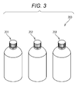
- the detection reagent kit 200 shown in Fig. 3 includes a first reagent container 201, a second reagent container 202 and a third reagent container 203.
- the first reagent container 201 accommodates a first trapping body capable of binding to an analyte.
- the second reagent container 202 accommodates a second trapping body capable of binding to a site different from a binding site for the first trapping body in the analyte.
- the third reagent container 203 accommodates a third trapping body capable of binding to the second trapping body.
- the detection reagent kit according to particular embodiments may further include a package leaflet.
- the package leaflet may include, for example, a description of operation procedures for carrying out the above-mentioned method for detecting an analyte using the detection reagent kit according to the present invention.
- a reagent for detecting an analyte is a reagent for detecting an analyte for use in the above-mentioned method for detecting an analyte.
- the reagent for detecting an analyte according to particular embodiments contains a second trapping body capable of binding to the analyte.
- the second trapping body contains a binding substance which binds to the analyte (the above-mentioned second binding substance), a support, and a linker which links the binding substance (the above-mentioned second binding substance) and the support with each other.
- the second trapping body, the binding substance (the above-mentioned second binding substance), the support and the linker are similar to those used in the above-mentioned method for detecting an analyte.
- the detection reagent according to particular embodiments may further contain an aid.
- the aid include, but are not particularly limited to, a preservative or a stabilizer for stably maintaining the second trapping body.
- the aid include a buffer.
- a detection antibody and TNF ⁇ manufactured by R&D systems, Inc., trade name: Quantikine Kitstandard
- Table 1 100 ⁇ L of an antibody solution containing the trapping antibody and the detection antibody and 100 ⁇ L of an analyte-containing solution were mixed in a tube.
- Buffer A 0.4 M sodium chloride, 0.1 mass% BSA and 0.1 M sodium phosphate buffer (pH 7.0)
- the trapping antibody shown in Table 1 was prepared as follows.
- anti-TNF ⁇ mouse IgG obtained from clone name: Mab1 manufactured by Biolegend, Inc. was fragmented with pepsin, thereby obtaining an F(ab') 2 fragment. The resultant F(ab') 2 fragment was reduced, thereby obtaining Fab'-SH.
- BSA-Bio-DNP and a linker manufactured by Life Technologies, trade name: SM(PEG)8) were reacted with each other, thereby obtaining (PEG) 8 -BSA-Bio-DNP.
- Fab'-SH and (PEG) 8 -BSA-Bio-DNP were reacted with each other, thereby obtaining a trapping antibody.
- the detection antibody shown in Table 1 was prepared as follows. In accordance with a conventional technique, an anti-mouse IgG antibody obtained from clone name: 28401 manufactured by R&D systems, Inc. was fragmented with pepsin, thereby obtaining an F(ab') 2 fragment. The resultant F(ab') 2 fragment was reduced, thereby obtaining Fab'-SH. EMCS was reacted with Gal, thereby obtaining ALP-mal. Fab'-SH and Gal-mal were reacted with each other, thereby obtaining a detection antibody. [Table 1] Amount used Trapping antibody Fab' -(PEG) 8 -BSA-Bio-DNP 300 fmol Detection antibody Fab'-Gal 30 fmol Test substance TNF ⁇ 0 or 10 pg
- One anti-DNP antibody solid phase (manufactured by Immunochemical, trade name: Immuno bead 6.35 ⁇ , solid phase having an anti-DNP antibody immobilized thereon) was added to the tube containing the sandwich complex obtained in Example 1, item (1).
- the resultant mixture was incubated at 25°C for 30 minutes, thereby trapping the sandwich complex on the anti-DNP solid phase.
- the mixture in the tube was washed twice with 2 mL of a washing liquid (0.1 M sodium chloride, 0.1 mass% BSA and 0.1 M sodium phosphate buffer (pH 7.0)). Thereafter, the anti-DNP antibody solid phase was recovered.
- Example 1 The complex recovered in Example 1, item (2) was added to 150 ⁇ L of a 2 mM DNP solution. The resultant mixture was incubated at 25°C for 30 minutes, thereby breaking a bond between the anti-DNP solid phase and the sandwich complex. A supernatant of the resultant product was transferred to another tube. The remaining anti-DNP antibody solid phase was washed twice with 2 mL of buffer B (0.1 M sodium chloride, 0.1 mass% BSA and 0.1 M sodium phosphate buffer (pH 7.0)).
- buffer B 0.1 M sodium chloride, 0.1 mass% BSA and 0.1 M sodium phosphate buffer (pH 7.0)
- Example 1 To the supernatant recovered in Example 1, item (3), one streptavidin solid phase (manufactured by Immunochemical, trade name: Immuno bead 6.35 ⁇ , solid phase having streptavidin immobilized thereon) was added. The resultant mixture was incubated at 25°C for 30 minutes, thereby trapping the sandwich complex on the streptavidin solid phase. Then, the mixture in the tube was washed three times with 2 mL of buffer B (0.1 M sodium chloride, 0.1 mass% BSA and 0.1 M sodium phosphate buffer (pH 7.0)), thereby recovering the streptavidin solid phase.
- buffer B 0.1 M sodium chloride, 0.1 mass% BSA and 0.1 M sodium phosphate buffer (pH 7.0)
- the anti-DNP antibody solid phase recovered in Example 1, item (2) and 200 ⁇ L of an aqueous solution of 0.2 mM 4-MUG were added to 200 ⁇ L of buffer B (0.1 M sodium chloride, 0.1 mass% BSA and 0.1 M sodium phosphate buffer (pH 7.0)) in a new tube, thereby obtaining a reaction solution.
- buffer B 0.1 M sodium chloride, 0.1 mass% BSA and 0.1 M sodium phosphate buffer (pH 7.0)
- the resultant reaction solution was incubated at 30°C for 2 hours, thereby obtaining a reaction product. Thereafter, the fluorescence intensity (hereinafter referred to as "fluorescence intensity A1”) of the reaction product in the tube was measured at an excitation wavelength: 360 nm and a fluorescence wavelength: 450 nm.
- Fluorescence intensity A2 was calculated by subtracting from the fluorescence intensity A1 value the fluorescence intensity value when the aqueous solution of 0.2 mM 4-MUG alone was added. The average value (hereinafter referred to as "fluorescence intensity A") of fluorescence intensity A2 was calculated based on the results of the three measurements.
- the streptavidin solid phase recovered in Example 1, item (4) and 200 ⁇ L of an aqueous solution of 0.2 mM 4-MUG were added to 200 ⁇ L of buffer B (0.1 M sodium chloride, 0.1 mass% BSA and 0.1 M sodium phosphate buffer (pH 7.0)) in a new tube, thereby obtaining a reaction solution.
- buffer B 0.1 M sodium chloride, 0.1 mass% BSA and 0.1 M sodium phosphate buffer (pH 7.0)
- the resultant reaction solution was incubated at 30°C for 20 hours, thereby obtaining a reaction product. Thereafter, the fluorescence intensity (hereinafter referred to as "fluorescence intensity B1”) of the reaction product in the tube was measured at an excitation wavelength: 360 nm and a fluorescence wavelength: 450 nm.
- Fluorescence intensity B2 was calculated by subtracting from the fluorescence intensity B1 value the fluorescence intensity value when the aqueous solution of 0.2 mM 4-MUG alone was added. The average value (hereinafter referred to as "fluorescence intensity B") of fluorescence intensity B2 was calculated based on the results of the three measurements.
- the anti-DNP antibody solid phase recovered in Example 1, item (3) and 200 ⁇ L of an aqueous solution of 0.2 mM 4-MUG were added to 200 ⁇ L of buffer B (0.1 M sodium chloride, 0.1 mass% BSA and 0.1 M sodium phosphate buffer (pH 7.0)) in a new tube, thereby obtaining a reaction solution.
- buffer B 0.1 M sodium chloride, 0.1 mass% BSA and 0.1 M sodium phosphate buffer (pH 7.0)
- the resultant reaction solution was incubated at 30°C for 2 hours, thereby obtaining a reaction product. Thereafter, the fluorescence intensity (hereinafter referred to as "fluorescence intensity C1”) of the reaction product in the tube was measured at an excitation wavelength: 360 nm and a fluorescence wavelength: 450 nm.
- Fluorescence intensity C2 was calculated by subtracting from the fluorescence intensity C1 value the value when the aqueous solution of 0.2 mM 4-MUG alone was added. The average value (hereinafter referred to as "fluorescence intensity C") of fluorescence intensity C2 was calculated based on the results of the three measurements.
- the trapping antibody was prepared as follows.
- the anti-TNF ⁇ mouse IgG obtained from clone name: Mab1 manufactured by Biolegend, Inc. was fragmented with pepsin, thereby obtaining an F(ab') 2 fragment.
- the resultant F(ab') 2 fragment was reduced, thereby obtaining Fab'-SH.
- BSA-Bio-DNP and EMCS were reacted with each other, thereby obtaining BSA-Bio-DNP-mal.
- Fab'-SH and BSA-Bio-DNP-mal were reacted with each other, thereby obtaining a trapping antibody.
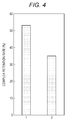
- Fig. 4 shows results of Example 1 and Comparative Example 1.
- Lane 1 represents a complex retention rate in Example 1
- Lane 2 represents a complex retention rate in Comparative Example 1.
- Fluorescence intensity B2 was calculated by subtracting from the fluorescence intensity B1 value the fluorescence intensity value when the aqueous solution of 0.2 mM 4-MUG alone was added. Fluorescence intensity B was calculated as the average value of fluorescence intensity B2 based on the results of the three measurements.
- Fluorescence intensity B in the presence of the analyte was used as the "fluorescence intensity in the presence of the analyte.
- Fluorescence intensity B in the absence of the analyte was used as the "fluorescence intensity in the absence of the analyte.”
- the anti-DNP antibody solid phase recovered in Comparative Example 3, item (2) and 200 ⁇ L of an aqueous solution of 0.2 mM 4-MUG were added to 200 ⁇ L of buffer B (0.1 M sodium chloride, 0.1 mass% BSA and 0.1 M sodium phosphate buffer (pH 7.0)) in a new tube, thereby obtaining a reaction solution.
- buffer B 0.1 M sodium chloride, 0.1 mass% BSA and 0.1 M sodium phosphate buffer (pH 7.0)
- the resultant reaction solution was incubated at 30°C for 2 hours, thereby obtaining a reaction product. Thereafter, the fluorescence intensity (hereinafter referred to as "fluorescence intensity A1”) of the reaction product in the tube was measured at an excitation wavelength: 360 nm and a fluorescence wavelength: 450 nm.
- Fluorescence intensity A2 was calculated by subtracting from the fluorescence intensity A1 value the value when the aqueous solution of 0.2 mM 4-MUG alone was added. Fluorescence intensity A was calculated as the average value of fluorescence intensity A2 based on the results of the three measurements.
- the S/N ratio in sandwich ELISA was calculated using fluorescence intensity A in the presence of the analyte and fluorescence intensity A in the absence of the analyte, in accordance with Formula (III).
- Fluorescence intensity A in the presence of the analyte was used as the "fluorescence intensity in the presence of the analyte.”
- Fluorescence intensity A in the absence of the analyte was used as the "fluorescence intensity in the absence of the analyte.”
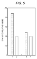
- Fig. 5 shows results of Example 2 and Comparative Examples 2 to 4.
- Lane 1 represents a relative value of the S/N ratio in Example 2
- Lane 2 represents a relative value of the S/N ratio in Comparative Example 2
- Lane 3 represents a relative value of the S/N ratio in Comparative Example 3
- Lane 4 represents a relative value of the S/N ratio in Comparative Example 4.
- the relative values of the S/N ratios in Example 2 and Comparative Example 2 are values when the calculated value of the S/N ratio in Comparative Example 2 is defined as 100.
- the relative values of the S/N ratios in Comparative Example 3 and Comparative Example 4 are values when the calculated value of the S/N ratio in Comparative Example 4 is defined as 100.
- the relative value of the S/N ratio in Example 2 was 220.
- the relative value of the S/N ratio in Comparative Example 3 was 120.
- ICT-EIA using a trapping antibody containing a linker can improve the S/N ratio more than sandwich ELISA using a trapping antibody containing a linker.
- a trapping antibody-containing solution a detection antibody-containing solution and an analyte-containing solution were mixed in a tube.
- analytes insulin (manufactured by Acris Antibodies GmbH, trade name: Human Insulin), IL-12/23p40 (manufactured by R&D Systems, trade name: Quantikine Kit Standard), and HBsAg (manufactured by Sysmex Co., trade name: HISCL HBsAg Calibrator) were used.
- the trapping antibody was prepared as follows.
- the IgG obtained from the clone shown in Table 1 was fragmented with pepsin, thereby obtaining an F(ab') 2 fragment.
- the resultant F(ab') 2 fragment was reduced, thereby obtaining Fab'-SH.
- BSA-Bio-DNP and a linker (manufactured by Life Technologies, trade name: SM(PEG) 8 ) were reacted with each other, thereby obtaining (PEG) 8 -BSA-Bio-DNP.
- BSA-Bio-DNP and EMCS were reacted with each other, thereby obtaining BSA-Bio-DNP-mal.
- Fab'-SH and (PEG) 8 -BSA-Bio-DNP were reacted with each other, thereby obtaining a trapping antibody (Examples 3 to 5).
- Fab'-SH and BSA-Bio-DNP-mal were reacted with each other, thereby obtaining a trapping antibody (Comparative Examples 5 to 7).
- the detection antibody was prepared as follows. In accordance with a conventional technique, the IgG obtained from the clone shown in Table 2 was fragmented with pepsin, thereby obtaining an F(ab') 2 fragment. The resultant F(ab') 2 fragment was reduced, thereby obtaining Fab'-SH. EMCS was reacted with Gal, thereby obtaining ALP-mal. Fab'-SH and Gal-mal were reacted with each other, thereby obtaining a detection antibody.
- Example 1 Except that the resultant mixture was used, operations similar to those of Example 1, item (1) were performed to form a sandwich complex. Thereafter, operations similar to those of Example 1, items (2) to (4) were performed to recover the streptavidin solid phase. Then, the recovered streptavidin solid phase and 200 ⁇ L of an aqueous solution of 0.2 mM 4-MUG were added to 200 ⁇ L of buffer B (0.1 M sodium chloride, 0.1 mass% BSA and 0.1 M sodium phosphate buffer (pH 7.0)) in a new tube, thereby obtaining a reaction solution. The resultant reaction solution was incubated at 30°C for 2 hours, thereby obtaining a reaction product.
- buffer B 0.1 M sodium chloride, 0.1 mass% BSA and 0.1 M sodium phosphate buffer (pH 7.0)
- fluorescence intensity B1 of the reaction product in the tube was measured at an excitation wavelength: 360 nm and a fluorescence wavelength: 450 nm.
- Fluorescence intensity B2 was calculated by subtracting from the fluorescence intensity B1 value the value when the aqueous solution of 0.2 mM 4-MUG alone was added.
- Fluorescence intensity B was calculated as the average value of fluorescence intensity B2 based on the results of the three measurements.
- the S/N ratio in ICT-EIA was calculated using the fluorescence intensity B in the presence of the analyte and the fluorescence intensity B in the absence of the analyte, in accordance with Formula (III). "Fluorescence intensity B in the presence of the analyte” was used as the "fluorescence intensity in the presence of the analyte.” "Fluorescence intensity B in the absence of the analyte" was used as the "fluorescence intensity in the absence of the analyte.”
- Fig. 6 shows results of Examples 3 to 5 and Comparative Examples 5 to 7.
- Lane 1 represents a relative value of the S/N ratio in Example 3
- Lane 2 represents a relative value of the S/N ratio in Comparative Example 5
- Lane 3 represents a relative value of the S/N ratio in Example 4
- Lane 4 represents a relative value of the S/N ratio in Comparative Example 6
- Lane 5 represents a relative value of the S/N ratio in Example 5
- Lane 6 represents a relative value of the S/N ratio in Comparative Example 7.
- the relative values of the S/N ratios in Example 3 and Comparative Example 5 are values when the calculated value of the S/N ratio in Comparative Example 5 is defined as 100.
- Fig. 6 the relative values of the S/N ratios in Example 3 and Comparative Example 5 are values when the calculated value of the S/N ratio in Comparative Example 5 is defined as 100.
- Fig. 6 the relative values of the S/N ratios in Example 3 and Comparative Example 5 are values when the calculated value of the S/
- the relative values of the S/N ratios in Example 4 and Comparative Example 6 are values when the calculated value of the S/N ratio in Comparative Example 6 is defined as 100.
- the relative values of the S/N ratios in Example 5 and Comparative Example 7 are values when the calculated value of the S/N ratio in Comparative Example 7 is defined as 100.
- Example 8 a product manufactured by Life Technologies, trade name: EMCS (Comparative Example 8), manufactured by Life Technologies, trade name: SM(PEG) 2 (Example 6), manufactured by Life Technologies, trade name: SM(PEG) 8 (Example 7) or manufactured by Life Technologies, trade name: SM(PEG) 24 (Example 8) was used as the trapping antibody, operations similar to those of Example 1 were performed to calculate the complex retention rate.
- EMCS Comparative Example 8
- SM(PEG) 2 Example 6
- SM(PEG) 8 Example 7
- SM(PEG) 24 Example 8
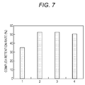
- Fig. 7 shows results of Examples 6 to 8 and Comparative Example 8.
- Lane 1 represents a complex retention rate in Comparative Example 8
- Lane 2 represents a complex retention rate in Example 6
- Lane 3 represents a complex retention rate in Example 7
- Lane 4 represents a complex retention rate in Example 8.
- a trapping body containing a binding substance, a support and a linker which links the binding substance and the support with each other can suppress the dissociation of an immune complex (namely, sandwich complex) and improve the S/N ratio in ICT-EIA.
Landscapes
- Health & Medical Sciences (AREA)
- Immunology (AREA)
- Life Sciences & Earth Sciences (AREA)
- Engineering & Computer Science (AREA)
- Chemical & Material Sciences (AREA)
- Molecular Biology (AREA)
- Biomedical Technology (AREA)
- Hematology (AREA)
- Urology & Nephrology (AREA)
- Food Science & Technology (AREA)
- General Physics & Mathematics (AREA)
- Cell Biology (AREA)
- Biotechnology (AREA)
- Medicinal Chemistry (AREA)
- Physics & Mathematics (AREA)
- Analytical Chemistry (AREA)
- Biochemistry (AREA)
- General Health & Medical Sciences (AREA)
- Microbiology (AREA)
- Pathology (AREA)
- Chemical Kinetics & Catalysis (AREA)
- Proteomics, Peptides & Aminoacids (AREA)
- Investigating Or Analysing Biological Materials (AREA)
- Peptides Or Proteins (AREA)
- Investigating, Analyzing Materials By Fluorescence Or Luminescence (AREA)
- Investigating Or Analysing Materials By The Use Of Chemical Reactions (AREA)
Abstract
Description
- The present invention relates to a method for detecting an analyte, a detection reagent kit, and a detection reagent.
- An immune complex transfer enzyme immunoassay is disclosed as a method for detecting an analyte (for example, see
JP H01-254868 JP H01-254868 - The method described in
JP H01-254868 - An object of the present invention is to provide a method for detecting an analyte, a detection kit, a detection reagent, and use of a detection reagent, by which an analyte can be detected with higher sensitivity.
- One aspect of the present invention includes a method for detecting an analyte in a sample, comprising the steps of: (A) forming a first complex comprising: an analyte; a first trapping body which specifically binds to the analyte; a second trapping body which specifically binds to a site of the analyte, the site being different from a site to which the first trapping body specifically binds; and a third trapping body which specifically binds to the second trapping body; (B) separating a part comprising the analyte and the first trapping body from the third trapping body; (C) allowing a fourth trapping body to trap the part to form a second complex; and (D) detecting the analyte of the second complex,
wherein the second trapping body comprises a binding substance which binds to the analyte, a support, and a linker which links the binding substance and the support with each other. - Another aspect of the present invention includes a reagent kit for detecting an analyte in a sample, comprising or consisting of: a first trapping body capable of binding to an analyte; a second trapping body capable of binding to a site of the analyte, the site being different from a site to which the first trapping body specifically binds; and a third trapping body capable of binding to the second trapping body,
wherein the second trapping body comprises a binding substance which binds to the analyte, a support, and a linker which links the binding substance and the support with each other. - Still another aspect of the present invention includes a reagent for detecting an analyte in a sample for use in the above-mentioned method for detecting an analyte, the reagent containing the second trapping body capable of binding to the analyte, wherein the second trapping body contains the binding substance which binds to the analyte, the support, and the linker which links the binding substance and the support with each other. In particular embodiments, the reagent contains no other binding substances than those present in the second trapping body.
- Still another aspect of the present invention includes use of a reagent for detecting an analyte in sample in the above-mentioned method for detecting an analyte, the reagent containing the second trapping body capable of binding to the analyte, wherein the second trapping body contains the binding substance which binds to the analyte, the support, and the linker which links the binding substance and the support with each other.
- The present invention can provide a method for detecting an analyte, a detection reagent kit, a detection reagent, and use of a detection reagent, by which an analyte can be detected with high sensitivity.
-
-
Fig. 1 is a schematic explanatory view of a second trapping body. -
Fig. 2 is a process chart of example procedures of a method for detecting an analyte. -
Fig. 3 is a configuration diagram of a detection reagent kit. -
Fig. 4 is a graph showing results of examination of a complex retention rate in Example 1 and Comparative Example 1. -
Fig. 5 is a graph showing results of examination of an S/N ratio in Example 2 and Comparative Examples 2 to 4. -
Fig. 6 is a graph showing results of examination of an S/N ratio in Examples 3 to 5 and Comparative Examples 5 to 7. -
Fig. 7 is a graph showing results of examination of an S/N ratio in Examples 6 to 8 and Comparative Example 8. - The method for detecting an analyte according to the present invention includes the steps of: (A) forming a first complex containing an analyte, a first trapping body which specifically binds to the analyte, a second trapping body which specifically binds to a site different from a binding site for the first trapping body in the analyte, and a third trapping body which specifically binds to the second trapping body; (B) separating a part including the first trapping body from the first complex; (C) allowing a fourth trapping body to trap the part including the first trapping body to form a second complex; and (D) detecting the analyte contained in the second complex. The second trapping body contains a binding substance which binds to the analyte, a support, and a linker which links the binding substance and the support with each other. The method for detecting an analyte according to particular embodiments can be carried out, for example, by a method involving use of an antigen-antibody reaction, such as immunoassay. In this case, the first trapping body and the analyte form a complex with use of an antigen-antibody reaction. The second trapping body and the analyte form a complex with use of an antigen-antibody reaction.
- An immune complex transfer enzyme immunoassay (hereinafter, referred to also as "ICT-EIA") involves trapping an analyte with two trapping bodies with the analyte being sandwiched therebetween to form an immune complex. The present inventors have found that, in a conventional immune complex transfer enzyme immunoassay, the sensitivity is lowered by dissociation of a part of the immune complex. In the case of a very small amount of the analyte, it is sometimes difficult to ensure sufficient sensitivity. Contrary to this, the method for detecting an analyte according to particular embodiments employs a second trapping body in which a binding substance and a support are linked with each other by a linker. Thus, in the method for detecting an analyte according to particular embodiments, the first trapping body and the second trapping body trap the analyte while sandwiching the analyte to form a sandwich complex. This is considered to suppress the occurrence of steric hindrance when the analyte is trapped by the second trapping body. Therefore, the dissociation of the complex made up of the first trapping body, the analyte and the second trapping body is considered to be suppressed, thereby making it possible to improve the sensitivity in detection of the analyte.
- Examples of the analyte include, but are not particularly limited to, an antibody, an antigen, a nucleic acid, a physiologically active substance, a bacterium, a virus, a peptide, and a therapeutic agent (blood drug). An antibody can serve also as an antigen. Examples of the antibody include, but are not particularly limited to, an antibody to an antigen and an antibody to an antibody. Examples of the antigen include, but are not particularly limited to, a nucleic acid, a physiologically active substance, a bacterium, a virus, and a peptide. Examples of the nucleic acid include, but are not particularly limited to, nucleic acids encoding, for example, a disease-causing gene, and a nucleic acid encoding a gene of a bacterium or a virus. Examples of the physiologically active substance include, but are not particularly limited to, a cell growth factor, a differentiation-inducing factor, a cell adhesion factor, an enzyme, a cytokine, a hormone, and a sugar chain.
- The first trapping body contains a first binding substance which specifically binds to the analyte. The term "specifically" as used therein means that the binding substance does not substantially bind to any substance other than a specific substance, or does not cause an antigen-antibody reaction with any substance other than a specific substance at a substantially detectable level. The first trapping body preferably further has a detectable labeling substance from the viewpoint of enhancing the ease of detecting the analyte.
- Examples of the first binding substance include, but are not particularly limited to, an antibody, an aptamer, an affibody (registered trademark of Affibody AB), a lectin, and a nucleic acid. The concept "antibody" as used herein encompasses also an "antibody fragment," unless otherwise noted. The antibody as the first binding substance functions to specifically bind to the analyte. Examples of the antibody include, but are not particularly limited to, a mouse-derived antibody, a rabbit-derived antibody, a goat-derived antibody, a sheep-derived antibody, a guinea pig-derived antibody, an eel-derived antibody, a shark-derived antibody, a humanized antibody, and a chimeric antibody. The antibody may be either a monoclonal antibody or a polyclonal antibody. Examples of the antibody fragment include, but are not particularly limited to, Fab, Fab', F(ab')2, and a single-chain antibody [scFc]. The aptamer may be either a nucleic acid aptamer or a peptide aptamer. The affibody (registered trademark) is a polypeptide having a specific domain of protein A as a backbone. The affibody functions to specifically bind to the analyte. A lectin is a protein which specifically binds to a sugar chain as the analyte. The nucleic acid is a nucleic acid which specifically binds to a nucleic acid as the analyte. Examples of the nucleic acid include, but are not particularly limited to, a DNA, an RNA, a peptide nucleic acid (PNA), and a bridged nucleic acid (BNA). Among these binding substances, an antibody is preferred.
- Examples of the labeling substance include, but are not particularly limited to, an enzyme, a fluorescent substance and a radioactive substance. Examples of the enzyme include, but are not particularly limited to, β-galactosidase, peroxidase, alkaline phosphatase and luciferase. Examples of the fluorescent substance include, but are not particularly limited to, fluorescein, fluorescein isothiocyanate, coumarin, rhodamine, fluorescein, Cy3, Cy5, Hoechst 33342, 4',6-diamino-2-phenylindole (DAPI), propidium iodide (PI), and pigments of the series Alexa Fluor (registered trademark of Molecular Probes). Examples of the radioactive substance include, but are not particularly limited to, 32P and 35S. The labeling substance is preferably an enzyme, more preferably β-galactosidase and alkaline phosphatase.
- The second trapping body specifically binds to a site different from a binding site for the first trapping body in the analyte. Thus, the second trapping body does not compete with the first trapping body at the time of binding to the analyte. The second trapping body contains a second binding substance which binds to the analyte, a support and a linker. The linker links the second binding substance and the support with each other. The support has a first reactive group and a second reactive group.
- The second binding substance is a substance which specifically binds to a site different from a binding site for the first trapping body in the analyte. Examples of the second binding substance include, but are not particularly limited to, an antibody, an aptamer, an affibody (registered trademark of Affibody AB), a lectin, and a nucleic acid. The antibody as the second binding substance functions to specifically bind to the analyte. Examples of the antibody include, but are not particularly limited to, a mouse-derived antibody, a rabbit-derived antibody, a goat-derived antibody, a sheep-derived antibody, a guinea pig-derived antibody, an eel-derived antibody, a shark-derived antibody, a humanized antibody, and a chimeric antibody.
- Examples of the support include, but are not particularly limited to, a polypeptide, dextran and casein. The polypeptide is a polypeptide which has no binding site for the first trapping body. The polypeptide varies depending on the kind of the analyte. Examples of the polypeptide include, but are not particularly limited to, albumins such as bovine serum albumin and human serum albumin.
- Any substance may be used as the first reactive group without particular limitation so long as the substance can bind to the third trapping body. Examples of the first reactive group include a dinitrophenyl (DNP) group, biotin, an antibody, an antigen, avidin, and streptavidin.
- Any substance may be used as the second reactive group so long as the substance can bind to the fourth trapping body. Examples of the second reactive group include a DNP group, biotin, an antibody, an antigen, avidin, and streptavidin. The second reactive group is the first reactive group.
- As used herein, the term "linker" refers to a molecule which links the second binding substance and the support with each other. Examples of the linker include, but are not particularly limited to, a polymer chain optionally having at least one selected from the group consisting of a substituent, an oxygen atom, a sulfur atom and a nitrogen atom. Examples of the polymer chain include, but are not particularly limited to, a polyalkylene glycol chain having an oxyalkylene group having 2 to 6 carbon atoms, and a polymer chain represented by Formula (I):

- In the polyalkylene glycol chain, the number of carbon atoms possessed by the oxyalkylene group is 2 to 6, preferably 2 to 4. In the polyalkylene glycol chain, the average addition molar number of the oxyalkylene groups is 2 to 100, preferably 2 to 20. The mass average molecular weight of the polyalkylene glycol chain is preferably 116 to 12000, more preferably 116 to 2000. Examples of the oxyalkylene group having 2 to 6 carbon atoms include, but are not particularly limited to, an oxyethylene group and an oxypropylene group. Examples of the polyalkylene glycol chain include, but are not particularly limited to, polyethylene glycol and polypropylene glycol. The polyalkylene glycol chain may be any of a homopolymer, an alternating copolymer, a block copolymer, and a random copolymer. The "mass average molecular weight" is a value obtained by gel filtration chromatography.
- In formula (I), X represents a nitrogen atom, an oxygen atom, a sulfur atom, an alkylene group having 1 to 4 carbon atoms, an -NHCO- group, an optionally substituted aryl group having 6 to 12 carbon atoms, or an optionally substituted cycloalkyl group having 3 to 8 carbon atoms. The number of carbon atoms possessed by the aryl group is 6 to 12, preferably 6 to 8, more preferably 6 to 7. Examples of the substituent which may be possessed by the aryl group include, but are not particularly limited to, a methyl group, an oxo group, a carboxyl group, and an amino group. Examples of the aryl group include, but are not particularly limited to, a phenyl group and a tolyl group. The number of carbon atoms possessed by the cycloalkyl group is 3 to 8, preferably 4 to 6. The substituent which may be possessed by the cycloalkyl group is similar to the substituent which may be possessed by the aryl group. Examples of the cycloalkyl group include, but are not particularly limited to, a cyclopropyl group, a cyclobutyl group, a cyclopentyl group, and a cyclohexyl group.
- Among these linkers, preferred is a polyalkylene glycol chain having an oxyalkylene group having 2 to 6 carbon atoms, and more preferred is a polyalkylene glycol chain having an oxyalkylene group having 2 to 6 carbon atoms whose average addition molar number is 2 to 100, from the viewpoint of suppressing, for example, the dissociation of the analyte at the time of forming a sandwich complex. Among the polyalkylene glycol chains, preferred is a polyethylene glycol chain, and more preferred is a polyethylene glycol chain having an oxyethylene group whose average addition molar number is 2 to 100, from the viewpoint of suppressing, for example, the dissociation of the analyte at the time of forming a sandwich complex with more certainty.
- The third trapping body specifically binds to the second trapping body. The third trapping body preferably contains a third binding substance which binds to the second trapping body and a solid phase which retains the binding substance. The third binding substance in the third trapping body binds to a site different from a binding site for the analyte in the second trapping body. Examples of the third binding substance in the third trapping body include, but are not particularly limited to, an antibody, an aptamer, an affibody (registered trademark of Affibody AB), a lectin, a nucleic acid, biotin, avidin, and streptavidin. The antibody as the third binding substance functions to specifically bind to the analyte. Examples of the antibody include, but are not particularly limited to, a mouse-derived antibody, a rabbit-derived antibody, a goat-derived antibody, a sheep-derived antibody, a guinea pig-derived antibody, an eel-derived antibody, a shark-derived antibody, a humanized antibody, and a chimeric antibody. Examples of the solid phase include, but are not particularly limited to, a particle and a plate. Examples of the particle include, but are not particularly limited to, a magnetic particle and a latex particle. Examples of the plate include, but are not particularly limited to, a polystyrene plate. Examples of a combination of the first reactive group in the second trapping body and the third binding substance in the third trapping body include, but are not particularly limited to, a combination of a hapten and an anti-hapten antibody and a combination of a different antigen and a different antibody. Examples of the combination of a hapten and an anti-hapten antibody include, but are not particularly limited to, DNP-anti-DNP antibody and biotin-anti-biotin antibody.
- The fourth trapping body traps a part including the first trapping body. The phrase "part including the first trapping body" refers to a portion enough to ensure the correlation between the amount of the first trapping body and the amount of the analyte. The part including the first trapping body is preferably a portion of the first complex except the third trapping body. Examples of the part including the first trapping body include a complex of the first trapping body and the analyte, and a complex of the first trapping body, the analyte and the second trapping body. The fourth trapping body preferably contains a fourth binding substance which binds to the part including the first trapping body, and a solid phase. Examples of the fourth binding substance includes an antibody, an antibody fragment, an aptamer, an affibody (registered trademark of Affibody AB), a lectin, a nucleic acid, biotin, avidin, streptavidin, and a substance which binds to the reactive group possessed by the support in the second trapping body. The solid phase fixes and retains the fourth binding substance. Examples of the solid phase in the fourth trapping body include those which are similar to the solid phases in the third trapping body. Examples of the combination of the second reactive group in the second trapping body and the fourth binding substance in the fourth trapping body include, but are not particularly limited to, a combination of a hapten and an anti-hapten antibody and a combination of a different antigen and a different antibody. Examples of the combination of a hapten and an anti-hapten antibody include, but are not particularly limited to, DNP-anti-DNP antibody and biotin-anti-biotin antibody. The combination of the second reactive group and the fourth trapping body is different from the combination of the first reactive group and the third trapping body.
-
Fig. 1 shows one example of the second trapping body. Asecond trapping body 2 as shown inFig. 1 contains Fab' as a secondbinding substance 51, asupport 52 and alinker 53. The secondbinding substance 51 and thesupport 52 are linked with each other by thelinker 53. Thesupport 52 has, on its surface, a firstreactive group 54 and a secondreactive group 55. The firstreactive group 54 is a portion capable of binding to or being detached from a third trapping body. The secondreactive group 55 is a portion capable of binding to a fourth trapping body. - The third binding substance in the third trapping body may be a substance which binds to the first
reactive group 54 possessed by thesupport 52 in thesecond trapping body 2 shown inFig. 1 . The substance which binds to the firstreactive group 54 shown inFig. 1 can appropriately be selected depending on the kind of the firstreactive group 54 inFig. 1 . Examples of the combination of the firstreactive group 54 inFig. 1 and the third binding substance in the third trapping body include, but are not particularly limited to, a combination of a DNP group and an anti-DNP antibody, a combination of a trinitrophenyl (TNP) group and an anti-TNP antibody, and a combination of biotin and avidin. - The substance which binds to the second
reactive group 55 inFig. 1 can appropriately be selected depending on the kind of the secondreactive group 55 inFig. 1 . Examples of the combination of the secondreactive group 55 and the fourth binding substance in the fourth trapping body include, but are not particularly limited to, a combination of a DNP group and an anti-DNP antibody, a combination of a TNP group and an anti-TNP antibody, a combination of biotin and avidin, and a combination of biotin and streptavidin. - Next, explained are example procedures of the method for detecting an analyte according to a particular embodiment of the invention.
Fig. 2 shows example procedures of the method for detecting an analyte according to a particular embodiment. InFig. 2 , explanation is given about an example case of detection of an analyte S in a specimen containing the analyte S and a contaminant F as shown inFig. 2A . InFig. 2 , the kinds of afirst trapping body 1, asecond trapping body 2, athird trapping body 3 and the like are not particularly limited. The term "contaminant" refers to a substance other than the analyte. InFig. 2 , thefirst trapping body 1, thesecond trapping body 2 and thethird trapping body 3 are brought in contact with the analyte S in this order to form a first complex 13. The order of mixing the analyte S, thefirst trapping body 1, thesecond trapping body 2, and thethird trapping body 3, however, is not particularly limited. - In the step (A), the first complex 13 containing the analyte S, the
first trapping body 1, thesecond trapping body 2, and thethird trapping body 3 is formed. Specifically, thefirst trapping body 1 is firstly contacted with a specimen containing the analyte S and the contaminant F, as shown inFigs. 2A and 2B . This allows formation of a complex 11 containing thefirst trapping body 1 and the analyte S. Then, as shown inFig. 2C , the complex 11 and thesecond trapping body 2 are contacted with each other. This allows formation of asandwich complex 12. Thesandwich complex 12 is a complex bound to thefirst trapping body 1 and thesecond trapping body 2 in such a manner that the analyte S is sandwiched between thefirst trapping body 1 and thesecond trapping body 2. Then, thesandwich complex 12 and thethird trapping body 3 are contacted with each other. This allows formation of the first complex 13, as shown inFig. 2D . The first complex 13 contains thefirst trapping body 1, the analyte S, thesecond trapping body 2, and thethird trapping body 3. InFig. 2D , thethird trapping body 3 contains asolid phase 61 and a thirdbinding substance 62 fixed onto thesolid phase 61. In the formation of the first complex 13, as shown inFig. 2D , the thirdbinding substance 62 in thethird trapping body 3 binds to the firstreactive group 54 in thesecond trapping body 2. - The first complex 13 can be formed, for example, with use of an antigen-antibody reaction. The antigen-antibody reaction is preferably utilized for the formation of the first complex 13, from the viewpoint of enhancing the specificity to the analyte S and improving the sensitivity. In this case, the
first trapping body 1 preferably contains a substance which specifically binds to the analyte S through an antigen-antibody reaction. Thesecond trapping body 2 preferably contains a substance which specifically binds to the analyte S through an antigen-antibody reaction as the second binding substance. When the analyte S is an antigen, there can be used an antibody which specifically binds to an antigen, an antibody fragment obtained by fragmentation of this antibody, and the like. On the other hand, when the analyte S is an antibody, there can be used an antigen for this antibody, an antibody which specifically binds to the antibody, and the like. - The first complex 13 can be formed under conditions in accordance with the kinds of the analyte S, the
first trapping body 1, thesecond trapping body 2, and thethird trapping body 3. The first complex 13 can be formed in a solution. Any solution may be used so long as it is suitable for the binding of thefirst trapping body 1, the analyte S, thesecond trapping body 2, and thethird trapping body 3. Any temperature for forming the first complex 13 may be employed so long as it is suitable for the binding of thefirst trapping body 1, the analyte S, thesecond trapping body 2, and thethird trapping body 3. Any time for forming the first complex 13 may be employed so long as it is enough for the binding of thefirst trapping body 1, the analyte S, thesecond trapping body 2, and thethird trapping body 3. - From the viewpoint of improving the sensitivity, a step of removing the
first trapping body 1 in a free state (i.e. unbound to the analyte) and thesecond trapping body 2 in a free state (hereinafter referred to also as a "first removal step") can further be carried out between the step (A) and the step (B) which will be described below. In the first removal step, washing with a buffer, for example, can be performed. Examples of the buffer include, but are not particularly limited to, phosphate buffered saline, a sodium phosphate buffer, and a tris-hydrochloric acid buffer. Any pH of the buffer may be employed so long as the pH falls within such a range as to ensure stable retention of thefirst complex 13. In the first removal step, the contaminant F in a free state is also removed. - In the step (B), a part including the
first trapping body 1 is separated from the first complex 13, as shown inFig. 2E . The part including thefirst trapping body 1 is preferably the complex 11 and thesandwich complex 12, more preferably thesandwich complex 12, from the viewpoint of ensuring ease of separation and sufficient quantitative capability. InFig. 2E , thesandwich complex 12 is separated from the first complex 13, as the part including thefirst trapping body 1. However, the part including thefirst trapping body 1 may be a portion other than thesandwich complex 12. - Separation can be performed, for example, by a separation method in accordance with the type of a bond included in the
first complex 13. Examples of the bond included in the first complex 13 include a bond between thefirst trapping body 1 and thesecond trapping body 2, a bond between thesecond trapping body 2 and thethird trapping body 3, a bond included in the complex 11, and a bond included in thesandwich complex 12. Specific examples of the bond included in the first complex include, but are not particularly limited to, a bond via a disulfide bond and a bond via a DNP group. Examples of the bond via a disulfide bond include, but are not particularly limited to, a bond between an antigen and an antibody via a disulfide bond and a bond between biotin and avidin or streptavidin. Examples of the bond via a DNP group include, but are not limited to, a bond between DNP and an anti-DNP antibody. For separation, there can be used a separation reagent for separating at least thethird trapping body 3 from the first complex 13 without breaking thefirst trapping body 1. When the first complex 13 has a disulfide bond in its portion except thefirst trapping body 1, a disulfide bond breaking reagent can be used as the separation reagent. Examples of the disulfide bond breaking reagent include, but are not particularly limited to, 2-mercaptoethanol and dithiothreitol. When the first complex 13 has a bond via a DNP group in its portion except thefirst trapping body 1, a dinitrophenyl amino acid can be used as the separation reagent. Examples of the dinitrophenyl amino acid include, but are not particularly limited to, dinitrophenyl lysine. - After the first removal step, the
first trapping body 1 in a free state and thesecond trapping body 2 in a free state remain in some cases. Thus, a step of removing thefirst trapping body 1 in a free state and thesecond trapping body 2 in a free state can further be carried out between the step (B) and the step (C) which will be described below, from the viewpoint of improving the sensitivity. - In the step (C), the part including the
first trapping body 1 is trapped by thefourth trapping body 4 as shown inFig. 2F . This allows formation of asecond complex 14. InFig. 2F , thefourth trapping body 4 contains asolid phase 71 and a fourthbinding substance 72 fixed onto thesolid phase 71. In the formation of the second complex 14, the fourth bindingsubstance 72 in thefourth trapping body 4 binds to the secondreactive group 55 in thesecond trapping body 2, as shown inFig. 2D . The second complex 14 can be formed by a technique similar to that for forming the first complex 13 in the step (A). - A step of removing the fourth trapping body in a free state can further be performed between the step (C) and the step (D) which will be described below, from the viewpoint of improving the sensitivity.
- In the step (D), the analyte S contained in the second complex 14 is detected as shown in
Fig. 2G . The analyte can be detected through detection of a signal based on a labeling substance contained in thefirst trapping body 1 when thefirst trapping body 1 contains the labeling substance. Examples of the signal include, but are not particularly limited to, luminescence, fluorescence, color development and radiation. The signal is preferably luminescence, fluorescence and color development because of ease of detection. The signal based on the labeling substance can be detected by a detection method according to the kind of the labeling substance. When the labeling substance is an enzyme, the signal can be detected, for example, by measuring the amount of a product generated from an enzyme substrate through an enzyme reaction. The enzyme substrate is preferably a color-developing substrate and a chemiluminescent substrate because of ease of measuring the amount of the product. The enzyme substrate can appropriately be selected according to the kind of the enzyme. When the labeling substance is a fluorescent substance, the signal can be detected, for example, by measuring the intensity of fluorescence based on the fluorescent substance or the shift of the fluorescence wavelength. When the labeling substance is a radioactive substance, the signal can be detected, for example, by measuring the amount of radiation generated from the radioactive substance. - A detection reagent kit according to a particular embodiment of the invention includes: a first trapping body capable of binding to an analyte; a second trapping body capable of binding to a site different from a binding site for the first trapping body in the analyte; and a third trapping body capable of binding to the second trapping body, and the second trapping body contains a binding substance which binds to the analyte (the above-mentioned second binding substance), a support, and a linker which links the binding substance and the support with each other. The first trapping body, the second trapping body, the third trapping body, the fourth trapping body, the binding substance (the above-mentioned second binding substance), the support and the linker are similar to those used in the above-mentioned method for detecting an analyte. The first trapping body, the second trapping body, the third trapping body and the fourth trapping body can be provided in a state where they are dissolved in an appropriate solvent. The first trapping body, the second trapping body and the third trapping body, and the fourth trapping body are preferably accommodated in separate containers, from the viewpoint of suppressing non-specific detection of a contaminant. The first trapping body, the second trapping body, the third trapping body and the fourth trapping body may be accommodated in separate containers. Two or more of the first trapping body, the second trapping body and the third trapping body may be accommodated in the same container. Two or more of the first trapping body, the second trapping body and the third trapping body may be accommodated in the same container.
- The detection reagent kit according to particular embodiments may further include an aid. Examples of the aid include, but are not particularly limited to, a preservative or a stabilizer for stably maintaining the first trapping body, the second trapping body, the third trapping body and the fourth trapping body; a reagent for forming the first complex containing the first trapping body, the second trapping body and the third trapping body; a separation reagent; and a reagent for forming the second complex containing the fourth trapping body and the part including the first trapping body. Examples of the aid include a buffer.
- When the first trapping body has a labeling substance, the detection reagent kit according to particular embodiments may further include a reagent necessary for the detection of a signal based on the labeling substance. The reagent necessary for the signal detection can appropriately be selected according to the kind of the labeling substance. Examples of the reagent necessary for the signal detection include, but are not particularly limited to, an enzyme substrate and a color developing agent.
- One example of the detection reagent kit according to the particular embodiments is a
detection reagent kit 200 as shown inFig. 3 , without particular limitation. Thereagent kit 200 shown inFig. 3 includes afirst reagent container 201, asecond reagent container 202 and athird reagent container 203. Thefirst reagent container 201 accommodates a first trapping body capable of binding to an analyte. Thesecond reagent container 202 accommodates a second trapping body capable of binding to a site different from a binding site for the first trapping body in the analyte. Thethird reagent container 203 accommodates a third trapping body capable of binding to the second trapping body. The detection reagent kit according to particular embodiments may further include a package leaflet. The package leaflet may include, for example, a description of operation procedures for carrying out the above-mentioned method for detecting an analyte using the detection reagent kit according to the present invention. - A reagent for detecting an analyte according to particular embodiments is a reagent for detecting an analyte for use in the above-mentioned method for detecting an analyte. The reagent for detecting an analyte according to particular embodiments contains a second trapping body capable of binding to the analyte. The second trapping body contains a binding substance which binds to the analyte (the above-mentioned second binding substance), a support, and a linker which links the binding substance (the above-mentioned second binding substance) and the support with each other. The second trapping body, the binding substance (the above-mentioned second binding substance), the support and the linker are similar to those used in the above-mentioned method for detecting an analyte.
- The detection reagent according to particular embodiments may further contain an aid. Examples of the aid include, but are not particularly limited to, a preservative or a stabilizer for stably maintaining the second trapping body. Examples of the aid include a buffer.
- Hereinafter, the meanings of abbreviations are as follows.
-
- BSA: bovine serum albumin
- DNP: 2,4-dinitrophenyl group
- Bio: biotinyl group
- EMCS: N-(6-maleimidecaprolyloxy) succinimide
- SH: thiol group mal: maleimide group
- BSA-Bio-DNP: BSA modified with biotin and DNP
- (PEG)8-BSA-Bio-DNP: BSA-Bio-DNP to which a PEG linker is added
- TNFα: tumor necrosis factor α
- 4-MUG: 4-methylumbelliferyl-β-D-galactopyranoside
- DMF: N,N-dimethylformamide
- PEG: polyethylene glycol chain
- (PEG)n: polyethylene glycol chain having an addition molar number of oxyethylene groups of n
- Gal: β-galactosidase
- In order that the amounts of a trapping antibody, a detection antibody and TNFα (manufactured by R&D systems, Inc., trade name: Quantikine Kitstandard) as an analyte which are shown in Table 1 were as shown in Table 1, 100 µL of an antibody solution containing the trapping antibody and the detection antibody and 100 µL of an analyte-containing solution were mixed in a tube. Buffer A (0.4 M sodium chloride, 0.1 mass% BSA and 0.1 M sodium phosphate buffer (pH 7.0)) was used as a solvent for the antibody solution and the analyte-containing solution. The trapping antibody shown in Table 1 was prepared as follows. In accordance with a conventional technique, anti-TNFα mouse IgG obtained from clone name: Mab1 manufactured by Biolegend, Inc. was fragmented with pepsin, thereby obtaining an F(ab')2 fragment. The resultant F(ab')2 fragment was reduced, thereby obtaining Fab'-SH. BSA-Bio-DNP and a linker (manufactured by Life Technologies, trade name: SM(PEG)8) were reacted with each other, thereby obtaining (PEG)8-BSA-Bio-DNP. Fab'-SH and (PEG)8-BSA-Bio-DNP were reacted with each other, thereby obtaining a trapping antibody.
- The detection antibody shown in Table 1 was prepared as follows. In accordance with a conventional technique, an anti-mouse IgG antibody obtained from clone name: 28401 manufactured by R&D systems, Inc. was fragmented with pepsin, thereby obtaining an F(ab')2 fragment. The resultant F(ab')2 fragment was reduced, thereby obtaining Fab'-SH. EMCS was reacted with Gal, thereby obtaining ALP-mal. Fab'-SH and Gal-mal were reacted with each other, thereby obtaining a detection antibody.
[Table 1] Amount used Trapping antibody Fab' -(PEG)8-BSA-Bio-DNP 300 fmol Detection antibody Fab'- Gal 30 fmol Test substance TNFα 0 or 10 pg - Two hundred (200) µL of the resultant mixture was incubated at 4°C for 12 hours, thereby forming a sandwich complex.
- One anti-DNP antibody solid phase (manufactured by Immunochemical, trade name: Immuno bead 6.35ϕ, solid phase having an anti-DNP antibody immobilized thereon) was added to the tube containing the sandwich complex obtained in Example 1, item (1). The resultant mixture was incubated at 25°C for 30 minutes, thereby trapping the sandwich complex on the anti-DNP solid phase. Then, the mixture in the tube was washed twice with 2 mL of a washing liquid (0.1 M sodium chloride, 0.1 mass% BSA and 0.1 M sodium phosphate buffer (pH 7.0)). Thereafter, the anti-DNP antibody solid phase was recovered.
- The complex recovered in Example 1, item (2) was added to 150 µL of a 2 mM DNP solution. The resultant mixture was incubated at 25°C for 30 minutes, thereby breaking a bond between the anti-DNP solid phase and the sandwich complex. A supernatant of the resultant product was transferred to another tube. The remaining anti-DNP antibody solid phase was washed twice with 2 mL of buffer B (0.1 M sodium chloride, 0.1 mass% BSA and 0.1 M sodium phosphate buffer (pH 7.0)).
- To the supernatant recovered in Example 1, item (3), one streptavidin solid phase (manufactured by Immunochemical, trade name: Immuno bead 6.35ϕ, solid phase having streptavidin immobilized thereon) was added. The resultant mixture was incubated at 25°C for 30 minutes, thereby trapping the sandwich complex on the streptavidin solid phase. Then, the mixture in the tube was washed three times with 2 mL of buffer B (0.1 M sodium chloride, 0.1 mass% BSA and 0.1 M sodium phosphate buffer (pH 7.0)), thereby recovering the streptavidin solid phase.
- The anti-DNP antibody solid phase recovered in Example 1, item (2) and 200 µL of an aqueous solution of 0.2 mM 4-MUG were added to 200 µL of buffer B (0.1 M sodium chloride, 0.1 mass% BSA and 0.1 M sodium phosphate buffer (pH 7.0)) in a new tube, thereby obtaining a reaction solution. The resultant reaction solution was incubated at 30°C for 2 hours, thereby obtaining a reaction product. Thereafter, the fluorescence intensity (hereinafter referred to as "fluorescence intensity A1") of the reaction product in the tube was measured at an excitation wavelength: 360 nm and a fluorescence wavelength: 450 nm. Fluorescence intensity A2 was calculated by subtracting from the fluorescence intensity A1 value the fluorescence intensity value when the aqueous solution of 0.2 mM 4-MUG alone was added. The average value (hereinafter referred to as "fluorescence intensity A") of fluorescence intensity A2 was calculated based on the results of the three measurements.
- The streptavidin solid phase recovered in Example 1, item (4) and 200 µL of an aqueous solution of 0.2 mM 4-MUG were added to 200 µL of buffer B (0.1 M sodium chloride, 0.1 mass% BSA and 0.1 M sodium phosphate buffer (pH 7.0)) in a new tube, thereby obtaining a reaction solution. The resultant reaction solution was incubated at 30°C for 20 hours, thereby obtaining a reaction product. Thereafter, the fluorescence intensity (hereinafter referred to as "fluorescence intensity B1") of the reaction product in the tube was measured at an excitation wavelength: 360 nm and a fluorescence wavelength: 450 nm. Fluorescence intensity B2 was calculated by subtracting from the fluorescence intensity B1 value the fluorescence intensity value when the aqueous solution of 0.2 mM 4-MUG alone was added. The average value (hereinafter referred to as "fluorescence intensity B") of fluorescence intensity B2 was calculated based on the results of the three measurements.
- The anti-DNP antibody solid phase recovered in Example 1, item (3) and 200 µL of an aqueous solution of 0.2 mM 4-MUG were added to 200 µL of buffer B (0.1 M sodium chloride, 0.1 mass% BSA and 0.1 M sodium phosphate buffer (pH 7.0)) in a new tube, thereby obtaining a reaction solution. The resultant reaction solution was incubated at 30°C for 2 hours, thereby obtaining a reaction product. Thereafter, the fluorescence intensity (hereinafter referred to as "fluorescence intensity C1") of the reaction product in the tube was measured at an excitation wavelength: 360 nm and a fluorescence wavelength: 450 nm. Fluorescence intensity C2 was calculated by subtracting from the fluorescence intensity C1 value the value when the aqueous solution of 0.2 mM 4-MUG alone was added. The average value (hereinafter referred to as "fluorescence intensity C") of fluorescence intensity C2 was calculated based on the results of the three measurements.
- Using fluorescence intensities A, B and C when the weight of the analyte was 10 pg, in accordance with Formula (II):
[Mathematical Formula 1]
- Except that Fab'-BSA-Bio-DNP was used as the trapping antibody, operations similar to those of Example 1 were performed to calculate the complex retention rate. The trapping antibody was prepared as follows. In accordance with a conventional technique, the anti-TNFα mouse IgG obtained from clone name: Mab1 manufactured by Biolegend, Inc. was fragmented with pepsin, thereby obtaining an F(ab')2 fragment. The resultant F(ab')2 fragment was reduced, thereby obtaining Fab'-SH. BSA-Bio-DNP and EMCS were reacted with each other, thereby obtaining BSA-Bio-DNP-mal. Fab'-SH and BSA-Bio-DNP-mal were reacted with each other, thereby obtaining a trapping antibody.
-
Fig. 4 shows results of Example 1 and Comparative Example 1. InFig. 4 ,Lane 1 represents a complex retention rate in Example 1, andLane 2 represents a complex retention rate in Comparative Example 1. - From the results shown in
Fig. 4 , it was found that the complex retention rate in Example 1 exceeded 50%. On the other hand, it was found that the complex retention rate in Comparative Example 1 was about 35%. From these results, it was found that the use of a trapping antibody containing a linker can improve the complex retention rate. - Operations similar to those of Example 1, items (1) to (4) were performed to recover the streptavidin solid phase. Then, the recovered streptavidin solid phase and 200 µL of an aqueous solution of 0.2 mM 4-MUG were added to 200 µL of buffer B (0.1 M sodium chloride, 0.1 mass% BSA and 0.1 M sodium phosphate buffer (pH 7.0)) in a new tube, thereby obtaining a reaction solution. The resultant reaction solution was incubated at 30°C for 20 hours, thereby obtaining a reaction product. Thereafter, the fluorescence intensity B1 of the reaction product in the tube was measured at an excitation wavelength: 360 nm and a fluorescence wavelength: 450 nm. Fluorescence intensity B2 was calculated by subtracting from the fluorescence intensity B1 value the fluorescence intensity value when the aqueous solution of 0.2 mM 4-MUG alone was added. Fluorescence intensity B was calculated as the average value of fluorescence intensity B2 based on the results of the three measurements.
- Using fluorescence intensity B in the presence of the analyte and fluorescence intensity B in the absence of the analyte, in accordance with Formula (III):
[Mathematical Formula 2]
- Except that Fab'-BSA-Bio-DNP was used as the trapping antibody, operations similar to those of Example 2 were performed to calculate the S/N ratio in ICT-EIA.
- Operations similar to those of Example 1, item (1) were performed to form a sandwich complex.
- Operations similar to those of Example 1, item (2) were performed to recover the anti-DNP antibody solid phase.
- The anti-DNP antibody solid phase recovered in Comparative Example 3, item (2) and 200 µL of an aqueous solution of 0.2 mM 4-MUG were added to 200 µL of buffer B (0.1 M sodium chloride, 0.1 mass% BSA and 0.1 M sodium phosphate buffer (pH 7.0)) in a new tube, thereby obtaining a reaction solution. The resultant reaction solution was incubated at 30°C for 2 hours, thereby obtaining a reaction product. Thereafter, the fluorescence intensity (hereinafter referred to as "fluorescence intensity A1") of the reaction product in the tube was measured at an excitation wavelength: 360 nm and a fluorescence wavelength: 450 nm. Fluorescence intensity A2 was calculated by subtracting from the fluorescence intensity A1 value the value when the aqueous solution of 0.2 mM 4-MUG alone was added. Fluorescence intensity A was calculated as the average value of fluorescence intensity A2 based on the results of the three measurements.
- The S/N ratio in sandwich ELISA was calculated using fluorescence intensity A in the presence of the analyte and fluorescence intensity A in the absence of the analyte, in accordance with Formula (III). "Fluorescence intensity A in the presence of the analyte" was used as the "fluorescence intensity in the presence of the analyte." "Fluorescence intensity A in the absence of the analyte" was used as the "fluorescence intensity in the absence of the analyte."
- Except that Fab'-BSA-Bio-DNP was used as the trapping antibody, operations similar to those of Comparative Example 3 were performed to calculate the S/N ratio in sandwich ELISA.
-
Fig. 5 shows results of Example 2 and Comparative Examples 2 to 4. InFig. 5 ,Lane 1 represents a relative value of the S/N ratio in Example 2;Lane 2 represents a relative value of the S/N ratio in Comparative Example 2;Lane 3 represents a relative value of the S/N ratio in Comparative Example 3; andLane 4 represents a relative value of the S/N ratio in Comparative Example 4. InFig. 5 , the relative values of the S/N ratios in Example 2 and Comparative Example 2 are values when the calculated value of the S/N ratio in Comparative Example 2 is defined as 100. InFig. 5 , the relative values of the S/N ratios in Comparative Example 3 and Comparative Example 4 are values when the calculated value of the S/N ratio in Comparative Example 4 is defined as 100. - From the results shown in
Fig. 5 , in the case of ICT-EIA, the relative value of the S/N ratio in Example 2 was 220. Thus, it was found that ICT-EIA using a trapping antibody containing a linker can improve the S/N ratio more than ICT-EIA using a trapping antibody containing no linker. On the other hand, the relative value of the S/N ratio in Comparative Example 3 was 120. Thus, it was found that ICT-EIA using a trapping antibody containing a linker can improve the S/N ratio more than sandwich ELISA using a trapping antibody containing a linker. - In order that the amounts of a trapping antibody, a detection antibody and an analyte which are shown in Table 1 were as shown in Table 1, a trapping antibody-containing solution, a detection antibody-containing solution and an analyte-containing solution were mixed in a tube. As the analytes, insulin (manufactured by Acris Antibodies GmbH, trade name: Human Insulin), IL-12/23p40 (manufactured by R&D Systems, trade name: Quantikine Kit Standard), and HBsAg (manufactured by Sysmex Co., trade name: HISCL HBsAg Calibrator) were used. The trapping antibody was prepared as follows. The IgG obtained from the clone shown in Table 1 was fragmented with pepsin, thereby obtaining an F(ab')2 fragment. The resultant F(ab')2 fragment was reduced, thereby obtaining Fab'-SH. BSA-Bio-DNP and a linker (manufactured by Life Technologies, trade name: SM(PEG)8) were reacted with each other, thereby obtaining (PEG)8-BSA-Bio-DNP. BSA-Bio-DNP and EMCS were reacted with each other, thereby obtaining BSA-Bio-DNP-mal.
- Fab'-SH and (PEG)8-BSA-Bio-DNP were reacted with each other, thereby obtaining a trapping antibody (Examples 3 to 5). Fab'-SH and BSA-Bio-DNP-mal were reacted with each other, thereby obtaining a trapping antibody (Comparative Examples 5 to 7).
- The detection antibody was prepared as follows. In accordance with a conventional technique, the IgG obtained from the clone shown in Table 2 was fragmented with pepsin, thereby obtaining an F(ab')2 fragment. The resultant F(ab')2 fragment was reduced, thereby obtaining Fab'-SH. EMCS was reacted with Gal, thereby obtaining ALP-mal. Fab'-SH and Gal-mal were reacted with each other, thereby obtaining a detection antibody.
[Table 2] Clone name Supply Amount used Example 3 Trapping antibody Fab'-(PEG)8-BSA-Bio-DNP Mouse monoclonal clone #6F7 Mikuri Immunolab 300 fmol Detection antibody Fab'-Gal Mouse monoclonal clone #16E9 Mikuri Immunolab 30 finol Test substance Insulin - - 0 or 0.3 µU Example 4 Trapping antibody Fab-(PEG)8-BSA-Bio-DNP Mouse monoclonal clone #31052 R & D Systems 300 fimol Detection antibody Fab'-Gal Goat polyclonal AF309 R & D Systems 30 finol Test substance IL-1223p40 - - 0 or 10 pg Example 5 Trapping antibody Fab'-(PEG)8-BSA-Bio-DNP Mouse monoclonal clone #1053 Sysmex Co. 100 fmol Detection antibody Fab'-Gal Mouse monoclonal clone #85 Sysmex Co. 100 fmol Test substance HBsAg - - 0 or 0.025 IU Comparative Example 5 Trapping antibody Fab'-BSA-Bio-DNP Mouse monoclonal clone #6F7 Mikuri Immunolab 300 fmol Detection antibody Fab'-Gal Mouse monoclonal clone #16E9 Mikuri Immunolab 100 fmol Test substance Insulin - - 0 or 0.3 µU Comparative Example 6 Trapping antibody Fab-BSA-Bio-DNP Mouse monoclonal clone #31052 R & D Systems 300 fmol Detection antibody Fab'-Gal Goat polyclonal AF309 R & D Systems 100 fmol Test substance IL-12/23p40 - - 0 or 10 pg Comparative Example 7 Trapping antibody Fab'-BSA-Bio-DNP Mouse monoclonal clone #1053 Sysmex Co. 100 fmol Detection antibody Fab'-Gal Mouse monoclonal clone #85 Sysmex Co. 100 fmol Test substance HBsAg - - 0 or 0.025 IU - Except that the resultant mixture was used, operations similar to those of Example 1, item (1) were performed to form a sandwich complex. Thereafter, operations similar to those of Example 1, items (2) to (4) were performed to recover the streptavidin solid phase. Then, the recovered streptavidin solid phase and 200 µL of an aqueous solution of 0.2 mM 4-MUG were added to 200 µL of buffer B (0.1 M sodium chloride, 0.1 mass% BSA and 0.1 M sodium phosphate buffer (pH 7.0)) in a new tube, thereby obtaining a reaction solution. The resultant reaction solution was incubated at 30°C for 2 hours, thereby obtaining a reaction product. Thereafter, fluorescence intensity B1 of the reaction product in the tube was measured at an excitation wavelength: 360 nm and a fluorescence wavelength: 450 nm. Fluorescence intensity B2 was calculated by subtracting from the fluorescence intensity B1 value the value when the aqueous solution of 0.2 mM 4-MUG alone was added. Fluorescence intensity B was calculated as the average value of fluorescence intensity B2 based on the results of the three measurements.
- The S/N ratio in ICT-EIA was calculated using the fluorescence intensity B in the presence of the analyte and the fluorescence intensity B in the absence of the analyte, in accordance with Formula (III). "Fluorescence intensity B in the presence of the analyte" was used as the "fluorescence intensity in the presence of the analyte." "Fluorescence intensity B in the absence of the analyte" was used as the "fluorescence intensity in the absence of the analyte."
-
Fig. 6 shows results of Examples 3 to 5 and Comparative Examples 5 to 7. InFig. 6 ,Lane 1 represents a relative value of the S/N ratio in Example 3;Lane 2 represents a relative value of the S/N ratio in Comparative Example 5;Lane 3 represents a relative value of the S/N ratio in Example 4;Lane 4 represents a relative value of the S/N ratio in Comparative Example 6;Lane 5 represents a relative value of the S/N ratio in Example 5; andLane 6 represents a relative value of the S/N ratio in Comparative Example 7. InFig. 6 , the relative values of the S/N ratios in Example 3 and Comparative Example 5 are values when the calculated value of the S/N ratio in Comparative Example 5 is defined as 100. InFig. 6 , the relative values of the S/N ratios in Example 4 and Comparative Example 6 are values when the calculated value of the S/N ratio in Comparative Example 6 is defined as 100. InFig. 6 , the relative values of the S/N ratios in Example 5 and Comparative Example 7 are values when the calculated value of the S/N ratio in Comparative Example 7 is defined as 100. - From the results shown in
Fig. 6 , when the analyte was insulin, the relative value of the S/N ratio in Example 3 was 190. When the analyte was IL-12/23p40, the relative value of the S/N ratio in Example 4 was 180. When the analyte was HBsAg, the relative value of the S/N ratio in Example 5 was 160. From these results, it was found that a trapping antibody containing a linker can improve the S/N ratio more than a trapping antibody containing no linker. It was also found that ICT-EIA using a trapping antibody containing a linker can be used to detect various test substances with high sensitivity. - Except that a product manufactured by Life Technologies, trade name: EMCS (Comparative Example 8), manufactured by Life Technologies, trade name: SM(PEG)2 (Example 6), manufactured by Life Technologies, trade name: SM(PEG)8 (Example 7) or manufactured by Life Technologies, trade name: SM(PEG)24 (Example 8) was used as the trapping antibody, operations similar to those of Example 1 were performed to calculate the complex retention rate.
-
Fig. 7 shows results of Examples 6 to 8 and Comparative Example 8. InFig. 7 ,Lane 1 represents a complex retention rate in Comparative Example 8;Lane 2 represents a complex retention rate in Example 6;Lane 3 represents a complex retention rate in Example 7; andLane 4 represents a complex retention rate in Example 8. - From the results shown in
Fig. 7 , the complex retention rates in Example 6 to 8 were higher than that in Comparative Example 8. From these results, it was found that trapping antibodies containing various PEG linkers different in addition molar number of the oxyalkylene groups can improve the complex retention rate. - As explained above, it was found that a trapping body containing a binding substance, a support and a linker which links the binding substance and the support with each other can suppress the dissociation of an immune complex (namely, sandwich complex) and improve the S/N ratio in ICT-EIA.
Claims (18)
- A method for detecting an analyte in a sample, comprising the steps of:(A) forming a first complex comprising:an analyte;a first trapping body which specifically binds to the analyte;a second trapping body which specifically binds to a site of the analyte, the site being different from a site to which the first trapping body specifically binds; anda third trapping body which specifically binds to the second trapping body;(B) separating a part comprising the analyte and the first trapping body from the third trapping body;(C) allowing a fourth trapping body to trap the part to form a second complex; and(D) detecting the analyte of the second complex,wherein the second trapping body comprises a binding substance which binds to the analyte, a support, and a linker which links the binding substance and the support with each other.
- The method according to claim 1, further comprising the step of removing the first trapping body in a free state and the second trapping body in a free state between the steps (A) and (B).
- The method according to claim 1 or 2, wherein the fourth trapping body comprises:a binding substance which binds to the part; anda solid phase which retains the binding substance.
- The method according to any one of claims 1 to 3, wherein the linker is a polymer chain optionally having at least one selected from the group consisting of a substituent, an oxygen atom, a sulfur atom and a nitrogen atom.
- The method according to claim 4, wherein the polymer chain is a polyalkylene glycol chain having an oxyalkylene group having 2 to 6 carbon atoms.
- The method according to claim 5, wherein the polyalkylene glycol chain comprises a polyethylene glycol chain.
- The method according to claim any one of claims 1 to 6, wherein the support is a polypeptide.
- The method according to claim 7, wherein the polypeptide is an albumin.
- The method according to any one of claims 1 to 8, wherein the first trapping body and the analyte form a complex by an antigen-antibody reaction, and the second trapping body and the analyte form a complex by an antigen-antibody reaction.
- The method according to claim any one of claims 1 to 9, wherein
the first trapping body has a labeling substance, and
a signal based on the labeling substance comprised in the second complex is detected in the step (D). - The method according to any one of claims 1 to 10, wherein the third trapping body comprises a binding substance which binds to the part and a solid phase which retains the binding substance.
- The method according to claim any one of claims 1 to 11, wherein the part is separated from the third trapping body by a separation reagent in the step (B).
- The method according to claim 12, wherein the part and the third trapping body are bound via a dinitrophenyl group, and the separation reagent is a dinitrophenyl amino acid.
- The method according to claim 10, wherein the labeling substance is at least one selected from the group consisting of an enzyme, a florescent substance and a radioactive substance.
- The method according to claim 10 or 14, wherein the labeling substance is one of β-galactosidase and alkaline phosphatase.
- A reagent for detecting an analyte for use in the method for detecting an analyte according to any one of claims 1 to 15,
the reagent comprising the second trapping body capable of binding to the analyte,
wherein the second trapping body comprises the binding substance which binds to the analyte, the support, and the linker which links the binding substance and the support with each other. - A reagent kit for detecting an analyte in a sample, comprising:a first trapping body capable of binding to an analyte;a second trapping body capable of binding to a site of the analyte, the site being different from a site to which the first trapping body specifically binds; anda third trapping body capable of binding to the second trapping body,wherein the second trapping body comprises a binding substance which binds to the analyte, a support, and a linker which links the binding substance and the support with each other.
- Use of a reagent for the method for detecting an analyte according to any one of claims 1 to 15,
the reagent comprising the second trapping body capable of binding to the analyte,
wherein the second trapping body comprises the binding substance which binds to the analyte, the support, and the linker which links the binding substance and the support with each other.
Applications Claiming Priority (1)
| Application Number | Priority Date | Filing Date | Title |
|---|---|---|---|
| JP2015152795A JP6675165B2 (en) | 2015-07-31 | 2015-07-31 | Test substance detection method, detection reagent kit and detection reagent |
Publications (1)
| Publication Number | Publication Date |
|---|---|
| EP3139164A1 true EP3139164A1 (en) | 2017-03-08 |
Family
ID=57882437
Family Applications (1)
| Application Number | Title | Priority Date | Filing Date |
|---|---|---|---|
| EP16181817.4A Withdrawn EP3139164A1 (en) | 2015-07-31 | 2016-07-29 | Method for detecting analyte, detection reagent kit, and detection reagent |
Country Status (5)
| Country | Link |
|---|---|
| US (1) | US11047852B2 (en) |
| EP (1) | EP3139164A1 (en) |
| JP (1) | JP6675165B2 (en) |
| CN (1) | CN106405099A (en) |
| AU (1) | AU2016208414B2 (en) |
Cited By (1)
| Publication number | Priority date | Publication date | Assignee | Title |
|---|---|---|---|---|
| EP3754335A1 (en) * | 2019-06-19 | 2020-12-23 | ARKRAY, Inc. | Target substance detection method, target substance detection kit, and target substance detection system |
Families Citing this family (2)
| Publication number | Priority date | Publication date | Assignee | Title |
|---|---|---|---|---|
| WO2017138497A1 (en) * | 2016-02-08 | 2017-08-17 | シスメックス株式会社 | Analyte detection method and reagent kit for detecting analyte |
| JP6765232B2 (en) * | 2016-06-30 | 2020-10-07 | シスメックス株式会社 | An antibody reagent for detecting a test substance by an immune complex transfer method, a method for producing the same, and the use of the antibody reagent. |
Citations (4)
| Publication number | Priority date | Publication date | Assignee | Title |
|---|---|---|---|---|
| JPH01254868A (en) | 1988-04-05 | 1989-10-11 | Eiji Ishikawa | Method of measuring antigen material with extra-high sensitivity |
| US20060246523A1 (en) * | 2005-04-28 | 2006-11-02 | Christopher Bieniarz | Antibody conjugates |
| US20110129815A1 (en) * | 2009-11-30 | 2011-06-02 | Sysmex Corporation | Method for pretreating sample for detection hcv core protein, reagent kit for detection of hcv core protein, method for determining the presence or absence of hepatitis c virus in sample, and method for immunoassay of hcv |
| US20140113315A1 (en) * | 2012-10-23 | 2014-04-24 | Miltenyi Biotec Gmbh | Cell separation method using a release system for cell-antibody-substrate conjugates containing a polyethylene glycol spacer unit |
Family Cites Families (8)
| Publication number | Priority date | Publication date | Assignee | Title |
|---|---|---|---|---|
| US4891311A (en) | 1984-02-03 | 1990-01-02 | Abbott Laboratories | Stabilized enzyme conjugate composition |
| US4782023A (en) | 1984-02-03 | 1988-11-01 | Abbott Laboratories | Stabilized horseradish peroxidase conjugate composition |
| US5236849A (en) | 1987-08-11 | 1993-08-17 | Eiji Ishikawa | Method of high sensitivity immunoassay |
| JPH08304397A (en) * | 1995-05-11 | 1996-11-22 | Sumitomo Pharmaceut Co Ltd | How to detect pathogen infection |
| JP2009085753A (en) * | 2007-09-28 | 2009-04-23 | Sysmex Corp | Sandwich immunoassay |
| JP5452217B2 (en) * | 2009-12-28 | 2014-03-26 | シスメックス株式会社 | Method for determining the presence or absence of hepatitis C virus in a sample, and reagent kit for determining the presence or absence of hepatitis C virus |
| JP6062439B2 (en) * | 2012-08-30 | 2017-01-18 | シスメックス株式会社 | Evaluation markers for early kidney injury and methods for measuring them |
| JP6196989B2 (en) * | 2013-02-05 | 2017-09-13 | シスメックス株式会社 | Highly sensitive measurement method for GAD antibody, an early diagnostic marker for type 1 diabetes |
-
2015
- 2015-07-31 JP JP2015152795A patent/JP6675165B2/en active Active
-
2016
- 2016-07-22 US US15/216,988 patent/US11047852B2/en active Active
- 2016-07-29 EP EP16181817.4A patent/EP3139164A1/en not_active Withdrawn
- 2016-07-29 AU AU2016208414A patent/AU2016208414B2/en not_active Ceased
- 2016-07-29 CN CN201610616364.1A patent/CN106405099A/en active Pending
Patent Citations (4)
| Publication number | Priority date | Publication date | Assignee | Title |
|---|---|---|---|---|
| JPH01254868A (en) | 1988-04-05 | 1989-10-11 | Eiji Ishikawa | Method of measuring antigen material with extra-high sensitivity |
| US20060246523A1 (en) * | 2005-04-28 | 2006-11-02 | Christopher Bieniarz | Antibody conjugates |
| US20110129815A1 (en) * | 2009-11-30 | 2011-06-02 | Sysmex Corporation | Method for pretreating sample for detection hcv core protein, reagent kit for detection of hcv core protein, method for determining the presence or absence of hepatitis c virus in sample, and method for immunoassay of hcv |
| US20140113315A1 (en) * | 2012-10-23 | 2014-04-24 | Miltenyi Biotec Gmbh | Cell separation method using a release system for cell-antibody-substrate conjugates containing a polyethylene glycol spacer unit |
Non-Patent Citations (4)
Cited By (1)
| Publication number | Priority date | Publication date | Assignee | Title |
|---|---|---|---|---|
| EP3754335A1 (en) * | 2019-06-19 | 2020-12-23 | ARKRAY, Inc. | Target substance detection method, target substance detection kit, and target substance detection system |
Also Published As
| Publication number | Publication date |
|---|---|
| AU2016208414B2 (en) | 2021-03-25 |
| CN106405099A (en) | 2017-02-15 |
| US11047852B2 (en) | 2021-06-29 |
| US20170030899A1 (en) | 2017-02-02 |
| AU2016208414A1 (en) | 2017-02-16 |
| JP6675165B2 (en) | 2020-04-01 |
| JP2017032411A (en) | 2017-02-09 |
Similar Documents
| Publication | Publication Date | Title |
|---|---|---|
| JP3958797B2 (en) | Antigen-specific IgM detection | |
| JP5559747B2 (en) | Immunoassay method and reagent with suppressed non-specific reaction | |
| JP6979941B2 (en) | Particle-based immunoassay with PEGylated analyte-specific binder | |
| EP3315969B1 (en) | Method of detecting test substance by immune complex transfer method | |
| JP5337101B2 (en) | Immune complex-specific antibodies for reducing blank values in array test formats when detecting antigen-specific antibodies of specific immunoglobulin class | |
| EP3514539A1 (en) | Cardiac troponin assay method and assay reagent | |
| WO2017222998A1 (en) | Dry-down processes for dye-conjugated reagents | |
| US11047852B2 (en) | Method for detecting analyte, detection reagent kit, and detection reagent | |
| JP2010107363A (en) | Method and reagent kit for measuring troponin i | |
| EP4390396A1 (en) | Lateral flow device for the detection of acute aortic syndrome | |
| CN106030305A (en) | Reagent for detection of sugar chain-containing target substance, detection method, carrier used for detection of sugar chain-containing target substance, and production method thereof | |
| US5437981A (en) | Method for the immunological determination of ligands | |
| FI100276B (en) | Method for non-competitive determination of analytes | |
| US8669120B2 (en) | High capacity solid phase | |
| IE61371B1 (en) | Agglutination assay | |
| JPH10319017A (en) | Measuring method for substance utilizing fluorescent energy transfer and reagent therefor | |
| TWI777963B (en) | Tandemly repeated antibody-binding protein and its applications | |
| JP4780405B2 (en) | Method for measuring test substance using binding affinity, and control method for binding affinity analysis for measurement of test substance | |
| JPWO2008078809A1 (en) | Immunological detection method using avian antibodies | |
| CA2034922A1 (en) | Method for the immunological determination of ligands | |
| AU633633B2 (en) | Agglutination assay | |
| CN117264076A (en) | Labeled polypeptides, modified polypeptides, methods for producing these polypeptides, reagents containing these polypeptides, and methods for measuring target substances | |
| CN108351361A (en) | Immunoassays for Determining Fc Region Modified Antibodies | |
| HK40006982A (en) | Dry-down processes for dye-conjugated reagents | |
| JP2000180449A (en) | Immunoassay method |
Legal Events
| Date | Code | Title | Description |
|---|---|---|---|
| PUAI | Public reference made under article 153(3) epc to a published international application that has entered the european phase |
Free format text: ORIGINAL CODE: 0009012 |
|
| STAA | Information on the status of an ep patent application or granted ep patent |
Free format text: STATUS: THE APPLICATION HAS BEEN PUBLISHED |
|
| AK | Designated contracting states |
Kind code of ref document: A1 Designated state(s): AL AT BE BG CH CY CZ DE DK EE ES FI FR GB GR HR HU IE IS IT LI LT LU LV MC MK MT NL NO PL PT RO RS SE SI SK SM TR |
|
| AX | Request for extension of the european patent |
Extension state: BA ME |
|
| STAA | Information on the status of an ep patent application or granted ep patent |
Free format text: STATUS: REQUEST FOR EXAMINATION WAS MADE |
|
| 17P | Request for examination filed |
Effective date: 20170906 |
|
| RBV | Designated contracting states (corrected) |
Designated state(s): AL AT BE BG CH CY CZ DE DK EE ES FI FR GB GR HR HU IE IS IT LI LT LU LV MC MK MT NL NO PL PT RO RS SE SI SK SM TR |
|
| STAA | Information on the status of an ep patent application or granted ep patent |
Free format text: STATUS: EXAMINATION IS IN PROGRESS |
|
| 17Q | First examination report despatched |
Effective date: 20180614 |
|
| STAA | Information on the status of an ep patent application or granted ep patent |
Free format text: STATUS: THE APPLICATION HAS BEEN WITHDRAWN |
|
| 18W | Application withdrawn |
Effective date: 20230810 |