EP3138479A1 - Tsm biosensor to ex-vivo characterize the viscoelastic properties of skin - Google Patents
Tsm biosensor to ex-vivo characterize the viscoelastic properties of skin Download PDFInfo
- Publication number
- EP3138479A1 EP3138479A1 EP15306357.3A EP15306357A EP3138479A1 EP 3138479 A1 EP3138479 A1 EP 3138479A1 EP 15306357 A EP15306357 A EP 15306357A EP 3138479 A1 EP3138479 A1 EP 3138479A1
- Authority
- EP
- European Patent Office
- Prior art keywords
- skin
- tsm
- skin explant
- explant
- biosensor
- Prior art date
- Legal status (The legal status is an assumption and is not a legal conclusion. Google has not performed a legal analysis and makes no representation as to the accuracy of the status listed.)
- Withdrawn
Links
- 210000003491 skin Anatomy 0.000 claims abstract description 207
- 239000010453 quartz Substances 0.000 claims abstract description 30
- VYPSYNLAJGMNEJ-UHFFFAOYSA-N silicon dioxide Inorganic materials O=[Si]=O VYPSYNLAJGMNEJ-UHFFFAOYSA-N 0.000 claims abstract description 30
- 210000004207 dermis Anatomy 0.000 claims abstract description 28
- 230000004044 response Effects 0.000 claims abstract description 19
- 210000000434 stratum corneum Anatomy 0.000 claims abstract description 19
- 238000012544 monitoring process Methods 0.000 claims abstract description 16
- 210000002615 epidermis Anatomy 0.000 claims abstract description 12
- 230000002463 transducing effect Effects 0.000 claims abstract 2
- 238000000034 method Methods 0.000 claims description 27
- 239000001963 growth medium Substances 0.000 claims description 24
- 230000000694 effects Effects 0.000 claims description 22
- 239000007788 liquid Substances 0.000 claims description 19
- 230000004083 survival effect Effects 0.000 claims description 11
- 238000012360 testing method Methods 0.000 claims description 8
- 230000003068 static effect Effects 0.000 claims description 7
- 238000004458 analytical method Methods 0.000 claims description 5
- 239000003990 capacitor Substances 0.000 claims description 4
- 239000006071 cream Substances 0.000 claims description 4
- 238000003860 storage Methods 0.000 claims description 4
- 239000004964 aerogel Substances 0.000 claims description 3
- 230000008878 coupling Effects 0.000 claims description 3
- 238000010168 coupling process Methods 0.000 claims description 3
- 238000005859 coupling reaction Methods 0.000 claims description 3
- 208000017520 skin disease Diseases 0.000 claims description 3
- 230000008021 deposition Effects 0.000 claims description 2
- 238000011282 treatment Methods 0.000 description 16
- 230000018044 dehydration Effects 0.000 description 13
- 238000006297 dehydration reaction Methods 0.000 description 13
- 238000005259 measurement Methods 0.000 description 11
- 239000000835 fiber Substances 0.000 description 8
- 230000008569 process Effects 0.000 description 8
- 239000012528 membrane Substances 0.000 description 7
- 102000008186 Collagen Human genes 0.000 description 6
- 108010035532 Collagen Proteins 0.000 description 6
- 229920001436 collagen Polymers 0.000 description 6
- 239000012530 fluid Substances 0.000 description 6
- 239000000499 gel Substances 0.000 description 6
- 230000000875 corresponding effect Effects 0.000 description 5
- 239000013078 crystal Substances 0.000 description 5
- 230000003993 interaction Effects 0.000 description 5
- 150000002632 lipids Chemical class 0.000 description 5
- 239000002609 medium Substances 0.000 description 5
- 239000000203 mixture Substances 0.000 description 5
- 235000016709 nutrition Nutrition 0.000 description 5
- 102000016942 Elastin Human genes 0.000 description 4
- 108010014258 Elastin Proteins 0.000 description 4
- 229920002549 elastin Polymers 0.000 description 4
- XLYOFNOQVPJJNP-UHFFFAOYSA-N water Substances O XLYOFNOQVPJJNP-UHFFFAOYSA-N 0.000 description 4
- 102000016611 Proteoglycans Human genes 0.000 description 3
- 108010067787 Proteoglycans Proteins 0.000 description 3
- 239000002537 cosmetic Substances 0.000 description 3
- 238000002316 cosmetic surgery Methods 0.000 description 3
- 238000002474 experimental method Methods 0.000 description 3
- 238000001727 in vivo Methods 0.000 description 3
- 229920002521 macromolecule Polymers 0.000 description 3
- 238000002360 preparation method Methods 0.000 description 3
- 230000035945 sensitivity Effects 0.000 description 3
- 239000000758 substrate Substances 0.000 description 3
- 241001465754 Metazoa Species 0.000 description 2
- 230000003187 abdominal effect Effects 0.000 description 2
- 238000012512 characterization method Methods 0.000 description 2
- 230000005684 electric field Effects 0.000 description 2
- 210000002950 fibroblast Anatomy 0.000 description 2
- PCHJSUWPFVWCPO-UHFFFAOYSA-N gold Chemical compound [Au] PCHJSUWPFVWCPO-UHFFFAOYSA-N 0.000 description 2
- 239000010931 gold Substances 0.000 description 2
- 229910052737 gold Inorganic materials 0.000 description 2
- 230000036571 hydration Effects 0.000 description 2
- 238000006703 hydration reaction Methods 0.000 description 2
- 238000011835 investigation Methods 0.000 description 2
- 239000000463 material Substances 0.000 description 2
- 239000011159 matrix material Substances 0.000 description 2
- 229910052751 metal Inorganic materials 0.000 description 2
- 239000002184 metal Substances 0.000 description 2
- 230000001681 protective effect Effects 0.000 description 2
- 238000000518 rheometry Methods 0.000 description 2
- 230000037067 skin hydration Effects 0.000 description 2
- 230000002123 temporal effect Effects 0.000 description 2
- 238000009864 tensile test Methods 0.000 description 2
- 238000002604 ultrasonography Methods 0.000 description 2
- 102000010834 Extracellular Matrix Proteins Human genes 0.000 description 1
- 108010037362 Extracellular Matrix Proteins Proteins 0.000 description 1
- 238000005481 NMR spectroscopy Methods 0.000 description 1
- 238000000333 X-ray scattering Methods 0.000 description 1
- 238000013459 approach Methods 0.000 description 1
- 239000012620 biological material Substances 0.000 description 1
- 238000001574 biopsy Methods 0.000 description 1
- 210000004027 cell Anatomy 0.000 description 1
- 239000011365 complex material Substances 0.000 description 1
- 230000002596 correlated effect Effects 0.000 description 1
- 238000013016 damping Methods 0.000 description 1
- 230000002950 deficient Effects 0.000 description 1
- 230000015961 delipidation Effects 0.000 description 1
- 238000009792 diffusion process Methods 0.000 description 1
- 238000006073 displacement reaction Methods 0.000 description 1
- 238000009826 distribution Methods 0.000 description 1
- 210000004177 elastic tissue Anatomy 0.000 description 1
- 229920001971 elastomer Polymers 0.000 description 1
- 239000000806 elastomer Substances 0.000 description 1
- 238000004146 energy storage Methods 0.000 description 1
- 230000005284 excitation Effects 0.000 description 1
- 210000002744 extracellular matrix Anatomy 0.000 description 1
- 238000000605 extraction Methods 0.000 description 1
- 230000006698 induction Effects 0.000 description 1
- 230000004048 modification Effects 0.000 description 1
- 238000012986 modification Methods 0.000 description 1
- 230000009022 nonlinear effect Effects 0.000 description 1
- 238000005457 optimization Methods 0.000 description 1
- 230000010355 oscillation Effects 0.000 description 1
- 230000035515 penetration Effects 0.000 description 1
- 230000035699 permeability Effects 0.000 description 1
- 230000021715 photosynthesis, light harvesting Effects 0.000 description 1
- 238000002203 pretreatment Methods 0.000 description 1
- 230000000644 propagated effect Effects 0.000 description 1
- 238000003380 quartz crystal microbalance Methods 0.000 description 1
- 230000001105 regulatory effect Effects 0.000 description 1
- 238000012552 review Methods 0.000 description 1
- 230000037394 skin elasticity Effects 0.000 description 1
- 230000003595 spectral effect Effects 0.000 description 1
- 238000004611 spectroscopical analysis Methods 0.000 description 1
- 210000004003 subcutaneous fat Anatomy 0.000 description 1
- 230000009897 systematic effect Effects 0.000 description 1
Images


Classifications
-
- G—PHYSICS
- G01—MEASURING; TESTING
- G01N—INVESTIGATING OR ANALYSING MATERIALS BY DETERMINING THEIR CHEMICAL OR PHYSICAL PROPERTIES
- G01N29/00—Investigating or analysing materials by the use of ultrasonic, sonic or infrasonic waves; Visualisation of the interior of objects by transmitting ultrasonic or sonic waves through the object
- G01N29/02—Analysing fluids
- G01N29/022—Fluid sensors based on microsensors, e.g. quartz crystal-microbalance [QCM], surface acoustic wave [SAW] devices, tuning forks, cantilevers, flexural plate wave [FPW] devices
-
- G—PHYSICS
- G01—MEASURING; TESTING
- G01N—INVESTIGATING OR ANALYSING MATERIALS BY DETERMINING THEIR CHEMICAL OR PHYSICAL PROPERTIES
- G01N29/00—Investigating or analysing materials by the use of ultrasonic, sonic or infrasonic waves; Visualisation of the interior of objects by transmitting ultrasonic or sonic waves through the object
- G01N29/02—Analysing fluids
- G01N29/036—Analysing fluids by measuring frequency or resonance of acoustic waves
-
- A—HUMAN NECESSITIES
- A61—MEDICAL OR VETERINARY SCIENCE; HYGIENE
- A61B—DIAGNOSIS; SURGERY; IDENTIFICATION
- A61B5/00—Measuring for diagnostic purposes; Identification of persons
- A61B5/44—Detecting, measuring or recording for evaluating the integumentary system, e.g. skin, hair or nails
- A61B5/441—Skin evaluation, e.g. for skin disorder diagnosis
- A61B5/442—Evaluating skin mechanical properties, e.g. elasticity, hardness, texture, wrinkle assessment
-
- G—PHYSICS
- G01—MEASURING; TESTING
- G01N—INVESTIGATING OR ANALYSING MATERIALS BY DETERMINING THEIR CHEMICAL OR PHYSICAL PROPERTIES
- G01N2291/00—Indexing codes associated with group G01N29/00
- G01N2291/02—Indexing codes associated with the analysed material
- G01N2291/024—Mixtures
- G01N2291/02483—Other human or animal parts, e.g. bones
-
- G—PHYSICS
- G01—MEASURING; TESTING
- G01N—INVESTIGATING OR ANALYSING MATERIALS BY DETERMINING THEIR CHEMICAL OR PHYSICAL PROPERTIES
- G01N2291/00—Indexing codes associated with group G01N29/00
- G01N2291/02—Indexing codes associated with the analysed material
- G01N2291/024—Mixtures
- G01N2291/02491—Materials with nonlinear acoustic properties
-
- G—PHYSICS
- G01—MEASURING; TESTING
- G01N—INVESTIGATING OR ANALYSING MATERIALS BY DETERMINING THEIR CHEMICAL OR PHYSICAL PROPERTIES
- G01N2291/00—Indexing codes associated with group G01N29/00
- G01N2291/02—Indexing codes associated with the analysed material
- G01N2291/025—Change of phase or condition
- G01N2291/0256—Adsorption, desorption, surface mass change, e.g. on biosensors
-
- G—PHYSICS
- G01—MEASURING; TESTING
- G01N—INVESTIGATING OR ANALYSING MATERIALS BY DETERMINING THEIR CHEMICAL OR PHYSICAL PROPERTIES
- G01N2291/00—Indexing codes associated with group G01N29/00
- G01N2291/04—Wave modes and trajectories
- G01N2291/042—Wave modes
- G01N2291/0426—Bulk waves, e.g. quartz crystal microbalance, torsional waves
Definitions
- the invention concerns a TSM biosensor to ex-vivo characterize the viscoelastic properties of skin.
- the skin which comprises dermis, epidermis and the stratum corneum, is a complex biological material similar to layers based on fibers, and on collagen and elastin membranes intermixed and immersed in a fluid, itself complex, in a gel phase (hyaluronic, fibroblasts, proteoglycans, fat and water).
- a gel phase hyaluronic, fibroblasts, proteoglycans, fat and water.
- the present invention concerns a Thickness Shear Mode (TSM) biosensor 1 which comprises:
- the word 'living' means that the skin explant 2 lives when the measures are realized and has biophysical behavior comparable to the in-vivo skin behavior.
- the skin explant can be extracted from a person or an animal with plastic surgery or can be reconstituted with biological cells in an extracellular matrix.
- conducting electrodes are in metal, for instance in gold.
- the AT cut quartz resonator 3C represents all the existing quartz crystal piezoelectric substrate used as a shear wave generator and as a thickness shear mode sensor.
- the shear waves generated are volume waves.
- TMS Thickness Shear Mode
- the thickness of the quartz resonator 3C is chosen to obtain a frequency resonance at RF range and to generate ultrasound shear waves.
- the fundamental frequency of the quartz resonator 3C used here for the figures is 5 MHz.
- the dimensions of the quartz resonator 3C can be determined so that the quartz resonator 3C can have a quality factor Q higher than 10 5 .
- the electrodes shape 4A, 4B can be designed in order to optimize the generation wave mode.
- the quartz resonator 3C is a piezoelectric disc with a lateral surface.
- the shape of electrodes placed at both surfaces of the quartz resonator 3C is also a disc, and advantageously can have the same dimensions and can cover the entire surface of the AT cut quartz resonator 3C.
- quartz resonator 3C and the electrodes 4A, 4B can have any form.
- the electrodes can be for example spiral or other form, in order to be controlled by induction, as described in the patent application WO2012/172253 .
- the skin explant 2 covers the entire surface of the electrode on which it is deposited.
- the TSM transducer 3 allows to measure and convert electrical parameters for determining mechanical characteristics of the living skin explant 2, and can be connected to a computer and a network analyser.
- the TSM transducer 3 presents:
- the skin explant 2 can comprise at least one of the skin layers among: hypodermis, dermis 2A, epidermis 2B and the stratum corneum 2C.
- the skin explant 2 comprises a stacking of layers which comprises dermis 2A, epidermis 2B and the stratum corneum 2C, the dermis 2A being in contact with the TSM transducer 3, the stratum corneum 2C being in contact with air.
- the layers can comprise a product or can be pretreated before measuring electrical response of the skin explant 2, function of shear waves of the TSM.
- the dermis 2A does not need to be glued or stick on the TSM transducer 3, and naturally adheres properly to the TSM transducer 3.
- both skin explant 2 and TSM transducer 3 can be considered as a unique device called the TSM biosensor 1.
- the invention allows to realize an instrumented skin.
- the electrodes 4A, 4B are in gold and when the dermis 2A is disposed on the electrode just after the preparation and the deposit of the electrode on the substrate resonator.
- a bottom surface of the skin explant 2 is in contact with the TSM transducer 3, a top surface of the skin explant 2 being in contact with air, considering a vertical axis perpendicular to the TSM and the skin explant that are horizontally maintained together.
- the skin explant 2 comprises dermis 2A, epidermis 2B and the stratum corneum 2C
- a bottom surface of the dermis 2A being in contact with the TSM transducer 3
- a top surface of the stratum corneum 2C being in contact with air.
- the skin explant 2, in particular the stratum corneum 2C can be treated by a product which is tested.
- the biosensor 1 is modeled by the modified equivalent electrical lumped elements Butterworth-van-Dyke circuit (BVD) which comprises:
- the static branch comprising a resistor and a capacitor connected in parallel.
- the dynamic branch comprising in series:
- Z load is the electrical response of the living skin explant 2 and is measured and monitored by the means 30 and 31.
- the complex shear modulus G characterizes the micro rheological behaviour of the living skin explant 2.
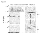
- the Figure 8 shows the capacity of the biosensor 1 to measure the skin viscoelasticity (comparison between experiments with nutritional medium only -left part of the figure- and with the living skin explant -right part of the figure) and to observe the effect of skin pretreatments.
- the loss modulus G" is identical in presence of nutritional medium whether the living skin explant is delipidated or not delipidated, whereas the storage modulus G' which is representative of the skin elasticity is clearly higher for the standard skin than for the lipid deficient skin (right part of the figure).
- the TSM transducer 3 comprises a culture medium 5 in contact with at least one of the surfaces of one of the two electrodes 4A, 4B and on which the hypodermis or dermis 2A of the skin explant 2 is in contact with its bottom surface, in order to guarantee the survival of the skin explant 2 on the TSM transducer 3 at least several hours.
- the survival of the skin explant 2 can be ensured several days (more than 10 days, for instance 16 days), depending upon the contact between the culture medium 5 and the skin explant 2, the exterior conditions (temperature%) and the nature of the culture medium 5.
- the culture medium 5 is a liquid, a gel, an aerogel, a cream and/or a functionalized layer.
- the culture medium 5 is a gel, an aerogel, or a cream deposited on the surface of one of the two conducting electrodes 4A, 4B.
- the culture medium 5 is a functionalized layer disposed on one of the two conducting electrodes 4A, 4B, and on which the bottom surface of the skin explant 2 is in contact, in order to guarantee the survival of the skin explant 2 on the TSM transducer 3 at least several hours.
- the TSM transducer presents at least one opening which is filled with culture medium 5, allowing the survival of the skin explant 2 during at least several hours or at least several days.
- the skin explant 2 is moved and removed between the TSM transducer 3 and a receptacle in which there is the culture medium 5.
- the skin explant 2 remains on the TSM transducer only for the measure for instance 15 minutes and is then reintegrated in the receptacle to feed the skin explant 2 and to maintain it alive.
- the invention concerns also, as drawn on figure 2 , a device which comprises a TSM biosensor 1 as defined above, and a receptacle in which is deposited the TSM biosensor 1, the TSM biosensor 1 being surrounded by culture medium 5, the TSM transducer 3 being immersed in the culture medium 5 so as to maintain the bottom surface of the dermis 2A in the culture medium 5 and the top surface of the stratum corneum 2C in contact with air, allowing the survival of the skin explant 2 during at least several hours or at least several days.
- the TSM biosensor 1 and the receptacle can be sold together, the skin explant 2 being able to survive at least several days.
- the invention also concerns a method for determining mechanical characteristics of an ex vivo living skin explant 2, the living skin explant 2 comprising at least one layer among: hypodermis, dermis 2A, epidermis 2B, and the stratum corneum 2C.
- the method comprises the following steps:
- hypodermis or dermis 2A is deposited on the TSM transducer 3, the stratum corneum 2C being in contact with air.
- steps (a), (b), (c), (d) are realized several times during several days.
- the living skin explant 2 is pretreated, for instance hydrated or delipidated.
- the living skin explant 2 is treated with at least one product, prior to or after its deposition, on the Thickness Shear Mode (TSM) transducer, for monitoring products-to-skin effects.
- TMS Thickness Shear Mode
- the living skin explant 2 is disposed in contact with one of the two conducting electrodes 4A, 4B.
- the living skin explant 2 is deposited on a gel which comprises culture medium 5, the gel being deposited on one of the two conducting electrodes 4A, 4B.
- the living skin explant 2 is deposited on a functionalized layer with culture medium 5, the functionalized layer being deposited on one of the two conducting electrodes 4A, 4B.
- the invention also concerns a use of the TSM biosensor 1 to monitor ex vivo mechanical behavior of the living skin explant 2 and thus to follow its structural evolution function of time, by measuring its viscoelastic characteristics.
- the complex study of the TSM response in time domain permits a control a) of the dehydration evolution at 37°C due to interpenetrated intercellular lipid membranes matrix including the first step of permeation process from dermis 2A to Stratum corneum 2C (SC); b) the impact on the kinetics of the permeation process by a product applied at the SC surface.
- This information includes the structure and properties evolution of the collagen and elastic fibers and the proteoglycans located in the skin.
- a comparison of mechanical results with other techniques in the literature confirms the validity of the model.
- Skin is a material of four main complex biological layers (epidermis 2B, dermis 2A, hypodermis or stratum corneum 2C) principally based on interconnected collagen and elastin fibers embedded in a gel-like mixture of fluids (hyaluronic, fibroblasts, proteoglycans, fat and water) [1]-[3].
- fluids hyaluronic, fibroblasts, proteoglycans, fat and water
- skin can be considered as a viscoelastic complex fluid based on membranes and fibers immersed in a liquid [4], [5].
- its properties can be characterized as nonlinear and anisotropic soft hybrid complex material or elastomers [6]-[11].
- the skin viscoelasticity can be mainly attributed to interaction between the interpenetrated elastin - collagen fibers and the liquid mixture.
- the anisotropy behavior is the result of the preferential orientation of collagen beams and residual in-vivo skin stress [12]-[14].
- Different non-invasive testing are used to characterize and macroscopically detect skin diseases [15].
- motional stress on ex-vivo sample the linear viscoelasticity measurement can be more easily reached.
- the most commonly used for ex vivo skin samples test is the tensile test. Using this method, the nonlinear effects and viscoelastic behavior of animal skin have been mostly pointed out [16]-[19].
- a high frequency rheological technique added to a suitable model is used to monitor the complex shear modulus G (related to viscoelasticity) of skin.
- the technique uses a biosensor 1 made of a Thickness Shear Mode (TSM) resonator (as a transducer) in contact to the dermis 2A of skin. Both transducer and material are then considered as an equivalent lossy TSM biosensor 1.
- TSM Thickness Shear Mode
- Ex-vivo human skin samples of 5mm thickness were prepared from abdominal biopsies (healthy plastic surgeries) of two male donors of respectively 36 years old with Negroid skin and 44 years old with Caucasian skin.
- 3 first layers from stratum corneum 2C (SC) to dermis 2A layer
- SC stratum corneum 2C
- Circular explants of 10 mm of diameter are cut out in order to embed each sample with a quartz crystal piezoelectric substrate (described in the following section) used as a shear wave generator and thickness shear mode sensor.
- Human skin explants 2 were maintained in survival on grids, in plates containing 2mL special BIO-EC medium
- BEM medium from BIO-EC Laboratory, France. BEM maintain explants in survival for several days, about 20 days [24].
- Circular explants preparation A) circular cut out for embedeed biosensing, B) Human skin maintained in survival ready for treatment.
- the samples from each donor are prepared in order to study the sensitivity of the new biosensor 1 versus skin hydration treatment.
- 3 type of treated skin are then studied: A) untreated skin used as the reference; B) delipidated skin; C) Hydrated skin with a special treatment cream-based application. Experiments were performed in triplicate
- the tested products and treatments were applied on explants as soon as their preparation. Treated and untreated skin explants 2 were incubated at 37°C in a normal atmosphere conditions. To monitor the dehydration process, a protective film is deposited at the surface of skin. This film ensures a standard skin hydration. As long as the film is present the hydration conditions remain unchanged.
- the TSM biosensor 1 consists of a thin disk of AT-cut quartz with metal electrodes 4A, 4B deposited on both sides (the resonator) in contact on one side with the dermis 2A of the ex-vivo human skin sample.
- the diameters of the crystal and the electrode are respectively 14 mm and 5 mm.
- the quartz thickness of 270 ⁇ m provide a resonance frequency about 5 MHz.
- the mass of the layer in contact involves a frequency shift [28], [29].
- the mechanical boundary conditions including skin loading are modified by the rheological properties of skin. These conditions induce an additional frequency shift and a damping of the resonance corresponding to an attenuation and a delay of the propagated ultrasonic shear waves [30]-[32].
- the experimental set-up consists of an impedance analysis of the biosensor 1 during skin dehydration.
- each sample is monitored at controlled temperature (37°C) using a laboratory oven.
- the admittance measurement is preferred.
- a span of 7 kHz around the resonance frequency is then analyzed to measure the total admittance response Y e ( ⁇ ) of loaded biosensor 1 using the HP4195A network analyzer. 400 spectral points are then regularly measured in less than 50 seconds.
- An online fit of this response is performed in the complex plan to extract the real and the imaginary part of Z load . The viscoelastic characteristics of skin can be then extracted.
- the mass effect due to the liquid embedded in the epidermis 2B and dermis 2A is firstly studied.
- Transducers without skin explant 2s
- This measurement gives access to characteristic parameters of unperturbed transducers.
- a measurement of theses transducers loaded by nutritional BEM liquid is after performed. Considering that this liquid is a Newtonian liquid (G' ⁇ 0), the mass effect can be directly extracted from the two first measurements (variation of unperturbed transducer admittance and transducer loaded with BEM liquid).
- the total reactance X for skin load is similar to the BEM load.
- skin can be considered as a viscous complex fluid having a viscosity mainly due to the liquid characteristic.
- the density of skin can be considered close to liquid density. This result is in concordance with the fact that the dermis 2A is continuously supplied with water mixture by way of the bloodstream.
- the dermis 2A a part of the liquid mixture is present in gel phase, attached to many hydrophilic macromolecules. Despite the large capacity of these molecules to capture water mixture, a part of it remains mobile and diffuses to upper layers of skin. Note that the reactance of the transducer loaded by the skin is slightly lower in proportion to the surface in contact with the load.
- this variation is due to the interactions of the macromolecules forming a rigid ultrastructure based on elastin and collagen fibers immersed in fat and liquid.
- the variation of R gives us access to elasticity of skin during dehydration. It can also be observed that allows for better interpenetration of layers increasing the rigidity as shown in figure 1 .
- the figure 7A shows the repeatability of the technique (untreated skin).
- the kinetic characteristic time is clearly modified as a function of skin treatment.
- the long kinetic of dilapidation skin shows the role of lipids in dehydration.
- the real part of Z load ( figure 7B ) for this treatment shows the skin stiffening due to the interactions of macromolecules. Comparing the effect of different treatments our technique can detect their structure modification and their efficiency. A systematic study should allow cosmetic product optimizing and effectiveness evidence.
- the TSM biosensor 1 allows a monitoring of ex-vivo human skin for treatment testing with a large dynamical range.
- the first results show that it is possible to completely follow the viscoelastic parameters evolution for dehydration monitoring or structure monitoring of skin from stratum corneum 2C to dermis 2A. From the temporal evolution of real and imaginary part of impedance load the effect of skin treatment to the skin layers structure and mechanical properties can be detected. This preliminary study shows that this technique can be suitable to accessing to the elastic G' and viscous G" moduli by micro-rheology.
- This new biosensor 1 added with our model should become a sensitive ex-vivo technique for cosmetic product efficiency and skin objective testing.
Landscapes
- Physics & Mathematics (AREA)
- Acoustics & Sound (AREA)
- Health & Medical Sciences (AREA)
- Life Sciences & Earth Sciences (AREA)
- Chemical & Material Sciences (AREA)
- Analytical Chemistry (AREA)
- Biochemistry (AREA)
- General Health & Medical Sciences (AREA)
- General Physics & Mathematics (AREA)
- Immunology (AREA)
- Pathology (AREA)
- Measuring And Recording Apparatus For Diagnosis (AREA)
Abstract
The invention concerns a Thickness Shear Mode (TSM) biosensor (1) which comprises an ex vivo living skin explant (2), the skin explant (2) comprising at least one of the skin layers among: hypodermis, dermis (2A), epidermis (2B) and the stratum corneum (2C), a TSM transducer (3) which comprises: an AT cut quartz resonator 3C which has two opposite exterior surfaces (3A,3B), and two conducting electrodes (4A, 4B), each conducting electrode being deposited on one of the two exterior surfaces (3A,3B), the TSM transducer (3) allowing to determine micro rheological characteristics of the living skin explant (2) by piezoelectric transducing using shear waves, the TSM transducer (3) presenting: measuring means (30), monitoring and calculating means (31) which monitor an evolution in time of an electrical response of the living skin explant (2), and which calculate in time, from the electrical response, micro rheological characteristics of the living skin explant (2), a bottom surface of the skin explant (2) being in contact with the TSM transducer (3), a top surface of the skin explant (2) being in contact with air.
Description
- The invention concerns a TSM biosensor to ex-vivo characterize the viscoelastic properties of skin.
- The skin which comprises dermis, epidermis and the stratum corneum, is a complex biological material similar to layers based on fibers, and on collagen and elastin membranes intermixed and immersed in a fluid, itself complex, in a gel phase (hyaluronic, fibroblasts, proteoglycans, fat and water). The orientation of these fibers and the very thin thickness of these layers imply that the viscoelastic properties of the the skin are often seen as non-linear and anisotropic, and are difficult to characterize either in vivo or ex vivo.
- Most commonly used skin samples ex vivo test is the tensile test. Using this method, it is difficult to have homogeneous and reproducible measurements particularly because of non-linear mechanical effects involved, the heterogeneity and variability of the skin.
-
-
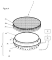
Figure 1 describes the TSM biosensor of the invention in an exploded view; the different elements of the bio sensor is not to scale. -
Figure 2 describes the TSM biosensor of the invention in a sectional view. -
Figure 3 shows the modified electrical model used to characterize the viscoelastic properties of skin.- A)
TSM Biosensor 1 used for ex-vivo skin characterization (untreated tired skin after dehydration process monitoring): thebiosensor 1 consist of two elements: the transducer made of an AT-cut quartz resonator 3C and an ex-vivo skin explant specially designed. B) The electrical model: two parallel branches can independently represent both electrical and mechanical effects. The static branch of impedance Z0 (in gray) includes the dielectric properties of the quartz as a function of the electrical field distribution between theelectrodes
- A)
-
Figure 4 shows typical fit of the total admittance of theTSM biosensor 1 corresponding to the 3rd order theoretical electrical complex function corresponding to the electrical circuit. -
Figure 5 describes the Masseffect skin explant 2 vs nutritional BEM: The total admittance X is compared for different samples. The mass effect is principally due to the liquid stored in thedermis 2A andepidermis 2B. -
Figure 6 is a comparison of R load for the 36 yo donor: for BEM load (left) and skin load (right). -
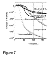
Figure 7 describes the kinetic of dehydration at 37°C, 22% Hygrometry for Negroid (points only) and Caucasian (line style) treated skins: A) Relative variation of imaginary part of Zload ; B) 44yo Caucasian skin donor. -
Figure 8 describes the complex shear modulus G = G '+ j G ", representative of skin viscoelasticity at micro-scale, function of time, for the nutritional medium alone without skin explant (left part of the figure), and with the skin explant (right part of the figure) at 37°C; the skin explant comes from a 44 years old Caucasian skin donor and the effect of delipidation (dotted line) versus standard skin (solid line) is shown. - The present invention concerns a Thickness Shear Mode (TSM)
biosensor 1 which comprises: - an ex vivo
living skin explant 2, - a
TSM transducer 3 which comprises: an ATcut quartz resonator 3C which has two oppositeexterior surfaces electrodes exterior surfaces - The word 'living' means that the skin explant 2 lives when the measures are realized and has biophysical behavior comparable to the in-vivo skin behavior.
- The skin explant can be extracted from a person or an animal with plastic surgery or can be reconstituted with biological cells in an extracellular matrix.
- Here the conducting electrodes are in metal, for instance in gold.
- In other words, the AT
cut quartz resonator 3C represents all the existing quartz crystal piezoelectric substrate used as a shear wave generator and as a thickness shear mode sensor. - The shear waves generated are volume waves.
- The wave mode generated is then Thickness Shear Mode (TSM).
- The thickness of the
quartz resonator 3C is chosen to obtain a frequency resonance at RF range and to generate ultrasound shear waves. - For instance, the fundamental frequency of the
quartz resonator 3C used here for the figures is 5 MHz. - The dimensions of the
quartz resonator 3C can be determined so that thequartz resonator 3C can have a quality factor Q higher than 105. - The electrodes shape 4A, 4B can be designed in order to optimize the generation wave mode. Here on the
figure 1 , thequartz resonator 3C is a piezoelectric disc with a lateral surface. The shape of electrodes placed at both surfaces of thequartz resonator 3C is also a disc, and advantageously can have the same dimensions and can cover the entire surface of the ATcut quartz resonator 3C. - But the
quartz resonator 3C and theelectrodes - The electrodes can be for example spiral or other form, in order to be controlled by induction, as described in the patent application
WO2012/172253 . - Advantageously, the
skin explant 2 covers the entire surface of the electrode on which it is deposited. - The
TSM transducer 3 allows to measure and convert electrical parameters for determining mechanical characteristics of theliving skin explant 2, and can be connected to a computer and a network analyser. - The
TSM transducer 3 presents: - measuring means 30,
- monitoring and calculating means 31 which monitor an evolution in time of an electrical response of the living skin explant, and which calculate, in time, from the electrical response, micro rheological characteristics of the
living skin explant 2. - The
skin explant 2 can comprise at least one of the skin layers among: hypodermis,dermis 2A,epidermis 2B and the stratum corneum 2C. - Advantageously, the
skin explant 2 comprises a stacking of layers which comprisesdermis 2A,epidermis 2B and the stratum corneum 2C, thedermis 2A being in contact with theTSM transducer 3, the stratum corneum 2C being in contact with air. - The layers can comprise a product or can be pretreated before measuring electrical response of the
skin explant 2, function of shear waves of the TSM. - Advantageously, the
dermis 2A does not need to be glued or stick on theTSM transducer 3, and naturally adheres properly to theTSM transducer 3. Thus, bothskin explant 2 andTSM transducer 3 can be considered as a unique device called theTSM biosensor 1. The invention allows to realize an instrumented skin. - It is particularly the case when the
electrodes dermis 2A is disposed on the electrode just after the preparation and the deposit of the electrode on the substrate resonator. - A bottom surface of the
skin explant 2 is in contact with theTSM transducer 3, a top surface of theskin explant 2 being in contact with air, considering a vertical axis perpendicular to the TSM and the skin explant that are horizontally maintained together. - When the
skin explant 2 comprisesdermis 2A,epidermis 2B and the stratum corneum 2C, a bottom surface of thedermis 2A being in contact with theTSM transducer 3, a top surface of the stratum corneum 2C being in contact with air. - The
skin explant 2, in particular the stratum corneum 2C can be treated by a product which is tested. - The
biosensor 1 is modeled by the modified equivalent electrical lumped elements Butterworth-van-Dyke circuit (BVD) which comprises: - a static branch modeling the electrical behavior of the
quartz resonator 3C, and - a dynamic branch parallel to the static branch and modeling the mechanical behavior of the
quartz resonator 3C and the mechanical behavior of the living skin explant. - The static branch comprising a resistor and a capacitor connected in parallel.
- The dynamic branch comprising in series:
- a capacitor, a self-inductance and a resistor modeling the mechanical behavior of the
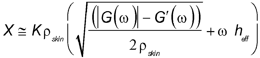
quartz resonator 3C, and - an additional equivalent electrical impedance denoted Zload = R + j X. This impedance is linked with the complex shear modulus G = G'+ j G", representative of the living skin explant (2) viscoelasticity at micro-scale, using following equations:

- Zload is the electrical response of the
living skin explant 2 and is measured and monitored by themeans - The complex shear modulus G characterizes the micro rheological behaviour of the
living skin explant 2. - The
Figure 8 shows the capacity of thebiosensor 1 to measure the skin viscoelasticity (comparison between experiments with nutritional medium only -left part of the figure- and with the living skin explant -right part of the figure) and to observe the effect of skin pretreatments. - Actually the loss modulus G" is identical in presence of nutritional medium whether the living skin explant is delipidated or not delipidated, whereas the storage modulus G' which is representative of the skin elasticity is clearly higher for the standard skin than for the lipid deficient skin (right part of the figure).
- In a first embodiment, the
TSM transducer 3 comprises aculture medium 5 in contact with at least one of the surfaces of one of the twoelectrodes dermis 2A of theskin explant 2 is in contact with its bottom surface, in order to guarantee the survival of theskin explant 2 on theTSM transducer 3 at least several hours. - In fact, the survival of the
skin explant 2 can be ensured several days (more than 10 days, for instance 16 days), depending upon the contact between theculture medium 5 and theskin explant 2, the exterior conditions (temperature...) and the nature of theculture medium 5. - The
culture medium 5 is a liquid, a gel, an aerogel, a cream and/or a functionalized layer. - In a first variant of the first embodiment, the
culture medium 5 is a gel, an aerogel, or a cream deposited on the surface of one of the two conductingelectrodes - In a second variant of the first embodiment, the
culture medium 5 is a functionalized layer disposed on one of the two conductingelectrodes skin explant 2 is in contact, in order to guarantee the survival of theskin explant 2 on theTSM transducer 3 at least several hours. - In a third variant of the first embodiment, the TSM transducer presents at least one opening which is filled with
culture medium 5, allowing the survival of theskin explant 2 during at least several hours or at least several days. - It is possible to realize many openings in the electrode and in the quartz of the TSM transducer and to form like a grid, each opening containing some
culture medium 5. - In a second embodiment, the
skin explant 2 is moved and removed between theTSM transducer 3 and a receptacle in which there is theculture medium 5. In this second embodiment, theskin explant 2 remains on the TSM transducer only for the measure for instance 15 minutes and is then reintegrated in the receptacle to feed theskin explant 2 and to maintain it alive. - In a third embodiment, the invention concerns also, as drawn on
figure 2 , a device which comprises aTSM biosensor 1 as defined above, and a receptacle in which is deposited theTSM biosensor 1,
theTSM biosensor 1 being surrounded byculture medium 5,
theTSM transducer 3 being immersed in theculture medium 5 so as to maintain the bottom surface of thedermis 2A in theculture medium 5 and the top surface of the stratum corneum 2C in contact with air, allowing the survival of theskin explant 2 during at least several hours or at least several days. - Thus, the
TSM biosensor 1 and the receptacle can be sold together, theskin explant 2 being able to survive at least several days. - The invention also concerns a method for determining mechanical characteristics of an ex vivo living
skin explant 2, the livingskin explant 2 comprising at least one layer among: hypodermis, dermis 2A,epidermis 2B, and the stratum corneum 2C. - The method comprises the following steps:
- (a) withdraw a living
skin explant 2 from aculture medium 5, - (b) deposit the living
skin explant 2 on theTSM transducer 3, a bottom surface of theskin explant 2 being deposited on theTSM transducer 3, a top surface of theskin explant 2 being in contact with air, - (c) measure and monitor electrical parameters for determining and calculating mechanical characteristics of the living
skin explant 2. - For instance, the hypodermis or
dermis 2A is deposited on theTSM transducer 3, the stratum corneum 2C being in contact with air. - In a step (d) after the step (c), the living
skin explant 2 is withdrawn from theTSM transducer 3 and is deposited in theculture medium 5. - In a step (e) after the step (d), the steps (a), (b), (c), (d) are realized again.
- In particular, the steps (a), (b), (c), (d) are realized several times during several days.
- The living
skin explant 2 is pretreated, for instance hydrated or delipidated. - The living
skin explant 2 is treated with at least one product, prior to or after its deposition, on the Thickness Shear Mode (TSM) transducer, for monitoring products-to-skin effects. - In the step (b), in a first embodiment, the living
skin explant 2 is disposed in contact with one of the two conductingelectrodes - In the step (b) in a first variant of the first embodiment, the living
skin explant 2 is deposited on a gel which comprisesculture medium 5, the gel being deposited on one of the two conductingelectrodes - In the step (b) in a second variant of the first embodiment, the living
skin explant 2 is deposited on a functionalized layer withculture medium 5, the functionalized layer being deposited on one of the two conductingelectrodes - The invention also concerns a use of the
TSM biosensor 1 to monitor ex vivo mechanical behavior of the livingskin explant 2 and thus to follow its structural evolution function of time, by measuring its viscoelastic characteristics. - In particular, it concerns a use to detect and/or follow skin diseases.
- In particular, it concerns a use to analyse and/or study the effect of a product applied to the
skin explant 2 or specific treatment. - Ex-vivo
abdominal skin explants 2 from plastic surgery and kept alive are used for this study. Considering the skin as a complex fluid made of membrane and fiber structures immersed in liquid, its mechanical response of a bulk thickness shear wave excitation (ie stress-strain analysis) involves both a viscous component associated with energy dissipation and an elastic component associated with energy storage. A tight monitoring of these two components from the response of a TSM sensor (based on an ATcut quartz resonator 3C at 5 MHz) in contact to thedermis 2A of the ex-vivo explant give us access to the complex dynamic shear moduli (G' and G") evolution of the skin; The appropriate mechanical model describing the sensor response vs shear waves/matrix interactions allows investigating the impact of the product (or treatment) to the viscoelastic properties of the skin. - The complex study of the TSM response in time domain permits a control a) of the dehydration evolution at 37°C due to interpenetrated intercellular lipid membranes matrix including the first step of permeation process from
dermis 2A to Stratum corneum 2C (SC); b) the impact on the kinetics of the permeation process by a product applied at the SC surface. This information includes the structure and properties evolution of the collagen and elastic fibers and the proteoglycans located in the skin. A comparison of mechanical results with other techniques in the literature confirms the validity of the model. These preliminary results show that our TSM technique can be an appreciable new way for ex-vivo skin investigation for test and optimization of new cosmetic products. - Skin is a material of four main complex biological layers (
epidermis 2B,dermis 2A, hypodermis or stratum corneum 2C) principally based on interconnected collagen and elastin fibers embedded in a gel-like mixture of fluids (hyaluronic, fibroblasts, proteoglycans, fat and water) [1]-[3]. From a mechanical point of view, skin can be considered as a viscoelastic complex fluid based on membranes and fibers immersed in a liquid [4], [5]. On these conditions, its properties can be characterized as nonlinear and anisotropic soft hybrid complex material or elastomers [6]-[11]. On one hand, the skin viscoelasticity can be mainly attributed to interaction between the interpenetrated elastin - collagen fibers and the liquid mixture. On the other hand the anisotropy behavior is the result of the preferential orientation of collagen beams and residual in-vivo skin stress [12]-[14]. Different non-invasive testing are used to characterize and macroscopically detect skin diseases [15]. Using motional stress on ex-vivo sample, the linear viscoelasticity measurement can be more easily reached. The most commonly used for ex vivo skin samples test is the tensile test. Using this method, the nonlinear effects and viscoelastic behavior of animal skin have been mostly pointed out [16]-[19]. Many other difficulties to perform homogeneous measurements exist because skin structure heterogeneity and the large variability between samples. To date other experimental methods based on stress-strain measurement are largely described in the literature. Some of them are based on measurements of torsion and deformation generated by ultrasound wave propagation. A recent review describe the relevant results available in the literature [20]. They can give access to the linear viscoelasticity of the skin at different scale depending on the wavelength. - A high frequency rheological technique added to a suitable model is used to monitor the complex shear modulus G (related to viscoelasticity) of skin. The technique uses a
biosensor 1 made of a Thickness Shear Mode (TSM) resonator (as a transducer) in contact to thedermis 2A of skin. Both transducer and material are then considered as an equivalentlossy TSM biosensor 1. Using an original approach based on Wheeler analysis for lossy micro-strip, this model gives access to mechanical evolution of skin considered as a heterogeneous complex fluid [21], [22]. - In order to determine the impact of the skin dehydration to ultrasonic shear wave interactions, the responses of the TSM resonator loaded by different skins and treatments are studied.
- Ex-vivo human skin samples of 5mm thickness were prepared from abdominal biopsies (healthy plastic surgeries) of two male donors of respectively 36 years old with Negroid skin and 44 years old with Caucasian skin. In order to study the 3 first layers (from stratum corneum 2C (SC) to
dermis 2A layer) the subcutaneous fat is removed from the skin. - Circular explants of 10 mm of diameter are cut out in order to embed each sample with a quartz crystal piezoelectric substrate (described in the following section) used as a shear wave generator and thickness shear mode sensor. Human skin explants 2 were maintained in survival on grids, in plates containing 2mL special BIO-EC medium
- (BEM: medium from BIO-EC Laboratory, France). BEM maintain explants in survival for several days, about 20 days [24].
- Circular explants preparation: A) circular cut out for embedeed biosensing, B) Human skin maintained in survival ready for treatment.
- The samples from each donor are prepared in order to study the sensitivity of the
new biosensor 1 versus skin hydration treatment. 3 type of treated skin are then studied: A) untreated skin used as the reference; B) delipidated skin; C) Hydrated skin with a special treatment cream-based application. Experiments were performed in triplicate - The tested products and treatments were applied on explants as soon as their preparation. Treated and
untreated skin explants 2 were incubated at 37°C in a normal atmosphere conditions. To monitor the dehydration process, a protective film is deposited at the surface of skin. This film ensures a standard skin hydration. As long as the film is present the hydration conditions remain unchanged. - The
TSM biosensor 1 consists of a thin disk of AT-cut quartz withmetal electrodes dermis 2A of the ex-vivo human skin sample. The diameters of the crystal and the electrode are respectively 14 mm and 5 mm. The quartz thickness of 270 µm provide a resonance frequency about 5 MHz. Considering the piezoelectric properties of the quartz disk, its crystal orientation, the shape ofelectrodes electrodes - Electro-mechanical models already describe the complete oscillations process of the resonator. They are derived from piezoelectric constitutive equations. A general one dimensional acoustic wideband model has been suggested by Mason [26]. In particular cases, such as low load, the Mason model can be easily simplified near the resonant frequencies into an equivalent electrical lumped elements Butterworth-van-Dyke circuit (BVD) [27].
- When the sensor is loaded by a rigid layer or a liquid, the mass of the layer in contact involves a frequency shift [28], [29]. Moreover the mechanical boundary conditions including skin loading are modified by the rheological properties of skin. These conditions induce an additional frequency shift and a damping of the resonance corresponding to an attenuation and a delay of the propagated ultrasonic shear waves [30]-[32].
- Zload = R + j X is representative of these changes. The variation of the electrical response can then accurately extract G, using the following expression [33]-[35].


- The experimental set-up consists of an impedance analysis of the
biosensor 1 during skin dehydration. In order to study the effect of treatments each sample is monitored at controlled temperature (37°C) using a laboratory oven. Considering the high Q factor of TSM resonator, the admittance measurement is preferred. A span of 7 kHz around the resonance frequency is then analyzed to measure the total admittance response Ye(ω) of loadedbiosensor 1 using the HP4195A network analyzer. 400 spectral points are then regularly measured in less than 50 seconds. An online fit of this response is performed in the complex plan to extract the real and the imaginary part of Zload. The viscoelastic characteristics of skin can be then extracted. - In order to observe the temporal evolution, the frequency response of the admittance is then sampled every minute.
- In order to study the sensitivity of the
biosensor 1 and to validate the proof of concept of a new technique to monitor the hydration ofex-vivo skin explants - To accurately monitor R and X variation due to the viscoelastic effects during the dehydration, the mass effect due to the liquid embedded in the
epidermis 2B anddermis 2A is firstly studied. Transducers (without skin explant 2s) are firstly characterized to extract the unloaded motional elements. This measurement gives access to characteristic parameters of unperturbed transducers. A measurement of theses transducers loaded by nutritional BEM liquid is after performed. Considering that this liquid is a Newtonian liquid (G' ≈ 0), the mass effect can be directly extracted from the two first measurements (variation of unperturbed transducer admittance and transducer loaded with BEM liquid). Assumingequation 1, the real part of Zload allows the extraction of the viscosity from viscous modulus G". As a consequence, the following substation gives the mass effect (Xm):
- Comparing the real and the imaginary part of of Zload for each measurement the typical mass effect can be estimated to Xm=10Ω. This first result shows that the total reactance is principally due to viscoelastic variation (Xm/X<1%). The
figure 5 shows the variability of the total reactance and the effect of skin fibers in comparison with BEM. - At the initial conditions, the total reactance X for skin load is similar to the BEM load. In agreement with the literature of skin tribology, skin can be considered as a viscous complex fluid having a viscosity mainly due to the liquid characteristic. The density of skin can be considered close to liquid density. This result is in concordance with the fact that the
dermis 2A is continuously supplied with water mixture by way of the bloodstream. In thedermis 2A, a part of the liquid mixture is present in gel phase, attached to many hydrophilic macromolecules. Despite the large capacity of these molecules to capture water mixture, a part of it remains mobile and diffuses to upper layers of skin. Note that the reactance of the transducer loaded by the skin is slightly lower in proportion to the surface in contact with the load. - In contrast, while the imaginary part remains constant, the real part of impedance load R increases significantly as shown in the
figure 6 . - Considering the
equation 1, this variation is due to the interactions of the macromolecules forming a rigid ultrastructure based on elastin and collagen fibers immersed in fat and liquid. The variation of R gives us access to elasticity of skin during dehydration. It can also be observed that allows for better interpenetration of layers increasing the rigidity as shown infigure 1 . - The intercellular SC lipid membranes play a crucial role in regulating the loss of intrinsic liquid from the skin. This process is in fact directly correlated with SC membranes permeability to this aqueous liquid. SC monitoring using X-ray scattering spectroscopy and NMR studies generally this process [36]-[38]. In order to preliminary study the capability of our technique to monitor the dehydration process, different treatments of ex-vivo human skin are tested. The
figure 7 shows typical variation of X for treated an untreated skin from the two donors. - The initial time (t=0) is the moment from which the protective film is removed. As a general shape, we note an already known exponential decrease due to the diffusivity of liquid vapor in the lipid membrane. In agreement with one-dimensional diffusion model, this result is also observed by an indirect relative humidity measurement in the QCM chamber as a function of time [39]. The
figure 7A) shows the repeatability of the technique (untreated skin). The kinetic characteristic time is clearly modified as a function of skin treatment. The long kinetic of dilapidation skin shows the role of lipids in dehydration. In addition the real part of Zload (figure 7B ) for this treatment shows the skin stiffening due to the interactions of macromolecules. Comparing the effect of different treatments our technique can detect their structure modification and their efficiency. A systematic study should allow cosmetic product optimizing and effectiveness evidence. - The
TSM biosensor 1 allows a monitoring of ex-vivo human skin for treatment testing with a large dynamical range. The first results show that it is possible to completely follow the viscoelastic parameters evolution for dehydration monitoring or structure monitoring of skin from stratum corneum 2C to dermis 2A. From the temporal evolution of real and imaginary part of impedance load the effect of skin treatment to the skin layers structure and mechanical properties can be detected. This preliminary study shows that this technique can be suitable to accessing to the elastic G' and viscous G" moduli by micro-rheology. Thisnew biosensor 1 added with our model should become a sensitive ex-vivo technique for cosmetic product efficiency and skin objective testing. -
- [1] R. F. Rushmer, K. J. K. Buettner, J. M. Short, and G. F. Odland, "The Skin," Science (80-.)., vol. 154, no. 3747, pp. 343-348, 1966 .
- [2] G. Odland, "Structure of the skin," in Physiology, Biochemistry, and Molecular Biology of the Skin, 1991, pp. 3-62 .
- [3] C. W. Oomens, D. H. van Campen, and H. J. Grootenboer, "A mixture approach to the mechanics of skin.,"J. Biomech., vol. 20, no. 9, pp. 877-885, 1987 .
- [4] F. Xu and T. Lu, "Introduction of Skin Biothermomechanics," in Introduction to Skin Biothermomechanics and Thermal Pain, 2011, pp. 209-220 .
- [5] F. Xu, T. J. Lu, and K. A. Seffen, "Biothermomechanical behavior of skin tissue, "Acta Mech. Sin. Xuebao, vol. 24, no. 1, pp. 1-23, 2008
- [6] F. Xu and T. J. Lu, " .
- [7] F. Xu, T. J. Lu, and K. A. Seffen, "Biothermomechanics of skin tissues, "J. Mech. Phys. Solids, vol. 56, no. 5, pp. 1852-1884, 2008 .
- [8] A. Delalleau, G. Josse, J.-M. Lagarde, H. Zahouani, and J.-M. Bergheau, "Characterization of the mechanical properties of skin by inverse analysis combined with the indentation test.," J. Biomech., vol. 39, no. 9, pp. 1603-1610, 2006 .
- [9] M. J. Koehler, T. Vogel, P. Elsner, K. König, R. Bückle, and M. Kaatz, "In vivo measurement of the human epidermal thickness in different localizations by multiphoton laser tomography," Ski. Res. Technol., vol. 16, no. 3, pp. 259-264, 2010 .
- [10] C. Sanchez, L. Rozes, F. Ribot, C. Laberty-Robert, D. Grosso, C. Sassoye, C. Boissiere, and L. Nicole, "'Chimie douce': A land of opportunities for the designed construction of functional inorganic and hybrid organic-inorganic nanomaterials," Comptes Rendus Chimie, vol. 13, no. 1-2. pp. 3-39, 2010 .
- [11] S. Hsu, A. M. Jamieson, and J. Blackwell, "Viscoelastic studies of extracellular matrix interactions in a model native collagen gel system.,"Biorheology, vol. 31, no. 1, pp. 21-36, 1994 .
- [12] X. Liang and S. A. Boppart, "Biomechanical properties of in vivo human skin from dynamic optical coherence elastography,"IEEE Trans. Biomed. Eng., vol. 57, no. 4, pp. 953-959, 2010 .
- [13] B. J. Bell, E. Nauman, and S. L. Voytik-Harbin, "Multiscale strain analysis of tissue equivalents using a custom-designed biaxial testing TSM biosensor," Biophys. J., vol. 102, no. 6, pp. 1303-1312, 2012 .
- [14] R. B. Groves, S. A. Coulman, J. C. Birchall, and S. L. Evans, "An anisotropic, hyperelastic model for skin: Experimental measurements, finite element modelling and identification of parameters for human and murine skin,"J. Mech. Behav. Biomed. Mater., vol. 18, pp. 167-180, 2013 .
- [15] J. Serup, G. B. E. Jemec, and G. L. Grove, Handbook of Non-Invasive Methods and the Skin, Second Edition -, CRC Press. 2006 .
- [16] J. M. Pereira, J. M. Mansour, and B. R. Davis, "Dynamic measurement of the viscoelastic properties of skin.," J. Biomech., vol. 24, no. 2, pp. 157-162, 1991 .
- [17] Z. Del Prete, S. Antoniucci, A. H. Hoffman, and P. Grigg, "Viscoelastic properties of skin in Mov-13 and Tsk mice,"J. Biomech., vol. 37, no. 10, pp. 1491-1497, 2004 .
- [18] H. G. Vogel, "Directional variations of mechanical parameters in rat skin depending on maturation and age.,"J. Invest. Dermatol., vol. 76, no. 6, pp. 493-497, 1981 .
- [19] O. Lokshin and Y. Lanir, "Viscoelasticity and preconditioning of rat skin under uniaxial stretch: microstructural constitutive characterization.,"J. Biomech. Eng., vol. 131, no. 3, p. 031009, 2009 .
- [20] S. Derler and L. C. Gerhardt, "Tribology of skin: Review and analysis of experimental results for the friction coefficient of human skin," Tribol. Lett., vol. 45, no. 1, pp. 1-27, 2012 .
- [21] H. A. Wheeler, "Transmission-Line Properties of Parallel Strips Separated by a Dielectric Sheet," IEEE Trans. Microw. Theory Tech., vol. 13, no. 2, pp. 172-185, Mar. 1965 .
- [22] C. Ould Ehssein, "Système instrumental pour la rhéologie ultrasonore," Université de Cergy-Pontoise, 2006 .
- [23] E. Lopez, E. Latie, P. Gasser, and M. Rousseau, "Calcium improves skin health," Househ. Pers. CareToday, vol. 3, pp. 32-35, 2008 .
- [24] P. Gasser, F. Arnold, L. Peno-Mazzarino, D. Bouzoud, M. T. Luu, E. Lati, and M. Mercier, "Glycation induction and antiglycation activity of skin care ingredients on living human skin explants.," Int. J. Cosmet. Sci., vol. 33, no. 4, pp. 366-70, Aug. 2011 .
- [25] P. Leclaire, J. Goossens, L. Martinez, N. Wilkie-Chancelier, S. Serfaty, and C. Glorieux, "Study of the bending modes in circular quartz resonator 3Cs," IEEE Trans. Ultrason. Ferroelectr. Freq. Control, vol. 53, no. 10, pp. 1934-1943, Oct. 2006 .
- [26] W. Mason, "A Dynamic Measurement of the Elastic, Electric and Piezoelectric Constants of Rochelle Salt,"Phys. Rev., vol. 55, no. 8, pp. 775-789, Apr. 1939 .
- [27] R. W. Cernosek, S. J. Martin, A. R. Hillman, and H. L. Bandey, "Comparison of lumped-element and transmission-line models for thickness-shear- .
- [28] G. Sauerbrey, "Verwendung von Schwingquarzen zur Wägung dünner Schichten und zur Mikrowägung, "Zeitschrift für Phys., vol. 155, no. 2, pp. 206-222, Apr. 1959 .
- [29] K. Keiji Kanazawa and J. G. Gordon, "The oscillation frequency of a .
- [30] H. L. Bandey, S. J. Martin, R. W. Cernosek, and A. R. Hillman, "Modeling the Responses of Thickness-Shear Mode Resonators under Various Loading Conditions.,"Anal. Chem., vol. 71, no. 11, pp. 2205-14, Jun. 1999 .
- [31] A. Arnau, Y. Jiménez, and T. Sogorb, "Thickness-shear mode quartz crystal resonators in viscoelastic fluid media,"J. Appl. Phys., vol. 88, no. 8, p. 4498, 2000 .
- [32] S. J. Martin, V. E. Granstaff, and G. C. Frye, "Characterization of a Quartz Crystal Microbalance with Simultaneous Mass and Liquid Loading," Anal. Chem., vol. 2281, no. 26, pp. 2272-2281, 1991 .
- [33] C. O. Ehssein, S. Serfaty, P. Griesmar, E. Caplain, L. Martinez, and M. Gindre, "Kinetic reaction monitoring of acidified milk gels with a .
- [34] E. Caplain, C. Ould Ehssein, S. Serfaty, L. Martinez, J.-Y. Le Huérou, and M. Gindre, "Toward embedded viscosimeter using quartz crystal resonator," in 3rd Meeting IEEE Sensors, Vienna, Austria, 2004, p. octobre-octobre .
- [35] C. Ould Ehssein, J. Y. Le Huerou, L. Martinez, E. Caplain, N. Wilkie-Chancellier, M. Gindre, G. Gouedard, and P. Figuiere, "Kinetic study of silica gels by a new rheological ultrasonic investigation," Ultrasonics, vol. 44 .
- [36] A. C. Rowat, N. Kitson, and J. L. Thewalt, "Interactions of oleic acid and model stratum corneum membranes as seen by 2H NMR,"Int. J. Pharm., vol. 307, no. 2, pp. 225-231, 2006 .
- [37] N. Y. Ryabova, M. A. Kiselev, S. Dante, T. Hauss, and A. M. Balagurov, "Investigation of stratum corneum lipid model membranes with free fatty acid composition by neutron diffraction.," Eur. Biophys. J., vol. 39, no. 8, pp. 1167-1176, 2010 .
- [38] X. Chen, S. Kwak, M. Lafleur, M. Bloom, N. Kitson, and J. Thewalt, "Fatty acids influence 'Solid' phase formation in models of stratum corneum intercellular membranes," Langmuir, vol. 23, no. 10, pp. 5548-5556, 2007 .
- [39] D. Lee, J. N. Ashcraft, E. Verploegen, E. Pashkovski, and D. A. Wertz, "Permeability of model stratum corneum lipid membrane measured using quartz crystal microbalance," Langmuir, vol. 25, no. 10, pp. 5762-5766, 2009 .
Claims (16)
- A Thickness Shear Mode (TSM) biosensor (1) which comprises:- an ex vivo living skin explant (2), the skin explant (2) comprising at least one of the skin layers among: hypodermis, dermis (2A), epidermis (2B) and the stratum corneum (2C),- a TSM transducer (3) which comprises: an AT cut quartz resonator (3C) which has two opposite exterior surfaces (3A,3B), and two conducting electrodes (4A, 4B), each conducting electrode being deposited on one of the two exterior surfaces (3A,3B),the TSM transducer (3) allowing to determine micro rheological characteristics of the living skin explant (2) by piezoelectric transducing using shear waves,
the TSM transducer (3) comprising:- measuring means (30),- monitoring and calculating means (31) which monitor an evolution in time of an electrical response of the living skin explant (2), and which calculate in time, from the electrical response, micro rheological characteristics of the living skin explant (2),a bottom surface of the skin explant (2) being in contact with the TSM transducer (3), a top surface of the skin explant (2) being in contact with air. - TSM biosensor (1) according to claim 1, wherein the skin explant (2) comprising at least three skin layers which are: dermis (2A), epidermis (2B) and the stratum corneum (2C), a bottom surface of the dermis (2A) being in contact with the TSM transducer (3), a top surface of the stratum corneum (2C) being in contact with air or a product.
- TSM biosensor (1) according to claim 1 or 2, wherein the monitoring and calculating means (31) comprise an additional equivalent electrical impedance denoted Zload = R + j X, which is linked with the complex shear modulus G = G'+ j G" representative of the living skin explant (2) viscoelasticity from the modified equivalent electrical lumped elements Butterworth-van-Dyke circuit (BVD), using following equations:


- TSM biosensor (1) according to one of the claims 1-3, wherein the TSM biosensor comprises a culture medium (5).
- TSM biosensor (1) according to claim 4, wherein the culture medium (5) is a gel, an aerogel, a liquid, and/or a cream.
- TSM biosensor (1) according to claim 4 or 5, wherein the culture medium (5) is a functionalized layer disposed on one of the two conducting electrodes (4A, 4B), and on which the bottom surface of the skin explant (2) is in contact, in order to guarantee the survival of the skin explant (2) on the TSM transducer (3) at least several hours.
- TSM biosensor (1) according to one of claims 1-6, wherein the TSM transducer (3) presents at least one opening which is filled with culture medium (5), allowing the survival of the skin explant (2) during at least several hours.
- TSM biosensor (1) according to one of claims 3-7, wherein the biosensor (1) is modeled by an electrical circuit which comprises:- a static branch modeling the electrical behavior of the quartz resonator (3C), and- a dynamic branch parallel to the static branch and modeling the mechanical behavior of the quartz resonator (3C) and the mechanical behavior of the living skin explant,the static branch comprising a resistor and a capacitor connected in parallel, the dynamic branch comprising in series:- a capacitor, a self-inductance and a resistor modeling the mechanical behavior of the quartz resonator 3C, and- an additional equivalent electrical impedance Zload modeling the mechanical behavior of the living skin explant.
- TSM biosensor (1) according to one of claims 1-8, wherein the conducting electrodes (4A, 4B) cover the entire surface of the AT cut quartz resonator (3C) and the living skin explant (2) covers the entire surface of one conducting electrode on which it is deposited.
- Device comprising a TSM biosensor (1) according to one of claims 1-9, and a receptacle in which is the TSM biosensor (1),
the TSM biosensor (1) being surrounded by culture medium (5),
the TSM transducer (3) being immersed in the culture medium (5) so as to maintain at least the bottom surface of the skin explant (2) in the culture medium (5) and the top surface of the skin explant (2) in contact with air, allowing the survival of the skin explant (2) during at least several hours. - Method for determining micro rheological characteristics of an ex vivo living skin explant (2),
the living skin explant (2) comprising at least one of the skin layers among: hypodermis, dermis (2A), epidermis (2B), and the stratum corneum (2C),
the method comprises the following steps:- (a) withdraw a living skin explant (2) from a culture medium (5),- (b) deposit the living skin explant (2) on a Thickness Shear Mode (TSM) transducer using shear waves and comprising: an AT cut quartz resonator (3C) having two opposite exterior surfaces (3A,3B), and two conducting electrodes (4A, 4B), each conducting electrode being deposited on one of the two exterior surfaces (3A,3B), a bottom surface of the skin explant (2) being deposited on the TSM transducer (3), a top surface of the skin explant (2) being in contact with air, the skin explant (2) and the TSM transducer (3) forming together the TSM biosensor (1) defined in claims 1-9;- (c) determine in time the micro rheological characteristics of the living skin explant (2), by measuring means (30), and by monitoring and calculating means (31) which monitor an evolution in time of an electrical response of the living skin explant (2), and which calculate, from the electrical response, the micro rheological characteristics of the living skin explant (2). - Method according to claim 11, wherein the living skin explant (2) is pretreated, for instance hydrated or delipidated.
- Method according to one of claims 11-12, wherein, the living skin explant (2) is treated with at least one product, prior to or after its deposition on the TSM transducer (3), for monitoring products-to-skin effects.
- Use of a Thickness Shear Mode (TSM) biosensor (1) according to one of claims 1-9, to test ex vivo micro rheological behavior of the living skin explant (2) and to determine viscoelastic characteristics of the living skin explant (2)
- Use of a Thickness Shear Mode (TSM) biosensor (1) according to claim 14, to detect and/or follow skin diseases.
- Use of a Thickness Shear Mode (TSM) biosensor (1) according to claim 14, to analyse and/or study the effect of a product on the skin explant (2).
Priority Applications (5)
| Application Number | Priority Date | Filing Date | Title |
|---|---|---|---|
| EP15306357.3A EP3138479A1 (en) | 2015-09-03 | 2015-09-03 | Tsm biosensor to ex-vivo characterize the viscoelastic properties of skin |
| ES16732958T ES2759914T3 (en) | 2015-06-08 | 2016-06-08 | TSM biosensor to characterize the viscoelastic properties of the skin ex vivo |
| US15/580,355 US10697936B2 (en) | 2015-06-08 | 2016-06-08 | TSM biosensor to ex-vivo characterize the viscoelastic properties of skin |
| PCT/EP2016/063083 WO2016198483A1 (en) | 2015-06-08 | 2016-06-08 | Tsm biosensor to ex-vivo characterize the viscoelastic properties of skin |
| EP16732958.0A EP3302237B1 (en) | 2015-06-08 | 2016-06-08 | Tsm biosensor to ex-vivo characterize the viscoelastic properties of skin |
Applications Claiming Priority (1)
| Application Number | Priority Date | Filing Date | Title |
|---|---|---|---|
| EP15306357.3A EP3138479A1 (en) | 2015-09-03 | 2015-09-03 | Tsm biosensor to ex-vivo characterize the viscoelastic properties of skin |
Publications (1)
| Publication Number | Publication Date |
|---|---|
| EP3138479A1 true EP3138479A1 (en) | 2017-03-08 |
Family
ID=54151227
Family Applications (1)
| Application Number | Title | Priority Date | Filing Date |
|---|---|---|---|
| EP15306357.3A Withdrawn EP3138479A1 (en) | 2015-06-08 | 2015-09-03 | Tsm biosensor to ex-vivo characterize the viscoelastic properties of skin |
Country Status (1)
| Country | Link |
|---|---|
| EP (1) | EP3138479A1 (en) |
Citations (2)
| Publication number | Priority date | Publication date | Assignee | Title |
|---|---|---|---|---|
| US20100075347A1 (en) * | 2006-08-18 | 2010-03-25 | Dasaratha Sridhar V | Methods of detection using acousto-mechanical detection systems |
| WO2012172253A1 (en) | 2011-06-16 | 2012-12-20 | Université De Cergy Pontoise | Piezoelectric sensor for the detection and characterization of at least one biochemical element |
-
2015
- 2015-09-03 EP EP15306357.3A patent/EP3138479A1/en not_active Withdrawn
Patent Citations (2)
| Publication number | Priority date | Publication date | Assignee | Title |
|---|---|---|---|---|
| US20100075347A1 (en) * | 2006-08-18 | 2010-03-25 | Dasaratha Sridhar V | Methods of detection using acousto-mechanical detection systems |
| WO2012172253A1 (en) | 2011-06-16 | 2012-12-20 | Université De Cergy Pontoise | Piezoelectric sensor for the detection and characterization of at least one biochemical element |
Non-Patent Citations (41)
| Title |
|---|
| A. ARNAU; Y. JIMENEZ; T. SOGORB: "Thickness-shear mode quartz crystal resonators in viscoelastic fluid media", J APPL. PHYS., vol. 88, no. 8, 2000, pages 4498 |
| A. C. ROWAT; N. KITSON; J. L. THEWALT: "Interactions of oleic acid and model stratum corneum membranes as seen by 2H NMR", INT. J. PHARM., vol. 307, no. 2, 2006, pages 225 - 231 |
| A. DELALLEAU; G. JOSSE; J.-M. LAGARDE; H. ZAHOUANI; J.-M. BERGHEAU: "Characterization of the mechanical properties of skin by inverse analysis combined with the indentation test.", J. BIOMECH., vol. 39, no. 9, 2006, pages 1603 - 1610 |
| B. J. BELL; E. NAUMAN; S. L. VOYTIK-HARBIN: "Multiscale strain analysis of tissue equivalents using a custom-designed biaxial testing TSM biosensor", BIOPHYS. J., vol. 102, no. 6, 2012, pages 1303 - 1312 |
| C. O. EHSSEIN; S. SERFATY; P. GRIESMAR; E. CAPLAIN; L. MARTINEZ; M. GINDRE: "Kinetic reaction monitoring of acidified milk gels with a quartz resonator 3C : effect of temperature and gdl quantities", IEEE INTER. ULTRASONICS, FERROELECTRIC, AND FREQUENCY CONTROLS, 50TH CONF., vol. 1, 2004, pages 319 - 322 |
| C. OULD EHSSEIN, SYSTÈME INSTRUMENTAL POUR LA RHEOLOGIE ULTRASONORE, 2006 |
| C. OULD EHSSEIN; J. Y. LE HUEROU; L. MARTINEZ; E. CAPLAIN; N. WILKIE-CHANCELLIER; M. GINDRE; G. GOUEDARD; P. FIGUIERE: "Kinetic study of silica gels by a new rheological ultrasonic investigation", ULTRASONICS, vol. 44, no. 1, December 2006 (2006-12-01), pages E881 - 5 |
| C. SANCHEZ; L. ROZES; F. RIBOT; C. LABERTY-ROBERT; D. GROSSO; C. SASSOYE; C. BOISSIERE; L. NICOLE: "'Chimie douce': A land of opportunities for the designed construction of functional inorganic and hybrid organic-inorganic nanomaterials", COMPTES RENDUS CHIMIE, vol. 13, no. 1-2, 2010, pages 3 - 39 |
| C. W. OOMENS; D. H. VAN CAMPEN; H. J. GROOTENBOER: "A mixture approach to the mechanics of skin.", J. BIOMECH., vol. 20, no. 9, 1987, pages 877 - 885 |
| D. LEE; J. N. ASHCRAFT; E. VERPLOEGEN; E. PASHKOVSKI; D. A. WERTZ: "Permeability of model stratum corneum lipid membrane measured using quartz crystal microbalance", LANGMUIR, vol. 25, no. 10, 2009, pages 5762 - 5766 |
| E. CAPLAIN; C. OULD EHSSEIN; S. SERFATY; L. MARTINEZ; J.-Y. LE HUÉROU; M. GINDRE: "Toward embedded viscosimeter using quartz crystal resonator", 3RD MEETING IEEE SENSORS, 2004 |
| E. LOPEZ; E. LATIE; P. GASSER; M. ROUSSEAU: "Calcium improves skin health", HOUSEH. PERS. CARE TODAY, vol. 3, 2008, pages 32 - 35 |
| EHSSEIN C O ET AL: "Kinetic reaction monitoring of acidified milk gels with a quartz resonator : effect of temperature and gdl quantities", 2004 IEEE ULTRASONICS SYMPOSIUM : 23 - 27 AUGUST 2004, PALAIS DES CONGR GBP ES, MONTRÉAL, CANADA, IEEE OPERATIONS CENTER, PISCATAWAY, NJ, vol. 1, 23 August 2004 (2004-08-23), pages 319 - 322, XP010784098, ISBN: 978-0-7803-8412-5, DOI: 10.1109/ULTSYM.2004.1417729 * |
| F. XU; T. J. LU: "Chapter 3 Skin Biothermomechanics: Modeling and Experimental Characterization", ADVANCES IN APPLIED MECHANICS, vol. 43, 2009, pages 147 - 248 |
| F. XU; T. J. LU; K. A. SEFFEN: "Biothermomechanical behavior of skin tissue", ACTA MECH. SIN. XUEBAO, vol. 24, no. 1, 2008, pages 1 - 23 |
| F. XU; T. J. LU; K. A. SEFFEN: "Biothermomechanics of skin tissues", J MECH. PHYS. SOLIDS, vol. 56, no. 5, 2008, pages 1852 - 1884 |
| F. XU; T. LU: "Introduction of Skin Biothermomechanics", INTRODUCTION TO SKIN BIOTHERMOMECHANICS AND THERMAL PAIN, 2011, pages 209 - 220 |
| FANG LI, JAMES H.-C. WANG: "An Acoustic Wave Biosensor for Probing the ViscoelasticProperties of Living Cells", CHEMICAL AND BIOLOGICAL SENSING VII, vol. 6218, 19 May 2006 (2006-05-19), BELLINGHAM, XP040224519, DOI: 10.1117/12.668256 * |
| G. ODLAND: "Structure of the skin", PHYSIOLOGY, BIOCHEMISTRY, AND MOLECULAR BIOLOGY OF THE SKIN, 1991, pages 3 - 62 |
| G. SAUERBREY: "Verwendung von Schwingquarzen zur Wägung dünner Schichten und zur Mikrowägung", ZEITSCHRIFT FÜR PHYS., vol. 155, no. 2, April 1959 (1959-04-01), pages 206 - 222 |
| H. A. WHEELER: "Transmission-Line Properties of Parallel Strips Separated by a Dielectric Sheet", IEEE TRANS. MICROW. THEORY TECH., vol. 13, no. 2, March 1965 (1965-03-01), pages 172 - 185 |
| H. G. VOGEL: "Directional variations of mechanical parameters in rat skin depending on maturation and age.", J INVEST. DERMATOL., vol. 76, no. 6, 1981, pages 493 - 497 |
| H. L. BANDEY; S. J. MARTIN; R. W. CERNOSEK; A. R. HILLMAN: "Modeling the Responses of Thickness-Shear Mode Resonators under Various Loading Conditions.", ANAL. CHEM., vol. 71, no. 11, June 1999 (1999-06-01), pages 2205 - 14 |
| J. M. PEREIRA; J. M. MANSOUR; B. R. DAVIS: "Dynamic measurement of the viscoelastic properties of skin.", J BIOMECH., vol. 24, no. 2, 1991, pages 157 - 162 |
| J. SERUP; G. B. E. JEMEC; G. L. GROVE: "Handbook of Non-Invasive Methods and the Skin", 2006, CRC PRESS |
| K. KEIJI KANAZAWA; J. G. GORDON: "The oscillation frequency of a quartz resonator 3C in contact with liquid", ANAL. CHIM. ACTA, vol. 175, no. C, 1985, pages 99 - 105 |
| M. J. KOEHLER; T. VOGEL; P. ELSNER; K. KÖNIG; R. BUCKLE; M. KAATZ: "In vivo measurement of the human epidermal thickness in different localizations by multiphoton laser tomography", SKI. RES. TECHNOL., vol. 16, no. 3, 2010, pages 259 - 264 |
| N. Y. RYABOVA; M. A. KISELEV; S. DANTE; T. HAUSS; A. M. BALAGUROV: "Investigation of stratum corneum lipid model membranes with free fatty acid composition by neutron diffraction.", EUR. BIOPHYS. J., vol. 39, no. 8, 2010, pages 1167 - 1176 |
| O. LOKSHIN; Y. LANIR: "Viscoelasticity and preconditioning of rat skin under uniaxial stretch: microstructural constitutive characterization", J BIOMECH. ENG., vol. 131, no. 3, 2009, pages 031009 |
| P. GASSER; F. ARNOLD; L. PENO-MAZZARINO; D. BOUZOUD; M. T. LUU; E. LATI; M. MERCIER: "Glycation induction and antiglycation activity of skin care ingredients on living human skin explants.", INT. J. COSMET. SCI., vol. 33, no. 4, August 2011 (2011-08-01), pages 366 - 70 |
| P. LECLAIRE; J. GOOSSENS; L. MARTINEZ; N. WILKIE-CHANCELIER; S. SERFATY,; C. GLORIEUX: "Study of the bending modes in circular quartz resonator 3Cs", IEEE TRANS. ULTRASON. FERROELECTR. FREQ. CONTROL, vol. 53, no. 10, October 2006 (2006-10-01), pages 1934 - 1943 |
| R. B. GROVES; S. A. COULMAN; J. C. BIRCHALL; S. L. EVANS: "An anisotropic, hyperelastic model for skin: Experimental measurements, finite element modelling and identification of parameters for human and murine skin", J. MECH. BEHAV. BIOMED. MATER., vol. 18, 2013, pages 167 - 180 |
| R. F. RUSHMER; K. J. K. BUETTNER; J. M. SHORT; G. F. ODLAND: "The Skin", SCIENCE, vol. 154, no. 3747, 1966, pages 343 - 348 |
| R. W. CERNOSEK; S. J. MARTIN; A. R. HILLMAN; H. L. BANDEY: "Comparison of lumped-element and transmission-line models for thickness-shear-mode quartz resonator 3C sensors", IEEE TRANS. ULTRASON. FERROELECTR. FREQ. CONTROL, vol. 45, no. 5, September 1998 (1998-09-01), pages 1399 - 1407 |
| S. DERLER; L. C. GERHARDT: "Tribology of skin: Review and analysis of experimental results for the friction coefficient of human skin", TRIBOL. LETT., vol. 45, no. 1, 2012, pages 1 - 27 |
| S. HSU; A. M. JAMIESON; J. BLACKWELL: "Viscoelastic studies of extracellular matrix interactions in a model native collagen gel system", BIORHEOLOGY, vol. 31, no. 1, 1994, pages 21 - 36 |
| S. J. MARTIN; V. E. GRANSTAFF; G. C. FRYE: "Characterization of a Quartz Crystal Microbalance with Simultaneous Mass and Liquid Loading", ANAL. CHEM., vol. 2281, no. 26, 1991, pages 2272 - 2281 |
| W. MASON: "A Dynamic Measurement of the Elastic, Electric and Piezoelectric Constants of Rochelle Salt", PHYS. REV., vol. 55, no. 8, April 1939 (1939-04-01), pages 775 - 789 |
| X. CHEN; S. KWAK; M. LAFLEUR; M. BLOOM; N. KITSON; J. THEWALT: "Fatty acids influence 'Solid' phase formation in models of stratum corneum intercellular membranes", LANGMUIR, vol. 23, no. 10, 2007, pages 5548 - 5556 |
| X. LIANG; S. A. BOPPART: "Biomechanical properties of in vivo human skin from dynamic optical coherence elastography", IEEE TRANS. BIOMED. ENG., vol. 57, no. 4, 2010, pages 953 - 959 |
| Z. DEL PRETE; S. ANTONIUCCI; A. H. HOFFMAN; P. GRIGG: "Viscoelastic properties of skin in Mov-13 and Tsk mice", J BIOMECH., vol. 37, no. 10, 2004, pages 1491 - 1497 |
Similar Documents
| Publication | Publication Date | Title |
|---|---|---|
| Geerligs | Skin layer mechanics | |
| Miri | Mechanical characterization of vocal fold tissue: a review study | |
| Schmitt et al. | Characterization of blood clot viscoelasticity by dynamic ultrasound elastography and modeling of the rheological behavior | |
| Oldenburg et al. | Resonant acoustic spectroscopy of soft tissues using embedded magnetomotive nanotransducers and optical coherence tomography | |
| Geerligs et al. | Linear shear response of the upper skin layers | |
| Renaud et al. | Exploration of trabecular bone nonlinear elasticity using time-of-flight modulation | |
| Silver et al. | Measurement of mechanical properties of natural and engineered implants | |
| Belmont et al. | Impedance of tissue-mimicking phantom material under compression | |
| Fan et al. | Cortical bone viscoelastic damping assessed with resonant ultrasound spectroscopy reflects porosity and mineral content | |
| Xu et al. | Temperature-Dependent Mechanical Behaviors of Skin Tissue. | |
| EP3302237B1 (en) | Tsm biosensor to ex-vivo characterize the viscoelastic properties of skin | |
| Dodd et al. | Ultrasound transmission loss across transverse and oblique bone fractures: an in vitro study | |
| Rosi et al. | Numerical investigations of ultrasound wave propagating in long bones using a poroelastic model | |
| Brewin et al. | Characterisation of elastic and acoustic properties of an agar-based tissue mimicking material | |
| EP3138479A1 (en) | Tsm biosensor to ex-vivo characterize the viscoelastic properties of skin | |
| Baghani et al. | Measurement of viscoelastic properties of tissue-mimicking material using longitudinal wave excitation | |
| Besse et al. | Ex vivo TSM online monitoring of skin dehydration | |
| Cordoba-Barco et al. | Determination of poro-viscoelastic properties of vegetal tissues as a function of moisture content by means of stress relaxation tests | |
| Gauthier et al. | Ultrasonic microrheology for ex vivo skin explants monitoring: A proof of concept | |
| Chen et al. | A Helmholtz resonator on elastic foundation for measurement of the elastic coefficient of human skin | |
| US12102435B2 (en) | Systems, devices, and methods for non-invasive and non-destructive imaging and measurement of tissue and material mechanical properties | |
| Guan et al. | Optical tracking of local surface wave for skin viscoelasticity | |
| Hu et al. | Young's Modulus Measurement Based on Micro‐Amplitude Resonance of Soft Tissue Materials Using Optical Coherence Technique | |
| Leahy et al. | Time-frequency analyses of fluid–solid interaction under sinusoidal translational shear deformation of the viscoelastic rat cerebrum | |
| Goodyer et al. | Devices and methods on analysis of biomechanical properties of laryngeal tissue and substitute materials |
Legal Events
| Date | Code | Title | Description |
|---|---|---|---|
| PUAI | Public reference made under article 153(3) epc to a published international application that has entered the european phase |
Free format text: ORIGINAL CODE: 0009012 |
|
| AK | Designated contracting states |
Kind code of ref document: A1 Designated state(s): AL AT BE BG CH CY CZ DE DK EE ES FI FR GB GR HR HU IE IS IT LI LT LU LV MC MK MT NL NO PL PT RO RS SE SI SK SM TR |
|
| AX | Request for extension of the european patent |
Extension state: BA ME |
|
| STAA | Information on the status of an ep patent application or granted ep patent |
Free format text: STATUS: THE APPLICATION IS DEEMED TO BE WITHDRAWN |
|
| 18D | Application deemed to be withdrawn |
Effective date: 20170909 |