EP3137631B1 - Srm/mrm assay for the tyrosine-protein kinase receptor ufo (axl) protein - Google Patents
Srm/mrm assay for the tyrosine-protein kinase receptor ufo (axl) protein Download PDFInfo
- Publication number
- EP3137631B1 EP3137631B1 EP15786066.9A EP15786066A EP3137631B1 EP 3137631 B1 EP3137631 B1 EP 3137631B1 EP 15786066 A EP15786066 A EP 15786066A EP 3137631 B1 EP3137631 B1 EP 3137631B1
- Authority
- EP
- European Patent Office
- Prior art keywords
- axl
- protein
- peptide
- amount
- peptides
- Prior art date
- Legal status (The legal status is an assumption and is not a legal conclusion. Google has not performed a legal analysis and makes no representation as to the accuracy of the status listed.)
- Active
Links
Images
Classifications
-
- G—PHYSICS
- G01—MEASURING; TESTING
- G01N—INVESTIGATING OR ANALYSING MATERIALS BY DETERMINING THEIR CHEMICAL OR PHYSICAL PROPERTIES
- G01N33/00—Investigating or analysing materials by specific methods not covered by groups G01N1/00 - G01N31/00
- G01N33/48—Biological material, e.g. blood, urine; Haemocytometers
- G01N33/50—Chemical analysis of biological material, e.g. blood, urine; Testing involving biospecific ligand binding methods; Immunological testing
- G01N33/68—Chemical analysis of biological material, e.g. blood, urine; Testing involving biospecific ligand binding methods; Immunological testing involving proteins, peptides or amino acids
-
- G—PHYSICS
- G01—MEASURING; TESTING
- G01N—INVESTIGATING OR ANALYSING MATERIALS BY DETERMINING THEIR CHEMICAL OR PHYSICAL PROPERTIES
- G01N33/00—Investigating or analysing materials by specific methods not covered by groups G01N1/00 - G01N31/00
- G01N33/48—Biological material, e.g. blood, urine; Haemocytometers
- G01N33/50—Chemical analysis of biological material, e.g. blood, urine; Testing involving biospecific ligand binding methods; Immunological testing
- G01N33/68—Chemical analysis of biological material, e.g. blood, urine; Testing involving biospecific ligand binding methods; Immunological testing involving proteins, peptides or amino acids
- G01N33/6803—General methods of protein analysis not limited to specific proteins or families of proteins
- G01N33/6848—Methods of protein analysis involving mass spectrometry
-
- G—PHYSICS
- G01—MEASURING; TESTING
- G01N—INVESTIGATING OR ANALYSING MATERIALS BY DETERMINING THEIR CHEMICAL OR PHYSICAL PROPERTIES
- G01N33/00—Investigating or analysing materials by specific methods not covered by groups G01N1/00 - G01N31/00
- G01N33/48—Biological material, e.g. blood, urine; Haemocytometers
- G01N33/50—Chemical analysis of biological material, e.g. blood, urine; Testing involving biospecific ligand binding methods; Immunological testing
- G01N33/53—Immunoassay; Biospecific binding assay; Materials therefor
- G01N33/575—Immunoassay; Biospecific binding assay; Materials therefor for cancer
- G01N33/5758—Immunoassay; Biospecific binding assay; Materials therefor for cancer involving compounds serving as markers for tumours, cancers or neoplasias, e.g. cellular determinants, receptors, heat shock/stress proteins, A-protein, oligosaccharides or metabolites
- G01N33/5759—Immunoassay; Biospecific binding assay; Materials therefor for cancer involving compounds serving as markers for tumours, cancers or neoplasias, e.g. cellular determinants, receptors, heat shock/stress proteins, A-protein, oligosaccharides or metabolites involving compounds localised on the membrane of tumour or cancer cells
-
- G—PHYSICS
- G01—MEASURING; TESTING
- G01N—INVESTIGATING OR ANALYSING MATERIALS BY DETERMINING THEIR CHEMICAL OR PHYSICAL PROPERTIES
- G01N2333/00—Assays involving biological materials from specific organisms or of a specific nature
- G01N2333/90—Enzymes; Proenzymes
- G01N2333/91—Transferases (2.)
- G01N2333/912—Transferases (2.) transferring phosphorus containing groups, e.g. kinases (2.7)
-
- G—PHYSICS
- G01—MEASURING; TESTING
- G01N—INVESTIGATING OR ANALYSING MATERIALS BY DETERMINING THEIR CHEMICAL OR PHYSICAL PROPERTIES
- G01N2333/00—Assays involving biological materials from specific organisms or of a specific nature
- G01N2333/90—Enzymes; Proenzymes
- G01N2333/91—Transferases (2.)
- G01N2333/912—Transferases (2.) transferring phosphorus containing groups, e.g. kinases (2.7)
- G01N2333/91205—Phosphotransferases in general
-
- G—PHYSICS
- G01—MEASURING; TESTING
- G01N—INVESTIGATING OR ANALYSING MATERIALS BY DETERMINING THEIR CHEMICAL OR PHYSICAL PROPERTIES
- G01N2560/00—Chemical aspects of mass spectrometric analysis of biological material
-
- G—PHYSICS
- G01—MEASURING; TESTING
- G01N—INVESTIGATING OR ANALYSING MATERIALS BY DETERMINING THEIR CHEMICAL OR PHYSICAL PROPERTIES
- G01N2800/00—Detection or diagnosis of diseases
- G01N2800/56—Staging of a disease; Further complications associated with the disease
-
- G—PHYSICS
- G01—MEASURING; TESTING
- G01N—INVESTIGATING OR ANALYSING MATERIALS BY DETERMINING THEIR CHEMICAL OR PHYSICAL PROPERTIES
- G01N2800/00—Detection or diagnosis of diseases
- G01N2800/70—Mechanisms involved in disease identification
- G01N2800/7023—(Hyper)proliferation
- G01N2800/7028—Cancer
Definitions
- Cancer is treated with a collection of therapeutic agents that kill growing and dividing cells and that function in a variety of ways.
- a common collection of chemotherapeutic agents has been used for decades, either individually or in combinations, and this common collection of agents has become the traditional and routine cancer treatment in clinical oncology practice.
- These traditional chemotherapeutics agents act by killing all cells that divide rapidly, one of the main properties of most cancer cells. However, these agents also kill growing normal cells and thus these agents are not considered to be “targeted” approaches to killing cancer cells.
- a large group of cancer therapeutic agents has been developed that target cancer cells where the therapeutic agent specifically attacks a protein that is only expressed by the cancer cells and not by normal cells. This approach is considered to be a "targeted” approach to cancer therapy.
- Another approach to killing cancer cells in a targeted fashion has been to specifically modulate the immune system to enhance that ability of the cancer patient's immune system to kill cancer cells.
- Therapeutic agents that target the tyrosine-protein kinase receptor UFO protein which can also be referred to as AXL
- AXL tyrosine-protein kinase receptor UFO protein
- Various methods for the detection of AXL levels in biological samples are known and disclosed, for example, in patent publications WO 2011/053779 A2 and WO 2010/103388 A2 as well as in Ekman et al. ( Ekman, C. et al. (2010) J. Thromb. Haemost. 8: 838-844 ).
- the method as specified in claim 1 provides a quantitative proteomics-based assay that delivers a relevant measure of activation of the AXL signal pathway, as AXL is not normally expressed in normal tissue and/or normal epithelial cells.
- the method provides a mass spectrometry assay that quantifies AXL in formalin fixed tissues from cancer patients and that enables improved treatment decisions for cancer therapy.
- Specific peptides derived from subsequences of the tyrosine-protein kinase receptor UFO protein, also referred to as AXL, and be referred to herein as AXL, are provided, with the peptide of SEQ ID NO: 4 being employed in the method of claim 1 for measuring AXL levels in a biological sample of formalin-fixed tissue.
- the peptide sequence and fragmentation/transition ions are particularly useful in a mass spectrometry-based Selected Reaction Monitoring (SRM), which can also be referred to as a Multiple Reaction Monitoring (MRM) assay, and will be referred to as SRM/MRM.
- SRM mass spectrometry-based Selected Reaction Monitoring
- MRM Multiple Reaction Monitoring
- This SRM/MRM assay can be used to measure relative or absolute quantitative levels of such peptide from the AXL protein and therefore provide a means of measuring by mass spectrometry the amount of the AXL protein in a given protein preparation obtained from a biological sample of formalin-fixed tissue as specified in claim 1.
- the SRM/MRM assay can measure such peptide directly in complex protein lysate samples prepared from cells procured from formalin-fixed tissue samples, such as formalin-fixed cancer patient tissue.
- formalin-fixed tissue samples such as formalin-fixed cancer patient tissue.
- Methods of preparing protein samples from formalin-fixed tissue are described in U.S. Patent No. 7,473,532 .
- the methods described in U.S. Patent No. 7,473,532 may conveniently be carried out using Liquid Tissue reagents and protocol available from Expression Pathology Inc. (Rockville, MD).
- formalin-fixed, paraffin-embedded tissue The most widely and advantageously available form of tissues from cancer patient tissue is formalin-fixed, paraffin-embedded tissue.
- Formaldehyde/formalin fixation of surgically removed tissue is by far the most common method of preserving cancer tissue samples worldwide and is the accepted convention for standard pathology practice.
- Aqueous solutions of formaldehyde are referred to as formalin.
- "100%" formalin consists of a saturated solution of formaldehyde (about 40% by volume or 37% by mass) in water, with a small amount of stabilizer, usually methanol, to limit oxidation and degree of polymerization.
- Results from the SRM/MRM assay can be used to correlate accurate and precise quantitative levels of the AXL protein within the specific tissue samples (e.g., cancer tissue sample) of the patient or subject from whom the tissue (biological sample) was collected and preserved. This not only provides diagnostic information about the cancer, but also permits a physician or other medical professional to determine appropriate therapy for the patient.
- tissue samples e.g., cancer tissue sample
- a companion diagnostic assay Such an assay that provides diagnostically and therapeutically important information about levels of protein expression in a diseased tissue or other patient sample is termed a companion diagnostic assay.
- such an assay can be designed to diagnose the stage or degree of a cancer and determine a therapeutic agent to which a patient is most likely to respond.
- the method as specified in claim 1 measures relative or absolute levels of the AXL fragment peptide of SEQ ID NO:4.
- the peotide may be modified or unmodified. Examples of modifications include phosphorylated amino acid residues and glycosylated amino acid residues that are present on the peptides.
- Relative quantitative levels of the AXL protein are determined by the SRM/MRM methodology, for example by comparing SRM/MRM signature peak areas (e.g., signature peak area or integrated fragment ion intensity) of the AXL fragment peptide of SEQ ID NO:4 in different samples of formalin-fixed tissue. Alternatively, it may also be possible to further compare multiple SRM/MRM signature peak areas for multiple other AXL signature peptides, where each peptide has its own specific SRM/MRM signature peak, to determine the relative AXL protein content in one biological sample with the AXL protein content in one or more additional or different biological samples.
- SRM/MRM signature peak areas e.g., signature peak area or integrated fragment ion intensity
- the amount of a particular peptide, or peptides, from the AXL protein, and therefore the amount of the AXL protein is determined relative to the same AXL peptide, or peptides, across 2 or more biological samples under the same experimental conditions.
- relative quantitation can also be determined for the AXL fragment peptide of SEQ ID NO:4, and for further peptides, if applicable, from the AXL protein within a single sample by comparing the signature peak area for that peptide by SRM/MRM methodology to the signature peak area for another and different peptide, or peptides, from a different protein, or proteins, within the same protein preparation from the biological sample.
- the amount of a particular peptide from the AXL protein is determined relative one to another within the same sample.
- These approaches generate quantitation of an individual peptide, or peptides, from the AXL protein to the amount of another peptide, or peptides, between samples and within samples, wherein the amounts as determined by signature peak area are relative one to another, regardless of the absolute weight to volume or weight to weight amounts of the AXL peptide in the protein preparation from the biological sample.
- Relative quantitative data about individual signature peak areas between different samples are normalized to the amount of protein analyzed per sample.
- Relative quantitation can be performed across many peptides from multiple proteins and the AXL protein simultaneously in a single sample and/or across many samples to gain insight into relative protein amounts, such as one peptide/protein with respect to other peptides/proteins.
- Absolute quantitative levels of the AXL protein are determined by, for example, the SRM/MRM methodology whereby the SRM/MRM signature peak area of the AXL fragment peptide of SEQ ID NO:4 in one biological sample is compared to the SRM/MRM signature peak area of a spiked internal standard.
- the internal standard is a synthetic version of the same exact AXL peptide that contains one or more amino acid residues labeled with one or more heavy isotopes.
- isotope labeled internal standards are synthesized so that when analyzed by mass spectrometry a standard generates a predictable and consistent SRM/MRM signature peak that is different and distinct from the native AXL peptide signature peak and which can be used as a comparator peak.
- the SRM/MRM signature peak area of the native peptide is compared to the SRM/MRM signature peak area of the internal standard peptide, and this numerical comparison indicates either the absolute molarity and/or absolute weight of the native peptide present in the original protein preparation from the biological sample.
- Absolute quantitative data for fragment peptides are displayed according to the amount of protein analyzed per sample. Absolute quantitation can be performed across many peptides, and thus proteins, simultaneously in a single sample and/or across many samples to gain insight into absolute protein amounts in individual biological samples and in entire cohorts of individual samples.
- the SRM/MRM assay method described herein can be used to aid diagnosis of the stage of cancer, for example, directly in patient-derived, formalin-fixed tissue, and to aid in determining which therapeutic agent would be most advantageous for use in treating that patient.
- Cancer tissue that is removed from a patient either through surgery, such as for therapeutic removal of partial or entire tumors, or through biopsy procedures conducted to determine the presence or absence of suspected disease is analyzed to determine whether or not a specific protein, or proteins, and which forms of proteins, are present in that patient tissue.
- the expression level of a protein, or multiple proteins can be determined and compared to a "normal" or reference level found in healthy tissue.
- Normal or reference levels of proteins found in healthy tissue may be derived from, for example, the relevant tissues of one or more individuals that do not have cancer. Alternatively, normal or reference levels may be obtained for individuals with cancer by analysis of relevant tissues not affected by the cancer. Assays of protein levels (e.g., AXL levels) can also be used to diagnose the stage of cancer in a patient or subject diagnosed with cancer by employing the AXL levels. The level of an individual AXL peptide is defined as the molar amount of the peptide determined by the SRM/MRM assay per total amount of protein lysate analyzed.
- Information regarding AXL can thus be used to aid in determining the stage or grade of a cancer by correlating the level of the AXL protein (or fragment peptides of the AXL protein) with levels observed in normal tissues. Once the quantitative amount of the AXL protein has been determined in the cancer cells, that information can be matched to a list of therapeutic agents (chemical and biological) developed to specifically treat cancer tissue that is characterized by, for example, abnormal expression of the protein or protein(s) (e.g., AXL) that were assayed. Matching information from an AXL protein assay to a list of therapeutic agents that specifically targets, for example, the AXL protein or cells/tissue expressing the protein, defines what has been termed a personalized medicine approach to treating disease.
- the assay methods described herein form the foundation of a personalized medicine approach by using analysis of proteins from the patient's own tissue as a source for diagnostic and treatment decisions.
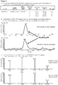
- Figure 1 shows results of an SRM/MRM assay of the AXL fragment peptide of SEQ ID NO:4 performed on a positive control sample (not formalin fixed) and a Liquid Tissue lysate from a formalin-fixed biological sample with quantitation of the AXL peptide conducted on a triple quadrupole mass spectrometer. The specific characteristics about the precursor and transitions ions used to measure, using the SRM/MRM method, this peptide in biological samples that have been fixed in formalin are shown.
- the AXL fragment peptide of SEQ ID NO:4 is used as a surrogate reporter to determine the abundance of AXL protein in a sample of formalin-fixed tissue using a mass spectrometry-based SRM/MRM assay.
- AXL fragment peptides may be generated by a variety of means including by the use of the Liquid Tissue protocol provided in US Patent 7,473,532 .
- the Liquid Tissue protocol and reagents are capable of producing peptide samples suitable for mass spectroscopic analysis from formalin-fixed paraffin-embedded tissue by proteolytic digestion of the proteins in the tissue/biological sample.
- the tissue/biological is heated in a buffer for an extended period of time (e.g., from about 80°C to about 100°C for a period of time from about 10 minutes to about 4 hours) to reverse or release protein cross-linking.
- the buffer employed is a neutral buffer, (e.g., a Tris-based buffer, or a buffer containing a detergent).
- a neutral buffer e.g., a Tris-based buffer, or a buffer containing a detergent.
- proteases including but not limited to trypsin, chymotrypsin, pepsin, and endoproteinase Lys-C for a time sufficient to disrupt the tissue and cellular structure of said biological sample and to liquefy said sample (e.g., a period of time from 30 minutes to 24 hours at a temperature from 37°C to 65°C).
- the result of the heating and proteolysis is a liquid, soluble, dilutable biomolecule lysate.
- the AXL peptide of SEQ ID NO: 4 and further AXL peptides identified were derived from the AXL protein by protease digestion of all the proteins within a complex Liquid Tissue lysate prepared from cells procured from formalin-fixed cancer tissue. Unless noted otherwise, in each instance the protease was trypsin. The Liquid Tissue lysate was then analyzed by mass spectrometry to determine those peptides derived from the AXL protein that are detected and analyzed by mass spectrometry.
- Identification of a specific preferred subset of peptides for mass-spectrometric analysis is based on; 1) experimental determination of which peptide or peptides from a protein ionize in mass spectrometry analyses of Liquid Tissue lysates, and 2) the ability of the peptide to survive the protocol and experimental conditions used in preparing a Liquid Tissue lysate. This latter property extends not only to the amino acid sequence of the peptide but also to the ability of a modified amino acid residue within a peptide to survive in modified form during the sample preparation.
- Protein lysates from cells procured directly from formalin (formaldehyde)-fixed tissue were prepared using the Liquid Tissue reagents and protocol that entails collecting cells into a sample tube via tissue microdissection followed by heating the cells in the Liquid Tissue buffer for an extended period of time. Once the formalin-induced cross linking has been negatively affected, the tissue/cells are then digested to completion in a predictable manner using a protease, as for example including but not limited to the protease trypsin. Each protein lysate is turned into a collection of peptides by digestion of intact polypeptides with the protease.
- Each Liquid Tissue lysate was analyzed (e.g., by ion trap mass spectrometry) to perform multiple global proteomic surveys of the peptides where the data was presented as identification of as many peptides as could be identified by mass spectrometry from all cellular proteins present in each protein lysate.
- An ion trap mass spectrometer or another form of a mass spectrometer that is capable of performing global profiling for identification of as many peptides as possible from a single complex protein/peptide lysate is typically employed. Ion trap mass spectrometers however may be the best type of mass spectrometer for conducting global profiling of peptides.
- an SRM/MRM assay can be developed and performed on any type of mass spectrometer, including a MALDI, ion trap, or triple quadrupole, the most advantageous instrument platform for an SRM/MRM assay is often considered to be a triple quadrupole instrument platform.
- That type of dataset can be considered to represent the peptides that can be detected in the type of biological sample that was analyzed (after protease digestion), and specifically in a Liquid Tissue lysate of the biological sample, and thus includes the peptides for specific proteins, such as for example the AXL protein.
- the AXL tryptic peptide identified as useful in the determination of absolute or relative amounts of the AXL protein was the peptide of SEQ ID NO:4. Further suitable peptides are listed in Table 1. Each of those peptides was detected by mass spectrometry in Liquid Tissue lysates prepared from formalin-fixed, paraffin-embedded tissue.
- the AXL tryptic peptides listed in Table 1 include those detected from multiple Liquid Tissue lysates of multiple different formalin-fixed tissues of different human organs including lung, colon, and breast. Each of those peptides is considered useful for quantitative SRM/MRM assay of the AXL protein in formalin-fixed tissue. Further data analysis of these experiments indicated no preference is observed for any specific peptides from any specific organ site. Thus, each of these peptides other than that of SEQ ID NO:4 is also believed to be suitable for conducting SRM/MRM assays of the AXL protein on a Liquid Tissue lysate from any formalin-fixed tissue originating from any biological sample or from any organ site in the body.
- SRM/MRM assays An important consideration when conducting an SRM/MRM assay is the type of instrument that may be employed in the analysis of the peptides.
- SRM/MRM assays can be developed and performed on any type of mass spectrometer, including a MALDI, ion trap, or triple quadrupole, the most advantageous instrument platform for an SRM/MRM assay is often considered to be a triple quadrupole instrument platform.
- That type of a mass spectrometer may be considered to be the most suitable instrument for analyzing a single isolated target peptide within a very complex protein lysate that may consist of hundreds of thousands to millions of individual peptides from all the proteins contained within a cell.
- an SRM/MRM assay for the AXL protein of SEQ ID NO:4 it is desirable to utilize information in addition to the peptide sequence in the analysis. That additional information may be used in directing and instructing the mass spectrometer (e.g. a triple quadrupole mass spectrometer) to perform the correct and focused analysis of specific targeted peptide(s), such that the assay may be effectively performed.
- the mass spectrometer e.g. a triple quadrupole mass spectrometer
- the additional information about target peptides in general, and about specific AXL peptides may include one or more of the mono isotopic mass of the peptide, its precursor charge state, the precursor m/z value, the m/z transition ions, and the ion type of each transition ion.
- Additional peptide information that may be used to develop an SRM/MRM assay for the AXL protein is shown by example for three (3) of the AXL peptides from the list in Table 1 and is shown in Table 2. Similar additional information described for the three (3) AXL peptides shown by example in Table 2 may be prepared, obtained, and applied to the analysis of the other peptides contained in Table 1.
- the method described below was used to: 1) identify candidate peptides from the AXL protein that can be used for a mass spectrometry-based SRM/MRM assay for the AXL protein, 2) develop an individual SRM/MRM assay, or assays, for target peptides from the AXL protein in order to correlate and 3) apply quantitative assays to cancer diagnosis and/or choice of optimal therapy.
- AXL peptide of SEQ ID NO:4 Specific and unique characteristics about the AXL peptide of SEQ ID NO:4 as well as further specific AXL peptides were developed by analysis of all AXL peptides on both an ion trap and triple quadrupole mass spectrometers. That information includes the monoisotopic mass of the peptide, its precursor charge state, the precursor m/z value, the transition m/z values of the precursor, and the ion types of each of the identified transitions.
- That information must be determined experimentally for each and every candidate SRM/MRM peptide directly in Liquid Tissue lysates from formalin fixed samples/tissue, because, interestingly, not all peptides from the AXL protein can be detected in such lysates using SRM/MRM as described herein, indicating that AXL peptides not detected cannot be considered candidate peptides for developing an SRM/MRM assay for use in quantitating peptides/proteins directly in Liquid Tissue lysates from formalin fixed samples/tissue.
- a particular SRM/MRM assay for a specific AXL peptide is performed on a triple quadrupole mass spectrometer.
- An experimental sample analyzed by a particular AXL SRM/MRM assay is for example a Liquid Tissue protein lysate prepared from a tissue that had been formalin-fixed and paraffin-embedded. Data from such as assay indicates the presence of the unique SRM/MRM signature peak for this AXL peptide in the formalin fixed sample.
- This disclosure describes a method for measuring the level of the tyrosine-protein kinase receptor UFO protein (AXL) in a biological sample of formalin-fixed tissue, comprising detecting and quantifying the amount of an AXL fragment peptide in a protein digest prepared from said biological sample using mass spectrometry; and calculating the level of AXL protein in said sample; wherein said AXL fragment peptide is the peptide of SEQ ID NO:4; and wherein said level is a relative level or an absolute level.
- AXL tyrosine-protein kinase receptor UFO protein
- quantifying the AXL fragment peptide comprises determining the amount of the AXL fragment peptide of SEQ ID NO:4 in a biological sample by comparison to an added internal standard peptide of known amount, wherein the AXL fragment peptide in the biological sample is compared to an internal standard peptide having the same amino acid sequence.
- the internal standard is an isotopically labeled internal standard peptide comprises one or more heavy stable isotopes selected from 18 O, 11 O, 34 S, 15 N, 13 C, 2 H, and combinations thereof.
- the method for measuring the level of the AXL protein in a biological sample described herein may be used as a diagnostic indicator of cancer in a patient or subject.
- the results from measurements of the level of the AXL protein may be employed to determine the diagnostic stage/grade/status of a cancer by correlating (e.g., comparing) the level of AXL protein found in a tissue with the level of that protein found in normal and/or cancerous or precancerous tissues.
- both nucleic acids and protein can be analyzed from the same Liquid TissueTM biomolecular preparation it is possible to generate additional information about disease diagnosis and drug treatment decisions from the nucleic acids in same sample upon which proteins were analyzed. For example, if the AXL protein is expressed by certain cells at increased levels, when assayed by SRM the data can provide information about the state of the cells and their potential for uncontrolled growth, potential drug resistance and the development of cancers can be obtained.
- information about the status of the AXL genes and/or the nucleic acids and proteins they encode can be obtained from nucleic acids present in the same Liquid TissueTM biomolecular preparation can be assessed simultaneously to the SRM analysis of the AXL protein. Any gene and/or nucleic acid not from the AXL and which is present in the same biomolecular preparation can be assessed simultaneously to the SRM analysis of the AXL protein.
- information about the AXL protein and/or one, two, three, four or more additional proteins may be assessed by examining the nucleic acids encoding those proteins.
- nucleic acids can be examined, for example, by one or more, two or more, or three or more of: sequencing methods, polymerase chain reaction methods, restriction fragment polymorphism analysis, identification of deletions, insertions, and/or determinations of the presence of mutations, including but not limited to, single base pair polymorphisms, transitions, transversions, or combinations thereof.
Landscapes
- Life Sciences & Earth Sciences (AREA)
- Health & Medical Sciences (AREA)
- Engineering & Computer Science (AREA)
- Molecular Biology (AREA)
- Immunology (AREA)
- Biomedical Technology (AREA)
- Chemical & Material Sciences (AREA)
- Hematology (AREA)
- Urology & Nephrology (AREA)
- Physics & Mathematics (AREA)
- Cell Biology (AREA)
- General Physics & Mathematics (AREA)
- Pathology (AREA)
- Medicinal Chemistry (AREA)
- Biotechnology (AREA)
- Analytical Chemistry (AREA)
- Biochemistry (AREA)
- General Health & Medical Sciences (AREA)
- Microbiology (AREA)
- Food Science & Technology (AREA)
- Bioinformatics & Computational Biology (AREA)
- Bioinformatics & Cheminformatics (AREA)
- Proteomics, Peptides & Aminoacids (AREA)
- Spectroscopy & Molecular Physics (AREA)
- Biophysics (AREA)
- Other Investigation Or Analysis Of Materials By Electrical Means (AREA)
- Oncology (AREA)
- Measuring Or Testing Involving Enzymes Or Micro-Organisms (AREA)
- Peptides Or Proteins (AREA)
Description
- Cancer is treated with a collection of therapeutic agents that kill growing and dividing cells and that function in a variety of ways. A common collection of chemotherapeutic agents has been used for decades, either individually or in combinations, and this common collection of agents has become the traditional and routine cancer treatment in clinical oncology practice. These traditional chemotherapeutics agents act by killing all cells that divide rapidly, one of the main properties of most cancer cells. However, these agents also kill growing normal cells and thus these agents are not considered to be "targeted" approaches to killing cancer cells. In recent years a large group of cancer therapeutic agents has been developed that target cancer cells where the therapeutic agent specifically attacks a protein that is only expressed by the cancer cells and not by normal cells. This approach is considered to be a "targeted" approach to cancer therapy. Most recently, another approach to killing cancer cells in a targeted fashion has been to specifically modulate the immune system to enhance that ability of the cancer patient's immune system to kill cancer cells.
- Therapeutic agents that target the tyrosine-protein kinase receptor UFO protein, which can also be referred to as AXL, have shown promise in early clinical trials. However, only those patients whose cancer cells express high amounts of the AXL protein are likely to benefit from treatment with such AXL-targeted therapeutic agents. Various methods for the detection of AXL levels in biological samples are known and disclosed, for example, in patent publications
WO 2011/053779 A2 andWO 2010/103388 A2 as well as in Ekman et al. (Ekman, C. et al. (2010) J. Thromb. Haemost. 8: 838-844). - The method as specified in claim 1 provides a quantitative proteomics-based assay that delivers a relevant measure of activation of the AXL signal pathway, as AXL is not normally expressed in normal tissue and/or normal epithelial cells. In particular, the method provides a mass spectrometry assay that quantifies AXL in formalin fixed tissues from cancer patients and that enables improved treatment decisions for cancer therapy.
- Specific peptides derived from subsequences of the tyrosine-protein kinase receptor UFO protein, also referred to as AXL, and be referred to herein as AXL, are provided, with the peptide of SEQ ID NO: 4 being employed in the method of claim 1 for measuring AXL levels in a biological sample of formalin-fixed tissue. The peptide sequence and fragmentation/transition ions are particularly useful in a mass spectrometry-based Selected Reaction Monitoring (SRM), which can also be referred to as a Multiple Reaction Monitoring (MRM) assay, and will be referred to as SRM/MRM. The use of such peptide for SRM/MRM quantitative analysis of the AXL protein is described.
- This SRM/MRM assay can be used to measure relative or absolute quantitative levels of such peptide from the AXL protein and therefore provide a means of measuring by mass spectrometry the amount of the AXL protein in a given protein preparation obtained from a biological sample of formalin-fixed tissue as specified in claim 1.
- More specifically, the SRM/MRM assay can measure such peptide directly in complex protein lysate samples prepared from cells procured from formalin-fixed tissue samples, such as formalin-fixed cancer patient tissue. Methods of preparing protein samples from formalin-fixed tissue are described in
U.S. Patent No. 7,473,532 . The methods described inU.S. Patent No. 7,473,532 may conveniently be carried out using Liquid Tissue reagents and protocol available from Expression Pathology Inc. (Rockville, MD). - The most widely and advantageously available form of tissues from cancer patient tissue is formalin-fixed, paraffin-embedded tissue. Formaldehyde/formalin fixation of surgically removed tissue is by far the most common method of preserving cancer tissue samples worldwide and is the accepted convention for standard pathology practice. Aqueous solutions of formaldehyde are referred to as formalin. "100%" formalin consists of a saturated solution of formaldehyde (about 40% by volume or 37% by mass) in water, with a small amount of stabilizer, usually methanol, to limit oxidation and degree of polymerization. The most common way in which tissue is preserved is to soak whole tissue for extended periods of time (8 hours to 48 hours) in aqueous formaldehyde, commonly termed 10% neutral buffered formalin, followed by embedding the fixed whole tissue in paraffin wax for long term storage at room temperature. Thus, molecular analytical methods to analyze formalin fixed cancer tissue will be the most accepted and heavily utilized methods for analysis of cancer patient tissue.
- Results from the SRM/MRM assay can be used to correlate accurate and precise quantitative levels of the AXL protein within the specific tissue samples (e.g., cancer tissue sample) of the patient or subject from whom the tissue (biological sample) was collected and preserved. This not only provides diagnostic information about the cancer, but also permits a physician or other medical professional to determine appropriate therapy for the patient. Such an assay that provides diagnostically and therapeutically important information about levels of protein expression in a diseased tissue or other patient sample is termed a companion diagnostic assay. For example, such an assay can be designed to diagnose the stage or degree of a cancer and determine a therapeutic agent to which a patient is most likely to respond.
- The method as specified in claim 1 measures relative or absolute levels of the AXL fragment peptide of SEQ ID NO:4. The peotide may be modified or unmodified. Examples of modifications include phosphorylated amino acid residues and glycosylated amino acid residues that are present on the peptides.
- Relative quantitative levels of the AXL protein are determined by the SRM/MRM methodology, for example by comparing SRM/MRM signature peak areas (e.g., signature peak area or integrated fragment ion intensity) of the AXL fragment peptide of SEQ ID NO:4 in different samples of formalin-fixed tissue. Alternatively, it may also be possible to further compare multiple SRM/MRM signature peak areas for multiple other AXL signature peptides, where each peptide has its own specific SRM/MRM signature peak, to determine the relative AXL protein content in one biological sample with the AXL protein content in one or more additional or different biological samples. In this way, the amount of a particular peptide, or peptides, from the AXL protein, and therefore the amount of the AXL protein, is determined relative to the same AXL peptide, or peptides, across 2 or more biological samples under the same experimental conditions. In addition, relative quantitation can also be determined for the AXL fragment peptide of SEQ ID NO:4, and for further peptides, if applicable, from the AXL protein within a single sample by comparing the signature peak area for that peptide by SRM/MRM methodology to the signature peak area for another and different peptide, or peptides, from a different protein, or proteins, within the same protein preparation from the biological sample. In this way, the amount of a particular peptide from the AXL protein, and therefore the amount of the AXL protein, is determined relative one to another within the same sample. These approaches generate quantitation of an individual peptide, or peptides, from the AXL protein to the amount of another peptide, or peptides, between samples and within samples, wherein the amounts as determined by signature peak area are relative one to another, regardless of the absolute weight to volume or weight to weight amounts of the AXL peptide in the protein preparation from the biological sample. Relative quantitative data about individual signature peak areas between different samples are normalized to the amount of protein analyzed per sample. Relative quantitation can be performed across many peptides from multiple proteins and the AXL protein simultaneously in a single sample and/or across many samples to gain insight into relative protein amounts, such as one peptide/protein with respect to other peptides/proteins.
- Absolute quantitative levels of the AXL protein are determined by, for example, the SRM/MRM methodology whereby the SRM/MRM signature peak area of the AXL fragment peptide of SEQ ID NO:4 in one biological sample is compared to the SRM/MRM signature peak area of a spiked internal standard. In one embodiment, the internal standard is a synthetic version of the same exact AXL peptide that contains one or more amino acid residues labeled with one or more heavy isotopes. Such isotope labeled internal standards are synthesized so that when analyzed by mass spectrometry a standard generates a predictable and consistent SRM/MRM signature peak that is different and distinct from the native AXL peptide signature peak and which can be used as a comparator peak. Thus, when the internal standard is spiked into a protein preparation from a biological sample in known amounts and analyzed by mass spectrometry, the SRM/MRM signature peak area of the native peptide is compared to the SRM/MRM signature peak area of the internal standard peptide, and this numerical comparison indicates either the absolute molarity and/or absolute weight of the native peptide present in the original protein preparation from the biological sample. Absolute quantitative data for fragment peptides are displayed according to the amount of protein analyzed per sample. Absolute quantitation can be performed across many peptides, and thus proteins, simultaneously in a single sample and/or across many samples to gain insight into absolute protein amounts in individual biological samples and in entire cohorts of individual samples.
- The SRM/MRM assay method described herein can be used to aid diagnosis of the stage of cancer, for example, directly in patient-derived, formalin-fixed tissue, and to aid in determining which therapeutic agent would be most advantageous for use in treating that patient. Cancer tissue that is removed from a patient either through surgery, such as for therapeutic removal of partial or entire tumors, or through biopsy procedures conducted to determine the presence or absence of suspected disease, is analyzed to determine whether or not a specific protein, or proteins, and which forms of proteins, are present in that patient tissue. Moreover, the expression level of a protein, or multiple proteins, can be determined and compared to a "normal" or reference level found in healthy tissue. Normal or reference levels of proteins found in healthy tissue may be derived from, for example, the relevant tissues of one or more individuals that do not have cancer. Alternatively, normal or reference levels may be obtained for individuals with cancer by analysis of relevant tissues not affected by the cancer. Assays of protein levels (e.g., AXL levels) can also be used to diagnose the stage of cancer in a patient or subject diagnosed with cancer by employing the AXL levels. The level of an individual AXL peptide is defined as the molar amount of the peptide determined by the SRM/MRM assay per total amount of protein lysate analyzed. Information regarding AXL can thus be used to aid in determining the stage or grade of a cancer by correlating the level of the AXL protein (or fragment peptides of the AXL protein) with levels observed in normal tissues. Once the quantitative amount of the AXL protein has been determined in the cancer cells, that information can be matched to a list of therapeutic agents (chemical and biological) developed to specifically treat cancer tissue that is characterized by, for example, abnormal expression of the protein or protein(s) (e.g., AXL) that were assayed. Matching information from an AXL protein assay to a list of therapeutic agents that specifically targets, for example, the AXL protein or cells/tissue expressing the protein, defines what has been termed a personalized medicine approach to treating disease. The assay methods described herein form the foundation of a personalized medicine approach by using analysis of proteins from the patient's own tissue as a source for diagnostic and treatment decisions.
-
Figure 1 (A-C) shows results of an SRM/MRM assay of the AXL fragment peptide of SEQ ID NO:4 performed on a positive control sample (not formalin fixed) and a Liquid Tissue lysate from a formalin-fixed biological sample with quantitation of the AXL peptide conducted on a triple quadrupole mass spectrometer. The specific characteristics about the precursor and transitions ions used to measure, using the SRM/MRM method, this peptide in biological samples that have been fixed in formalin are shown. - The AXL fragment peptide of SEQ ID NO:4 is used as a surrogate reporter to determine the abundance of AXL protein in a sample of formalin-fixed tissue using a mass spectrometry-based SRM/MRM assay.
- AXL fragment peptides may be generated by a variety of means including by the use of the Liquid Tissue protocol provided in
US Patent 7,473,532 . The Liquid Tissue protocol and reagents are capable of producing peptide samples suitable for mass spectroscopic analysis from formalin-fixed paraffin-embedded tissue by proteolytic digestion of the proteins in the tissue/biological sample. In the Liquid Tissue protocol, the tissue/biological is heated in a buffer for an extended period of time (e.g., from about 80°C to about 100°C for a period of time from about 10 minutes to about 4 hours) to reverse or release protein cross-linking. The buffer employed is a neutral buffer, (e.g., a Tris-based buffer, or a buffer containing a detergent). Following heat treatment, the tissue/biological sample is treated with one or more proteases, including but not limited to trypsin, chymotrypsin, pepsin, and endoproteinase Lys-C for a time sufficient to disrupt the tissue and cellular structure of said biological sample and to liquefy said sample (e.g., a period of time from 30 minutes to 24 hours at a temperature from 37°C to 65°C). The result of the heating and proteolysis is a liquid, soluble, dilutable biomolecule lysate. - Surprisingly, it was found that many potential peptide sequences from the AXL protein are unsuitable or ineffective for use in mass spectrometry-based SRM/MRM assays for reasons that are not immediately evident. As it was not possible to predict the most suitable peptides for MRM/SRM assay, it was necessary to experimentally identify suitable peptides in actual Liquid Tissue lysates to develop a reliable and accurate SRM/MRM assay for the AXL protein. While not wishing to be bound by any theory, it is believed that some peptides might, for example, be difficult to detect by mass spectrometry because they do not ionize well or produce fragments distinct from other proteins. Peptides may also fail to resolve well in separation (e.g., liquid chromatography), or may adhere to glass or plastic ware.
- The AXL peptide of SEQ ID NO: 4 and further AXL peptides identified (e.g., Tables 1 and 2) were derived from the AXL protein by protease digestion of all the proteins within a complex Liquid Tissue lysate prepared from cells procured from formalin-fixed cancer tissue. Unless noted otherwise, in each instance the protease was trypsin. The Liquid Tissue lysate was then analyzed by mass spectrometry to determine those peptides derived from the AXL protein that are detected and analyzed by mass spectrometry. Identification of a specific preferred subset of peptides for mass-spectrometric analysis is based on; 1) experimental determination of which peptide or peptides from a protein ionize in mass spectrometry analyses of Liquid Tissue lysates, and 2) the ability of the peptide to survive the protocol and experimental conditions used in preparing a Liquid Tissue lysate. This latter property extends not only to the amino acid sequence of the peptide but also to the ability of a modified amino acid residue within a peptide to survive in modified form during the sample preparation.
- Protein lysates from cells procured directly from formalin (formaldehyde)-fixed tissue were prepared using the Liquid Tissue reagents and protocol that entails collecting cells into a sample tube via tissue microdissection followed by heating the cells in the Liquid Tissue buffer for an extended period of time. Once the formalin-induced cross linking has been negatively affected, the tissue/cells are then digested to completion in a predictable manner using a protease, as for example including but not limited to the protease trypsin. Each protein lysate is turned into a collection of peptides by digestion of intact polypeptides with the protease. Each Liquid Tissue lysate was analyzed (e.g., by ion trap mass spectrometry) to perform multiple global proteomic surveys of the peptides where the data was presented as identification of as many peptides as could be identified by mass spectrometry from all cellular proteins present in each protein lysate. An ion trap mass spectrometer or another form of a mass spectrometer that is capable of performing global profiling for identification of as many peptides as possible from a single complex protein/peptide lysate is typically employed. Ion trap mass spectrometers however may be the best type of mass spectrometer for conducting global profiling of peptides. Although an SRM/MRM assay can be developed and performed on any type of mass spectrometer, including a MALDI, ion trap, or triple quadrupole, the most advantageous instrument platform for an SRM/MRM assay is often considered to be a triple quadrupole instrument platform.
- Once as many peptides as possible were identified in a single MS analysis of a single lysate under the conditions employed, then that list of peptides was collated and used to determine the proteins that were detected in that lysate. That process was repeated for multiple Liquid Tissue lysates, and the very large list of peptides was collated into a single dataset. That type of dataset can be considered to represent the peptides that can be detected in the type of biological sample that was analyzed (after protease digestion), and specifically in a Liquid Tissue lysate of the biological sample, and thus includes the peptides for specific proteins, such as for example the AXL protein.
- The AXL tryptic peptide identified as useful in the determination of absolute or relative amounts of the AXL protein was the peptide of SEQ ID NO:4. Further suitable peptides are listed in Table 1. Each of those peptides was detected by mass spectrometry in Liquid Tissue lysates prepared from formalin-fixed, paraffin-embedded tissue.
Table 1 Peptide Peptide Sequence SEQ ID NO: 1 GLTGTLR SEQ ID NO: 2 TATITVLPQQPR SEQ ID NO: 3 LGSLHPHTPYHIR SEQ ID NO: 4 APLQGTLLGYR SEQ ID NO: 5 YGEVFEPTVER SEQ ID NO: 6 TTEATLNSLGISEELK SEQ ID NO: 7 HGDLHSFLLYSR SEQ ID NO: 8 IYNGDYYR SEQ ID NO: 9 GQTPYPGVENSELYDYLR SEQ ID NO: 10 EDLENTLK - The AXL tryptic peptides listed in Table 1 include those detected from multiple Liquid Tissue lysates of multiple different formalin-fixed tissues of different human organs including lung, colon, and breast. Each of those peptides is considered useful for quantitative SRM/MRM assay of the AXL protein in formalin-fixed tissue. Further data analysis of these experiments indicated no preference is observed for any specific peptides from any specific organ site. Thus, each of these peptides other than that of SEQ ID NO:4 is also believed to be suitable for conducting SRM/MRM assays of the AXL protein on a Liquid Tissue lysate from any formalin-fixed tissue originating from any biological sample or from any organ site in the body.
- An important consideration when conducting an SRM/MRM assay is the type of instrument that may be employed in the analysis of the peptides. Although SRM/MRM assays can be developed and performed on any type of mass spectrometer, including a MALDI, ion trap, or triple quadrupole, the most advantageous instrument platform for an SRM/MRM assay is often considered to be a triple quadrupole instrument platform. That type of a mass spectrometer may be considered to be the most suitable instrument for analyzing a single isolated target peptide within a very complex protein lysate that may consist of hundreds of thousands to millions of individual peptides from all the proteins contained within a cell.
- In order to most efficiently implement an SRM/MRM assay for the AXL protein of SEQ ID NO:4 it is desirable to utilize information in addition to the peptide sequence in the analysis. That additional information may be used in directing and instructing the mass spectrometer (e.g. a triple quadrupole mass spectrometer) to perform the correct and focused analysis of specific targeted peptide(s), such that the assay may be effectively performed.
- The additional information about target peptides in general, and about specific AXL peptides, may include one or more of the mono isotopic mass of the peptide, its precursor charge state, the precursor m/z value, the m/z transition ions, and the ion type of each transition ion. Additional peptide information that may be used to develop an SRM/MRM assay for the AXL protein is shown by example for three (3) of the AXL peptides from the list in Table 1 and is shown in Table 2. Similar additional information described for the three (3) AXL peptides shown by example in Table 2 may be prepared, obtained, and applied to the analysis of the other peptides contained in Table 1.
Table 2 SEQ ID Peptide sequence Mono Isotopic Mass Precursor Charge State Precursor m/z Transition m/z Ion Type SEQ ID NO: 2 TATITVLPQQPR 1324.5435 2 662.883 625.341 y5 2 662.883 738.425 y6 2 662.883 837.494 y7 2 662.883 938.541 y8 SEQ ID NO: 4 APLQGTLLGYR 1188.3926 2 594.84 559.322 y10 2 594.84 779.441 y7 2 594.84 907.499 y8 2 594.84 1020.583 y9 SEQ ID NO: 5 YGEVFEPTVER 1325.4408 2 663.322 350.134 b3 2 663.322 601.33 y5 2 663.322 730.372 y6 2 663.322 877.441 y7 - The method described below was used to: 1) identify candidate peptides from the AXL protein that can be used for a mass spectrometry-based SRM/MRM assay for the AXL protein, 2) develop an individual SRM/MRM assay, or assays, for target peptides from the AXL protein in order to correlate and 3) apply quantitative assays to cancer diagnosis and/or choice of optimal therapy.
-
- 1. Identification of SRM/MRM candidate fragment peptides for the AXL protein
- a. Prepare a Liquid Tissue protein lysate from a formalin-fixed biological sample using a protease or proteases, (that may or may not include trypsin), to digest proteins.
- b. Analyze all protein fragments in the Liquid Tissue lysate on an ion trap tandem mass spectrometer and identify all fragment peptides from the AXL protein, where individual fragment peptides do not contain any peptide modifications, such as phosphorylations or glycosylations.
- c. Analyze all protein fragments in the Liquid Tissue lysate on an ion trap tandem mass spectrometer and identify all fragment peptides from the AXL protein that carry peptide modifications such as for example phosphorylated or glycosylated residues.
- d. All peptides generated by a specific digestion method from the entire, full length AXL protein potentially can be measured, but preferred peptides used for development of the SRM/MRM assay are those that are identified by mass spectrometry directly in a complex Liquid Tissue protein lysate prepared from a formalin fixed biological sample.
- e. Peptides that are specifically modified (phosphorylated, glycosylated, etc.) in patient tissue and which ionize, and thus detected, in a mass spectrometer when analyzing a Liquid Tissue lysate from a formalin-fixed biological sample are identified as candidate peptides for assaying peptide modifications of the AXL protein.
- 2. Mass Spectrometry Assay for Fragment Peptides from the AXL Protein
- a. SRM/MRM assay on a triple quadrupole mass spectrometer for individual fragment peptides identified in a Liquid Tissue lysate is applied to peptides from the AXL protein.
- i. Determine optimal retention time for a fragment peptide for optimal chromatography conditions including liquid chromatography, nano-reversed phase liquid chromatography, high performance liquid chromatography, and reverse phase high performance liquid chromatography.
- ii. Determine the mono isotopic mass of the peptide, the precursor charge state for each peptide, the precursor m/z value for each peptide, the m/z transition ions for each peptide, and the ion type of each transition ion for each fragment peptide in order to develop an SRM/MRM assay for each peptide.
- iii. SRM/MRM assay can then be conducted using the information from (i) and (ii) on a triple quadrupole mass spectrometer where each peptide has a characteristic and unique SRM/MRM signature peak that precisely defines the unique SRM/MRM assay as performed on a triple quadrupole mass spectrometer.
- b. Perform SRM/MRM analysis so that the amount of the fragment peptide of the AXL protein that is detected, as a function of the unique SRM/MRM signature peak area from an SRM/MRM mass spectrometry analysis, can indicate both the relative and absolute amount of the protein in a particular protein lysate.
- i. Relative quantitation may be achieved by:
- 1. Determining increased or decreased presence of the AXL protein by comparing the SRM/MRM signature peak area from a given AXL peptide detected in a Liquid Tissue lysate from one formalin-fixed biological sample to the same SRM/MRM signature peak area of the same AXL fragment peptide in at least a second, third, fourth or more Liquid Tissue lysates from least a second, third, fourth or more formalin-fixed biological samples.
- 2. Determining increased or decreased presence of the AXL protein by comparing the SRM/MRM signature peak area from a given AXL peptide detected in a Liquid Tissue lysate from one formalin-fixed biological sample to SRM/MRM signature peak areas developed from fragment peptides from other proteins, in other samples derived from different and separate biological sources, where the SRM/MRM signature peak area comparison between the 2 samples for a peptide fragment are normalized to amount of protein analyzed in each sample.
- 3. Determining increased or decreased presence of the AXL protein by comparing the SRM/MRM signature peak area for a given AXL peptide to the SRM/MRM signature peak areas from other fragment peptides derived from different proteins within the same Liquid Tissue lysate from the formalin-fixed biological sample in order to normalize changing levels of AXL protein to levels of other proteins that do not change their levels of expression under various cellular conditions.
- 4. These assays can be applied to both unmodified fragment peptides and for modified fragment peptides of the AXL protein, where the modifications include but are not limited to phosphorylation and/or glycosylation, and where the relative levels of modified peptides are determined in the same manner as determining relative amounts of unmodified peptides.
- ii. Absolute quantitation of a given peptide may be achieved by comparing the SRM/MRM signature peak area for a given fragment peptide from the AXL protein in an individual biological sample to the SRM/MRM signature peak area of an internal fragment peptide standard spiked into the protein lysate from the biological sample.
- 1. The internal standard is a labeled synthetic version of the fragment peptide from the AXL protein that is being interrogated. This standard is spiked into a sample in known amounts, and the SRM/MRM signature peak area can be determined for both the internal fragment peptide standard and the native fragment peptide in the biological sample separately, followed by comparison of both peak areas.
- 2. This can be applied to unmodified fragment peptides and modified fragment peptides, where the modifications include but are not limited to phosphorylation and/or glycosylation, and where the absolute levels of modified peptides can be determined in the same manner as determining absolute levels of unmodified peptides.
- i. Relative quantitation may be achieved by:
- a. SRM/MRM assay on a triple quadrupole mass spectrometer for individual fragment peptides identified in a Liquid Tissue lysate is applied to peptides from the AXL protein.
- 3. Apply Fragment Peptide Quantitation to Cancer Diagnosis and Treatment
- a. Perform relative and/or absolute quantitation of fragment peptide levels of the AXL protein and demonstrate that the previously-determined association, as well understood in the field of cancer, of AXL protein expression to the stage/grade/status of cancer in patient tumor tissue is confirmed.
- b. Perform relative and/or absolute quantitation of fragment peptide levels of the AXL protein and demonstrate correlation with clinical outcomes from different treatment strategies, wherein this correlation has already been demonstrated in the field or can be demonstrated in the future through correlation studies across cohorts of patients and tissue from those patients. Once either previously established correlations or correlations derived in the future are confirmed by this assay then the assay method can be used to determine optimal treatment strategy.
- Specific and unique characteristics about the AXL peptide of SEQ ID NO:4 as well as further specific AXL peptides were developed by analysis of all AXL peptides on both an ion trap and triple quadrupole mass spectrometers. That information includes the monoisotopic mass of the peptide, its precursor charge state, the precursor m/z value, the transition m/z values of the precursor, and the ion types of each of the identified transitions. That information must be determined experimentally for each and every candidate SRM/MRM peptide directly in Liquid Tissue lysates from formalin fixed samples/tissue, because, interestingly, not all peptides from the AXL protein can be detected in such lysates using SRM/MRM as described herein, indicating that AXL peptides not detected cannot be considered candidate peptides for developing an SRM/MRM assay for use in quantitating peptides/proteins directly in Liquid Tissue lysates from formalin fixed samples/tissue.
- A particular SRM/MRM assay for a specific AXL peptide is performed on a triple quadrupole mass spectrometer. An experimental sample analyzed by a particular AXL SRM/MRM assay is for example a Liquid Tissue protein lysate prepared from a tissue that had been formalin-fixed and paraffin-embedded. Data from such as assay indicates the presence of the unique SRM/MRM signature peak for this AXL peptide in the formalin fixed sample.
- Specific transition ion characteristics for this peptide are used to quantitatively measure a particular AXL peptide in formalin fixed biological samples. These data indicate absolute amounts of this AXL peptide as a function of molar amount of the peptide per microgram of protein lysate analyzed. Assessment of AXL protein levels in tissues based on analysis of formalin-fixed patient-derived tissue can provide diagnostic, prognostic, and therapeutically-relevant information about each particular patient.
- This disclosure describes a method for measuring the level of the tyrosine-protein kinase receptor UFO protein (AXL) in a biological sample of formalin-fixed tissue, comprising detecting and quantifying the amount of an AXL fragment peptide in a protein digest prepared from said biological sample using mass spectrometry; and calculating the level of AXL protein in said sample; wherein said AXL fragment peptide is the peptide of SEQ ID NO:4; and wherein said level is a relative level or an absolute level. In one embodiment, quantifying the AXL fragment peptide comprises determining the amount of the AXL fragment peptide of SEQ ID NO:4 in a biological sample by comparison to an added internal standard peptide of known amount, wherein the AXL fragment peptide in the biological sample is compared to an internal standard peptide having the same amino acid sequence. In some embodiments the internal standard is an isotopically labeled internal standard peptide comprises one or more heavy stable isotopes selected from 18O, 11O, 34S, 15N, 13C, 2H, and combinations thereof.
- The method for measuring the level of the AXL protein in a biological sample described herein may be used as a diagnostic indicator of cancer in a patient or subject. In one embodiment, the results from measurements of the level of the AXL protein may be employed to determine the diagnostic stage/grade/status of a cancer by correlating (e.g., comparing) the level of AXL protein found in a tissue with the level of that protein found in normal and/or cancerous or precancerous tissues.
- Because both nucleic acids and protein can be analyzed from the same Liquid Tissue™ biomolecular preparation it is possible to generate additional information about disease diagnosis and drug treatment decisions from the nucleic acids in same sample upon which proteins were analyzed. For example, if the AXL protein is expressed by certain cells at increased levels, when assayed by SRM the data can provide information about the state of the cells and their potential for uncontrolled growth, potential drug resistance and the development of cancers can be obtained. At the same time, information about the status of the AXL genes and/or the nucleic acids and proteins they encode (e.g., mRNA molecules and their expression levels or splice variations) can be obtained from nucleic acids present in the same Liquid Tissue™ biomolecular preparation can be assessed simultaneously to the SRM analysis of the AXL protein. Any gene and/or nucleic acid not from the AXL and which is present in the same biomolecular preparation can be assessed simultaneously to the SRM analysis of the AXL protein. In one embodiment, information about the AXL protein and/or one, two, three, four or more additional proteins may be assessed by examining the nucleic acids encoding those proteins. Those nucleic acids can be examined, for example, by one or more, two or more, or three or more of: sequencing methods, polymerase chain reaction methods, restriction fragment polymorphism analysis, identification of deletions, insertions, and/or determinations of the presence of mutations, including but not limited to, single base pair polymorphisms, transitions, transversions, or combinations thereof.
-
- <110> Expression Pathology Inc
- <120> SRM/MRM Assay for the tyrosine-protein kinase receptor UFO (AXL) protein
- <130> 001152-8035.WO00
- <150>
61/986,688
<151> 2014-04-30 - <160> 10
- <170> PatentIn version 3.5
- <210> 1
<211> 7
<212> PRT
<213> Homo sapiens - <400> 1

- <210> 2
<211> 12
<212> PRT
<213> Homo sapiens - <400> 2

- <210> 3
<211> 13
<212> PRT
<213> Homo sapiens - <400> 3

- <210> 4
<211> 11
<212> PRT
<213> Homo sapiens - <400> 4

- <210> 5
<211> 11
<212> PRT
<213> Homo sapiens - <400> 5

- <210> 6
<211> 16
<212> PRT
<213> Homo sapiens - <400> 6

- <210> 7
<211> 12
<212> PRT
<213> Homo sapiens - <400> 7

- <210> 8
<211> 8
<212> PRT
<213> Homo sapiens - <400> 8

- <210> 9
<211> 18
<212> PRT
<213> Homo sapiens - <400> 9

- <210> 10
<211> 8
<212> PRT
<213> Homo sapiens - <400> 10

Claims (14)
- A method for measuring the level of the tyrosine-protein kinase receptor UFO protein (AXL) in a biological sample of formalin-fixed tissue, comprising:detecting and quantifying the amount of an AXL fragment peptide in a protein digest prepared from said biological sample using mass spectrometry; andcalculating the level of AXL protein in said sample; wherein said AXL fragment peptide is the peptide of SEQ ID NO:4; and
wherein said level is a relative level or an absolute level. - The method of claim 1, further comprising the step of fractionating said protein digest prior to detecting and quantifying the amount of said AXL fragment peptide.
- The method of claim 2, wherein said fractionating step is selected from the group consisting of liquid chromatography, nano-reversed phase liquid chromatography, high performance liquid chromatography, and reverse phase high performance liquid chromatography.
- The method of any of claims 1 to 3, wherein said mass spectrometry comprises tandem mass spectrometry, ion trap mass spectrometry, triple quadrupole mass spectrometry, MALDI-TOF mass spectrometry, MALDI mass spectrometry, and/or time of flight mass spectrometry, and particularly wherein the mode of mass spectrometry used is Selected Reaction Monitoring (SRM), Multiple Reaction Monitoring (MRM), and/or multiple Selected Reaction Monitoring (mSRM).
- The method of any one of claims 1 to 4, wherein the tissue is obtained from a tumor.
- The method of any of claims 1 to 5, wherein quantifying the AXL fragment peptide comprises comparing an amount of said AXL fragment peptide in one biological sample to the amount of the same AXL fragment peptide in a different and separate biological sample.
- The method of any one of claims 1 to 6, wherein quantifying said AXL fragment peptide comprises determining the amount of the AXL fragment peptide in a biological sample by comparison to an added internal standard peptide of known amount, wherein the AXL fragment peptides in the biological sample is compared to an internal standard peptide having the same amino acid sequence.
- The method of claim 7, wherein the internal standard peptide is an isotopically labeled peptide, wherein the isotopically labeled internal standard peptide comprises one or more heavy stable isotopes selected from 18O, 11O, 34S, 15N, 13C, 2H, and combinations thereof.
- The method of any of claims 1 to 8, wherein detecting and quantifying the amount of said AXL fragment peptide in the protein digest indicates the presence of AXL protein and an association with cancer in the subject from which said biological sample was obtained.
- The method of claim 9, further comprising correlating the results of said detecting and quantifying the amount of said AXL fragment peptide, or the level of said AXL protein to the diagnostic stage/grade/status of the cancer.
- The method of claim 10, wherein correlating the results of said detecting and quantifying the amount of said AXL fragment peptide, or the level of said AXL protein to the diagnostic stage/grade/status of the cancer is combined with detecting and/or quantifying the amount of other proteins or peptides from other proteins in a multiplex format to provide additional information about the diagnostic stage/grade/status of the cancer.
- The method of any one of claims 1 to 11, further comprising selecting for the subject from which said biological sample was obtained a treatment regimen or a therapeutic agent based on the presence, absence, or amount of said AXL fragment peptide or the level of AXL protein.
- The method of claim 12, wherein the therapeutic agent binds the AXL protein and/or inhibits its biological activity.
- The method of claim 13, wherein the therapeutic agent is selected to specifically target AXL-expressing cancer cells.
Priority Applications (1)
| Application Number | Priority Date | Filing Date | Title |
|---|---|---|---|
| EP20151305.8A EP3656871A1 (en) | 2014-04-30 | 2015-04-30 | Srm/mrm assay for the tyrosine-protein kinase receptor ufo (axl) protein |
Applications Claiming Priority (2)
| Application Number | Priority Date | Filing Date | Title |
|---|---|---|---|
| US201461986688P | 2014-04-30 | 2014-04-30 | |
| PCT/US2015/028667 WO2015168484A1 (en) | 2014-04-30 | 2015-04-30 | Srm/mrm assay for the tyrosine-protein kinase receptor ufo (axl) protein |
Related Child Applications (1)
| Application Number | Title | Priority Date | Filing Date |
|---|---|---|---|
| EP20151305.8A Division EP3656871A1 (en) | 2014-04-30 | 2015-04-30 | Srm/mrm assay for the tyrosine-protein kinase receptor ufo (axl) protein |
Publications (3)
| Publication Number | Publication Date |
|---|---|
| EP3137631A1 EP3137631A1 (en) | 2017-03-08 |
| EP3137631A4 EP3137631A4 (en) | 2017-11-15 |
| EP3137631B1 true EP3137631B1 (en) | 2020-01-15 |
Family
ID=54359356
Family Applications (2)
| Application Number | Title | Priority Date | Filing Date |
|---|---|---|---|
| EP15786066.9A Active EP3137631B1 (en) | 2014-04-30 | 2015-04-30 | Srm/mrm assay for the tyrosine-protein kinase receptor ufo (axl) protein |
| EP20151305.8A Withdrawn EP3656871A1 (en) | 2014-04-30 | 2015-04-30 | Srm/mrm assay for the tyrosine-protein kinase receptor ufo (axl) protein |
Family Applications After (1)
| Application Number | Title | Priority Date | Filing Date |
|---|---|---|---|
| EP20151305.8A Withdrawn EP3656871A1 (en) | 2014-04-30 | 2015-04-30 | Srm/mrm assay for the tyrosine-protein kinase receptor ufo (axl) protein |
Country Status (9)
| Country | Link |
|---|---|
| US (2) | US10203336B2 (en) |
| EP (2) | EP3137631B1 (en) |
| JP (1) | JP6668256B2 (en) |
| KR (1) | KR20160146993A (en) |
| CN (1) | CN106255765B (en) |
| AU (1) | AU2015252961B2 (en) |
| CA (1) | CA2946888A1 (en) |
| IL (1) | IL248587A0 (en) |
| WO (1) | WO2015168484A1 (en) |
Families Citing this family (3)
| Publication number | Priority date | Publication date | Assignee | Title |
|---|---|---|---|---|
| EP3137631B1 (en) | 2014-04-30 | 2020-01-15 | Expression Pathology, Inc. | Srm/mrm assay for the tyrosine-protein kinase receptor ufo (axl) protein |
| WO2017184905A1 (en) * | 2016-04-20 | 2017-10-26 | Expression Pathology, Inc. | Method for improved hepatocellular cancer diagnosis |
| WO2021091541A1 (en) * | 2019-11-05 | 2021-05-14 | Kri Technologies Incorporated | Identifying cancer neoantigens for personalized cancer immunotherapy |
Family Cites Families (13)
| Publication number | Priority date | Publication date | Assignee | Title |
|---|---|---|---|---|
| PT1601450E (en) | 2003-03-10 | 2013-09-06 | Expression Pathology Inc | Liquid tissue preparation from histopatologically processed biological samples, tissues and cells |
| US20090099340A1 (en) | 2007-10-12 | 2009-04-16 | Cell Signaling Technology, Inc. | Reagents for the detection of protein phosphorylation in carcinoma signaling pathways |
| US7906301B2 (en) * | 2005-05-25 | 2011-03-15 | Expression Pathology, Inc. | Multiplex method for increased proteomic coverage from histopathologically processed biological samples, tissues cells |
| US20090253156A1 (en) | 2006-05-05 | 2009-10-08 | Perkinelmer Las, Inc. | Mass spectrometry methods for multiplexed quantification of protein kinases and phosphatases |
| WO2007140352A2 (en) * | 2006-05-26 | 2007-12-06 | Invitrogen Corporation | Plasma membrane and secreted cancer biomarkers |
| EP1961825A1 (en) * | 2007-02-26 | 2008-08-27 | INSERM (Institut National de la Santé et de la Recherche Medicale) | Method for predicting the occurrence of metastasis in breast cancer patients |
| WO2010103388A2 (en) * | 2009-03-13 | 2010-09-16 | Bergen Teinologioverforing As | Method |
| US20120220594A1 (en) * | 2009-10-30 | 2012-08-30 | Bristol-Meyers Squibb Company | Methods for treating cancer in patients having igf-1r inhibitor resistance |
| CA2798562A1 (en) | 2010-05-07 | 2011-11-10 | Ranju Ralhan | Methods and compositions for the diagnosis and treatment of thyroid cancer |
| US20130288233A1 (en) | 2010-12-24 | 2013-10-31 | Map Diagnostics Pty Ltd. | Selective Reaction Monitoring (SRM) Derived Protein Profiles for Cancer and other Pathologic Entities |
| WO2012092302A1 (en) | 2010-12-27 | 2012-07-05 | Expression Pathology, Inc. | Cmet protein srm/mrm assay |
| EP2766723A4 (en) | 2011-10-12 | 2015-06-10 | Univ North Carolina | MULTIPLEXED KINASE INHIBITOR BALLS AND USES THEREOF |
| EP3137631B1 (en) | 2014-04-30 | 2020-01-15 | Expression Pathology, Inc. | Srm/mrm assay for the tyrosine-protein kinase receptor ufo (axl) protein |
-
2015
- 2015-04-30 EP EP15786066.9A patent/EP3137631B1/en active Active
- 2015-04-30 KR KR1020167033095A patent/KR20160146993A/en not_active Withdrawn
- 2015-04-30 AU AU2015252961A patent/AU2015252961B2/en not_active Ceased
- 2015-04-30 WO PCT/US2015/028667 patent/WO2015168484A1/en not_active Ceased
- 2015-04-30 US US15/307,753 patent/US10203336B2/en active Active
- 2015-04-30 EP EP20151305.8A patent/EP3656871A1/en not_active Withdrawn
- 2015-04-30 CN CN201580020711.8A patent/CN106255765B/en not_active Expired - Fee Related
- 2015-04-30 JP JP2016565214A patent/JP6668256B2/en not_active Expired - Fee Related
- 2015-04-30 CA CA2946888A patent/CA2946888A1/en not_active Withdrawn
-
2016
- 2016-10-27 IL IL248587A patent/IL248587A0/en unknown
-
2019
- 2019-02-12 US US16/273,480 patent/US10718780B2/en active Active
Non-Patent Citations (1)
| Title |
|---|
| None * |
Also Published As
| Publication number | Publication date |
|---|---|
| IL248587A0 (en) | 2016-12-29 |
| AU2015252961B2 (en) | 2018-07-19 |
| EP3137631A1 (en) | 2017-03-08 |
| KR20160146993A (en) | 2016-12-21 |
| WO2015168484A1 (en) | 2015-11-05 |
| JP2017519195A (en) | 2017-07-13 |
| US20170052196A1 (en) | 2017-02-23 |
| US10718780B2 (en) | 2020-07-21 |
| CN106255765A (en) | 2016-12-21 |
| US10203336B2 (en) | 2019-02-12 |
| WO2015168484A8 (en) | 2016-11-10 |
| EP3656871A1 (en) | 2020-05-27 |
| EP3137631A4 (en) | 2017-11-15 |
| CA2946888A1 (en) | 2015-11-05 |
| JP6668256B2 (en) | 2020-03-25 |
| US20190265252A1 (en) | 2019-08-29 |
| CN106255765B (en) | 2020-07-21 |
| AU2015252961A1 (en) | 2016-10-27 |
Similar Documents
| Publication | Publication Date | Title |
|---|---|---|
| AU2011352231A1 (en) | cMET protein SRM/MRM assay | |
| US10718780B2 (en) | SRM/MRM assay for the tyrosine-protein kinase receptor UFO(AXL) protein | |
| US10620184B2 (en) | SRM/MRM assay for the cyclin-dependent kinase inhibitor 2A (p16) protein | |
| EP3167293B1 (en) | Srm/mrm assay for the gtpase kras protein (kras) | |
| EP3137632B1 (en) | Srm/mrm assay for the androgen receptor (ar) protein | |
| US10060927B2 (en) | SRM/MRM assay for the 6-O-methylguanine-DNA methyltransferase (MGMT) protein | |
| EP3167289B1 (en) | Srm/mrm assay for the serine/threonine-protein kinase b-raf (braf) | |
| EP3295180B1 (en) | Srm/mrm assay for the mesothelin (msln) protein | |
| US10620223B2 (en) | SRM/MRM assay for the fibroblast growth factor receptor 2 (FGFR2) protein | |
| US20200132694A1 (en) | SRM/MRM Assay for the Tumor Necrosis Factor Receptor Superfamily Member 8 (CD30) Protein |
Legal Events
| Date | Code | Title | Description |
|---|---|---|---|
| STAA | Information on the status of an ep patent application or granted ep patent |
Free format text: STATUS: THE INTERNATIONAL PUBLICATION HAS BEEN MADE |
|
| PUAI | Public reference made under article 153(3) epc to a published international application that has entered the european phase |
Free format text: ORIGINAL CODE: 0009012 |
|
| STAA | Information on the status of an ep patent application or granted ep patent |
Free format text: STATUS: REQUEST FOR EXAMINATION WAS MADE |
|
| 17P | Request for examination filed |
Effective date: 20160927 |
|
| AK | Designated contracting states |
Kind code of ref document: A1 Designated state(s): AL AT BE BG CH CY CZ DE DK EE ES FI FR GB GR HR HU IE IS IT LI LT LU LV MC MK MT NL NO PL PT RO RS SE SI SK SM TR |
|
| AX | Request for extension of the european patent |
Extension state: BA ME |
|
| DAV | Request for validation of the european patent (deleted) | ||
| DAX | Request for extension of the european patent (deleted) | ||
| A4 | Supplementary search report drawn up and despatched |
Effective date: 20170914 |
|
| RIC1 | Information provided on ipc code assigned before grant |
Ipc: G01N 33/48 20060101ALI20170908BHEP Ipc: G01N 33/68 20060101ALI20170908BHEP Ipc: G01N 33/574 20060101ALI20170908BHEP Ipc: C12Q 1/68 20060101AFI20170908BHEP |
|
| RA4 | Supplementary search report drawn up and despatched (corrected) |
Effective date: 20171016 |
|
| STAA | Information on the status of an ep patent application or granted ep patent |
Free format text: STATUS: EXAMINATION IS IN PROGRESS |
|
| 17Q | First examination report despatched |
Effective date: 20180730 |
|
| GRAP | Despatch of communication of intention to grant a patent |
Free format text: ORIGINAL CODE: EPIDOSNIGR1 |
|
| STAA | Information on the status of an ep patent application or granted ep patent |
Free format text: STATUS: GRANT OF PATENT IS INTENDED |
|
| INTG | Intention to grant announced |
Effective date: 20190807 |
|
| GRAS | Grant fee paid |
Free format text: ORIGINAL CODE: EPIDOSNIGR3 |
|
| GRAA | (expected) grant |
Free format text: ORIGINAL CODE: 0009210 |
|
| STAA | Information on the status of an ep patent application or granted ep patent |
Free format text: STATUS: THE PATENT HAS BEEN GRANTED |
|
| AK | Designated contracting states |
Kind code of ref document: B1 Designated state(s): AL AT BE BG CH CY CZ DE DK EE ES FI FR GB GR HR HU IE IS IT LI LT LU LV MC MK MT NL NO PL PT RO RS SE SI SK SM TR |
|
| REG | Reference to a national code |
Ref country code: CH Ref legal event code: EP Ref country code: GB Ref legal event code: FG4D |
|
| REG | Reference to a national code |
Ref country code: IE Ref legal event code: FG4D |
|
| REG | Reference to a national code |
Ref country code: DE Ref legal event code: R096 Ref document number: 602015045681 Country of ref document: DE |
|
| REG | Reference to a national code |
Ref country code: AT Ref legal event code: REF Ref document number: 1225174 Country of ref document: AT Kind code of ref document: T Effective date: 20200215 |
|
| REG | Reference to a national code |
Ref country code: NL Ref legal event code: MP Effective date: 20200115 |
|
| REG | Reference to a national code |
Ref country code: LT Ref legal event code: MG4D |
|
| PG25 | Lapsed in a contracting state [announced via postgrant information from national office to epo] |
Ref country code: NL Free format text: LAPSE BECAUSE OF FAILURE TO SUBMIT A TRANSLATION OF THE DESCRIPTION OR TO PAY THE FEE WITHIN THE PRESCRIBED TIME-LIMIT Effective date: 20200115 Ref country code: PT Free format text: LAPSE BECAUSE OF FAILURE TO SUBMIT A TRANSLATION OF THE DESCRIPTION OR TO PAY THE FEE WITHIN THE PRESCRIBED TIME-LIMIT Effective date: 20200607 Ref country code: FI Free format text: LAPSE BECAUSE OF FAILURE TO SUBMIT A TRANSLATION OF THE DESCRIPTION OR TO PAY THE FEE WITHIN THE PRESCRIBED TIME-LIMIT Effective date: 20200115 Ref country code: RS Free format text: LAPSE BECAUSE OF FAILURE TO SUBMIT A TRANSLATION OF THE DESCRIPTION OR TO PAY THE FEE WITHIN THE PRESCRIBED TIME-LIMIT Effective date: 20200115 Ref country code: NO Free format text: LAPSE BECAUSE OF FAILURE TO SUBMIT A TRANSLATION OF THE DESCRIPTION OR TO PAY THE FEE WITHIN THE PRESCRIBED TIME-LIMIT Effective date: 20200415 |
|
| PG25 | Lapsed in a contracting state [announced via postgrant information from national office to epo] |
Ref country code: BG Free format text: LAPSE BECAUSE OF FAILURE TO SUBMIT A TRANSLATION OF THE DESCRIPTION OR TO PAY THE FEE WITHIN THE PRESCRIBED TIME-LIMIT Effective date: 20200415 Ref country code: HR Free format text: LAPSE BECAUSE OF FAILURE TO SUBMIT A TRANSLATION OF THE DESCRIPTION OR TO PAY THE FEE WITHIN THE PRESCRIBED TIME-LIMIT Effective date: 20200115 Ref country code: GR Free format text: LAPSE BECAUSE OF FAILURE TO SUBMIT A TRANSLATION OF THE DESCRIPTION OR TO PAY THE FEE WITHIN THE PRESCRIBED TIME-LIMIT Effective date: 20200416 Ref country code: IS Free format text: LAPSE BECAUSE OF FAILURE TO SUBMIT A TRANSLATION OF THE DESCRIPTION OR TO PAY THE FEE WITHIN THE PRESCRIBED TIME-LIMIT Effective date: 20200515 Ref country code: SE Free format text: LAPSE BECAUSE OF FAILURE TO SUBMIT A TRANSLATION OF THE DESCRIPTION OR TO PAY THE FEE WITHIN THE PRESCRIBED TIME-LIMIT Effective date: 20200115 Ref country code: LV Free format text: LAPSE BECAUSE OF FAILURE TO SUBMIT A TRANSLATION OF THE DESCRIPTION OR TO PAY THE FEE WITHIN THE PRESCRIBED TIME-LIMIT Effective date: 20200115 |
|
| REG | Reference to a national code |
Ref country code: DE Ref legal event code: R097 Ref document number: 602015045681 Country of ref document: DE |
|
| PG25 | Lapsed in a contracting state [announced via postgrant information from national office to epo] |
Ref country code: LT Free format text: LAPSE BECAUSE OF FAILURE TO SUBMIT A TRANSLATION OF THE DESCRIPTION OR TO PAY THE FEE WITHIN THE PRESCRIBED TIME-LIMIT Effective date: 20200115 Ref country code: EE Free format text: LAPSE BECAUSE OF FAILURE TO SUBMIT A TRANSLATION OF THE DESCRIPTION OR TO PAY THE FEE WITHIN THE PRESCRIBED TIME-LIMIT Effective date: 20200115 Ref country code: SM Free format text: LAPSE BECAUSE OF FAILURE TO SUBMIT A TRANSLATION OF THE DESCRIPTION OR TO PAY THE FEE WITHIN THE PRESCRIBED TIME-LIMIT Effective date: 20200115 Ref country code: DK Free format text: LAPSE BECAUSE OF FAILURE TO SUBMIT A TRANSLATION OF THE DESCRIPTION OR TO PAY THE FEE WITHIN THE PRESCRIBED TIME-LIMIT Effective date: 20200115 Ref country code: CZ Free format text: LAPSE BECAUSE OF FAILURE TO SUBMIT A TRANSLATION OF THE DESCRIPTION OR TO PAY THE FEE WITHIN THE PRESCRIBED TIME-LIMIT Effective date: 20200115 Ref country code: RO Free format text: LAPSE BECAUSE OF FAILURE TO SUBMIT A TRANSLATION OF THE DESCRIPTION OR TO PAY THE FEE WITHIN THE PRESCRIBED TIME-LIMIT Effective date: 20200115 Ref country code: SK Free format text: LAPSE BECAUSE OF FAILURE TO SUBMIT A TRANSLATION OF THE DESCRIPTION OR TO PAY THE FEE WITHIN THE PRESCRIBED TIME-LIMIT Effective date: 20200115 Ref country code: ES Free format text: LAPSE BECAUSE OF FAILURE TO SUBMIT A TRANSLATION OF THE DESCRIPTION OR TO PAY THE FEE WITHIN THE PRESCRIBED TIME-LIMIT Effective date: 20200115 |
|
| REG | Reference to a national code |
Ref country code: AT Ref legal event code: MK05 Ref document number: 1225174 Country of ref document: AT Kind code of ref document: T Effective date: 20200115 |
|
| PLBE | No opposition filed within time limit |
Free format text: ORIGINAL CODE: 0009261 |
|
| STAA | Information on the status of an ep patent application or granted ep patent |
Free format text: STATUS: NO OPPOSITION FILED WITHIN TIME LIMIT |
|
| PG25 | Lapsed in a contracting state [announced via postgrant information from national office to epo] |
Ref country code: MC Free format text: LAPSE BECAUSE OF FAILURE TO SUBMIT A TRANSLATION OF THE DESCRIPTION OR TO PAY THE FEE WITHIN THE PRESCRIBED TIME-LIMIT Effective date: 20200115 |
|
| REG | Reference to a national code |
Ref country code: CH Ref legal event code: PL |
|
| 26N | No opposition filed |
Effective date: 20201016 |
|
| PG25 | Lapsed in a contracting state [announced via postgrant information from national office to epo] |
Ref country code: IT Free format text: LAPSE BECAUSE OF FAILURE TO SUBMIT A TRANSLATION OF THE DESCRIPTION OR TO PAY THE FEE WITHIN THE PRESCRIBED TIME-LIMIT Effective date: 20200115 Ref country code: AT Free format text: LAPSE BECAUSE OF FAILURE TO SUBMIT A TRANSLATION OF THE DESCRIPTION OR TO PAY THE FEE WITHIN THE PRESCRIBED TIME-LIMIT Effective date: 20200115 Ref country code: LU Free format text: LAPSE BECAUSE OF NON-PAYMENT OF DUE FEES Effective date: 20200430 Ref country code: LI Free format text: LAPSE BECAUSE OF NON-PAYMENT OF DUE FEES Effective date: 20200430 Ref country code: CH Free format text: LAPSE BECAUSE OF NON-PAYMENT OF DUE FEES Effective date: 20200430 |
|
| REG | Reference to a national code |
Ref country code: BE Ref legal event code: MM Effective date: 20200430 |
|
| PG25 | Lapsed in a contracting state [announced via postgrant information from national office to epo] |
Ref country code: SI Free format text: LAPSE BECAUSE OF FAILURE TO SUBMIT A TRANSLATION OF THE DESCRIPTION OR TO PAY THE FEE WITHIN THE PRESCRIBED TIME-LIMIT Effective date: 20200115 Ref country code: PL Free format text: LAPSE BECAUSE OF FAILURE TO SUBMIT A TRANSLATION OF THE DESCRIPTION OR TO PAY THE FEE WITHIN THE PRESCRIBED TIME-LIMIT Effective date: 20200115 Ref country code: BE Free format text: LAPSE BECAUSE OF NON-PAYMENT OF DUE FEES Effective date: 20200430 |
|
| PG25 | Lapsed in a contracting state [announced via postgrant information from national office to epo] |
Ref country code: IE Free format text: LAPSE BECAUSE OF NON-PAYMENT OF DUE FEES Effective date: 20200430 |
|
| PG25 | Lapsed in a contracting state [announced via postgrant information from national office to epo] |
Ref country code: TR Free format text: LAPSE BECAUSE OF FAILURE TO SUBMIT A TRANSLATION OF THE DESCRIPTION OR TO PAY THE FEE WITHIN THE PRESCRIBED TIME-LIMIT Effective date: 20200115 Ref country code: MT Free format text: LAPSE BECAUSE OF FAILURE TO SUBMIT A TRANSLATION OF THE DESCRIPTION OR TO PAY THE FEE WITHIN THE PRESCRIBED TIME-LIMIT Effective date: 20200115 Ref country code: CY Free format text: LAPSE BECAUSE OF FAILURE TO SUBMIT A TRANSLATION OF THE DESCRIPTION OR TO PAY THE FEE WITHIN THE PRESCRIBED TIME-LIMIT Effective date: 20200115 |
|
| PG25 | Lapsed in a contracting state [announced via postgrant information from national office to epo] |
Ref country code: MK Free format text: LAPSE BECAUSE OF FAILURE TO SUBMIT A TRANSLATION OF THE DESCRIPTION OR TO PAY THE FEE WITHIN THE PRESCRIBED TIME-LIMIT Effective date: 20200115 Ref country code: AL Free format text: LAPSE BECAUSE OF FAILURE TO SUBMIT A TRANSLATION OF THE DESCRIPTION OR TO PAY THE FEE WITHIN THE PRESCRIBED TIME-LIMIT Effective date: 20200115 |
|
| P01 | Opt-out of the competence of the unified patent court (upc) registered |
Effective date: 20230529 |
|
| PGFP | Annual fee paid to national office [announced via postgrant information from national office to epo] |
Ref country code: DE Payment date: 20230420 Year of fee payment: 9 |
|
| PGFP | Annual fee paid to national office [announced via postgrant information from national office to epo] |
Ref country code: GB Payment date: 20240419 Year of fee payment: 10 |
|
| PGFP | Annual fee paid to national office [announced via postgrant information from national office to epo] |
Ref country code: FR Payment date: 20240425 Year of fee payment: 10 |
|
| REG | Reference to a national code |
Ref country code: DE Ref legal event code: R119 Ref document number: 602015045681 Country of ref document: DE |
|
| GBPC | Gb: european patent ceased through non-payment of renewal fee |
Effective date: 20250430 |
|
| PG25 | Lapsed in a contracting state [announced via postgrant information from national office to epo] |
Ref country code: DE Free format text: LAPSE BECAUSE OF NON-PAYMENT OF DUE FEES Effective date: 20251104 |
|
| PG25 | Lapsed in a contracting state [announced via postgrant information from national office to epo] |
Ref country code: GB Free format text: LAPSE BECAUSE OF NON-PAYMENT OF DUE FEES Effective date: 20250430 |
|
| PG25 | Lapsed in a contracting state [announced via postgrant information from national office to epo] |
Ref country code: FR Free format text: LAPSE BECAUSE OF NON-PAYMENT OF DUE FEES Effective date: 20250430 |