EP3125912B1 - Method for treating or enhancing muscle tissue - Google Patents
Method for treating or enhancing muscle tissue Download PDFInfo
- Publication number
- EP3125912B1 EP3125912B1 EP15764383.4A EP15764383A EP3125912B1 EP 3125912 B1 EP3125912 B1 EP 3125912B1 EP 15764383 A EP15764383 A EP 15764383A EP 3125912 B1 EP3125912 B1 EP 3125912B1
- Authority
- EP
- European Patent Office
- Prior art keywords
- muscle
- injury
- composition
- glandulosa
- use according
- Prior art date
- Legal status (The legal status is an assumption and is not a legal conclusion. Google has not performed a legal analysis and makes no representation as to the accuracy of the status listed.)
- Active
Links
Images




Classifications
-
- A—HUMAN NECESSITIES
- A61—MEDICAL OR VETERINARY SCIENCE; HYGIENE
- A61K—PREPARATIONS FOR MEDICAL, DENTAL OR TOILETRY PURPOSES
- A61K36/00—Medicinal preparations of undetermined constitution containing material from algae, lichens, fungi or plants, or derivatives thereof, e.g. traditional herbal medicines
- A61K36/18—Magnoliophyta (angiosperms)
- A61K36/185—Magnoliopsida (dicotyledons)
- A61K36/48—Fabaceae or Leguminosae (Pea or Legume family); Caesalpiniaceae; Mimosaceae; Papilionaceae
-
- A—HUMAN NECESSITIES
- A61—MEDICAL OR VETERINARY SCIENCE; HYGIENE
- A61P—SPECIFIC THERAPEUTIC ACTIVITY OF CHEMICAL COMPOUNDS OR MEDICINAL PREPARATIONS
- A61P21/00—Drugs for disorders of the muscular or neuromuscular system
-
- A—HUMAN NECESSITIES
- A61—MEDICAL OR VETERINARY SCIENCE; HYGIENE
- A61P—SPECIFIC THERAPEUTIC ACTIVITY OF CHEMICAL COMPOUNDS OR MEDICINAL PREPARATIONS
- A61P29/00—Non-central analgesic, antipyretic or antiinflammatory agents, e.g. antirheumatic agents; Non-steroidal antiinflammatory drugs [NSAID]
Definitions
- the invention relates to the use of Prosopis glandulosa for treating muscle injury.
- antioxidants such as found in herbal preparations
- Korean ginseng is one example of a herbal preparation shown to enhance exercise and sport performance [Chen et al., 2011; Bucci, 2000; Wang et al., 2010], and a grape-seed derived polyphenol, for example, has recently been described as providing accelerated skeletal muscle recovery [Myburgh et al., 2012; Kruger et al., 2014].
- compositions for treating muscle injury in a subject comprising plant material from Prosopis glandulosa or an extract thereof.
- the plant material could be from any part of the P . glandulosa plant, such as leaves, flowers, roots, stems, bark, seeds and the like.
- the plant material is from seed pods of P . glandulosa, and even more preferably, the plant material is from seed pods which have been dried and ground.
- composition may further include pharmaceutically acceptable excipients, binders, adjuvants or fillers.
- the composition may be in an oral formulation, such as in the form of a tablet, sublingual tablet, wafer, sachet, capsule, suspension, syrup, powder, liquid beverage or edible bar.
- the composition may be in a topical formulation, such as a suspension, gel, cream or ointment which may be applied to the skin in the region of the muscle tissue.
- the composition may also be in an injectable formulation.
- the composition may also be in the form of a nutritional or dietary supplement.
- the muscle tissue to be treated is preferably skeletal muscle.
- the composition according is for treating muscle injury, such as a direct impact injury.
- the direct impact injury may be a contusion injury.
- the subject may be a human.
- composition may be intended to be administered to the subject at a daily dosage of P . glandulosa of from about 50 to about 200 mg/kg/day, such as about 100 mg/kg/day.
- Plant material from the Prosopis glandulosa (Torr.) [Fabaceae] tree (commonly known as Honey mesquite) is described herein.
- the plant material is typically the dried and ground pods from the tree, but could also be from other parts of the tree, such as the leaves, bark or roots.
- Soft tissue injuries are very common, accounting for between 35% and 55% of all sporting injuries [1]. Soft tissue injuries can result in significant pain, swelling and bruising, culminating in delayed and impaired functionality of the affected muscle [2].
- the pathophysiology of muscle injuries is a complex process, progressing through a sequence of overlapping phases, which include degeneration, inflammation, regeneration and the formation of fibrotic scar tissue [3,4,5,6]. Injuries to skeletal muscle not only damage the muscle cells, but may also lead to capillary rupture, infiltrative bleeding, inflammation, oxidative stress and fibrosis, depending on the extent of the injury. Inflammation stands central to these processes, with inflammatory cytokines largely responsible for modulating the cellular environment, thereby largely controlling the progress of other repair processes.
- peripheral fatigue Physical fatigue, also referred to as peripheral fatigue, is denoted as the deterioration of muscle performance during prolonged activity [Roots et al., 2008; Fitts, 1994]. This decrease in force is reversible, as muscle performance can be recovered after sufficient rest and appropriate nutrition.
- the magnitude of muscle force generation is determined by two main factors, namely, (i) the size of the muscle recruited to generate the force and (ii) the muscle fiber type [Maughan et al., 1983; Lee et al., 2013].
- Force generation during contraction is related to the number of crosslinks made between the actin and myosin chains [Fitts et al., 1991]. Therefore, the more crosslinks formed, the stronger the force of contraction.
- the maximal force of contraction depends upon the number of fibers a muscle contains.
- the type of fibers also plays an important role, as different types of muscle fibers possess different contractile properties [Schaiffino and Reggiani, 2011].
- Fiber composition is regulated in response to changes in physical activity, environment and pathological conditions [Schiaffino et al., 2007], for example, endurance exercise training induces a fast-to-slow fiber type transition, transforming the myofibers to an increased oxidative metabolism [Demirel et al., 1999; Pette and Staron, 2001; Yuan et al., 2011]. Additional factors leading to fiber type transition include mechanical loading and unloading, hormones and aging [Pette and Staron, 2001].
- Example 1 Therapeutic application of P. glandulosa for soft tissue injuries
- P. glandulosa as a possible treatment option after a contusion injury was studied. Effects on neutrophil and macrophage infiltration into the injured area were investigated, as well as associated consequences in the context of muscle regeneration.
- a recognized NSAID commonly used in the treatment of muscle injury and inflammation (diclofenac) was used as a comparative control.
- Rats in the different experimental groups were matched for body mass at the start of the protocol (PLA: 456.47 ⁇ 9.74 g; PG-CHR: 445.98 ⁇ 11.21 g; PG-AI: 439.12 ⁇ 14.84 g; NSAID: 442.25 ⁇ 12.58 g).
- P. glandulosa powder consisting solely of dry-milled P. glandulosa pods was used in this experiment [26].
- P. glandulosa powder from P. Schoeman was weighed daily for each animal in the treatment group and set into a mixture of commercially available gelatine/jelly cubes of 1 mL volume. These jelly cubes were fed to each animal individually for 8 weeks, to ensure absolute compliance and dose control.
- the dosage of 100 mg/kg/day P . glandulosa was calculated based on the daily dosage prescribed for human adults on a commercially available food supplement. This dose has previously been shown by the Applicant to elicit beneficial metabolic changes in rats [26,27].
- the control animals received placebo jelly cubes.
- Diclofenac a known NSAID, served as a positive control for the anti-inflammatory effects of treatment.
- Diclofenac sodium in the form of Voltaren Emulgel® from Novartis, was applied topically to the injured area on the hindlimb of the rats after different time periods post-injury.
- the dosage of Voltaren Emulgel® was calculated at 57.14 mg/kg/day, which was equal to 0.57 mg/kg Diclofenac. The dosage was calculated based on the daily dosage prescribed for human adults.
- the contusion injury to the rat hind-limb was produced using the mass-drop model injury first described by Stratton et al. (1984) [28] and optimized for our laboratory by Myburgh and colleagues (2012) [7]. Briefly, the technique entails dropping a 200 g weight from the height of 50 cm onto the medial surface of the right gastrocnemius muscle of sodium pentobarbital (40 mg/kg, intraperitoneal) anaesthetized rats. This contusion injury was moderately severe, did not result in bone injury or affect gait in the injured animals.
- rats were euthanized by sodium pentobarbital overdose (200 mg/kg, intraperitoneal) and the central section of the damaged gastrocnemius muscle harvested.
- the harvested muscle was divided into two parts, one part processed for immunohistochemistry and the other part snap-frozen for Western blotting analysis.
- Protein levels were determined by standard Western blotting technique [26]. Briefly, proteins were extracted from the gastrocnemius muscle tissue, equal concentration of total protein loaded and separated on a SDS poly-acrylamide gel and electro-transferred to ImmobilonTM-P PVDF membranes. Ponceau red reversible stain was used to determine transfer efficacy of proteins. The membranes were incubated overnight in ADAM 12 primary antibody (1:5000; Abcam, England, UK). For detection, horseradish peroxidase coupled secondary antibody (1:4000; Amersham Life Sciences, Sandton, JHB, South Africa) was used.
- Antigen-antibody complexes were visualized using ECL detection reagent (Amersham Life Sciences, Sandton, JHB, South Africa) and exposed to an autoradiography film (Hyperfilm ECL, RPN 2103) and light emission was detected. All films were analyzed by means of densitometry (UN-SCAN-IT; Silk Scientific Inc., Utah, UT, USA) and normalized data expressed in arbitrary units (AU). In all instances the membranes were stripped, by incubating in 0.2 M NaOH and reblotted with antibody against ⁇ -tubulin (1:1000; Cell Signalling Technology, Beverley, MA, USA) to verify the uniformity of protein load across the test samples.
- ADAM 12 expression is enhanced in response to chronic P. glandulosa treatment
- Desmin expression is increased in response to chronic P . glandulosa treatment
- Desmin expression was found to steadily increase after injury, with highest values at the 7 days post-injury time point, in all four different experimental groups.
- the chronically treated P. glandulosa group (PG-CHR) displayed significantly elevated desmin expression compared to all other groups ( Figure 5 ). While post-injury P. glandulosa- treatment had no effect on the expression of desmin, the NSAID-treated group displayed significantly decreased desmin expression, when compared to all other groups, indicative of delayed regeneration.
- the NSAID treatment resulted in significantly suppressed ADAM 12 expression (particularly at 3 h), pointing to an inhibitory effect on repair. This undesired effect was also evident from the desmin response.
Landscapes
- Health & Medical Sciences (AREA)
- Natural Medicines & Medicinal Plants (AREA)
- Life Sciences & Earth Sciences (AREA)
- Animal Behavior & Ethology (AREA)
- Chemical & Material Sciences (AREA)
- Veterinary Medicine (AREA)
- Medicinal Chemistry (AREA)
- Public Health (AREA)
- General Health & Medical Sciences (AREA)
- Pharmacology & Pharmacy (AREA)
- Engineering & Computer Science (AREA)
- Microbiology (AREA)
- Mycology (AREA)
- Alternative & Traditional Medicine (AREA)
- Biotechnology (AREA)
- Botany (AREA)
- Medical Informatics (AREA)
- Epidemiology (AREA)
- Nuclear Medicine, Radiotherapy & Molecular Imaging (AREA)
- General Chemical & Material Sciences (AREA)
- Chemical Kinetics & Catalysis (AREA)
- Organic Chemistry (AREA)
- Rheumatology (AREA)
- Pain & Pain Management (AREA)
- Bioinformatics & Cheminformatics (AREA)
- Neurology (AREA)
- Orthopedic Medicine & Surgery (AREA)
- Physical Education & Sports Medicine (AREA)
- Pharmaceuticals Containing Other Organic And Inorganic Compounds (AREA)
- Medicines Containing Plant Substances (AREA)
- Medicines That Contain Protein Lipid Enzymes And Other Medicines (AREA)
Description
- The invention relates to the use of Prosopis glandulosa for treating muscle injury.
- From accumulating scientific evidence, it is apparent that the phyto-chemicals present in herbal substances have beneficial effects on long-term human health and can be used to effectively treat various diseases.
- Numerous studies have shown that antioxidants, such as found in herbal preparations, can result in prolonged performance in endurance exercise, by decreasing oxidative stress [Zheng et al., 2012; Chen et al., 2011; Bucci, 2000] as well as aiding in the muscle regenerative process, resulting in accelerated muscle recovery following muscle injury. Korean ginseng is one example of a herbal preparation shown to enhance exercise and sport performance [Chen et al., 2011; Bucci, 2000; Wang et al., 2010], and a grape-seed derived polyphenol, for example, has recently been described as providing accelerated skeletal muscle recovery [Myburgh et al., 2012; Kruger et al., 2014].
- However, the abilities of many herbal remedies to enhance exercise and sport performance or reduce inflammation, as a result of injury, are based on anecdotal claims and lack scientific evidence. Therefore, as a result, many people experience unpleasant or undesirable side effects, due to incorrect dosage and interaction with other prescription medication, when taking commercially available herbal supplements which are unresearched. In addition, synthetic pharmaceuticals are also known for enhancing sport performance or recovery. However many of these (such as non-steroidal anti-inflammatory drugs) are not permitted in competitive sports.
-
-
Figure 1 shows ultrastructural changes over time after experimental contusion injury, stained by H & E. Figure (A) represent uninjured samples; Figures (B-D) represent samples taken from PLA animals at 3 h (B), 1 day (C) and 7 days (D) post-injury; Figures (E-G), (H-J) and (K-M) represent similar time points in the PG-CHR, PG-AI and NSAID groups respectively. Scale bar represents 100 µm; Figures (B), (E), (H) and (K) represent muscle fiber destruction and vascular disruption; Immune cells infiltration into the injured area is visible from 1 day post-injury (Figures (C), (F), (I) and (L)). Solid white arrows indicate red blood cells, solid black arrows points to newly regenerated muscle fibers and dashed arrows indicate immune cells. -
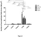
Figure 2 shows neutrophil (His48 stain) infiltration into injured area after contusion injury with/without P. glandulosa treatment. The data are expressed as mean ± SEM. Analysis was done by two-way ANOVA. n = 5 per time-point/per group; Differences over time are indicated by solid lines and broken black lines indicate group differences at specific time points; Significance: All groups: *** p < 0.0001 uninjured vs. 1 day; 1 h vs. 1 day; 3 h vs. 1 day; 1 day vs. 7 days. 1 Day: *** p < 0.0001 PLA vs. PG-CHR; PLA vs. NSAID; ** p < 0.05 PLA vs. PG-AI. -
Figure 3 shows macrophage infiltration into injured area after contusion injury with/without P. glandulosa treatment. The data are expressed as mean ± SEM. Analysis was done by two-way ANOVA. n = 5 per time-point/per group; Differences over time are indicated by solid lines; Significance: All groups: **p < 0.001 uninjured vs. 1 day; 1 h vs. 1 day; 3 h vs. 1 day; 7 daysvs. 1 day. -
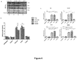
Figure 4 shows ADAM12 expression in skeletal muscle following a contusion injury. (A) Representative Western blots. Top 4 bands represent ADAM12 expression and the bottom bands represent β-tubulin expression (confirm equal loading of the protein); (B) Combined data for all the different groups; (C) Statistical differences observed between the different time-points in each experimental group. Values are expressed relative to the uninjured values. The data are expressed as mean ± SEM; Analysis by two-way ANOVA; η = 5 per time-point/per group; Differences over time are indicated by solid lines and broken black lines indicate group differences at specific time points; Significance: *** p < 0.0001, ** p < 0.001 and * p < 0.05. -
Figure 5 shows desmin expression in skeletal muscle following a contusion injury. The data are expressed as mean ± SEM; Analysis by two-way ANOVA; n = 5 per time-point/per group; Differences overtime are indicated by solid black lines (PLA), solid blue lines (PG-CHR), solid red lines (PG-AI) and broken black lines indicate group differences at specific time points; Significance: *** p < 0.0001, ** p < 0.001 and * p < 0.05. - According to the invention, there is provided a composition for treating muscle injury in a subject, the composition comprising plant material from Prosopis glandulosa or an extract thereof.
- The plant material could be from any part of the P. glandulosa plant, such as leaves, flowers, roots, stems, bark, seeds and the like. Preferably, the plant material is from seed pods of P. glandulosa, and even more preferably, the plant material is from seed pods which have been dried and ground.
- The composition may further include pharmaceutically acceptable excipients, binders, adjuvants or fillers.
- The composition may be in an oral formulation, such as in the form of a tablet, sublingual tablet, wafer, sachet, capsule, suspension, syrup, powder, liquid beverage or edible bar. Alternatively, the composition may be in a topical formulation, such as a suspension, gel, cream or ointment which may be applied to the skin in the region of the muscle tissue. The composition may also be in an injectable formulation. The composition may also be in the form of a nutritional or dietary supplement.
- The muscle tissue to be treated is preferably skeletal muscle.
- In one embodiment, the composition according is for treating muscle injury, such as a direct impact injury. The direct impact injury may be a contusion injury.
- The subject may be a human.
- The composition may be intended to be administered to the subject at a daily dosage of P. glandulosa of from about 50 to about 200 mg/kg/day, such as about 100 mg/kg/day.
- The use of plant material from the Prosopis glandulosa (Torr.) [Fabaceae] tree (commonly known as Honey mesquite) is described herein. The plant material is typically the dried and ground pods from the tree, but could also be from other parts of the tree, such as the leaves, bark or roots.
- Due to their high protein content, the pods of P. glandulosa have traditionally been used as a food or general food supplement by the residents of the south-western regions of the North American deserts [Simpson, 1977; Zimmermann, 1991]. This plant species was commonly found in the dry, arid regions of the northern and north-western Cape, South Africa [Jurriaanse, 1973; Harding, 1987], but due to its invasive potential is now classified as a category 2 invader under the Conservation of Agricultural Resources Act of 1983 (Act No. 43 of 1983) [Zimmermann, 1991].
- Very few studies have been conducted on the P. glandulosa plant and the only literature which could be found regarding its potential clinical benefits were from the Applicant's own studies relating to diabetes and cardiovascular health [26,27, George et al., 2011; Huisamen et al., 2012]. The Applicant is not aware of any literature on the effect of P. glandulosa on force generation, fatigue tolerance or muscle recovery after injury.
- The Applicant has now found that administration of P. glandulosa is effective in treatment of muscle injury and inflammation. Material from P. glandulosa could therefore be useful for treating muscle injury.
- Soft tissue injuries are very common, accounting for between 35% and 55% of all sporting injuries [1]. Soft tissue injuries can result in significant pain, swelling and bruising, culminating in delayed and impaired functionality of the affected muscle [2]. The pathophysiology of muscle injuries is a complex process, progressing through a sequence of overlapping phases, which include degeneration, inflammation, regeneration and the formation of fibrotic scar tissue [3,4,5,6]. Injuries to skeletal muscle not only damage the muscle cells, but may also lead to capillary rupture, infiltrative bleeding, inflammation, oxidative stress and fibrosis, depending on the extent of the injury. Inflammation stands central to these processes, with inflammatory cytokines largely responsible for modulating the cellular environment, thereby largely controlling the progress of other repair processes.
- In recent years, researchers have focused on manipulation of inflammation to accelerate muscle regeneration, for example, by targeting immune cells activated during the inflammatory phase [7,8]. Neutrophils and macrophages enter the site of injury in response to chemotactic signals and phagocytize the local debris [5,9,10]. Neutrophils, along with macrophages and satellite cells, release oxygen free radicals, resulting in oxidative stress and direct damage to surrounding tissue unaffected by the primary injury, which results in secondary muscle damage. However, even though early stage phenotypes of macrophages partially contributes to the sustainability of the inflammatory response and thus also secondary damage, these cells also secrete various growth factors that directly contribute to tissue repair and regeneration [5,11]. Additionally, both neutrophils and macrophages stimulate the release of cytokines (IL-1, IL-6, IL-8) and other chemotactic factors by T-cells, which inevitably results in the recruitment of satellite cells, with thus a greater capacity for muscle regeneration [5,12,13]. It is therefore clear that the inflammatory response, even though a contributor to secondary damage, is crucial to the repair of skeletal muscle after injury. Thus in the event of this total process being severely blunted for a prolonged period of time, such as through non-steroidal anti-inflammatory drug (NSAID) treatment, the potential clinical outcome may be suboptimal, resulting in delayed and/or incomplete tissue healing, as well as excessive scar formation, which increases the risk for recurrence of injury. There is evidence that suggests that the prolonged inhibition of the cyclooxygenase-2 pathway (more than 7 days continually) with prostaglandin inhibitors (NSAIDs) compromises muscle repair [4,14,15]. In addition to the use of NSAID's, there are various other muscle injury treatment options, such as the RICE approach (rest, ice, compression and elevation) [4,16,17,18], therapeutic ultrasound [19,20], hyperbaric oxygen therapy [21] and the use of growth factors [22]. However, these therapies remain suboptimal, as in many instances it either does not translate into increased myotube formation, therefore does not enhance muscle healing [19,20] and may be associated with severe risks and side effects [23].
- Physical fatigue, also referred to as peripheral fatigue, is denoted as the deterioration of muscle performance during prolonged activity [Roots et al., 2008; Fitts, 1994]. This decrease in force is reversible, as muscle performance can be recovered after sufficient rest and appropriate nutrition.
- The magnitude of muscle force generation is determined by two main factors, namely, (i) the size of the muscle recruited to generate the force and (ii) the muscle fiber type [Maughan et al., 1983; Lee et al., 2013]. Force generation during contraction is related to the number of crosslinks made between the actin and myosin chains [Fitts et al., 1991]. Therefore, the more crosslinks formed, the stronger the force of contraction. Hence, the maximal force of contraction depends upon the number of fibers a muscle contains. The type of fibers also plays an important role, as different types of muscle fibers possess different contractile properties [Schaiffino and Reggiani, 2011]. It is known that a muscle composed of a high proportion of slow-twitch (Type I) fibers will be relatively weaker than a muscle of similar size with a high proportion of fast-twitch (Type II) fibers. Fiber composition is regulated in response to changes in physical activity, environment and pathological conditions [Schiaffino et al., 2007], for example, endurance exercise training induces a fast-to-slow fiber type transition, transforming the myofibers to an increased oxidative metabolism [Demirel et al., 1999; Pette and Staron, 2001; Yuan et al., 2011]. Additional factors leading to fiber type transition include mechanical loading and unloading, hormones and aging [Pette and Staron, 2001].
- The data from this study indicates that P. glandulosa treatment might prove beneficial as a supplement, aiding physical ability, and results in more effective muscle repair after contusion injury than without treatment or with NSAID treatment.
- The fact that P. glandulosa is categorized as an invader tree in South Africa, and possibly in other countries too, illustrates the ethnopharmacological significance of these findings. The use of an economical, natural and readily available substance as treatment, could have far-reaching implications in not only the sporting arena and health sector, but also in plant and wildlife conservation.
- The invention will now be described in more detail by way of the following non-limiting examples.
- P. glandulosa as a possible treatment option after a contusion injury was studied. Effects on neutrophil and macrophage infiltration into the injured area were investigated, as well as associated consequences in the context of muscle regeneration. A recognized NSAID commonly used in the treatment of muscle injury and inflammation (diclofenac) was used as a comparative control.
- Age- and weight-matched adult, male, Wistar rats were divided into four main groups, namely: (1) control placebo (PLA); (2) PG-CHR, animals treated with P. glandulos for 8 weeks prior to injury and after injury, up to the time of sacrifice; (3) PG-AI, animals treated with P. glandulosa after injury (first treatment, 2 h after injury), up to the time of sacrifice and (4) NSAID, animals treated with Voltaren Emulgel® (diclofenac) directly after injury, up to the time of sacrifice. All four groups were subdivided into sacrifice and data collection time points of t = 0 h (before injury), 1 h, 3 h, 1 day and 7 days, post-injury. Each main group had an n = 25, i.e., 5 rats per time point per group (total of 100 rats).
- Rats in the different experimental groups were matched for body mass at the start of the protocol (PLA: 456.47 ± 9.74 g; PG-CHR: 445.98 ± 11.21 g; PG-AI: 439.12 ± 14.84 g; NSAID: 442.25 ± 12.58 g).
- P. glandulosa powder consisting solely of dry-milled P. glandulosa pods was used in this experiment [26]. To prepare treatment, P. glandulosa powder from P. Schoeman was weighed daily for each animal in the treatment group and set into a mixture of commercially available gelatine/jelly cubes of 1 mL volume. These jelly cubes were fed to each animal individually for 8 weeks, to ensure absolute compliance and dose control. The dosage of 100 mg/kg/day P. glandulosa was calculated based on the daily dosage prescribed for human adults on a commercially available food supplement. This dose has previously been shown by the Applicant to elicit beneficial metabolic changes in rats [26,27]. During the 8-week experimental period, the control animals received placebo jelly cubes.
- Diclofenac, a known NSAID, served as a positive control for the anti-inflammatory effects of treatment. Diclofenac sodium, in the form of Voltaren Emulgel® from Novartis, was applied topically to the injured area on the hindlimb of the rats after different time periods post-injury. The dosage of Voltaren Emulgel® was calculated at 57.14 mg/kg/day, which was equal to 0.57 mg/kg Diclofenac. The dosage was calculated based on the daily dosage prescribed for human adults.
- The contusion injury to the rat hind-limb was produced using the mass-drop model injury first described by Stratton et al. (1984) [28] and optimized for our laboratory by Myburgh and colleagues (2012) [7]. Briefly, the technique entails dropping a 200 g weight from the height of 50 cm onto the medial surface of the right gastrocnemius muscle of sodium pentobarbital (40 mg/kg, intraperitoneal) anaesthetized rats. This contusion injury was moderately severe, did not result in bone injury or affect gait in the injured animals.
- For sample collection, rats were euthanized by sodium pentobarbital overdose (200 mg/kg, intraperitoneal) and the central section of the damaged gastrocnemius muscle harvested. The harvested muscle was divided into two parts, one part processed for immunohistochemistry and the other part snap-frozen for Western blotting analysis.
- For cross-sectional histology and immunohistochemistry, muscles were fixed in 10% formal saline, processed and embedded in paraffin wax. Five-micrometer thick cross-sections were prepared (Leica RM 2125 RT microtome, Nussloch, Germany) and stained with haematoxylin and eosin (H & E) for qualitative histological analysis.
- Immunostaining with mouse anti-rat His48 (neutrophil; 1:200; Santa Cruz Biotechnology, Santa Cruz, CA, USA), goat anti-mouse F4/80 (macrophage; 1:200; Santa Cruz Biotechnology, Santa Cruz, CA, USA) and rabbit anti-human desmin (1:200; Santa Cruz Biotechnology, Santa Cruz, CA, USA) antibodies was performed on the fully automated Leica Bond-Max Autostainer system (Leica Microsystems, Germany) using an onboard detection kit, which included the Bond Epitope Retrieval Solution, peroxide block, primary antibody, post primary reagent, Bond Wash solution and Bond Polymer [29,30]. DAB (3,3'-diaminobenzidine tetrahydrochloride) was used as the chromogen (Leica Microsystems, Germany). Appropriate positive controls were used throughout the study.
- All imaging data were obtained by analyzing two sections from each muscle sample, at each time point for each antibody. In the injured area, five fields of view per section were imaged using a microscope (Nikon ECLIPSE E400; 400x objective used), equipped with a color digital camera (Nikon 5.0 Mega Pixels Color Digital Camera head DS-Fi2). The images presented in this article are only partial images of those taken at 200× magnification. Photos were used to count positively labeled neutrophils, macrophages and desmin-stains. Immune cells were counted manually and expressed as the average number of positively labeled immune cells per field of view (350 µm2) in the injured area, using the NIS-Elements BR imaging software package. In order for a cell to be classified a true neutrophil and macrophage, it had to have multilobular nuclei or single nuclei with surrounding cytoplasm, respectively.
- Protein levels were determined by standard Western blotting technique [26]. Briefly, proteins were extracted from the gastrocnemius muscle tissue, equal concentration of total protein loaded and separated on a SDS poly-acrylamide gel and electro-transferred to ImmobilonTM-P PVDF membranes. Ponceau red reversible stain was used to determine transfer efficacy of proteins. The membranes were incubated overnight in ADAM12 primary antibody (1:5000; Abcam, England, UK). For detection, horseradish peroxidase coupled secondary antibody (1:4000; Amersham Life Sciences, Sandton, JHB, South Africa) was used. Antigen-antibody complexes were visualized using ECL detection reagent (Amersham Life Sciences, Sandton, JHB, South Africa) and exposed to an autoradiography film (Hyperfilm ECL, RPN 2103) and light emission was detected. All films were analyzed by means of densitometry (UN-SCAN-IT; Silk Scientific Inc., Utah, UT, USA) and normalized data expressed in arbitrary units (AU). In all instances the membranes were stripped, by incubating in 0.2 M NaOH and reblotted with antibody against β-tubulin (1:1000; Cell Signalling Technology, Beverley, MA, USA) to verify the uniformity of protein load across the test samples.
- All data are presented as mean ± standard error of the mean (SEM). Statistical significance was analyzed by a two-way ANOVA, followed by a Bonferroni post hoc test. p < 0.05 was considered as statistically significant. Statistical analysis of data was performed using
GraphPad Prism version 5. - Qualitative microscopic analysis of the fiber architecture post-contusion injury indicated that irrespective of treatment, the blunt force to the muscle belly significantly damaged and disrupted the skeletal muscle fibers, resulting in red blood cell accumulation in the interstitial spaces at 1 h and 3 h after injury (
Figure 1B,E,H,K ). Representative pictures for 1 h after injury are not included as differences between 1 and 3 h post-injury cannot be easily discerned visually. Edema was present in both treated and untreated groups, confirmed by the widening of the interstitial spaces between the fibers at this early time point. Histological comparison between the placebo (PLA) and the treatment groups illustrated a significant influx ofimmune cells 1 day after injury in all four groups. However, this influx was relatively limited in the group chronically treated with P. glandulosa (PG-CHR), when compared to all other groups. The immune cells remained visible in the injured area of the PLA, post-injury treated group (PG-AI) and the non-steroidal anti-inflammatory treated (NSAID) groups for up today 7 post-injury (Figure 1D,J,M ), but were undetectable in the PG-CHR group at the same time point (Figure 1G ). Byday 7 only the chronically treated P. glandulosa group (Figure 1G ) displayed near normal muscle architecture, indicative of successful progressing muscle regeneration. - Clear differences were evident between the various experimental groups with regards to neutrophil infiltration. No neutrophils were present in the any of the experimental groups before injury, whereas contusion injury resulted in a significant (between 30- and 40-fold) transient elevation in neutrophils on
day 1 after injury, which normalised byday 7 post-injury (p < 0.0001) (Figure 2 ). Onday 1 post-injury, the PG-CHR (p < 0.0001), PG-AI (p < 0.001) as well as the NSAID treatment groups (p < 0.0001) displayed a significantly lower number of neutrophils compared to the untreated group (PLA). Furthermore, the magnitude of the neutrophil response as assessed onday 1 post-injury was similar in these three treatments groups. - Similar to the neutrophil data, the presence of macrophages was undetectable in the uninjured control samples (
Figure 3 ). Of the time-points assessed, the peak number of macrophages (p < 0.001) present in the injured area, was 1 day after injury in all four experimental groups. These increased values had again normalised byday 7 after injury (p < 0.001). None of the treatments seem to have any effect on macrophage infiltration at the time points assessed. - According to the Western blot analysis, expression of the satellite cell proliferation marker, ADAM12, was significantly elevated from 3 h post-injury (p < 0.0001) and this significant elevation persisted for at least 24 h (p < 0.0001), with the expression again normalized to uninjured levels on
day 7 after injury, in all experimental groups (p < 0.0001) (Figure 4A,B ). Of all three treatments assessed, the 8-week chronic treatment with P. glandulosa (PG-CHR) showed the most significant effect with significantly increased (p < 0.05) expression of ADAM12, onday 1 post-injury, when compared to the PLA group. Although post-injury treatment (PG-AI) seemed to suppress ADAM12 expression at 3 h, when compared to PLA, it was associated with a significant increase in ADAM12 expression from 3 to 1 day. NSAID treatment was associated with a similarly suppressed ADAM12 expression at 3 h, but with this treatment, the relative suppression persisted at 1 day after injury. Indeed, the NSAID group expressed lower levels of ADAM12 compared to the chronically treated P. glandulosa group at both 3 h and 1 day post-injury, significantly so on the latter (p < 0.001). For the sake of clarity, the statistical differences observed between the different time-points in each experimental group is illustrated inFigure 4C . - Desmin expression was found to steadily increase after injury, with highest values at the 7 days post-injury time point, in all four different experimental groups. At the 7-day post-injury time-point, the chronically treated P. glandulosa group (PG-CHR) displayed significantly elevated desmin expression compared to all other groups (
Figure 5 ). While post-injury P. glandulosa-treatment had no effect on the expression of desmin, the NSAID-treated group displayed significantly decreased desmin expression, when compared to all other groups, indicative of delayed regeneration. - Chronic P. glandulosa treatment was found to significantly reduce neutrophil infiltration into the injured area, suggestive of a decreased pro-inflammatory signal and probably less neutrophil-associated secondary damage. In support of this interpretation, an associated significant increase in the expression of ADAM12 (
day 1 post-injury) and desmin (day 7 post-injury) was observed, suggesting an enhanced regenerative process. - Large differences between P. glandulosa treatment and the NSAID treatment were evident.
- The level of ADAM12 expression in gastrocnemius muscles, at different time-points after injury, has not been previously measured in an in vivo model. The fact that chronic P. glandulosa treatment increased the ADAM12 expression at early time-points relative to the PLA indicates that the treatment may facilitate more effective recovery by enhancing early proliferation. In stark contrast, the NSAID treatment resulted in significantly suppressed ADAM12 expression (particularly at 3 h), pointing to an inhibitory effect on repair. This undesired effect was also evident from the desmin response.
- Desmin levels usually increase significantly during myogenesis and remain elevated in newly matured myofibers [49], which explains the relative late response in our protocol time course. On
day 7, and in accordance with the other data suggesting more effective muscle fiber repair, desmin expression was significantly higher after chronic P. glandulosa treatment. In contrast, desmin expression was significantly lower than PLA after NSAID treatment, again pointing to an inhibitory effect of NSAID on repair, as suggested in the literature [3]. -
- 1. Quintero, A.J.; Wright, V.J.; Fu, F.H.; Haurd, J. Stem cells for the treatment of skeletal muscle injury. Clin. Sports Med. 2009, 28, 1-11.
- 2. Lovering, R.M. Physical therapy and related interventions. In Skeletal Muscle Damage and Repair, Tiidus, P.M., Ed.; Human Kinetics: Champaign, IL, USA, 2008; pp. 219-230.
- 3. Smith, C.; Kruger, M.J.; Smith, R.M.; Myburgh, K.H. The inflammatory response to skeletal muscle injury: Illuminating complexities. J. Sports Med. 2008, 38, 947-969.
- 4. Jarvinen, T.A.; Jarvinen, T.L.; Kääriäinen, M.; Kalimo, H.; Järvinen, M. Muscle injuries: Biology and treatment. Am. J. Sports Med. 2005, 33, 745-764.
- 5. Tidball, J.G. Inflammatory processes in muscle injury and repair. Am. J. Physiol. Regul. Integr. Comp. Physiol. 2005, 288, 345-353.
- 6. Arrington, E.D.; Miller, M.D. Skeletal muscle injuries. Orthop. Clin. North Am. 1995, 26, 411-422.
- 7. Myburgh, K.H.; Kruger, M.J.; Smith, C. Accelerated skeletal muscle recovery after in vivo polyphenol administration. J. Nutr. Biochem. 2012, 23, 1072-1079.
- 8. Summan, M.; Warren, G.L.; Mercer, R.R.; Chapman, R.; Hulderman, T.; van Rooijen, N.; Simeonova, P.P. Macrophages and skeletal muscle regeneration: A clodronate-containing liposome depletion study. Am. J. Physiol. Regul. Integr. Comp. Physiol. 2006, 290, 1488-1495.
- 9. Fielding, R.A.; Manfredi, T.J.; Ding, W.; Fiatarone, M.A.; Evans, W.J.; Cannon, J.G. Acute phase response in exercise. III. Neutrophil and IL-1β accumulation in skeletal muscle. Am. J. Physiol. Regul. Integr. Comp. Physiol. 1993, 265, 166-172.
- 10. Orimo, S.; Hiyamuta, E.; Arahata, K.; Sugita, H. Analysis of inflammatory cells and complement C3 in bupivacaine-induced myonecrosis. Muscle Nerve 1991, 14, 515-520.
- 11. Tidball, J.G.; Villalta, S.A. Regulatory interactions between muscle and the immune system during muscle regeneration. Am. J. Physiol. Regul. Integr. Comp. Physiol. 2010, 298, 1173-1187.
- 12. Hurme, T.; Kalimo, H. Activation of myogenic precursor cells after muscle injury. Med. Sci. Sports Exerc. 1992, 24, 197-205.
- 13. Massimino, M.L.; Rapizzi, E.; Cantini, M.; Libera, L.D.; Mazzoleni, F.; Arslan, P.; Carraro, U. ED2+ macrophages increase selectively myoblast proliferation in muscle cultures. Biochem. Biophys. Res. Commun. 1997, 235, 754-759.
- 14. Shen, W.; Prisk, V.; Foster, W.; Huard, J. Inhibited skeletal muscle healing in cyclooxygenase-2 gene-deficient mice: The role of PGE2 and PGF2alpha. J. Appl. Physiol. 2006, 101, 1215-1221.
- 15. Mishra, D.K.; Friden, J.; Schmitz, M.C.; Lieber, R.L. Anti-inflammatory medication after muscle injury. A treatment resulting in short-term improvement but subsequent loss of muscle function. J. Bone Joint Surg. Am. 1995, 77, 1510-1519.
- 16. Järvinen, M.; Lehto, M. The effect of early mobilization and immobilization on the healing process following muscle injuries. Sports Med. 1993, 15, 78-89.
- 17. Deal, D.N.; Tipton, J.; Rosencrance, E.; Curl, W.W.; Smith, T.L. Ice reduces edema: A study of microvascular permeability in rats. J. Bone Joint Surg. Am. 2002, 84, 1573-1578.
- 18. Hurme, T.; Rantanen, J.; Kalimo, H. Effects of early cryotherapy in experimental skeletal muscle injury. Scand. J. Med. .
- 19. Rantanen, J.; Thorsson, O.; Wollmer, P.; Hurme, T.; Kalimo, H. Effects of therapeutic ultrasound on the regeneration of skeletal muscle myofibers after experimental muscle injury. Am. J. Sports Med. 1999, 27, 54-59.
- 20. Wilkin, L.D.; Merrick, M.A.; Kirby, T.E.; Devor, S.T. Influence of therapeutic ultrasound on skeletal muscle regeneration following blunt contusion. Int. J. Sports Med. 2004, 25, 73-77.
- 21. Best, T.M.; Loitz-Ramage, B.; Corr, D.T.; Vanderby, R. Hyperbaric oxygen in the treatment of acute muscle stretch injuries. Results in an animal model. Am. J. Sports Med. 1998, 26, 367-372.
- 22. Kasemkijwattana, C.; Menetrey, J.; Somogyi, G.; Moreland, M.S.; Fu, F.H.; Buranapanitkit, B.; Watkins, S.S.; Huard, J. Development of approaches to improve the healing following muscle contusion. Cell Transplant. 1998, 7, 585-598.
- 23. Plafki, C.; Peters, P.; Almeling, M.; Welslau, W.; Busch, R. Complications and side effects of hyperbaric oxygen therapy. Aviat. Space Environ. Med. 2000, 71, 119-124.
- 24. Kruger, M.J.; Myburgh, K.H.; Smith, C. Contusion injury with chronic in vivo polyphenol supplementation: Leukocyte responses. Med. Sci. Sport Exerc. 2014, 46, 225-231.
- 25. Gibson, S.; Hands, R.; Martinez, C. Medicinal Plants of the Southwest. New Mexico State University (NMSU). 2001. Available online: http://medplant.nmsu.edu/mesquite4.shtm (accessed on 5 September 2014).
- 26. George, C.; Lochner, A.; Huisamen, B. The efficacy of Prosopis glandulosa as antidiabetic treatment in rat models of diabetes and insulin resistance. J. Ethnopharmacol. 2011, 137, 298-304.
- 27. Huisamen, B.; George, C.; Dietrich, D.; Genade, S.; Lochner, A. Cardioprotective and antihypertensive effects of Prosopis glandulosa in rat model of pre-diabetes. Cardiovasc. J. Afr. 2013, 24, 31-37.
- 28. Stratton, S.A.; Heckmann, R.; Francis, R.S. Therapeutic ultrasound: Its effects on the integrity of a nonpenetrating wound. J. Orthop. Sports Phys. Ther. 1984, 5, 278-281
- 29. Hsu, J.D.; Yao, C.C.; Lee, M.Y.; Kok, L.F.; Wang, P.H.; Tyan, Y.S.; Han, C.P. True cytokeratin 8/18 immunohistochemistry is of no use in distinguishing between primary endocervical and endometrial adenocarcinomas in a tissue microarray study. Int. J. Gynecol. Pathol. 2010, 29, 282-289.
- 30. Lu, D.Y.; Qian, J.; Easley, K.A.; Waldrop, S.M.; Cohen, C. Automated in situ hybridization and immunohistochemistry for cytomegalovirus detection in paraffin-embedded tissue sections. Appl. Immunohistochem. Mol. Morphol. 2009, 17, 158-164.
- 31. Cannon, J.G., St.; Pierre, B.A. Cytokines in exertion-induced skeletal muscle injury. Mol. Cell. Biochem. 1998, 179, 159-167.
- 32. Kruger, M.J.; Smith, C. Postcontusion polyphenol treatment alters inflammation and muscle regeneration. Med. Sci. Sports Exerc. 2012, 44, 872-880.
- 33. Donnelly, L.E.; Newton, R.; Kennedy, G.E.; Fenwick, P.S.; Leung, R.H.; Ito, K.; Russell, R.E.; Barnes, P.J. Anti-inflammatory effects of resveratrol in lung epithelial cells: Molecular mechanisms. Am. J. Physiol. Lung Cell. Mol. Physiol. 2004, 287, 774-783.
- 34. Andrade, J.M.; Aboy, A.L.; Apel, M.A.; Raseira, M.C.; Pereira, J.F.; Henriques, A.T. Phenolic composition in different genotypes of Guabiju fruit (Myrcianthes pungens) and their potential as antioxidant and antichemotactic agent. J. Food Sci. 2011, 76, 1181-1187.
- 35. Fialkow, L.; Wang, Y.; Downey, G.P. Reactive oxygen and nitrogen species as signaling molecules regulating neutrophil function. Free Radic. Biol. Med. 2007, 2, 153-164.
- 36. Lewis, M.S.; Whatley, R.E.; Cain, P.; McIntyre, T.M.; Prescott, S.M.; Zimmerman, G.A. Hydrogen peroxide stimulates the synthesis of platelet-activating factor by endothelium and induces endothelial cell-dependent neutrophil adhesion. J. Clin. Invest. 1988, 6, 2045-2055.
- 37. Judge, A.R.; Dodd, S.L. Oxidative damage to skeletal muscle following an acute bout of contractile claudication. Atherosclerosis 2003, 2, 219-224.
- 38. Mackey, A.L.; Kjaer, M.; Dandanell, S.; Mikkelsen, K.H.; Holm, L.; Dossing, S.; Kadi, F.; Koskinen, S.O.; Jensen, C.H.; Schroder, H.D.; et al. The influence of anti-inflammatory medication on exercise-induced myogenic precursor cell responses in humans. J. Appl. Physiol. 2007, 103, 425-431.
- 39. Mikkelsen, U.R.; Langberg, H.; Helmark, I.C.; Skovgaard, D.; Andersen, L.L.; Kjaer, M.; Mackey, A.L. Local NSAID infusion inhibits satellite cell proliferation in human skeletal muscle after eccentric exercise. J. Appl. Physiol. 2009, 107, 1600-1611.
- 40. Vignaud, A.; Cebrian, J.; Martelly, I.; Caruelle, J.P.; Ferry, A. Effect of anti-inflammatory and antioxidant drugs on the long-term repair of severely injured mouse skeletal muscle. Exp. Physiol. 2005, 90, 487-495.
- 41. Bornemann, A.; Kuschel, R.; Fujisawa-Sehara, A. Analysis for transcript expression of meltrin alpha in normal, regenerating, and denervated rat muscle. J. Muscle Res. Cell Motil. 2000, 21, 475-480.
- 42. Gilpin, B.J.; Loechel, F.; Mattei, M.G.; Engvall, E.; Albrechtsen, R.; Wewer, U.M. A novel, secreted form of human ADAM12 (meltrin alpha) provokes myogenesis in vivo. J. Biol. Chem. 1998, 273, 157-166.
- 43. Yagami-Hiromasa, T.; Sato, T.; Kurisaki, T.; Kamijo, K.; Nabeshima, Y.; Fujisawa-Sehara, A. A metalloprotease-disintegrin participating in myoblast fusion. Nature 1995, 377, 652-656.
- 44. Cao, Y.; Zhao, Z.; Gruszczynska-Biegala, J.; Zolkiewska, A. Role of metalloprotease disintegrin ADAM12 in determination of quiescent reserve cells during myogenic differentiation in vitro. Mol. Cell. Biol. 2003, 23, 6725-6738.
- 45. Rantanen, J.; Hurme, T.; Lukka, R.; Heino, J.; Kalimo, H. Satellite cell proliferation and the expression of myogenin and desmin in regenerating skeletal muscle: Evidence for two different populations of satellite cells. Lab. Invest. 1995, 72, 341-347.
- 46. Vater, R.; Cullen, M.; Harris, J. The fate of desmin and titin during the degeneration and regeneration of the soleus muscle of the rat. Acta Neuropathol. 1992, 84, 278-288.
- 47. Creuzet, S.; Lescaudron, L.; Li, Z.; Fontaine-Perus, J. MyoD, myogenin, and desmin-nls-lacZ transgene emphasize the distinct patterns of satellite cell activation in growth and regeneration. Exp. Cell Res. 1998, 243, 241-253.
- 48. Przewozniak, M.; Czaplicka, I.; Czerwińska, A.M.; Markowska-Zagrajek, A.; Moraczewski, J.; Stremińska, W.; Jańczyk-Ilach, K.; Ciemerych, M.A.; Brzoska, E. Adhesion proteins-an impact on skeletal myoblast differentiation. PLoS One 2013, 8.
- 49. Vaittinen, S.; Lukka, R.; Sahlgren, C.; Hurme, T.; Rantanen, J.; Lendahl, U.; Eriksson, J.E.; Kalimo, H. The expression of intermediate filament protein nestin as related to vimentin and desmin in regenerating skeletal muscle. J. Neuropathol. Exp. Neurol. 2001, 60, 588-597.
- 50. Allen, D.G., Lamb, G.D., Westerblad, H. 2008. Skeletal muscle fatigue: cellular mechanisms. Physiological Reviews, 88: 287-332.
- 51. Ariano, M.A., Armstrong, R.B., Edgerton, V.R. 1973. Hindlimb muscle fiber populations of five mammals. Journal of Histochemistry and Cytochemistry, 21: 51-55.
- 52. Belluardo, N., Westerblad, H., Mudó, G., Casabona, A., Bruton, J., Caniglia, G., Pastoris, O., Grassi, F., Ibáñez, C.F. 2001. Neuromuscular junction disassembly and muscle fatigue in mice lacking neurotrophin-4. Molecular and Cellular Neuroscience, 18: 56-67.
- 53. Bruton, J., Tavi, P., Aydin, J., Westerblad, H., Lännergren, J. 2003. Mitochondrial and myoplasmic [Ca2+] in single fibres from mouse limb muscles during repeated tetanic contractions. Journal of Physiology, 551: 179-190.
- 54. Bucci, L.R. 2000. Selected herbals and human exercise performance. American Journal of Clinical Nutrition, 72: 624S-636S.
- 55. Calfee, R., Fadale, P. 2006. Popular Ergogenic Drugs and Supplements in Young Athletes. Pediatrics, 117: e577-e589.
- 56. Chen, Y.Z., Lin, F., Li, P.P. 2011. Anti-fatigue effect of Renshen Yangrong decoction in mice Chinise Journal of Integrative Medicine, 17: 770-704.
- 57. Close, G.L., Ashton, T., McArdle, A., Jackson, M.J. 2005. Microdialysis studies of extracellular reactive oxygen species in skeletal muscle: factors influencing the reduction of cytochrome c and hydroxylation of salicylate. Free Radical Biology and Medicine, 39: 1460-1467.
- 58. Demirel, H.A., Powers, S.K., Naito, H., Hughes, M., Coombes, J.S. 1999. Exercise-induced alterations in skeletal muscle myosin heavy chain phenotype: dose-response relationship. Journal of Applied Physiology, 86: 1002-1008.
- 59. El-Khoury, R., Bradford, A., O'Halloran, K.D. 2012. Chronic hypobaric increases isolated rat fast-twitch and slow-twitch limb muscle force and fatigue. Physiological Research, 61: 195-201.
- 60. Fitts, R.H. 1994. Cellular mechanisms of muscle fatigue. Physiological Reviews, 74: 49-94.
- 61. Fitts, R.H., McDonald, K.S., Schluter, J.M. 1991. The determinants of skeletal muscle force and power: their adaptability with changes in activity pattern. Journal of Biomechanics, 24: 111-122.
- 62. George, C., Lochner, A., Huisamen, B. 2011. The efficacy of Prosopis glandulosa as antidiabetic treatment in rat models of diabetes and insulin resistance. Journal of Ethnopharmacology, 137: 298- 304.
- 63. Gordon, C.S., Serino, A.S., Krause, M.P., Campbell, J.E., Cafarelli, E., Adegoke, O.A., Hawke, T.J., Riddell, M.C. 2010. Impaired growth and force production in skeletal muscles of young partially pancreatectomized rats: a model of .
- 64. Grivetti, L.E., Applegate, E.A. 1997. From Olympia to Atlanta: a cultural historical perspective on diet and athletic training. Journal of Nutrition, 127: 860-868.
- 65. Harding, G.B. 1987. The status of Prosopis spp. as a weed. Applied Plant Science, 1: 43-48.
- 66. Hasegawa, A., Suzuki, S., Matsumoto, Y., Okubo, T. 1997. In vivo fatiguing contraction of rat diaphragm produces hydroxyl radicals. Free Radical Biology and Medicine, 22: 349-354.
- 67. Higashiura, K., Ura, N., Takada, T., Agata, J., Yoshida, H., Miyazaki, Y., Shimamoto, K. 1999. Alteration of muscle fiber composition linking to insulin resistance and hypertension in fructose-fed rats. American Journal of Hypertension, 12: 596-602.
- 68. Huisamen, B., George, C., Dietrich, D., Genade, S., Lochner, A. 2013. Cardioprotective and antihypertensive effects of Prosopis glandulosa in rat model of pre-diabetes. Cardiovascular Journal of Africa, 24: 31-37.
- 69. Jurriaanse, A. 1973. Are they fodder trees? Pamphlet 16. Department of Forestry, South Africa .
- 70. Lee, S.S., de Boef, Miara, M., Arnold, A.S., Biewener, A.A., Wakeling, M. 2013. Recruitment of faster motor units is associated with greater rates of fascicle strain and rapid changes in muscle force during locomotion. Journal of Experimental Biology, 216: 198-207.
- 71. Lunde, P.K., Sejersted, O.M., Schiøtz Thorud, H.M., Tønnessen, T., Henriksen, U.L., Christensen, G., Westerblad, H., Bruton, J. 2006. Effects of congestive heart failure on Ca2+ handling in skeletal muscle during fatigue. Circulation Research, 98: 1514-1519.
- 72. Maughan, R.J., Watson, J.S., Weir, J. 1983. Strength and cross-sectional area of human skeletal muscle. Journal of Physiology, 338: 37-49.
- 73. McArdle, F., Pattwell, D. M., Vasilaki, A., McArdle, A., Jackson, M.J. 2005. Intracellular generation of reactive oxygen species by contracting skeletal muscle cells. Free Radical Biology and Medicine, 39: 651-657.
- 74. Pette, D., Staron, R.S. 2001. Transitions of muscle fiber phenotypic profiles. Histochemistry and Cell Biology, 115: 359-372.
- 75. Reid, M.B., Haack, K.E., Franchek, K.M., Valberg, P.A., Kobzik, L., West, M.S. 1992. Reactive oxygen in skeletal muscle. I. Intracellular oxidant kinetics and fatigue in vitro. Journal of Applied Physiology, 73: 1797-1804.
- 76. Roots, H., Ball, G., Talbot-Ponsonby, J., King, M., McBeath, K., Ranatunga, K.W. 2009. Muscle fatigue examined at different temperatures in experiments on intact mammalian (rat) muscle fibers. Journal of Applied Physiology, 6: 378-384.
- 77. Schiaffino, S., Reggiani, C. 2011. Fiber types in mammalian skeletal muscles. Physiological Reviews, 91: 1447-531.
- 78. Schiaffino, S., Sandri, M., Murgia, M. 2007. Activity-dependent signaling pathways controlling muscle diversity and plasticity. Physiology (Bethesda), 22: 269-278.
- 79. Segal, S.S, Faulkner, J.A, White, T.P. 1986. Skeletal muscle fatigue in vitro is temperature dependent. Journal of Applied Physiology, 61:660-665.
- 80. Silverthorn, D.U. 2004. Skeletal muscle, in W.C. Ober, C.W. Garrison, A.C. Silverthron, B.R. Johnson (eds). Human Physiology: An integrated approach. San Francisco: Pearson, Benjamin Cummings. 391-413.
- 81. Simpson, B.B. 1977. Mesquite: its biology in two desert scrub ecosystems. Hutchinson and Ross, Dowden.
- 82. Thein, L.A., Thein, J.M., Landry, G.L. 1995. Ergogenic aids. Physical Therapy, 75: 426-39.
- 83. Vasilaki, A., Mansouri, A., Remmen, H., van der Meulen, J.H., Larkin, L., Richardson, A.G., McArdle, A., Faulkner, J.A,; Jackson, M.J. 2006. Free radical generation by skeletal muscle of adult and old mice: effect of contractile activity. Aging Cell, 5: 109-117.
- 84. Wang, J., Li, S., Fan, Y., Chen, Y., Liu, D., Cheng, H., Gao, X., Zhou, Y. 2010. Anti-fatigue activity of the water-soluble polysaccharides isolated from Panax ginseng C.A. Meyer. Journal of Ethnopharmacology, 130: 421-423.
- 85. Wang, L., Higashiura, K., Ura, N., Miura, T., Shimamoto, K. 2003. Chinese medicine, Jiang-Tang-Ke-Li, improves insulin resistance by modulating muscle fiber composition and muscle tumor necrosis factor-alpha in fructose-fed rats. Hypertension Research, 26: 527-532.
- 86. Westerblad, H., Allen, D.G. 1993. The contribution of [Ca2+]i to the slowing of relaxation in fatigued single fibers from mouse skeletal muscle. Journal of Physiology, 468: 729-740.
- 87. Westerblad, H., Lännergren, J., Allen, D.G. 1997. Slowed relaxation in fatigued skeletal muscle fibers of Xenopus and mouse. Contribution of [Ca2+]i and crossbridges. Journal of General Physiology, 109: 385-399.
- 88. Williams, M. 2006. Dietary Supplements and Sports Performance: Metabolites, Constituents, and Extracts. Journal of the International Society of Sports Nutrition 3: 1-5.
- 89. You, L., Zhao, M., Regenstein, J.M., Ren, J. 2011. In vitro antioxidant activity and in vivo anti-fatigue effect of loach (Misgurnus anguillicaudatus) peptides prepared by papain digestion. Food Chemistry, 124: 188-194.
- 90. Yu, F.R., Liu, Y., Cui, Y.Z. Chan, E.Q., Xie, M.R., McGuire, P.P., Yu, F.H. 2010. Effects of a flavonoid extract from Cynomorium songaricum on the swimming endurance of rats. American Journal of Chinese Medicine, 38: 65-73.
- 91. Yu, F.R., Lu, S.Q., Yu, F.H., Feng, S.T., McGuire, P.M., Li, R., Wang, R. 2006. Protective effects of polysaccharide from Euphorbia kansui (Euphorbiaceae) on the swimming exercise-induced oxidative stress in mice. Canadian Journal of Physiology and Pharmacology, 84: 1071-1079.
- 92. Yuan, Y., Shi,X.E., Liu, Y.G., Yang, G.S. 2011. FoxO1 regulates muscle fiber-type specification and inhibits calcineurin signaling during C2C12 myoblast differentiation. Molecular and Cellular Biochemistry, 348: 77-87.
- 93. Zheng, X., Long, W., Liu, G., Zhang, X., Yang, X., 2012. Effect of seabuckthorn (Hippophae rhamnoides ssp. sinensis) leaf extract on the swimming endurance and exhaustive exercise-induced oxidative stress of rats. Journal of the Science of Food and Agriculture, 92: 736-742.
- 94. Zimmermann, H.G. 1991. Biological control of mesquite, Prosopis spp (Fabaceae), in South Africa. Agriculture, Ecosystems and Environment, 37: 175-186.
Claims (10)
- A composition for use in a method of treating a muscle injury in a subject, the composition comprising plant material from Prosopis glandulosa or an extract thereof.
- A composition for use according to claim 1, wherein the plant material is from pods of the P. glandulosa.
- A composition for use according to either of claims 1 or 2, which further includes pharmaceutically acceptable excipients, binders, adjuvants or fillers.
- A composition for use according to any one of claims 1 to 3, which is in an oral formulation, such as in the form of a tablet, sublingual tablet, wafer, sachet, capsule, suspension, syrup, powder, liquid beverage or edible bar.
- A composition for use according to any one of claims 1 to 3, which is in a topical formulation, such as in the form of a suspension, gel, cream or ointment which may be applied to the skin in the region of the muscle tissue.
- A composition for use according to any one of claims 1 to 3, which is in an injectable formulation.
- A composition for use according to any one of claims 1 to 6, wherein the muscle is skeletal muscle.
- A composition for use according to any one of claims 1 to 7, wherein the muscle injury is a direct impact injury, such as a contusion injury.
- A composition for use according to any one of claims 1 to 8, wherein the subject is a human.
- A composition for use according to any one of claims 1 to 9, which is intended to be administered to the subject at a daily dosage of P. glandulosa of from about 50 to about 200 mg/kg/day, more particularly at a daily dosage of P. glandulosa of about 100 mg/kg/day.
Applications Claiming Priority (2)
| Application Number | Priority Date | Filing Date | Title |
|---|---|---|---|
| ZA201401988 | 2014-03-19 | ||
| PCT/IB2015/052024 WO2015140752A1 (en) | 2014-03-19 | 2015-03-19 | Method for treating or enhancing muscle tissue |
Publications (3)
| Publication Number | Publication Date |
|---|---|
| EP3125912A1 EP3125912A1 (en) | 2017-02-08 |
| EP3125912A4 EP3125912A4 (en) | 2017-11-22 |
| EP3125912B1 true EP3125912B1 (en) | 2019-05-29 |
Family
ID=54143825
Family Applications (1)
| Application Number | Title | Priority Date | Filing Date |
|---|---|---|---|
| EP15764383.4A Active EP3125912B1 (en) | 2014-03-19 | 2015-03-19 | Method for treating or enhancing muscle tissue |
Country Status (5)
| Country | Link |
|---|---|
| US (1) | US10265365B2 (en) |
| EP (1) | EP3125912B1 (en) |
| CN (1) | CN106456686A (en) |
| WO (1) | WO2015140752A1 (en) |
| ZA (1) | ZA201607027B (en) |
Families Citing this family (1)
| Publication number | Priority date | Publication date | Assignee | Title |
|---|---|---|---|---|
| CN112370463B (en) * | 2020-11-23 | 2023-04-07 | 中国科学院西北高原生物研究所 | Application of compound F-C in preparation of product for preventing and/or treating heart injury |
Family Cites Families (3)
| Publication number | Priority date | Publication date | Assignee | Title |
|---|---|---|---|---|
| JP2006514947A (en) * | 2002-12-17 | 2006-05-18 | カウンシル オブ サイエンティフィック アンド インダストリアル リサーチ | New antioxidants from natural sources |
| US7507424B2 (en) * | 2004-04-26 | 2009-03-24 | Mmi Corporation | Natural immunostimulant compositions, methods for obtaining the same and pharmaceutical formulations thereof |
| CN102164884B (en) * | 2008-08-21 | 2015-03-11 | 伊希斯创新有限公司 | Hydroxybutyrate ester and medical use thereof |
-
2015
- 2015-03-19 US US15/126,870 patent/US10265365B2/en active Active
- 2015-03-19 WO PCT/IB2015/052024 patent/WO2015140752A1/en not_active Ceased
- 2015-03-19 EP EP15764383.4A patent/EP3125912B1/en active Active
- 2015-03-19 CN CN201580025640.0A patent/CN106456686A/en active Pending
-
2016
- 2016-10-12 ZA ZA2016/07027A patent/ZA201607027B/en unknown
Non-Patent Citations (1)
| Title |
|---|
| None * |
Also Published As
| Publication number | Publication date |
|---|---|
| EP3125912A1 (en) | 2017-02-08 |
| WO2015140752A1 (en) | 2015-09-24 |
| CN106456686A (en) | 2017-02-22 |
| ZA201607027B (en) | 2019-05-29 |
| US10265365B2 (en) | 2019-04-23 |
| US20170087198A1 (en) | 2017-03-30 |
| EP3125912A4 (en) | 2017-11-22 |
Similar Documents
| Publication | Publication Date | Title |
|---|---|---|
| Hartman et al. | Pomegranate juice decreases amyloid load and improves behavior in a mouse model of Alzheimer's disease | |
| Spilmont et al. | Pomegranate and its derivatives can improve bone health through decreased inflammation and oxidative stress in an animal model of postmenopausal osteoporosis | |
| Xu et al. | Up-regulated fractalkine (FKN) and its receptor CX3CR1 are involved in fructose-induced neuroinflammation: Suppression by curcumin | |
| US9937215B2 (en) | Preparations with rosehip extracts, and method of producing rosehip extracts | |
| Christiansen et al. | Effects of Aronia melanocarpa on cardiometabolic diseases: A systematic review of quasi-design studies and randomized controlled trials | |
| Sharma et al. | Ananas comosus (Pineapple): A Comprehensive Review of Its Medicinal Properties, Phytochemical Composition, and Pharmacological Activities. | |
| Blando et al. | Opuntia, ficus‐indica [L.] Mill. and Other Species: Source of Bioactives and Their Molecular Mechanisms of Action to Promote Human Health | |
| Lin et al. | Aqueous extract of Solanum nigrum attenuates Angiotensin-II induced cardiac hypertrophy and improves cardiac function by repressing protein kinase C-ζ to restore HSF2 deSUMOlyation and Mel-18-IGF-IIR signaling suppression | |
| JP5758561B2 (en) | Anti-obesity agent | |
| AL-Marzooq | Phenolic compounds of Napek leave (Zizyphus spina-christi L.) as natural antioxidants | |
| Ly et al. | Pharmacological properties of Ensete glaucum seed extract: novel insights for antidiabetic effects via modulation of oxidative stress, inflammation, apoptosis and MAPK signaling pathways | |
| US10265365B2 (en) | Method for treating or enhancing muscle tissue | |
| US20190160126A1 (en) | Compositions and methods for treating reproductive indicators symptomatic of male infertility | |
| Chabib et al. | Potential tropical fruits to aid sports performance and its prospect to be developed into nano supplement | |
| Naik et al. | Study of antiulcer activity of leaves of Sesbania grandiflora Linn.(Fabaceae) | |
| WO2016037971A1 (en) | Compositions comprising glucosinolates precursors of sulforaphane in combination with extracts of rosemary | |
| Douaouri et al. | In vivo anti-inflammatory activity and chemical composition of Algerian pomegranate (Punica granatum L.) | |
| Sugiyanta et al. | F2-Isoprostane levels in Deoxycorticosterone acetate (DOCA)-salt induced hypertensive rats administered with coffee-corn mixture | |
| Fontes et al. | Combined aerobic and resistance exercise training restores perivascular adipose tissue function in the thoracic aorta of rats with heart failure | |
| KR100580576B1 (en) | Antihypercholesterolemic composition comprising plant extract | |
| US20140295011A1 (en) | Composition Containing Triticum Aestivum Lamarck Leaf Extract or Fraction Thereof as Active Ingredient | |
| Muchtaromah et al. | Effect of Chitosan-Coated Centella asiatica Nanoparticles on Kidney Histology Profile of Complicated Diabetic Mice | |
| CN101181349A (en) | Application of Ganxi sage extract in preparation of medicine for treating glomerulonephritis | |
| Bisen et al. | Cardioprotective effect of ethanolic extract of leaves of amaranthus cruentus in isoprenaline-induced myocardial infarction in rats | |
| US10314877B2 (en) | Method for lymph drainage |
Legal Events
| Date | Code | Title | Description |
|---|---|---|---|
| STAA | Information on the status of an ep patent application or granted ep patent |
Free format text: STATUS: THE INTERNATIONAL PUBLICATION HAS BEEN MADE |
|
| PUAI | Public reference made under article 153(3) epc to a published international application that has entered the european phase |
Free format text: ORIGINAL CODE: 0009012 |
|
| STAA | Information on the status of an ep patent application or granted ep patent |
Free format text: STATUS: REQUEST FOR EXAMINATION WAS MADE |
|
| 17P | Request for examination filed |
Effective date: 20161019 |
|
| AK | Designated contracting states |
Kind code of ref document: A1 Designated state(s): AL AT BE BG CH CY CZ DE DK EE ES FI FR GB GR HR HU IE IS IT LI LT LU LV MC MK MT NL NO PL PT RO RS SE SI SK SM TR |
|
| AX | Request for extension of the european patent |
Extension state: BA ME |
|
| DAV | Request for validation of the european patent (deleted) | ||
| DAX | Request for extension of the european patent (deleted) | ||
| A4 | Supplementary search report drawn up and despatched |
Effective date: 20171020 |
|
| RIC1 | Information provided on ipc code assigned before grant |
Ipc: A61P 21/00 20060101ALI20171016BHEP Ipc: A61P 29/00 20060101ALI20171016BHEP Ipc: A61K 36/48 20060101AFI20171016BHEP |
|
| RIC1 | Information provided on ipc code assigned before grant |
Ipc: A61K 36/48 20060101AFI20181026BHEP Ipc: A61P 21/00 20060101ALI20181026BHEP Ipc: A61P 29/00 20060101ALI20181026BHEP |
|
| GRAP | Despatch of communication of intention to grant a patent |
Free format text: ORIGINAL CODE: EPIDOSNIGR1 |
|
| STAA | Information on the status of an ep patent application or granted ep patent |
Free format text: STATUS: GRANT OF PATENT IS INTENDED |
|
| INTG | Intention to grant announced |
Effective date: 20181221 |
|
| GRAS | Grant fee paid |
Free format text: ORIGINAL CODE: EPIDOSNIGR3 |
|
| GRAA | (expected) grant |
Free format text: ORIGINAL CODE: 0009210 |
|
| STAA | Information on the status of an ep patent application or granted ep patent |
Free format text: STATUS: THE PATENT HAS BEEN GRANTED |
|
| AK | Designated contracting states |
Kind code of ref document: B1 Designated state(s): AL AT BE BG CH CY CZ DE DK EE ES FI FR GB GR HR HU IE IS IT LI LT LU LV MC MK MT NL NO PL PT RO RS SE SI SK SM TR |
|
| REG | Reference to a national code |
Ref country code: GB Ref legal event code: FG4D |
|
| REG | Reference to a national code |
Ref country code: CH Ref legal event code: EP |
|
| REG | Reference to a national code |
Ref country code: AT Ref legal event code: REF Ref document number: 1137868 Country of ref document: AT Kind code of ref document: T Effective date: 20190615 |
|
| REG | Reference to a national code |
Ref country code: DE Ref legal event code: R096 Ref document number: 602015031143 Country of ref document: DE |
|
| REG | Reference to a national code |
Ref country code: IE Ref legal event code: FG4D |
|
| REG | Reference to a national code |
Ref country code: NL Ref legal event code: MP Effective date: 20190529 |
|
| REG | Reference to a national code |
Ref country code: LT Ref legal event code: MG4D |
|
| PG25 | Lapsed in a contracting state [announced via postgrant information from national office to epo] |
Ref country code: ES Free format text: LAPSE BECAUSE OF FAILURE TO SUBMIT A TRANSLATION OF THE DESCRIPTION OR TO PAY THE FEE WITHIN THE PRESCRIBED TIME-LIMIT Effective date: 20190529 Ref country code: AL Free format text: LAPSE BECAUSE OF FAILURE TO SUBMIT A TRANSLATION OF THE DESCRIPTION OR TO PAY THE FEE WITHIN THE PRESCRIBED TIME-LIMIT Effective date: 20190529 Ref country code: SE Free format text: LAPSE BECAUSE OF FAILURE TO SUBMIT A TRANSLATION OF THE DESCRIPTION OR TO PAY THE FEE WITHIN THE PRESCRIBED TIME-LIMIT Effective date: 20190529 Ref country code: HR Free format text: LAPSE BECAUSE OF FAILURE TO SUBMIT A TRANSLATION OF THE DESCRIPTION OR TO PAY THE FEE WITHIN THE PRESCRIBED TIME-LIMIT Effective date: 20190529 Ref country code: LT Free format text: LAPSE BECAUSE OF FAILURE TO SUBMIT A TRANSLATION OF THE DESCRIPTION OR TO PAY THE FEE WITHIN THE PRESCRIBED TIME-LIMIT Effective date: 20190529 Ref country code: PT Free format text: LAPSE BECAUSE OF FAILURE TO SUBMIT A TRANSLATION OF THE DESCRIPTION OR TO PAY THE FEE WITHIN THE PRESCRIBED TIME-LIMIT Effective date: 20190930 Ref country code: FI Free format text: LAPSE BECAUSE OF FAILURE TO SUBMIT A TRANSLATION OF THE DESCRIPTION OR TO PAY THE FEE WITHIN THE PRESCRIBED TIME-LIMIT Effective date: 20190529 Ref country code: NO Free format text: LAPSE BECAUSE OF FAILURE TO SUBMIT A TRANSLATION OF THE DESCRIPTION OR TO PAY THE FEE WITHIN THE PRESCRIBED TIME-LIMIT Effective date: 20190829 |
|
| PG25 | Lapsed in a contracting state [announced via postgrant information from national office to epo] |
Ref country code: RS Free format text: LAPSE BECAUSE OF FAILURE TO SUBMIT A TRANSLATION OF THE DESCRIPTION OR TO PAY THE FEE WITHIN THE PRESCRIBED TIME-LIMIT Effective date: 20190529 Ref country code: BG Free format text: LAPSE BECAUSE OF FAILURE TO SUBMIT A TRANSLATION OF THE DESCRIPTION OR TO PAY THE FEE WITHIN THE PRESCRIBED TIME-LIMIT Effective date: 20190829 Ref country code: LV Free format text: LAPSE BECAUSE OF FAILURE TO SUBMIT A TRANSLATION OF THE DESCRIPTION OR TO PAY THE FEE WITHIN THE PRESCRIBED TIME-LIMIT Effective date: 20190529 Ref country code: GR Free format text: LAPSE BECAUSE OF FAILURE TO SUBMIT A TRANSLATION OF THE DESCRIPTION OR TO PAY THE FEE WITHIN THE PRESCRIBED TIME-LIMIT Effective date: 20190830 |
|
| REG | Reference to a national code |
Ref country code: AT Ref legal event code: MK05 Ref document number: 1137868 Country of ref document: AT Kind code of ref document: T Effective date: 20190529 |
|
| PG25 | Lapsed in a contracting state [announced via postgrant information from national office to epo] |
Ref country code: DK Free format text: LAPSE BECAUSE OF FAILURE TO SUBMIT A TRANSLATION OF THE DESCRIPTION OR TO PAY THE FEE WITHIN THE PRESCRIBED TIME-LIMIT Effective date: 20190529 Ref country code: NL Free format text: LAPSE BECAUSE OF FAILURE TO SUBMIT A TRANSLATION OF THE DESCRIPTION OR TO PAY THE FEE WITHIN THE PRESCRIBED TIME-LIMIT Effective date: 20190529 Ref country code: SK Free format text: LAPSE BECAUSE OF FAILURE TO SUBMIT A TRANSLATION OF THE DESCRIPTION OR TO PAY THE FEE WITHIN THE PRESCRIBED TIME-LIMIT Effective date: 20190529 Ref country code: RO Free format text: LAPSE BECAUSE OF FAILURE TO SUBMIT A TRANSLATION OF THE DESCRIPTION OR TO PAY THE FEE WITHIN THE PRESCRIBED TIME-LIMIT Effective date: 20190529 Ref country code: CZ Free format text: LAPSE BECAUSE OF FAILURE TO SUBMIT A TRANSLATION OF THE DESCRIPTION OR TO PAY THE FEE WITHIN THE PRESCRIBED TIME-LIMIT Effective date: 20190529 Ref country code: EE Free format text: LAPSE BECAUSE OF FAILURE TO SUBMIT A TRANSLATION OF THE DESCRIPTION OR TO PAY THE FEE WITHIN THE PRESCRIBED TIME-LIMIT Effective date: 20190529 Ref country code: AT Free format text: LAPSE BECAUSE OF FAILURE TO SUBMIT A TRANSLATION OF THE DESCRIPTION OR TO PAY THE FEE WITHIN THE PRESCRIBED TIME-LIMIT Effective date: 20190529 |
|
| PG25 | Lapsed in a contracting state [announced via postgrant information from national office to epo] |
Ref country code: SM Free format text: LAPSE BECAUSE OF FAILURE TO SUBMIT A TRANSLATION OF THE DESCRIPTION OR TO PAY THE FEE WITHIN THE PRESCRIBED TIME-LIMIT Effective date: 20190529 Ref country code: IT Free format text: LAPSE BECAUSE OF FAILURE TO SUBMIT A TRANSLATION OF THE DESCRIPTION OR TO PAY THE FEE WITHIN THE PRESCRIBED TIME-LIMIT Effective date: 20190529 |
|
| REG | Reference to a national code |
Ref country code: DE Ref legal event code: R097 Ref document number: 602015031143 Country of ref document: DE |
|
| PG25 | Lapsed in a contracting state [announced via postgrant information from national office to epo] |
Ref country code: TR Free format text: LAPSE BECAUSE OF FAILURE TO SUBMIT A TRANSLATION OF THE DESCRIPTION OR TO PAY THE FEE WITHIN THE PRESCRIBED TIME-LIMIT Effective date: 20190529 |
|
| PLBE | No opposition filed within time limit |
Free format text: ORIGINAL CODE: 0009261 |
|
| STAA | Information on the status of an ep patent application or granted ep patent |
Free format text: STATUS: NO OPPOSITION FILED WITHIN TIME LIMIT |
|
| PG25 | Lapsed in a contracting state [announced via postgrant information from national office to epo] |
Ref country code: PL Free format text: LAPSE BECAUSE OF FAILURE TO SUBMIT A TRANSLATION OF THE DESCRIPTION OR TO PAY THE FEE WITHIN THE PRESCRIBED TIME-LIMIT Effective date: 20190529 |
|
| 26N | No opposition filed |
Effective date: 20200303 |
|
| PG25 | Lapsed in a contracting state [announced via postgrant information from national office to epo] |
Ref country code: SI Free format text: LAPSE BECAUSE OF FAILURE TO SUBMIT A TRANSLATION OF THE DESCRIPTION OR TO PAY THE FEE WITHIN THE PRESCRIBED TIME-LIMIT Effective date: 20190529 |
|
| REG | Reference to a national code |
Ref country code: DE Ref legal event code: R119 Ref document number: 602015031143 Country of ref document: DE |
|
| PG25 | Lapsed in a contracting state [announced via postgrant information from national office to epo] |
Ref country code: MC Free format text: LAPSE BECAUSE OF FAILURE TO SUBMIT A TRANSLATION OF THE DESCRIPTION OR TO PAY THE FEE WITHIN THE PRESCRIBED TIME-LIMIT Effective date: 20190529 |
|
| REG | Reference to a national code |
Ref country code: CH Ref legal event code: PL |
|
| REG | Reference to a national code |
Ref country code: BE Ref legal event code: MM Effective date: 20200331 |
|
| PG25 | Lapsed in a contracting state [announced via postgrant information from national office to epo] |
Ref country code: LU Free format text: LAPSE BECAUSE OF NON-PAYMENT OF DUE FEES Effective date: 20200319 |
|
| PG25 | Lapsed in a contracting state [announced via postgrant information from national office to epo] |
Ref country code: CH Free format text: LAPSE BECAUSE OF NON-PAYMENT OF DUE FEES Effective date: 20200331 Ref country code: DE Free format text: LAPSE BECAUSE OF NON-PAYMENT OF DUE FEES Effective date: 20201001 Ref country code: IE Free format text: LAPSE BECAUSE OF NON-PAYMENT OF DUE FEES Effective date: 20200319 Ref country code: LI Free format text: LAPSE BECAUSE OF NON-PAYMENT OF DUE FEES Effective date: 20200331 Ref country code: FR Free format text: LAPSE BECAUSE OF NON-PAYMENT OF DUE FEES Effective date: 20200331 |
|
| PG25 | Lapsed in a contracting state [announced via postgrant information from national office to epo] |
Ref country code: BE Free format text: LAPSE BECAUSE OF NON-PAYMENT OF DUE FEES Effective date: 20200331 |
|
| GBPC | Gb: european patent ceased through non-payment of renewal fee |
Effective date: 20200319 |
|
| PG25 | Lapsed in a contracting state [announced via postgrant information from national office to epo] |
Ref country code: GB Free format text: LAPSE BECAUSE OF NON-PAYMENT OF DUE FEES Effective date: 20200319 |
|
| PG25 | Lapsed in a contracting state [announced via postgrant information from national office to epo] |
Ref country code: MT Free format text: LAPSE BECAUSE OF FAILURE TO SUBMIT A TRANSLATION OF THE DESCRIPTION OR TO PAY THE FEE WITHIN THE PRESCRIBED TIME-LIMIT Effective date: 20190529 Ref country code: CY Free format text: LAPSE BECAUSE OF FAILURE TO SUBMIT A TRANSLATION OF THE DESCRIPTION OR TO PAY THE FEE WITHIN THE PRESCRIBED TIME-LIMIT Effective date: 20190529 |
|
| PG25 | Lapsed in a contracting state [announced via postgrant information from national office to epo] |
Ref country code: MK Free format text: LAPSE BECAUSE OF FAILURE TO SUBMIT A TRANSLATION OF THE DESCRIPTION OR TO PAY THE FEE WITHIN THE PRESCRIBED TIME-LIMIT Effective date: 20190529 Ref country code: IS Free format text: LAPSE BECAUSE OF FAILURE TO SUBMIT A TRANSLATION OF THE DESCRIPTION OR TO PAY THE FEE WITHIN THE PRESCRIBED TIME-LIMIT Effective date: 20190929 |