EP3113830B2 - Microneedle based cell delivery - Google Patents
Microneedle based cell delivery Download PDFInfo
- Publication number
- EP3113830B2 EP3113830B2 EP15707761.1A EP15707761A EP3113830B2 EP 3113830 B2 EP3113830 B2 EP 3113830B2 EP 15707761 A EP15707761 A EP 15707761A EP 3113830 B2 EP3113830 B2 EP 3113830B2
- Authority
- EP
- European Patent Office
- Prior art keywords
- microneedles
- skin
- cells
- cell
- base member
- Prior art date
- Legal status (The legal status is an assumption and is not a legal conclusion. Google has not performed a legal analysis and makes no representation as to the accuracy of the status listed.)
- Active
Links
Images


Classifications
-
- A—HUMAN NECESSITIES
- A61—MEDICAL OR VETERINARY SCIENCE; HYGIENE
- A61M—DEVICES FOR INTRODUCING MEDIA INTO, OR ONTO, THE BODY; DEVICES FOR TRANSDUCING BODY MEDIA OR FOR TAKING MEDIA FROM THE BODY; DEVICES FOR PRODUCING OR ENDING SLEEP OR STUPOR
- A61M37/00—Other apparatus for introducing media into the body; Percutany, i.e. introducing medicines into the body by diffusion through the skin
- A61M37/0015—Other apparatus for introducing media into the body; Percutany, i.e. introducing medicines into the body by diffusion through the skin by using microneedles
-
- A—HUMAN NECESSITIES
- A61—MEDICAL OR VETERINARY SCIENCE; HYGIENE
- A61B—DIAGNOSIS; SURGERY; IDENTIFICATION
- A61B10/00—Instruments for taking body samples for diagnostic purposes; Other methods or instruments for diagnosis, e.g. for vaccination diagnosis, sex determination or ovulation-period determination; Throat striking implements
- A61B10/0035—Vaccination diagnosis other than by injuring the skin, e.g. allergy test patches
-
- A—HUMAN NECESSITIES
- A61—MEDICAL OR VETERINARY SCIENCE; HYGIENE
- A61B—DIAGNOSIS; SURGERY; IDENTIFICATION
- A61B10/00—Instruments for taking body samples for diagnostic purposes; Other methods or instruments for diagnosis, e.g. for vaccination diagnosis, sex determination or ovulation-period determination; Throat striking implements
- A61B10/02—Instruments for taking cell samples or for biopsy
- A61B10/0233—Pointed or sharp biopsy instruments
-
- A—HUMAN NECESSITIES
- A61—MEDICAL OR VETERINARY SCIENCE; HYGIENE
- A61M—DEVICES FOR INTRODUCING MEDIA INTO, OR ONTO, THE BODY; DEVICES FOR TRANSDUCING BODY MEDIA OR FOR TAKING MEDIA FROM THE BODY; DEVICES FOR PRODUCING OR ENDING SLEEP OR STUPOR
- A61M37/00—Other apparatus for introducing media into the body; Percutany, i.e. introducing medicines into the body by diffusion through the skin
- A61M37/0015—Other apparatus for introducing media into the body; Percutany, i.e. introducing medicines into the body by diffusion through the skin by using microneedles
- A61M2037/0023—Drug applicators using microneedles
-
- A—HUMAN NECESSITIES
- A61—MEDICAL OR VETERINARY SCIENCE; HYGIENE
- A61M—DEVICES FOR INTRODUCING MEDIA INTO, OR ONTO, THE BODY; DEVICES FOR TRANSDUCING BODY MEDIA OR FOR TAKING MEDIA FROM THE BODY; DEVICES FOR PRODUCING OR ENDING SLEEP OR STUPOR
- A61M37/00—Other apparatus for introducing media into the body; Percutany, i.e. introducing medicines into the body by diffusion through the skin
- A61M37/0015—Other apparatus for introducing media into the body; Percutany, i.e. introducing medicines into the body by diffusion through the skin by using microneedles
- A61M2037/003—Other apparatus for introducing media into the body; Percutany, i.e. introducing medicines into the body by diffusion through the skin by using microneedles having a lumen
-
- A—HUMAN NECESSITIES
- A61—MEDICAL OR VETERINARY SCIENCE; HYGIENE
- A61M—DEVICES FOR INTRODUCING MEDIA INTO, OR ONTO, THE BODY; DEVICES FOR TRANSDUCING BODY MEDIA OR FOR TAKING MEDIA FROM THE BODY; DEVICES FOR PRODUCING OR ENDING SLEEP OR STUPOR
- A61M37/00—Other apparatus for introducing media into the body; Percutany, i.e. introducing medicines into the body by diffusion through the skin
- A61M37/0015—Other apparatus for introducing media into the body; Percutany, i.e. introducing medicines into the body by diffusion through the skin by using microneedles
- A61M2037/0046—Solid microneedles
-
- A—HUMAN NECESSITIES
- A61—MEDICAL OR VETERINARY SCIENCE; HYGIENE
- A61M—DEVICES FOR INTRODUCING MEDIA INTO, OR ONTO, THE BODY; DEVICES FOR TRANSDUCING BODY MEDIA OR FOR TAKING MEDIA FROM THE BODY; DEVICES FOR PRODUCING OR ENDING SLEEP OR STUPOR
- A61M37/00—Other apparatus for introducing media into the body; Percutany, i.e. introducing medicines into the body by diffusion through the skin
- A61M37/0015—Other apparatus for introducing media into the body; Percutany, i.e. introducing medicines into the body by diffusion through the skin by using microneedles
- A61M2037/0061—Methods for using microneedles
Definitions
- the invention relates to a device for use in skin improvement or repair, and a cosmetic method employing the use of same.
- the field of regenerative medicine is focussed on regenerating damaged tissues and organs in the body by replacing damaged tissue and/or by stimulating the body's own repair mechanisms to heal previously irreparable tissues or organs. This often involves the use of cells or tissue, in cell or tissue therapy, either from the same person (autologous) or from another donor (allogeneic).
- cell therapy including: the transplantation of stem cells or progenitor cells; the transplantation of mature, functional cells; and the application of modified human cells that are used to produce a needed substance (cell-based gene therapy).
- Cell based therapy is targeted at many indications, both medical and non-medical, and in multiple organs and tissues using several modes of cell delivery. Accordingly, the specific mechanisms of action involved are wide ranging. However, there are two main principles by which cells facilitate their desired action:
- melanin producing cells which are found in the bottom layer (the stratum basale) of the skin's epidermis.
- Melanin is the pigment primarily responsible for skin colour, produced through a process called melanogenesis.
- Numerous cosmetic conditions, or cosmetic effects as a consequence of an underlying disorder can result in generalized or localized hyperpigmentation (increased skin colour) and hypopigmentation (reduced skin colour) of the skin.
- this is attributed to partial or complete loss of melanin such as experienced in:
- Keratinocytes constitute around 90% of the cells of the epidermis, where at the skin surface they produce increasing amounts of keratin and synthesize and extrude lipids into the intracellular space.
- the cells At the top of the epidermis the cells have differentiated to a cell type that are called corneocytes, which form the outermost skin layer and are constantly shed and replaced by new cells. This differentiation process is tightly controlled to maintain the integrity of the physical skin barrier.
- epidermal melanocytes form a functional and structural unit with neighboring keratinocytes to stimulate melanocyte proliferation, with growth factors produced by adjacent keratinocytes regulating the proliferation and differentiation of melanocytes. Structural changes in keratinocytes may result in loss of melanocytes and evidence suggests that keratinocytes in depigmented epidermis are more vulnerable to apoptosis. Keratinocyte apoptosis will result in reduced expression of keratinocyte-derived factors in depigmented epidermis, resulting in melanocyte death.
- keratinocytes and melanocytes will therefore clearly play a role in skin depigmentation, and the delivery of healthy keratinocytes to depigmented skin, possibly, although not necessarily, in combination with healthy melanocytes (or vice versa), represents a viable strategy for re-pigmentation.
- Epidermal stem cells such as those in the stratum basale or hair follicle, have attracted similar interest from the cosmetics industry and various companies are already exploring their potential for skin-care products, for example, skin regeneration and repair, or anti-ageing products that contain proteins derived from specialized stem-cell lines that affect specific receptors in both fibroblasts and keratinocytes that increase the production of collagen.
- Dermal fibroblasts produce collagen (type I, type III and type VII), elastin, hyaluronic acid and matrix metalloproteinases and therefore are essential in forming elongated fibres and extracellular matrix.
- Dermal fibroblasts produce the structural components that unite separate cell layers and allow epithelial cells of the epidermis to join together to form upper skin barrier layers. The cells also allow skin to recover from injury. For this reason, transfer of dermal fibroblasts has been used cosmetically as anti-ageing and scar remodeling products.
- Autologous (self) fibroblasts are also being used to treat skin wrinkles, scars, folds and depressions and for lip augmentation.
- the advantage of using fibroblasts over direct collagen injection relates to the breaking down of collagen protein by endogenous enzymes.
- target depigmentation the transfer and/or culture of target cells, such as keratinocytes, fibroblasts, or melanocytes, from pigmented (normal unaffected skin) to depigmented skin has been tested.
- target cells such as keratinocytes, fibroblasts, or melanocytes
- US20100310526 discloses a method for increasing or intensifying the pigmentation of skin, by the application of melanocyte precursor cells from hair root sheaths onto the depigmented area. It is disclosed that keratinocyte precursor cells are also applied to the depigmented area. The procedure requires the recipient site is dermabraded (surgical skin planing) before the application of a cell solution.
- US20120064049 discloses a method for the regeneration of aged skin for cosmetic purposes and for the prevention of skin diseases, using stem cells from hair root sheaths and/or keratinocyte and melanocyte precursor cells. According to this method, before the application of the cell solution the epidermis at the recipient site needs to be physically or mechanically ablated, preferably by means of superficial dermabrasion, laser treatment (fraxel laser), or superficial needle puncture (dermaroller).
- US20110150848 discloses a method for producing a transplantable cellular suspension of living tissue for grafting to a patient using the ReCell ® Spray-On Skin TM system.
- the method requires harvesting donor tissue from the patient (4cm 2 biopsy, thickness 150-200 ⁇ m), performing in-theatre preparation of a spray-on suspension consisting of cells derived from the biopsy, and applying this suspension immediately over the recipient graft site, on a surface up to 80 times the size of the biopsy.
- the recipient site needs to be dermabraded or laser-treated before treatment.
- the ReCell suspension contains basal keratinocytes, melanocytes, fibroblasts and Langerhans cells.
- WO 2012/018486 A2 discloses cross sectional dimensions of microneedles that include a vast range of diameters from 50um to 5000um (5mm) i.e. covering two orders of magnitude. Additionally, microneedles are disclosed that include a vast range of lengths from 50um to 5000um (5mm) i.e. covering two orders of magnitude.
- the device is said to be for insertion into skin to deliver a substance, therapeutics or for receiving fluids such as blood from the skin and/or from beneath the skin.
- fluid to be any bodily fluid but can include solid or semi-solid material such as cells.
- Document CN 103263727 A discloses a microneedle array for painless transdermal delivery in both medical and cosmetic applications.
- the document discloses a microneedle array comprising a substrate having multiple integral microneedles attached thereto. Said microneedles are disclosed to be 10-1500um long and have a diameter of 3-1000um i.e. a range encompassing three orders of magnitude.
- the device concerns transdermal drug delivery in both medical and cosmetic applications.
- the many existing methods for transferring tissue (skin grafting) or cells have associated inherent problems, including:
- the present invention therefore concerns a new minimally-invasive method for the repair of the skin comprising step a) the extraction and step b) the transfer and delivery of skin cells, such as melanocytes, fibroblasts or keratinocytes, using microneedles (MNs), including the targeting of a cellular suspension of freshly extracted, or cultured, cells to the skin.
- skin cells such as melanocytes, fibroblasts or keratinocytes
- MNs microneedles
- different layers of the skin can be targeted as the cells will reposition to the appropriate area.
- Microneedles are micron-sized, needle-like projections often, but not always, organized in an array of a defined geometric pattern on a planar base plate. They are an established technology currently being exploited for the targeted intra-epidermal and intradermal delivery of drugs and vaccines. Due to their microscopic dimensions MNs do not penetrate skin deep enough to cause any significant pain, bleeding or scarring, as demonstrated through numerous clinical trials. Application of MNs to the skin surface results in penetration of the outer skin barrier, the stratum corneum (SC), and the creation of multiple transient micro-pathways that permit the delivery of materials without impinging significantly on nerves or blood vessels.
- SC stratum corneum
- micron-scale dimensions of the microneedle shafts allow for simple and direct application into skin that does not require professional training.
- microneedles A pilot study by a team at Cambridge University has shown the minimally invasive nature of microneedles, compared with conventional hypodermic injection, demonstrating that microneedles caused significantly less pain than normal needles. Further, advantageously, following withdrawal of the microneedle the induced disruptions in the skin surface rapidly reseal thus leaving minimal or no scarring and minimal barrier defect.
- This disclosed method therefore paves the way for a new minimally-invasive and pain-free approach wherein cells can be extracted and delivered to the various layers of the skin using microneedles, with little or no recovery time required. There would be no need for highly trained surgeons and expensive equipment to perform the procedure, making the procedure more affordable. Additionally, the use of microneedles minimizes scarring with no perceived change in skin texture or pigment, and given the minimal invasiveness of the technology, the need for prior treatment of the targeted areas is circumvented.
- a device for skin improvement or repair as defined by the features of claim 1.
- Reference herein to a circular pattern includes reference to a round or elliptical pattern or indeed in some embodiments a 'circle' with relatively angular corners as provided by a pentagon, hexagon, octagon or the like.
- reference herein to a circular pattern includes reference to a continuous line of adjacent microneedles which may be separate or contiguous.
- microneedle is reference to any fine, minimally invasive structure or projection typically less than 1mm in length and most suitably, but not necessarily, hollow, most typically but not exclusively said microneedle is also straight or curved, further said microneedle is tapered, barbed, bevelled or hooked at its tip.
- said base member is adapted for attachment to, or attached to, a manipulating member, such as a handle or syringe, whereby a user can extract cells from said skin, ideally by scraping in a circular fashion or depressing and rotating said device, against a target area of skin.
- a manipulating member such as a handle or syringe
- microneedles are straight, curved, hooked or barbed.
- any suitable microneedles having the requisite length and diameter to extract and/or inject the desired cell(s) can be used in accordance with the invention herein disclosed.
- the nature of said microneedles is to provide optimum extraction and injection, of certain cells resident in certain layers. Therefore, depending upon the nature of the cell to be extracted and/or tissue layer of the skin to be injected, the physical parameters such as, but not limited to, the length and/or diameter of the microneedles can be varied accordingly to achieve the desired technical effect i.e. to penetrate skin to specific and superficial depths. This will also depend accordingly on the region of the body from which the cells are extracted, as the relative skin layer composition can vary from one region to the next, thus routine changes can be made to the needles to ensure the correct skin layers are extracted from and/or injected into.
- microneedles are hollow and have a bore size of 75-150 ⁇ m diameter needles, including all 1 ⁇ m intervals there between, most ideally at least 75 ⁇ m diameter.
- a plurality of concentric circular patterns of microneedles are provided on said base member and, ideally, two or more such circular patterns are provided.
- said microneedles are attached to or integral with said base member so that their longitudinal axis is normal to the supporting axis of said base member.
- said microneedles are attached to or integral with said base member so that their longitudinal axis is at an angle to the supporting axis of said base member and ideally at an angle that results in said microneedles splaying outwards with respect to the supporting axis of said base member.
- microneedles Preferably, between 6 and 48 microneedles, ideally between 12 and 36 microneedles, are used in each circular pattern and, ideally, 24 microneedles are used in one outer concentric circular pattern and 12 microneedles are used in one inner concentric circular pattern. Other preferred numbers of microneedles may be used and a plurality of circular concentric patterns may be provided on said base member.
- said microneedles are about 750 ⁇ m in length. More preferably still said microneedles are made from silicon or steel. Alternatively, said microneedles are made from polymers, co-polymers, polysaccharides or sugar materials such as SU-8, PMMA, polycarbonate, carboxymethylcellulose, polycaprolactone, PLGA, dextran, dextrin, PVA, PVP or maltose.
- Reference herein to skin improvement or repair refers to any process whose purpose is to improve functionality of the skin, overcome defects or achieve a desired particular outcome such as appearance or restoration. This may include, but is not limited to, skin re-pigmentation, skin smoothing, skin firming, skin radiance, skin plumping, skin regeneration, improved or enhanced scar and wound repair, improved skin barrier functionality, improved skin elasticity, hair growth, extracellular matrix stimulation including production of collagen, angiogenesis and re-epithelialisation, or the like.
- Reference herein to extraction/extracted is reference to the use of said microneedles to remove cells from the preferred layers of the skin as disclosed herein by, for example but not limited to, withdrawal through said microneedles by negative pressure, absorption of said cells by said microneedles, collection into microneedle bores after skin insertion (a microscopic cell biopsy) or alternatively scraping in a linear fashion a layer of the skin with said microneedles or depressing the microneedles into the skin and moving them in a circular fashion.
- said injecting also involves the use of a single microneedle or a plurality of microneedles attached to a supporting base member and where a plurality of microneedles are provided they are arranged in a row, a rectangular array or at least one circular pattern on same, but in this instance, according to the invention, said microneedles are hollow and have a bore size of between 75-150 ⁇ m diameter, including all 1 ⁇ m intervals there between, most ideally at least 75 ⁇ m diameter.
- the same microneedles are used for step a) and step b).
- different microneedles are used for each step depending upon the cells to be extracted and tissue to be targeted.
- said cell could be any one or more cell type resident in the skin and in the superficial layers that could reasonably be expected to be extracted by use of microneedles whilst, ideally, not compromising the cosmetic and pain considerations of the invention as herein disclosed.
- Such cell further include a melanocyte, keratinocyte, dermal fibroblast, comeocyte, Langerhans cell, dermal dendritic cell, epidermal stem cell such as epidermal keratinocyte stem cell, Merkel cell, mast cell, macrophage, T-cell, dermal sheath cell or follicular outer root sheath cell, or the like.
- said cell is selected from the group comprising: a melanocyte, keratinocyte, dermal fibroblast, epidermal stem cell and follicular outer root sheath cell.
- said method comprises the extraction and/or injection of a single cell type from the skin surface such as but not limited to melanocyte or a number of melanocytes.
- said method comprises the extraction and/or injection of at least two cell types, such as but not limited to a combination of a melanocyte and keratinocyte and, ideally a combination of a melanocytes and keratinocytes.
- said cell(s) is/are injected into the viable epidermis, papillary dermis and reticular dermis layers.
- said cell can be injected into any layer of the skin with equal effect.
- said cells would be preferably injected into the supra basal epidermal layer.
- steps a) and b) are performed sequentially and step b) is undertaken after or shortly after step a) has been undertaken.
- said method comprises extracting at least one cell, or more ideally, a plurality of cells.
- steps a) and b) are performed on the same person i.e. said cell(s) injected in step b) is/are autologous (from the same individual as the extracted cell(s) in step a).
- steps a) and b) are performed on different people i.e. said cell(s) injected in step b) is/are allogeneic (from a different individual to that/those from whom the cell(s) in step a) was/were extracted).
- said cell(s) extracted in step a) may be xenogeneic (i.e. cells extracted from different species), such as but not limited to mammals, and injected into the skin of a different animal such as a human.
- xenogeneic i.e. cells extracted from different species
- such cell(s) will be need to be immunologically compatible, or modified such that they do not elicit an immunogenic response.
- said cell(s) in step b) is/are autologous to the cell(s) with respect to step a).
- said method further comprises the step of in vitro / ex vivo culturing the cell(s) extracted in step a) prior to their injection in step b) into a patient/person.
- certain uses may require a greater number of cells to be injected than can be extracted, or the outcome of the method may be more favourable if a greater number of cells is used. Therefore, cells can be cultured and expanded in vitro using cell culture techniques well known to those skilled in the art. Additionally, said culture can be used to select the cells that are desired to be grown, such as through the use of selective growth media, to promote culture of a selected cell type, which is dependent upon said pre-selected culture conditions.
- said cells can be stimulated with appropriate cell culture factors, supplements, proteins, growth factors or the like to result in a preferred cell phenotype and/or differentiation of the cells into a pre-selected phenotype, which phenotype is dependent upon said pre-selected culture conditions.
- said cells may be genetically modified such that they show altered protein expression (e.g. increased melanin or collagen expression) or reduced immunogenicity or the like, using techniques known to those skilled in the art such as recombinant nucleic acid (DNA or RNA) techniques to incorporate recombinant material into said cells either indirectly through a vector system or directly through micro-injection, macro-injection and micro-encapsulation techniques.
- altered protein expression e.g. increased melanin or collagen expression
- RNA recombinant nucleic acid
- said method in steps a) and b) are repeated for all areas of the skin for which improvement or repair is desired. Moreover, said method can be repeated as necessary for the same area after a defined time period in order to improve or maintain the overall outcome desired.
- said extracted and/or cultured cells are preserved for long term use using any suitable means known to those skilled in the art, such as but not limited to, liquid nitrogen storage.
- said preserved cells can be used for repeated procedures whereby measured amounts of the preserved cells are repeatedly used under step b) above to effect improvement or repair of skin. In this way the need to repeatedly undertake step a) above is circumvented.
- Melanocytes and keratinocytes were isolated from non-affected skin (biopsy or cell scraping) or from the hair follicles. These were used directly or cultured to increase cell numbers.
- microneedle injection of primary melanocytes and keratinocytes obtained from commercial sources (Life Technologies) was also undertaken.
- the selective growth media was Medium 254 (Life Technologies) or equivalent, supplemented with Human Melanocyte Growth Supplement - 2, PMA-free (Life Technologies) or equivalent.
- the selective growth media was Epi-Life Medium (Life Technologies) or equivalent, supplemented with Human Keratinocyte Growth Supplement (Life Technologies) or equivalent.
- Cells were delivered to freshly excised human breast skin. A fraction of the cell culture was also stored in liquid nitrogen for subsequent application if needed, removing the need to repeat the isolation step.
- Cells were delivered to the recipient site by the use of specifically designed microneedles. According to preliminary studies, using a marker dye instead of a cell suspension ( Figure 1 ), approximately 100 ⁇ L of a 10 6 cells/mL solution was sufficient to re-pigment a skin area of approximately 1 cm 2 , delivering 10 5 cells to the area.
- microneedles used were hollow silicon microneedles, arranged in an array, to cover an area of approximately 0.5 cm 2 .
- Microneedles had a bore size between 75 and 150 ⁇ m, a wall thickness between 50 and 150 ⁇ m, spacing between 500 and 1000 ⁇ m, and a length between 300 and 700 ⁇ m.
- microneedles were used to perform cell extraction from skin. These included commercially available silicon microneedles (A, B, C) and our proprietary stainless steel microneedles (D, E, F, G).
- Design A, B, C, and D A 2 cm 2 area of the skin surface is scraped multiple time with a linear movement (left to right and right to left).
- Design E The disk is rolled multiple times on a 2 cm 2 area of the skin surface.
- Design F and G A 2 cm 2 area of the skin surface is scraped multiple times with a circular movement (clockwise).
- the material collected on the microneedles is transferred to a tube containing cold trypsin and incubated overnight at 4°C. The following day, the trypsin is inactivated with the same volume of serum. The cell containing solution is then filtered and centrifuged, and cells are resuspended, ready for injection.
- keratinocytes different types of cells are extracted, including keratinocytes, melanocytes, fibroblasts, Merkel cells, Langerhans cells, macrophages, adipocytes, dendritic cells, etc.
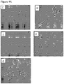
- culturing the cells in a specific growth media (Medium 254 from Life Technologies or equivalent, supplemented with Human Melanocyte Growth Supplement - 2, PMA-free from Life Technologies or equivalent) promotes melanocytes to differentiate from the pool of extracted cells, as shown in Figure 14 .
- Cell survival has been tested after passing suspensions ranging from 10 5 to 10 7 cells/mL through different types of hollow microneedles. Cell survival is near 100% when cells at all these concentrations are injected thorough hollow microneedles with a bore size ⁇ 75 ⁇ m. Cell survival is significantly reduced if the cells are passed through hollow microneedles with smaller bore size, dropping to approximately 50% when the bore size is 50 ⁇ m.
- Cell delivery to skin can be performed efficiently, with cells maintaining their original phenotype once injected, as shown in Figure 15 .
- microneedles We also used prototype microneedles to test the ability of microneedles to extract cells from the skin.
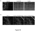
- Microneedle extraction of cells surprisingly allowed us to capture and culture a significant number of skin cells ( Figure 9 ).
- microneedles can deliver cells into relevant skin compartments.
- human keratinocytes were labelled prior to skin injection (excised human breast skin).
- Hollow microneedle delivery of 5000, 50000 and 500000 cells into excised human skin resulted in deposition of the cells in the upper dermis ( Figure 10 ).
- microneedles were used to perform cell extraction from skin. These included commercially available silicon microneedles (A, B, C) and our own proprietary stainless steel microneedles (D, E, F, G).
- This disclosed method therefore paves the way for a new minimally-invasive and pain-free approach wherein cells can be extracted and delivered to the various layers of the skin using microneedles, with little or no recovery time required.
- Type of microneedles used for extraction Length of microneedles Design name Material Number of cells extracted by scraping a 2 cm 2 area Are melanocytes extracted and viable?
Landscapes
- Health & Medical Sciences (AREA)
- Life Sciences & Earth Sciences (AREA)
- Engineering & Computer Science (AREA)
- Veterinary Medicine (AREA)
- General Health & Medical Sciences (AREA)
- Biomedical Technology (AREA)
- Heart & Thoracic Surgery (AREA)
- Public Health (AREA)
- Medical Informatics (AREA)
- Animal Behavior & Ethology (AREA)
- Pathology (AREA)
- Molecular Biology (AREA)
- Surgery (AREA)
- Hematology (AREA)
- Dermatology (AREA)
- Anesthesiology (AREA)
- Materials For Medical Uses (AREA)
- Micro-Organisms Or Cultivation Processes Thereof (AREA)
- Medicines Containing Material From Animals Or Micro-Organisms (AREA)
Description
- The invention relates to a device for use in skin improvement or repair, and a cosmetic method employing the use of same.
- Scientific research has led to extensive developments in cell-based and tissue-based therapies. In recent years, with the advancement of regenerative medicine and tissue engineering, fundamental studies and treatments using living cells have been performed widely.
- The field of regenerative medicine is focussed on regenerating damaged tissues and organs in the body by replacing damaged tissue and/or by stimulating the body's own repair mechanisms to heal previously irreparable tissues or organs. This often involves the use of cells or tissue, in cell or tissue therapy, either from the same person (autologous) or from another donor (allogeneic). There are many potential forms of cell therapy, including: the transplantation of stem cells or progenitor cells; the transplantation of mature, functional cells; and the application of modified human cells that are used to produce a needed substance (cell-based gene therapy).
- Cell based therapy is targeted at many indications, both medical and non-medical, and in multiple organs and tissues using several modes of cell delivery. Accordingly, the specific mechanisms of action involved are wide ranging. However, there are two main principles by which cells facilitate their desired action:
- i) Stem cell or progenitor cell engraftment, differentiation, and long term replacement of damaged tissue. In this paradigm multipotent or unipotent cells differentiate into a specific cell type in the laboratory or after reaching the target site, where they integrate to replace lost tissue, and thus facilitate regeneration and renewal.
- ii) Cells that have the capacity to release cellular factors such as cytokines, chemokines, growth factors or proteins. The delivered cells (via local or systemic administration) remain viable for a relatively short period (days-weeks) and then die. This includes cells that naturally secrete the relevant factors, or which undergo epigenetic changes or genetic engineering that causes the cells to release large quantities of a specific molecule. Examples of this include cells that secrete factors which facilitate angiogenesis, anti-inflammation, and anti-apoptosis.
- Whilst it is widely recognized that such cell-based transplantations have application in the medical field to alleviate disease conditions and disorders, current research has led to the increased application of such techniques for non-medical purpose, including use in the cosmetic industry. Many recent cosmetic products and techniques are based on advanced scientific research that includes the use of biotechnology-derived ingredients, nutritional regimens, stem-cell-based products and therapies to regenerate ageing tissues, or use cell and tissue engineering for cosmetic purposes.
- Through improved understanding of the structure of the skin and its underlying repair and maintenance processes, researchers are increasingly able to intervene to reduce the effects of premature ageing, improve healing processes or 'enhance' the appearance of skin. The cosmetics and pharmaceutical industries have also expended considerable effort to understand the ageing processes of the skin and to devise countermeasures.
- In relation to point ii) above, a recent development is the use and transplantation of one particular skin cell type, the melanin producing cells called melanocytes, which are found in the bottom layer (the stratum basale) of the skin's epidermis. Melanin is the pigment primarily responsible for skin colour, produced through a process called melanogenesis. Numerous cosmetic conditions, or cosmetic effects as a consequence of an underlying disorder, can result in generalized or localized hyperpigmentation (increased skin colour) and hypopigmentation (reduced skin colour) of the skin. Commonly, in the case of localized hypopigmentation, this is attributed to partial or complete loss of melanin such as experienced in:
- Pityriasis alba: a sequelae of eczema in which asymptomatic oval pink scaly patches resolve to leave pale macules for some months or longer. Reduced numbers of active melanocytes and a decrease in number and size of melanosomes are seen in affected skin for unknown reasons;
- Treatment-induced hypopigmentation: Medications and treatments used to treat various skin conditions can result in lightening of the skin. These include dermabrasion, use of chemical peels, and local steroid injections;
- Postinflammatory hypopigmentation (leukoderma): as a consequence of many inflammatory skin conditions, such as skin infection, blisters, bums, cryotherapy, dermal injury with scarring or eczema, loss of pigmentation may occur in the affected area;
- Vitiligo: a condition characterised by chronic and progressive depigmentation of areas of the skin. Vitiligo occurs when melanocytes in the basal layer of the epidermis are defective or die. The cause of vitiligo is unknown, but research suggests that different factors might act independently or synergistically to determine the phenotype, including autoimmunity, genetic susceptibility, viral infection, environmental factors, or oxidative stress;
- Piebaldism: a rare autosomal dominant disorder of melanocyte development and migration. Common characteristics include a congenital white forelock, scattered normal pigmented and hypopigmented macules and a triangular shaped depigmented patch on the forehead;
- Alezzandrini syndrome: a very rare syndrome characterized by a unilateral degenerative retinitis, followed after several months by ipsilateral vitiligo on the face and ipsilateral poliosis; and
- Melanoma-associated leukoderma: a cutaneous condition characterized by vitiligo-like depigmentation that can occur in patients with cutaneous or ocular melanoma.
- Whilst the loss of pigmentation observed in the likes of the above may be of a purely cosmetic concern and only secondary to the underlying pathology leading to same, often the cosmetic dysfunction has a significant negative impact on quality of life. The altered pigmentation is often immediately visible to others and individuals may suffer social and emotional consequences including low self-esteem, social anxiety, relationship problems and depression. Therefore, in addition to finding treatments for the underlying causes of the disease e.g. in the case of vitiligo, it is also paramount to the patient to consider the cosmetic aspects.
- In addition to melanocytes, other skin cell types exist with similar potential utility.
- Keratinocytes constitute around 90% of the cells of the epidermis, where at the skin surface they produce increasing amounts of keratin and synthesize and extrude lipids into the intracellular space. At the top of the epidermis the cells have differentiated to a cell type that are called corneocytes, which form the outermost skin layer and are constantly shed and replaced by new cells. This differentiation process is tightly controlled to maintain the integrity of the physical skin barrier.
- It is known that epidermal melanocytes form a functional and structural unit with neighboring keratinocytes to stimulate melanocyte proliferation, with growth factors produced by adjacent keratinocytes regulating the proliferation and differentiation of melanocytes. Structural changes in keratinocytes may result in loss of melanocytes and evidence suggests that keratinocytes in depigmented epidermis are more vulnerable to apoptosis. Keratinocyte apoptosis will result in reduced expression of keratinocyte-derived factors in depigmented epidermis, resulting in melanocyte death. The relationship between keratinocytes and melanocytes will therefore clearly play a role in skin depigmentation, and the delivery of healthy keratinocytes to depigmented skin, possibly, although not necessarily, in combination with healthy melanocytes (or vice versa), represents a viable strategy for re-pigmentation.
- Epidermal stem cells, such as those in the stratum basale or hair follicle, have attracted similar interest from the cosmetics industry and various companies are already exploring their potential for skin-care products, for example, skin regeneration and repair, or anti-ageing products that contain proteins derived from specialized stem-cell lines that affect specific receptors in both fibroblasts and keratinocytes that increase the production of collagen.
- Dermal fibroblasts produce collagen (type I, type III and type VII), elastin, hyaluronic acid and matrix metalloproteinases and therefore are essential in forming elongated fibres and extracellular matrix. Dermal fibroblasts produce the structural components that unite separate cell layers and allow epithelial cells of the epidermis to join together to form upper skin barrier layers. The cells also allow skin to recover from injury. For this reason, transfer of dermal fibroblasts has been used cosmetically as anti-ageing and scar remodeling products. Autologous (self) fibroblasts are also being used to treat skin wrinkles, scars, folds and depressions and for lip augmentation. The advantage of using fibroblasts over direct collagen injection relates to the breaking down of collagen protein by endogenous enzymes.
- Therefore, the potential for transplantation of cells is readily recognized in the field. A plethora of techniques have thus been developed for delivering cells. For example, to target depigmentation, the transfer and/or culture of target cells, such as keratinocytes, fibroblasts, or melanocytes, from pigmented (normal unaffected skin) to depigmented skin has been tested.
US20100310526 discloses a method for increasing or intensifying the pigmentation of skin, by the application of melanocyte precursor cells from hair root sheaths onto the depigmented area. It is disclosed that keratinocyte precursor cells are also applied to the depigmented area. The procedure requires the recipient site is dermabraded (surgical skin planing) before the application of a cell solution. -
US20120064049 discloses a method for the regeneration of aged skin for cosmetic purposes and for the prevention of skin diseases, using stem cells from hair root sheaths and/or keratinocyte and melanocyte precursor cells. According to this method, before the application of the cell solution the epidermis at the recipient site needs to be physically or mechanically ablated, preferably by means of superficial dermabrasion, laser treatment (fraxel laser), or superficial needle puncture (dermaroller). -
US20110150848 discloses a method for producing a transplantable cellular suspension of living tissue for grafting to a patient using the ReCell® Spray-On Skin™ system. The method requires harvesting donor tissue from the patient (4cm2 biopsy, thickness 150-200 µm), performing in-theatre preparation of a spray-on suspension consisting of cells derived from the biopsy, and applying this suspension immediately over the recipient graft site, on a surface up to 80 times the size of the biopsy. The recipient site needs to be dermabraded or laser-treated before treatment. The ReCell suspension contains basal keratinocytes, melanocytes, fibroblasts and Langerhans cells. The metabolically responsive epithelial cells migrate across the wound surface, leading to regeneration of skin of normal colour and texture.WO 2012/018486 A2 discloses cross sectional dimensions of microneedles that include a vast range of diameters from 50um to 5000um (5mm) i.e. covering two orders of magnitude. Additionally, microneedles are disclosed that include a vast range of lengths from 50um to 5000um (5mm) i.e. covering two orders of magnitude. The device is said to be for insertion into skin to deliver a substance, therapeutics or for receiving fluids such as blood from the skin and/or from beneath the skin. The document teaches fluid to be any bodily fluid but can include solid or semi-solid material such as cells. - Document
CN 103263727 A discloses a microneedle array for painless transdermal delivery in both medical and cosmetic applications. The document discloses a microneedle array comprising a substrate having multiple integral microneedles attached thereto. Said microneedles are disclosed to be 10-1500um long and have a diameter of 3-1000um i.e. a range encompassing three orders of magnitude. The device concerns transdermal drug delivery in both medical and cosmetic applications. However it is apparent that the many existing methods for transferring tissue (skin grafting) or cells have associated inherent problems, including: - Koebner phenomenon: This is the development of disease at sites of trauma. For example if someone with psoriasis scratches themselves, they can develop the phenomenon along the skin that was scratched.
- Scarring: The transfer of skin via grafts or punches leads to defects, trauma and scarring to skin at a second site.
- Skin preparation: Cells can only be transplanted onto prepared skin, i.e. skin that has had its epidermis removed via invasive techniques such as dermabrasion.
- Pain: These invasive techniques can be very painful and are not without risk.
- Infection: Techniques such as dermabrasion cause major barrier defect and increase the risk of infection.
- Skill: Methods are heavily dependent on the skill of a surgeon to carry out the procedure.
- Recovery: current procedures require 1-2 weeks recovery.
- There is a real unmet need to find more efficient, pain free and cost-effective technologies to repair the skin and permit transfer of cells into same thus achieving a better cosmetic outcome (e.g. 80% or more re-pigmentation without scarring).
- Heretofore it has not been shown that cells can be successfully delivered using microneedles. Unexpectedly, the inventors have determined that cells can be injected into surface layers of the skin using fine bore microneedles with no observed loss of viability. Moreover, using a similar such approach, it has been found that cells can be extracted from the skin and that these extracted cells are viable in culture.
- The present invention therefore concerns a new minimally-invasive method for the repair of the skin comprising step a) the extraction and step b) the transfer and delivery of skin cells, such as melanocytes, fibroblasts or keratinocytes, using microneedles (MNs), including the targeting of a cellular suspension of freshly extracted, or cultured, cells to the skin. Notably, different layers of the skin can be targeted as the cells will reposition to the appropriate area.
- Microneedles are micron-sized, needle-like projections often, but not always, organized in an array of a defined geometric pattern on a planar base plate. They are an established technology currently being exploited for the targeted intra-epidermal and intradermal delivery of drugs and vaccines. Due to their microscopic dimensions MNs do not penetrate skin deep enough to cause any significant pain, bleeding or scarring, as demonstrated through numerous clinical trials. Application of MNs to the skin surface results in penetration of the outer skin barrier, the stratum corneum (SC), and the creation of multiple transient micro-pathways that permit the delivery of materials without impinging significantly on nerves or blood vessels.
- Most notably, the micron-scale dimensions of the microneedle shafts allow for simple and direct application into skin that does not require professional training.
- A pilot study by a team at Cardiff University has shown the minimally invasive nature of microneedles, compared with conventional hypodermic injection, demonstrating that microneedles caused significantly less pain than normal needles. Further, advantageously, following withdrawal of the microneedle the induced disruptions in the skin surface rapidly reseal thus leaving minimal or no scarring and minimal barrier defect.
- This disclosed method therefore paves the way for a new minimally-invasive and pain-free approach wherein cells can be extracted and delivered to the various layers of the skin using microneedles, with little or no recovery time required. There would be no need for highly trained surgeons and expensive equipment to perform the procedure, making the procedure more affordable. Additionally, the use of microneedles minimizes scarring with no perceived change in skin texture or pigment, and given the minimal invasiveness of the technology, the need for prior treatment of the targeted areas is circumvented.
- According to a first aspect of the invention there is provided a device for skin improvement or repair as defined by the features of
claim 1. - Reference herein to a circular pattern includes reference to a round or elliptical pattern or indeed in some embodiments a 'circle' with relatively angular corners as provided by a pentagon, hexagon, octagon or the like. Thus reference herein to a circular pattern includes reference to a continuous line of adjacent microneedles which may be separate or contiguous.
- Reference herein to microneedle is reference to any fine, minimally invasive structure or projection typically less than 1mm in length and most suitably, but not necessarily, hollow, most typically but not exclusively said microneedle is also straight or curved, further said microneedle is tapered, barbed, bevelled or hooked at its tip.
- In a preferred embodiment of the invention said base member is adapted for attachment to, or attached to, a manipulating member, such as a handle or syringe, whereby a user can extract cells from said skin, ideally by scraping in a circular fashion or depressing and rotating said device, against a target area of skin.
- More preferably still said microneedles are straight, curved, hooked or barbed. Indeed, any suitable microneedles having the requisite length and diameter to extract and/or inject the desired cell(s) can be used in accordance with the invention herein disclosed. As will be appreciated by those skilled in the art, the nature of said microneedles is to provide optimum extraction and injection, of certain cells resident in certain layers. Therefore, depending upon the nature of the cell to be extracted and/or tissue layer of the skin to be injected, the physical parameters such as, but not limited to, the length and/or diameter of the microneedles can be varied accordingly to achieve the desired technical effect i.e. to penetrate skin to specific and superficial depths. This will also depend accordingly on the region of the body from which the cells are extracted, as the relative skin layer composition can vary from one region to the next, thus routine changes can be made to the needles to ensure the correct skin layers are extracted from and/or injected into.
- According to the invention, where said microneedles are to be used for injection (or replacement) of melanocytes said microneedles are hollow and have a bore size of 75-150 µm diameter needles, including all 1µm intervals there between, most ideally at least 75 µm diameter.
- In a further preferred embodiment of the invention a plurality of concentric circular patterns of microneedles are provided on said base member and, ideally, two or more such circular patterns are provided.
- In yet a further preferred embodiment said microneedles are attached to or integral with said base member so that their longitudinal axis is normal to the supporting axis of said base member. Alternatively, said microneedles are attached to or integral with said base member so that their longitudinal axis is at an angle to the supporting axis of said base member and ideally at an angle that results in said microneedles splaying outwards with respect to the supporting axis of said base member.
- Preferably, between 6 and 48 microneedles, ideally between 12 and 36 microneedles, are used in each circular pattern and, ideally, 24 microneedles are used in one outer concentric circular pattern and 12 microneedles are used in one inner concentric circular pattern. Other preferred numbers of microneedles may be used and a plurality of circular concentric patterns may be provided on said base member.
- According to the invention, said microneedles are about 750 µm in length. More preferably still said microneedles are made from silicon or steel. Alternatively, said microneedles are made from polymers, co-polymers, polysaccharides or sugar materials such as SU-8, PMMA, polycarbonate, carboxymethylcellulose, polycaprolactone, PLGA, dextran, dextrin, PVA, PVP or maltose.
- Reference herein to skin improvement or repair refers to any process whose purpose is to improve functionality of the skin, overcome defects or achieve a desired particular outcome such as appearance or restoration. This may include, but is not limited to, skin re-pigmentation, skin smoothing, skin firming, skin radiance, skin plumping, skin regeneration, improved or enhanced scar and wound repair, improved skin barrier functionality, improved skin elasticity, hair growth, extracellular matrix stimulation including production of collagen, angiogenesis and re-epithelialisation, or the like.
- Reference herein to extraction/extracted is reference to the use of said microneedles to remove cells from the preferred layers of the skin as disclosed herein by, for example but not limited to, withdrawal through said microneedles by negative pressure, absorption of said cells by said microneedles, collection into microneedle bores after skin insertion (a microscopic cell biopsy) or alternatively scraping in a linear fashion a layer of the skin with said microneedles or depressing the microneedles into the skin and moving them in a circular fashion.
- In a further preferred embodiment of the invention said injecting also involves the use of a single microneedle or a plurality of microneedles attached to a supporting base member and where a plurality of microneedles are provided they are arranged in a row, a rectangular array or at least one circular pattern on same, but in this instance, according to the invention, said microneedles are hollow and have a bore size of between 75-150 µm diameter, including all 1 µm intervals there between, most ideally at least 75 µm diameter.
- According to the invention, the same microneedles are used for step a) and step b). Alternatively, different microneedles are used for each step depending upon the cells to be extracted and tissue to be targeted.
- In yet a further preferred embodiment of this aspect of the invention, as will be appreciated by those skilled in the art, said cell could be any one or more cell type resident in the skin and in the superficial layers that could reasonably be expected to be extracted by use of microneedles whilst, ideally, not compromising the cosmetic and pain considerations of the invention as herein disclosed. This includes, but is not limited to, a cell from the epidermal and dermal layers, including a hair follicle. Such cell further include a melanocyte, keratinocyte, dermal fibroblast, comeocyte, Langerhans cell, dermal dendritic cell, epidermal stem cell such as epidermal keratinocyte stem cell, Merkel cell, mast cell, macrophage, T-cell, dermal sheath cell or follicular outer root sheath cell, or the like.
- Most ideally, said cell is selected from the group comprising: a melanocyte, keratinocyte, dermal fibroblast, epidermal stem cell and follicular outer root sheath cell.
- In yet a further preferred embodiment of this aspect of the invention, said method comprises the extraction and/or injection of a single cell type from the skin surface such as but not limited to melanocyte or a number of melanocytes. Alternatively, and more ideally, said method comprises the extraction and/or injection of at least two cell types, such as but not limited to a combination of a melanocyte and keratinocyte and, ideally a combination of a melanocytes and keratinocytes.
- In a preferred embodiment of this aspect of the invention, said cell(s) is/are injected into the viable epidermis, papillary dermis and reticular dermis layers. However, as will be appreciated by those skilled in the art, depending upon the nature of the cell to be injected and the result to be achieved, said cell can be injected into any layer of the skin with equal effect. For example, in the case of the injection of one or more melanocytes for skin re-pigmentation said cells would be preferably injected into the supra basal epidermal layer.
- In a preferred method of the invention steps a) and b) are performed sequentially and step b) is undertaken after or shortly after step a) has been undertaken.
- In a preferred embodiment of this aspect of the invention, said method comprises extracting at least one cell, or more ideally, a plurality of cells.
- In the case where a plurality of cells are extracted, advantageously said cells can be separated further into the single cell types using techniques well known to those skilled in the art.
- In yet a further preferred embodiment of the second aspect of the invention, steps a) and b) are performed on the same person i.e. said cell(s) injected in step b) is/are autologous (from the same individual as the extracted cell(s) in step a).
- Alternatively, steps a) and b) are performed on different people i.e. said cell(s) injected in step b) is/are allogeneic (from a different individual to that/those from whom the cell(s) in step a) was/were extracted). Additionally, said cell(s) extracted in step a) may be xenogeneic (i.e. cells extracted from different species), such as but not limited to mammals, and injected into the skin of a different animal such as a human. As will be appreciated by those skilled in the art, such cell(s) will be need to be immunologically compatible, or modified such that they do not elicit an immunogenic response.
- Most preferably, said cell(s) in step b) is/are autologous to the cell(s) with respect to step a).
- In yet a further preferred embodiment of the invention, said method further comprises the step of in vitro/ex vivo culturing the cell(s) extracted in step a) prior to their injection in step b) into a patient/person. As will be appreciated by those skilled in the art, certain uses may require a greater number of cells to be injected than can be extracted, or the outcome of the method may be more favourable if a greater number of cells is used. Therefore, cells can be cultured and expanded in vitro using cell culture techniques well known to those skilled in the art. Additionally, said culture can be used to select the cells that are desired to be grown, such as through the use of selective growth media, to promote culture of a selected cell type, which is dependent upon said pre-selected culture conditions.
- Additionally, as will be appreciated by those skilled in the field, said cells can be stimulated with appropriate cell culture factors, supplements, proteins, growth factors or the like to result in a preferred cell phenotype and/or differentiation of the cells into a pre-selected phenotype, which phenotype is dependent upon said pre-selected culture conditions.
- Further, said cells may be genetically modified such that they show altered protein expression (e.g. increased melanin or collagen expression) or reduced immunogenicity or the like, using techniques known to those skilled in the art such as recombinant nucleic acid (DNA or RNA) techniques to incorporate recombinant material into said cells either indirectly through a vector system or directly through micro-injection, macro-injection and micro-encapsulation techniques.
- According to yet a further preferred second aspect of the invention, said method in steps a) and b) are repeated for all areas of the skin for which improvement or repair is desired. Moreover, said method can be repeated as necessary for the same area after a defined time period in order to improve or maintain the overall outcome desired.
- According to yet a further preferred method of the invention, said extracted and/or cultured cells are preserved for long term use using any suitable means known to those skilled in the art, such as but not limited to, liquid nitrogen storage. Advantageously, in this further preferred method of the invention, said preserved cells can be used for repeated procedures whereby measured amounts of the preserved cells are repeatedly used under step b) above to effect improvement or repair of skin. In this way the need to repeatedly undertake step a) above is circumvented.
- Throughout the description and claims of this specification, the words "comprise" and "contain" and variations of the words, for example "comprising" and "comprises", mean "including but not limited to" and do not exclude other moieties, additives, components, integers or steps. Throughout the description and claims of this specification, the singular encompasses the plural unless the context otherwise requires. In particular, where the indefinite article is used, the specification is to be understood as contemplating plurality as well as singularity, unless the context requires otherwise.
- Preferred features of each aspect of the invention may be as described in connection with any of the other aspects.
- Other features of the present invention will become apparent from the following examples. Generally speaking, the invention extends to any novel one, or any novel combination, of the features disclosed in this specification (including the accompanying claims and drawings). Thus, features, integers, characteristics, compounds or chemical moieties described in conjunction with a particular aspect, embodiment or example of the invention are to be understood to be applicable to any other aspect, embodiment or example described herein, unless incompatible therewith.
- Moreover, unless stated otherwise, any feature disclosed herein may be replaced by an alternative feature serving the same or a similar purpose. References to embodiments throughout the description which are not under the scope of the appended claims merely represent possible exemplary executions and are therefore not part of the present invention.
- The Invention will now be described by way of example only with reference to the Examples below and to the following Figures wherein:
-
Figure 1 . Methylene blue staining to evaluate the diffusion of liquid through skin following microneedle injection; -
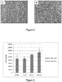
Figure 2 . [A] Keratinocytes in culture, with the typical cobblestone appearance. [B] Melanocytes in culture, with the typical dendritic phenotype; -
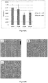
Figure 3 . Number of total keratinocytes counted after passing through microneedles with different bore sizes, divided into alive (blue) and dead (red) cells. The error bars represent the standard error on the means obtained on 3 repeats; -
Figure 4 . Number of total melanocytes counted after passing through microneedles with different bore sizes, divided into alive (blue) and dead (red) cells. The error bars represent the standard error on the means obtained on 3 repeats; -
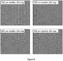
Figure 5 . Keratinocytes in culture at 48h after passing through microneedles with different bore sizes: 75 µm [A], 100 µm [B] and 150 µm [C]; -
Figure 6 . Melanocytes in culture at 48h after passing through microneedles with different bore sizes: 75 µm [A], 100 µm [B] and 150 µm [C]; -
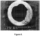
Figure 7 . Hooked microneedles for cell extraction before (top row) and after (bottom row) skin penetration, viewed using Scanning Electron Microscopy; -
Figure 8 . Use of hollow microneedles for cell extraction, viewed using Scanning Electron Microscopy; -
Figure 9 . Skin cells in culture at 96h after collection from human skin by microneedle scraping; -
Figure 10 . Human keratinocyte cells (stained blue with Hoechst staining) injected using microneedles at a concentration of 500,000 cells/50 µl into excised human breast skin. Skin was imaged using light and fluorescence microscopy. Skin autofluorescence appears as green. The white line denotes the skin surface; -
Figure 11 . Shows pictures of the proprietary designs used to work the invention; -
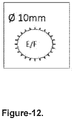
Figure 12 . Arrangement of microneedles on a supporting base member Design E and F: Disk diameter = 10 mm; Number of needles per disk = 24; Needles length = 750 µm; Material: Stainless Steel; In design E the needles are in plane; In design F the needles are bent out of plane; Needles manufactured by wire Electrical Discharge Machining (EDM); -
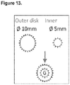
Figure 13 . A further arrangement of microneedles on a supporting base member, Design G: Outer disk diameter = 10 mm; Number of needles in the outer disk = 24; Inner disk diameter = 5 mm; Number of needles in the inner disk = 12; Total number of needles = 36; Needles length = 750 µm; Material: Stainless steel; The needles are all bent out of plane; Manufactured by wire EDM; The arrays are stacked and so the number of needles could be expanded easily; -
Figure 14 . Shows images of the survival of extracted cells after passing through microneedles was also assessed. All the cells survived after passing through hollow microneedles with a bore size ≥ 75 µm; -
Figure 15 . Upper image shows that Hoechst labelled cells (blue nuclei) were injected in skin explants and successfully delivered to the dermis; lower image shows that the injected cells (blue nuclei) maintained their phenotype after injection in skin. Cells with a green cytoplasm are melanocytes, while cells with a red cytoplasm are keratinocytes; and - Table 1 contains information relating to the efficiency of cell extraction using the specified microneedles, together with the technical features of each microneedle design.
- Melanocytes and keratinocytes were isolated from non-affected skin (biopsy or cell scraping) or from the hair follicles. These were used directly or cultured to increase cell numbers.
- Additionally, microneedle injection of primary melanocytes and keratinocytes obtained from commercial sources (Life Technologies) was also undertaken.
- Melanocytes and keratinocytes were expanded in vitro, using selective growth media.
- For melanocytes, the selective growth media was Medium 254 (Life Technologies) or equivalent, supplemented with Human Melanocyte Growth Supplement - 2, PMA-free (Life Technologies) or equivalent.
- For keratinocytes, the selective growth media was Epi-Life Medium (Life Technologies) or equivalent, supplemented with Human Keratinocyte Growth Supplement (Life Technologies) or equivalent.
- Cells were incubated at 37°C in 5% CO2 and media replaced every 48 to 72h until a sufficient number of cells were obtained (determined depending on the extension of the area to be treated, approximately 105 melanocytes/cm2).
- Extraction of viable live cells from the skin was assessed using light microscopy.
- Cells were delivered to freshly excised human breast skin. A fraction of the cell culture was also stored in liquid nitrogen for subsequent application if needed, removing the need to repeat the isolation step.
- Cells were delivered to the recipient site by the use of specifically designed microneedles. According to preliminary studies, using a marker dye instead of a cell suspension (
Figure 1 ), approximately 100 µL of a 106 cells/mL solution was sufficient to re-pigment a skin area of approximately 1 cm2, delivering 105 cells to the area. - The microneedles used were hollow silicon microneedles, arranged in an array, to cover an area of approximately 0.5 cm2.
- Microneedles had a bore size between 75 and 150 µm, a wall thickness between 50 and 150 µm, spacing between 500 and 1000 µm, and a length between 300 and 700 µm.
- Different types of microneedles were used to perform cell extraction from skin. These included commercially available silicon microneedles (A, B, C) and our proprietary stainless steel microneedles (D, E, F, G).
- Pictures of the proprietary stainless steel microneedle designs (D, E, F, G) are shown in
Figure 11 . The specifications of these needles are as follow. - Design D: Array width = 1.1 cm;
Number of needles per array = 10;
Needles length = between 350 and 420 µm;
Number of arrays stacked = 3;
Total number of microneedles = 30;
Material: Stainless Steel. Seefigure 11D (made by Electrical Discharge Machining (EDM))
Design E and F: Disk diameter = 10 mm;
Number of needles per disk = 24;
Needles length = 750 µm;
Material: Stainless Steel;
In design E the needles are in plane;
In design F the needles are bent out of plane; - Needles manufactured by wire Electrical Discharge Machining (EDM). See
Figure 11E and F , andFigure 12 . - Design G: Outer disk diameter = 10 mm;
Number of needles in the outer disk = 24;
Inner disk diameter = 5 mm;
Number of needles in the inner disk = 12;
Total number of needles = 36;
Needles length = 750 µm;
Material: Stainless steel;
The needles are all bent out of plane;
Manufactured by wire EDM;
The arrays are stacked and so the number of needles could be expanded easily. SeeFigure 11G andFigure 13 . - Design A, B, C, and D: A 2 cm2 area of the skin surface is scraped multiple time with a linear movement (left to right and right to left).
- Design E: The disk is rolled multiple times on a 2 cm2 area of the skin surface.
- Design F and G: A 2 cm2 area of the skin surface is scraped multiple times with a circular movement (clockwise).
- After scraping, the material collected on the microneedles is transferred to a tube containing cold trypsin and incubated overnight at 4°C. The following day, the trypsin is inactivated with the same volume of serum. The cell containing solution is then filtered and centrifuged, and cells are resuspended, ready for injection.
- With this method, different types of cells are extracted, including keratinocytes, melanocytes, fibroblasts, Merkel cells, Langerhans cells, macrophages, adipocytes, dendritic cells, etc.
- The efficiency of cell extraction using these microneedles was assessed and it is reported in table 1 below, together with the technical features of each microneedle design.
- After extraction, culturing the cells in a specific growth media (Medium 254 from Life Technologies or equivalent, supplemented with Human Melanocyte Growth Supplement - 2, PMA-free from Life Technologies or equivalent) promotes melanocytes to differentiate from the pool of extracted cells, as shown in
Figure 14 . - The survival of extracted cells after passing through microneedles was also assessed. All the cells survived after passing through hollow microneedles with a bore size ≥ 75 µm.
- Cell survival has been tested after passing suspensions ranging from 105 to 107 cells/mL through different types of hollow microneedles. Cell survival is near 100% when cells at all these concentrations are injected thorough hollow microneedles with a bore size ≥ 75 µm. Cell survival is significantly reduced if the cells are passed through hollow microneedles with smaller bore size, dropping to approximately 50% when the bore size is 50 µm.
- Cell adhesion, cell proliferation, and cell phenotype are maintained after passing through microneedles with a bore size ≥ 75 µm at all the concentrations tested.
- Cell delivery to skin can be performed efficiently, with cells maintaining their original phenotype once injected, as shown in
Figure 15 . - Cell cultures from commercially available human epidermal keratinocytes and melanocytes were established, and culturing conditions were optimized (
Figure 2 ). - Cell survival after passing through microneedles with different bore diameters (50, 60, 75, 100 and 150 µm) was tested using a 105, 108, 2×10⁶ and 107 cells/mL suspension. Trypan Blue staining was used to confirm cell viability. Both keratinocytes (
Figure 3 ) and melanocytes (Figure 4 ) survived the procedure with all the concentrations tested when the bore diameter was greater than 60 µm. There was also no loss of cells during the procedure, showing that cells are not retained inside the microneedles. - The ability of cells to adhere to a surface and proliferate after passing through microneedles with different bore sizes (75, 100 and 150 µm) was tested. Both keratinocytes (
Figure 5 ) and melanocytes (Figure 6 ) were able to attach to culture plates and proliferate after the procedure. - We also used prototype microneedles to test the ability of microneedles to extract cells from the skin.
- Firstly, we tested the ability of hooked stainless steel microneedles to extract skin cells after skin penetration (excised human breast skin) and removal (
Figure 7 ). - We also tested hollow microneedles using the same approach (
Figure 8 ). - Subsequently, as proof-of-concept, we also tested the ability of different types of solid and hollow microneedles to collect skin cells by scraping along the skin's surface (excised human breast skin). After scraping the skin, cells were detached from the microneedles by rinsing in culture media and captured in a culture plate.
- Microneedle extraction of cells surprisingly allowed us to capture and culture a significant number of skin cells (
Figure 9 ). - In further studies we tested whether microneedles can deliver cells into relevant skin compartments. In these studies human keratinocytes were labelled prior to skin injection (excised human breast skin). Hollow microneedle delivery of 5000, 50000 and 500000 cells into excised human skin resulted in deposition of the cells in the upper dermis (
Figure 10 ). - Different types of microneedles were used to perform cell extraction from skin. These included commercially available silicon microneedles (A, B, C) and our own proprietary stainless steel microneedles (D, E, F, G).
- Pictures of the stainless steel proprietary designs are shown in
Figure 11 with schematics showing the microneedles designs used in E, F and G shown inFigures 12 and13 . - After extraction, culturing the captured cells in a specific growth media promotes melanocytes to differentiate from the pool of extracted cells, as shown in
Figure 14 for designs C, D, E, F and G. The survival of extracted cells after passing through microneedles was also assessed. All the cells survived after passing through hollow microneedles with a bore size ≥ 75 µm. - Cell adhesion, cell proliferation, and cell phenotype are maintained after passing through microneedles with a bore size ≥ 75 µm at all the concentrations tested.
- As further evidence of efficient cell delivery using microneedles, cells (with a blue nuclear cell staining) maintain their original phenotype once injected, as shown in
Figure 15 . - In summary our studies have shown that:
- 1) Cells can be collected from skin using microneedles;
- 2) Cells can be injected through hollow microneedles with a variety of bore diameters. All of the cells are injected with none retained, with high viability (cell survival is near 100%);
- 3) Cells can be injected into skin using microneedles.
- This disclosed method therefore paves the way for a new minimally-invasive and pain-free approach wherein cells can be extracted and delivered to the various layers of the skin using microneedles, with little or no recovery time required.
Type of microneedles used for extraction Length of microneedles Design name Material Number of cells extracted by scraping a 2 cm2 area Are melanocytes extracted and viable? Hollow, 50 µm bore size 450 µm A Silicon 5×10⁵ NO Hollow, 60 µm bore size 600 µm B Silicon 4×10⁵ NO Hollow, 80 µm bore size 750 µm C Silicon 6×10⁵ YES 2 cells (0.0003%) Solid, 3 rows of 10 microneedles 350 to 420 µm D Steel 5×10⁵ Yes 12 cells (0.0024%) Solid, disk of 24 needles - in plane 750 µm E Steel 6×10⁵ YES 2 cells (0.0003%) Solid, disk of 24 needles - out of plane 750 µm F Steel 8×10⁵ YES approximately 120 cells (0.015%) Solid, 2 concentric disks, total of 36 needles - out of plane 750 µm G Steel 1×10⁶ YES approximately 300 cells (0.03%)
Claims (8)
- A device for skin improvement or repair comprising: a plurality of microneedles attached to or integral with a supporting base member and arranged in at least one circular pattern on same wherein said microneedles are hollow and have a bore size of between 75-150 µm and are about 750 µm in length.
- The device according to claim 1 wherein said base member is adapted for attachment to, or attached to, a manipulating member.
- The device according to any one of the preceding claims wherein a plurality of concentric circular patterns of microneedles is provided on said base member.
- The device according to claim 3 wherein two concentric circular patterns of miconeedles are provided on said base member.
- The device according to any one of the preceding claims wherein said microneedles are attached to or integral with said base member so that their longitudinal axis is normal to the supporting axis of said base member or so that their longitudinal axis is at an angle to the supporting axis of said base member such that said microneedles splay outwards with respect to the supporting axis of said base member.
- The device according to any one of the preceding claims wherein between 6 and 48 microneedles are used in each of said circular pattern(s).
- The device according to any one of claims 1-5 wherein 24 microneedles are used in an outer concentric circular pattern and 12 microneedles are used in an inner concentric circular pattern.
- The device according to any one of the preceding claims wherein said microneedles are made from a polymer, co-polymer, polysaccharide, sugar, silicon or steel
Applications Claiming Priority (2)
| Application Number | Priority Date | Filing Date | Title |
|---|---|---|---|
| GBGB1403773.3A GB201403773D0 (en) | 2014-03-04 | 2014-03-04 | Microneedle based cell delivery |
| PCT/GB2015/050594 WO2015132568A1 (en) | 2014-03-04 | 2015-03-02 | Microneedle based cell delivery |
Publications (3)
| Publication Number | Publication Date |
|---|---|
| EP3113830A1 EP3113830A1 (en) | 2017-01-11 |
| EP3113830B1 EP3113830B1 (en) | 2019-12-18 |
| EP3113830B2 true EP3113830B2 (en) | 2022-10-05 |
Family
ID=50490748
Family Applications (1)
| Application Number | Title | Priority Date | Filing Date |
|---|---|---|---|
| EP15707761.1A Active EP3113830B2 (en) | 2014-03-04 | 2015-03-02 | Microneedle based cell delivery |
Country Status (5)
| Country | Link |
|---|---|
| US (1) | US10232159B2 (en) |
| EP (1) | EP3113830B2 (en) |
| AU (1) | AU2015225914B2 (en) |
| GB (1) | GB201403773D0 (en) |
| WO (1) | WO2015132568A1 (en) |
Families Citing this family (7)
| Publication number | Priority date | Publication date | Assignee | Title |
|---|---|---|---|---|
| US10893865B2 (en) * | 2015-04-13 | 2021-01-19 | Mayo Foundation For Medical Education And Research | Magnetic wound closure systems |
| WO2021021587A1 (en) * | 2019-07-26 | 2021-02-04 | University Of Florida Research Foundation | Calibrated pneumatic otoscope |
| JP2023523952A (en) | 2020-04-28 | 2023-06-08 | ティコナ・エルエルシー | microneedle assembly |
| US11951272B2 (en) | 2020-08-28 | 2024-04-09 | City University Of Hong Kong | Cryo formulation-based microneedle device for ocular delivery of bioactive therapeutic agents using a cryo-microneedle patch |
| US12005221B2 (en) | 2020-08-28 | 2024-06-11 | City University Of Hong Kong | Cryo formulation-based microneedle device for transdermal delivery of bioactive therapeutic agents and cancer immunotherapy using a cryo-microneedle patch |
| US11904126B2 (en) | 2020-08-28 | 2024-02-20 | City University Of Hong Kong | Cryo formulation-based microneedle device for transdermal delivery of bioactive therapeutic agents and performing vaccination using a cryo-microneedle patch |
| CN117721000A (en) * | 2023-11-13 | 2024-03-19 | 辛伯至生物科技(浙江)有限责任公司 | Device and method for the precise extraction of specific cells from a population of cells or biological tissue grown in a culture dish |
Citations (8)
| Publication number | Priority date | Publication date | Assignee | Title |
|---|---|---|---|---|
| US20040094503A1 (en) † | 2002-11-14 | 2004-05-20 | Gennady Ozeryansky | Microfabrication method based on metal matrix composite technology |
| WO2007035843A2 (en) † | 2005-09-21 | 2007-03-29 | Dask Technologies, Llc | Methods and compositions for organ and tissue functionality |
| US20070225779A1 (en) † | 2006-03-07 | 2007-09-27 | Reliant Technologies, Inc. | Treatment of vitiligo by micropore delivery of cells |
| WO2010059605A2 (en) † | 2008-11-18 | 2010-05-27 | 3M Innovative Properties Company | Hollow microneedle array and method |
| WO2011034627A2 (en) † | 2009-09-21 | 2011-03-24 | Harvard Bioscience, Inc. | Methods and apparatus for introducing cells at a tissue site |
| US20110306853A1 (en) † | 2010-03-19 | 2011-12-15 | Michael Darryl Black | Body fluid sampling/fluid delivery device |
| WO2012018486A2 (en) † | 2010-07-26 | 2012-02-09 | Seventh Sense Biosystems, Inc. | Rapid delivery and/or receiving of fluids |
| CN103263727A (en) † | 2013-05-22 | 2013-08-28 | 清华大学 | Metal microneedle array, percutaneous medicine feeding paster, Dermaroller and microneedle electrode array |
Family Cites Families (9)
| Publication number | Priority date | Publication date | Assignee | Title |
|---|---|---|---|---|
| EP1086214B1 (en) * | 1998-06-10 | 2009-11-25 | Georgia Tech Research Corporation | Microneedle devices and methods of their manufacture |
| US6743211B1 (en) * | 1999-11-23 | 2004-06-01 | Georgia Tech Research Corporation | Devices and methods for enhanced microneedle penetration of biological barriers |
| US6607513B1 (en) * | 2000-06-08 | 2003-08-19 | Becton, Dickinson And Company | Device for withdrawing or administering a substance and method of manufacturing a device |
| US8361037B2 (en) * | 2001-09-19 | 2013-01-29 | Valeritas, Inc. | Microneedles, microneedle arrays, and systems and methods relating to same |
| US7429258B2 (en) * | 2001-10-26 | 2008-09-30 | Massachusetts Institute Of Technology | Microneedle transport device |
| US7422567B2 (en) * | 2002-08-29 | 2008-09-09 | Becton, Dickinson And Company | Microabrader with controlled abrasion features |
| ES2759856T3 (en) * | 2004-04-12 | 2020-05-12 | Allergan Inc | Multi-site injection system |
| US20120201786A1 (en) * | 2010-07-15 | 2012-08-09 | Nikolai Tankovich | Methods for the use of stem cells and stem cell factors in the treatment of skin conditions |
| JP6243839B2 (en) * | 2011-06-29 | 2017-12-06 | バイオリストーラティブ セラピーズ, インコーポレイテッド | Brown adipocyte composition and method |
-
2014
- 2014-03-04 GB GBGB1403773.3A patent/GB201403773D0/en not_active Ceased
-
2015
- 2015-03-02 AU AU2015225914A patent/AU2015225914B2/en not_active Ceased
- 2015-03-02 US US15/122,664 patent/US10232159B2/en active Active
- 2015-03-02 WO PCT/GB2015/050594 patent/WO2015132568A1/en not_active Ceased
- 2015-03-02 EP EP15707761.1A patent/EP3113830B2/en active Active
Patent Citations (8)
| Publication number | Priority date | Publication date | Assignee | Title |
|---|---|---|---|---|
| US20040094503A1 (en) † | 2002-11-14 | 2004-05-20 | Gennady Ozeryansky | Microfabrication method based on metal matrix composite technology |
| WO2007035843A2 (en) † | 2005-09-21 | 2007-03-29 | Dask Technologies, Llc | Methods and compositions for organ and tissue functionality |
| US20070225779A1 (en) † | 2006-03-07 | 2007-09-27 | Reliant Technologies, Inc. | Treatment of vitiligo by micropore delivery of cells |
| WO2010059605A2 (en) † | 2008-11-18 | 2010-05-27 | 3M Innovative Properties Company | Hollow microneedle array and method |
| WO2011034627A2 (en) † | 2009-09-21 | 2011-03-24 | Harvard Bioscience, Inc. | Methods and apparatus for introducing cells at a tissue site |
| US20110306853A1 (en) † | 2010-03-19 | 2011-12-15 | Michael Darryl Black | Body fluid sampling/fluid delivery device |
| WO2012018486A2 (en) † | 2010-07-26 | 2012-02-09 | Seventh Sense Biosystems, Inc. | Rapid delivery and/or receiving of fluids |
| CN103263727A (en) † | 2013-05-22 | 2013-08-28 | 清华大学 | Metal microneedle array, percutaneous medicine feeding paster, Dermaroller and microneedle electrode array |
Also Published As
| Publication number | Publication date |
|---|---|
| GB201403773D0 (en) | 2014-04-16 |
| AU2015225914B2 (en) | 2020-01-30 |
| US10232159B2 (en) | 2019-03-19 |
| US20170065803A1 (en) | 2017-03-09 |
| AU2015225914A1 (en) | 2016-10-06 |
| EP3113830A1 (en) | 2017-01-11 |
| WO2015132568A1 (en) | 2015-09-11 |
| EP3113830B1 (en) | 2019-12-18 |
Similar Documents
| Publication | Publication Date | Title |
|---|---|---|
| EP3113830B2 (en) | Microneedle based cell delivery | |
| CN112336749B (en) | Stem cell exosome microneedle patch for removing freckles and wrinkles and preparation method thereof | |
| JP2022502032A (en) | Composition for skin regeneration and wound healing containing induced exosomes | |
| CN104288129A (en) | Stem cell micro-needle patch for resisting wrinkles and removing freckles and preparation method thereof | |
| Chen et al. | Gas-propelled anti-hair follicle aging microneedle patch for the treatment of androgenetic alopecia | |
| Wang et al. | Management of the refractory vitiligo patient: current therapeutic strategies and future options | |
| CN114401718A (en) | YAP inhibition for wound healing | |
| Xing et al. | Microenvironment-responsive recombinant collagen XVII-based composite microneedles for the treatment of androgenetic alopecia | |
| RU2498809C2 (en) | Using stem cells of hair roots and progenitor (prodormal) keratinocyte cells for aged skin recovery | |
| US10918590B2 (en) | Fat stem cell attractant-containing agent for improving skin looseness or aging caused by dermal cavitation | |
| Semsarzadeh et al. | Rise of stem cell therapies in aesthetics | |
| CN121969415A (en) | Acupuncture devices, beauty kits and beauty methods | |
| Ghasemi et al. | Hair follicle as a source of pigment-producing cells for treatment of vitiligo: an alternative to epidermis? | |
| CN110709506A (en) | Mesenchymal stem cell guiding agent | |
| US20160287855A1 (en) | Surgical methods/devices for tissue injury removal by tattooing of autologous stem cells | |
| Zhang et al. | Microneedle systems: cell, exosome, and nucleic acid based strategies | |
| CN103120827A (en) | Micro-needle roller device | |
| US20130197480A1 (en) | Methods and systems for delivery of live cells | |
| Xing et al. | Borate-modified recombinant type XVII collagen microneedles loaded with IGF-1 for the treatment of androgenetic alopecia | |
| CN117243886A (en) | A microneedle patch for treating diabetic lower limb ischemic lesions and its application | |
| CN109718195A (en) | Facial mask liquid containing stem cell factor | |
| CN115678833A (en) | Novel technical method for treating chloasma through stem cell transplantation operation | |
| TWI674104B (en) | Skin sagging or aging improver due to dermal hollowing out of adipose stem cell attractant | |
| Bambale et al. | Exploring Novel Treatments for Alopecia: Nanoparticles, Iontophoresis, and Cell Therapy | |
| US20200255808A1 (en) | Method for producing a skin equivalent, and use thereof for in vitro tests and in vivo transplants |
Legal Events
| Date | Code | Title | Description |
|---|---|---|---|
| STAA | Information on the status of an ep patent application or granted ep patent |
Free format text: STATUS: THE INTERNATIONAL PUBLICATION HAS BEEN MADE |
|
| PUAI | Public reference made under article 153(3) epc to a published international application that has entered the european phase |
Free format text: ORIGINAL CODE: 0009012 |
|
| STAA | Information on the status of an ep patent application or granted ep patent |
Free format text: STATUS: REQUEST FOR EXAMINATION WAS MADE |
|
| 17P | Request for examination filed |
Effective date: 20160920 |
|
| AK | Designated contracting states |
Kind code of ref document: A1 Designated state(s): AL AT BE BG CH CY CZ DE DK EE ES FI FR GB GR HR HU IE IS IT LI LT LU LV MC MK MT NL NO PL PT RO RS SE SI SK SM TR |
|
| AX | Request for extension of the european patent |
Extension state: BA ME |
|
| DAV | Request for validation of the european patent (deleted) | ||
| DAX | Request for extension of the european patent (deleted) | ||
| GRAP | Despatch of communication of intention to grant a patent |
Free format text: ORIGINAL CODE: EPIDOSNIGR1 |
|
| STAA | Information on the status of an ep patent application or granted ep patent |
Free format text: STATUS: GRANT OF PATENT IS INTENDED |
|
| INTG | Intention to grant announced |
Effective date: 20190802 |
|
| GRAS | Grant fee paid |
Free format text: ORIGINAL CODE: EPIDOSNIGR3 |
|
| GRAA | (expected) grant |
Free format text: ORIGINAL CODE: 0009210 |
|
| STAA | Information on the status of an ep patent application or granted ep patent |
Free format text: STATUS: THE PATENT HAS BEEN GRANTED |
|
| AK | Designated contracting states |
Kind code of ref document: B1 Designated state(s): AL AT BE BG CH CY CZ DE DK EE ES FI FR GB GR HR HU IE IS IT LI LT LU LV MC MK MT NL NO PL PT RO RS SE SI SK SM TR |
|
| REG | Reference to a national code |
Ref country code: CH Ref legal event code: EP |
|
| REG | Reference to a national code |
Ref country code: IE Ref legal event code: FG4D |
|
| REG | Reference to a national code |
Ref country code: DE Ref legal event code: R096 Ref document number: 602015043804 Country of ref document: DE |
|
| REG | Reference to a national code |
Ref country code: AT Ref legal event code: REF Ref document number: 1213914 Country of ref document: AT Kind code of ref document: T Effective date: 20200115 |
|
| REG | Reference to a national code |
Ref country code: NL Ref legal event code: MP Effective date: 20191218 |
|
| PG25 | Lapsed in a contracting state [announced via postgrant information from national office to epo] |
Ref country code: LT Free format text: LAPSE BECAUSE OF FAILURE TO SUBMIT A TRANSLATION OF THE DESCRIPTION OR TO PAY THE FEE WITHIN THE PRESCRIBED TIME-LIMIT Effective date: 20191218 Ref country code: FI Free format text: LAPSE BECAUSE OF FAILURE TO SUBMIT A TRANSLATION OF THE DESCRIPTION OR TO PAY THE FEE WITHIN THE PRESCRIBED TIME-LIMIT Effective date: 20191218 Ref country code: BG Free format text: LAPSE BECAUSE OF FAILURE TO SUBMIT A TRANSLATION OF THE DESCRIPTION OR TO PAY THE FEE WITHIN THE PRESCRIBED TIME-LIMIT Effective date: 20200318 Ref country code: GR Free format text: LAPSE BECAUSE OF FAILURE TO SUBMIT A TRANSLATION OF THE DESCRIPTION OR TO PAY THE FEE WITHIN THE PRESCRIBED TIME-LIMIT Effective date: 20200319 Ref country code: NO Free format text: LAPSE BECAUSE OF FAILURE TO SUBMIT A TRANSLATION OF THE DESCRIPTION OR TO PAY THE FEE WITHIN THE PRESCRIBED TIME-LIMIT Effective date: 20200318 Ref country code: SE Free format text: LAPSE BECAUSE OF FAILURE TO SUBMIT A TRANSLATION OF THE DESCRIPTION OR TO PAY THE FEE WITHIN THE PRESCRIBED TIME-LIMIT Effective date: 20191218 Ref country code: LV Free format text: LAPSE BECAUSE OF FAILURE TO SUBMIT A TRANSLATION OF THE DESCRIPTION OR TO PAY THE FEE WITHIN THE PRESCRIBED TIME-LIMIT Effective date: 20191218 |
|
| REG | Reference to a national code |
Ref country code: LT Ref legal event code: MG4D |
|
| PG25 | Lapsed in a contracting state [announced via postgrant information from national office to epo] |
Ref country code: HR Free format text: LAPSE BECAUSE OF FAILURE TO SUBMIT A TRANSLATION OF THE DESCRIPTION OR TO PAY THE FEE WITHIN THE PRESCRIBED TIME-LIMIT Effective date: 20191218 Ref country code: RS Free format text: LAPSE BECAUSE OF FAILURE TO SUBMIT A TRANSLATION OF THE DESCRIPTION OR TO PAY THE FEE WITHIN THE PRESCRIBED TIME-LIMIT Effective date: 20191218 |
|
| PG25 | Lapsed in a contracting state [announced via postgrant information from national office to epo] |
Ref country code: AL Free format text: LAPSE BECAUSE OF FAILURE TO SUBMIT A TRANSLATION OF THE DESCRIPTION OR TO PAY THE FEE WITHIN THE PRESCRIBED TIME-LIMIT Effective date: 20191218 |
|
| PG25 | Lapsed in a contracting state [announced via postgrant information from national office to epo] |
Ref country code: RO Free format text: LAPSE BECAUSE OF FAILURE TO SUBMIT A TRANSLATION OF THE DESCRIPTION OR TO PAY THE FEE WITHIN THE PRESCRIBED TIME-LIMIT Effective date: 20191218 Ref country code: CZ Free format text: LAPSE BECAUSE OF FAILURE TO SUBMIT A TRANSLATION OF THE DESCRIPTION OR TO PAY THE FEE WITHIN THE PRESCRIBED TIME-LIMIT Effective date: 20191218 Ref country code: PT Free format text: LAPSE BECAUSE OF FAILURE TO SUBMIT A TRANSLATION OF THE DESCRIPTION OR TO PAY THE FEE WITHIN THE PRESCRIBED TIME-LIMIT Effective date: 20200513 Ref country code: EE Free format text: LAPSE BECAUSE OF FAILURE TO SUBMIT A TRANSLATION OF THE DESCRIPTION OR TO PAY THE FEE WITHIN THE PRESCRIBED TIME-LIMIT Effective date: 20191218 Ref country code: NL Free format text: LAPSE BECAUSE OF FAILURE TO SUBMIT A TRANSLATION OF THE DESCRIPTION OR TO PAY THE FEE WITHIN THE PRESCRIBED TIME-LIMIT Effective date: 20191218 |
|
| PG25 | Lapsed in a contracting state [announced via postgrant information from national office to epo] |
Ref country code: SK Free format text: LAPSE BECAUSE OF FAILURE TO SUBMIT A TRANSLATION OF THE DESCRIPTION OR TO PAY THE FEE WITHIN THE PRESCRIBED TIME-LIMIT Effective date: 20191218 Ref country code: SM Free format text: LAPSE BECAUSE OF FAILURE TO SUBMIT A TRANSLATION OF THE DESCRIPTION OR TO PAY THE FEE WITHIN THE PRESCRIBED TIME-LIMIT Effective date: 20191218 Ref country code: IS Free format text: LAPSE BECAUSE OF FAILURE TO SUBMIT A TRANSLATION OF THE DESCRIPTION OR TO PAY THE FEE WITHIN THE PRESCRIBED TIME-LIMIT Effective date: 20200418 |
|
| REG | Reference to a national code |
Ref country code: DE Ref legal event code: R026 Ref document number: 602015043804 Country of ref document: DE |
|
| PLBI | Opposition filed |
Free format text: ORIGINAL CODE: 0009260 |
|
| REG | Reference to a national code |
Ref country code: AT Ref legal event code: MK05 Ref document number: 1213914 Country of ref document: AT Kind code of ref document: T Effective date: 20191218 |
|
| PLAX | Notice of opposition and request to file observation + time limit sent |
Free format text: ORIGINAL CODE: EPIDOSNOBS2 |
|
| 26 | Opposition filed |
Opponent name: CUTISS AG Effective date: 20200918 |
|
| PG25 | Lapsed in a contracting state [announced via postgrant information from national office to epo] |
Ref country code: DK Free format text: LAPSE BECAUSE OF FAILURE TO SUBMIT A TRANSLATION OF THE DESCRIPTION OR TO PAY THE FEE WITHIN THE PRESCRIBED TIME-LIMIT Effective date: 20191218 Ref country code: MC Free format text: LAPSE BECAUSE OF FAILURE TO SUBMIT A TRANSLATION OF THE DESCRIPTION OR TO PAY THE FEE WITHIN THE PRESCRIBED TIME-LIMIT Effective date: 20191218 Ref country code: ES Free format text: LAPSE BECAUSE OF FAILURE TO SUBMIT A TRANSLATION OF THE DESCRIPTION OR TO PAY THE FEE WITHIN THE PRESCRIBED TIME-LIMIT Effective date: 20191218 |
|
| REG | Reference to a national code |
Ref country code: CH Ref legal event code: PL |
|
| PG25 | Lapsed in a contracting state [announced via postgrant information from national office to epo] |
Ref country code: AT Free format text: LAPSE BECAUSE OF FAILURE TO SUBMIT A TRANSLATION OF THE DESCRIPTION OR TO PAY THE FEE WITHIN THE PRESCRIBED TIME-LIMIT Effective date: 20191218 Ref country code: SI Free format text: LAPSE BECAUSE OF FAILURE TO SUBMIT A TRANSLATION OF THE DESCRIPTION OR TO PAY THE FEE WITHIN THE PRESCRIBED TIME-LIMIT Effective date: 20191218 |
|
| REG | Reference to a national code |
Ref country code: BE Ref legal event code: MM Effective date: 20200331 |
|
| PG25 | Lapsed in a contracting state [announced via postgrant information from national office to epo] |
Ref country code: LU Free format text: LAPSE BECAUSE OF NON-PAYMENT OF DUE FEES Effective date: 20200302 |
|
| PG25 | Lapsed in a contracting state [announced via postgrant information from national office to epo] |
Ref country code: IE Free format text: LAPSE BECAUSE OF NON-PAYMENT OF DUE FEES Effective date: 20200302 Ref country code: LI Free format text: LAPSE BECAUSE OF NON-PAYMENT OF DUE FEES Effective date: 20200331 Ref country code: CH Free format text: LAPSE BECAUSE OF NON-PAYMENT OF DUE FEES Effective date: 20200331 |
|
| PLBB | Reply of patent proprietor to notice(s) of opposition received |
Free format text: ORIGINAL CODE: EPIDOSNOBS3 |
|
| PG25 | Lapsed in a contracting state [announced via postgrant information from national office to epo] |
Ref country code: PL Free format text: LAPSE BECAUSE OF FAILURE TO SUBMIT A TRANSLATION OF THE DESCRIPTION OR TO PAY THE FEE WITHIN THE PRESCRIBED TIME-LIMIT Effective date: 20191218 Ref country code: BE Free format text: LAPSE BECAUSE OF NON-PAYMENT OF DUE FEES Effective date: 20200331 |
|
| PG25 | Lapsed in a contracting state [announced via postgrant information from national office to epo] |
Ref country code: TR Free format text: LAPSE BECAUSE OF FAILURE TO SUBMIT A TRANSLATION OF THE DESCRIPTION OR TO PAY THE FEE WITHIN THE PRESCRIBED TIME-LIMIT Effective date: 20191218 Ref country code: MT Free format text: LAPSE BECAUSE OF FAILURE TO SUBMIT A TRANSLATION OF THE DESCRIPTION OR TO PAY THE FEE WITHIN THE PRESCRIBED TIME-LIMIT Effective date: 20191218 Ref country code: CY Free format text: LAPSE BECAUSE OF FAILURE TO SUBMIT A TRANSLATION OF THE DESCRIPTION OR TO PAY THE FEE WITHIN THE PRESCRIBED TIME-LIMIT Effective date: 20191218 |
|
| PG25 | Lapsed in a contracting state [announced via postgrant information from national office to epo] |
Ref country code: MK Free format text: LAPSE BECAUSE OF FAILURE TO SUBMIT A TRANSLATION OF THE DESCRIPTION OR TO PAY THE FEE WITHIN THE PRESCRIBED TIME-LIMIT Effective date: 20191218 |
|
| PUAH | Patent maintained in amended form |
Free format text: ORIGINAL CODE: 0009272 |
|
| STAA | Information on the status of an ep patent application or granted ep patent |
Free format text: STATUS: PATENT MAINTAINED AS AMENDED |
|
| 27A | Patent maintained in amended form |
Effective date: 20221005 |
|
| AK | Designated contracting states |
Kind code of ref document: B2 Designated state(s): AL AT BE BG CH CY CZ DE DK EE ES FI FR GB GR HR HU IE IS IT LI LT LU LV MC MK MT NL NO PL PT RO RS SE SI SK SM TR |
|
| REG | Reference to a national code |
Ref country code: DE Ref legal event code: R102 Ref document number: 602015043804 Country of ref document: DE |
|
| PGFP | Annual fee paid to national office [announced via postgrant information from national office to epo] |
Ref country code: DE Payment date: 20240306 Year of fee payment: 10 Ref country code: GB Payment date: 20240308 Year of fee payment: 10 |
|
| PGFP | Annual fee paid to national office [announced via postgrant information from national office to epo] |
Ref country code: IT Payment date: 20240304 Year of fee payment: 10 Ref country code: FR Payment date: 20240307 Year of fee payment: 10 |
|
| REG | Reference to a national code |
Ref country code: DE Ref legal event code: R119 Ref document number: 602015043804 Country of ref document: DE |
|
| GBPC | Gb: european patent ceased through non-payment of renewal fee |
Effective date: 20250302 |
|
| PG25 | Lapsed in a contracting state [announced via postgrant information from national office to epo] |
Ref country code: DE Free format text: LAPSE BECAUSE OF NON-PAYMENT OF DUE FEES Effective date: 20251001 |
|
| PG25 | Lapsed in a contracting state [announced via postgrant information from national office to epo] |
Ref country code: GB Free format text: LAPSE BECAUSE OF NON-PAYMENT OF DUE FEES Effective date: 20250302 |
|
| PG25 | Lapsed in a contracting state [announced via postgrant information from national office to epo] |
Ref country code: FR Free format text: LAPSE BECAUSE OF NON-PAYMENT OF DUE FEES Effective date: 20250331 Ref country code: IT Free format text: LAPSE BECAUSE OF NON-PAYMENT OF DUE FEES Effective date: 20250302 |