EP3111828A1 - Device and method for training a preferred retinal locus of fixation - Google Patents
Device and method for training a preferred retinal locus of fixation Download PDFInfo
- Publication number
- EP3111828A1 EP3111828A1 EP15001911.5A EP15001911A EP3111828A1 EP 3111828 A1 EP3111828 A1 EP 3111828A1 EP 15001911 A EP15001911 A EP 15001911A EP 3111828 A1 EP3111828 A1 EP 3111828A1
- Authority
- EP
- European Patent Office
- Prior art keywords
- region
- vision
- inefficient
- retinal
- eye
- Prior art date
- Legal status (The legal status is an assumption and is not a legal conclusion. Google has not performed a legal analysis and makes no representation as to the accuracy of the status listed.)
- Granted
Links
Images





Classifications
-
- A—HUMAN NECESSITIES
- A61—MEDICAL OR VETERINARY SCIENCE; HYGIENE
- A61B—DIAGNOSIS; SURGERY; IDENTIFICATION
- A61B3/00—Apparatus for testing the eyes; Instruments for examining the eyes
- A61B3/0091—Fixation targets for viewing direction
-
- A—HUMAN NECESSITIES
- A61—MEDICAL OR VETERINARY SCIENCE; HYGIENE
- A61B—DIAGNOSIS; SURGERY; IDENTIFICATION
- A61B3/00—Apparatus for testing the eyes; Instruments for examining the eyes
- A61B3/02—Subjective types, i.e. testing apparatus requiring the active assistance of the patient
- A61B3/024—Subjective types, i.e. testing apparatus requiring the active assistance of the patient for determining the visual field, e.g. perimeter types
-
- A—HUMAN NECESSITIES
- A61—MEDICAL OR VETERINARY SCIENCE; HYGIENE
- A61H—PHYSICAL THERAPY APPARATUS, e.g. DEVICES FOR LOCATING OR STIMULATING REFLEX POINTS IN THE BODY; ARTIFICIAL RESPIRATION; MASSAGE; BATHING DEVICES FOR SPECIAL THERAPEUTIC OR HYGIENIC PURPOSES OR SPECIFIC PARTS OF THE BODY
- A61H5/00—Exercisers for the eyes
-
- A—HUMAN NECESSITIES
- A61—MEDICAL OR VETERINARY SCIENCE; HYGIENE
- A61H—PHYSICAL THERAPY APPARATUS, e.g. DEVICES FOR LOCATING OR STIMULATING REFLEX POINTS IN THE BODY; ARTIFICIAL RESPIRATION; MASSAGE; BATHING DEVICES FOR SPECIAL THERAPEUTIC OR HYGIENIC PURPOSES OR SPECIFIC PARTS OF THE BODY
- A61H99/00—Subject matter not provided for in other groups of this subclass
-
- G—PHYSICS
- G09—EDUCATION; CRYPTOGRAPHY; DISPLAY; ADVERTISING; SEALS
- G09B—EDUCATIONAL OR DEMONSTRATION APPLIANCES; APPLIANCES FOR TEACHING, OR COMMUNICATING WITH, THE BLIND, DEAF OR MUTE; MODELS; PLANETARIA; GLOBES; MAPS; DIAGRAMS
- G09B5/00—Electrically-operated educational appliances
- G09B5/02—Electrically-operated educational appliances with visual presentation of the material to be studied, e.g. using film strip
-
- A—HUMAN NECESSITIES
- A61—MEDICAL OR VETERINARY SCIENCE; HYGIENE
- A61B—DIAGNOSIS; SURGERY; IDENTIFICATION
- A61B3/00—Apparatus for testing the eyes; Instruments for examining the eyes
- A61B3/10—Objective types, i.e. instruments for examining the eyes independent of the patients' perceptions or reactions
- A61B3/113—Objective types, i.e. instruments for examining the eyes independent of the patients' perceptions or reactions for determining or recording eye movement
-
- A—HUMAN NECESSITIES
- A61—MEDICAL OR VETERINARY SCIENCE; HYGIENE
- A61H—PHYSICAL THERAPY APPARATUS, e.g. DEVICES FOR LOCATING OR STIMULATING REFLEX POINTS IN THE BODY; ARTIFICIAL RESPIRATION; MASSAGE; BATHING DEVICES FOR SPECIAL THERAPEUTIC OR HYGIENIC PURPOSES OR SPECIFIC PARTS OF THE BODY
- A61H2201/00—Characteristics of apparatus not provided for in the preceding codes
- A61H2201/50—Control means thereof
- A61H2201/5023—Interfaces to the user
- A61H2201/5043—Displays
Definitions
- the present invention relates to a method for training an efficient preferred retinal locus of fixation with the features of the preamble of claim 1 as well as to a device for training an efficient preferred retinal locus of fixation with the features of the preamble of claim 11.
- the central region of the human retina provides high-acuity vision.
- the oculomotor system continually brings targets of interest into the fovea via ballistic eye movements (saccades).
- the fovea thus serves both as the locus for fixations and as the oculomotor reference for saccades.
- a scotoma is an area of partial alteration in the field of vision consisting of a partially diminished or entirely degenerated visual acuity that is surrounded by a field of normal or relatively well-preserved vision. Scotoma is a symptom of damage to any part of the visual system, such as retinal damage from exposure to high-powered lasers, direct sunlight, macular degeneration and brain damage.
- Central scotoma or central vision loss, respectively, is one of the visually most restrictive indications in vision loss. Central vision loss might occur due to a variety of reasons such as Stargadt's disease or macular degeneration. The majority of cases occur due to macular degeneration. Despite the invention is applicable for all kinds of scotoma, for demonstration purposes the invention is explained with reference to typical macular degeneration caused defects of the eye.
- Macular degeneration often age-related macular degeneration (abbreviated AMD or ARMD), is a medical condition that usually affects older adults and results in a loss of vision in the center of the visual field (the macula) because of damage to the retina.
- AMD age-related macular degeneration
- ARMD age-related macular degeneration
- Figure 1 shows a drafted picture of two children 1, 2, each having a ball 3,4 in its hands 8, 9.
- the children's 1, 2 heads 5, 6 are located in front of a wooden fence 7.
- the picture according to Figure 1 represents a situation as perceived from a person without having any visual loss.
- Figure 2 shows a picture of the same situation as perceived from a person with central vision loss due to macular degeneration.
- the heads 5, 6 of the children 1,2 are vanished completely which is indicated by a dark cloud 10.
- the environment surrounding the cloud 10 being represented by the balls 3, 4, the hands 8, 9 and the fence 7 may be more or less blurred but still visible.
- PRL is characterized in that 1) it is a retinal area used for fixation, 2) it is task specific, 3) more than one PRL can be used, 4) it is a well-defined region of the retina, and 5) the same PRL is used on repeated testing.
- the exact position of the PRL decides upon its efficiency. In particular, it should be placed as close as possible to the fovea and it should be selected based on the specific vision task.
- Figure 3 shows a plan view of the visual field 11 of a human retina of a person having a central scotoma due to macular degeneration.
- the outer ring 12 indicates the outer contour of the visual field 11 of the retina.
- the cloud 13 indicates the area of vision loss.
- Reference number 16 indicates the outer contour of the area of vision loss 13.
- the person having said central scotoma 13 may establish two (or even more) PRLs 14, 15 as e.g. explained in Kwon et al in "Rapid and persistent adaptability of human oculomotor control in response to simulated central vision loss". Current Biology, 2013 September 9; 23(17): 1663-1669. Due to the decaying acuity in the periphery of the retina away from the fovea centralis the acuity at the location of PRL 15 exceeds the acuity at the location of PRL 14. Thus, PRL 15 is more efficient than PRL 14. Therefore, in the following a PRL of the type of PRL 14 is called non-efficient PRL and a PRL of the type of PRL 15 is called efficient PRL.
- Figure 4 shows a plan view of the visual field 11 of a human retina of a person having a central scotoma 13 due to macular degeneration and a fixation target 17 represented by a text passage in a book on the right hand side of the plan view of the visual field 11 of the retina.
- the outer ring 12 indicates the outer contour of the visual field 11 of the retina.
- the cloud 13 indicates the area of vision loss.
- Reference number 16 indicates the outer contour of the area of vision loss 13.
- Figure 4 assumes that the person has a PRL 18 being located on the left hand side of the scotoma 13.
- the person will move head and/or eye in order to position the PRL 18 to the top and left hand side of the fixation target 17 (the letters "Ma").
- the respective movement is indicated by means of arrow 19.
- the location of the beginning of the text passage at which the PRL 18 is located after head and/or eye movement is indicated with circle 20 in Figure 4 .
- scotoma 13 will be located at the position of cross 21.
- Figure 5 shows the relative positions of the visual field 11 of the human retina and the fixation target 17 after head and/or eye movement 19 in order to start reading.
- FIG. 6 corresponds to the situation shown in Figure 4 with the difference that the person has a PRL 22 being located on the right hand side of the scotoma 13.
- the person When starting the reading task, the person will move head and/or eye in order to position the PRL 22 to the top and left hand side of the fixation target 17. The respective movement is indicated by means of arrow 23.
- the location of the beginning of the text passage at which the PRL 22 is located after head and/or eye movement is indicated with circle 24 in Figure 6 .
- scotoma 13 will be located at the position of cross 25.
- Figure 7 shows the relative positions of the visual field 11 of the human retina and the fixation target 17 after head and/or eye movement 23 in order to start reading.
- a PRL 22 using the visual field to the right of the fovea is especially efficient in reading.
- an eye movement 23 to a word has only to be kept shorter, whereas a PRL 18 using the left visual field from the fovea would require to make an eye movement 19 over the fixation target 17. Therefore, the PRL 22 to the right of the fovea is more efficient than the PRL 18 to the left of the fovea
- Kwon et al propose an explicit training for refinement of a fixational PRL, i.e. to stabilize a self-trained PRL.
- Kwon et al displayed a small white cross at the retinal location of each subject's emerged fixational PRL and instructed subjects to follow the target with this gaze marker.
- Kwon et al propose to superimpose a simulated scotoma on a real one in order to provide accurate positional feedback and speed up PRL development.
- Kwon et al's proposed methods provide satisfying results in order to train persons in stabilizing one single efficient preferred retinal locus of fixation.
- the persons have established more than one preferred retinal locus of fixation or in cases when the persons have established an inefficient PRL there is a need for further improvement.
- the problem of the present invention consists in providing a method as well as a device for training an efficient preferred retinal locus of fixation not only four persons having already established an adequate preferred retinal locus of fixation but also for persons who lack any preferred retinal locus of fixation or who have established an inefficient preferred retinal locus of fixation.
- Deviating from the prior art represented by Kwon et al training and stabilizing an already established PRL the idea of the present invention consists in establishing an efficient, preferably the most efficient PRL one may think of by inducing said efficient preferred retinal locus of fixation for a specific vision task.
- Said PRL shall be located in a region or an area outside an inefficient or non-efficient retinal region.
- said method for training a preferred retinal locus of fixation for a person having an eye with a field of vision comprising an area of partially diminished or entirely degenerated visual acuity is according to the invention characterized in the steps:
- Inducing said preferred retinal locus of fixation for said vision task outside said inefficient retinal region comprises measures to encourage said person to move head and/or eye in order to gaze such that said retinal location of fixation of the eye of said person lies outside said inefficient retinal region.
- inducing may comprise any measures to discourage said person to move head and/or eye in order to gaze in a direction such that said retinal location of fixation of the eye of said person lies inside said inefficient retinal region.
- Said step of inducing a preferred retinal locus of fixation for said vision task outside said inefficient retinal region may for example comprise the step:
- Blocking said inefficient retinal region for said vision task has the effect that the person is discouraged to move eye and/or head such that said PRL is in said inefficient retinal region for said specific vision task.
- a PRL is actively induced at the position most beneficial for the person. This is done by moving the visual information of said fixation target from a less efficient retinal location into a region of an efficient retinal location or into a region being completely outside the visual field.
- the point of time, when the patient chooses the retinal location used for fixation is directly after a gaze shift. If now the fixation target falls within an area of non-efficient PRL, it is shifted either into the preferred region for the development of a PRL or the region outside the visual field encouraging the person to start a new attempt to arrange the fixation target in an unblocked retinal area for developing a PRL by moving his head and/or eye.
- the visual information from said fixation target may not be shifted. It may be blocked in a ring-shaped area around the field of vision loss, thus encouraging said person to place said fixation target in the area of an efficient PRL.
- the exact shape of the ring, in particular its thickness and the open angle, may vary. Thus, even a pinhole-like unblocked region may remain in the field of vision.
- gaze tracking i.e. using a so-called eye-tracker
- eye-tracker allows the implementation of the method in a gaze-contingent training tool.
- the person may perceive stimuli, such as several symbols, well-separated letters or short words, on a display.
- stimuli may vanish (be blocked) or may occur at another location of the display (be shifted).
- This embodiment leaves no choice to the person to establish a PRL than the efficient retinal region in said field of vision of said eye of said person for said vision task being pre-determined by the operator of said training tool or the operator guiding said person through said training procedure.
- This measure increases the velocity of establishing a PRL.
- the computer program may be stored on a non-transitory storage medium.
- said gaze dependent shifting arrangement may comprise or may consist in a gaze dependent prismatic arrangement.
- the prismatic arrangement brings the image of said fixation target to the desired retinal location. It shifts said fixation target to the above in connection with the inventive method described location.
- Still another embodiment of a device according to the invention is characterized in that at least one of said blocking arrangement and/or said shifting arrangement is detachably fixed to said eye.
- a contact lens or a intra ocular lens may carry a blocking arrangement and/or a shifting arrangement.
- Said blocking arrangement may be a tinted region on the contact lens or the intra ocular lens.
- Said shifting region may be a prism being implemented into said contact lens or said intra ocular lens.
- a further embodiment of a device according to the invention may be characterized in comprising a gaze dependent diffractive element, whereby said gaze dependent diffractive element may comprise at least one of said gaze dependent blocking arrangement and/or said gaze dependent shifting arrangement.
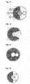
- Figure 8 shows examples of efficient and inefficient PRL regions. Persons with central vision loss can develop PRL at inefficient regions (e.g. right hand sided region on Figure 8 ). The main idea is to induce the location of the PRL at new favorable and efficient regions (e.g. left hand side region on figure 8 ).
- the realization according to the invention is described in the following with reference to Figure 9 .
- the main strategy used to induce new efficient PRL locations is by discouraging inefficient PRL and encouraging efficient PRL regions.
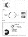
- Figure 9 shows the three realizations to induce efficient PRL.
- the physiological sketch shows the developed undesired PRL (right hand side as in Figure 8 ) and the region in which the new PRL is desired to be induced (left hand side as in Figure 9 ).
- the three realizations can be summarized as follows:
- an PRL is actively induced at the position most beneficial for the person with central vision loss. This is done by moving the visual information of an eye movement target from the less efficient RL into the region of an efficient RL.
- the principle exploits the fact that eye movements consist of fast, large gaze shifts combined with fixation phases, in which visual information is collected.
- the point in time, when the patient chooses the RL used for fixation is directly after a gaze shift. If now the target falls within an area of a non-efficient PRL, it is shifted into the preferred region for development of a PRL. By doing so, the patient is encouraged to develop a PRL in the efficient area of the visual field.
- Fig 10 shows a scheme of PRL induction according to said in the foregoing prescribed manner. At the region of a non-efficient. PRL, targets are shifted into the region of an efficient PRL.
- the non-efficient PRL is discouraged by shifting the target away from the fovea.
- Figure 11 shows a scheme of PRL induction according to this realization. At the region of a non-efficient PRL, targets are shifted away from the fovea, thus encouraging the person with scotoma to place the target in the area of an efficient PRL.
- the visual information is not shifted, but instead blocked in a ring shaped area around the field loss.
- Figures 12 and 13 show such schemes of PRL induction.
- the exact shape of the ring (its thickness and the open angle) can be varied. Thus, even a pinhole-like training tool can be designed.
- this gaze-contingent system the patient is viewing stimuli (such as several well-separated letters or short words) on a presentation screen. If provided with a high quality eye tracker, this screen can also be a video-enabled head mounted display, such as a device sold by the applicant under the trade name VR One.
- a gaze-contingent electro-optical device where a prism can be switched on and off in real-time. Whenever the person is eliciting an eye-movement which brings one of the targets into a non-efficient region for a PRL (such as the dark area in Fig. 10 ), the target will be shifted. Thus it will be presented in the preferred region of a PRL. The patient will adopt his behavior in two ways. First, he will use this preferred region to identify the target. Furthermore, he will try to elicit future eye movements directly in a way, which bring the target into the area of the efficient PRL.
- Figure 16 shows an example of such a gaze-contingent training tool.
- the same behavior can be evoked by a locally prismatic contact lens.
- the contact lens will have a prism at the areas of non-efficient regions for a PRL.
- the prismatic region of the contact lens can be manufactured either refractive, but also diffractive.
- Figures 14 and 15 show a scheme of a prismatic contact lens, in two different realizations discouraging a PRL, in the left visual field.
- FIG. 20a shows such a contact lens with prismatic and blocking function.
- IOL Intra-ocular lens
- FIG. 20b shows such an intraocular lens with prismatic and blocking function.
- the blocking can be realized via a electrochromic device, which blocks transmission in the non-efficient PRL-areas.
- FIG. 21a shows an example of viewing of natural images with such an electro chromic gaze dependent device shown in Figure 21a .
- the subject uses the window to see the object under interest.
- prismatic and light blocking optical properties are realized in an electro-optical device. This might be positioned either eye-centric (as a contact lens), or head-centric (as a spectacle), were gaze tracking is applied to determine the retinal locations, where information should be blocked or shifted.
- Figure 17a shows an electro optic - electrochromic device with gaze tracker.
- Figure 17b shows the two possible eye movements (upper picture) and their inducing consequence by shifting or by freely locating the target on the desired area (lower pictures).
- Figure 18 shows a diffractive spectacle with two main components, a prismatic component and a blocking component, both gaze angle dependent.
- the image shows at a fixed gaze the two components of the spectacle and their respective consequence on the retina.
- the prismatic component brings the image to the desired retinal location and the blocking component blanks the image at the non-desired retinal location.
- HMD Head mounted display
- gaze tracker
- This tool may provide training using live video imaging or stimuli.
- training using live video imaging the natural scene is presented and an algorithm makes realization 1,2 and 3 described above possible.
- stimuli the training is carried out as in solution 1.
- the training can be applied monocularly or binocularly.
- monocular training the second (in most cases healthy) eye is covered. If both eyes are affected, a common efficient PRL is defined.
- the training should be applied in several sessions (e.g. daily for 30 minutes over a period of 2 weeks).
- the training can also be reapplied to induce a new PRL.
- the training device can be used as perimetry device, too. It can show test lights in a determined position relative to the gaze position. Thus, the exact power of the prism applied and the exact direction of the shift can be determined.
- a flexible realization electro-optical device
- the optical properties needed to induce the PRL at the most efficient position can be determined at regular intervals if necessary. Together with an automated workflow this might even be done by the patient himself.
- the decision criteria for the application of the optical function are the above mentioned (as close as possible to the fovea, preferred position in the right visual field).
Landscapes
- Health & Medical Sciences (AREA)
- Life Sciences & Earth Sciences (AREA)
- Animal Behavior & Ethology (AREA)
- Veterinary Medicine (AREA)
- Public Health (AREA)
- General Health & Medical Sciences (AREA)
- Ophthalmology & Optometry (AREA)
- Engineering & Computer Science (AREA)
- Rehabilitation Therapy (AREA)
- Epidemiology (AREA)
- Physics & Mathematics (AREA)
- Physical Education & Sports Medicine (AREA)
- Pain & Pain Management (AREA)
- Biomedical Technology (AREA)
- Biophysics (AREA)
- Molecular Biology (AREA)
- Heart & Thoracic Surgery (AREA)
- Medical Informatics (AREA)
- Surgery (AREA)
- Business, Economics & Management (AREA)
- Educational Administration (AREA)
- Educational Technology (AREA)
- General Physics & Mathematics (AREA)
- Theoretical Computer Science (AREA)
- Rehabilitation Tools (AREA)
Abstract
Description
- The present invention relates to a method for training an efficient preferred retinal locus of fixation with the features of the preamble of
claim 1 as well as to a device for training an efficient preferred retinal locus of fixation with the features of the preamble ofclaim 11. - As e.g. explained by Kwon et al in "Rapid and persistent adaptability of human oculomotor control in response to simulated central vision loss", Current Biology, 2013 September 9; 23(17): 1663-1669, the central region of the human retina, the fovea, provides high-acuity vision. The oculomotor system continually brings targets of interest into the fovea via ballistic eye movements (saccades). The fovea thus serves both as the locus for fixations and as the oculomotor reference for saccades.
- There are persons which suffer from scotoma, in particular central scotoma. A scotoma is an area of partial alteration in the field of vision consisting of a partially diminished or entirely degenerated visual acuity that is surrounded by a field of normal or relatively well-preserved vision. Scotoma is a symptom of damage to any part of the visual system, such as retinal damage from exposure to high-powered lasers, direct sunlight, macular degeneration and brain damage.
- Central scotoma or central vision loss, respectively, is one of the visually most restrictive indications in vision loss. Central vision loss might occur due to a variety of reasons such as Stargadt's disease or macular degeneration. The majority of cases occur due to macular degeneration. Despite the invention is applicable for all kinds of scotoma, for demonstration purposes the invention is explained with reference to typical macular degeneration caused defects of the eye.
- Macular degeneration, often age-related macular degeneration (abbreviated AMD or ARMD), is a medical condition that usually affects older adults and results in a loss of vision in the center of the visual field (the macula) because of damage to the retina. Approximately 10 % of patients 66 to 74 years of age will have findings of macular degeneration. The prevalence increases to 30 % in patients 75 to 85 years of age.
- The loss of central vision profoundly affects visual functioning. It is quite difficult, for example, to read without central vision. Pictures that attempt to depict the central visual loss of macular degeneration with a black spot do not really do justice to the devastating nature of the visual loss.
-
Figure 1 shows a drafted picture of twochildren ball hands heads 5, 6 are located in front of a wooden fence 7. The picture according toFigure 1 represents a situation as perceived from a person without having any visual loss.Figure 2 shows a picture of the same situation as perceived from a person with central vision loss due to macular degeneration. Theheads 5, 6 of thechildren dark cloud 10. The environment surrounding thecloud 10 being represented by theballs hands - Although there are several promising treatments of AMD under consideration, all of them only aim at a slowing down the progression or stopping the progression of AMD. The vision, which is lost already, cannot be restored. Patients with central vision loss have to learn to use another part of the retina for fixation. Kwon et al describe this cognitive process as that the oculomotor system has to adopt a peripheral locus for fixation and to re-reference saccades to this locus.
- Until now, the patients learn to use another eccentric location of the retina as a new locus for fixations and to guide their eye movements in a self-paced way. This is very cumbersome, because the eye movement system is naturally trained to bring a target onto the fovea. This other location of the retina used as a new locus for fixations in literature is called as preferred retinal locus (PRL) for fixation. According to Crossland et al, "The preferred retinal locus in macular disease: toward a consensus definition." in Retina; 2011 Nov; 31 (10): 2109-2114 said PRL is characterized in that 1) it is a retinal area used for fixation, 2) it is task specific, 3) more than one PRL can be used, 4) it is a well-defined region of the retina, and 5) the same PRL is used on repeated testing.
- It is known that the exact position of the PRL decides upon its efficiency. In particular, it should be placed as close as possible to the fovea and it should be selected based on the specific vision task. These two prerequisites for the efficiency of a PRL are explained in more detail in the following:
-
Figure 3 shows a plan view of thevisual field 11 of a human retina of a person having a central scotoma due to macular degeneration. Theouter ring 12 indicates the outer contour of thevisual field 11 of the retina. Thecloud 13 indicates the area of vision loss.Reference number 16 indicates the outer contour of the area ofvision loss 13. - The person having said
central scotoma 13 may establish two (or even more)PRLs PRL 15 exceeds the acuity at the location ofPRL 14. Thus, PRL 15 is more efficient thanPRL 14. Therefore, in the following a PRL of the type ofPRL 14 is called non-efficient PRL and a PRL of the type ofPRL 15 is called efficient PRL. - The above assumption that a PRL being closer to the fovea is more efficient than a PRL being further away from the fovea in the periphery of the retina is e.g. valid for a face recognition task (see e.g. a situation according to
Figure 2 ). This may not necessarily be valid for a reading task as will be explained with reference toFigures 4 to 7 . -
Figure 4 shows a plan view of thevisual field 11 of a human retina of a person having acentral scotoma 13 due to macular degeneration and afixation target 17 represented by a text passage in a book on the right hand side of the plan view of thevisual field 11 of the retina. Theouter ring 12 indicates the outer contour of thevisual field 11 of the retina. Thecloud 13 indicates the area of vision loss.Reference number 16 indicates the outer contour of the area ofvision loss 13. -
Figure 4 assumes that the person has aPRL 18 being located on the left hand side of thescotoma 13. When starting the reading task, the person will move head and/or eye in order to position thePRL 18 to the top and left hand side of the fixation target 17 (the letters "Ma"). The respective movement is indicated by means ofarrow 19. The location of the beginning of the text passage at which thePRL 18 is located after head and/or eye movement is indicated withcircle 20 inFigure 4 . As aconsequence scotoma 13 will be located at the position ofcross 21.Figure 5 shows the relative positions of thevisual field 11 of the human retina and thefixation target 17 after head and/oreye movement 19 in order to start reading. - The situation shown in
Figure 6 corresponds to the situation shown inFigure 4 with the difference that the person has aPRL 22 being located on the right hand side of thescotoma 13. When starting the reading task, the person will move head and/or eye in order to position thePRL 22 to the top and left hand side of thefixation target 17. The respective movement is indicated by means ofarrow 23. The location of the beginning of the text passage at which thePRL 22 is located after head and/or eye movement is indicated withcircle 24 inFigure 6 . As aconsequence scotoma 13 will be located at the position ofcross 25.Figure 7 shows the relative positions of thevisual field 11 of the human retina and thefixation target 17 after head and/oreye movement 23 in order to start reading. - A
PRL 22 using the visual field to the right of the fovea is especially efficient in reading. When moving the eyes from word to word from left to right, aneye movement 23 to a word has only to be kept shorter, whereas aPRL 18 using the left visual field from the fovea would require to make aneye movement 19 over thefixation target 17. Therefore, thePRL 22 to the right of the fovea is more efficient than thePRL 18 to the left of the fovea - Since during eye movement the complete line of an actually read text passage is visible if a PRL below the fovea is used (not shown here), said PRL below the fovea is even more efficient than
PRL 22. This has already be found by Kwon et al, see in particular p. 1666, first complete paragraph. - In the current above explained self-trained way the above mentioned benefits cannot be considered, because the choice of PRL does more or less depend on chance and habitual behavior. In addition, a person may establish more than one PRLs.
- In order to overcome the latter drawback Kwon et al propose an explicit training for refinement of a fixational PRL, i.e. to stabilize a self-trained PRL. Kwon et al displayed a small white cross at the retinal location of each subject's emerged fixational PRL and instructed subjects to follow the target with this gaze marker. In addition Kwon et al propose to superimpose a simulated scotoma on a real one in order to provide accurate positional feedback and speed up PRL development.
- Despite Kwon et al's proposed methods provide satisfying results in order to train persons in stabilizing one single efficient preferred retinal locus of fixation. In particular in cases when the persons have established more than one preferred retinal locus of fixation or in cases when the persons have established an inefficient PRL there is a need for further improvement.
- The problem of the present invention consists in providing a method as well as a device for training an efficient preferred retinal locus of fixation not only four persons having already established an adequate preferred retinal locus of fixation but also for persons who lack any preferred retinal locus of fixation or who have established an inefficient preferred retinal locus of fixation.
- This problem is solved by a method comprising the features of
claim 1 and a device comprising the features ofclaim 11. Advantageous embodiments and developments of the invention are the subject matter of the dependent claims. - Deviating from the prior art represented by Kwon et al training and stabilizing an already established PRL the idea of the present invention consists in establishing an efficient, preferably the most efficient PRL one may think of by inducing said efficient preferred retinal locus of fixation for a specific vision task. Said PRL shall be located in a region or an area outside an inefficient or non-efficient retinal region.
- Concretely, said method for training a preferred retinal locus of fixation for a person having an eye with a field of vision comprising an area of partially diminished or entirely degenerated visual acuity is according to the invention characterized in the steps:
- determining an inefficient retinal region in said field of vision of said eye of said person
- inducing a preferred retinal locus of fixation for a vision task outside said inefficient retinal region.
- Inducing said preferred retinal locus of fixation for said vision task outside said inefficient retinal region comprises measures to encourage said person to move head and/or eye in order to gaze such that said retinal location of fixation of the eye of said person lies outside said inefficient retinal region. Simultaneously and/or alternatively, inducing may comprise any measures to discourage said person to move head and/or eye in order to gaze in a direction such that said retinal location of fixation of the eye of said person lies inside said inefficient retinal region.
- The foregoing problem is completely solved by said method as claimed in
claim 1. - Said step of inducing a preferred retinal locus of fixation for said vision task outside said inefficient retinal region may for example comprise the step:
- blocking said inefficient retinal region for said vision task.
- Blocking said inefficient retinal region for said vision task has the effect that the person is discouraged to move eye and/or head such that said PRL is in said inefficient retinal region for said specific vision task.
- The method according to the invention may further be characterized by the step:
- providing a fixation target for said vision task to said person.
- Providing a fixation target to said person gives guidance to said person for conducting his vision task. In addition, flexibility is increased as described below with reference to the drawings.
- In addition or alternatively to said step of blocking said inefficient retinal region for said vision task said method according to the invention may be characterized in further comprising the step:
- shifting said fixation target to an unblocked region outside said blocked inefficient retinal region.
- A PRL is actively induced at the position most beneficial for the person. This is done by moving the visual information of said fixation target from a less efficient retinal location into a region of an efficient retinal location or into a region being completely outside the visual field.
- The principal exploits the fact that eye movements consist of fast, large gaze shifts combined with fixation phases, in which visual information is collected. The point of time, when the patient chooses the retinal location used for fixation is directly after a gaze shift. If now the fixation target falls within an area of non-efficient PRL, it is shifted either into the preferred region for the development of a PRL or the region outside the visual field encouraging the person to start a new attempt to arrange the fixation target in an unblocked retinal area for developing a PRL by moving his head and/or eye.
- In other words an advantageous embodiment of said method according to the invention is characterized in that said step of shifting said fixation target to an unblocked region outside said blocked inefficient retinal region comprises the step:
- shifting said fixation target from said blocked inefficient retinal region to an efficient retinal region
- And another advantageous embodiment of said method according to the invention, whereby said field of vision having a foveal region is characterized in that said step of shifting said fixation target to an unblocked region outside said blocked inefficient region comprises the step:
- shifting said fixation target from said blocked inefficient region away from said foveal region
- In another advantageous realization, the visual information from said fixation target may not be shifted. It may be blocked in a ring-shaped area around the field of vision loss, thus encouraging said person to place said fixation target in the area of an efficient PRL. The exact shape of the ring, in particular its thickness and the open angle, may vary. Thus, even a pinhole-like unblocked region may remain in the field of vision.
- In an embodiment of the invention the method may comprise the steps:
- tracking a gaze of said eye during conducting said vision task and
- determining said inefficient retinal region to be blocked based on said tracked gaze and/or determining said region to which said fixation target is to be shifted based on said tracked gaze.
- The use of gaze tracking, i.e. using a so-called eye-tracker, allows the implementation of the method in a gaze-contingent training tool. In this gaze-contingent training tool the person may perceive stimuli, such as several symbols, well-separated letters or short words, on a display. Dependent on the tracked gaze the stimuli (fixation target) may vanish (be blocked) or may occur at another location of the display (be shifted).
- In one embodiment of the invention the method may be characterized in that said step determining an inefficient retinal region in said field of vision of said eye of said person for said vision task comprises the steps:
- determining an efficient retinal region in said field of vision of said eye of said person for said vision task
- setting a region outside said efficient retinal region in said field of vision of said eye of said person for said vision task as defining said inefficient retinal region in said field of vision of said eye of said person for said vision task.
- This embodiment leaves no choice to the person to establish a PRL than the efficient retinal region in said field of vision of said eye of said person for said vision task being pre-determined by the operator of said training tool or the operator guiding said person through said training procedure.
- Still another embodiment of said inventive concept may be characterized in that said step of blocking said inefficient retinal region when providing said fixation target to said person for conducting said vision task comprises one or more of the following steps:
- hiding a region of said fixation target corresponding to said inefficient retinal region when providing said fixation target to said person for conducting said vision task
- exclusively revealing a region of said fixation target corresponding to a region outside said inefficient retinal region when providing said fixation target to said person for conducting said vision task
- exclusively presenting a region of said fixation target corresponding to a region outside said inefficient retinal region when providing said fixation target to said person for conducting said vision task.
- These features provide alternatives how to realize said previously described blocking step.
- In order to provide a well-defined shape of the area of said central vision loss to said person said method according to the invention may be characterized in comprising the step:
- at least partially blocking said area of entirely degenerated visual acuity, preferably fully blocking said area of entirely degenerated visual acuity.
- This measure increases the velocity of establishing a PRL.
- It is herewith explicitly outlined that said method and any advantageous embodiment thereof described above may be computer-implemented. In particular, according to the invention there may be a computer program with a program code to execute the method steps according to the embodiments described in detail above, if the computer program is loaded in the computer and/or executed in the computer.
- The computer program may be stored on a non-transitory storage medium.
- The main concept for a training tool, in particular to conduct a method described in detail above, is outlined in the following paragraphs.
- The inventive device for training a preferred retinal locus of fixation for a person having an eye with a field of vision comprising an area of partially diminished or entirely degenerated visual acuity is characterized in
- an inducing arrangement for inducing a preferred retinal locus of fixation for a vision task outside an inefficient retinal region in said field of vision of said eye of said person.
- Such a device completely solves the problem outlined above.
- One embodiment of a device according to the invention may be characterized in at least one of
- a gaze dependent blocking arrangement for blocking an area of said visual field dependent on direction of gaze of said eye, and/or
- a gaze dependent shifting arrangement for shifting a fixation target dependent on direction of gaze of said eye.
- In case said one embodiment comprises a gaze dependent shifting arrangement for shifting a fixation target dependent on direction of gaze of said eye an advantageous variant of said one embodiment may be characterized in
- a gaze tracking arrangement for tracking a gaze of said eye during conducting a vision task
- a control arrangement for controlling said area of said visual field to be blocked basted on said tracked gaze of said eye, and/or
- a control arrangement for controlling said shifting of said fixation target based on said tracked gaze of said eye.
- In another embodiment said gaze dependent shifting arrangement may comprise or may consist in a gaze dependent prismatic arrangement. The prismatic arrangement brings the image of said fixation target to the desired retinal location. It shifts said fixation target to the above in connection with the inventive method described location.
- Still another embodiment of a device according to the invention is characterized in that at least one of said blocking arrangement and/or said shifting arrangement is detachably fixed to said eye. In particular a contact lens or a intra ocular lens may carry a blocking arrangement and/or a shifting arrangement. Said blocking arrangement may be a tinted region on the contact lens or the intra ocular lens. Said shifting region may be a prism being implemented into said contact lens or said intra ocular lens.
- A further embodiment of a device according to the invention may be characterized in comprising a gaze dependent diffractive element, whereby said gaze dependent diffractive element may comprise at least one of said gaze dependent blocking arrangement and/or said gaze dependent shifting arrangement. A concrete example of this embodiment is described in the following with reference to the drawings.
- The invention in the following is described with reference to the drawing, in which
- Figure 1
- shows a photography of two children representing a view of a person without any vision loss
- Figure 2
- shows a simulation of central vision loss
- Figure 3
- shows efficient and non-efficient PRLs due to decaying acuity in the periphery
- Figure 4
- shows the demonstration of a non-efficient PRL due the direction of reading. The person has to make an eye movement "over" the fixation target (which is the beginning of the word "Macular" here.
- Figure 5
- shows the situation of
Figure 4 after eye movement "over" the fixation target - Figure 6
- shows the demonstration of an efficient PRL due the direction of reading. The person has to make an eye movement "before" the fixation target.
- Figure 7
- shows the situation of
Figure 6 after eye movement "before" the fixation target - Figure 8
- shows examples of efficient and inefficient PRL regions
- Figure 9
- shows three realizations to induce PRL regions indicated with 1., 2. and 3.
- Figure 10
- shows a scheme of PRL induction. At the region of a non-efficient PRL, targets are shifted into the region of an efficient PRL.
- Figure 11
- shows a scheme of PRL induction. At the region of a non-efficient PRL, targets are shifted away from the fovea, thus encouraging the patient to place the target in the area of an efficient PRL.
- Figure 12
- shows a scheme of PRL induction. At the region of a non-efficient PRL, the target information is blocked, thus encouraging the person to place the target in the area of an efficient PRL.
- Figure 13
- shows another scheme of PRL induction. At the region of a non-efficient PRL, the target information is blocked, thus encouraging the person to place the target in the area of an efficient PRL.
- Figure 14
- shows a scheme of prismatic contact lens, in a first realization discouraging a PRL in the left visual field.
- Figure 15
- shows a scheme of prismatic contact lens, in a second realization discouraging a PRL in the left visual field.
- Figure 16a
- shows a scheme of the gaze contingent training tool.
- Figure 16b
- shows an example of the realization where the target falls within an area of a bad PRL and is shifted into the preferred region (left half of the visual field).
- Figure 17a
- shows an electro optic and electrochromic gaze dependent device with a gaze tracker.
Figure 17b shows the two possible eye movements (upper picture) and their inducing consequence by shifting or by freely locating the target on the desired area (lower pictures). - Figure 18
- shows a gaze dependent diffractive spectacle. On the right, the image shows at a fixed gaze the two components of the spectacle and their respective consequence on the retina. The prismatic component brings the image to the desired retinal location and the blocking component blanks the image at the non-desired retinal location
- Figure 19
- shows large and local field view and example of presented stimuli
- Figure 20a
- shows a contact lens with prismatic and blocking function
- Figure 20b
- shows an intraocular lens with prismatic and blocking function
- Figure 21a
- shows an electrochromic spectacle with gaze tracker
- Figure 21b
- shows an example of viewing of natural images with the electro chromic gaze dependent device according to
Figure 21a . In this example, the subject uses the window to see the object under interest -
Figure 8 shows examples of efficient and inefficient PRL regions. Persons with central vision loss can develop PRL at inefficient regions (e.g. right hand sided region onFigure 8 ). The main idea is to induce the location of the PRL at new favorable and efficient regions (e.g. left hand side region onfigure 8 ). - The realization according to the invention is described in the following with reference to
Figure 9 . The main strategy used to induce new efficient PRL locations is by discouraging inefficient PRL and encouraging efficient PRL regions. -
Figure 9 shows the three realizations to induce efficient PRL. In the Figure, the physiological sketch shows the developed undesired PRL (right hand side as inFigure 8 ) and the region in which the new PRL is desired to be induced (left hand side as inFigure 9 ). The three realizations can be summarized as follows: - 1. Blocking inefficient PRL regions, acts as an extension of the scotoma and blocks visual inputs located at the inefficient PRL regions, leaving an unblocked region at an efficient PRL region (represented by 1. In
Figure 9 ) - 2. Blocking inefficient PRL regions and shifting image to the desired region or letting person chose the region (represented by 2. In
Figure 9 ) - 3. Blocking inefficient PRL regions and partial shift/ free choice regions (represented by 3. In
Figure 9 ) - The current invention an PRL is actively induced at the position most beneficial for the person with central vision loss. This is done by moving the visual information of an eye movement target from the less efficient RL into the region of an efficient RL.
- The principle exploits the fact that eye movements consist of fast, large gaze shifts combined with fixation phases, in which visual information is collected. The point in time, when the patient chooses the RL used for fixation is directly after a gaze shift. If now the target falls within an area of a non-efficient PRL, it is shifted into the preferred region for development of a PRL. By doing so, the patient is encouraged to develop a PRL in the efficient area of the visual field.
-
Fig 10 shows a scheme of PRL induction according to said in the foregoing prescribed manner. At the region of a non-efficient. PRL, targets are shifted into the region of an efficient PRL. - In another realization, the non-efficient PRL is discouraged by shifting the target away from the fovea.
Figure 11 shows a scheme of PRL induction according to this realization. At the region of a non-efficient PRL, targets are shifted away from the fovea, thus encouraging the person with scotoma to place the target in the area of an efficient PRL. - In another realization, the visual information is not shifted, but instead blocked in a ring shaped area around the field loss.
Figures 12 and 13 show such schemes of PRL induction. - At the region of a non-efficient PRL, target information is blocked, thus encouraging the person to place the target in the area of an efficient PRL.
- The exact shape of the ring (its thickness and the open angle) can be varied. Thus, even a pinhole-like training tool can be designed.
- Technically, this training can be done in several different ways:
- In this gaze-contingent system the patient is viewing stimuli (such as several well-separated letters or short words) on a presentation screen. If provided with a high quality eye tracker, this screen can also be a video-enabled head mounted display, such as a device sold by the applicant under the trade name VR One. Another implementation is a gaze-contingent electro-optical device, where a prism can be switched on and off in real-time. Whenever the person is eliciting an eye-movement which brings one of the targets into a non-efficient region for a PRL (such as the dark area in
Fig. 10 ), the target will be shifted. Thus it will be presented in the preferred region of a PRL. The patient will adopt his behavior in two ways. First, he will use this preferred region to identify the target. Furthermore, he will try to elicit future eye movements directly in a way, which bring the target into the area of the efficient PRL. -
Figure 16 shows an example of such a gaze-contingent training tool. - The same behavior can be evoked by a locally prismatic contact lens. The contact lens will have a prism at the areas of non-efficient regions for a PRL. The prismatic region of the contact lens can be manufactured either refractive, but also diffractive.
Figures 14 and15 show a scheme of a prismatic contact lens, in two different realizations discouraging a PRL, in the left visual field. - There may be a contact lens with prismatic and blocking function, the training will be performed with a stimulus presented at a reading distance on a sheet of paper.
Figure 20a shows such a contact lens with prismatic and blocking function. - The same behavior can be evoked by a locally prismatic or blinded contact lens. In this case it is benefitial to use a technique, where the prism is only existing temporally in the IOL during the training period.
- For such an intraocular lens with prismatic and blocking function, the training will be performed with a stimulus presented at a reading distance on a sheet of paper.
Figure 20b shows such an intraocular lens with prismatic and blocking function. - In the realization, in which only blocking occurs, the blocking can be realized via a electrochromic device, which blocks transmission in the non-efficient PRL-areas.
- There may also exist embodiments combining an electrochromic and gaze tracking device as is e.g. shown in
Figure 21a .Figure 21b shows an example of viewing of natural images with such an electro chromic gaze dependent device shown inFigure 21a . In this example, the subject uses the window to see the object under interest. < - In a specifically flexible realization, prismatic and light blocking optical properties are realized in an electro-optical device. This might be positioned either eye-centric (as a contact lens), or head-centric (as a spectacle), were gaze tracking is applied to determine the retinal locations, where information should be blocked or shifted.
-
Figure 17a shows an electro optic - electrochromic device with gaze tracker.Figure 17b shows the two possible eye movements (upper picture) and their inducing consequence by shifting or by freely locating the target on the desired area (lower pictures). - Especially the prismatic functions, but also other functions needed might be realized in a diffractive way. Furthermore, in a diffractive realization, the wavelength spectrum of the unmodified areas can be selectively filtered to provide maximum contrast andor to block harming blue wavelengths.
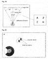
Figure 18 shows a diffractive spectacle with two main components, a prismatic component and a blocking component, both gaze angle dependent. On the right ofFigure 18 , the image shows at a fixed gaze the two components of the spectacle and their respective consequence on the retina. The prismatic component brings the image to the desired retinal location and the blocking component blanks the image at the non-desired retinal location. - The previous solution can be applied in two different field of view sizes as shown in
Figure 19 : - Large field: stimuli presented on a display
- Local field: stimuli presented at a reading distance in a cellphone
- This tool may provide training using live video imaging or stimuli. In training using live video imaging the natural scene is presented and an algorithm makes
realization solution 1. - The training can be applied monocularly or binocularly. In monocular training the second (in most cases healthy) eye is covered. If both eyes are affected, a common efficient PRL is defined.
- The training should be applied in several sessions (e.g. daily for 30 minutes over a period of 2 weeks).
- In case the area of vision loss increases and covers the induced PRL, the training can also be reapplied to induce a new PRL.
- Generally, a PRL in the right visual field might be beneficial, thus the solution depicted in
Figures 10 or11 can be considered standard solutions (because of the direction of reading). - But, to determine the size of the area of vision loss, perimetry applies. In the realization of the gaze contingent training tool (see A. above), the training device can be used as perimetry device, too. It can show test lights in a determined position relative to the gaze position. Thus, the exact power of the prism applied and the exact direction of the shift can be determined. In case, a flexible realization (electrochromic device, electro-optical device) is used (see e.g. D. and E. above), the optical properties needed to induce the PRL at the most efficient position can be determined at regular intervals if necessary. Together with an automated workflow this might even be done by the patient himself. The decision criteria for the application of the optical function are the above mentioned (as close as possible to the fovea, preferred position in the right visual field).
Claims (18)
- Method, in particular computer-implemented method, for training a preferred retinal locus of fixation for a person having an eye with a field of vision comprising an area of partially diminished or entirely degenerated visual acuity characterized in the steps:- determining an inefficient retinal region in said field of vision of said eye of said person- inducing a preferred retinal locus of fixation for a vision task outside said inefficient retinal region.
- Method according to claim 1, characterized in that said step inducing a preferred retinal locus of fixation for said vision task outside said inefficient retinal region comprising the step:- blocking said inefficient retinal region for said vision task.
- Method according to claim 1 or 2, characterized in further comprising the step:- providing a fixation target for said vision task to said person.
- Method according to claim 3, characterized in further comprising the step:- shifting said fixation target to an unblocked region outside said blocked inefficient retinal region.
- Method according to claim 4, characterized in that said step of shifting said fixation target to an unblocked region outside said blocked inefficient retinal region comprising the step:- shifting said fixation target from said blocked inefficient retinal region to an efficient retinal region.
- Method according to claim 4, whereby said field of vision comprising a foveal region, characterized in that said step of shifting said fixation target to an unblocked region outside said blocked inefficient region comprising the step:- shifting said fixation target from said blocked inefficient region away from said foveal region.
- Method according to one of claims 2 to 6, characterized in comprising the steps:- tracking a gaze of said eye during conducting said vision task- determining said inefficient retinal region to be blocked based on said tracked gaze and/or determining said region said fixation target to be shifted to based on said tracked gaze.
- Method according to one of the preceding claims, characterized in that said step determining an inefficient retinal region in said field of vision of said eye of said person for said vision task comprising the steps:- determining an efficient retinal region in said field of vision of said eye of said person for said vision task- setting a region outside said efficient retinal region in said field of vision of said eye of said person for said vision task as defining said inefficient retinal region in said field of vision of said eye of said person for said vision task.
- Method according to one of claims 3 to 8, characterized in that said step blocking said inefficient retinal region when providing said fixation target to said person for conducting said vision task comprising one or more of the following steps:- hiding a region of said fixation target corresponding to said inefficient retinal region when providing said fixation target to said person for conducting said vision task- exclusively revealing a region of said fixation target corresponding to a region outside said inefficient retinal region when providing said fixation target to said person for conducting said vision task- exclusively presenting a region of said fixation target corresponding to a region outside said inefficient retinal region when providing said fixation target to said person for conducting said vision task.
- Method according to one of the preceding claims, characterized in comprising the step:- at least partially blocking said area of entirely degenerated visual acuity, preferable fully blocking said area of entirely degenerated visual acuity.
- Device for training a preferred retinal locus of fixation for a person having an eye with a field of vision comprising an area of partially diminished or entirely degenerated visual acuity characterized in- an inducing arrangement for inducing a preferred retinal locus of fixation for a vision task outside an inefficient retinal region in said field of vision of said eye of said person.
- Device according to claim 11, characterized in at least one of- a gaze dependent blocking arrangement for blocking an area of said visual field dependent on direction of gaze of said eye- a gaze dependent shifting arrangement for shifting a fixation target dependent on direction of gaze of said eye.
- Device according to claim 12, characterized in- a gaze tracking arrangement for tracking a gaze of said eye during conducting a vision taskand at least one of- a control arrangement for controlling said area of said visual field to be blocked based on said tracked gaze of said eye- a control arrangement for controlling said shifting of said fixation target based on said tracked gaze of said eye.
- Device according to one of claims 12 to 13, characterized in that said gaze dependent shifting arrangement comprising or consisting in a gaze dependent prismatic arrangement.
- Device according to one of claims 12 or 14, characterized in that at least one of said blocking arrangement and said shifting arrangement being detachably fixed to said eye.
- Device according to one of claims 12 to 15, characterized in comprising a gaze dependent diffractive element, whereby said gaze dependent diffractive element comprising at least one of said gaze dependent blocking arrangement and said gaze dependent shifting arrangement.
- Computer program with program code to execute the method steps according to one of the claims 1 to 10, if the computer program is loaded in the computer and/or executed in the computer.
- Non-transitory storage medium with a computer program according to claim 17 being stored thereon.
Priority Applications (5)
| Application Number | Priority Date | Filing Date | Title |
|---|---|---|---|
| EP15001911.5A EP3111828B1 (en) | 2015-06-29 | 2015-06-29 | Device for training a preferred retinal locus of fixation |
| ES15001911.5T ES2641484T3 (en) | 2015-06-29 | 2015-06-29 | Device for training a preferred retinal fixation site |
| PCT/EP2016/064848 WO2017001335A1 (en) | 2015-06-29 | 2016-06-27 | Device and computer program for training a preferred retinal locus of fixation |
| CN201680038960.4A CN107787213B (en) | 2015-06-29 | 2016-06-27 | Apparatus and computer program for training a preferred retinal gaze point |
| US15/856,974 US11826306B2 (en) | 2015-06-29 | 2017-12-28 | Device and computer program for training a preferred retinal locus of fixation |
Applications Claiming Priority (1)
| Application Number | Priority Date | Filing Date | Title |
|---|---|---|---|
| EP15001911.5A EP3111828B1 (en) | 2015-06-29 | 2015-06-29 | Device for training a preferred retinal locus of fixation |
Publications (2)
| Publication Number | Publication Date |
|---|---|
| EP3111828A1 true EP3111828A1 (en) | 2017-01-04 |
| EP3111828B1 EP3111828B1 (en) | 2017-08-09 |
Family
ID=53513915
Family Applications (1)
| Application Number | Title | Priority Date | Filing Date |
|---|---|---|---|
| EP15001911.5A Active EP3111828B1 (en) | 2015-06-29 | 2015-06-29 | Device for training a preferred retinal locus of fixation |
Country Status (5)
| Country | Link |
|---|---|
| US (1) | US11826306B2 (en) |
| EP (1) | EP3111828B1 (en) |
| CN (1) | CN107787213B (en) |
| ES (1) | ES2641484T3 (en) |
| WO (1) | WO2017001335A1 (en) |
Cited By (3)
| Publication number | Priority date | Publication date | Assignee | Title |
|---|---|---|---|---|
| DE102018106125A1 (en) | 2018-03-16 | 2019-09-19 | Carl Zeiss Vision International Gmbh | Apparatus and method for detecting a visual field of a person having a scotoma |
| DE202021106061U1 (en) | 2021-11-05 | 2021-12-17 | SFZ Förderzentrum gGmbH | System and computer program for using a decentralized retinal fixation site |
| DE102021128808A1 (en) | 2021-11-05 | 2023-05-11 | SFZ Förderzentrum gGmbH | System, computer program and training method for using a decentralized retinal fixation site |
Families Citing this family (8)
| Publication number | Priority date | Publication date | Assignee | Title |
|---|---|---|---|---|
| ES2641484T3 (en) * | 2015-06-29 | 2017-11-10 | Carl Zeiss Vision International Gmbh | Device for training a preferred retinal fixation site |
| CN112022642B (en) * | 2020-09-16 | 2023-01-10 | 杭州集视智能科技有限公司 | Edge visual field training equipment and training method based on visual field center damage |
| JP7667275B2 (en) * | 2020-12-11 | 2025-04-22 | アパーチャー・イン・モーション・エルエルシー | Novel retinal irradiance distribution modification device and method to improve and restore vision without corneal vitrification |
| CN113080844B (en) * | 2021-03-31 | 2024-01-09 | 上海青研科技有限公司 | Visual inspection and visual training device for preferential retina areas |
| EP4204896A4 (en) * | 2021-06-11 | 2024-09-25 | HES IP Holdings, LLC | SYSTEMS AND METHODS FOR IMPROVING THE VISION OF THE EYE OF A VIEWER WITH IMPAIRED RETINAL VISION |
| KR102720141B1 (en) * | 2021-09-06 | 2024-10-22 | 재단법인 아산사회복지재단 | Apparatus, method and program for searching and training preferred retinal locus of sight damaged patient |
| US20230149248A1 (en) * | 2021-11-16 | 2023-05-18 | Case Western Reserve University | Methods and systems for dynamic ocular training using therapeutic games |
| CN117357381A (en) * | 2023-03-29 | 2024-01-09 | 深圳视力棒医疗科技有限公司 | Visual impairment rehabilitation training equipment, system and method based on expanded reality |
Citations (4)
| Publication number | Priority date | Publication date | Assignee | Title |
|---|---|---|---|---|
| WO2010129711A1 (en) * | 2009-05-05 | 2010-11-11 | The Trustees Of Columbia University In The City Of New York | Devices, systems, and methods for evaluating vision and diagnosing and compensating impairment of vision |
| WO2013050926A1 (en) * | 2011-10-07 | 2013-04-11 | Iurilli Michele | Device and method for facilitating the identification from a distance of preferred retinal loci (prls) and the eccentric fixation |
| WO2015022514A1 (en) * | 2013-08-12 | 2015-02-19 | London Eye Hospital Pharma | Intraocular lens system |
| US9028067B1 (en) * | 2014-05-28 | 2015-05-12 | Sightex LLC | Relocated virtual retinal image method and system |
Family Cites Families (16)
| Publication number | Priority date | Publication date | Assignee | Title |
|---|---|---|---|---|
| US4544246A (en) * | 1983-06-13 | 1985-10-01 | Sri International | System for producing selective stabilization of a portion of the retinal image |
| US5035500A (en) * | 1988-08-12 | 1991-07-30 | Rorabaugh Dale A | Automated ocular perimetry, particularly kinetic perimetry |
| JP2002523183A (en) * | 1998-08-27 | 2002-07-30 | ノヴァヴィズィオン アーゲー | Method and apparatus for training human vision |
| US6464256B1 (en) * | 2000-09-11 | 2002-10-15 | Donald V. Edwards | Furniture slide |
| US7753524B2 (en) | 2002-02-08 | 2010-07-13 | Novavision, Inc. | Process and device for treating blind regions of the visual field |
| CA2475042C (en) * | 2002-02-08 | 2011-05-10 | Novavision, Inc. | Improved process and device for the training of human vision |
| US8668334B2 (en) * | 2006-02-27 | 2014-03-11 | Vital Art And Science Incorporated | Vision measurement and training system and method of operation thereof |
| WO2009091568A2 (en) * | 2008-01-14 | 2009-07-23 | Sina Fateh | System and method for improving the peripheral vision of a subject |
| US7901072B1 (en) | 2009-11-13 | 2011-03-08 | Vuzix Corporation | Prismatic ocular device and personal video display device incorporating same |
| JP5643004B2 (en) * | 2010-06-10 | 2014-12-17 | 株式会社ニデック | Ophthalmic equipment |
| US8454159B1 (en) * | 2012-05-23 | 2013-06-04 | Donald M. Cislo | Method and devices for treatment of macular degeneration visual impairment |
| JP6098061B2 (en) * | 2012-07-27 | 2017-03-22 | 株式会社ニデック | Fundus photographing device |
| WO2015134733A1 (en) * | 2014-03-07 | 2015-09-11 | Aguren Jerry G | Stereo 3d head mounted display applied as a low vision aid |
| AU2015242298B2 (en) * | 2014-03-10 | 2019-11-14 | Amo Groningen B.V. | Intraocular lens that improves overall vision where there is a local loss of retinal function |
| EP3294243A4 (en) * | 2015-05-12 | 2019-01-02 | Agency For Science, Technology And Research | A system and method for displaying a video image |
| ES2641484T3 (en) * | 2015-06-29 | 2017-11-10 | Carl Zeiss Vision International Gmbh | Device for training a preferred retinal fixation site |
-
2015
- 2015-06-29 ES ES15001911.5T patent/ES2641484T3/en active Active
- 2015-06-29 EP EP15001911.5A patent/EP3111828B1/en active Active
-
2016
- 2016-06-27 CN CN201680038960.4A patent/CN107787213B/en active Active
- 2016-06-27 WO PCT/EP2016/064848 patent/WO2017001335A1/en not_active Ceased
-
2017
- 2017-12-28 US US15/856,974 patent/US11826306B2/en active Active
Patent Citations (4)
| Publication number | Priority date | Publication date | Assignee | Title |
|---|---|---|---|---|
| WO2010129711A1 (en) * | 2009-05-05 | 2010-11-11 | The Trustees Of Columbia University In The City Of New York | Devices, systems, and methods for evaluating vision and diagnosing and compensating impairment of vision |
| WO2013050926A1 (en) * | 2011-10-07 | 2013-04-11 | Iurilli Michele | Device and method for facilitating the identification from a distance of preferred retinal loci (prls) and the eccentric fixation |
| WO2015022514A1 (en) * | 2013-08-12 | 2015-02-19 | London Eye Hospital Pharma | Intraocular lens system |
| US9028067B1 (en) * | 2014-05-28 | 2015-05-12 | Sightex LLC | Relocated virtual retinal image method and system |
Non-Patent Citations (3)
| Title |
|---|
| CROSSLAND ET AL.: "The preferred retinal locus in macular disease: toward a consensus definition", RETINA, vol. 31, no. 10, November 2011 (2011-11-01), pages 2109 - 2114 |
| KWON ET AL.: "Rapid and persistent adaptability of human oculomotor control in response to simulated central vision loss", CURRENT BIOLOGY, vol. 23, no. 17, 9 September 2013 (2013-09-09), pages 1663 - 1669 |
| KWON MIYOUNG ET AL: "Rapid and Persistent Adaptability of Human Oculomotor Control in Response to Simulated Central Vision Loss", CURRENT BIOLOGY, vol. 23, no. 17, 9 September 2013 (2013-09-09), pages 1663 - 1669, XP028712584, ISSN: 0960-9822, DOI: 10.1016/J.CUB.2013.06.056 * |
Cited By (6)
| Publication number | Priority date | Publication date | Assignee | Title |
|---|---|---|---|---|
| DE102018106125A1 (en) | 2018-03-16 | 2019-09-19 | Carl Zeiss Vision International Gmbh | Apparatus and method for detecting a visual field of a person having a scotoma |
| DE102018106125B4 (en) | 2018-03-16 | 2019-09-26 | Carl Zeiss Vision International Gmbh | Apparatus and method for detecting a visual field of a person having a scotoma |
| US11259698B2 (en) | 2018-03-16 | 2022-03-01 | Carl Zeiss Vision International Gmbh | Apparatus and method for capturing a visual field of a person having a scotoma |
| DE202021106061U1 (en) | 2021-11-05 | 2021-12-17 | SFZ Förderzentrum gGmbH | System and computer program for using a decentralized retinal fixation site |
| DE102021128808A1 (en) | 2021-11-05 | 2023-05-11 | SFZ Förderzentrum gGmbH | System, computer program and training method for using a decentralized retinal fixation site |
| DE102021128808B4 (en) | 2021-11-05 | 2023-08-31 | SFZ Förderzentrum gGmbH | Computer program and training method for using a decentralized retinal fixation site |
Also Published As
| Publication number | Publication date |
|---|---|
| CN107787213A (en) | 2018-03-09 |
| ES2641484T3 (en) | 2017-11-10 |
| WO2017001335A1 (en) | 2017-01-05 |
| CN107787213B (en) | 2020-10-09 |
| US20180116900A1 (en) | 2018-05-03 |
| US11826306B2 (en) | 2023-11-28 |
| EP3111828B1 (en) | 2017-08-09 |
Similar Documents
| Publication | Publication Date | Title |
|---|---|---|
| US11826306B2 (en) | Device and computer program for training a preferred retinal locus of fixation | |
| US12016629B2 (en) | Screening apparatus and method | |
| Stidwill et al. | Normal binocular vision: Theory, investigation and practical aspects | |
| EP3488284B1 (en) | Light field processor system and method | |
| NZ773817A (en) | Methods and systems for diagnosing and treating health ailments | |
| US10857060B2 (en) | Method and device for improving functional stereopsis in individuals with central vision loss | |
| US12462351B2 (en) | Image processing system | |
| Zapf et al. | Assistive peripheral phosphene arrays deliver advantages in obstacle avoidance in simulated end-stage retinitis pigmentosa: a virtual-reality study | |
| US8882273B2 (en) | Neurochromatic prescription determination | |
| Grüsser et al. | Physiology of vision | |
| Kraut et al. | Adaptation to monocular vision | |
| Vrubel et al. | Improving functional vision through training based on principles of behaviour analysis | |
| EP3827735A1 (en) | Device, method and computer programs for visual field rehabilitation | |
| Hickton | Exploring visual field perception | |
| Markowitz | Low vision: diagnosis and rehabilitation | |
| Trauzettel-Klosinski | Rehabilitation in neuroophthalmology | |
| Dewhurst | Evaluating strategies for visual search and stimulus discrimination: Implications for training eye movements | |
| Bowman et al. | Disorders of vision in children | |
| CN114594615A (en) | Two-step method for preventing myopia | |
| Orlandi et al. | The optometrist | |
| Kurtev | School of Engineering and Science | |
| UA46640A (en) | METHOD OF INFLUENCE ON VISUAL ACUTE ACUTION IN AMBLIOPIA |
Legal Events
| Date | Code | Title | Description |
|---|---|---|---|
| PUAI | Public reference made under article 153(3) epc to a published international application that has entered the european phase |
Free format text: ORIGINAL CODE: 0009012 |
|
| STAA | Information on the status of an ep patent application or granted ep patent |
Free format text: STATUS: REQUEST FOR EXAMINATION WAS MADE |
|
| 17P | Request for examination filed |
Effective date: 20160721 |
|
| AK | Designated contracting states |
Kind code of ref document: A1 Designated state(s): AL AT BE BG CH CY CZ DE DK EE ES FI FR GB GR HR HU IE IS IT LI LT LU LV MC MK MT NL NO PL PT RO RS SE SI SK SM TR |
|
| AX | Request for extension of the european patent |
Extension state: BA ME |
|
| GRAP | Despatch of communication of intention to grant a patent |
Free format text: ORIGINAL CODE: EPIDOSNIGR1 |
|
| STAA | Information on the status of an ep patent application or granted ep patent |
Free format text: STATUS: GRANT OF PATENT IS INTENDED |
|
| INTG | Intention to grant announced |
Effective date: 20170220 |
|
| GRAS | Grant fee paid |
Free format text: ORIGINAL CODE: EPIDOSNIGR3 |
|
| GRAA | (expected) grant |
Free format text: ORIGINAL CODE: 0009210 |
|
| STAA | Information on the status of an ep patent application or granted ep patent |
Free format text: STATUS: THE PATENT HAS BEEN GRANTED |
|
| AK | Designated contracting states |
Kind code of ref document: B1 Designated state(s): AL AT BE BG CH CY CZ DE DK EE ES FI FR GB GR HR HU IE IS IT LI LT LU LV MC MK MT NL NO PL PT RO RS SE SI SK SM TR |
|
| REG | Reference to a national code |
Ref country code: GB Ref legal event code: FG4D |
|
| REG | Reference to a national code |
Ref country code: CH Ref legal event code: EP Ref country code: AT Ref legal event code: REF Ref document number: 915909 Country of ref document: AT Kind code of ref document: T Effective date: 20170815 |
|
| REG | Reference to a national code |
Ref country code: IE Ref legal event code: FG4D |
|
| REG | Reference to a national code |
Ref country code: DE Ref legal event code: R096 Ref document number: 602015003924 Country of ref document: DE |
|
| REG | Reference to a national code |
Ref country code: ES Ref legal event code: FG2A Ref document number: 2641484 Country of ref document: ES Kind code of ref document: T3 Effective date: 20171110 |
|
| REG | Reference to a national code |
Ref country code: NL Ref legal event code: MP Effective date: 20170809 |
|
| REG | Reference to a national code |
Ref country code: LT Ref legal event code: MG4D |
|
| REG | Reference to a national code |
Ref country code: AT Ref legal event code: MK05 Ref document number: 915909 Country of ref document: AT Kind code of ref document: T Effective date: 20170809 |
|
| PG25 | Lapsed in a contracting state [announced via postgrant information from national office to epo] |
Ref country code: NL Free format text: LAPSE BECAUSE OF FAILURE TO SUBMIT A TRANSLATION OF THE DESCRIPTION OR TO PAY THE FEE WITHIN THE PRESCRIBED TIME-LIMIT Effective date: 20170809 Ref country code: LT Free format text: LAPSE BECAUSE OF FAILURE TO SUBMIT A TRANSLATION OF THE DESCRIPTION OR TO PAY THE FEE WITHIN THE PRESCRIBED TIME-LIMIT Effective date: 20170809 Ref country code: HR Free format text: LAPSE BECAUSE OF FAILURE TO SUBMIT A TRANSLATION OF THE DESCRIPTION OR TO PAY THE FEE WITHIN THE PRESCRIBED TIME-LIMIT Effective date: 20170809 Ref country code: AT Free format text: LAPSE BECAUSE OF FAILURE TO SUBMIT A TRANSLATION OF THE DESCRIPTION OR TO PAY THE FEE WITHIN THE PRESCRIBED TIME-LIMIT Effective date: 20170809 Ref country code: NO Free format text: LAPSE BECAUSE OF FAILURE TO SUBMIT A TRANSLATION OF THE DESCRIPTION OR TO PAY THE FEE WITHIN THE PRESCRIBED TIME-LIMIT Effective date: 20171109 Ref country code: FI Free format text: LAPSE BECAUSE OF FAILURE TO SUBMIT A TRANSLATION OF THE DESCRIPTION OR TO PAY THE FEE WITHIN THE PRESCRIBED TIME-LIMIT Effective date: 20170809 Ref country code: SE Free format text: LAPSE BECAUSE OF FAILURE TO SUBMIT A TRANSLATION OF THE DESCRIPTION OR TO PAY THE FEE WITHIN THE PRESCRIBED TIME-LIMIT Effective date: 20170809 |
|
| PG25 | Lapsed in a contracting state [announced via postgrant information from national office to epo] |
Ref country code: BG Free format text: LAPSE BECAUSE OF FAILURE TO SUBMIT A TRANSLATION OF THE DESCRIPTION OR TO PAY THE FEE WITHIN THE PRESCRIBED TIME-LIMIT Effective date: 20171109 Ref country code: IS Free format text: LAPSE BECAUSE OF FAILURE TO SUBMIT A TRANSLATION OF THE DESCRIPTION OR TO PAY THE FEE WITHIN THE PRESCRIBED TIME-LIMIT Effective date: 20171209 Ref country code: RS Free format text: LAPSE BECAUSE OF FAILURE TO SUBMIT A TRANSLATION OF THE DESCRIPTION OR TO PAY THE FEE WITHIN THE PRESCRIBED TIME-LIMIT Effective date: 20170809 Ref country code: GR Free format text: LAPSE BECAUSE OF FAILURE TO SUBMIT A TRANSLATION OF THE DESCRIPTION OR TO PAY THE FEE WITHIN THE PRESCRIBED TIME-LIMIT Effective date: 20171110 Ref country code: PL Free format text: LAPSE BECAUSE OF FAILURE TO SUBMIT A TRANSLATION OF THE DESCRIPTION OR TO PAY THE FEE WITHIN THE PRESCRIBED TIME-LIMIT Effective date: 20170809 Ref country code: LV Free format text: LAPSE BECAUSE OF FAILURE TO SUBMIT A TRANSLATION OF THE DESCRIPTION OR TO PAY THE FEE WITHIN THE PRESCRIBED TIME-LIMIT Effective date: 20170809 |
|
| PG25 | Lapsed in a contracting state [announced via postgrant information from national office to epo] |
Ref country code: CZ Free format text: LAPSE BECAUSE OF FAILURE TO SUBMIT A TRANSLATION OF THE DESCRIPTION OR TO PAY THE FEE WITHIN THE PRESCRIBED TIME-LIMIT Effective date: 20170809 Ref country code: DK Free format text: LAPSE BECAUSE OF FAILURE TO SUBMIT A TRANSLATION OF THE DESCRIPTION OR TO PAY THE FEE WITHIN THE PRESCRIBED TIME-LIMIT Effective date: 20170809 |
|
| REG | Reference to a national code |
Ref country code: DE Ref legal event code: R097 Ref document number: 602015003924 Country of ref document: DE |
|
| PG25 | Lapsed in a contracting state [announced via postgrant information from national office to epo] |
Ref country code: SK Free format text: LAPSE BECAUSE OF FAILURE TO SUBMIT A TRANSLATION OF THE DESCRIPTION OR TO PAY THE FEE WITHIN THE PRESCRIBED TIME-LIMIT Effective date: 20170809 Ref country code: EE Free format text: LAPSE BECAUSE OF FAILURE TO SUBMIT A TRANSLATION OF THE DESCRIPTION OR TO PAY THE FEE WITHIN THE PRESCRIBED TIME-LIMIT Effective date: 20170809 Ref country code: SM Free format text: LAPSE BECAUSE OF FAILURE TO SUBMIT A TRANSLATION OF THE DESCRIPTION OR TO PAY THE FEE WITHIN THE PRESCRIBED TIME-LIMIT Effective date: 20170809 |
|
| PLBE | No opposition filed within time limit |
Free format text: ORIGINAL CODE: 0009261 |
|
| STAA | Information on the status of an ep patent application or granted ep patent |
Free format text: STATUS: NO OPPOSITION FILED WITHIN TIME LIMIT |
|
| REG | Reference to a national code |
Ref country code: FR Ref legal event code: PLFP Year of fee payment: 4 |
|
| 26N | No opposition filed |
Effective date: 20180511 |
|
| PG25 | Lapsed in a contracting state [announced via postgrant information from national office to epo] |
Ref country code: SI Free format text: LAPSE BECAUSE OF FAILURE TO SUBMIT A TRANSLATION OF THE DESCRIPTION OR TO PAY THE FEE WITHIN THE PRESCRIBED TIME-LIMIT Effective date: 20170809 |
|
| REG | Reference to a national code |
Ref country code: CH Ref legal event code: PL |
|
| REG | Reference to a national code |
Ref country code: BE Ref legal event code: MM Effective date: 20180630 |
|
| PG25 | Lapsed in a contracting state [announced via postgrant information from national office to epo] |
Ref country code: MC Free format text: LAPSE BECAUSE OF FAILURE TO SUBMIT A TRANSLATION OF THE DESCRIPTION OR TO PAY THE FEE WITHIN THE PRESCRIBED TIME-LIMIT Effective date: 20170809 Ref country code: LU Free format text: LAPSE BECAUSE OF NON-PAYMENT OF DUE FEES Effective date: 20180629 |
|
| REG | Reference to a national code |
Ref country code: IE Ref legal event code: MM4A |
|
| PG25 | Lapsed in a contracting state [announced via postgrant information from national office to epo] |
Ref country code: LI Free format text: LAPSE BECAUSE OF NON-PAYMENT OF DUE FEES Effective date: 20180630 Ref country code: CH Free format text: LAPSE BECAUSE OF NON-PAYMENT OF DUE FEES Effective date: 20180630 Ref country code: IE Free format text: LAPSE BECAUSE OF NON-PAYMENT OF DUE FEES Effective date: 20180629 |
|
| PG25 | Lapsed in a contracting state [announced via postgrant information from national office to epo] |
Ref country code: BE Free format text: LAPSE BECAUSE OF NON-PAYMENT OF DUE FEES Effective date: 20180630 |
|
| PG25 | Lapsed in a contracting state [announced via postgrant information from national office to epo] |
Ref country code: MT Free format text: LAPSE BECAUSE OF NON-PAYMENT OF DUE FEES Effective date: 20180629 |
|
| PG25 | Lapsed in a contracting state [announced via postgrant information from national office to epo] |
Ref country code: TR Free format text: LAPSE BECAUSE OF FAILURE TO SUBMIT A TRANSLATION OF THE DESCRIPTION OR TO PAY THE FEE WITHIN THE PRESCRIBED TIME-LIMIT Effective date: 20170809 |
|
| PG25 | Lapsed in a contracting state [announced via postgrant information from national office to epo] |
Ref country code: PT Free format text: LAPSE BECAUSE OF FAILURE TO SUBMIT A TRANSLATION OF THE DESCRIPTION OR TO PAY THE FEE WITHIN THE PRESCRIBED TIME-LIMIT Effective date: 20170809 |
|
| PG25 | Lapsed in a contracting state [announced via postgrant information from national office to epo] |
Ref country code: MK Free format text: LAPSE BECAUSE OF NON-PAYMENT OF DUE FEES Effective date: 20170809 Ref country code: HU Free format text: LAPSE BECAUSE OF FAILURE TO SUBMIT A TRANSLATION OF THE DESCRIPTION OR TO PAY THE FEE WITHIN THE PRESCRIBED TIME-LIMIT; INVALID AB INITIO Effective date: 20150629 Ref country code: RO Free format text: LAPSE BECAUSE OF FAILURE TO SUBMIT A TRANSLATION OF THE DESCRIPTION OR TO PAY THE FEE WITHIN THE PRESCRIBED TIME-LIMIT Effective date: 20170809 Ref country code: CY Free format text: LAPSE BECAUSE OF FAILURE TO SUBMIT A TRANSLATION OF THE DESCRIPTION OR TO PAY THE FEE WITHIN THE PRESCRIBED TIME-LIMIT Effective date: 20170809 |
|
| PG25 | Lapsed in a contracting state [announced via postgrant information from national office to epo] |
Ref country code: AL Free format text: LAPSE BECAUSE OF FAILURE TO SUBMIT A TRANSLATION OF THE DESCRIPTION OR TO PAY THE FEE WITHIN THE PRESCRIBED TIME-LIMIT Effective date: 20170809 |
|
| PGFP | Annual fee paid to national office [announced via postgrant information from national office to epo] |
Ref country code: ES Payment date: 20250728 Year of fee payment: 11 |
|
| PGFP | Annual fee paid to national office [announced via postgrant information from national office to epo] |
Ref country code: GB Payment date: 20260625 Year of fee payment: 12 |
|
| PGFP | Annual fee paid to national office [announced via postgrant information from national office to epo] |
Ref country code: IT Payment date: 20260623 Year of fee payment: 12 |
|
| PGFP | Annual fee paid to national office [announced via postgrant information from national office to epo] |
Ref country code: DE Payment date: 20260622 Year of fee payment: 12 |
|
| PGFP | Annual fee paid to national office [announced via postgrant information from national office to epo] |
Ref country code: FR Payment date: 20260626 Year of fee payment: 12 |