EP3110952B1 - Co-expression of magnetotactic bacterial genes and genes encoding iron handling proteins in cells - Google Patents
Co-expression of magnetotactic bacterial genes and genes encoding iron handling proteins in cells Download PDFInfo
- Publication number
- EP3110952B1 EP3110952B1 EP14884055.6A EP14884055A EP3110952B1 EP 3110952 B1 EP3110952 B1 EP 3110952B1 EP 14884055 A EP14884055 A EP 14884055A EP 3110952 B1 EP3110952 B1 EP 3110952B1
- Authority
- EP
- European Patent Office
- Prior art keywords
- iron
- genes
- cell
- cells
- expression
- Prior art date
- Legal status (The legal status is an assumption and is not a legal conclusion. Google has not performed a legal analysis and makes no representation as to the accuracy of the status listed.)
- Active
Links
Images

Classifications
-
- A—HUMAN NECESSITIES
- A61—MEDICAL OR VETERINARY SCIENCE; HYGIENE
- A61K—PREPARATIONS FOR MEDICAL, DENTAL OR TOILETRY PURPOSES
- A61K49/00—Preparations for testing in vivo
- A61K49/06—Nuclear magnetic resonance [NMR] contrast preparations; Magnetic resonance imaging [MRI] contrast preparations
- A61K49/18—Nuclear magnetic resonance [NMR] contrast preparations; Magnetic resonance imaging [MRI] contrast preparations characterised by a special physical form, e.g. emulsions, microcapsules, liposomes
- A61K49/1896—Nuclear magnetic resonance [NMR] contrast preparations; Magnetic resonance imaging [MRI] contrast preparations characterised by a special physical form, e.g. emulsions, microcapsules, liposomes not provided for elsewhere, e.g. cells, viruses, ghosts, red blood cells, virus capsides
-
- C—CHEMISTRY; METALLURGY
- C07—ORGANIC CHEMISTRY
- C07K—PEPTIDES
- C07K14/00—Peptides having more than 20 amino acids; Gastrins; Somatostatins; Melanotropins; Derivatives thereof
- C07K14/195—Peptides having more than 20 amino acids; Gastrins; Somatostatins; Melanotropins; Derivatives thereof from bacteria
-
- C—CHEMISTRY; METALLURGY
- C12—BIOCHEMISTRY; BEER; SPIRITS; WINE; VINEGAR; MICROBIOLOGY; ENZYMOLOGY; MUTATION OR GENETIC ENGINEERING
- C12N—MICROORGANISMS OR ENZYMES; COMPOSITIONS THEREOF; PROPAGATING, PRESERVING, OR MAINTAINING MICROORGANISMS; MUTATION OR GENETIC ENGINEERING; CULTURE MEDIA
- C12N15/00—Mutation or genetic engineering; DNA or RNA concerning genetic engineering, vectors, e.g. plasmids, or their isolation, preparation or purification; Use of hosts therefor
- C12N15/09—Recombinant DNA-technology
- C12N15/63—Introduction of foreign genetic material using vectors; Vectors; Use of hosts therefor; Regulation of expression
- C12N15/79—Vectors or expression systems specially adapted for eukaryotic hosts
- C12N15/85—Vectors or expression systems specially adapted for eukaryotic hosts for animal cells
-
- C—CHEMISTRY; METALLURGY
- C12—BIOCHEMISTRY; BEER; SPIRITS; WINE; VINEGAR; MICROBIOLOGY; ENZYMOLOGY; MUTATION OR GENETIC ENGINEERING
- C12Q—MEASURING OR TESTING PROCESSES INVOLVING ENZYMES, NUCLEIC ACIDS OR MICROORGANISMS; COMPOSITIONS OR TEST PAPERS THEREFOR; PROCESSES OF PREPARING SUCH COMPOSITIONS; CONDITION-RESPONSIVE CONTROL IN MICROBIOLOGICAL OR ENZYMOLOGICAL PROCESSES
- C12Q1/00—Measuring or testing processes involving enzymes, nucleic acids or microorganisms; Compositions therefor; Processes of preparing such compositions
- C12Q1/68—Measuring or testing processes involving enzymes, nucleic acids or microorganisms; Compositions therefor; Processes of preparing such compositions involving nucleic acids
- C12Q1/6876—Nucleic acid products used in the analysis of nucleic acids, e.g. primers or probes
- C12Q1/6883—Nucleic acid products used in the analysis of nucleic acids, e.g. primers or probes for diseases caused by alterations of genetic material
-
- C—CHEMISTRY; METALLURGY
- C12—BIOCHEMISTRY; BEER; SPIRITS; WINE; VINEGAR; MICROBIOLOGY; ENZYMOLOGY; MUTATION OR GENETIC ENGINEERING
- C12Q—MEASURING OR TESTING PROCESSES INVOLVING ENZYMES, NUCLEIC ACIDS OR MICROORGANISMS; COMPOSITIONS OR TEST PAPERS THEREFOR; PROCESSES OF PREPARING SUCH COMPOSITIONS; CONDITION-RESPONSIVE CONTROL IN MICROBIOLOGICAL OR ENZYMOLOGICAL PROCESSES
- C12Q1/00—Measuring or testing processes involving enzymes, nucleic acids or microorganisms; Compositions therefor; Processes of preparing such compositions
- C12Q1/68—Measuring or testing processes involving enzymes, nucleic acids or microorganisms; Compositions therefor; Processes of preparing such compositions involving nucleic acids
- C12Q1/6876—Nucleic acid products used in the analysis of nucleic acids, e.g. primers or probes
- C12Q1/6888—Nucleic acid products used in the analysis of nucleic acids, e.g. primers or probes for detection or identification of organisms
- C12Q1/689—Nucleic acid products used in the analysis of nucleic acids, e.g. primers or probes for detection or identification of organisms for bacteria
-
- C—CHEMISTRY; METALLURGY
- C12—BIOCHEMISTRY; BEER; SPIRITS; WINE; VINEGAR; MICROBIOLOGY; ENZYMOLOGY; MUTATION OR GENETIC ENGINEERING
- C12N—MICROORGANISMS OR ENZYMES; COMPOSITIONS THEREOF; PROPAGATING, PRESERVING, OR MAINTAINING MICROORGANISMS; MUTATION OR GENETIC ENGINEERING; CULTURE MEDIA
- C12N2510/00—Genetically modified cells
Definitions
- the invention relates to the production of magnetosome-like structures in cells with increased iron storage. More specifically, the invention provides gene expression systems using a combination of magnetotactic bacterial gene(s) with mammalian genes involved in iron handling in order to augment a cell's iron pool. The invention further provides in vitro and in vivo imaging, diagnostic and therapeutic methods using cells co-expressing the resultant increased iron containing magnetosome-like structures in order to enhance iron contrast using a variety of imaging technologies.
- Non-invasive mapping of cellular or subcellular events in living organisms, or molecular imaging is an evolving and largely unexplored field.
- Magnetic resonance imaging (MRI) is used for in vivo cellular imaging and requires the use of cellular contrast agents. While many of the current developments in contrast agents continue to revolve around SPIO (superparamagnetic iron oxide) particles, progress in the field of gene-based contrast is being recognized (6).
- SPIO superparamagnetic iron oxide
- the bacterial, putative iron transport protein MagA has been implicated, among other proteins, in the formation of magnetosomes: membrane-enclosed vesicles that compartmentalize iron biominerals (1, 2). Magnetosomes are not toxic and impart magnetic properties to the cell that may be clinically useful (3, 4).
- Canadian patent application 2,655,118 describes the use of magnetosome genes in eukaryotic cells, including MagA - derived iron contrast, for cell tracking using MRI.
- WO-A1-2007140617 discloses a magnetosome-like structure, where however simultaneous expression of ferritin and MagA is not disclosed.
- Frontiers in microbiology 5, Art. 29, 5 February 2014, pages 1-9 discloses (see abstract and throughout) cells where both ferritin and MagA are expressed. These cells show improvement when used in the MRI. However, at no point are ferritin and MagA expressed simultaneously.
- the present invention provides a gene based iron contrast by the augmentation of a cell's iron pool.
- the present invention combines the use of magnetotactic genes that form magnetosome-like structures in cells with genes encoding iron handling proteins.
- the invention in its broadest sense is the co-expression of (a) one or more bacterial magnetotactic genes to amplify iron uptake, compartmentalization and biomineralization in cells, with (b) one or more genes encoding iron handling proteins that together augment(s) and/or regulate the cells' iron pool.
- the invention provides an improved modified magnetosome that contains more iron therein with the combination of the expressed genes. More iron encompasses the aspect of amount as well as size of the iron biomineral produced and/or crystal structure all of which contribute to a better signal for imaging technologies. As a result, mammalian cells or bacterial cells that are transfected or transformed, respectively, can be more effectively tracked using various imaging technologies.
- the disclosure provides the co-expression of ferritin subunits (heavy and/or light chains) with one or more magnetotactic bacterial genes, including magnetosome genes such as but not limited to MagA, MamK and MMS16.
- the ferritin subunits are heavy (HF) and light subunits (LF) each of which lacks the iron response elements (IRE).
- the ferritin subunits are mammalian, in aspects human.
- magnetotactic bacterial genes and mammalian iron handling proteins such as modified human ferritin heavy and light subunits, each lacking iron response elements (IRE), promotes iron biomineralization and enhances cellular contrast for molecular imaging using any variety of techniques such as magnetic resonance imaging (MRI).
- MRI magnetic resonance imaging
- the disclosure thus provides enhancement and/or regulation of gene-based, iron contrast by augmenting a cell's iron pool, as for example stored by a modified form of ferritin, in addition to increasing iron uptake through for example the MagA transporter or other magnetotactic bacterial protein(s), including those associated with forming a magnetosome-like structure.
- the present invention addresses current limitations in cell tracking using exogenously-introduced iron particles, such as superparamagnetic iron oxide (SPIO) nanoparticles.
- SPIO superparamagnetic iron oxide
- a plasmid comprising (a) one or more bacterial magnetotactic genes to amplify iron uptake, compartmentalization and biomineralization in cells in combination with (b) one or more iron handling proteins that together augment(s) and/or regulates the cells' iron pool.
- the iron handling proteins may be of mammalian origin.
- an expression system comprising one or more magnetotactic genes and one or more mammalian genes encoding iron handling proteins under the control of one or more suitable promoters.
- the one or more magnetotactic genes may be provided as an operon.
- a double or triple gene expression system comprising a magnetotactic bacterial gene and modified human heavy and/or light ferritin subunits lacking iron response elements (IRE).
- the iron response element is deleted from the 5' untranslated region of the human heavy and light ferritin gene.
- the gene expression system is provided as a construct for transfection into a variety of eukaryotic and prokaryotic cell types including mammalian cells, human or animal as well as bacterial cells.
- the mammalian cells may be of any type and are not limited and may include any type of cell such as stem cells and the like as is understood by one of skill in the art.
- the entire iron response element is deleted or a portion thereof, the portion being sufficient to lack the expression of the IRE as is understood by one of skill in the art.
- a cell transfected with a gene construct comprising a magnetotactic bacterial gene and modified human heavy and/or light ferritin subunits lacking iron response elements (IRE).
- the magnetotactic bacterial gene is a MagA gene.
- a cell transfected with a gene construct comprising MagA and modified human heavy and/or light ferritin subunits lacking iron response elements (IRE) in the 5' untranslated regions.
- the cell can be mammalian or bacterial.
- the present disclosure is a dual expression system where the same promoter element induces magnetotactic bacterial gene(s), including magnetosome gene(s), and mammalian genes encoding iron handling proteins.
- this inducible expression may be engineered to respond to antibiotic stimulation or a specific cellular transcription factor, as in reporter gene expression. It is understood that a single cell may be engineered to express multiple genes.
- a method for making a cell that co-expresses one or more magnetotactic bacterial genes and mammalian modified ferritin heavy and light subunits comprising; transfecting a cell with a construct comprising one or more magnetotactic bacterial genes and genes encoding modified ferritin heavy and light subunits under the control of an inducible promoter.
- a method for making a cell that co-expresses MagA and modified ferritin heavy and light subunits lacking the IRE comprising; transfecting a cell with a construct comprising MagA and modified ferritin heavy and light subunits lacking the IRE in the 5' untranslated region of the heavy and light gene under the control of a suitable inducible promoter.
- MRI and MPI imaging platforms as well as hybrid imaging platforms, such as PET/MRI ( 4, 11 ), SPECT/MRI, PET/SPECT/MRI, PET/CT, SPECT/CT, bioluminescence tomography (BLT)/MRI, BLT/CT, BLT/PET, BLT/PET/MRI ( 12 ) and MPI.
- PET/MRI 4, 11
- SPECT/MRI PET/SPECT/MRI
- PET/CT PET/CT
- SPECT/CT SPECT/CT
- BLT/CT bioluminescence tomography
- BLT/PET BLT/PET
- BLT/PET/MRI 12
- MPI MPI
- magnetotactic bacterial gene(s) including magnetosome gene(s), and mammalian genes encoding iron handling proteins, like HF and/or LF genes, by two or more distinct promoters.
- magnetotactic bacterial gene(s) including magnetosome gene(s), and mammalian genes encoding iron handling proteins, like HF and/or LF genes, by two or more distinct promoters.
- more than one gene may be monitored at the same time and the simultaneous expression will provide the maximal or distinct MR contrast signal.
- a beneficial feature of the present disclosure is the fact that the genetically altered cells of the invention can continue to express and produce magnetosome-like structures and HF and/or LF in vivo during growth, differentiation and replication of the cell. As a result, such cells can be visually tracked as they grow, differentiate and replicate inside a host without dilution of the genetically-programmed contrast agents. Therefore, these structures provide long-term in vivo contrast agents in a wide variety of cells, tissues, organs, etc. With respect to the genetically altered cells being bacterial cells, these can be transformed in vitro or ex vivo with the expression systems of the disclosure and reintroduced into the gut in order to provide an in vivo contrast in the gastrointestinal tract.
- the expression systems of the disclosure may in embodiments comprise MagA and modified heavy and light ferritin subunits lacking iron response elements and be used in eukaryotic cells, such as mammalian cells.
- the cells may be stem cells or the construct may be used for transfection into tissues.
- the construct in cells and tissues may be imaged using magnetic resonance imaging (MRI) or magnetic particle imaging (MPI) alone or in combination with other imaging modalities, such as positron emission tomography (e.g., PET/MRI or PET/MPI) or single photon emission computed tomography (e.g., SPECT/MRI or SPECT/MPI).
- MRI magnetic resonance imaging
- MPI magnetic particle imaging
- positron emission tomography e.g., PET/MRI or PET/MPI
- single photon emission computed tomography e.g., SPECT/MRI or SPECT/MPI
- an expression system for augmenting and/or regulating iron levels in cells comprising (a) one or more bacterial magnetotactic genes to amplify iron uptake, compartmentalization and biomineralization in cells, and (b) one or more iron handling proteins, wherein (a) and (b) are under control of one or more promoters and, optionally, one or more selection markers.
- a method for making a cell that co-expresses one or more magnetotactic bacterial genes and modified ferritin heavy and light subunits comprising; transfecting a cell with a construct comprising one or more magnetotactic bacterial genes and modified ferritin heavy and light subunits under the control of an inducible promoter.
- a further aspect is method for making a cell that co-expresses MagA and modified ferritin heavy and light subunits lacking the IRE, the method comprising; transfecting a cell with a construct comprising MagA and modified ferritin heavy and light subunits lacking the IRE in the 5' untranslated region of the heavy and light genes under the control of a suitable inducible promoter.
- a method for producing a magnetosome-like structure comprising increased amounts of iron contained therein, the method comprising cultivating a transformed cell according to any one of claims 22 to 26 under conditions suitable to effect expression of the genes contained therein.
- a DNA construct for expression of multiple gene products in a cell leading to augmented iron levels comprising: (a) one or more promoters located at the 5' end of the construct, which is operable in a cell, (b) one or more bacterial magnetotactic genes, (c) one or more mammalian iron handling proteins, and (d) a 3' transcription termination sequence comprising a polyadenylation signal following the protein coding sequences.
- constructs of the invention as described herein encompass promoters selected from the group consisting of inducible promoters, constitutive promoters and tissue specific promoters.
- modified breast cancer cell co-expressing MagA and ferritin heavy and light subunits modified to lack an iron response element (IRE), wherein said modified breast cancer cell contains a higher amount of iron than a matched unmodified breast cancer cell.
- IRE iron response element
- a method for detecting changes in cellular iron comprising co-expressing a magnetotactic bacterial gene and a eukaryotic iron handling gene in a cell and monitoring said cell using an imaging method.
- a method for tracking cancer growth and/or metastasis comprising co-expressing a magnetotactic bacterial gene and a eukaryotic iron handling gene in a cancer cell and monitoring said cell using an imaging method.
- a method for monitoring cardiac stem cell transplantation comprising co-expressing a magnetotactic bacterial gene and a eukaryotic iron handling gene in a cardiac stem cell and monitoring said cell using an imaging method.
- a method for tracking neurodegenerative disease comprising co-expressing a magnetotactic bacterial gene and a eukaryotic iron handling gene in a neural cell and monitoring said cell using an imaging method.
- a method for tracking internal bacteria comprising co-expressing a magnetotactic bacterial gene and a eukaryotic iron handling gene in a bacterial cell and monitoring said cell using an imaging method.
- modified cell co-expressing one or more magnetosome genes and one or more iron handling genes, wherein said modified cancer cell contains a higher amount of iron than a matched unmodified cell.
- the modified ferritin subunits (HF and/or LF) lack the noncoding regulatory IRE sequences; therefore, this modified form of ferritin is not subject to control by IRP.
- the invention provides for co-expression of (a) one or more bacterial magnetotactic genes to amplify iron uptake, compartmentalization and biomineralization in cells, with (b) one or more genes encoding iron handling proteins that together augment(s) and/or regulate the cells' iron pool.
- the invention provides an improved modified magnetosome that contains more iron or distinct iron biomineral therein with the combination of the expressed genes.
- a higher amount of iron may encompass one or more of a quantified amount, the size of the iron biomineral produced and the crystal structure of the iron biomineral produced by the present invention.
- the invention is achieved by way of an expression vector (an expression construct) that is a plasmid or virus in which the one or more bacterial magnetotactic genes and the one or more genes encoding iron handling proteins are introduced into a target cell, to commandeer the cell's mechanism for protein synthesis to produce a magnetosome-like structure containing higher levels of iron or a unique iron biomineral.
- the plasmid further comprises regulatory sequences that act as enhancer and promoter regions and lead to efficient transcription of the gene(s) carried on the expression vector.
- the expression vector as described herein will produce a significant amount of stable messenger RNA, and therefore proteins.
- codon optimization may be used with respect to the genes discussed herein to determine and obtain optimal expression for a particular cell type whether eukaryotic or bacterial.
- codon optimization an appropropriate DNA sequence is synthesized specifically to efficiently express the mammalian gene in bacteria for example by first finding out which codons are the most widely used in the species of interest, and synthesize a DNA sequence made up of these.
- Various commercial programs are available and widely used for such purpose.
- Suitable magnetotactic bacterial genes for use in the present invention may be selected from magnetosome genes MagA, MamK, MMS16 and also those encoded on a magnetosome genomic island and organized into operons such as the magnetosome membrane operons MamAB (17 genes including MamA/Mam22, MamB, MamE, MamJ, MamK, MamM, MamN, MamO, MamP), MamGFDC (4 genes including MamC/Mms13, MamG, MamD, MamF), MamXY (4 genes), the magnetic particle membrane specific operon Mms6 (5 genes including Mms6, Mms16, Mms24), the monocistronic MamW, and genes located outside the magnetosome genomic island, such as ChpA and those on the magnetotaxis operon MtxA and the magnetosome membrane operon MmeA ( Kolinko, I., LohBe, A., Borg, S., Raschdorf, O., Jogler,
- Suitable genes encoding iron handling proteins may be selected from the group consisting of ferritin heavy and light subunits lacking the IRE and any protein subject to IRE regulation.
- Representative proteins include the transferrin receptor (UniProt P02786), transferrin (Transferrin St. Edward's University. 2005-07-18. Retrieved 2009-04-24; Yang F, Lum JB, McGill JR, Moore CM, Naylor SL, van Bragt PH, Baldwin WD, Bowman BH (May 1984). "Human transferrin: cDNA characterization and chromosomal localization". Proceedings of the National Academy of Sciences of the United States of America 81 (9): 2752-6. doi:10.1073/pnas.81.9.2752. PMC 345148.
- Ferritin is a protein of 450 kDa consisting of 24 subunits that is present in every cell type. In vertebrates, these subunits are both the light (L) (UniProt 02792) and the heavy (H) type (UniProt P02794) with an apparent molecular weight of 19 kDa or 21 kDa respectively; their sequences are about 50% homologous. Ferritin genes are highly conserved between species. All vertebrate ferritin genes have three introns and four exons. The tyrosine residue at amino acid position 27 in the H subunit is thought to be associated with biomineralization.
- the expression systems of the present invention can be used with any type of eukaryotic and prokaryotic cell.
- eukaryotic cells are mammalian, and may be human or animal. Mammalian cells may include for example cancer cells, nerve cells, muscle cells, cardiac cells, epithelial cells, adipocytes and stem cells.
- the expression systems of the present invention may also be used with prokaryotic cells, that is bacterial cells. Bacterial cells that are native to the gut of mammals can also be transformed with the expression systems of the present invention.
- Suitable bacteria may belong to the genera Bacteroides, Clostridium , Fusobacterium , Eubacterium , Ruminococcus , Peptococcus , Peptostreptococcus, Bifidobacterium , Escherichia and Lactobacillus .
- the gastrointestinal tract of a mammal can be effectively imaged when the transformed bacteria are reintroduced into a mammal's gut.
- Suitable promoters for use in the present invention are generally known in the field of molecular biology and may include inducible promoters, constitutive promoters and tissue specific promoters as is understood by one of skill in the art.
- Viral promoters may be chosen from, for example, cytomegalovirus (CMV; human immediate early), Rous sarcoma virus (RSV), simian virus 40 (SV40), and the long terminal repeat from Moloney murine leukemia virus (LTR).
- CMV cytomegalovirus
- RSV Rous sarcoma virus
- SV40 simian virus 40
- LTR Moloney murine leukemia virus
- Mammalian promoters may be chosen from, for example, the elongation factor (EF) 1 ⁇ promoter, widely useful in mammalian cells; the cytokeratin 18 (K18) promoter; the cytokeratin 19 (K19) promoter; the tissue kallikrein promoter (Kall); the amylase promoter (AMY); and rat aquaporin-5 (rAQP5).
- EF elongation factor
- K18 cytokeratin 18
- K19 tissue kallikrein promoter
- AY amylase promoter
- rAQP5 rat aquaporin-5
- the invention covers the use of more selective promoters that respond to particular cellular transcription factors, such as the minimal NIS (sodium iodide symporter) promoter that responds to Nkx2.5 ( Dentice et al. (2004), Mol Cell Biol 24, 7863-7877 ).
- the combination of genes expressed to produce a magnetosome-like structure may be
- the invention as incorporated into plants and plant cells, one of skill in the art understands that several plant expression vectors are based on the Ti plasmid of Agrobacterium tumefaciens, where DNA to be inserted into plant is cloned into the T-DNA, a stretch of DNA flanked by a 25-bp direct repeat sequence at either end, and which can integrate into the plant genome.
- the T-DNA also contains the selectable marker.
- the Agrobacterium provides a mechanism for transformation, integration of into the plant genome, and the promoters for its vir genes may also be used for the cloned genes. Plant viruses may be used as vectors when Agrobacterium is not suitable for use in a specific plant..
- plant virus examples include the tobacco mosaic virus (TMV), potato virus X, and cowpea mosaic virus.
- TMV tobacco mosaic virus
- the protein may be expressed as a fusion to the coat protein of the virus and is displayed on the surface of assembled viral particles, or as an unfused protein that accumulates within the plant.
- Expression in plant using plant vectors is often constitutive, and a commonly used constitutive promoter in plant expression vectors is the cauliflower mosaic virus (CaMV) 35S promoter.
- CaMV cauliflower mosaic virus
- Eukaryotic expression systems employing insect cell hosts are based upon one of two vector types: plasmid or plasmid-virion hybrids, the latter being most commonly used.
- the typical insect host is the common fruit fly, Drosophila melanogaster,, however, other insect hosts include mosquito ( Aedes albopictus ), fall army worm ( Spodoptera frugiperda ), cabbage looper ( Trichoplusia ni ), salt marsh caterpillar ( Estigmene acrea ) and silkworm ( Bombyx mori ).
- mosquito Aedes albopictus
- fall army worm Spodoptera frugiperda
- cabbage looper Trichoplusia ni
- salt marsh caterpillar Estigmene acrea
- silkworm Bombyx mori
- heterologous protein overexpression occurs in suspension cell cultures.
- the exception, and one of the advantages of plasmid-virion systems, is that the recombinant virus may also be injected into larval host hemo
- Plasmid-based vector systems provide a mechanism for both transient and long-term expression of recombinant protein and are commercially available such as the Drosophila Expression System (DES) (InvitrogenTM).
- DES Drosophila Expression System
- the transfection of competent D . melanogaster cells with engineered plasmid will mediate the transient (2-7 days) expression of heterologous protein.
- Establishing transformed cells that will express protein for longer time periods requires that the host cells be cotransfected with a "selection" vector, resulting in the stable integration of the expression cassette into the host genome.
- Constitutive expression is mediated using the Ac5 Drosophila promoter, whereas a metallothionein promoter guides copper-inducible expression.
- the DES vectors are designed with multiple cloning sites for insertion of the heterologous protein gene in any of three reading frames.
- a choice of vectors also provides for the expression of a variety of C-terminal fusion tags: V5 epitope for identification of expressed protein with V5 epitope antibody, polyhistidine peptide for simplified purification with metal chelate affinity resin, and the BiP secretion leader peptide.
- MagA was overexpressed in combination with a dysregulated form of ferritin.
- ferritin does not respond to cellular iron binding proteins 1 and 2, thereby circumventing a key step in iron homeostasis: regulation of iron storage through receptor-mediated uptake by the interaction of transferrin, transferrin receptor and ferritin.
- IRE allows greater flexibility in the control of iron levels and biominerals by magnetotactic bacterial genes.
- the invention provides genetically engineered cells that produce magnetosome-like structures and HF and/or LF iron storage, as contrast agents for mapping and/or imaging of organs, tissues, cells, sub-cellular structures, proteins and peptides in living organisms both in vivo and in vitro.
- the method of the invention genetically alters cells to enhance iron storage in HF and/or LF and produce magnetosome-like structures as dense core vesicles that form MR contrast agents in the cell.
- the invention also provides genetic constructs from magnetotactic bacterial genes, including those of the magnetosome, and from mammalian genes, such as HF and/or LF, that may be expressed from vectors bearing inducible promoters or encoding other useful genes for targeting cells or for therapeutic treatments that can be followed by functional imaging or long-term tracking of transfected cells.
- magnetotactic bacterial genes including those of the magnetosome
- mammalian genes such as HF and/or LF
- magA magnetotactic bacterial gene and putative iron transporter
- the MR contrast has been compared in clonal lines of MagA expressing cells to those expressing ferritin heavy and light chains, lacking iron response elements (HF and/or LF).
- the transverse relaxation rates, R2' in particular, are sensitive to cellular iron content. Changes in this MR parameter may be used to quantify cellular iron and improve the specificity of detection.
- the relationship between R2 and inter-echo time is investigated to improve cellular detection of iron using molecular MRI.
- the present invention demonstrates the triple expression system/construct of the present invention for use to further augment MR contrast ( Figure 1 , Table 1).
- the invention encompasses various expression strategies for magnetotactic bacterial, magnetosome and mammalian genes like the modified ferritin genes. Since each set of genes may provide contrast in the context of mammalian cell expression, they may operate independently and in an additive manner. Thus the possibility of distinct MR signatures are encompassed and within the scope of the invention for all types of genes and cells selected.
- the invention has particular use with eukaryotic cells that express and overexpress one or more magnetotactic bacterial and/or magnetosome proteins as magnetosome-like structure contrast agents, in combination with mammalian genes like HF and/or LF for iron storage, permitting in vivo MRI, MPI, hybrid MR or x-ray imaging studies to follow the localization, proliferation, and long-term tracking of iron loaded cells.
- These cells may be tissue specific and permit non-invasive in vivo imaging of specific physiological or molecular functions.
- Imaging systems that can be used with the present invention include at least the following, which can also be used in combination: positron emission tomography (PET), magnetic resonance imaging (MRI), gamma-ray imaging camera (GREI-II), computer tomography (CT), Single photon emission computed tomography (SPEC), bioluminescence tomography (BLT) and magnetic particle imaging (MPI).
- PET positron emission tomography
- MRI magnetic resonance imaging
- GREI-II gamma-ray imaging camera
- CT computer tomography
- SPEC Single photon emission computed tomography
- BLT bioluminescence tomography
- MPI magnetic particle imaging
- Magnetic particle imaging (MPI) is a new modality for imaging distributions of iron oxide nanoparticle tracers in vivo with good contrast, high sensitivity, and good spatial resolution, and thus is promising for clinical imaging in angiography and oncology.
- MPI requires high-quality iron oxide nanoparticle tracers with tailored magnetic and surface properties to achieve its full
- the present invention has further wide potential clinical uses such as, but not limited to, those described below.
- Mammalian cells change their iron requirements to meet a given physiological need in both health ( 13 ) and disease ( 14 ).
- MagA and HF and/or LF expression respond to changes in extracellular iron while cellular iron homeostasis, as measured by changes in the level of transferrin receptor, appears intact.
- Expression of MagA and HF and/or LF may be utilized as sensors of changes in physiological iron levels.
- MR detection methods such as transverse relaxation rates
- changes in cellular iron levels and iron biomineralization may be measured using a combination of magnetotactic bacterial genes, such as MagA, including magnetosome genes, and HF and/or LF.
- MagA magnetotactic bacterial genes
- HF and/or LF magnetotactic bacterial genes
- the onset of changes in iron uptake or export and monitoring fluctuations in iron handling may be tracked by expressing a combination of magnetotactic bacterial genes, like MagA, including magnetosome genes, and HF and/or LF. Since transverse relaxation rates are a quantifiable measure of MR contrast and are related to cellular iron content, fluctuations in cellular iron throughout the cell's life cycle may be monitored.
- Iron biomineralization may be amplified in mammalian cells, using a combination of magnetotactic bacterial genes, like MagA, including magnetosome genes, and HF and/or LF. This amplification of gene-based iron contrast will improve MR detection of cells without introducing cytotoxicity or altering endogenous cellular programming.
- a combination of magnetotactic bacterial genes like MagA, including magnetosome genes, and HF and/or LF may be genetically engineered in the cell in order to introduce changes in iron handling. Such changes may influence the cell's labile iron pool and iron import, storage and export by creating a magnetosome-like compartment. Synthesis of a magnetosome-like structure may be genetically associated with a cellular function, reflecting an endogenous activity or overexpression of a desired macromolecule.
- Bacteria harboring a combination of magnetotactic bacterial genes, like MagA, including magnetosome genes, and HF and/or LF may interact with mammalian cells and localize to sites within the body, such as the gut. This interaction may be detected by MRI or its hybrid modalities and signal the onset of a physiological process, be it in health or illness. This type of interaction has been identified as inter-kingdom signalling (15).
- Cancer cells generally consist of a proliferative phenotype; however, dormant cancer cells may assume a quiescent state until reactivated by a stimulatory signal. It will be possible to use the combination of magnetotactic bacterial genes, like MagA, including magnetosome genes, and HF and/or LF to track the changes in cancer cell activity.
- MagA magnetotactic bacterial genes
- HF and/or LF magnetotactic bacterial genes
- the present invention has use to image, quantify and monitor the progression of transplanted stem cells from undifferentiated to differentiated states, as the cells repopulate the scar tissue with normally functioning heart tissue.
- the use of magnetosome-like structures, augmented by increased ferritin, to track the progress of tissue regeneration has both research and therapeutic applications.
- Iron misregulation has been acknowledged in a number of diseases, including Alzheimer's disease (21).
- the Amyloid Precursor Protein (APP) has an IRE in the 5' untranslated region of the mRNA.
- the role of biometal dysregulation and oxidative stress in A ⁇ amyloid formation may be detected by the expression of magnetotactic bacterial, like MagA, including magnetosome, and HF and/or LF genes in neural cells.
- the invention relates to the use of magnetotactic bacterial genes, including magnetosome genes, and mammalian genes, including the modified ferritin subunits, in combinations that augment or modulate MR contrast by regulating cellular iron biomineralization and/or the formation of magnetosome-like structures.
- magnetotactic bacterial genes including magnetosome genes, and mammalian genes, including the modified ferritin subunits
- These structures may generate all or part of the magnetosome, including modifications to iron uptake, iron biomineralization, iron compartmentalization and the arrangement of magnetosome-like structures within the cell.
- Magnetic Particle Imaging is an emerging area of MRI where nanoparticles of iron are non-invasively tracked and quantified ( Saritas, EU, Goodwill, PW, Croft, LR, Konkle, JJ, Lu, K, Zheng, B and Conolly, SM (2013) Magnetic Particle Imaging (MPI) for NMR and MRI researchers. J Magn Reson 229,116-126 ). Magnetosome-like structures can be used for MPI where genes from magnetotactic bacteria are co-expressed with mammalian genes to create the iron nanoparticles that have specific MPI signals Biomed Tech (Berl). 2013 Dec;58(6):493-507.
- Bacteria that live in the human gut may be genetically programmed to co-express magnetotactic bacterial genes, including magnetosome genes, and human genes, like the ferritin heavy and light subunits, to produce magnetosome-like structures.
- Transcripts were amplified from 1 ⁇ g of total RNA, isolated from stably-expressing cells (clone 12), by reverse transcription (RT).
- PCR polymerase chain reaction
- MagA primers lamA primers
- HF primers lamA primers
- LF primers lamA primers
- ⁇ -actin primers lamA primers
- Lane 6 contains an RT negative control for ⁇ -actin.
- Lane 1 contains a 1 Kb DNA ladder (Life Technologies). Table 1.
Landscapes
- Chemical & Material Sciences (AREA)
- Health & Medical Sciences (AREA)
- Life Sciences & Earth Sciences (AREA)
- Organic Chemistry (AREA)
- Proteomics, Peptides & Aminoacids (AREA)
- Genetics & Genomics (AREA)
- Engineering & Computer Science (AREA)
- Zoology (AREA)
- Wood Science & Technology (AREA)
- Analytical Chemistry (AREA)
- General Health & Medical Sciences (AREA)
- Bioinformatics & Cheminformatics (AREA)
- Biochemistry (AREA)
- Biotechnology (AREA)
- General Engineering & Computer Science (AREA)
- Biophysics (AREA)
- Molecular Biology (AREA)
- Microbiology (AREA)
- Physics & Mathematics (AREA)
- Immunology (AREA)
- Biomedical Technology (AREA)
- Epidemiology (AREA)
- Animal Behavior & Ethology (AREA)
- Hematology (AREA)
- Virology (AREA)
- Nuclear Medicine, Radiotherapy & Molecular Imaging (AREA)
- Radiology & Medical Imaging (AREA)
- Pathology (AREA)
- Veterinary Medicine (AREA)
- Public Health (AREA)
- Plant Pathology (AREA)
- Gastroenterology & Hepatology (AREA)
- Medicinal Chemistry (AREA)
- Micro-Organisms Or Cultivation Processes Thereof (AREA)
- Medicines Containing Antibodies Or Antigens For Use As Internal Diagnostic Agents (AREA)
Description
- The invention relates to the production of magnetosome-like structures in cells with increased iron storage. More specifically, the invention provides gene expression systems using a combination of magnetotactic bacterial gene(s) with mammalian genes involved in iron handling in order to augment a cell's iron pool. The invention further provides in vitro and in vivo imaging, diagnostic and therapeutic methods using cells co-expressing the resultant increased iron containing magnetosome-like structures in order to enhance iron contrast using a variety of imaging technologies.
- Non-invasive mapping of cellular or subcellular events in living organisms, or molecular imaging, is an evolving and largely unexplored field. Magnetic resonance imaging (MRI) is used for in vivo cellular imaging and requires the use of cellular contrast agents. While many of the current developments in contrast agents continue to revolve around SPIO (superparamagnetic iron oxide) particles, progress in the field of gene-based contrast is being recognized (6).
- The bacterial, putative iron transport protein MagA has been implicated, among other proteins, in the formation of magnetosomes: membrane-enclosed vesicles that compartmentalize iron biominerals (1, 2). Magnetosomes are not toxic and impart magnetic properties to the cell that may be clinically useful (3, 4). Canadian patent application
2,655,118 describes the use of magnetosome genes in eukaryotic cells, including MagA - derived iron contrast, for cell tracking using MRI.WO-A1-2007140617 discloses a magnetosome-like structure, where however simultaneous expression of ferritin and MagA is not disclosed. Frontiers in discloses (see abstract and throughout) cells where both ferritin and MagA are expressed. These cells show improvement when used in the MRI. However, at no point are ferritin and MagA expressed simultaneously. - There however, remains a need to improve the MR signal for a variety of clinical diagnostic and therapeutic applications.
- The present invention provides a gene based iron contrast by the augmentation of a cell's iron pool. The present invention combines the use of magnetotactic genes that form magnetosome-like structures in cells with genes encoding iron handling proteins.
- The invention in its broadest sense is the co-expression of (a) one or more bacterial magnetotactic genes to amplify iron uptake, compartmentalization and biomineralization in cells, with (b) one or more genes encoding iron handling proteins that together augment(s) and/or regulate the cells' iron pool. The invention provides an improved modified magnetosome that contains more iron therein with the combination of the expressed genes. More iron encompasses the aspect of amount as well as size of the iron biomineral produced and/or crystal structure all of which contribute to a better signal for imaging technologies. As a result, mammalian cells or bacterial cells that are transfected or transformed, respectively, can be more effectively tracked using various imaging technologies.
- The disclosure provides the co-expression of ferritin subunits (heavy and/or light chains) with one or more magnetotactic bacterial genes, including magnetosome genes such as but not limited to MagA, MamK and MMS16. The ferritin subunits are heavy (HF) and light subunits (LF) each of which lacks the iron response elements (IRE). In aspects, the ferritin subunits are mammalian, in aspects human.
- The co-expression of one or more magnetotactic bacterial genes, and mammalian iron handling proteins such as modified human ferritin heavy and light subunits, each lacking iron response elements (IRE), promotes iron biomineralization and enhances cellular contrast for molecular imaging using any variety of techniques such as magnetic resonance imaging (MRI).
- The disclosure thus provides enhancement and/or regulation of gene-based, iron contrast by augmenting a cell's iron pool, as for example stored by a modified form of ferritin, in addition to increasing iron uptake through for example the MagA transporter or other magnetotactic bacterial protein(s), including those associated with forming a magnetosome-like structure. The present invention addresses current limitations in cell tracking using exogenously-introduced iron particles, such as superparamagnetic iron oxide (SPIO) nanoparticles.
- According to an aspect of the present disclosure is a plasmid comprising (a) one or more bacterial magnetotactic genes to amplify iron uptake, compartmentalization and biomineralization in cells in combination with (b) one or more iron handling proteins that together augment(s) and/or regulates the cells' iron pool. In aspects, the iron handling proteins may be of mammalian origin.
- According to another aspect of the present disclosure is an expression system comprising one or more magnetotactic genes and one or more mammalian genes encoding iron handling proteins under the control of one or more suitable promoters. The one or more magnetotactic genes may be provided as an operon.
- According to a further aspect of the disclosure is a double or triple gene expression system comprising a magnetotactic bacterial gene and modified human heavy and/or light ferritin subunits lacking iron response elements (IRE). In aspects, the iron response element is deleted from the 5' untranslated region of the human heavy and light ferritin gene. The gene expression system is provided as a construct for transfection into a variety of eukaryotic and prokaryotic cell types including mammalian cells, human or animal as well as bacterial cells. The mammalian cells may be of any type and are not limited and may include any type of cell such as stem cells and the like as is understood by one of skill in the art.
- In aspects the entire iron response element is deleted or a portion thereof, the portion being sufficient to lack the expression of the IRE as is understood by one of skill in the art.
- According to an aspect of the disclosure is a cell transfected with a gene construct comprising a magnetotactic bacterial gene and modified human heavy and/or light ferritin subunits lacking iron response elements (IRE). In aspects, the magnetotactic bacterial gene is a MagA gene.
- According to an aspect of the disclosure is a cell transfected with a gene construct comprising MagA and modified human heavy and/or light ferritin subunits lacking iron response elements (IRE) in the 5' untranslated regions. The cell can be mammalian or bacterial.
- According to an aspect of the present disclosure is a dual expression system where the same promoter element induces magnetotactic bacterial gene(s), including magnetosome gene(s), and mammalian genes encoding iron handling proteins. Those skilled in the art will recognize that this inducible expression may be engineered to respond to antibiotic stimulation or a specific cellular transcription factor, as in reporter gene expression. It is understood that a single cell may be engineered to express multiple genes.
- According to another aspect of the present disclosure is the constitutive expression of one set of genes and inducible expression of additional gene(s).
- According to a further aspect of the present disclosure is a method for making a cell that co-expresses one or more magnetotactic bacterial genes and mammalian modified ferritin heavy and light subunits, the method comprising; transfecting a cell with a construct comprising one or more magnetotactic bacterial genes and genes encoding modified ferritin heavy and light subunits under the control of an inducible promoter.
- According to an aspect of the present disclosure is a method for making a cell that co-expresses MagA and modified ferritin heavy and light subunits lacking the IRE (iron responsive element), the method comprising; transfecting a cell with a construct comprising MagA and modified ferritin heavy and light subunits lacking the IRE in the 5' untranslated region of the heavy and light gene under the control of a suitable inducible promoter.
- According to an aspect of the present disclosure is the detection of multiple contrast genes using MRI and MPI imaging platforms as well as hybrid imaging platforms, such as PET/MRI (4, 11), SPECT/MRI, PET/SPECT/MRI, PET/CT, SPECT/CT, bioluminescence tomography (BLT)/MRI, BLT/CT, BLT/PET, BLT/PET/MRI (12) and MPI. Those skilled in the art will recognize that non-invasive imaging methods and hardware are adaptable to multiple applications.
- According to another aspect of the present disclosure is the inducible expression of magnetotactic bacterial gene(s), including magnetosome gene(s), and mammalian genes encoding iron handling proteins, like HF and/or LF genes, by two or more distinct promoters. Thus more than one gene may be monitored at the same time and the simultaneous expression will provide the maximal or distinct MR contrast signal.
- A beneficial feature of the present disclosure is the fact that the genetically altered cells of the invention can continue to express and produce magnetosome-like structures and HF and/or LF in vivo during growth, differentiation and replication of the cell. As a result, such cells can be visually tracked as they grow, differentiate and replicate inside a host without dilution of the genetically-programmed contrast agents. Therefore, these structures provide long-term in vivo contrast agents in a wide variety of cells, tissues, organs, etc. With respect to the genetically altered cells being bacterial cells, these can be transformed in vitro or ex vivo with the expression systems of the disclosure and reintroduced into the gut in order to provide an in vivo contrast in the gastrointestinal tract.
- The expression systems of the disclosure may in embodiments comprise MagA and modified heavy and light ferritin subunits lacking iron response elements and be used in eukaryotic cells, such as mammalian cells. The cells may be stem cells or the construct may be used for transfection into tissues.
- The construct in cells and tissues may be imaged using magnetic resonance imaging (MRI) or magnetic particle imaging (MPI) alone or in combination with other imaging modalities, such as positron emission tomography (e.g., PET/MRI or PET/MPI) or single photon emission computed tomography (e.g., SPECT/MRI or SPECT/MPI).
- According to an aspect of the disclosure there is provided an expression system for augmenting and/or regulating iron levels in cells, said expression system comprising (a) one or more bacterial magnetotactic genes to amplify iron uptake, compartmentalization and biomineralization in cells, and (b) one or more iron handling proteins, wherein (a) and (b) are under control of one or more promoters and, optionally, one or more selection markers.
- According to a further aspect is a mammalian cell transfected with a plasmid comprising MagA and mammalian ferritin heavy and light subunit genes lacking the IRE in the 5' untranslated regions under control of one or more promoters and, optionally, one or more selection markers.
- According to a further aspect is a method for making a cell that co-expresses one or more magnetotactic bacterial genes and modified ferritin heavy and light subunits, the method comprising; transfecting a cell with a construct comprising one or more magnetotactic bacterial genes and modified ferritin heavy and light subunits under the control of an inducible promoter.
- According to a further aspect is method for making a cell that co-expresses MagA and modified ferritin heavy and light subunits lacking the IRE, the method comprising; transfecting a cell with a construct comprising MagA and modified ferritin heavy and light subunits lacking the IRE in the 5' untranslated region of the heavy and light genes under the control of a suitable inducible promoter.
- According to another aspect is a method for producing a magnetosome-like structure comprising increased amounts of iron contained therein, the method comprising cultivating a transformed cell according to any one of claims 22 to 26 under conditions suitable to effect expression of the genes contained therein.
- According to another aspect is a DNA construct for expression of multiple gene products in a cell leading to augmented iron levels, the construct comprising: (a) one or more promoters located at the 5' end of the construct, which is operable in a cell, (b) one or more bacterial magnetotactic genes, (c) one or more mammalian iron handling proteins, and (d) a 3' transcription termination sequence comprising a polyadenylation signal following the protein coding sequences.
- Any of the constructs of the invention as described herein encompass promoters selected from the group consisting of inducible promoters, constitutive promoters and tissue specific promoters.
- According to another aspect is a modified breast cancer cell co-expressing MagA and ferritin heavy and light subunits modified to lack an iron response element (IRE), wherein said modified breast cancer cell contains a higher amount of iron than a matched unmodified breast cancer cell.
- According to a further aspect is a method for detecting changes in cellular iron, the method comprising co-expressing a magnetotactic bacterial gene and a eukaryotic iron handling gene in a cell and monitoring said cell using an imaging method.
- According to a further aspect is a method for tracking cancer growth and/or metastasis, the method comprising co-expressing a magnetotactic bacterial gene and a eukaryotic iron handling gene in a cancer cell and monitoring said cell using an imaging method.
- According to a further aspect is a method for monitoring cardiac stem cell transplantation, the method comprising co-expressing a magnetotactic bacterial gene and a eukaryotic iron handling gene in a cardiac stem cell and monitoring said cell using an imaging method.
- According to a further aspect is a method for tracking neurodegenerative disease, the method comprising co-expressing a magnetotactic bacterial gene and a eukaryotic iron handling gene in a neural cell and monitoring said cell using an imaging method.
- According to a further aspect is a method for tracking internal bacteria, the method comprising co-expressing a magnetotactic bacterial gene and a eukaryotic iron handling gene in a bacterial cell and monitoring said cell using an imaging method.
- According to a further aspect is modified cell co-expressing one or more magnetosome genes and one or more iron handling genes, wherein said modified cancer cell contains a higher amount of iron than a matched unmodified cell.
- The foregoing has broadly outlined the features and technical advantages of the present invention in order that the detailed description of the invention that follows may be better understood. Additional features and advantages of the invention will be described hereinafter which form the subject of the claims of the invention. It should be appreciated by those skilled in the art that the specific embodiment disclosed herein may be readily utilized as a basis for modifying or designing other structures for carrying out the same purposes of the present invention. It should also be realized by those skilled in the art that such equivalent constructions do not depart from the scope of the invention. The novel features which are believed to be characteristic of the invention, both as to its organization and method of operation, together with further objects and advantages will be better understood from the following description when considered in connection with the accompanying figures. It is to be expressly understood, however, that each of the figures is provided for the purpose of illustration and description only and is not intended as a definition of the limits of the present invention.
-
-
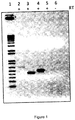
Figure 1 shows RT-PCR of MDA-MB-435 cells expressing MagA and modified ferritin subunits HF+LF. Transcripts were amplified from 1 µg of total RNA, isolated from stably-expressing cells (clone 12), by reverse transcription (RT). The polymerase chain reaction (PCR) was performed on 4 µl RT, using 60°C for the annealing temperature and 35 cycles. Ethidium bromide staining of complimentary DNA (cDNA) separated on a 1% agarose gel shows the product amplified with MagA primers (lane 2, 650 bp), HF primers (lane 3, 450 bp), LF primers (lane 4, 500 bp) and âactin primers (lane 5, 150 bp).Lane 6 contains an RT negative control for α-actin.Lane 1 contains a 1 Kb DNA ladder (Life Technologies). -
Figure 2 shows how iron response elements regulate the expression of ferritin subunits. The iron response element (IRE) is a hairpin loop structure in the 5' noncoding sequences of the mRNA of ferritin heavy and light subunits. Under low cellular iron conditions, when the cell needs to release more iron from storage, ferritin synthesis is downregulated by the interaction of iron response protein (IRP) with the IRE. Iron uptake is increased by stabilizing the synthesis of transferrin receptor mRNA, through IRP-IRE interactions in the 3' noncoding sequence of the transferrin receptor transcript. The reverse is true when cellular iron is high and the cell needs to store more iron and downregulate iron import (Ponka and Lok (1999) The transferrin receptor: role in health and disease. Int J Biochem Cell Biol 31, 1111-37). - The modified ferritin subunits (HF and/or LF) lack the noncoding regulatory IRE sequences; therefore, this modified form of ferritin is not subject to control by IRP.
- The invention provides for co-expression of (a) one or more bacterial magnetotactic genes to amplify iron uptake, compartmentalization and biomineralization in cells, with (b) one or more genes encoding iron handling proteins that together augment(s) and/or regulate the cells' iron pool. The invention provides an improved modified magnetosome that contains more iron or distinct iron biomineral therein with the combination of the expressed genes. As a result, mammalian cells or bacterial cells that are transfected or transformed, respectively, can be more effectively tracked using various imaging technologies.
- As used herein "a higher amount of iron" may encompass one or more of a quantified amount, the size of the iron biomineral produced and the crystal structure of the iron biomineral produced by the present invention.
- The invention is achieved by way of an expression vector (an expression construct) that is a plasmid or virus in which the one or more bacterial magnetotactic genes and the one or more genes encoding iron handling proteins are introduced into a target cell, to commandeer the cell's mechanism for protein synthesis to produce a magnetosome-like structure containing higher levels of iron or a unique iron biomineral. The plasmid further comprises regulatory sequences that act as enhancer and promoter regions and lead to efficient transcription of the gene(s) carried on the expression vector. The expression vector as described herein will produce a significant amount of stable messenger RNA, and therefore proteins.
- The term "gene" will include the use of the gene, functional fragments thereof or cDNA. One of skill in the art would also understand that codon optimization may be used with respect to the genes discussed herein to determine and obtain optimal expression for a particular cell type whether eukaryotic or bacterial. In codon optimization an appropropriate DNA sequence is synthesized specifically to efficiently express the mammalian gene in bacteria for example by first finding out which codons are the most widely used in the species of interest, and synthesize a DNA sequence made up of these. Various commercial programs are available and widely used for such purpose.
- Suitable magnetotactic bacterial genes for use in the present invention may be selected from magnetosome genes MagA, MamK, MMS16 and also those encoded on a magnetosome genomic island and organized into operons such as the magnetosome membrane operons MamAB (17 genes including MamA/Mam22, MamB, MamE, MamJ, MamK, MamM, MamN, MamO, MamP), MamGFDC (4 genes including MamC/Mms13, MamG, MamD, MamF), MamXY (4 genes), the magnetic particle membrane specific operon Mms6 (5 genes including Mms6, Mms16, Mms24), the monocistronic MamW, and genes located outside the magnetosome genomic island, such as ChpA and those on the magnetotaxis operon MtxA and the magnetosome membrane operon MmeA (Kolinko, I., LohBe, A., Borg, S., Raschdorf, O., Jogler, C., Tu, Q., Posfai, M., Tompa, E., Plitzko, J., Brachmann, A., Wanner, G., Muller, R., Zhang, Y., and Schuler, D. (2014) Biosynthesis of magnetic nanostructures in a foreign organism by transfer of bacterial magnetosome gene clusters, Nat Nanotechnol 9, 193-197). Combinations of any of these genes and operons are envisaged in the invention. While bacterial operons are not normally expressed in mammalian cells, the multiple genes can be expressed each under control of its own promoter and selection marker. In this embodiment a single transient transfection can be done or multiple sequential transfections. These magnetotactic genes are utilized to amplify iron biomineralization in cells, in combination with the mammalian genes such as those encoding ferritin overexpression that is not subject to regulation by the iron response elements (
Figure 2 ). These reporter gene constructs for use in MRI, permit high resolution molecular imaging and non-invasive, in vivo tracking of cellular and molecular events. - Suitable genes encoding iron handling proteins may be selected from the group consisting of ferritin heavy and light subunits lacking the IRE and any protein subject to IRE regulation. Representative proteins include the transferrin receptor (UniProt P02786), transferrin (Transferrin St. Edward's University. 2005-07-18. Retrieved 2009-04-24; Yang F, Lum JB, McGill JR, Moore CM, Naylor SL, van Bragt PH, Baldwin WD, Bowman BH (May 1984). "Human transferrin: cDNA characterization and chromosomal localization". Proceedings of the National Academy of Sciences of the United States of America 81 (9): 2752-6. doi:10.1073/pnas.81.9.2752. PMC 345148. PMID 6585826), Iron Response Proteins (IRP) 1 and 2 (UniProt P21399; Samaniego F, Chin J, Iwai K, Rouault TA, Klausner RD (December 1994). Molecular characterization of a second iron-responsive element-binding protein, iron ), ferroportin (UniProt Q9NP59 and Q9JHI9), DMT1 (UniProt P49281), STEAP3 (UniProt Q658P3) and hepcidin (UniProt P81172). In aspects of the invention the genes may be mammalian as is understood by one of skill in the art.
- Ferritin is a protein of 450 kDa consisting of 24 subunits that is present in every cell type. In vertebrates, these subunits are both the light (L) (UniProt 02792) and the heavy (H) type (UniProt P02794) with an apparent molecular weight of 19 kDa or 21 kDa respectively; their sequences are about 50% homologous. Ferritin genes are highly conserved between species. All vertebrate ferritin genes have three introns and four exons. The tyrosine residue at amino acid position 27 in the H subunit is thought to be associated with biomineralization.
- The expression systems of the present invention can be used with any type of eukaryotic and prokaryotic cell. One of skill in the art would understand that the invention is applicable to eukaryotes in general, birds, plants, fungi, insects, fish, yeast, amphibians, reptiles, and invertebrates such as insects. In aspects the eukaryotic cells are mammalian, and may be human or animal. Mammalian cells may include for example cancer cells, nerve cells, muscle cells, cardiac cells, epithelial cells, adipocytes and stem cells. The expression systems of the present invention may also be used with prokaryotic cells, that is bacterial cells. Bacterial cells that are native to the gut of mammals can also be transformed with the expression systems of the present invention. Suitable bacteria may belong to the genera Bacteroides, Clostridium, Fusobacterium, Eubacterium, Ruminococcus, Peptococcus, Peptostreptococcus, Bifidobacterium, Escherichia and Lactobacillus. In this embodiment, the gastrointestinal tract of a mammal can be effectively imaged when the transformed bacteria are reintroduced into a mammal's gut.
- Suitable promoters for use in the present invention are generally known in the field of molecular biology and may include inducible promoters, constitutive promoters and tissue specific promoters as is understood by one of skill in the art. Viral promoters may be chosen from, for example, cytomegalovirus (CMV; human immediate early), Rous sarcoma virus (RSV), simian virus 40 (SV40), and the long terminal repeat from Moloney murine leukemia virus (LTR). Mammalian promoters may be chosen from, for example, the elongation factor (EF) 1α promoter, widely useful in mammalian cells; the cytokeratin 18 (K18) promoter; the cytokeratin 19 (K19) promoter; the tissue kallikrein promoter (Kall); the amylase promoter (AMY); and rat aquaporin-5 (rAQP5). In addition to these promoters that provide constitutive expression, the invention covers the use of more selective promoters that respond to particular cellular transcription factors, such as the minimal NIS (sodium iodide symporter) promoter that responds to Nkx2.5 (Dentice et al. (2004), Mol Cell Biol 24, 7863-7877). The combination of genes expressed to produce a magnetosome-like structure may be regulated by different promoters, constitutive or selective.
- The invention as incorporated into plants and plant cells, one of skill in the art understands that several plant expression vectors are based on the Ti plasmid of Agrobacterium tumefaciens, where DNA to be inserted into plant is cloned into the T-DNA, a stretch of DNA flanked by a 25-bp direct repeat sequence at either end, and which can integrate into the plant genome. The T-DNA also contains the selectable marker. The Agrobacterium provides a mechanism for transformation, integration of into the plant genome, and the promoters for its vir genes may also be used for the cloned genes. Plant viruses may be used as vectors when Agrobacterium is not suitable for use in a specific plant.. Examples of plant virus that can be used are the tobacco mosaic virus (TMV), potato virus X, and cowpea mosaic virus. The protein may be expressed as a fusion to the coat protein of the virus and is displayed on the surface of assembled viral particles, or as an unfused protein that accumulates within the plant. Expression in plant using plant vectors is often constitutive, and a commonly used constitutive promoter in plant expression vectors is the cauliflower mosaic virus (CaMV) 35S promoter.
- Eukaryotic expression systems employing insect cell hosts are based upon one of two vector types: plasmid or plasmid-virion hybrids, the latter being most commonly used. The typical insect host is the common fruit fly, Drosophila melanogaster,, however, other insect hosts include mosquito (Aedes albopictus), fall army worm (Spodoptera frugiperda), cabbage looper (Trichoplusia ni), salt marsh caterpillar (Estigmene acrea) and silkworm (Bombyx mori). In most all cases, heterologous protein overexpression occurs in suspension cell cultures. The exception, and one of the advantages of plasmid-virion systems, is that the recombinant virus may also be injected into larval host hemocel or literally fed to the mature host.
- Plasmid-based vector systems provide a mechanism for both transient and long-term expression of recombinant protein and are commercially available such as the Drosophila Expression System (DES) (Invitrogen™). The transfection of competent D. melanogaster cells with engineered plasmid will mediate the transient (2-7 days) expression of heterologous protein. Establishing transformed cells that will express protein for longer time periods requires that the host cells be cotransfected with a "selection" vector, resulting in the stable integration of the expression cassette into the host genome. Constitutive expression is mediated using the Ac5 Drosophila promoter, whereas a metallothionein promoter guides copper-inducible expression. The DES vectors are designed with multiple cloning sites for insertion of the heterologous protein gene in any of three reading frames. A choice of vectors also provides for the expression of a variety of C-terminal fusion tags: V5 epitope for identification of expressed protein with V5 epitope antibody, polyhistidine peptide for simplified purification with metal chelate affinity resin, and the BiP secretion leader peptide.
- To amplify cellular iron storage for the purpose of biomineralization, in one nonlimiting embodiment of the present invention MagA was overexpressed in combination with a dysregulated form of ferritin. In the absence of the IRE, ferritin does not respond to cellular
iron binding proteins - The invention provides genetically engineered cells that produce magnetosome-like structures and HF and/or LF iron storage, as contrast agents for mapping and/or imaging of organs, tissues, cells, sub-cellular structures, proteins and peptides in living organisms both in vivo and in vitro. The method of the invention genetically alters cells to enhance iron storage in HF and/or LF and produce magnetosome-like structures as dense core vesicles that form MR contrast agents in the cell. The invention also provides genetic constructs from magnetotactic bacterial genes, including those of the magnetosome, and from mammalian genes, such as HF and/or LF, that may be expressed from vectors bearing inducible promoters or encoding other useful genes for targeting cells or for therapeutic treatments that can be followed by functional imaging or long-term tracking of transfected cells.
- It is herein demonstrated that the magnetotactic bacterial gene and putative iron transporter, MagA, enhances MR contrast in iron-supplemented cells and in tissue formed from transplanted MagA-expressing cells. The MR contrast has been compared in clonal lines of MagA expressing cells to those expressing ferritin heavy and light chains, lacking iron response elements (HF and/or LF). The transverse relaxation rates, R2' in particular, are sensitive to cellular iron content. Changes in this MR parameter may be used to quantify cellular iron and improve the specificity of detection. The relationship between R2 and inter-echo time is investigated to improve cellular detection of iron using molecular MRI.
- The present invention demonstrates the triple expression system/construct of the present invention for use to further augment MR contrast (
Figure 1 , Table 1). - The invention encompasses various expression strategies for magnetotactic bacterial, magnetosome and mammalian genes like the modified ferritin genes. Since each set of genes may provide contrast in the context of mammalian cell expression, they may operate independently and in an additive manner. Thus the possibility of distinct MR signatures are encompassed and within the scope of the invention for all types of genes and cells selected.
- The invention has particular use with eukaryotic cells that express and overexpress one or more magnetotactic bacterial and/or magnetosome proteins as magnetosome-like structure contrast agents, in combination with mammalian genes like HF and/or LF for iron storage, permitting in vivo MRI, MPI, hybrid MR or x-ray imaging studies to follow the localization, proliferation, and long-term tracking of iron loaded cells. These cells may be tissue specific and permit non-invasive in vivo imaging of specific physiological or molecular functions.
- Imaging systems that can be used with the present invention include at least the following, which can also be used in combination: positron emission tomography (PET), magnetic resonance imaging (MRI), gamma-ray imaging camera (GREI-II), computer tomography (CT), Single photon emission computed tomography (SPEC), bioluminescence tomography (BLT) and magnetic particle imaging (MPI). Magnetic particle imaging (MPI) is a new modality for imaging distributions of iron oxide nanoparticle tracers in vivo with good contrast, high sensitivity, and good spatial resolution, and thus is promising for clinical imaging in angiography and oncology. MPI requires high-quality iron oxide nanoparticle tracers with tailored magnetic and surface properties to achieve its full potential. Thus this technique is suitable for use with the presently claimed invention.
- The present invention has further wide potential clinical uses such as, but not limited to, those described below.
- Mammalian cells change their iron requirements to meet a given physiological need in both health (13) and disease (14). We demonstrate that both MagA and HF and/or LF expression respond to changes in extracellular iron while cellular iron homeostasis, as measured by changes in the level of transferrin receptor, appears intact. Expression of MagA and HF and/or LF may be utilized as sensors of changes in physiological iron levels.
- Using MR detection methods, such as transverse relaxation rates, changes in cellular iron levels and iron biomineralization may be measured using a combination of magnetotactic bacterial genes, such as MagA, including magnetosome genes, and HF and/or LF. This provides a noninvasive tool for assessing the iron status of a given cell type.
- The onset of changes in iron uptake or export and monitoring fluctuations in iron handling may be tracked by expressing a combination of magnetotactic bacterial genes, like MagA, including magnetosome genes, and HF and/or LF. Since transverse relaxation rates are a quantifiable measure of MR contrast and are related to cellular iron content, fluctuations in cellular iron throughout the cell's life cycle may be monitored.
- Iron biomineralization may be amplified in mammalian cells, using a combination of magnetotactic bacterial genes, like MagA, including magnetosome genes, and HF and/or LF. This amplification of gene-based iron contrast will improve MR detection of cells without introducing cytotoxicity or altering endogenous cellular programming.
- Under circumstances where altering the cell's iron handling abilities is desirable, a combination of magnetotactic bacterial genes, like MagA, including magnetosome genes, and HF and/or LF may be genetically engineered in the cell in order to introduce changes in iron handling. Such changes may influence the cell's labile iron pool and iron import, storage and export by creating a magnetosome-like compartment. Synthesis of a magnetosome-like structure may be genetically associated with a cellular function, reflecting an endogenous activity or overexpression of a desired macromolecule.
- Bacteria harboring a combination of magnetotactic bacterial genes, like MagA, including magnetosome genes, and HF and/or LF may interact with mammalian cells and localize to sites within the body, such as the gut. This interaction may be detected by MRI or its hybrid modalities and signal the onset of a physiological process, be it in health or illness. This type of interaction has been identified as inter-kingdom signalling (15).
- Cancer cells generally consist of a proliferative phenotype; however, dormant cancer cells may assume a quiescent state until reactivated by a stimulatory signal. It will be possible to use the combination of magnetotactic bacterial genes, like MagA, including magnetosome genes, and HF and/or LF to track the changes in cancer cell activity.
- a) Many transplanted cancer cells will form tumour xenografts in animal models (6). A combination of magnetotactic bacterial genes, like MagA, including magnetosome genes, and HF and/or LF may be used to monitor tumour growth and metastasis in tumour xenografts using MRI.
- b) P19 cells are a mouse embryonic carcinoma with stem cell characteristics and will differentiate into each of the 3 tissue types: endoderm, mesoderm and ectoderm. Introduction of a combination of magnetotactic bacterial genes, like MagA, including magnetosome genes, and HF and/or LF may be used to monitor cancer cell differentiation by MRI and better understand stem cell activity (11). Using this method, features of cancer stem cell activity such as the fraction of cells that differentiate in response to a given stimulus can be monitored.
- c) Using specific promoters to drive contrast gene expression, transcription factor activities will be examined that are important in a given type of cancer. For example, a combination of magnetotactic bacterial genes, like MagA, including magnetosome genes, and HF and/or LF may be expressed to monitor Nkx2.5 activity by reporter gene expression (12). This method will also permit simultaneous tracking of more than one oncoprotein activity by using different promoters for each reporter gene involved in generating MR contrast.
- d) One of the advantages of non-invasive imaging is the ability to investigate environmental cues of tumour formation within an in vivo context (16). Expressing a combination of magnetotactic bacterial genes, like MagA, including magnetosome genes, and HF and/or LF may be used to examine the context of tissue-specific tumour growth and metastasis. This method will combine the superior anatomy of MRI with molecular function and reveal the similarities and differences in cancer growth among transformed cell types and target organs.
- e) Combinations of magnetotactic bacterial genes, like MagA, including magnetosome genes, and HF and/or LF may be expressed in cells derived from primary tumours to non-invasively track growth and metastasis of human disease in orthotopic animal models. This will provide a measure of personalized medicine for developing therapies and predicting outcomes ahead of, or concurrent to, the course of human disease.
- After a heart attack, the patient is often left with a large amount of scar tissue in place of normally contracting heart muscle. If the amount of tissue is large enough, the patient's physical activity is limited and they may go on to chronic heart failure and death. Heart transplantation remains the treatment of choice, but the number of available organs will never meet more than a small fraction of the growing demand (17, 18). As another treatment option, stem cell therapy to regenerate the damaged heart is being aggressively pursued (19). The present invention has use to image, quantify and monitor the progression of transplanted stem cells from undifferentiated to differentiated states, as the cells repopulate the scar tissue with normally functioning heart tissue. The use of magnetosome-like structures, augmented by increased ferritin, to track the progress of tissue regeneration has both research and therapeutic applications.
- a) Prior to transplantation, stem cells may be transfected with genes that produce magnetosome-like structures and HF and/or LF genes of the invention to follow the fate of the transplanted cells by MR imaging. This would follow the original stem cell population, from transplantation to lineage commitment and differentiation, by the combination of magnetosome-like structures and augmented ferritin storage.
To monitor the switch from proliferation to differentiation, cells would be transfected with magnetotactic bacterial, including magnetosome, and HF and/or LF constructs in which the promoter responds to stage-specific differentiation signals. This will indicate early and late events in stem cell differentiation and localize these events within the injured heart. - b) Different types of cellular differentiation may be monitored by transfecting stem cells with magnetotactic bacterial, including magnetosome, and HF and/or LF constructs of the invention that respond to transcription factors present in specific types of cardiac cells, such as smooth muscle cells, endothelial cells or myocytes. This will indicate the number of stem cells needed for successful treatment and how efficient stem cells are at producing functional myocardium.
- c) The rate of stem cell loss after transplantation may also be monitored by placing magnetotactic bacterial, including magnetosome, and HF and/or LF genes behind promoters sensitive to cell death signals, including apoptosis, necrosis, autophagy and cornification (20). Alternatively, magnetotactic bacterial, including magnetosome, and HF and/or LF genes may be co-expressed with early markers of cell death. In this context, magnetosome-like structure formation and augmented ferritin storage protein would localize the extent of cell death that undermines stem cell therapy in a given organ.
- Iron misregulation has been acknowledged in a number of diseases, including Alzheimer's disease (21). The Amyloid Precursor Protein (APP) has an IRE in the 5' untranslated region of the mRNA. The role of biometal dysregulation and oxidative stress in Aβ amyloid formation may be detected by the expression of magnetotactic bacterial, like MagA, including magnetosome, and HF and/or LF genes in neural cells.
- a) Iron uptake may be monitored by the combination of magnetotactic bacterial, like MagA, including magnetosome, and HF and/or LF gene expression, which respond to iron supplementation. In this example, MagA and HF and/or LF are acting like a sensor of iron. MRI may be used to detect iron accumulation ahead of the amyloid plaque. The constructs and methods of the invention may also reveal the balance of IRE+/IRE- isoforms of key proteins in health and disease.
- b) Changes in APP expression may be monitored using MRI by expression of the combination of magnetotactic bacterial genes, like MagA, including magnetosome, and HF and/or LF genes using vectors that respond to the APP promoter.
- c) Potentially, therapies will be devised such that MagA and HF and/or LF remove cytotoxic iron by storing it in a safe form: as an iron biomineral in modified ferritin and/or a magnetosome-like structure.
- d) New PET probes have recently been developed to Aβ amyloid and tau proteins to enable imaging of the onset and progression of Alzheimer's disease. This paves the way for PET/MRI in pre/clinical models of the disease. Magnetotactic bacterial, including MagA and magnetosome, and HF and/or LF gene expression in cells, tissues and animals can be now used for the purpose of PET/MR imaging in this disease.
- The invention relates to the use of magnetotactic bacterial genes, including magnetosome genes, and mammalian genes, including the modified ferritin subunits, in combinations that augment or modulate MR contrast by regulating cellular iron biomineralization and/or the formation of magnetosome-like structures. These structures may generate all or part of the magnetosome, including modifications to iron uptake, iron biomineralization, iron compartmentalization and the arrangement of magnetosome-like structures within the cell.
- a) MamJ and MamK are magnetosome proteins needed for the alignment of magnetosomes into a chain, connected to the cytoskeleton. Disruption of this interaction removes the chain-like structure and results in magnetosomes that are either dispersed or in clusters within the cytoplasm. Since each magnetosome normally constitutes a single magnetic domain, their arrangement within the cell will influence the MR contrast signal.
- b) Regulatory proteins such as Maml may be used to dictate when the magnetosome-like structure will be properly synthesized (Kolinko, I., LohBe, A., Borg, S., Raschdorf, O., Jogler, C., Tu, Q., Posfai, M., Tompa, E., Plitzko, J., Brachmann, A., Wanner, G., Muller, R., Zhang, Y., and Schuler, D. (2014) Biosynthesis of magnetic nanostructures in a foreign organism by transfer of bacterial magnetosome gene clusters, Nat Nanotechnol 9, 193-197). In this way essential magnetosome genes may be used to regulate when and where the magnetosome-like structure will be expressed.
- c) The iron biomineral in magnetosomes is partially regulated by Mms6. In this way, characteristics of the iron biomineral may be altered by select gene expression to influence the MR signal. Such regulation may include the size and composition of the biomineral, giving hematite, magnetite (Fe3O4), greigite (Fe3S4), or another biomineral.
- d) Using select magnetotactic and mammalian genes to impart distinct magnetic signatures for MRI, changes in cellular activity may be tracked by expressing different combinations of these genes at different developmental stages. In this embodiment, the MR signal from a magnetosome-like structure appears at the onset of promoter stimulation by a given transcription factor. As that activity changes so does the MR signal. In addition, as new activities appear the MR signal may be modified again by genetically programming a different feature of the magnetosome-like structure to respond to these subsequent cellular factors. This strategy may be used to understand the factors that dictate a disease process, ahead of permanent tissue damage.
- Magnetic Particle Imaging (MPI) is an emerging area of MRI where nanoparticles of iron are non-invasively tracked and quantified (Saritas, EU, Goodwill, PW, Croft, LR, Konkle, JJ, Lu, K, Zheng, B and Conolly, SM (2013) Magnetic Particle Imaging (MPI) for NMR and MRI Researchers. J Magn Reson 229,116-126). Magnetosome-like structures can be used for MPI where genes from magnetotactic bacteria are co-expressed with mammalian genes to create the iron nanoparticles that have specific MPI signals Biomed Tech (Berl). 2013 Dec;58(6):493-507. doi: 10.1515/bmt-2012-0058.Tailoring the magnetic and pharmacokinetic properties of iron oxide magnetic particle imaging tracers. Ferguson RM, Khandhar AP, Arami H, Hua L, Hovorka O, Krishnan KM.
- Bacteria that live in the human gut may be genetically programmed to co-express magnetotactic bacterial genes, including magnetosome genes, and human genes, like the ferritin heavy and light subunits, to produce magnetosome-like structures.
- a) In an aspect of the invention, these gut microbes may secrete therapeutic proteins to treat intestinal diseases and the magnetosome-like structures they harbour will permit non-invasive tracking of their location. Since this type of therapy may be stopped by flushing out the microbes with the use of laxatives, MRI can be used to verify the removal the magnetic bacteria.
- b) In another aspect of the invention, gut bacteria may be engineered for production of magnetosome-like structures once a stimulatory signal is generated by the subject to initiate formation of the contrast agent in situ. The stimulatory agent may be endocrine, paracrine or autocrine. Furthermore, the combination of genes expressed may be programmed to give a unique magnetosome-like structure that depends on the nature of the stimulant.
- c) Administration and monitoring of this therapy may be externally regulated. Gut microbes expressing a combination of magnetotactic bacterial and human genes to form magnetosome-like structures may be administered and allowed to migrate to the desired location in the GI tract, prior to activation of therapeutic gene expression by an oral antibiotic like tetracycline. This would ensure that the bacteria have reached the correct target along the intestine and minimize side effects elsewhere, such as in the stomach or bowel.
- Transcripts were amplified from 1 µg of total RNA, isolated from stably-expressing cells (clone 12), by reverse transcription (RT). The polymerase chain reaction (PCR) was performed on 4 µl RT, using 60°C for the annealing temperature and 35 cycles. Ethidium bromide staining of complimentary DNA (cDNA) separated on a 1% agarose gel shows the product amplified with MagA primers (
lane 2, 650 bp), HF primers (lane 3, 450 bp), LF primers (lane 4, 500 bp) and α-actin primers (lane 5, 150 bp).Lane 6 contains an RT negative control for β-actin.Lane 1 contains a 1 Kb DNA ladder (Life Technologies).Table 1. ICP-MS∼ analysis of iron and zinc content in MDA-MB-435 cells expressing MagA and HF+LF Sample [Protein]* (mg/ml) [Fe] (µg/ml) Amount of Fe / Protein (µg/mg) [Zn] (µg/ml) Amount of Zn / Protein (µg/mg) Ratio of Fe/Zn RIPA / inhibitors‡ 0 0 ND 0 ND ND 1. Cells minus Fe 1.00 0.029 0.029 0.108 0.108 0.273 2. Cells minus Fe 1.00 0.038 0.038 0.112 0.112 0.342 3. Cells plus Fe 1 day1.00 0.341 0.341 0.115 0.115 2.969 4. Cells plus Fe 3 days1.00 0.471 0.471 0.123 0.123 3.835 5. Cells plus Fe 5 days1.00 0.510 0.510 0.085 0.085 5.973 ∼ ICP-MS, Inductively-coupled plasma mass spectrometry
^ Stably-expressing cells (clone 12) were cultured in the presence (plus Fe) and absence (minus Fe) of medium supplemented with iron: 250 µM ferric nitrate.
* Protein was measured by the BCA assay: Smith et al. (1985) Anal. Biochem. 150, 76-85.
‡ Cells were lysed in RIPA buffer (10 mM Tris-HCI pH 7.5 / 140 mM NaCl / 1% NP-40 / 1% sodium deoxycholate / 0.1% SDS) containing protease inhibitors (Complete Mini, Roche Diagnostic Systems).Table 2. Transverse relaxation ratesa in MDA-MB-435 cells co-expressing MagA and the modified ferritin subunits (HF+LF) Sampleb R2* R2 MagA+HF+LF 13.65 ± 0.15 11.74 ± 0.64 MagA+HF+LF+Fe 14.07 ± 0.33 13.20 ± 0.50 a Cells were mounted in spherical gelatin phantoms for measurement of relaxation rates at 3T following the method of Sengupta et al, 2014. Results are the average of 2 experiments.
b Stably-expressing cells (clone 12) were cultured in the presence (+Fe) and absence of medium supplemented with iron: 250 µM ferric nitrate. -
- 1. Komeili, A. (2007) Molecular mechanisms of magnetosome formation, Annu Rev Biochem 76, 1-27.
- 2. Zurkiya, O., Chan, A. W., and Hu, X. (2008) MagA is sufficient for producing magnetic nanoparticles in mammalian cells, making it an MRI reporter, Magn Reson Med 59, 1225- 1231.
- 3. Benoit, M., Mayer, D., Barak, Y., Chen, I., Hu, W., Cheng, Z., Wang, S., Spielman, D., Gambhir, S., and Matin, A. (2009) Visualizing implanted tumors in mice with magnetic resonance imaging using magnetotactic bacteria, Clin Cancer Res 15, 5170-5177.
- 4. Goldhawk, D., Rohani, R., Sengupta, A., Gelman, N., and Prato, F. (2012) Using the magnetosome to model effective gene-based contrast for magnetic resonance imaging, WIRES Nanomed Nanobiotechnol Epub ahead of print, doi: 10. 1002/wnan.1165.
- 5. Goldhawk, D., Lemaire, C., McCreary, C., McGirr, R., Dhanvantari, S., Thompson, R., Figueredo, R., Koropatnick, J., Foster, P., and Prato, F. (2009) Magnetic resonance imaging of cells overexpressing MagA, an endogenous contrast agent for live cell imaging, Mol Imaging 8, 129-139.
- 6. Rohani, R., Figueredo, R., Bureau, Y., Koropatnick, J., Foster, P., Thompson, R., Prato, F., and Goldhawk, D. (2013) Imaging tumor growth non-invasively using expression of MagA or modified ferritin subunits to augment intracellular contrast for repetitive MRI, Mol Imaging Biol , Epub ahead of print, doi: 10.1007/s11307-11013-10661-11308.
- 7. Anderson, C., Shen, M., Eisenstein, R., and Leibold, E. (2012) Mammalian iron metabolism and its control by iron regulatory proteins, Biochem Biophys Acta 1823, 1468-1483.
- 8. Sengupta, A., Quiaoit, K., Thompson, R., Prato, F., Gelman, N., and Goldhawk, D. (2013) Biophysical features of MagA expression in mammalian cells: implications for MRI contrast, Frontiers in Microbiotechnology, Ecotoxicology and Bioremediation , submitted
- 9. Lee, C., Thompson, R., Prato, F., Goldhawk, D., and Gelman, N. (2013) Transverse relaxation rate (R2) dependence on refocusing pulse interval (2tau) in MagA expressing breast/melanoma tumor cells, In World Molecular Imaging Conference, Abstract LBAP 129, Savannah, USA.
- 10. Sengupta, A., Rohani, R., Thompson, R., Prato, F., Goldhawk, D., and Gelman, N. (2012) MRI relaxation rates in tumour cells expressing the MagA reporter gene In International Society for Magnetic Resonance in Medicine Scientific Workshop on MRI-based Cell Tracking, Abstract 26, Miami, USA.
- 11. Liu, L., McGirr, R., Bondoc, A., Thompson, R., Prato, F., Goldhawk, D., and Hoffman, L. (2013) Non-Invasively Targeting Myocardial Progenitor Cells for the Treatment of Duchenne Muscular Dystrophy-Related Cardiomyopathy using MRI/PET, In Stem Cell Network, Abstract accepted, Banff, Canada.
- 12. Quiaoit, K., Koropatnick, J., Gelman, N., and Goldhawk, D. (2013) MagA Reporter Gene Expression for Magnetic Resonance Imaging of Breast Carcinoma, In London Health Research Day, Biomedical Imaging & Engineering Abstract 33, London, Canada.
- 13. Recalcati, S., Locati, M., Marini, A., Santambrogio, P., Zaninotto, F., De Pizzol, M., Zammataro, L., Girelli, D., and Cairo, G. (2010) Differential regulation of iron homeostasis during human macrophage polarized activation, Eur J Immunol 40, 824-835.
- 14. Pantopoulos, K., Porwal, S., Tartakoff, A., and Devireddy, L. (2012) Mechanisms of mammalian iron homeostasis, Biochemistry 51, 5705-5724.
- 15. Braun, R. (2013) When harmless bacteria go bad, Scienfific American .
- 16. Ghajar, C., Peinado, H., Mori, H., Matei, I., Evason, K., Brazier, H., Almeida, D., Koller, A., Hajjar, K., Stainier, D., Chen, E., Lyden, D., and Bissell, M. (2013) The perivascular niche regulates breast tumour dormancy, Nat Cell Biol 15, 807-817.
- 17. IHME, and Cohen, J. (2012) Health metrics. A controversial close-up of humanity's health, Science 338, 1414-1416.
- 18. News. (2013) World Disease by the Numbers, Science 341, 325 .
- 19. Makkar, R., Smith, R., Cheng, K., Malliaras, K., Thomson, L., Berman, D., Czer, L., Marban, L., Mendizabal, A., Johnston, P., Russell, S., Schuleri, K., Lardo, A., Gerstenblith, G., and Marban, E. (2012) Intracoronary cardiosphere-derived cells for heart regeneration after myocardial infarction (CADUCEUS): a prospective, randomised .
- 20. Kroemer, G., Galluzzi, L., Vandenabeele, P., Abrams, J., Alnemri, E., Baehrecke, E., Blagosklonny, M., El-Deiry, W., Golstein, P., Green, D., Hengartner, M., Knight, R., Kumar, S., Lipton, S., Malorni, W., Nuñez, G., Peter, M., Tschopp, J., Yuan, J., Piacentini, M., Zhivotovsky, B., and Melino, G. (2009) Classification of cell death: recommendations of the Nomenclature Committee on Cell Death 2009, Cell Death Differ 16, 3-11.
- 21. Huang, X., Moir, R., Tanzi, R., Bush, A., and Rogers, J. (2004) Redox-active metals, oxidative stress, and Alzheimer's disease pathology, Ann NY Acad Sci 1012, 153-163.
- 22. Ferguson RM, Khandhar AP, Arami H, Hua L, Hovorka O, Krishnan KM.Biomed Tech (Berl). 2013 Dec;58(6):493-507. doi: 10.1515/bmt-2012-0058. Tailoring the magnetic and pharmacokinetic properties of iron oxide magnetic particle imaging tracers.
Claims (12)
- An expression system for augmenting and/or regulating iron levels in cells, preferably a mammalian cell, more preferably a human or animal cell, for example, a human cell selected from the group consisting of nerve, muscle, epithelial, adipocyte and stem cell, or a bacterial cell native to the gut of mammals, for example a bacterial cell belonging to a genera selected from the group consisting of Bacteroides, Clostridium, Fusobacterium, Eubacterium, Ruminococcus, Peptococcus, Peptostreptococcus, Bifidobacterium, Escherichia and Lactobacillus, said expression system comprising (a) one or more bacterial magnetotactic genes to amplify iron uptake, compartmentalization and biomineralization in cells, and (b) one or more genes each encoding one or more eukaryotic iron handling proteins, wherein (a) and (b) are under control of one or more promoters; said promoters preferably selected to provide constitutive expression and are more preferably viral promoters selected from the group consisting of cytomegalovirus (CMV; human immediate early), Rous sarcoma virus (RSV), simian virus 40 (SV40), and the long terminal repeat from Moloney murine leukemia virus (LTR); or human promoters selected from the group consisting of elongation factor (EF) 1α promoter, cytokeratin 18 (K18) promoter; cytokeratin 19 (K19) promoter; tissue kallikrein promoter (Kall); amylase promoter (AMY); and rat aquaporin-5 (rAQP5), most preferably said one or more promoters are selective promoters that respond to particular cellular transcription factors and are selected from the group consisting of minimal NIS (sodium iodide symporter) promoter and transcription factor Nkx-2.5; and, optionally, one or more selection markers.
- The expression system of claim 1, wherein said one or more bacterial magnetotactic genes is selected from the group consisting of MagA, MamK, MMS16, MtxA, MmeA, MamW and ChpA and genes encoded on a magnetosome genomic island and organized into operons selected from the group consisting of: MamAB, preferably comprising 17 genes including MamA/Mam22, MamB, MamE, MamJ, MamK, MamM, MamO and MamP; MamGFDC, preferably comprising 4 genes including MamC/Mms13, MamG, MamD and MamF; MamXY, preferably comprising 4 genes; and Mms6, preferably comprising 5 genes including Mms6, Mms16 and Mms24.
- The expression system of any of the preceding claims, wherein said one or more iron handling proteins are a protein subject to regulation by an iron responsive element, said one or more iron handling proteins preferably selected from the group consisting of ferritin heavy subunit lacking an iron responsive element (IRE), a ferritin light subunit lacking an iron responsive element (IRE), transferrin receptor, transferrin, Iron Response Proteins (IRP) 1 and 2, ferroportin, DMT1, STEAP3, hepcidin and combinations thereof.
- The expression system of any one of the preceding claims wherein said one or more iron handling proteins are mammalian, preferably human or animal.
- The expression system of any one of the preceding claims, wherein the one or more iron handling proteins comprise both a ferritin heavy subunit, preferably modified to lack an iron response element (IRE) and a ferritin light subunit, preferably modified to lack an IRE.
- The expression system of any of the preceding claims, wherein said one or more magnetotactic bacterial genes is MagA.
- A cell transformed with the expression system of any one of the preceding claims.
- The cell of claim 7, wherein said cell is detected using an imaging system selected from the group consisting of positron emission tomography (PET), magnetic resonance imaging (MRI), gamma-ray imaging camera (GREI-II), computer tomography (CT), Single photon emission computed tomography (SPEC), bioluminescence tomography (BLT), magnetic particle imaging (MPI) and combinations thereof.
- A mammalian cell transfected with a plasmid comprising MagA and mammalian ferritin heavy and light subunit genes lacking the IRE in the 5' untranslated regions under control of one or more promoters and, optionally, one or more selection markers.
- The expression system of any one of claims 1 to 6 for use in one or more of the following:detecting changes in cellular iron;tracking cancer growth and metastasis;image, quantify and/or monitor progression of transplanted stem cells;tracking cardiovascular inflammation disorders;tracking neurodegenerative disorders; andcell tracking in vivo,wherein co-expression of said one or more magnetotactic bacterial genes and said one or more iron handling proteins forms a magnetosome-like structure containing augmented amounts of iron therein that can be detected using an imaging system selected from the group consisting of positron emission tomography (PET), magnetic resonance imaging (MRI), gamma-ray imaging camera (GREI-II), computer tomography (CT), Single photon emission computed tomography (SPEC), bioluminescence tomography (BLT), magnetic particle imaging (MPI) and combinations thereof.
- A method for making a cell having an increased amount of iron therein as compared to a wild type cell, the method comprising; transfecting a cell with a construct comprising one or more magnetotactic bacterial genes and modified ferritin heavy and light subunits with or lacking an iron response element (IRE) under the control of one or more promoters and cultivating said cell under conditions suitable to effect expression of the one or more magnetotactic bacterial genes and modified ferritin heavy and light subunits.
- A method for detecting changes in intracellular iron, the method comprising co-expressing a magnetotactic bacterial gene and a eukaryotic iron handling gene in a cell utilizing the expression system of any one of claims 1-6 and monitoring said cell using an imaging method.
Applications Claiming Priority (2)
| Application Number | Priority Date | Filing Date | Title |
|---|---|---|---|
| US201461944146P | 2014-02-25 | 2014-02-25 | |
| PCT/CA2014/000555 WO2015127527A1 (en) | 2014-02-25 | 2014-07-08 | Co-expression of magnetotactic bacterial genes and genes encoding iron handling proteins in cells |
Publications (3)
| Publication Number | Publication Date |
|---|---|
| EP3110952A1 EP3110952A1 (en) | 2017-01-04 |
| EP3110952A4 EP3110952A4 (en) | 2017-11-15 |
| EP3110952B1 true EP3110952B1 (en) | 2019-03-27 |
Family
ID=54008070
Family Applications (1)
| Application Number | Title | Priority Date | Filing Date |
|---|---|---|---|
| EP14884055.6A Active EP3110952B1 (en) | 2014-02-25 | 2014-07-08 | Co-expression of magnetotactic bacterial genes and genes encoding iron handling proteins in cells |
Country Status (4)
| Country | Link |
|---|---|
| US (2) | US11077215B2 (en) |
| EP (1) | EP3110952B1 (en) |
| CA (1) | CA2940654A1 (en) |
| WO (1) | WO2015127527A1 (en) |
Families Citing this family (2)
| Publication number | Priority date | Publication date | Assignee | Title |
|---|---|---|---|---|
| EP3169779A4 (en) * | 2014-07-15 | 2018-03-21 | Bell Biosystems, Inc. | Eukaryotic cells with artificial endosymbionts for multimodal detection |
| CN116077680B (en) * | 2022-07-25 | 2024-03-26 | 汕头大学医学院第一附属医院 | Targeting therapeutic preparation for depositing iron neurons and application thereof |
Family Cites Families (2)
| Publication number | Priority date | Publication date | Assignee | Title |
|---|---|---|---|---|
| ATE519777T1 (en) * | 2006-06-08 | 2011-08-15 | Multi Magnetics Inc | MAGNETOSOME GENE EXPRESSION IN EUKARYOTIC CELLS |
| US9387264B2 (en) * | 2012-04-05 | 2016-07-12 | Yeda Research And Development Co. Ltd. | Reporter genes for magnetic resonance imaging and methods of use thereof |
-
2014
- 2014-07-08 WO PCT/CA2014/000555 patent/WO2015127527A1/en not_active Ceased
- 2014-07-08 US US15/121,653 patent/US11077215B2/en active Active
- 2014-07-08 CA CA2940654A patent/CA2940654A1/en not_active Abandoned
- 2014-07-08 EP EP14884055.6A patent/EP3110952B1/en active Active
-
2021
- 2021-07-15 US US17/376,484 patent/US20210338843A1/en not_active Abandoned
Non-Patent Citations (1)
| Title |
|---|
| None * |
Also Published As
| Publication number | Publication date |
|---|---|
| EP3110952A4 (en) | 2017-11-15 |
| US11077215B2 (en) | 2021-08-03 |
| US20160367706A1 (en) | 2016-12-22 |
| US20210338843A1 (en) | 2021-11-04 |
| CA2940654A1 (en) | 2015-09-03 |
| WO2015127527A1 (en) | 2015-09-03 |
| EP3110952A1 (en) | 2017-01-04 |
Similar Documents
| Publication | Publication Date | Title |
|---|---|---|
| EP2029754B1 (en) | Magnetosome gene expression in eukaryotic cells | |
| EP3013353B1 (en) | Cell-permeable peptide inhibitors of the jnk signal transduction pathway for the treatment of cystitis | |
| Zhang et al. | A red light-controlled probiotic bio-system for in-situ gut-brain axis regulation | |
| US20210338843A1 (en) | Co-Expression of Magnetotactic Bacterial Genes and Genes Encoding Iron Handling Proteins in Cells | |
| US11041157B2 (en) | Slit2D2-HSA fusion protein and use thereof against tumours | |
| KR20220007084A (en) | BNIP3 Peptide for Treatment of Reperfusion Injury | |
| Chen et al. | Multimodal nanoprobe based on upconversion nanoparticles for monitoring implanted stem cells in bone defect of big animal | |
| US20180334486A1 (en) | Recombinant protein for preventing or treating tissue fibrosis and composition for preventing or treating tissue fibrosis comprising the same | |
| CN104592352B (en) | The HGG polypeptides combined with cerebral arterial thrombosis tissue specificity and its application | |
| US12268760B2 (en) | Compositions of and methods of making ferritin-based imaging agents | |
| US20230392126A1 (en) | Tissue organoids | |
| CN107969126A (en) | The cell-penetrating inhibitor peptides of JNK signal transduction pathways are used for the new application for treating mild cognitive impairment | |
| ES2343672T3 (en) | NEW ORGANIC ANION TRANSPORTER PROTEINS. | |
| Ono et al. | Ferritin reporter used for gene expression imaging by magnetic resonance | |
| ES2957688T3 (en) | Double and inducible suicidal gene construction and its use in gene therapy | |
| CN107075501B (en) | Inflammation reporter system | |
| Wang et al. | Molecular identification and putative role of insulin growth factor binding protein-related protein (IGFBP-rp) in the swimming crab Portunus trituberculatus | |
| Yang et al. | Effect and mechanism of DDX21 on improving myocardial ischemia-reperfusion injury by regulating ATP5J alternative splicing | |
| Yang et al. | Transplantation of mesenchymal stromal cells expressing the human preproenkephalin gene can relieve pain in a rat model of neuropathic pain | |
| CN104479024A (en) | Novel anti-tumor fusion peptide and application thereof | |
| KR102806545B1 (en) | Molecular switch for selective removal of Mitochondria damaged by reactive oxygen species and use thereof | |
| CN120641080A (en) | Compositions comprising octadecanoneuropeptide (ODN) and its synthetic derivatives and methods for regulating food intake, obesity, body weight, nausea and vomiting | |
| Semenza | Hypoxia-inducible factor 1 | |
| Patrick et al. | Reporter genes for magnetic resonance | |
| CN118286455A (en) | An RNA plasmid delivery system for the treatment of spinocerebellar ataxia type III |
Legal Events
| Date | Code | Title | Description |
|---|---|---|---|
| STAA | Information on the status of an ep patent application or granted ep patent |
Free format text: STATUS: THE INTERNATIONAL PUBLICATION HAS BEEN MADE |
|
| PUAI | Public reference made under article 153(3) epc to a published international application that has entered the european phase |
Free format text: ORIGINAL CODE: 0009012 |
|
| STAA | Information on the status of an ep patent application or granted ep patent |
Free format text: STATUS: REQUEST FOR EXAMINATION WAS MADE |
|
| 17P | Request for examination filed |
Effective date: 20160920 |
|
| AK | Designated contracting states |
Kind code of ref document: A1 Designated state(s): AL AT BE BG CH CY CZ DE DK EE ES FI FR GB GR HR HU IE IS IT LI LT LU LV MC MK MT NL NO PL PT RO RS SE SI SK SM TR |
|
| AX | Request for extension of the european patent |
Extension state: BA ME |
|
| DAX | Request for extension of the european patent (deleted) | ||
| RIN1 | Information on inventor provided before grant (corrected) |
Inventor name: GOLDHAWK, DONNA E. Inventor name: FIGUEREDO, RENE Inventor name: THOMPSON, R. TERRY Inventor name: KOROPATNICK, D. JAMES Inventor name: PRATO, FRANK S. Inventor name: GELMAN, NEIL |
|
| RIC1 | Information provided on ipc code assigned before grant |
Ipc: C12N 15/63 20060101AFI20171005BHEP Ipc: C12N 15/85 20060101ALI20171005BHEP Ipc: C12Q 1/68 20060101ALI20171005BHEP Ipc: C12N 5/10 20060101ALI20171005BHEP Ipc: C12N 1/21 20060101ALI20171005BHEP Ipc: A61K 49/00 20060101ALI20171005BHEP Ipc: C12N 15/31 20060101ALI20171005BHEP |
|
| A4 | Supplementary search report drawn up and despatched |
Effective date: 20171013 |
|
| STAA | Information on the status of an ep patent application or granted ep patent |
Free format text: STATUS: EXAMINATION IS IN PROGRESS |
|
| 17Q | First examination report despatched |
Effective date: 20180426 |
|
| GRAP | Despatch of communication of intention to grant a patent |
Free format text: ORIGINAL CODE: EPIDOSNIGR1 |
|
| STAA | Information on the status of an ep patent application or granted ep patent |
Free format text: STATUS: GRANT OF PATENT IS INTENDED |
|
| INTG | Intention to grant announced |
Effective date: 20181019 |
|
| GRAS | Grant fee paid |
Free format text: ORIGINAL CODE: EPIDOSNIGR3 |
|
| GRAA | (expected) grant |
Free format text: ORIGINAL CODE: 0009210 |
|
| STAA | Information on the status of an ep patent application or granted ep patent |
Free format text: STATUS: THE PATENT HAS BEEN GRANTED |
|
| AK | Designated contracting states |
Kind code of ref document: B1 Designated state(s): AL AT BE BG CH CY CZ DE DK EE ES FI FR GB GR HR HU IE IS IT LI LT LU LV MC MK MT NL NO PL PT RO RS SE SI SK SM TR |
|
| REG | Reference to a national code |
Ref country code: GB Ref legal event code: FG4D |
|
| REG | Reference to a national code |
Ref country code: CH Ref legal event code: EP |
|
| REG | Reference to a national code |
Ref country code: AT Ref legal event code: REF Ref document number: 1113125 Country of ref document: AT Kind code of ref document: T Effective date: 20190415 |
|
| REG | Reference to a national code |
Ref country code: IE Ref legal event code: FG4D |
|
| REG | Reference to a national code |
Ref country code: DE Ref legal event code: R096 Ref document number: 602014043840 Country of ref document: DE |
|
| PG25 | Lapsed in a contracting state [announced via postgrant information from national office to epo] |
Ref country code: FI Free format text: LAPSE BECAUSE OF FAILURE TO SUBMIT A TRANSLATION OF THE DESCRIPTION OR TO PAY THE FEE WITHIN THE PRESCRIBED TIME-LIMIT Effective date: 20190327 Ref country code: SE Free format text: LAPSE BECAUSE OF FAILURE TO SUBMIT A TRANSLATION OF THE DESCRIPTION OR TO PAY THE FEE WITHIN THE PRESCRIBED TIME-LIMIT Effective date: 20190327 Ref country code: LT Free format text: LAPSE BECAUSE OF FAILURE TO SUBMIT A TRANSLATION OF THE DESCRIPTION OR TO PAY THE FEE WITHIN THE PRESCRIBED TIME-LIMIT Effective date: 20190327 Ref country code: NO Free format text: LAPSE BECAUSE OF FAILURE TO SUBMIT A TRANSLATION OF THE DESCRIPTION OR TO PAY THE FEE WITHIN THE PRESCRIBED TIME-LIMIT Effective date: 20190627 |
|
| REG | Reference to a national code |
Ref country code: NL Ref legal event code: MP Effective date: 20190327 |
|
| PG25 | Lapsed in a contracting state [announced via postgrant information from national office to epo] |
Ref country code: HR Free format text: LAPSE BECAUSE OF FAILURE TO SUBMIT A TRANSLATION OF THE DESCRIPTION OR TO PAY THE FEE WITHIN THE PRESCRIBED TIME-LIMIT Effective date: 20190327 Ref country code: NL Free format text: LAPSE BECAUSE OF FAILURE TO SUBMIT A TRANSLATION OF THE DESCRIPTION OR TO PAY THE FEE WITHIN THE PRESCRIBED TIME-LIMIT Effective date: 20190327 Ref country code: RS Free format text: LAPSE BECAUSE OF FAILURE TO SUBMIT A TRANSLATION OF THE DESCRIPTION OR TO PAY THE FEE WITHIN THE PRESCRIBED TIME-LIMIT Effective date: 20190327 Ref country code: GR Free format text: LAPSE BECAUSE OF FAILURE TO SUBMIT A TRANSLATION OF THE DESCRIPTION OR TO PAY THE FEE WITHIN THE PRESCRIBED TIME-LIMIT Effective date: 20190628 Ref country code: LV Free format text: LAPSE BECAUSE OF FAILURE TO SUBMIT A TRANSLATION OF THE DESCRIPTION OR TO PAY THE FEE WITHIN THE PRESCRIBED TIME-LIMIT Effective date: 20190327 Ref country code: BG Free format text: LAPSE BECAUSE OF FAILURE TO SUBMIT A TRANSLATION OF THE DESCRIPTION OR TO PAY THE FEE WITHIN THE PRESCRIBED TIME-LIMIT Effective date: 20190627 |
|
| REG | Reference to a national code |
Ref country code: AT Ref legal event code: MK05 Ref document number: 1113125 Country of ref document: AT Kind code of ref document: T Effective date: 20190327 |
|
| PG25 | Lapsed in a contracting state [announced via postgrant information from national office to epo] |
Ref country code: EE Free format text: LAPSE BECAUSE OF FAILURE TO SUBMIT A TRANSLATION OF THE DESCRIPTION OR TO PAY THE FEE WITHIN THE PRESCRIBED TIME-LIMIT Effective date: 20190327 Ref country code: RO Free format text: LAPSE BECAUSE OF FAILURE TO SUBMIT A TRANSLATION OF THE DESCRIPTION OR TO PAY THE FEE WITHIN THE PRESCRIBED TIME-LIMIT Effective date: 20190327 Ref country code: CZ Free format text: LAPSE BECAUSE OF FAILURE TO SUBMIT A TRANSLATION OF THE DESCRIPTION OR TO PAY THE FEE WITHIN THE PRESCRIBED TIME-LIMIT Effective date: 20190327 Ref country code: ES Free format text: LAPSE BECAUSE OF FAILURE TO SUBMIT A TRANSLATION OF THE DESCRIPTION OR TO PAY THE FEE WITHIN THE PRESCRIBED TIME-LIMIT Effective date: 20190327 Ref country code: AL Free format text: LAPSE BECAUSE OF FAILURE TO SUBMIT A TRANSLATION OF THE DESCRIPTION OR TO PAY THE FEE WITHIN THE PRESCRIBED TIME-LIMIT Effective date: 20190327 Ref country code: PT Free format text: LAPSE BECAUSE OF FAILURE TO SUBMIT A TRANSLATION OF THE DESCRIPTION OR TO PAY THE FEE WITHIN THE PRESCRIBED TIME-LIMIT Effective date: 20190727 Ref country code: SK Free format text: LAPSE BECAUSE OF FAILURE TO SUBMIT A TRANSLATION OF THE DESCRIPTION OR TO PAY THE FEE WITHIN THE PRESCRIBED TIME-LIMIT Effective date: 20190327 Ref country code: IT Free format text: LAPSE BECAUSE OF FAILURE TO SUBMIT A TRANSLATION OF THE DESCRIPTION OR TO PAY THE FEE WITHIN THE PRESCRIBED TIME-LIMIT Effective date: 20190327 |
|
| PG25 | Lapsed in a contracting state [announced via postgrant information from national office to epo] |
Ref country code: PL Free format text: LAPSE BECAUSE OF FAILURE TO SUBMIT A TRANSLATION OF THE DESCRIPTION OR TO PAY THE FEE WITHIN THE PRESCRIBED TIME-LIMIT Effective date: 20190327 Ref country code: SM Free format text: LAPSE BECAUSE OF FAILURE TO SUBMIT A TRANSLATION OF THE DESCRIPTION OR TO PAY THE FEE WITHIN THE PRESCRIBED TIME-LIMIT Effective date: 20190327 |
|
| PG25 | Lapsed in a contracting state [announced via postgrant information from national office to epo] |
Ref country code: AT Free format text: LAPSE BECAUSE OF FAILURE TO SUBMIT A TRANSLATION OF THE DESCRIPTION OR TO PAY THE FEE WITHIN THE PRESCRIBED TIME-LIMIT Effective date: 20190327 Ref country code: IS Free format text: LAPSE BECAUSE OF FAILURE TO SUBMIT A TRANSLATION OF THE DESCRIPTION OR TO PAY THE FEE WITHIN THE PRESCRIBED TIME-LIMIT Effective date: 20190727 |
|
| REG | Reference to a national code |
Ref country code: DE Ref legal event code: R097 Ref document number: 602014043840 Country of ref document: DE |
|
| PG25 | Lapsed in a contracting state [announced via postgrant information from national office to epo] |
Ref country code: DK Free format text: LAPSE BECAUSE OF FAILURE TO SUBMIT A TRANSLATION OF THE DESCRIPTION OR TO PAY THE FEE WITHIN THE PRESCRIBED TIME-LIMIT Effective date: 20190327 |
|
| PLBE | No opposition filed within time limit |
Free format text: ORIGINAL CODE: 0009261 |
|
| STAA | Information on the status of an ep patent application or granted ep patent |
Free format text: STATUS: NO OPPOSITION FILED WITHIN TIME LIMIT |
|
| PG25 | Lapsed in a contracting state [announced via postgrant information from national office to epo] |
Ref country code: SI Free format text: LAPSE BECAUSE OF FAILURE TO SUBMIT A TRANSLATION OF THE DESCRIPTION OR TO PAY THE FEE WITHIN THE PRESCRIBED TIME-LIMIT Effective date: 20190327 Ref country code: MC Free format text: LAPSE BECAUSE OF FAILURE TO SUBMIT A TRANSLATION OF THE DESCRIPTION OR TO PAY THE FEE WITHIN THE PRESCRIBED TIME-LIMIT Effective date: 20190327 |
|
| REG | Reference to a national code |
Ref country code: CH Ref legal event code: PL |
|
| 26N | No opposition filed |
Effective date: 20200103 |
|
| PG25 | Lapsed in a contracting state [announced via postgrant information from national office to epo] |
Ref country code: TR Free format text: LAPSE BECAUSE OF FAILURE TO SUBMIT A TRANSLATION OF THE DESCRIPTION OR TO PAY THE FEE WITHIN THE PRESCRIBED TIME-LIMIT Effective date: 20190327 |
|
| REG | Reference to a national code |
Ref country code: BE Ref legal event code: MM Effective date: 20190731 |
|
| PG25 | Lapsed in a contracting state [announced via postgrant information from national office to epo] |
Ref country code: LI Free format text: LAPSE BECAUSE OF NON-PAYMENT OF DUE FEES Effective date: 20190731 Ref country code: BE Free format text: LAPSE BECAUSE OF NON-PAYMENT OF DUE FEES Effective date: 20190731 Ref country code: CH Free format text: LAPSE BECAUSE OF NON-PAYMENT OF DUE FEES Effective date: 20190731 Ref country code: LU Free format text: LAPSE BECAUSE OF NON-PAYMENT OF DUE FEES Effective date: 20190708 |
|
| PG25 | Lapsed in a contracting state [announced via postgrant information from national office to epo] |
Ref country code: IE Free format text: LAPSE BECAUSE OF NON-PAYMENT OF DUE FEES Effective date: 20190708 |
|
| PG25 | Lapsed in a contracting state [announced via postgrant information from national office to epo] |
Ref country code: CY Free format text: LAPSE BECAUSE OF FAILURE TO SUBMIT A TRANSLATION OF THE DESCRIPTION OR TO PAY THE FEE WITHIN THE PRESCRIBED TIME-LIMIT Effective date: 20190327 |
|
| PG25 | Lapsed in a contracting state [announced via postgrant information from national office to epo] |
Ref country code: HU Free format text: LAPSE BECAUSE OF FAILURE TO SUBMIT A TRANSLATION OF THE DESCRIPTION OR TO PAY THE FEE WITHIN THE PRESCRIBED TIME-LIMIT; INVALID AB INITIO Effective date: 20140708 Ref country code: MT Free format text: LAPSE BECAUSE OF FAILURE TO SUBMIT A TRANSLATION OF THE DESCRIPTION OR TO PAY THE FEE WITHIN THE PRESCRIBED TIME-LIMIT Effective date: 20190327 |
|
| PG25 | Lapsed in a contracting state [announced via postgrant information from national office to epo] |
Ref country code: MK Free format text: LAPSE BECAUSE OF FAILURE TO SUBMIT A TRANSLATION OF THE DESCRIPTION OR TO PAY THE FEE WITHIN THE PRESCRIBED TIME-LIMIT Effective date: 20190327 |
|
| PGFP | Annual fee paid to national office [announced via postgrant information from national office to epo] |
Ref country code: GB Payment date: 20230725 Year of fee payment: 10 |
|
| PGFP | Annual fee paid to national office [announced via postgrant information from national office to epo] |
Ref country code: FR Payment date: 20230725 Year of fee payment: 10 |
|
| GBPC | Gb: european patent ceased through non-payment of renewal fee |
Effective date: 20240708 |
|
| PG25 | Lapsed in a contracting state [announced via postgrant information from national office to epo] |
Ref country code: FR Free format text: LAPSE BECAUSE OF NON-PAYMENT OF DUE FEES Effective date: 20240731 |
|
| PG25 | Lapsed in a contracting state [announced via postgrant information from national office to epo] |
Ref country code: GB Free format text: LAPSE BECAUSE OF NON-PAYMENT OF DUE FEES Effective date: 20240708 |
|
| PGFP | Annual fee paid to national office [announced via postgrant information from national office to epo] |
Ref country code: DE Payment date: 20250728 Year of fee payment: 12 |