EP3094281B1 - Detection of hard and soft tissue mass/density - Google Patents
Detection of hard and soft tissue mass/density Download PDFInfo
- Publication number
- EP3094281B1 EP3094281B1 EP15737330.9A EP15737330A EP3094281B1 EP 3094281 B1 EP3094281 B1 EP 3094281B1 EP 15737330 A EP15737330 A EP 15737330A EP 3094281 B1 EP3094281 B1 EP 3094281B1
- Authority
- EP
- European Patent Office
- Prior art keywords
- detection
- patient
- bone
- soft tissue
- hard
- Prior art date
- Legal status (The legal status is an assumption and is not a legal conclusion. Google has not performed a legal analysis and makes no representation as to the accuracy of the status listed.)
- Not-in-force
Links
Images

Classifications
-
- A—HUMAN NECESSITIES
- A61—MEDICAL OR VETERINARY SCIENCE; HYGIENE
- A61B—DIAGNOSIS; SURGERY; IDENTIFICATION
- A61B5/00—Measuring for diagnostic purposes; Identification of persons
- A61B5/45—For evaluating or diagnosing the musculoskeletal system or teeth
- A61B5/4504—Bones
- A61B5/4509—Bone density determination
-
- A—HUMAN NECESSITIES
- A61—MEDICAL OR VETERINARY SCIENCE; HYGIENE
- A61B—DIAGNOSIS; SURGERY; IDENTIFICATION
- A61B5/00—Measuring for diagnostic purposes; Identification of persons
- A61B5/0059—Measuring for diagnostic purposes; Identification of persons using light, e.g. diagnosis by transillumination, diascopy, fluorescence
- A61B5/0075—Measuring for diagnostic purposes; Identification of persons using light, e.g. diagnosis by transillumination, diascopy, fluorescence by spectroscopy, i.e. measuring spectra, e.g. Raman spectroscopy, infrared absorption spectroscopy
-
- A—HUMAN NECESSITIES
- A61—MEDICAL OR VETERINARY SCIENCE; HYGIENE
- A61B—DIAGNOSIS; SURGERY; IDENTIFICATION
- A61B5/00—Measuring for diagnostic purposes; Identification of persons
- A61B5/0059—Measuring for diagnostic purposes; Identification of persons using light, e.g. diagnosis by transillumination, diascopy, fluorescence
- A61B5/0082—Measuring for diagnostic purposes; Identification of persons using light, e.g. diagnosis by transillumination, diascopy, fluorescence adapted for particular medical purposes
- A61B5/0088—Measuring for diagnostic purposes; Identification of persons using light, e.g. diagnosis by transillumination, diascopy, fluorescence adapted for particular medical purposes for oral or dental tissue
-
- A—HUMAN NECESSITIES
- A61—MEDICAL OR VETERINARY SCIENCE; HYGIENE
- A61B—DIAGNOSIS; SURGERY; IDENTIFICATION
- A61B5/00—Measuring for diagnostic purposes; Identification of persons
- A61B5/05—Detecting, measuring or recording for diagnosis by means of electric currents or magnetic fields; Measuring using microwaves or radio waves
- A61B5/0507—Detecting, measuring or recording for diagnosis by means of electric currents or magnetic fields; Measuring using microwaves or radio waves using microwaves or terahertz waves
-
- A—HUMAN NECESSITIES
- A61—MEDICAL OR VETERINARY SCIENCE; HYGIENE
- A61B—DIAGNOSIS; SURGERY; IDENTIFICATION
- A61B5/00—Measuring for diagnostic purposes; Identification of persons
- A61B5/45—For evaluating or diagnosing the musculoskeletal system or teeth
- A61B5/4538—Evaluating a particular part of the muscoloskeletal system or a particular medical condition
- A61B5/4542—Evaluating the mouth, e.g. the jaw
-
- A—HUMAN NECESSITIES
- A61—MEDICAL OR VETERINARY SCIENCE; HYGIENE
- A61B—DIAGNOSIS; SURGERY; IDENTIFICATION
- A61B5/00—Measuring for diagnostic purposes; Identification of persons
- A61B5/68—Arrangements of detecting, measuring or recording means, e.g. sensors, in relation to patient
- A61B5/6801—Arrangements of detecting, measuring or recording means, e.g. sensors, in relation to patient specially adapted to be attached to or worn on the body surface
- A61B5/6813—Specially adapted to be attached to a specific body part
- A61B5/6814—Head
- A61B5/682—Mouth, e.g., oral cavity; tongue; Lips; Teeth
-
- A—HUMAN NECESSITIES
- A61—MEDICAL OR VETERINARY SCIENCE; HYGIENE
- A61B—DIAGNOSIS; SURGERY; IDENTIFICATION
- A61B5/00—Measuring for diagnostic purposes; Identification of persons
- A61B5/68—Arrangements of detecting, measuring or recording means, e.g. sensors, in relation to patient
- A61B5/6801—Arrangements of detecting, measuring or recording means, e.g. sensors, in relation to patient specially adapted to be attached to or worn on the body surface
- A61B5/683—Means for maintaining contact with the body
- A61B5/6835—Supports or holders, e.g., articulated arms
-
- A—HUMAN NECESSITIES
- A61—MEDICAL OR VETERINARY SCIENCE; HYGIENE
- A61B—DIAGNOSIS; SURGERY; IDENTIFICATION
- A61B6/00—Apparatus or devices for radiation diagnosis; Apparatus or devices for radiation diagnosis combined with radiation therapy equipment
- A61B6/50—Apparatus or devices for radiation diagnosis; Apparatus or devices for radiation diagnosis combined with radiation therapy equipment specially adapted for specific body parts; specially adapted for specific clinical applications
- A61B6/51—Apparatus or devices for radiation diagnosis; Apparatus or devices for radiation diagnosis combined with radiation therapy equipment specially adapted for specific body parts; specially adapted for specific clinical applications for dentistry
- A61B6/512—Intraoral means
-
- A—HUMAN NECESSITIES
- A63—SPORTS; GAMES; AMUSEMENTS
- A63B—APPARATUS FOR PHYSICAL TRAINING, GYMNASTICS, SWIMMING, CLIMBING, OR FENCING; BALL GAMES; TRAINING EQUIPMENT
- A63B71/00—Games or sports accessories not covered in groups A63B1/00 - A63B69/00
- A63B71/08—Body-protectors for players or sportsmen, i.e. body-protecting accessories affording protection of body parts against blows or collisions
- A63B71/085—Mouth or teeth protectors
-
- A—HUMAN NECESSITIES
- A61—MEDICAL OR VETERINARY SCIENCE; HYGIENE
- A61B—DIAGNOSIS; SURGERY; IDENTIFICATION
- A61B2562/00—Details of sensors; Constructional details of sensor housings or probes; Accessories for sensors
- A61B2562/04—Arrangements of multiple sensors of the same type
- A61B2562/043—Arrangements of multiple sensors of the same type in a linear array
-
- A—HUMAN NECESSITIES
- A61—MEDICAL OR VETERINARY SCIENCE; HYGIENE
- A61B—DIAGNOSIS; SURGERY; IDENTIFICATION
- A61B5/00—Measuring for diagnostic purposes; Identification of persons
- A61B5/48—Other medical applications
- A61B5/4842—Monitoring progression or stage of a disease
Definitions
- An apparatus and method for use in the medical field for detecting bone density and/or diseased hard and soft tissue, particularly changes in bone density resulting from the use of anti-resorptive drugs, is described in which non-ionizing electromagnetic waves are transmitted and received to provide measurements of hard and/or soft tissue abnormalities incidental to dental and/or systemic disease.
- the hertz is a unit of frequency in the International System of Units (SI). It is defined as the number of cycles per second of a periodic phenomenon.
- the electromagnetic spectrum is the range of all possible frequencies of electromagnetic radiation.
- the infrared spectrum (including near infrared, mid-infrared and far infrared), are segments of the electromagnetic spectrum with longer wavelengths than those of visible light, extending from a nominal red edge of the visible spectrum at 700 nanometers (nm) to 1 mm. This range of wavelengths corresponds to a frequency range of approximately 430 Terahertz (THz - 10 12 Hz) down to about 300 GHz (three hundred billion hertz).
- THz and sub-THz include segments of the electromagnetic spectrum between about far infrared and microwave, which have a wavelength of about 3mm to about 1 ⁇ m, corresponding to frequencies that range from about 100 GHz to 30 THz. These segments of the spectrum cannot be easily gauged with the optical and electronic measurement techniques normally associated with adjacent regions of the spectrum.
- the terahertz segment of the electromagnetic spectrum for diagnosis of dental disease or tissue changes in the hard and soft tissues of an intra and extra oral complexes.
- use of the electromagnetic spectrum within the terahertz range is well established to diagnose dental decay more accurately and at an earlier stage than other ionizing x-ray techniques.
- This concept can also be used to illustrate multi-dimensional shapes of such anatomical structures using appropriate hardware and software to convert wave forms into identifiable anatomical images.
- the waves of this spectrum penetrate the tissue and reflect back to a detector, where they will be read and analyzed.
- This specific segment of the electromagnetic spectrum will only emit non-ionizing radiation, as compared to other conventional diagnostic tools such as the x-ray or gamma ray that can damage tissue cells.
- What is needed is a device and method that uses an alternative diagnostic means that is mobile, encompasses use of a wide range of the electromagnetic spectrum, has minimal biological adverse effect on the human cells and is practical enough to be used multiple times.
- an apparatus for measuring hard and/or soft tissue abnormalities incidental to dental and/or systemic disease may include using an infrared portion of an electromagnetic spectrum in the detection of bone perfusion and the THZ portion of an electromagnetic spectrum, in detection of bone loss and diseased soft and hard tissue.
- the method and device use the terahertz or infrared waves for detection.
- a diagnostic device 100 is provided that is configured for transmitting and receiving non-ionizing electromagnetic waves to measure the patient's hard and/or soft tissue abnormalities associated with the underlying hard and/or soft tissue.
- components of one embodiment are intended for use in multiple embodiments, as would be understood by one of ordinary skill in the art.
- a computer 30 and/or spectrometer 50 depicted in Fig. 3 is capable of use with a stent 20 of Fig. 2a and 2b , as will become more apparent with greater discussion hereinbelow.
- the waves can be used to identify vascular changes occurring in a jawbone with satisfactory tissue differentiating abilities, as would be found, for instance in dental and/or systemic disease.
- infrared spectroscopy is used as a diagnostic tool for measuring blood perfusion within the jawbone (specifically Near Infrared Spectroscopy - NIRS).
- the terahertz wave is used to detect the soft and hard tissue changes.
- the soft tissue is the tissue that connects, supports, or surrounds other structures and organs of the body, not being bone and includes but is not limited to gums, gingiva, intestinal tissue, vasculature, skin and the like, while the hard tissue includes but is not limited to bone, teeth, and the like.
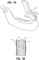
- a mandible (a lower jaw or jawbone) 10 of a patient is shown. Although not specifically shown, embodiments described and shown herein that are operational with respect to the mandible are also applicable to the maxilla (upper jaw bone).
- a cross-sectional view of the mandible of Fig. 1a the bone 10 includes a cortical bone 12 surrounding spongy bone 14, with the gums and soft tissue 16 covering the bone 10, which comprise hard and/or soft tissue as referenced herein.
- a patient on a specific medication regimen that may change the density of the bone within short time intervals may need to be followed up closely as these changes occur.
- electromagnetic radiation i.e., THZ Time Domain, Infrared Fourier Transform
- THZ Time Domain, Infrared Fourier Transform is particularly beneficial to patients who are receiving medications for osteoporosis or metastatic bone disease secondary to multiple myeloma, breast or prostate cancer, to name a few.
- anti-resorptive medications is necessary to mitigate the loss of bone mass, leading to pathologic fracture of the bone.
- the full electromagnetic spectrum can be used to measure bone density/mass and bone vascular perfusion, as described herein. Since the infrared wave has a low tissue absorption rate, it is capable of penetrating through soft and hard tissue, up to several centimeters. The infrared wave is mainly absorbed by hemoglobin molecules, which aid in obtaining a value for the amount of vascular perfusion within the bone being measured.
- anti-resorptive therapy drugs or bisphosphonate medicines such as XGEVA® (denosumab) manufactured by Amgen Inc.
- XGEVA® denosumab
- these drugs are administered to countermand bone loss and generate tissue healing, in rare instances, death of bone cells and/or tissue result.
- ONJ also known as avascular necrosis of the jaw, resulting in drastic and unpleasant symptoms, including pain, inflammation of the surrounding soft tissue, secondary infection and/or drainage.
- the definitive sign of ONJ is the exposure of mandibular or maxillary bone through the gingiva over 8 weeks in duration. There may be no symptoms for weeks or months, until lesions with exposed bone appear.
- Use of the infrared wave and associated detection equipment as discussed in greater detail below for early detection and to measure the progressive diminishing of the alveolar vasculature can greatly benefit patients suffering from this debilitating disease.
- a stent 20 is selected to overlay a patient's hard and/or soft tissue (see, for instance, Fig. 1 ).
- the stent 20 is selected based on the relative size of the mouth of the patient.
- the stent 20 is customizable, that is, the stent is created from impressions of each individual patient's mouth, and sized to overlay each patient's hard and/or soft tissue.
- the lower and/or upper jaw may be limited in number of teeth (including having no teeth), may have bone loss, and the like, when the stent is customized.
- the overall dimensions and shape of the stent 20 may be formed through conventional dental molding techniques and are designed to fit over the teeth 18 (see Fig. 3 ) or edentulous jaw of the patient and lay in proximity to the gums 16 of the patient.
- the stent 20 has a plurality of detection ports or openings 26, which are strategically positioned along the stent and selected for positioning adjacent segments of the jawbone 10 having different bone density.
- an anterior portion of bone of the lower jaw 10 ( Fig. 1 ) may have a lower bone density than a posterior portion or segment of the bone, and is hence considered more vascular.
- the stent 20 typically includes an upper surface 25, flanked by sides 27 of the stent 20, along an edge 28.
- a pair of the plurality of detection ports 26 comprises a buccal-side 21 detection port 26 and a lingual-side 23 detection port 26.
- the lingual-side detection port is positioned opposite the buccal-side detection port.
- the buccal-side detection port is configured as a mirror image of the lingual-side detection port.
- the buccal-side detection port is configured to transmit the electromagnetic wave, while the lingual-side detection port is configured to receive data associated with the transmitted/received wave, and return data to an attached computer 30, which is configured for calculating, from the received data, the bone density and/or bone vascular perfusion adjacent and across each of the pairs of the plurality of ports 26.
- the pairs of detection ports 26 are positioned along an edge 28 of the stent 20.
- the buccal side 21 of the stent 20 includes wires 22/24 connected to outer edges of each buccal-side port 26 as shown in Figs. 2a and 2b for initiation of transmission of the electromagnetic wave and for receiving data related to measured bone density.
- a wire in 22 is connected to a posterior edge of port 26, which is positioned on the buccal side 21 of the stent 20, while a wire out 24 is connected to an anterior edge of the same port 26.
- the port(s) 26 positioned on the lingual side 23 may include, for instance, a detector or sensor for receiving the transmitted wave (not shown).
- the plurality of ports 26 are capable of transmitting the sensed or measured IR wave related to the bone density and feeding the data back to a computer for analysis (as discussed with greater detail with reference to Fig. 3 hereinbelow).
- the wires 22, 24 are connected to a computer 30 and/or a spectrometer (see Fig. 3 ) or alternatively, a wireless connection may be made, which initiates a wave having a length and/or frequency in the electromagnetic spectrum described herein that ranges from the infrared range to the THZ range (approximate frequency range of 10 11 - 10 14 Hz) and transmits the wave from, for instance, the buccal side of the stent 20 through the bone across the port 26, and detects the wave to create an image of the bone density.
- the wires 22/24 are thus configured for transmitting the electromagnetic waves and receiving data associated therewith.
- the wires 22/24 are attached at a first end 29 of the wires to the buccal side ports 26, and attached at a second end to the computer 30 (not shown).
- the diagnostic device is configured to transmit the electromagnetic waves from the buccal-side detections ports, through the patient's bone, and the lingual-side detection ports being configured to receive the electromagnetic waves, such that changes occurring in the bone density and/or bone vascular perfusion are repetitiously and accurately measured, wherein the electromagnetic waves being infrared or terahertz electromagnetic waves
- a portable THZ or IR (Infrared) device will allow a flat surface or a double surface probe to be placed on the skin or gum tissue, over the lower or upper jaw bone, through a stent that fits securely, repetitiously and consistently over the upper or lower jaw.
- the electromagnetic waves are transmitted/detected to/from a diagnostic probe 40, 40' from a spectrometer 50, configured to measure electromagnetic waves having a frequency of between about 100 GHz to about 430 THz, or any portions or segments thereof.
- a flat surface diagnostic probe 40' can be placed in proximity to the jawbone 10, and/or a double or multiple surface diagnostic probe 40 having a plurality or arms. As shown in Fig.
- the probe 40 has a first arm 42 and a second arm 44, which can be placed in proximity to the teeth 18 or bone 10, such that the first arm 42 of the probe extends along a lingual surface of the teeth or bone, while an opposite arm or second arm 44 extends along a buccal surface of the teeth or bone.
- the plurality of arms 42, 44 of the probe 40 straddle opposing sides of the teeth 18 of bone 10.
- the computer 30 is configured to receive the electromagnetic "fingerprint" spectra waves transmitted through the bone and to detect minute changes in the density of the bone, over the course of treatment using the anti-resorptive therapeutics.
- the probe 40, 40' could be used in conjunction with a stent 20.
- the stent 20 which in an embodiment, has been customized for the particular, individual patient, would be used by the practitioner for locating the individual areas of the jaw to be measured.
- the use of ionizing radiation is cumulative and harmful when used numerous times over a long period of time.
- the patient may benefit from an alternate technique, which uses non-ionizing electromagnetic waves that can be used multiple times without the harmful side effects of the ionizing radiation such as X-rays and gamma rays.
- the shorter wavelengths of the infrared band will result in more clear spatial resolution and are therefore more accurate for diagnosis and identification of these subtle tissue changes.
- THz radiation was able to differentiate between the normal versus the diseased tissue even when compared with a conventional in vitro histological test.
- the objective according to an embodiment is to diagnose the extent of the diseased tissue (cancer of the oral soft tissue or skin) and in the process, conservatively remove as little of the surrounding healthy tissue as possible.
- An abnormal oral tissue to the naked eye or touch, can be evaluated first with the use of infrared radiation, which can distinguish quite accurately between normal tissue and abnormal or cancerous tissue.
- the invasive nature of diseased tissue can also be detected, in an effort to establish a more conservative yet accurate approach to its treatment.
- this technology can be used to view the internal tissue integrity such as tonsillar, genitourinary and the upper or lower gastrointestinal passages.
- the THz spectrometer 50 transmits/receives waves through an endoscopic probe 60 to detect unhealthy tissue 70 endoscopically.
- the flexible endoscopic device with THz probe arrangement 60 is used, and the internal tissue 70 surfaces are scanned with the use of the THz radiation and fed back to the spectrometer machine 50 for analysis and diagnosis in a 3D format.
- the endoscopic probe 60 has a plurality of transducers 62 arranged along the surface of the probe 60 for transmitting and detecting the infrared waves to detect tissue abnormalities.
- computerized diagnostic three-dimensional illustrative models can be generated, which can be used to plan for surgical procedures.
- models would be beneficial include those in the field of craniofacial surgery in congenitally deformed patients, orthognathic surgery (surgery to correct conditions of the jaw and face related to structure, growth, sleep apnea, TMJ disorders, malocclusion problems owing to skeletal disharmonies, or other orthodontic problems that cannot be easily treated with braces) or dental implant placement in the jawbone.
- head and neck cancer therapy for example, prior to elimination of the diseased tissues, it is essential to delineate the extent of pathology in the affected area.
- THz radiation can be instrumental in planning a conservative mapping of the diseased area in an attempt to identify the extent of pathology and minimize damage to the healthy surrounding tissue.
- the computer is configured to evaluate progression of tissue healing, secondary to ONJ.
Landscapes
- Health & Medical Sciences (AREA)
- Life Sciences & Earth Sciences (AREA)
- Physics & Mathematics (AREA)
- Medical Informatics (AREA)
- General Health & Medical Sciences (AREA)
- Engineering & Computer Science (AREA)
- Veterinary Medicine (AREA)
- Heart & Thoracic Surgery (AREA)
- Biomedical Technology (AREA)
- Molecular Biology (AREA)
- Surgery (AREA)
- Animal Behavior & Ethology (AREA)
- Pathology (AREA)
- Public Health (AREA)
- Biophysics (AREA)
- Oral & Maxillofacial Surgery (AREA)
- Dentistry (AREA)
- Orthopedic Medicine & Surgery (AREA)
- Rheumatology (AREA)
- Nuclear Medicine, Radiotherapy & Molecular Imaging (AREA)
- Radiology & Medical Imaging (AREA)
- Physical Education & Sports Medicine (AREA)
- Audiology, Speech & Language Pathology (AREA)
- Spectroscopy & Molecular Physics (AREA)
- High Energy & Nuclear Physics (AREA)
- Optics & Photonics (AREA)
- Investigating Or Analysing Materials By Optical Means (AREA)
- Endoscopes (AREA)
- Dental Tools And Instruments Or Auxiliary Dental Instruments (AREA)
- Radiation-Therapy Devices (AREA)
Description
- This application claims benefit of
U.S. Provisional Application No. 61/927,214 filed January 14, 2014 - An apparatus and method for use in the medical field for detecting bone density and/or diseased hard and soft tissue, particularly changes in bone density resulting from the use of anti-resorptive drugs, is described in which non-ionizing electromagnetic waves are transmitted and received to provide measurements of hard and/or soft tissue abnormalities incidental to dental and/or systemic disease.
- The hertz (symbol Hz) is a unit of frequency in the International System of Units (SI). It is defined as the number of cycles per second of a periodic phenomenon. The electromagnetic spectrum is the range of all possible frequencies of electromagnetic radiation. The infrared spectrum, (including near infrared, mid-infrared and far infrared), are segments of the electromagnetic spectrum with longer wavelengths than those of visible light, extending from a nominal red edge of the visible spectrum at 700 nanometers (nm) to 1 mm. This range of wavelengths corresponds to a frequency range of approximately 430 Terahertz (THz - 1012 Hz) down to about 300 GHz (three hundred billion hertz). THz and sub-THz include segments of the electromagnetic spectrum between about far infrared and microwave, which have a wavelength of about 3mm to about 1 µm, corresponding to frequencies that range from about 100 GHz to 30 THz. These segments of the spectrum cannot be easily gauged with the optical and electronic measurement techniques normally associated with adjacent regions of the spectrum.
- It is understood by those skilled in the art that it is possible to use the terahertz segment of the electromagnetic spectrum for diagnosis of dental disease or tissue changes in the hard and soft tissues of an intra and extra oral complexes. For example, use of the electromagnetic spectrum within the terahertz range is well established to diagnose dental decay more accurately and at an earlier stage than other ionizing x-ray techniques. This concept can also be used to illustrate multi-dimensional shapes of such anatomical structures using appropriate hardware and software to convert wave forms into identifiable anatomical images. Typically the waves of this spectrum penetrate the tissue and reflect back to a detector, where they will be read and analyzed. This specific segment of the electromagnetic spectrum will only emit non-ionizing radiation, as compared to other conventional diagnostic tools such as the x-ray or gamma ray that can damage tissue cells.
- Advances in technology have made possible the production and detection of infrared radiation with devices that are mobile and operate at room temperature. Perhaps the most commonly used generation method, in medical applications, employs optical rectification, whereby high frequency oscillations of a femtosecond laser pulse are rectified by an optical crystal, leaving only the envelope of the laser signal which is a THz pulse.
- What is needed is a device and method that uses an alternative diagnostic means that is mobile, encompasses use of a wide range of the electromagnetic spectrum, has minimal biological adverse effect on the human cells and is practical enough to be used multiple times.
-
USA patent publications US2006/047190 (A1 ),US5259761(A ),US2004/122306(A1 ),US5803730 (A ) andUS2010/074401 (A1 ) are relevant documents to understand the background of the present invention. - According to the present invention, there is provided an apparatus for measuring hard and/or soft tissue abnormalities incidental to dental and/or systemic disease according to claim 1. The apparatus may include using an infrared portion of an electromagnetic spectrum in the detection of bone perfusion and the THZ portion of an electromagnetic spectrum, in detection of bone loss and diseased soft and hard tissue. In a further embodiment, the method and device use the terahertz or infrared waves for detection.
- A more particular description briefly described above will be rendered by reference to specific embodiments thereof that are illustrated in the appended drawings. Understanding that these drawings depict only typical embodiments and are not therefore to be considered to be limiting of its scope, exemplary embodiments will be described and explained with additional specificity and detail through the use of the accompanying drawings in which:
-
Fig. 1a is a perspective view of a typical mandible (lower jaw) of a patient for which the device and method are adapted according to an embodiment; -
Fig. 1b is a cross-sectional view of the mandible ofFig. 1a ; -
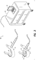
Fig. 2a is a perspective view of a stent according to an embodiment; -
Fig. 2b is a perspective view of a stent according to an embodiment; -
Fig. 3 is a perspective view of bone density measuring device according to an embodiment; and -
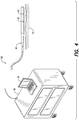
Fig. 4 is a perspective view of an alternative bone density measuring and diseased tissue detection device according to an embodiment. - Various features, aspects, and advantages of the embodiments will become more apparent from the following detailed description, along with the accompanying figures in which like numerals represent like components throughout the figures and text. The various described features are not necessarily drawn to scale, but are drawn to emphasize specific features relevant to embodiments.
- Reference will now be made in detail to various embodiments. Each example is provided by way of explanation, and is not meant as a limitation and does not constitute a definition of all possible embodiments.
- According to an aspect, a
diagnostic device 100 is provided that is configured for transmitting and receiving non-ionizing electromagnetic waves to measure the patient's hard and/or soft tissue abnormalities associated with the underlying hard and/or soft tissue. As described herein, components of one embodiment are intended for use in multiple embodiments, as would be understood by one of ordinary skill in the art. For instance, acomputer 30 and/orspectrometer 50 depicted inFig. 3 , is capable of use with astent 20 ofFig. 2a and 2b , as will become more apparent with greater discussion hereinbelow. - In an embodiment the waves can be used to identify vascular changes occurring in a jawbone with satisfactory tissue differentiating abilities, as would be found, for instance in dental and/or systemic disease. According to a further aspect, infrared spectroscopy is used as a diagnostic tool for measuring blood perfusion within the jawbone (specifically Near Infrared Spectroscopy - NIRS). In yet another aspect, the terahertz wave is used to detect the soft and hard tissue changes. As used herein, the soft tissue is the tissue that connects, supports, or surrounds other structures and organs of the body, not being bone and includes but is not limited to gums, gingiva, intestinal tissue, vasculature, skin and the like, while the hard tissue includes but is not limited to bone, teeth, and the like.
- With reference to
Fig. 1a , a mandible (a lower jaw or jawbone) 10 of a patient is shown. Although not specifically shown, embodiments described and shown herein that are operational with respect to the mandible are also applicable to the maxilla (upper jaw bone). With reference toFig. 1b , a cross-sectional view of the mandible ofFig. 1a , thebone 10 includes acortical bone 12 surroundingspongy bone 14, with the gums andsoft tissue 16 covering thebone 10, which comprise hard and/or soft tissue as referenced herein. - Any comparative and relative hypo- or hyper-calcification within the medullary cavity associated with and/or incidental to dental and/or systemic disease, (which contains bone marrow (not shown)), for instance, shows itself as a decrease or increase in its density. Additionally, any reduction in bone perfusion within the medullary bone, may signify hyper calcification or increase in osteoblastic activity of the alveolar bone. A patient on a specific medication regimen that may change the density of the bone within short time intervals may need to be followed up closely as these changes occur. The use of electromagnetic radiation (i.e., THZ Time Domain, Infrared Fourier Transform), is particularly beneficial to patients who are receiving medications for osteoporosis or metastatic bone disease secondary to multiple myeloma, breast or prostate cancer, to name a few. The use of anti-resorptive medications is necessary to mitigate the loss of bone mass, leading to pathologic fracture of the bone.
- In an embodiment, it is important to repetitiously and accurately measure the changes occurring in the bone for at least the following reasons: 1) gage the efficacy of these anti-resorptive drugs; 2) accurately follow and measure, in short time intervals, the incremental changes that occur in the jawbone during the administration of the anti-resorptive therapy, which according to current technology capabilities is not practical (the only current methods for bone density evaluation are ionizing radiation or MRI); 3) to develop an alternative protocol for drug administration, based on the changes that occur in the jawbone, hence reducing the incidence of osteonecrosis of the jaw (ONJ); 4) to detect the excessive and sometimes irreversible damage to the vital components of the bone complex (for example in the jawbone, this condition could lead to ONJ), with the aim to reduce or stop the use of these medications, in time, when such changes are detected; and 5) to evaluate the healing progression within the alveolar bone, post ONJ.
- As discussed above, the full electromagnetic spectrum can be used to measure bone density/mass and bone vascular perfusion, as described herein. Since the infrared wave has a low tissue absorption rate, it is capable of penetrating through soft and hard tissue, up to several centimeters. The infrared wave is mainly absorbed by hemoglobin molecules, which aid in obtaining a value for the amount of vascular perfusion within the bone being measured. By measuring the vascular abundance in, for instance, the alveolar bone, a degree of osteoblastic activity, (bone deposition), during the administration of anti-resorptive therapy drugs or bisphosphonate medicines such as XGEVA® (denosumab) manufactured by Amgen Inc., to patients with metastatic bone disease (such as Giant Cell Tumor of Bone) and osteoporosis. Although these drugs are administered to countermand bone loss and generate tissue healing, in rare instances, death of bone cells and/or tissue result. Approximately 4-5% of patients taking such drugs experience a serious side effect of ONJ, also known as avascular necrosis of the jaw, resulting in drastic and unpleasant symptoms, including pain, inflammation of the surrounding soft tissue, secondary infection and/or drainage. The definitive sign of ONJ is the exposure of mandibular or maxillary bone through the gingiva over 8 weeks in duration. There may be no symptoms for weeks or months, until lesions with exposed bone appear. Use of the infrared wave and associated detection equipment as discussed in greater detail below for early detection and to measure the progressive diminishing of the alveolar vasculature can greatly benefit patients suffering from this debilitating disease.
- Currently, as previously mentioned, the only way to closely monitor such changes is with the use of ionizing radiation and expensive MRI techniques.
- According to an aspect, an apparatus and method for measuring hard and/or soft tissue abnormalities incidental to dental and/or systemic disease is provided. In an embodiment, and with reference for instance to
Figs. 2a and 2b , astent 20 is selected to overlay a patient's hard and/or soft tissue (see, for instance,Fig. 1 ). According to an aspect, thestent 20 is selected based on the relative size of the mouth of the patient. According to an aspect, thestent 20 is customizable, that is, the stent is created from impressions of each individual patient's mouth, and sized to overlay each patient's hard and/or soft tissue. As would be understood by one of ordinary skill in the art, depending on the particular circumstances of each individual patient, the lower and/or upper jaw may be limited in number of teeth (including having no teeth), may have bone loss, and the like, when the stent is customized. - With reference to
Figs. 2a and 2b , such anoral stent 20 is provided. In an embodiment, the overall dimensions and shape of thestent 20 may be formed through conventional dental molding techniques and are designed to fit over the teeth 18 (seeFig. 3 ) or edentulous jaw of the patient and lay in proximity to thegums 16 of the patient. In an embodiment, thestent 20 has a plurality of detection ports oropenings 26, which are strategically positioned along the stent and selected for positioning adjacent segments of thejawbone 10 having different bone density. As an example, an anterior portion of bone of the lower jaw 10 (Fig. 1 ) may have a lower bone density than a posterior portion or segment of the bone, and is hence considered more vascular. Thestent 20 typically includes anupper surface 25, flanked bysides 27 of thestent 20, along anedge 28. - In an embodiment, a pair of the plurality of
detection ports 26 comprises a buccal-side 21detection port 26 and a lingual-side 23detection port 26. In an embodiment, the lingual-side detection port is positioned opposite the buccal-side detection port. In a further embodiment, the buccal-side detection port is configured as a mirror image of the lingual-side detection port. According to an aspect, the buccal-side detection port is configured to transmit the electromagnetic wave, while the lingual-side detection port is configured to receive data associated with the transmitted/received wave, and return data to an attachedcomputer 30, which is configured for calculating, from the received data, the bone density and/or bone vascular perfusion adjacent and across each of the pairs of the plurality ofports 26. According to an aspect, the pairs ofdetection ports 26 are positioned along anedge 28 of thestent 20. - In an embodiment, the
buccal side 21 of thestent 20 includeswires 22/24 connected to outer edges of each buccal-side port 26 as shown inFigs. 2a and 2b for initiation of transmission of the electromagnetic wave and for receiving data related to measured bone density. In an embodiment, a wire in 22 is connected to a posterior edge ofport 26, which is positioned on thebuccal side 21 of thestent 20, while a wire out 24 is connected to an anterior edge of thesame port 26. Positioned on thestent 20 opposite to thewires lingual side 23 may include, for instance, a detector or sensor for receiving the transmitted wave (not shown). The plurality ofports 26 are capable of transmitting the sensed or measured IR wave related to the bone density and feeding the data back to a computer for analysis (as discussed with greater detail with reference toFig. 3 hereinbelow). - In an embodiment, the
wires computer 30 and/or a spectrometer (seeFig. 3 ) or alternatively, a wireless connection may be made, which initiates a wave having a length and/or frequency in the electromagnetic spectrum described herein that ranges from the infrared range to the THZ range (approximate frequency range of 1011 - 1014 Hz) and transmits the wave from, for instance, the buccal side of thestent 20 through the bone across theport 26, and detects the wave to create an image of the bone density. Thewires 22/24 are thus configured for transmitting the electromagnetic waves and receiving data associated therewith. According to an aspect, thewires 22/24 are attached at afirst end 29 of the wires to thebuccal side ports 26, and attached at a second end to the computer 30 (not shown). In an embodiment, the diagnostic device is configured to transmit the electromagnetic waves from the buccal-side detections ports, through the patient's bone, and the lingual-side detection ports being configured to receive the electromagnetic waves, such that changes occurring in the bone density and/or bone vascular perfusion are repetitiously and accurately measured, wherein the electromagnetic waves being infrared or terahertz electromagnetic waves - In an embodiment, use of a portable THZ or IR (Infrared) device will allow a flat surface or a double surface probe to be placed on the skin or gum tissue, over the lower or upper jaw bone, through a stent that fits securely, repetitiously and consistently over the upper or lower jaw.
- In an alternative embodiment and as seen in
Fig. 3 , the electromagnetic waves are transmitted/detected to/from adiagnostic probe 40, 40' from aspectrometer 50, configured to measure electromagnetic waves having a frequency of between about 100 GHz to about 430 THz, or any portions or segments thereof. As shown herein, a flat surface diagnostic probe 40' can be placed in proximity to thejawbone 10, and/or a double or multiple surfacediagnostic probe 40 having a plurality or arms. As shown inFig. 3 , theprobe 40 has afirst arm 42 and asecond arm 44, which can be placed in proximity to theteeth 18 orbone 10, such that thefirst arm 42 of the probe extends along a lingual surface of the teeth or bone, while an opposite arm orsecond arm 44 extends along a buccal surface of the teeth or bone. In other words, the plurality ofarms probe 40 straddle opposing sides of theteeth 18 ofbone 10. Thecomputer 30 is configured to receive the electromagnetic "fingerprint" spectra waves transmitted through the bone and to detect minute changes in the density of the bone, over the course of treatment using the anti-resorptive therapeutics. - Although not shown, it is further contemplated herein that the
probe 40, 40' could be used in conjunction with astent 20. In such an embodiment, and as would be understood by one of ordinary skill in the art, thestent 20, which in an embodiment, has been customized for the particular, individual patient, would be used by the practitioner for locating the individual areas of the jaw to be measured. - It is well understood by those of ordinary skill in the art that the use of ionizing radiation is cumulative and harmful when used numerous times over a long period of time. Hence, according to an aspect, the patient may benefit from an alternate technique, which uses non-ionizing electromagnetic waves that can be used multiple times without the harmful side effects of the ionizing radiation such as X-rays and gamma rays. The shorter wavelengths of the infrared band will result in more clear spatial resolution and are therefore more accurate for diagnosis and identification of these subtle tissue changes. There have been many in vitro experiments in dentistry to determine the characteristics of enamel and dentin in detecting changes that can occur in these tissues in early dental caries.
- In soft tissue studies, THz radiation was able to differentiate between the normal versus the diseased tissue even when compared with a conventional in vitro histological test. The objective according to an embodiment is to diagnose the extent of the diseased tissue (cancer of the oral soft tissue or skin) and in the process, conservatively remove as little of the surrounding healthy tissue as possible. An abnormal oral tissue (tongue, gum, inside of the cheek) to the naked eye or touch, can be evaluated first with the use of infrared radiation, which can distinguish quite accurately between normal tissue and abnormal or cancerous tissue. In an embodiment, the invasive nature of diseased tissue can also be detected, in an effort to establish a more conservative yet accurate approach to its treatment.
- Similarly, according to an aspect, this technology can be used to view the internal tissue integrity such as tonsillar, genitourinary and the upper or lower gastrointestinal passages. In this alternative embodiment as shown in
Fig. 4 , theTHz spectrometer 50 transmits/receives waves through anendoscopic probe 60 to detectunhealthy tissue 70 endoscopically. In this embodiment, the flexible endoscopic device withTHz probe arrangement 60 is used, and theinternal tissue 70 surfaces are scanned with the use of the THz radiation and fed back to thespectrometer machine 50 for analysis and diagnosis in a 3D format. In an embodiment, theendoscopic probe 60 has a plurality oftransducers 62 arranged along the surface of theprobe 60 for transmitting and detecting the infrared waves to detect tissue abnormalities. - According to an aspect, computerized diagnostic three-dimensional illustrative models can be generated, which can be used to plan for surgical procedures. Examples whereby such models would be beneficial include those in the field of craniofacial surgery in congenitally deformed patients, orthognathic surgery (surgery to correct conditions of the jaw and face related to structure, growth, sleep apnea, TMJ disorders, malocclusion problems owing to skeletal disharmonies, or other orthodontic problems that cannot be easily treated with braces) or dental implant placement in the jawbone. Additionally, in head and neck cancer therapy, for example, prior to elimination of the diseased tissues, it is essential to delineate the extent of pathology in the affected area. Subsequent surgery and/or ionizing radiation therapy to the region is then necessary to treat the disease. THz radiation can be instrumental in planning a conservative mapping of the diseased area in an attempt to identify the extent of pathology and minimize damage to the healthy surrounding tissue. In a further embodiment, the computer is configured to evaluate progression of tissue healing, secondary to ONJ.
- The components and methods illustrated are not limited to the specific embodiments described herein, but rather, features illustrated or described as part of one embodiment can be used on or in conjunction with other embodiments to yield yet a further embodiment. It is intended that the embodiments include such modifications and variations. Further, steps described in the method may be utilized independently and separately from other steps described herein.
- While the embodiments have been described with reference to a preferred embodiment, it will be understood by those skilled in the art that various changes may be made and equivalents may be substituted for elements thereof without departing from the scope of the disclosure. In addition, many modifications may be made to adapt a particular situation or material to the teachings without departing from the essential scope thereof.
- In this specification and the claims that follow, reference will be made to a number of terms that have the following meanings. The singular forms "a," "an" and "the" include plural referents unless the context clearly dictates otherwise. Furthermore, references to "an embodiment" are not intended to be interpreted as excluding the existence of additional embodiments that also incorporate the recited features.
- Advances in science and technology may make equivalents and substitutions possible that are not now contemplated by reason of the imprecision of language; these variations should be covered by the appended claims. This written description uses examples to disclose the various embodiments, including the best mode, and also to enable any person of ordinary skill in the art to practice the embodiments, including making and using any devices or systems and performing any incorporated methods. The patentable scope is defined by the claims.
Claims (12)
- An apparatus for measuring hard and/or soft tissue abnormalities incidental to dental and/or systemic disease, comprising:a stent (20) that fits over a patient's teeth and selected to overlay a patient's hard and/or soft tissue, wherein the stent (20) comprises a plurality of detection ports (26) positioned along the stent, the plurality of detection ports being selected for positioning adjacent differing density found in the hard and/or soft tissue of the patient, whereinat least a pair of the plurality of detection ports comprise a buccal-side (21) detection port (26) and a lingual-side (23) detection port, the lingual-side detection port being positioned opposite the buccal-side detection port; and,a diagnostic device (100) configured for transmitting and receiving non-ionizing electromagnetic waves to measure the patient's hard and/or soft tissue abnormalities associated with the underlying hard and/or soft tissue adjacent the plurality of detection ports, whereinthe diagnostic device is configured to transmit the electromagnetic waves from the buccal-side (21) detection port (26), through a bone of a patient, and the lingual-side (23) detection port is configured to receive the electromagnetic waves, such that changes occurring in the patient's bone density and/or bone vascular perfusion are repetitiously and accurately measured, andthe diagnostic device is configured to initiate electromagnetic waves being infrared or terahertz electromagnetic waves.
- The apparatus of Claim 1, wherein the pairs of detection ports are positioned along an edge of the stent.
- The apparatus of Claim 1, wherein the diagnostic device (100) comprises wires (22/24) connected to at least a portion of the plurality of detection ports at a first end and the wires being connected to a computer (30) at a second end, the wires being configured for transmitting the electromagnetic waves and receiving data associated therewith, and the computer configured for calculating, from the received data, the bone density and/or bone vascular perfusion adjacent and across each of the plurality of detection ports.
- The apparatus of Claim 1, wherein the stent (20) is customizable and sized to overlay the patient's hard and/or soft tissue.
- The apparatus of claim 1 wherein a pair of the plurality of detection ports comprises a buccal-side (21) detection port (26) and a lingual-side (23) detection port (26) positioned opposite the buccal-side detection port; and,
wherein the apparatus comprises:a diagnostic probe (40, 40'), the diagnostic probe being configured to transmit and receive the electromagnetic waves when positioned adjacent the pair of the plurality of detection ports; anda computer (30) connected to the diagnostic probe, the computer being configured for calculating the bone density and/or bone vascular perfusion adjacent and across each of the pair of the plurality of detection ports,wherein the electromagnetic waves transmitted to or received from the diagnostic probe (40, 40') are from a spectrometer (50) configured to measure electromagnetic waves having a frequency of between about 100 GHz (0.1 THz) to about 430 THz. - The apparatus of Claim 5, wherein the pair of detection ports are positioned along an edge of the stent (20).
- The apparatus of Claim 5 or Claim 6, wherein the stent (20) is customizable and sized to overlay each patient's hard and/or soft tissue.
- The apparatus of Claim 4 or 7, wherein the computer (30) is configured to produce three dimensional images for mapping surgical procedures.
- The apparatus of Claim 5, wherein the diagnostic probe (40, 40') comprises a portable Infrared device that allows a single, flat surface or a plurality of surfaces to be placed on the patient's skin or gum tissue.
- The apparatus of Claim 5, wherein the diagnostic probe (40, 40') is configured for use in diagnosing the patient's soft tissue, the soft tissue being an internal body cavity soft tissue.
- The apparatus of Claim 5 or 8, wherein the computer (30) is configured to evaluate progression of tissue healing, secondary to osteonecrosis of the jaw.
- The apparatus of Claim 5, wherein the diagnostic probe (40, 40') is configured for use in detection of the patient's bone loss and diseased hard tissue.
Priority Applications (1)
| Application Number | Priority Date | Filing Date | Title |
|---|---|---|---|
| EP20163162.9A EP3685789A1 (en) | 2014-01-14 | 2015-01-13 | Detection of hard and soft tissue mass/density |
Applications Claiming Priority (2)
| Application Number | Priority Date | Filing Date | Title |
|---|---|---|---|
| US201461927214P | 2014-01-14 | 2014-01-14 | |
| PCT/US2015/011102 WO2015108836A1 (en) | 2014-01-14 | 2015-01-13 | Detection of hard and soft tissue mass/density |
Related Child Applications (2)
| Application Number | Title | Priority Date | Filing Date |
|---|---|---|---|
| EP20163162.9A Division EP3685789A1 (en) | 2014-01-14 | 2015-01-13 | Detection of hard and soft tissue mass/density |
| EP20163162.9A Division-Into EP3685789A1 (en) | 2014-01-14 | 2015-01-13 | Detection of hard and soft tissue mass/density |
Publications (3)
| Publication Number | Publication Date |
|---|---|
| EP3094281A1 EP3094281A1 (en) | 2016-11-23 |
| EP3094281A4 EP3094281A4 (en) | 2017-10-11 |
| EP3094281B1 true EP3094281B1 (en) | 2020-09-09 |
Family
ID=53543353
Family Applications (2)
| Application Number | Title | Priority Date | Filing Date |
|---|---|---|---|
| EP15737330.9A Not-in-force EP3094281B1 (en) | 2014-01-14 | 2015-01-13 | Detection of hard and soft tissue mass/density |
| EP20163162.9A Withdrawn EP3685789A1 (en) | 2014-01-14 | 2015-01-13 | Detection of hard and soft tissue mass/density |
Family Applications After (1)
| Application Number | Title | Priority Date | Filing Date |
|---|---|---|---|
| EP20163162.9A Withdrawn EP3685789A1 (en) | 2014-01-14 | 2015-01-13 | Detection of hard and soft tissue mass/density |
Country Status (5)
| Country | Link |
|---|---|
| US (2) | US10244982B2 (en) |
| EP (2) | EP3094281B1 (en) |
| CN (1) | CN105960215A (en) |
| CA (1) | CA2936238C (en) |
| WO (1) | WO2015108836A1 (en) |
Families Citing this family (15)
| Publication number | Priority date | Publication date | Assignee | Title |
|---|---|---|---|---|
| US11375951B2 (en) | 2016-04-07 | 2022-07-05 | Achaemenid, Llc | Intra-oral electroencephalography device and method |
| CN109414192B (en) * | 2016-07-06 | 2022-02-18 | 皇家飞利浦有限公司 | Monitoring bone properties |
| TWI617292B (en) * | 2016-09-02 | 2018-03-11 | 國立中央大學 | Implant osseointegration detection device |
| WO2019161284A1 (en) * | 2018-02-16 | 2019-08-22 | Board Of Supervisors Of Louisiana State University And Agricultural And Mechanical College | Ionizing radiation-free dental imaging by near-infrared fluorescence, and related systems |
| EP3758587A4 (en) * | 2018-02-28 | 2022-02-09 | Live Vue Technologies Inc. | Apparatus and method for in vivo imaging of soft and hard tissue interfaces |
| CN109199382A (en) * | 2018-09-30 | 2019-01-15 | 北京联合大学 | A kind of oral cavity information integration detection method and system based on terahertz imaging |
| BR102018074667A2 (en) * | 2018-11-29 | 2020-06-09 | Univ Federal Rio Grande Norte | bone density meter using electromagnetic waves |
| CN109758116B (en) * | 2018-11-30 | 2021-07-13 | 广东工业大学 | A system for judging bone density distribution in bone reaming surgery |
| CN111345912B (en) * | 2020-04-02 | 2021-06-04 | 苏成帅 | A Fixed Incremental Nail System With Long-Term Monitoring |
| DE102020123167A1 (en) | 2020-09-04 | 2022-03-10 | Technische Universität Dresden, Körperschaft des öffentlichen Rechts | DENTAL DETECTION DEVICE AND METHOD OF OPERATING THE SAME |
| EP4274480A4 (en) * | 2021-01-08 | 2024-10-30 | Achaemenid, LLC | INTRAORAL TESTING DEVICE AND METHOD |
| WO2022150568A1 (en) * | 2021-01-11 | 2022-07-14 | Resmed Sensor Technologies Limited | Tongue fat screening assay |
| CN114452026B (en) * | 2022-01-12 | 2023-10-10 | 深圳北航新兴产业技术研究院 | An enamel caries detection system based on terahertz spectroscopy imaging |
| CN116965794A (en) * | 2022-04-21 | 2023-10-31 | 中国信息通信研究院 | Osteoporosis auxiliary diagnosis device based on artificial intelligence and electromagnetic waves |
| US12403033B2 (en) | 2023-04-21 | 2025-09-02 | Achaemenid, Llc | Oral appliance for the treatment of sleep apnea |
Citations (3)
| Publication number | Priority date | Publication date | Assignee | Title |
|---|---|---|---|---|
| US5803730A (en) * | 1996-03-15 | 1998-09-08 | Khademazad; Behrooz | Mobilometer |
| US20100074401A1 (en) * | 2006-09-28 | 2010-03-25 | Anatoly Vinogratzki | Intraoral x-ray system |
| US20110270053A1 (en) * | 2004-05-28 | 2011-11-03 | David Scott Utley | Intra-Oral Detector and System for Modification of Undesired Behaviors and Methods Thereof |
Family Cites Families (10)
| Publication number | Priority date | Publication date | Assignee | Title |
|---|---|---|---|---|
| US4564355A (en) * | 1984-01-09 | 1986-01-14 | Dentonaut Lab, Ltd. | Method and apparatus for the non-invasive examination of the tooth-jaw structure of a patient to determine the characteristics of unerupted teeth and to control nutritional intake pursuant thereto |
| CA1259206A (en) * | 1985-07-08 | 1989-09-12 | Harry A. Traiger | Method and apparatus for the non-invasive examination of the tooth-jaw structure of a patient to determine the characteristics of unerupted teeth and to control nutritional intake pursuant thereto |
| US5259761A (en) * | 1990-08-06 | 1993-11-09 | Jenifer M. Schnettler | Tooth vitality probe and process |
| US5570182A (en) * | 1994-05-27 | 1996-10-29 | Regents Of The University Of California | Method for detection of dental caries and periodontal disease using optical imaging |
| GB2352512B (en) | 1999-07-23 | 2002-03-13 | Toshiba Res Europ Ltd | A radiation probe and detecting tooth decay |
| US7477925B2 (en) * | 2002-01-17 | 2009-01-13 | Charlotte-Mecklenburg Hospital Authority | Erythema measuring device and method |
| US20040122306A1 (en) * | 2002-12-20 | 2004-06-24 | Eastman Kodak Company | Intra-oral imaging system and method for dimensional measurement below the gumline |
| US7440788B2 (en) * | 2004-08-26 | 2008-10-21 | Kelvyn Enterprises, Inc. | Oral health measurement clamping probe, system, and method |
| GB0706155D0 (en) | 2007-03-30 | 2007-05-09 | Univ Strathclyde | A non-ionising imager |
| US20100235180A1 (en) * | 2009-03-11 | 2010-09-16 | William Atkinson | Synergistic Medicodental Outpatient Imaging Center |
-
2015
- 2015-01-13 US US15/111,177 patent/US10244982B2/en active Active
- 2015-01-13 CN CN201580004469.5A patent/CN105960215A/en active Pending
- 2015-01-13 EP EP15737330.9A patent/EP3094281B1/en not_active Not-in-force
- 2015-01-13 EP EP20163162.9A patent/EP3685789A1/en not_active Withdrawn
- 2015-01-13 WO PCT/US2015/011102 patent/WO2015108836A1/en not_active Ceased
- 2015-01-13 CA CA2936238A patent/CA2936238C/en active Active
-
2018
- 2018-11-20 US US16/196,745 patent/US20190083026A1/en not_active Abandoned
Patent Citations (3)
| Publication number | Priority date | Publication date | Assignee | Title |
|---|---|---|---|---|
| US5803730A (en) * | 1996-03-15 | 1998-09-08 | Khademazad; Behrooz | Mobilometer |
| US20110270053A1 (en) * | 2004-05-28 | 2011-11-03 | David Scott Utley | Intra-Oral Detector and System for Modification of Undesired Behaviors and Methods Thereof |
| US20100074401A1 (en) * | 2006-09-28 | 2010-03-25 | Anatoly Vinogratzki | Intraoral x-ray system |
Also Published As
| Publication number | Publication date |
|---|---|
| EP3685789A1 (en) | 2020-07-29 |
| US20190083026A1 (en) | 2019-03-21 |
| WO2015108836A1 (en) | 2015-07-23 |
| EP3094281A4 (en) | 2017-10-11 |
| CN105960215A (en) | 2016-09-21 |
| CA2936238A1 (en) | 2015-07-23 |
| CA2936238C (en) | 2020-01-07 |
| US20160331301A1 (en) | 2016-11-17 |
| US10244982B2 (en) | 2019-04-02 |
| EP3094281A1 (en) | 2016-11-23 |
Similar Documents
| Publication | Publication Date | Title |
|---|---|---|
| CA2936238C (en) | Detection of hard and soft tissue mass/density | |
| Mah et al. | Practical applications of cone-beam computed tomography in orthodontics | |
| Yang et al. | Use of cone-beam computed tomography to evaluate root canal morphology and locate root canal orifices of maxillary second premolars in a Chinese subpopulation | |
| Almuhtaseb et al. | Three-dimensional localization of impacted canines and root resorption assessment using cone beam computed tomography | |
| Su et al. | Detection and location of second mesiobuccal canal in permanent maxillary teeth: A cone-beam computed tomography analysis in a Taiwanese population | |
| Wang et al. | Relationship between the mental foramen, mandibular canal, and the surgical access line of the mandibular posterior teeth: a cone-beam computed tomographic analysis | |
| Abella et al. | Endodontic applications of cone beam computed tomography: case series and literature review | |
| Levin et al. | Use of cone-beam computed tomography during retreatment of a 2-rooted maxillary central incisor: case report of a complex diagnosis and treatment | |
| Baek et al. | Optical approach to the periodontal ligament under orthodontic tooth movement: a preliminary study with optical coherence tomography | |
| Gambarini et al. | Detection of bone defects using CBCT exam in an Italian population | |
| US20240057898A1 (en) | Intra-oral test device and method | |
| RU2709249C1 (en) | Method for differential diagnosis of lower jaw motions of myogenic and arthrogenic aetiology | |
| Solda et al. | Effect of at-home bleaching on oxygen saturation levels in the dental pulp of maxillary central incisors | |
| Pirani et al. | Use of dynamic navigation with an educational interest for finding of root canals | |
| Fuentes et al. | Bilateral bifid mandibular canal: a case report using cone beam computed tomography | |
| Sharma et al. | Estimating the prevalence and variation in anterior loop of inferior alveolar nerve with cone-beam computed tomography in North Indian population: an in vivo study | |
| Kabak et al. | Сross-sectional anatomic study of direct positional relationships between mandibular Canal and roots of posterior teeth using cone beam computed tomography | |
| Böhmer et al. | Objective interpretation of dental disease in rabbits, guinea pigs and chinchillas | |
| Zaghden et al. | Anatomical Evaluation of the Greater Palatine Foramen: A Cone-Beam Computed Tomography Study in Tunisian Patients | |
| Yanık et al. | Evaluation of the bone thickness of mandibular molars using cone beam computed tomography | |
| RU2849136C1 (en) | Method for determining the location of the great palatine artery when planning surgical operations | |
| Ormenișan et al. | Case Report. Practical Advantages of CBCT in the Surgical Treatment of Impacted Lower Third Molar | |
| Gibelli et al. | Quantification of dental movements in orthodontic follow-up: a novel approach based on registration of 3D models of dental casts | |
| Schiffer et al. | Session 32: Medical measuring techniques II | |
| Jain et al. | Recent Advances in Endodontic Diagnosis: A Review |
Legal Events
| Date | Code | Title | Description |
|---|---|---|---|
| PUAI | Public reference made under article 153(3) epc to a published international application that has entered the european phase |
Free format text: ORIGINAL CODE: 0009012 |
|
| 17P | Request for examination filed |
Effective date: 20160810 |
|
| AK | Designated contracting states |
Kind code of ref document: A1 Designated state(s): AL AT BE BG CH CY CZ DE DK EE ES FI FR GB GR HR HU IE IS IT LI LT LU LV MC MK MT NL NO PL PT RO RS SE SI SK SM TR |
|
| AX | Request for extension of the european patent |
Extension state: BA ME |
|
| RAP1 | Party data changed (applicant data changed or rights of an application transferred) |
Owner name: ACHAEMENID, LLC |
|
| DAX | Request for extension of the european patent (deleted) | ||
| A4 | Supplementary search report drawn up and despatched |
Effective date: 20170912 |
|
| RIC1 | Information provided on ipc code assigned before grant |
Ipc: A61B 5/00 20060101ALN20170906BHEP Ipc: A61C 5/00 20170101AFI20170906BHEP Ipc: A61B 5/05 20060101ALI20170906BHEP |
|
| STAA | Information on the status of an ep patent application or granted ep patent |
Free format text: STATUS: EXAMINATION IS IN PROGRESS |
|
| 17Q | First examination report despatched |
Effective date: 20180430 |
|
| RIC1 | Information provided on ipc code assigned before grant |
Ipc: A61C 5/00 20170101AFI20191010BHEP Ipc: A63B 71/08 20060101ALI20191010BHEP Ipc: A61B 6/14 20060101ALI20191010BHEP Ipc: A61B 5/00 20060101ALN20191010BHEP Ipc: A61B 5/05 20060101ALI20191010BHEP |
|
| RIC1 | Information provided on ipc code assigned before grant |
Ipc: A61C 5/00 20170101AFI20191105BHEP Ipc: A63B 71/08 20060101ALI20191105BHEP Ipc: A61B 5/00 20060101ALN20191105BHEP Ipc: A61B 6/14 20060101ALI20191105BHEP Ipc: A61B 5/05 20060101ALI20191105BHEP |
|
| GRAP | Despatch of communication of intention to grant a patent |
Free format text: ORIGINAL CODE: EPIDOSNIGR1 |
|
| RIC1 | Information provided on ipc code assigned before grant |
Ipc: A63B 71/08 20060101ALI20191128BHEP Ipc: A61C 5/00 20170101AFI20191128BHEP Ipc: A61B 6/14 20060101ALI20191128BHEP Ipc: A61B 5/00 20060101ALN20191128BHEP Ipc: A61B 5/05 20060101ALI20191128BHEP |
|
| STAA | Information on the status of an ep patent application or granted ep patent |
Free format text: STATUS: GRANT OF PATENT IS INTENDED |
|
| INTG | Intention to grant announced |
Effective date: 20200102 |
|
| GRAS | Grant fee paid |
Free format text: ORIGINAL CODE: EPIDOSNIGR3 |
|
| GRAA | (expected) grant |
Free format text: ORIGINAL CODE: 0009210 |
|
| STAA | Information on the status of an ep patent application or granted ep patent |
Free format text: STATUS: THE PATENT HAS BEEN GRANTED |
|
| AK | Designated contracting states |
Kind code of ref document: B1 Designated state(s): AL AT BE BG CH CY CZ DE DK EE ES FI FR GB GR HR HU IE IS IT LI LT LU LV MC MK MT NL NO PL PT RO RS SE SI SK SM TR |
|
| REG | Reference to a national code |
Ref country code: GB Ref legal event code: FG4D |
|
| REG | Reference to a national code |
Ref country code: AT Ref legal event code: REF Ref document number: 1310603 Country of ref document: AT Kind code of ref document: T Effective date: 20200915 Ref country code: CH Ref legal event code: EP |
|
| REG | Reference to a national code |
Ref country code: IE Ref legal event code: FG4D |
|
| REG | Reference to a national code |
Ref country code: DE Ref legal event code: R096 Ref document number: 602015058775 Country of ref document: DE |
|
| REG | Reference to a national code |
Ref country code: LT Ref legal event code: MG4D |
|
| PG25 | Lapsed in a contracting state [announced via postgrant information from national office to epo] |
Ref country code: NO Free format text: LAPSE BECAUSE OF FAILURE TO SUBMIT A TRANSLATION OF THE DESCRIPTION OR TO PAY THE FEE WITHIN THE PRESCRIBED TIME-LIMIT Effective date: 20201209 Ref country code: FI Free format text: LAPSE BECAUSE OF FAILURE TO SUBMIT A TRANSLATION OF THE DESCRIPTION OR TO PAY THE FEE WITHIN THE PRESCRIBED TIME-LIMIT Effective date: 20200909 Ref country code: GR Free format text: LAPSE BECAUSE OF FAILURE TO SUBMIT A TRANSLATION OF THE DESCRIPTION OR TO PAY THE FEE WITHIN THE PRESCRIBED TIME-LIMIT Effective date: 20201210 Ref country code: HR Free format text: LAPSE BECAUSE OF FAILURE TO SUBMIT A TRANSLATION OF THE DESCRIPTION OR TO PAY THE FEE WITHIN THE PRESCRIBED TIME-LIMIT Effective date: 20200909 Ref country code: LT Free format text: LAPSE BECAUSE OF FAILURE TO SUBMIT A TRANSLATION OF THE DESCRIPTION OR TO PAY THE FEE WITHIN THE PRESCRIBED TIME-LIMIT Effective date: 20200909 Ref country code: BG Free format text: LAPSE BECAUSE OF FAILURE TO SUBMIT A TRANSLATION OF THE DESCRIPTION OR TO PAY THE FEE WITHIN THE PRESCRIBED TIME-LIMIT Effective date: 20201209 |
|
| REG | Reference to a national code |
Ref country code: AT Ref legal event code: MK05 Ref document number: 1310603 Country of ref document: AT Kind code of ref document: T Effective date: 20200909 |
|
| PG25 | Lapsed in a contracting state [announced via postgrant information from national office to epo] |
Ref country code: LV Free format text: LAPSE BECAUSE OF FAILURE TO SUBMIT A TRANSLATION OF THE DESCRIPTION OR TO PAY THE FEE WITHIN THE PRESCRIBED TIME-LIMIT Effective date: 20200909 Ref country code: PL Free format text: LAPSE BECAUSE OF FAILURE TO SUBMIT A TRANSLATION OF THE DESCRIPTION OR TO PAY THE FEE WITHIN THE PRESCRIBED TIME-LIMIT Effective date: 20200909 Ref country code: RS Free format text: LAPSE BECAUSE OF FAILURE TO SUBMIT A TRANSLATION OF THE DESCRIPTION OR TO PAY THE FEE WITHIN THE PRESCRIBED TIME-LIMIT Effective date: 20200909 |
|
| PG25 | Lapsed in a contracting state [announced via postgrant information from national office to epo] |
Ref country code: CZ Free format text: LAPSE BECAUSE OF FAILURE TO SUBMIT A TRANSLATION OF THE DESCRIPTION OR TO PAY THE FEE WITHIN THE PRESCRIBED TIME-LIMIT Effective date: 20200909 Ref country code: EE Free format text: LAPSE BECAUSE OF FAILURE TO SUBMIT A TRANSLATION OF THE DESCRIPTION OR TO PAY THE FEE WITHIN THE PRESCRIBED TIME-LIMIT Effective date: 20200909 Ref country code: PT Free format text: LAPSE BECAUSE OF FAILURE TO SUBMIT A TRANSLATION OF THE DESCRIPTION OR TO PAY THE FEE WITHIN THE PRESCRIBED TIME-LIMIT Effective date: 20210111 Ref country code: SM Free format text: LAPSE BECAUSE OF FAILURE TO SUBMIT A TRANSLATION OF THE DESCRIPTION OR TO PAY THE FEE WITHIN THE PRESCRIBED TIME-LIMIT Effective date: 20200909 Ref country code: RO Free format text: LAPSE BECAUSE OF FAILURE TO SUBMIT A TRANSLATION OF THE DESCRIPTION OR TO PAY THE FEE WITHIN THE PRESCRIBED TIME-LIMIT Effective date: 20200909 |
|
| PG25 | Lapsed in a contracting state [announced via postgrant information from national office to epo] |
Ref country code: IS Free format text: LAPSE BECAUSE OF FAILURE TO SUBMIT A TRANSLATION OF THE DESCRIPTION OR TO PAY THE FEE WITHIN THE PRESCRIBED TIME-LIMIT Effective date: 20210109 Ref country code: AL Free format text: LAPSE BECAUSE OF FAILURE TO SUBMIT A TRANSLATION OF THE DESCRIPTION OR TO PAY THE FEE WITHIN THE PRESCRIBED TIME-LIMIT Effective date: 20200909 Ref country code: AT Free format text: LAPSE BECAUSE OF FAILURE TO SUBMIT A TRANSLATION OF THE DESCRIPTION OR TO PAY THE FEE WITHIN THE PRESCRIBED TIME-LIMIT Effective date: 20200909 Ref country code: ES Free format text: LAPSE BECAUSE OF FAILURE TO SUBMIT A TRANSLATION OF THE DESCRIPTION OR TO PAY THE FEE WITHIN THE PRESCRIBED TIME-LIMIT Effective date: 20200909 |
|
| REG | Reference to a national code |
Ref country code: DE Ref legal event code: R097 Ref document number: 602015058775 Country of ref document: DE |
|
| PG25 | Lapsed in a contracting state [announced via postgrant information from national office to epo] |
Ref country code: SK Free format text: LAPSE BECAUSE OF FAILURE TO SUBMIT A TRANSLATION OF THE DESCRIPTION OR TO PAY THE FEE WITHIN THE PRESCRIBED TIME-LIMIT Effective date: 20200909 |
|
| PLBE | No opposition filed within time limit |
Free format text: ORIGINAL CODE: 0009261 |
|
| STAA | Information on the status of an ep patent application or granted ep patent |
Free format text: STATUS: NO OPPOSITION FILED WITHIN TIME LIMIT |
|
| 26N | No opposition filed |
Effective date: 20210610 |
|
| PG25 | Lapsed in a contracting state [announced via postgrant information from national office to epo] |
Ref country code: DK Free format text: LAPSE BECAUSE OF FAILURE TO SUBMIT A TRANSLATION OF THE DESCRIPTION OR TO PAY THE FEE WITHIN THE PRESCRIBED TIME-LIMIT Effective date: 20200909 Ref country code: MC Free format text: LAPSE BECAUSE OF FAILURE TO SUBMIT A TRANSLATION OF THE DESCRIPTION OR TO PAY THE FEE WITHIN THE PRESCRIBED TIME-LIMIT Effective date: 20200909 Ref country code: SI Free format text: LAPSE BECAUSE OF FAILURE TO SUBMIT A TRANSLATION OF THE DESCRIPTION OR TO PAY THE FEE WITHIN THE PRESCRIBED TIME-LIMIT Effective date: 20200909 |
|
| REG | Reference to a national code |
Ref country code: CH Ref legal event code: PL |
|
| PG25 | Lapsed in a contracting state [announced via postgrant information from national office to epo] |
Ref country code: LU Free format text: LAPSE BECAUSE OF NON-PAYMENT OF DUE FEES Effective date: 20210113 |
|
| REG | Reference to a national code |
Ref country code: BE Ref legal event code: MM Effective date: 20210131 Ref country code: NL Ref legal event code: FP |
|
| PG25 | Lapsed in a contracting state [announced via postgrant information from national office to epo] |
Ref country code: IT Free format text: LAPSE BECAUSE OF FAILURE TO SUBMIT A TRANSLATION OF THE DESCRIPTION OR TO PAY THE FEE WITHIN THE PRESCRIBED TIME-LIMIT Effective date: 20200909 |
|
| PG25 | Lapsed in a contracting state [announced via postgrant information from national office to epo] |
Ref country code: CH Free format text: LAPSE BECAUSE OF NON-PAYMENT OF DUE FEES Effective date: 20210131 Ref country code: LI Free format text: LAPSE BECAUSE OF NON-PAYMENT OF DUE FEES Effective date: 20210131 |
|
| PG25 | Lapsed in a contracting state [announced via postgrant information from national office to epo] |
Ref country code: IE Free format text: LAPSE BECAUSE OF NON-PAYMENT OF DUE FEES Effective date: 20210113 |
|
| PG25 | Lapsed in a contracting state [announced via postgrant information from national office to epo] |
Ref country code: BE Free format text: LAPSE BECAUSE OF NON-PAYMENT OF DUE FEES Effective date: 20210131 |
|
| PG25 | Lapsed in a contracting state [announced via postgrant information from national office to epo] |
Ref country code: HU Free format text: LAPSE BECAUSE OF FAILURE TO SUBMIT A TRANSLATION OF THE DESCRIPTION OR TO PAY THE FEE WITHIN THE PRESCRIBED TIME-LIMIT; INVALID AB INITIO Effective date: 20150113 |
|
| PG25 | Lapsed in a contracting state [announced via postgrant information from national office to epo] |
Ref country code: CY Free format text: LAPSE BECAUSE OF FAILURE TO SUBMIT A TRANSLATION OF THE DESCRIPTION OR TO PAY THE FEE WITHIN THE PRESCRIBED TIME-LIMIT Effective date: 20200909 |
|
| PGFP | Annual fee paid to national office [announced via postgrant information from national office to epo] |
Ref country code: NL Payment date: 20240126 Year of fee payment: 10 |
|
| PG25 | Lapsed in a contracting state [announced via postgrant information from national office to epo] |
Ref country code: MK Free format text: LAPSE BECAUSE OF FAILURE TO SUBMIT A TRANSLATION OF THE DESCRIPTION OR TO PAY THE FEE WITHIN THE PRESCRIBED TIME-LIMIT Effective date: 20200909 |
|
| PGFP | Annual fee paid to national office [announced via postgrant information from national office to epo] |
Ref country code: DE Payment date: 20240129 Year of fee payment: 10 Ref country code: GB Payment date: 20240129 Year of fee payment: 10 |
|
| PGFP | Annual fee paid to national office [announced via postgrant information from national office to epo] |
Ref country code: SE Payment date: 20240127 Year of fee payment: 10 Ref country code: FR Payment date: 20240125 Year of fee payment: 10 |
|
| PG25 | Lapsed in a contracting state [announced via postgrant information from national office to epo] |
Ref country code: MT Free format text: LAPSE BECAUSE OF FAILURE TO SUBMIT A TRANSLATION OF THE DESCRIPTION OR TO PAY THE FEE WITHIN THE PRESCRIBED TIME-LIMIT Effective date: 20200909 |
|
| REG | Reference to a national code |
Ref country code: DE Ref legal event code: R119 Ref document number: 602015058775 Country of ref document: DE |
|
| REG | Reference to a national code |
Ref country code: SE Ref legal event code: EUG |
|
| REG | Reference to a national code |
Ref country code: NL Ref legal event code: MM Effective date: 20250201 |
|
| GBPC | Gb: european patent ceased through non-payment of renewal fee |
Effective date: 20250113 |
|
| PG25 | Lapsed in a contracting state [announced via postgrant information from national office to epo] |
Ref country code: DE Free format text: LAPSE BECAUSE OF NON-PAYMENT OF DUE FEES Effective date: 20250801 |
|
| PG25 | Lapsed in a contracting state [announced via postgrant information from national office to epo] |
Ref country code: NL Free format text: LAPSE BECAUSE OF NON-PAYMENT OF DUE FEES Effective date: 20250201 |
|
| PG25 | Lapsed in a contracting state [announced via postgrant information from national office to epo] |
Ref country code: GB Free format text: LAPSE BECAUSE OF NON-PAYMENT OF DUE FEES Effective date: 20250113 |
|
| PG25 | Lapsed in a contracting state [announced via postgrant information from national office to epo] |
Ref country code: FR Free format text: LAPSE BECAUSE OF NON-PAYMENT OF DUE FEES Effective date: 20250131 |
|
| PG25 | Lapsed in a contracting state [announced via postgrant information from national office to epo] |
Ref country code: TR Free format text: LAPSE BECAUSE OF FAILURE TO SUBMIT A TRANSLATION OF THE DESCRIPTION OR TO PAY THE FEE WITHIN THE PRESCRIBED TIME-LIMIT Effective date: 20200909 |