EP3091348A1 - Image processing device, program, storage medium, and image processing method - Google Patents
Image processing device, program, storage medium, and image processing method Download PDFInfo
- Publication number
- EP3091348A1 EP3091348A1 EP14875850.1A EP14875850A EP3091348A1 EP 3091348 A1 EP3091348 A1 EP 3091348A1 EP 14875850 A EP14875850 A EP 14875850A EP 3091348 A1 EP3091348 A1 EP 3091348A1
- Authority
- EP
- European Patent Office
- Prior art keywords
- detection
- sample
- target
- degree
- difficulty
- Prior art date
- Legal status (The legal status is an assumption and is not a legal conclusion. Google has not performed a legal analysis and makes no representation as to the accuracy of the status listed.)
- Granted
Links
Images



Classifications
-
- G—PHYSICS
- G02—OPTICS
- G02B—OPTICAL ELEMENTS, SYSTEMS OR APPARATUS
- G02B21/00—Microscopes
- G02B21/36—Microscopes arranged for photographic purposes or projection purposes or digital imaging or video purposes including associated control and data processing arrangements
- G02B21/365—Control or image processing arrangements for digital or video microscopes
-
- G—PHYSICS
- G01—MEASURING; TESTING
- G01N—INVESTIGATING OR ANALYSING MATERIALS BY DETERMINING THEIR CHEMICAL OR PHYSICAL PROPERTIES
- G01N15/00—Investigating characteristics of particles; Investigating permeability, pore-volume or surface-area of porous materials
- G01N15/10—Investigating individual particles
- G01N15/14—Optical investigation techniques, e.g. flow cytometry
- G01N15/1429—Signal processing
- G01N15/1433—Signal processing using image recognition
-
- G—PHYSICS
- G01—MEASURING; TESTING
- G01N—INVESTIGATING OR ANALYSING MATERIALS BY DETERMINING THEIR CHEMICAL OR PHYSICAL PROPERTIES
- G01N21/00—Investigating or analysing materials by the use of optical means, i.e. using sub-millimetre waves, infrared, visible or ultraviolet light
- G01N21/17—Systems in which incident light is modified in accordance with the properties of the material investigated
- G01N21/25—Colour; Spectral properties, i.e. comparison of effect of material on the light at two or more different wavelengths or wavelength bands
- G01N21/27—Colour; Spectral properties, i.e. comparison of effect of material on the light at two or more different wavelengths or wavelength bands using photo-electric detection ; circuits for computing concentration
-
- G—PHYSICS
- G06—COMPUTING OR CALCULATING; COUNTING
- G06V—IMAGE OR VIDEO RECOGNITION OR UNDERSTANDING
- G06V20/00—Scenes; Scene-specific elements
- G06V20/60—Type of objects
- G06V20/69—Microscopic objects, e.g. biological cells or cellular parts
-
- H—ELECTRICITY
- H04—ELECTRIC COMMUNICATION TECHNIQUE
- H04N—PICTORIAL COMMUNICATION, e.g. TELEVISION
- H04N7/00—Television systems
- H04N7/18—Closed-circuit television [CCTV] systems, i.e. systems in which the video signal is not broadcast
- H04N7/183—Closed-circuit television [CCTV] systems, i.e. systems in which the video signal is not broadcast for receiving images from a single remote source
-
- G—PHYSICS
- G01—MEASURING; TESTING
- G01N—INVESTIGATING OR ANALYSING MATERIALS BY DETERMINING THEIR CHEMICAL OR PHYSICAL PROPERTIES
- G01N15/00—Investigating characteristics of particles; Investigating permeability, pore-volume or surface-area of porous materials
- G01N15/10—Investigating individual particles
- G01N2015/1006—Investigating individual particles for cytology
Definitions
- the present invention relates to an image processing apparatus, a program, a storage medium, and an image processing method.
- a determination target area is set on the basis of a nucleus candidate area extracted from a microscope image of the specimen, and it is determined whether or not the determination target area which is set contains the target cell.
- the present invention provides an image processing apparatus including difficulty calculation means and setting means.
- the difficulty calculation means calculates a degree of detection difficulty indicating a degree of difficulty in detection of a target cell contained in a sample, on the basis of a preparation condition of the sample.
- the setting means sets a detection parameter on a process of the detection of the target cell from a captured image of the sample, on the basis of the degree of detection difficulty.
- the present invention according to Claim 9 provides an image processing method including the steps of: calculating a degree of detection difficulty indicating a degree of difficulty in detection of a target cell contained in a sample, on the basis of a preparation condition of the sample; and setting a detection parameter on a process of the detection of the target cell from a captured image of the sample, on the basis of the degree of detection difficulty.
- the present invention according to Claim 2 provides the image processing apparatus according to Claim 1, further including determination means.
- the detection parameter includes information for defining a setting condition for setting determination target areas on which determination as to whether or not the captured image contains the target cell area is made.
- the determination means makes determination as to whether or not the target cell is contained in the determination target areas which are set on the basis of the detection parameter, on the captured image.
- the present invention according to Claim 3 provides the image processing apparatus according to Claim 2, in which the setting means sets the setting condition in such a manner that the degree of detection difficulty having a higher value causes the determination target areas, the number of which is larger, to be set.
- the present invention according to Claim 4 provides the image processing apparatus according to Claim 2 or 3, further including integrating means.
- the integrating means integrates the plurality of determination target areas into a determination target area which is determined, by the determination means, to have the highest probability that the target cell is contained.
- the present invention according to Claim 5 provides the image processing apparatus according to any one of Claims 1 to 4, in which the detection-difficulty calculation unit calculates the degree of detection difficulty at a lower value for a higher degree of evenness of thickness of the sample, calculates the degree of detection difficulty at a lower value for a lower degree of brightness of a nucleus contained in the sample, and calculates the degree of detection difficulty at a higher value for a higher ratio of pixels which are contained in the sample and whose density is lower than a predetermined density.
- the present invention according to Claim 6 provides the image processing apparatus according to any one of Claims 1 to 5, in which the detection parameter includes information for determining whether or not each of a plurality of samples generated from a target specimen is to be subjected to detection of the target cell in the sample.
- the setting means sets information about a specimen to be subjected to the detection, among the plurality of samples generated from the target specimen, on the basis of the degree of detection difficulty.
- the present invention according to Claim 7 provides a program for causing a computer to function as difficulty calculation means and setting means.
- the difficulty calculation means calculates a degree of detection difficulty indicating a degree of difficulty in detection of a target cell contained in a sample, on the basis of a preparation condition of the sample.
- the setting means sets a detection parameter on a process of the detection of the target cell from a captured image of the sample, on the basis of the degree of detection difficulty.
- the present invention according to Claim 8 provides a computer-readable storage medium storing a program for causing a computer to function as difficulty calculation means and setting means.
- the difficulty calculation means calculates a degree of detection difficulty indicating a degree of difficulty in detection of a target cell contained in a sample, on the basis of a preparation condition of the sample.
- the setting means sets a detection parameter on a process of the detection of the target cell from a captured image of the sample, on the basis of the degree of detection difficulty.
- parameters for the process of detecting a target cell from a sample may be set in accordance with characteristics of the sample.
- determination target areas which are set for a sample may be set in accordance with the degree of difficulty in detection of a target cell from the sample.
- a determination result having high accuracy may be obtained on the basis of a determination result using multiple determination target areas obtained from the same target.
- the degree of difficulty in detection of a target cell from a sample may be calculated with high accuracy.
- a sample to be subjected to the detection may be set from multiple samples generated from a specimen, in accordance with the degree of detection difficulty for the sample.
- Fig. 1 illustrates an exemplary system configuration of an image processing system 1 according to the present embodiment.
- the image processing system 1 includes an optical microscope 10, an image processing apparatus 20, an input apparatus 50, and a display apparatus 60.
- the image processing apparatus 20 is connected to the optical microscope 10, the input apparatus 50, and the display apparatus 60 which are capable of performing data communication with the image processing apparatus 20.
- the optical microscope 10 captures an image of a preparation on slide glass 11 disposed on a platform, with a CCD camera 14 via an optical system such as an objective lens 12.
- the optical microscope 10 includes a focusing mechanism 13 which changes the distance between the slide glass 11 and the objective lens 12 so as to be capable of capturing an image of the preparation on the slide glass 11 with different focal lengths.
- the preparation is obtained by applying maternal blood to the slide glass 11 and treating it with May-Giemsa staining.
- fetus-derived nucleated red blood cells (NRBCs) in the maternal blood are stained bluish purple.
- NRBCs are referred to as target cells.
- the image processing apparatus 20 obtains an image captured by the optical microscope 10, and detects target cells from the captured image which is obtained. For example, the image processing apparatus 20 may determine a score (for example, a probability) indicating probability that a determination target area which is set in the image captured by the optical microscope 10 contains a target cell, on the basis of a discriminator which has learned discrimination conditions for discriminating a target cell.
- a score for example, a probability
- the input apparatus 50 which is a device, such as a keyboard or a mouse, inputs an operation received from a user to the image processing apparatus 20.
- the image processing apparatus 20 may obtain information about an image area specified by a user using the input apparatus 50, as learning information for learning positive examples and negative examples of target cells or image characteristics of other specified cells, and may cause the discriminator to learn the discrimination conditions (discrimination parameters) for discriminating a target cell, on the basis of the learning information.
- the display apparatus 60 which is, for example, a liquid-crystal display apparatus 60 displays a screen on the basis of the result of a process performed by the image processing apparatus 20.
- the display apparatus 60 displays an image captured by the optical microscope 10, and a result of detection of target cells which is performed by the image processing apparatus 20.
- Fig. 2 is a functional block diagram illustrating exemplary functions provided for the image processing apparatus 20.
- the image processing apparatus 20 includes a specimen-information acquiring unit 21, a specimen-characteristics information storage unit 22, a target-cell count estimating unit 23, a captured-image acquiring unit 24, a sample-characteristics extraction unit 25, a detection-difficulty determination unit 26, a detection-target-sample setting unit 27, a detection-parameter setting unit 28, a nucleus-candidate-area extraction unit 29, a determination-target-area setting unit 30, an image-characteristics generating unit 31, a target-cell determination unit 32, a target-cell-area setting unit 33, a target-cell-area integrating unit 34, and a determination-result output unit 35.
- the image processing apparatus 20 which is a computer including control means such as a CPU, storage means such as a memory, input/output means for receiving/transmitting data from/to an external device, and the like reads programs stored in a computer-readable information storage medium and executes them.
- the programs may be supplied to the image processing apparatus 20 through an information storage medium, such as an optical disk, a magnetic disk, a magnetic tape, a magneto-optical disk, or a flash memory, or may be supplied to the image processing apparatus 20 through a data network such as the Internet.
- the functions of the units provided for the image processing apparatus 20 will be described in detail below.
- the specimen-information acquiring unit 21 obtains information (specimen information) about a specimen (mother's body) to be examined.
- the specimen-information acquiring unit 21 may obtain the specimen information on the basis of data inputted via the input apparatus 50.
- the specimen information may include the age, the medical history, weeks of pregnancy or the likes of a person who is to be checked and from whom maternal blood is collected.
- the specimen-characteristics information storage unit 22 stores information used when target cells contained in a specimen are estimated on the basis of the characteristics of the specimen.
- the specimen-characteristics information storage unit 22 may use classifications according to the characteristics of the specimen to store, for example, the target cell count per unit blood volume for each classification.
- the image processing apparatus 20 may generate a feature vector for each of specimens on the basis of each piece of information of the age, the medical history, the number of weeks in pregnancy, and may perform clustering on the specimens.
- the image processing apparatus 20 may store a representative value (for example, an average) of the target cell (nucleated red blood cell) count per unit blood volume which is measured in advance for the specimens included in each cluster, as the representative nucleated red blood cell count in association with a corresponding one of the clusters in the specimen-characteristics information storage unit 22.
- the specimen-characteristics information storage unit 22 may store a table or a mathematical expression which defines a relationship between information about the age, the medical history, and the number of weeks in pregnancy, and the nucleated red blood cell count corresponding to the information.
- the target-cell count estimating unit 23 estimates the target cell count contained in the specimen obtained by the specimen-information acquiring unit 21, on the basis of the specimen information about the specimen (information representing characteristics of the specimen) and the information stored in the specimen-characteristics information storage unit 22. For example, the target-cell count estimating unit 23 estimates the target cell count contained in a preparation (maternal blood) obtained from the specimen, on the basis of the representative target cell count stored in the specimen-characteristics information storage unit 22 in association with the classification corresponding to the specimen information obtained by the specimen-information acquiring unit 21.
- the target-cell count estimating unit 23 may obtain the estimated target cell count by multiplying the blood volume of the preparation by the nucleated red blood cell count per unit blood volume stored in the specimen-characteristics information storage unit 22 in association with the classification corresponding to the specimen information obtained by the specimen-information acquiring unit 21.
- the method of estimating the target cell count is not limited to the above-described method.
- the specimen-characteristics information storage unit 22 may store a feature vector of the specimen and the target cell count of the specimen in advance in such a manner that the feature vector is associated with the target cell count.
- the target-cell count estimating unit 23 may specify a feature vector similar to that of the target specimen (for example, the closest feature vector) from the feature vectors of the specimens stored in the specimen-characteristics information storage unit 22, and the target cell count which is associated with the specified feature vector may be used as the target cell count for the target specimen.
- the captured-image acquiring unit 24 obtains a captured image obtained by capturing an image of the preparation (maternal blood) which is obtained from a specimen and which is put on the slide glass.
- the captured-image acquiring unit 24 obtains a captured image obtained by capturing an image of the preparation (maternal blood) with a CCD camera 5 provided for an optical microscope 102, from the optical microscope 102.
- the captured-image acquiring unit 24 may obtain a captured image.
- the detection target flag of the target specimen is set to false (F)
- the captured-image acquiring unit 24 may obtain no captured images.
- the sample-characteristics extraction unit 25 extracts characteristics of a target sample.
- the characteristics of a sample include information representing sample preparation conditions.
- the sample-characteristics extraction unit 25 may extract evenness of the thickness of a sample generated from a specimen, the staining degree of cell nuclei, the content percentage of white blood cells, and the like as the sample characteristics.
- the sample-characteristics extraction unit 25 may extract the sample characteristics for each of all or some (for example, those limited to samples to be subjected to the detection) of samples generated from the target specimen.
- the sample-characteristics extraction unit 25 may measure the depths from the surface of a sample to the slide glass at multiple points of the sample by using the optical microscope 10, and may calculate the evenness (A) of the thickness of the sample as the inverse of the variance of the multiple depths obtained through the measurement.
- the sample-characteristics extraction unit 25 may calculate the staining degree (B) of cell nuclei in a sample as a value obtained by dividing a predetermined threshold by the average brightness value of a captured image (which may be captured by using the optical microscope 10) of the sample (that is, threshold/average brightness value), or may calculate the staining degree (B) as a ratio of pixels having a brightness value lower than a predetermined threshold in a captured image of the sample.
- the sample-characteristics extraction unit 25 may calculate the content percentage (C) of white blood cells in a sample as a ratio of pixels whose color is lighter than a predetermined density, in a captured image (which may be captured by using the optical microscope 10) of the sample.
- the detection-difficulty determination unit 26 determines the degree of difficulty (Df) in detection of target cells from a sample, on the basis of the sample characteristics extracted by the sample-characteristics extraction unit 25. For example, the detection-difficulty determination unit 26 may calculate the degree of detection difficulty Df for a sample by using the following equation (1) on the basis of the evenness A of the thickness of the sample, the staining degree B of cell nuclei in the sample, and the content percentage C of white blood cells in the sample which are extracted by the sample-characteristics extraction unit 25.
- the symbol A0 may represent a reference value of the evenness of the sample thickness.
- the symbol B0 may represent a reference value of the staining degree of cell nuclei in the sample.
- the calculated degree of detection difficulty Df becomes lower as the evenness A of the thickness of a sample becomes larger.
- the calculated degree of detection difficulty Df becomes lower as the staining degree B of cell nuclei in the sample becomes larger.
- the calculated degree of detection difficulty Df becomes higher as the content percentage C of white blood cells in the sample becomes larger.
- the detection-difficulty determination unit 26 may change the degree of detection difficulty Df calculated by using the above-described expression (1) to a value from 0 to 1. For example, the detection-difficulty determination unit 26 may set Df to 0 when Df is lower than the lower threshold limit.
- the detection-difficulty determination unit 26 may set Df to 1 when Df is larger than the upper threshold limit (for example, a value larger than 1).
- the detection-target-sample setting unit 27 sets samples which are to be subjected to the detection, from multiple samples generated from the target specimen, on the basis of at least one of the estimated target cell count (Cb) estimated by the target-cell count estimating unit 23 and the degree of detection difficulty (Df) determined by the detection-difficulty determination unit 26.
- the detection-target-sample setting unit 27 may select samples (for example, in order of identification number or in order of create date and time), the number of which is equal to the number of samples to be subjected to the detection, from the N samples generated from the preparation, and may set the detection target flags of the selected samples to true (T) and set those of the unselected samples to false (F).
- the detection-target-sample setting unit 27 may set samples having the degree of detection difficulty which is lower than a threshold, among multiple samples generated from the target specimen, as samples to be subjected to the detection.
- the detection-parameter setting unit 28 sets detection parameter sets used when a detection process is to be performed on samples generated from the preparation. For example, the detection-parameter setting unit 28 sets a detection parameter set on the basis of at least one of a sample which is to be subjected to the detection and which is set by the detection-target-sample setting unit 27, and the degree of detection difficulty determined by the detection-difficulty determination unit 26.
- a detection parameter set may include the detection target flag indicating whether or not the sample is to be subjected to the detection, nucleus-candidate-area parameters indicating a condition of an image area to be extracted as a nucleus candidate area from a captured image of the sample, and determination-target-area parameters indicating a setting condition of setting determination target areas for a captured image of the sample.
- Fig. 3 illustrates an exemplary detection parameter management table which stores detection parameter sets which are set by the detection-parameter setting unit 28.
- the detection parameter management table stores a specimen ID for identifying a specimen, a sample ID for identifying each of multiple samples obtained from the specimen, a detection target flag indicating whether or not the sample is to be subjected to the detection, nucleus-candidate-area parameters, and determination-target-area parameters in such a manner that these pieces of information are associated with each other.
- the nucleus-candidate-area parameters include a color range of target pixels to be extracted as a nucleus candidate area, and a threshold of the number of connected pixels.
- the determination-target-area parameters include a stepsize indicating the shift amount between pixels used as the base points of determination target areas for the nucleus candidate area, a maximum magnification indicating a size ratio between the smallest determination target area and the largest determination target area among determination target areas which have different sizes and which are set by using pixels used as the base points of the determination target areas, and a magnification stage number which indicates how many stages of magnification are to be performed when areas from the smallest determination target area to the largest determination target area are to be generated.
- the detection-parameter setting unit 28 may predetermine a detection parameter set for each of stage levels L1 to LM whose number is equal to M which is an integer equal to or larger than 2 (for the same image, the number of nucleus candidate areas and the number of determination target areas for the level Li+1 which are extracted/set by using a detection parameter set are more than those for the level Li), may determine a level on the basis of at least one value of the margin Y and the degree of detection difficulty Df, and may set a detection parameter set on the basis of the determined level.
- the detection-parameter setting unit 28 may predetermine the range of the margin for each of the M stage levels, and may determine a level on the basis of determination as to which level has a range including the calculated margin Y.
- the detection-parameter setting unit 28 may predetermine the range of the degree of detection difficulty for each of the M stage levels, and may determine a level on the basis of determination as to which level has a range including the calculated degree of detection difficulty Df. Further, for example, the detection-parameter setting unit 28 may predetermine the range of the sum of the margin and the degree of detection difficulty for each of the M stage levels, and may determine a level on the basis of determination as to which level has a range including the sum of the calculated value Y and the degree Df.
- the detection-parameter setting unit 28 may set a different detection parameter set on the basis of the degree of detection difficulty which is calculated for the sample.
- the nucleus-candidate-area extraction unit 29 extracts nucleus candidate areas on the basis of the nucleus-candidate-area parameters which are set by the detection-parameter setting unit 28 for the sample, from the captured image obtained by the captured-image acquiring unit 24 for the sample.
- the nucleus-candidate-area extraction unit 29 may perform binarization so as to convert a pixel included in the color range in the nucleus-candidate-area parameters into a black pixel and convert a pixel which is not included in the color range into a white pixel, and may extract, as nucleus candidate areas, connected pixel groups, the number of which is larger than that of connected pixel groups which is included in the nucleus-candidate-area parameters, from connected pixel groups obtained by connecting adjacent black pixels.
- the determination-target-area setting unit 30 sets determination target areas on which determination as to whether or not target cells are present is to made, on the basis of a nucleus candidate area extracted by the nucleus-candidate-area extraction unit 29 and the determination-target-area parameters which are set by the detection-parameter setting unit 28 for the sample. For example, for each of one or more pixels included in a nucleus candidate area extracted by the nucleus-candidate-area extraction unit 29, the determination-target-area setting unit 30 sets a rectangular area in which the pixel is present at the center (or the base point), as a determination target area.
- the determination-target-area setting unit 30 may sequentially set determination target areas by moving the pixel which is present in the nucleus candidate area and which serves as the base point of a determination target area, by the stepsize step by step.
- the determination-target-area setting unit 30 may set determination target areas having different sizes by using one pixel which serves as the base point of a determination target area and by changing the size of the determination target area from 1 x to the maximum magnification multiple times, as many as the number of stages predetermined as the magnification stage number.
- the image-characteristics generating unit 31 generates an image feature value for a determination target area which is set by the determination-target-area setting unit 30. For example, the image-characteristics generating unit 31 may calculate an HOG feature value of a determination target area, and may use the HOG feature value as an image feature value. Processes of calculating two types of HOG feature values will be specifically described below.
- the image-characteristics generating unit 31 obtains the luminance gradient direction and the luminance gradient intensity for each pixel in a determination target area.
- the image-characteristics generating unit 31 divides the target image into blocks, the number of which is Y and each of which is constituted by cells whose number is X.
- the image-characteristics generating unit 31 obtains a luminance gradient direction histogram ([a first gradient direction value, a second gradient direction value, ..., a ninth gradient direction value]) from the luminance gradient directions and the luminance gradient intensities for each of the cells included in a corresponding one of the blocks, and performs normalization for each of the blocks so that the mean square of the gradient direction values is equal to 1.
- the image-characteristics generating unit 31 uses, as a feature value of a block, the values, the number of which is equal to X x 9 and which are obtained by combining the normalized luminance gradient direction histograms in the block, and further uses, as the HOG feature value of the determination target area, the values, the number of which is equal to Y ⁇ X ⁇ 9 and which are obtained by combining all of the blocks in the target image.
- the image-characteristics generating unit 31 obtains the luminance gradient direction and the luminance gradient intensity for each pixel in a determination target area.
- the image-characteristics generating unit 31 divides the target image into blocks, the number of which is Y and each of which is constituted by cells, the number of which is X.
- the image-characteristics generating unit 31 obtains a luminance gradient direction histogram ([a first gradient direction value, a second gradient direction value, ..., an eighteenth gradient direction value]) from the luminance gradient directions and the luminance gradient intensities for each of the cells included in a corresponding one of the blocks.
- the image-characteristics generating unit 31 uses, as a feature value of a block, the values, the number of which is equal to X x 18 and which are obtained by combining the luminance gradient direction histograms in the block, and further uses, as the Cell-HOG feature value of the determination target area, the values, the number of which is equal to Y ⁇ X ⁇ 18 and which are obtained by combining all of the blocks in the target image.
- the target-cell determination unit 32 determines a probability (the degree of confidence) that the determination target area contains a target cell, on the basis of the image feature value of a determination target area. For example, on the basis of the image feature values of image areas in which target cells are shown, the discriminator may be made to learn discrimination conditions (discrimination parameters) for a target cell in advance, and a discrimination result for the image feature value of a determination target area may be obtained by using the discriminator.
- the discriminator may use AdaBoost, SVM (support vector machine), or the like.
- the discriminator outputs a score (the degree of confidence) indicating probability that the determination target area contains a target cell, on the basis of the image feature value of the determination target area. For example, the discriminator may output a positive value score when a cell contained in the determination target area is a target cell, and may output a negative value score when the cell is not a target cell.
- the target-cell-area setting unit 33 sets a candidate area (target cell area) containing a target cell, on the basis of a result of determination on a determination target area which is obtained by the target-cell determination unit 32.
- the target-cell-area setting unit 33 may set a determination target area among the determination target areas as a candidate area for a target cell when the degree of confidence for the determination target area which is output from the discriminator is equal to or larger than 0.
- the target-cell-area integrating unit 34 integrates candidate areas which are set by the target-cell-area setting unit 33 and which overlap or match each other, into one area. For example, when multiple candidate areas overlap each other, the target-cell-area integrating unit 34 may integrate the multiple candidate areas into one of the multiple candidate areas (for example, an area whose degree of confidence is the largest). The target-cell-area integrating unit 34 may determine that the multiple candidate areas overlap each other, if the multiple candidate areas are the determination target areas which are set from the same nucleus candidate area. Alternatively, the target-cell-area integrating unit 34 may determine that the multiple candidate areas overlap each other, if the multiple candidate areas overlap each other in such a manner that each of the overlapping areas is equal to or larger than a predetermined threshold.
- the target-cell-area integrating unit 34 may leave only a candidate area having the highest degree of confidence, or may use an area including all of the candidate areas overlapping each other, as a candidate area after integration.
- the target-cell-area integrating unit 34 may add the degrees of confidence of the integrated candidate areas to that of the candidate area after integration, or may use a value obtained by multiplying the degree of confidence of the candidate area after integration by the number of candidate areas overlapping each other, as the degree of confidence of the candidate area after integration.
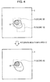
- Fig. 4 illustrates a diagram for describing an exemplary integration of target cell candidate areas.
- a captured image I includes a nucleus candidate area 70, and two target cell areas 71 and 72 which overlap each other are set for the nucleus candidate area 70.
- the target cell area 71 has a score of 30, and the target cell area 72 has a score of 10.
- the target-cell-area integrating unit 34 may integrate the target cell area 72 into the target cell area 71 having the largest score among the target cell areas 71 and 72, and may add the score of the target cell area 72 to that of the target cell area 71 so as to update the score of the target cell area 71.
- the determination-result output unit 35 outputs information about the candidate areas obtained through integration performed by the target-cell-area integrating unit 34, as a result of detection of target cells for the preparation. For example, the determination-result output unit 35 may display a display screen on which candidate areas obtained through integration performed by the target-cell-area integrating unit 34 are displayed as a list in such a manner that the candidate areas are sorted according to their degrees of confidence, on the display apparatus 60. The determination-result output unit 35 may exclude candidate areas whose degree of confidence is less than a specified or predetermined threshold, from the list.
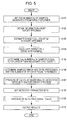
- the image processing apparatus 20 sets the number (N: N is an integer equal to or larger than 1) of samples generated from a target specimen (S101), and obtains information (specimen information) about the target specimen (S102).
- the specimen information may include at least one of the age, the medical history, and the number of weeks in pregnancy.
- the image processing apparatus 20 estimates the target cell count (a) in the target specimen on the basis of the specimen information obtained in S102 (S103). For example, the image processing apparatus 20 may specify a classification to which the specimen information obtained in S102 belongs, and may calculate the estimated target cell count in the specimen on the basis of the representative target cell count (reference target cell count) per unit blood which is predetermined for the specified classification and the blood volume of the preparation, i.e., the specimen.
- the image processing apparatus 20 may specify a classification to which the specimen information obtained in S102 belongs, and may calculate the estimated target cell count in the specimen on the basis of the representative target cell count (reference target cell count) per unit blood which is predetermined for the specified classification and the blood volume of the preparation, i.e., the specimen.
- the image processing apparatus 20 calculates the target cell count (b) per sample on the basis of the target cell count (a) calculated in S103 and the number (N) of samples which is set in S101 (S104).
- the image processing apparatus 20 determines the number (d) of samples which are to be subjected to the detection and which contain a cell count which is equal to or larger than the necessary cell count (c) necessary for the detection, on the basis of the target cell count (b) per sample which is calculated in S104 (S105).
- the image processing apparatus 20 selects and determines samples, which are to be processed and the number of which is equal to the number (d), which is determined in S105, of samples to be subjected to the detection, from the multiple samples generated from the target specimen (S106).
- the image processing apparatus 20 performs a process of calculating the degree of detection difficulty which indicates the degree of difficulty in detection of target cells from a sample which is to be processed and which is determined in S106 (S107).
- the process of calculating the degrees of detection difficulty will be described in detail on the basis of the flowchart in Fig. 6 .
- the image processing apparatus 20 initializes a variable i to 1 (S201), and measures depths (A) of a sample Si (S202). For example, for each of multiple points of the sample Si, a depth from the surface of the sample Si to the slide glass may be measured by using the optical microscope 10.
- the image processing apparatus 20 obtains a captured image by capturing an image of the sample Si by using the optical microscope 10 (S203), and obtains the staining intensity (B) of the sample Si on the basis of the captured image which is obtained (S204). For example, the image processing apparatus 20 may calculate the staining degree (B) of cell nuclei in the sample Si by dividing a predetermined threshold by the average brightness value of the captured image of the sample Si (that is, threshold/average brightness value), or by calculating a ratio of pixels having a brightness value lower than a predetermined threshold in the captured image of the sample Si.
- the image processing apparatus 20 obtains the ratio (C) of white blood cells in the sample Si on the basis of the captured image obtained in S203 (S205). For example, the image processing apparatus 20 may calculate the ratio (C) of white blood cells in the sample Si, as a ratio of pixels having a color lighter than a predetermined density in the captured image of the sample Si.
- the image processing apparatus 20 calculates the degree of detection difficulty Df (Dfi) in detection of target cells from the sample Si, for example, by using the above-described expression (1) on the basis of the variance of the depths (A), the staining intensity (B) of the preparation, and the ratio (C) of white blood cells (S206).
- the image processing apparatus 20 increments (adds 1 to) the variable i (S208), and returns the process back to S202. If the variable i reaches d (S207: Y), the image processing apparatus 20 causes the process to return. Back to the flowchart in Fig. 5 , the description will be continued.
- the image processing apparatus 20 sets a detection parameter set on the basis of at least one of the degree of detection difficulty Df calculated in S107 and information about a sample which is to be processed and which is determined in S106 (S108).
- the detection parameter set may include the detection target flag indicating whether or not the sample is to be subjected to the detection, the nucleus-candidate-area parameters indicating a condition of an image area to be extracted as a nucleus candidate area from the sample, and the determination-target-area parameters indicating a condition of setting determination target areas for an image of the sample.
- the image processing apparatus 20 may set the detection target flag on the basis of information about a sample which is to be processed and which is determined in S106, and may set the nucleus-candidate-area parameters and the determination-target-area parameters on the basis of the degree of detection difficulty Dfi calculated for the sample Si.
- the image processing apparatus 20 performs a process of detecting target cells from the specimen on the basis of the detection parameter sets which are set in S108 (S109). The process of detecting target cells will be described in detail on the basis of the flowchart in Fig. 7 .
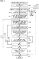
- the image processing apparatus 20 initializes each of variables i, j, and k to 1 (S301), and determines whether or not the sample Si of the target specimen is to be subjected to the detection (S302). For example, if the detection target flag of the sample Si is true (T), the image processing apparatus 20 determines that the sample Si is to be subjected to the detection. If the detection target flag is false (F), the image processing apparatus 20 determines that the sample Si is not to be subjected to the detection.
- the image processing apparatus 20 causes, for example, the optical microscope 10 to capture an image of the sample Si so as to obtain a captured image Ti of the sample Si (S303).
- the image processing apparatus 20 extracts nucleus candidate areas (Ai1 to AiM) from the captured image Ti on the basis of the nucleus-candidate-area parameters included in the detection parameter set which is set for the sample Si (S304), where M is the number of nucleus candidate areas contained in the captured image Ti.
- the extraction of the nucleus candidate areas may be performed by the above-described nucleus-candidate-area extraction unit 29.
- the image processing apparatus 20 sets determination target areas (Bij1 to BijL) for the nucleus candidate area Aij extracted from the captured image Ti on the basis of the determination-target-area parameters included in the detection parameter set which is set for the sample Si (S305), where L is the number of determination target areas which are set for the nucleus candidate area Aij.
- the determination target areas may be set by the above-described determination-target-area setting unit 30.
- the image processing apparatus 20 calculates an image feature value Vijk of the determination target area Bijk (S306).
- the calculation of an image feature value may be performed by the above-described image-characteristics generating unit 31.
- the image processing apparatus 20 calculates a score Pijk indicating probability that the determination target area Bijk contains a target cell, on the basis of the image feature value Vijk calculated in S306 (S307).
- the image processing apparatus 20 sets the determination target area Bijk as a target cell area (S309).
- the image processing apparatus 20 determines whether or not the variable k reaches L (S310). If the variable k does not reach L (S310: N), the image processing apparatus 20 increments k (adds 1 to k) (S311), and returns the process back to S306. If the variable k reaches L in S310 (S310: Y), the image processing apparatus 20 determines whether or not the variable j reaches M (S312).
- the image processing apparatus 20 increments j (adds 1 to j) and initializes k to 1 (S313), and returns the process back to S305. If the variable j reaches M in S312 (S312: Y), the image processing apparatus 20 performs a process of integrating target cell areas which are set for the sample Si (S314). The process of integrating target cell areas will be described in detail on the basis of the flowchart in Fig. 8 .
- the image processing apparatus 20 provides a process completion flag for each of the target cell areas which are set for the captured image Ti of the sample Si (S401).
- the process completion flag has a true/false value.
- a value of true (T) indicates that the target cell area has been processed, and a value of false (F) indicates that the target cell area has not been processed.
- the initial value of the process completion flag provided in S401 is false (F).
- the image processing apparatus 20 selects one of the target cell areas, each of which has a process completion flag indicating that the target cell area has not been processed (S402).
- the target cell area selected in S402 is referred to as the target cell area As.
- the image processing apparatus 20 extracts target cell areas which overlap the target cell area As selected in S402, from the target cell areas which are set for the captured image Ti (S403).
- the image processing apparatus 20 selects a target cell area having the largest score, from the target cell areas extracted in S403 (S404).
- the target cell area selected in S404 is referred to as the target cell area At.
- the image processing apparatus 20 integrates the target cell areas extracted in S403 into the target cell area As (S406), and adds the scores of the integrated target cell areas to that of the target cell area As (S407).
- the target cell areas integrated into the target cell area As may be removed, or may be updated so as to have an indication that the target cell areas has been integrated.
- the image processing apparatus 20 updates the process completion flags of the target cell area As and the target cell areas integrated into the target cell area As, to true (T) (S408).
- the image processing apparatus 20 integrates the target cell area As into the target cell area At (S409), and adds the score of the target cell area As to that of the target cell area At (S410).
- the image processing apparatus 20 updates the process completion flag of the target cell area As to true (T) (S411).
- S412 After S408 or S411, if a target cell area which has not been processed is present (S412: Y), the image processing apparatus 20 returns the process back to S402. If no target cell areas which have not been processed are present (S412: N), the image processing apparatus 20 ends the process of integrating target cells and causes the process to return.
- the image processing apparatus 20 completes the process of integrating target cell areas which are set for the sample Si, if the variable i does not reach d (the number of samples to be subjected to the detection or the number of all samples) (S315: N), the image processing apparatus 20 increments (adds 1 to) i, initializes j and k to 1 (S316), and returns the process back to S302. If the variable i reaches d in S315 (S315: Y), the image processing apparatus 20 causes the process to return.
- d the number of samples to be subjected to the detection or the number of all samples
- the image processing apparatus 20 increments (adds 1 to) i, initializes j and k to 1 (S316), and returns the process back to S302. If the variable i reaches d (S317: Y), the image processing apparatus 20 causes the process to return.
- the image processing apparatus 20 When the image processing apparatus 20 completes the process of detecting target cells for each of the specimens to be processed, the image processing apparatus 20 outputs information about the target cell areas on the basis of the target cell areas after integration for each specimen (S110), and ends the process. For example, the image processing apparatus 20 may sort the target cell areas in descending order of the scores of the target cell areas after integration for each specimen, and may display the target cell areas on the display apparatus 60.
- parameters used in the detection process may be set on the basis of the estimated target cell count in a specimen to be processed and the degree of difficulty in detection of target cells from a specimen to be processed.
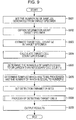
- the second example is different from the first example in that a detection parameter set is set without using the degree of detection difficulty.
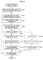
- the image processing apparatus 20 sets the number (N: N is an integer equal to or larger than 1) of samples generated from a target specimen (S501), and obtains information (specimen information) about the target specimen (S502).
- the specimen information may include at least one of the age, the medical history, and the number of weeks in pregnancy.
- the image processing apparatus 20 estimates the target cell count (a) in the target specimen on the basis of the specimen information obtained in S502 (S503).
- the image processing apparatus 20 may specify a classification to which the specimen information obtained in S502 belongs, and may calculate the estimated target cell count in the specimen on the basis of the representative target cell count per unit blood which is predetermined for the specified classification and the blood volume of the preparation, i.e., the specimen.
- the image processing apparatus 20 calculates the target cell count (b) per sample on the basis of the target cell count (a) calculated in S503 and the number (N) of samples which is set in S501 (S504).
- the image processing apparatus 20 determines the number (d) of samples which are to be subjected to the detection and which contain cell count which is equal to or larger than the necessary cell count (c) necessary for the detection, on the basis of the target cell count (b) per sample which is calculated in S504 (S505).
- the image processing apparatus 20 selects and determines samples, which are to be processed and the number of which is equal to the number (d), which is determined in S505, of samples to be subjected to the detection, from the multiple samples generated from the target specimen (S506).
- the image processing apparatus 20 sets a detection parameter set on the basis of the information about a sample which is to be processed and which is determined in S506 and the target cell count (b) per sample calculated in S504 (S507).
- the detection parameter set may include the detection target flag indicating whether or not the sample is to be subjected to the detection, the nucleus-candidate-area parameters indicating a condition of an image area to be extracted as a nucleus candidate area from the sample, and the determination-target-area parameters indicating a condition of setting determination target areas for an image of the sample.
- the image processing apparatus 20 may set the detection target flag on the basis of the information about a sample which is to be processed and which is determined in S506, and may set the nucleus-candidate-area parameters and the determination-target-area parameters on the basis of a margin (b ⁇ d - c) obtained by subtracting the necessary cell count (c) from a value obtained from the expression b ⁇ d which indicates multiplication of the number (d) of samples to be subjected to the detection, by the target cell count per sample.
- a margin (b ⁇ d - c) obtained by subtracting the necessary cell count (c) from a value obtained from the expression b ⁇ d which indicates multiplication of the number (d) of samples to be subjected to the detection, by the target cell count per sample.
- the image processing apparatus 20 performs the process of detecting target cells from a specimen on the basis of the detection parameter sets which are set in S507 (S508).
- the process of detecting target cells are described in detail by using the flowchart in Fig. 7 , and will not be described since the process is also used in the first example.
- the image processing apparatus 20 When the image processing apparatus 20 completes the process of detecting target cells for each of the specimens to be processed, the image processing apparatus 20 outputs information about the target cell areas on the basis of the target cell areas after integration for each specimen (S509), and ends the process.
- parameters used in the detection process may be set on the basis of the estimated target cell count in the specimen to be processed.
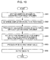
- the third example is different from the first example in that a detection parameter set is set without using the target cell count estimated for a target specimen.
- the image processing apparatus 20 sets the number (N: N is an integer equal to or larger than 1) of samples generated from a target specimen (S601).
- the image processing apparatus 20 determines samples to be processed, from multiple samples generated from the target specimen (S602). For example, the image processing apparatus 20 may set all of the multiple samples generated from the target specimen as samples to be processed, or may determine samples to be processed, on the basis of the degree of detection difficulty Df calculated for the target specimen.
- the image processing apparatus 20 may predetermine a ratio in accordance with the range of the degree of detection difficulty (specifically, the higher the degree of detection difficulty is, the larger the ratio is), may determine the number of samples to be processed, on the basis of a value obtained by multiplying the ratio corresponding to the degree of detection difficulty Df by the number of all samples (specifically, the value may be an integer by rounding up the obtained multiplication value), and may determine samples to be processed, on the basis of the determined number of samples.
- the number of samples to be processed is represented by d.
- the image processing apparatus 20 performs the process of calculating the degree of detection difficulty which indicates the degree of difficulty in detection of target cells from a sample to be processed (S603).
- the process of calculating the degree of detection difficulty is described in detail by using the flowchart in Fig. 6 , and will not be described because the process is also used in the first example.
- the image processing apparatus 20 sets a detection parameter set for the sample Si on the basis of the degree of detection difficulty Dfi calculated in S603 (S604).
- the detection parameter set may include the detection target flag indicating whether or not the sample is to be subjected to the detection, the nucleus-candidate-area parameters indicating a condition of an image area to be extracted as a nucleus candidate area from the sample, and the determination-target-area parameters indicating a condition of setting determination target areas for an image of the sample.
- the image processing apparatus 20 may set the detection target flag on the basis of the information about a sample which is to be processed and which is determined in S603, and may set the nucleus-candidate-area parameters and the determination-target-area parameters on the basis of the degree of detection difficulty Dfi calculated for the sample Si.
- the image processing apparatus 20 performs the process of detecting target cells from the specimen, on the basis of the detection parameter sets which are set in S604 (S605).
- the process of detecting target cells is described in detail by using the flowchart in Fig. 7 , and will not be described because the process is also used in the first example.
- the image processing apparatus 20 When the image processing apparatus 20 completes the process of detecting target cells for each of the specimens to be processed, the image processing apparatus 20 outputs information about the target cell areas on the basis of the target cell areas after integration for each specimen (S606), and ends the process.
- the parameters used in the detection process may be set on the basis of the degree of difficulty in detection of target cells from the specimen to be processed.
- the present invention is not limited to the above-described embodiment.
- the target cell areas may be displayed in order of their scores.
- the image processing apparatus 20 does not necessarily obtain a captured image of a specimen from the optical microscope 10, and may obtain a captured image of a specimen from another computer.
Landscapes
- Engineering & Computer Science (AREA)
- Physics & Mathematics (AREA)
- General Physics & Mathematics (AREA)
- Chemical & Material Sciences (AREA)
- Multimedia (AREA)
- Analytical Chemistry (AREA)
- Health & Medical Sciences (AREA)
- Life Sciences & Earth Sciences (AREA)
- General Health & Medical Sciences (AREA)
- Immunology (AREA)
- Pathology (AREA)
- Theoretical Computer Science (AREA)
- Biochemistry (AREA)
- Signal Processing (AREA)
- Computer Vision & Pattern Recognition (AREA)
- Optics & Photonics (AREA)
- Spectroscopy & Molecular Physics (AREA)
- Mathematical Physics (AREA)
- Biomedical Technology (AREA)
- Molecular Biology (AREA)
- Dispersion Chemistry (AREA)
- Investigating Or Analysing Materials By Optical Means (AREA)
- Apparatus Associated With Microorganisms And Enzymes (AREA)
- Image Processing (AREA)
- Investigating Or Analysing Biological Materials (AREA)
- Image Analysis (AREA)
Abstract
Description
- The present invention relates to an image processing apparatus, a program, a storage medium, and an image processing method.
- For example, in
PTL 1, by using maternal blood as a specimen and by using a nucleated red blood cell in the maternal blood as a target cell, a determination target area is set on the basis of a nucleus candidate area extracted from a microscope image of the specimen, and it is determined whether or not the determination target area which is set contains the target cell. - PTL 1:
Japanese Unexamined Patent Application Publication No. 2012-254042 - It is an object of the present invention to provide an image processing apparatus, a program, a storage medium, and an image processing method which are capable of setting parameters for a process of detecting target cells from a sample, in accordance with characteristics of the sample.
- The present invention according to
Claim 1 provides an image processing apparatus including difficulty calculation means and setting means. The difficulty calculation means calculates a degree of detection difficulty indicating a degree of difficulty in detection of a target cell contained in a sample, on the basis of a preparation condition of the sample. The setting means sets a detection parameter on a process of the detection of the target cell from a captured image of the sample, on the basis of the degree of detection difficulty. The present invention according to Claim 9 provides an image processing method including the steps of: calculating a degree of detection difficulty indicating a degree of difficulty in detection of a target cell contained in a sample, on the basis of a preparation condition of the sample; and setting a detection parameter on a process of the detection of the target cell from a captured image of the sample, on the basis of the degree of detection difficulty. - The present invention according to
Claim 2 provides the image processing apparatus according toClaim 1, further including determination means. The detection parameter includes information for defining a setting condition for setting determination target areas on which determination as to whether or not the captured image contains the target cell area is made. The determination means makes determination as to whether or not the target cell is contained in the determination target areas which are set on the basis of the detection parameter, on the captured image. - The present invention according to
Claim 3 provides the image processing apparatus according toClaim 2, in which the setting means sets the setting condition in such a manner that the degree of detection difficulty having a higher value causes the determination target areas, the number of which is larger, to be set. - The present invention according to
Claim 4 provides the image processing apparatus according toClaim - The present invention according to Claim 5 provides the image processing apparatus according to any one of
Claims 1 to 4, in which the detection-difficulty calculation unit calculates the degree of detection difficulty at a lower value for a higher degree of evenness of thickness of the sample, calculates the degree of detection difficulty at a lower value for a lower degree of brightness of a nucleus contained in the sample, and calculates the degree of detection difficulty at a higher value for a higher ratio of pixels which are contained in the sample and whose density is lower than a predetermined density. - The present invention according to
Claim 6 provides the image processing apparatus according to any one ofClaims 1 to 5, in which the detection parameter includes information for determining whether or not each of a plurality of samples generated from a target specimen is to be subjected to detection of the target cell in the sample. The setting means sets information about a specimen to be subjected to the detection, among the plurality of samples generated from the target specimen, on the basis of the degree of detection difficulty. - The present invention according to Claim 7 provides a program for causing a computer to function as difficulty calculation means and setting means. The difficulty calculation means calculates a degree of detection difficulty indicating a degree of difficulty in detection of a target cell contained in a sample, on the basis of a preparation condition of the sample. The setting means sets a detection parameter on a process of the detection of the target cell from a captured image of the sample, on the basis of the degree of detection difficulty. The present invention according to Claim 8 provides a computer-readable storage medium storing a program for causing a computer to function as difficulty calculation means and setting means. The difficulty calculation means calculates a degree of detection difficulty indicating a degree of difficulty in detection of a target cell contained in a sample, on the basis of a preparation condition of the sample. The setting means sets a detection parameter on a process of the detection of the target cell from a captured image of the sample, on the basis of the degree of detection difficulty.
- According to the invention according to
Claims 1, 7, 8, and 9, parameters for the process of detecting a target cell from a sample may be set in accordance with characteristics of the sample. - According to the invention according to
Claim 2, determination target areas which are set for a sample may be set in accordance with the degree of difficulty in detection of a target cell from the sample. - According to the invention according to
Claim 3, when the degree of difficulty in detection of a target cell from a sample is high, the number of areas to be subjected to the detection is increased, achieving suppression of occurrence of detection failure. - According to the invention according to
Claim 4, in comparison with the case in which multiple determination target areas are not integrated, a determination result having high accuracy may be obtained on the basis of a determination result using multiple determination target areas obtained from the same target. - According to the invention according to Claim 5, in comparison with the case in which the configuration is not included, the degree of difficulty in detection of a target cell from a sample may be calculated with high accuracy.
- According to the invention according to
Claim 6, a sample to be subjected to the detection may be set from multiple samples generated from a specimen, in accordance with the degree of detection difficulty for the sample. -
- [
Fig. 1] Fig. 1 is a diagram illustrating an exemplary system configuration of an image processing system according to the present embodiment. - [
Fig. 2] Fig. 2 is a functional block diagram illustrating exemplary functions provided for an image processing apparatus. - [
Fig. 3] Fig. 3 is a diagram illustrating an exemplary detection parameter management table. - [
Fig. 4] Fig. 4 is a diagram for describing an exemplary integration of target cell candidate areas. - [
Fig. 5] Fig. 5 is a flowchart of a process according to a first example. - [
Fig. 6] Fig. 6 is a flowchart of a process of calculating the degree of detection difficulty. - [
Fig. 7] Fig. 7 is a flowchart of a process of detecting target cells. - [
Fig. 8] Fig. 8 is a flowchart of a process of integrating target cell areas. - [
Fig. 9] Fig. 9 is a flowchart of a process according to a second example. - [
Fig. 10] Fig. 10 is a flowchart of a process according to a third example. - An embodiment for implementing the present invention (hereinafter referred to as an embodiment) will be described below on the basis of the drawings.
-
Fig. 1 illustrates an exemplary system configuration of animage processing system 1 according to the present embodiment. As illustrated inFig. 1 , theimage processing system 1 includes anoptical microscope 10, animage processing apparatus 20, aninput apparatus 50, and adisplay apparatus 60. Theimage processing apparatus 20 is connected to theoptical microscope 10, theinput apparatus 50, and thedisplay apparatus 60 which are capable of performing data communication with theimage processing apparatus 20. - The
optical microscope 10 captures an image of a preparation onslide glass 11 disposed on a platform, with aCCD camera 14 via an optical system such as anobjective lens 12. Theoptical microscope 10 includes afocusing mechanism 13 which changes the distance between theslide glass 11 and theobjective lens 12 so as to be capable of capturing an image of the preparation on theslide glass 11 with different focal lengths. In the present embodiment, the preparation is obtained by applying maternal blood to theslide glass 11 and treating it with May-Giemsa staining. Thus, fetus-derived nucleated red blood cells (NRBCs) in the maternal blood are stained bluish purple. Hereinafter, NRBCs are referred to as target cells. - The
image processing apparatus 20 obtains an image captured by theoptical microscope 10, and detects target cells from the captured image which is obtained. For example, theimage processing apparatus 20 may determine a score (for example, a probability) indicating probability that a determination target area which is set in the image captured by theoptical microscope 10 contains a target cell, on the basis of a discriminator which has learned discrimination conditions for discriminating a target cell. The process of detecting target cells, which is performed in theimage processing apparatus 20, will be described in detail below. - The
input apparatus 50 which is a device, such as a keyboard or a mouse, inputs an operation received from a user to theimage processing apparatus 20. For example, for an image displayed on thedisplay apparatus 60, theimage processing apparatus 20 may obtain information about an image area specified by a user using theinput apparatus 50, as learning information for learning positive examples and negative examples of target cells or image characteristics of other specified cells, and may cause the discriminator to learn the discrimination conditions (discrimination parameters) for discriminating a target cell, on the basis of the learning information. - The
display apparatus 60 which is, for example, a liquid-crystal display apparatus 60 displays a screen on the basis of the result of a process performed by theimage processing apparatus 20. For example, thedisplay apparatus 60 displays an image captured by theoptical microscope 10, and a result of detection of target cells which is performed by theimage processing apparatus 20. - Functions provided for the
image processing apparatus 20 according to the present embodiment will be described. -
Fig. 2 is a functional block diagram illustrating exemplary functions provided for theimage processing apparatus 20. As illustrated inFig. 2 , theimage processing apparatus 20 includes a specimen-information acquiring unit 21, a specimen-characteristicsinformation storage unit 22, a target-cellcount estimating unit 23, a captured-image acquiring unit 24, a sample-characteristics extraction unit 25, a detection-difficulty determination unit 26, a detection-target-sample setting unit 27, a detection-parameter setting unit 28, a nucleus-candidate-area extraction unit 29, a determination-target-area setting unit 30, an image-characteristics generating unit 31, a target-cell determination unit 32, a target-cell-area setting unit 33, a target-cell-area integrating unit 34, and a determination-result output unit 35. - Functions of the above-described units provided for the
image processing apparatus 20 may be achieved in such a manner that theimage processing apparatus 20 which is a computer including control means such as a CPU, storage means such as a memory, input/output means for receiving/transmitting data from/to an external device, and the like reads programs stored in a computer-readable information storage medium and executes them. The programs may be supplied to theimage processing apparatus 20 through an information storage medium, such as an optical disk, a magnetic disk, a magnetic tape, a magneto-optical disk, or a flash memory, or may be supplied to theimage processing apparatus 20 through a data network such as the Internet. The functions of the units provided for theimage processing apparatus 20 will be described in detail below. - The specimen-
information acquiring unit 21 obtains information (specimen information) about a specimen (mother's body) to be examined. For example, the specimen-information acquiring unit 21 may obtain the specimen information on the basis of data inputted via theinput apparatus 50. For example, the specimen information may include the age, the medical history, weeks of pregnancy or the likes of a person who is to be checked and from whom maternal blood is collected. - The specimen-characteristics
information storage unit 22 stores information used when target cells contained in a specimen are estimated on the basis of the characteristics of the specimen. For example, the specimen-characteristicsinformation storage unit 22 may use classifications according to the characteristics of the specimen to store, for example, the target cell count per unit blood volume for each classification. For example, theimage processing apparatus 20 may generate a feature vector for each of specimens on the basis of each piece of information of the age, the medical history, the number of weeks in pregnancy, and may perform clustering on the specimens. Theimage processing apparatus 20 may store a representative value (for example, an average) of the target cell (nucleated red blood cell) count per unit blood volume which is measured in advance for the specimens included in each cluster, as the representative nucleated red blood cell count in association with a corresponding one of the clusters in the specimen-characteristicsinformation storage unit 22. The specimen-characteristicsinformation storage unit 22 may store a table or a mathematical expression which defines a relationship between information about the age, the medical history, and the number of weeks in pregnancy, and the nucleated red blood cell count corresponding to the information. - The target-cell
count estimating unit 23 estimates the target cell count contained in the specimen obtained by the specimen-information acquiring unit 21, on the basis of the specimen information about the specimen (information representing characteristics of the specimen) and the information stored in the specimen-characteristicsinformation storage unit 22. For example, the target-cellcount estimating unit 23 estimates the target cell count contained in a preparation (maternal blood) obtained from the specimen, on the basis of the representative target cell count stored in the specimen-characteristicsinformation storage unit 22 in association with the classification corresponding to the specimen information obtained by the specimen-information acquiring unit 21. For example, the target-cellcount estimating unit 23 may obtain the estimated target cell count by multiplying the blood volume of the preparation by the nucleated red blood cell count per unit blood volume stored in the specimen-characteristicsinformation storage unit 22 in association with the classification corresponding to the specimen information obtained by the specimen-information acquiring unit 21. - The method of estimating the target cell count is not limited to the above-described method. For example, for each of one or more specimens, the specimen-characteristics
information storage unit 22 may store a feature vector of the specimen and the target cell count of the specimen in advance in such a manner that the feature vector is associated with the target cell count. The target-cellcount estimating unit 23 may specify a feature vector similar to that of the target specimen (for example, the closest feature vector) from the feature vectors of the specimens stored in the specimen-characteristicsinformation storage unit 22, and the target cell count which is associated with the specified feature vector may be used as the target cell count for the target specimen. - The captured-
image acquiring unit 24 obtains a captured image obtained by capturing an image of the preparation (maternal blood) which is obtained from a specimen and which is put on the slide glass. For example, the captured-image acquiring unit 24 obtains a captured image obtained by capturing an image of the preparation (maternal blood) with a CCD camera 5 provided for an optical microscope 102, from the optical microscope 102. On the basis of detection parameters described below, when a detection target flag of the target specimen is set to true (T), the captured-image acquiring unit 24 may obtain a captured image. When the detection target flag of the target specimen is set to false (F), the captured-image acquiring unit 24 may obtain no captured images. - The sample-
characteristics extraction unit 25 extracts characteristics of a target sample. The characteristics of a sample include information representing sample preparation conditions. For example, the sample-characteristics extraction unit 25 may extract evenness of the thickness of a sample generated from a specimen, the staining degree of cell nuclei, the content percentage of white blood cells, and the like as the sample characteristics. The sample-characteristics extraction unit 25 may extract the sample characteristics for each of all or some (for example, those limited to samples to be subjected to the detection) of samples generated from the target specimen. - For example, the sample-
characteristics extraction unit 25 may measure the depths from the surface of a sample to the slide glass at multiple points of the sample by using theoptical microscope 10, and may calculate the evenness (A) of the thickness of the sample as the inverse of the variance of the multiple depths obtained through the measurement. - For example, the sample-
characteristics extraction unit 25 may calculate the staining degree (B) of cell nuclei in a sample as a value obtained by dividing a predetermined threshold by the average brightness value of a captured image (which may be captured by using the optical microscope 10) of the sample (that is, threshold/average brightness value), or may calculate the staining degree (B) as a ratio of pixels having a brightness value lower than a predetermined threshold in a captured image of the sample. - For example, the sample-
characteristics extraction unit 25 may calculate the content percentage (C) of white blood cells in a sample as a ratio of pixels whose color is lighter than a predetermined density, in a captured image (which may be captured by using the optical microscope 10) of the sample. - The detection-
difficulty determination unit 26 determines the degree of difficulty (Df) in detection of target cells from a sample, on the basis of the sample characteristics extracted by the sample-characteristics extraction unit 25. For example, the detection-difficulty determination unit 26 may calculate the degree of detection difficulty Df for a sample by using the following equation (1) on the basis of the evenness A of the thickness of the sample, the staining degree B of cell nuclei in the sample, and the content percentage C of white blood cells in the sample which are extracted by the sample-characteristics extraction unit 25. The symbols w1, w2, and w3 may represent predetermined coefficients, each of which is equal to or more than 0 and which satisfy the equation w1 + w2 + w3 = 1. The symbol A0 may represent a reference value of the evenness of the sample thickness. The symbol B0 may represent a reference value of the staining degree of cell nuclei in the sample. The symbol C0 may represent a reference value of the content percentage of white blood cells in the sample.
- According to the above-described expression (1), the calculated degree of detection difficulty Df becomes lower as the evenness A of the thickness of a sample becomes larger. The calculated degree of detection difficulty Df becomes lower as the staining degree B of cell nuclei in the sample becomes larger. The calculated degree of detection difficulty Df becomes higher as the content percentage C of white blood cells in the sample becomes larger. The detection-
difficulty determination unit 26 may change the degree of detection difficulty Df calculated by using the above-described expression (1) to a value from 0 to 1. For example, the detection-difficulty determination unit 26 may set Df to 0 when Df is lower than the lower threshold limit. The detection-difficulty determination unit 26 may set Df to 1 when Df is larger than the upper threshold limit (for example, a value larger than 1). - The detection-target-
sample setting unit 27 sets samples which are to be subjected to the detection, from multiple samples generated from the target specimen, on the basis of at least one of the estimated target cell count (Cb) estimated by the target-cellcount estimating unit 23 and the degree of detection difficulty (Df) determined by the detection-difficulty determination unit 26. - For example, the detection-target-
sample setting unit 27 calculates the target cell count per one sample (a = Cb/N) by dividing the estimated target cell count (Cb) estimated for the target specimen by the number (N) of samples generated from the preparation. For the necessary target cell count (Z), when Cb > Z, the detection-target-sample setting unit 27 may determine the number of samples which are to be subjected to the detection, to be set at an integer X which satisfies the expression a X ≥ Z. When Cb ≤ Z, the detection-target-sample setting unit 27 may determine the number of samples which are to be subjected to the detection, to be set at N (the number of all samples). The detection-target-sample setting unit 27 may select samples (for example, in order of identification number or in order of create date and time), the number of which is equal to the number of samples to be subjected to the detection, from the N samples generated from the preparation, and may set the detection target flags of the selected samples to true (T) and set those of the unselected samples to false (F). - When samples to be subjected to the detection are to be set on the basis of the degree of detection difficulty calculated by the detection-
difficulty determination unit 26, for example, the detection-target-sample setting unit 27 may set samples having the degree of detection difficulty which is lower than a threshold, among multiple samples generated from the target specimen, as samples to be subjected to the detection. - The detection-
parameter setting unit 28 sets detection parameter sets used when a detection process is to be performed on samples generated from the preparation. For example, the detection-parameter setting unit 28 sets a detection parameter set on the basis of at least one of a sample which is to be subjected to the detection and which is set by the detection-target-sample setting unit 27, and the degree of detection difficulty determined by the detection-difficulty determination unit 26. For example, a detection parameter set may include the detection target flag indicating whether or not the sample is to be subjected to the detection, nucleus-candidate-area parameters indicating a condition of an image area to be extracted as a nucleus candidate area from a captured image of the sample, and determination-target-area parameters indicating a setting condition of setting determination target areas for a captured image of the sample. -
Fig. 3 illustrates an exemplary detection parameter management table which stores detection parameter sets which are set by the detection-parameter setting unit 28. As illustrated inFig. 3 , the detection parameter management table stores a specimen ID for identifying a specimen, a sample ID for identifying each of multiple samples obtained from the specimen, a detection target flag indicating whether or not the sample is to be subjected to the detection, nucleus-candidate-area parameters, and determination-target-area parameters in such a manner that these pieces of information are associated with each other. The nucleus-candidate-area parameters include a color range of target pixels to be extracted as a nucleus candidate area, and a threshold of the number of connected pixels. The determination-target-area parameters include a stepsize indicating the shift amount between pixels used as the base points of determination target areas for the nucleus candidate area, a maximum magnification indicating a size ratio between the smallest determination target area and the largest determination target area among determination target areas which have different sizes and which are set by using pixels used as the base points of the determination target areas, and a magnification stage number which indicates how many stages of magnification are to be performed when areas from the smallest determination target area to the largest determination target area are to be generated. - For example, the detection-
parameter setting unit 28 may set a detection parameter set on the basis of at least one of the degree of detection difficulty Df determined by the detection-difficulty determination unit 26 and a margin Y (Y = a · X - Z) which is the difference between the necessary target cell count Z and the product of the target cell count a per sample and the number X of samples which are to be subjected to the detection and which are set by the detection-target-sample setting unit 27. More specifically, the detection-parameter setting unit 28 may predetermine a detection parameter set for each of stage levels L1 to LM whose number is equal to M which is an integer equal to or larger than 2 (for the same image, the number of nucleus candidate areas and the number of determination target areas for the level Li+1 which are extracted/set by using a detection parameter set are more than those for the level Li), may determine a level on the basis of at least one value of the margin Y and the degree of detection difficulty Df, and may set a detection parameter set on the basis of the determined level. For example, the detection-parameter setting unit 28 may predetermine the range of the margin for each of the M stage levels, and may determine a level on the basis of determination as to which level has a range including the calculated margin Y. In addition, for example, the detection-parameter setting unit 28 may predetermine the range of the degree of detection difficulty for each of the M stage levels, and may determine a level on the basis of determination as to which level has a range including the calculated degree of detection difficulty Df. Further, for example, the detection-parameter setting unit 28 may predetermine the range of the sum of the margin and the degree of detection difficulty for each of the M stage levels, and may determine a level on the basis of determination as to which level has a range including the sum of the calculated value Y and the degree Df. - For each of the samples which are set by the detection-target-
sample setting unit 27, the detection-parameter setting unit 28 may set a different detection parameter set on the basis of the degree of detection difficulty which is calculated for the sample. - For a sample to be processed, the nucleus-candidate-
area extraction unit 29 extracts nucleus candidate areas on the basis of the nucleus-candidate-area parameters which are set by the detection-parameter setting unit 28 for the sample, from the captured image obtained by the captured-image acquiring unit 24 for the sample. For example, the nucleus-candidate-area extraction unit 29 may perform binarization so as to convert a pixel included in the color range in the nucleus-candidate-area parameters into a black pixel and convert a pixel which is not included in the color range into a white pixel, and may extract, as nucleus candidate areas, connected pixel groups, the number of which is larger than that of connected pixel groups which is included in the nucleus-candidate-area parameters, from connected pixel groups obtained by connecting adjacent black pixels. - For the captured image obtained by the captured-
image acquiring unit 24 for a sample to be processed, the determination-target-area setting unit 30 sets determination target areas on which determination as to whether or not target cells are present is to made, on the basis of a nucleus candidate area extracted by the nucleus-candidate-area extraction unit 29 and the determination-target-area parameters which are set by the detection-parameter setting unit 28 for the sample. For example, for each of one or more pixels included in a nucleus candidate area extracted by the nucleus-candidate-area extraction unit 29, the determination-target-area setting unit 30 sets a rectangular area in which the pixel is present at the center (or the base point), as a determination target area. On the basis of the stepsize (shift amount) included in the determination-target-area parameters which are set for a sample to be processed, the determination-target-area setting unit 30 may sequentially set determination target areas by moving the pixel which is present in the nucleus candidate area and which serves as the base point of a determination target area, by the stepsize step by step. In addition, on the basis of the maximum magnification and the magnification stage number included in the determination-target-area parameters which are set for a sample to be processed, the determination-target-area setting unit 30 may set determination target areas having different sizes by using one pixel which serves as the base point of a determination target area and by changing the size of the determination target area from 1 x to the maximum magnification multiple times, as many as the number of stages predetermined as the magnification stage number. - The image-
characteristics generating unit 31 generates an image feature value for a determination target area which is set by the determination-target-area setting unit 30. For example, the image-characteristics generating unit 31 may calculate an HOG feature value of a determination target area, and may use the HOG feature value as an image feature value. Processes of calculating two types of HOG feature values will be specifically described below. - The image-
characteristics generating unit 31 obtains the luminance gradient direction and the luminance gradient intensity for each pixel in a determination target area. The image-characteristics generating unit 31 divides the target image into blocks, the number of which is Y and each of which is constituted by cells whose number is X. The image-characteristics generating unit 31 obtains a luminance gradient direction histogram ([a first gradient direction value, a second gradient direction value, ..., a ninth gradient direction value]) from the luminance gradient directions and the luminance gradient intensities for each of the cells included in a corresponding one of the blocks, and performs normalization for each of the blocks so that the mean square of the gradient direction values is equal to 1. The image-characteristics generating unit 31 uses, as a feature value of a block, the values, the number of which is equal to X x 9 and which are obtained by combining the normalized luminance gradient direction histograms in the block, and further uses, as the HOG feature value of the determination target area, the values, the number of which is equal to Y × X × 9 and which are obtained by combining all of the blocks in the target image. - Instead, the image-
characteristics generating unit 31 obtains the luminance gradient direction and the luminance gradient intensity for each pixel in a determination target area. The image-characteristics generating unit 31 divides the target image into blocks, the number of which is Y and each of which is constituted by cells, the number of which is X. The image-characteristics generating unit 31 obtains a luminance gradient direction histogram ([a first gradient direction value, a second gradient direction value, ..., an eighteenth gradient direction value]) from the luminance gradient directions and the luminance gradient intensities for each of the cells included in a corresponding one of the blocks. The image-characteristics generating unit 31 uses, as a feature value of a block, the values, the number of which is equal to X x 18 and which are obtained by combining the luminance gradient direction histograms in the block, and further uses, as the Cell-HOG feature value of the determination target area, the values, the number of which is equal to Y × X × 18 and which are obtained by combining all of the blocks in the target image. - The target-
cell determination unit 32 determines a probability (the degree of confidence) that the determination target area contains a target cell, on the basis of the image feature value of a determination target area. For example, on the basis of the image feature values of image areas in which target cells are shown, the discriminator may be made to learn discrimination conditions (discrimination parameters) for a target cell in advance, and a discrimination result for the image feature value of a determination target area may be obtained by using the discriminator. The discriminator may use AdaBoost, SVM (support vector machine), or the like. The discriminator outputs a score (the degree of confidence) indicating probability that the determination target area contains a target cell, on the basis of the image feature value of the determination target area. For example, the discriminator may output a positive value score when a cell contained in the determination target area is a target cell, and may output a negative value score when the cell is not a target cell. - The target-cell-
area setting unit 33 sets a candidate area (target cell area) containing a target cell, on the basis of a result of determination on a determination target area which is obtained by the target-cell determination unit 32. For example, the target-cell-area setting unit 33 may set a determination target area among the determination target areas as a candidate area for a target cell when the degree of confidence for the determination target area which is output from the discriminator is equal to or larger than 0. - The target-cell-
area integrating unit 34 integrates candidate areas which are set by the target-cell-area setting unit 33 and which overlap or match each other, into one area. For example, when multiple candidate areas overlap each other, the target-cell-area integrating unit 34 may integrate the multiple candidate areas into one of the multiple candidate areas (for example, an area whose degree of confidence is the largest). The target-cell-area integrating unit 34 may determine that the multiple candidate areas overlap each other, if the multiple candidate areas are the determination target areas which are set from the same nucleus candidate area. Alternatively, the target-cell-area integrating unit 34 may determine that the multiple candidate areas overlap each other, if the multiple candidate areas overlap each other in such a manner that each of the overlapping areas is equal to or larger than a predetermined threshold. After integration, the target-cell-area integrating unit 34 may leave only a candidate area having the highest degree of confidence, or may use an area including all of the candidate areas overlapping each other, as a candidate area after integration. The target-cell-area integrating unit 34 may add the degrees of confidence of the integrated candidate areas to that of the candidate area after integration, or may use a value obtained by multiplying the degree of confidence of the candidate area after integration by the number of candidate areas overlapping each other, as the degree of confidence of the candidate area after integration. -
Fig. 4 illustrates a diagram for describing an exemplary integration of target cell candidate areas. As illustrated inFig. 4 , a captured image I includes anucleus candidate area 70, and twotarget cell areas nucleus candidate area 70. Thetarget cell area 71 has a score of 30, and thetarget cell area 72 has a score of 10. The target-cell-area integrating unit 34 may integrate thetarget cell area 72 into thetarget cell area 71 having the largest score among thetarget cell areas target cell area 72 to that of thetarget cell area 71 so as to update the score of thetarget cell area 71. - The determination-
result output unit 35 outputs information about the candidate areas obtained through integration performed by the target-cell-area integrating unit 34, as a result of detection of target cells for the preparation. For example, the determination-result output unit 35 may display a display screen on which candidate areas obtained through integration performed by the target-cell-area integrating unit 34 are displayed as a list in such a manner that the candidate areas are sorted according to their degrees of confidence, on thedisplay apparatus 60. The determination-result output unit 35 may exclude candidate areas whose degree of confidence is less than a specified or predetermined threshold, from the list. - On the basis of
Figs. 5 to 10 , an exemplary process performed in theimage processing system 1 according to the present embodiment will be described in detail below. - On the basis of the flowcharts illustrated in
Figs. 5 to 8 , a process according to a first example which is performed by theimage processing system 1 will be described in detail. - As illustrated in
Fig. 5 , on the basis of data which is input from theinput apparatus 50, theimage processing apparatus 20 sets the number (N: N is an integer equal to or larger than 1) of samples generated from a target specimen (S101), and obtains information (specimen information) about the target specimen (S102). The specimen information may include at least one of the age, the medical history, and the number of weeks in pregnancy. - The
image processing apparatus 20 estimates the target cell count (a) in the target specimen on the basis of the specimen information obtained in S102 (S103). For example, theimage processing apparatus 20 may specify a classification to which the specimen information obtained in S102 belongs, and may calculate the estimated target cell count in the specimen on the basis of the representative target cell count (reference target cell count) per unit blood which is predetermined for the specified classification and the blood volume of the preparation, i.e., the specimen. - The
image processing apparatus 20 calculates the target cell count (b) per sample on the basis of the target cell count (a) calculated in S103 and the number (N) of samples which is set in S101 (S104). Theimage processing apparatus 20 determines the number (d) of samples which are to be subjected to the detection and which contain a cell count which is equal to or larger than the necessary cell count (c) necessary for the detection, on the basis of the target cell count (b) per sample which is calculated in S104 (S105). - The
image processing apparatus 20 selects and determines samples, which are to be processed and the number of which is equal to the number (d), which is determined in S105, of samples to be subjected to the detection, from the multiple samples generated from the target specimen (S106). - The
image processing apparatus 20 performs a process of calculating the degree of detection difficulty which indicates the degree of difficulty in detection of target cells from a sample which is to be processed and which is determined in S106 (S107). The process of calculating the degrees of detection difficulty will be described in detail on the basis of the flowchart inFig. 6 . - As illustrated in
Fig. 6 , theimage processing apparatus 20 initializes a variable i to 1 (S201), and measures depths (A) of a sample Si (S202). For example, for each of multiple points of the sample Si, a depth from the surface of the sample Si to the slide glass may be measured by using theoptical microscope 10. - The
image processing apparatus 20 obtains a captured image by capturing an image of the sample Si by using the optical microscope 10 (S203), and obtains the staining intensity (B) of the sample Si on the basis of the captured image which is obtained (S204). For example, theimage processing apparatus 20 may calculate the staining degree (B) of cell nuclei in the sample Si by dividing a predetermined threshold by the average brightness value of the captured image of the sample Si (that is, threshold/average brightness value), or by calculating a ratio of pixels having a brightness value lower than a predetermined threshold in the captured image of the sample Si. - The
image processing apparatus 20 obtains the ratio (C) of white blood cells in the sample Si on the basis of the captured image obtained in S203 (S205). For example, theimage processing apparatus 20 may calculate the ratio (C) of white blood cells in the sample Si, as a ratio of pixels having a color lighter than a predetermined density in the captured image of the sample Si. - The
image processing apparatus 20 calculates the degree of detection difficulty Df (Dfi) in detection of target cells from the sample Si, for example, by using the above-described expression (1) on the basis of the variance of the depths (A), the staining intensity (B) of the preparation, and the ratio (C) of white blood cells (S206). - If the variable i does not reach d (S207: N), the
image processing apparatus 20 increments (adds 1 to) the variable i (S208), and returns the process back to S202. If the variable i reaches d (S207: Y), theimage processing apparatus 20 causes the process to return. Back to the flowchart inFig. 5 , the description will be continued. - As illustrated in
Fig. 5 , when theimage processing apparatus 20 completes the calculation of the degrees of detection difficulty Df in detection of target cells in the samples to be processed, theimage processing apparatus 20 sets a detection parameter set on the basis of at least one of the degree of detection difficulty Df calculated in S107 and information about a sample which is to be processed and which is determined in S106 (S108). For example, the detection parameter set may include the detection target flag indicating whether or not the sample is to be subjected to the detection, the nucleus-candidate-area parameters indicating a condition of an image area to be extracted as a nucleus candidate area from the sample, and the determination-target-area parameters indicating a condition of setting determination target areas for an image of the sample. For example, theimage processing apparatus 20 may set the detection target flag on the basis of information about a sample which is to be processed and which is determined in S106, and may set the nucleus-candidate-area parameters and the determination-target-area parameters on the basis of the degree of detection difficulty Dfi calculated for the sample Si. - The
image processing apparatus 20 performs a process of detecting target cells from the specimen on the basis of the detection parameter sets which are set in S108 (S109). The process of detecting target cells will be described in detail on the basis of the flowchart inFig. 7 . - As illustrated in
Fig. 7 , theimage processing apparatus 20 initializes each of variables i, j, and k to 1 (S301), and determines whether or not the sample Si of the target specimen is to be subjected to the detection (S302). For example, if the detection target flag of the sample Si is true (T), theimage processing apparatus 20 determines that the sample Si is to be subjected to the detection. If the detection target flag is false (F), theimage processing apparatus 20 determines that the sample Si is not to be subjected to the detection. - If the sample Si is to be subjected to the detection (S302: Y), the
image processing apparatus 20 causes, for example, theoptical microscope 10 to capture an image of the sample Si so as to obtain a captured image Ti of the sample Si (S303). - The
image processing apparatus 20 extracts nucleus candidate areas (Ai1 to AiM) from the captured image Ti on the basis of the nucleus-candidate-area parameters included in the detection parameter set which is set for the sample Si (S304), where M is the number of nucleus candidate areas contained in the captured image Ti. The extraction of the nucleus candidate areas may be performed by the above-described nucleus-candidate-area extraction unit 29. - The
image processing apparatus 20 sets determination target areas (Bij1 to BijL) for the nucleus candidate area Aij extracted from the captured image Ti on the basis of the determination-target-area parameters included in the detection parameter set which is set for the sample Si (S305), where L is the number of determination target areas which are set for the nucleus candidate area Aij. The determination target areas may be set by the above-described determination-target-area setting unit 30. - The
image processing apparatus 20 calculates an image feature value Vijk of the determination target area Bijk (S306). The calculation of an image feature value may be performed by the above-described image-characteristics generating unit 31. - The
image processing apparatus 20 calculates a score Pijk indicating probability that the determination target area Bijk contains a target cell, on the basis of the image feature value Vijk calculated in S306 (S307). - If the score Pijk calculated in S307 is equal to or larger than a threshold Th (S308: Y), the
image processing apparatus 20 sets the determination target area Bijk as a target cell area (S309). - After S309 or if the score Pijk calculated in S307 is less than the threshold Th (S308: N), the
image processing apparatus 20 determines whether or not the variable k reaches L (S310). If the variable k does not reach L (S310: N), theimage processing apparatus 20 increments k (adds 1 to k) (S311), and returns the process back to S306. If the variable k reaches L in S310 (S310: Y), theimage processing apparatus 20 determines whether or not the variable j reaches M (S312). - If the variable j does not reach M in S312 (S312: N), the
image processing apparatus 20 increments j (adds 1 to j) and initializes k to 1 (S313), and returns the process back to S305. If the variable j reaches M in S312 (S312: Y), theimage processing apparatus 20 performs a process of integrating target cell areas which are set for the sample Si (S314). The process of integrating target cell areas will be described in detail on the basis of the flowchart inFig. 8 . - As illustrated in
Fig. 8 , theimage processing apparatus 20 provides a process completion flag for each of the target cell areas which are set for the captured image Ti of the sample Si (S401). The process completion flag has a true/false value. A value of true (T) indicates that the target cell area has been processed, and a value of false (F) indicates that the target cell area has not been processed. The initial value of the process completion flag provided in S401 is false (F). - The
image processing apparatus 20 selects one of the target cell areas, each of which has a process completion flag indicating that the target cell area has not been processed (S402). The target cell area selected in S402 is referred to as the target cell area As. - The
image processing apparatus 20 extracts target cell areas which overlap the target cell area As selected in S402, from the target cell areas which are set for the captured image Ti (S403). Theimage processing apparatus 20 selects a target cell area having the largest score, from the target cell areas extracted in S403 (S404). The target cell area selected in S404 is referred to as the target cell area At. - If the score of the target cell area As is equal to or larger than that of the target cell area At (S405: Y), the
image processing apparatus 20 integrates the target cell areas extracted in S403 into the target cell area As (S406), and adds the scores of the integrated target cell areas to that of the target cell area As (S407). For example, the target cell areas integrated into the target cell area As may be removed, or may be updated so as to have an indication that the target cell areas has been integrated. Theimage processing apparatus 20 updates the process completion flags of the target cell area As and the target cell areas integrated into the target cell area As, to true (T) (S408). - If the score of the target cell area As is less than that of the target cell area At (S405: N), the
image processing apparatus 20 integrates the target cell area As into the target cell area At (S409), and adds the score of the target cell area As to that of the target cell area At (S410). Theimage processing apparatus 20 updates the process completion flag of the target cell area As to true (T) (S411). - After S408 or S411, if a target cell area which has not been processed is present (S412: Y), the
image processing apparatus 20 returns the process back to S402. If no target cell areas which have not been processed are present (S412: N), theimage processing apparatus 20 ends the process of integrating target cells and causes the process to return. - Back to
Fig. 7 , the description will be continued. When theimage processing apparatus 20 completes the process of integrating target cell areas which are set for the sample Si, if the variable i does not reach d (the number of samples to be subjected to the detection or the number of all samples) (S315: N), theimage processing apparatus 20 increments (adds 1 to) i, initializes j and k to 1 (S316), and returns the process back to S302. If the variable i reaches d in S315 (S315: Y), theimage processing apparatus 20 causes the process to return. - If the sample Si is not to be subjected to the detection in S302 (S302: N), and if the variable i does not reach d (S317: N), the
image processing apparatus 20 increments (adds 1 to) i, initializes j and k to 1 (S316), and returns the process back to S302. If the variable i reaches d (S317: Y), theimage processing apparatus 20 causes the process to return. - Back to the flowchart in
Fig. 5 , the description will be continued. When theimage processing apparatus 20 completes the process of detecting target cells for each of the specimens to be processed, theimage processing apparatus 20 outputs information about the target cell areas on the basis of the target cell areas after integration for each specimen (S110), and ends the process. For example, theimage processing apparatus 20 may sort the target cell areas in descending order of the scores of the target cell areas after integration for each specimen, and may display the target cell areas on thedisplay apparatus 60. - According to the process according to the first example described above, parameters used in the detection process may be set on the basis of the estimated target cell count in a specimen to be processed and the degree of difficulty in detection of target cells from a specimen to be processed.
- On the basis of the flowchart in
Fig. 9 , a process according to a second example which is performed by theimage processing system 1 will be described in detail below. The second example is different from the first example in that a detection parameter set is set without using the degree of detection difficulty. - As illustrated in
Fig. 9 , for example, on the basis of data which is input from theinput apparatus 50, theimage processing apparatus 20 sets the number (N: N is an integer equal to or larger than 1) of samples generated from a target specimen (S501), and obtains information (specimen information) about the target specimen (S502). The specimen information may include at least one of the age, the medical history, and the number of weeks in pregnancy. - The
image processing apparatus 20 estimates the target cell count (a) in the target specimen on the basis of the specimen information obtained in S502 (S503). For example, theimage processing apparatus 20 may specify a classification to which the specimen information obtained in S502 belongs, and may calculate the estimated target cell count in the specimen on the basis of the representative target cell count per unit blood which is predetermined for the specified classification and the blood volume of the preparation, i.e., the specimen. - The
image processing apparatus 20 calculates the target cell count (b) per sample on the basis of the target cell count (a) calculated in S503 and the number (N) of samples which is set in S501 (S504). Theimage processing apparatus 20 determines the number (d) of samples which are to be subjected to the detection and which contain cell count which is equal to or larger than the necessary cell count (c) necessary for the detection, on the basis of the target cell count (b) per sample which is calculated in S504 (S505). - The
image processing apparatus 20 selects and determines samples, which are to be processed and the number of which is equal to the number (d), which is determined in S505, of samples to be subjected to the detection, from the multiple samples generated from the target specimen (S506). - The
image processing apparatus 20 sets a detection parameter set on the basis of the information about a sample which is to be processed and which is determined in S506 and the target cell count (b) per sample calculated in S504 (S507). For example, the detection parameter set may include the detection target flag indicating whether or not the sample is to be subjected to the detection, the nucleus-candidate-area parameters indicating a condition of an image area to be extracted as a nucleus candidate area from the sample, and the determination-target-area parameters indicating a condition of setting determination target areas for an image of the sample. For example, theimage processing apparatus 20 may set the detection target flag on the basis of the information about a sample which is to be processed and which is determined in S506, and may set the nucleus-candidate-area parameters and the determination-target-area parameters on the basis of a margin (b · d - c) obtained by subtracting the necessary cell count (c) from a value obtained from the expression b · d which indicates multiplication of the number (d) of samples to be subjected to the detection, by the target cell count per sample. - The
image processing apparatus 20 performs the process of detecting target cells from a specimen on the basis of the detection parameter sets which are set in S507 (S508). The process of detecting target cells are described in detail by using the flowchart inFig. 7 , and will not be described since the process is also used in the first example. - When the
image processing apparatus 20 completes the process of detecting target cells for each of the specimens to be processed, theimage processing apparatus 20 outputs information about the target cell areas on the basis of the target cell areas after integration for each specimen (S509), and ends the process. - According to the process according to the second example described above, parameters used in the detection process may be set on the basis of the estimated target cell count in the specimen to be processed.
- On the basis of the flowchart in
Fig. 10 , a process according to a third example which is performed by theimage processing system 1 will be described in detail. The third example is different from the first example in that a detection parameter set is set without using the target cell count estimated for a target specimen. - As illustrated in
Fig. 10 , for example, on the basis of data which is input from theinput apparatus 50, theimage processing apparatus 20 sets the number (N: N is an integer equal to or larger than 1) of samples generated from a target specimen (S601). - The
image processing apparatus 20 determines samples to be processed, from multiple samples generated from the target specimen (S602). For example, theimage processing apparatus 20 may set all of the multiple samples generated from the target specimen as samples to be processed, or may determine samples to be processed, on the basis of the degree of detection difficulty Df calculated for the target specimen. For example, theimage processing apparatus 20 may predetermine a ratio in accordance with the range of the degree of detection difficulty (specifically, the higher the degree of detection difficulty is, the larger the ratio is), may determine the number of samples to be processed, on the basis of a value obtained by multiplying the ratio corresponding to the degree of detection difficulty Df by the number of all samples (specifically, the value may be an integer by rounding up the obtained multiplication value), and may determine samples to be processed, on the basis of the determined number of samples. The number of samples to be processed is represented by d. - The
image processing apparatus 20 performs the process of calculating the degree of detection difficulty which indicates the degree of difficulty in detection of target cells from a sample to be processed (S603). The process of calculating the degree of detection difficulty is described in detail by using the flowchart inFig. 6 , and will not be described because the process is also used in the first example. - For a sample Si (i = 1 to d) to be processed, the
image processing apparatus 20 sets a detection parameter set for the sample Si on the basis of the degree of detection difficulty Dfi calculated in S603 (S604). For example, the detection parameter set may include the detection target flag indicating whether or not the sample is to be subjected to the detection, the nucleus-candidate-area parameters indicating a condition of an image area to be extracted as a nucleus candidate area from the sample, and the determination-target-area parameters indicating a condition of setting determination target areas for an image of the sample. For example, theimage processing apparatus 20 may set the detection target flag on the basis of the information about a sample which is to be processed and which is determined in S603, and may set the nucleus-candidate-area parameters and the determination-target-area parameters on the basis of the degree of detection difficulty Dfi calculated for the sample Si. - The
image processing apparatus 20 performs the process of detecting target cells from the specimen, on the basis of the detection parameter sets which are set in S604 (S605). The process of detecting target cells is described in detail by using the flowchart inFig. 7 , and will not be described because the process is also used in the first example. - When the
image processing apparatus 20 completes the process of detecting target cells for each of the specimens to be processed, theimage processing apparatus 20 outputs information about the target cell areas on the basis of the target cell areas after integration for each specimen (S606), and ends the process. - According to the process according to the third example described above, the parameters used in the detection process may be set on the basis of the degree of difficulty in detection of target cells from the specimen to be processed.
- The present invention is not limited to the above-described embodiment. For example, instead of integrating target cell areas, the target cell areas may be displayed in order of their scores. The
image processing apparatus 20 does not necessarily obtain a captured image of a specimen from theoptical microscope 10, and may obtain a captured image of a specimen from another computer. -
- 1
- image processing system
- 10
- optical microscope
- 11
- slide glass
- 12
- objective lens
- 13
- focusing mechanism
- 14
- CCD camera
- 20
- image processing apparatus
- 21
- specimen-information acquiring unit
- 22
- specimen-characteristics information storage unit
- 23
- target-cell count estimating unit
- 24
- captured-image acquiring unit
- 25
- sample-characteristics extraction unit
- 26
- detection-difficulty determination unit
- 27
- detection-target-sample setting unit
- 28
- detection-parameter setting unit
- 29
- nucleus-candidate-area extraction unit
- 30
- determination-target-area setting unit
- 31
- image-characteristics generating unit
- 32
- target-cell determination unit
- 33
- target-cell-area setting unit
- 34
- target-cell-area integrating unit
- 35
- determination-result output unit
- 50
- input apparatus
- 60
- display apparatus
- 70
- nucleus candidate area
- 71
- target cell area
- 72
- target cell area
Claims (9)
- An image processing apparatus comprising:difficulty calculation means for calculating a degree of detection difficulty indicating a degree of difficulty in detection of a target cell contained in a sample, on the basis of a preparation condition of the sample; andsetting means for setting a detection parameter on a process of the detection of the target cell from a captured image of the sample, on the basis of the degree of detection difficulty.
- The image processing apparatus according to Claim 1, further comprising:determination means,wherein the detection parameter includes information for defining a setting condition for setting determination target areas on which determination as to whether or not the captured image contains the target cell area is made, andthe determination means makes determination as to whether or not the target cell is contained in the determination target areas which are set on the basis of the detection parameter, on the captured image.
- The image processing apparatus according to Claim 2,
wherein the setting means sets the setting condition in such a manner that the degree of detection difficulty having a higher value causes the determination target areas, the number of which is larger, to be set. - The image processing apparatus according to Claim 2 or 3, further comprising:integrating means,wherein, when a plurality of determination target areas which are determined to contain the target cell and which overlap or match each other are present, the integrating means integrates the plurality of determination target areas into a determination target area which is determined, by the determination means, to have the highest probability that the target cell is contained.
- The image processing apparatus according to any one of Claims 1 to 4,
wherein the detection-difficulty calculation unit calculates the degree of detection difficulty at a lower value for a higher degree of evenness of thickness of the sample, calculates the degree of detection difficulty at a lower value for a lower degree of brightness of a nucleus contained in the sample, and calculates the degree of detection difficulty at a higher value for a higher ratio of pixels which are contained in the sample and whose density is lower than a predetermined density. - The image processing apparatus according to any one of Claims 1 to 5,
wherein the detection parameter includes information for determining whether or not each of a plurality of samples generated from a target specimen is to be subjected to detection of the target cell in the sample, and
the setting means sets information about a specimen to be subjected to the detection, among the plurality of samples generated from the target specimen, on the basis of the degree of detection difficulty. - A program for causing a computer to function as:difficulty calculation means for calculating a degree of detection difficulty indicating a degree of difficulty in detection of a target cell contained in a sample, on the basis of a preparation condition of the sample; andsetting means for setting a detection parameter on a process of the detection of the target cell from a captured image of the sample, on the basis of the degree of detection difficulty.
- A computer-readable storage medium storing a program for causing a computer to function as:difficulty calculation means for calculating a degree of detection difficulty indicating a degree of difficulty in detection of a target cell contained in a sample, on the basis of a preparation condition of the sample; andsetting means for setting a detection parameter on a process of the detection of the target cell from a captured image of the sample, on the basis of the degree of detection difficulty.
- An image processing method comprising the steps of:calculating a degree of detection difficulty indicating a degree of difficulty in detection of a target cell contained in a sample, on the basis of a preparation condition of the sample; andsetting a detection parameter on a process of the detection of the target cell from a captured image of the sample, on the basis of the degree of detection difficulty.
Applications Claiming Priority (2)
| Application Number | Priority Date | Filing Date | Title |
|---|---|---|---|
| JP2013271141A JP6156137B2 (en) | 2013-12-27 | 2013-12-27 | Image processing apparatus and program |
| PCT/JP2014/074016 WO2015098199A1 (en) | 2013-12-27 | 2014-09-11 | Image processing device, program, storage medium, and image processing method |
Publications (3)
| Publication Number | Publication Date |
|---|---|
| EP3091348A1 true EP3091348A1 (en) | 2016-11-09 |
| EP3091348A4 EP3091348A4 (en) | 2017-05-10 |
| EP3091348B1 EP3091348B1 (en) | 2020-07-08 |
Family
ID=53478082
Family Applications (1)
| Application Number | Title | Priority Date | Filing Date |
|---|---|---|---|
| EP14875850.1A Active EP3091348B1 (en) | 2013-12-27 | 2014-09-11 | Image processing device, program, storage medium, and image processing method |
Country Status (5)
| Country | Link |
|---|---|
| US (1) | US10146042B2 (en) |
| EP (1) | EP3091348B1 (en) |
| JP (1) | JP6156137B2 (en) |
| CN (1) | CN105637344B (en) |
| WO (1) | WO2015098199A1 (en) |
Families Citing this family (3)
| Publication number | Priority date | Publication date | Assignee | Title |
|---|---|---|---|---|
| JP6194791B2 (en) * | 2013-12-27 | 2017-09-13 | 富士ゼロックス株式会社 | Image processing apparatus and program |
| BR112022023145A2 (en) * | 2020-05-13 | 2022-12-20 | Syngenta Crop Protection Ag | A PEST MONITORING DEVICE |
| JP7652358B2 (en) * | 2021-03-25 | 2025-03-27 | 日本電気株式会社 | Slide number estimation device, control method, and program |
Family Cites Families (12)
| Publication number | Priority date | Publication date | Assignee | Title |
|---|---|---|---|---|
| JPH0991430A (en) * | 1995-09-27 | 1997-04-04 | Hitachi Ltd | Pattern recognizer |
| US6151405A (en) * | 1996-11-27 | 2000-11-21 | Chromavision Medical Systems, Inc. | System and method for cellular specimen grading |
| US7756305B2 (en) * | 2002-01-23 | 2010-07-13 | The Regents Of The University Of California | Fast 3D cytometry for information in tissue engineering |
| JP4346923B2 (en) * | 2003-02-21 | 2009-10-21 | 晴夫 高林 | Target cell automatic search system |
| JP2006317406A (en) * | 2005-05-16 | 2006-11-24 | Olympus Corp | Cell imaging device, method, and program, and cell observation device |
| EP2042853B1 (en) * | 2006-07-12 | 2014-04-23 | Toyobo Co., Ltd. | Analyzer and use thereof |
| JP2008124885A (en) * | 2006-11-14 | 2008-05-29 | Sony Corp | Imaging system and imaging method |
| CN101339185B (en) * | 2008-06-23 | 2011-03-02 | 武汉呵尔医疗科技发展有限公司 | Automatic microscopic imager for detecting cast-off cells and detection method |
| US11105686B2 (en) * | 2010-05-10 | 2021-08-31 | University of Pittshurgh-Of the Commonwealth System of Higher Education | Spatial-domain low-coherence quantitative phase microscopy |
| JP5413408B2 (en) | 2011-06-09 | 2014-02-12 | 富士ゼロックス株式会社 | Image processing apparatus, program, and image processing system |
| JP5333570B2 (en) * | 2011-12-21 | 2013-11-06 | 富士ゼロックス株式会社 | Image processing apparatus, program, and image processing system |
| JP5907125B2 (en) * | 2013-07-25 | 2016-04-20 | 富士ゼロックス株式会社 | Image processing apparatus, program, and image processing system |
-
2013
- 2013-12-27 JP JP2013271141A patent/JP6156137B2/en active Active
-
2014
- 2014-09-11 CN CN201480056915.2A patent/CN105637344B/en active Active
- 2014-09-11 EP EP14875850.1A patent/EP3091348B1/en active Active
- 2014-09-11 WO PCT/JP2014/074016 patent/WO2015098199A1/en not_active Ceased
-
2016
- 2016-03-09 US US15/065,335 patent/US10146042B2/en active Active
Also Published As
| Publication number | Publication date |
|---|---|
| WO2015098199A1 (en) | 2015-07-02 |
| CN105637344B (en) | 2019-03-01 |
| US20160187637A1 (en) | 2016-06-30 |
| EP3091348B1 (en) | 2020-07-08 |
| JP6156137B2 (en) | 2017-07-05 |
| JP2015125098A (en) | 2015-07-06 |
| CN105637344A (en) | 2016-06-01 |
| EP3091348A4 (en) | 2017-05-10 |
| US10146042B2 (en) | 2018-12-04 |
Similar Documents
| Publication | Publication Date | Title |
|---|---|---|
| US10169861B2 (en) | Image processing apparatus, non-transitory computer readable medium, and image processing method | |
| EP2889367B1 (en) | Image processing device, program, image processing method, computer-readable medium, and image processing system | |
| JP5464244B2 (en) | Image processing apparatus, program, and image processing system | |
| US9684958B2 (en) | Image processing device, program, image processing method, computer-readable medium, and image processing system | |
| US10007834B2 (en) | Detection control device, detection system, non-transitory storage medium, and detection control method | |
| EP3006551B1 (en) | Image processing device, image processing method, program, and storage medium | |
| EP3006550B1 (en) | Image processing device, image processing method, computer-implemented method and storage medium | |
| EP3091348B1 (en) | Image processing device, program, storage medium, and image processing method |
Legal Events
| Date | Code | Title | Description |
|---|---|---|---|
| PUAI | Public reference made under article 153(3) epc to a published international application that has entered the european phase |
Free format text: ORIGINAL CODE: 0009012 |
|
| 17P | Request for examination filed |
Effective date: 20160406 |
|
| AK | Designated contracting states |
Kind code of ref document: A1 Designated state(s): AL AT BE BG CH CY CZ DE DK EE ES FI FR GB GR HR HU IE IS IT LI LT LU LV MC MK MT NL NO PL PT RO RS SE SI SK SM TR |
|
| AX | Request for extension of the european patent |
Extension state: BA ME |
|
| DAX | Request for extension of the european patent (deleted) | ||
| A4 | Supplementary search report drawn up and despatched |
Effective date: 20170407 |
|
| RIC1 | Information provided on ipc code assigned before grant |
Ipc: G01N 15/14 20060101ALI20170403BHEP Ipc: G01N 15/10 20060101ALI20170403BHEP Ipc: G02B 21/36 20060101ALI20170403BHEP Ipc: G01N 21/27 20060101ALI20170403BHEP Ipc: G06K 9/00 20060101ALI20170403BHEP Ipc: G01N 21/17 20060101AFI20170403BHEP |
|
| RAP1 | Party data changed (applicant data changed or rights of an application transferred) |
Owner name: FUJIFILM CORPORATION |
|
| GRAP | Despatch of communication of intention to grant a patent |
Free format text: ORIGINAL CODE: EPIDOSNIGR1 |
|
| STAA | Information on the status of an ep patent application or granted ep patent |
Free format text: STATUS: GRANT OF PATENT IS INTENDED |
|
| INTG | Intention to grant announced |
Effective date: 20200224 |
|
| RIN1 | Information on inventor provided before grant (corrected) |
Inventor name: ODA, HIDETO Inventor name: OZAKI, RYOTA Inventor name: KATO, NORIJI |
|
| GRAS | Grant fee paid |
Free format text: ORIGINAL CODE: EPIDOSNIGR3 |
|
| GRAA | (expected) grant |
Free format text: ORIGINAL CODE: 0009210 |
|
| STAA | Information on the status of an ep patent application or granted ep patent |
Free format text: STATUS: THE PATENT HAS BEEN GRANTED |
|
| AK | Designated contracting states |
Kind code of ref document: B1 Designated state(s): AL AT BE BG CH CY CZ DE DK EE ES FI FR GB GR HR HU IE IS IT LI LT LU LV MC MK MT NL NO PL PT RO RS SE SI SK SM TR |
|
| REG | Reference to a national code |
Ref country code: CH Ref legal event code: EP Ref country code: AT Ref legal event code: REF Ref document number: 1288984 Country of ref document: AT Kind code of ref document: T Effective date: 20200715 |
|
| REG | Reference to a national code |
Ref country code: DE Ref legal event code: R096 Ref document number: 602014067599 Country of ref document: DE |
|
| REG | Reference to a national code |
Ref country code: IE Ref legal event code: FG4D |
|
| REG | Reference to a national code |
Ref country code: CH Ref legal event code: NV Representative=s name: SERVOPATENT GMBH, CH |
|
| REG | Reference to a national code |
Ref country code: LT Ref legal event code: MG4D |
|
| REG | Reference to a national code |
Ref country code: AT Ref legal event code: MK05 Ref document number: 1288984 Country of ref document: AT Kind code of ref document: T Effective date: 20200708 |
|
| REG | Reference to a national code |
Ref country code: NL Ref legal event code: MP Effective date: 20200708 |
|
| PG25 | Lapsed in a contracting state [announced via postgrant information from national office to epo] |
Ref country code: GR Free format text: LAPSE BECAUSE OF FAILURE TO SUBMIT A TRANSLATION OF THE DESCRIPTION OR TO PAY THE FEE WITHIN THE PRESCRIBED TIME-LIMIT Effective date: 20201009 Ref country code: NO Free format text: LAPSE BECAUSE OF FAILURE TO SUBMIT A TRANSLATION OF THE DESCRIPTION OR TO PAY THE FEE WITHIN THE PRESCRIBED TIME-LIMIT Effective date: 20201008 Ref country code: SE Free format text: LAPSE BECAUSE OF FAILURE TO SUBMIT A TRANSLATION OF THE DESCRIPTION OR TO PAY THE FEE WITHIN THE PRESCRIBED TIME-LIMIT Effective date: 20200708 Ref country code: FI Free format text: LAPSE BECAUSE OF FAILURE TO SUBMIT A TRANSLATION OF THE DESCRIPTION OR TO PAY THE FEE WITHIN THE PRESCRIBED TIME-LIMIT Effective date: 20200708 Ref country code: LT Free format text: LAPSE BECAUSE OF FAILURE TO SUBMIT A TRANSLATION OF THE DESCRIPTION OR TO PAY THE FEE WITHIN THE PRESCRIBED TIME-LIMIT Effective date: 20200708 Ref country code: HR Free format text: LAPSE BECAUSE OF FAILURE TO SUBMIT A TRANSLATION OF THE DESCRIPTION OR TO PAY THE FEE WITHIN THE PRESCRIBED TIME-LIMIT Effective date: 20200708 Ref country code: AT Free format text: LAPSE BECAUSE OF FAILURE TO SUBMIT A TRANSLATION OF THE DESCRIPTION OR TO PAY THE FEE WITHIN THE PRESCRIBED TIME-LIMIT Effective date: 20200708 Ref country code: BG Free format text: LAPSE BECAUSE OF FAILURE TO SUBMIT A TRANSLATION OF THE DESCRIPTION OR TO PAY THE FEE WITHIN THE PRESCRIBED TIME-LIMIT Effective date: 20201008 Ref country code: ES Free format text: LAPSE BECAUSE OF FAILURE TO SUBMIT A TRANSLATION OF THE DESCRIPTION OR TO PAY THE FEE WITHIN THE PRESCRIBED TIME-LIMIT Effective date: 20200708 Ref country code: PT Free format text: LAPSE BECAUSE OF FAILURE TO SUBMIT A TRANSLATION OF THE DESCRIPTION OR TO PAY THE FEE WITHIN THE PRESCRIBED TIME-LIMIT Effective date: 20201109 |
|
| PG25 | Lapsed in a contracting state [announced via postgrant information from national office to epo] |
Ref country code: PL Free format text: LAPSE BECAUSE OF FAILURE TO SUBMIT A TRANSLATION OF THE DESCRIPTION OR TO PAY THE FEE WITHIN THE PRESCRIBED TIME-LIMIT Effective date: 20200708 Ref country code: LV Free format text: LAPSE BECAUSE OF FAILURE TO SUBMIT A TRANSLATION OF THE DESCRIPTION OR TO PAY THE FEE WITHIN THE PRESCRIBED TIME-LIMIT Effective date: 20200708 Ref country code: RS Free format text: LAPSE BECAUSE OF FAILURE TO SUBMIT A TRANSLATION OF THE DESCRIPTION OR TO PAY THE FEE WITHIN THE PRESCRIBED TIME-LIMIT Effective date: 20200708 Ref country code: IS Free format text: LAPSE BECAUSE OF FAILURE TO SUBMIT A TRANSLATION OF THE DESCRIPTION OR TO PAY THE FEE WITHIN THE PRESCRIBED TIME-LIMIT Effective date: 20201108 |
|
| PG25 | Lapsed in a contracting state [announced via postgrant information from national office to epo] |
Ref country code: NL Free format text: LAPSE BECAUSE OF FAILURE TO SUBMIT A TRANSLATION OF THE DESCRIPTION OR TO PAY THE FEE WITHIN THE PRESCRIBED TIME-LIMIT Effective date: 20200708 |
|
| REG | Reference to a national code |
Ref country code: DE Ref legal event code: R097 Ref document number: 602014067599 Country of ref document: DE |
|
| RAP4 | Party data changed (patent owner data changed or rights of a patent transferred) |
Owner name: FUJIFILM CORPORATION |
|
| PG25 | Lapsed in a contracting state [announced via postgrant information from national office to epo] |
Ref country code: IT Free format text: LAPSE BECAUSE OF FAILURE TO SUBMIT A TRANSLATION OF THE DESCRIPTION OR TO PAY THE FEE WITHIN THE PRESCRIBED TIME-LIMIT Effective date: 20200708 Ref country code: CZ Free format text: LAPSE BECAUSE OF FAILURE TO SUBMIT A TRANSLATION OF THE DESCRIPTION OR TO PAY THE FEE WITHIN THE PRESCRIBED TIME-LIMIT Effective date: 20200708 Ref country code: DK Free format text: LAPSE BECAUSE OF FAILURE TO SUBMIT A TRANSLATION OF THE DESCRIPTION OR TO PAY THE FEE WITHIN THE PRESCRIBED TIME-LIMIT Effective date: 20200708 Ref country code: MC Free format text: LAPSE BECAUSE OF FAILURE TO SUBMIT A TRANSLATION OF THE DESCRIPTION OR TO PAY THE FEE WITHIN THE PRESCRIBED TIME-LIMIT Effective date: 20200708 Ref country code: SM Free format text: LAPSE BECAUSE OF FAILURE TO SUBMIT A TRANSLATION OF THE DESCRIPTION OR TO PAY THE FEE WITHIN THE PRESCRIBED TIME-LIMIT Effective date: 20200708 Ref country code: RO Free format text: LAPSE BECAUSE OF FAILURE TO SUBMIT A TRANSLATION OF THE DESCRIPTION OR TO PAY THE FEE WITHIN THE PRESCRIBED TIME-LIMIT Effective date: 20200708 Ref country code: EE Free format text: LAPSE BECAUSE OF FAILURE TO SUBMIT A TRANSLATION OF THE DESCRIPTION OR TO PAY THE FEE WITHIN THE PRESCRIBED TIME-LIMIT Effective date: 20200708 |
|
| PLBE | No opposition filed within time limit |
Free format text: ORIGINAL CODE: 0009261 |
|
| STAA | Information on the status of an ep patent application or granted ep patent |
Free format text: STATUS: NO OPPOSITION FILED WITHIN TIME LIMIT |
|
| PG25 | Lapsed in a contracting state [announced via postgrant information from national office to epo] |
Ref country code: AL Free format text: LAPSE BECAUSE OF FAILURE TO SUBMIT A TRANSLATION OF THE DESCRIPTION OR TO PAY THE FEE WITHIN THE PRESCRIBED TIME-LIMIT Effective date: 20200708 |
|
| 26N | No opposition filed |
Effective date: 20210409 |
|
| REG | Reference to a national code |
Ref country code: BE Ref legal event code: MM Effective date: 20200930 |
|
| GBPC | Gb: european patent ceased through non-payment of renewal fee |
Effective date: 20201008 |
|
| PG25 | Lapsed in a contracting state [announced via postgrant information from national office to epo] |
Ref country code: LU Free format text: LAPSE BECAUSE OF NON-PAYMENT OF DUE FEES Effective date: 20200911 Ref country code: SK Free format text: LAPSE BECAUSE OF FAILURE TO SUBMIT A TRANSLATION OF THE DESCRIPTION OR TO PAY THE FEE WITHIN THE PRESCRIBED TIME-LIMIT Effective date: 20200708 |
|
| PG25 | Lapsed in a contracting state [announced via postgrant information from national office to epo] |
Ref country code: FR Free format text: LAPSE BECAUSE OF NON-PAYMENT OF DUE FEES Effective date: 20200930 |
|
| PG25 | Lapsed in a contracting state [announced via postgrant information from national office to epo] |
Ref country code: BE Free format text: LAPSE BECAUSE OF NON-PAYMENT OF DUE FEES Effective date: 20200930 Ref country code: SI Free format text: LAPSE BECAUSE OF FAILURE TO SUBMIT A TRANSLATION OF THE DESCRIPTION OR TO PAY THE FEE WITHIN THE PRESCRIBED TIME-LIMIT Effective date: 20200708 Ref country code: IE Free format text: LAPSE BECAUSE OF NON-PAYMENT OF DUE FEES Effective date: 20200911 Ref country code: GB Free format text: LAPSE BECAUSE OF NON-PAYMENT OF DUE FEES Effective date: 20201008 |
|
| PG25 | Lapsed in a contracting state [announced via postgrant information from national office to epo] |
Ref country code: TR Free format text: LAPSE BECAUSE OF FAILURE TO SUBMIT A TRANSLATION OF THE DESCRIPTION OR TO PAY THE FEE WITHIN THE PRESCRIBED TIME-LIMIT Effective date: 20200708 Ref country code: MT Free format text: LAPSE BECAUSE OF FAILURE TO SUBMIT A TRANSLATION OF THE DESCRIPTION OR TO PAY THE FEE WITHIN THE PRESCRIBED TIME-LIMIT Effective date: 20200708 Ref country code: CY Free format text: LAPSE BECAUSE OF FAILURE TO SUBMIT A TRANSLATION OF THE DESCRIPTION OR TO PAY THE FEE WITHIN THE PRESCRIBED TIME-LIMIT Effective date: 20200708 |
|
| PG25 | Lapsed in a contracting state [announced via postgrant information from national office to epo] |
Ref country code: MK Free format text: LAPSE BECAUSE OF FAILURE TO SUBMIT A TRANSLATION OF THE DESCRIPTION OR TO PAY THE FEE WITHIN THE PRESCRIBED TIME-LIMIT Effective date: 20200708 |
|
| P01 | Opt-out of the competence of the unified patent court (upc) registered |
Effective date: 20230515 |
|
| REG | Reference to a national code |
Ref country code: CH Ref legal event code: U11 Free format text: ST27 STATUS EVENT CODE: U-0-0-U10-U11 (AS PROVIDED BY THE NATIONAL OFFICE) Effective date: 20251001 |
|
| PGFP | Annual fee paid to national office [announced via postgrant information from national office to epo] |
Ref country code: DE Payment date: 20250730 Year of fee payment: 12 |
|
| PGFP | Annual fee paid to national office [announced via postgrant information from national office to epo] |
Ref country code: CH Payment date: 20251001 Year of fee payment: 12 |