EP3090752A1 - Oncolytic viruses with aven knockdown information and their medical uses - Google Patents
Oncolytic viruses with aven knockdown information and their medical uses Download PDFInfo
- Publication number
- EP3090752A1 EP3090752A1 EP15166746.6A EP15166746A EP3090752A1 EP 3090752 A1 EP3090752 A1 EP 3090752A1 EP 15166746 A EP15166746 A EP 15166746A EP 3090752 A1 EP3090752 A1 EP 3090752A1
- Authority
- EP
- European Patent Office
- Prior art keywords
- aven
- rna
- virus
- oncolytic
- cells
- Prior art date
- Legal status (The legal status is an assumption and is not a legal conclusion. Google has not performed a legal analysis and makes no representation as to the accuracy of the status listed.)
- Withdrawn
Links
- 244000309459 oncolytic virus Species 0.000 title claims abstract description 37
- 101100004031 Mus musculus Aven gene Proteins 0.000 title 1
- 101000698131 Homo sapiens Cell death regulator Aven Proteins 0.000 claims abstract description 86
- 206010028980 Neoplasm Diseases 0.000 claims abstract description 54
- 201000011510 cancer Diseases 0.000 claims abstract description 30
- 239000008194 pharmaceutical composition Substances 0.000 claims abstract description 24
- 230000002068 genetic effect Effects 0.000 claims abstract description 18
- 230000003612 virological effect Effects 0.000 claims abstract description 14
- 208000037265 diseases, disorders, signs and symptoms Diseases 0.000 claims abstract description 11
- 201000010099 disease Diseases 0.000 claims abstract description 10
- 238000011282 treatment Methods 0.000 claims abstract description 10
- 108020004491 Antisense DNA Proteins 0.000 claims abstract description 9
- 108020005544 Antisense RNA Proteins 0.000 claims abstract description 9
- 239000003816 antisense DNA Substances 0.000 claims abstract description 9
- 239000003184 complementary RNA Substances 0.000 claims abstract description 9
- 230000002401 inhibitory effect Effects 0.000 claims abstract description 8
- 238000012379 oncolytic virotherapy Methods 0.000 claims abstract description 8
- 238000001514 detection method Methods 0.000 claims abstract description 7
- 238000003745 diagnosis Methods 0.000 claims abstract description 6
- 230000002265 prevention Effects 0.000 claims abstract description 6
- 238000001476 gene delivery Methods 0.000 claims abstract description 5
- 210000004027 cell Anatomy 0.000 claims description 161
- 102100027835 Cell death regulator Aven Human genes 0.000 claims description 74
- 241000700605 Viruses Species 0.000 claims description 56
- 108090000623 proteins and genes Proteins 0.000 claims description 40
- 241000700618 Vaccinia virus Species 0.000 claims description 25
- 230000000174 oncolytic effect Effects 0.000 claims description 25
- 210000004881 tumor cell Anatomy 0.000 claims description 15
- 102000004169 proteins and genes Human genes 0.000 claims description 14
- 238000002347 injection Methods 0.000 claims description 10
- 239000007924 injection Substances 0.000 claims description 10
- 238000001990 intravenous administration Methods 0.000 claims description 10
- 230000002238 attenuated effect Effects 0.000 claims description 9
- 238000002560 therapeutic procedure Methods 0.000 claims description 9
- 108091028043 Nucleic acid sequence Proteins 0.000 claims description 6
- 239000003814 drug Substances 0.000 claims description 6
- 150000007523 nucleic acids Chemical group 0.000 claims description 6
- 238000011275 oncology therapy Methods 0.000 claims description 6
- 238000000185 intracerebroventricular administration Methods 0.000 claims description 5
- 238000007918 intramuscular administration Methods 0.000 claims description 5
- 238000007912 intraperitoneal administration Methods 0.000 claims description 5
- 238000007913 intrathecal administration Methods 0.000 claims description 5
- 239000003550 marker Substances 0.000 claims description 5
- 238000007920 subcutaneous administration Methods 0.000 claims description 5
- 102000007238 Transferrin Receptors Human genes 0.000 claims description 4
- 108010033576 Transferrin Receptors Proteins 0.000 claims description 4
- 230000003213 activating effect Effects 0.000 claims description 4
- 239000003795 chemical substances by application Substances 0.000 claims description 4
- 230000035772 mutation Effects 0.000 claims description 4
- 241000701161 unidentified adenovirus Species 0.000 claims description 4
- 101710154606 Hemagglutinin Proteins 0.000 claims description 3
- 241000700586 Herpesviridae Species 0.000 claims description 3
- 101710093908 Outer capsid protein VP4 Proteins 0.000 claims description 3
- 101710135467 Outer capsid protein sigma-1 Proteins 0.000 claims description 3
- 241000709664 Picornaviridae Species 0.000 claims description 3
- 241000700625 Poxviridae Species 0.000 claims description 3
- 101710176177 Protein A56 Proteins 0.000 claims description 3
- 241000242739 Renilla Species 0.000 claims description 3
- 241000702247 Reoviridae Species 0.000 claims description 3
- 241001492409 Retro-transcribing viruses Species 0.000 claims description 3
- 241000712907 Retroviridae Species 0.000 claims description 3
- 241000711931 Rhabdoviridae Species 0.000 claims description 3
- 102000006601 Thymidine Kinase Human genes 0.000 claims description 3
- 108020004440 Thymidine kinase Proteins 0.000 claims description 3
- 241000710924 Togaviridae Species 0.000 claims description 3
- 238000002512 chemotherapy Methods 0.000 claims description 3
- 229940079593 drug Drugs 0.000 claims description 3
- 239000003937 drug carrier Substances 0.000 claims description 3
- 239000000185 hemagglutinin Substances 0.000 claims description 3
- 239000000546 pharmaceutical excipient Substances 0.000 claims description 3
- 238000001959 radiotherapy Methods 0.000 claims description 3
- 230000009885 systemic effect Effects 0.000 claims description 3
- 241000701242 Adenoviridae Species 0.000 claims description 2
- 102000004127 Cytokines Human genes 0.000 claims description 2
- 108090000695 Cytokines Proteins 0.000 claims description 2
- 241000991587 Enterovirus C Species 0.000 claims description 2
- 102000004190 Enzymes Human genes 0.000 claims description 2
- 108090000790 Enzymes Proteins 0.000 claims description 2
- 241000711504 Paramyxoviridae Species 0.000 claims description 2
- 241000702263 Reovirus sp. Species 0.000 claims description 2
- 241000837158 Senecavirus A Species 0.000 claims description 2
- -1 TK) Proteins 0.000 claims description 2
- 108010040002 Tumor Suppressor Proteins Proteins 0.000 claims description 2
- 102000001742 Tumor Suppressor Proteins Human genes 0.000 claims description 2
- 108700010877 adenoviridae proteins Proteins 0.000 claims description 2
- 239000002246 antineoplastic agent Substances 0.000 claims description 2
- 229940127089 cytotoxic agent Drugs 0.000 claims description 2
- 102000034287 fluorescent proteins Human genes 0.000 claims description 2
- 108091006047 fluorescent proteins Proteins 0.000 claims description 2
- 239000003102 growth factor Substances 0.000 claims description 2
- 230000003308 immunostimulating effect Effects 0.000 claims description 2
- 101150066555 lacZ gene Proteins 0.000 claims description 2
- 229940002612 prodrug Drugs 0.000 claims description 2
- 239000000651 prodrug Substances 0.000 claims description 2
- 230000003439 radiotherapeutic effect Effects 0.000 claims description 2
- 238000012385 systemic delivery Methods 0.000 claims description 2
- 239000000225 tumor suppressor protein Substances 0.000 claims description 2
- 241001492478 dsDNA viruses, no RNA stage Species 0.000 claims 1
- 241000114864 ssRNA viruses Species 0.000 claims 1
- 206010039509 Scab Diseases 0.000 abstract 1
- 230000014509 gene expression Effects 0.000 description 47
- 239000002924 silencing RNA Substances 0.000 description 30
- 208000015181 infectious disease Diseases 0.000 description 24
- 230000006907 apoptotic process Effects 0.000 description 22
- 108091032973 (ribonucleotides)n+m Proteins 0.000 description 17
- 241000282414 Homo sapiens Species 0.000 description 17
- 238000003197 gene knockdown Methods 0.000 description 16
- 108020004414 DNA Proteins 0.000 description 14
- 238000012413 Fluorescence activated cell sorting analysis Methods 0.000 description 14
- 230000002424 anti-apoptotic effect Effects 0.000 description 14
- 230000003828 downregulation Effects 0.000 description 13
- 239000005090 green fluorescent protein Substances 0.000 description 13
- 101150038469 Aven gene Proteins 0.000 description 12
- 230000000694 effects Effects 0.000 description 12
- 238000000034 method Methods 0.000 description 12
- 230000029812 viral genome replication Effects 0.000 description 12
- 102000015735 Beta-catenin Human genes 0.000 description 11
- 108060000903 Beta-catenin Proteins 0.000 description 11
- 230000001640 apoptogenic effect Effects 0.000 description 11
- 108010031425 Casein Kinases Proteins 0.000 description 10
- 230000001105 regulatory effect Effects 0.000 description 10
- 230000010076 replication Effects 0.000 description 10
- 102000005403 Casein Kinases Human genes 0.000 description 9
- 102100031181 Glyceraldehyde-3-phosphate dehydrogenase Human genes 0.000 description 9
- 102000019058 Glycogen Synthase Kinase 3 beta Human genes 0.000 description 9
- 108010051975 Glycogen Synthase Kinase 3 beta Proteins 0.000 description 9
- 108020004445 glyceraldehyde-3-phosphate dehydrogenase Proteins 0.000 description 9
- 201000001441 melanoma Diseases 0.000 description 9
- 235000018102 proteins Nutrition 0.000 description 9
- 102000004039 Caspase-9 Human genes 0.000 description 8
- 108090000566 Caspase-9 Proteins 0.000 description 8
- JGSARLDLIJGVTE-MBNYWOFBSA-N Penicillin G Chemical compound N([C@H]1[C@H]2SC([C@@H](N2C1=O)C(O)=O)(C)C)C(=O)CC1=CC=CC=C1 JGSARLDLIJGVTE-MBNYWOFBSA-N 0.000 description 8
- 238000002474 experimental method Methods 0.000 description 8
- 239000012091 fetal bovine serum Substances 0.000 description 8
- 238000013467 fragmentation Methods 0.000 description 8
- 238000006062 fragmentation reaction Methods 0.000 description 8
- 239000004055 small Interfering RNA Substances 0.000 description 8
- 210000001519 tissue Anatomy 0.000 description 8
- 108010089941 Apoptosomes Proteins 0.000 description 7
- 108010003723 Single-Domain Antibodies Proteins 0.000 description 7
- 108091027967 Small hairpin RNA Proteins 0.000 description 7
- 230000004913 activation Effects 0.000 description 7
- 230000030833 cell death Effects 0.000 description 7
- 238000000126 in silico method Methods 0.000 description 7
- 238000012211 viral plaque assay Methods 0.000 description 7
- 102000000412 Annexin Human genes 0.000 description 6
- 108050008874 Annexin Proteins 0.000 description 6
- 102000003952 Caspase 3 Human genes 0.000 description 6
- 108090000397 Caspase 3 Proteins 0.000 description 6
- 239000002609 medium Substances 0.000 description 6
- 230000035755 proliferation Effects 0.000 description 6
- 230000002829 reductive effect Effects 0.000 description 6
- 230000004083 survival effect Effects 0.000 description 6
- 238000001890 transfection Methods 0.000 description 6
- 238000001262 western blot Methods 0.000 description 6
- 238000000338 in vitro Methods 0.000 description 5
- IJGRMHOSHXDMSA-UHFFFAOYSA-N Atomic nitrogen Chemical compound N#N IJGRMHOSHXDMSA-UHFFFAOYSA-N 0.000 description 4
- 206010006187 Breast cancer Diseases 0.000 description 4
- 208000026310 Breast neoplasm Diseases 0.000 description 4
- 239000012980 RPMI-1640 medium Substances 0.000 description 4
- QTENRWWVYAAPBI-YZTFXSNBSA-N Streptomycin sulfate Chemical compound OS(O)(=O)=O.OS(O)(=O)=O.OS(O)(=O)=O.CN[C@H]1[C@H](O)[C@@H](O)[C@H](CO)O[C@H]1O[C@@H]1[C@](C=O)(O)[C@H](C)O[C@H]1O[C@H]1[C@H](N=C(N)N)[C@@H](O)[C@H](N=C(N)N)[C@@H](O)[C@@H]1O.CN[C@H]1[C@H](O)[C@@H](O)[C@H](CO)O[C@H]1O[C@@H]1[C@](C=O)(O)[C@H](C)O[C@H]1O[C@H]1[C@H](N=C(N)N)[C@@H](O)[C@H](N=C(N)N)[C@@H](O)[C@@H]1O QTENRWWVYAAPBI-YZTFXSNBSA-N 0.000 description 4
- 238000000692 Student's t-test Methods 0.000 description 4
- 238000004113 cell culture Methods 0.000 description 4
- 238000006243 chemical reaction Methods 0.000 description 4
- 239000003153 chemical reaction reagent Substances 0.000 description 4
- 230000006870 function Effects 0.000 description 4
- 230000001771 impaired effect Effects 0.000 description 4
- 239000003999 initiator Substances 0.000 description 4
- 230000001404 mediated effect Effects 0.000 description 4
- 108020004999 messenger RNA Proteins 0.000 description 4
- 239000002773 nucleotide Substances 0.000 description 4
- 125000003729 nucleotide group Chemical group 0.000 description 4
- 229940056360 penicillin g Drugs 0.000 description 4
- 239000000523 sample Substances 0.000 description 4
- 238000012353 t test Methods 0.000 description 4
- 102000051485 Bcl-2 family Human genes 0.000 description 3
- 108700038897 Bcl-2 family Proteins 0.000 description 3
- 102000011727 Caspases Human genes 0.000 description 3
- 108010076667 Caspases Proteins 0.000 description 3
- 206010052360 Colorectal adenocarcinoma Diseases 0.000 description 3
- 102000053602 DNA Human genes 0.000 description 3
- KFZMGEQAYNKOFK-UHFFFAOYSA-N Isopropanol Chemical compound CC(C)O KFZMGEQAYNKOFK-UHFFFAOYSA-N 0.000 description 3
- 102000001253 Protein Kinase Human genes 0.000 description 3
- 208000036142 Viral infection Diseases 0.000 description 3
- 238000004458 analytical method Methods 0.000 description 3
- 210000000170 cell membrane Anatomy 0.000 description 3
- 230000018109 developmental process Effects 0.000 description 3
- 239000012636 effector Substances 0.000 description 3
- 230000030279 gene silencing Effects 0.000 description 3
- 238000012226 gene silencing method Methods 0.000 description 3
- 239000001963 growth medium Substances 0.000 description 3
- 230000006882 induction of apoptosis Effects 0.000 description 3
- 230000005764 inhibitory process Effects 0.000 description 3
- 230000006623 intrinsic pathway Effects 0.000 description 3
- 150000002632 lipids Chemical class 0.000 description 3
- 230000017074 necrotic cell death Effects 0.000 description 3
- 230000001338 necrotic effect Effects 0.000 description 3
- 230000037361 pathway Effects 0.000 description 3
- 230000035699 permeability Effects 0.000 description 3
- 108060006633 protein kinase Proteins 0.000 description 3
- 230000008261 resistance mechanism Effects 0.000 description 3
- 230000002441 reversible effect Effects 0.000 description 3
- 230000003827 upregulation Effects 0.000 description 3
- 230000009385 viral infection Effects 0.000 description 3
- 108700012813 7-aminoactinomycin D Proteins 0.000 description 2
- 102000007469 Actins Human genes 0.000 description 2
- 108010085238 Actins Proteins 0.000 description 2
- 102000004121 Annexin A5 Human genes 0.000 description 2
- 108090000672 Annexin A5 Proteins 0.000 description 2
- 241001156002 Anthonomus pomorum Species 0.000 description 2
- 206010004146 Basal cell carcinoma Diseases 0.000 description 2
- 102100026596 Bcl-2-like protein 1 Human genes 0.000 description 2
- 102100034357 Casein kinase I isoform alpha Human genes 0.000 description 2
- 102000018832 Cytochromes Human genes 0.000 description 2
- 108010052832 Cytochromes Proteins 0.000 description 2
- 230000005778 DNA damage Effects 0.000 description 2
- 231100000277 DNA damage Toxicity 0.000 description 2
- KCXVZYZYPLLWCC-UHFFFAOYSA-N EDTA Chemical compound OC(=O)CN(CC(O)=O)CCN(CC(O)=O)CC(O)=O KCXVZYZYPLLWCC-UHFFFAOYSA-N 0.000 description 2
- 244000060234 Gmelina philippensis Species 0.000 description 2
- 208000002250 Hematologic Neoplasms Diseases 0.000 description 2
- 101000756632 Homo sapiens Actin, cytoplasmic 1 Proteins 0.000 description 2
- 101000994700 Homo sapiens Casein kinase I isoform alpha Proteins 0.000 description 2
- 101001066129 Homo sapiens Glyceraldehyde-3-phosphate dehydrogenase Proteins 0.000 description 2
- 108010001336 Horseradish Peroxidase Proteins 0.000 description 2
- 102000043136 MAP kinase family Human genes 0.000 description 2
- 108091054455 MAP kinase family Proteins 0.000 description 2
- 238000000134 MTT assay Methods 0.000 description 2
- 231100000002 MTT assay Toxicity 0.000 description 2
- 102000035195 Peptidases Human genes 0.000 description 2
- 108091005804 Peptidases Proteins 0.000 description 2
- RJKFOVLPORLFTN-LEKSSAKUSA-N Progesterone Chemical compound C1CC2=CC(=O)CC[C@]2(C)[C@@H]2[C@@H]1[C@@H]1CC[C@H](C(=O)C)[C@@]1(C)CC2 RJKFOVLPORLFTN-LEKSSAKUSA-N 0.000 description 2
- 206010060862 Prostate cancer Diseases 0.000 description 2
- 208000000236 Prostatic Neoplasms Diseases 0.000 description 2
- 239000004365 Protease Substances 0.000 description 2
- 238000012228 RNA interference-mediated gene silencing Methods 0.000 description 2
- UIIMBOGNXHQVGW-UHFFFAOYSA-M Sodium bicarbonate Chemical compound [Na+].OC([O-])=O UIIMBOGNXHQVGW-UHFFFAOYSA-M 0.000 description 2
- FAPWRFPIFSIZLT-UHFFFAOYSA-M Sodium chloride Chemical compound [Na+].[Cl-] FAPWRFPIFSIZLT-UHFFFAOYSA-M 0.000 description 2
- 102000013814 Wnt Human genes 0.000 description 2
- 108050003627 Wnt Proteins 0.000 description 2
- 230000004156 Wnt signaling pathway Effects 0.000 description 2
- 230000005775 apoptotic pathway Effects 0.000 description 2
- 238000013459 approach Methods 0.000 description 2
- 239000000090 biomarker Substances 0.000 description 2
- 230000015572 biosynthetic process Effects 0.000 description 2
- 238000010805 cDNA synthesis kit Methods 0.000 description 2
- 238000004364 calculation method Methods 0.000 description 2
- 230000022131 cell cycle Effects 0.000 description 2
- 230000003833 cell viability Effects 0.000 description 2
- 230000005754 cellular signaling Effects 0.000 description 2
- 230000008859 change Effects 0.000 description 2
- 238000003776 cleavage reaction Methods 0.000 description 2
- 201000010897 colon adenocarcinoma Diseases 0.000 description 2
- 208000029742 colonic neoplasm Diseases 0.000 description 2
- 239000002299 complementary DNA Substances 0.000 description 2
- 230000009089 cytolysis Effects 0.000 description 2
- 210000000172 cytosol Anatomy 0.000 description 2
- 230000006378 damage Effects 0.000 description 2
- 230000009849 deactivation Effects 0.000 description 2
- 238000011161 development Methods 0.000 description 2
- 238000005516 engineering process Methods 0.000 description 2
- 230000009368 gene silencing by RNA Effects 0.000 description 2
- 238000009650 gentamicin protection assay Methods 0.000 description 2
- 102000048337 human AVEN Human genes 0.000 description 2
- 102000047486 human GAPDH Human genes 0.000 description 2
- 239000003112 inhibitor Substances 0.000 description 2
- 239000007788 liquid Substances 0.000 description 2
- 239000000463 material Substances 0.000 description 2
- 238000005259 measurement Methods 0.000 description 2
- 238000002493 microarray Methods 0.000 description 2
- 210000003470 mitochondria Anatomy 0.000 description 2
- 239000013642 negative control Substances 0.000 description 2
- 229910052757 nitrogen Inorganic materials 0.000 description 2
- 239000002245 particle Substances 0.000 description 2
- 210000003819 peripheral blood mononuclear cell Anatomy 0.000 description 2
- 230000009467 reduction Effects 0.000 description 2
- 238000010839 reverse transcription Methods 0.000 description 2
- 230000007017 scission Effects 0.000 description 2
- 239000012679 serum free medium Substances 0.000 description 2
- DAEPDZWVDSPTHF-UHFFFAOYSA-M sodium pyruvate Chemical compound [Na+].CC(=O)C([O-])=O DAEPDZWVDSPTHF-UHFFFAOYSA-M 0.000 description 2
- 239000000243 solution Substances 0.000 description 2
- 238000005728 strengthening Methods 0.000 description 2
- 239000006228 supernatant Substances 0.000 description 2
- 230000008685 targeting Effects 0.000 description 2
- 238000012360 testing method Methods 0.000 description 2
- 230000001225 therapeutic effect Effects 0.000 description 2
- 101150072531 10 gene Proteins 0.000 description 1
- JKMHFZQWWAIEOD-UHFFFAOYSA-N 2-[4-(2-hydroxyethyl)piperazin-1-yl]ethanesulfonic acid Chemical compound OCC[NH+]1CCN(CCS([O-])(=O)=O)CC1 JKMHFZQWWAIEOD-UHFFFAOYSA-N 0.000 description 1
- QKNYBSVHEMOAJP-UHFFFAOYSA-N 2-amino-2-(hydroxymethyl)propane-1,3-diol;hydron;chloride Chemical compound Cl.OCC(N)(CO)CO QKNYBSVHEMOAJP-UHFFFAOYSA-N 0.000 description 1
- AZKSAVLVSZKNRD-UHFFFAOYSA-M 3-(4,5-dimethylthiazol-2-yl)-2,5-diphenyltetrazolium bromide Chemical compound [Br-].S1C(C)=C(C)N=C1[N+]1=NC(C=2C=CC=CC=2)=NN1C1=CC=CC=C1 AZKSAVLVSZKNRD-UHFFFAOYSA-M 0.000 description 1
- YXHLJMWYDTXDHS-IRFLANFNSA-N 7-aminoactinomycin D Chemical compound C[C@H]1OC(=O)[C@H](C(C)C)N(C)C(=O)CN(C)C(=O)[C@@H]2CCCN2C(=O)[C@@H](C(C)C)NC(=O)[C@H]1NC(=O)C1=C(N)C(=O)C(C)=C2OC(C(C)=C(N)C=C3C(=O)N[C@@H]4C(=O)N[C@@H](C(N5CCC[C@H]5C(=O)N(C)CC(=O)N(C)[C@@H](C(C)C)C(=O)O[C@@H]4C)=O)C(C)C)=C3N=C21 YXHLJMWYDTXDHS-IRFLANFNSA-N 0.000 description 1
- 206010000830 Acute leukaemia Diseases 0.000 description 1
- 241000796533 Arna Species 0.000 description 1
- 206010003594 Ataxia telangiectasia Diseases 0.000 description 1
- 208000023275 Autoimmune disease Diseases 0.000 description 1
- 102100026189 Beta-galactosidase Human genes 0.000 description 1
- 108091003079 Bovine Serum Albumin Proteins 0.000 description 1
- 229940122396 Caspase 9 inhibitor Drugs 0.000 description 1
- 102000003908 Cathepsin D Human genes 0.000 description 1
- 108090000258 Cathepsin D Proteins 0.000 description 1
- 102100025064 Cellular tumor antigen p53 Human genes 0.000 description 1
- 241000282552 Chlorocebus aethiops Species 0.000 description 1
- 230000012746 DNA damage checkpoint Effects 0.000 description 1
- 241000450599 DNA viruses Species 0.000 description 1
- 102000016928 DNA-directed DNA polymerase Human genes 0.000 description 1
- 108010014303 DNA-directed DNA polymerase Proteins 0.000 description 1
- 102000009058 Death Domain Receptors Human genes 0.000 description 1
- 108010049207 Death Domain Receptors Proteins 0.000 description 1
- 239000006144 Dulbecco’s modified Eagle's medium Substances 0.000 description 1
- 108700039887 Essential Genes Proteins 0.000 description 1
- 101150112014 Gapdh gene Proteins 0.000 description 1
- 108010060309 Glucuronidase Proteins 0.000 description 1
- 102000053187 Glucuronidase Human genes 0.000 description 1
- 239000007995 HEPES buffer Substances 0.000 description 1
- 241000282412 Homo Species 0.000 description 1
- 101000721661 Homo sapiens Cellular tumor antigen p53 Proteins 0.000 description 1
- 206010027480 Metastatic malignant melanoma Diseases 0.000 description 1
- 239000000020 Nitrocellulose Substances 0.000 description 1
- 241000283973 Oryctolagus cuniculus Species 0.000 description 1
- 102000003992 Peroxidases Human genes 0.000 description 1
- BELBBZDIHDAJOR-UHFFFAOYSA-N Phenolsulfonephthalein Chemical compound C1=CC(O)=CC=C1C1(C=2C=CC(O)=CC=2)C2=CC=CC=C2S(=O)(=O)O1 BELBBZDIHDAJOR-UHFFFAOYSA-N 0.000 description 1
- 108091000080 Phosphotransferase Proteins 0.000 description 1
- 239000012083 RIPA buffer Substances 0.000 description 1
- 238000002123 RNA extraction Methods 0.000 description 1
- 238000011530 RNeasy Mini Kit Methods 0.000 description 1
- 108091028664 Ribonucleotide Proteins 0.000 description 1
- 108020004459 Small interfering RNA Proteins 0.000 description 1
- 229920004890 Triton X-100 Polymers 0.000 description 1
- 239000013504 Triton X-100 Substances 0.000 description 1
- 102000004142 Trypsin Human genes 0.000 description 1
- 108090000631 Trypsin Proteins 0.000 description 1
- 241000315425 Vaccinia virus GLV-1h68 Species 0.000 description 1
- 239000012190 activator Substances 0.000 description 1
- 230000004075 alteration Effects 0.000 description 1
- 238000012863 analytical testing Methods 0.000 description 1
- 230000000259 anti-tumor effect Effects 0.000 description 1
- 238000003491 array Methods 0.000 description 1
- 108010005774 beta-Galactosidase Proteins 0.000 description 1
- 239000012148 binding buffer Substances 0.000 description 1
- 230000033228 biological regulation Effects 0.000 description 1
- 210000000481 breast Anatomy 0.000 description 1
- 208000014581 breast ductal adenocarcinoma Diseases 0.000 description 1
- 239000012830 cancer therapeutic Substances 0.000 description 1
- 238000012512 characterization method Methods 0.000 description 1
- 208000018805 childhood acute lymphoblastic leukemia Diseases 0.000 description 1
- 201000011633 childhood acute lymphocytic leukemia Diseases 0.000 description 1
- 230000034994 death Effects 0.000 description 1
- 230000007423 decrease Effects 0.000 description 1
- 230000003247 decreasing effect Effects 0.000 description 1
- 230000001419 dependent effect Effects 0.000 description 1
- 238000013461 design Methods 0.000 description 1
- 238000010790 dilution Methods 0.000 description 1
- 239000012895 dilution Substances 0.000 description 1
- 208000035475 disorder Diseases 0.000 description 1
- 241001493065 dsRNA viruses Species 0.000 description 1
- 238000001378 electrochemiluminescence detection Methods 0.000 description 1
- 229960005309 estradiol Drugs 0.000 description 1
- 238000011156 evaluation Methods 0.000 description 1
- 238000010195 expression analysis Methods 0.000 description 1
- 230000006624 extrinsic pathway Effects 0.000 description 1
- 210000002950 fibroblast Anatomy 0.000 description 1
- 238000000684 flow cytometry Methods 0.000 description 1
- MHMNJMPURVTYEJ-UHFFFAOYSA-N fluorescein-5-isothiocyanate Chemical compound O1C(=O)C2=CC(N=C=S)=CC=C2C21C1=CC=C(O)C=C1OC1=CC(O)=CC=C21 MHMNJMPURVTYEJ-UHFFFAOYSA-N 0.000 description 1
- 108020001507 fusion proteins Proteins 0.000 description 1
- 102000037865 fusion proteins Human genes 0.000 description 1
- 101150118163 h gene Proteins 0.000 description 1
- 238000003306 harvesting Methods 0.000 description 1
- 230000036541 health Effects 0.000 description 1
- 230000009459 hedgehog signaling Effects 0.000 description 1
- 230000028993 immune response Effects 0.000 description 1
- 238000001727 in vivo Methods 0.000 description 1
- 238000011534 incubation Methods 0.000 description 1
- 230000002458 infectious effect Effects 0.000 description 1
- 210000003734 kidney Anatomy 0.000 description 1
- 230000000670 limiting effect Effects 0.000 description 1
- 239000006166 lysate Substances 0.000 description 1
- 230000007246 mechanism Effects 0.000 description 1
- 239000012528 membrane Substances 0.000 description 1
- 208000021039 metastatic melanoma Diseases 0.000 description 1
- 230000002438 mitochondrial effect Effects 0.000 description 1
- 230000006667 mitochondrial pathway Effects 0.000 description 1
- 239000000203 mixture Substances 0.000 description 1
- 238000012986 modification Methods 0.000 description 1
- 230000004048 modification Effects 0.000 description 1
- 230000001613 neoplastic effect Effects 0.000 description 1
- 230000004770 neurodegeneration Effects 0.000 description 1
- 208000015122 neurodegenerative disease Diseases 0.000 description 1
- 229920001220 nitrocellulos Polymers 0.000 description 1
- 238000010606 normalization Methods 0.000 description 1
- 231100000590 oncogenic Toxicity 0.000 description 1
- 230000002246 oncogenic effect Effects 0.000 description 1
- 230000003287 optical effect Effects 0.000 description 1
- 230000002018 overexpression Effects 0.000 description 1
- 239000008188 pellet Substances 0.000 description 1
- 108040007629 peroxidase activity proteins Proteins 0.000 description 1
- 229960003531 phenolsulfonphthalein Drugs 0.000 description 1
- 102000020233 phosphotransferase Human genes 0.000 description 1
- 229920000136 polysorbate Polymers 0.000 description 1
- 238000010837 poor prognosis Methods 0.000 description 1
- 239000013641 positive control Substances 0.000 description 1
- 230000008569 process Effects 0.000 description 1
- 229960003387 progesterone Drugs 0.000 description 1
- 239000000186 progesterone Substances 0.000 description 1
- XJMOSONTPMZWPB-UHFFFAOYSA-M propidium iodide Chemical compound [I-].[I-].C12=CC(N)=CC=C2C2=CC=C(N)C=C2[N+](CCC[N+](C)(CC)CC)=C1C1=CC=CC=C1 XJMOSONTPMZWPB-UHFFFAOYSA-M 0.000 description 1
- 238000003908 quality control method Methods 0.000 description 1
- BOLDJAUMGUJJKM-LSDHHAIUSA-N renifolin D Natural products CC(=C)[C@@H]1Cc2c(O)c(O)ccc2[C@H]1CC(=O)c3ccc(O)cc3O BOLDJAUMGUJJKM-LSDHHAIUSA-N 0.000 description 1
- 238000012107 replication analysis Methods 0.000 description 1
- 238000011160 research Methods 0.000 description 1
- 230000004044 response Effects 0.000 description 1
- 230000028617 response to DNA damage stimulus Effects 0.000 description 1
- 238000003757 reverse transcription PCR Methods 0.000 description 1
- 238000012552 review Methods 0.000 description 1
- 239000002336 ribonucleotide Substances 0.000 description 1
- 125000002652 ribonucleotide group Chemical group 0.000 description 1
- 238000013207 serial dilution Methods 0.000 description 1
- 210000002966 serum Anatomy 0.000 description 1
- 230000011664 signaling Effects 0.000 description 1
- 229910000030 sodium bicarbonate Inorganic materials 0.000 description 1
- 235000017557 sodium bicarbonate Nutrition 0.000 description 1
- 239000011780 sodium chloride Substances 0.000 description 1
- FHHPUSMSKHSNKW-SMOYURAASA-M sodium deoxycholate Chemical compound [Na+].C([C@H]1CC2)[C@H](O)CC[C@]1(C)[C@@H]1[C@@H]2[C@@H]2CC[C@H]([C@@H](CCC([O-])=O)C)[C@@]2(C)[C@@H](O)C1 FHHPUSMSKHSNKW-SMOYURAASA-M 0.000 description 1
- 238000002415 sodium dodecyl sulfate polyacrylamide gel electrophoresis Methods 0.000 description 1
- 229940054269 sodium pyruvate Drugs 0.000 description 1
- 239000012192 staining solution Substances 0.000 description 1
- 238000007619 statistical method Methods 0.000 description 1
- 230000002311 subsequent effect Effects 0.000 description 1
- 230000001629 suppression Effects 0.000 description 1
- 239000000725 suspension Substances 0.000 description 1
- 230000030968 tissue homeostasis Effects 0.000 description 1
- 238000013518 transcription Methods 0.000 description 1
- 230000035897 transcription Effects 0.000 description 1
- 238000012546 transfer Methods 0.000 description 1
- 238000013519 translation Methods 0.000 description 1
- 239000012588 trypsin Substances 0.000 description 1
- 230000005748 tumor development Effects 0.000 description 1
- 238000005406 washing Methods 0.000 description 1
Images





Classifications
-
- C—CHEMISTRY; METALLURGY
- C12—BIOCHEMISTRY; BEER; SPIRITS; WINE; VINEGAR; MICROBIOLOGY; ENZYMOLOGY; MUTATION OR GENETIC ENGINEERING
- C12N—MICROORGANISMS OR ENZYMES; COMPOSITIONS THEREOF; PROPAGATING, PRESERVING, OR MAINTAINING MICROORGANISMS; MUTATION OR GENETIC ENGINEERING; CULTURE MEDIA
- C12N15/00—Mutation or genetic engineering; DNA or RNA concerning genetic engineering, vectors, e.g. plasmids, or their isolation, preparation or purification; Use of hosts therefor
- C12N15/09—Recombinant DNA-technology
- C12N15/63—Introduction of foreign genetic material using vectors; Vectors; Use of hosts therefor; Regulation of expression
- C12N15/79—Vectors or expression systems specially adapted for eukaryotic hosts
- C12N15/85—Vectors or expression systems specially adapted for eukaryotic hosts for animal cells
- C12N15/86—Viral vectors
-
- A—HUMAN NECESSITIES
- A61—MEDICAL OR VETERINARY SCIENCE; HYGIENE
- A61K—PREPARATIONS FOR MEDICAL, DENTAL OR TOILETRY PURPOSES
- A61K35/00—Medicinal preparations containing materials or reaction products thereof with undetermined constitution
- A61K35/66—Microorganisms or materials therefrom
- A61K35/76—Viruses; Subviral particles; Bacteriophages
- A61K35/768—Oncolytic viruses not provided for in groups A61K35/761 - A61K35/766
-
- C—CHEMISTRY; METALLURGY
- C12—BIOCHEMISTRY; BEER; SPIRITS; WINE; VINEGAR; MICROBIOLOGY; ENZYMOLOGY; MUTATION OR GENETIC ENGINEERING
- C12N—MICROORGANISMS OR ENZYMES; COMPOSITIONS THEREOF; PROPAGATING, PRESERVING, OR MAINTAINING MICROORGANISMS; MUTATION OR GENETIC ENGINEERING; CULTURE MEDIA
- C12N15/00—Mutation or genetic engineering; DNA or RNA concerning genetic engineering, vectors, e.g. plasmids, or their isolation, preparation or purification; Use of hosts therefor
- C12N15/09—Recombinant DNA-technology
- C12N15/11—DNA or RNA fragments; Modified forms thereof; Non-coding nucleic acids having a biological activity
- C12N15/113—Non-coding nucleic acids modulating the expression of genes, e.g. antisense oligonucleotides; Antisense DNA or RNA; Triplex- forming oligonucleotides; Catalytic nucleic acids, e.g. ribozymes; Nucleic acids used in co-suppression or gene silencing
-
- C—CHEMISTRY; METALLURGY
- C12—BIOCHEMISTRY; BEER; SPIRITS; WINE; VINEGAR; MICROBIOLOGY; ENZYMOLOGY; MUTATION OR GENETIC ENGINEERING
- C12N—MICROORGANISMS OR ENZYMES; COMPOSITIONS THEREOF; PROPAGATING, PRESERVING, OR MAINTAINING MICROORGANISMS; MUTATION OR GENETIC ENGINEERING; CULTURE MEDIA
- C12N2310/00—Structure or type of the nucleic acid
- C12N2310/10—Type of nucleic acid
- C12N2310/14—Type of nucleic acid interfering nucleic acids [NA]
-
- C—CHEMISTRY; METALLURGY
- C12—BIOCHEMISTRY; BEER; SPIRITS; WINE; VINEGAR; MICROBIOLOGY; ENZYMOLOGY; MUTATION OR GENETIC ENGINEERING
- C12N—MICROORGANISMS OR ENZYMES; COMPOSITIONS THEREOF; PROPAGATING, PRESERVING, OR MAINTAINING MICROORGANISMS; MUTATION OR GENETIC ENGINEERING; CULTURE MEDIA
- C12N2320/00—Applications; Uses
- C12N2320/30—Special therapeutic applications
- C12N2320/32—Special delivery means, e.g. tissue-specific
-
- C—CHEMISTRY; METALLURGY
- C12—BIOCHEMISTRY; BEER; SPIRITS; WINE; VINEGAR; MICROBIOLOGY; ENZYMOLOGY; MUTATION OR GENETIC ENGINEERING
- C12N—MICROORGANISMS OR ENZYMES; COMPOSITIONS THEREOF; PROPAGATING, PRESERVING, OR MAINTAINING MICROORGANISMS; MUTATION OR GENETIC ENGINEERING; CULTURE MEDIA
- C12N2330/00—Production
- C12N2330/50—Biochemical production, i.e. in a transformed host cell
- C12N2330/51—Specially adapted vectors
-
- C—CHEMISTRY; METALLURGY
- C12—BIOCHEMISTRY; BEER; SPIRITS; WINE; VINEGAR; MICROBIOLOGY; ENZYMOLOGY; MUTATION OR GENETIC ENGINEERING
- C12N—MICROORGANISMS OR ENZYMES; COMPOSITIONS THEREOF; PROPAGATING, PRESERVING, OR MAINTAINING MICROORGANISMS; MUTATION OR GENETIC ENGINEERING; CULTURE MEDIA
- C12N2710/00—MICROORGANISMS OR ENZYMES; COMPOSITIONS THEREOF; PROPAGATING, PRESERVING, OR MAINTAINING MICROORGANISMS; MUTATION OR GENETIC ENGINEERING; CULTURE MEDIA dsDNA viruses
- C12N2710/00011—Details
- C12N2710/24011—Poxviridae
- C12N2710/24111—Orthopoxvirus, e.g. vaccinia virus, variola
- C12N2710/24132—Use of virus as therapeutic agent, other than vaccine, e.g. as cytolytic agent
-
- C—CHEMISTRY; METALLURGY
- C12—BIOCHEMISTRY; BEER; SPIRITS; WINE; VINEGAR; MICROBIOLOGY; ENZYMOLOGY; MUTATION OR GENETIC ENGINEERING
- C12N—MICROORGANISMS OR ENZYMES; COMPOSITIONS THEREOF; PROPAGATING, PRESERVING, OR MAINTAINING MICROORGANISMS; MUTATION OR GENETIC ENGINEERING; CULTURE MEDIA
- C12N2710/00—MICROORGANISMS OR ENZYMES; COMPOSITIONS THEREOF; PROPAGATING, PRESERVING, OR MAINTAINING MICROORGANISMS; MUTATION OR GENETIC ENGINEERING; CULTURE MEDIA dsDNA viruses
- C12N2710/00011—Details
- C12N2710/24011—Poxviridae
- C12N2710/24111—Orthopoxvirus, e.g. vaccinia virus, variola
- C12N2710/24141—Use of virus, viral particle or viral elements as a vector
- C12N2710/24143—Use of virus, viral particle or viral elements as a vector viral genome or elements thereof as genetic vector
-
- Y—GENERAL TAGGING OF NEW TECHNOLOGICAL DEVELOPMENTS; GENERAL TAGGING OF CROSS-SECTIONAL TECHNOLOGIES SPANNING OVER SEVERAL SECTIONS OF THE IPC; TECHNICAL SUBJECTS COVERED BY FORMER USPC CROSS-REFERENCE ART COLLECTIONS [XRACs] AND DIGESTS
- Y02—TECHNOLOGIES OR APPLICATIONS FOR MITIGATION OR ADAPTATION AGAINST CLIMATE CHANGE
- Y02A—TECHNOLOGIES FOR ADAPTATION TO CLIMATE CHANGE
- Y02A50/00—TECHNOLOGIES FOR ADAPTATION TO CLIMATE CHANGE in human health protection, e.g. against extreme weather
- Y02A50/30—Against vector-borne diseases, e.g. mosquito-borne, fly-borne, tick-borne or waterborne diseases whose impact is exacerbated by climate change
Definitions
- the present invention relates to recombinant oncolytic viruses that comprise in their viral genome the genetic information for a si-RNA, sh-RNA, antisense-DNA, antisense-RNA, sc-AB or sd-AB for inhibiting the gene AVEN in a target or host cell, and pharmaceutical compositions thereof.
- the present invention further relates to the use of said oncolytic viruses as gene delivery tool and/or for tumor detection.
- the present invention further relates to the medical uses of said oncolytic viruses, in particular in oncolytic virotherapy and for the diagnosis, prevention and/or treatment of diseases, such as cancer.
- An oncolytic virus is a virus that has the potential to infect and destroy cancerous cells (Bartlett et al., 2013; Patil et al., 2012). As the infected cancer cells are destroyed by lysis, they release new infectious virus particles to help destroy the remaining tumor. Oncolytic viruses are thought not only to cause direct destruction of the tumor cells, but also to stimulate host anti-tumour immune responses. However, these viruses are not effective against all types of cancer due to certain resistance mechanisms against oncolytic virus mediated apoptosis (Cecil et al., 2014).
- Apoptosis as a mechanism of cell death is essential for tissue homeostasis and developmental processes. Aberrations of the tightly controlled process of apoptosis result in a number of human disorders including cancer, neurodegenerative diseases or autoimmune diseases (Hajra, 2004). Described as one of the hallmarks of cancer, the avoidance of apoptosis to prevent cell death is one crucial step in tumor development (Hanahan et al., 2000; Hanahan 2011). There are two major apoptotoc pathways leading to the activation of the caspase cascade: the extrinsic pathway, activated by extracellular signals via cell death receptors and the intrinsic / mitochondrial pathway activated by endogenous signals such as DNA damage (see e.g.
- the apoptosome complex is a central part of the intrinsic pathway. Its formation is the result of a Bcl-2 family-regulated release of cytochrome C from the mitochondria into the cytosol that binds to the apoptosis protease activating factor 1 (APAF-1). A conformational change of APAF-1 results in the binding of the initiator procaspase-9 (Brenner and Mak, 2009).
- APAF-1 apoptosis protease activating factor 1
- the apoptosome complex is tightly controlled by a number of endogenous regulator (Bratton and Salvesen, 2010).
- One of those anti-apoptotic factors is AVEN, recently identified by Chau et al. (2010).
- AVEN is an ubiquitous protein, found to be expressed in a great number of adult tissues and cell lines (Chau et al., 2010; Hawley et al., 2012). It binds to Bcl-xL strengthening its anti-apoptotic activity and also interacts with APAF-1 suppressing the APAF-1 mediated activation of caspase-9 (Chau et al., 2010). In addition, AVEN directly binds and activates the cell-cycle regulating ataxia-telangiectasia (ATM) protein kinase, a critical regulator of the G2/M DNA damage checkpoint in the DNA damage response pathway (Guo et al., 2008). Melzer et al.
- ATM ataxia-telangiectasia
- AVEN is proteolytically cleaved by cathepsin D resulting in the removal of an inhibitory N-terminal domain and the activation of the anti-apoptotic function of the protein.
- WNT signaling pathway and especially the regulation of the glycogen synthase kinase 3 beta (GSK3 ⁇ ), the primary inhibitor of ⁇ -Catenin, has been shown to be of great importance to tumor proliferation (Kotliarova et al., 2008)
- ⁇ -Catenin has been reported previously to be responsible for tumor proliferation, e.g. in basal cell carcinoma (Saldanha et al., 2004) or in prostate cancer (Kypta and Waxman, 2012).
- Oncolytic virotherapy is one such promising method (see e.g. the review Goldufsky et al., 2013). It is defined by activation of apoptosis and/or necrosis in tumor cells leading to the specific destruction and lysis of neoplastic tissues, all the while normal tissues remain unharmed (see e.g. Bourke et al., 2011; Liu et al., 2007; Chen and Szalay, 2011).
- Oncolytic vaccinia viruses have been demonstrated to be very promising candidates to combat different tumor types in several pre-clinical and clinical trials (see e.g. Advani et al., 2012; Donat et al., 2012; Donat et al., 2014; Ehrig et al., 2013; Weibel et al., 2013-a; Zhang et al., 2007).
- Some tumor cell lines show only a weak response to oncolytic VACV therapy compared to other cell lines (Worschech et al., 2009).
- this object is solved by a recombinant virus comprising or carrying in its viral genome the genetic information for inhibiting the gene AVEN in a target or host cell, wherein said genetic information is selected from genetic information for a si-RNA, sh-RNA, antisense-DNA, antisense-RNA, sc-AB or sd-AB.
- this object is solved by using the the recombinant virus of the present invention or the pharmaceutical composition of the present invention, as gene delivery tool and/or for tumor detection.
- this object is solved by providing the recombinant virus of the present invention or the pharmaceutical composition of the present invention for use in medicine.
- this object is solved by providing the recombinant virus of the present invention or the pharmaceutical composition of the present invention for use in the diagnosis, prevention and/or treatment of a disease, wherein said disease is cancer.
- the present invention provides recombinant viruses.
- the recombinant viruses comprise or carry in their viral genome the genetic information for inhibiting the gene AVEN in a target or host cell, in particular in a target or host cell infected by the virus.
- the AVEN gene in an infected target or host cell is to be inhibited by gene silencing or gene knockdown.
- gene silencing or “gene knockdown” as used herein refer to the ability to reduce or prevent the expression of a certain gene in a target or host cell. Gene silencing can occur during either transcription or translation.
- said genetic information which is comprised or carried in the recombinant viruses of the invention, for inhibiting the gene AVEN is selected from genetic information for a silencing RNA (si-RNA), small hairpin RNA (sh-RNA), antisense-DNA, antisense-RNA, single chain antibody (sc-AB), or single domain antibody (sd-AB).
- si-RNA silencing RNA
- sh-RNA small hairpin RNA
- antisense-DNA antisense-RNA
- antisense-RNA single chain antibody
- sc-AB single domain antibody
- the recombinant viruses of the present invention are selected from:
- an "oncolytic virus” as used herein refers to a virus that has the potential to infect and destroy target or host cells, in particular cancerous cells.
- the oncolytic virus is therefore selected from the Poxviridae, Adenoviridae, Reoviridae and Herpesviridae families of double stranded DNA viruses, or from the Picornaviridae, Rhabdoviridae, Paramyxoviridae and Togaviridae families of single stranded RNA viruses, or from the Retroviridae family of retro-transcribing viruses.
- Their viral genomes are to be modified to carry the genetic information for expression of a si-RNA, sh-RNA, antisense-DNA, antisense-RNA, sc-AB or sd-AB for inhibiting the gene AVEN in an infected target or host cell.
- the recombinant virus in particular the recombinant oncolytic virus is
- the genetic information for said AVEN si-RNA, sh-RNA, antisense-DNA, antisense-RNA, sc-AB or sd-AB is combined with / linked to a late/very late promoter.
- said late/very late promoter is selected from:
- the recombinant oncolytic virus comprises or carries
- the recombinant oncolytic virus is an oncolytic vaccinia virus (VACV) which comprises or carries in its viral genome deletional mutation(s)
- VACV oncolytic vaccinia virus
- the recombinant oncolytic virus is for example the oncolytic vaccinia virus strain GLV-1h68.
- the si-RNA for AVEN comprises a nucleic acid sequence which comprises preferably about 20 to about 100 nucleotides, more preferably at least 25 nucleotides, of which preferably at least 23 are ribonucleotides and at least two are desoxyribonucleotides.
- the si-RNA for AVEN comprises or consists of the nucleic acid sequence of any one of SEQ ID NOs. 1 to 3:
- the si-RNA for AVEN comprises or consists of the nucleic acid sequence of SEQ ID NO. 1:
- the si-RNA for AVEN comprises or consists of the nucleic acid sequence of SEQ ID NO. 2:
- the si-RNA for AVEN comprises or consists of the nucleic acid sequence of SEQ ID NO. 3:
- the target or host cell is a cancerous or tumor cell, which is to be infected by the oncolytic virus of the invention.
- compositions comprising
- the pharmaceutical composition of the present invention comprises further drug(s), such as
- the pharmaceutical composition of the present invention is formulated for systemic delivery, tumor injection, intravenous administration, and/or for intradermal, subcutaneous, intramuscular, intravenous, intraosseous, intraperitoneal, intrathecal, epidural, intracardiac, intraarticular, intracavernous, intracerebral, intracerebroventricular and intravitreal injection(s).
- the present invention provides the use of the recombinant (oncolytic) virus of the present invention or the pharmaceutical composition of the present invention, as gene delivery tool and/or for tumor detection.
- the present invention provides the recombinant (oncolytic) virus of the present invention or the pharmaceutical composition of the present invention, for use in medicine.
- the present invention provides the recombinant (oncolytic) virus of the present invention or the pharmaceutical composition of the present invention, for use in the diagnosis, prevention and/or treatment of a disease.
- Said disease is preferably cancer.
- the recombinant oncolytic virus or pharmaceutical composition is for use in oncolytic virotherapy.
- oncolytic virotherapy refers to therapy of cancer by administration of oncolytic viruses to induce tumor regression.
- the recombinant oncolytic virus or pharmaceutical composition is for use in combination with other therapies, preferably other cancer therapies, such as chemotherapy and/or radiation therapy.
- the administration is systemic, intravenous, via injection into tumor, and/or via intradermal, subcutaneous, intramuscular, intravenous, intraosseous, intraperitoneal, intrathecal, epidural, intracardiac, intraarticular, intracavernous, intracerebral, intracerebroventricular and intravitreal injection(s).
- the present invention provides a method for the diagnosis, prevention and/or treatment of a disease, comprising the step of administering a therapeutically effective amount of a recombinant (oncolytic) virus of the present invention or a pharmaceutical composition of the present invention to a subject in need thereof.
- Said disease is preferably cancer.
- a therapeutically effective amount of a recombinant (oncolytic) virus of the present invention is the amount which results in the desired therapeutic result, in particular tumor regression.
- the recombinant viruses are preferably administered in multiple cycles over a period of time, such as for several days up to several weeks.
- a therapeutically effective amount of VACV can be in the range from about 10 5 pfu and about 10 9 pfu, which is preferably delivered to the subject/patient in need thereof in multiple cycles, such as via a 3 cycle regimen over about 28 days.
- the recombinant oncolytic virus or pharmaceutical composition can be administered in combination with other therapies, preferably other cancer therapies, such as chemotherapy, radiation therapy and/or near infrared laser-induced targeted cancer therapy.
- other cancer therapies such as chemotherapy, radiation therapy and/or near infrared laser-induced targeted cancer therapy.
- the administration is systemic, intravenous, via injection into tumor, and/or via intradermal, subcutaneous, intramuscular, intravenous, intraosseous, intraperitoneal, intrathecal, epidural, intracardiac, intraarticular, intracavernous, intracerebral, intracerebroventricular and intravitreal injection(s).
- the method comprises oncolytic virotherapy.
- Oncolytic viruses have the potential to infect and destroy cancerous cells (Bartlett et al., 2013; Patil et al., 2012). However, these viruses are not effective against all types of cancer due to certain resistance mechanisms against oncolytic virus mediated apoptosis (Cecil et al., 2014).
- One of the most crucial resistance mechanisms is found in the suppression of the apoptosome.
- the apoptosome complex is a central part of the intrinsic pathway. Its formation is the result of a Bcl-2 family-regulated release of cytochrome C from the mitochondria into the cytosol that binds to the apoptosis protease activating factor 1 (APAF-1).
- APAF-1 apoptosis protease activating factor 1
- a conformational change of APAF-1 results in the binding of the initiator procaspase-9 (Brenner and Mak, 2009; Bao 2007).
- the apoptosome complex is tightly controlled by a number of endogenous regulators (Bratton and Salvesen, 2010).
- AVEN anti-apoptotic factor
- AVEN is a ubiquitous protein, found to be expressed in a great number of adult tissues and cell lines (Chau et al., 2000; Hawley et al., 2012). It binds to Bcl-xL and thus strengthening its anti-apoptotic activity (Figueroa et al., 2004). It also interacts with APAF-1: it suppresses the APAF-1 mediated activation of caspase-9 (Chau et al., 2000).
- a microarray study revealed decreased nuclear AVEN gene expression levels in breast cancer tissue compared to healthy breast tissue - providing evidence that expression of AVEN leads to resistance against apoptosis in breast cancer tissue induced by DNA damage (Kutuk et al., 2010).
- a suitable oncolytic virus - as for example GLV-1h68 - it is possible to perform a knockdown of the expression of AVEN and thus availability of this anti-apoptotic factor inside the tumor cells.
- the genetic code for small interfering RNA targeting the gene AVEN is put onto the genome of an oncolytic virus and preferably be coupled with a late/very late promotor. This leads to the expression of si-AVEN after replication of the virus has taken place in the infected tumor cell.
- the knockdown of AVEN subsequently leads to enhanced apoptosis of cancer strain cells, when oncolytic virus therapy is performed.
- HT-29 human colorectal adenocarcinoma cells
- si-control a scrambled RNA
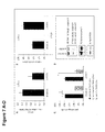
- AVEN gene expression was significantly down-regulated in HT-29 cells compared to cells transfected with si-control (shown in Figure 7A ).
- DNA fragmentation as one characteristic to distinguish apoptotic from necrotic cell death (Gentschev et al., 2010) was tested.
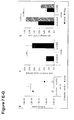
- Percentage of cells in sub-G1 phase 24 hpi was significantly enhanced in GLV-1h68 infected si-AVEN transfected HT-29 cells compared to si-control transfected HT-29 cells (shown in Figure 7G ).
- GFP + cells a reduced infection rate
- an impaired viral replication took place after down-regulation of the anti-apoptotic factor AVEN in HT-29 cells.
- this down-regulation also resulted in an increase of apoptosis in HT-29.
- This novel approach can also be adapted to various different oncolytic viruses: the genetic layout of a wide array of these viruses is very similar and easily modifiable (Table 1). These viruses have similar characteristics regarding tumor regression (Chen et al., 2011), which can be vastly improved by the invention.
- Vaccinia Virus GLV-1h68 has been proven to be a viable candidate for cancer treatment and is currently being tested in several clinical trials.
- cell culture studies revealed that GLV-1h68 is less effective against certain kinds of tumor cell lines such as the human colon adenocarcinoma cell line HT-29 or the human melanoma cell line 1936-MEL whereas it is very effective against other cell lines such as 888-MEL another human melanoma cell line.
- HT-29, GI-101A, 1936-MEL, and 888-MEL the effects of the genetic variances in four different cell lines (HT-29, GI-101A, 1936-MEL, and 888-MEL) in silico by a dynamic modeling approach and by analytical testing in cell culture.
- Boolean modeling in combination with gene expression analysis proved viable to identify a target gene responsible for reduced apoptosis in HT-29 tumor cells.
- This gene not found to be significantly up-regulated in GI-101A, suppresses the effects of GLV-1h68 in HT-29.
- the Boolean models also predicted the effects of a si-RNA knockdown of AVEN: the efficacy of GLV-1h68 against HT-29 could be restored. In vitro the effects of this si-RNA knockdown were not as expected by Boolean modeling. The rate of infection with GLV-1h68 was reduced, however the rate of apoptosis induced by GLV-1h68 also increased significantly.
- AVEN in vitro in 888-MEL and 1936-MEL cells. AVEN does also not show up to be significantly up-regulated in the gene expression data of those two cell lines we evaluated.
- 1936-MEL does not react as strongly to GLV-1h68 as 888-MEL does.
- a subsequent si-RNA knockdown of AVEN in 1936-MEL improves viral efficacy in the weak responder 1936-MEL.
- GLV-1h68 is a genetically modified attenuated virus construct based on the LIVP strain genome that consists of three expression cassettes namely Renilla luciferase-green fluorescent protein (RUC-GFP) fusion protein, ⁇ -galactosidase and ⁇ -glucuronidase that were recombined into the F14.5L, J2R (encoding thymidine kinase) and A56R (enoding hemagglutinin) loci of the viral genome.
- ROC-GFP Renilla luciferase-green fluorescent protein
- African green monkey kidney fibroblasts were purchased from the American Type Culture Collection (ATCC) and cultured in DMEM (Sigma-Aldrich, Steinheim, Germany) supplemented with 10% fetal bovine serum (FBS; PAA Laboratories, Cölbe, Germany) and penicillin G/streptomycin solution (100 U/ml; Sigma-Aldrich).
- the human colorectal adenocarcinoma cell line HT-29 (Duke's type B) was obtained from ATCC.
- melanoma cell lines 1936-MEL and 888-MEL were kindly provided by F. M. Marincola (National Institutes of Health, Bethesda, MD, USA) (Ascierto et al., 2011; Reinboth et al., 2012; Weibel et al., 2013b) and cultured in RPMI-1640 supplemented with 10% FBS and penicillin G/streptomycin solution (100 U/ml).
- the human ductal breast adenocarcinoma cell line GI-101A was kindly provided by A. Aller (Rumbaugh-Goodwin Institute for Cancer Research, Inc., FL, USA)) (Ascierto et al., 2011; Weibel et al., 2011) and maintained in RPMI-1640 supplemented with 5 ng/ml b-estradiol (Sigma-Aldrich) and 5 ng/ml progesterone (Sigma Aldrich), 1 mM sodium pyruvate (Sigma-Aldrich), 10 mM HEPES (PAA Laboratories), 20% FBS and penicillin G/streptomycin solution (100 U/ml). Cells were maintained at 37°C and 5% CO 2 .
- RNA isolation For total RNA isolation the RNeasy Mini Kit (Qiagen GmbH, Hilden, Germany) was used according to the manufacturer's instructions. Genomic DNA was excluded from the samples using DNA- free TM Kit (Ambion, Austin, TX, USA) according to the manufacturer's instructions. The amount of RNA was measured with a Nanodrop and RNA was converted to cDNA using the RevertAidTM First Strand cDNA Synthesis Kit (Fermentas, St. Leon-Rot, Germany) or the Transcriptor High Fidelity cDNA Synthesis Kit (Roche Diagnostics, Mannheim, Germany). Sequences for human AVEN, human ⁇ -Actin and human GAPDH on mRNA level were amplified with Phusion DNA Polymerase (Finnzymes, Espoo, Finland) by PCR with specific primers for
- the PCR reaction was run in a T-Gradient Thermoblock PCR machine (Biometra, Göttingen, Germany) under the following conditions: 98°C/1 min, (30 cycles of 98 °C/15 sec, 56°C/15 sec, 72°C/5 sec) and 72°C/5 min.
- si-RNA-27 kit OriGene, Rockville, USA
- three unique human AVEN-27mer si-RNA duplexes Licus ID 57099
- Trilencer-27 universal scrambled negative control si-RNA duplex in combination with SiLentFect tm Lipid Reagent (Bio-Rad Laboratories GmbH, Ober, Germany) were used.
- Si-RNA sequences (si-AVEN) were as follows:
- Negative control sequence (si-control) was as follows:
- r denotes the sequence being RNA and not DNA.
- si-RNA transfection of the AVEN gene cells were cultured for 24 h in wells of a 6-well plate to 50-60% confluence.
- SiLentFectTM Lipid Reagent 5 ⁇ l/well
- si-RNA 15 ⁇ M/well scrambled control or 3x5 ⁇ M/well of specific AVEN si-RNA duplexes
- 100 ⁇ l serum-free medium 100 ⁇ l serum-free medium and incubated at room temperature for 20 min.
- a sample with SiLentFect tm Lipid Reagent but without si-RNA duplex was used.
- cells were harvested and lysed in RIPA buffer (50 mM Tris-HCl pH 7.4, 150 mM NaCl, 1 mM EDTA, 1 % Triton X-100, 1 % Sodium desoxycholate, 0.1 % SDS, 1 tablet Complete (Roche Diagnostics)).
- RIPA buffer 50 mM Tris-HCl pH 7.4, 150 mM NaCl, 1 mM EDTA, 1 % Triton X-100, 1 % Sodium desoxycholate, 0.1 % SDS, 1 tablet Complete (Roche Diagnostics)
- the lysates were separated on a 12 or 14% SDS-PAGE, and afterwards proteins were transferred onto a nitrocellulose transfer membrane (Whatman GmbH, Dassel, Germany).
- Actin was detected using monoclonal anti-mouse actin antibody (ab6276, 1:10,000, abcam) and a secondary horseradish peroxidase-conjugated antibody (anti-mouse IgG, ab6728, 1:2000, abcam). Peroxidase-bound protein bands were visualized using the enhanced chemiluminescence method.
- a FITC Annexin V Apoptosis Detection Kit with 7-AAD BioLegend Inc. San Diego, USA
- 7-AAD BioLegend Inc. San Diego, USA
- cells were washed once with PBS, resuspended in 100 ⁇ l Annexin V Binding Buffer supplemented with 2 ⁇ l FITC-Annexin V and 2 ⁇ l of 7-AAD cell viability staining solution.
- Annexin V-APC Immunotools GmbH, Oldenburg, Germany
- PI was used.
- Cells were analyzed, using an Accuri C6 Cytometer and FACS analysis software CFlow Version 1.0.227.4 (Accuri Cytometers, Inc. Ann Arbor, MI USA). Cell debris was excluded from the measurements by defining a proper gate for the cell population of interest by using forward (FSC) and sideward (SSC) scatter. 10 000 events were analyzed. Experiments were performed in triplicate and repeated twice.
- Cells were infected with GLV-1h68 at an MOI of 0.1 and 1.0. Cell viability was determined 24, 48 and 72 h after viral infection. For this purpose, culture medium was replaced by 500 ⁇ l sterile filtrated MTT 3-(4,5-Dimethylthiazol-2-yl)-2,5-diphenyltetrazolium Bromide (2.5 mg/ml; Sigma) dissolved in RPMI-1640 without Phenol Red (Sigma-Aldrich) and incubated for 2 h at 37°C. The color reaction was stopped by adding 1 N HCl diluted in isopropyl alcohol (Sigma-Aldrich).
- the optical density was measured at a wavelength of 570 nm in an Elisa Photometer Sunrise tm (TECAN Group, Gurnnedorf, Germany). Uninfected cells were used as positive control, defined as 100% viable. The experiment was performed twice in triplicate.
- PBMCs peripheral blood mononuclear cells
- the dynamic modeling was done with CellDesigner 3.5.2 (Funahashi et al., 2008) and consisted of the apoptosis, MAPK, p53, WNT, Hedgehog and mitochondrial Ca 2+ - signaling.
- the evaluation of the resulting signal strength was done by SQUAD (Di Cara et al., 2007).
- the colorectal adenocarcinoma HT29 and the breast cancer cell line GI-101A show a different gene expression than the MEL cell lines: Here mainly caspases in the apoptosis pathway are up-regulated ( Figure 3 ).
- the gene AVEN which is only upregulated in this cell line out of the four studied here, is of particular interest: it works as a caspase-9 inhibitor and anti-apoptotic BCL2-family activator (Chau et al., 2000).
- AVEN is found ubiquitously expressed in various cell lines and tissues (Chau et al., 2000). We wanted to verify this for 888-MEL, 1936-MEL, GI-101A and HT-29. Cells were cultured for 24 and 48 h and AVEN gene expression was tested on RNA level ( Figure 6A ). AVEN could be detected in all four cell lines cultured for 24 and 48 h respectively. GAPDH, a housekeeping gene, was used as control. Calculation of the ratio between AVEN and GAPDH gene expression levels revealed a higher AVEN gene expression after 24 (ratio >1) than after 48 h (ratio ⁇ 1) in culture ( Figure 6B ).
- the AVEN/GAPDH ratio was highest in HT-29 compared to the other cell lines. Testing AVEN on protein level exemplarily shown for HT-29 and 1936-MEL revealed that it was expressed in both cell lines 6, 24 and 48 hpi in control and GLV-1h68 (MOI 1.0) infected samples ( Figure 6C ).
- AVEN was found to be a novel regulator of the apoptosome complex and further was identified as potential biomarker e.g. in hematological malignancies (Chau et al., 2000; Eißmann et al., 2012), we wanted to elucidate whether the anti-apoptotic factor AVEN would affect the permissiveness of the weak/non-responder cell line HT-29 to GLV-1h68 infection.
- RNA interference was used to down-regulate AVEN in HT-29 cells. For this, cells were transfected for 48 to 72 h with a si-RNA targeting AVEN (si-AVEN) and a scrambled RNA (si-control) as control. AVEN gene expression was significantly down-regulated in HT-29 cells compared to cells transfected with si-control ( Figure 7A ).
- VACV effects are a general phenomenon or unique for the non-responder cell line HT-29
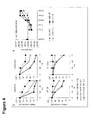
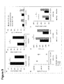
- knockdown of AVEN was also tested in the responder cell line 888-MEL and weak responder 1936-MEL ( Figures 8 and 9 ). In these analyses similar results could be obtained: viral replication is reduced but at the same time apoptosis of cancer strains is enhanced.
Landscapes
- Health & Medical Sciences (AREA)
- Life Sciences & Earth Sciences (AREA)
- Genetics & Genomics (AREA)
- Engineering & Computer Science (AREA)
- Biomedical Technology (AREA)
- Chemical & Material Sciences (AREA)
- Biotechnology (AREA)
- Organic Chemistry (AREA)
- General Engineering & Computer Science (AREA)
- Bioinformatics & Cheminformatics (AREA)
- Wood Science & Technology (AREA)
- Zoology (AREA)
- Virology (AREA)
- Molecular Biology (AREA)
- General Health & Medical Sciences (AREA)
- Microbiology (AREA)
- Biophysics (AREA)
- Biochemistry (AREA)
- Plant Pathology (AREA)
- Physics & Mathematics (AREA)
- Oncology (AREA)
- Mycology (AREA)
- Medicinal Chemistry (AREA)
- Pharmacology & Pharmacy (AREA)
- Epidemiology (AREA)
- Animal Behavior & Ethology (AREA)
- Public Health (AREA)
- Veterinary Medicine (AREA)
- Medicines That Contain Protein Lipid Enzymes And Other Medicines (AREA)
Abstract
The present invention relates to recombinant oncolytic viruses that comprise in their viral genome the genetic information for a si-RNA, sh-RNA, antisense-DNA, antisense-RNA, scAB or sd-AB for inhibiting the gene AVEN in a target or host cell, and pharmaceutical compositions thereof. The present invention further relates to the use of said oncolytic viruses as gene delivery tool and/or for tumor detection. The present invention further relates to the medical uses of said oncolytic viruses, in particular in oncolytic virotherapy and for the diagnosis, prevention and/or treatment of diseases, such as cancer.
Description
- The present invention relates to recombinant oncolytic viruses that comprise in their viral genome the genetic information for a si-RNA, sh-RNA, antisense-DNA, antisense-RNA, sc-AB or sd-AB for inhibiting the gene AVEN in a target or host cell, and pharmaceutical compositions thereof. The present invention further relates to the use of said oncolytic viruses as gene delivery tool and/or for tumor detection. The present invention further relates to the medical uses of said oncolytic viruses, in particular in oncolytic virotherapy and for the diagnosis, prevention and/or treatment of diseases, such as cancer.
- An oncolytic virus is a virus that has the potential to infect and destroy cancerous cells (Bartlett et al., 2013; Patil et al., 2012). As the infected cancer cells are destroyed by lysis, they release new infectious virus particles to help destroy the remaining tumor. Oncolytic viruses are thought not only to cause direct destruction of the tumor cells, but also to stimulate host anti-tumour immune responses. However, these viruses are not effective against all types of cancer due to certain resistance mechanisms against oncolytic virus mediated apoptosis (Cecil et al., 2014).
- Apoptosis as a mechanism of cell death is essential for tissue homeostasis and developmental processes. Aberrations of the tightly controlled process of apoptosis result in a number of human disorders including cancer, neurodegenerative diseases or autoimmune diseases (Hajra, 2004). Described as one of the hallmarks of cancer, the avoidance of apoptosis to prevent cell death is one crucial step in tumor development (Hanahan et al., 2000; Hanahan 2011). There are two major apoptotoc pathways leading to the activation of the caspase cascade: the extrinsic pathway, activated by extracellular signals via cell death receptors and the intrinsic / mitochondrial pathway activated by endogenous signals such as DNA damage (see e.g. Ledgerwood and Morrison, 2009). The apoptosome complex is a central part of the intrinsic pathway. Its formation is the result of a Bcl-2 family-regulated release of cytochrome C from the mitochondria into the cytosol that binds to the apoptosis protease activating factor 1 (APAF-1). A conformational change of APAF-1 results in the binding of the initiator procaspase-9 (Brenner and Mak, 2009). The apoptosome complex is tightly controlled by a number of endogenous regulator (Bratton and Salvesen, 2010). One of those anti-apoptotic factors is AVEN, recently identified by Chau et al. (2010). AVEN is an ubiquitous protein, found to be expressed in a great number of adult tissues and cell lines (Chau et al., 2010; Hawley et al., 2012). It binds to Bcl-xL strengthening its anti-apoptotic activity and also interacts with APAF-1 suppressing the APAF-1 mediated activation of caspase-9 (Chau et al., 2010). In addition, AVEN directly binds and activates the cell-cycle regulating ataxia-telangiectasia (ATM) protein kinase, a critical regulator of the G2/M DNA damage checkpoint in the DNA damage response pathway (Guo et al., 2008). Melzer et al. (2012) showed that AVEN is proteolytically cleaved by cathepsin D resulting in the removal of an inhibitory N-terminal domain and the activation of the anti-apoptotic function of the protein. Also, the WNT signaling pathway, and especially the regulation of the
glycogen synthase kinase 3 beta (GSK3β), the primary inhibitor of β-Catenin, has been shown to be of great importance to tumor proliferation (Kotliarova et al., 2008) β-Catenin has been reported previously to be responsible for tumor proliferation, e.g. in basal cell carcinoma (Saldanha et al., 2004) or in prostate cancer (Kypta and Waxman, 2012). - The specific interference and modifications of molecules and factors being part of the apoptotic cell death pathways such as the apoptosome complex are of special interest in the development of novel cancer therapeutics (see e.g. Ledgerwood and Morrison, 2009). Oncolytic virotherapy is one such promising method (see e.g. the review Goldufsky et al., 2013). It is defined by activation of apoptosis and/or necrosis in tumor cells leading to the specific destruction and lysis of neoplastic tissues, all the while normal tissues remain unharmed (see e.g. Bourke et al., 2011; Liu et al., 2007; Chen and Szalay, 2011). Oncolytic vaccinia viruses (VACVs) have been demonstrated to be very promising candidates to combat different tumor types in several pre-clinical and clinical trials (see e.g. Advani et al., 2012; Donat et al., 2012; Donat et al., 2014; Ehrig et al., 2013; Weibel et al., 2013-a; Zhang et al., 2007). Some tumor cell lines, however, show only a weak response to oncolytic VACV therapy compared to other cell lines (Worschech et al., 2009).
- Thus, there is a need in the art for improved means and methods for oncolytic virotherapy as well as for improved oncolytic viruses.
- According to the present invention this object is solved by a recombinant virus comprising or carrying in its viral genome the genetic information for inhibiting the gene AVEN in a target or host cell, wherein said genetic information is selected from genetic information for a si-RNA, sh-RNA, antisense-DNA, antisense-RNA, sc-AB or sd-AB.
- According to the present invention this object is solved by a pharmaceutical composition, comprising
- (i) the recombinant virus of the present invention; and
- (ii) optionally, pharmaceutically acceptable carrier(s) and/or excipient(s).
- According to the present invention this object is solved by using the the recombinant virus of the present invention or the pharmaceutical composition of the present invention, as gene delivery tool and/or for tumor detection.
- According to the present invention this object is solved by providing the recombinant virus of the present invention or the pharmaceutical composition of the present invention for use in medicine.
- According to the present invention this object is solved by providing the recombinant virus of the present invention or the pharmaceutical composition of the present invention for use in the diagnosis, prevention and/or treatment of a disease, wherein said disease is cancer.
- Before the present invention is described in more detail below, it is to be understood that this invention is not limited to the particular methodology, protocols and reagents described herein as these may vary. It is also to be understood that the terminology used herein is for the purpose of describing particular embodiments only, and is not intended to limit the scope of the present invention which will be limited only by the appended claims. Unless defined otherwise, all technical and scientific terms used herein have the same meanings as commonly understood by one of ordinary skill in the art. For the purpose of the present invention, all references cited herein are incorporated by reference in their entireties.
- Concentrations, amounts, and other numerical data may be expressed or presented herein in a range format. It is to be understood that such a range format is used merely for convenience and brevity and thus should be interpreted flexibly to include not only the numerical values explicitly recited as the limits of the range, but also to include all the individual numerical values or sub-ranges encompassed within that range as if each numerical value and sub-range is explicitly recited. As an illustration, a numerical range of "20 to 100 nucleotides" should be interpreted to include not only the explicitly recited values of 20 to 100, but also include individual values and sub-ranges within the indicated range. Thus, included in this numerical range are individual values such as 20, 21, 22, 23, 24, 25, 26, 27, 28, 29 ,... 97, 98, 100 and sub-ranges such as from 25 to 35, from 20 to 40, from 25 to 50, etc. This same principle applies to ranges reciting only one numerical value, such as "at least 25 nucleotides". Furthermore, such an interpretation should apply regardless of the breadth of the range or the characteristics being described.
- As discussed above, the present invention provides recombinant viruses.
- The recombinant viruses comprise or carry in their viral genome the genetic information for inhibiting the gene AVEN in a target or host cell, in particular in a target or host cell infected by the virus.
- According to the invention, the AVEN gene in an infected target or host cell is to be inhibited by gene silencing or gene knockdown.
- The terms "gene silencing" or "gene knockdown" as used herein refer to the ability to reduce or prevent the expression of a certain gene in a target or host cell. Gene silencing can occur during either transcription or translation.
- According to the invention, said genetic information, which is comprised or carried in the recombinant viruses of the invention, for inhibiting the gene AVEN is selected from genetic information for a
silencing RNA (si-RNA),
small hairpin RNA (sh-RNA),
antisense-DNA,
antisense-RNA,
single chain antibody (sc-AB), or
single domain antibody (sd-AB). - Preferably, the recombinant viruses of the present invention are selected from:
- the Poxviridae, Adenovinidae, Reoviridae and Herpesviridae families of double stranded DNA (dsDNA) viruses,
- the Picornaviridae, Rhabdoviridae, Panamyxovinidae and Togaviridae families of single stranded RNA (ssRNA) viruses, and
- the Retroviridae family of retro-transcribing viruses.
- An "oncolytic virus" as used herein refers to a virus that has the potential to infect and destroy target or host cells, in particular cancerous cells.
- The oncolytic virus is therefore selected from the Poxviridae, Adenoviridae, Reoviridae and Herpesviridae families of double stranded DNA viruses, or from the Picornaviridae, Rhabdoviridae, Paramyxoviridae and Togaviridae families of single stranded RNA viruses, or from the Retroviridae family of retro-transcribing viruses. Their viral genomes are to be modified to carry the genetic information for expression of a si-RNA, sh-RNA, antisense-DNA, antisense-RNA, sc-AB or sd-AB for inhibiting the gene AVEN in an infected target or host cell.
- In one embodiment, the recombinant virus, in particular the recombinant oncolytic virus is
- an attenuated oncolytic virus,
such as
an attenuated adenovirus (e.g. ONYX-105),
an attenuated vaccinia virus (VACV),
an attenuated poliovirus (e.g. PV1), - a replication-competent oncolytic virus,
such as
a replication-competent oncolytic adenovirus (e.g. ONYX-411),
a replication-competent oncolytic vaccinia virus (VACV),
a replication-competent oncolytic seneca valley virus (e.g. SVV-01),
a replication-competent oncolytic reovirus. - In one embodiment, the genetic information for said AVEN si-RNA, sh-RNA, antisense-DNA, antisense-RNA, sc-AB or sd-AB is combined with / linked to a late/very late promoter.
- In one embodiment, said late/very late promoter is selected from:
- PE/L (early/late promoter), or
- P11 (late/very late promoter).
- Preferebly, the recombinant oncolytic virus comprises or carries
- in its viral genome deletional mutation(s) in nonessential gene(s) and/or in small ORF(s),
- foreign or heterologous genes,
such as- genes encoding for tumor suppressor proteins, immunostimulatory proteins and cytokines,
- genes encoding for pro-drug activating enzymes,
- genes encoding for marker proteins,
such as fluorescent proteins, e.g. GFP or its derivatives, - genes encoding for glucoronidase as a marker for oncolysis.
- In one embodiment, the recombinant oncolytic virus is an oncolytic vaccinia virus (VACV) which comprises or carries in its viral genome deletional mutation(s)
- in nonessential gene(s),
such as JR2 (encoding thymidine kinase, TK), A56R (encoding hemagglutinin), vaccinina growth factor gene,
and/or - in small ORF(s)
such as F14.5,
and comprises or carries foreign or heterologous genes,
such as
renilla luciferase-aequorea GFP (Ruc-GFP), and/or
glucoronidase as a marker for oncolysis, and/or
transferrin receptor (TFR) and lacZ. - In one embodiment, the recombinant oncolytic virus is for example the oncolytic vaccinia virus strain GLV-1h68.
- In one embodiment, the si-RNA for AVEN comprises a nucleic acid sequence which comprises preferably about 20 to about 100 nucleotides, more preferably at least 25 nucleotides, of which preferably at least 23 are ribonucleotides and at least two are desoxyribonucleotides.
- In one embodiment, the si-RNA for AVEN comprises or consists of the nucleic acid sequence of any one of SEQ ID NOs. 1 to 3:
- In one embodiment, the si-RNA for AVEN comprises or consists of the nucleic acid sequence of SEQ ID NO. 1:
- SR311345A rGrCrCrArGrUrGrUrUrUrArCrArGrCrArUrArGrCrArGrATG Wherein "r" denotes the sequence being RNA and not DNA.
- In one embodiment, the si-RNA for AVEN comprises or consists of the nucleic acid sequence of SEQ ID NO. 2:
- SR311345B rUrGrUrGrGrArUrArGrUrGrArGrUrUrArUrUrGrGrUrUrCGA Wherein "r" denotes the sequence being RNA and not DNA.
- In one embodiment, the si-RNA for AVEN comprises or consists of the nucleic acid sequence of SEQ ID NO. 3:
- SR311345C rGrCrUrGrCrUrGrArUrUrArArGrArGrArArUrArGrArUrGCA Wherein "r" denotes the sequence being RNA and not DNA.
- Preferably, the target or host cell is a cancerous or tumor cell, which is to be infected by the oncolytic virus of the invention.
- As discussed above, the present invention provides pharmaceutical compositions, comprising
- (i) the recombinant (oncolytic) virus of the present invention; and
- (ii) optionally, pharmaceutically acceptable carrier(s) and/or excipient(s).
- In one embodiment, the pharmaceutical composition of the present invention comprises further drug(s), such as
- chemotherapeutic agent(s),
- radiotherapeutic agent(s), and/or
- agent(s) for near infrared laser-induced targeted cancer therapy.
- In one embodiment, the pharmaceutical composition of the present invention is formulated for systemic delivery, tumor injection, intravenous administration, and/or for intradermal, subcutaneous, intramuscular, intravenous, intraosseous, intraperitoneal, intrathecal, epidural, intracardiac, intraarticular, intracavernous, intracerebral, intracerebroventricular and intravitreal injection(s).
- As discussed above, the present invention provides the use of the recombinant (oncolytic) virus of the present invention or the pharmaceutical composition of the present invention, as gene delivery tool and/or for tumor detection.
- As discussed above, the present invention provides the recombinant (oncolytic) virus of the present invention or the pharmaceutical composition of the present invention, for use in medicine.
- As discussed above, the present invention provides the recombinant (oncolytic) virus of the present invention or the pharmaceutical composition of the present invention, for use in the diagnosis, prevention and/or treatment of a disease.
- Said disease is preferably cancer.
- In one embodiment, the recombinant oncolytic virus or pharmaceutical composition is for use in oncolytic virotherapy.
- The term "oncolytic virotherapy" as used herein refers to therapy of cancer by administration of oncolytic viruses to induce tumor regression.
- In one embodiment, the recombinant oncolytic virus or pharmaceutical composition is for use in combination with other therapies, preferably other cancer therapies, such as chemotherapy and/or radiation therapy.
- Preferably, the administration is systemic, intravenous, via injection into tumor, and/or via intradermal, subcutaneous, intramuscular, intravenous, intraosseous, intraperitoneal, intrathecal, epidural, intracardiac, intraarticular, intracavernous, intracerebral, intracerebroventricular and intravitreal injection(s).
- As discussed above, the present invention provides a method for the diagnosis, prevention and/or treatment of a disease,
comprising the step of
administering a therapeutically effective amount of a recombinant (oncolytic) virus of the present invention or a pharmaceutical composition of the present invention to a subject in need thereof. - Said disease is preferably cancer.
- A therapeutically effective amount of a recombinant (oncolytic) virus of the present invention is the amount which results in the desired therapeutic result, in particular tumor regression.
- The recombinant viruses are preferably administered in multiple cycles over a period of time, such as for several days up to several weeks.
- For example, a therapeutically effective amount of VACV can be in the range from about 105 pfu and about 109 pfu, which is preferably delivered to the subject/patient in need thereof in multiple cycles, such as via a 3 cycle regimen over about 28 days.
- The skilled artisan will be able to determine suitable therapeutically effective amounts.
- As discussed above, the recombinant oncolytic virus or pharmaceutical composition can be administered in combination with other therapies, preferably other cancer therapies, such as chemotherapy, radiation therapy and/or near infrared laser-induced targeted cancer therapy.
- As discussed above, the administration is systemic, intravenous, via injection into tumor, and/or via intradermal, subcutaneous, intramuscular, intravenous, intraosseous, intraperitoneal, intrathecal, epidural, intracardiac, intraarticular, intracavernous, intracerebral, intracerebroventricular and intravitreal injection(s).
- In one embodiment, the method comprises oncolytic virotherapy.
- Oncolytic viruses have the potential to infect and destroy cancerous cells (Bartlett et al., 2013; Patil et al., 2012). However, these viruses are not effective against all types of cancer due to certain resistance mechanisms against oncolytic virus mediated apoptosis (Cecil et al., 2014). One of the most crucial resistance mechanisms is found in the suppression of the apoptosome. The apoptosome complex is a central part of the intrinsic pathway. Its formation is the result of a Bcl-2 family-regulated release of cytochrome C from the mitochondria into the cytosol that binds to the apoptosis protease activating factor 1 (APAF-1). A conformational change of APAF-1 results in the binding of the initiator procaspase-9 (Brenner and Mak, 2009; Bao 2007). The apoptosome complex is tightly controlled by a number of endogenous regulators (Bratton and Salvesen, 2010).
- One of those anti-apoptotic factors is AVEN, recently identified by Chau et al. (2000). AVEN is a ubiquitous protein, found to be expressed in a great number of adult tissues and cell lines (Chau et al., 2000; Hawley et al., 2012). It binds to Bcl-xL and thus strengthening its anti-apoptotic activity (Figueroa et al., 2004). It also interacts with APAF-1: it suppresses the APAF-1 mediated activation of caspase-9 (Chau et al., 2000).
- In a clinical study, Paydas et al. (2003) found elevated AVEN gene expression levels in adult acute leukemia patients: Higher levels were found in those patients with relapse compared to those without relapse. Also in childhood acute lymphoblastic leukemia, poor prognosis was associated with AVEN overexpression (Choi et al., 2006). Eißmann et al. (2012) concluded in their study an oncogenic potential of AVEN during development of hematopoietic neoplasms. A microarray study revealed decreased nuclear AVEN gene expression levels in breast cancer tissue compared to healthy breast tissue - providing evidence that expression of AVEN leads to resistance against apoptosis in breast cancer tissue induced by DNA damage (Kutuk et al., 2010).
- By genetically modifying the genome of a suitable oncolytic virus - as for example GLV-1h68 - it is possible to perform a knockdown of the expression of AVEN and thus availability of this anti-apoptotic factor inside the tumor cells. To this end the genetic code for small interfering RNA targeting the gene AVEN (si-AVEN) is put onto the genome of an oncolytic virus and preferably be coupled with a late/very late promotor. This leads to the expression of si-AVEN after replication of the virus has taken place in the infected tumor cell. The knockdown of AVEN subsequently leads to enhanced apoptosis of cancer strain cells, when oncolytic virus therapy is performed.
- To provide proof of concept, human colorectal adenocarcinoma cells (HT-29) were transfected for 48 to 72 h with si-AVEN and a scrambled RNA (si-control) as control. AVEN gene expression was significantly down-regulated in HT-29 cells compared to cells transfected with si-control (shown in
Figure 7A ). To verify whether the knockdown of AVEN would enhance apoptotic characteristics in the cancer cells, DNA fragmentation as one characteristic to distinguish apoptotic from necrotic cell death (Gentschev et al., 2010) was tested. Down-regulation of the anti-apoptotic factor AVEN in HT-29 cells revealed a significant increase of DNA-fragmentation, detected as an increase of cells in sub-G1 phase in si-AVEN transfected cells compared to si-control transfected cells (shown inFigure 7B ). No significant difference could be observed in Annexin+/7AAD+/- (apoptotic) or Annexin-/7AAD+ (necrotic) cells between si-control and si-AVEN transfected HT-29 cells (shown inFigure 7C ). Western Blot analysis revealed the expression of procaspase-3 (32 kda in both si-AVEN and si-control transfected cells. The cleaved and activated form of the effector caspase-3 was not expressed (shown inFigure 7D ). In accordance with the literature, down-regulation of AVEN results in an activation and cleavage of the initiator caspase-9 (35 kd) (shown inFigure 7D ). GAPDH was used as loading control. - Following the functional proof of AVEN down-regulation in HT-29 cells, replication of one oncolytic virus from the Genelux corporation (GLV-1h68) was tested at a MOI of 1.0 and the infection rate (GFP+ cells) 24 and 48 hpi in these cells. A significant reduction of the virus titer (shown in
Figure 7E ) and a significantly smaller number of GFP+ cells (shown inFigure 7F ) were detected in si-AVEN transfected cells compared to si-control transfected cells at 24 and 48 hpi. Percentage of cells insub-G1 phase 24 hpi was significantly enhanced in GLV-1h68 infected si-AVEN transfected HT-29 cells compared to si-control transfected HT-29 cells (shown inFigure 7G ). Taken together, a reduced infection rate (GFP+ cells) and an impaired viral replication took place after down-regulation of the anti-apoptotic factor AVEN in HT-29 cells. However, this down-regulation also resulted in an increase of apoptosis in HT-29. - To see, whether the observed oncolytic virus effects are a general phenomenon or unique for the cell line HT-29, knockdown of AVEN was also tested in metastatic melanoma cancer cell lines 888-MEL (see
Figures 8A-F ) and 1936-MEL (seeFigures 9A-F ). In these tests similar results could be obtained: viral replication is reduced but at the same time apoptosis of cancer strains is enhanced. - This novel approach can also be adapted to various different oncolytic viruses: the genetic layout of a wide array of these viruses is very similar and easily modifiable (Table 1). These viruses have similar characteristics regarding tumor regression (Chen et al., 2011), which can be vastly improved by the invention.

- Vaccinia Virus GLV-1h68 has been proven to be a viable candidate for cancer treatment and is currently being tested in several clinical trials. However, cell culture studies revealed that GLV-1h68 is less effective against certain kinds of tumor cell lines such as the human colon adenocarcinoma cell line HT-29 or the human melanoma cell line 1936-MEL whereas it is very effective against other cell lines such as 888-MEL another human melanoma cell line. In this study we calculate the effects of the genetic variances in four different cell lines (HT-29, GI-101A, 1936-MEL, and 888-MEL) in silico by a dynamic modeling approach and by analytical testing in cell culture. We also establish a method of RNA interference to render the non- or weak-responding tumor cell lines susceptible to GLV-1h68. With these methods we found a very strong correlation of deactivation of the gene AVEN, an anti-apoptotic effector, and susceptibility to GLV-1h68. By deactivation of AVEN we were able to restore the full efficacy of GLV-1h68 and only marginally decrease its tumor infection rate.
- In order to understand why some tumor cells are able to survive an infection by the oncolytic VACV GLV-1h68, we statistically evaluated the gene expression data from infected and non-infected melanoma cell lines (888-MEL, 1936-MEL), the breast cancer cell line GI-101A, as well as colon adenocarcinoma cells HT-29. For convenience, the evaluated gene expression data acquired were visualized by heatmapping. Boolean modeling of the apoptosis network was also performed and the significant gene expression changes were mapped onto the models. Based on this data, a special focus was set on the expression and function of the anti-apoptotic protein AVEN in different cancer cell lines. We elucidated that there is a specific role and function of AVEN for successful infection and replication of GLV-1h68 in those tumor cells and that therapeutic success or failure is dependent on AVEN levels in the cancer strains.
- Boolean modeling in combination with gene expression analysis proved viable to identify a target gene responsible for reduced apoptosis in HT-29 tumor cells. This gene, not found to be significantly up-regulated in GI-101A, suppresses the effects of GLV-1h68 in HT-29. The Boolean models also predicted the effects of a si-RNA knockdown of AVEN: the efficacy of GLV-1h68 against HT-29 could be restored. In vitro the effects of this si-RNA knockdown were not as expected by Boolean modeling. The rate of infection with GLV-1h68 was reduced, however the rate of apoptosis induced by GLV-1h68 also increased significantly.
- We also found AVEN in vitro in 888-MEL and 1936-MEL cells. AVEN does also not show up to be significantly up-regulated in the gene expression data of those two cell lines we evaluated. The effects of AVEN in 888-MEL and 1936-MEL, due to their non-significant expression compared to HT-29, show in vitro to be not strong enough to impede GLV-1h68 efficacy as they do in HT-29. However, 1936-MEL does not react as strongly to GLV-1h68 as 888-MEL does. A subsequent si-RNA knockdown of AVEN in 1936-MEL improves viral efficacy in the weak responder 1936-MEL. These findings are in accordance with the Boolean models for 888-MEL and 1936-MEL. However, according to Boolean modeling, in the melanoma cancer strains the effects of casein kinases and their role to regulate GSK3β seem to be more predominant then the effect of AVEN.
- Therefore, the expression of AVEN and up-regulation of casein kinases could also serve as biomarkers for tumor proliferation.
- Moreover, by identification of AVEN as one of the most important regulators of resistance against a treatment of cancer by oncolytic virus therapy, we herewith describe new designs for more specific viruses against cancer strains with actively expressed AVEN. By inserting a si-RNA for AVEN on a late/very late promoter into the viral genome, we are now able to provide effectively treatment of various cancer strains in humans. This method is also applicable for sh-RNA, antisense-DNA, antisense-RNA, sc-AB or sd-AB for gene knockdown of AVEN.
- The following examples and drawings illustrate the present invention without, however, limiting the same thereto.
-
-
Figure 1 . Genes from the human melanoma cell line 1936-MEL associated to the apoptosis signaling network with the highest activities overall.
Especially the casein kinases are of interest, as they are significantly upregulated. This leads to a higher inhibition of GSK3β and subsequently higher activation of β-Catenin. β-Catenin in turn is then responsible for uncontrolled tumor proliferation. -
Figure 2 . Upregulation of the genes associated to the apoptosis network for the Human melanoma cell line 888-MEL.
As for 1936-MEL, the casein kinases are of special interest due to their influence on β-Catenin through the WNT- and Hedgehog signaling cascades and its subsequent effect of uncontrolled tumor proliferation. -
Figure 3 . Differences in gene expression of HT-29 and GI-101A cancer strains.
Especially interesting is the difference in expression of AVEN, which is highly upregulated only in HT-29. -
Figure 4 . Characterization of GLV-1h68 permissiveness in various human tumor cell lines.- (A) MTT assay was performed to detect the percentage of living cells (888-MEL, 1936-MEL, GI101A and HT-29) after infection with GLV-1h68 (MOI 0.01, 0.1 and 1.0).
- (B) Viral replication in 888-MEL, 1936-MEL, GI-101A, HT-29 cells, infected with GLV-1h68 at an MOI of 0.01 was analyzed by standard viral plaque assay in triplicate. The red line separates active replication from no replication efficiency.
-
Figure 5 . Boolean calculations of survival vs. apoptosis ratios of different cancer strains. 1936-MEL shows a twice as high survival rate as 888-MEL when compared to an in silico untreated mock cancer strain, whereas the rate of cell death is higher in 888-MEL as in 1936-MEL. Therefore these differences in gene expression are enough to render the GLV-1h68 against 1936-MEL almost ineffective, whereas it is very effective against 888-MEL. -
Figure 6 . Expression levels of AVEN in human tumor cell lines.- (A) AVEN gene expression levels in 888-MEL, 1936-MEL, GI-101A and HT-29 cultured for 24 or 48 h were analyzed by PCR. PCR reactions were performed on templates of cDNA from these human cancer cell lines using a set of primers for AVEN and GAPDH as internal control.
- (B) The bar chart represents the ratio between AVEN gene expression and GAPDH expression in the analyzed cell lines at 24 and 48 hours. The red line marks a ratio of 1.0.
- (C) The protein expression of AVEN and GAPDH in HT-29 and 1936-MEL cells infected with GLV-1h68 (MOI 1.0) at 6, 24 and 48 hpi. Mock-infected cells were used as control. GFP expression represents viral infection. The experiments were performed 2-3 times.
-
Figure 7 . AVEN interference enhances apoptotic characteristics and inhibits GLV-1h68 replication in HT-29 cells.- (A) Downregulation of AVEN mRNA expression in HT-29 cells transfected with AVEN si-RNA (si-AVEN) or scrambled control RNA (si-control). Cells were transfected for 72 h.
- (B) The percentage of si-control or si-AVEN transfected HT-29 cells in sub-G1 phase indicating DNA fragmentation (apoptotic characteristic) measured by FACS analysis.
- (C) For analyzing cell membrane permeability, transfected cells were stained with the vital dye 7AAD and Annexin (Ax) and were subsequently analyzed by FACS analysis.
- (D) Protein expression levels of caspase-9 and caspase-3 were analyzed in si-control and si-AVEN transfected HT-29 cells by western blot analysis. GAPDH was used as loading control.
- (E) Viral replication of GLV-1h68 (MOI 1.0) was analyzed in si-control and si-AVEN transfected HT-29 cells by standard
viral plaque assay 24 hpi. - (F) Percentage of transfected and infected HT-29 cells in sub-G1 phase were analyzed by FACS analysis.
- (G) In parallel to the virus titer, the infection rate of the samples was determined by measuring the percentage of GFP+-cells by
FACS analysis
- All transfection experiments were performed in 6-well plates in triplicate and repeated in an independent experiment. Two-sided t-test with unequal variances was used for statistics *p<0.05, ** p< 0.01, ***p<0.001.
-
Figure 8 . Knockdown of AVEN results in impaired GLV-1h68 replication in 888-MEL cells.- (A) Downregulation of AVEN mRNA expression in 888-MEL cells transfected with AVEN si-RNA (si-AVEN) or scrambled control RNA (si-control). Cells were transfected for 72 h.
- (B) The percentage of si-control or si-AVEN transfected 888-MEL cells in sub-G1 phase indicating DNA fragmentation (apoptotic characteristic) measured by FACS analysis.
- (C) For analyzing cell membrane permeability, transfected cells were stained with PI and Annexin (Ax) and were subsequently analyzed by FACS analysis.
- (D) Viral replication of GLV-1h68 (MOI 0.5) was analyzed in si-control and si-AVEN transfected 888-MEL cells by standard
viral plaque assay - (E) In parallel to the virus titer, the infection rate of the samples was determined by measuring the percentage of GFP+-cells by
FACS analysis - (F) Percentage of transfected and infected 888-MEL cells in
sub-G1 phase 24 hpi and 48 hpi were analyzed by FACS analysis.
-
Figure 9 . Knockdown of AVEN results in impaired GLV-1h68 replication in 1936-MEL cells.- (A) Downregulation of AVEN mRNA expression in 1936-MEL cells transfected with AVEN si-RNA (si-AVEN) or scrambled control RNA (si-control). Cells were transfected for 72 h.
- (B) The percentage of si-control or si-AVEN transfected 1936-MEL cells in sub-G1 phase indicating DNA fragmentation (apoptotic characteristic) measured by FACS analysis.
- (C) For analyzing cell membrane permeability, transfected cells were stained with PI and Annexin (Ax) and were subsequently analyzed by FACS analysis.
- (D) Viral replication of GLV-1h68 (MOI 0.5) was analyzed in si-control and si-AVEN transfected 1936-MEL cells by standard
viral plaque assay - (E) In parallel to the virus titer, the infection rate of the samples was determined by measuring the percentage of GFP+-cells by
FACS analysis - (F) Percentage of transfected and infected 1936-MEL cells in
sub-G1 phase 24 hpi and 48 hpi were analyzed by FACS analysis.
All transfection experiments were performed in 6 well plates in triplicate and repeated two times. Two-sided t-test with unequal variances was used for statistics *p<0.05, ** p< 0.01, ***p<0.001.
- The generation of the oncolytic vaccinia virus strain GLV-1h68 was described in detail by Zhang et.al. (2007). In brief, GLV-1h68 is a genetically modified attenuated virus construct based on the LIVP strain genome that consists of three expression cassettes namely Renilla luciferase-green fluorescent protein (RUC-GFP) fusion protein, β-galactosidase and β-glucuronidase that were recombined into the F14.5L, J2R (encoding thymidine kinase) and A56R (enoding hemagglutinin) loci of the viral genome.
- African green monkey kidney fibroblasts (CV-1 cells) were purchased from the American Type Culture Collection (ATCC) and cultured in DMEM (Sigma-Aldrich, Steinheim, Germany) supplemented with 10% fetal bovine serum (FBS; PAA Laboratories, Cölbe, Germany) and penicillin G/streptomycin solution (100 U/ml; Sigma-Aldrich). The human colorectal adenocarcinoma cell line HT-29 (Duke's type B) was obtained from ATCC. Cells were cultured in RPMI-1640 (Sigma-Aldrich) supplemented with 10% FBS, penicillin G/streptomycin solution (100 U/ml) and sodium bicarbonate solution 7.5% (2.2 g/l; Sigma-Aldrich). The human melanoma cell lines 1936-MEL and 888-MEL were kindly provided by F. M. Marincola (National Institutes of Health, Bethesda, MD, USA) (Ascierto et al., 2011; Reinboth et al., 2012; Weibel et al., 2013b) and cultured in RPMI-1640 supplemented with 10% FBS and penicillin G/streptomycin solution (100 U/ml). The human ductal breast adenocarcinoma cell line GI-101A was kindly provided by A. Aller (Rumbaugh-Goodwin Institute for Cancer Research, Inc., FL, USA)) (Ascierto et al., 2011; Weibel et al., 2011) and maintained in RPMI-1640 supplemented with 5 ng/ml b-estradiol (Sigma-Aldrich) and 5 ng/ml progesterone (Sigma Aldrich), 1 mM sodium pyruvate (Sigma-Aldrich), 10 mM HEPES (PAA Laboratories), 20% FBS and penicillin G/streptomycin solution (100 U/ml). Cells were maintained at 37°C and 5% CO2.
- For total RNA isolation the RNeasy Mini Kit (Qiagen GmbH, Hilden, Germany) was used according to the manufacturer's instructions. Genomic DNA was excluded from the samples using DNA-free™ Kit (Ambion, Austin, TX, USA) according to the manufacturer's instructions. The amount of RNA was measured with a Nanodrop and RNA was converted to cDNA using the RevertAid™ First Strand cDNA Synthesis Kit (Fermentas, St. Leon-Rot, Germany) or the Transcriptor High Fidelity cDNA Synthesis Kit (Roche Diagnostics, Mannheim, Germany). Sequences for human AVEN, human β-Actin and human GAPDH on mRNA level were amplified with Phusion DNA Polymerase (Finnzymes, Espoo, Finland) by PCR with specific primers for
- human AVEN
forward 5'-GATTTCAGTGTCCTCCTTAG-3' SEQ ID NO. 5, and
reverse 5'-CCTTGCCATCATCAGTTCTC-3' SEQ ID NO. 6, (according to Paydas, 2003); - human GAPDH
forward 5'-GCCTTCCGTGTCCCCACTGC-3' SEQ ID NO. 7, and
reverse 5'-CAATGCCAGCCCCAGCGTCA -3' SEQ ID NO. 8, and - human β-Actin
forward: 5'-CCTCTCCCAAGTCCACACAG-3' SEQ ID NO. 9, and
reverse 5'- CTGCCTCCACCCACTC-3' SEQ ID NO. 10,
(see Gentschev et al., 2010). - The PCR reaction was run in a T-Gradient Thermoblock PCR machine (Biometra, Göttingen, Germany) under the following conditions: 98°C/1 min, (30 cycles of 98 °C/15 sec, 56°C/15 sec, 72°C/5 sec) and 72°C/5 min.
- For AVEN gene knockdown, a si-RNA-27 kit (OriGene, Rockville, USA) containing three unique human AVEN-27mer si-RNA duplexes (Locus ID 57099) and a Trilencer-27 universal scrambled negative control si-RNA duplex in combination with SiLentFecttm Lipid Reagent (Bio-Rad Laboratories GmbH, München, Deutschland) were used.
- Si-RNA sequences (si-AVEN) were as follows:
- SEQ ID NO. 1:
- SR311345A rGrCrCrArGrUrGrUrUrUrArCrArGrCrArUrArGrCrArGrATG SEQ ID NO. 2:
- SR311345B rUrGrUrGrGrArUrArGrUrGrArGrUrUrArUrUrGrGrUrUrCGA SEQ ID NO. 3:
- SR311345C rGrCrUrGrCrUrGrArUrUrArArGrArGrArArUrArGrArUrGCA
- Negative control sequence (si-control) was as follows:
- SEQ ID NO. 4:
- SR30004 rCrGrUrUrArArUrCrGrCrGrUrArUrArArUrArCrGrCrGrUAT
- Wherein "r" denotes the sequence being RNA and not DNA.
- For si-RNA transfection of the AVEN gene, cells were cultured for 24 h in wells of a 6-well plate to 50-60% confluence. For transfection, SiLentFect™ Lipid Reagent (5 µl/well) and si-RNA (15 µM/well scrambled control or 3x5 µM/well of specific AVEN si-RNA duplexes) were mixed in 100 µl serum-free medium and incubated at room temperature for 20 min. As a control, a sample with SiLentFecttm Lipid Reagent but without si-RNA duplex was used. Cells were incubated for 4 h with 1 ml of serum-free medium and the SiLentFecttm/si-RNA (si-control/si-AVEN) mixture. Afterwards 1 ml serum-containing medium was added and cells were cultivated for 48 or 72 h (final concentration of the si-RNA 37.5 nM). Transfected cells were used for RT-PCR, Western Blot experiments, FACS analysis or cells were infected with VACV.
- For Western Blot analysis, cells were harvested and lysed in RIPA buffer (50 mM Tris-HCl pH 7.4, 150 mM NaCl, 1 mM EDTA, 1 % Triton X-100, 1 % Sodium desoxycholate, 0.1 % SDS, 1 tablet Complete (Roche Diagnostics)). The lysates were separated on a 12 or 14% SDS-PAGE, and afterwards proteins were transferred onto a nitrocellulose transfer membrane (Whatman GmbH, Dassel, Germany).
- Polyclonal rabbit antibodies against AVEN (#2300, 1:100, Cell Signaling Technology, Inc., Danvers, USA), caspase-3 (sc-7148, 1:100, Santa Cruz Biotechnology Inc., Heidelberg, Germany) caspase-9 (#9502, 1:100, Cell Signaling technology, Inc.) and GAPDH (ab 8245, 1:2500, abcam, Cambridge, UK) were used and incubated O/N at 4°C. The secondary antibody (horseradish peroxidase-conjugated anti-rabbit IgG; Santa Cruz Biotechnology Inc.) exposure was for 1 h at a 1:5000 dilution in PBS-Tween. Actin was detected using monoclonal anti-mouse actin antibody (ab6276, 1:10,000, abcam) and a secondary horseradish peroxidase-conjugated antibody (anti-mouse IgG, ab6728, 1:2000, abcam). Peroxidase-bound protein bands were visualized using the enhanced chemiluminescence method.
- Cells transfected with si-RNA AVEN (si-AVEN) or scrambled RNA (si-control)
si-AVEN SEQ ID NO. 1, 2 and 3: SR311345A rGrCrCrArGrUrGrUrUrUrArCrArGrCrArUrArGrCrArGrATG SR311345B rUrGrUrGrGrArUrArGrUrGrArGrUrUrArUrUrGrGrUrUrCGA SR311345C rGrCrUrGrCrUrGrArUrUrArArGrArGrArArUrArGrArUrGCA si-control SEQ ID NO. 4: SR30004 rCrGrUrUrArArUrCrGrCrGrUrArUrArArUrArCrGrCrGrUAT - For cell cycle analysis and detection of DNA fragmentation (sub-G1 phase) (Krysko et al., 2008; Darzynkiewicz et al., 1997) cell pellets were resuspended in a solution of 100 µl PBS + 2% FBS and 5 µl propidium iodide (PI, Sigma-Aldrich). Prior to measurement, the suspension was frozen in liquid nitrogen and thawed at 37°C.
- To distinguish apoptotic from necrotic cells, a FITC Annexin V Apoptosis Detection Kit with 7-AAD (BioLegend Inc. San Diego, USA) was used. For this, cells were washed once with PBS, resuspended in 100 µl Annexin V Binding Buffer supplemented with 2 µl FITC-Annexin V and 2 µl of 7-AAD cell viability staining solution. In addition, the combination of Annexin V-APC (Immunotools GmbH, Oldenburg, Germany) and PI was used. Cells were analyzed, using an Accuri C6 Cytometer and FACS analysis software CFlow Version 1.0.227.4 (Accuri Cytometers, Inc. Ann Arbor, MI USA). Cell debris was excluded from the measurements by defining a proper gate for the cell population of interest by using forward (FSC) and sideward (SSC) scatter. 10 000 events were analyzed. Experiments were performed in triplicate and repeated twice.
- Cells transfected with si-RNA AVEN (si-AVEN) and scrambled RNA (si-control) were cultured in wells of a 6-well plate for 48 or 72 h and were infected with GLV-1h68 at a multiplicity of infection (MOI) of 0.1, 0.5 or 1.0 in 1 ml infection medium with 2% FBS for 1 h. Every 15 min, cells were agitated slightly for homogenous infection. Infection medium was collected and replaced by 1.5 ml culture medium. Cells were resuspended in 1 ml PBS after harvesting and supernatants were collected separately 24 and 48 hours post infection (hpi). Prior to analysis three freeze and thaw cycles in liquid nitrogen were accomplished to release viral particles. All samples were infected in triplicate. Serial dilutions of the samples were titrated on 100% confluent CV-1 monolayers by standard viral plaque assay in duplicate.
- Cells were infected with GLV-1h68 at an MOI of 0.1 and 1.0. Cell viability was determined 24, 48 and 72 h after viral infection. For this purpose, culture medium was replaced by 500 µl sterile filtrated MTT 3-(4,5-Dimethylthiazol-2-yl)-2,5-diphenyltetrazolium Bromide (2.5 mg/ml; Sigma) dissolved in RPMI-1640 without Phenol Red (Sigma-Aldrich) and incubated for 2 h at 37°C. The color reaction was stopped by adding 1 N HCl diluted in isopropyl alcohol (Sigma-Aldrich). The optical density was measured at a wavelength of 570 nm in an Elisa Photometer Sunrisetm (TECAN Group, Männedorf, Germany). Uninfected cells were used as positive control, defined as 100% viable. The experiment was performed twice in triplicate.
- To determine significance between 2 conditions or groups, a 2-tailed t-test with unequal variances was used (Excel 2010 for Windows). P-values were defined as follows *p<0.05, **p<0.01, ***p<0.001.
- Gene expression data was generated as described previously (Reinboth et al., 2012). In brief, 1936-MEL, 888-MEL, GI-101A, and HT-29 cells were infected with GLV-1h68 at an MOI of 0.01 or mock-treated with infection medium (2%-FBS). Cells were harvested at respective time points and total RNA isolated via Qiagen miRNeasy mini kit. RNA was amplified (Wang et al., 2000) and purified for microarray performance (36k human array platforms, manufactured in-house at the Department of Transfusion Medicine, NIH, Bethesda, MD, USA). Combined peripheral blood mononuclear cells (PBMCs) from 4 normal donors were utilized as a reference. Reference and sample aRNA were labeled directly with Cy3 and Cy5, respectively (two-color system) and cohybridized to the array platform. Following incubation, washing steps, and scanning (Agilent scanner) gene expression data was uploaded to the mAdb databank (http://nciarray.nci.nih.gov). Data from human in-house arrays was processed via BRBArrayTools (Simon et al., 2007) including a quality-control for imperfect spots (Spot filter), normalization via median log-ratio subtraction, and specification of the maximum intensity ratio plus the truncation of larger intensities ratios to this maximum.
- Gene expression data were obtained for 1936-MEL, 888-MEL, GI-101A, and HT-29 cell lines. For the 18,800 probes of the 1936-MEL and 888-MEL cell lines and the 32,000 probes of the GI-101A and HT-29 cell lines, a gene expression library was set up in MySQL. Identification of the most relevant genes from the general apoptosis network was done in R (R Core Team, 2013) and custom scripting for the "pheatmap"- package (Kolde, 2013). The dataset of interest consisted primarily of caspases, protein kinases and casein kinases (
Figures 1 to 3 ). The dynamic modeling was done with CellDesigner 3.5.2 (Funahashi et al., 2008) and consisted of the apoptosis, MAPK, p53, WNT, Hedgehog and mitochondrial Ca2+- signaling. The evaluation of the resulting signal strength was done by SQUAD (Di Cara et al., 2007). - To elucidate genetic differences and the specific role and function of the anti-apoptotic factor AVEN for oncolytic vaccinia virus (VACV) therapy, we chose a panel of four human tumor cell lines of different histological background for our study, namely:
- 888-Mel,
- 1936-MEL,
- GI-101A, and
- HT-29.
- The various aspects of oncolytic VACV therapy have already been studied in detail in these cell lines (see Ehrig et al., 2013; Zhang et al., 2007; Worschech et al., 2009; Reinbooth et al., 2012; Ascertio et al., 2011; Weibel et al., 2013-b; Wang et al., 2012; Chen et al., 2011; Weibel et al., 2011).
- We performed an in vitro MTT-cell survival assay to compare GLV-1h68-induced cell death in 888-MEL, 1936-MEL, GI-101A and HT-29 (
Figure 4A ). A time course from 24-72 h and three different MOIs (0.01, 0.1 and 1.0) were analyzed. Virus-induced cell death was more pronounced in 888-MEL and 1936-MEL than in HT-29 and GI-101A at all MOIs tested. By standard viral plaque assay (MOI 0.01) we could show that all four cell lines were permissive for viral replication, indicated by an increase of the virus titer (pfu/ml) in cells and supernatants from the initial infection medium (Figure 4B ). In accordance with the MTT-cell survival assay strongest viral replication was detected in 888-MEL with a virus titer (total pfu/well) higher than the infection medium already 24 hpi. Weakest viral replication was detected in GI-101A where active viral replication started for the first time at 72 hpi. Taken together, the infection/replication pattern to GLV-1h68 is different in all four cell lines analyzed and does not correspond to the in vivo T.I. classification of responder (888-MEL and GI-101A) and non-responder (1936-MEL and HT-29) cell lines (Worschech et al., 2009). - We could show that 1936-MEL and 888-MEL show similar gene expression patterns (see
Figures 1 to 2 ). The expression of genes coding for kinases in general - and protein kinases and casein kinases in particular - are elevated in both, mock- and with GLV-1h68 infected cells. The proteins encoded by these genes are located in the WNT, Hedgehog, MAPK and apoptosis pathways. Of special interest are the casein kinases. These proteins have a very significant role to regulateGlycogen synthase kinase 3 beta (GSK3β), the primary inhibitor of β-Catenin. β-Catenin has been reported previously to be responsible for tumor proliferation, e.g. in basal cell carcinoma (Saldanha et al., 2004) or in prostate cancer (Kypta and Waxman, 2012). According to literature up-regulated casein kinases lead to the following changes of the β-Catenin activity: - 1. GSK3β is inhibited by a CK1ε / CK2 / DVL scaffold in the WNT- signaling pathway. GSK3β itself is responsible for an inhibition of β-Catenin. Therefore the inhibition of GSK3β leads to higher β-Catenin activity.
- 2. β-Catenin itself can be activated by another casein kinase: CK1α. CK1α is up-regulated in its gene expression in our data.
- Although the up-regulation of the casein kinase genes in the melanoma cell line 888-MEL is not as high as in the melanoma cell line 1936-MEL, it is still significant. Modeling of these differences in gene expression on an apoptosis signaling network, showed to specific differences in tumor survival and death: The in silico modeling in CellDesigner and SQUAD shows a twice as high survival rate of the 1936-MEL than observed for 888-MEL when compared to an in silico untreated mock cancer cell line, whereas the rate of cell death is higher in 888-MEL as in 1936-MEL (
Figure 5 ). Therefore these differences in gene expression are sufficient to render the oncolytic virus GLV-1h68 against 1936-MEL almost ineffective, whereas it is very effective against 888-MEL. - The colorectal adenocarcinoma HT29 and the breast cancer cell line GI-101A show a different gene expression than the MEL cell lines: Here mainly caspases in the apoptosis pathway are up-regulated (
Figure 3 ). In the case of HT-29 the gene AVEN, which is only upregulated in this cell line out of the four studied here, is of particular interest: it works as a caspase-9 inhibitor and anti-apoptotic BCL2-family activator (Chau et al., 2000). - In silico modeling of all four cell lines shows a significant increase in tumor survival when compared to an untreated GI-101A cell line. The rate of survival is even higher than in 1936-MEL and leads to the conclusion that AVEN plays a very vital role in resistance against the oncolytic effects of the GLV-1h68 virus. The in silico findings are conform to previous in vitro findings (Reinboth et al., 2012) of the efficacy of GLV-1h68 against 1936-MEL, 888-MEL, GI-101A and HT-29.
- To verify the in silico modeling predictions, further cell culture studies were next performed: AVEN is found ubiquitously expressed in various cell lines and tissues (Chau et al., 2000). We wanted to verify this for 888-MEL, 1936-MEL, GI-101A and HT-29. Cells were cultured for 24 and 48 h and AVEN gene expression was tested on RNA level (
Figure 6A ). AVEN could be detected in all four cell lines cultured for 24 and 48 h respectively. GAPDH, a housekeeping gene, was used as control. Calculation of the ratio between AVEN and GAPDH gene expression levels revealed a higher AVEN gene expression after 24 (ratio >1) than after 48 h (ratio <1) in culture (Figure 6B ). This could be due to increasing cell densities. The AVEN/GAPDH ratio was highest in HT-29 compared to the other cell lines. Testing AVEN on protein level exemplarily shown for HT-29 and 1936-MEL revealed that it was expressed in bothcell lines Figure 6C ). - As AVEN was found to be a novel regulator of the apoptosome complex and further was identified as potential biomarker e.g. in hematological malignancies (Chau et al., 2000; Eißmann et al., 2012), we wanted to elucidate whether the anti-apoptotic factor AVEN would affect the permissiveness of the weak/non-responder cell line HT-29 to GLV-1h68 infection. RNA interference was used to down-regulate AVEN in HT-29 cells. For this, cells were transfected for 48 to 72 h with a si-RNA targeting AVEN (si-AVEN) and a scrambled RNA (si-control) as control. AVEN gene expression was significantly down-regulated in HT-29 cells compared to cells transfected with si-control (
Figure 7A ). - To verify whether the knockdown of AVEN would enhance apoptotic characteristics in the cancer cells, DNA fragmentation as one characteristic to distinguish apoptotic from necrotic cell death (Gentschev et al., 2010) was tested. Down-regulation of the anti-apoptotic factor AVEN in HT-29 cells revealed a significant increase of DNA-fragmentation, detected as an increase of cells in sub-G1 phase in si-AVEN transfected cells compared to si-control transfected cells (
Figure 7B ). No significant difference could be observed in Annexin+/7AAD+/- (apoptotic) or Annexin- /7AAD+ (necrotic) cells between si-control and si-AVEN transfected HT-29 cells (Figure 7C ). Western Blot analysis revealed the expression of procaspase-3 (32 kDa in both si-AVEN and si-control transfected cells. The cleaved and activated form of the effector caspase-3 was not expressed (Figure 7D ). In accordance with the literature, down-regulation of AVEN results in an activation and cleavage of the initiator caspase-9 (35 kd) (Figure 7D ). GAPDH was used as loading control. - Following the functional proof of AVEN down-regulation in HT-29 cells, we tested GLV-1h68 replication (MOI 1.0) and the infection rate (GFP+ cells) in these cells. A significant reduction of the virus titer (
Figure 7E ) and a significantly smaller number of GFP+ cells (Figure 7F ) were detected in si-AVEN transfected cells compared to si-control transfected cells. Percentage of cells insub-G1 phase 24 hpi was significantly enhanced in GLV-1h68 infected si-AVEN transfected HT-29 cells compared to si-control transfected HT-29 cells (Figure 7G ). - Taken together, a reduced infection rate (GFP+ cells) and an impaired viral replication took place after down-regulation of the anti-apoptotic factor AVEN in HT-29 cells. However, this down-regulation also resulted in an increase of apoptosis in HT-29.
- To see, whether the observed VACV effects are a general phenomenon or unique for the non-responder cell line HT-29, knockdown of AVEN was also tested in the responder cell line 888-MEL and weak responder 1936-MEL (
Figures 8 and9 ). In these analyses similar results could be obtained: viral replication is reduced but at the same time apoptosis of cancer strains is enhanced. - The features disclosed in the foregoing description, in the claims and/or in the accompanying drawings may, both separately and in any combination thereof, be material for realizing the invention in diverse forms thereof.
-
- Advani, S. J. et al. Preferential replication of systemically delivered oncolytic vaccinia virus in focally irradiated glioma xenografts. Clin. Cancer Res. 18, 2579-90 (2012).
- Ascierto, M. L. et al. Permissivity of the NCI-60 cancer cell lines to oncolytic Vaccinia Virus GLV-1h68. BMC Cancer 11,451 (2011).
- Bartlett DL, Liu Z, Sathaiah M, Ravindranathan R, Guo Z, He Y, Guo ZS. Oncolytic viruses as therapeutic cancer vaccines., Mol. Cancer, vol. 12, no. 1, p. 103, Jan. 2013.
- Bao Q S. Y. Apoptosome: a platform for the activation of initiator caspases, Cell Death Differ., vol. 14, pp. 56-65, 2007.
- Bourke, M. G. et al. The emerging role of viruses in the treatment of solid tumours. Cancer Treat. Rev. 37, 618-32 (2011).
- Bratton, S. B. & Salvesen, G. S. Regulation of the Apaf-1-caspase-9 apoptosome J. Cell Sci. 123, 3209-14 (2010).
- Brenner, D. & Mak, T. W. Mitochondrial cell death effectors. Curr. Opin. Cell Biol. 21, 871-7 (2009).
- Cecil A, Gentschev I, Adelfinger M, Nolte I, Dandekar T, Szalay AA. Antigen profiling analysis of vaccinia virus injected canine tumors: Oncolytic virus efficiency predicted by boolean models, Bioengineered, vol. 5, no. 5, pp. 1-7, Sep. 2014.
- Chau, BN, Cheng, EH, Kerr, DA, Hardwick, JM. Aven, a novel inhibitor of caspase activation, binds Bcl-xL and Apaf-1. Mol. ).
- Chen, NG, Yu, YA, Zhang, Q, Szalay, AA. Replication efficiency of oncolytic vaccinia virus in cell cultures prognosticates the virulence and antitumor efficacy in mice. J. Transl. Med. 9, 164 (2011).
- Chen NG, Szalay AA. "Oncolytic Virotherapy of Cancer" in Cancer Management in Man: Chemotherapy, Biological Therapy, Hyperthermia and Supporting Measures. 524 (Springer Netherlands, 2011), paged 295-316. doi:10.1007/978-90-481-9704-0
- Choi, J. et al. Aven overexpression: association with poor prognosis in childhood acute lymphoblastic leukemia. Leuk. Res. 30, 1019-25 (2006).
- Darzynkiewicz Z, Juan G, Li X, Gorczyca W, Murakami T, T. F. Cytometry in cell necrobiology: analysis of apoptosis and accidental cell death (necrosis). Cytometry 27, 1-20 (1997).
- Di Cara, A., Garg, A., De Micheli, G., Xenarios, I. & Mendoza, L. Dynamic simulation of regulatory networks using SQUAD. ).
- Donat, U. et al. Characterization of Metastasis Formation and Virotherapy in the Human C33A Cervical Cancer Model. PLoS One 9, e98533 (2014).
- Donat, U. et al. Preferential colonization of metastases by oncolytic vaccinia virus strain GLV-1h68 in a human PC-3 prostate cancer model in nude mice. PLoS One 7, e45942 (2012).
- Ehrig, K. et al. Growth inhibition of different human colorectal cancer xenografts after a single intravenous injection of oncolytic vaccinia virus GLV-1h68. J. Transl. Med. 11, 1 (2013).
- Eißmann, M. et al. Overexpression of the anti-apoptotic protein AVEN contributes to increased malignancy in hematopoietic neoplasms. ).
- Figueroa B Jr, Chen S, Oyler GA, Hardwick JM, Betenbaugh MJ. Aven and Bcl-xL enhance protection against apoptosis for mammalian cells exposed to various culture conditions. Biotechnol Bioeng. 2004;85(6):589-600.
- Funahashi A, Matsuoka Y, Jouraku A, Morohashi M, Kikuchi N, K. H. CellDesigner 3.5: A Versatile Modeling Tool for Biochemical Networks. Proc. IEEE 96, 1254-1265 (2008).
- Gentschev I, Donat U, Hofmann E, Weibel S, Adelfinger M, Raab V, Heisig M, Chen N, Yu YA, Stritzker J, Szalay AA. Regression of human prostate tumors and metastases in nude mice following treatment with the recombinant oncolytic vaccinia virus GLV-1h68. J. Biomed. Biotechnol. 2010, 489759 (2010).
- Goldufsky J, Sivendran S, Harcharik S, Pan M, Bernardo S, Stern RH, Friedlander P, Ruby CE, Saenger Y, Kaufman HL. Oncolytic virus therapy for cancer. Oncolytic Virotherapy 2013, 2:31-46. Review.
- Guo, J. Y. et al. Aven-dependent activation of ATM following DNA damage. Curr. Biol. 18, 933-42 (2008).
- Hajra KM, L. J. Apoptosome dysfunction in human cancer. ).
- Hanahan, D., Weinberg, R. A. & Francisco, S. The Hallmarks of Cancer Review. ).
- Hanahan, D. & Weinberg, R. A. Hallmarks of cancer: the next generation. Cell 144, 646-74 (2011).
- Hawley RG, Chen Y, Riz I, Z. C. An Integrated Bioinformatics and Computational Biology Approach Identifies New BH3-Only Protein Candidates. Open Biol J5, 6-16 (2012).
- Kolde, R. pheatmap: Pretty Heatmaps. (2013). at <ttp://cran.r-project.org/web/packages/pheatmap/index.html>
- Kotliarova, S. et al. Glycogen synthase kinase-3 inhibition induces glioma cell death through c-MYC, nuclear factor-kappaB, and glucose regulation. Cancer Res. 68, 6643-51 (2008).
- Krysko, D. V, Vanden Berghe, T., D'Herde, K. & Vandenabeele, P. Apoptosis and necrosis: detection, discrimination and phagocytosis. Methods 44, 205-21 (2008).
- Kutuk, O., Temel, S. G., Tolunay, S. & Basaga, H. Aven blocks DNA damage-induced apoptosis by stabilising Bcl-xL. Eur. J Cancer 46, 2494-505 (2010).
- Kypta RM, Waxman J. Wnt/β-catenin signalling in prostate cancer. Nat Rev Urol. 2012;(8):418-28.
- Ledgerwood, E. C. & Morison, I. M. Targeting the apoptosome for cancer therapy. Clin. Cancer Res. 15, 420-4 (2009).
- Liu, T., Galanis, E. & Kim, D. Clinical trial results with oncolytic virotherapy: a century of promise, a decade of progress. Nat. Clin. Pract. Oncol. 4, 101-117 (2007).
- Melzer IM, Fernández SB, Bösser S, Lohrig K, Lewandrowski U, Wolters D, Kehrloesser S, Brezniceanu ML, Theos AC, Irusta PM, Impens F, Gevaert K, Zörnig M. The Apaf-1-binding protein Aven is cleaved by Cathepsin D to unleash its anti-apoptotic potential. Cell Death Differ. 2012;19(9):1435-45.
- Patil SS, Gentschev I, Adelfinger M, Donat U, Hess M, Weibel S, Nolte I, Frentzen A, Szalay AA. Virotherapy of canine tumors with oncolytic vaccinia virus GLV-1h109 expressing an anti-VEGF single-chain antibody., PLoS One, vol. 7, no. 10, p. e47472, Jan. 2012.
- Paydas, S. Survivin and aven: two distinct antiapoptotic signals in acute leukemias. Ann. Oncol. 14, 1045-1050 (2003).
- Reinboth, J. et al. Correlates between host and viral transcriptional program associated with different oncolytic vaccinia virus isolates. Hum. Gene Ther. Methods 23, 285-96 (2012).
- Saldanha, G., Ghura, V., Potter, L. & Fletcher, A. Nuclear beta-catenin in basal cell carcinoma correlates with increased proliferation. Br. J. Dermatol. 151, 157-64 (2004).
- Simon, R., Lam, A., Li, M. & Ngan, M. Analysis of gene expression data using BRB-array tools. Cancer Inform. 2007; 3: 11-17.
- Team, R. core. R: A Language and Environment for Statistical Computing. (2013). at http://www.r-project.org
- Wang, E., Miller, L. & Ohrnnacht, G. High-fidelity mRNA amplification for gene profiling. Natur biotech 18, 457-459 (2000).
- Wang, H., Chen, N. G., Minev, B. R. & Szalay, A. A. Oncolytic vaccinia virus GLV-1h68 strain shows enhanced replication in human breast cancer stem-like cells in comparison to breast cancer cells. J. Transl. Med. 10, 167 (2012).
- Weibel, S. et al. Viral-mediated oncolysis is the most critical factor in the late-phase of the tumor regression process upon vaccinia virus infection. BMC Cancer 11, 68 (2011).
- Weibel, S. et al. Treatment of malignant effusion by oncolytic virotherapy in an experimental subcutaneous xenograft model of lung cancer. J. Transl. Med. 11, 106 (2013-a).
- Weibel, S. et al. Imaging of intratumoral inflammation during oncolytic virotherapy of tumors by 19F-magnetic resonance imaging (MRI). PLoS One 8, e56317 (2013-b).
- Worschech, A. et al. Systemic treatment of xenografts with vaccinia virus GLV-1h68 reveals the immunologic facet of oncolytic therapy. ).
- Zhang YA, Nemunaitis J, Samuel SK, Chen P, Shen Y, Tong AW. Antitumor activity of an oncolytic adenovirus-delivered oncogene small interfering RNA. Cancer Res. 2006; 66(19):9736-43.
- Zhang Q, Yu YA, Wang E, Chen N, Danner RL, Munson PJ, Marincola FM, Szalay AA. Eradication of Solid Human Breast Tumors in Nude Mice with an Intravenously Injected Light-Emitting Oncolytic Vaccinia Virus. Cancer Res. 67, 10038-10046 (2007).


Claims (15)
- A recombinant virus comprising in its viral genome the genetic information for inhibiting the gene AVEN in a target or host cell,
wherein said genetic information is selected from genetic information for a si-RNA, sh-RNA, antisense-DNA, antisense-RNA, sc-AB or sd-AB. - The recombinant virus of claim 1, wherein the genetic information for said AVEN si-RNA, sh-RNA, antisense DNA, antisense-RNA, sc-AB or sd-AB is combined with / linked to a late/very late promoter,
wherein said very late promoter is preferably selected from PE/L (early/late promoter) or P11 (late/very late promoter). - The recombinant virus of claim 1 or 2, wherein the recombinant virus is selected from:- the Poxviridae, Adenoviridae, Reoviridae and Herpesviridae families of dsDNA viruses,- the Picornaviridae, Rhabdoviridae, Paramyxoviridae and Togaviridae families of ssRNA viruses, and- the Retroviridae family of retro-transcribing viruses.
- The recombinant virus of claim 3, wherein the recombinant virus is- an attenuated oncolytic virus,
such as
an attenuated adenovirus (e.g. ONYX-105),
an attenuated vaccinia virus (VACV),
an attenuated poliovirus (e.g. PV1),- a replication-competent oncolytic virus,
such as
a replication-competent oncolytic adenovirus (e.g. ONYX-411),
a replication-competent oncolytic vaccinia virus (VACV),
a replication-competent oncolytic seneca valley virus (e.g. SVV-01),
a replication-competent oncolytic reovirus. - The recombinant virus of claim 3 or 4, which comprises or carries- in its viral genome deletional mutation(s) in nonessential gene(s) and/or in small ORF(s),- foreign or heterologous genes,
such as- genes encoding for tumor suppressor proteins, immunostimulatory proteins and cytokines,- genes encoding for pro-drug activating enzymes,- genes encoding for marker proteins,
such as fluorescent proteins, e.g. GFP or its derivatives,- genes encoding for glucoronidase as a marker for oncolysis. - The recombinant virus of claim 5, which is an oncolytic vaccinia virus (VACV) which comprises or carries in its viral genome deletional mutation(s)- in nonessential gene(s),
such as JR2 (encoding thymidine kinase, TK), A56R (encoding hemagglutinin), vaccinina growth factor gene,
and/or- in small ORF(s)
such as F14.5,
and comprises or carries foreign or heterologous genes,
such as renilla luciferase-aequorea GFP (Ruc-GFP), and/or glucoronidase, and/or transferrin receptor (TFR) and lacZ,
which is preferably the oncolytic vaccinia virus strain GLV-1h68 - The recombinant virus of any of the preceding claims, wherein the si-RNA AVEN comprises or consists of the nucleic acid sequence of SEQ ID NO. 1, 2 or 3.
- The recombinant virus of any of the preceding claims, wherein the target or host cell is a cancerous or tumor cell.
- A pharmaceutical composition, comprising(i) the recombinant virus of any one of claims 1 to 8; and(ii) optionally, pharmaceutically acceptable carrier(s) and/or excipient(s).
- The pharmaceutical composition of claim 9, comprising further drug(s), such as chemotherapeutic agent(s), radiotherapeutic agent(s) and/or agent(s) for near infrared laser-induced targeted cancer therapy,
and/or formulated for systemic delivery, tumor injection, intravenous administration,
and/or for intradermal, subcutaneous, intramuscular, intravenous, intraosseous, intraperitoneal, intrathecal, epidural, intracardiac, intraarticular, intracavernous, intracerebral, intracerebroventricular and intravitreal injection(s). - Use of the recombinant virus of any of claims 1 to 8 or the pharmaceutical composition of claim 9 or 10, as gene delivery tool and/or for tumor detection.
- The recombinant virus of any of claims 1 to 8 or the pharmaceutical composition of claim 9 or 10, for use in medicine.
- The recombinant virus of any of claims 1 to 8 or the pharmaceutical composition of claim 9 or 10, for use in the diagnosis, prevention and/or treatment of a disease,
wherein said disease is cancer. - The recombinant virus or pharmaceutical composition for use according to claim 13, for use in oncolytic virotherapy.
- The recombinant virus or pharmaceutical composition for use according to claim 13 or 14, for use in combination with other therapies, preferably other cancer therapies, such as chemotherapy and/or radiation therapy,
and/or wherein administration is systemic, intravenous, via injection into tumor,
and/or via intradermal, subcutaneous, intramuscular, intravenous, intraosseous, intraperitoneal, intrathecal, epidural, intracardiac, intraarticular, intracavernous, intracerebral, intracerebroventricular and intravitreal injection(s).
Priority Applications (2)
| Application Number | Priority Date | Filing Date | Title |
|---|---|---|---|
| EP15166746.6A EP3090752A1 (en) | 2015-05-07 | 2015-05-07 | Oncolytic viruses with aven knockdown information and their medical uses |
| PCT/EP2016/059420 WO2016177616A1 (en) | 2015-05-07 | 2016-04-27 | Oncolytic viruses with aven knockdown information and their medical uses |
Applications Claiming Priority (1)
| Application Number | Priority Date | Filing Date | Title |
|---|---|---|---|
| EP15166746.6A EP3090752A1 (en) | 2015-05-07 | 2015-05-07 | Oncolytic viruses with aven knockdown information and their medical uses |
Publications (1)
| Publication Number | Publication Date |
|---|---|
| EP3090752A1 true EP3090752A1 (en) | 2016-11-09 |
Family
ID=53054936
Family Applications (1)
| Application Number | Title | Priority Date | Filing Date |
|---|---|---|---|
| EP15166746.6A Withdrawn EP3090752A1 (en) | 2015-05-07 | 2015-05-07 | Oncolytic viruses with aven knockdown information and their medical uses |
Country Status (2)
| Country | Link |
|---|---|
| EP (1) | EP3090752A1 (en) |
| WO (1) | WO2016177616A1 (en) |
-
2015
- 2015-05-07 EP EP15166746.6A patent/EP3090752A1/en not_active Withdrawn
-
2016
- 2016-04-27 WO PCT/EP2016/059420 patent/WO2016177616A1/en not_active Ceased
Non-Patent Citations (54)
| Title |
|---|
| ADVANI, S. J. ET AL.: "Preferential replication of systemically delivered oncolytic vaccinia virus in focally irradiated glioma xenografts", CLIN. CANCER RES., vol. 18, 2012, pages 2579 - 90 |
| ASCIERTO, M. L. ET AL.: "Peimissivity of the NCI-60 cancer cell lines to oncolytic Vaccinia Virus GLV-lh68", BMC CANCER, vol. 11, 2011, pages 451 |
| BAO Q S. Y: "Apoptosome: a platform for the activation of initiator caspases", CELL DEATH DIFFER., vol. 14, 2007, pages 56 - 65 |
| BARTLETT DL; LIU Z; SATHAIAH M; RAVINDRANATHAN R; GUO Z; HE Y; GUO ZS: "Oncolytic viruses as therapeutic cancer vaccines", MOL. CANCER, vol. 12, no. 1, January 2013 (2013-01-01), pages 103 |
| BOURKE, M. G. ET AL.: "The emerging role of viruses in the treatment of solid tumours", CANCER TREAT. REV., vol. 37, 2011, pages 618 - 32 |
| BRATTON, S. B.; SALVESEN, G. S.: "Regulation of the Apaf-l-caspase-9 apoptosome", J CELL SCI., vol. 123, 2010, pages 3209 - 14 |
| BRENNER, D.; MALT, T. W.: "Mitochondrial cell death effectors", CURR. OPIN. CELL BIOL., vol. 21, 2009, pages 871 - 7 |
| CECIL A; GENTSCHEV I; ADELFINGER M; NOLTE I; DANDEKAR T; SZALAY AA: "Antigen profiling analysis of vaccinia virus injected canine tumors: Oncolytic virus efficiency predicted by boolean models", BIOENGINEERED, vol. 5, no. 5, September 2014 (2014-09-01), pages 1 - 7 |
| CHAU, BN; CHENG, EH; KERR, DA; HARDWICK, JM: "Aven, a novel inhibitor of caspase activation, binds Bcl-xL and Apaf-1", MOL. CELL, vol. 6, 2000, pages 31 - 40 |
| CHEN NG; SZALAY AA: "Cancer Management in Man: Chemotherapy, Biological Therapy, Hyperthermia and Supporting Measures", vol. 524, 2011, SPRINGER, article "Oncolytic Virotherapy of Cancer", pages: 295 - 316 |
| CHEN, NG; YU, YA; ZHANG, Q; SZALAY, AA: "Replication efficiency of oncolytic vaccinia virus in cell cultures prognosticates the virulence and antitumor efficacy in mice", J TRANSL. MED, vol. 9, 2011, pages 164 |
| CHOI, J. ET AL.: "Aven overexpression: association with poor prognosis in childhood acute lymphoblastic leukemia", LEUK. RES., vol. 30, 2006, pages 1019 - 25 |
| DARZYNKIEWICZ Z; JUAN G; LI X; GORCZYCA W; MURAKAMI T: "T. F. Cytometry in cell necrobiology: analysis of apoptosis and accidental cell death (necrosis", CYTOMETR, vol. 27, 1997, pages 1 - 20 |
| DI CARA, A.; GARG, A.; DE MICHELI, G.; XENARIOS, I.; MENDOZA, L.: "Dynamic simulation of regulatory networks using SQUAD", BMC BIOINFORMATICS, vol. 8, 2007, pages 462 |
| DONAT, U. ET AL.: "Characterization of Metastasis Formation and Virotherapy in the Human C33A Cervical Cancer Model", PLOS ONE, vol. 9, 2014, pages E98533 |
| DONAT, U. ET AL.: "Preferential colonization of metastases by oncolytic vaccinia virus strain GLV-lh68 in a human PC-3 prostate cancer model in nude mice", PLOS ONE, vol. 7, 2012, pages E45942 |
| EIBMANN, M. ET AL.: "Overexpression of the anti-apoptotic protein AVEN contributes to increased malignancy in hematopoietic neoplasms", ONCOGENE, vol. 32, 2012, pages 2586 - 2591 |
| ELUIG, K. ET AL.: "Growth inhibition of different human colorectal cancer xenografts after a single intravenous injection of oncolytic vaccinia virus GLV-lh68", J TRANSL. MED., vol. 11, 2013, pages 1 |
| FIGUEROA B JR; CHEN S; OYLER GA; HARDWICK JM; BETENBAUGH MJ: "Aven and Bcl-xL enhance protection against apoptosis for mammalian cells exposed to various culture conditions", BIOTECHNOL BIOENG, vol. 85, no. 6, 2004, pages 589 - 600 |
| FUNAHASHI A; MATSUOKA Y; JOURAKU A; MOROHASHI M; KIKUCHI N; K. H. CELL: "Designer 3.5: A Versatile Modeling Tool for Biochemical Networks", PROC. IEEE, vol. 96, 2008, pages 1254 - 1265 |
| GENTSCHEV I; DONAT U; HOFMANN E; WEIBEL S; ADELFINGER M; RAAB V; HEISIG M; CHEN N; YU YA; STRITZKER J: "Regression of human prostate tumors and metastases in nude mice following treatment with the recombinant oncolytic vaccinia virus GLV-lh68", J BIOMED. BIOTECHNOL. 2010, 2010, pages 489759 |
| GOLDUFSKY J; SIVENDRAN S; HARCHARIK S; PAN M; BERNARDO S; STEM RH; FRIEDLANDER P; RUBY CE; SAENGER Y; KAUFMAN HL: "Oncolytic virus therapy for cancer", ONCOLYTIC VIROTHERAPY, vol. 2, 2013, pages 31 - 46 |
| GUO JESSIE YANXIANG ET AL: "Aven-Dependent Activation of ATM Following DNA Damage", CURRENT BIOLOGY, vol. 18, no. 13, 8 July 2008 (2008-07-08), pages 933 - 942, XP028813776, ISSN: 0960-9822, DOI: 10.1016/J.CUB.2008.05.045 * |
| GUO, J. Y. ET AL.: "Aven-dependent activation of ATM following DNA damage", CZ R. BIOL., vol. 18, 2008, pages 933 - 42 |
| HAJRA KM: "L. J. Apoptosome dysfunction in human cancer", APOPTOSIS, vol. 9, 2004, pages 691 - 704 |
| HANAHAN, D.; WEINBERG, R. A.; FRANCISCO, S.: "The Hallmarks of Cancer Review", CELL, vol. 100, 2000, pages 57 - 70 |
| HANAHAN, D.; WEINBERG, R. A: "Hallmarks of cancer: the next generation", CELL, vol. 144, 2011, pages 646 - 74 |
| HAWLEY RG; CHEN Y; RIZ I: "Z. C. An Integrated Bioinformatics and Computational Biology Approach Identifies New BH3-Only Protein Candidates", OPEN BIOL J, vol. 5, 2012, pages 6 - 16 |
| J N KYULA ET AL: "Synergistic cytotoxicity of radiation and oncolytic Lister strain vaccinia in V600D/EBRAF mutant melanoma depends on JNK and TNF-[alpha] signaling", ONCOGENE, vol. 33, no. 13, 29 April 2013 (2013-04-29), GB, pages 1700 - 1712, XP055222890, ISSN: 0950-9232, DOI: 10.1038/onc.2013.112 * |
| KOLDE, R., PHEATMAP: PRETTY HEATMAPS, 2013, Retrieved from the Internet <URL:<ttp://cran.r-project.org/web/packages/pheatmap/index.html> |
| KOTLIAROVA, S. ET AL.: "Glycogen synthase kinase-3 inhibition induces glioma cell death through c-MYC, nuclear factor-kappaB, and glucose regulation", CANCER RES., vol. 68, 2008, pages 6643 - 51 |
| KRYSKO, D. V; VANDEN BERGHE, T.; D'HERDE, K.; VANDENABEELE, P.: "Apoptosis and necrosis: detection, discrimination and phagocytosis", METHODS, vol. 44, 2008, pages 205 - 21 |
| KUTUK O ET AL: "Aven blocks DNA damage-induced apoptosis by stabilising Bcl-xL", EUROPEAN JOURNAL OF CANCER, vol. 46, no. 13, 1 September 2010 (2010-09-01), pages 2494 - 2505, XP027218867, ISSN: 0959-8049, [retrieved on 20100707] * |
| KUTUK, O.; TEMEL, S. G.; TOLUNAY, S.; BASAGA, H.: "Aven blocks DNA damage-induced apoptosis by stabilising Bcl-xL. Eur", J CANCER, vol. 46, 2010, pages 2494 - 505 |
| KYPTA RM; WAXMAN J: "Wnt/p-catenin signalling in prostate cancer", NAT REV UROL., 2012, pages 418 - 28 |
| LEDGERWOOD, E. C.; MORISON, 1. M: "Targeting the apoptosome for cancer therapy", CLIN. CANCER RES., vol. 15, 2009, pages 420 - 4 |
| LIU, T.; GALANIS, E.; KIM, D.: "Clinical trial results with oncolytic virotherapy: a century of promise, a decade of progress", NAT. CLIN. PRACT. ONCOL., vol. 4, 2007, pages 101 - 117 |
| M EISSMANN ET AL: "Overexpression of the anti-apoptotic protein AVEN contributes to increased malignancy in hematopoietic neoplasms", ONCOGENE, vol. 32, no. 20, 2 July 2012 (2012-07-02), GB, pages 2586 - 2591, XP055222880, ISSN: 0950-9232, DOI: 10.1038/onc.2012.263 * |
| MELZER IM; FEMANDEZ SB; BOSSER S; LOHRIG K; LEWANDROWSKI U; WOLTERS D; KEHRLOESSER S; BREZNICEANU ML; THEOS AC; IRUSTA PM: "The Apaf-1-binding protein Aven is cleaved by Cathepsin D to unleash its anti-apoptotic potential", CELL DEATH DIFFER, vol. 19, no. 9, 2012, pages 1435 - 45 |
| PATIL SS; GENTSCHEV I; ADELFINGER M; DONAT U; HESS M; WEIBEL S; NOLTE I; FRENTZEN A; SZALAY AA: "Virotherapy of canine tumors with oncolytic vaccinia virus GLV-lhl09 expressing an anti-VEGF single-chain antibody", PLOS ONE, vol. 7, no. 10, January 2012 (2012-01-01), pages E47472 |
| PAYDAS, S.: "Survivin and aven: two distinct antiapoptotic signals in acute leukemias", ANN. ONCOL., vol. 14, 2003, pages 1045 - 1050 |
| REINBOTH, J. ET AL.: "Correlates between host and viral transcriptional program associated with different oncolytic vaccinia virus isolates", HUM. GENE THER. METHODS, vol. 23, 2012, pages 285 - 96 |
| SALDANHA, G.; GHURA, V.; POTTER, L.; FLETCHER, A: "Nuclear beta-catenin in basal cell carcinoma correlates with increased proliferation", BR. J DERMATOL., vol. 151, 2004, pages 157 - 64 |
| SHEN WEI ET AL: "Oncolytic adenovirus mediated Survivin knockdown by RNA interference suppresses human colorectal carcinoma growth in vitro and in vivo", JOURNAL OF EXPERIMENTAL & CLINICAL CANCER RESEARCH, vol. 28, no. 1, 15 June 2009 (2009-06-15), pages 81, XP021060918, ISSN: 1756-9966, DOI: 10.1186/1756-9966-28-81 * |
| SIMON, R.; LAM, A.; LI, M.; NGAN, M.: "Analysis of gene expression data using BRB-array tools", CANCER INFORM, vol. 3, 2007, pages 11 - 17 |
| TEAM, R. CORE. R: A LANGUAGE AND ENVIRONMENT FOR STATISTICAL COMPUTING, 2013, Retrieved from the Internet <URL:http://www.r-project.org> |
| WANG, E.; MILLER, L.; OHMNACHT, G.: "High-fidelity mRNA amplification for gene profiling", NATUR BIOTECH, vol. 18, 2000, pages 457 - 459 |
| WANG, H.; CHEN, N. G.; MINEV, B. R.; SZALAY, A. A: "Oncolytic vaccinia virus GLV-lh68 strain shows enhanced replication in human breast cancer stem-like cells in comparison to breast cancer cells", J. TI ANSL. MED., vol. 10, 2012, pages 167 |
| WEIBEL, S. ET AL.: "Imaging ofintratumoral inflammation during oncolytic virotherapy of tumors by 19F-magnetic resonance imaging (MRI", PLOS ONE, vol. 8, 2013, pages E56317 |
| WEIBEL, S. ET AL.: "Treatment of malignant effusion by oncolytic virotherapy in an experimental subcutaneous xenograft model of lung cancer", J. TRANSL. MED., vol. 11, 2013, pages 106 |
| WEIBEL, S. ET AL.: "Viral-mediated oncolysis is the most critical factor in the late-phase of the tumor regression process upon vaccinia virus infection", BMC CANCER, vol. 11, 2011, pages 68 |
| WORSCHECH, A. ET AL.: "Systemic treatment ofxenografts with vaccinia virus GLV-lh68 reveals the immunologic facet of oncolytic therapy", BMC GENOMICS, vol. 10, 2009, pages 301 |
| ZHANG Q; YU YA; WANG E; CHEN N; DANNER RL; MUNSON PJ; MARINCOLA FM; SZALAY AA: "Eradication of Solid Human Breast Tumors in Nude Mice with an Intravenously Injected Light-Emitting Oncolytic Vaccinia Virus", CANCER RES., vol. 67, 2007, pages 10038 - 10046 |
| ZHANG YA; NEMUNAITIS J; SAMUEL SK; CHEN P; SHEN Y; TONG AW: "Antitumor activity of an oncolytic adenovirus-delivered oncogene small interfering RNA", CANCER RES., vol. 66, no. 19, 2006, pages 9736 - 43 |
Also Published As
| Publication number | Publication date |
|---|---|
| WO2016177616A1 (en) | 2016-11-10 |
Similar Documents
| Publication | Publication Date | Title |
|---|---|---|
| Zhang et al. | Targeting breast cancer stem cells | |
| Zhang et al. | Treatment of radioresistant stem-like esophageal cancer cells by an apoptotic gene-armed, telomerase-specific oncolytic adenovirus | |
| Huang et al. | Engineered sTRAIL-armed MSCs overcome STING deficiency to enhance the therapeutic efficacy of radiotherapy for immune checkpoint blockade | |
| Yang et al. | Combined analysis of surface protein profile and microRNA expression profile of exosomes derived from brain microvascular endothelial cells in early cerebral ischemia | |
| Ogawa et al. | Modulation of p53 expression in cancer-associated fibroblasts prevents peritoneal metastasis of gastric cancer | |
| Nadaradjane et al. | miR-370-3p is a therapeutic tool in anti-glioblastoma therapy but is not an intratumoral or cell-free circulating biomarker | |
| Pei et al. | Aberrant nuclear lamina contributes to the malignancy of human gliomas | |
| Mistarz et al. | Induction of cell death in ovarian cancer cells by doxorubicin and oncolytic vaccinia virus is associated with CREB3L1 activation | |
| Zhang et al. | Calpain 2 knockdown promotes cell apoptosis and restores gefitinib sensitivity through epidermal growth factor receptor/protein kinase B/survivin signaling | |
| Ghose et al. | Oncolytic herpes simplex virus infects myeloma cells in vitro and in vivo | |
| Zhang et al. | Novel combination therapy using recombinant oncolytic adenovirus silk hydrogel and PD-L1 inhibitor for bladder cancer treatment | |
| Jiang et al. | SATB2 plays a critical role in pancreatic cancer cell proliferation, migration and T cell cytotoxicity | |
| Qu et al. | Inhibitor of growth 4 suppresses colorectal cancer growth and invasion by inducing G1 arrest, inhibiting tumor angiogenesis and reversing epithelial-mesenchymal transition | |
| Gupta et al. | The key role of microRNA-766 in the cancer development | |
| Vastrad et al. | Panoramic view of key cross-talks underpinning the oral squamous cell carcinoma stemness-unearthing the future opportunities | |
| Loh et al. | Pteropine orthoreovirus, PRV7S (sikamat virus) demonstrates oncolysis in nasopharyngeal carcinoma cell lines | |
| Zhang et al. | Efficacy of a new oncolytic adenovirus armed with IL-13 against oral carcinoma models | |
| Zhang et al. | Neutralization of matrix metalloproteinase-9 potentially enhances oncolytic efficacy of tanapox virus for melanoma therapy | |
| Liu et al. | Combined oncolytic adenovirus carrying MnSOD and mK5 genes both regulated by survivin promoter has synergistic inhibitory effect on gastric cancer | |
| Zhao et al. | Dual‑regulated oncolytic adenovirus carrying ERCC1‑siRNA gene possesses potent antitumor effect on ovarian cancer cells | |
| Yi et al. | Alteration of cell cycle progression by Sindbis virus infection | |
| WO2016177616A1 (en) | Oncolytic viruses with aven knockdown information and their medical uses | |
| Roy et al. | HPV E5 Mediates Resistance to Chemotherapy and Radiation by Promoting a Stem Cell–Like Phenotype in Head and Neck Squamous Cell Carcinoma | |
| Ausserlechner et al. | FOXO transcription factors as potential therapeutic targets in neuroblastoma | |
| Behbahani et al. | Irradiation and conditioned media from human umbilical cord stem cells suppress epithelial-mesenchymal transition biomarkers in breast cancer cells |
Legal Events
| Date | Code | Title | Description |
|---|---|---|---|
| PUAI | Public reference made under article 153(3) epc to a published international application that has entered the european phase |
Free format text: ORIGINAL CODE: 0009012 |
|
| AK | Designated contracting states |
Kind code of ref document: A1 Designated state(s): AL AT BE BG CH CY CZ DE DK EE ES FI FR GB GR HR HU IE IS IT LI LT LU LV MC MK MT NL NO PL PT RO RS SE SI SK SM TR |
|
| AX | Request for extension of the european patent |
Extension state: BA ME |
|
| STAA | Information on the status of an ep patent application or granted ep patent |
Free format text: STATUS: THE APPLICATION IS DEEMED TO BE WITHDRAWN |
|
| 18D | Application deemed to be withdrawn |
Effective date: 20170510 |