EP3087908A1 - Perimeter - Google Patents
Perimeter Download PDFInfo
- Publication number
- EP3087908A1 EP3087908A1 EP14875261.1A EP14875261A EP3087908A1 EP 3087908 A1 EP3087908 A1 EP 3087908A1 EP 14875261 A EP14875261 A EP 14875261A EP 3087908 A1 EP3087908 A1 EP 3087908A1
- Authority
- EP
- European Patent Office
- Prior art keywords
- test
- ganglion cell
- cell
- retina
- visual field
- Prior art date
- Legal status (The legal status is an assumption and is not a legal conclusion. Google has not performed a legal analysis and makes no representation as to the accuracy of the status listed.)
- Granted
Links
Images



Classifications
-
- A—HUMAN NECESSITIES
- A61—MEDICAL OR VETERINARY SCIENCE; HYGIENE
- A61B—DIAGNOSIS; SURGERY; IDENTIFICATION
- A61B3/00—Apparatus for testing the eyes; Instruments for examining the eyes
- A61B3/02—Subjective types, i.e. testing apparatus requiring the active assistance of the patient
- A61B3/024—Subjective types, i.e. testing apparatus requiring the active assistance of the patient for determining the visual field, e.g. perimeter types
-
- A—HUMAN NECESSITIES
- A61—MEDICAL OR VETERINARY SCIENCE; HYGIENE
- A61B—DIAGNOSIS; SURGERY; IDENTIFICATION
- A61B3/00—Apparatus for testing the eyes; Instruments for examining the eyes
- A61B3/0016—Operational features thereof
- A61B3/0025—Operational features thereof characterised by electronic signal processing, e.g. eye models
-
- A—HUMAN NECESSITIES
- A61—MEDICAL OR VETERINARY SCIENCE; HYGIENE
- A61B—DIAGNOSIS; SURGERY; IDENTIFICATION
- A61B3/00—Apparatus for testing the eyes; Instruments for examining the eyes
- A61B3/0016—Operational features thereof
- A61B3/0041—Operational features thereof characterised by display arrangements
- A61B3/0058—Operational features thereof characterised by display arrangements for multiple images
Definitions
- the invention relates to a perimeter for measuring a visual field of an examinee through presentation of stimuli into his (her) visual field.
- a retinal ganglion cell which may be referred to as "the ganglion cell” hereinafter, is a type of neuron located near the inner surface of the retina of the eye, and receives visual information from photoreceptors (cone cells and rod cells) via two intermediate neuron types; bipolar cells and retina amacrine cells. If the ganglion cell is damaged, a person has glaucoma that leads to narrow visual field and vision loss. So, it is important to find weak function of the ganglion cell at an earlier stage and stop the progress and deterioration of the disease. As one of methods of finding deterioration of the function of the ganglion cell is a perimetry, and perimeters having various kinds of structures for carrying out such a perimetry have been proposed (see the Patent-related document 1, for instance).
- a method for the perimetry is to find portions where retinal inner thickness is thin based on various kinds of fundus images, such as an OCT image and a GCC thickness map made from the OCT image, and to present a stimulus on the portion so as to carry out the perimetry preferentially thereon.
- Patent-related document 1 Japanese Patent No. 5231085
- a positional gap occurs between a position where the ganglion cell is located (the position on the retina surface) and a position where a photoreceptor cell for sending information to the ganglion cell (that is, the photoreceptor cell linked with the ganglion cell) is located (the position on the retina surface) (see NON PATENT-RELATED DOCUMENTS 1 and 2), for instance).
- Fig.4 is a schematic diagram that typically shows such a positional relation between the photoreceptor cell and the ganglion cell (shows a coordinate system along a surface of the retina).
- each ganglion cell RGC is offset in a direction going away from the retinal center regarding each photoreceptor cell C. It has been found that the quantity of the offset is not constant, and is the most at a part near the center of the retina and becomes less as deviating from the center of the retina, and there is almost no positional offset on the outer side of a macula.
- a conventional problem is that even if the portion where the inner layer thickness of the retina (see, P1, for instance) is found in the macula of the retina and the stimulus is presented onto the portion P1 for the perimetry on the ganglion cell of the portion P1, the information regarding the stimulus received by the photoreceptor cell existing at the portion P1 is sent to the ganglion cell at the position P3 away from the center of the retina so as to test the ganglion cell at the position P3, thus it is not possible to test the ganglion cell of the designated portion P1.
- An object of the invention is to provide the perimeter for solving the above-mentioned problem.
- a first aspect of the invention is a perimeter (1) for measuring a visual field of an examinee through presentation of stimuli into his (her) visual field, comprising:
- a second aspect of the invention is a perimeter (1), wherein the test-position input (2) has a monitor (20), a fundus image output (21) that displays a fundus image on the monitor (20), and a position designator (22) that designates the test position (P1) on the fundus image.
- a third aspect of the invention is a perimeter (1), further comprising a memory (23) that stores data showing a distribution of inner layer thickness of the retina of the examinee, wherein the data that shows the distribution of the inner layer thickness of the retina is displayed on the monitor (20).
- the 3rd aspect of the invention it is easily grasp an abnormality area and it is possible to properly designate the perimetry on the abnormality area.
- a position on a retinal surface to be tested that is, the position to be tested that designated by an examiner and the position on which function of the ganglion cell is to be tested, is referred to as "a test position", and a position on the retinal surface onto which stimuli are actually presented at the time of the perimetry is referred to as "a stimulus presentation position”.
- a perimeter according to the invention exemplarily shown with a reference number 1, measures a visual field of an examinee through presentation of the stimulus onto a desired position in his (her) visual field (the above-mentioned stimulus presentation position).
- the perimeter 1 has a test-position input 2 for inputting the above-mentioned test position.
- this test-position input 2 is comprised of a monitor 20, a fundus image output 21 for displaying a fundus image, that is, the image of the fundus surface, on the monitor 20 (see S1 of Fig.3 ), and a position designator 22 for designating the test position on the fundus image, such as a mouse and a touch-pen (see S2 of Fig. 3 ) .
- the perimeter 1 is provided with a memory 23 for storing data of a distribution of an inner layer thickness of the retina of the examinee so as to display the data that shows the inner layer thickness of the retina on the monitor 20.
- the monitor 20 displays the fundus image that shows the distribution of the inner layer thickness of the retina, such as a GCC thickness map of OCT, or both usual fundus image that does not show the distribution of the inner layer thickness of the retina and data from which the distribution of the inner layer thickness of the retina is found, such as a sectional image of the fundus, so that it is possible to easily grasp an abnormality area and properly designate the perimetry at the abnormality area.
- the above-mentioned fundus image may be imaged just before the perimetry, or may be one that has been imaged and stored in a filing unit (a memory).
- the monitor 20 may display the test position or the stimulus presentation position.
- the perimeter according to the invention has a position memory 3 for storing "the position on the retina surface where the ganglion cell (see RGC of Fig.4 ) is located” so as to correlate with "the position on the retina surface where a photoreceptor cell (see “C” of Fig. 4 ) that sends information to the ganglion cell RGC is located", and a position obtaining portion 4 (see S3 of Fig.3 ) for obtaining a position P2 of the photoreceptor cell C that sends information to a ganglion cell RCG existing at the test position P1 inputted by the test-position input 2 (that is, the stimulus presentation position) based on the test position, such as P1 of Fig.4 , and the data stored in the position memory 3.
- a position memory 3 for storing "the position on the retina surface where the ganglion cell (see RGC of Fig.4 ) is located" so as to correlate with "the position on the retina surface where a photoreceptor cell (see “C” of Fig
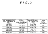
- the data stored in the position memory 3 are a table shown in NON PATENT-RELATED DOCUMENT 1 (see the table of Fig.2 ), for instance.
- Such a table shows displacement of the photoreceptor cell, the total displacement, the displacement of the ganglion cell and area ratio of the photoreceptor cell and the ganglion cell.
- the position memory 3 stores "the displacement of the photoreceptor cell” and "the displacement of the ganglion cell".
- one of values of the displacement of the photoreceptor cell is 0.25/0.90, and this value indicates the photoreceptor cell 0.25mm away from the center of the retina (that is, 0.90 deg away from the center of the retina).
- the displacement of the ganglion cell at the same line in the table is 0.56/2.02, and indicates the position where the ganglion cell linked to the photoreceptor cell is located is 0.56mm away from the center of the retina (that is, 2.02 deg away from the center of the retina). Then, "the distance from the center of the retina to the stimulus presentation position P2 (mm or deg)" is obtained based upon the distance (mm or deg) from "the center of the retina to the test position P2". Then, the stimulus presentation position P2 is specified on the assumption that the position P2 of the photoreceptor cell C is offset in a direction closer to the center of the retina rather than the position P1 of the ganglion cell RGC.
- NON PATENT-RELATED DOCUMENT 2 also describes the relation between the displacement of the photoreceptor cell regarding the center of the retina and the displacement of the ganglion cell.
- test position P1 inputted through the test-position input 2 (the displacement from the center of the retina) is substituted for B of the above-mentioned equation so as to obtain A, the position onto which the stimulus is to be presented is obtained.
- the data excluding the above-mentioned may be used as long as the data shows the positional relation between the photoreceptor cell and the ganglion cell.
- the perimeter 1 has a stimulus presentator 5 for presenting the stimulus at the position obtained through the position obtaining portion 4 (the above-mentioned stimulus presentation position that is not the position P1 where the ganglion cell RGC is located, but the position P2 where the photoreceptor cell linked to the ganglion cell RGC is located), and an operation portion 6 to be operated by the examinee who perceives the presented stimulus, and a visual field judger 7 for judging the visual field of the examinee based upon a response from the examinee through the operation portion 6, and is configured to carry out the perimetry (see S5 of Fig.3 ).
- a stimulus presentator 5 for presenting the stimulus at the position obtained through the position obtaining portion 4 (the above-mentioned stimulus presentation position that is not the position P1 where the ganglion cell RGC is located, but the position P2 where the photoreceptor cell linked to the ganglion cell RGC is located)
- an operation portion 6 to be operated by the examinee who perceives the presented stimulus
- the perimeter 1 has a judged-result output 8 (see S6 of Fig.3 ) for displaying the result judged by the visual field judger 7 so as to correlate with the above-mentioned test position.
- a judged-result output 8 for displaying the result judged by the visual field judger 7 so as to correlate with the above-mentioned test position.
- the judged-result output 8 may be a monitor (the monitor 20 or the other monitors), such as a liquid crystal display, a printer and an output portion for sending information to an external monitor or a printer as long as it displays the judged result of the visual field and the test position so as to be correlated with each other.
- the stimulus presentation position may be simultaneously displayed with the test position.
- the test position and the stimulus presentation position may be differentiated from each other by changing these colors. For instance, one is shown with red, the other is shown in blue, and the overlapped is shown with green.
- the test position and the stimulus presentation position may be alternately shown without simultaneous display (representation is switched through the operation of a mouse or the like).
- the result judged by the visual field judger may be displayed so as to link with the stimulus presentation position as well as the representation linking to the test position.
- the position onto which the stimulus is presented through the stimulus presentator 5 is not “the test position P1 designated by the test-position input 2", but “the position P2 where the photoreceptor cell (that is, the photoreceptor cell that sends information to the ganglion cell existing at the test position P1) exists”.
- the information of the presented stimulus is received by the photoreceptor cell C at the position P2 and is sent to the ganglion cell RGC at the position P1, thereby judging function of the ganglion cell RGC at the position P1. Accordingly, it is possible to grasp "the position P1 designated to be tested” and “the function of the ganglion cell RGC located at the position P1", correlating with each other.
- the test position When a portion where the inner layer thickness of the retina is thin is designated as the test position, it is possible to judge the function of the ganglion cell RGC located at such a portion (the portion where the inner layer thickness of the retina is thin), to grasp "the structure of the retina (the inner layer thickness of the retina)” and “the function of the ganglion cell RGC", correlating with each other, and to grasp the relation between the thickness of the inner layer of the retina and the visual field.
- the test-position input may automatically designate the test position as well as manual designation.
- the fundus image is displayed on the monitor 20 of the test-position input 2, and the test position is manually designated through the position designator 22 on the monitor 20.
- the perimeter according to the invention may be configured that coordinate information of the abnormality area is obtained directly from the other inspection equipment, such as an OCT so as to automatically input (designate) the test position.
Landscapes
- Health & Medical Sciences (AREA)
- Life Sciences & Earth Sciences (AREA)
- Engineering & Computer Science (AREA)
- Heart & Thoracic Surgery (AREA)
- Molecular Biology (AREA)
- Biophysics (AREA)
- Ophthalmology & Optometry (AREA)
- Biomedical Technology (AREA)
- Veterinary Medicine (AREA)
- Medical Informatics (AREA)
- Physics & Mathematics (AREA)
- Surgery (AREA)
- Animal Behavior & Ethology (AREA)
- General Health & Medical Sciences (AREA)
- Public Health (AREA)
- Signal Processing (AREA)
- Eye Examination Apparatus (AREA)
Abstract
Description
- The invention relates to a perimeter for measuring a visual field of an examinee through presentation of stimuli into his (her) visual field.
- A retinal ganglion cell (RGC), which may be referred to as "the ganglion cell" hereinafter, is a type of neuron located near the inner surface of the retina of the eye, and receives visual information from photoreceptors (cone cells and rod cells) via two intermediate neuron types; bipolar cells and retina amacrine cells. If the ganglion cell is damaged, a person has glaucoma that leads to narrow visual field and vision loss. So, it is important to find weak function of the ganglion cell at an earlier stage and stop the progress and deterioration of the disease. As one of methods of finding deterioration of the function of the ganglion cell is a perimetry, and perimeters having various kinds of structures for carrying out such a perimetry have been proposed (see the Patent-
related document 1, for instance). - It has been found that there is a closer correlation between the deterioration of the function of the above-mentioned ganglion cell and thickness of retinal inner layer (GCC thickness, the thickness of Ganglion Cell Complex), and the thickness of the retinal inner layer is thin at a portion where the function of the ganglion cell is weak. Under these situations, a method for the perimetry is to find portions where retinal inner thickness is thin based on various kinds of fundus images, such as an OCT image and a GCC thickness map made from the OCT image, and to present a stimulus on the portion so as to carry out the perimetry preferentially thereon.
- [Patent-related document 1]: Japanese Patent No.
5231085 -
- [Non Patent-related document 1]: Johann Sjostrand. Quantitative estimations of foveal and extrafoveal retinal circuitry in humans, Vision Research 39 (1999) 2987-2998
- [Non Patent-related document 2]: JohannSjostrand and other three. [Morphometric Study of the displacement of retinal ganglion cellssubserving cones within the human fovea], Graefe's archive for clinical and experimentalophthalmology, Springer Berlin/Heidelberg, Vol.237, Number 12/December, 1999
- It has been found that a positional gap occurs between a position where the ganglion cell is located (the position on the retina surface) and a position where a photoreceptor cell for sending information to the ganglion cell (that is, the photoreceptor cell linked with the ganglion cell) is located (the position on the retina surface) (see NON PATENT-RELATED
DOCUMENTS 1 and 2), for instance).Fig.4 is a schematic diagram that typically shows such a positional relation between the photoreceptor cell and the ganglion cell (shows a coordinate system along a surface of the retina). In the figure, the origin is a center of the retina, and the vertical axis and the horizontal axis indicate an eccentricity from a retinal center (unit of the vertical axis is degee (deg) and unit of the horizontal axis is millimeter (mm)). As shown in the figure, each ganglion cell RGC is offset in a direction going away from the retinal center regarding each photoreceptor cell C. It has been found that the quantity of the offset is not constant, and is the most at a part near the center of the retina and becomes less as deviating from the center of the retina, and there is almost no positional offset on the outer side of a macula. - A conventional problem is that even if the portion where the inner layer thickness of the retina (see, P1, for instance) is found in the macula of the retina and the stimulus is presented onto the portion P1 for the perimetry on the ganglion cell of the portion P1, the information regarding the stimulus received by the photoreceptor cell existing at the portion P1 is sent to the ganglion cell at the position P3 away from the center of the retina so as to test the ganglion cell at the position P3, thus it is not possible to test the ganglion cell of the designated portion P1.
- An object of the invention is to provide the perimeter for solving the above-mentioned problem.
- A first aspect of the invention is a perimeter (1) for measuring a visual field of an examinee through presentation of stimuli into his (her) visual field, comprising:
- a position memory (3) that stores a position on a retinal surface where a ganglion cell (RGC of
Fig.4 ) is located and a position on the retinal surface where a photoreceptor cell (c) for sending information to the ganglion cell (RGC) is located, correlating with each other; - a test-position input (2) for inputting a position (P1 of
Fig.4 ) on the retinal surface to be tested ("the test position" hereinafter); - a position obtaining portion (4) that obtains the position (P2) of the photoreceptor cell (C) that sends information to the ganglion cell (RGC) at the test position (P1) inputted through the test-position input (2) based upon the test position (P1) and data stored in the position memory (3);
- a stimulus presentator (5) that presents a stimulus to the position (P2) obtained through the position obtaining portion (4);
- an operation portion (6) to be operated by an examinee who perceives the presented stimulus;
- a visual field judger (7) that judges the visual field of the examinee based upon a response from the examinee through the operation portion (6); and
- a judged-result output (8) that displays the result judged through the visual field judger (7), correlating with the test position (P1).
- A second aspect of the invention is a perimeter (1), wherein the test-position input (2) has a monitor (20), a fundus image output (21) that displays a fundus image on the monitor (20), and a position designator (22) that designates the test position (P1) on the fundus image.
- A third aspect of the invention is a perimeter (1), further comprising a memory (23) that stores data showing a distribution of inner layer thickness of the retina of the examinee, wherein the data that shows the distribution of the inner layer thickness of the retina is displayed on the monitor (20).
- The number in parentheses shows the corresponding element in the drawings for the sake of convenience, accordingly, the descriptions are not restricted and bound by the descriptions on the drawings.
- According to the 1st and 2nd aspects of the invention, it is possible to grasp "the position designated to be tested" and "the function of the ganglion cell located on the position", correlating with each other.
- According to the 3rd aspect of the invention, it is easily grasp an abnormality area and it is possible to properly designate the perimetry on the abnormality area.
-
- [
Fig.1] Fig.1 is a block diagram that shows an example of a perimeter according to the present invention. - [
Fig.2] Fig.2 is a table that shows a positional relation between a photoreceptor cell and a ganglion cell. - [
Fig.3] Fig.3 is a flow chart that shows an example of a way of carrying out the perimetry with the perimeter according to the present invention. - [
Fig.4] Fig.4 is a schematic diagram that typically shows the positional relation between the photoreceptor cell and the ganglion cell. - An embodiment of the present invention is now explained, referring to appended figures
Figs.1 through 4 . - In the specification, a position on a retinal surface to be tested, that is, the position to be tested that designated by an examiner and the position on which function of the ganglion cell is to be tested, is referred to as "a test position", and a position on the retinal surface onto which stimuli are actually presented at the time of the perimetry is referred to as "a stimulus presentation position".
- A perimeter according to the invention exemplarily shown with a
reference number 1, measures a visual field of an examinee through presentation of the stimulus onto a desired position in his (her) visual field (the above-mentioned stimulus presentation position). Theperimeter 1 has a test-position input 2 for inputting the above-mentioned test position. Preferably, this test-position input 2 is comprised of amonitor 20, afundus image output 21 for displaying a fundus image, that is, the image of the fundus surface, on the monitor 20 (see S1 ofFig.3 ), and aposition designator 22 for designating the test position on the fundus image, such as a mouse and a touch-pen (see S2 ofFig. 3 ) . Besides, theperimeter 1 is provided with amemory 23 for storing data of a distribution of an inner layer thickness of the retina of the examinee so as to display the data that shows the inner layer thickness of the retina on themonitor 20. Concretely speaking, themonitor 20 displays the fundus image that shows the distribution of the inner layer thickness of the retina, such as a GCC thickness map of OCT, or both usual fundus image that does not show the distribution of the inner layer thickness of the retina and data from which the distribution of the inner layer thickness of the retina is found, such as a sectional image of the fundus, so that it is possible to easily grasp an abnormality area and properly designate the perimetry at the abnormality area. In such a case, the above-mentioned fundus image may be imaged just before the perimetry, or may be one that has been imaged and stored in a filing unit (a memory). Themonitor 20 may display the test position or the stimulus presentation position. - And, the perimeter according to the invention has a
position memory 3 for storing "the position on the retina surface where the ganglion cell (see RGC ofFig.4 ) is located" so as to correlate with "the position on the retina surface where a photoreceptor cell (see "C" ofFig. 4 ) that sends information to the ganglion cell RGC is located", and a position obtaining portion 4 (see S3 ofFig.3 ) for obtaining a position P2 of the photoreceptor cell C that sends information to a ganglion cell RCG existing at the test position P1 inputted by the test-position input 2 (that is, the stimulus presentation position) based on the test position, such as P1 ofFig.4 , and the data stored in theposition memory 3. The data stored in theposition memory 3 are a table shown in NON PATENT-RELATED DOCUMENT 1 (see the table ofFig.2 ), for instance. Such a table shows displacement of the photoreceptor cell, the total displacement, the displacement of the ganglion cell and area ratio of the photoreceptor cell and the ganglion cell. Preferably, theposition memory 3 stores "the displacement of the photoreceptor cell" and "the displacement of the ganglion cell". In this table, one of values of the displacement of the photoreceptor cell is 0.25/0.90, and this value indicates the photoreceptor cell 0.25mm away from the center of the retina (that is, 0.90 deg away from the center of the retina). The displacement of the ganglion cell at the same line in the table is 0.56/2.02, and indicates the position where the ganglion cell linked to the photoreceptor cell is located is 0.56mm away from the center of the retina (that is, 2.02 deg away from the center of the retina). Then, "the distance from the center of the retina to the stimulus presentation position P2 (mm or deg)" is obtained based upon the distance (mm or deg) from "the center of the retina to the test position P2". Then, the stimulus presentation position P2 is specified on the assumption that the position P2 of the photoreceptor cell C is offset in a direction closer to the center of the retina rather than the position P1 of the ganglion cell RGC. - The above-mentioned NON PATENT-RELATED
DOCUMENT 2 also describes the relation between the displacement of the photoreceptor cell regarding the center of the retina and the displacement of the ganglion cell. - This paper states the displacement A of the photoreceptor cell and the displacement B of the ganglion cell have the following relationship.

- If the test position P1 inputted through the test-position input 2 (the displacement from the center of the retina) is substituted for B of the above-mentioned equation so as to obtain A, the position onto which the stimulus is to be presented is obtained. In this case, the data excluding the above-mentioned may be used as long as the data shows the positional relation between the photoreceptor cell and the ganglion cell.
- Besides, the
perimeter 1 according to the present invention has astimulus presentator 5 for presenting the stimulus at the position obtained through the position obtaining portion 4 (the above-mentioned stimulus presentation position that is not the position P1 where the ganglion cell RGC is located, but the position P2 where the photoreceptor cell linked to the ganglion cell RGC is located), and anoperation portion 6 to be operated by the examinee who perceives the presented stimulus, and avisual field judger 7 for judging the visual field of the examinee based upon a response from the examinee through theoperation portion 6, and is configured to carry out the perimetry (see S5 ofFig.3 ). - Furthermore, the
perimeter 1 according to the present invention has a judged-result output 8 (see S6 ofFig.3 ) for displaying the result judged by thevisual field judger 7 so as to correlate with the above-mentioned test position. Preferably, when displaying both, correlating with each other, it is possible to obtain information regarding the test position P1, the result of the perimetry and information as to at which test position the test result is obtained. The judged-result output 8 may be a monitor (themonitor 20 or the other monitors), such as a liquid crystal display, a printer and an output portion for sending information to an external monitor or a printer as long as it displays the judged result of the visual field and the test position so as to be correlated with each other. Besides, when the judged-result output 8 is the monitor, the stimulus presentation position may be simultaneously displayed with the test position. In such a case, the test position and the stimulus presentation position may be differentiated from each other by changing these colors. For instance, one is shown with red, the other is shown in blue, and the overlapped is shown with green. Furthermore, the test position and the stimulus presentation position may be alternately shown without simultaneous display (representation is switched through the operation of a mouse or the like). Moreover, the result judged by the visual field judger may be displayed so as to link with the stimulus presentation position as well as the representation linking to the test position. - According to the present invention, the position onto which the stimulus is presented through the
stimulus presentator 5 is not "the test position P1 designated by the test-position input 2", but "the position P2 where the photoreceptor cell (that is, the photoreceptor cell that sends information to the ganglion cell existing at the test position P1) exists". The information of the presented stimulus is received by the photoreceptor cell C at the position P2 and is sent to the ganglion cell RGC at the position P1, thereby judging function of the ganglion cell RGC at the position P1. Accordingly, it is possible to grasp "the position P1 designated to be tested" and "the function of the ganglion cell RGC located at the position P1", correlating with each other. When a portion where the inner layer thickness of the retina is thin is designated as the test position, it is possible to judge the function of the ganglion cell RGC located at such a portion (the portion where the inner layer thickness of the retina is thin), to grasp "the structure of the retina (the inner layer thickness of the retina)" and "the function of the ganglion cell RGC", correlating with each other, and to grasp the relation between the thickness of the inner layer of the retina and the visual field. - The test-position input may automatically designate the test position as well as manual designation. In the above-mentioned embodiment, the fundus image is displayed on the
monitor 20 of the test-position input 2, and the test position is manually designated through theposition designator 22 on themonitor 20. The perimeter according to the invention may be configured that coordinate information of the abnormality area is obtained directly from the other inspection equipment, such as an OCT so as to automatically input (designate) the test position. -
- 1 ... ... perimeter
- 2 ...... test-position input
- 3 ... ... position memory
- 4 ... ... position obtaining portion
- 5 ... ... stimulus presentator
- 6 ... ... operation portion
- 7 ... ... visual field judger
- 8 ... ... judged-result output
- 20 ... ... monitor
- 21 ... ... fundus image output
- 22 ... ... position designator
- 23...... memory
- C... ... photoreceptor cell
- P1... ... test position
- P2... ... position of photoreceptor cell
- RGC ... ... ganglion cell
Claims (3)
- A perimeter for measuring a visual field of an examinee through presentation of stimuli into his (her) visual field, comprising:a position memory that stores a position on a retinal surface where a ganglion cell is located and a position on the retinal surface where a photoreceptor cell for sending information to the ganglion cell is located, correlating with each other;a test-position input for inputting a position on the retinal surface to be tested ("the test position" hereinafter);a position obtaining portion that obtains the position of the photoreceptor cell that sends information to the ganglion cell at the test position inputted through the test-position input based upon the test position and data stored in the position memory;a stimulus presentator that presents a stimulus to the position obtained through the position obtaining portion;an operation portion to be operated by an examinee who perceives the presented stimulus;a visual field judger that judges a visual field of the examinee based upon a response from the examinee through the operation portion; anda judged-result output that displays the result judged through the visual field judger, correlating with the test position.
- The perimeter according to claim 1, wherein the test-position input has a monitor, a fundus image output that displays a fundus image on the monitor, and a position designator that designates the test position on the fundus image.
- The perimeter according to claim 2, further comprising a memory that stores data showing a distribution of inner layer thickness of the retina of the examinee, wherein the data that shows the distribution of the inner layer thickness of the retina is displayed on the monitor.
Applications Claiming Priority (2)
| Application Number | Priority Date | Filing Date | Title |
|---|---|---|---|
| JP2013265028 | 2013-12-24 | ||
| PCT/JP2014/083898 WO2015098828A1 (en) | 2013-12-24 | 2014-12-22 | Perimeter |
Publications (3)
| Publication Number | Publication Date |
|---|---|
| EP3087908A1 true EP3087908A1 (en) | 2016-11-02 |
| EP3087908A4 EP3087908A4 (en) | 2017-08-16 |
| EP3087908B1 EP3087908B1 (en) | 2018-10-03 |
Family
ID=53478682
Family Applications (1)
| Application Number | Title | Priority Date | Filing Date |
|---|---|---|---|
| EP14875261.1A Active EP3087908B1 (en) | 2013-12-24 | 2014-12-22 | Perimeter |
Country Status (4)
| Country | Link |
|---|---|
| US (1) | US10413170B2 (en) |
| EP (1) | EP3087908B1 (en) |
| JP (1) | JP6461819B2 (en) |
| WO (1) | WO2015098828A1 (en) |
Families Citing this family (1)
| Publication number | Priority date | Publication date | Assignee | Title |
|---|---|---|---|---|
| JP7196606B2 (en) * | 2016-03-04 | 2022-12-27 | 株式会社ニデック | Ophthalmic information processing device and ophthalmic information processing program |
Family Cites Families (3)
| Publication number | Priority date | Publication date | Assignee | Title |
|---|---|---|---|---|
| US4634243A (en) * | 1984-12-26 | 1987-01-06 | Lkc Systems, Inc. | Glaucoma detection utilizing pattern discrimination test |
| US7641339B2 (en) * | 2007-07-31 | 2010-01-05 | Kabushiki Kaisha Topcon | Ophthalmologic information processing apparatus and ophthalmologic examination apparatus |
| EP2793682A1 (en) * | 2011-12-20 | 2014-10-29 | ICheck Health Connection Inc. | Video game to monitor visual field loss in glaucoma |
-
2014
- 2014-12-22 JP JP2015554879A patent/JP6461819B2/en active Active
- 2014-12-22 WO PCT/JP2014/083898 patent/WO2015098828A1/en not_active Ceased
- 2014-12-22 EP EP14875261.1A patent/EP3087908B1/en active Active
- 2014-12-22 US US15/107,504 patent/US10413170B2/en active Active
Also Published As
| Publication number | Publication date |
|---|---|
| EP3087908A4 (en) | 2017-08-16 |
| EP3087908B1 (en) | 2018-10-03 |
| JP6461819B2 (en) | 2019-01-30 |
| JPWO2015098828A1 (en) | 2017-03-23 |
| US20160317024A1 (en) | 2016-11-03 |
| WO2015098828A1 (en) | 2015-07-02 |
| US10413170B2 (en) | 2019-09-17 |
Similar Documents
| Publication | Publication Date | Title |
|---|---|---|
| EP2219512B1 (en) | A method for performing micro-perimetry exams based on a retinal volume image and a well registered fundus image | |
| US7942528B2 (en) | Perimeter | |
| JP2011036431A (en) | Tomograph and control method therefor, program, storage medium | |
| Hong et al. | Role of ultra-widefield fluorescein angiography in the management of uveitis | |
| CN105916432A (en) | Method for displaying optometry font, display of optometry font, related application and image output device | |
| Charbel Issa et al. | Metamorphopsia in patients with macular telangiectasia type 2 | |
| Correnti et al. | Comparison of optic nerve head assessment with a digital stereoscopic camera (discam), scanning laser ophthalmoscopy, and stereophotography | |
| Bernardes et al. | Multimodal macula mapping: a new approach to study diseases of the macula | |
| EP3087908B1 (en) | Perimeter | |
| Roshandel et al. | Impact of reference center choice on adaptive optics imaging cone mosaic analysis | |
| CN105686795B (en) | A kind of dynamic display method of en face OCT images | |
| Ooto et al. | High-resolution imaging of photoreceptors in macular microholes | |
| US20140125953A1 (en) | Visual field visual function mapping apparatus | |
| US8157379B2 (en) | Perimeter and method of controlling perimeter | |
| Schlimgen et al. | OCT imaging in eyes with an implanted telescope | |
| Danilichev et al. | Optical and psychophysical studies of the visual system of cosmonauts before and after long orbital flights | |
| Duff et al. | Single-line macular optic coherence tomography to confirm optic neuropathies in awake infants and young children | |
| RU2645415C1 (en) | Method of simultaneous evaluation of the degree of functional suppression of one eye by the other in the field of fixation of vision and in different points of the vision field and computerized system for its implementation | |
| Eppenberger et al. | Characterization of retinal pigment epithelium layer in healthy and diseased retinas with high‐resolution adaptive optics transscleral flood illumination imaging | |
| Anderson et al. | A new method for estimating effects of visual field loss in a panoramic driving environment | |
| Midena | Microperimetry: an introduction | |
| Preston et al. | The Teller Acuity Card Procedure: an acuity screening technique for pre-verbal children | |
| Bolsunov et al. | Module for Automated Assessment of Visual Function Safety in the Diagnosis of Multiple Sclerosis | |
| JP2023003154A (en) | Image processing device and method for controlling image processing device | |
| JP2001149314A (en) | Test chart for myopia |
Legal Events
| Date | Code | Title | Description |
|---|---|---|---|
| PUAI | Public reference made under article 153(3) epc to a published international application that has entered the european phase |
Free format text: ORIGINAL CODE: 0009012 |
|
| 17P | Request for examination filed |
Effective date: 20160622 |
|
| AK | Designated contracting states |
Kind code of ref document: A1 Designated state(s): AL AT BE BG CH CY CZ DE DK EE ES FI FR GB GR HR HU IE IS IT LI LT LU LV MC MK MT NL NO PL PT RO RS SE SI SK SM TR |
|
| AX | Request for extension of the european patent |
Extension state: BA ME |
|
| DAX | Request for extension of the european patent (deleted) | ||
| A4 | Supplementary search report drawn up and despatched |
Effective date: 20170719 |
|
| RIC1 | Information provided on ipc code assigned before grant |
Ipc: A61B 3/024 20060101AFI20170713BHEP Ipc: A61B 3/00 20060101ALI20170713BHEP |
|
| GRAP | Despatch of communication of intention to grant a patent |
Free format text: ORIGINAL CODE: EPIDOSNIGR1 |
|
| STAA | Information on the status of an ep patent application or granted ep patent |
Free format text: STATUS: GRANT OF PATENT IS INTENDED |
|
| INTG | Intention to grant announced |
Effective date: 20180629 |
|
| GRAS | Grant fee paid |
Free format text: ORIGINAL CODE: EPIDOSNIGR3 |
|
| GRAA | (expected) grant |
Free format text: ORIGINAL CODE: 0009210 |
|
| STAA | Information on the status of an ep patent application or granted ep patent |
Free format text: STATUS: THE PATENT HAS BEEN GRANTED |
|
| AK | Designated contracting states |
Kind code of ref document: B1 Designated state(s): AL AT BE BG CH CY CZ DE DK EE ES FI FR GB GR HR HU IE IS IT LI LT LU LV MC MK MT NL NO PL PT RO RS SE SI SK SM TR |
|
| REG | Reference to a national code |
Ref country code: GB Ref legal event code: FG4D |
|
| REG | Reference to a national code |
Ref country code: CH Ref legal event code: EP Ref country code: AT Ref legal event code: REF Ref document number: 1047728 Country of ref document: AT Kind code of ref document: T Effective date: 20181015 |
|
| REG | Reference to a national code |
Ref country code: IE Ref legal event code: FG4D Ref country code: DE Ref legal event code: R096 Ref document number: 602014033582 Country of ref document: DE |
|
| REG | Reference to a national code |
Ref country code: NL Ref legal event code: MP Effective date: 20181003 |
|
| REG | Reference to a national code |
Ref country code: LT Ref legal event code: MG4D |
|
| REG | Reference to a national code |
Ref country code: AT Ref legal event code: MK05 Ref document number: 1047728 Country of ref document: AT Kind code of ref document: T Effective date: 20181003 |
|
| PG25 | Lapsed in a contracting state [announced via postgrant information from national office to epo] |
Ref country code: NL Free format text: LAPSE BECAUSE OF FAILURE TO SUBMIT A TRANSLATION OF THE DESCRIPTION OR TO PAY THE FEE WITHIN THE PRESCRIBED TIME-LIMIT Effective date: 20181003 |
|
| PG25 | Lapsed in a contracting state [announced via postgrant information from national office to epo] |
Ref country code: ES Free format text: LAPSE BECAUSE OF FAILURE TO SUBMIT A TRANSLATION OF THE DESCRIPTION OR TO PAY THE FEE WITHIN THE PRESCRIBED TIME-LIMIT Effective date: 20181003 Ref country code: LV Free format text: LAPSE BECAUSE OF FAILURE TO SUBMIT A TRANSLATION OF THE DESCRIPTION OR TO PAY THE FEE WITHIN THE PRESCRIBED TIME-LIMIT Effective date: 20181003 Ref country code: CZ Free format text: LAPSE BECAUSE OF FAILURE TO SUBMIT A TRANSLATION OF THE DESCRIPTION OR TO PAY THE FEE WITHIN THE PRESCRIBED TIME-LIMIT Effective date: 20181003 Ref country code: HR Free format text: LAPSE BECAUSE OF FAILURE TO SUBMIT A TRANSLATION OF THE DESCRIPTION OR TO PAY THE FEE WITHIN THE PRESCRIBED TIME-LIMIT Effective date: 20181003 Ref country code: LT Free format text: LAPSE BECAUSE OF FAILURE TO SUBMIT A TRANSLATION OF THE DESCRIPTION OR TO PAY THE FEE WITHIN THE PRESCRIBED TIME-LIMIT Effective date: 20181003 Ref country code: PL Free format text: LAPSE BECAUSE OF FAILURE TO SUBMIT A TRANSLATION OF THE DESCRIPTION OR TO PAY THE FEE WITHIN THE PRESCRIBED TIME-LIMIT Effective date: 20181003 Ref country code: BG Free format text: LAPSE BECAUSE OF FAILURE TO SUBMIT A TRANSLATION OF THE DESCRIPTION OR TO PAY THE FEE WITHIN THE PRESCRIBED TIME-LIMIT Effective date: 20190103 Ref country code: FI Free format text: LAPSE BECAUSE OF FAILURE TO SUBMIT A TRANSLATION OF THE DESCRIPTION OR TO PAY THE FEE WITHIN THE PRESCRIBED TIME-LIMIT Effective date: 20181003 Ref country code: NO Free format text: LAPSE BECAUSE OF FAILURE TO SUBMIT A TRANSLATION OF THE DESCRIPTION OR TO PAY THE FEE WITHIN THE PRESCRIBED TIME-LIMIT Effective date: 20190103 Ref country code: IS Free format text: LAPSE BECAUSE OF FAILURE TO SUBMIT A TRANSLATION OF THE DESCRIPTION OR TO PAY THE FEE WITHIN THE PRESCRIBED TIME-LIMIT Effective date: 20190203 Ref country code: AT Free format text: LAPSE BECAUSE OF FAILURE TO SUBMIT A TRANSLATION OF THE DESCRIPTION OR TO PAY THE FEE WITHIN THE PRESCRIBED TIME-LIMIT Effective date: 20181003 |
|
| PG25 | Lapsed in a contracting state [announced via postgrant information from national office to epo] |
Ref country code: GR Free format text: LAPSE BECAUSE OF FAILURE TO SUBMIT A TRANSLATION OF THE DESCRIPTION OR TO PAY THE FEE WITHIN THE PRESCRIBED TIME-LIMIT Effective date: 20190104 Ref country code: SE Free format text: LAPSE BECAUSE OF FAILURE TO SUBMIT A TRANSLATION OF THE DESCRIPTION OR TO PAY THE FEE WITHIN THE PRESCRIBED TIME-LIMIT Effective date: 20181003 Ref country code: AL Free format text: LAPSE BECAUSE OF FAILURE TO SUBMIT A TRANSLATION OF THE DESCRIPTION OR TO PAY THE FEE WITHIN THE PRESCRIBED TIME-LIMIT Effective date: 20181003 Ref country code: RS Free format text: LAPSE BECAUSE OF FAILURE TO SUBMIT A TRANSLATION OF THE DESCRIPTION OR TO PAY THE FEE WITHIN THE PRESCRIBED TIME-LIMIT Effective date: 20181003 Ref country code: PT Free format text: LAPSE BECAUSE OF FAILURE TO SUBMIT A TRANSLATION OF THE DESCRIPTION OR TO PAY THE FEE WITHIN THE PRESCRIBED TIME-LIMIT Effective date: 20190203 |
|
| REG | Reference to a national code |
Ref country code: DE Ref legal event code: R097 Ref document number: 602014033582 Country of ref document: DE |
|
| PG25 | Lapsed in a contracting state [announced via postgrant information from national office to epo] |
Ref country code: DK Free format text: LAPSE BECAUSE OF FAILURE TO SUBMIT A TRANSLATION OF THE DESCRIPTION OR TO PAY THE FEE WITHIN THE PRESCRIBED TIME-LIMIT Effective date: 20181003 Ref country code: IT Free format text: LAPSE BECAUSE OF FAILURE TO SUBMIT A TRANSLATION OF THE DESCRIPTION OR TO PAY THE FEE WITHIN THE PRESCRIBED TIME-LIMIT Effective date: 20181003 |
|
| REG | Reference to a national code |
Ref country code: CH Ref legal event code: PL |
|
| PLBE | No opposition filed within time limit |
Free format text: ORIGINAL CODE: 0009261 |
|
| STAA | Information on the status of an ep patent application or granted ep patent |
Free format text: STATUS: NO OPPOSITION FILED WITHIN TIME LIMIT |
|
| PG25 | Lapsed in a contracting state [announced via postgrant information from national office to epo] |
Ref country code: SK Free format text: LAPSE BECAUSE OF FAILURE TO SUBMIT A TRANSLATION OF THE DESCRIPTION OR TO PAY THE FEE WITHIN THE PRESCRIBED TIME-LIMIT Effective date: 20181003 Ref country code: RO Free format text: LAPSE BECAUSE OF FAILURE TO SUBMIT A TRANSLATION OF THE DESCRIPTION OR TO PAY THE FEE WITHIN THE PRESCRIBED TIME-LIMIT Effective date: 20181003 Ref country code: LU Free format text: LAPSE BECAUSE OF NON-PAYMENT OF DUE FEES Effective date: 20181222 Ref country code: MC Free format text: LAPSE BECAUSE OF FAILURE TO SUBMIT A TRANSLATION OF THE DESCRIPTION OR TO PAY THE FEE WITHIN THE PRESCRIBED TIME-LIMIT Effective date: 20181003 Ref country code: EE Free format text: LAPSE BECAUSE OF FAILURE TO SUBMIT A TRANSLATION OF THE DESCRIPTION OR TO PAY THE FEE WITHIN THE PRESCRIBED TIME-LIMIT Effective date: 20181003 Ref country code: SM Free format text: LAPSE BECAUSE OF FAILURE TO SUBMIT A TRANSLATION OF THE DESCRIPTION OR TO PAY THE FEE WITHIN THE PRESCRIBED TIME-LIMIT Effective date: 20181003 |
|
| 26N | No opposition filed |
Effective date: 20190704 |
|
| REG | Reference to a national code |
Ref country code: IE Ref legal event code: MM4A |
|
| REG | Reference to a national code |
Ref country code: BE Ref legal event code: MM Effective date: 20181231 |
|
| PG25 | Lapsed in a contracting state [announced via postgrant information from national office to epo] |
Ref country code: SI Free format text: LAPSE BECAUSE OF FAILURE TO SUBMIT A TRANSLATION OF THE DESCRIPTION OR TO PAY THE FEE WITHIN THE PRESCRIBED TIME-LIMIT Effective date: 20181003 Ref country code: FR Free format text: LAPSE BECAUSE OF NON-PAYMENT OF DUE FEES Effective date: 20181231 Ref country code: IE Free format text: LAPSE BECAUSE OF NON-PAYMENT OF DUE FEES Effective date: 20181222 |
|
| PG25 | Lapsed in a contracting state [announced via postgrant information from national office to epo] |
Ref country code: BE Free format text: LAPSE BECAUSE OF NON-PAYMENT OF DUE FEES Effective date: 20181231 |
|
| PG25 | Lapsed in a contracting state [announced via postgrant information from national office to epo] |
Ref country code: LI Free format text: LAPSE BECAUSE OF NON-PAYMENT OF DUE FEES Effective date: 20181231 Ref country code: CH Free format text: LAPSE BECAUSE OF NON-PAYMENT OF DUE FEES Effective date: 20181231 |
|
| PG25 | Lapsed in a contracting state [announced via postgrant information from national office to epo] |
Ref country code: MT Free format text: LAPSE BECAUSE OF NON-PAYMENT OF DUE FEES Effective date: 20181222 |
|
| PG25 | Lapsed in a contracting state [announced via postgrant information from national office to epo] |
Ref country code: TR Free format text: LAPSE BECAUSE OF FAILURE TO SUBMIT A TRANSLATION OF THE DESCRIPTION OR TO PAY THE FEE WITHIN THE PRESCRIBED TIME-LIMIT Effective date: 20181003 |
|
| PG25 | Lapsed in a contracting state [announced via postgrant information from national office to epo] |
Ref country code: CY Free format text: LAPSE BECAUSE OF FAILURE TO SUBMIT A TRANSLATION OF THE DESCRIPTION OR TO PAY THE FEE WITHIN THE PRESCRIBED TIME-LIMIT Effective date: 20181003 Ref country code: HU Free format text: LAPSE BECAUSE OF FAILURE TO SUBMIT A TRANSLATION OF THE DESCRIPTION OR TO PAY THE FEE WITHIN THE PRESCRIBED TIME-LIMIT; INVALID AB INITIO Effective date: 20141222 Ref country code: MK Free format text: LAPSE BECAUSE OF NON-PAYMENT OF DUE FEES Effective date: 20181003 |
|
| PGFP | Annual fee paid to national office [announced via postgrant information from national office to epo] |
Ref country code: GB Payment date: 20231102 Year of fee payment: 10 |
|
| REG | Reference to a national code |
Ref country code: DE Ref legal event code: R082 Ref document number: 602014033582 Country of ref document: DE Representative=s name: SEYER & NOBBE PATENTANWAELTE PARTNERSCHAFTSGES, DE |
|
| GBPC | Gb: european patent ceased through non-payment of renewal fee |
Effective date: 20241222 |
|
| PG25 | Lapsed in a contracting state [announced via postgrant information from national office to epo] |
Ref country code: GB Free format text: LAPSE BECAUSE OF NON-PAYMENT OF DUE FEES Effective date: 20241222 |
|
| PGFP | Annual fee paid to national office [announced via postgrant information from national office to epo] |
Ref country code: DE Payment date: 20251028 Year of fee payment: 12 |