EP3081941A1 - Method for determination of sensitivity to anti-cancer agent - Google Patents
Method for determination of sensitivity to anti-cancer agent Download PDFInfo
- Publication number
- EP3081941A1 EP3081941A1 EP16169127.4A EP16169127A EP3081941A1 EP 3081941 A1 EP3081941 A1 EP 3081941A1 EP 16169127 A EP16169127 A EP 16169127A EP 3081941 A1 EP3081941 A1 EP 3081941A1
- Authority
- EP
- European Patent Office
- Prior art keywords
- protein
- cancer agent
- cancer
- peak
- detected
- Prior art date
- Legal status (The legal status is an assumption and is not a legal conclusion. Google has not performed a legal analysis and makes no representation as to the accuracy of the status listed.)
- Granted
Links
Images






Classifications
-
- G—PHYSICS
- G01—MEASURING; TESTING
- G01N—INVESTIGATING OR ANALYSING MATERIALS BY DETERMINING THEIR CHEMICAL OR PHYSICAL PROPERTIES
- G01N33/00—Investigating or analysing materials by specific methods not covered by groups G01N1/00 - G01N31/00
- G01N33/48—Biological material, e.g. blood, urine; Haemocytometers
- G01N33/50—Chemical analysis of biological material, e.g. blood, urine; Testing involving biospecific ligand binding methods; Immunological testing
- G01N33/5005—Chemical analysis of biological material, e.g. blood, urine; Testing involving biospecific ligand binding methods; Immunological testing involving human or animal cells
- G01N33/5008—Chemical analysis of biological material, e.g. blood, urine; Testing involving biospecific ligand binding methods; Immunological testing involving human or animal cells for testing or evaluating the effect of chemical or biological compounds, e.g. drugs, cosmetics
- G01N33/5011—Chemical analysis of biological material, e.g. blood, urine; Testing involving biospecific ligand binding methods; Immunological testing involving human or animal cells for testing or evaluating the effect of chemical or biological compounds, e.g. drugs, cosmetics for testing antineoplastic activity
-
- A—HUMAN NECESSITIES
- A61—MEDICAL OR VETERINARY SCIENCE; HYGIENE
- A61K—PREPARATIONS FOR MEDICAL, DENTAL OR TOILETRY PURPOSES
- A61K31/00—Medicinal preparations containing organic active ingredients
- A61K31/33—Heterocyclic compounds
- A61K31/395—Heterocyclic compounds having nitrogen as a ring hetero atom, e.g. guanethidine or rifamycins
- A61K31/495—Heterocyclic compounds having nitrogen as a ring hetero atom, e.g. guanethidine or rifamycins having six-membered rings with two or more nitrogen atoms as the only ring heteroatoms, e.g. piperazine or tetrazines
- A61K31/505—Pyrimidines; Hydrogenated pyrimidines, e.g. trimethoprim
- A61K31/513—Pyrimidines; Hydrogenated pyrimidines, e.g. trimethoprim having oxo groups directly attached to the heterocyclic ring, e.g. cytosine
-
- A—HUMAN NECESSITIES
- A61—MEDICAL OR VETERINARY SCIENCE; HYGIENE
- A61K—PREPARATIONS FOR MEDICAL, DENTAL OR TOILETRY PURPOSES
- A61K31/00—Medicinal preparations containing organic active ingredients
- A61K31/33—Heterocyclic compounds
- A61K31/555—Heterocyclic compounds containing heavy metals, e.g. hemin, hematin, melarsoprol
-
- A—HUMAN NECESSITIES
- A61—MEDICAL OR VETERINARY SCIENCE; HYGIENE
- A61K—PREPARATIONS FOR MEDICAL, DENTAL OR TOILETRY PURPOSES
- A61K45/00—Medicinal preparations containing active ingredients not provided for in groups A61K31/00 - A61K41/00
- A61K45/06—Mixtures of active ingredients without chemical characterisation, e.g. antiphlogistics and cardiaca
-
- A—HUMAN NECESSITIES
- A61—MEDICAL OR VETERINARY SCIENCE; HYGIENE
- A61P—SPECIFIC THERAPEUTIC ACTIVITY OF CHEMICAL COMPOUNDS OR MEDICINAL PREPARATIONS
- A61P35/00—Antineoplastic agents
-
- A—HUMAN NECESSITIES
- A61—MEDICAL OR VETERINARY SCIENCE; HYGIENE
- A61P—SPECIFIC THERAPEUTIC ACTIVITY OF CHEMICAL COMPOUNDS OR MEDICINAL PREPARATIONS
- A61P43/00—Drugs for specific purposes, not provided for in groups A61P1/00-A61P41/00
-
- G—PHYSICS
- G01—MEASURING; TESTING
- G01N—INVESTIGATING OR ANALYSING MATERIALS BY DETERMINING THEIR CHEMICAL OR PHYSICAL PROPERTIES
- G01N33/00—Investigating or analysing materials by specific methods not covered by groups G01N1/00 - G01N31/00
- G01N33/48—Biological material, e.g. blood, urine; Haemocytometers
- G01N33/50—Chemical analysis of biological material, e.g. blood, urine; Testing involving biospecific ligand binding methods; Immunological testing
- G01N33/53—Immunoassay; Biospecific binding assay; Materials therefor
- G01N33/575—Immunoassay; Biospecific binding assay; Materials therefor for cancer
-
- G—PHYSICS
- G01—MEASURING; TESTING
- G01N—INVESTIGATING OR ANALYSING MATERIALS BY DETERMINING THEIR CHEMICAL OR PHYSICAL PROPERTIES
- G01N33/00—Investigating or analysing materials by specific methods not covered by groups G01N1/00 - G01N31/00
- G01N33/48—Biological material, e.g. blood, urine; Haemocytometers
- G01N33/50—Chemical analysis of biological material, e.g. blood, urine; Testing involving biospecific ligand binding methods; Immunological testing
- G01N33/53—Immunoassay; Biospecific binding assay; Materials therefor
- G01N33/575—Immunoassay; Biospecific binding assay; Materials therefor for cancer
- G01N33/57535—Immunoassay; Biospecific binding assay; Materials therefor for cancer of the large intestine, e.g. colon, rectum or anus
-
- G—PHYSICS
- G01—MEASURING; TESTING
- G01N—INVESTIGATING OR ANALYSING MATERIALS BY DETERMINING THEIR CHEMICAL OR PHYSICAL PROPERTIES
- G01N2333/00—Assays involving biological materials from specific organisms or of a specific nature
- G01N2333/435—Assays involving biological materials from specific organisms or of a specific nature from animals; from humans
- G01N2333/46—Assays involving biological materials from specific organisms or of a specific nature from animals; from humans from vertebrates
- G01N2333/47—Assays involving proteins of known structure or function as defined in the subgroups
- G01N2333/4701—Details
- G01N2333/4727—Calcium binding proteins, e.g. calmodulin
-
- G—PHYSICS
- G01—MEASURING; TESTING
- G01N—INVESTIGATING OR ANALYSING MATERIALS BY DETERMINING THEIR CHEMICAL OR PHYSICAL PROPERTIES
- G01N2800/00—Detection or diagnosis of diseases
- G01N2800/52—Predicting or monitoring the response to treatment, e.g. for selection of therapy based on assay results in personalised medicine; Prognosis
-
- G—PHYSICS
- G01—MEASURING; TESTING
- G01N—INVESTIGATING OR ANALYSING MATERIALS BY DETERMINING THEIR CHEMICAL OR PHYSICAL PROPERTIES
- G01N30/00—Investigating or analysing materials by separation into components using adsorption, absorption or similar phenomena or using ion-exchange, e.g. chromatography or field flow fractionation
- G01N30/02—Column chromatography
- G01N30/62—Detectors specially adapted therefor
- G01N30/72—Mass spectrometers
- G01N30/7233—Mass spectrometers interfaced to liquid or supercritical fluid chromatograph
Definitions
- the present invention relates to a marker for use in determination of the sensitivity of a cancer patient to an anti-cancer agent to be administered thereto, which marker can determine whether or not the cancer of the patient has a therapeutic response to the anti-cancer agent, and to application of the marker.
- Anti-cancer agents have various types such as an alkylating agent, a platinum agent, an antimetabolite, an antitumor antibiotic, and an antitumor plant alkaloid. These anti-cancer agents are effective for some cancers but not effective for other cancers. Even when an anti-cancer agent is confirmed to be effective for a certain cancer, the anti-cancer agent is effective for some patients and not effective for other patients, leading to interindividual differences. Whether or not a cancer of a specific patient has response to an anti-cancer agent is designated as sensitivity to the anti-cancer agent.
- Oxaliplatin, (SP-4-2)-[(1R,2R)-cyclohexane-1,2-diamine- ⁇ N, ⁇ N'][ethanedioato(2-)- ⁇ O 1 , ⁇ O 2 ]platinum (IUPAC), is a third-generation platinum-based complex anti-cancer agent. Similar to precedent drugs (cisplatin (CDDP) and carboplatin (CBDCA)), the action mechanism thereof is thought to be based on inhibition of DNA synthesis and/or protein synthesis via cross-linking with DNA bases.
- Oxaliplatin exhibits anti-tumor effect on colorectal cancer, to which CDDP or CBDCA is ineffective, and shows different spectrum of anti-tumor activity from that of a precedent platinum-based complex anti-cancer agent.
- oxaliplatin for use in combination with fluorouracil (5-FU) and levofolinate (LV) was approved as a first line therapy for metastatic colorectal cancer in January, 2004.
- oxaliplatin was listed in the National Health Insurance (NHI) price list in the case of combination use thereof with continuous infusional fluorouracil and levofolinate (FOLFOX4 regimen) for "advanced/recurrent colorectal cancer not amenable to curative surgical resection" in April, 2005.
- NHS National Health Insurance
- FOLFOX4 regimen continuous infusional fluorouracil and levofolinate
- 5-FU/LV regimen to advanced/recurrent colorectal cancer has provided a survival of 10 to 12 months.
- a FOLFOX regimen combined with oxaliplatin results in a prolonged survival of 19.5 months (about twice the survival time).
- oxaliplatin is a promising drug having an efficacy in an increased number of colorectal cancer patients.
- the response rate of FOLFOX regimen against advanced/recurrent colorectal cancer is still as low as about 50%. In other words, about half of the treated patients achieve no effects.
- peripheral neuropathy frequently occurs in addition to neutropenia. Although not being fatal, these adverse events are factors which make continuation of the therapy difficult. Therefore, if there is a biomarker which can predict, before start of the treatment, a patient who is expected to achieve the response (i.e., a responder) and a patient who is not expected to achieve the response (i.e., a non-responder), to thereby diagnose the therapeutic response earlier, a chemotherapy ensuring high effectiveness and high safety can be established.
- Non-Patent Documents 11 and 12 oxaliplatin exhibits a therapeutic response differing from that of cisplatin, depending on the type of cancer.
- oxaliplatin exhibits a therapeutic response differing from that of cisplatin, depending on the type of cancer.
- no definite biomarker which can predict the therapeutic response to chemotherapy employing oxaliplatin.
- fluorouracil is a fluoro-pyrimidine anti-cancer agent developed in 1957 and even now serves as a basic drug for use in the chemotherapy of gastrointestinal cancer.
- fluorouracil exerts cytotoxic effect through a principle action mechanism of DNA synthesis inhibition induced by inhibition of thymidylate synthase (TS) by an active metabolite, fluorodeoxyuridine-5'-monophosphate (FdUMP), and another mechanism of RNA function inhibition by an active metabolite, 5-fluorouridine triphosphate (FUTP).
- TS thymidylate synthase
- FdUMP fluorodeoxyuridine-5'-monophosphate
- FUTP 5-fluorouridine triphosphate
- DPD dihydropyrimidine dehydrogenase
- TS thymidylate synthase
- TS expression level is reported to be a possible factor that determines the therapeutic effect by a fluoro-pyrimidine anti-cancer agent, even when the expression level is determined through any assay method such as the enzymatic activity method, protein level assay, or RNA level assay (Non-Patent Documents 14 and 15).
- any assay method such as the enzymatic activity method, protein level assay, or RNA level assay.
- the above-obtained results are not completely the same, and there has been known no definite biomarker which can predict the therapeutic response to fluorouracil in an early treatment stage.
- Non-Patent Document 16 In metastatic colorectal cancer patients having fluorouracil resistance, ERCC1 and TS mRNA expression levels in tumor cells are reported to serve as factors that predict therapeutic response to 5-FU/L-OHP combination therapy (Non-Patent Document 16).
- FOCUS trial large-scale prospective clinical trial for investigating biomarkers that predict therapeutic response of advanced colorectal cancer patients to chemotherapy has revealed that ERCC1 does not serve as a significant predictive factor for 5-FU/L-OHP combination therapy, and that only topoisomerase-1 (Topol) exhibits weak relationship thereto. This indicates that there has been established no biomarker that can reliably select a patient who is expected to be effectively treated through the 5-FU/L-OHP combination therapy.
- An object of the present invention is to provide a marker for determining sensitivity of a patient to an anti-cancer agent, which marker can determine whether or not the patient has a therapeutic response to the anti-cancer agent. Another object is to provide novel cancer therapeutic means employing the marker.
- the present inventors have searched for a marker for determining sensitivity to an anti-cancer agent by culturing human cancer cells, exposing the cells to a drug, and comprehensively analyzing the time-dependent expression profiles of intracellular proteins after exposure to the drug by means of a surface-enhanced laser desorption/ionization time-of-flight mass spectrometer (SELDI-TOF MS).
- SELDI-TOF MS surface-enhanced laser desorption/ionization time-of-flight mass spectrometer
- proteins or fragments thereof have been detected as a peak at m/z, as determined by means of a mass spectrometer, of 5,300 to 5,400, 6,130 to 6,230, 7,000 to 7,080, 7,840 to 7,920, 17,100 to 17,270, 18,290 to 18,470, 24,660 to 24,750, 35,980 to 36,290, or 9,100 to 9,200, or identified as a calcium-binding protein S100A10.
- the inventors have found, after exposure to L-OHP, several proteins exhibiting time-dependent intracellular expression level profiles which differ between high-sensitivity cells and low-sensitivity cells. These proteins or fragments thereof have been detected as a peak at m/z, as determined by means of a mass spectrometer, of 5,300 to 5,400, 6,130 to 6,230, 7,000 to 7,080, 7,840 to 7,920, 12,440 to 12,560, 17,100 to 17,270, 18,290 to 18,470, 24,660 to 24,750, 35,980 to 36,290, or 9,100 to 9,200.
- the inventors have found, after exposure to 5-FU/L-OHP in combination, several proteins exhibiting time-dependent intracellular expression level profiles which differ between high-sensitivity cells and between low-sensitivity cells. These proteins or fragments thereof have been detected as a peak at m/z, as determined by means of a mass spectrometer, peak of 5,300 to 5,400, 6,130 to 6,230, 7,000 to 7,080, 7,840 to 7,920, 8,920 to 9,000, 12,440 to 12,560, 17,100 to 17,270, 18,290 to 18,470, 24,660 to 24,750, 35,980 to 36,290, or 9,100 to 9,200, or identified as a calcium-binding protein S100A10.
- the inventors have found, before exposure to any of these drugs, several proteins exhibiting intracellular expression levels which differ between high-sensitivity cells and low-sensitivity cells. These proteins or fragments thereof have been detected as a peak at m/z, as determined by means of a mass spectrometer, of 6,130 to 6,230, 17,100 to 17,270, 8,650 to 8,750, or 11,760 to 11,890.
- a cancer of a target cancer patient has a sensitivity to 5-FU therapy, L-OHP therapy, or 5-FU/L-OHP combination therapy can be determined through measuring the relevant protein level of a biological sample derived from the cancer patient; that screening of an anti-cancer agent sensitivity enhancer can be accomplished through employment of variation in expression of the substance as an index; and that the therapeutic effect of the relevant anti-cancer agent can be drastically enhanced by use, in combination, of the anti-cancer agent sensitivity enhancer and the anti-cancer agent which is a sensitivity enhancement target of the enhancer.
- the present invention has been accomplished on the basis of these findings.
- the present invention provides a marker for determining sensitivity to an anti-cancer agent, the marker comprising a protein selected from the group consisting of a protein or a fragment thereof which is detected as a peak at m/z of 5,300 to 5,400 (hereinafter referred to as protein A), a protein or a fragment thereof which is detected as a peak at m/z of 6,130 to 6,230 (hereinafter referred to as protein B), a protein or a fragment thereof which is detected as a peak at m/z of 7,000 to 7,080 (hereinafter referred to as protein C), a protein or a fragment thereof which is detected as a peak at m/z of 7,840 to 7,920 (hereinafter referred to as protein D), a protein or a fragment thereof which is detected as a peak at m/z of 8,920 to 9,000 (hereinafter referred to as protein E), a protein or a fragment thereof which is detected as a peak at m/z of 12,440 to 12,560 (hereinafter referred to
- the present invention also provides a marker for determining sensitivity to an anti-cancer agent containing fluorouracil or a salt thereof or a combination of fluorouracil or a salt thereof and oxaliplatin or a salt thereof, the marker comprising a calcium-binding protein S100A10 (hereinafter referred to as protein F).
- protein F a calcium-binding protein S100A10
- the present invention also provides a method for determining sensitivity of a subject to an anti-cancer agent, the method comprising measuring the level of any of proteins A to N in a specimen derived from the subject.
- the present invention also provides a kit for carrying out the method for determining sensitivity of a subject to an anti-cancer agent, the kit comprising a protocol for measuring the level of any of proteins A to N in a specimen derived from the subject.
- the present invention also provides a screening method for an anti-cancer agent sensitivity enhancer, the method comprising employing variation in expression of any of the proteins A to N as an index.
- the present invention also provides an anti-cancer agent sensitivity enhancer obtained through the screening method.
- the present invention also provides a composition for cancer therapy comprising, in combination, the anti-cancer agent sensitivity enhancer and an anti-cancer agent which is a sensitivity enhancement target of the enhancer.
- the present invention also provides the proteins A to N for use in determining the anti-cancer agent sensitivity.
- the sensitivity to an anti-cancer agent of a patient can be appropriately determined before the therapy or in an early stage after start of the therapy.
- an anti-cancer agent having higher therapeutic effect can be selected, and unnecessary adverse events, which would otherwise result from administration of an anti-cancer agent exerting no expected therapeutic effect, can be prevented.
- the therapy schedule employing an anti-cancer agent generally requires a long period of time.
- the sensitivity of the target cancer to an anti-cancer agent can be evaluated in a time-dependent manner through determination of the anti-cancer agent sensitivity in each therapy cycle, whereby a determination can be made on whether or not the therapy should be continued.
- progression of cancer and aggravation of adverse events which would otherwise result from continuous administration of an anti-cancer agent exerting no expected therapeutic effect, can be prevented.
- reductions can be expected in the burden on patients and medical cost.
- the marker of the present invention when used, a drug which can promote anti-cancer agent sensitivity can be selected through screening.
- a drug which can promote anti-cancer agent sensitivity can be selected through screening.
- the assay reagent for measuring the marker for determining sensitivity to an anti-cancer agent of the present invention is useful as an reagent for determining sensitivity to an anti-cancer agent.
- the marker for determining sensitivity to an anti-cancer agent of the present invention is any of proteins A to N. More specifically, the proteins are detected by means of a cation-exchange chip at a pH of 4.5, and m/z peaks are determined by means of a surface-enhanced laser desorption/ionization time-of-flight mass spectrometer (SELDI-TOF MS).
- SELDI-TOF MS surface-enhanced laser desorption/ionization time-of-flight mass spectrometer
- the proteins are a protein or a fragment thereof which is detected as a peak at m/z of 5,300 to 5,400 (protein A), a protein or a fragment thereof which is detected as a peak at m/z of 6,130 to 6,230 (protein B), a protein or a fragment thereof which is detected as a peak at m/z of 7,000 to 7,080 (protein C), a protein or a fragment thereof which is detected as a peak at m/z of 7,840 to 7,920 (protein D), a protein or a fragment thereof which is detected as a peak at m/z of 8,920 to 9,000 (protein E), a protein or a fragment thereof which is detected as a peak at m/z of 11,020 to 11,120 (protein F), a protein or a fragment thereof which is detected as a peak at m/z of 12,440 to 12,560 (protein G), a protein or a fragment thereof which is detected as a peak at m/z of 17,100 to 17,270 (protein H), a protein or a fragment thereof
- protein F has been identified as calcium-binding protein S100A10.
- S100A10 is known to be a member of the S100 protein family having calcium-binding EF-hand motif.
- S100A10 is also known to form hetero-tetramer from S100A10 dimer and Annexin A2 (Annexin-2, Annexin II, Lipocortin II, Calpactin I heavy chain, Chromobindin-8, p36, Protein I, Placental anticoagulant protein IV, or PAP-IV) dimer. Therefore, similar to S100A10, Annexin A2 may be possibly employed as a marker for determining sensitivity to an anti-cancer agent.
- proteins A to C, F, and H to K are useful as markers for determining sensitivity to an anti-cancer agent, particularly as markers for determining sensitivity to 5-FU.
- proteins D and M are useful as markers for determining sensitivity to an anti-cancer agent, particularly as markers for determining sensitivity to 5-FU.
- proteins A to C and G to K are useful as markers for determining sensitivity to an anti-cancer agent, particularly as markers for determining sensitivity to L-OHP.
- protein F calcium-binding protein S100A10
- WO 2009/96196 a precedent patent application of the same inventors.
- the same results were also obtained in the Examples of the present invention.
- proteins D and M are useful as markers for determining sensitivity to an anti-cancer agent, particularly as markers for determining sensitivity to L-OHP.
- proteins A to C, F, and H to K are useful as markers for determining sensitivity to an anti-cancer agent, particularly as markers for determining sensitivity to 5-FU/L-OHP combination.
- protein F is a useful marker for determining sensitivity to L-OHP.
- protein F serves as a useful marker for determining sensitivity to an anti-cancer agent when 5-FU/L-OHP combination is employed.
- proteins E and G are useful as markers for determining sensitivity to an anti-cancer agent, particularly as markers for determining sensitivity to 5-FU/L-OHP combination.
- proteins D and M are useful as markers for determining sensitivity to an anti-cancer agent, particularly as markers for determining sensitivity to 5-FU/L-OHP combination.
- proteins B, H, L, and N in HCT116 cells which are high-sensitive to 5-FU, L-OHP, and 5-FU/L-OHP combination, before exposure to any of the drugs (not exposed) were significantly higher than those in DLD-1 cells, which are low-sensitive to the aforementioned drugs. Therefore, proteins B, H, L, and N are useful as markers for determining sensitivity to an anti-cancer agent, particularly as markers for determining sensitivity to 5-FU, L-OHP, or 5-FU/L-OHP combination.
- the anti-cancer agent examples include oxaliplatin, cyclophosphamide, ifosfamide, thiotepa, melphalan, busulfan, nimustine, ranimustine, dacarbazine, procarbazine, temozolomide, cisplatin, carboplatin, nedaplatin, methotrexate, pemetrexed, fluorouracil, tegaful/uracil, doxifluridine, tegaful/gimeracil/oteracil, capecitabine, cytarabine, enocitabine, gemcitabine, 6-mercaptopurine, fuludarabin, pentostatin, cladribine, hydroxyurea, doxorubicin, epirubicin, daunorubicin,
- fluoro-pyrimidine anti-cancer agents and platinum-based complex anti-cancer agents are preferred, with fluorouracil, oxaliplatin, and salts thereof being particularly preferred.
- the marker of the present invention is preferably applied to a combination of fluorouracil or a salt thereof and oxaliplatin or a salt thereof.
- any of the protein A level to protein N level in a specimen may be measured.
- the specimen include biological samples derived from subjects having cancer (i.e., cancer patients) such as blood, serum, plasma, cancer tissue biopsy specimens, cancer isolated preparations, feces, urine, ascitic fluid, pleural fluid, cerebrospinal fluid, and expectoration. Of these, serum is particularly preferred.
- Examples of the target cancer of the present invention include lip, oral, pharyngeal cancers such as pharyngeal cancer; gastrointestinal cancers such as esophageal cancer, gastric cancer, and colorectal cancer; respiratory and intrathoracic organ cancers such as lung cancer; bone cancer and articular cartilage cancer; skin melanoma, squamous cell cancer, and other skin cancers; mesothelial and soft tissue cancers such as mesothelioma; female genital cancers such as breast cancer, uterine cancer, and ovarian cancer; male genital cancers such as prostate cancer; urinary tract cancers such as bladder cancer; eye, brain, and central nervous system cancers such as brain tumor; thyroid and other endocrine cancers; lymphoid tissue, hematopoietic tissue, and related tissue cancers such as non-Hodgkin's lymphoma and lymphoid leukemia; and metastatic cancers from these cancers as primary lesions.
- the present invention is
- Proteins A to N contained in a specimen may be measured through, for example, SELDI-TOF MS or immunoassay.
- an immunoassay employing anti-protein A antibody to anti-protein N antibody are preferably employed.
- the employed anti-protein A antibody to anti-protein N antibody may be a monoclonal or polyclonal antibody.
- Specific examples of the immunoassay include radioimmunoassay, enzyme immunoassay, fluorescent immunoassay, luminescent immunoassay, immunoprecipitation, immunonephelometry, Western blotting, immunostaining, and immunodiffusion.
- Western blotting and enzyme immunoassay are preferably employed.
- Western blotting and enzyme-linked immunosorbent assay (ELISA) e.g., sandwich ELISA are particularly preferred.
- the sensitivity of the target cancer to the anti-cancer agent is determined as follows.
- the level of any of the proteins in a biological sample derived from a cancer patient is measured before and after administration of the anti-cancer agent.
- the cancer is determined to have no sensitivity to the anti-cancer agent, whereas when the protein level is constant or decreases after administration of the anti-cancer agent, the cancer is determined to have sensitivity to the anti-cancer agent.
- the cancer when the level of any of proteins A to C, F, and H to K is lower than a predetermined standard level in an early stage after administration of the anti-cancer agent, the cancer can be determined to have sensitivity to the anti-cancer agent.
- the proteins may be employed as a marker for indicating that the patient is expected to receive therapeutic effect and may positively undergo continuous therapy employing the anti-cancer agent.
- the cancer can be determined to have no sensitivity to the anti-cancer agent.
- the cancer has no sensitivity to the anti-cancer agent, no pharmaceutical effect can be expected from the anti-cancer agent. If such an ineffective anti-cancer agent is continuously administered to the patient, the cancer may progress, and adverse events may be aggravated.
- the marker for determining sensitivity to an anti-cancer agent of the present invention may be employed not only to determine therapeutic response to the anti-cancer agent, but also to avoid progression of cancer and aggravation of adverse events which would otherwise be caused by continuous administration of an ineffective anti-cancer agent.
- the sensitivity of the target cancer to the anti-cancer agent is determined in the same manner as described above.
- the sensitivity of the target cancer to the anti-cancer agent is determined as follows.
- the level of any of the proteins in a biological sample derived from a cancer patient is measured before and after administration of the anti-cancer agent.
- the cancer is determined to have sensitivity to the anti-cancer agent, whereas when the protein level is constant or increases after administration of the anti-cancer agent, the cancer is determined to have no sensitivity to the anti-cancer agent.
- the cancer when the level of any of proteins D and M is lower than a predetermined standard level in an early stage after administration of the anti-cancer agent, the cancer can be determined to have sensitivity to the anti-cancer agent.
- the proteins may be employed as a marker for indicating that the patient is expected to receive therapeutic effect and may positively undergo continuous therapy employing the anti-cancer agent.
- the cancer can be determined to have no sensitivity to the anti-cancer agent.
- the cancer has no sensitivity to the anti-cancer agent, no pharmaceutical effect can be expected from the anti-cancer agent. If such an ineffective anti-cancer agent is continuously administered to the patient, the cancer may progress, and adverse events may be aggravated.
- the marker for determining sensitivity to an anti-cancer agent of the present invention may be employed not only to determine therapeutic response to the anti-cancer agent, but also to avoid progression of cancer and aggravation of adverse events which would otherwise be caused by continuous administration of an ineffective anti-cancer agent.
- the sensitivity of the target cancer to the anti-cancer agent is determined in the same manner as described above.
- the sensitivity of the target cancer to the anti-cancer agent is determined as follows.
- the level of any of the proteins in a biological sample derived from a cancer patient is measured before and after administration of the anti-cancer agent.
- the cancer is determined to have sensitivity to the anti-cancer agent, whereas when the protein level is constant or decreases after administration of the anti-cancer agent, the cancer is determined to have no sensitivity to the anti-cancer agent.
- the cancer when the level of any of proteins E and G is higher than a predetermined standard level in an early stage after administration of the anti-cancer agent, the cancer can be determined to have sensitivity to the anti-cancer agent.
- the proteins may be employed as a marker for indicating that the patient is expected to receive therapeutic effect and may positively undergo continuous therapy employing the anti-cancer agent.
- the cancer can be determined to have no sensitivity to the anti-cancer agent.
- the cancer has no sensitivity to the anti-cancer agent, no pharmaceutical effect can be expected from the anti-cancer agent. If such an ineffective anti-cancer agent is continuously administered to the patient, the cancer may progress, and adverse events may be aggravated.
- the marker for determining sensitivity to an anti-cancer agent of the present invention may be employed not only to determine therapeutic response to the anti-cancer agent, but also to avoid progression of cancer and aggravation of adverse events which would otherwise be caused by continuous administration of an ineffective anti-cancer agent.
- the sensitivity of the target cancer to the anti-cancer agent is determined as follows.
- the level of any of the proteins in a biological sample derived from a cancer patient is measured before administration of the anti-cancer agent.
- the cancer can be determined to have no sensitivity to the anti-cancer agent.
- the marker for determining sensitivity to an anti-cancer agent of the present invention may be employed to avoid occurrence of unnecessary adverse events as well as to avoid progression of cancer and aggravation of adverse events which would otherwise be caused by continuation of ineffective therapy.
- the cancer when the level of any of these proteins is higher than a predetermined standard level, the cancer can be determined to have sensitivity to the anti-cancer agent.
- the proteins may also be employed as a marker for positively selecting a patient who is expected to receive therapeutic effect.
- proteins B and H when any of the protein is employed as a marker for use during administration of the anti-cancer agent, in the case where the protein level is constant or decreases after administration, the cancer can be determined to have sensitivity to the anti-cancer agent, whereas when any of the protein is employed as a marker for use before administration of the anti-cancer agent, in the case where the protein level is lower than a predetermined level, the cancer can be determined to have no sensitivity to the anti-cancer agent. Since the marker (protein B or H) plays different roles between during administration of the anti-cancer agent and before administration thereof, the marker is preferably employed in consideration of the difference.
- kits containing a protocol for measuring the level of proteins A to N of a specimen contains a reagent for measuring any of proteins A to N, an indication of an instruction manual for use of the reagent, standards for determining the presence or absence of sensitivity to the anti-cancer agent, etc.
- the standards include standard levels of proteins A to N, a high threshold level, a low threshold level, factors affecting the measurements, the degree of the effects, etc. These levels may be set so as to suit the target anti-cancer agent selected.
- the sensitivity determination may be performed as described above on the basis of the standards.
- an anti-cancer agent sensitivity enhancer can be performed through employment of variation in expression of any of the proteins, specifically suppression of the expression. That is, a substance which suppresses expression of protein in vitro or in vivo enhances sensitivity to an anti-cancer agent. For example, a substance which lowers protein levels in various cancer cells in the presence of an anti-cancer agent (in vitro) is a substance which enhances the sensitivity to the anti-cancer agent (i.e., anti-cancer agent sensitivity enhancer).
- a substance which promotes lowering of protein levels in a cancer-bearing animal after administration of an anti-cancer agent is a substance which enhances the sensitivity to the anti-cancer agent (i.e., anti-cancer agent sensitivity enhancer).
- anti-cancer agent sensitivity enhancer a substance which enhances the sensitivity to the anti-cancer agent
- screening of the anti-cancer agent sensitivity enhancer may be performed in the same manner as described above.
- an anti-cancer agent sensitivity enhancer can be performed through employment of variation in expression of any of the proteins, specifically elevation of the expression. That is, a substance which elevates expression of protein in vitro or in vivo enhances sensitivity to an anti-cancer agent.
- a substance which elevates protein levels in various cancer cells in the presence of an anti-cancer agent (in vitro) is a substance which enhances the sensitivity to the anti-cancer agent (i.e., anti-cancer agent sensitivity enhancer).
- a substance which promotes elevation of protein levels in a cancer-bearing animal after administration of an anti-cancer agent is a substance which enhances the sensitivity to the anti-cancer agent (i.e., anti-cancer agent sensitivity enhancer).
- an anti-cancer agent sensitivity enhancer can be performed through employment of variation in expression of any of the proteins, specifically elevation of the expression. That is, a substance which elevates expression of protein in vitro or in vivo enhances sensitivity to an anti-cancer agent.
- a substance which elevates protein levels in various cancer cells in the absence of an anti-cancer agent (in vitro) is a substance which enhances the sensitivity to the anti-cancer agent (i.e., anti-cancer agent sensitivity enhancer).
- a substance which promotes elevation of protein levels in a cancer-bearing animal before administration of an anti-cancer agent is a substance which enhances the sensitivity to the anti-cancer agent (i.e., anti-cancer agent sensitivity enhancer).
- the therapeutic effect of the anti-cancer agent is drastically enhanced.
- the combination of the anti-cancer agent sensitivity enhancer and the anti-cancer agent which is a sensitivity enhancement target of the enhancer may be a composition containing both ingredients, or a combined drug of preparations containing individual ingredients. These two ingredients may be administered through different routes.
- the target anti-cancer agents which may be employed here are the same as described above.
- the anti-cancer agent include oxaliplatin, cyclophosphamide, ifosfamide, thiotepa, melphalan, busulfan, nimustine, ranimustine, dacarbazine, procarbazine, temozolomide, cisplatin, carboplatin, nedaplatin, methotrexate, pemetrexed, fluorouracil, tegaful/uracil, doxifluridine, tegaful/gimeracil/oteracil, capecitabine, cytarabine, enocitabine, gemcitabine, 6-mercaptopurine, fuludarabin, pentostatin, cladribine, hydroxyurea, doxorubicin, epirubicin, daunorubicin, idarubicine, pirarubicin, mitoxan
- fluoro-pyrimidine anti-cancer agents and platinum-based complex anti-cancer agents are preferred, with fluorouracil, oxaliplatin, and salts thereof being particularly preferred. It is preferably applied to a combination of fluorouracil or a salt thereof and oxaliplatin or a salt thereof.
- HCT116 and DLD-1 Two human colorectal cancer cell lines (HCT116 and DLD-1) were obtained from ECACC. Cell culturing was performed by means of a ⁇ 100 mm/Tissue Culture Dish (IWAKI) with a medium (D-MEM, 2mM glutamine, 10% fetal bovine serum) at 37°C under 5% CO 2 .
- IWAKI tissue Culture Dish
- D-MEM 2mM glutamine, 10% fetal bovine serum
- Fluorouracil (5-FU) was purchased from Sigma, and oxaliplatin (L-OHP) powder was obtained from Kabushiki Kaisha Yakult Honsha.
- Two colorectal cancer cell lines (HCT116 and DLD-1) were exposed to a drug, and 24 hours and 48 hours after drug exposure, cell viability was determined by means of an MTS assay (CellTiter96 TM AQ ueous One Solution Cell Proliferation Assay, Promega). Drug exposure conditions were as follows. 5-FU (single agent) was used at the following 11 concentrations: control (0 ⁇ M), 0.001 ⁇ M, 0.01 ⁇ M, 0.1 ⁇ M, 1 ⁇ M, 3 ⁇ M, 10 ⁇ M, 30 ⁇ M, 100 ⁇ M, 1,000 ⁇ M, and 10,000 ⁇ M.
- 5-FU single agent
- L-OHP single agent
- 5-FU/L-OHP combination 5-FU was used at 2 ⁇ M, 10 ⁇ M, 30 ⁇ M, and 100 ⁇ M (4 concentrations), and L-OHP was used at 0.01 ⁇ M, 0.1 ⁇ M, 0.3 ⁇ M, 1 ⁇ M, 3 ⁇ M, 10 ⁇ M, 30 ⁇ M, 100 ⁇ M, and 1,000 ⁇ M (9 concentrations); i.e., 36 concentrations were employed.
- the above four 5-FU (single agent) concentrations which were the same as employed in the case of 5-FU/L-OHP combination, and control (0 ⁇ M) were employed. Sensitivity evaluation was performed by use of three samples with respect to each cell line, drug exposure time, and drug exposure concentration in one experiment. The experiment was performed thrice by use of cells at different passage numbers.
- Sensitivity analysis was performed from viability data calculated from MTS assay results. The presence or absence of combined effect was determined through comparison of viability obtained by exposure to a combination with that obtained by exposure to a single agent at the same concentration as employed at the exposure to the combination. When the viability obtained by exposure to a combination is significantly lowered as compared with each single agent case, the presence of a combination effect was confirmed.
- the drug concentration employed in the exposure test was determined.
- the 5-FU + L-OHP combination concentration was adjusted to the following four levels: 2 ⁇ M + 1 ⁇ M, 2 ⁇ M + 10 ⁇ M, 100 ⁇ M + 1 ⁇ M, and 100 ⁇ M + 10 ⁇ M, the 5-FU concentration (exposure to single agent) was adjusted to 2 ⁇ M and 100 ⁇ M, and the L-OHP concentration (exposure to single agent) was adjusted to 1 ⁇ M and 10 ⁇ M.
- the exposure test was conducted at a drug-free concentration (control). That is, nine concentrations in total were employed.
- Drug exposure time was adjusted to the following five periods of time: 0 hour (just before exposure), 4 hours, 12 hours, 24 hours, and 48 hours. After completion of exposure, cell count was performed, and intracellular proteins were extracted.
- the medium was removed from the dish and washed thrice with ice-cooled PBS.
- the cells on the dish was collected by scraping with a rubber policeman.
- the thus-obtained cell suspension was transferred to a 1.5-mL microtube.
- the cell suspension was centrifuged at 4°C and 1,200xg for 10 minutes, and the cells were recovered.
- a cell lysis buffer (9 mol/L urea, 2% CHAPS, 1 mM DTT, protease-inhibitor cocktail (Sigma) was added in a volume of 200 ⁇ L with respect to 10,000,000 cells.
- the liquid was subjected to untrasonic treatment under cooling with ice.
- the thus-treated product was centrifuged at 4°C and 16,000xg for 20 minutes, and the supernatant was quickly frozen with liquid nitrogen.
- the frozen product was stored at -80°C before analysis.
- An aliquot of the supernatant was subjected to protein quantification (DC Protein Assay Kit, Bio-Rad).
- the protein sample was mixed with a cell lysis buffer (excluding protease inhibitor), to thereby adjust the protein concentration to 2.5 mg/mL.
- the liquid was further mixed with a dilution/washing buffer (pH: 4.5, 50 mM sodium acetate buffer) (hereinafter referred to simply as "buffer”), to a protein concentration of 0.5 mg/mL.
- buffer a dilution/washing buffer
- the thus-prepared sample 100 ⁇ L
- Energy absorbing molecule (EAM: saturated solution of sinapinic acid in 50% ACN/0.5% TFA) (1.0 ⁇ L (0.5 mL ⁇ 2) was added to each spot. After the surface of the spot was dried
- Protein expression analysis was performed through surface-enhanced laser desorption/ionization time-of-flight mass spectrometry (SELDI-TOF MS).
- ProteinChip TM Reader Model PCS4000 Personal Edition, Bio-Rad
- Peak extraction signal-to-noise ratio (S/N) ⁇ 2)
- protein expression comparative analysis were performed by means of CiphergenExpress TM Data Manager 3.0.
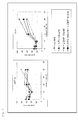
- the cell viabilities after exposure to 100 ⁇ M 5-FU for 48 hours were about 24% (HCT116) and about 49% (DLD-1). Thus, DLD-1 was found to have sensitivity to 5-FU lower than that of HCT116.
- the cell viabilities after exposure to 1 ⁇ M L-OHP for 48 hours were about 37% (HCT116) and about 82% (DLD-1). Thus, DLD-1 was found to have sensitivity to L-OHP lower than that of HCT116.
- HCT116 exhibited significantly lower cell viabilities after exposure to 2 ⁇ M 5-FU + 1 ⁇ M L-OHP for 24 hours and 100 ⁇ M 5-FU + 10 ⁇ M L-OHP for 48 hours, as compared with the cases where corresponding single agents were used, indicating a combination effect.
- DLD-1 exhibited no significant combination effect. Therefore, HCT116 was found to have high sensitivity to 5-FU/L-OHP combination, and DLD-1 was found to have low sensitivity to 5-FU/L-OHP combination ( Fig. 1 ).
- intracellular protein L levels were 88.2 ⁇ 2.2 (HCT116) and 32.1 ⁇ 1.3 (DLD-1)
- these proteins exhibited an intracellular level significantly higher in HCT116 than in DLD-1, before drug exposure (not exposed).
- Example 1 All the peaks (m/z) detected in Example 1 were confirmed through internal molecular weight calibration by use of two molecular weight standards sandwiching a target peak.
- the following standard substances having a known molecular weight were employed: insulin oxidized B chain (bovine) (m/z: 3496.94+1H), insulin (bovine) (m/z: 5733.51+1H), cytochrome c (equine) (m/z: 12360.96+1H), apomyoglobin (equine) (m/z: 16952.27+1H), and aldorase (rabbit muscle) (m/z: 39212.28+1H).
- Example 1 the peaks of Example 1 were found to be detected at an m/z of 5,300 to 5,400 (protein A), an m/z of 6,130 to 6,230 (protein B), an m/z of 7,000 to 7,080 (protein C), an m/z of 7,840 to 7,920 (protein D), an m/z of 8,920 to 9,000 (protein E), an m/z of 11,020 to 11,120 (protein F), an m/z of 12,440 to 12,560 (protein G), an m/z of 17,100 to 17,270 (protein H), an m/z of 18,290 to 18,470 (protein I), an m/z of 24,660 to 24,750 (protein J), an m/z of 35,980 to 36,290 (protein K), an m/z of 8,650 to 8,750 (protein L), an m/z of 9,100 to 9,200 (protein M), and an m/z of 11,760 to 11,890 (protein N).
- Example 1 The features of the peaks found in Example 1 were further investigated in terms of variation in peak intensity associated with change in pH.
- CM10 cation-exchange ProteinChip array
- pH: 3.0 50 mM glycine-HCl buffer
- pH: 3.5 50 mM sodium acetate buffer
- pH: 4.0 50 mM sodium acetate buffer
- pH: 4.5 50 mM sodium acetate buffer
- pH: 5.0 50 mM sodium acetate buffer
- pH: 5.5 50 mM sodium acetate buffer
- pH: 6.0 50 mM phosphate buffer
- pH: 6.5 50 mM phosphate buffer
- pH: 7.0 50 mM phosphate buffer
- pH: 7.5 50 mM phosphate buffer
- pH: 8.0 50 mM Tris-HCl buffer
- pH: 8.5 50 mM Tris-HCl buffer
- pH: 9.0 50 mM glycine-NaOH buffer
- pH: 9.5 50 mM glycine-NaOH buffer
- pH: 9.0 50 mM glycine-NaOH buffer
- Example 1 In the CM10 chip array analysis, the pH at which the relevant peak intensity drops is thought to be the isoelectric point (pI) of the protein, where an ionized form is neutralized. As a result, the peaks detected in Example 1 were found to have the following estimated isoelectric points (pIs) :
- Example 1 Based on the results of Examples 2 and 3, the peaks detected in Example 1 were subjected to database searching by use of TagIdent tool (http://au.expasy.org/tools/tagident.html) of The ExPASy proteomics server. The results are as follows. [Table 1] Proteins or fragments thereof detected Results of retrieval Protein name (UniProtKB/Swiss-Prot Accessio n number) m/z 5, 300 ⁇ 5, 400 (protein A) Putative inactivation escape 1 protein. (015225), Cytochrome c oxidase subunit 7C, mitochondri al. (P15954), Beta-defensin 134. ( Q4QY38), Putative spermatogenes is-related protein 7.
- TagIdent tool http://au.expasy.org/tools/tagident.html
- A6NGQ2 Putative calcium-activated potassium channel subunit beta-3-like protein.
- A8MYL6 Heat shock protein beta-6.(014558), HERV-K_5q33.3 provirus ancestral Pro protein.
- P10265 Nucleoside diphosphate kinase A.(P15531), Stathmin.(P16949), Interleukin-1 receptor antagonist protein.
- P18510 3-hydroxyanthranilate 3,4-dioxygenase.
- Lymphocyte antigen 6 complex locus protein G6c. (095867), Lymphocyte antigen 6 complex locus protein G6d. (095868), TC-2.(P02775), Saposin-A. (P07602), Saposin-D.(P07602), Putative DAP-2 like protein C8orf68. (P0C838), MDNCF-a.(P10145), U6 snRNA-associated Sm-like protein LSm6.(P62312), 40S ribosomal protein S21. (P63220), Selenoprotein W.(P63302), Keratin-associated protein 19-4.(Q3LI73), Late cornified envelope protein 3A.
- Q5TA76 Putative BMS1-like protein.
- Q6IPT3 Protein FAM138A/B/C/F.
- Q6VEP3 Protein ADMP.
- Q8NFR3 V-type proton ATPase subunit e 2.
- Q8NHE4 Pancreatic progenitor cell differentiation and proliferation factor-like protein.
- Q9NSA3 Beta-catenin-interacting protein 1.
- [Table 11] m/z 11,760 to 11,890 (protein N) Ig kappa chain V-I region Lay. (P01605), Ig kappa chain V-I region Roy.
- Q9NZD4 Transcription initiation factor IIA beta chain.(P52655), Death-associated protein-like 1. (A0PJW8), Putative glutaredoxin-like protein. (A6NLA2), High mobility group protein HMGI-C. (P52926), Apolipoprotein C-IV.(P55056), Protein TMEM155. (Q4W5P6), Putative uncharacterized protein FP588. (Q71RF5), Caspase-10 subunit p12. (Q92851), Transmembrane protein 141. (Q96I45), DET1- and DDB1-associated protein 1. (Q9BW61), 39S ribosomal protein L20, mitochondrial. (Q9BYC9), 39S ribosomal protein L36, mitochondrial. (Q9P0J6), Signal peptidase complex subunit 1. (Q9Y6A9)
- Protein S100-A10 protein F (m/z: 11,020 to 11,120).
- protein F calcium-binding protein S100A10
- protein F can be used as a marker for determining sensitivity to L-OHP in a precedent patent application ( WO 2009/96196 ) of the same inventors.
- protein F was found to also serve as a marker which can predict therapeutic response in an early stage after start of the treatment and which can be used in the case of 5-FU (single agent) or the case of a combination of 5-FU and L-OHP.
- Protein S100A10 was retrieved as protein F (m/z: 11,020 to 11,120).
- protein F expression of protein F in HCT116 and DLD-1 was confirmed.
- intracellular proteins were extracted from HCT116 cells and DLD-1 cells.
- the thus-extracted proteins were subjected to SDS-PAGE at constant 100 V.
- the proteins were blotted onto a PVDF membrane and blocked.
- primary antibody reaction was performed by use of an anti-S100A10 monoclonal antibody (Purified Mouse Anti-Annexin II light chain monoclonal antibody, BD Transduction Laboratories).
- Secondary antibody reaction was performed by use of an alkaline phosphatase-labeled anti-mouse IgG antibody.
- CDP-StarTM chemiluminescent substrate was added as a reaction substrate, and the target protein was detected by means of a lunino-image analyzer (LAS-4000mini, FUJIFILM).
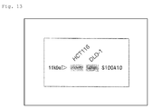
- Example 1 Expression of S100A10 detected in Example 1 as protein F (m/z: 11,020 to 11,120) in HCT116 and in DLD-1 was confirmed through the Western blotting technique employing an anti-S100A10 monoclonal antibody ( Fig. 13 ).
Landscapes
- Health & Medical Sciences (AREA)
- Life Sciences & Earth Sciences (AREA)
- Chemical & Material Sciences (AREA)
- Engineering & Computer Science (AREA)
- Immunology (AREA)
- Medicinal Chemistry (AREA)
- General Health & Medical Sciences (AREA)
- Biomedical Technology (AREA)
- Urology & Nephrology (AREA)
- Molecular Biology (AREA)
- Hematology (AREA)
- Veterinary Medicine (AREA)
- Public Health (AREA)
- Pharmacology & Pharmacy (AREA)
- Animal Behavior & Ethology (AREA)
- Epidemiology (AREA)
- Food Science & Technology (AREA)
- Physics & Mathematics (AREA)
- Pathology (AREA)
- General Physics & Mathematics (AREA)
- Biotechnology (AREA)
- Cell Biology (AREA)
- Analytical Chemistry (AREA)
- Microbiology (AREA)
- Biochemistry (AREA)
- General Chemical & Material Sciences (AREA)
- Organic Chemistry (AREA)
- Chemical Kinetics & Catalysis (AREA)
- Nuclear Medicine, Radiotherapy & Molecular Imaging (AREA)
- Bioinformatics & Cheminformatics (AREA)
- Toxicology (AREA)
- Tropical Medicine & Parasitology (AREA)
- Pharmaceuticals Containing Other Organic And Inorganic Compounds (AREA)
- Investigating Or Analysing Biological Materials (AREA)
- Medicines That Contain Protein Lipid Enzymes And Other Medicines (AREA)
- Acyclic And Carbocyclic Compounds In Medicinal Compositions (AREA)
- Other Investigation Or Analysis Of Materials By Electrical Means (AREA)
Abstract
Description
- The present invention relates to a marker for use in determination of the sensitivity of a cancer patient to an anti-cancer agent to be administered thereto, which marker can determine whether or not the cancer of the patient has a therapeutic response to the anti-cancer agent, and to application of the marker.
- Anti-cancer agents have various types such as an alkylating agent, a platinum agent, an antimetabolite, an antitumor antibiotic, and an antitumor plant alkaloid. These anti-cancer agents are effective for some cancers but not effective for other cancers. Even when an anti-cancer agent is confirmed to be effective for a certain cancer, the anti-cancer agent is effective for some patients and not effective for other patients, leading to interindividual differences. Whether or not a cancer of a specific patient has response to an anti-cancer agent is designated as sensitivity to the anti-cancer agent.
- Oxaliplatin, (SP-4-2)-[(1R,2R)-cyclohexane-1,2-diamine-κN,κN'][ethanedioato(2-)-κO1,κO2]platinum (IUPAC), is a third-generation platinum-based complex anti-cancer agent. Similar to precedent drugs (cisplatin (CDDP) and carboplatin (CBDCA)), the action mechanism thereof is thought to be based on inhibition of DNA synthesis and/or protein synthesis via cross-linking with DNA bases. Oxaliplatin (L-OHP) exhibits anti-tumor effect on colorectal cancer, to which CDDP or CBDCA is ineffective, and shows different spectrum of anti-tumor activity from that of a precedent platinum-based complex anti-cancer agent. In the United States of America, oxaliplatin for use in combination with fluorouracil (5-FU) and levofolinate (LV) was approved as a first line therapy for metastatic colorectal cancer in January, 2004. In Japan, oxaliplatin was listed in the National Health Insurance (NHI) price list in the case of combination use thereof with continuous infusional fluorouracil and levofolinate (FOLFOX4 regimen) for "advanced/recurrent colorectal cancer not amenable to curative surgical resection" in April, 2005. Until the early 1990's, 5-FU/LV regimen to advanced/recurrent colorectal cancer has provided a survival of 10 to 12 months. In contrast, a FOLFOX regimen combined with oxaliplatin results in a prolonged survival of 19.5 months (about twice the survival time). In August, 2009, an indication of oxaliplatin combined with continuous infusional fluorouracil and levofolinate to "postoperative adjuvant chemotherapy for colon cancer" was added to efficacy and effectiveness. Thus, oxaliplatin is a promising drug having an efficacy in an increased number of colorectal cancer patients.
- However, the response rate of FOLFOX regimen against advanced/recurrent colorectal cancer is still as low as about 50%. In other words, about half of the treated patients achieve no effects. During administration of oxaliplatin, peripheral neuropathy frequently occurs in addition to neutropenia. Although not being fatal, these adverse events are factors which make continuation of the therapy difficult. Therefore, if there is a biomarker which can predict, before start of the treatment, a patient who is expected to achieve the response (i.e., a responder) and a patient who is not expected to achieve the response (i.e., a non-responder), to thereby diagnose the therapeutic response earlier, a chemotherapy ensuring high effectiveness and high safety can be established.
- There have been a number of reports on sensitivity/resistance factor with respect to platinum-based chemotherapy, but most of them are focused on cisplatin. The therapeutic response of patients to oxaliplatin, which is structurally and functionally similar to cisplatin, is thought to generally relate to, for example, the following:
- 1) enhancement of the ability of excising and repairing DNA damaged by oxaliplatin;
- 2) inactivation (detoxification) of oxaliplatin (active form) in cells; and
- 3) reduction in accumulation amount of oxaliplatin in cells. In the oxaliplatin + 5-FU combination therapy for colorectal cancer patients, the therapeutic response and prognosis-predicting factor are now under study on the basis of 1) to 3).
- Regarding 1), the excision repair cross-complementing group 1 (ERCC1) gene expression level in tumor cells is reported to serve as a prognosis factor, the ERCC1 playing an important role in nucleotide excision repair (NER) (Non-Patent Document 1). Patients having a C/C homozygote of C118T (a type of single nucleotide polymorphism (SNP) of ERCC1) exhibit a survival longer than that of patients having at least one T allele (Non-Patent Document 2). In Xeroderma pigmentosum D (XPD, also known as ERCC2), a genetic polymorphism involving Lys751Gln amino acid mutation is reported to relate to percent tumor reduction and survival (
Non-Patent Documents 2 and 3). In base excision repair (BER), there has been reported a relationship between the tumor reduction effect and a genetic polymorphism involving Arg399Gln amino acid mutation in X-ray repair cross-complementing group 1 (XRCC1) encoding a protein relating to effective repair of DNA single strand breakage caused by exposure to an alkylating agent or the like (Non-Patent Document 4). However, further analysis of the same patients has revealed that the genetic polymorphism does not affect the clinical prognosis (Non-Patent Document 2). DNA mismatch repair (MMR) is thought to relate to lowering sensitivity to cisplatin. However, in vitro studies have revealed that MMR does not involve repair of DNA damaged by oxaliplatin (Non-Patent Document 5). - Regarding 2), glutathione-S-transferase (GST) is an enzyme which catalyzes phase II reaction in the detoxification and metabolism. GST catalyzes formation of a conjugation of a DNA-platinum adduct and glutathione, to thereby inactivate a drug. Among GST subtypes, GSTP1 has a high expression level in colorectal cancer, and a genetic polymorphism involving Ile105Val amino acid mutation relates to survival (median survival: Ile/Ile 7.9 months, Ile/Val 13.3 months, Val/Val 24.9 months) (Non-Patent Document 6).
- Regarding 3), studies employing cultured cells have revealed that organic cation transporters (OCTs) relate to transportation of oxaliplatin into cells and sensitivity to oxaliplatin (Non-Patent Document 7). A relationship between copper- and heavy-metal-transporters such as ATP7A and ATP7B and sensitivity has also been reported (
Non-Patent Documents 8 and 9). However, the relationship between expression of these transporters and therapeutic response to oxaliplatin has not been clinically elucidated. - Recent clinical studies for advanced colorectal cancer patients having received FOLFOX regimen have revealed that a genetic polymorphism of ERCC1 (Asn118Asn and that of XPD (Lys751Gln) independently relate to progression-free survival (PFS), and that a genetic polymorphism of GSTP1 (Ile105Val) does not relate to PFS but tends to have a relationship with oxaliplatin-induced neurotoxicity (Non-Patent Document 10).
- In vitro studies have revealed a number of resistance-related factors of cisplatin (a precedent platinum-based complex drug), and the relationship between oxaliplatin and apoptosis-related factors such as FAS/FASL and Bcl-xL have been reported (Non-Patent Documents 11 and 12). However, oxaliplatin exhibits a therapeutic response differing from that of cisplatin, depending on the type of cancer. In addition, there has not been substantially elucidated the cell response of cancer cells with respect to a platinum-DNA adduct, which exerts cytotoxic activity of oxaliplatin. Thus, there has been established no definite biomarker which can predict the therapeutic response to chemotherapy employing oxaliplatin.
- Meanwhile, fluorouracil is a fluoro-pyrimidine anti-cancer agent developed in 1957 and even now serves as a basic drug for use in the chemotherapy of gastrointestinal cancer. When incorporated into cancer cells, fluorouracil exerts cytotoxic effect through a principle action mechanism of DNA synthesis inhibition induced by inhibition of thymidylate synthase (TS) by an active metabolite, fluorodeoxyuridine-5'-monophosphate (FdUMP), and another mechanism of RNA function inhibition by an active metabolite, 5-fluorouridine triphosphate (FUTP).
- Hitherto, many studies have been conducted to predict therapeutic response to fluoro-pyrimidine anti-cancer agents. In particular, many studies have been focused on dihydropyrimidine dehydrogenase (DPD), which is a fluorouracil degrading enzyme, and thymidylate synthase (TS), which is a target enzyme of an active metabolite. A tumor in which DPD, a rate-limiting enzyme in the catabolism of fluorouracil, is highly expressed is reported to have resistance to fluorouracil (Non-Patent Document 13), but a limited number of studies have been conducted with clinical specimens. The TS expression level is reported to be a possible factor that determines the therapeutic effect by a fluoro-pyrimidine anti-cancer agent, even when the expression level is determined through any assay method such as the enzymatic activity method, protein level assay, or RNA level assay (Non-Patent Documents 14 and 15). However, the above-obtained results are not completely the same, and there has been known no definite biomarker which can predict the therapeutic response to fluorouracil in an early treatment stage.
- In metastatic colorectal cancer patients having fluorouracil resistance, ERCC1 and TS mRNA expression levels in tumor cells are reported to serve as factors that predict therapeutic response to 5-FU/L-OHP combination therapy (Non-Patent Document 16). However, recently, large-scale prospective clinical trial (FOCUS trial) for investigating biomarkers that predict therapeutic response of advanced colorectal cancer patients to chemotherapy has revealed that ERCC1 does not serve as a significant predictive factor for 5-FU/L-OHP combination therapy, and that only topoisomerase-1 (Topol) exhibits weak relationship thereto (Non-Patent Document 17). This indicates that there has been established no biomarker that can reliably select a patient who is expected to be effectively treated through the 5-FU/L-OHP combination therapy. Furthermore, since the therapy schedule of cancer chemotherapy generally requires a long period of time, continuous monitoring of sensitivity of a target patient to a target anti-cancer agent during the therapy can determine whether or not the therapy should be continued. Thus, such monitoring is thought to be meritorious from the viewpoints of reduction or mitigation of the burden on patients and adverse events, leading to reduction in medical cost. Therefore, there is keen demand for establishment of a biomarker that can predict the effect of 5-FU, L-OHP, or a combination of 5-FU/L-OHP or that can determine therapeutic response in an early stage, for the purpose of predicting therapeutic response of individual patients and establishing diagnosis in an early stage to select an appropriate drug and treatment regimen; i.e., for realizing "personalized therapy."
-
- Non-Patent Document 1: J. Clin. Oncol. 19, 4298-4304 (2001)
- Non-Patent Document 2: Br. J. Cancer 91, 344-354 (2004)
- Non-Patent Document 3: Cancer Res. 61, 8654-8658 (2001)
- Non-Patent Document 4: Anticancer Res. 21, 3075-3079 (2001)
- Non-Patent Document 5: Cancer Res. 56, 4881-4886 (1996)
- Non-Patent Document 6: J. Natl. Cancer Inst. 94, 936-942 (2002)
- Non-Patent Document 7: Cancer Res. 66, 8847-8857 (2006)
- Non-Patent Document 8: Mol. Pharmacol. 66, 25-32 (2004)
- Non-Patent Document 9: Clin. Cancer Res. 10, 4661-4669 (2004)
- Non-Patent Document 10: J. Clin. Oncol. 25, 1247-1254 (2007)
- Non-Patent Document 11: Clin. Cancer Res. 11, 4770-4774 (2005)
- Non-Patent Document 12: J. Biol. Chem. 279, 46113-46121 (2004)
- Non-Patent Document 13: European Journal of Cancer 2004; 40: 939-950
- Non-Patent Document 14: Cancer Treatment Reviews 2002; 28: 27-47
- Non-Patent Document 15: J. Clin. Oncol. 2004; 22: 529-536
- Non-Patent Document 16: J. Clin. Oncol. 2001; 19: 4298-4304
- Non-Patent Document 17: J. Clin. Oncol. 2008; 26: 2690-2698
- An object of the present invention is to provide a marker for determining sensitivity of a patient to an anti-cancer agent, which marker can determine whether or not the patient has a therapeutic response to the anti-cancer agent. Another object is to provide novel cancer therapeutic means employing the marker.
- In order to attain the aforementioned objects, the present inventors have searched for a marker for determining sensitivity to an anti-cancer agent by culturing human cancer cells, exposing the cells to a drug, and comprehensively analyzing the time-dependent expression profiles of intracellular proteins after exposure to the drug by means of a surface-enhanced laser desorption/ionization time-of-flight mass spectrometer (SELDI-TOF MS). More specifically, two types of human colorectal cancer cells, one having high sensitivity to 5-FU, L-OHP, and combination of 5-FU/L-OHP and the other having low sensitivity thereto, were exposed to 5-FU (single drug), L-OHP (single drug), or 5-FU/L-OHP (combination), and the time-dependent expression profiles of intracellular proteins after exposure to the drug were investigated. As a result, the inventors have found, after exposure to 5-FU, several proteins exhibiting time-dependent intracellular expression level profiles which differ between high-sensitivity cells and low-sensitivity cells. These proteins or fragments thereof have been detected as a peak at m/z, as determined by means of a mass spectrometer, of 5,300 to 5,400, 6,130 to 6,230, 7,000 to 7,080, 7,840 to 7,920, 17,100 to 17,270, 18,290 to 18,470, 24,660 to 24,750, 35,980 to 36,290, or 9,100 to 9,200, or identified as a calcium-binding protein S100A10.
- Similarly, the inventors have found, after exposure to L-OHP, several proteins exhibiting time-dependent intracellular expression level profiles which differ between high-sensitivity cells and low-sensitivity cells. These proteins or fragments thereof have been detected as a peak at m/z, as determined by means of a mass spectrometer, of 5,300 to 5,400, 6,130 to 6,230, 7,000 to 7,080, 7,840 to 7,920, 12,440 to 12,560, 17,100 to 17,270, 18,290 to 18,470, 24,660 to 24,750, 35,980 to 36,290, or 9,100 to 9,200.
- Furthermore, the inventors have found, after exposure to 5-FU/L-OHP in combination, several proteins exhibiting time-dependent intracellular expression level profiles which differ between high-sensitivity cells and between low-sensitivity cells. These proteins or fragments thereof have been detected as a peak at m/z, as determined by means of a mass spectrometer, peak of 5,300 to 5,400, 6,130 to 6,230, 7,000 to 7,080, 7,840 to 7,920, 8,920 to 9,000, 12,440 to 12,560, 17,100 to 17,270, 18,290 to 18,470, 24,660 to 24,750, 35,980 to 36,290, or 9,100 to 9,200, or identified as a calcium-binding protein S100A10.
- Separately, the inventors have found, before exposure to any of these drugs, several proteins exhibiting intracellular expression levels which differ between high-sensitivity cells and low-sensitivity cells. These proteins or fragments thereof have been detected as a peak at m/z, as determined by means of a mass spectrometer, of 6,130 to 6,230, 17,100 to 17,270, 8,650 to 8,750, or 11,760 to 11,890.
- On the basis of these findings, the inventors have carried out further studies, and have found that whether or not a cancer of a target cancer patient has a sensitivity to 5-FU therapy, L-OHP therapy, or 5-FU/L-OHP combination therapy can be determined through measuring the relevant protein level of a biological sample derived from the cancer patient; that screening of an anti-cancer agent sensitivity enhancer can be accomplished through employment of variation in expression of the substance as an index; and that the therapeutic effect of the relevant anti-cancer agent can be drastically enhanced by use, in combination, of the anti-cancer agent sensitivity enhancer and the anti-cancer agent which is a sensitivity enhancement target of the enhancer. The present invention has been accomplished on the basis of these findings.
- Accordingly, the present invention provides a marker for determining sensitivity to an anti-cancer agent, the marker comprising a protein selected from the group consisting of a protein or a fragment thereof which is detected as a peak at m/z of 5,300 to 5,400 (hereinafter referred to as protein A), a protein or a fragment thereof which is detected as a peak at m/z of 6,130 to 6,230 (hereinafter referred to as protein B), a protein or a fragment thereof which is detected as a peak at m/z of 7,000 to 7,080 (hereinafter referred to as protein C), a protein or a fragment thereof which is detected as a peak at m/z of 7,840 to 7,920 (hereinafter referred to as protein D), a protein or a fragment thereof which is detected as a peak at m/z of 8,920 to 9,000 (hereinafter referred to as protein E), a protein or a fragment thereof which is detected as a peak at m/z of 12,440 to 12,560 (hereinafter referred to as protein G), a protein or a fragment thereof which is detected as a peak at m/z of 17,100 to 17,270 (hereinafter referred to as protein H), a protein or a fragment thereof which is detected as a peak at m/z of 18,290 to 18,470 (hereinafter referred to as protein I), a protein or a fragment thereof which is detected as a peak at m/z 24,660 to 24,750 (hereinafter referred to as protein J), a protein or a fragment thereof which is detected as a peak at m/z of 35,980 to 36,290 (hereinafter referred to as protein K), a protein or a fragment thereof which is detected as a peak at m/z of 8,650 to 8,750 (hereinafter referred to as protein L), a protein or a fragment thereof which is detected as a peak at m/z of 9,100 to 9,200 (hereinafter referred to as protein M), and a protein or a fragment thereof which is detected as a peak at m/z of 11,760 to 11,890 (hereinafter referred to as protein N), the peaks being determined by means of a mass spectrometer.
- The present invention also provides a marker for determining sensitivity to an anti-cancer agent containing fluorouracil or a salt thereof or a combination of fluorouracil or a salt thereof and oxaliplatin or a salt thereof, the marker comprising a calcium-binding protein S100A10 (hereinafter referred to as protein F).
- The present invention also provides a method for determining sensitivity of a subject to an anti-cancer agent, the method comprising measuring the level of any of proteins A to N in a specimen derived from the subject.
- The present invention also provides a kit for carrying out the method for determining sensitivity of a subject to an anti-cancer agent, the kit comprising a protocol for measuring the level of any of proteins A to N in a specimen derived from the subject.
- The present invention also provides a screening method for an anti-cancer agent sensitivity enhancer, the method comprising employing variation in expression of any of the proteins A to N as an index.
- The present invention also provides an anti-cancer agent sensitivity enhancer obtained through the screening method.
- The present invention also provides a composition for cancer therapy comprising, in combination, the anti-cancer agent sensitivity enhancer and an anti-cancer agent which is a sensitivity enhancement target of the enhancer.
- The present invention also provides the proteins A to N for use in determining the anti-cancer agent sensitivity.
- According to the marker for determining sensitivity to an anti-cancer agent of the present invention, the sensitivity to an anti-cancer agent of a patient can be appropriately determined before the therapy or in an early stage after start of the therapy. As a result, an anti-cancer agent having higher therapeutic effect can be selected, and unnecessary adverse events, which would otherwise result from administration of an anti-cancer agent exerting no expected therapeutic effect, can be prevented. Meanwhile, the therapy schedule employing an anti-cancer agent generally requires a long period of time. Even in on-going therapy, the sensitivity of the target cancer to an anti-cancer agent can be evaluated in a time-dependent manner through determination of the anti-cancer agent sensitivity in each therapy cycle, whereby a determination can be made on whether or not the therapy should be continued. As a result, progression of cancer and aggravation of adverse events, which would otherwise result from continuous administration of an anti-cancer agent exerting no expected therapeutic effect, can be prevented. Thus, reductions can be expected in the burden on patients and medical cost.
- In addition, when the marker of the present invention is used, a drug which can promote anti-cancer agent sensitivity can be selected through screening. Thus, through employment, in combination, of the target anti-cancer agent and an anti-cancer agent sensitivity enhancer to the anti-cancer agent, the expected cancer therapeutic effect can be drastically enhanced. The assay reagent for measuring the marker for determining sensitivity to an anti-cancer agent of the present invention is useful as an reagent for determining sensitivity to an anti-cancer agent.
-
- [
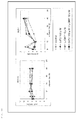
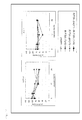
Fig. 1 ] Graphs showing the survival (%) of HCT116 cells under exposure to 5-FU, L-OHP, or 5-FU/L-OHP, and graphs showing the survival (%) of DLD-1 cells under exposure to 5-FU, L-OHP, or 5-FU/L-OHP. - [
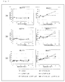
Fig. 2 ] A graph showing a time-dependent profile of the intracellular protein A level in HCT116 cells after exposure to 5-FU, L-OHP, or 5-FU/L-OHP, and a graph showing the time-dependent profile of the intracellular protein A level in DLD-1 cells after exposure to 5-FU, L-OHP, or 5-FU/L-OHP. - [
Fig. 3 ] A graph showing a time-dependent profile of the intracellular protein B level in HCT116 cells after exposure to 5-FU, L-OHP, or 5-FU/L-OHP, and a graph showing a time-dependent profile of the intracellular protein B level in DLD-1 cells after exposure to 5-FU, L-OHP, or 5-FU/L-OHP. - [
Fig. 4 ] Graphs showing a time-dependent profile of the intracellular protein C level in HCT116 cells after exposure to 5-FU, L-OHP, or 5-FU/L-OHP, and graphs showing a time-dependent profile of the intracellular protein C level in DLD-1 cells after exposure to 5-FU, L-OHP, or 5-FU/L-OHP. - [
Fig. 5 ] Graphs showing a time-dependent profile of the intracellular protein D level in HCT116 cells after exposure to 5-FU, L-OHP, or 5-FU/L-OHP, and graphs showing a time-dependent profile of the intracellular protein D level in DLD-1 cells after exposure to 5-FU, L-OHP, or 5-FU/L-OHP. - [
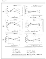
Fig. 6 ] A graph showing a time-dependent profile of the intracellular protein E level in HCT116 cells after exposure to 5-FU/L-OHP, and a graph showing a time-dependent profile of the intracellular protein E level in DLD-1 cells after exposure to 5-FU/L-OHP. - [
Fig. 7 ] Graphs showing a time-dependent profile of the intracellular protein F level in HCT116 cells after exposure to 5-FU, L-OHP, or 5-FU/L-OHP, and graphs showing a time-dependent profile of the intracellular protein F level in DLD-1 cells after exposure to 5-FU, L-OHP, or 5-FU/L-OHP. - [
Fig. 8 ] Graphs showing a time-dependent profile of the intracellular protein G level in HCT116 cells after exposure to L-OHP or 5-FU/L-OHP, and graphs showing a time-dependent profile of the intracellular protein G level in DLD-1 cells after exposure to L-OHP or 5-FU/L-OHP. - [
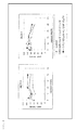
Fig. 9 ] A graph showing a time-dependent profile of the intracellular protein H level in HCT116 cells after exposure to 5-FU, L-OHP, or 5-FU/L-OHP, and a graph showing a time-dependent profile of the intracellular protein H level in DLD-1 cells after exposure to 5-FU, L-OHP, or 5-FU/L-OHP. - [
Fig. 10 ] A graph showing a time-dependent profile of the intracellular protein I level in HCT116 cells after exposure to 5-FU, L-OHP, or 5-FU/L-OHP, and a graph showing a time-dependent profile of the intracellular protein I level in DLD-1 cells after exposure to 5-FU, L-OHP, or 5-FU/L-OHP. - [
Fig. 11 ] Graphs showing a time-dependent profile of the intracellular protein J level in HCT116 cells after exposure to 5-FU, L-OHP, or 5-FU/L-OHP, and graphs showing a time-dependent profile of the intracellular protein J level in DLD-1 cells after exposure to 5-FU, L-OHP, or 5-FU/L-OHP. - [
Fig. 12 ] A graph showing a time-dependent profile of the intracellular protein K level in HCT116 cells after exposure to 5-FU, L-OHP, or 5-FU/L-OHP, and a graph showing a time-dependent profile of the intracellular protein K level in DLD-1 cells after exposure to 5-FU, L-OHP, or 5-FU/L-OHP. - [
Fig. 13 ] A western blot feature showing detection of S100A10 in HCT116 cells and DLD-1 cells. - [
Fig. 14 ] A graph showing a time-dependent profile of the intracellular protein M level in HCT116 cells after exposure to 5-FU, L-OHP, or 5-FU/L-OHP, and a graph showing a time-dependent profile of the intracellular protein M level in DLD-1 cells after exposure to 5-FU, L-OHP, or 5-FU/L-OHP. Modes for Carrying Out the Invention - The marker for determining sensitivity to an anti-cancer agent of the present invention is any of proteins A to N. More specifically, the proteins are detected by means of a cation-exchange chip at a pH of 4.5, and m/z peaks are determined by means of a surface-enhanced laser desorption/ionization time-of-flight mass spectrometer (SELDI-TOF MS). Thus, the proteins are a protein or a fragment thereof which is detected as a peak at m/z of 5,300 to 5,400 (protein A), a protein or a fragment thereof which is detected as a peak at m/z of 6,130 to 6,230 (protein B), a protein or a fragment thereof which is detected as a peak at m/z of 7,000 to 7,080 (protein C), a protein or a fragment thereof which is detected as a peak at m/z of 7,840 to 7,920 (protein D), a protein or a fragment thereof which is detected as a peak at m/z of 8,920 to 9,000 (protein E), a protein or a fragment thereof which is detected as a peak at m/z of 11,020 to 11,120 (protein F), a protein or a fragment thereof which is detected as a peak at m/z of 12,440 to 12,560 (protein G), a protein or a fragment thereof which is detected as a peak at m/z of 17,100 to 17,270 (protein H), a protein or a fragment thereof which is detected as a peak at m/z of 18,290 to 18,470 (protein I), a protein or a fragment thereof which is detected as a peak at m/z of 24,660 to 24,750 (protein J), a protein or a fragment thereof which is detected as a peak at m/z of 35,980 to 36,290 (protein K), a protein or a fragment thereof which is detected as a peak at m/z of 8,650 to 8,750 (protein L), a protein or a fragment thereof which is detected as a peak at m/z of 9,100 to 9,200 (protein M), and a protein or a fragment thereof which is detected as a peak at m/z of 11,760 to 11,890 (protein N), the peaks being determined by means of the mass spectrometer.
- As shown in the Examples described hereinbelow, protein F has been identified as calcium-binding protein S100A10. S100A10 is known to be a member of the S100 protein family having calcium-binding EF-hand motif. S100A10 is also known to form hetero-tetramer from S100A10 dimer and Annexin A2 (Annexin-2, Annexin II, Lipocortin II, Calpactin I heavy chain, Chromobindin-8, p36, Protein I, Placental anticoagulant protein IV, or PAP-IV) dimer. Therefore, similar to S100A10, Annexin A2 may be possibly employed as a marker for determining sensitivity to an anti-cancer agent.
- As shown in the Examples described hereinbelow, through investigation of intracellular protein expression in cultured cancer cells by means of a SELDI-TOF MS, levels of proteins A to C, F, and H to K were found to increase in DLD-1 cells, which are low-sensitive to 5-FU, after exposure to 5-FU. In contrast, no significant difference was observed in HCT116 cells, which are high-sensitive to 5-FU. Therefore, proteins A to C, F, and H to K are useful as markers for determining sensitivity to an anti-cancer agent, particularly as markers for determining sensitivity to 5-FU.
- As shown in the Examples described hereinbelow, levels of proteins D and M were found to decrease in HCT116 cells, which are high-sensitive to 5-FU, after exposure to 5-FU. In contrast, no significant difference was observed in DLD-1 cells, which are low-sensitive to 5-FU. Therefore, proteins D and M are useful as markers for determining sensitivity to an anti-cancer agent, particularly as markers for determining sensitivity to 5-FU.
- As shown in the Examples described hereinbelow, levels of proteins A to C and G to K were found to increase in DLD-1 cells, which are low-sensitive to L-OHP, after exposure to L-OHP. In contrast, no significant difference was observed in HCT116 cells, which are high-sensitive to L-OHP. Therefore, proteins A to C and G to K are useful as markers for determining sensitivity to an anti-cancer agent, particularly as markers for determining sensitivity to L-OHP.
- Notably, the present inventors previously disclosed that protein F (calcium-binding protein S100A10) can be used as a marker for determining sensitivity to L-OHP in a precedent patent application (
WO 2009/96196 - As shown in the Examples described hereinbelow, levels of proteins D and M were found to decrease in HCT116 cells, which are high-sensitive to L-OHP, after exposure to L-OHP. In contrast, no significant difference was observed in DLD-1 cells, which are low-sensitive to L-OHP. Therefore, proteins D and M are useful as markers for determining sensitivity to an anti-cancer agent, particularly as markers for determining sensitivity to L-OHP.
- As shown in the Examples described hereinbelow, levels of proteins A to C, F, and H to K were found to increase in DLD-1 cells, which are low-sensitive to 5-FU/L-OHP combination, after exposure to 5-FU/L-OHP combination. In contrast, no significant difference was observed in HCT116 cells, which are high-sensitive to 5-FU/L-OHP combination. Therefore, proteins A to C, F, and H to K are useful as markers for determining sensitivity to an anti-cancer agent, particularly as markers for determining sensitivity to 5-FU/L-OHP combination. As described above, there has been known that protein F is a useful marker for determining sensitivity to L-OHP. However, it is a new finding by the present inventors that protein F serves as a useful marker for determining sensitivity to an anti-cancer agent when 5-FU/L-OHP combination is employed.
- As shown in the Examples described hereinbelow, levels of proteins E and G were found to increase in HCT116 cells, which are high-sensitive to 5-FU/L-OHP combination, after exposure to 5-FU/L-OHP combination. In contrast, no significant difference was observed in DLD-1 cells, which are low-sensitive to 5-FU/L-OHP combination. Therefore, proteins E and G are useful as markers for determining sensitivity to an anti-cancer agent, particularly as markers for determining sensitivity to 5-FU/L-OHP combination.
- As shown in the Examples described hereinbelow, levels of proteins D and M were found to decrease in HCT116 cells, which are high-sensitive to 5-FU/L-OHP combination, after exposure to 5-FU/L-OHP combination. In contrast, no significant difference was observed in DLD-1 cells, which are low-sensitive to 5-FU/L-OHP combination. Therefore, proteins D and M are useful as markers for determining sensitivity to an anti-cancer agent, particularly as markers for determining sensitivity to 5-FU/L-OHP combination.
- As shown in the Examples described hereinbelow, levels of proteins B, H, L, and N in HCT116 cells, which are high-sensitive to 5-FU, L-OHP, and 5-FU/L-OHP combination, before exposure to any of the drugs (not exposed) were significantly higher than those in DLD-1 cells, which are low-sensitive to the aforementioned drugs. Therefore, proteins B, H, L, and N are useful as markers for determining sensitivity to an anti-cancer agent, particularly as markers for determining sensitivity to 5-FU, L-OHP, or 5-FU/L-OHP combination.
- No particular limitation is imposed on the anti-cancer agent to which the marker for determining sensitivity to an anti-cancer agent of the present invention is applied. Examples of the anti-cancer agent include oxaliplatin, cyclophosphamide, ifosfamide, thiotepa, melphalan, busulfan, nimustine, ranimustine, dacarbazine, procarbazine, temozolomide, cisplatin, carboplatin, nedaplatin, methotrexate, pemetrexed, fluorouracil, tegaful/uracil, doxifluridine, tegaful/gimeracil/oteracil, capecitabine, cytarabine, enocitabine, gemcitabine, 6-mercaptopurine, fuludarabin, pentostatin, cladribine, hydroxyurea, doxorubicin, epirubicin, daunorubicin, idarubicine, pirarubicin, mitoxantrone, amurubicin, actinomycin D, bleomycine, pepleomycin, mytomycin C, aclarubicin, zinostatin, vincristine, vindesine, vinblastine, vinorelbine, paclitaxel, docetaxel, irinotecan, irinotecan active metabolite (SN-38), nogitecan (topotecan), etoposide, prednisolone, dexamethasone, tamoxifen, toremifene, medroxyprogesterone, anastrozole, exemestane, letrozole, rituximab, imatinib, gefitinib, gemtuzumab/ozogamicin, bortezomib, erlotinib, cetuximab, bevacizumab, sunitinib, sorafenib, dasatinib, panitumumab, asparaginase, tretinoin, arsenic trioxide, salts thereof, and active metabolites thereof. Among them, fluoro-pyrimidine anti-cancer agents and platinum-based complex anti-cancer agents are preferred, with fluorouracil, oxaliplatin, and salts thereof being particularly preferred. The marker of the present invention is preferably applied to a combination of fluorouracil or a salt thereof and oxaliplatin or a salt thereof.
- In order to determine sensitivity of a subject to an anti-cancer agent by use of the marker for determining sensitivity to an anti-cancer agent of the present invention, any of the protein A level to protein N level in a specimen may be measured. Examples of the specimen include biological samples derived from subjects having cancer (i.e., cancer patients) such as blood, serum, plasma, cancer tissue biopsy specimens, cancer isolated preparations, feces, urine, ascitic fluid, pleural fluid, cerebrospinal fluid, and expectoration. Of these, serum is particularly preferred.
- Examples of the target cancer of the present invention include lip, oral, pharyngeal cancers such as pharyngeal cancer; gastrointestinal cancers such as esophageal cancer, gastric cancer, and colorectal cancer; respiratory and intrathoracic organ cancers such as lung cancer; bone cancer and articular cartilage cancer; skin melanoma, squamous cell cancer, and other skin cancers; mesothelial and soft tissue cancers such as mesothelioma; female genital cancers such as breast cancer, uterine cancer, and ovarian cancer; male genital cancers such as prostate cancer; urinary tract cancers such as bladder cancer; eye, brain, and central nervous system cancers such as brain tumor; thyroid and other endocrine cancers; lymphoid tissue, hematopoietic tissue, and related tissue cancers such as non-Hodgkin's lymphoma and lymphoid leukemia; and metastatic cancers from these cancers as primary lesions. The present invention is particularly preferably applied to gastric cancer and colorectal cancer.
- Proteins A to N contained in a specimen may be measured through, for example, SELDI-TOF MS or immunoassay.
- Measurement through SELDI-TOF MS may be performed through the procedure as described in the Examples. In the immunoassay techniques, an immunoassay employing anti-protein A antibody to anti-protein N antibody are preferably employed. The employed anti-protein A antibody to anti-protein N antibody may be a monoclonal or polyclonal antibody. Specific examples of the immunoassay include radioimmunoassay, enzyme immunoassay, fluorescent immunoassay, luminescent immunoassay, immunoprecipitation, immunonephelometry, Western blotting, immunostaining, and immunodiffusion. Among them, Western blotting and enzyme immunoassay are preferably employed. Western blotting and enzyme-linked immunosorbent assay (ELISA) (e.g., sandwich ELISA) are particularly preferred.
- In the case where proteins A to C, F, and H to K are employed and 5-FU is a target anti-cancer agent, the sensitivity of the target cancer to the anti-cancer agent is determined as follows. The level of any of the proteins in a biological sample derived from a cancer patient is measured before and after administration of the anti-cancer agent. When the protein level increases after administration of the anti-cancer agent, the cancer is determined to have no sensitivity to the anti-cancer agent, whereas when the protein level is constant or decreases after administration of the anti-cancer agent, the cancer is determined to have sensitivity to the anti-cancer agent. Specifically, when the level of any of proteins A to C, F, and H to K is lower than a predetermined standard level in an early stage after administration of the anti-cancer agent, the cancer can be determined to have sensitivity to the anti-cancer agent. Thus, the proteins may be employed as a marker for indicating that the patient is expected to receive therapeutic effect and may positively undergo continuous therapy employing the anti-cancer agent.
- When the level of any of proteins A to C, F, and H to K is higher than a predetermined standard level in an early stage after administration of the anti-cancer agent, the cancer can be determined to have no sensitivity to the anti-cancer agent. When the cancer has no sensitivity to the anti-cancer agent, no pharmaceutical effect can be expected from the anti-cancer agent. If such an ineffective anti-cancer agent is continuously administered to the patient, the cancer may progress, and adverse events may be aggravated. Thus, the marker for determining sensitivity to an anti-cancer agent of the present invention may be employed not only to determine therapeutic response to the anti-cancer agent, but also to avoid progression of cancer and aggravation of adverse events which would otherwise be caused by continuous administration of an ineffective anti-cancer agent.
- In the case where proteins A to C, and G to K are employed and L-OHP is a target anti-cancer agent, or where proteins A to C, F, and H to K are employed and 5-FU/L-OHP combination is a target anti-cancer agent, the sensitivity of the target cancer to the anti-cancer agent is determined in the same manner as described above.
- In the case where proteins D and M are employed and 5-FU is a target anti-cancer agent, the sensitivity of the target cancer to the anti-cancer agent is determined as follows. The level of any of the proteins in a biological sample derived from a cancer patient is measured before and after administration of the anti-cancer agent. When the protein level decreases after administration of the anti-cancer agent, the cancer is determined to have sensitivity to the anti-cancer agent, whereas when the protein level is constant or increases after administration of the anti-cancer agent, the cancer is determined to have no sensitivity to the anti-cancer agent. Specifically, when the level of any of proteins D and M is lower than a predetermined standard level in an early stage after administration of the anti-cancer agent, the cancer can be determined to have sensitivity to the anti-cancer agent. Thus, the proteins may be employed as a marker for indicating that the patient is expected to receive therapeutic effect and may positively undergo continuous therapy employing the anti-cancer agent.
- When the level of any of proteins D and M is higher than a predetermined standard level in an early stage after administration of the anti-cancer agent, the cancer can be determined to have no sensitivity to the anti-cancer agent. When the cancer has no sensitivity to the anti-cancer agent, no pharmaceutical effect can be expected from the anti-cancer agent. If such an ineffective anti-cancer agent is continuously administered to the patient, the cancer may progress, and adverse events may be aggravated. Thus, the marker for determining sensitivity to an anti-cancer agent of the present invention may be employed not only to determine therapeutic response to the anti-cancer agent, but also to avoid progression of cancer and aggravation of adverse events which would otherwise be caused by continuous administration of an ineffective anti-cancer agent.
- In the case where proteins D and M are employed and L-OHP is a target cancer agent or 5-FU/L-OHP combination is a target anti-cancer agent, the sensitivity of the target cancer to the anti-cancer agent is determined in the same manner as described above.
- In the case where proteins E and G are employed and 5-FU/L-OHP combination is a target anti-cancer agent, the sensitivity of the target cancer to the anti-cancer agent is determined as follows. The level of any of the proteins in a biological sample derived from a cancer patient is measured before and after administration of the anti-cancer agent. When the protein level increases after administration of the anti-cancer agent, the cancer is determined to have sensitivity to the anti-cancer agent, whereas when the protein level is constant or decreases after administration of the anti-cancer agent, the cancer is determined to have no sensitivity to the anti-cancer agent. Specifically, when the level of any of proteins E and G is higher than a predetermined standard level in an early stage after administration of the anti-cancer agent, the cancer can be determined to have sensitivity to the anti-cancer agent. Thus, the proteins may be employed as a marker for indicating that the patient is expected to receive therapeutic effect and may positively undergo continuous therapy employing the anti-cancer agent.
- When the level of any of proteins E and G is lower than a predetermined standard level in an early stage after administration of the anti-cancer agent, the cancer can be determined to have no sensitivity to the anti-cancer agent. When the cancer has no sensitivity to the anti-cancer agent, no pharmaceutical effect can be expected from the anti-cancer agent. If such an ineffective anti-cancer agent is continuously administered to the patient, the cancer may progress, and adverse events may be aggravated. Thus, the marker for determining sensitivity to an anti-cancer agent of the present invention may be employed not only to determine therapeutic response to the anti-cancer agent, but also to avoid progression of cancer and aggravation of adverse events which would otherwise be caused by continuous administration of an ineffective anti-cancer agent.
- In the case where proteins B, H, L, and N are employed and 5-FU, L-OHP, or 5-FU/L-OHP combination is a target anti-cancer agent, the sensitivity of the target cancer to the anti-cancer agent is determined as follows. The level of any of the proteins in a biological sample derived from a cancer patient is measured before administration of the anti-cancer agent. When the level of any of these proteins is lower than a predetermined standard level, the cancer can be determined to have no sensitivity to the anti-cancer agent. In the case where the cancer has no sensitivity to the target anti-cancer agent, conceivably, no pharmaceutical effect is expected, and merely adverse events caused by the anti-cancer agent occur. Thus, the marker for determining sensitivity to an anti-cancer agent of the present invention may be employed to avoid occurrence of unnecessary adverse events as well as to avoid progression of cancer and aggravation of adverse events which would otherwise be caused by continuation of ineffective therapy.
- In contrast, when the level of any of these proteins is higher than a predetermined standard level, the cancer can be determined to have sensitivity to the anti-cancer agent. Thus, the proteins may also be employed as a marker for positively selecting a patient who is expected to receive therapeutic effect.
- Regarding proteins B and H, when any of the protein is employed as a marker for use during administration of the anti-cancer agent, in the case where the protein level is constant or decreases after administration, the cancer can be determined to have sensitivity to the anti-cancer agent, whereas when any of the protein is employed as a marker for use before administration of the anti-cancer agent, in the case where the protein level is lower than a predetermined level, the cancer can be determined to have no sensitivity to the anti-cancer agent. Since the marker (protein B or H) plays different roles between during administration of the anti-cancer agent and before administration thereof, the marker is preferably employed in consideration of the difference.
- In order to carry out the method of the present invention for determining sensitivity of a subject to an anti-cancer agent, preferably, a kit containing a protocol for measuring the level of proteins A to N of a specimen is employed. The kit contains a reagent for measuring any of proteins A to N, an indication of an instruction manual for use of the reagent, standards for determining the presence or absence of sensitivity to the anti-cancer agent, etc. The standards include standard levels of proteins A to N, a high threshold level, a low threshold level, factors affecting the measurements, the degree of the effects, etc. These levels may be set so as to suit the target anti-cancer agent selected. The sensitivity determination may be performed as described above on the basis of the standards.
- In the case where proteins A to D, F, H to K, and M are employed and 5-FU is a target anti-cancer agent, screening of an anti-cancer agent sensitivity enhancer can be performed through employment of variation in expression of any of the proteins, specifically suppression of the expression. That is, a substance which suppresses expression of protein in vitro or in vivo enhances sensitivity to an anti-cancer agent. For example, a substance which lowers protein levels in various cancer cells in the presence of an anti-cancer agent (in vitro) is a substance which enhances the sensitivity to the anti-cancer agent (i.e., anti-cancer agent sensitivity enhancer). Also, a substance which promotes lowering of protein levels in a cancer-bearing animal after administration of an anti-cancer agent (in vivo) is a substance which enhances the sensitivity to the anti-cancer agent (i.e., anti-cancer agent sensitivity enhancer). In the case where proteins A to D, G to K, and M are employed and L-OHP is a target anti-cancer agent, or in the case where proteins A to D, F, H to K, and M are employed and 5-FU/L-OHP combination is a target anti-cancer agent, screening of the anti-cancer agent sensitivity enhancer may be performed in the same manner as described above.
- In the case where proteins E and G are employed and 5-FU/L-OHP combination is a target anti-cancer agent, screening of an anti-cancer agent sensitivity enhancer can be performed through employment of variation in expression of any of the proteins, specifically elevation of the expression. That is, a substance which elevates expression of protein in vitro or in vivo enhances sensitivity to an anti-cancer agent. For example, a substance which elevates protein levels in various cancer cells in the presence of an anti-cancer agent (in vitro) is a substance which enhances the sensitivity to the anti-cancer agent (i.e., anti-cancer agent sensitivity enhancer). Also, a substance which promotes elevation of protein levels in a cancer-bearing animal after administration of an anti-cancer agent (in vivo) is a substance which enhances the sensitivity to the anti-cancer agent (i.e., anti-cancer agent sensitivity enhancer).
- In the case where proteins B, H, L, and N are employed and 5-FU, L-OHP, or 5-FU/L-OHP combination is a target anti-cancer agent, screening of an anti-cancer agent sensitivity enhancer can be performed through employment of variation in expression of any of the proteins, specifically elevation of the expression. That is, a substance which elevates expression of protein in vitro or in vivo enhances sensitivity to an anti-cancer agent. For example, a substance which elevates protein levels in various cancer cells in the absence of an anti-cancer agent (in vitro) is a substance which enhances the sensitivity to the anti-cancer agent (i.e., anti-cancer agent sensitivity enhancer). Also, a substance which promotes elevation of protein levels in a cancer-bearing animal before administration of an anti-cancer agent (in vivo) is a substance which enhances the sensitivity to the anti-cancer agent (i.e., anti-cancer agent sensitivity enhancer).
- Through employment, in combination, of the thus-obtained anti-cancer agent sensitivity enhancer and an anti-cancer agent which is a sensitivity enhancement target of the enhancer, the therapeutic effect of the anti-cancer agent is drastically enhanced. The combination of the anti-cancer agent sensitivity enhancer and the anti-cancer agent which is a sensitivity enhancement target of the enhancer may be a composition containing both ingredients, or a combined drug of preparations containing individual ingredients. These two ingredients may be administered through different routes.
- The target anti-cancer agents which may be employed here are the same as described above. Examples of the anti-cancer agent include oxaliplatin, cyclophosphamide, ifosfamide, thiotepa, melphalan, busulfan, nimustine, ranimustine, dacarbazine, procarbazine, temozolomide, cisplatin, carboplatin, nedaplatin, methotrexate, pemetrexed, fluorouracil, tegaful/uracil, doxifluridine, tegaful/gimeracil/oteracil, capecitabine, cytarabine, enocitabine, gemcitabine, 6-mercaptopurine, fuludarabin, pentostatin, cladribine, hydroxyurea, doxorubicin, epirubicin, daunorubicin, idarubicine, pirarubicin, mitoxantrone, amurubicin, actinomycin D, bleomycine, pepleomycin, mytomycin C, aclarubicin, zinostatin, vincristine, vindesine, vinblastine, vinorelbine, paclitaxel, docetaxel, irinotecan, irinotecan active metabolite (SN-38), nogitecan (topotecan), etoposide, prednisolone, dexamethasone, tamoxifen, toremifene, medroxyprogesterone, anastrozole, exemestane, letrozole, rituximab, imatinib, gefitinib, gemtuzumab/ozogamicin, bortezomib, erlotinib, cetuximab, bevacizumab, sunitinib, sorafenib, dasatinib, panitumumab, asparaginase, tretinoin, arsenic trioxide, salts thereof, and active metabolites thereof. Among them, fluoro-pyrimidine anti-cancer agents and platinum-based complex anti-cancer agents are preferred, with fluorouracil, oxaliplatin, and salts thereof being particularly preferred. It is preferably applied to a combination of fluorouracil or a salt thereof and oxaliplatin or a salt thereof.
- The present invention will next be described in more detail by way of examples.
- Two human colorectal cancer cell lines (HCT116 and DLD-1) were obtained from ECACC. Cell culturing was performed by means of a φ100 mm/Tissue Culture Dish (IWAKI) with a medium (D-MEM, 2mM glutamine, 10% fetal bovine serum) at 37°C under 5% CO2.
- Fluorouracil (5-FU) was purchased from Sigma, and oxaliplatin (L-OHP) powder was obtained from Kabushiki Kaisha Yakult Honsha.
- Two colorectal cancer cell lines (HCT116 and DLD-1) were exposed to a drug, and 24 hours and 48 hours after drug exposure, cell viability was determined by means of an MTS assay (CellTiter96™AQueous One Solution Cell Proliferation Assay, Promega). Drug exposure conditions were as follows. 5-FU (single agent) was used at the following 11 concentrations: control (0 µM), 0.001 µM, 0.01 µM, 0.1 µM, 1 µM, 3 µM, 10 µM, 30 µM, 100 µM, 1,000 µM, and 10,000 µM. L-OHP (single agent) was used at the following 11 concentrations; control (0 µM), 0.001 µM, 0.01 µM, 0.1 µM, 0.3 µM, 1 µM, 3 µM, 10 µM, 30 µM, 100 µM, and 1,000 µM. In the case of 5-FU/L-OHP combination, 5-FU was used at 2 µM, 10 µM, 30 µM, and 100 µM (4 concentrations), and L-OHP was used at 0.01 µM, 0.1 µM, 0.3 µM, 1 µM, 3 µM, 10 µM, 30 µM, 100 µM, and 1,000 µM (9 concentrations); i.e., 36 concentrations were employed. In addition, the above four 5-FU (single agent) concentrations, which were the same as employed in the case of 5-FU/L-OHP combination, and control (0 µM) were employed. Sensitivity evaluation was performed by use of three samples with respect to each cell line, drug exposure time, and drug exposure concentration in one experiment. The experiment was performed thrice by use of cells at different passage numbers.
- Sensitivity analysis was performed from viability data calculated from MTS assay results. The presence or absence of combined effect was determined through comparison of viability obtained by exposure to a combination with that obtained by exposure to a single agent at the same concentration as employed at the exposure to the combination. When the viability obtained by exposure to a combination is significantly lowered as compared with each single agent case, the presence of a combination effect was confirmed.
- Based on the results of (c) above, the drug concentration employed in the exposure test was determined. The 5-FU + L-OHP combination concentration was adjusted to the following four levels: 2 µM + 1 µM, 2 µM + 10 µM, 100 µM + 1 µM, and 100 µM + 10 µM, the 5-FU concentration (exposure to single agent) was adjusted to 2 µM and 100 µM, and the L-OHP concentration (exposure to single agent) was adjusted to 1 µM and 10 µM. In addition, the exposure test was conducted at a drug-free concentration (control). That is, nine concentrations in total were employed. Drug exposure time was adjusted to the following five periods of time: 0 hour (just before exposure), 4 hours, 12 hours, 24 hours, and 48 hours. After completion of exposure, cell count was performed, and intracellular proteins were extracted.
- The medium was removed from the dish and washed thrice with ice-cooled PBS. The cells on the dish was collected by scraping with a rubber policeman. The thus-obtained cell suspension was transferred to a 1.5-mL microtube. The cell suspension was centrifuged at 4°C and 1,200xg for 10 minutes, and the cells were recovered. After removal of the supernatant, a cell lysis buffer (9 mol/L urea, 2% CHAPS, 1 mM DTT, protease-inhibitor cocktail (Sigma)) was added in a volume of 200 µL with respect to 10,000,000 cells. The liquid was subjected to untrasonic treatment under cooling with ice. The thus-treated product was centrifuged at 4°C and 16,000xg for 20 minutes, and the supernatant was quickly frozen with liquid nitrogen. The frozen product was stored at -80°C before analysis. An aliquot of the supernatant was subjected to protein quantification (DC Protein Assay Kit, Bio-Rad).
- The protein sample was mixed with a cell lysis buffer (excluding protease inhibitor), to thereby adjust the protein concentration to 2.5 mg/mL. The liquid was further mixed with a dilution/washing buffer (pH: 4.5, 50 mM sodium acetate buffer) (hereinafter referred to simply as "buffer"), to a protein concentration of 0.5 mg/mL. The thus-prepared sample (100 µL) was applied to spots of a cation-exchange ProteinChip array (CM10, Bio-Rad) which had been conditioned in advance with the same buffer. Incubation was performed for one hour for reaction, and the chip array was washed thrice with the buffer and rinsed twice with milliQ water, followed by drying in air. Energy absorbing molecule (EAM: saturated solution of sinapinic acid in 50% ACN/0.5% TFA) (1.0 µL (0.5 mL×2)) was added to each spot. After the surface of the spot was dried, analysis of the ProteinChip array was performed.
- Protein expression analysis was performed through surface-enhanced laser desorption/ionization time-of-flight mass spectrometry (SELDI-TOF MS). As an analyzer, ProteinChip™ Reader (Model PCS4000 Personal Edition, Bio-Rad) was used and the analysis was performed under the following conditions: mass range of 0 to 70,000 Da, focus mass of 8,000 Da, energy of 3,000 or 4,000 nJ, and 265 shots/sample. Peak extraction (signal-to-noise ratio (S/N)≥2) and protein expression comparative analysis were performed by means of CiphergenExpress™ Data Manager 3.0.
- Through SELDI-TOF MS analysis, 88 to 143 protein peaks were selected from each sample (S/N≥2). Firstly, a peak cluster was made by means of CiphergenExpress™ Data Manager 3.0. Then, under tested conditions, there were selected peaks exhibiting a significant change in intensity over time after exposure to a drug, and peaks exhibiting significantly different intensities depending on the selected drug at exposure times (4, 12, 24, and 48 hours). Then, peaks overlapping the above two conditions; i.e., peaks exhibiting variation in expression due to exposure time and depending on the type of the drug, were selected.
- The cell viabilities after exposure to 100 µM 5-FU for 48 hours were about 24% (HCT116) and about 49% (DLD-1). Thus, DLD-1 was found to have sensitivity to 5-FU lower than that of HCT116. The cell viabilities after exposure to 1 µM L-OHP for 48 hours were about 37% (HCT116) and about 82% (DLD-1). Thus, DLD-1 was found to have sensitivity to L-OHP lower than that of HCT116. Regarding 5-FU/L-OHP combination, HCT116 exhibited significantly lower cell viabilities after exposure to 2 µM 5-FU + 1 µM L-OHP for 24 hours and 100 µM 5-FU + 10 µM L-OHP for 48 hours, as compared with the cases where corresponding single agents were used, indicating a combination effect. However, DLD-1 exhibited no significant combination effect. Therefore, HCT116 was found to have high sensitivity to 5-FU/L-OHP combination, and DLD-1 was found to have low sensitivity to 5-FU/L-OHP combination (
Fig. 1 ). - Through proteome analysis employing SELDI-TOF MS, variation in intracellular protein level associated with exposure to 5-FU, L-OHP, or 5-FU/L-OHP combination was comprehensively analyzed. The analysis was performed through a technique as described in (1) above. As a result, the following proteins exhibiting characteristic level variations after exposure to the drugs were selected.
- As a result of analysis of intracellular protein level time-dependent profile after exposure to 5-FU, the followings were observed.
- (1) Peaks exhibiting, after exposure to 5-FU, a protein level increase only in DLD-1, or a protein level increase larger in DLD-1 than in HCT116 (
Fig. 2 ,Fig. 3 ,Fig. 4(a) ,Fig. 7(a) ,Fig. 9 ,Fig. 10 ,Fig. 11(a) , andFig. 12 )- m/z of 5,300 to 5,400 (protein A)
- m/z of 6,130 to 6,230 (protein B)
- m/z of 7,000 to 7,080 (protein C)
- m/z of 11,020 to 11,120 (protein F)
- m/z of 17,100 to 17,270 (protein H)
- m/z of 18,290 to 18,470 (protein I)
- m/z of 24,660 to 24,750 (protein J)
- m/z of 35,980 to 36,290 (protein K)
- (2) Peaks exhibiting, after exposure to 5-FU, a protein level decrease in HCT116 (
Fig. 5(c) andFig. 14 )- m/z of 7,840 to 7,920 (protein D)
- m/z of 9,100 to 9,200 (protein M)
- As a result of analysis of intracellular protein level time-dependent profile after exposure to L-OHP, the followings were observed.
- (1) Peaks exhibiting, after exposure to L-OHP, a protein level increase only in DLD-1, or a protein level increase larger in DLD-1 than in HCT116 (
Fig. 2 ,Fig. 3 ,Fig. 4(b) ,Fig. 7(b) ,Fig. 8(a) ,Fig. 9 ,Fig. 10 ,Fig. 11(c) , andFig. 12 )- m/z of 5,300 to 5,400 (protein A)
- m/z of 6,130 to 6,230 (protein B)
- m/z of 7,000 to 7,080 (protein C)
- m/z of 11,020 to 11,120 (protein F)
- m/z of 12,440 to 12,560 (protein G)
- m/z of 17,100 to 17,270 (protein H)
- m/z of 18,290 to 18,470 (protein I)
- m/z of 24,660 to 24,750 (protein J)
- m/z of 35,980 to 36,290 (protein K)
- (2) Peaks exhibiting, after exposure to L-OHP, a protein level decrease in HCT116 (
Fig. 5(a) andFig. 14 )- m/z of 7,840 to 7,920 (protein D)
- m/z of 9,100 to 9,200 (protein M)
- As a result of analysis of intracellular protein level time-dependent profile after exposure to 5-FU/L-OHP combination, the followings were observed.
- (1) Peaks exhibiting, after exposure to 5-FU/L-OHP combination, a protein level increase only in DLD-1, or a protein level increase larger in DLD-1 than in HCT116 (
Fig. 2 ,Fig. 3 ,Fig. 4(c) ,Fig. 7(c) ,Fig. 9 ,Fig. 10 ,Fig. 11(b) , andFig. 12 )- m/z of 5,300 to 5,400 (protein A)
- m/z of 6,130 to 6,230 (protein B)
- m/z of 7,000 to 7,080 (protein C)
- m/z of 11,020 to 11,120 (protein F)
- m/z of 17,100 to 17,270 (protein H)
- m/z of 18,290 to 18,470 (protein I)
- m/z of 24,660 to 24,750 (protein J)
- m/z of 35,980 to 36,290 (protein K)
- (2) Peaks exhibiting, after exposure to 5-FU/L-OHP combination, a protein level increase in HCT116 (
Fig. 6 andFig. 8(b) )- m/z of 8,920 to 9,000 (protein E)
- m/z of 12,440 to 12,560 (protein G)
- (3) Peaks exhibiting, after exposure to 5-FU/L-OHP, a protein level decrease in HCT116 (
Fig. 5(b) andFig. 14 )- m/z of 7,840 to 7,920 (protein D)
- m/z of 9,100 to 9,200 (protein M)
- Peaks exhibiting, before drug exposure (not exposed), a protein level significantly higher in HCT116 than in DLD-1
- m/z of 6,130 to 6,230 (protein B)
- m/z of 8,650 to 8,750 (protein L)
- m/z of 11,760 to 11,890 (protein N)
- m/z of 17,100 to 17,270 (protein H)
- Intracellular protein B levels before drug exposure, represented by a peak intensity obtained by SELDI-TOF MS analysis (µA) (av. ± S.D., n=27), were 55.4 ± 9.2 (HCT116) and 28.9 ± 6.4 (DLD-1). Similarly, intracellular protein L levels were 88.2 ± 2.2 (HCT116) and 32.1 ± 1.3 (DLD-1), protein N levels 85.1 ± 9.3 (HCT116) and 50.6 ± 6.0 (DLD-1), and protein H levels 15.5 ± 2.1 (HCT116) and 3.86 ± 1.34 (DLD-1). Thus, these proteins exhibited an intracellular level significantly higher in HCT116 than in DLD-1, before drug exposure (not exposed).
- All the peaks (m/z) detected in Example 1 were confirmed through internal molecular weight calibration by use of two molecular weight standards sandwiching a target peak. The following standard substances having a known molecular weight were employed: insulin oxidized B chain (bovine) (m/z: 3496.94+1H), insulin (bovine) (m/z: 5733.51+1H), cytochrome c (equine) (m/z: 12360.96+1H), apomyoglobin (equine) (m/z: 16952.27+1H), and aldorase (rabbit muscle) (m/z: 39212.28+1H). As a result, the peaks of Example 1 were found to be detected at an m/z of 5,300 to 5,400 (protein A), an m/z of 6,130 to 6,230 (protein B), an m/z of 7,000 to 7,080 (protein C), an m/z of 7,840 to 7,920 (protein D), an m/z of 8,920 to 9,000 (protein E), an m/z of 11,020 to 11,120 (protein F), an m/z of 12,440 to 12,560 (protein G), an m/z of 17,100 to 17,270 (protein H), an m/z of 18,290 to 18,470 (protein I), an m/z of 24,660 to 24,750 (protein J), an m/z of 35,980 to 36,290 (protein K), an m/z of 8,650 to 8,750 (protein L), an m/z of 9,100 to 9,200 (protein M), and an m/z of 11,760 to 11,890 (protein N).
- The features of the peaks found in Example 1 were further investigated in terms of variation in peak intensity associated with change in pH.
- For a cation-exchange ProteinChip array (CM10, Bio-Rad), the following 15 types of buffers were used: pH: 3.0 (50 mM glycine-HCl buffer), pH: 3.5 (50 mM sodium acetate buffer), pH: 4.0 (50 mM sodium acetate buffer), pH: 4.5 (50 mM sodium acetate buffer), pH: 5.0 (50 mM sodium acetate buffer), pH: 5.5 (50 mM sodium acetate buffer), pH: 6.0 (50 mM phosphate buffer), pH: 6.5 (50 mM phosphate buffer), pH: 7.0 (50 mM phosphate buffer), pH: 7.5 (50 mM phosphate buffer), pH: 8.0 (50 mM Tris-HCl buffer), pH: 8.5 (50 mM Tris-HCl buffer), pH: 9.0 (50 mM glycine-NaOH buffer), pH: 9.5 (50 mM glycine-NaOH buffer), and pH: 10.0 (50 mM glycine-NaOH buffer).
- Preparation of samples for CM10 chip array analysis, and production of protein chips were performed by use of buffers of (a) in accordance with (f) of method (1) of Example 1.
- In the CM10 chip array analysis, the pH at which the relevant peak intensity drops is thought to be the isoelectric point (pI) of the protein, where an ionized form is neutralized. As a result, the peaks detected in Example 1 were found to have the following estimated isoelectric points (pIs) :
- a pH of 3.5 to 6.5 (m/z of 7,000 to 7,080 (protein C)),
- a pH of 4.0 to 7.0 (m/z of 8,920 to 9,000 (protein E)),
- a pH of 7.0 to 8.0 (m/z of 11,020 to 11,120 (protein F)),
- a pH of 4.5 to 7.5 (m/z of 12,440 to 12,560 (protein G)),
- a pH of 5.0 to 8.0 (m/z of 17,100 to 17,270 (protein H)),
- a pH of 6.5 to 9.5 (m/z of 18,290 to 18,470 (protein I)),
- a pH of 3.5 to 6.5 (m/z of 24,660 to 24,750 (protein J)),
- a pH of 3.0 to 6.5 (m/z of 35,980 to 36,290 (protein K)),
- a pH of 4.5 to 7.0 (m/z of 8,650 to 8,750 (protein L)),
- a pH of 4.5 to 7.5 (m/z of 9,100 to 9,200 (protein M)), and
- a pH of 4.5 to 7.5 (m/z of 11,760 to 11,890 (protein N)). Regarding protein A (m/z of 5,300 to 5,400) protein B (m/z of 6,130 to 6,230), and protein D (m/z of 7,840 to 7,920), the pH at which the peak drop occurred was not definitely detected.
- Based on the results of Examples 2 and 3, the peaks detected in Example 1 were subjected to database searching by use of TagIdent tool (http://au.expasy.org/tools/tagident.html) of The ExPASy proteomics server. The results are as follows.
[Table 1] Proteins or fragments thereof detected Results of retrieval Protein name (UniProtKB/Swiss-Prot Accessio n number) m/ z 5, 300~5, 400 (protein A)Putative inactivation escape 1 protein. (015225), Cytochrome c oxidase subunit 7C, mitochondri al. (P15954), Beta-defensin 134. ( Q4QY38), Putative spermatogenes is-related protein 7. (Q6W4Z2), Oculomedin. (Q9Y5M6)m/z 6, 130~6, 230 (protein B) Epidermal growth factor. (P01133 ), Metallothionein-1A. (P04731), Metallothionein-1G. (P13640), 60 S ribosomal protein L40. (P62987 ), Beta-defensin 135. (Q30KP9), K eratin-associated protein 20-1. (Q3LI63), Putative 60S ribosoma l protein L39-like 5. (Q59GN2), 6 0S ribosomal protein L-39 like. (Q96EH5), Uncharacterized prote in C9 or f70. (Q96IN3). Putataive u ncharacterized protein PRO1617. (Q9P1J0), Uncharacterized prote in C6 or f123. (Q9Y6Z2) m/z 7, 000~7, 080 (protein C) Guanine nucleotide-binding pro tein G(I)/G(S)/G (O) subunit gam ma-7. (060262), Neutrophil-activ ating peptide 2(1-63). (P02775), CCB peptide. (P05060), DNA-direc ted RNA polymerases I, II, and III subunit RPABC4. (P53803), Un characterized protein C21orf90. (P59090), Putative uncharac ter i zed protein C1orf134. (Q5TEV5), Putative uncharacterized prote in LOC550643. (Q63Z42), Putative uncharacterized protein SNHG12. (Q9BXW3), Coiled-coil domain-co ntaining protein 72. (Q9Y2S6) [Table 2] m/z 7,840 to 7,920 (protein D) Putative uncharacterized protein C9orf118. (A6NHY6), Progonadoliberin-1. (P01148), Growth-regulated alpha protein. (P09341), Platelet factor 4 variant(4-74).(P10720), Uteroglobin. (P11684), C-C motif chemokine 5.(P13501), C-X-C motif chemokine 2.(P19875), C-X-C motif chemokine 3.(P19876), Small proline-rich protein 2E.(P22531), Small proline-rich protein 2D.(P22532),C-C motif chemokine 18.(P55774),cAMP-dependent protein kinase inhibitor alpha.(P61925), 40S ribosomal protein S28.(P62857), Small-inducible cytokine B6,N-processedvariant 2. (P80162), Protein FAM7A.(Q0P5Q8), Adipose mostabundant gene transcript 2 protein. (Q15847), Neuronal protein 3.1.(Q16612), Putative uncharacterized protein LOC100133313.(Q5VUR6), Uncharacterized protein C9orf165.(Q6UWT2), Otospiralin. (Q8NHW6), Secretoglobinfamily 3A member 2. (Q96PL1),Putative transcript Y 9 protein. (Q9BZA1), Augurin.(Q9H1Z8), Uncharacterized protein C9orf27.(Q9P2X8), Putative uncharacterized protein LOC51152.(Q9UMX8), cAMP-dependent protein kinase inhibitor gamma. (Q9Y2B9)[Table 3] m/z 8,920 to 9,000 (protein E) Stimulated by retinoic acid gene 13 protein homolog.(A8MT69), Leukemia-associated protein 1.(043261), GnRH-associated peptide 2.(043555), Scrapie-responsive protein 1.(075711), Activation peptide.(P00747), Apolipoprotein C-II.(P02655), Saposin-B-Val.(P07602), Cortexin-2.(P0C2S0), Interleukin-8.(P10145), CD59 glycoprotein.(P13987), ATP synthase-coupling factor 6, mitochondrial.(P18859), Betacellulin.(P35070), Secreted Ly-6/uPAR-related protein 1.(P55000), CCL23(22-99).(P55773), HERV-K_3q12.3 provirus Np9 protein.(P61583), Ubiquitin-fold modifier 1.(P61960), C-C motif chemokine 8.(P80075), C-C motif chemokine 7.(P80098), Putative uncharacterized protein LOC440356.(Q0VD67), Putative uncharacterized protein C21orf49.(Q17RA5), Cortexin-3.(Q4LDR2), MORN repeat-containing protein 2.(Q502X0),Putative Rho GTPase-activating protein 27-like protein. (Q66K37),Putative protein RNF216-like.(Q6NUR6), Keratin-associated protein 19-1.(Q8IUB9), Musculoskeletal embryonic nuclear protein 1.(Q8IVN3), CDC42 small effector protein 1.(Q9NRR8), Kidney-associated antigen 1.(Q9UBP8), Immediate early response 3-interacting protein 1.(Q9Y5U9) [Table 4] m/z 11,020 to 11,120 (protein F) Protein 5100-A10.(P60903) m/z 12,440 to 12,560 (protein G) Transmembrane protein 218.(A2RU14),Transmembrane protein 210.(A6NLX4), Agouti-related protein.(000253), Growth/differentiation factor 11.(095390), Follitropin subunit beta.(P01225), Cytochrome c oxidase subunit 5A, mitochondrial.(P20674), Inhibin beta C chain.(P55103), Vesicle-associated membrane protein 2.(P63027), Dynein light chain Tctex-type 1.(P63172), Eukaryotic translation initiation factor 4E-binding protein 1.(Q13541), Transcription elongation factor B polypeptide 1.(Q15369), Apolipoprotein(a)-like protein 2.(Q16609), Putative G antigen family D member 1.(Q5JUK9), UPF0545 protein C22orf39.(Q6P5X5), Notch-regulated ankyrin repeat-containing protein.(Q7Z6K4), Coiled-coil-helix-coiled-coil-helix domain-containing protein 10, mitochondrial.(Q8WYQ3), Growth/differentiation factor 15.(Q99988), Otoraplin.(Q9NRC9), Placenta-specific gene 8 protein.(Q9NZF1), Ig heavy chain V-I region EU.(P01742), Transcription initiation factor IIA subunit 2.(P52657), UPF0258 protein KIAA1024-like.(P59773), Uncharacterized protein C2orf14-like 2.(A8MVP8), Ig heavy chain V-III region TEI.(P01777), Thy-1 membrane glycoprotein.(P04216), 60S ribosomal protein L35a.(Pl8077), Transcription factor BTF3 homolog 1.(Q13890), COX assembly mitochondrial protein homolog.(Q7Z7K0), Putative uncharacterized protein C19orf49.(Q86XP5), Uncharacterized protein Cl6orf52.(Q8NHV5) [Table 5a] m/z 17,100 to 17,270 (protein H) Oocyte-expressed protein homolog. (A6NGQ2), Putative calcium-activated potassium channel subunit beta-3-like protein. (A8MYL6), Heat shock protein beta-6.(014558), HERV-K_5q33.3 provirus ancestral Pro protein. (P10265), Nucleoside diphosphate kinase A.(P15531), Stathmin.(P16949), Interleukin-1 receptor antagonist protein. (P18510), 3- hydroxyanthranilate 3,4-dioxygenase. (P46952-2), Glycoprotein Xg.(P55808), Caveolin-3.(P56539), Ubiquitin-conjugating enzyme E2 N.(P61088), Protein mago nashi homolog.(P61326), HERV-K_12q14.1 provirus ancestral Pro protein. (P63119), HERV-K_19q12 provirus ancestral Pro protein. (P63120), HERV-K_19p13.11 provirus ancestral Pro protein. (P63121), HERV-K_1q23.3 provirus ancestral Pro protein. (P63123), HERV-K_5q13.3 provirus ancestral Pro protein. (P63124), HERV-K_22q11.21 provirus ancestral Pro protein. (P63129), Microfibrillar-associated protein 5. (Q13361), Protein CROC-4.(Q13S36), Frataxin(56-210).(Q16595), Protein FAM182A. (Q5T1J6), Putative uncharacterized protein FLJ42147. (Q6ZVS6), Uncharacterized protein C14orf65. (Q8N9R9), Protein magonashi homolog 2. (Q96A72), Uncharacterized protein UNQ773/PR01567. (Q96DA0), FXYD domain-containing ion transport regulator 5. (Q96DB9),[Table 5b] m/z 17,100 to 17,270 (protein H) Transcription factor BTF3 homolog 4. (Q96K17), Lipopolysaccharide-induced tumor necrosis factor-alpha factor. (Q99732), Complexin-3. (Q8WVH0), Nucleoside diphosphate kinase, mitochondrial.(000746), Tumor necrosis factor ligand superfamily member 12.(043508), Cytochromec oxidase subunit 4isoform 1, mitochondrial.(P13073), HERV-K_8p23.1 provirus ancestral Pro protein. (P63122), HERV-K_6q14.1 provirus ancestral Pro protein. (P63127), Putative RNA-binding protein 3.(P98179), Putative RRN3-like protein FLJ77916. (Q2M238), Protein ZNF767. (Q75MW2), FUN14 domain-containingprotein 1. (Q8IVP5), Putative uncharacterized protein C20orf78. (Q9BR46), Transmembrane protein C9orf46. (Q9HBL7), H/ACAribonucleoprotein complex subunit 2. (Q9NX24), Chemokine-like factor. (Q9UBR5),RING finger protein 24. (Q9Y225), Uncharacterized protein C5orf50. (A6NLE4), ATP synthase subunit d, mitochondrial.(075947), HERV-K_1p13.3 provirus ancestral Gag polyprotein.(P62686), Troponin C, slow skeletal and cardiac muscles.(P63316), UPF0484 protein FAM167B. (Q9BTA0), Protein FAM176B. (Q9NVM1), Uncharacterized protein LOC389203. (Q8N5G0), Putative uncharacterized protein C12orf33. (Q8N6U2)[Table 6] m/z 18,290 to 18,470 (protein I) Putative ankyrin repeat domain-containing protein 20A5 (A0PJZ0), Probable ubiquitin-conjugating enzyme E2 FLJ25076. (A1L167), UPF0627 protein ENSP00000341061/ENSP00000339743. (A6NEA5), Putative protein FAM86C-like 2.(A6NEL3), Uncharacterized protein C15orf62, mitochondrial.(A8K5M9), Uncharacterized protein C16orf90. (A8MZG2), Ubiquitin D. (015205), Mitochondrial import inner membrane translocase subunit Tim17-B. (060830), Interleukin-18-binding protein. (095998), Erythropoietin. (P01588), Cofilin-1.(P23528), Rhombotin-2. (P25791), Translocon-associated protein subunit beta.(P43308), membrane protein 3.(P54852), Tubulin polymerization-promoting protein family member 2.(P59282), Destrin.(P60981), Putative survival-related protein. (Q2EN02), Dual specificity phosphatase 28. (Q4G0W2), UPF0627 protein ENSP00000358171. (Q6ZW35), Gastrokine-2. (Q86XP6), Keratin-associated protein 13-1. (Q8IUC0), Putative uncharacterized protein CXorf62. (Q8N2A0), 39S ribosomal protein L50, mitochondrial. (Q8N5N7), Bcl2 antagonist of cell death. (Q92934), Uncharacterized protein C19orf43. (Q9BQ61), Keratin-associated protein 9-2. (Q9BYQ4), Williams-Beuren syndrome chromosomal region 23 protein. (Q9H6D5), Transmembrane protein (By similarity). (Q9N2J8),Protein FAM86C. (Q9NVL1) [Table 7] m/z 24,660 to 24,750 (protein J) Transmembrane protein ENSP00000339174. (A6NI61), Protein Mis18-beta.(043482), Cell death activator CIDE-A. (060543), Acyl-protein thioesterase 1.(075608), Acyl-protein thioesterase 2.(095372), High mobility group protein B1. (P09429), Folate receptor alpha.(P15328), Ras-related protein Rab-3B.(P20337), Myelin P0 protein. (P25189), 60S ribosomal protein L10a. (P62906), UPF0418 protein FAM164B. (Q5TFG8), UPF0510 protein C19orf63. (Q5UCC4), Complement Clq- like protein 3. (Q5VWW1), Synaptonemal complexcentral element protein 2. (Q6PIF2), GLIPR1-like protein 1.(Q6UWM5), Putative uncharacterized protein FLJ43582. (Q6ZUL3), Putative triosephosphate isomerase-like protein LOC286016. (Q7Z6K2), Lipoma HMGIC fusion partner-like 3 protein. (Q86UP9), ADP-ribosylation factor-like protein 6-interacting protein 6 (Q8N6S5), C-typelectin domain family 4 member D. (Q8WXI8), Ras-related protein Rab-37.(Q96AX2), Pleckstrin homology domain-containing family B member 2 (Q96CS7), GTP-binding protein Rit2. (Q99578), Programmedcell death protein 10. (Q9BUL8), COMM domain-containing protein 5. (Q9GZQ3), Homeobox protein MIXL1. (Q9H2W2), Protein Plunc. (Q9NP55), Kallikrein-14. (Q9POG3),Nucleolar protein 12. (Q9UGY1), Cell death activator CIDE-B. (Q9UHD4), SERTA domain-containingprotein 1. (Q9UHV2)[Table 8] m/z 35,980 to 36,290 (protein K) GATS- like protein 2. (A6NHX0), GATS-like protein 1. (A6NNH0), Transcription cofactor vestigial-like protein 3.(A8MV65), PDZ domain-containing protein GIPC1. (O14908), CD5 antigen-like.(043866), Aldo-keto reductase family 1 member B10. (O60218), Ornithine carbamoyltransferase, mitochondrial.(P00480), Eukaryotictranslation initiation factor 2subunit 1. (P05198), L-lactate dehydrogenase C chain. (P07864), Annexin A3. (P12429), Heme oxygenase 2.(P30519), Killer cell immunoglobulin-like receptor 2DL1.(P43626), Killer cell immunoglobulin-like receptor 2DL2.(P43627), BRCA1/BRCA2-containing complex subunit 3.(P46736), Arginase-2, mitochondrial.(P78540), BCL2/adenovirus E1B 19 kDa protein- interactingprotein 2. (Q12982), Ankyrin repeat domain-containingprotein 1. (Q15327), Renalase. (Q5VYX0), RNA (guanine-9-)-methyltransferase domain-containingprotein 3. (Q6PF06), COP9 signalosome complex subunit 6. (Q7L5N1), Uncharacterized protein C2orf34. (Q7Z624), Olfactory receptor 2T4. (Q8NH00), GATS-like protein 3. (Q8WTX7), Tsukushin. (Q8WUA8), Thioredoxin domain-containingprotein 15. (Q96J42), Lymphokine-activated killer T-cell-originated protein kinase. (Q96KB5), Mas-related G-protein coupled receptor member X1. (Q96LB2), Doublesex- and mab-3-related transcription factor B1. (Q96MA1), Uncharacterized protein Clorf198. (Q9H425), BRCA2 and CDKN1A-interacting protein. (Q9P287), Dickkopf-relatedprotein 3. (Q9UBP4), Putativeribosomal RNA methyltransferase 1. (Q9UET6), Putativeribosomal RNA methyltransferase 1. (Q9UET6), Zinc finger protein 330. (Q9Y3S2)[Table 9] m/z 8,650 to 8,750 (protein L) Putative transmembrane protein ENSP00000369095.(A6NJY4), Putative uncharacterized protein ENSP00000362624.(A6NLI3), Uncharacterized protein C21orf99-like.(A6NMZ6), Uncharacterized protein LOC388588.(B2RUZ4), Apolipoprotein A-II.(P02652), Basic nuclear protein HPS2. (P04554), Pulmonary surfactant-associated protein B.(P07988), C-C motif chemokine 2.(P13500), Uncharacterized protein C17orf67. (Q0P5P2), C-C motif chemokine 14. (Q16627), Embryonic stem cell-related gene protein. (Q1W209), UPF0731 protein C6orf225. (Q4G0N7), Putative serine protease inhibitor Kazal- type 5-like 2. (Q6IE38), Saposin A-like (By similarity). (Q6NUJ1), Kunitz- type protease inhibitor 4. (Q6UDR6), Ly6/PLAUR domain-containingprotein 2. (Q6UXB3), Putative uncharacterized protein UNQ5830/PRO19650/PRO19816. (Q6UY13), UPF0729 protein C18orf32. (Q8TCD1), Probable protein BRICK1. (Q8WUW1), UPF0640 protein. (Q8WVI)0, Calcium/calmodulin-dependent proteinkinase II inhibitor 2. (Q96S95), Putative DPH3 homolog B.(Q9H4G8)[Table 10] m/z 9,100 to 9,200 (protein M) Cytochrome c oxidase subunit 7A3, mitochondrial.(060397), Putative double homeobox protein 2.(075505), Dolichol phosphate-mannose biosynthesis regulatory protein. (094777), Lymphocyte antigen 6 complex locus protein G6c. (095867), Lymphocyte antigen 6 complex locus protein G6d. (095868), TC-2.(P02775), Saposin-A. (P07602), Saposin-D.(P07602), Putative DAP-2 like protein C8orf68. (P0C838), MDNCF-a.(P10145), U6 snRNA-associated Sm-like protein LSm6.(P62312), 40S ribosomal protein S21. (P63220), Selenoprotein W.(P63302), Keratin-associated protein 19-4.(Q3LI73), Late cornified envelope protein 3A. (Q5TA76), Putative BMS1-like protein. (Q6IPT3), Protein FAM138A/B/C/F. (Q6VEP3), Protein ADMP. (Q8NFR3), V-type proton ATPase subunit e 2. (Q8NHE4), Pancreatic progenitor cell differentiation and proliferation factor-like protein. (Q8WWR9), Beta-catenin-interactingprotein 1. (Q9NSA3)[Table 11] m/z 11,760 to 11,890 (protein N) Ig kappa chain V-I region Lay. (P01605), Ig kappa chain V-I region Roy. (P01608), Ig kappa chain V-I region Mev.(P01612), Ig lambda chain V-II region BO. (P01710), Pterin-4-alpha-carbinolamine dehydratase.(P61457), Peptidyl-prolyl cis-trans isomerase FKBP1A. (P62942), Ig gamma lambda chain V-II region DOT. (P80422), Putative olfactory receptor-like protein ENSP00000367619. (Q3C1V7), Putative palmitoyltransferase ZDHHC8-like protein. (Q3KR32), Caspase recruitment domain-containing protein 17. (Q5XLA6), Insulin growth factor- like family member 4. (Q6B9Z1), Keratin-associated protein 5-6. (Q6L8G9), UPF0697 protein C8orf40. (Q96E16), Protein S100-A16.(Q96FQ6), C-Myc-binding protein. (Q99417), Thioredoxin, mitochondrial. (Q99757), Exocrine differentiation and proliferation factor. (Q9H3Y8), Sperm protein associated with the nucleus on the X chromosome B/F. (Q9NS25), Protein reprimo. (Q9NS64), Alpha-hemoglobin-stabilizing protein. (Q9NZD4), Transcription initiation factor IIA beta chain.(P52655), Death-associated protein-like 1. (A0PJW8), Putative glutaredoxin-like protein. (A6NLA2), High mobility group protein HMGI-C. (P52926), Apolipoprotein C-IV.(P55056), Protein TMEM155. (Q4W5P6), Putative uncharacterized protein FP588. (Q71RF5), Caspase-10 subunit p12. (Q92851), Transmembrane protein 141. (Q96I45), DET1- and DDB1-associatedprotein 1. (Q9BW61), 39S ribosomal protein L20, mitochondrial. (Q9BYC9), 39S ribosomal protein L36, mitochondrial. (Q9P0J6), Signal peptidasecomplex subunit 1. (Q9Y6A9) - Through the database searching, only one protein (Protein S100-A10) was retrieved as protein F (m/z: 11,020 to 11,120). Meanwhile, the present inventors previously disclosed that protein F (calcium-binding protein S100A10) can be used as a marker for determining sensitivity to L-OHP in a precedent patent application (
WO 2009/96196 - In the database searching carried out in Example 4, only one protein (Protein S100A10) was retrieved as protein F (m/z: 11,020 to 11,120). Thus, expression of protein F in HCT116 and DLD-1 was confirmed.
- Through the same procedure as employed in Example 1, intracellular proteins were extracted from HCT116 cells and DLD-1 cells. In each case, the thus-extracted proteins were subjected to SDS-PAGE at constant 100 V. After completion of electrophoresis, the proteins were blotted onto a PVDF membrane and blocked. Then, primary antibody reaction was performed by use of an anti-S100A10 monoclonal antibody (Purified Mouse Anti-Annexin II light chain monoclonal antibody, BD Transduction Laboratories). Secondary antibody reaction was performed by use of an alkaline phosphatase-labeled anti-mouse IgG antibody. CDP-Star™ chemiluminescent substrate was added as a reaction substrate, and the target protein was detected by means of a lunino-image analyzer (LAS-4000mini, FUJIFILM).
- Expression of S100A10 detected in Example 1 as protein F (m/z: 11,020 to 11,120) in HCT116 and in DLD-1 was confirmed through the Western blotting technique employing an anti-S100A10 monoclonal antibody (
Fig. 13 ). - The content of European patent applcation No.
10 826 879.8
Claims (10)
- A method for determining sensitivity of a subject to an anti-cancer agent, comprising measuring the level of a protein selected from the group consisting of a protein or a fragment thereof which is detected as a peak at m/z of 5,300 to 5,400, a protein or a fragment thereof which is detected as a peak at m/z of 6,130 to 6,230, a protein or a fragment thereof which is detected as a peak at m/z of 7,000 to 7,080, a protein or a fragment thereof which is detected as a peak at m/z of 7,840 to 7,920, a protein or a fragment thereof which is detected as a peak at m/z of 8,920 to 9,000, a protein or a fragment thereof which is detected as a peak at m/z of 12,440 to 12,560, a protein or a fragment thereof which is detected as a peak at m/z of 17,100 to 17,270, a protein or a fragment thereof which is detected as a peak at m/z of 18,290 to 18,470, a protein or a fragment thereof which is detected as a peak at m/z of 24,660 to 24,750, a protein or a fragment thereof which is detected as a peak at m/z of 35,980 to 36,290, a protein or a fragment thereof which is detected as a peak at m/z of 8,650 to 8,750, a protein or a fragment thereof which is detected as a peak at m/z of 9,100 to 9,200, and a protein or a fragment thereof which is detected as a peak at m/z of 11,760 to 11,890, in a specimen derived from the subject, the peaks being determined by means of a mass spectrometer.
- The method according to claim 1, wherein the specimen is a biological sample derived from a subject having cancer.
- The method according to claim 1 or 2, wherein the specimen is a biological sample derived from a subject having cancer to which subject an anti-cancer agent has been administered.
- The method according to any one of claims 1 to 3, wherein the anti-cancer agent is selected from the group consisting of a fluoro-pyrimidine anti-cancer agent and a platinum-based complex anti-cancer agent.
- The method according to any one of claims 1 to 4, wherein the anti-cancer agent is selected from the group consisting of fluorouracil, oxaliplatin, and a salt thereof.
- The method according to any one of claims 1 to 5, wherein the anti-cancer agent is a combination of fluorouracil or a salt thereof and oxaliplatin or a salt thereof.
- Use of a protein selected from the group consisting of a protein or a fragment thereof which is detected as a peak at m/z of 5,300 to 5,400, a protein or a fragment thereof which is detected as a peak at m/z of 6,130 to 6,230, a protein or a fragment thereof which is detected as a peak at m/z of 7,000 to 7,080, a protein or a fragment thereof which is detected as a peak at m/z of 7,840 to 7,920, a protein or a fragment thereof which is detected as a peak at m/z of 8,920 to 9,000, a protein or a fragment thereof which is detected as a peak at m/z of 12,440 to 12,560, a protein or a fragment thereof which is detected as a peak at m/z of 17,100 to 17,270, a protein or a fragment thereof which is detected as a peak at m/z of 18,290 to 18,470, a protein or a fragment thereof which is detected as a peak at m/z of 24,660 to 24,750, a protein or a fragment thereof which is detected as a peak at m/z of 35,980 to 36,290, a protein or a fragment thereof which is detected as a peak at m/z of 8,650 to 8,750, a protein or a fragment thereof which is detected as a peak at m/z of 9,100 to 9,200, and a protein or a fragment thereof which is detected as a peak at m/z of 11,760 to 11,890, for determining sensitivity to an anti-cancer agent, wherein the use is carried out on a specimen derived from a subject.
- The use according to claim 7, wherein the anti-cancer agent is selected from the group consisting of a fluoro-pyrimidine anti-cancer agent and a platinum-based complex anti-cancer agent.
- The use according to claim 7 or 8, wherein the anti-cancer agent is selected from the group consisting of fluorouracil, oxaliplatin, and a salt thereof.
- The use according to any one of claims 7 to 9, wherein the anti-cancer agent is a combination of fluorouracil or a salt thereof and oxaliplatin or a salt thereof.
Applications Claiming Priority (3)
| Application Number | Priority Date | Filing Date | Title |
|---|---|---|---|
| JP2009250259 | 2009-10-30 | ||
| JP2010020457 | 2010-02-01 | ||
| EP10826879.8A EP2495569B1 (en) | 2009-10-30 | 2010-10-29 | Method for determination of sensitivity to anti-cancer agent |
Related Parent Applications (2)
| Application Number | Title | Priority Date | Filing Date |
|---|---|---|---|
| EP10826879.8A Division EP2495569B1 (en) | 2009-10-30 | 2010-10-29 | Method for determination of sensitivity to anti-cancer agent |
| EP10826879.8A Division-Into EP2495569B1 (en) | 2009-10-30 | 2010-10-29 | Method for determination of sensitivity to anti-cancer agent |
Publications (2)
| Publication Number | Publication Date |
|---|---|
| EP3081941A1 true EP3081941A1 (en) | 2016-10-19 |
| EP3081941B1 EP3081941B1 (en) | 2018-06-27 |
Family
ID=43922171
Family Applications (2)
| Application Number | Title | Priority Date | Filing Date |
|---|---|---|---|
| EP10826879.8A Not-in-force EP2495569B1 (en) | 2009-10-30 | 2010-10-29 | Method for determination of sensitivity to anti-cancer agent |
| EP16169127.4A Not-in-force EP3081941B1 (en) | 2009-10-30 | 2010-10-29 | Method for determination of sensitivity to anti-cancer agent |
Family Applications Before (1)
| Application Number | Title | Priority Date | Filing Date |
|---|---|---|---|
| EP10826879.8A Not-in-force EP2495569B1 (en) | 2009-10-30 | 2010-10-29 | Method for determination of sensitivity to anti-cancer agent |
Country Status (5)
| Country | Link |
|---|---|
| US (1) | US8765713B2 (en) |
| EP (2) | EP2495569B1 (en) |
| JP (1) | JP5548694B2 (en) |
| CN (2) | CN105699638B (en) |
| WO (1) | WO2011052749A1 (en) |
Families Citing this family (11)
| Publication number | Priority date | Publication date | Assignee | Title |
|---|---|---|---|---|
| CN107252428B (en) | 2011-03-24 | 2020-04-14 | 学校法人庆应义塾 | Determination marker for anticancer drug susceptibility |
| CN108508168B (en) * | 2012-02-23 | 2021-08-17 | 学校法人庆应义塾 | Combined use of anticancer drug susceptibility markers |
| WO2015200790A2 (en) * | 2014-06-26 | 2015-12-30 | Yale University | Compositions and methods to regulate renalase in the treatment of diseases and disorders |
| JPWO2016031816A1 (en) | 2014-08-26 | 2017-06-15 | 学校法人慶應義塾 | Anticancer drug susceptibility marker |
| JP6621623B2 (en) * | 2015-09-04 | 2019-12-18 | 学校法人 岩手医科大学 | Expression profile method for microcell populations |
| CN116148481A (en) * | 2017-03-31 | 2023-05-23 | 学校法人庆应义塾 | Sensitivity judgment markers for anticancer agents |
| CN108187053B (en) * | 2018-03-07 | 2021-07-09 | 南京脑科医院 | Application of gene KRTAP20-1 |
| WO2020067228A1 (en) * | 2018-09-28 | 2020-04-02 | 学校法人慶應義塾 | Marker for assessing sensitivity to combination anticancer drug |
| KR20210111254A (en) * | 2018-11-30 | 2021-09-10 | 캐리스 엠피아이, 아이엔씨. | Next-generation molecular profiling |
| CN111638261B (en) * | 2020-04-17 | 2023-04-07 | 融智生物科技(青岛)有限公司 | Computing equipment, storage medium and thalassemia screening device and system |
| CN111812220A (en) * | 2020-05-29 | 2020-10-23 | 南京品生医学检验实验室有限公司 | Method for detecting concentration of antitumor drug in blood plasma |
Citations (2)
| Publication number | Priority date | Publication date | Assignee | Title |
|---|---|---|---|---|
| WO2005066371A2 (en) * | 2003-12-31 | 2005-07-21 | The Penn State Research Foundation | Methods for predicting and overcoming resistance to chemotherapy in ovarian cancer and for predicting colon cancer occurrence |
| WO2009096196A1 (en) | 2008-01-31 | 2009-08-06 | Keio University | Method for determination of sensitivity to anti-cancer agent |
Family Cites Families (14)
| Publication number | Priority date | Publication date | Assignee | Title |
|---|---|---|---|---|
| CN1650033A (en) * | 2002-03-07 | 2005-08-03 | 约翰霍普金斯大学医学院 | Genomic screen for epigenetically silenced genes associated with cancer |
| JPWO2005007846A1 (en) * | 2003-04-25 | 2006-08-31 | 財団法人癌研究会 | Method for determining the sensitivity of tumor cells to anticancer agents |
| CN1852974A (en) * | 2003-06-09 | 2006-10-25 | 密歇根大学董事会 | Compositions and methods for treating and diagnosing cancer |
| EP1510588B1 (en) * | 2003-08-28 | 2007-01-31 | Roche Diagnostics GmbH | Correlation of the ratio of thymidine phosphorylase and dihydropyrimidine dehydrogenase mRNA expression levels in colorectal cancer with disease-free survival in 5-FU treated patients |
| JP2005341862A (en) * | 2004-06-02 | 2005-12-15 | Shin Sasaki | Method for forecasting sensitivity of anticancer agent, method for discriminating presence or absence of tumorous lesion, method for evaluating degree of progress of tumorous lesion, method for screening compound effective for treating cancer and method for screening gene associated with sensitivity of anticancer agent |
| EP2281841A3 (en) | 2004-12-03 | 2013-10-23 | Merck Sharp & Dohme Corp. | Biomarkers for pre-selection of patients for anti-IGF1R therapy |
| WO2007002069A2 (en) | 2005-06-21 | 2007-01-04 | Anthony Goncalves | Serum biomarkers for breast cancer |
| GB0525900D0 (en) | 2005-12-20 | 2006-02-01 | Cyclacel Ltd | Markers |
| WO2007147877A1 (en) * | 2006-06-22 | 2007-12-27 | Oryzon Genomics, S.A. | Prognostic method in colorectal cancer |
| US20080076134A1 (en) * | 2006-09-21 | 2008-03-27 | Nuclea Biomarkers, Llc | Gene and protein expression profiles associated with the therapeutic efficacy of irinotecan |
| BRPI0718413A2 (en) * | 2006-10-23 | 2014-03-11 | The Uab Research Foundation | METHODS FOR PREDICTING SENSITIVITY OF A CONCEROUS CELL TO A FIRST ANTICCAN AGENT, TO PREDICT OR MONITOR EFFECTIVENESS OF AN ANTICAN AGENT, AND TO DETERMINE AN EFFECTIVE DOSE FOR ANTICCAN AGENT, DETECTION KIT, AND AXIS, A SYSTEM OF DETECTION |
| CN101011351A (en) | 2007-02-12 | 2007-08-08 | 济南帅华医药科技有限公司 | Anti-cancer composition loading both platinum compound and synergist |
| EP2140020A2 (en) * | 2007-03-15 | 2010-01-06 | Genomic Health, Inc. | Gene expression markers for prediction of patient response to chemotherapy |
| US8067178B2 (en) * | 2008-03-14 | 2011-11-29 | Genomic Health, Inc. | Gene expression markers for prediction of patient response to chemotherapy |
-
2010
- 2010-10-29 EP EP10826879.8A patent/EP2495569B1/en not_active Not-in-force
- 2010-10-29 JP JP2011538507A patent/JP5548694B2/en not_active Expired - Fee Related
- 2010-10-29 EP EP16169127.4A patent/EP3081941B1/en not_active Not-in-force
- 2010-10-29 CN CN201610094199.8A patent/CN105699638B/en not_active Expired - Fee Related
- 2010-10-29 CN CN201080049208.2A patent/CN102597779B/en not_active Expired - Fee Related
- 2010-10-29 WO PCT/JP2010/069363 patent/WO2011052749A1/en not_active Ceased
- 2010-10-29 US US13/504,985 patent/US8765713B2/en not_active Expired - Fee Related
Patent Citations (2)
| Publication number | Priority date | Publication date | Assignee | Title |
|---|---|---|---|---|
| WO2005066371A2 (en) * | 2003-12-31 | 2005-07-21 | The Penn State Research Foundation | Methods for predicting and overcoming resistance to chemotherapy in ovarian cancer and for predicting colon cancer occurrence |
| WO2009096196A1 (en) | 2008-01-31 | 2009-08-06 | Keio University | Method for determination of sensitivity to anti-cancer agent |
Non-Patent Citations (17)
| Title |
|---|
| ANTICANCER RES., vol. 21, 2001, pages 3075 - 3079 |
| BR. J. CANCER, vol. 91, 2004, pages 344 - 354 |
| CANCER RES., vol. 56, 1996, pages 4881 - 4886 |
| CANCER RES., vol. 61, 2001, pages 8654 - 8658 |
| CANCER RES., vol. 66, 2006, pages 8847 - 8857 |
| CANCER TREATMENT REVIEWS, vol. 28, 2002, pages 27 - 47 |
| CLIN. CANCER RES., vol. 10, 2004, pages 4661 - 4669 |
| CLIN. CANCER RES., vol. 11, 2005, pages 4770 - 4774 |
| EUROPEAN JOURNAL OF CANCER, vol. 40, 2004, pages 939 - 950 |
| J. BIOL. CHEM., vol. 279, 2004, pages 46113 - 46121 |
| J. CLIN. ONCOL, vol. 19, 2001, pages 4298 - 4304 |
| J. CLIN. ONCOL, vol. 22, 2004, pages 529 - 536 |
| J. CLIN. ONCOL, vol. 26, 2008, pages 2690 - 2698 |
| J. CLIN. ONCOL., vol. 25, 2007, pages 1247 - 1254 |
| J. NATL. CANCER INST, vol. 94, 2002, pages 936 - 942 |
| MOL. PHARMACOL., vol. 66, 2004, pages 25 - 32 |
| SUZUKI SAYO ET AL: "S100A10 protein expression is associated with oxaliplatin sensitivity in human colorectal cancer cells", PROTEOME SCIENCE, vol. 9, December 2011 (2011-12-01), XP002690352, ISSN: 1477-5956 * |
Also Published As
| Publication number | Publication date |
|---|---|
| CN105699638A (en) | 2016-06-22 |
| EP2495569A1 (en) | 2012-09-05 |
| JP5548694B2 (en) | 2014-07-16 |
| US8765713B2 (en) | 2014-07-01 |
| EP3081941B1 (en) | 2018-06-27 |
| CN102597779B (en) | 2016-03-23 |
| EP2495569A4 (en) | 2013-03-13 |
| US20120214831A1 (en) | 2012-08-23 |
| CN102597779A (en) | 2012-07-18 |
| JPWO2011052749A1 (en) | 2013-03-21 |
| CN105699638B (en) | 2018-05-22 |
| EP2495569B1 (en) | 2016-06-15 |
| WO2011052749A1 (en) | 2011-05-05 |
Similar Documents
| Publication | Publication Date | Title |
|---|---|---|
| EP2495569B1 (en) | Method for determination of sensitivity to anti-cancer agent | |
| US9089540B2 (en) | Method for determination of sensitivity to anti-cancer agent | |
| JP2022091828A (en) | Marker for determining susceptibility to concomitant anticancer drugs | |
| CA2840129A1 (en) | Method for predicting the clinical response to chemotherapy in a subject with cancer | |
| US9459254B2 (en) | Method for determining sensitivity to an anticancer agent | |
| JP7203391B2 (en) | Susceptibility marker for anticancer drug therapy including irinotecan | |
| EP3858344A1 (en) | Marker for assessing sensitivity to combination anticancer drug |
Legal Events
| Date | Code | Title | Description |
|---|---|---|---|
| PUAI | Public reference made under article 153(3) epc to a published international application that has entered the european phase |
Free format text: ORIGINAL CODE: 0009012 |
|
| AC | Divisional application: reference to earlier application |
Ref document number: 2495569 Country of ref document: EP Kind code of ref document: P |
|
| AK | Designated contracting states |
Kind code of ref document: A1 Designated state(s): AL AT BE BG CH CY CZ DE DK EE ES FI FR GB GR HR HU IE IS IT LI LT LU LV MC MK MT NL NO PL PT RO RS SE SI SK SM TR |
|
| 17P | Request for examination filed |
Effective date: 20170131 |
|
| RBV | Designated contracting states (corrected) |
Designated state(s): AL AT BE BG CH CY CZ DE DK EE ES FI FR GB GR HR HU IE IS IT LI LT LU LV MC MK MT NL NO PL PT RO RS SE SI SK SM TR |
|
| 17Q | First examination report despatched |
Effective date: 20170328 |
|
| GRAP | Despatch of communication of intention to grant a patent |
Free format text: ORIGINAL CODE: EPIDOSNIGR1 |
|
| INTG | Intention to grant announced |
Effective date: 20180205 |
|
| GRAS | Grant fee paid |
Free format text: ORIGINAL CODE: EPIDOSNIGR3 |
|
| GRAA | (expected) grant |
Free format text: ORIGINAL CODE: 0009210 |
|
| RIN1 | Information on inventor provided before grant (corrected) |
Inventor name: SUZUKI, TETSUYA Inventor name: NISHIMUTA, AKITO Inventor name: SUGIMOTO, SHINJI Inventor name: IKOMA, YUSUKE Inventor name: SUZUKI, SAYO Inventor name: TANIGAWARA, YUSUKE |
|
| AC | Divisional application: reference to earlier application |
Ref document number: 2495569 Country of ref document: EP Kind code of ref document: P |
|
| AK | Designated contracting states |
Kind code of ref document: B1 Designated state(s): AL AT BE BG CH CY CZ DE DK EE ES FI FR GB GR HR HU IE IS IT LI LT LU LV MC MK MT NL NO PL PT RO RS SE SI SK SM TR |
|
| RAP1 | Party data changed (applicant data changed or rights of an application transferred) |
Owner name: KEIO UNIVERSITY Owner name: KABUSHIKI KAISHA YAKULT HONSHA |
|
| REG | Reference to a national code |
Ref country code: GB Ref legal event code: FG4D |
|
| REG | Reference to a national code |
Ref country code: AT Ref legal event code: REF Ref document number: 1012780 Country of ref document: AT Kind code of ref document: T Effective date: 20180715 |
|
| REG | Reference to a national code |
Ref country code: IE Ref legal event code: FG4D |
|
| REG | Reference to a national code |
Ref country code: DE Ref legal event code: R096 Ref document number: 602010051587 Country of ref document: DE |
|
| REG | Reference to a national code |
Ref country code: FR Ref legal event code: PLFP Year of fee payment: 9 |
|
| PG25 | Lapsed in a contracting state [announced via postgrant information from national office to epo] |
Ref country code: LT Free format text: LAPSE BECAUSE OF FAILURE TO SUBMIT A TRANSLATION OF THE DESCRIPTION OR TO PAY THE FEE WITHIN THE PRESCRIBED TIME-LIMIT Effective date: 20180627 Ref country code: BG Free format text: LAPSE BECAUSE OF FAILURE TO SUBMIT A TRANSLATION OF THE DESCRIPTION OR TO PAY THE FEE WITHIN THE PRESCRIBED TIME-LIMIT Effective date: 20180927 Ref country code: FI Free format text: LAPSE BECAUSE OF FAILURE TO SUBMIT A TRANSLATION OF THE DESCRIPTION OR TO PAY THE FEE WITHIN THE PRESCRIBED TIME-LIMIT Effective date: 20180627 Ref country code: NO Free format text: LAPSE BECAUSE OF FAILURE TO SUBMIT A TRANSLATION OF THE DESCRIPTION OR TO PAY THE FEE WITHIN THE PRESCRIBED TIME-LIMIT Effective date: 20180927 Ref country code: SE Free format text: LAPSE BECAUSE OF FAILURE TO SUBMIT A TRANSLATION OF THE DESCRIPTION OR TO PAY THE FEE WITHIN THE PRESCRIBED TIME-LIMIT Effective date: 20180627 |
|
| REG | Reference to a national code |
Ref country code: NL Ref legal event code: MP Effective date: 20180627 |
|
| REG | Reference to a national code |
Ref country code: LT Ref legal event code: MG4D |
|
| PG25 | Lapsed in a contracting state [announced via postgrant information from national office to epo] |
Ref country code: LV Free format text: LAPSE BECAUSE OF FAILURE TO SUBMIT A TRANSLATION OF THE DESCRIPTION OR TO PAY THE FEE WITHIN THE PRESCRIBED TIME-LIMIT Effective date: 20180627 Ref country code: RS Free format text: LAPSE BECAUSE OF FAILURE TO SUBMIT A TRANSLATION OF THE DESCRIPTION OR TO PAY THE FEE WITHIN THE PRESCRIBED TIME-LIMIT Effective date: 20180627 Ref country code: GR Free format text: LAPSE BECAUSE OF FAILURE TO SUBMIT A TRANSLATION OF THE DESCRIPTION OR TO PAY THE FEE WITHIN THE PRESCRIBED TIME-LIMIT Effective date: 20180928 Ref country code: HR Free format text: LAPSE BECAUSE OF FAILURE TO SUBMIT A TRANSLATION OF THE DESCRIPTION OR TO PAY THE FEE WITHIN THE PRESCRIBED TIME-LIMIT Effective date: 20180627 |
|
| REG | Reference to a national code |
Ref country code: AT Ref legal event code: MK05 Ref document number: 1012780 Country of ref document: AT Kind code of ref document: T Effective date: 20180627 |
|
| PG25 | Lapsed in a contracting state [announced via postgrant information from national office to epo] |
Ref country code: NL Free format text: LAPSE BECAUSE OF FAILURE TO SUBMIT A TRANSLATION OF THE DESCRIPTION OR TO PAY THE FEE WITHIN THE PRESCRIBED TIME-LIMIT Effective date: 20180627 |
|
| PG25 | Lapsed in a contracting state [announced via postgrant information from national office to epo] |
Ref country code: PL Free format text: LAPSE BECAUSE OF FAILURE TO SUBMIT A TRANSLATION OF THE DESCRIPTION OR TO PAY THE FEE WITHIN THE PRESCRIBED TIME-LIMIT Effective date: 20180627 Ref country code: SK Free format text: LAPSE BECAUSE OF FAILURE TO SUBMIT A TRANSLATION OF THE DESCRIPTION OR TO PAY THE FEE WITHIN THE PRESCRIBED TIME-LIMIT Effective date: 20180627 Ref country code: RO Free format text: LAPSE BECAUSE OF FAILURE TO SUBMIT A TRANSLATION OF THE DESCRIPTION OR TO PAY THE FEE WITHIN THE PRESCRIBED TIME-LIMIT Effective date: 20180627 Ref country code: CZ Free format text: LAPSE BECAUSE OF FAILURE TO SUBMIT A TRANSLATION OF THE DESCRIPTION OR TO PAY THE FEE WITHIN THE PRESCRIBED TIME-LIMIT Effective date: 20180627 Ref country code: EE Free format text: LAPSE BECAUSE OF FAILURE TO SUBMIT A TRANSLATION OF THE DESCRIPTION OR TO PAY THE FEE WITHIN THE PRESCRIBED TIME-LIMIT Effective date: 20180627 Ref country code: IS Free format text: LAPSE BECAUSE OF FAILURE TO SUBMIT A TRANSLATION OF THE DESCRIPTION OR TO PAY THE FEE WITHIN THE PRESCRIBED TIME-LIMIT Effective date: 20181027 Ref country code: AT Free format text: LAPSE BECAUSE OF FAILURE TO SUBMIT A TRANSLATION OF THE DESCRIPTION OR TO PAY THE FEE WITHIN THE PRESCRIBED TIME-LIMIT Effective date: 20180627 |
|
| PG25 | Lapsed in a contracting state [announced via postgrant information from national office to epo] |
Ref country code: ES Free format text: LAPSE BECAUSE OF FAILURE TO SUBMIT A TRANSLATION OF THE DESCRIPTION OR TO PAY THE FEE WITHIN THE PRESCRIBED TIME-LIMIT Effective date: 20180627 Ref country code: SM Free format text: LAPSE BECAUSE OF FAILURE TO SUBMIT A TRANSLATION OF THE DESCRIPTION OR TO PAY THE FEE WITHIN THE PRESCRIBED TIME-LIMIT Effective date: 20180627 Ref country code: IT Free format text: LAPSE BECAUSE OF FAILURE TO SUBMIT A TRANSLATION OF THE DESCRIPTION OR TO PAY THE FEE WITHIN THE PRESCRIBED TIME-LIMIT Effective date: 20180627 |
|
| REG | Reference to a national code |
Ref country code: DE Ref legal event code: R097 Ref document number: 602010051587 Country of ref document: DE |
|
| PLBE | No opposition filed within time limit |
Free format text: ORIGINAL CODE: 0009261 |
|
| STAA | Information on the status of an ep patent application or granted ep patent |
Free format text: STATUS: NO OPPOSITION FILED WITHIN TIME LIMIT |
|
| PG25 | Lapsed in a contracting state [announced via postgrant information from national office to epo] |
Ref country code: DK Free format text: LAPSE BECAUSE OF FAILURE TO SUBMIT A TRANSLATION OF THE DESCRIPTION OR TO PAY THE FEE WITHIN THE PRESCRIBED TIME-LIMIT Effective date: 20180627 |
|
| REG | Reference to a national code |
Ref country code: CH Ref legal event code: PL |
|
| 26N | No opposition filed |
Effective date: 20190328 |
|
| REG | Reference to a national code |
Ref country code: BE Ref legal event code: MM Effective date: 20181031 |
|
| PG25 | Lapsed in a contracting state [announced via postgrant information from national office to epo] |
Ref country code: LU Free format text: LAPSE BECAUSE OF NON-PAYMENT OF DUE FEES Effective date: 20181029 Ref country code: MC Free format text: LAPSE BECAUSE OF FAILURE TO SUBMIT A TRANSLATION OF THE DESCRIPTION OR TO PAY THE FEE WITHIN THE PRESCRIBED TIME-LIMIT Effective date: 20180627 |
|
| REG | Reference to a national code |
Ref country code: IE Ref legal event code: MM4A |
|
| PG25 | Lapsed in a contracting state [announced via postgrant information from national office to epo] |
Ref country code: SI Free format text: LAPSE BECAUSE OF FAILURE TO SUBMIT A TRANSLATION OF THE DESCRIPTION OR TO PAY THE FEE WITHIN THE PRESCRIBED TIME-LIMIT Effective date: 20180627 Ref country code: CH Free format text: LAPSE BECAUSE OF NON-PAYMENT OF DUE FEES Effective date: 20181031 Ref country code: LI Free format text: LAPSE BECAUSE OF NON-PAYMENT OF DUE FEES Effective date: 20181031 Ref country code: BE Free format text: LAPSE BECAUSE OF NON-PAYMENT OF DUE FEES Effective date: 20181031 |
|
| PG25 | Lapsed in a contracting state [announced via postgrant information from national office to epo] |
Ref country code: IE Free format text: LAPSE BECAUSE OF NON-PAYMENT OF DUE FEES Effective date: 20181029 |
|
| PG25 | Lapsed in a contracting state [announced via postgrant information from national office to epo] |
Ref country code: AL Free format text: LAPSE BECAUSE OF FAILURE TO SUBMIT A TRANSLATION OF THE DESCRIPTION OR TO PAY THE FEE WITHIN THE PRESCRIBED TIME-LIMIT Effective date: 20180627 |
|
| PG25 | Lapsed in a contracting state [announced via postgrant information from national office to epo] |
Ref country code: MT Free format text: LAPSE BECAUSE OF NON-PAYMENT OF DUE FEES Effective date: 20181029 |
|
| PG25 | Lapsed in a contracting state [announced via postgrant information from national office to epo] |
Ref country code: TR Free format text: LAPSE BECAUSE OF FAILURE TO SUBMIT A TRANSLATION OF THE DESCRIPTION OR TO PAY THE FEE WITHIN THE PRESCRIBED TIME-LIMIT Effective date: 20180627 |
|
| PG25 | Lapsed in a contracting state [announced via postgrant information from national office to epo] |
Ref country code: PT Free format text: LAPSE BECAUSE OF FAILURE TO SUBMIT A TRANSLATION OF THE DESCRIPTION OR TO PAY THE FEE WITHIN THE PRESCRIBED TIME-LIMIT Effective date: 20180627 |
|
| PG25 | Lapsed in a contracting state [announced via postgrant information from national office to epo] |
Ref country code: MK Free format text: LAPSE BECAUSE OF NON-PAYMENT OF DUE FEES Effective date: 20180627 Ref country code: HU Free format text: LAPSE BECAUSE OF FAILURE TO SUBMIT A TRANSLATION OF THE DESCRIPTION OR TO PAY THE FEE WITHIN THE PRESCRIBED TIME-LIMIT; INVALID AB INITIO Effective date: 20101029 Ref country code: CY Free format text: LAPSE BECAUSE OF FAILURE TO SUBMIT A TRANSLATION OF THE DESCRIPTION OR TO PAY THE FEE WITHIN THE PRESCRIBED TIME-LIMIT Effective date: 20180627 |
|
| PGFP | Annual fee paid to national office [announced via postgrant information from national office to epo] |
Ref country code: FR Payment date: 20221031 Year of fee payment: 13 |
|
| PGFP | Annual fee paid to national office [announced via postgrant information from national office to epo] |
Ref country code: GB Payment date: 20221020 Year of fee payment: 13 Ref country code: DE Payment date: 20220620 Year of fee payment: 13 |
|
| REG | Reference to a national code |
Ref country code: DE Ref legal event code: R119 Ref document number: 602010051587 Country of ref document: DE |
|
| GBPC | Gb: european patent ceased through non-payment of renewal fee |
Effective date: 20231029 |
|
| PG25 | Lapsed in a contracting state [announced via postgrant information from national office to epo] |
Ref country code: GB Free format text: LAPSE BECAUSE OF NON-PAYMENT OF DUE FEES Effective date: 20231029 |
|
| PG25 | Lapsed in a contracting state [announced via postgrant information from national office to epo] |
Ref country code: GB Free format text: LAPSE BECAUSE OF NON-PAYMENT OF DUE FEES Effective date: 20231029 Ref country code: FR Free format text: LAPSE BECAUSE OF NON-PAYMENT OF DUE FEES Effective date: 20231031 Ref country code: DE Free format text: LAPSE BECAUSE OF NON-PAYMENT OF DUE FEES Effective date: 20240501 |